Nerve Degeneration
Optic Nerve
Wallerian Degeneration
Optic Nerve Diseases
Retinal Ganglion Cells
Disease Models, Animal
Retinal Degeneration
Macular Degeneration
Sciatic Nerve
Peripheral Nerves
Nerve Fibers
Intervertebral Disc Degeneration
Sural Nerve
Nerve Endings
Nerve Block
Facial Nerve
Median Nerve
Tibial Nerve
Ulnar Nerve
Wet Macular Degeneration
Frontotemporal Lobar Degeneration
Spinal Nerves
Femoral Nerve
Nerve Growth Factors
Nerve Growth Factor
Trigeminal Nerve
Photoreceptor Cells, Vertebrate
Retina
Spinal Nerve Roots
Phrenic Nerve
Intervertebral Disc
Cranial Nerves
Radial Nerve
Nerve Compression Syndromes
Nerve Tissue
Ophthalmic Nerve
Retrograde Degeneration
Optic Nerve Injuries
Mandibular Nerve
Cochlear Nerve
Nerve Fibers, Myelinated
Neural Conduction
Nerve Tissue Proteins
Choroidal Neovascularization
Glossopharyngeal Nerve
Retinitis Pigmentosa
Splanchnic Nerves
Microvessels from Alzheimer's disease brains kill neurons in vitro. (1/3121)
Understanding the pathogenesis of Alzheimer's disease is of widespread interest because it is an increasingly prevalent disorder that is progressive, fatal, and currently untreatable. The dementia of Alzheimer's disease is caused by neuronal cell death. We demonstrate for the first time that blood vessels isolated from the brains of Alzheimer's disease patients can directly kill neurons in vitro. Either direct co-culture of Alzheimer's disease microvessels with neurons or incubation of cultured neurons with conditioned medium from microvessels results in neuronal cell death. In contrast, vessels from elderly nondemented donors are significantly (P<0.001) less lethal and brain vessels from younger donors are not neurotoxic. Neuronal killing by either direct co-culture with Alzheimer's disease microvessels or conditioned medium is dose- and time-dependent. Neuronal death can occur by either apoptotic or necrotic mechanisms. The microvessel factor is neurospecific, killing primary cortical neurons, cerebellar granule neurons, and differentiated PC-12 cells, but not non-neuronal cell types or undifferentiated PC-12 cells. Appearance of the neurotoxic factor is decreased by blocking microvessel protein synthesis with cycloheximide. The neurotoxic factor is soluble and likely a protein, because its activity is heat labile and trypsin sensitive. These findings implicate a novel mechanism of vascular-mediated neuronal cell death in Alzheimer's disease. (+info)Correlation between hypermetabolism and neuronal damage during status epilepticus induced by lithium and pilocarpine in immature and adult rats. (2/3121)
The correlation between seizure-induced hypermetabolism and subsequent neuronal damage was studied in 10-day-old (P10), 21-day-old (P21), and adult rats subjected to lithium-pilocarpine status epilepticus (SE). Local CMRglc (LCMRglc) values were measured by the [14C]2-deoxyglucose method for a duration of 45 minutes starting at 60 minutes after the onset of SE, and neuronal damage was assessed by cresyl violet staining at 6 days after SE. In P21 and adult rats, LCMRglc values were increased by 275 to 875% in all thalamic, cortical, forebrain, and hypothalamic regions plus the substantia nigra. In addition, at P21 there were also large increases in LCMRglc in brainstem regions. In P10 rats, metabolic increases were mostly located in cortical and forebrain regions plus the substantia nigra but did not affect hypothalamic, thalamic, or brainstem areas. In adult rats, there was an anatomical correlation between hypermetabolism and neuronal damage. At P21, although hypermetabolism occurred in regions with damage, the extent of damage varied considerably with the animals and ranged from an almost negligible to a very extended degree. Finally, in P10 rats, although quite pronounced hypermetabolism occurred, there was no neuronal damage induced by the seizures. Thus, in the present model of epilepsy, the correlation between marked hypermetabolism and neuronal damage can be shown in adult rats. Conversely, immature rats can sustain major metabolic activations that lead either to a variable extent of damage, as seen at P21, or no damage, as recorded at P10. (+info)Effect of riluzole on the neurological and neuropathological changes in an animal model of cardiac arrest-induced movement disorder. (3/3121)
Posthypoxic myoclonus and seizures precipitate as secondary neurological consequences in ischemic/hypoxic insults of the central nervous system. Neuronal hyperexcitation may be due to excessive activation of glutamatergic neurotransmission, an effect that has been shown to follow ischemic/hypoxic events. Therefore, riluzole, an anticonvulsant that inhibits the release of glutamate by stabilizing the inactivated state of activated voltage-sensitive sodium channels, was tested for its antimyoclonic and neuroprotective properties in the cardiac arrest-induced animal model of posthypoxic myoclonus. Riluzole (4-12 mg/kg i.p.) dose-dependently attenuated the audiogenic seizures and action myoclonus seen in this animal model. Histological examination using Nissl staining and the novel Fluoro-Jade histochemistry in cardiac-arrested animals showed an extensive neuronal degeneration in the hippocampus and cerebellum. Riluzole treatment almost completely prevented the neuronal degeneration in these brain areas. The neuroprotective effect was more pronounced in hippocampal pyramidal neurons and cerebellar Purkinje cells. These effects were seen at therapeutically relevant doses of riluzole, and the animals tolerated the treatment well. These findings indicate that the pathogenesis of posthypoxic myoclonus and seizure may involve excessive activation of glutamate neurotransmission, and that riluzole may serve as an effective pharmacological agent with neuroprotective potential for the treatment of neurological conditions associated with cardiac arrest in humans. (+info)Non-motor associative learning in patients with isolated degenerative cerebellar disease. (4/3121)
In recent decades it has become clear that the cerebellum is involved in associative motor learning, but its exact role in motor learning as such is still controversial. Recently, a contribution of the cerebellum to different cognitive abilities has also been considered, but it remains unclear whether the cerebellum contributes to cognitive associative learning. We compared nine patients with an isolated cerebellar degenerative disease in a cognitive associative learning task with 10 controls. Patients and controls were matched for age, sex, handedness, level of education, intelligence and capabilities of visual memory. The subjects were asked to learn the association between six pairs of colours and numerals by trial and error. Additionally, a simple reaction time and a visual scanning test were conducted in order to control for the influence of motor performance deficits in cerebellar patients. In comparison with the controls, it took the patients significantly longer to learn the correct associations between colours and numerals, and they were impaired in recognizing them later on. Two patients showed no associative learning effect at all. Neither the simple reaction time nor the visual scanning time correlated substantially with the results of associative learning. Therefore, motor-associated disabilities are unlikely to be the reason for the learning deficit in cerebellar patients. Our results suggest that the cerebellum might contribute to motor-independent processes that are generally involved in associative learning. (+info)Hypothermic neuroprotection of peripheral nerve of rats from ischaemia-reperfusion injury. (5/3121)
Although there is much information on experimental ischaemic neuropathy, there are only scant data on neuroprotection. We evaluated the effectiveness of hypothermia in protecting peripheral nerve from ischaemia-reperfusion injury using the model of experimental nerve ischaemia. Forty-eight male Sprague-Dawley rats were divided into six groups. We used a ligation-reperfusion model of nerve ischaemia where each of the supplying arteries to the sciatic-tibial nerves of the right hind limb was ligated and the ligatures were released after a predetermined period of ischaemia. The right hind limbs of one group (24 rats) were made ischaemic for 5 h and those of the other group (24 rats) for 3 h. Each group was further divided into three and the limbs were maintained at 37 degrees C (36 degrees C for 5 h of ischaemia) in one, 32 degrees C in the second and 28 degrees C in the third of these groups for the final 2 h of the ischaemic period and an additional 2 h of the reperfusion period. A behavioural score was recorded and nerve electrophysiology of motor and sensory nerves was undertaken 1 week after surgical procedures. At that time, entire sciatic-tibial nerves were harvested and fixed in situ. Four portions of each nerve were examined: proximal sciatic nerve, distal sciatic nerve, mid-tibial nerve and distal tibial nerve. To determine the degree of fibre degeneration, each section was studied by light microscopy, and we estimated an oedema index and a fibre degeneration index. The groups treated at 36-37 degrees C underwent marked fibre degeneration, associated with a reduction in action potential and impairment in behavioural score. The groups treated at 28 degrees C (for both 3 and 5 h) showed significantly less (P < 0.01; ANOVA, Bonferoni post hoc test) reperfusion injury for all indices (behavioural score, electrophysiology and neuropathology), and the groups treated at 32 degrees C had scores intermediate between the groups treated at 36-37 degrees C and 28 degrees C. Our results showed that cooling the limbs dramatically protects the peripheral nerve from ischaemia-reperfusion injury. (+info)N-Methyl-D-aspartate antagonists and apoptotic cell death triggered by head trauma in developing rat brain. (6/3121)
Morbidity and mortality from head trauma is highest among children. No animal model mimicking traumatic brain injury in children has yet been established, and the mechanisms of neuronal degeneration after traumatic injury to the developing brain are not understood. In infant rats subjected to percussion head trauma, two types of brain damage could be characterized. The first type or primary damage evolved within 4 hr and occurred by an excitotoxic mechanism. The second type or secondary damage evolved within 6-24 hr and occurred by an apoptotic mechanism. Primary damage remained localized to the parietal cortex at the site of impact. Secondary damage affected distant sites such as the cingulate/retrosplenial cortex, subiculum, frontal cortex, thalamus and striatum. Secondary apoptotic damage was more severe than primary excitotoxic damage. Morphometric analysis demonstrated that the N-methyl-D-aspartate receptor antagonists 3-(2-carboxypiperazin-4-yl)-propyl-1-phosphonate and dizocilpine protected against primary excitotoxic damage but increased severity of secondary apoptotic damage. 2-Sulfo-alpha-phenyl-N-tert-butyl-nitrone, a free radical scavenger, did not affect primary excitotoxic damage but mitigated apoptotic damage. These observations demonstrate that apoptosis and not excitotoxicity determine neuropathologic outcome after traumatic injury to the developing brain. Whereas free radical scavengers may prove useful in therapy of head trauma in children, N-methyl-D-aspartate antagonists should be avoided because of their propensity to increase severity of apoptotic damage. (+info)Necrosis and apoptosis after retinal ischemia: involvement of NMDA-mediated excitotoxicity and p53. (7/3121)
PURPOSE: Accumulated evidence has shown that apoptosis and necrosis contribute to neuronal death after ischemia. The present study was performed to study the temporal and spatial patterns of neuronal necrosis and apoptosis after ischemia in retina and to outline mechanisms underlying necrosis and apoptosis. METHODS: Retinal ischemia was induced by increasing intraocular pressure to a range of 160 mm Hg to 180 mm Hg for 90 minutes in adult rats. The patterns of neuronal cell death were determined using light and electron microscopy and were visualized by TdT-dUTP nick-end labeling (TUNEL). The mRNA expression profile of p53 was examined using reverse transcription-polymerase chain reaction (RT-PCR) and in situ hybridization histochemistry. Immunohistochemistry was performed using anti-p53, anti-microtubule associated protein-2, and anti-glial fibrillary acidic protein antibodies. RESULTS: Within 4 hours after ischemia, neurons in the inner nuclear cell layer (INL) and ganglion cell layer (GCL) underwent marked necrosis, made apparent by swelling of the cell body and mitochondria, early fenestration of the plasma membrane, and irregularly scattered condensation of nuclear chromatin. After 3 days, the INL and GCL neurons showed further degeneration through apoptosis marked by cell body shrinkage, aggregation, and condensation of nuclear chromatin. Apoptotic neurons were also observed sparsely in the outer nuclear cell layer. Intravitreal injections of MK-801 prevented early neuronal degeneration after ischemia. Of note, mRNA and protein levels of p53, the tumor suppressor gene known to induce apoptosis, were increased in the retinal areas undergoing apoptosis 1 to 3 days after ischemic injury. CONCLUSIONS: Ischemia produces the N-methyl-D-aspartate-mediated necrosis and slowly evolving apoptosis of neurons in the retina. The latter may depend on the expression of the p53 proapoptosis gene. (+info)Riluzole improves functional recovery after ischemia in the rat retina. (8/3121)
PURPOSE: Retinal ischemia leads to neuronal death. The effects of riluzole, a drug that protects against the deleterious effect of cerebral ischemia by acting on several types of ion channels and blocking glutamatergic neurotransmission, were investigated in a rat model of retinal ischemic injury. METHODS: Retinal ischemia was induced by increasing intraocular pressure above systolic blood pressure for 30 minutes. Electroretinograms were recorded before ischemia and at different periods of reperfusion. Riluzole was injected or topically applied to the eye before or after ischemia and twice daily during the reperfusion period. Retinas were harvested for histopathology (toluidine blue and silver-impregnation stainings, Tdt-dUTP terminal nick-end labeling [TUNEL] method) and immunohistochemistry for cytoskeletal glial fibrillary acid protein and c-jun NH2-terminal kinase (p-JNK). RESULTS: Ischemia for 30 minutes caused a reduction of a- and b-waves of the electroretinogram. Systemic and topical treatments with riluzole significantly enhanced the recovery of the reduced a- and b-waves after defined reperfusion times. Riluzole also prevented or attenuated ischemia-induced retinal cell death (necrosis and apoptosis) and reduced the activation of p-JNK, c-jun phosphorylation, and the increase of cytoskeletal proteins induced by ischemic injury. CONCLUSIONS: Riluzole acted in vivo as a potent neuroprotective agent against pressure-induced ischemia. Therefore, riluzole may be a major drug for use in protection against retinal injury. (+info)Nerve degeneration, also known as neurodegeneration, is the progressive loss of structure and function of neurons, which can lead to cognitive decline, motor impairment, and various other symptoms. This process occurs due to a variety of factors, including genetics, environmental influences, and aging. It is a key feature in several neurological disorders such as Alzheimer's disease, Parkinson's disease, Huntington's disease, and multiple sclerosis. The degeneration can affect any part of the nervous system, leading to different symptoms depending on the location and extent of the damage.
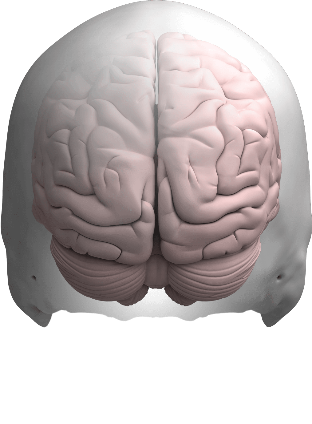
The optic nerve, also known as the second cranial nerve, is the nerve that transmits visual information from the retina to the brain. It is composed of approximately one million nerve fibers that carry signals related to vision, such as light intensity and color, from the eye's photoreceptor cells (rods and cones) to the visual cortex in the brain. The optic nerve is responsible for carrying this visual information so that it can be processed and interpreted by the brain, allowing us to see and perceive our surroundings. Damage to the optic nerve can result in vision loss or impairment.
Wallerian degeneration is a process that occurs following damage to the axons of neurons (nerve cells). After an axon is severed or traumatically injured, it undergoes a series of changes including fragmentation and removal of the distal segment of the axon, which is the part that is separated from the cell body. This process is named after Augustus Waller, who first described it in 1850.
The degenerative changes in the distal axon are characterized by the breakdown of the axonal cytoskeleton, the loss of myelin sheath (the fatty insulating material that surrounds and protects the axon), and the infiltration of macrophages to clear away the debris. These events lead to the degeneration of the distal axon segment, which is necessary for successful regeneration of the injured nerve.
Wallerian degeneration is a crucial process in the nervous system's response to injury, as it enables the regrowth of axons and the reestablishment of connections between neurons. However, if the regenerative capacity of the neuron is insufficient or the environment is not conducive to growth, functional recovery may be impaired, leading to long-term neurological deficits.
Optic nerve diseases refer to a group of conditions that affect the optic nerve, which transmits visual information from the eye to the brain. These diseases can cause various symptoms such as vision loss, decreased visual acuity, changes in color vision, and visual field defects. Examples of optic nerve diseases include optic neuritis (inflammation of the optic nerve), glaucoma (damage to the optic nerve due to high eye pressure), optic nerve damage from trauma or injury, ischemic optic neuropathy (lack of blood flow to the optic nerve), and optic nerve tumors. Treatment for optic nerve diseases varies depending on the specific condition and may include medications, surgery, or lifestyle changes.
Retinal Ganglion Cells (RGCs) are a type of neuron located in the innermost layer of the retina, the light-sensitive tissue at the back of the eye. These cells receive visual information from photoreceptors (rods and cones) via intermediate cells called bipolar cells. RGCs then send this visual information through their long axons to form the optic nerve, which transmits the signals to the brain for processing and interpretation as vision.
There are several types of RGCs, each with distinct morphological and functional characteristics. Some RGCs are specialized in detecting specific features of the visual scene, such as motion, contrast, color, or brightness. The diversity of RGCs allows for a rich and complex representation of the visual world in the brain.
Damage to RGCs can lead to various visual impairments, including loss of vision, reduced visual acuity, and altered visual fields. Conditions associated with RGC damage or degeneration include glaucoma, optic neuritis, ischemic optic neuropathy, and some inherited retinal diseases.
Nerve regeneration is the process of regrowth and restoration of functional nerve connections following damage or injury to the nervous system. This complex process involves various cellular and molecular events, such as the activation of support cells called glia, the sprouting of surviving nerve fibers (axons), and the reformation of neural circuits. The goal of nerve regeneration is to enable the restoration of normal sensory, motor, and autonomic functions impaired due to nerve damage or injury.
An axon is a long, slender extension of a neuron (a type of nerve cell) that conducts electrical impulses (nerve impulses) away from the cell body to target cells, such as other neurons or muscle cells. Axons can vary in length from a few micrometers to over a meter long and are typically surrounded by a myelin sheath, which helps to insulate and protect the axon and allows for faster transmission of nerve impulses.
Axons play a critical role in the functioning of the nervous system, as they provide the means by which neurons communicate with one another and with other cells in the body. Damage to axons can result in serious neurological problems, such as those seen in spinal cord injuries or neurodegenerative diseases like multiple sclerosis.
Animal disease models are specialized animals, typically rodents such as mice or rats, that have been genetically engineered or exposed to certain conditions to develop symptoms and physiological changes similar to those seen in human diseases. These models are used in medical research to study the pathophysiology of diseases, identify potential therapeutic targets, test drug efficacy and safety, and understand disease mechanisms.
The genetic modifications can include knockout or knock-in mutations, transgenic expression of specific genes, or RNA interference techniques. The animals may also be exposed to environmental factors such as chemicals, radiation, or infectious agents to induce the disease state.
Examples of animal disease models include:
1. Mouse models of cancer: Genetically engineered mice that develop various types of tumors, allowing researchers to study cancer initiation, progression, and metastasis.
2. Alzheimer's disease models: Transgenic mice expressing mutant human genes associated with Alzheimer's disease, which exhibit amyloid plaque formation and cognitive decline.
3. Diabetes models: Obese and diabetic mouse strains like the NOD (non-obese diabetic) or db/db mice, used to study the development of type 1 and type 2 diabetes, respectively.
4. Cardiovascular disease models: Atherosclerosis-prone mice, such as ApoE-deficient or LDLR-deficient mice, that develop plaque buildup in their arteries when fed a high-fat diet.
5. Inflammatory bowel disease models: Mice with genetic mutations affecting intestinal barrier function and immune response, such as IL-10 knockout or SAMP1/YitFc mice, which develop colitis.
Animal disease models are essential tools in preclinical research, but it is important to recognize their limitations. Differences between species can affect the translatability of results from animal studies to human patients. Therefore, researchers must carefully consider the choice of model and interpret findings cautiously when applying them to human diseases.
Retinal degeneration is a broad term that refers to the progressive loss of photoreceptor cells (rods and cones) in the retina, which are responsible for converting light into electrical signals that are sent to the brain. This process can lead to vision loss or blindness. There are many different types of retinal degeneration, including age-related macular degeneration, retinitis pigmentosa, and Stargardt's disease, among others. These conditions can have varying causes, such as genetic mutations, environmental factors, or a combination of both. Treatment options vary depending on the specific type and progression of the condition.
Macular degeneration, also known as age-related macular degeneration (AMD), is a medical condition that affects the central part of the retina, called the macula. The macula is responsible for sharp, detailed vision, which is necessary for activities such as reading, driving, and recognizing faces.
In AMD, there is a breakdown or deterioration of the macula, leading to gradual loss of central vision. There are two main types of AMD: dry (atrophic) and wet (exudative). Dry AMD is more common and progresses more slowly, while wet AMD is less common but can cause rapid and severe vision loss if left untreated.
The exact causes of AMD are not fully understood, but risk factors include age, smoking, family history, high blood pressure, obesity, and exposure to sunlight. While there is no cure for AMD, treatments such as vitamin supplements, laser therapy, and medication injections can help slow its progression and reduce the risk of vision loss.
The sciatic nerve is the largest and longest nerve in the human body, running from the lower back through the buttocks and down the legs to the feet. It is formed by the union of the ventral rami (branches) of the L4 to S3 spinal nerves. The sciatic nerve provides motor and sensory innervation to various muscles and skin areas in the lower limbs, including the hamstrings, calf muscles, and the sole of the foot. Sciatic nerve disorders or injuries can result in symptoms such as pain, numbness, tingling, or weakness in the lower back, hips, legs, and feet, known as sciatica.
Peripheral nerves are nerve fibers that transmit signals between the central nervous system (CNS, consisting of the brain and spinal cord) and the rest of the body. These nerves convey motor, sensory, and autonomic information, enabling us to move, feel, and respond to changes in our environment. They form a complex network that extends from the CNS to muscles, glands, skin, and internal organs, allowing for coordinated responses and functions throughout the body. Damage or injury to peripheral nerves can result in various neurological symptoms, such as numbness, weakness, or pain, depending on the type and severity of the damage.
Nerve fibers are specialized structures that constitute the long, slender processes (axons) of neurons (nerve cells). They are responsible for conducting electrical impulses, known as action potentials, away from the cell body and transmitting them to other neurons or effector organs such as muscles and glands. Nerve fibers are often surrounded by supportive cells called glial cells and are grouped together to form nerve bundles or nerves. These fibers can be myelinated (covered with a fatty insulating sheath called myelin) or unmyelinated, which influences the speed of impulse transmission.
Intervertebral disc degeneration is a physiological and biochemical process that occurs in the spinal discs, which are located between each vertebra in the spine. These discs act as shock absorbers and allow for movement and flexibility of the spine.
The degenerative process involves changes in the structure and composition of the disc, including loss of water content, decreased production of proteoglycans (which help to maintain the disc's elasticity), and disorganization of the collagen fibers that make up the disc's outer layer (annulus fibrosus). These changes can lead to a decrease in the disc's height and mobility, as well as the development of tears or cracks in the annulus fibrosus.
In advanced stages of degeneration, the disc may herniate or bulge outward, causing pressure on nearby nerves and potentially leading to pain, numbness, tingling, or weakness in the affected area. It's worth noting that while intervertebral disc degeneration is a normal part of aging, certain factors such as injury, smoking, obesity, and repetitive stress can accelerate the process.
A nerve crush injury is a type of peripheral nerve injury that occurs when there is excessive pressure or compression applied to a nerve, causing it to become damaged or dysfunctional. This can happen due to various reasons such as trauma from accidents, surgical errors, or prolonged pressure on the nerve from tight casts, clothing, or positions.
The compression disrupts the normal functioning of the nerve, leading to symptoms such as numbness, tingling, weakness, or pain in the affected area. In severe cases, a nerve crush injury can cause permanent damage to the nerve, leading to long-term disability or loss of function. Treatment for nerve crush injuries typically involves relieving the pressure on the nerve, providing supportive care, and in some cases, surgical intervention may be necessary to repair the damaged nerve.
The sural nerve is a purely sensory peripheral nerve in the lower leg and foot. It provides sensation to the outer ( lateral) aspect of the little toe and the adjacent side of the fourth toe, as well as a small portion of the skin on the back of the leg between the ankle and knee joints.
The sural nerve is formed by the union of branches from the tibial and common fibular nerves (branches of the sciatic nerve) in the lower leg. It runs down the calf, behind the lateral malleolus (the bony prominence on the outside of the ankle), and into the foot.
The sural nerve is often used as a donor nerve during nerve grafting procedures due to its consistent anatomy and relatively low risk for morbidity at the donor site.
Nerve endings, also known as terminal branches or sensory receptors, are the specialized structures present at the termination point of nerve fibers (axons) that transmit electrical signals to and from the central nervous system (CNS). They primarily function in detecting changes in the external environment or internal body conditions and converting them into electrical impulses.
There are several types of nerve endings, including:
1. Free Nerve Endings: These are unencapsulated nerve endings that respond to various stimuli like temperature, pain, and touch. They are widely distributed throughout the body, especially in the skin, mucous membranes, and visceral organs.
2. Encapsulated Nerve Endings: These are wrapped by specialized connective tissue sheaths, which can modify their sensitivity to specific stimuli. Examples include Pacinian corpuscles (responsible for detecting deep pressure and vibration), Meissner's corpuscles (for light touch), Ruffini endings (for stretch and pressure), and Merkel cells (for sustained touch).
3. Specialised Nerve Endings: These are nerve endings that respond to specific stimuli, such as auditory, visual, olfactory, gustatory, and vestibular information. They include hair cells in the inner ear, photoreceptors in the retina, taste buds in the tongue, and olfactory receptors in the nasal cavity.
Nerve endings play a crucial role in relaying sensory information to the CNS for processing and initiating appropriate responses, such as reflex actions or conscious perception of the environment.
A nerve block is a medical procedure in which an anesthetic or neurolytic agent is injected near a specific nerve or bundle of nerves to block the transmission of pain signals from that area to the brain. This technique can be used for both diagnostic and therapeutic purposes, such as identifying the source of pain, providing temporary or prolonged relief, or facilitating surgical procedures in the affected region.
The injection typically contains a local anesthetic like lidocaine or bupivacaine, which numbs the nerve, preventing it from transmitting pain signals. In some cases, steroids may also be added to reduce inflammation and provide longer-lasting relief. Depending on the type of nerve block and its intended use, the injection might be administered close to the spine (neuraxial blocks), at peripheral nerves (peripheral nerve blocks), or around the sympathetic nervous system (sympathetic nerve blocks).
While nerve blocks are generally safe, they can have side effects such as infection, bleeding, nerve damage, or in rare cases, systemic toxicity from the anesthetic agent. It is essential to consult with a qualified medical professional before undergoing this procedure to ensure proper evaluation, technique, and post-procedure care.
The facial nerve, also known as the seventh cranial nerve (CN VII), is a mixed nerve that carries both sensory and motor fibers. Its functions include controlling the muscles involved in facial expressions, taste sensation from the anterior two-thirds of the tongue, and secretomotor function to the lacrimal and salivary glands.
The facial nerve originates from the brainstem and exits the skull through the internal acoustic meatus. It then passes through the facial canal in the temporal bone before branching out to innervate various structures of the face. The main branches of the facial nerve include:
1. Temporal branch: Innervates the frontalis, corrugator supercilii, and orbicularis oculi muscles responsible for eyebrow movements and eyelid closure.
2. Zygomatic branch: Supplies the muscles that elevate the upper lip and wrinkle the nose.
3. Buccal branch: Innervates the muscles of the cheek and lips, allowing for facial expressions such as smiling and puckering.
4. Mandibular branch: Controls the muscles responsible for lower lip movement and depressing the angle of the mouth.
5. Cervical branch: Innervates the platysma muscle in the neck, which helps to depress the lower jaw and wrinkle the skin of the neck.
Damage to the facial nerve can result in various symptoms, such as facial weakness or paralysis, loss of taste sensation, and dry eyes or mouth due to impaired secretion.
Peripheral nerve injuries refer to damage or trauma to the peripheral nerves, which are the nerves outside the brain and spinal cord. These nerves transmit information between the central nervous system (CNS) and the rest of the body, including sensory, motor, and autonomic functions. Peripheral nerve injuries can result in various symptoms, depending on the type and severity of the injury, such as numbness, tingling, weakness, or paralysis in the affected area.
Peripheral nerve injuries are classified into three main categories based on the degree of damage:
1. Neuropraxia: This is the mildest form of nerve injury, where the nerve remains intact but its function is disrupted due to a local conduction block. The nerve fiber is damaged, but the supporting structures remain intact. Recovery usually occurs within 6-12 weeks without any residual deficits.
2. Axonotmesis: In this type of injury, there is damage to both the axons and the supporting structures (endoneurium, perineurium). The nerve fibers are disrupted, but the connective tissue sheaths remain intact. Recovery can take several months or even up to a year, and it may be incomplete, with some residual deficits possible.
3. Neurotmesis: This is the most severe form of nerve injury, where there is complete disruption of the nerve fibers and supporting structures (endoneurium, perineurium, epineurium). Recovery is unlikely without surgical intervention, which may involve nerve grafting or repair.
Peripheral nerve injuries can be caused by various factors, including trauma, compression, stretching, lacerations, or chemical exposure. Treatment options depend on the type and severity of the injury and may include conservative management, such as physical therapy and pain management, or surgical intervention for more severe cases.
The median nerve is one of the major nerves in the human body, providing sensation and motor function to parts of the arm and hand. It originates from the brachial plexus, a network of nerves that arise from the spinal cord in the neck. The median nerve travels down the arm, passing through the cubital tunnel at the elbow, and continues into the forearm and hand.
In the hand, the median nerve supplies sensation to the palm side of the thumb, index finger, middle finger, and half of the ring finger. It also provides motor function to some of the muscles that control finger movements, allowing for flexion of the fingers and opposition of the thumb.
Damage to the median nerve can result in a condition called carpal tunnel syndrome, which is characterized by numbness, tingling, and weakness in the hand and fingers.
The Tibial nerve is a major branch of the sciatic nerve that originates in the lower back and runs through the buttock and leg. It provides motor (nerve impulses that control muscle movement) and sensory (nerve impulses that convey information about touch, temperature, and pain) innervation to several muscles and skin regions in the lower limb.
More specifically, the Tibial nerve supplies the following structures:
1. Motor Innervation: The Tibial nerve provides motor innervation to the muscles in the back of the leg (posterior compartment), including the calf muscles (gastrocnemius and soleus) and the small muscles in the foot (intrinsic muscles). These muscles are responsible for plantarflexion (pointing the foot downward) and inversion (turning the foot inward) of the foot.
2. Sensory Innervation: The Tibial nerve provides sensory innervation to the skin on the sole of the foot, as well as the heel and some parts of the lower leg.
The Tibial nerve travels down the leg, passing behind the knee and through the calf, where it eventually joins with the common fibular (peroneal) nerve to form the tibial-fibular trunk. This trunk then divides into several smaller nerves that innervate the foot's intrinsic muscles and skin.
Damage or injury to the Tibial nerve can result in various symptoms, such as weakness or paralysis of the calf and foot muscles, numbness or tingling sensations in the sole of the foot, and difficulty walking or standing on tiptoes.
The Ulnar nerve is one of the major nerves in the forearm and hand, which provides motor function to the majority of the intrinsic muscles of the hand (except for those innervated by the median nerve) and sensory innervation to the little finger and half of the ring finger. It originates from the brachial plexus, passes through the cubital tunnel at the elbow, and continues down the forearm, where it runs close to the ulna bone. The ulnar nerve then passes through the Guyon's canal in the wrist before branching out to innervate the hand muscles and provide sensation to the skin on the little finger and half of the ring finger.
Wet macular degeneration, also known as neovascular or exudative age-related macular degeneration (AMD), is a medical condition that affects the central part of the retina called the macula. It's characterized by the growth of new blood vessels (neovascularization) from the choroid layer behind the retina into the macula, which is not typical in healthy eyes. These abnormal blood vessels are fragile and prone to leakage, leading to the accumulation of fluid or blood in the macula, causing distortion or loss of central vision.
The wet form of AMD can progress rapidly and often leads to more severe visual loss compared to the dry form. It's essential to diagnose and treat wet AMD promptly to preserve as much vision as possible. Common treatments include anti-vascular endothelial growth factor (VEGF) injections, photodynamic therapy, or thermal laser treatment, depending on the specific case and individual patient factors.
Frontotemporal lobar degeneration (FTLD) is a group of disorders characterized by the progressive degeneration of the frontal and temporal lobes of the brain. These areas of the brain are involved in decision-making, behavior, emotion, and language. FTLD can be divided into several subtypes based on the specific clinical features and the underlying protein abnormalities.
The three main subtypes of FTLD are:
1. Behavioral variant frontotemporal dementia (bvFTD): This subtype is characterized by changes in personality, behavior, and judgment. People with bvFTD may lose their social inhibitions, become impulsive, or develop compulsive behaviors. They may also have difficulty with emotional processing and empathy.
2. Primary progressive aphasia (PPA): This subtype is characterized by the gradual deterioration of language skills. People with PPA may have difficulty speaking, understanding spoken or written language, or both. There are three subtypes of PPA: nonfluent/agrammatic variant, semantic variant, and logopenic variant.
3. Motor neuron disease (MND) with FTLD: This subtype is characterized by the degeneration of motor neurons, which are the nerve cells responsible for controlling voluntary muscle movements. People with MND with FTLD may develop symptoms of amyotrophic lateral sclerosis (ALS), such as muscle weakness, stiffness, and twitching, as well as cognitive and behavioral changes associated with FTLD.
The underlying protein abnormalities in FTLD include:
1. Tau protein: In some forms of FTLD, the tau protein accumulates and forms clumps called tangles inside nerve cells. This is also seen in Alzheimer's disease.
2. TDP-43 protein: In other forms of FTLD, the TDP-43 protein accumulates and forms clumps inside nerve cells.
3. Fused in sarcoma (FUS) protein: In a small number of cases, the FUS protein accumulates and forms clumps inside nerve cells.
FTLD is typically a progressive disorder, meaning that symptoms worsen over time. There is currently no cure for FTLD, but there are treatments available to help manage symptoms and improve quality of life.
Spinal nerves are the bundles of nerve fibers that transmit signals between the spinal cord and the rest of the body. There are 31 pairs of spinal nerves in the human body, which can be divided into five regions: 8 cervical, 12 thoracic, 5 lumbar, 5 sacral, and 1 coccygeal. Each spinal nerve carries both sensory information (such as touch, temperature, and pain) from the periphery to the spinal cord, and motor information (such as muscle control) from the spinal cord to the muscles and other structures in the body. Spinal nerves also contain autonomic fibers that regulate involuntary functions such as heart rate, digestion, and blood pressure.
The femoral nerve is a major nerve in the thigh region of the human body. It originates from the lumbar plexus, specifically from the ventral rami (anterior divisions) of the second, third, and fourth lumbar nerves (L2-L4). The femoral nerve provides motor and sensory innervation to various muscles and areas in the lower limb.
Motor Innervation:
The femoral nerve is responsible for providing motor innervation to several muscles in the anterior compartment of the thigh, including:
1. Iliacus muscle
2. Psoas major muscle
3. Quadriceps femoris muscle (consisting of four heads: rectus femoris, vastus lateralis, vastus medialis, and vastus intermedius)
These muscles are involved in hip flexion, knee extension, and stabilization of the hip joint.
Sensory Innervation:
The sensory distribution of the femoral nerve includes:
1. Anterior and medial aspects of the thigh
2. Skin over the anterior aspect of the knee and lower leg (via the saphenous nerve, a branch of the femoral nerve)
The saphenous nerve provides sensation to the skin on the inner side of the leg and foot, as well as the medial malleolus (the bony bump on the inside of the ankle).
In summary, the femoral nerve is a crucial component of the lumbar plexus that controls motor functions in the anterior thigh muscles and provides sensory innervation to the anterior and medial aspects of the thigh and lower leg.
Nerve Growth Factors (NGFs) are a family of proteins that play an essential role in the growth, maintenance, and survival of certain neurons (nerve cells). They were first discovered by Rita Levi-Montalcini and Stanley Cohen in 1956. NGF is particularly crucial for the development and function of the peripheral nervous system, which connects the central nervous system to various organs and tissues throughout the body.
NGF supports the differentiation and survival of sympathetic and sensory neurons during embryonic development. In adults, NGF continues to regulate the maintenance and repair of these neurons, contributing to neuroplasticity – the brain's ability to adapt and change over time. Additionally, NGF has been implicated in pain transmission and modulation, as well as inflammatory responses.
Abnormal levels or dysfunctional NGF signaling have been associated with various medical conditions, including neurodegenerative diseases (e.g., Alzheimer's and Parkinson's), chronic pain disorders, and certain cancers (e.g., small cell lung cancer). Therefore, understanding the role of NGF in physiological and pathological processes may provide valuable insights into developing novel therapeutic strategies for these conditions.
Nerve Growth Factor (NGF) is a small secreted protein that is involved in the growth, maintenance, and survival of certain neurons (nerve cells). It was the first neurotrophin to be discovered and is essential for the development and function of the nervous system. NGF binds to specific receptors on the surface of nerve cells and helps to promote their differentiation, axonal growth, and synaptic plasticity. Additionally, NGF has been implicated in various physiological processes such as inflammation, immune response, and wound healing. Deficiencies or excesses of NGF have been linked to several neurological disorders, including Alzheimer's disease, Parkinson's disease, and pain conditions.
The trigeminal nerve, also known as the fifth cranial nerve or CNV, is a paired nerve that carries both sensory and motor information. It has three major branches: ophthalmic (V1), maxillary (V2), and mandibular (V3). The ophthalmic branch provides sensation to the forehead, eyes, and upper portion of the nose; the maxillary branch supplies sensation to the lower eyelid, cheek, nasal cavity, and upper lip; and the mandibular branch is responsible for sensation in the lower lip, chin, and parts of the oral cavity, as well as motor function to the muscles involved in chewing. The trigeminal nerve plays a crucial role in sensations of touch, pain, temperature, and pressure in the face and mouth, and it also contributes to biting, chewing, and swallowing functions.
Photoreceptor cells in vertebrates are specialized types of neurons located in the retina of the eye that are responsible for converting light stimuli into electrical signals. These cells are primarily responsible for the initial process of vision and have two main types: rods and cones.
Rods are more numerous and are responsible for low-light vision or scotopic vision, enabling us to see in dimly lit conditions. They do not contribute to color vision but provide information about the shape and movement of objects.
Cones, on the other hand, are less numerous and are responsible for color vision and high-acuity vision or photopic vision. There are three types of cones, each sensitive to different wavelengths of light: short (S), medium (M), and long (L) wavelengths, which correspond to blue, green, and red, respectively. The combination of signals from these three types of cones allows us to perceive a wide range of colors.
Both rods and cones contain photopigments that consist of a protein called opsin and a light-sensitive chromophore called retinal. When light hits the photopigment, it triggers a series of chemical reactions that ultimately lead to the generation of an electrical signal that is transmitted to the brain via the optic nerve. This process enables us to see and perceive our visual world.
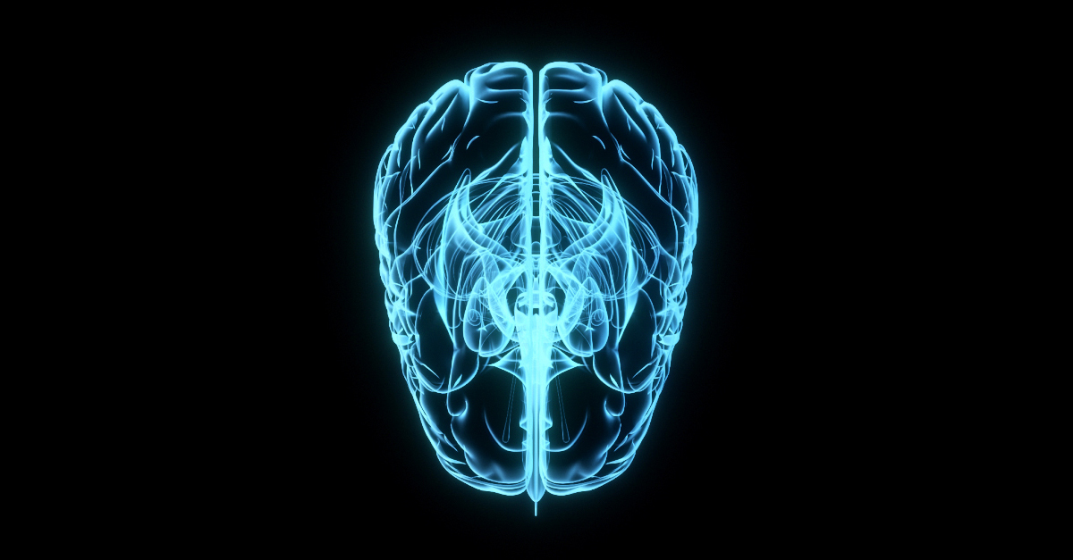
The retina is the innermost, light-sensitive layer of tissue in the eye of many vertebrates and some cephalopods. It receives light that has been focused by the cornea and lens, converts it into neural signals, and sends these to the brain via the optic nerve. The retina contains several types of photoreceptor cells including rods (which handle vision in low light) and cones (which are active in bright light and are capable of color vision).
In medical terms, any pathological changes or diseases affecting the retinal structure and function can lead to visual impairment or blindness. Examples include age-related macular degeneration, diabetic retinopathy, retinal detachment, and retinitis pigmentosa among others.
Spinal nerve roots are the initial parts of spinal nerves that emerge from the spinal cord through the intervertebral foramen, which are small openings between each vertebra in the spine. These nerve roots carry motor, sensory, and autonomic fibers to and from specific regions of the body. There are 31 pairs of spinal nerve roots in total, with 8 cervical, 12 thoracic, 5 lumbar, 5 sacral, and 1 coccygeal pair. Each root has a dorsal (posterior) and ventral (anterior) ramus that branch off to form the peripheral nervous system. Irritation or compression of these nerve roots can result in pain, numbness, weakness, or loss of reflexes in the affected area.
The phrenic nerve is a motor nerve that originates from the cervical spine (C3-C5) and descends through the neck to reach the diaphragm, which is the primary muscle used for breathing. The main function of the phrenic nerve is to innervate the diaphragm and control its contraction and relaxation, thereby enabling respiration.
Damage or injury to the phrenic nerve can result in paralysis of the diaphragm, leading to difficulty breathing and potentially causing respiratory failure. Certain medical conditions, such as neuromuscular disorders, spinal cord injuries, and tumors, can affect the phrenic nerve and impair its function.
An intervertebral disc is a fibrocartilaginous structure found between the vertebrae of the spinal column in humans and other animals. It functions as a shock absorber, distributes mechanical stress during weight-bearing activities, and allows for varying degrees of mobility between adjacent vertebrae.
The disc is composed of two parts: the annulus fibrosus, which forms the tough, outer layer; and the nucleus pulposus, which is a gel-like substance in the center that contains proteoglycans and water. The combination of these components provides the disc with its unique ability to distribute forces and allow for movement.
The intervertebral discs are essential for the normal functioning of the spine, providing stability, flexibility, and protection to the spinal cord and nerves. However, they can also be subject to degeneration and injury, which may result in conditions such as herniated discs or degenerative disc disease.
Cranial nerves are a set of twelve pairs of nerves that originate from the brainstem and skull, rather than the spinal cord. These nerves are responsible for transmitting sensory information (such as sight, smell, hearing, and taste) to the brain, as well as controlling various muscles in the head and neck (including those involved in chewing, swallowing, and eye movement). Each cranial nerve has a specific function and is named accordingly. For example, the optic nerve (cranial nerve II) transmits visual information from the eyes to the brain, while the vagus nerve (cranial nerve X) controls parasympathetic functions in the body such as heart rate and digestion.
The Radial nerve is a major peripheral nerve in the human body that originates from the brachial plexus, which is a network of nerves formed by the union of the ventral rami (anterior divisions) of spinal nerves C5-T1. The radial nerve provides motor function to extensor muscles of the upper limb and sensation to parts of the skin on the back of the arm, forearm, and hand.
More specifically, the radial nerve supplies motor innervation to:
* Extensor muscles of the shoulder (e.g., teres minor, infraspinatus)
* Rotator cuff muscles
* Elbow joint stabilizers (e.g., lateral head of the triceps)
* Extensors of the wrist, fingers, and thumb
The radial nerve also provides sensory innervation to:
* Posterior aspect of the upper arm (from the lower third of the humerus to the elbow)
* Lateral forearm (from the lateral epicondyle of the humerus to the wrist)
* Dorsum of the hand (skin over the radial side of the dorsum, including the first web space)
Damage or injury to the radial nerve may result in various symptoms, such as weakness or paralysis of the extensor muscles, numbness or tingling sensations in the affected areas, and difficulty with extension movements of the wrist, fingers, and thumb. Common causes of radial nerve injuries include fractures of the humerus bone, compression during sleep or prolonged pressure on the nerve (e.g., from crutches), and entrapment syndromes like radial tunnel syndrome.
Nerve compression syndromes refer to a group of conditions characterized by the pressure or irritation of a peripheral nerve, causing various symptoms such as pain, numbness, tingling, and weakness in the affected area. This compression can occur due to several reasons, including injury, repetitive motion, bone spurs, tumors, or swelling. Common examples of nerve compression syndromes include carpal tunnel syndrome, cubital tunnel syndrome, radial nerve compression, and ulnar nerve entrapment at the wrist or elbow. Treatment options may include physical therapy, splinting, medications, injections, or surgery, depending on the severity and underlying cause of the condition.
Electroretinography (ERG) is a medical test used to evaluate the functioning of the retina, which is the light-sensitive tissue located at the back of the eye. The test measures the electrical responses of the retina to light stimulation.
During the procedure, a special contact lens or electrode is placed on the surface of the eye to record the electrical activity generated by the retina's light-sensitive cells (rods and cones) and other cells in the retina. The test typically involves presenting different levels of flashes of light to the eye while the electrical responses are recorded.
The resulting ERG waveform provides information about the overall health and function of the retina, including the condition of the photoreceptors, the integrity of the inner retinal layers, and the health of the retinal ganglion cells. This test is often used to diagnose and monitor various retinal disorders, such as retinitis pigmentosa, macular degeneration, and diabetic retinopathy.
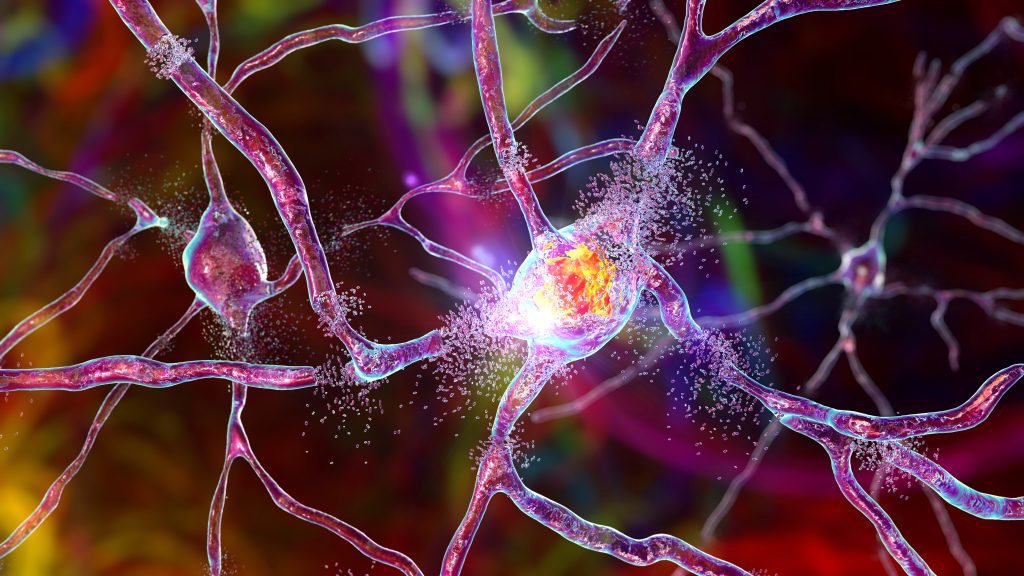
Nerve tissue, also known as neural tissue, is a type of specialized tissue that is responsible for the transmission of electrical signals and the processing of information in the body. It is a key component of the nervous system, which includes the brain, spinal cord, and peripheral nerves. Nerve tissue is composed of two main types of cells: neurons and glial cells.
Neurons are the primary functional units of nerve tissue. They are specialized cells that are capable of generating and transmitting electrical signals, known as action potentials. Neurons have a unique structure, with a cell body (also called the soma) that contains the nucleus and other organelles, and processes (dendrites and axons) that extend from the cell body and are used to receive and transmit signals.
Glial cells, also known as neuroglia or glia, are non-neuronal cells that provide support and protection for neurons. There are several different types of glial cells, including astrocytes, oligodendrocytes, microglia, and Schwann cells. These cells play a variety of roles in the nervous system, such as providing structural support, maintaining the proper environment for neurons, and helping to repair and regenerate nerve tissue after injury.
Nerve tissue is found throughout the body, but it is most highly concentrated in the brain and spinal cord, which make up the central nervous system (CNS). The peripheral nerves, which are the nerves that extend from the CNS to the rest of the body, also contain nerve tissue. Nerve tissue is responsible for transmitting sensory information from the body to the brain, controlling muscle movements, and regulating various bodily functions such as heart rate, digestion, and respiration.
The ophthalmic nerve, also known as the first cranial nerve or CN I, is a sensory nerve that primarily transmits information about vision, including light intensity and color, and sensation in the eye and surrounding areas. It is responsible for the sensory innervation of the upper eyelid, conjunctiva, cornea, iris, ciliary body, and nasal cavity. The ophthalmic nerve has three major branches: the lacrimal nerve, frontal nerve, and nasociliary nerve. Damage to this nerve can result in various visual disturbances and loss of sensation in the affected areas.
Retrograde degeneration is a medical term that refers to the process of degeneration or damage in neurons (nerve cells) that occurs backward from the site of injury or disease along the axon, which is the part of the neuron that transmits electrical signals to other neurons. This can lead to functional loss and may eventually result in the death of the neuron. Retrograde degeneration is often seen in neurodegenerative disorders such as Amyotrophic Lateral Sclerosis (ALS) and Alzheimer's disease, as well as in spinal cord injuries.
Optic nerve injuries refer to damages or trauma inflicted on the optic nerve, which is a crucial component of the visual system. The optic nerve transmits visual information from the retina to the brain, enabling us to see. Injuries to the optic nerve can result in various visual impairments, including partial or complete vision loss, decreased visual acuity, changes in color perception, and reduced field of view.
These injuries may occur due to several reasons, such as:
1. Direct trauma to the eye or head
2. Increased pressure inside the eye (glaucoma)
3. Optic neuritis, an inflammation of the optic nerve
4. Ischemia, or insufficient blood supply to the optic nerve
5. Compression from tumors or other space-occupying lesions
6. Intrinsic degenerative conditions affecting the optic nerve
7. Toxic exposure to certain chemicals or medications
Optic nerve injuries are diagnosed through a comprehensive eye examination, including visual acuity testing, slit-lamp examination, dilated fundus exam, and additional diagnostic tests like optical coherence tomography (OCT) and visual field testing. Treatment options vary depending on the cause and severity of the injury but may include medications, surgery, or vision rehabilitation.
The mandibular nerve is a branch of the trigeminal nerve (the fifth cranial nerve), which is responsible for sensations in the face and motor functions such as biting and chewing. The mandibular nerve provides both sensory and motor innervation to the lower third of the face, below the eye and nose down to the chin.
More specifically, it carries sensory information from the lower teeth, lower lip, and parts of the oral cavity, as well as the skin over the jaw and chin. It also provides motor innervation to the muscles of mastication (chewing), which include the masseter, temporalis, medial pterygoid, and lateral pterygoid muscles.
Damage to the mandibular nerve can result in numbness or loss of sensation in the lower face and mouth, as well as weakness or difficulty with chewing and biting.
The cochlear nerve, also known as the auditory nerve, is the sensory nerve that transmits sound signals from the inner ear to the brain. It consists of two parts: the outer spiral ganglion and the inner vestibular portion. The spiral ganglion contains the cell bodies of the bipolar neurons that receive input from hair cells in the cochlea, which is the snail-shaped organ in the inner ear responsible for hearing. These neurons then send their axons to form the cochlear nerve, which travels through the internal auditory meatus and synapses with neurons in the cochlear nuclei located in the brainstem.
Damage to the cochlear nerve can result in hearing loss or deafness, depending on the severity of the injury. Common causes of cochlear nerve damage include acoustic trauma, such as exposure to loud noises, viral infections, meningitis, and tumors affecting the nerve or surrounding structures. In some cases, cochlear nerve damage may be treated with hearing aids, cochlear implants, or other assistive devices to help restore or improve hearing function.
Myelinated nerve fibers are neuronal processes that are surrounded by a myelin sheath, a fatty insulating substance that is produced by Schwann cells in the peripheral nervous system and oligodendrocytes in the central nervous system. This myelin sheath helps to increase the speed of electrical impulse transmission, also known as action potentials, along the nerve fiber. The myelin sheath has gaps called nodes of Ranvier where the electrical impulses can jump from one node to the next, which also contributes to the rapid conduction of signals. Myelinated nerve fibers are typically found in the peripheral nerves and the optic nerve, but not in the central nervous system (CNS) tracts that are located within the brain and spinal cord.
Neural conduction is the process by which electrical signals, known as action potentials, are transmitted along the axon of a neuron (nerve cell) to transmit information between different parts of the nervous system. This electrical impulse is generated by the movement of ions across the neuronal membrane, and it propagates down the length of the axon until it reaches the synapse, where it can then stimulate the release of neurotransmitters to communicate with other neurons or target cells. The speed of neural conduction can vary depending on factors such as the diameter of the axon, the presence of myelin sheaths (which act as insulation and allow for faster conduction), and the temperature of the environment.
Nerve tissue proteins are specialized proteins found in the nervous system that provide structural and functional support to nerve cells, also known as neurons. These proteins include:
1. Neurofilaments: These are type IV intermediate filaments that provide structural support to neurons and help maintain their shape and size. They are composed of three subunits - NFL (light), NFM (medium), and NFH (heavy).
2. Neuronal Cytoskeletal Proteins: These include tubulins, actins, and spectrins that provide structural support to the neuronal cytoskeleton and help maintain its integrity.
3. Neurotransmitter Receptors: These are specialized proteins located on the postsynaptic membrane of neurons that bind neurotransmitters released by presynaptic neurons, triggering a response in the target cell.
4. Ion Channels: These are transmembrane proteins that regulate the flow of ions across the neuronal membrane and play a crucial role in generating and transmitting electrical signals in neurons.
5. Signaling Proteins: These include enzymes, receptors, and adaptor proteins that mediate intracellular signaling pathways involved in neuronal development, differentiation, survival, and death.
6. Adhesion Proteins: These are cell surface proteins that mediate cell-cell and cell-matrix interactions, playing a crucial role in the formation and maintenance of neural circuits.
7. Extracellular Matrix Proteins: These include proteoglycans, laminins, and collagens that provide structural support to nerve tissue and regulate neuronal migration, differentiation, and survival.
Choroidal neovascularization (CNV) is a medical term that refers to the growth of new, abnormal blood vessels in the choroid layer of the eye, which is located between the retina and the sclera. This condition typically occurs as a complication of age-related macular degeneration (AMD), although it can also be caused by other eye diseases or injuries.
In CNV, the new blood vessels that grow into the choroid layer are fragile and can leak fluid or blood, which can cause distortion or damage to the retina, leading to vision loss. Symptoms of CNV may include blurred or distorted vision, a blind spot in the center of the visual field, or changes in color perception.
Treatment for CNV typically involves medications that are designed to stop the growth of new blood vessels, such as anti-VEGF drugs, which target a protein called vascular endothelial growth factor (VEGF) that is involved in the development of new blood vessels. Laser surgery or photodynamic therapy may also be used in some cases to destroy the abnormal blood vessels and prevent further vision loss.
The glossopharyngeal nerve, also known as the ninth cranial nerve (IX), is a mixed nerve that carries both sensory and motor fibers. It originates from the medulla oblongata in the brainstem and has several functions:
1. Sensory function: The glossopharyngeal nerve provides general sensation to the posterior third of the tongue, the tonsils, the back of the throat (pharynx), and the middle ear. It also carries taste sensations from the back one-third of the tongue.
2. Special visceral afferent function: The nerve transmits information about the stretch of the carotid artery and blood pressure to the brainstem.
3. Motor function: The glossopharyngeal nerve innervates the stylopharyngeus muscle, which helps elevate the pharynx during swallowing. It also provides parasympathetic fibers to the parotid gland, stimulating saliva production.
4. Visceral afferent function: The glossopharyngeal nerve carries information about the condition of the internal organs in the thorax and abdomen to the brainstem.
Overall, the glossopharyngeal nerve plays a crucial role in swallowing, taste, saliva production, and monitoring blood pressure and heart rate.
Retinitis pigmentosa (RP) is a group of rare, genetic disorders that involve a breakdown and loss of cells in the retina - a light-sensitive tissue located at the back of the eye. The retina converts light into electrical signals which are then sent to the brain and interpreted as visual images.
In RP, the cells that detect light (rods and cones) degenerate more slowly than other cells in the retina, leading to a progressive loss of vision. Symptoms typically begin in childhood with night blindness (difficulty seeing in low light), followed by a gradual narrowing of the visual field (tunnel vision). Over time, this can lead to significant vision loss and even blindness.
The condition is usually inherited and there are several different genes that have been associated with RP. The diagnosis is typically made based on a combination of genetic testing, family history, and clinical examination. Currently, there is no cure for RP, but researchers are actively working to develop new treatments that may help slow or stop the progression of the disease.
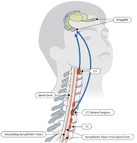
The splanchnic nerves are a set of nerve fibers that originate from the thoracic and lumbar regions of the spinal cord and innervate various internal organs. They are responsible for carrying both sensory information, such as pain and temperature, from the organs to the brain, and motor signals, which control the function of the organs, from the brain to the organs.
There are several splanchnic nerves, including the greater, lesser, and least splanchnic nerves, as well as the lumbar splanchnic nerves. These nerves primarily innervate the autonomic nervous system, which controls the involuntary functions of the body, such as heart rate, digestion, and respiration.
The greater splanchnic nerve arises from the fifth to the ninth thoracic ganglia and passes through the diaphragm to reach the abdomen. It innervates the stomach, esophagus, liver, pancreas, and adrenal glands.
The lesser splanchnic nerve arises from the tenth and eleventh thoracic ganglia and innervates the upper part of the small intestine, the pancreas, and the adrenal glands.
The least splanchnic nerve arises from the twelfth thoracic ganglion and innervates the lower part of the small intestine and the colon.
The lumbar splanchnic nerves arise from the first three or four lumbar ganglia and innervate the lower parts of the colon, the rectum, and the reproductive organs.
 Transneuronal degeneration
Nerve conduction velocity
Peripheral nerve injury classification
Chloroprocaine
Alfredo Sadun
Drusen
Nerve injury
Low-affinity nerve growth factor receptor
Sudomotor
Neuropathic pain
Hagemoser-Weinstein-Bresnick syndrome
Nerve compression syndrome
Cerebellar degeneration
Social degeneration
Jan Boeke
Friedreich's ataxia
HFE H63D gene mutation
Corneal-cerebellar syndrome
Omigapil
Facial nerve decompression
Rheobase
Critical illness polyneuropathy
Calpain
Coherent Raman scattering microscopy
Henryk Tomaszewski (poster artist)
Charcot-Marie-Tooth disease
Memory disorder
Augustus Volney Waller
Spinal fusion
Sharon Kujawa
Goldmann-Favre syndrome
William Ewart Gye
Wallerian degeneration
Theodor Axenfeld
 Frontiers | Commentary: Selective Fiber Degeneration in the Peripheral Nerve of a Patient With Severe Complex Regional Pain...
 Medical Acupuncture on Nerve Degeneration Meridian: ALS, MS and Parkinson's Disease on the Rise! « The Healthy Planet
 Autoimmunity Contributes To The Mechanism Of Optic Nerve Degeneration And Rgc Neuropathy Following Elevation Of Intraocular...
 Optic Nerve Degeneration & Glaucoma | BrightFocus Foundation
 Back anatomy: Diagram and overview
 Striatonigral Degeneration: Background, Etiology and Pathophysiology, Epidemiology
 Subacute Combined Degeneration - Brain, Spinal Cord, and Nerve Disorders - MSD Manual Consumer Version
Transneuronal degeneration - Wikipedia
Chiropractic Help for Disc Degeneration and Irritated Nerves | Johnson Chiropractic
 February 1968 - Volume 41 - Issue 2 : Plastic and Reconstructive Surgery
 Wallerian degeneration after cerebral infarction: evaluation with sequential MR imaging
 Chiropractic Help for Disc Degeneration and Irritated Nerves | Spine & Sports Rehab Center
 Optic nerve crush as a model of retinal ganglion cell degeneration - Cammalleri
- Annals of Eye Science
Optic nerve crush as a model of retinal ganglion cell degeneration - Cammalleri
- Annals of Eye Science
Corticobasal degeneration: etiopathological significance of the cytoskeletal alterations
 Brain and Nerves: MedlinePlus
 NERVE DEGENERATION IN POLIOMYELITIS: VI. CHANGES IN THE MOTOR NERVE ENDINGS | Archives of Neurology & Psychiatry | JAMA Network
 Remove Chronic Pain without Drugs within 20 minutes
 Professor Simon Parson | People | The University of Aberdeen
Ultrastructural observations on the progress of nerve degeneration and regeneration at the suture site following vagal...
Elimination of motor nerve terminals in neonatal mice expressing a gene for slow wallerian degeneration (C57Bl/Wlds)<...
Sural nerve biopsies in Guillain-Barre syndrome: axonal degeneration and macrophage-associated demyelination and absence of...
Macular Ganglion Cell Complex and Retinal Nerve Fiber Layer Comparison in Different Stages of Age-Related Macular Degeneration ...
 DYNC2H1 hypomorphic or retina-predominant variants cause nonsyndromic retinal degeneration | Genetics in Medicine
 Madness and Society from Bedlam to the Present - HI383 - Syllabus
 Pharmacotherapy Exam 1 September 27: Lecture 3 - Sympathomimetic Drugs, ETC - ProProfs Quiz
 Dr. Avery Katz, MD, Neurology Specialist - Clifton, NJ | Sharecare
 Free Histology Flashcards about Histology Spec Stain
Acute Flaccid Paralysis and West Nile Virus Infection - Volume 9, Number 7-July 2003 - Emerging Infectious Diseases journal -...
 Keep an Eye on Your Vision Health | CDC
Retinal5
- Therefore, the retinal ganglion cell plays a critical role as the output nerve cell of the eye that transmits visual information to the brain. (brightfocus.org)
- Primary outcome measures were the changes in ganglion cell complex (GCC) and retinal nerve fiber layer (RNFL). (entokey.com)
- and the retinal nerve fiber layer (RNFL), where the ganglion axons are located. (entokey.com)
- Inherited retinal diseases (IRDs) are a clinically and genetically heterogeneous group of disorders that often lead to photoreceptor degeneration. (nature.com)
- Biallelic variants in the cilia gene DYNC2H1 have been associated with two severe ciliopathies: Jeune asphyxiating thoracic dystrophy (JATD, MIM 613091) and short-rib polydactyly (SRP, MIM 613091) with only four documented cases of associated complex early retinal degeneration at ages 2 months, and 2, 5, and 11 years old. (nature.com)
Macular2
- To employ optical coherence tomography (OCT) to analyze the morphologic changes in the inner retina in different categories of age-related macular degeneration (AMD). (entokey.com)
- Age-related macular degeneration (AMD) is the leading cause of visual loss among the elderly in industrialized countries. (entokey.com)
Optic nerve18
- Photo showing how degeneration of the optic nerve narrows the field of vision, starting with loss of vision in the periphery, which then ultimately also causes loss of central vision. (brightfocus.org)
- Glaucoma is actually a group of diseases, but the common feature among all types of glaucoma is optic nerve degeneration. (brightfocus.org)
- Much like Alzheimer's disease is a neurodegenerative disease of the brain, glaucoma is considered a neurodegenerative disorder of the optic nerve. (brightfocus.org)
- What is the Optic Nerve? (brightfocus.org)
- The optic nerve is composed of approximately 1.5 million nerve fibers at the back of the eye that carry visual messages from the retina to the brain. (brightfocus.org)
- These cells reside in the retina, but their output "cables" or "fibers," called axons, extend from the head of the optic nerve at the back of the eye to the brain. (brightfocus.org)
- When your eyes are examined, the optic nerve is actually visualized by your eye doctor with the help of special lenses. (brightfocus.org)
- The axons of the optic nerve are bundled and insert in the back of the eye, and this "optic disc" is seen in the back of the eye along with blood vessels. (brightfocus.org)
- In optic nerve degeneration related to glaucoma, the optic disc displays changes that are characteristic of glaucoma, which your doctor may refer to as "cupping. (brightfocus.org)
- The normal optic nerve has a healthy appearing "rim" of tissue, which is assessed by both the contour of the rim as well as the color. (brightfocus.org)
- Cupping" is the result of changes in the optic nerve related to optic nerve degeneration, where there is a backward bowing of the central part of the disc. (brightfocus.org)
- An excellent visualization of optic nerve "cupping" starts at approximately 29 seconds into the video. (brightfocus.org)
- However, it is possible to have elevated eye pressure or "ocular hypertension" and show no changes of optic nerve degeneration (although one would need to be monitored over time for signs of degeneration), and conversely it is possible to have normal eye pressure and significant optic nerve degeneration. (brightfocus.org)
- What we do know is that there is likely mechanical damage at the site where the optic nerve inserts into the back of the eye. (brightfocus.org)
- This is because the axons of the optic nerve leave the eye by passing through a structure called the "lamina cribrosa," which is a mesh-like structure composed of pores or holes through which the axon bundles must cross. (brightfocus.org)
- Another contributing factor may be blood flow to the optic nerve. (brightfocus.org)
- Some believe that in cases of normal-tension glaucoma , impaired blood flow to the optic nerve may be playing a more important role in the degenerative process. (brightfocus.org)
- birth defects: coloboma of choroid or optic nerve disc, etc. (who.int)
Cell degeneration and death2
- The hyperexcitable corticomotoneurons drive anterior horn cells into metabolic deficit, resulting in cell degeneration and death. (wikipedia.org)
- Microglia , the brain's immune cells, then react to this by releasing chemicals that promote further inflammation and interfere with synaptic function, leading, possibly, to nerve cell degeneration and death. (bigthink.com)
Corticobasal degeneration1
- We have studied brain tissues from three patients with corticobasal degeneration (CBD) histologically, ultrastructurally and immunohistochemically. (nih.gov)
Wallerian Degeneration4
- The dynamic signal intensity changes at magnetic resonance (MR) imaging in active and chronic wallerian degeneration in the corticospinal tract were evaluated. (nih.gov)
- Forty-three patients with wallerian degeneration seen on MR images after cerebral infarction were studied. (nih.gov)
- We conclude (i) that the Wlds gene has no direct impact on the normal rate of postnatal synapse elimination, (ii) that Wallerian degeneration and synapse elimination must occur by distinct and different mechanisms, and (iii) that muscle fibres are able to sustain polyneuronal synaptic inputs even after motor axons have become disconnected from their cell bodies. (elsevierpure.com)
- Parson, SH , Mackintosh, CL & Ribchester, RR 1997, ' Elimination of motor nerve terminals in neonatal mice expressing a gene for slow wallerian degeneration (C57Bl/Wlds) ', European Journal of Neuroscience , vol. 9, no. 8, pp. 1586-1592. (elsevierpure.com)
Axonal2
- Although there are different causes, transneuronal degeneration generally results in the same effects (whether they be cellular, dendritic, or axonal) to varying degrees. (wikipedia.org)
- Focal infarction without distal axonal degeneration is demonstrated for the 1st month following onset of clinical symptoms. (nih.gov)
Neurons8
- Transneuronal degeneration is the death of neurons resulting from the disruption of input from or output to other nearby neurons. (wikipedia.org)
- The technical cause of transneuronal degeneration is the death of neurons resulting from the disruption of input from or output to other nearby neurons. (wikipedia.org)
- Ballooned neurons in the cerebral cortex and severe degeneration of the substantia nigra were observed in them all and weakly basophilic neurofibrillary tangles (NFTs) were distributed widely in the basal ganglia and brain stem. (nih.gov)
- GDNF, but not nerve growth factor (NGF), can prevent several axotomy-induced changes in these neurons, including the downregulation of IB4 binding, TMP activity, and somatostatin expression. (jneurosci.org)
- The progressive degeneration of the motor neurons in ALS eventually lead to their death. (caringbridge.org)
- Experimentally, a well-accepted model to mimic axotomy injury retrograde repercussion to spinal neurons is the neonatal peripheral nerve axotomy [ 13 - 18 ]. (hindawi.com)
- Sciatic nerve transection, early after birth, results in significant degeneration of spinal motoneurons as well as sensory neurons present in the dorsal root ganglia. (hindawi.com)
- The sensory root (nervus intermedius) consists of (1) central projections of neurons located in the geniculate ganglion (general somatic fibers that synapse in the spinal nucleus of the trigeminal nerve and special afferent fibers that synapse in the nucleus solitarius) and (2) axons of parasympathetic neurons from the superior salivatory (lacrimal) nucleus. (medscape.com)
Sciatic Nerve3
- Three to 4 days after sciatic nerve section, near-normal numbers of motor units were evident in isometric tension recordings of the soleus muscle, and intracellular recordings revealed many polyneuronally innervated fibres. (elsevierpure.com)
- ihown previously that trcatment With AT11 diminishei the durease in the pAosphorylating ability of dener%ated ~svctiun of the sciatic nerve) rat ma-icles. (cia.gov)
- Budaftst) 1957, 11/ 3-4 (277 -289) Graphs 5 Tables 2 Rat sciatic nerve was Injured over a length of 2 mm. by pinching with ophthalmo- logical forceps. (cia.gov)
Glaucoma1
- Here, we investigate the contribution of autoimmune responses to axon and RGC degeneration in glaucoma using mice deficient for T and B cell function and a microbead-induced model of ocular hypertension. (arvojournals.org)
Spinal cord3
- Subacute combined degeneration is progressive degeneration of the spinal cord due to vitamin B12 deficiency. (msdmanuals.com)
- In subacute combined degeneration, the myelin is damaged, causing sensory and motor nerve fibers from the spinal cord to degenerate. (msdmanuals.com)
- ALS) is a progressive neurodegenerative disease that affects nerve cells in the brain and the spinal cord. (caringbridge.org)
Fibers4
- examined radial, median, and ulnar nerve specimens from a CRPS-affected amputated limb and showed widespread (47-58%) selective degeneration in the larger myelinated Aα fibers (motor/proprioception) and in groups of small unmyelinated C fibers (Remak bundles), while the smaller Aδ (pain/temperature) fibers were spared. (frontiersin.org)
- Brain lesions create structural or transient deafferntation (the interruption or elimination of sensory nerve impulses by injuring or damaging sensory nerve fibers) because injury to the area causes a loss of excitatory input to other areas in the brain, causing them to be less responsive to stimuli. (wikipedia.org)
- Nerve fibers that control movement and sensation are damaged. (msdmanuals.com)
- In the earlier papers of this series by O'Leary, Heinbecker and Bishop, 1 the changes in nerve fibers resulting from the action of the poliomyelitis virus have been described from both the histologic and the physiologic point of view. (jamanetwork.com)
Myelin sheath1
- Vitamin B12 is necessary for the formation and maintenance of a fatty sheath (myelin sheath) that surrounds some nerve cells and that speeds transmission of nerve signals. (msdmanuals.com)
Muscles7
- The authors postulated that large Aα fiber degeneration could consequently affect the associated innervated muscles. (frontiersin.org)
- 3 - Nerve irritation in the spine can lead to weak muscles, muscle tremors, and urinary incontinence . (johnsonspinaldecompression.com)
- 2 The status of the motor nerve endings received passing comment by him to the effect that in the specimens of paralyzed muscles studied no endings were demonstrable. (jamanetwork.com)
- Mononeuronally and polyneuronally innervated fibres were also observed in silver-stained preparations of soleus and transversus abdominis muscles made 3-4 days after sciatic or intercostal nerve section. (elsevierpure.com)
- They affect nerves, muscles, and glands by inhibiting acetyl cholinesterase, an enzyme required for proper function of these tissues. (cdc.gov)
- Complete fatty degeneration of lumbar erector spinae muscles caused by a primary dysferlinopathy. (bvsalud.org)
- Mesenchymal concentrations that form the cephalic muscles are seen in association with their nerves, while the epibranchial placode disappears and the geniculate ganglion is identifiable. (medscape.com)
Regeneration3
- So see your Richmond chiropractor at Johnson Chiropractic for specialized spinal manipulation to stimulate circulation in the disc, relieve nerve irritation, and take proper nutrients to retard disc degeneration and stimulate regeneration. (johnsonspinaldecompression.com)
- Therefore, this study evaluated neuroprotection and regeneration after neonatal peripheral nerve coaptation with fibrin sealant. (hindawi.com)
- Overall, the present data suggest that acute repair of neonatal peripheral nerves with fibrin sealant results in neuroprotection and regeneration of motor and sensory axons. (hindawi.com)
Sciatica1
- Leg pain from a pinched nerve is commonly called sciatica . (spine-health.com)
Synapses1
- New evidence indicates that TTS-inducing exposures may cause an irreversible loss of neural synapses and degeneration of the cochlear nerve even after hearing thresholds completely recover. (cdc.gov)
Corticospinal1
- In many cases, the disease process begins with 1 of the 3 presentations predominating (ie, parkinsonism, autonomic failure, cerebellar signs) and then later converges to include a combination of all 3 plus additional degeneration of the corticospinal system, including tract and motor neuron degeneration, as well as cognitive deterioration. (medscape.com)
Substantia nigra1
- A study done shows that after excitotoxic injury to the striatum of adult rats, anterograde transneuronal degeneration occurs in the substantia nigra pars reticulata. (wikipedia.org)
Alzheimer's3
- Transneuronal degeneration is thought to be linked to a number of diseases, most notably Huntington's disease and Alzheimer's disease, and researchers recently have been performing experiments with monkeys and rats, monitoring lesions in different parts of the body to study more closely how exactly the process works. (wikipedia.org)
- The reorganization of cellular structures that results from transneuronal degeneration can be seen in Alzheimer's disease. (wikipedia.org)
- For example, retrovirus-type jumping genes are more abundant in postmortem human brain tissue obtained from Alzheimer's disease patients than in tissue from healthy controls, and these same jumping genes promote nerve cell death in fruit flies. (bigthink.com)
Retina1
- Loss of sensory receptors in the cochlea, vestibules, and retina result in ganglion degeneration and transneuronal degeneration. (wikipedia.org)
Ulnar nerve1
- For example if the numbness or tingling occurs in the ring or small fingers the problem is with the ulnar nerve, while the thumb, and index and middle fingers are governed by the median nerve - this is the affected nerve in carpal tunnel syndrome. (vitalitymagazine.com)
Progressive neurodegenerative1
- Striatonigral degeneration is a sporadic, progressive neurodegenerative disorder that represents one manifestation of multiple system atrophy (MSA). (medscape.com)
Disorders2
- Nerve Root Injury and Plexus Disorders (incl. (sharecare.com)
- Complete separation of the facial and acoustic nerves is apparent, and a discrete nervus intermedius develops, making this an important temporal reference point for gestational disorders that affect both systems. (medscape.com)
Brain6
- There are varying causes for transneuronal degeneration such as brain lesions, disconnection syndromes, respiratory chain deficient neuron interaction, and lobectomies. (wikipedia.org)
- Anterograde degeneration can occur at a late stage of brain injury and result in diaschisis. (wikipedia.org)
- Lesions to the brain cause pathological changes that can cause anterograde transneuronal degeneration and lead to system degeneration. (wikipedia.org)
- Delayed secondary transneuronal degeneration can also occur at a late stage after brain injury because after the period of latency, neuroplastic rearrangement follows deafferentation. (wikipedia.org)
- The brain, nerves of the eyes, and peripheral nerves are sometimes also damaged. (msdmanuals.com)
- Huntington's disease is an inherited condition that causes nerve cells in the brain to break down. (healthline.com)
Impulses2
- Anticonvulsants, such as gabapentin, can calm these nerve impulses. (medicalnewstoday.com)
- Strengthens nerve impulses and positively affects the functional state of the nervous tissue of the visual center. (kalinka-store.com)
Autonomic3
- Other manifestations of multiple system atrophy are Shy-Drager syndrome, in which autonomic failure predominates, and sporadic olivopontocerebellar degeneration, which is characterized primarily by cerebellar signs. (medscape.com)
- While symptoms of autonomic failure and cerebellar degeneration may be present in striatonigral degeneration, the predominant finding is parkinsonism. (medscape.com)
- The bladder and urethra are innervated by 3 sets of peripheral nerves arising from the autonomic nervous system (ANS) and somatic nervous system. (medscape.com)
Retrograde4
- This can be either anterograde or retrograde, indicating the direction of the degeneration relative to the original site of damage (see types). (wikipedia.org)
- citation needed] Transneuronal degeneration can be grouped into two general categories: anterograde and retrograde. (wikipedia.org)
- Retrograde transneuronal degeneration is degeneration caused by loss of trophic support from the target. (wikipedia.org)
- Anterograde and retrograde transneuronal degeneration is typically seen in humans around lesions in the limbic, visual, or dentate-rubro-olivary pathways. (wikipedia.org)
Electron1
- At 2 to 8 weeks after microbead injection, the degrees of RGC and axon degeneration were assessed quantitatively using electron microscopy and immunohistochemistry with a primary antibody against an RGC specific marker. (arvojournals.org)
Trigeminal2
- The ND meridian is influenced by many elements, manifest through the Trigeminal nerve by the Dental meridian, and through the Vagus nerve by the Gallbladder meridian. (thehealthyplanet.com)
- The chorda tympani nerve enters the mandibular arch and terminates just proximal to the submandibular ganglion, near a branch of the trigeminal nerve that will become the lingual nerve. (medscape.com)
Therapeutic1
- ATP) (ADJUYLPYROPHOSPHLTB, therapeutic use, nerve degen. (cia.gov)
Cranial2
- Note the interconnections of cranial nerve (CN) VII with CN V, CN IX, and CN X. (medscape.com)
- Upon leaving the motor nucleus, axons extend dorsally and medially, cranially and superficially, to bend around the abducens (sixth cranial nerve) nucleus. (medscape.com)
Vitamin B12 defici1
- Large doses of vitamin B12 taken by mouth can be used if vitamin B12 deficiency is mild and symptoms of nerve damage have not developed. (msdmanuals.com)
Occur1
- There are several different mechanisms by which transneuronal degeneration can occur. (wikipedia.org)
Affects1
- Subacute combined degeneration usually affects people older than 40. (msdmanuals.com)
Shingles2
- Although shingles usually resolve after a few weeks, complications can develop that affect the nerves, ears, eyes, or organ systems. (medicalnewstoday.com)
- The most common complication is PHN, which results in nerve pain in the area where shingles have occurred. (medicalnewstoday.com)
Injury1
- ATP was found to retard the degencration caused by injury of this kind, but not that following cutting of the nerve. (cia.gov)
Acute1
- Nerve degeneration is considered an unlikely outcome either from acute intoxication with any of the nerve agents or from long-term exposure to them. (cdc.gov)
Neonatal2
- This observation prompted us to re-examine the possible role of degeneration and intrinsic axon withdrawal during neonatal synapse elimination. (elsevierpure.com)
- To date, neonatal peripheral nerve repair following neurotmesis is largely limited due to technical drawbacks. (hindawi.com)
Degrees1
- The degrees of RGC and axon degeneration were quantified as described above. (arvojournals.org)
Motor3
- Degeneration of motor terminals after nerve section occurs much more slowly than normal in young adult mice of the C57Bl/Wlds strain. (elsevierpure.com)
- Decreased nerve conduction velocities as well as somatosensory, motor, brainstem, and visual evoked potentials all relate to peripheral neuropathy. (medscape.com)
- The motor nucleus of the facial nerve is located in the reticular formation of the caudal pons. (medscape.com)
Received cell1
- Adoptive transfer of CD4+ T cells isolated from high IOP C57BL/6J mice significantly facilitated the axon and RGC degeneration in Rag1-/- mice as compared to Rag1-/- mice received cell transfer from the normal IOP group. (arvojournals.org)
C57BL1
- To further investigate the roles of T cell-mediated responses in glaucomatous RGC and axon degeneration, CD4+ T cells isolated from C57BL/6J wild-type mice that were induced to develop high IOP were adoptively transferred to Rag1-/- mice. (arvojournals.org)
Damage2
- See your Richmond chiropractor for relief of nerve compression and for prevention of damage to the nerve being compressed. (johnsonspinaldecompression.com)
- Nerve damage can often business lead to pain or reduction in motion. (selfgrowth.com)
Disc14
- Those are a lot of pages about disc degeneration! (johnsonspinaldecompression.com)
- Bing shows 3,540,000 pages about disc degeneration. (johnsonspinaldecompression.com)
- There are extensive sources of information about disc degeneration if you want to study it more, but when you are hurting because of a degenerated disc, you just want to know how to get rid of the pain . (johnsonspinaldecompression.com)
- So why is disc degeneration such a hot topic? (johnsonspinaldecompression.com)
- 1 - As a disc degenerates it becomes more heavily supplied with nerves . (johnsonspinaldecompression.com)
- Why disc degeneration is such an issue for so many Richmond chiropractic patients? (johnsonspinaldecompression.com)
- When a disc degenerates, the nerves are irritated. (johnsonspinaldecompression.com)
- Your Richmond chiropractor uses specialized Cox Technic which may increase the size of the nerve opening, drop the pressure inside the intervertebral disc , and regain motion of the involved vertebra at the site of nerve compression. (johnsonspinaldecompression.com)
- That is an amazing amount of websites talking about disc degeneration! (docshay.com)
- What's a Herniated Disc, Pinched Nerve, Bulging Disc. (spine-health.com)
- The inner portion of the disc that extrudes can then irritate or compress nearby nerves, causing radiating pain. (spine-health.com)
- Any contact between the nerves and inflammatory proteins present in the disc's nucleus, or pressure from the disc itself on nerves in the lower spine can result in pain, numbness, tingling or weakness radiating down the leg. (spine-health.com)
- A herniated disc may compress the nearby nerve root, causing radicular pain. (spine-health.com)
- In many cases a herniated disc itself is not painful, but rather the material leaking out of the disc pinches, inflames, or irritates a nearby nerve, causing radicular pain . (spine-health.com)
Implications2
- The objective of this article is to outline the embryology of the facial nerve and its common clinical implications. (medscape.com)
- The reader is referred to Embryology and Anomalies of the Facial Nerve and Their Surgical Implications (Sataloff, 1991) for a comprehensive review of the development of the facial nerve and the associated development of the ear. (medscape.com)
Pain8
- The more nerves, the more pain we'll experience. (johnsonspinaldecompression.com)
- TENS Unit, When the nerves cannot get or send indicators properly, pain or difficulty to go will observe. (selfgrowth.com)
- This nerve pain may be intermittent or continuous. (medicalnewstoday.com)
- Newer TCAs can be effective for nerve pain and have fewer side effects than older TCAs. (medicalnewstoday.com)
- Misfiring nerve cells are a common link between seizures and nerve pain. (medicalnewstoday.com)
- Lidocaine is the topical anesthetic doctors most often prescribe to ease nerve pain. (medicalnewstoday.com)
- Radicular pain (also called nerve root pain), describes sharp, shooting pains that radiate to other parts of the body, such as from the low back down the leg or from the neck down the arm. (spine-health.com)
- But on Friday the pain remained -- she tweeted, "Pinched a nerve in my neck on a photoshoot and got adjusted this morning. (scienceblogs.com)