Myoblasts
Myoblasts, Skeletal
Muscle Development
Myoblasts, Cardiac
MyoD Protein
Muscle, Skeletal
Myogenin
Cell Differentiation
Cell Fusion
Muscle Fibers, Skeletal
Muscle Proteins
Cells, Cultured
Satellite Cells, Skeletal Muscle
Myogenic Regulatory Factors
Muscular Dystrophy, Facioscapulohumeral
Muscle Cells
PAX7 Transcription Factor
Desmin
Quail
Cell Transplantation
Myogenic Regulatory Factor 5
Chick Embryo
Myosin Heavy Chains
Transfection
Creatine Kinase
Mice, Inbred mdx
RNA, Messenger
Myostatin
Gene Expression Regulation
Gene Expression Regulation, Developmental
MEF2 Transcription Factors
Signal Transduction
Cell Division
Muscular Dystrophy, Duchenne
Transcription Factors
Dystrophin
Myosins
Molecular Sequence Data
Base Sequence
Muscular Dystrophies
Gene Expression
Coturnix
DNA-Binding Proteins
Fluorescent Antibody Technique
Actins
Transcription, Genetic
Immunohistochemistry
Bromodeoxyuridine
Myofibrils
Stem Cells
Caveolin 3
Muscular Dystrophy, Animal
Membrane Proteins
Microscopy, Fluorescence
Fibroblast Growth Factor 6
Insulin-Like Growth Factor I
Phosphorylation
Promoter Regions, Genetic
Cardiomyoplasty
Blotting, Western
Integrin alpha Chains
Paired Box Transcription Factors
Cell Cycle
Insulin-Like Growth Factor II
Drosophila Proteins
RNA Interference
Reverse Transcriptase Polymerase Chain Reaction
DNA
Cell Nucleus
Mice, Inbred C57BL
Mutation
Connectin
Rhabdomyosarcoma
Fibroblasts
Down-Regulation
Cell Survival
Cell Lineage
Myocardium
Embryo, Nonmammalian
Mesoderm
Cardiotoxins
Amino Acid Sequence
Nuclear Proteins
beta-Galactosidase
RNA, Small Interfering
Muscle specific fragile X related protein 1 isoforms are sequestered in the nucleus of undifferentiated myoblast. (1/1504)
BACKGROUND: The family of Fragile X Mental Retardation Proteins is composed of three members: Fragile Mental Retardation 1, Fragile X Related 1 and X Related 2 proteins. These proteins are associated with mRNPs within translating ribosomes and have the capacity to shuttle between the nucleus and the cytoplasm. Great attention has been given to FMRP due to its implication in human hereditary mental retardation while FXR1P and FXR2P have only recently been studied. RESULTS: Using antibodies directed against several epitopes of FXR1P, we have detected protein isoforms generated by small peptides pocket inserts. Four isoforms of MW 70, 74, 78, 80 kDa are widely distributed in mouse organs, while in striated muscles these isoforms are replaced by proteins of 82 and 84 kDa containing an extra pocket of 27 aa. Expression of these muscle isoforms is an early event during in vitro differentiation of myoblasts into myotubes and correlates with the activation of muscle-specific genes. However, while FXR1P82,84 are associated with cytoplasmic mRNPs in myotubes, they are sequestered in the nuclei of undifferentiated myoblasts. These observations suggest that, in addition to a cytoplasmic function yet to be defined, FXR1P82,84 may play a nuclear role in pre-mRNA metabolism. CONCLUSIONS: The pattern of subcellular partitioning of FXR1P isoforms during myogenesis is unique among the family of the FXR proteins. The model system described here should be considered as a powerful tool for ongoing attempts to unravel structure-function relationships of the different FMR family members since the potential role(s) of FXR1P as a compensatory factor in Fragile X syndrome is still elusive. (+info)Nuclear genetic control of mitochondrial translation in skeletal muscle revealed in patients with mitochondrial myopathy. (2/1504)
Oxidative phosphorylation deficiencies can be caused by mutations in either the nuclear genome or the mitochondrial genome (mtDNA); however, most pathogenic mutations reported in adults occur in mtDNA. Such mutations often impair mitochondrial translation, and are associated with a characteristic muscle pathology consisting of a mosaic pattern of normal fibres interspersed with fibres that show mitochondrial proliferation (ragged-red fibres) and little or no complex IV (COX) activity. We investigated two adult patients with a severe mitochondrial myopathy in whom all muscle fibres showed mitochondrial proliferation with barely detectable COX activity - a pattern never before reported. Biochemical studies of the respiratory chain in muscle showed decreased activities of complexes I and IV (5% of control) and complex II+III (41% of control). Immunoblot analysis of nuclear and mitochondrial subunits of complexes I, III and IV showed a greater than 90% decrease in the steady-state level of these subunits in mature muscle, but no change in nuclear-encoded subunits of complexes II and V. A generalized mitochondrial translation defect was identified in pulse-label experiments in myotubes, but not in myoblasts cultured from both patients. This defect moved with the nucleus in patient cybrid cells. Myoblasts from one patient transplanted into the muscle bed of SCID mice differentiated into mature human muscle fibres that displayed a defect similar to that seen in the patient muscle. These results suggest a defect in a developmentally regulated nuclear factor important for mitochondrial translation in skeletal muscle. (+info)Mouse PeP: a novel peroxisomal protein linked to myoblast differentiation and development. (3/1504)
The identification of several peroxisomal proteins in the past decade has deepened our understanding of the biology of peroxisomes and their involvement in human disorders. We report the cloning and expression pattern during the mouse development of a cDNA encoding a novel protein, named PeP, and show that its product is imported specifically to the peroxisome matrix in a variety of cell types. We also demonstrate that PeP is imported to the organelle through the PEX5 receptor pathway, which indicates that the C-terminal tripeptide SKI behaves as a type 1 peroxisomal targeting signal (PTS1). PeP expression is tightly regulated, as shown by Northern and in situ hybridization experiments. Thus during embryonic development in the mouse, PeP mRNA is detected almost exclusively in the skeletal muscle, whereas in adult mice, strong expression is also found in the heart and brain. In addition, PeP mRNA accumulation is induced after myoblast differentiation in vitro, when myotube formation is promoted. Sequence analysis reveals that PeP has no significant homology to any known protein, except for a short stretch of amino acids containing the fingerprint of the fibronectin type III superfamily, a domain present in proteins often related to molecular and cellular recognition and binding processes. Thus our data suggest a connection between the function of PeP and murine cell differentiation and development. (+info)The small heat shock protein alpha B-crystallin negatively regulates apoptosis during myogenic differentiation by inhibiting caspase-3 activation. (4/1504)
Myoblasts respond to growth factor deprivation either by differentiating into multinucleated myotubes or by undergoing apoptosis; hence, the acquisition of apoptosis resistance by myogenic precursors is essential for their development. Here we demonstrate that the expression of the small heat shock protein alpha B-crystallin is selectively induced in C2C12 myoblasts that are resistant to differentiation-induced apoptosis, and we show that this induction occurs at an early stage in their differentiation in vitro. In contrast, the expression of several known anti-apoptotic proteins (FLIP, XIAP, Bcl-x(L)) was not altered during myogenesis. We also demonstrate that ectopic expression of alpha B-crystallin, but not the closely related small heat shock protein Hsp27, renders C2C12 myoblasts resistant to differentiation-induced apoptosis. Furthermore, we show that the myopathy-causing R120G alpha B-crystallin mutant is partly impaired in its cytoprotective function, whereas a pseudophosphorylation alpha B-crystallin mutant that mimics stress-induced phosphorylation is completely devoid of anti-apoptotic activity. Finally, we demonstrate that alpha B-crystallin negatively regulates apoptosis during myogenesis by inhibiting the proteolytic activation of caspase-3, whereas the R120G and pseudophosphorylation mutants are defective in this function. Taken together, our findings indicate that alpha B-crystallin is a novel negative regulator of myogenic apoptosis that directly links the differentiation program to apoptosis resistance. (+info)Effect of insulin-like growth factor II on protecting myoblast cells against cisplatin-induced apoptosis through p70 S6 kinase pathway. (5/1504)
Insulin-like growth factor (IGF-II) is overexpressed in a variety of human tumors and has both mitogenic and antiapoptotic activity. Although the mechanisms of IGF-II-induced proliferation have been well studied, the mechanisms underlying its survival signaling have been less well characterized. In this report, we investigated the role of IGF-II on cisplatin-induced apoptosis. We found that IGF-II overexpression was associated with an increase in p70 ribosomal protein S6 kinase (p70 S6K). Cisplatin treatment of C2C12 mouse myoblasts led to cell death associated with an inhibition of p70 S6K activity. Endogenous or exogenous IGF-II addition to C2C12 cells caused protection to cisplatin-induced apoptosis. This protection was associated in both cases with an increase in p70 S6K basal activity as well as resistance to cisplatin-induced decreased activity. Blockade of p70 S6K activation by rapamycin abrogated the IGF-II-mediated protection of cells to cisplatin-induced apoptosis. Furthermore, treatment of IGF-II-overexpressing Rh30 and CTR rhabdomyosarcoma cells with rapamycin restored sensitivity to cisplatin-induced apoptosis. These data together suggest that IGF-II-associated protection to cisplatin-induced apoptosis is mediated through an activation of the p70 S6K pathway. Thus, inhibition of the p70 S6 pathway may enhance chemotherapy-induced apoptosis in the treatment of IGF-II-overexpressing tumors. (+info)Delivery of erythropoietin by encapsulated myoblasts in a genetic model of severe anemia. (6/1504)
BACKGROUND: Existing animal models of anemia inadequately reflect the hematocrit usually present in chronic renal failure (CRF) patients and do not permit long-term treatment studies. The transgenic mouse strain 134.3LC (Epo-TAg(H)) displays a severe chronic anemia resembling that observed clinically during CRF, while displaying an active, normal life span. This phenotype makes it a particularly interesting mouse model for testing erythropoietin (Epo)-based gene transfer strategies. METHODS: Ex vivo gene therapy was employed to administer mouse Epo to homozygous anemic Epo-TAg(H) mice. Encapsulated C(2)C(12) myoblasts genetically engineered to secrete 163 IU mouse Epo/10(6) cells/day were subcutaneously transplanted on the dorsal flank of the mice. Efficacy of delivered Epo was monitored by weekly measurements of animal hematocrit. RESULTS: Most treated homozygous Epo-TAg(H) mice displayed only a transient rise in hematocrit before eventually decreasing to levels as low as 3%. Administering the immunosuppressor anti-CD4+ monoclonal antibody (mAb) to homozygous Epo-TAg(H) mice, beginning at the time of implantation, permitted a rise in hematocrit that remained stable at elevated levels in cases of continued immunosuppression. CONCLUSIONS: Mice having the T antigen insertion in both Epo alleles appeared to develop an immune response to the natural mouse Epo delivered by encapsulated cells. By preventing this reaction using immunosuppression, we demonstrate that encapsulated myoblasts can deliver therapeutic doses of mouse Epo systemically and restore hemopoiesis in a genetic model of severe anemia. (+info)Two mammalian UNC-45 isoforms are related to distinct cytoskeletal and muscle-specific functions. (7/1504)
Previous studies have shown that the UNC-45 protein of C. elegans is required for normal thick filament assembly, binds Hsp90 and the myosin head, and shows molecular chaperone activity. We report here that mice and humans each have two genes that are located on different chromosomes, encode distinct UNC-45-like protein isoforms, and are expressed either in multiple tissues or only in cardiac and skeletal muscles. Their expression is regulated during muscle differentiation in vitro, with the striated muscle isoform mRNA appearing during myoblast fusion. Antisense experiments in C2C12 skeletal myogenic cells demonstrate that decreasing the general cell isoform mRNA reduces proliferation and fusion, while decreasing the striated muscle isoform mRNA affects fusion and sarcomere organization. These results suggest that the general cell UNC-45 isoform may have primarily cytoskeletal functions and that the striated muscle UNC-45 isoform may be restricted to roles in muscle-specific differentiation. (+info)The LIM-only protein FHL2 interacts with beta-catenin and promotes differentiation of mouse myoblasts. (8/1504)
FHL2 is a LIM-domain protein expressed in myoblasts but down-regulated in malignant rhabdomyosarcoma cells, suggesting an important role of FHL2 in muscle development. To investigate the importance of FHL2 during myoblast differentiation, we performed a yeast two-hybrid screen using a cDNA library derived from myoblasts induced for differentiation. We identified beta-catenin as a novel interaction partner of FHL2 and confirmed the specificity of association by direct in vitro binding tests and coimmunoprecipitation assays from cell lysates. Deletion analysis of both proteins revealed that the NH2-terminal part of beta-catenin is sufficient for binding in yeast, but addition of the first armadillo repeat is necessary for binding FHL2 in mammalian cells, whereas the presence of all four LIM domains of FHL2 is needed for the interaction. Expression of FHL2 counteracts beta-catenin-mediated activation of a TCF/LEF-dependent reporter gene in a dose-dependent and muscle cell-specific manner. After injection into Xenopus embryos, FHL2 inhibited the beta-catenin-induced axis duplication. C2C12 mouse myoblasts stably expressing FHL2 show increased myogenic differentiation reflected by accelerated myotube formation and expression of muscle-specific proteins. These data imply that FHL2 is a muscle-specific repressor of LEF/TCF target genes and promotes myogenic differentiation by interacting with beta-catenin. (+info)Myoblasts are types of cells that are responsible for the development and growth of muscle tissue in the body. They are undifferentiated cells, meaning they have not yet developed into their final form or function. Myoblasts fuse together to form myotubes, which then develop into muscle fibers, also known as myofibers. This process is called myogenesis and it plays a crucial role in the growth, repair, and maintenance of skeletal muscle tissue throughout an individual's life.
Myoblasts can be derived from various sources, including embryonic stem cells, induced pluripotent stem cells, or satellite cells, which are adult stem cells found within mature muscle tissue. Satellite cells are typically quiescent but can be activated in response to muscle damage or injury, proliferate and differentiate into myoblasts, and fuse together to repair and replace damaged muscle fibers.
Dysregulation of myogenesis and impaired myoblast function have been implicated in various muscle-related disorders, including muscular dystrophies, sarcopenia, and cachexia. Therefore, understanding the biology of myoblasts and their role in muscle development and regeneration is an important area of research with potential therapeutic implications for muscle-related diseases.
Skeletal myoblasts are the precursor cells responsible for the formation and repair of skeletal muscle fibers. They are also known as satellite cells, located in a quiescent state between the basal lamina and sarcolemma of mature muscle fibers. Upon muscle injury or damage, these cells become activated, proliferate, differentiate into myocytes, align with existing muscle fibers, and fuse to form new muscle fibers or repair damaged ones. This process is crucial for postnatal growth, maintenance, and regeneration of skeletal muscles.
Muscle development, also known as muscle hypertrophy, refers to the increase in size and mass of the muscles through a process called myofiber growth. This is primarily achieved through resistance or strength training exercises that cause micro-tears in the muscle fibers, leading to an inflammatory response and the release of hormones that promote muscle growth. As the muscles repair themselves, they become larger and stronger than before. Proper nutrition, including adequate protein intake, and rest are also essential components of muscle development.
It is important to note that while muscle development can lead to an increase in strength and muscular endurance, it does not necessarily result in improved athletic performance or overall fitness. A well-rounded exercise program that includes cardiovascular activity, flexibility training, and resistance exercises is recommended for optimal health and fitness outcomes.
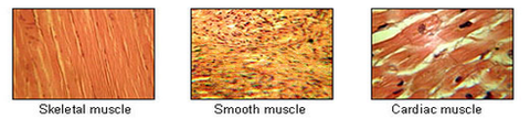
A muscle is a soft tissue in our body that contracts to produce force and motion. It is composed mainly of specialized cells called muscle fibers, which are bound together by connective tissue. There are three types of muscles: skeletal (voluntary), smooth (involuntary), and cardiac. Skeletal muscles attach to bones and help in movement, while smooth muscles are found within the walls of organs and blood vessels, helping with functions like digestion and circulation. Cardiac muscle is the specific type that makes up the heart, allowing it to pump blood throughout the body.
Myoblasts are immature cells that later develop into muscle cells (also known as myocytes). Cardiac myoblasts, therefore, are the immature cells that will specialize and develop into cardiac muscle cells. These cells play a crucial role in the growth, repair, and regeneration of heart muscles. In adults, however, the ability of these cells to regenerate damaged heart muscle tissue is limited. Recent research has focused on the potential use of cardiac myoblasts in cell-based therapies for various heart conditions, such as heart failure and myocardial infarction (heart attack).
MyoD protein is a member of the family of muscle regulatory factors (MRFs) that play crucial roles in the development and regulation of skeletal muscle. MyoD is a transcription factor, which means it binds to specific DNA sequences and helps control the transcription of nearby genes into messenger RNA (mRNA).
MyoD protein is encoded by the MYOD1 gene and is primarily expressed in skeletal muscle cells, where it functions as a master regulator of muscle differentiation. During myogenesis, MyoD is activated and initiates the expression of various genes involved in muscle-specific functions, such as contractile proteins and ion channels.
MyoD protein can also induce cell cycle arrest and promote the differentiation of non-muscle cells into muscle cells, a process known as transdifferentiation. This property has been explored in regenerative medicine for potential therapeutic applications.
In summary, MyoD protein is a key regulator of skeletal muscle development, differentiation, and maintenance, and it plays essential roles in the regulation of gene expression during myogenesis.
Skeletal muscle, also known as striated or voluntary muscle, is a type of muscle that is attached to bones by tendons or aponeuroses and functions to produce movements and support the posture of the body. It is composed of long, multinucleated fibers that are arranged in parallel bundles and are characterized by alternating light and dark bands, giving them a striped appearance under a microscope. Skeletal muscle is under voluntary control, meaning that it is consciously activated through signals from the nervous system. It is responsible for activities such as walking, running, jumping, and lifting objects.
Myogenin is defined as a protein that belongs to the family of myogenic regulatory factors (MRFs). These proteins play crucial roles in the development, growth, and repair of skeletal muscle cells. Myogenin is specifically involved in the differentiation and fusion of myoblasts to form multinucleated myotubes, which are essential for the formation of mature skeletal muscle fibers. It functions as a transcription factor that binds to specific DNA sequences, thereby regulating the expression of genes required for muscle cell differentiation. Myogenin also plays a role in maintaining muscle homeostasis and may contribute to muscle regeneration following injury or disease.
Cell differentiation is the process by which a less specialized cell, or stem cell, becomes a more specialized cell type with specific functions and structures. This process involves changes in gene expression, which are regulated by various intracellular signaling pathways and transcription factors. Differentiation results in the development of distinct cell types that make up tissues and organs in multicellular organisms. It is a crucial aspect of embryonic development, tissue repair, and maintenance of homeostasis in the body.
Cell fusion is the process by which two or more cells combine to form a single cell with a single nucleus, containing the genetic material from all of the original cells. This can occur naturally in certain biological processes, such as fertilization (when a sperm and egg cell fuse to form a zygote), muscle development (where multiple muscle precursor cells fuse together to create multinucleated muscle fibers), and during the formation of bone (where osteoclasts, the cells responsible for breaking down bone tissue, are multinucleated).
Cell fusion can also be induced artificially in laboratory settings through various methods, including chemical treatments, electrical stimulation, or viral vectors. Induced cell fusion is often used in research to create hybrid cells with unique properties, such as cybrid cells (cytoplasmic hybrids) and heterokaryons (nuclear hybrids). These hybrid cells can help scientists study various aspects of cell biology, genetics, and disease mechanisms.
In summary, cell fusion is the merging of two or more cells into one, resulting in a single cell with combined genetic material. This process occurs naturally during certain biological processes and can be induced artificially for research purposes.
Skeletal muscle fibers, also known as striated muscle fibers, are the type of muscle cells that make up skeletal muscles, which are responsible for voluntary movements of the body. These muscle fibers are long, cylindrical, and multinucleated, meaning they contain multiple nuclei. They are surrounded by a connective tissue layer called the endomysium, and many fibers are bundled together into fascicles, which are then surrounded by another layer of connective tissue called the perimysium.
Skeletal muscle fibers are composed of myofibrils, which are long, thread-like structures that run the length of the fiber. Myofibrils contain repeating units called sarcomeres, which are responsible for the striated appearance of skeletal muscle fibers. Sarcomeres are composed of thick and thin filaments, which slide past each other during muscle contraction to shorten the sarcomere and generate force.
Skeletal muscle fibers can be further classified into two main types based on their contractile properties: slow-twitch (type I) and fast-twitch (type II). Slow-twitch fibers have a high endurance capacity and are used for sustained, low-intensity activities such as maintaining posture. Fast-twitch fibers, on the other hand, have a higher contractile speed and force generation capacity but fatigue more quickly and are used for powerful, explosive movements.
A cell line is a culture of cells that are grown in a laboratory for use in research. These cells are usually taken from a single cell or group of cells, and they are able to divide and grow continuously in the lab. Cell lines can come from many different sources, including animals, plants, and humans. They are often used in scientific research to study cellular processes, disease mechanisms, and to test new drugs or treatments. Some common types of human cell lines include HeLa cells (which come from a cancer patient named Henrietta Lacks), HEK293 cells (which come from embryonic kidney cells), and HUVEC cells (which come from umbilical vein endothelial cells). It is important to note that cell lines are not the same as primary cells, which are cells that are taken directly from a living organism and have not been grown in the lab.
Muscle proteins are a type of protein that are found in muscle tissue and are responsible for providing structure, strength, and functionality to muscles. The two major types of muscle proteins are:
1. Contractile proteins: These include actin and myosin, which are responsible for the contraction and relaxation of muscles. They work together to cause muscle movement by sliding along each other and shortening the muscle fibers.
2. Structural proteins: These include titin, nebulin, and desmin, which provide structural support and stability to muscle fibers. Titin is the largest protein in the human body and acts as a molecular spring that helps maintain the integrity of the sarcomere (the basic unit of muscle contraction). Nebulin helps regulate the length of the sarcomere, while desmin forms a network of filaments that connects adjacent muscle fibers together.
Overall, muscle proteins play a critical role in maintaining muscle health and function, and their dysregulation can lead to various muscle-related disorders such as muscular dystrophy, myopathies, and sarcopenia.
"Cells, cultured" is a medical term that refers to cells that have been removed from an organism and grown in controlled laboratory conditions outside of the body. This process is called cell culture and it allows scientists to study cells in a more controlled and accessible environment than they would have inside the body. Cultured cells can be derived from a variety of sources, including tissues, organs, or fluids from humans, animals, or cell lines that have been previously established in the laboratory.
Cell culture involves several steps, including isolation of the cells from the tissue, purification and characterization of the cells, and maintenance of the cells in appropriate growth conditions. The cells are typically grown in specialized media that contain nutrients, growth factors, and other components necessary for their survival and proliferation. Cultured cells can be used for a variety of purposes, including basic research, drug development and testing, and production of biological products such as vaccines and gene therapies.
It is important to note that cultured cells may behave differently than they do in the body, and results obtained from cell culture studies may not always translate directly to human physiology or disease. Therefore, it is essential to validate findings from cell culture experiments using additional models and ultimately in clinical trials involving human subjects.
Satellite cells in skeletal muscle are undifferentiated stem cells that are crucial for postnatal growth, maintenance, and repair of skeletal muscle. They are located between the basal lamina and plasma membrane of myofibers. In response to muscle damage or injury, satellite cells become activated, proliferate, differentiate into myoblasts, fuse with existing muscle fibers, and contribute to muscle regeneration. Satellite cells also play a role in maintaining muscle homeostasis by fusing with mature muscle fibers to replace damaged proteins and organelles. They are essential for the adaptation of skeletal muscle to various stimuli such as exercise or mechanical load.
Myogenic regulatory factors (MRFs) are a group of transcription factors that play crucial roles in the development, growth, and maintenance of skeletal muscle cells. They are essential for the determination and differentiation of myoblasts into multinucleated myotubes and ultimately mature muscle fibers. The MRF family includes four key members: MyoD, Myf5, Mrf4 (also known as Myf6), and myogenin. These factors work together to regulate the expression of genes involved in various aspects of skeletal muscle formation and function.
1. MyoD: This MRF is a critical regulator of muscle cell differentiation and can induce non-muscle cells to adopt a muscle-like fate. It binds to specific DNA sequences, known as E-boxes, within the regulatory regions of target genes to activate or repress their transcription.
2. Myf5: Similar to MyoD, Myf5 is involved in the early determination and differentiation of myoblasts. However, it has a more restricted expression pattern during development compared to MyoD.
3. Mrf4 (Myf6): This MRF plays a role in both muscle cell differentiation and maintenance. It is expressed later than MyoD and Myf5 during development and helps regulate the terminal differentiation of myotubes into mature muscle fibers.
4. Myogenin: Among all MRFs, myogenin has the most specific function in muscle cell differentiation. It is required for the fusion of myoblasts to form multinucleated myotubes and is essential for the maturation and maintenance of skeletal muscle fibers.
In summary, Myogenic Regulatory Factors are a group of transcription factors that regulate skeletal muscle development, growth, and maintenance by controlling the expression of genes involved in various aspects of muscle cell differentiation and function.
Facioscapulohumeral Muscular Dystrophy (FSHD) is a genetic muscle disorder characterized by the progressive weakness and wasting (atrophy) of muscles in the face, shoulders, arms, and legs. It is caused by the abnormal expression of a gene called DUX4, which is normally only active during early embryonic development. In FSHD, this gene becomes reactivated in muscle cells, leading to their degeneration and death.
The symptoms of FSHD typically begin in late childhood or adolescence, although they can also appear in adulthood. The first noticeable sign is often difficulty raising the arms above the head or a weakened grip. Over time, the muscles of the face may become affected, leading to problems with smiling, swallowing, and speaking. The muscle weakness in FSHD tends to progress slowly, but it can vary widely from person to person. Some people with FSHD may require wheelchair assistance, while others may continue to walk with only minor limitations.
FSHD is inherited in an autosomal dominant manner, which means that a child has a 50% chance of inheriting the disease-causing gene from an affected parent. However, about 30% of cases are the result of new mutations and occur in people with no family history of the disorder. Currently, there is no cure for FSHD, but various treatments can help manage its symptoms and improve quality of life. These may include physical therapy, orthotics, assistive devices, and medications to treat pain or other complications.
Muscle cells, also known as muscle fibers, are specialized cells that have the ability to contract and generate force, allowing for movement of the body and various internal organ functions. There are three main types of muscle tissue: skeletal, cardiac, and smooth.
Skeletal muscle cells are voluntary striated muscles attached to bones, enabling body movements and posture. They are multinucleated, with numerous nuclei located at the periphery of the cell. These cells are often called muscle fibers and can be quite large, extending the entire length of the muscle.
Cardiac muscle cells form the contractile tissue of the heart. They are also striated but have a single nucleus per cell and are interconnected by specialized junctions called intercalated discs, which help coordinate contraction throughout the heart.
Smooth muscle cells are found in various internal organs such as the digestive, respiratory, and urinary tracts, blood vessels, and the reproductive system. They are involuntary, non-striated muscles that control the internal organ functions. Smooth muscle cells have a single nucleus per cell and can either be spindle-shaped or stellate (star-shaped).
In summary, muscle cells are specialized contractile cells responsible for movement and various internal organ functions in the human body. They can be categorized into three types: skeletal, cardiac, and smooth, based on their structure, location, and function.
Regeneration in a medical context refers to the process of renewal, restoration, and growth that replaces damaged or missing cells, tissues, organs, or even whole limbs in some organisms. This complex biological process involves various cellular and molecular mechanisms, such as cell proliferation, differentiation, and migration, which work together to restore the structural and functional integrity of the affected area.
In human medicine, regeneration has attracted significant interest due to its potential therapeutic applications in treating various conditions, including degenerative diseases, trauma, and congenital disorders. Researchers are actively studying the underlying mechanisms of regeneration in various model organisms to develop novel strategies for promoting tissue repair and regeneration in humans.
Examples of regeneration in human medicine include liver regeneration after partial hepatectomy, where the remaining liver lobes can grow back to their original size within weeks, and skin wound healing, where keratinocytes migrate and proliferate to close the wound and restore the epidermal layer. However, the regenerative capacity of humans is limited compared to some other organisms, such as planarians and axolotls, which can regenerate entire body parts or even their central nervous system.
PAX7 is a transcription factor that belongs to the PAX (paired box) family of proteins, which are characterized by the presence of a paired domain that binds to DNA. Specifically, PAX7 contains two DNA-binding domains: a paired domain and a homeodomain.
PAX7 is primarily expressed in satellite cells, which are muscle stem cells responsible for postnatal muscle growth, maintenance, and regeneration. PAX7 plays a critical role in the self-renewal and survival of satellite cells, and its expression is required for their activation and differentiation into mature muscle fibers.
As a transcription factor, PAX7 binds to specific DNA sequences in the regulatory regions of target genes and regulates their expression. This regulation can either activate or repress gene transcription, depending on the context and other factors that interact with PAX7.
PAX7 has been implicated in various muscle-related diseases, including muscular dystrophies and muscle wasting disorders. Its expression is often downregulated in these conditions, leading to a decrease in satellite cell function and muscle regeneration capacity. Therefore, understanding the role of PAX7 in muscle biology and disease has important implications for developing new therapies for muscle-related diseases.
Desmin is a type of intermediate filament protein that is primarily found in the cardiac and skeletal muscle cells, as well as in some types of smooth muscle cells. It is an important component of the cytoskeleton, which provides structural support to the cell and helps maintain its shape. Desmin plays a crucial role in maintaining the integrity of the sarcomere, which is the basic contractile unit of the muscle fiber. Mutations in the desmin gene can lead to various forms of muscular dystrophy and other inherited muscle disorders.
I believe there may be some confusion in your question. "Quail" is typically used to refer to a group of small birds that belong to the family Phasianidae and the subfamily Perdicinae. There is no established medical definition for "quail."
However, if you're referring to the verb "to quail," it means to shrink back, draw back, or cower, often due to fear or intimidation. In a medical context, this term could be used metaphorically to describe a patient's psychological response to a threatening situation, such as receiving a difficult diagnosis. But again, "quail" itself is not a medical term.
Cell transplantation is the process of transferring living cells from one part of the body to another or from one individual to another. In medicine, cell transplantation is often used as a treatment for various diseases and conditions, including neurodegenerative disorders, diabetes, and certain types of cancer. The goal of cell transplantation is to replace damaged or dysfunctional cells with healthy ones, thereby restoring normal function to the affected area.
In the context of medical research, cell transplantation may involve the use of stem cells, which are immature cells that have the ability to develop into many different types of specialized cells. Stem cell transplantation has shown promise in the treatment of a variety of conditions, including spinal cord injuries, stroke, and heart disease.
It is important to note that cell transplantation carries certain risks, such as immune rejection and infection. As such, it is typically reserved for cases where other treatments have failed or are unlikely to be effective.
Myogenic Regulatory Factor 5 (MRF5) is a protein that belongs to the family of muscle regulatory factors. It is a transcription factor, which means it regulates the expression of genes, specifically those involved in muscle development and differentiation. MRF5 plays a crucial role in skeletal muscle formation during embryonic development and also contributes to the maintenance and repair of skeletal muscles in adults.
MRF5 is expressed in developing muscle cells, where it helps to activate genes required for muscle-specific functions and represses genes associated with other cell fates. In addition, MRF5 has been implicated in the regulation of muscle stem cell (satellite cell) function and may play a role in the adaptation of skeletal muscles to various stimuli, such as exercise or injury.
Defects in MRF5 have been linked to certain muscular disorders, highlighting its importance in maintaining proper muscle function.
A chick embryo refers to the developing organism that arises from a fertilized chicken egg. It is often used as a model system in biological research, particularly during the stages of development when many of its organs and systems are forming and can be easily observed and manipulated. The study of chick embryos has contributed significantly to our understanding of various aspects of developmental biology, including gastrulation, neurulation, organogenesis, and pattern formation. Researchers may use various techniques to observe and manipulate the chick embryo, such as surgical alterations, cell labeling, and exposure to drugs or other agents.
Myosin Heavy Chains are the large, essential components of myosin molecules, which are responsible for the molecular motility in muscle cells. These heavy chains have a molecular weight of approximately 200 kDa and form the motor domain of myosin, which binds to actin filaments and hydrolyzes ATP to generate force and movement during muscle contraction. There are several different types of myosin heavy chains, each with specific roles in various tissues and cellular functions. In skeletal and cardiac muscles, for example, myosin heavy chains have distinct isoforms that contribute to the contractile properties of these tissues.
Transfection is a term used in molecular biology that refers to the process of deliberately introducing foreign genetic material (DNA, RNA or artificial gene constructs) into cells. This is typically done using chemical or physical methods, such as lipofection or electroporation. Transfection is widely used in research and medical settings for various purposes, including studying gene function, producing proteins, developing gene therapies, and creating genetically modified organisms. It's important to note that transfection is different from transduction, which is the process of introducing genetic material into cells using viruses as vectors.
Creatine kinase (CK) is a muscle enzyme that is normally present in small amounts in the blood. It is primarily found in tissues that require a lot of energy, such as the heart, brain, and skeletal muscles. When these tissues are damaged or injured, CK is released into the bloodstream, causing the levels to rise.
Creatine kinase exists in several forms, known as isoenzymes, which can be measured in the blood to help identify the location of tissue damage. The three main isoenzymes are:
1. CK-MM: Found primarily in skeletal muscle
2. CK-MB: Found primarily in heart muscle
3. CK-BB: Found primarily in the brain
Elevated levels of creatine kinase, particularly CK-MB, can indicate damage to the heart muscle, such as occurs with a heart attack. Similarly, elevated levels of CK-BB may suggest brain injury or disease. Overall, measuring creatine kinase levels is a useful diagnostic tool for assessing tissue damage and determining the severity of injuries or illnesses.
'Mice, Inbred mdx' is a genetic strain of laboratory mice that are widely used as a model to study Duchenne muscular dystrophy (DMD), a severe and progressive muscle-wasting disorder in humans. The 'mdx' designation refers to the specific genetic mutation present in these mice, which is a point mutation in the gene encoding for dystrophin, a crucial protein involved in maintaining the structural integrity of muscle fibers.
Inbred mdx mice carry a spontaneous mutation in exon 23 of the dystrophin gene, resulting in the production of a truncated and nonfunctional form of the protein. This leads to a phenotype that closely resembles DMD in humans, including muscle weakness, degeneration, and fibrosis. The inbred nature of these mice ensures consistent genetic backgrounds and disease manifestations, making them valuable tools for studying the pathophysiology of DMD and testing potential therapies.
It is important to note that while the inbred mdx mouse model has been instrumental in advancing our understanding of DMD, it does not fully recapitulate all aspects of the human disease. Therefore, findings from these mice should be carefully interpreted and validated in more complex models or human studies before translating them into clinical applications.
Messenger RNA (mRNA) is a type of RNA (ribonucleic acid) that carries genetic information copied from DNA in the form of a series of three-base code "words," each of which specifies a particular amino acid. This information is used by the cell's machinery to construct proteins, a process known as translation. After being transcribed from DNA, mRNA travels out of the nucleus to the ribosomes in the cytoplasm where protein synthesis occurs. Once the protein has been synthesized, the mRNA may be degraded and recycled. Post-transcriptional modifications can also occur to mRNA, such as alternative splicing and addition of a 5' cap and a poly(A) tail, which can affect its stability, localization, and translation efficiency.
Myostatin is a protein that is primarily known for its role in regulating muscle growth. It's also called "growth differentiation factor 8" or GDF-8. Produced by muscle cells, myostatin inhibits the process of muscle growth by preventing the transformation of stem cells into muscle fibers and promoting the breakdown of existing muscle proteins.
In essence, myostatin acts as a negative regulator of muscle mass, keeping it in check to prevent excessive growth. Mutations leading to reduced myostatin activity or expression have been associated with increased muscle mass and strength in both animals and humans, making it a potential target for therapeutic interventions in muscle-wasting conditions such as muscular dystrophy and age-related sarcopenia.
'Gene expression regulation' refers to the processes that control whether, when, and where a particular gene is expressed, meaning the production of a specific protein or functional RNA encoded by that gene. This complex mechanism can be influenced by various factors such as transcription factors, chromatin remodeling, DNA methylation, non-coding RNAs, and post-transcriptional modifications, among others. Proper regulation of gene expression is crucial for normal cellular function, development, and maintaining homeostasis in living organisms. Dysregulation of gene expression can lead to various diseases, including cancer and genetic disorders.
Developmental gene expression regulation refers to the processes that control the activation or repression of specific genes during embryonic and fetal development. These regulatory mechanisms ensure that genes are expressed at the right time, in the right cells, and at appropriate levels to guide proper growth, differentiation, and morphogenesis of an organism.
Developmental gene expression regulation is a complex and dynamic process involving various molecular players, such as transcription factors, chromatin modifiers, non-coding RNAs, and signaling molecules. These regulators can interact with cis-regulatory elements, like enhancers and promoters, to fine-tune the spatiotemporal patterns of gene expression during development.
Dysregulation of developmental gene expression can lead to various congenital disorders and developmental abnormalities. Therefore, understanding the principles and mechanisms governing developmental gene expression regulation is crucial for uncovering the etiology of developmental diseases and devising potential therapeutic strategies.
MEF2 (Myocyte Enhancer Factor-2) transcription factors are a family of proteins that regulate the transcription of genes, particularly in muscle cells. They play crucial roles in the development, growth, and maintenance of skeletal, cardiac, and smooth muscles. MEF2 transcription factors bind to specific DNA sequences, known as MEF2 response elements (MREs), in the promoter regions of target genes. This binding can either activate or repress gene transcription, depending on the context and interacting proteins. MEF2 transcription factors are involved in various cellular processes, such as muscle differentiation, metabolism, and stress responses. Dysregulation of MEF2 transcription factors has been implicated in several diseases, including muscular dystrophies, cardiovascular disorders, and neurodegenerative conditions.
Signal transduction is the process by which a cell converts an extracellular signal, such as a hormone or neurotransmitter, into an intracellular response. This involves a series of molecular events that transmit the signal from the cell surface to the interior of the cell, ultimately resulting in changes in gene expression, protein activity, or metabolism.
The process typically begins with the binding of the extracellular signal to a receptor located on the cell membrane. This binding event activates the receptor, which then triggers a cascade of intracellular signaling molecules, such as second messengers, protein kinases, and ion channels. These molecules amplify and propagate the signal, ultimately leading to the activation or inhibition of specific cellular responses.
Signal transduction pathways are highly regulated and can be modulated by various factors, including other signaling molecules, post-translational modifications, and feedback mechanisms. Dysregulation of these pathways has been implicated in a variety of diseases, including cancer, diabetes, and neurological disorders.
Cell division is the process by which a single eukaryotic cell (a cell with a true nucleus) divides into two identical daughter cells. This complex process involves several stages, including replication of DNA, separation of chromosomes, and division of the cytoplasm. There are two main types of cell division: mitosis and meiosis.
Mitosis is the type of cell division that results in two genetically identical daughter cells. It is a fundamental process for growth, development, and tissue repair in multicellular organisms. The stages of mitosis include prophase, prometaphase, metaphase, anaphase, and telophase, followed by cytokinesis, which divides the cytoplasm.
Meiosis, on the other hand, is a type of cell division that occurs in the gonads (ovaries and testes) during the production of gametes (sex cells). Meiosis results in four genetically unique daughter cells, each with half the number of chromosomes as the parent cell. This process is essential for sexual reproduction and genetic diversity. The stages of meiosis include meiosis I and meiosis II, which are further divided into prophase, prometaphase, metaphase, anaphase, and telophase.
In summary, cell division is the process by which a single cell divides into two daughter cells, either through mitosis or meiosis. This process is critical for growth, development, tissue repair, and sexual reproduction in multicellular organisms.
Duchenne Muscular Dystrophy (DMD) is a genetic disorder characterized by progressive muscle weakness and degeneration. It is caused by the absence of dystrophin, a protein that helps keep muscle cells intact. Without dystrophin, the muscle cells break down and are replaced with scar tissue, leading to loss of muscle function over time.
DMD primarily affects boys, as it is inherited in an X-linked recessive pattern, meaning that females who carry one affected X chromosome typically do not show symptoms but can pass the gene on to their offspring. Symptoms usually begin in early childhood and include difficulty with motor skills such as walking, running, and climbing stairs. Over time, the muscle weakness progresses and can lead to loss of ambulation, respiratory and cardiac complications, and ultimately, premature death.
Currently, there is no cure for DMD, but various treatments such as corticosteroids, physical therapy, and assisted ventilation can help manage symptoms and improve quality of life. Gene therapy approaches are also being investigated as potential treatments for this disorder.
Transcription factors are proteins that play a crucial role in regulating gene expression by controlling the transcription of DNA to messenger RNA (mRNA). They function by binding to specific DNA sequences, known as response elements, located in the promoter region or enhancer regions of target genes. This binding can either activate or repress the initiation of transcription, depending on the properties and interactions of the particular transcription factor. Transcription factors often act as part of a complex network of regulatory proteins that determine the precise spatiotemporal patterns of gene expression during development, differentiation, and homeostasis in an organism.
Dystrophin is a protein that provides structural stability to muscle fibers. It is an essential component of the dystrophin-glycoprotein complex, which helps maintain the integrity of the sarcolemma (the membrane surrounding muscle cells) during muscle contraction and relaxation. Dystrophin plays a crucial role in connecting the cytoskeleton of the muscle fiber to the extracellular matrix, allowing for force transmission and protecting the muscle cell from damage.
Mutations in the DMD gene, which encodes dystrophin, can lead to various forms of muscular dystrophy, including Duchenne muscular dystrophy (DMD) and Becker muscular dystrophy (BMD). In DMD, a severe form of the disease, genetic alterations typically result in little or no production of functional dystrophin, causing progressive muscle weakness, wasting, and degeneration. In BMD, a milder form of the disorder, partially functional dystrophin is produced, leading to less severe symptoms and later onset of the disease.
Myosins are a large family of motor proteins that play a crucial role in various cellular processes, including muscle contraction and intracellular transport. They consist of heavy chains, which contain the motor domain responsible for generating force and motion, and light chains, which regulate the activity of the myosin. Based on their structural and functional differences, myosins are classified into over 35 classes, with classes II, V, and VI being the most well-studied.
Class II myosins, also known as conventional myosins, are responsible for muscle contraction in skeletal, cardiac, and smooth muscles. They form filaments called thick filaments, which interact with actin filaments to generate force and movement during muscle contraction.
Class V myosins, also known as unconventional myosins, are involved in intracellular transport and organelle positioning. They have a long tail that can bind to various cargoes, such as vesicles, mitochondria, and nuclei, and a motor domain that moves along actin filaments to transport the cargoes to their destinations.
Class VI myosins are also unconventional myosins involved in intracellular transport and organelle positioning. They have two heads connected by a coiled-coil tail, which can bind to various cargoes. Class VI myosins move along actin filaments in a unique hand-over-hand motion, allowing them to transport their cargoes efficiently.
Overall, myosins are essential for many cellular functions and have been implicated in various diseases, including cardiovascular diseases, neurological disorders, and cancer.
Molecular sequence data refers to the specific arrangement of molecules, most commonly nucleotides in DNA or RNA, or amino acids in proteins, that make up a biological macromolecule. This data is generated through laboratory techniques such as sequencing, and provides information about the exact order of the constituent molecules. This data is crucial in various fields of biology, including genetics, evolution, and molecular biology, allowing for comparisons between different organisms, identification of genetic variations, and studies of gene function and regulation.
A base sequence in the context of molecular biology refers to the specific order of nucleotides in a DNA or RNA molecule. In DNA, these nucleotides are adenine (A), guanine (G), cytosine (C), and thymine (T). In RNA, uracil (U) takes the place of thymine. The base sequence contains genetic information that is transcribed into RNA and ultimately translated into proteins. It is the exact order of these bases that determines the genetic code and thus the function of the DNA or RNA molecule.
Cell proliferation is the process by which cells increase in number, typically through the process of cell division. In the context of biology and medicine, it refers to the reproduction of cells that makes up living tissue, allowing growth, maintenance, and repair. It involves several stages including the transition from a phase of quiescence (G0 phase) to an active phase (G1 phase), DNA replication in the S phase, and mitosis or M phase, where the cell divides into two daughter cells.
Abnormal or uncontrolled cell proliferation is a characteristic feature of many diseases, including cancer, where deregulated cell cycle control leads to excessive and unregulated growth of cells, forming tumors that can invade surrounding tissues and metastasize to distant sites in the body.
Muscular dystrophies are a group of genetic disorders that primarily affect skeletal muscles, causing progressive weakness and degeneration. They are characterized by the lack or deficiency of a protein called dystrophin, which is essential for maintaining the integrity of muscle fibers. The most common form is Duchenne muscular dystrophy (DMD), but there are many other types with varying symptoms and severity. Over time, muscle wasting and weakness can lead to disability and shortened lifespan, depending on the type and progression of the disease. Treatment typically focuses on managing symptoms, maintaining mobility, and supporting quality of life.
Gene expression is the process by which the information encoded in a gene is used to synthesize a functional gene product, such as a protein or RNA molecule. This process involves several steps: transcription, RNA processing, and translation. During transcription, the genetic information in DNA is copied into a complementary RNA molecule, known as messenger RNA (mRNA). The mRNA then undergoes RNA processing, which includes adding a cap and tail to the mRNA and splicing out non-coding regions called introns. The resulting mature mRNA is then translated into a protein on ribosomes in the cytoplasm through the process of translation.
The regulation of gene expression is a complex and highly controlled process that allows cells to respond to changes in their environment, such as growth factors, hormones, and stress signals. This regulation can occur at various stages of gene expression, including transcriptional activation or repression, RNA processing, mRNA stability, and translation. Dysregulation of gene expression has been implicated in many diseases, including cancer, genetic disorders, and neurological conditions.
"Coturnix" is a genus of birds that includes several species of quails. The most common species is the Common Quail (Coturnix coturnix), which is also known as the European Quail or the Eurasian Quail. This small ground-dwelling bird is found throughout Europe, Asia, and parts of Africa, and it is known for its distinctive call and its migratory habits. Other species in the genus Coturnix include the Rain Quail (Coturnix coromandelica), the Stubble Quail (Coturnix pectoralis), and the Harlequin Quail (Coturnix delegorguei). These birds are all similar in appearance and behavior, with small, round bodies, short wings, and strong legs that are adapted for running and scratching in leaf litter. They are also known for their cryptic coloration, which helps them blend in with their surroundings and avoid predators. Quails are popular game birds and are also kept as pets and for ornamental purposes in some parts of the world.
DNA-binding proteins are a type of protein that have the ability to bind to DNA (deoxyribonucleic acid), the genetic material of organisms. These proteins play crucial roles in various biological processes, such as regulation of gene expression, DNA replication, repair and recombination.
The binding of DNA-binding proteins to specific DNA sequences is mediated by non-covalent interactions, including electrostatic, hydrogen bonding, and van der Waals forces. The specificity of binding is determined by the recognition of particular nucleotide sequences or structural features of the DNA molecule.
DNA-binding proteins can be classified into several categories based on their structure and function, such as transcription factors, histones, and restriction enzymes. Transcription factors are a major class of DNA-binding proteins that regulate gene expression by binding to specific DNA sequences in the promoter region of genes and recruiting other proteins to modulate transcription. Histones are DNA-binding proteins that package DNA into nucleosomes, the basic unit of chromatin structure. Restriction enzymes are DNA-binding proteins that recognize and cleave specific DNA sequences, and are widely used in molecular biology research and biotechnology applications.
The Fluorescent Antibody Technique (FAT) is a type of immunofluorescence assay used in laboratory medicine and pathology for the detection and localization of specific antigens or antibodies in tissues, cells, or microorganisms. In this technique, a fluorescein-labeled antibody is used to selectively bind to the target antigen or antibody, forming an immune complex. When excited by light of a specific wavelength, the fluorescein label emits light at a longer wavelength, typically visualized as green fluorescence under a fluorescence microscope.
The FAT is widely used in diagnostic microbiology for the identification and characterization of various bacteria, viruses, fungi, and parasites. It has also been applied in the diagnosis of autoimmune diseases and certain cancers by detecting specific antibodies or antigens in patient samples. The main advantage of FAT is its high sensitivity and specificity, allowing for accurate detection and differentiation of various pathogens and disease markers. However, it requires specialized equipment and trained personnel to perform and interpret the results.
Actin is a type of protein that forms part of the contractile apparatus in muscle cells, and is also found in various other cell types. It is a globular protein that polymerizes to form long filaments, which are important for many cellular processes such as cell division, cell motility, and the maintenance of cell shape. In muscle cells, actin filaments interact with another type of protein called myosin to enable muscle contraction. Actins can be further divided into different subtypes, including alpha-actin, beta-actin, and gamma-actin, which have distinct functions and expression patterns in the body.
Genetic transcription is the process by which the information in a strand of DNA is used to create a complementary RNA molecule. This process is the first step in gene expression, where the genetic code in DNA is converted into a form that can be used to produce proteins or functional RNAs.
During transcription, an enzyme called RNA polymerase binds to the DNA template strand and reads the sequence of nucleotide bases. As it moves along the template, it adds complementary RNA nucleotides to the growing RNA chain, creating a single-stranded RNA molecule that is complementary to the DNA template strand. Once transcription is complete, the RNA molecule may undergo further processing before it can be translated into protein or perform its functional role in the cell.
Transcription can be either "constitutive" or "regulated." Constitutive transcription occurs at a relatively constant rate and produces essential proteins that are required for basic cellular functions. Regulated transcription, on the other hand, is subject to control by various intracellular and extracellular signals, allowing cells to respond to changing environmental conditions or developmental cues.
Immunohistochemistry (IHC) is a technique used in pathology and laboratory medicine to identify specific proteins or antigens in tissue sections. It combines the principles of immunology and histology to detect the presence and location of these target molecules within cells and tissues. This technique utilizes antibodies that are specific to the protein or antigen of interest, which are then tagged with a detection system such as a chromogen or fluorophore. The stained tissue sections can be examined under a microscope, allowing for the visualization and analysis of the distribution and expression patterns of the target molecule in the context of the tissue architecture. Immunohistochemistry is widely used in diagnostic pathology to help identify various diseases, including cancer, infectious diseases, and immune-mediated disorders.
Bromodeoxyuridine (BrdU) is a synthetic thymidine analog that can be incorporated into DNA during cell replication. It is often used in research and medical settings as a marker for cell proliferation or as a tool to investigate DNA synthesis and repair. When cells are labeled with BrdU and then examined using immunofluorescence or other detection techniques, the presence of BrdU can indicate which cells have recently divided or are actively synthesizing DNA.
In medical contexts, BrdU has been used in cancer research to study tumor growth and response to treatment. It has also been explored as a potential therapeutic agent for certain conditions, such as neurodegenerative diseases, where promoting cell proliferation and replacement of damaged cells may be beneficial. However, its use as a therapeutic agent is still experimental and requires further investigation.
Myofibrils are the basic contractile units of muscle fibers, composed of highly organized arrays of thick and thin filaments. They are responsible for generating the force necessary for muscle contraction. The thick filaments are primarily made up of the protein myosin, while the thin filaments are mainly composed of actin. Myofibrils are surrounded by a membrane called the sarcolemma and are organized into repeating sections called sarcomeres, which are the functional units of muscle contraction.
According to the National Institutes of Health (NIH), stem cells are "initial cells" or "precursor cells" that have the ability to differentiate into many different cell types in the body. They can also divide without limit to replenish other cells for as long as the person or animal is still alive.
There are two main types of stem cells: embryonic stem cells, which come from human embryos, and adult stem cells, which are found in various tissues throughout the body. Embryonic stem cells have the ability to differentiate into all cell types in the body, while adult stem cells have more limited differentiation potential.
Stem cells play an essential role in the development and repair of various tissues and organs in the body. They are currently being studied for their potential use in the treatment of a wide range of diseases and conditions, including cancer, diabetes, heart disease, and neurological disorders. However, more research is needed to fully understand the properties and capabilities of these cells before they can be used safely and effectively in clinical settings.
Caveolin 3 is a protein that is primarily expressed in muscle cells, including cardiac and skeletal muscles. It is the principal structural component of caveolae, which are small invaginations of the plasma membrane that function as specialized microdomains involved in various cellular processes such as signal transduction, cholesterol homeostasis, and endocytosis.
Caveolin 3 plays a critical role in muscle physiology by regulating several signaling pathways that are important for muscle function, including the nitric oxide signaling pathway. Mutations in the gene encoding caveolin 3 have been associated with various inherited muscle disorders, such as limb-girdle muscular dystrophy type 1C (LGMD1C), rippling muscle disease (RMD), and distal myopathies. These genetic conditions are characterized by progressive muscle weakness, wasting, and degeneration.
The term "extremities" in a medical context refers to the most distant parts of the body, including the hands and feet (both fingers and toes), as well as the arms and legs. These are the farthest parts from the torso and head. Medical professionals may examine a patient's extremities for various reasons, such as checking circulation, assessing nerve function, or looking for injuries or abnormalities.
I'm sorry for any confusion, but "Muscular Dystrophy, Animal" is not a standard medical term. Muscular Dystrophy is a group of genetic disorders that cause progressive weakness and loss of muscle mass. They are primarily human diseases and there are no known animal models of muscular dystrophy that directly correspond to any type of muscular dystrophy in humans.
However, scientists often use animals (like mice, dogs, and cats) as models for human diseases, including various types of muscular dystrophies. These animal models are used to study the disease process and to test potential treatments. For example, the mdx mouse is a well-known model of Duchenne Muscular Dystrophy (DMD), which is caused by a mutation in the dystrophin gene. This mouse lacks the muscle protein dystrophin, similar to humans with DMD, and shows many of the same symptoms, making it a valuable tool for research.
Membrane proteins are a type of protein that are embedded in the lipid bilayer of biological membranes, such as the plasma membrane of cells or the inner membrane of mitochondria. These proteins play crucial roles in various cellular processes, including:
1. Cell-cell recognition and signaling
2. Transport of molecules across the membrane (selective permeability)
3. Enzymatic reactions at the membrane surface
4. Energy transduction and conversion
5. Mechanosensation and signal transduction
Membrane proteins can be classified into two main categories: integral membrane proteins, which are permanently associated with the lipid bilayer, and peripheral membrane proteins, which are temporarily or loosely attached to the membrane surface. Integral membrane proteins can further be divided into three subcategories based on their topology:
1. Transmembrane proteins, which span the entire width of the lipid bilayer with one or more alpha-helices or beta-barrels.
2. Lipid-anchored proteins, which are covalently attached to lipids in the membrane via a glycosylphosphatidylinositol (GPI) anchor or other lipid modifications.
3. Monotopic proteins, which are partially embedded in the membrane and have one or more domains exposed to either side of the bilayer.
Membrane proteins are essential for maintaining cellular homeostasis and are targets for various therapeutic interventions, including drug development and gene therapy. However, their structural complexity and hydrophobicity make them challenging to study using traditional biochemical methods, requiring specialized techniques such as X-ray crystallography, nuclear magnetic resonance (NMR) spectroscopy, and single-particle cryo-electron microscopy (cryo-EM).
Fluorescence microscopy is a type of microscopy that uses fluorescent dyes or proteins to highlight and visualize specific components within a sample. In this technique, the sample is illuminated with high-energy light, typically ultraviolet (UV) or blue light, which excites the fluorescent molecules causing them to emit lower-energy, longer-wavelength light, usually visible light in the form of various colors. This emitted light is then collected by the microscope and detected to produce an image.
Fluorescence microscopy has several advantages over traditional brightfield microscopy, including the ability to visualize specific structures or molecules within a complex sample, increased sensitivity, and the potential for quantitative analysis. It is widely used in various fields of biology and medicine, such as cell biology, neuroscience, and pathology, to study the structure, function, and interactions of cells and proteins.
There are several types of fluorescence microscopy techniques, including widefield fluorescence microscopy, confocal microscopy, two-photon microscopy, and total internal reflection fluorescence (TIRF) microscopy, each with its own strengths and limitations. These techniques can provide valuable insights into the behavior of cells and proteins in health and disease.
Fibroblast Growth Factor 6 (FGF6), also known as Myostatin-induced gene-A (MIG-A), is a member of the fibroblast growth factor family, which plays crucial roles in various biological processes including cell survival, proliferation, migration, and differentiation. Specifically, FGF6 has been identified to be involved in skeletal muscle development and regeneration. It binds to heparin and specific fibroblast growth factor receptors (FGFRs) and activates intracellular signaling pathways that regulate the aforementioned processes. However, its precise functions and mechanisms are still under investigation in the scientific community.
Insulin-like growth factor I (IGF-I) is a hormone that plays a crucial role in growth and development. It is a small protein with structural and functional similarity to insulin, hence the name "insulin-like." IGF-I is primarily produced in the liver under the regulation of growth hormone (GH).
IGF-I binds to its specific receptor, the IGF-1 receptor, which is widely expressed throughout the body. This binding activates a signaling cascade that promotes cell proliferation, differentiation, and survival. In addition, IGF-I has anabolic effects on various tissues, including muscle, bone, and cartilage, contributing to their growth and maintenance.
IGF-I is essential for normal growth during childhood and adolescence, and it continues to play a role in maintaining tissue homeostasis throughout adulthood. Abnormal levels of IGF-I have been associated with various medical conditions, such as growth disorders, diabetes, and certain types of cancer.
Phosphorylation is the process of adding a phosphate group (a molecule consisting of one phosphorus atom and four oxygen atoms) to a protein or other organic molecule, which is usually done by enzymes called kinases. This post-translational modification can change the function, localization, or activity of the target molecule, playing a crucial role in various cellular processes such as signal transduction, metabolism, and regulation of gene expression. Phosphorylation is reversible, and the removal of the phosphate group is facilitated by enzymes called phosphatases.
Myositis is a medical term that refers to inflammation of the muscle tissue. This condition can cause various symptoms, including muscle weakness, pain, swelling, and stiffness. There are several types of myositis, such as polymyositis, dermatomyositis, and inclusion body myositis, which have different causes and characteristics.
Polymyositis is a type of myositis that affects multiple muscle groups, particularly those close to the trunk of the body. Dermatomyositis is characterized by muscle inflammation as well as a skin rash. Inclusion body myositis is a less common form of myositis that typically affects older adults and can cause both muscle weakness and wasting.
The causes of myositis vary depending on the type, but they can include autoimmune disorders, infections, medications, and other medical conditions. Treatment for myositis may involve medication to reduce inflammation, physical therapy to maintain muscle strength and flexibility, and lifestyle changes to manage symptoms and prevent complications.
In the field of medicine, "time factors" refer to the duration of symptoms or time elapsed since the onset of a medical condition, which can have significant implications for diagnosis and treatment. Understanding time factors is crucial in determining the progression of a disease, evaluating the effectiveness of treatments, and making critical decisions regarding patient care.
For example, in stroke management, "time is brain," meaning that rapid intervention within a specific time frame (usually within 4.5 hours) is essential to administering tissue plasminogen activator (tPA), a clot-busting drug that can minimize brain damage and improve patient outcomes. Similarly, in trauma care, the "golden hour" concept emphasizes the importance of providing definitive care within the first 60 minutes after injury to increase survival rates and reduce morbidity.
Time factors also play a role in monitoring the progression of chronic conditions like diabetes or heart disease, where regular follow-ups and assessments help determine appropriate treatment adjustments and prevent complications. In infectious diseases, time factors are crucial for initiating antibiotic therapy and identifying potential outbreaks to control their spread.
Overall, "time factors" encompass the significance of recognizing and acting promptly in various medical scenarios to optimize patient outcomes and provide effective care.
Promoter regions in genetics refer to specific DNA sequences located near the transcription start site of a gene. They serve as binding sites for RNA polymerase and various transcription factors that regulate the initiation of gene transcription. These regulatory elements help control the rate of transcription and, therefore, the level of gene expression. Promoter regions can be composed of different types of sequences, such as the TATA box and CAAT box, and their organization and composition can vary between different genes and species.
Cardiomyoplasty is a surgical procedure that involves wrapping skeletal muscle around the heart to help it pump more effectively. In this procedure, the surgeon typically uses the latissimus dorsi muscle, which is a large muscle in the back, and connects it to the heart with a special type of suture called a Dacron mesh.
The skeletal muscle used in cardiomyoplasty can be stimulated to contract using an electrical impulse, which helps to augment the contractions of the heart and improve its overall function. This procedure is typically reserved for patients with severe heart failure who are not candidates for other forms of treatment, such as a heart transplant.
While cardiomyoplasty has shown promise in some studies, it is still considered an experimental procedure and is not widely performed due to the risks involved and the limited number of patients who may benefit from it. Some of the potential complications of this procedure include infection, bleeding, muscle weakness, and damage to the heart or surrounding tissues.
Western blotting is a laboratory technique used in molecular biology to detect and quantify specific proteins in a mixture of many different proteins. This technique is commonly used to confirm the expression of a protein of interest, determine its size, and investigate its post-translational modifications. The name "Western" blotting distinguishes this technique from Southern blotting (for DNA) and Northern blotting (for RNA).
The Western blotting procedure involves several steps:
1. Protein extraction: The sample containing the proteins of interest is first extracted, often by breaking open cells or tissues and using a buffer to extract the proteins.
2. Separation of proteins by electrophoresis: The extracted proteins are then separated based on their size by loading them onto a polyacrylamide gel and running an electric current through the gel (a process called sodium dodecyl sulfate-polyacrylamide gel electrophoresis or SDS-PAGE). This separates the proteins according to their molecular weight, with smaller proteins migrating faster than larger ones.
3. Transfer of proteins to a membrane: After separation, the proteins are transferred from the gel onto a nitrocellulose or polyvinylidene fluoride (PVDF) membrane using an electric current in a process called blotting. This creates a replica of the protein pattern on the gel but now immobilized on the membrane for further analysis.
4. Blocking: The membrane is then blocked with a blocking agent, such as non-fat dry milk or bovine serum albumin (BSA), to prevent non-specific binding of antibodies in subsequent steps.
5. Primary antibody incubation: A primary antibody that specifically recognizes the protein of interest is added and allowed to bind to its target protein on the membrane. This step may be performed at room temperature or 4°C overnight, depending on the antibody's properties.
6. Washing: The membrane is washed with a buffer to remove unbound primary antibodies.
7. Secondary antibody incubation: A secondary antibody that recognizes the primary antibody (often coupled to an enzyme or fluorophore) is added and allowed to bind to the primary antibody. This step may involve using a horseradish peroxidase (HRP)-conjugated or alkaline phosphatase (AP)-conjugated secondary antibody, depending on the detection method used later.
8. Washing: The membrane is washed again to remove unbound secondary antibodies.
9. Detection: A detection reagent is added to visualize the protein of interest by detecting the signal generated from the enzyme-conjugated or fluorophore-conjugated secondary antibody. This can be done using chemiluminescent, colorimetric, or fluorescent methods.
10. Analysis: The resulting image is analyzed to determine the presence and quantity of the protein of interest in the sample.
Western blotting is a powerful technique for identifying and quantifying specific proteins within complex mixtures. It can be used to study protein expression, post-translational modifications, protein-protein interactions, and more. However, it requires careful optimization and validation to ensure accurate and reproducible results.
Integrins are a family of cell-surface receptors that play crucial roles in various biological processes, including cell adhesion, migration, and signaling. Integrin alpha chains are one of the two subunits that make up an integrin heterodimer, with the other subunit being an integrin beta chain.
Integrin alpha chains are transmembrane glycoproteins consisting of a large extracellular domain, a single transmembrane segment, and a short cytoplasmic tail. The extracellular domain contains several domains that mediate ligand binding, while the cytoplasmic tail interacts with various cytoskeletal proteins and signaling molecules to regulate intracellular signaling pathways.
There are 18 different integrin alpha chains known in humans, each of which can pair with one or more beta chains to form distinct integrin heterodimers. These heterodimers exhibit unique ligand specificities and functions, allowing them to mediate diverse cell-matrix and cell-cell interactions.
In summary, integrin alpha chains are essential subunits of integrin receptors that play crucial roles in regulating cell adhesion, migration, and signaling by mediating interactions between cells and their extracellular environment.
Paired box (PAX) transcription factors are a group of proteins that regulate gene expression during embryonic development and in some adult tissues. They are characterized by the presence of a paired box domain, a conserved DNA-binding motif that recognizes specific DNA sequences. PAX proteins play crucial roles in various developmental processes, such as the formation of the nervous system, eyes, and pancreas. Dysregulation of PAX genes has been implicated in several human diseases, including cancer.
The cell cycle is a series of events that take place in a cell leading to its division and duplication. It consists of four main phases: G1 phase, S phase, G2 phase, and M phase.
During the G1 phase, the cell grows in size and synthesizes mRNA and proteins in preparation for DNA replication. In the S phase, the cell's DNA is copied, resulting in two complete sets of chromosomes. During the G2 phase, the cell continues to grow and produces more proteins and organelles necessary for cell division.
The M phase is the final stage of the cell cycle and consists of mitosis (nuclear division) and cytokinesis (cytoplasmic division). Mitosis results in two genetically identical daughter nuclei, while cytokinesis divides the cytoplasm and creates two separate daughter cells.
The cell cycle is regulated by various checkpoints that ensure the proper completion of each phase before progressing to the next. These checkpoints help prevent errors in DNA replication and division, which can lead to mutations and cancer.
Insulin-like Growth Factor II (IGF-II) is a growth factor that is structurally and functionally similar to insulin. It is a single-chain polypeptide hormone, primarily produced by the liver under the regulation of growth hormone. IGF-II plays an essential role in fetal growth and development, and continues to have important functions in postnatal life, including promoting cell growth, proliferation, and differentiation in various tissues.
IGF-II binds to and activates the IGF-I receptor and the insulin receptor, leading to intracellular signaling cascades that regulate metabolic and mitogenic responses. Dysregulation of IGF-II expression and signaling has been implicated in several pathological conditions, such as cancer, growth disorders, and diabetes.
It is important to note that IGF-II should not be confused with Insulin-like Growth Factor I (IGF-I), which is another hormone with structural and functional similarities to insulin but has distinct roles in growth and development.
'Drosophila proteins' refer to the proteins that are expressed in the fruit fly, Drosophila melanogaster. This organism is a widely used model system in genetics, developmental biology, and molecular biology research. The study of Drosophila proteins has contributed significantly to our understanding of various biological processes, including gene regulation, cell signaling, development, and aging.
Some examples of well-studied Drosophila proteins include:
1. HSP70 (Heat Shock Protein 70): A chaperone protein involved in protein folding and protection from stress conditions.
2. TUBULIN: A structural protein that forms microtubules, important for cell division and intracellular transport.
3. ACTIN: A cytoskeletal protein involved in muscle contraction, cell motility, and maintenance of cell shape.
4. BETA-GALACTOSIDASE (LACZ): A reporter protein often used to monitor gene expression patterns in transgenic flies.
5. ENDOGLIN: A protein involved in the development of blood vessels during embryogenesis.
6. P53: A tumor suppressor protein that plays a crucial role in preventing cancer by regulating cell growth and division.
7. JUN-KINASE (JNK): A signaling protein involved in stress response, apoptosis, and developmental processes.
8. DECAPENTAPLEGIC (DPP): A member of the TGF-β (Transforming Growth Factor Beta) superfamily, playing essential roles in embryonic development and tissue homeostasis.
These proteins are often studied using various techniques such as biochemistry, genetics, molecular biology, and structural biology to understand their functions, interactions, and regulation within the cell.
RNA interference (RNAi) is a biological process in which RNA molecules inhibit the expression of specific genes. This process is mediated by small RNA molecules, including microRNAs (miRNAs) and small interfering RNAs (siRNAs), that bind to complementary sequences on messenger RNA (mRNA) molecules, leading to their degradation or translation inhibition.
RNAi plays a crucial role in regulating gene expression and defending against foreign genetic elements, such as viruses and transposons. It has also emerged as an important tool for studying gene function and developing therapeutic strategies for various diseases, including cancer and viral infections.
Reverse Transcriptase Polymerase Chain Reaction (RT-PCR) is a laboratory technique used in molecular biology to amplify and detect specific DNA sequences. This technique is particularly useful for the detection and quantification of RNA viruses, as well as for the analysis of gene expression.
The process involves two main steps: reverse transcription and polymerase chain reaction (PCR). In the first step, reverse transcriptase enzyme is used to convert RNA into complementary DNA (cDNA) by reading the template provided by the RNA molecule. This cDNA then serves as a template for the PCR amplification step.
In the second step, the PCR reaction uses two primers that flank the target DNA sequence and a thermostable polymerase enzyme to repeatedly copy the targeted cDNA sequence. The reaction mixture is heated and cooled in cycles, allowing the primers to anneal to the template, and the polymerase to extend the new strand. This results in exponential amplification of the target DNA sequence, making it possible to detect even small amounts of RNA or cDNA.
RT-PCR is a sensitive and specific technique that has many applications in medical research and diagnostics, including the detection of viruses such as HIV, hepatitis C virus, and SARS-CoV-2 (the virus that causes COVID-19). It can also be used to study gene expression, identify genetic mutations, and diagnose genetic disorders.
Deoxyribonucleic acid (DNA) is the genetic material present in the cells of organisms where it is responsible for the storage and transmission of hereditary information. DNA is a long molecule that consists of two strands coiled together to form a double helix. Each strand is made up of a series of four nucleotide bases - adenine (A), guanine (G), cytosine (C), and thymine (T) - that are linked together by phosphate and sugar groups. The sequence of these bases along the length of the molecule encodes genetic information, with A always pairing with T and C always pairing with G. This base-pairing allows for the replication and transcription of DNA, which are essential processes in the functioning and reproduction of all living organisms.
The cell nucleus is a membrane-bound organelle found in the eukaryotic cells (cells with a true nucleus). It contains most of the cell's genetic material, organized as DNA molecules in complex with proteins, RNA molecules, and histones to form chromosomes.
The primary function of the cell nucleus is to regulate and control the activities of the cell, including growth, metabolism, protein synthesis, and reproduction. It also plays a crucial role in the process of mitosis (cell division) by separating and protecting the genetic material during this process. The nuclear membrane, or nuclear envelope, surrounding the nucleus is composed of two lipid bilayers with numerous pores that allow for the selective transport of molecules between the nucleoplasm (nucleus interior) and the cytoplasm (cell exterior).
The cell nucleus is a vital structure in eukaryotic cells, and its dysfunction can lead to various diseases, including cancer and genetic disorders.
C57BL/6 (C57 Black 6) is an inbred strain of laboratory mouse that is widely used in biomedical research. The term "inbred" refers to a strain of animals where matings have been carried out between siblings or other closely related individuals for many generations, resulting in a population that is highly homozygous at most genetic loci.
The C57BL/6 strain was established in 1920 by crossing a female mouse from the dilute brown (DBA) strain with a male mouse from the black strain. The resulting offspring were then interbred for many generations to create the inbred C57BL/6 strain.
C57BL/6 mice are known for their robust health, longevity, and ease of handling, making them a popular choice for researchers. They have been used in a wide range of biomedical research areas, including studies of cancer, immunology, neuroscience, cardiovascular disease, and metabolism.
One of the most notable features of the C57BL/6 strain is its sensitivity to certain genetic modifications, such as the introduction of mutations that lead to obesity or impaired glucose tolerance. This has made it a valuable tool for studying the genetic basis of complex diseases and traits.
Overall, the C57BL/6 inbred mouse strain is an important model organism in biomedical research, providing a valuable resource for understanding the genetic and molecular mechanisms underlying human health and disease.
A mutation is a permanent change in the DNA sequence of an organism's genome. Mutations can occur spontaneously or be caused by environmental factors such as exposure to radiation, chemicals, or viruses. They may have various effects on the organism, ranging from benign to harmful, depending on where they occur and whether they alter the function of essential proteins. In some cases, mutations can increase an individual's susceptibility to certain diseases or disorders, while in others, they may confer a survival advantage. Mutations are the driving force behind evolution, as they introduce new genetic variability into populations, which can then be acted upon by natural selection.
Connectin is also known as titin, which is a giant protein that plays a crucial role in the elasticity and stiffness of muscle fibers. It is the largest protein in humans, spanning half the length of a muscle cell's sarcomere, the basic unit of muscle contraction. Connectin/titin has several domains with different functions, including binding to other proteins, regulating muscle contraction, and signaling within the muscle cell. Mutations in the connectin/titin gene have been associated with various forms of muscular dystrophy and cardiomyopathy.
Muscular diseases, also known as myopathies, refer to a group of conditions that affect the functionality and health of muscle tissue. These diseases can be inherited or acquired and may result from inflammation, infection, injury, or degenerative processes. They can cause symptoms such as weakness, stiffness, cramping, spasms, wasting, and loss of muscle function.
Examples of muscular diseases include:
1. Duchenne Muscular Dystrophy (DMD): A genetic disorder that results in progressive muscle weakness and degeneration due to a lack of dystrophin protein.
2. Myasthenia Gravis: An autoimmune disease that causes muscle weakness and fatigue, typically affecting the eyes and face, throat, and limbs.
3. Inclusion Body Myositis (IBM): A progressive muscle disorder characterized by muscle inflammation and wasting, typically affecting older adults.
4. Polymyositis: An inflammatory myopathy that causes muscle weakness and inflammation throughout the body.
5. Metabolic Myopathies: A group of inherited disorders that affect muscle metabolism, leading to exercise intolerance, muscle weakness, and other symptoms.
6. Muscular Dystonias: Involuntary muscle contractions and spasms that can cause abnormal postures or movements.
It is important to note that muscular diseases can have a significant impact on an individual's quality of life, mobility, and overall health. Proper diagnosis and treatment are crucial for managing symptoms and improving outcomes.
Rhabdomyosarcoma is a type of cancer that develops in the body's soft tissues, specifically in the muscle cells. It is a rare and aggressive form of sarcoma, which is a broader category of cancers that affect the connective tissues such as muscles, tendons, cartilages, bones, blood vessels, and fatty tissues.
Rhabdomyosarcomas can occur in various parts of the body, including the head, neck, arms, legs, trunk, and genitourinary system. They are more common in children than adults, with most cases diagnosed before the age of 18. The exact cause of rhabdomyosarcoma is not known, but genetic factors and exposure to radiation or certain chemicals may increase the risk.
There are several subtypes of rhabdomyosarcoma, including embryonal, alveolar, pleomorphic, and spindle cell/sclerosing. The type and stage of the cancer determine the treatment options, which may include surgery, radiation therapy, chemotherapy, or a combination of these approaches. Early diagnosis and prompt treatment are crucial for improving the prognosis and long-term survival rates.
Fibroblasts are specialized cells that play a critical role in the body's immune response and wound healing process. They are responsible for producing and maintaining the extracellular matrix (ECM), which is the non-cellular component present within all tissues and organs, providing structural support and biochemical signals for surrounding cells.
Fibroblasts produce various ECM proteins such as collagens, elastin, fibronectin, and laminins, forming a complex network of fibers that give tissues their strength and flexibility. They also help in the regulation of tissue homeostasis by controlling the turnover of ECM components through the process of remodeling.
In response to injury or infection, fibroblasts become activated and start to proliferate rapidly, migrating towards the site of damage. Here, they participate in the inflammatory response, releasing cytokines and chemokines that attract immune cells to the area. Additionally, they deposit new ECM components to help repair the damaged tissue and restore its functionality.
Dysregulation of fibroblast activity has been implicated in several pathological conditions, including fibrosis (excessive scarring), cancer (where they can contribute to tumor growth and progression), and autoimmune diseases (such as rheumatoid arthritis).
Down-regulation is a process that occurs in response to various stimuli, where the number or sensitivity of cell surface receptors or the expression of specific genes is decreased. This process helps maintain homeostasis within cells and tissues by reducing the ability of cells to respond to certain signals or molecules.
In the context of cell surface receptors, down-regulation can occur through several mechanisms:
1. Receptor internalization: After binding to their ligands, receptors can be internalized into the cell through endocytosis. Once inside the cell, these receptors may be degraded or recycled back to the cell surface in smaller numbers.
2. Reduced receptor synthesis: Down-regulation can also occur at the transcriptional level, where the expression of genes encoding for specific receptors is decreased, leading to fewer receptors being produced.
3. Receptor desensitization: Prolonged exposure to a ligand can lead to a decrease in receptor sensitivity or affinity, making it more difficult for the cell to respond to the signal.
In the context of gene expression, down-regulation refers to the decreased transcription and/or stability of specific mRNAs, leading to reduced protein levels. This process can be induced by various factors, including microRNA (miRNA)-mediated regulation, histone modification, or DNA methylation.
Down-regulation is an essential mechanism in many physiological processes and can also contribute to the development of several diseases, such as cancer and neurodegenerative disorders.
Cell survival refers to the ability of a cell to continue living and functioning normally, despite being exposed to potentially harmful conditions or treatments. This can include exposure to toxins, radiation, chemotherapeutic drugs, or other stressors that can damage cells or interfere with their normal processes.
In scientific research, measures of cell survival are often used to evaluate the effectiveness of various therapies or treatments. For example, researchers may expose cells to a particular drug or treatment and then measure the percentage of cells that survive to assess its potential therapeutic value. Similarly, in toxicology studies, measures of cell survival can help to determine the safety of various chemicals or substances.
It's important to note that cell survival is not the same as cell proliferation, which refers to the ability of cells to divide and multiply. While some treatments may promote cell survival, they may also inhibit cell proliferation, making them useful for treating diseases such as cancer. Conversely, other treatments may be designed to specifically target and kill cancer cells, even if it means sacrificing some healthy cells in the process.
'Cell lineage' is a term used in biology and medicine to describe the developmental history or relationship of a cell or group of cells to other cells, tracing back to the original progenitor or stem cell. It refers to the series of cell divisions and differentiation events that give rise to specific types of cells in an organism over time.
In simpler terms, cell lineage is like a family tree for cells, showing how they are related to each other through a chain of cell division and specialization events. This concept is important in understanding the development, growth, and maintenance of tissues and organs in living beings.
The myocardium is the middle layer of the heart wall, composed of specialized cardiac muscle cells that are responsible for pumping blood throughout the body. It forms the thickest part of the heart wall and is divided into two sections: the left ventricle, which pumps oxygenated blood to the rest of the body, and the right ventricle, which pumps deoxygenated blood to the lungs.
The myocardium contains several types of cells, including cardiac muscle fibers, connective tissue, nerves, and blood vessels. The muscle fibers are arranged in a highly organized pattern that allows them to contract in a coordinated manner, generating the force necessary to pump blood through the heart and circulatory system.
Damage to the myocardium can occur due to various factors such as ischemia (reduced blood flow), infection, inflammation, or genetic disorders. This damage can lead to several cardiac conditions, including heart failure, arrhythmias, and cardiomyopathy.
A nonmammalian embryo refers to the developing organism in animals other than mammals, from the fertilized egg (zygote) stage until hatching or birth. In nonmammalian species, the developmental stages and terminology differ from those used in mammals. The term "embryo" is generally applied to the developing organism up until a specific stage of development that is characterized by the formation of major organs and structures. After this point, the developing organism is referred to as a "larva," "juvenile," or other species-specific terminology.
The study of nonmammalian embryos has played an important role in our understanding of developmental biology and evolutionary developmental biology (evo-devo). By comparing the developmental processes across different animal groups, researchers can gain insights into the evolutionary origins and diversification of body plans and structures. Additionally, nonmammalian embryos are often used as model systems for studying basic biological processes, such as cell division, gene regulation, and pattern formation.
In medical and embryological terms, the mesoderm is one of the three primary germ layers in the very early stages of embryonic development. It forms between the ectoderm and endoderm during gastrulation, and it gives rise to a wide variety of cell types, tissues, and organs in the developing embryo.
The mesoderm contributes to the formation of structures such as:
1. The connective tissues (including tendons, ligaments, and most of the bones)
2. Muscular system (skeletal, smooth, and cardiac muscles)
3. Circulatory system (heart, blood vessels, and blood cells)
4. Excretory system (kidneys and associated structures)
5. Reproductive system (gonads, including ovaries and testes)
6. Dermis of the skin
7. Parts of the eye and inner ear
8. Several organs in the urogenital system
Dysfunctions or abnormalities in mesoderm development can lead to various congenital disorders and birth defects, highlighting its importance during embryogenesis.
Cardiotoxins are substances or drugs that have a toxic effect on the heart muscle (myocardium), leading to impaired cardiac function and potentially causing serious complications such as arrhythmias, reduced contractility, and decreased cardiac output. Cardiotoxins can be found in certain animals, plants, and medications.
Animal-derived cardiotoxins include some venoms from snakes, spiders, and scorpions. For example, the venom of the Australian taipan snake contains a powerful cardiotoxin that can cause rapid heart rate, low blood pressure, and even cardiac arrest in severe cases.
Plant-derived cardiotoxins are found in some species of digitalis (foxglove), which have been used traditionally to treat heart conditions but can also be toxic if not administered correctly. The active compounds in digitalis, such as digoxin and digitoxin, affect the electrical activity of the heart by inhibiting the sodium-potassium pump in cardiac muscle cells, leading to increased contractility and potentially causing arrhythmias.
Medications can also have cardiotoxic effects when used inappropriately or at high doses. Certain chemotherapeutic agents, such as doxorubicin and daunorubicin, are known to cause cardiac damage and dysfunction, particularly with long-term use or when administered in high cumulative doses. These drugs can lead to a condition called "chemotherapy-induced cardiomyopathy," which is characterized by reduced heart function and increased risk of congestive heart failure.
Other medications that may have cardiotoxic effects include certain antibiotics (such as erythromycin, clarithromycin, and azithromycin), antifungal agents (such as amphotericin B), and illicit drugs (such as cocaine and methamphetamine).
It is essential to use cardiotoxic substances with caution and under the supervision of a healthcare professional, as improper use or overexposure can lead to severe heart complications.
An amino acid sequence is the specific order of amino acids in a protein or peptide molecule, formed by the linking of the amino group (-NH2) of one amino acid to the carboxyl group (-COOH) of another amino acid through a peptide bond. The sequence is determined by the genetic code and is unique to each type of protein or peptide. It plays a crucial role in determining the three-dimensional structure and function of proteins.
Nuclear proteins are a category of proteins that are primarily found in the nucleus of a eukaryotic cell. They play crucial roles in various nuclear functions, such as DNA replication, transcription, repair, and RNA processing. This group includes structural proteins like lamins, which form the nuclear lamina, and regulatory proteins, such as histones and transcription factors, that are involved in gene expression. Nuclear localization signals (NLS) often help target these proteins to the nucleus by interacting with importin proteins during active transport across the nuclear membrane.
Beta-galactosidase is an enzyme that catalyzes the hydrolysis of beta-galactosides into monosaccharides. It is found in various organisms, including bacteria, yeast, and mammals. In humans, it plays a role in the breakdown and absorption of certain complex carbohydrates, such as lactose, in the small intestine. Deficiency of this enzyme in humans can lead to a disorder called lactose intolerance. In scientific research, beta-galactosidase is often used as a marker for gene expression and protein localization studies.
Small interfering RNA (siRNA) is a type of short, double-stranded RNA molecule that plays a role in the RNA interference (RNAi) pathway. The RNAi pathway is a natural cellular process that regulates gene expression by targeting and destroying specific messenger RNA (mRNA) molecules, thereby preventing the translation of those mRNAs into proteins.
SiRNAs are typically 20-25 base pairs in length and are generated from longer double-stranded RNA precursors called hairpin RNAs or dsRNAs by an enzyme called Dicer. Once generated, siRNAs associate with a protein complex called the RNA-induced silencing complex (RISC), which uses one strand of the siRNA (the guide strand) to recognize and bind to complementary sequences in the target mRNA. The RISC then cleaves the target mRNA, leading to its degradation and the inhibition of protein synthesis.
SiRNAs have emerged as a powerful tool for studying gene function and have shown promise as therapeutic agents for a variety of diseases, including viral infections, cancer, and genetic disorders. However, their use as therapeutics is still in the early stages of development, and there are challenges associated with delivering siRNAs to specific cells and tissues in the body.
Protein binding, in the context of medical and biological sciences, refers to the interaction between a protein and another molecule (known as the ligand) that results in a stable complex. This process is often reversible and can be influenced by various factors such as pH, temperature, and concentration of the involved molecules.
In clinical chemistry, protein binding is particularly important when it comes to drugs, as many of them bind to proteins (especially albumin) in the bloodstream. The degree of protein binding can affect a drug's distribution, metabolism, and excretion, which in turn influence its therapeutic effectiveness and potential side effects.
Protein-bound drugs may be less available for interaction with their target tissues, as only the unbound or "free" fraction of the drug is active. Therefore, understanding protein binding can help optimize dosing regimens and minimize adverse reactions.
 Myoblast city
Helen Blau
C2C12
KCNH5
Ceramide kinase
MyoD
Myogenesis
Ferlins
Muscle cell
Gap junction
Directed differentiation
Induced stem cells
KCNH1
Transdifferentiation
Examples of in vitro transdifferentiation by lineage-instructive approach
Cellular cardiomyoplasty
DYRK1B
DPYSL3
ADAM12
Apoptosis
African clawed frog
RALBP1
RALA
Concanavalin A
PLD3
Annexin A7
Phospholipase D1
Iodine in biology
Rhabdomyosarcoma
IFRD1
Myoblast city - Wikipedia
 Invited review: myoblast transfer: a possible therapy for inherited myopathies?
 UBIRA ETheses - Analysis of cell signalling in dystrophin-deficient myoblasts
 Re-building muscle from cultured satellite cells: Novel Approaches to improve myoblast transplantation | | Project | Results |...
 AID 1079886 - DNDI: Cytotoxicity against L-6 rat myoblasts from skeletal musclE - PubChem
 myoblast migration involved in skeletal muscle regeneration Antibodies | Invitrogen
...
myoblast migration involved in skeletal muscle regeneration Antibodies | Invitrogen
...
 Stowers Institute for Medical Research | Myoblast fusion in…
 "GENES RELATED TO MITOCHONDRIAL HEALTH IN MYOBLASTS ARE NEGATIVELY IMPA" by EL Zumbro, AA Duplanty et al.
Skeletal Myoblasts
 Initiation of human myoblast diff... | Archive ouverte UNIGE
 MyoScreen™: Human myotubes derived from primary myoblasts | CYTOO
 Epigallocatechin‑3‑gallate and zinc provide anti‑apoptotic protection against hypoxia/reoxygenation injury in H9c2 rat cardiac...
 Valosin containing protein associated inclusion body myopathy: abnormal vacuolization, autophagy and cell fusion in myoblasts
 Primary mouse myoblast purification, characterization, and transplantation for cell-mediated gene therapy. | Journal of Cell...
 Embryonic Rat Thoracic Aorta Medial Layer Myoblast Cells (A-10 Line) | Olympus LS
 Myoblast - THE EAGLE ELITE
 Human Skeletal Muscle Myoblasts (HSkMM) | ScienCell Research Laboratories
 Hardwiring tissue-specific AAV transduction in mice through engineered receptor expression | Nature Methods
 Stac3 Inhibits Myoblast Differentiation into Myotubes
 Frontiers | Carnosine Supplementation Enhances Post Ischemic Hind Limb Revascularization
 Pacific Northwest Friends of FSH Research: Myoblasts from affected and non-affected FSHD muscles
 Bioassay Guided Fractionation of Ciplukan (Physalis angulata L.) Monitored by Glucose Consumption Assay and Thin Layer...
 IRUCAA@TDC : Expression of myosin heavy-chain mRNA in cultured myoblasts induced by centrifugal force
 Myoblasts are muscle tissue derived mesenchymal control cell progenitors that have - Review of GnRH antagonists
Spatial geometries of self-assembled chitohexaose monolayers regulate myoblast fusion<...
 Regenerating functional myocardium: improved performance after skeletal myoblast transplantation.
Myogenic commitment of human stem cells by myoblasts Co-culture: a static vs. a dynamic approach
 ncRNA | Free Full-Text | Non-Coding RNAs in Retinoic Acid as Differentiation and Disease Drivers
C2C1212
- Moreover, YK-11 has anabolic activity in vitro in C2C12 myoblasts and shows greater potency than dihydrotestosterone (DHT) in this regard. (theeagleelite.com)
- Neither siRNA-mediated Stac3 knockdown nor plasmid-mediated Stac3 overexpression affected the proliferation of C2C12 myoblasts. (vt.edu)
- Stac3 knockdown promoted the differentiation of C2C12 myoblasts into myotubes as evidenced by increased fusion index, increased number of nuclei per myotube, and increased mRNA and protein expression of myogenic markers including myogenin and myosin heavy chain. (vt.edu)
- Pretreatment of murine myoblast (C2C12) cells with octyl-D-carnosine or carnosine enhanced HIF-1α protein expression, VEGF mRNA levels and VEGF release under hypoxic conditions. (frontiersin.org)
- cell separations reported here relied on the different cell types (C2C12 myoblasts, myotubes and MRC-5 fibroblasts) exhibiting different the effective permittivity values. (technuc.com)
- On the basis of the effective DEP-based cell separations reported right here, the same conclusion might be reached regarding splendour between stages of C2C12 myoblast cell difference. (technuc.com)
- Cell civilizations (1) C2C12 mouse myoblast cell range: Cells had been plated at a thickness of 3000 cells/cm2 and expanded in high blood sugar Dulbecco's customized Eagle moderate (DMEM) development moderate (General motors) supplemented with 10% fetal bovine serum (FBS) (HyClone), 2?millimeter of 4-HQN IC50 L-glutamine, 100 products/ml of penicillin and 100?beliefs. (technuc.com)
- t) Superimposed forwards spread plots of land … The forwards scatter plots of land attained for C2C12 myoblasts (n? (technuc.com)
- In this study, we investigated whether or not melatonin inhibits apoptotic and autophagic cell death in C2C12 murine myoblast cells. (elsevierpure.com)
- Together, these results suggest that melatonin protects against apoptotic and autophagic cell death through the common pathway resulted in the increment of Bcl-2 expression and the reduction of Bax expression in C2C12 murine myoblast cells. (elsevierpure.com)
- The IGF-1 gene was cloned into the lentiviral shuttle plasmid pCDH-cGFP and the recombinant lentiviral vector was transducted into myoblast C2C12 cell line. (cellmolbiol.org)
- The results of our study suggests that constructed recombinant lentiviral vector can potentially be used for regulating the expression of IGF-1 in myoblast C2C12 cells. (cellmolbiol.org)
Myotubes7
- Compared to wild-type myoblasts, myoblasts from Stac3 knockout mouse embryos showed accelerated differentiation into myotubes in culture as evidenced by increased fusion index, increased number of nuclei per myotube, and increased mRNA expression of myogenic markers. (vt.edu)
- Moreover, although myoblasts from patients affected with FSHD fully differentiated into multinucleated myotubes, they fused to form either thin and branched myotubes with aligned nuclei or large myotubes with random nuclei distribution. (fshfriends.org)
- Myoblast fusion into functionally-distinct myotubes to form in vitro skeletal muscle constructs under differentiation serum-free conditions still remains a challenge. (elsevierpure.com)
- a mouse myoblast cell line) into robust myotubes was found only on GlcNAc6 micropatterns, whereas the myoblasts on control, non-patterned GlcNAc6 substrates or GlcNAc6-free patterns exhibited an undifferentiated form. (elsevierpure.com)
- Myoblasts were isolated from the muscle biopsies and differentiated to myotubes ex vivo. (lu.se)
- TNC RNA was quantified in the biopsies, myotubes and myoblasts using RNA sequencing. (lu.se)
- TNC RNA expression is higher in myoblasts and myotubes compared to skeletal muscle tissue. (lu.se)
Vitro5
- The purpose of this study is to investigate the molecular effects of an in vitro SKM myoblast aging protocol in combination with a β-2 adrenergic receptor agonist treatment on the expression of genes related to mitochondrial health. (wku.edu)
- An in-vitro model of human bone marrow mesenchymal stem cells (hBM-MSCs) myogenic commitment by synergic effect of a differentiation media coupled with human primary skeletal myoblasts (hSkMs) co-culture was developed adopting both conventional static co-seeding and perfused culture systems. (unisa.it)
- This study is aimed at determining the effects of C. vulgaris on promoting muscle regeneration by evaluating myoblast regenerative capacity in vitro. (iium.edu.my)
- However, the insufficient stemness of bovine myoblasts cultivated in vitro declined the ability of cell expansion and myogenic differentiation, which limited the production of cultured meat . (bvsalud.org)
- Therefore, in this study, we introduced proanthocyanidins (PC, natural polyphenolic compounds) and dialdehyde chitosan (DAC, natural polysaccharides ) to explore the effects of proliferation and differentiation of bovine myoblasts in vitro . (bvsalud.org)
Transplantation10
- Primary mouse myoblast purification, characterization, and transplantation for cell-mediated gene therapy. (rupress.org)
- The transplantation of cultured myoblasts into mature skeletal muscle is the basis for a new therapeutic approach to muscle and non-muscle diseases: myoblast-mediated gene therapy. (rupress.org)
- The success of myoblast transplantation for correction of intrinsic muscle defects depends on the fusion of implanted cells with host myofibers. (rupress.org)
- The fate of the pure myoblast populations after transplantation was monitored by labeling the cells with the marker enzyme beta-galactosidase (beta-gal) using retroviral mediated gene transfer. (rupress.org)
- Within five days of transplantation into muscle of mature mice, primary myoblasts had fused with host muscle cells to form hybrid myofibers. (rupress.org)
- Even without immune suppression, the hybrid fibers persisted with continued beta-gal expression up to six months after myoblast transplantation in syngeneic hosts. (rupress.org)
- The properties of these cells after transplantation--the stability of resulting hybrid myofibers without immune suppression, the persistence of transgene expression, and the lack of tumorigenicity--suggest that studies of cell-mediated gene therapy using primary myoblasts can now be broadly applied to mouse models of human muscle and non-muscle diseases. (rupress.org)
- Myoblasts are muscle tissue derived mesenchymal control cell progenitors that have got great potential for make use of in regenerative medication, for cardiomyogenesis grafts and intracardiac cell transplantation especially. (technuc.com)
- Scholars@Duke publication: Regenerating functional myocardium: improved performance after skeletal myoblast transplantation. (duke.edu)
- The ability to regenerate functioning muscle after autologous myoblast transplantation could have a important effect on patients after acute myocardial infarction. (duke.edu)
Cells12
- Human skeletal myoblasts satellite cells are isolated from consented patients with good representation from all levels of adiposity and age. (zen-bio.com)
- To examine the immunobiology of primary myoblasts, we compared transplanted cells in syngeneic and allogeneic hosts. (rupress.org)
- However, other cells such as myoblasts are being currently studied and showing high interest. (ehu.eus)
- Scientists identified miRNA that were up-regulated in satellite cell-derived myoblasts treated with stromal derived factor-1 (SDF-1) and/or down-regulated in cells in which the expression of CXCR4 or CXCR7, that is, SDF-1 receptors, was silenced. (stemcellsciencenews.com)
- Human skeletal myoblast cells were cultured and underwent serial passaging into young and senescent phases and were then treated with C. vulgaris, followed by the induction of differentiation. (iium.edu.my)
- The results obtained showed that senescent myoblasts exhibited an enlarged and flattened morphology, with increased SA-β-gal expression, reduced myogenic differentiation, decreased expression of myogenin, and an increased percentage of cells in the G0/G1 phase. (iium.edu.my)
- These results indicate that differentiated chick muscle cells provide some factor that induces L6 myoblasts to synthesize rat myosin light chains. (elsevierpure.com)
- The expression of sarcomeric muscle-specific contractile protein genes in BC3H1 cells: BC3H1 cells resemble skeletal myoblasts that are defective for commitment to terminal differentiation. (silverchair.com)
- Myogenic regeneration occurs through a chain of events beginning with the output of satellite cells from quiescent state, formation of competent myoblasts and later fusion and differentiation into myofibres. (gla.ac.uk)
- PLF was located in satellite cells and/ or myoblasts, which increased in number with continued task performance, supporting our hypothesis that PLF plays a role in muscle repair or regeneration. (cdc.gov)
- Periostin, on the other hand, was not present in satellite cells and/ or myoblasts. (cdc.gov)
- With higher expression in undifferentiated myoblast cells than muscle tissue, it is likely that TNC plays a role in muscle tissue remodelling in humans. (lu.se)
Proliferation6
- Both enriched and clonal populations of primary myoblasts were characterized in assays of cell proliferation and differentiation. (rupress.org)
- In this study we determined the potential role of Stac3 in myoblast proliferation and differentiation, two important steps of muscle development. (vt.edu)
- The ability of C. vulgaris to promote myoblast differentiation was analysed through cellular morphology, real-time monitoring, cell proliferation, senescence-associated β-galactosidase (SA-β-gal) expression, myogenic differentiation, myogenin expression, and cell cycle profiling. (iium.edu.my)
- We show that zinc promotes myoblast proliferation, differentiation and maturation of myofibres. (gla.ac.uk)
- Effects of proanthocyanidins and dialdehyde chitosan on the proliferation and differentiation of bovine myoblast for cultured meat production. (bvsalud.org)
- It is concluded that both PC and DAC promote the proliferation and differentiation of bovine myoblasts , contributing to the development of cultured meat production systems. (bvsalud.org)
Terminal differentiation1
- however, dfdl3 myoblasts did not achieve terminal differentiation. (bham.ac.uk)
Skeletal myocytes1
- Wright, WE 1981, ' Synthesis of rat myosin light chains in heterokaryons formed between undifferentiated rat myoblasts and chick skeletal myocytes ', Journal of Cell Biology , vol. 91, no. 1, pp. 11-16. (elsevierpure.com)
Myogenic3
- To assess tumorigenicity, primary myoblasts and myoblasts from the C2 myogenic cell line were transplanted into immunodeficient mice. (rupress.org)
- BMPs are known to inhibit myogenic differentiation and in a previous study we found an increased expression of a BMP family member BMP4 in DMD myoblasts. (nih.gov)
- Rationale: Skeletal myoblasts (SMs) with inherent myogenic properties are better candidates for reprogramming to pluripotency. (elsevierpure.com)
Mitochondrial3
- FORM appears to stabilize mitochondrial biogenesis-related genes in AGED myoblasts, indicating improved overall mitochondrial health. (wku.edu)
- Depletion of RBFOX2 adversely affects mitochondrial health in myoblasts, correlating with disrupted APA of mitochondrial gene Slc25a4. (utmb.edu)
- In sum, our results unveil a role for RBFOX2 in fine-tuning expression of mitochondrial and contractile genes via APA in myoblasts relevant to heart diseases. (utmb.edu)
Drosophila2
- Myoblast city (Mbc) is the Drosophila melanogaster ortholog of the mammalian protein Dock180. (wikipedia.org)
- Myoblast fusion in Drosophila. (stowers.org)
Myotube2
- CYTOO has developed a first-in-class myotube model by fully maturing human primary myoblasts. (cytoo.com)
- Our findings indicated that GlcNAc6-mediated integrin interactions are responsible for guiding myoblast fusion forward along with myotube formation. (elsevierpure.com)
Gene1
- The Myoblast city locus was identified by deletion mapping, using this technique researchers were able to isolate the location of the gene on the right arm of the third chromosome. (wikipedia.org)
Fusion2
- Additionally, mutant myoblasts show increased autophagy when cultured in the absence of nutrients, as well as defective cell fusion and increased apoptosis. (escholarship.org)
- Herein, we report that our microtopographical carbohydrate substrates composed of bioactive hexa-N-acetyl-D-glucosamine (GlcNAc6) modulated the efficiency of myoblast fusion without requiring horse serum or any differentiation medium during cell culture. (elsevierpure.com)
Regeneration2
- The aim of the current study was therefore to investigate whether inhibition of BMP signaling could be beneficial for myoblast differentiation and muscle regeneration processes in a DMD context. (nih.gov)
- In conclusion, C. vulgaris improves the regenerative capacity of young and senescent myoblasts and promotes myoblast differentiation, indicating its potential to promote muscle regeneration. (iium.edu.my)
Phenotype1
- Human primary cultured skeletal myoblasts can directly reflect a patient's metabolic phenotype, because many of the signaling pathways are maintained intact. (zen-bio.com)
Myofibers1
- myofibers formed from prematurely differentiated myoblasts are dysfunctional. (vt.edu)
Molecular1
- Lysosomal membrane proteins Lamp1 and Lamp2 show increased molecular weights in patients' myoblasts due to differential N-glycosylation. (escholarship.org)
Cellular1
- To understand the pathological mechanisms underlying the myopathy in IBMPFD, we have studied the cellular consequences of VCP mutations in human primary myoblasts. (escholarship.org)
Intrinsic1
- Occipital somites give rise to myoblasts, which form the intrinsic tongue musculature. (medscape.com)
Myosin1
- Control heterokaryons formed between undifferentiated rat myoblasts and chick fibroblasts also failed to synthesize myosin light chains. (elsevierpure.com)
Differentiation Medium1
- Human skeletal myoblast are differentiated using Skeletal Muscle Differentiation Medium (cat# SKM-D) for at least 6 days. (zen-bio.com)
Expression2
- It was established that Pax7 localises in the cytoplasm of dystrophindeficient myoblasts and high expression is retained during differentiation. (bham.ac.uk)
- However, the main reason was the viral-induced inhibition of the expression of a major gap junction component, Cx43 (Connexin 43), in the transformed myoblasts. (canada.ca)
Zinc1
- In the present study, the protective effects of EGCG with zinc were assessed on cultures of rat cardiac myoblasts exposed to hypoxia/reoxygenation (H/R) injury. (spandidos-publications.com)
Membrane1
- We propose that Kir2.1 channels are already present at the membrane of proliferating, undifferentiated human myoblasts but in a silent state, and that Kir2.1 tyrosine 242 dephosphorylation triggers differentiation. (unige.ch)
Primary5
- ZenBio offers human primary skeletal myoblasts from a variety of donors, including obese donors with Type 2 diabetes. (zen-bio.com)
- To overcome these difficulties, we developed novel culture conditions that permit the purification of mouse myoblasts from primary cultures. (rupress.org)
- Primary myoblasts were dependent on added bFGF for growth and retained the ability to differentiate even after 30 population doublings. (rupress.org)
- The ease of isolation, growth, and transfection of primary mouse myoblasts under the conditions described here expand the opportunities to study muscle cell growth and differentiation using myoblasts from normal as well as mutant strains of mice. (rupress.org)
- However, dorsomorphin repressed both BMP and TGFβ signaling and was found to be toxic to primary myoblast cell cultures. (nih.gov)
Progression1
- To attempt to prevent this progression, we transplanted skeletal myoblasts into cryoinfarcted myocardium of the same rabbits (autologous transfer), monitored cardiac function in vivo for two to six weeks and examined serial sections of the hearts by light and electron microscopy. (duke.edu)
Chick2
- The round nucleoli of chick embryo myoblasts, when grown in a culture medium devoid of arginine, unravel in several days into 5-20 µ long, beaded strands termed nucleolar necklaces (NN). (silverchair.com)
- 95-99% pure populations of heterokaryons formed between undifferentiated rat myoblasts and differentiated chick myocytes were obtained. (elsevierpure.com)
Examine1
- Finally, minidystrophintransfection of both types of myoblasts was utilised to examine the effect, especially in dystrophin-deficient myoblasts. (bham.ac.uk)
Conclusion1
- In conclusion, this study shows that myoblasts derived from both clinically unaffected and affected muscles of patients with FSHD are more susceptible to oxidative stress than control myoblasts. (fshfriends.org)
Resting potential1
- The earliest detectable event leading to human myoblast differentiation is an upregulation of Kir2.1 channel activity, which causes a negative shift (hyperpolarization) of the resting potential of myoblasts. (unige.ch)
Results1
- Our results revealed that patients' myoblasts accumulate large vacuoles. (escholarship.org)
Patients1
- Therefore, these abnormalities could be responsible for the muscle weakness observed in patients with FSHD and provide an important marker for FSHD myoblasts. (fshfriends.org)
Formation1
- Myoblast differentiation is essential to skeletal muscle formation and repair. (unige.ch)
Review1
- Invited review: myoblast transfer: a possible therapy for inherited myopathies? (nih.gov)