Myelin Proteins
Myelin P0 Protein
Myelin Sheath
Myelin Basic Protein
Myelin Proteolipid Protein
Charcot-Marie-Tooth Disease
Schwann Cells
Myelin-Associated Glycoprotein
Demyelinating Diseases
Oligodendroglia
Sciatic Nerve
Hereditary Sensory and Motor Neuropathy
Sural Nerve
Early Growth Response Protein 2
2',3'-Cyclic-Nucleotide Phosphodiesterases
Peripheral Nerves
Myelin P2 Protein
Peripheral Nervous System Diseases
Myelin-Oligodendrocyte Glycoprotein
Wallerian Degeneration
Pelizaeus-Merzbacher Disease
SOXE Transcription Factors
Mice, Quaking
Nerve Fibers, Myelinated
Central Nervous System
Encephalomyelitis, Autoimmune, Experimental
Multiple Sclerosis
Hereditary Central Nervous System Demyelinating Diseases
Demyelinating Autoimmune Diseases, CNS
Peripheral Nervous System
Proteolipids
Myelin and Lymphocyte-Associated Proteolipid Proteins
Neural Conduction
Optic Nerve
Spinal Cord
Neuroglia
Brain
Molecular Sequence Data
Cells, Cultured
Nerve Tissue Proteins
Mice, Transgenic
Microscopy, Electron
Amino Acid Sequence
Mutation
Mice, Knockout
Phenotype
Rats, Inbred Lew
Microscopy, Electron, Transmission
Disease Models, Animal
Ganglia, Spinal
Mice, Inbred C57BL
Chromosomes, Human, Pair 17
Cell Differentiation
Point Mutation
Pedigree
RNA, Messenger
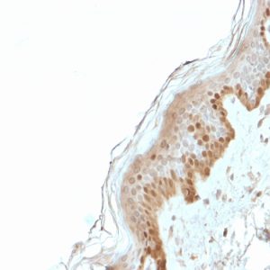
Immunohistochemistry
Gene Expression Regulation
2',3'-Cyclic Nucleotide 3'-Phosphodiesterase
Blotting, Western
Rats, Sprague-Dawley
Stem Cells
Base Sequence
Sulfoglycosphingolipids
Electrophoresis, Polyacrylamide Gel
Glycosylation
Receptors, Cell Surface
Neurons
Transport of Trembler-J mutant peripheral myelin protein 22 is blocked in the intermediate compartment and affects the transport of the wild-type protein by direct interaction. (1/1164)
Peripheral myelin protein 22 (PMP22) is an integral membrane protein that is essential for the normal formation and maintenance of peripheral myelin. Duplications, deletions, or mutations in the PMP22 gene account for a set of dominantly inherited peripheral neuropathies. The heterozygous Trembler-J (TrJ) genotype in mice is similar genetically to a Charcot-Marie-Tooth disease type 1A pedigree in humans, whereas the homozygous TrJ condition leads to the most severe form of PMP22-associated neuropathies. To characterize the consequences of the TrJ mutation, we labeled wild-type (wt-) and TrJ-PMP22 in the third loop of the protein with different epitope tags and expressed them separately or together in COS7 cells and primary Schwann cells. Here we show that the transport of the mutant TrJ-PMP22 is interrupted in the intermediate compartment, preventing its insertion into the plasma membrane and affecting the morphology of the endoplasmic reticulum. In addition, TrJ-PMP22 forms a heterodimer with the wt-PMP22. This interaction causes a fraction of the wt-PMP22 to be retained with TrJ-PMP22 in the intermediate compartment of COS7 and Schwann cells. The relative stability of a wt-mutant PMP22 heterodimer as compared with the wt-wt PMP22 homodimer may determine whether a particular mutation is semidominant or dominant. The neuropathy itself appears to result both from decreased trafficking of wt-PMP22 to the plasma membrane and from a toxic gain of function via the accumulation of wt- and TrJ-PMP22 in the intermediate compartment. (+info)Localization of a candidate surfactant convertase to type II cells, macrophages, and surfactant subfractions. (2/1164)
Pulmonary surfactant exists in the alveolus in several distinct subtypes that differ in their morphology, composition, and surface activity. Experiments by others have implicated a serine hydrolase in the production of the inactive small vesicular subtype of surfactant (N. J. Gross and R. M. Schultz. Biochim. Biophys. Acta 1044: 222-230, 1990). Our laboratory recently identified this enzyme in the rat as the serine carboxylesterase ES-2 [F. Barr, H. Clark, and S. Hawgood. Am. J. Physiol. 274 (Lung Cell. Mol. Physiol. 18): L404-L410, 1998]. In the present study, we determined the cellular sites of expression of ES-2 in rat lung using a digoxygenin-labeled ES-2 riboprobe. ES-2 mRNA was localized to type II cells and alveolar macrophages but not to Clara cells. Using a specific ES-2 antibody, we determined the protein distribution of ES-2 in the lung by immunohistochemistry, and it was found to be consistent with the sites of mRNA expression. Most of the ES-2 in rat bronchoalveolar lavage is in the surfactant-depleted supernatant, but ES-2 was also consistently localized to the small vesicular surfactant subfraction presumed to form as a consequence of conversion activity. These results are consistent with a role for endogenous lung ES-2 in surfactant metabolism. (+info)Macrophage electrophoretic mobility (MEM) with myelin basic protein. (3/1164)
Lymphocytes from a total of 161 subjects, including normal controls and patients with malignant and non-malignant conditions, have been investigated for their response to myelin basic protein, using the macrophage electrophoretic mobility (MEM) test. It has been confirmed that there was a high level of association between clinically evident cancer and a positive response. Lymphocytes from 24/25 patients with non-malignant inflammatory and ischaemic diseases also gave positive responses. In 46 patients with breast lumps studied before mastectomy or biopsy, the test was positive in 15/19 cases which proved to be malignant and in 5/27 which proved benign on histological examination. In its present form the test is not sufficiently reliable for the diagnosis of early cancer. Our results suggest that tissue necrosis in malignant and non-malignant conditions may be one of the factors resulting in sensitization to antigenic determinants present in preparations of myelin basic protein. Despite its technical difficulties, the test may provide a means of examing some aspects of immune recall not readily revealed by other test systems. (+info)The MAL proteolipid is necessary for normal apical transport and accurate sorting of the influenza virus hemagglutinin in Madin-Darby canine kidney cells. (4/1164)
The MAL (MAL/VIP17) proteolipid is a nonglycosylated integral membrane protein expressed in a restricted pattern of cell types, including T lymphocytes, myelin-forming cells, and polarized epithelial cells. Transport of the influenza virus hemagglutinin (HA) to the apical surface of epithelial Madin-Darby canine kidney (MDCK) cells appears to be mediated by a pathway involving glycolipid- and cholesterol- enriched membranes (GEMs). In MDCK cells, MAL has been proposed previously as being an element of the protein machinery for the GEM-dependent apical transport pathway. Using an antisense oligonucleotide-based strategy and a newly generated monoclonal antibody to canine MAL, herein we have approached the effect of MAL depletion on HA transport in MDCK cells. We have found that MAL depletion diminishes the presence of HA in GEMs, reduces the rate of HA transport to the cell surface, inhibits the delivery of HA to the apical surface, and produces partial missorting of HA to the basolateral membrane. These effects were corrected by ectopic expression of MAL in MDCK cells whose endogenous MAL protein was depleted. Our results indicate that MAL is necessary for both normal apical transport and accurate sorting of HA. (+info)Spontaneous regression of primary autoreactivity during chronic progression of experimental autoimmune encephalomyelitis and multiple sclerosis. (5/1164)
Experimental autoimmune encephalomyelitis (EAE) is a widely used animal model for multiple sclerosis (MS). EAE is typically initiated by CD4(+) T helper cell type 1 (Th1) autoreactivity directed against a single priming immunodominant myelin peptide determinant. Recent studies have shown that clinical progression of EAE involves the accumulation of neo-autoreactivity, commonly referred to as epitope spreading, directed against peptide determinants not involved in the priming process. This study directly addresses the relative roles of primary autoreactivity and secondary epitope spreading in the progression of both EAE and MS. To this end we serially evaluated the development of several epitope-spreading cascades in SWXJ mice primed with distinctly different encephalitogenic determinants of myelin proteolipid protein. In a series of analogous experiments, we examined the development of epitope spreading in patients with isolated monosymptomatic demyelinating syndrome as their disease progressed to clinically definite MS. Our results indicate that in both EAE and MS, primary proliferative autoreactivity associated with onset of clinical disease invariably regresses with time and is often undetectable during periods of disease progression. In contrast, the emergence of sustained secondary autoreactivity to spreading determinants is consistently associated with disease progression in both EAE and MS. Our results indicate that chronic progression of EAE and MS involves a shifting of autoreactivity from primary initiating self-determinants to defined cascades of secondary determinants that sustain the self-recognition process during disease progression. (+info)Differentially expressed genes in C6.9 glioma cells during vitamin D-induced cell death program. (6/1164)
C6.9 rat glioma cells undergo a cell death program when exposed to 1, 25-dihydroxyvitamin D3 (1,25-D3). As a global analytical approach, we have investigated gene expression in C6.9 engaged in this cell death program using differential screening of a rat brain cDNA library with probes derived from control and 1,25-D3-treated cells. Using this methodology we report the isolation of 61 differentially expressed cDNAs. Forty-seven cDNAs correspond to genes already characterized in rat cells or tissues. Seven cDNAs are homologous to yeast, mouse or human genes and seven are not related to known genes. Some of the characterized genes have been reported to be differentially expressed following induction of programmed cell death. These include PMP22/gas3, MGP and beta-tubulin. For the first time, we also show a cell death program induced up-regulation of the c-myc associated primary response gene CRP, and of the proteasome RN3 subunit and TCTP/mortalin genes. Another interesting feature of this 1,25-D3 induced-cell death program is the down-regulated expression of transcripts for the microtubule motor dynein heavy chain/MAP 1C and of the calcium-binding S100beta protein. Finally 15 upregulated cDNAs encode ribosomal proteins suggesting a possible involvement of the translational apparatus in this cell program. Alternatively, these ribosomal protein genes could be up-regulated in response to altered rates of cellular metabolism, as has been demonstrated for most of the other isolated genes which encode proteins involved in metabolic pathways. Thus, this study presents to our knowledge the first characterization of genes which are differentially expressed during a cell death program induced by 1, 25-D3. Therefore, this data provides new information on the fundamental mechanisms which participate in the antineoplastic effects of 1,25-D3 and on the machinery of a cell death program in a glioma cell line. (+info)Peripheral myelin protein 22 and protein zero: a novel association in peripheral nervous system myelin. (7/1164)
Mutations found in the two major glycosylated transmembrane proteins of the PNS myelin, the peripheral myelin protein zero (P0) and peripheral myelin protein 22 (PMP22), have been independently associated with the most common hereditary demyelinating peripheral neuropathies. Genotype-phenotype correlations in humans and transgenic animals have provided functional evidence that P0 and PMP22 are involved in formation and maintenance of compact myelin. Here, we demonstrate for the first time that P0 and PMP22 proteins form complexes in the myelin membrane, as shown by coimmunoprecipitation experiments, and that glycosylation is not involved in mediating these interactions. Complex formation was also detected when the two proteins were coexpressed in heterologous cells. In transfected cells, P0 and PMP22 are recruited and colocalize at the apposed plasma membranes of expressors as shown by confocal microscopy. These findings provide a new basis for a better understanding of myelin assembly and of the pathomechanisms involved in demyelinating peripheral neuropathies. Furthermore, these results propose a possible explanation why alterations in either of these molecules are sufficient to destabilize the myelin structure and cause a similar disease phenotype. (+info)Cloning and characterization of a 22 kDa protein from rat adipocytes: a new member of the reticulon family. (8/1164)
In the course of our examination of proteins associated with the GLUT4-containing vesicles of rat adipocytes we have identified a new 22 kDa member of the family of endoplasmic reticulum (ER) proteins known as reticulons. The protein, which we refer to as vp20, was purified from a preparation of GLUT4-containing vesicles of rat adipocytes, and tryptic peptides were micro-sequenced. From this information a cDNA encoding a single open reading frame for a protein of 22 kDa was cloned. This protein is homologous to known members of the reticulon protein family. vp20 has two hydrophobic stretches of about 35 amino acids that could be membrane spanning domains and an ER retention motif at its carboxy-terminus. vp20 was most abundant in the high density microsome fraction of adipocytes, which is the fraction most enriched in ER. Only a small fraction of vp20 was present in the GLUT4 vesicle population, and that fraction appears to be due to ER vesicles that were non-specifically bound to the adsorbent. Analysis of tissue distribution of vp20 in rats revealed that it is concentrated in muscle, fat and the brain. (+info)Myelin proteins are proteins that are found in the myelin sheath, which is a fatty (lipid-rich) substance that surrounds and insulates nerve fibers (axons) in the nervous system. The myelin sheath enables the rapid transmission of electrical signals (nerve impulses) along the axons, allowing for efficient communication between different parts of the nervous system.
There are several types of myelin proteins, including:
1. Proteolipid protein (PLP): This is the most abundant protein in the myelin sheath and plays a crucial role in maintaining the structure and function of the myelin sheath.
2. Myelin basic protein (MBP): This protein is also found in the myelin sheath and helps to stabilize the compact structure of the myelin sheath.
3. Myelin-associated glycoprotein (MAG): This protein is involved in the adhesion of the myelin sheath to the axon and helps to maintain the integrity of the myelin sheath.
4. 2'3'-cyclic nucleotide 3' phosphodiesterase (CNP): This protein is found in oligodendrocytes, which are the cells that produce the myelin sheath in the central nervous system. CNP plays a role in maintaining the structure and function of the oligodendrocytes.
Damage to myelin proteins can lead to demyelination, which is a characteristic feature of several neurological disorders, including multiple sclerosis (MS), Guillain-Barré syndrome, and Charcot-Marie-Tooth disease.
Myelin P0 protein, also known as P0 or MPZ (myelin protein zero), is a major structural component of the myelin sheath in the peripheral nervous system. The myelin sheath is a multilayered membrane that surrounds and insulates nerve fibers to increase the speed of electrical impulse transmission.
P0 protein is a transmembrane glycoprotein, which means it spans the lipid bilayer of the myelin membrane and has sugar molecules (glycans) attached to it. It plays a crucial role in maintaining the compact structure of the myelin sheath by forming homodimers that interact with each other through their extracellular domains, creating tight junctions between the apposing layers of the myelin membrane.
P0 protein also contributes to the stability and integrity of the myelin sheath by interacting with other myelin proteins, such as connexin 32 and peripheral myelin protein 22 (PMP22). Mutations in the MPZ gene can lead to various peripheral neuropathies, including Charcot-Marie-Tooth disease type 1B and Dejerine-Sottas syndrome.
The myelin sheath is a multilayered, fatty substance that surrounds and insulates many nerve fibers in the nervous system. It is essential for the rapid transmission of electrical signals, or nerve impulses, along these nerve fibers, allowing for efficient communication between different parts of the body. The myelin sheath is produced by specialized cells called oligodendrocytes in the central nervous system (CNS) and Schwann cells in the peripheral nervous system (PNS). Damage to the myelin sheath, as seen in conditions like multiple sclerosis, can significantly impair nerve function and result in various neurological symptoms.
Myelin Basic Protein (MBP) is a key structural protein found in the myelin sheath, which is a multilayered membrane that surrounds and insulates nerve fibers (axons) in the nervous system. The myelin sheath enables efficient and rapid transmission of electrical signals (nerve impulses) along the axons, allowing for proper communication between different neurons.
MBP is one of several proteins responsible for maintaining the structural integrity and organization of the myelin sheath. It is a basic protein, meaning it has a high isoelectric point due to its abundance of positively charged amino acids. MBP is primarily located in the intraperiod line of the compact myelin, which is a region where the extracellular leaflets of the apposing membranes come into close contact without fusing.
MBP plays crucial roles in the formation, maintenance, and repair of the myelin sheath:
1. During development, MBP helps mediate the compaction of the myelin sheath by interacting with other proteins and lipids in the membrane.
2. MBP contributes to the stability and resilience of the myelin sheath by forming strong ionic bonds with negatively charged phospholipids in the membrane.
3. In response to injury or disease, MBP can be cleaved into smaller peptides that act as chemoattractants for immune cells, initiating the process of remyelination and repair.
Dysregulation or damage to MBP has been implicated in several demyelinating diseases, such as multiple sclerosis (MS), where the immune system mistakenly attacks the myelin sheath, leading to its degradation and loss. The presence of autoantibodies against MBP is a common feature in MS patients, suggesting that an abnormal immune response to this protein may contribute to the pathogenesis of the disease.
Myelin Proteolipid Protein (PLP) is a major component of the myelin sheath, which is a fatty insulating substance that covers and protects nerve fibers in the central nervous system (CNS). PLP makes up about 50% of the proteins found in the myelin sheath. It plays a crucial role in the structure and function of the myelin sheath, including maintaining its compactness and stability. Defects or mutations in the gene that encodes for PLP can lead to various demyelinating diseases, such as X-linked adrenoleukodystrophy (X-ALD) and Pelizaeus-Merzbacher disease (PMD), which are characterized by the degeneration of the myelin sheath and subsequent neurological impairments.
Charcot-Marie-Tooth disease (CMT) is a group of inherited disorders that cause nerve damage, primarily affecting the peripheral nerves. These are the nerves that transmit signals between the brain and spinal cord to the rest of the body. CMT affects both motor and sensory nerves, leading to muscle weakness and atrophy, as well as numbness or tingling in the hands and feet.
The disease is named after the three physicians who first described it: Jean-Martin Charcot, Pierre Marie, and Howard Henry Tooth. CMT is characterized by its progressive nature, meaning symptoms typically worsen over time, although the rate of progression can vary significantly among individuals.
There are several types of CMT, classified based on their genetic causes and patterns of inheritance. The two most common forms are CMT1 and CMT2:
1. CMT1: This form is caused by mutations in the genes responsible for the myelin sheath, which insulates peripheral nerves and allows for efficient signal transmission. As a result, demyelination occurs, slowing down nerve impulses and causing muscle weakness, particularly in the lower limbs. Symptoms usually begin in childhood or adolescence and include foot drop, high arches, and hammertoes.
2. CMT2: This form is caused by mutations in the genes responsible for the axons, the nerve fibers that transmit signals within peripheral nerves. As a result, axonal degeneration occurs, leading to muscle weakness and atrophy. Symptoms usually begin in early adulthood and progress more slowly than CMT1. They primarily affect the lower limbs but can also involve the hands and arms.
Diagnosis of CMT typically involves a combination of clinical evaluation, family history, nerve conduction studies, and genetic testing. While there is no cure for CMT, treatment focuses on managing symptoms and maintaining mobility and function through physical therapy, bracing, orthopedic surgery, and pain management.
Schwann cells, also known as neurolemmocytes, are a type of glial cell that form the myelin sheath around peripheral nervous system (PNS) axons, allowing for the rapid and efficient transmission of nerve impulses. These cells play a crucial role in the maintenance and function of the PNS.
Schwann cells originate from the neural crest during embryonic development and migrate to the developing nerves. They wrap around the axons in a spiral fashion, forming multiple layers of myelin, which insulates the nerve fibers and increases the speed of electrical impulse transmission. Each Schwann cell is responsible for myelinating a single segment of an axon, with the gaps between these segments called nodes of Ranvier.
Schwann cells also provide structural support to the neurons and contribute to the regeneration of injured peripheral nerves by helping to guide the regrowth of axons to their targets. Additionally, Schwann cells can participate in immune responses within the PNS, such as releasing cytokines and chemokines to recruit immune cells during injury or infection.
Myelin-Associated Glycoprotein (MAG) is a glycoprotein found on the surface of myelin sheaths, which are the protective insulating layers around nerve fibers in the nervous system. MAG plays a role in the adhesion and interaction between the myelin sheath and the axon it surrounds. It's particularly important during the development and maintenance of the nervous system. Additionally, MAG has been implicated in the regulation of neuronal growth and signal transmission. In certain autoimmune diseases like Guillain-Barré syndrome, the immune system may mistakenly attack MAG, leading to damage of the myelin sheath and associated neurological symptoms.
Demyelinating diseases are a group of disorders that are characterized by damage to the myelin sheath, which is the protective covering surrounding nerve fibers in the brain, optic nerves, and spinal cord. Myelin is essential for the rapid transmission of nerve impulses, and its damage results in disrupted communication between the brain and other parts of the body.
The most common demyelinating disease is multiple sclerosis (MS), where the immune system mistakenly attacks the myelin sheath. Other demyelinating diseases include:
1. Acute Disseminated Encephalomyelitis (ADEM): An autoimmune disorder that typically follows a viral infection or vaccination, causing widespread inflammation and demyelination in the brain and spinal cord.
2. Neuromyelitis Optica (NMO) or Devic's Disease: A rare autoimmune disorder that primarily affects the optic nerves and spinal cord, leading to severe vision loss and motor disability.
3. Transverse Myelitis: Inflammation of the spinal cord causing damage to both sides of one level (segment) of the spinal cord, resulting in various neurological symptoms such as muscle weakness, numbness, or pain, depending on which part of the spinal cord is affected.
4. Guillain-Barré Syndrome: An autoimmune disorder that causes rapid-onset muscle weakness, often beginning in the legs and spreading to the upper body, including the face and breathing muscles. It occurs when the immune system attacks the peripheral nerves' myelin sheath.
5. Central Pontine Myelinolysis (CPM): A rare neurological disorder caused by rapid shifts in sodium levels in the blood, leading to damage to the myelin sheath in a specific area of the brainstem called the pons.
These diseases can result in various symptoms, such as muscle weakness, numbness, vision loss, difficulty with balance and coordination, and cognitive impairment, depending on the location and extent of the demyelination. Treatment typically focuses on managing symptoms, modifying the immune system's response, and promoting nerve regeneration and remyelination when possible.
Oligodendroglia are a type of neuroglial cell found in the central nervous system (CNS) of vertebrates, including humans. These cells play a crucial role in providing support and insulation to nerve fibers (axons) in the CNS, which includes the brain and spinal cord.
More specifically, oligodendroglia produce a fatty substance called myelin that wraps around axons, forming myelin sheaths. This myelination process helps to increase the speed of electrical impulse transmission (nerve impulses) along the axons, allowing for efficient communication between different neurons.
In addition to their role in myelination, oligodendroglia also contribute to the overall health and maintenance of the CNS by providing essential nutrients and supporting factors to neurons. Dysfunction or damage to oligodendroglia has been implicated in various neurological disorders, such as multiple sclerosis (MS), where demyelination of axons leads to impaired nerve function and neurodegeneration.
The sciatic nerve is the largest and longest nerve in the human body, running from the lower back through the buttocks and down the legs to the feet. It is formed by the union of the ventral rami (branches) of the L4 to S3 spinal nerves. The sciatic nerve provides motor and sensory innervation to various muscles and skin areas in the lower limbs, including the hamstrings, calf muscles, and the sole of the foot. Sciatic nerve disorders or injuries can result in symptoms such as pain, numbness, tingling, or weakness in the lower back, hips, legs, and feet, known as sciatica.
Hereditary Sensory and Motor Neuropathy (HSMN) is a group of inherited disorders that affect the peripheral nerves, which are the nerves outside the brain and spinal cord. These nerves transmit information between the brain and muscles, as well as sensations such as touch, pain, heat, and cold.
HSMN is characterized by progressive degeneration of these peripheral nerves, leading to muscle weakness, numbness, and tingling sensations, particularly in the hands and feet. The condition can also affect the autonomic nervous system, which controls involuntary functions such as heart rate, blood pressure, and digestion.
HSMN is caused by genetic mutations that are inherited from one or both parents. There are several types of HSMN, each with its own specific symptoms, severity, and pattern of inheritance. The most common form is Charcot-Marie-Tooth disease (CMT), which affects both motor and sensory nerves.
Treatment for HSMN typically focuses on managing the symptoms and preventing complications. This may include physical therapy, bracing or orthopedic surgery to support weakened muscles, pain management, and lifestyle modifications such as avoiding activities that aggravate symptoms. There is currently no cure for HSMN, but ongoing research is aimed at developing new treatments and therapies to slow or halt the progression of the disease.
The sural nerve is a purely sensory peripheral nerve in the lower leg and foot. It provides sensation to the outer ( lateral) aspect of the little toe and the adjacent side of the fourth toe, as well as a small portion of the skin on the back of the leg between the ankle and knee joints.
The sural nerve is formed by the union of branches from the tibial and common fibular nerves (branches of the sciatic nerve) in the lower leg. It runs down the calf, behind the lateral malleolus (the bony prominence on the outside of the ankle), and into the foot.
The sural nerve is often used as a donor nerve during nerve grafting procedures due to its consistent anatomy and relatively low risk for morbidity at the donor site.
Early Growth Response Protein 2 (EGR2) is a transcription factor that belongs to the EGR family of proteins, which are involved in various biological processes such as cell proliferation, differentiation, and apoptosis. EGR2 is specifically known to play crucial roles in the development and function of the nervous system, including the regulation of neuronal survival, axon guidance, and myelination. It is also expressed in immune cells and has been implicated in the regulation of immune responses. Mutations in the EGR2 gene have been associated with certain neurological disorders and diseases, such as Charcot-Marie-Tooth disease type 1B and congenital hypomyelinating neuropathy.
2,3'-Cyclic-nucleotide phosphodiesterases (PDEs) are a subclass of enzymes that belong to the family of phosphodiesterases. These enzymes are responsible for the hydrolysis of 2,3'-cyclic nucleotides, which are cyclic forms of nucleotides that act as second messengers in various cellular signaling pathways.
The two primary types of 2,3'-cyclic nucleotides are 2',3'-cGMP and 2',3'-cAMP, which are produced by the action of certain enzymes on their respective precursors, guanosine triphosphate (GTP) and adenosine triphosphate (ATP). These cyclic nucleotides play important roles in regulating various cellular processes, including metabolism, gene expression, and ion channel activity.
2,3'-Cyclic-nucleotide phosphodiesterases catalyze the hydrolysis of these cyclic nucleotides to their corresponding 5'-monophosphates, thereby terminating their signaling activity. There are several isoforms of 2,3'-cyclic-nucleotide PDEs that have been identified, each with distinct substrate specificities and regulatory properties.
Dysregulation of 2,3'-cyclic-nucleotide PDE activity has been implicated in various diseases, including cancer, cardiovascular disease, and neurological disorders. Therefore, these enzymes have emerged as important targets for the development of therapeutic agents that can modulate their activity and restore normal cellular function.
Peripheral nerves are nerve fibers that transmit signals between the central nervous system (CNS, consisting of the brain and spinal cord) and the rest of the body. These nerves convey motor, sensory, and autonomic information, enabling us to move, feel, and respond to changes in our environment. They form a complex network that extends from the CNS to muscles, glands, skin, and internal organs, allowing for coordinated responses and functions throughout the body. Damage or injury to peripheral nerves can result in various neurological symptoms, such as numbness, weakness, or pain, depending on the type and severity of the damage.
Myelin P2 protein, also known as proteolipid protein 1 (PLP1), is a major structural component of the myelin sheath in the central nervous system. The myelin sheath is a protective and insulating layer that surrounds nerve cell fibers (axons), allowing for efficient and rapid transmission of electrical signals.
The P2 protein is a transmembrane protein, with four transmembrane domains, and it plays a crucial role in maintaining the stability and integrity of the myelin sheath. Mutations in the gene that encodes for this protein (PLP1) have been associated with several demyelinating diseases, including Pelizaeus-Merzbacher disease (PMD), a rare X-linked recessive disorder characterized by abnormalities in the development and maintenance of the myelin sheath.
The P2 protein is also involved in various cellular processes, such as signal transduction, ion transport, and immune response regulation. However, the precise mechanisms through which these functions are carried out remain to be fully elucidated.
Peripheral Nervous System (PNS) diseases, also known as Peripheral Neuropathies, refer to conditions that affect the functioning of the peripheral nervous system, which includes all the nerves outside the brain and spinal cord. These nerves transmit signals between the central nervous system (CNS) and the rest of the body, controlling sensations, movements, and automatic functions such as heart rate and digestion.
PNS diseases can be caused by various factors, including genetics, infections, toxins, metabolic disorders, trauma, or autoimmune conditions. The symptoms of PNS diseases depend on the type and extent of nerve damage but often include:
1. Numbness, tingling, or pain in the hands and feet
2. Muscle weakness or cramps
3. Loss of reflexes
4. Decreased sensation to touch, temperature, or vibration
5. Coordination problems and difficulty with balance
6. Sexual dysfunction
7. Digestive issues, such as constipation or diarrhea
8. Dizziness or fainting due to changes in blood pressure
Examples of PNS diseases include Guillain-Barre syndrome, Charcot-Marie-Tooth disease, diabetic neuropathy, and peripheral nerve injuries. Treatment for these conditions varies depending on the underlying cause but may involve medications, physical therapy, lifestyle changes, or surgery.
Myelin-Oligodendrocyte Glycoprotein (MOG) is a protein found exclusively on the outermost layer of myelin sheath in the central nervous system (CNS). The myelin sheath is a fatty substance that surrounds and insulates nerve fibers, allowing for efficient and rapid transmission of electrical signals. MOG plays a crucial role in maintaining the integrity and structure of the myelin sheath. It is involved in the adhesion of oligodendrocytes to the surface of neuronal axons and contributes to the stability of the compact myelin structure. Autoimmune reactions against MOG have been implicated in certain inflammatory demyelinating diseases, such as optic neuritis, transverse myelitis, and acute disseminated encephalomyelitis (ADEM).
An axon is a long, slender extension of a neuron (a type of nerve cell) that conducts electrical impulses (nerve impulses) away from the cell body to target cells, such as other neurons or muscle cells. Axons can vary in length from a few micrometers to over a meter long and are typically surrounded by a myelin sheath, which helps to insulate and protect the axon and allows for faster transmission of nerve impulses.
Axons play a critical role in the functioning of the nervous system, as they provide the means by which neurons communicate with one another and with other cells in the body. Damage to axons can result in serious neurological problems, such as those seen in spinal cord injuries or neurodegenerative diseases like multiple sclerosis.
Wallerian degeneration is a process that occurs following damage to the axons of neurons (nerve cells). After an axon is severed or traumatically injured, it undergoes a series of changes including fragmentation and removal of the distal segment of the axon, which is the part that is separated from the cell body. This process is named after Augustus Waller, who first described it in 1850.
The degenerative changes in the distal axon are characterized by the breakdown of the axonal cytoskeleton, the loss of myelin sheath (the fatty insulating material that surrounds and protects the axon), and the infiltration of macrophages to clear away the debris. These events lead to the degeneration of the distal axon segment, which is necessary for successful regeneration of the injured nerve.
Wallerian degeneration is a crucial process in the nervous system's response to injury, as it enables the regrowth of axons and the reestablishment of connections between neurons. However, if the regenerative capacity of the neuron is insufficient or the environment is not conducive to growth, functional recovery may be impaired, leading to long-term neurological deficits.
Neurologic mutant mice are genetically engineered or spontaneously mutated rodents that are used as models to study various neurological disorders and conditions. These mice have specific genetic modifications or mutations that affect their nervous system, leading to phenotypes that resemble human neurological diseases.
Some examples of neurologic mutant mice include:
1. Alzheimer's disease models: Mice that overexpress genes associated with Alzheimer's disease, such as the amyloid precursor protein (APP) or presenilin 1 (PS1), to study the pathogenesis and potential treatments of this disorder.
2. Parkinson's disease models: Mice that have genetic mutations in genes associated with Parkinson's disease, such as alpha-synuclein or parkin, to investigate the mechanisms underlying this condition and develop new therapies.
3. Huntington's disease models: Mice that carry an expanded CAG repeat in the huntingtin gene to replicate the genetic defect seen in humans with Huntington's disease and study disease progression and treatment strategies.
4. Epilepsy models: Mice with genetic mutations that cause spontaneous seizures or increased susceptibility to seizures, used to investigate the underlying mechanisms of epilepsy and develop new treatments.
5. Stroke models: Mice that have surgical induction of stroke or genetic modifications that increase the risk of stroke, used to study the pathophysiology of stroke and identify potential therapeutic targets.
Neurologic mutant mice are essential tools in biomedical research, allowing scientists to investigate the complex interactions between genes and the environment that contribute to neurological disorders. These models help researchers better understand disease mechanisms, develop new therapies, and test their safety and efficacy before moving on to clinical trials in humans.
Pelizaeus-Merzbacher disease (PMD) is a rare X-linked recessive genetic disorder affecting the nervous system. It is caused by mutations in the PLP1 gene, which provides instructions for making proteins that are important for the formation and maintenance of the myelin sheath, the protective covering that wraps around nerve cell fibers (axons) in the brain and spinal cord to ensure efficient transmission of electrical signals.
In individuals with PMD, the myelin sheath is either partially or completely absent, leading to progressive neurological symptoms. The classic form of PMD is characterized by early onset of nystagmus (involuntary eye movements), ataxia (loss of muscle coordination and balance), and intellectual disability. Other features may include hypotonia (low muscle tone), spasticity (stiff or rigid muscles), and seizures. The severity and progression of the disease can vary widely among affected individuals, ranging from a severe, lethal form to a milder form with a slower disease course.
Currently, there is no cure for PMD, and treatment is focused on managing symptoms and improving quality of life.
SOXE transcription factors are a subgroup of the SOX (SRY-related HMG box) family of proteins, which are involved in various developmental processes, including cell fate specification and differentiation. The SOXE group includes SOX8, SOX9, and SOX10, all of which contain a conserved high mobility group (HMG) box DNA-binding domain. They play crucial roles in the development of several tissues, such as the nervous system, skeletal system, and urogenital system.
SOXE transcription factors are known to regulate gene expression by binding to specific DNA sequences, often acting in combination with other transcription factors to control various cellular processes. Dysregulation of SOXE transcription factors has been implicated in several human diseases, including cancer and neurodevelopmental disorders.
I'm not aware of a medical condition known as "Quaking Mice." However, "quaking" is a phenotype observed in laboratory mice that are used as models for certain genetic disorders.
The "quaking" phenotype is caused by a mutation in the QKI gene, which plays a crucial role in the development and function of the nervous system. Mice with this mutation have abnormal myelination (the process of forming a protective sheath around nerve fibers) in their central nervous system, leading to tremors, ataxia (loss of coordination), and other neurological symptoms.
The Quaking mouse model is often used in research to study the genetic and molecular mechanisms underlying demyelinating disorders, such as multiple sclerosis, and to test potential therapies for these conditions.
Myelinated nerve fibers are neuronal processes that are surrounded by a myelin sheath, a fatty insulating substance that is produced by Schwann cells in the peripheral nervous system and oligodendrocytes in the central nervous system. This myelin sheath helps to increase the speed of electrical impulse transmission, also known as action potentials, along the nerve fiber. The myelin sheath has gaps called nodes of Ranvier where the electrical impulses can jump from one node to the next, which also contributes to the rapid conduction of signals. Myelinated nerve fibers are typically found in the peripheral nerves and the optic nerve, but not in the central nervous system (CNS) tracts that are located within the brain and spinal cord.
The Central Nervous System (CNS) is the part of the nervous system that consists of the brain and spinal cord. It is called the "central" system because it receives information from, and sends information to, the rest of the body through peripheral nerves, which make up the Peripheral Nervous System (PNS).
The CNS is responsible for processing sensory information, controlling motor functions, and regulating various autonomic processes like heart rate, respiration, and digestion. The brain, as the command center of the CNS, interprets sensory stimuli, formulates thoughts, and initiates actions. The spinal cord serves as a conduit for nerve impulses traveling to and from the brain and the rest of the body.
The CNS is protected by several structures, including the skull (which houses the brain) and the vertebral column (which surrounds and protects the spinal cord). Despite these protective measures, the CNS remains vulnerable to injury and disease, which can have severe consequences due to its crucial role in controlling essential bodily functions.
Autoimmune encephalomyelitis (EAE) is a model of inflammatory demyelinating disease used in medical research to study the mechanisms of multiple sclerosis (MS) and develop new therapies. It is experimentally induced in laboratory animals, typically mice or rats, through immunization with myelin antigens or T-cell transfer. The resulting immune response leads to inflammation, demyelination, and neurological dysfunction in the central nervous system (CNS), mimicking certain aspects of MS.
EAE is a valuable tool for understanding the pathogenesis of MS and testing potential treatments. However, it is essential to recognize that EAE is an experimental model and may not fully recapitulate all features of human autoimmune encephalomyelitis.
Multiple Sclerosis (MS) is a chronic autoimmune disease that affects the central nervous system (CNS), which includes the brain, spinal cord, and optic nerves. In MS, the immune system mistakenly attacks the protective covering of nerve fibers, called myelin, leading to damage and scarring (sclerosis). This results in disrupted communication between the brain and the rest of the body, causing a variety of neurological symptoms that can vary widely from person to person.
The term "multiple" refers to the numerous areas of scarring that occur throughout the CNS in this condition. The progression, severity, and specific symptoms of MS are unpredictable and may include vision problems, muscle weakness, numbness or tingling, difficulty with balance and coordination, cognitive impairment, and mood changes. There is currently no cure for MS, but various treatments can help manage symptoms, modify the course of the disease, and improve quality of life for those affected.
Hereditary Central Nervous System (CNS) Demyelinating Diseases are a group of rare, inherited genetic disorders that affect the nervous system. These diseases are characterized by damage to the myelin sheath, which is the protective covering surrounding nerve fibers in the CNS (brain and spinal cord). The damage to the myelin sheath results in disrupted communication between the brain and other parts of the body, leading to various neurological symptoms.
Examples of Hereditary CNS Demyelinating Diseases include:
1. Leukodystrophies - A group of genetic disorders that affect the white matter (myelin) in the brain. Examples include Pelizaeus-Merzbacher disease, Krabbe disease, and Metachromatic leukodystrophy.
2. Hereditary Spastic Paraplegias (HSPs) - A group of inherited disorders that cause progressive stiffness and weakness in the legs due to damage to the nerve fibers in the spinal cord. Some forms of HSP can also involve CNS demyelination.
3. Neurodegenerative disorders with brain iron accumulation (NBIA) - A group of rare genetic disorders characterized by abnormal accumulation of iron in the brain, which can lead to damage to the myelin sheath and other structures in the brain. Examples include Pantothenate kinase-associated neurodegeneration (PKAN) and Neuroferritinopathy.
4. Cerebrotendinous xanthomatosis - A rare inherited disorder of bile acid metabolism that can lead to progressive neurological symptoms, including demyelination in the brain and spinal cord.
These disorders are typically diagnosed through genetic testing, medical history, physical examination, and imaging studies such as MRI. Treatment is focused on managing symptoms and slowing disease progression, and may include medications, physical therapy, and other supportive care measures.
Demyelinating autoimmune diseases of the central nervous system (CNS) are a group of disorders characterized by inflammation and damage to the myelin sheath, which is the protective covering that surrounds nerve fibers in the brain and spinal cord. This damage can result in various neurological symptoms, including muscle weakness, sensory loss, vision problems, and cognitive impairment.
The most common demyelinating autoimmune disease of the CNS is multiple sclerosis (MS), which affects approximately 2.3 million people worldwide. Other examples include neuromyelitis optica spectrum disorder (NMOSD), acute disseminated encephalomyelitis (ADEM), and transverse myelitis.
These conditions are thought to arise when the immune system mistakenly attacks the myelin sheath, leading to inflammation, damage, and scarring (sclerosis) in the CNS. The exact cause of this autoimmune response is not fully understood, but it is believed to involve a complex interplay between genetic, environmental, and immunological factors.
Treatment for demyelinating autoimmune diseases of the CNS typically involves a combination of medications to manage symptoms, reduce inflammation, and modify the course of the disease. These may include corticosteroids, immunosuppressive drugs, and disease-modifying therapies (DMTs) that target specific components of the immune system.
The Peripheral Nervous System (PNS) is that part of the nervous system which lies outside of the brain and spinal cord. It includes all the nerves and ganglia ( clusters of neurons) outside of the central nervous system (CNS). The PNS is divided into two components: the somatic nervous system and the autonomic nervous system.
The somatic nervous system is responsible for transmitting sensory information from the skin, muscles, and joints to the CNS, and for controlling voluntary movements of the skeletal muscles.
The autonomic nervous system, on the other hand, controls involuntary actions, such as heart rate, digestion, respiratory rate, salivation, perspiration, pupillary dilation, and sexual arousal. It is further divided into the sympathetic and parasympathetic systems, which generally have opposing effects and maintain homeostasis in the body.
Damage to the peripheral nervous system can result in various medical conditions such as neuropathies, neuritis, plexopathies, and radiculopathies, leading to symptoms like numbness, tingling, pain, weakness, or loss of reflexes in the affected area.
Proteolipids are a type of complex lipid-containing proteins that are insoluble in water and have a high content of hydrophobic amino acids. They are primarily found in the plasma membrane of cells, where they play important roles in maintaining the structural integrity and function of the membrane. Proteolipids are also found in various organelles, including mitochondria, lysosomes, and peroxisomes.
Proteolipids are composed of a hydrophobic protein core that is tightly associated with a lipid bilayer through non-covalent interactions. The protein component of proteolipids typically contains several transmembrane domains that span the lipid bilayer, as well as hydrophilic regions that face the cytoplasm or the lumen of organelles.
Proteolipids have been implicated in various cellular processes, including signal transduction, membrane trafficking, and ion transport. They are also associated with several neurological disorders, such as Alzheimer's disease, Parkinson's disease, and multiple sclerosis. The study of proteolipids is an active area of research in biochemistry and cell biology, with potential implications for the development of new therapies for neurological disorders.
Nerve regeneration is the process of regrowth and restoration of functional nerve connections following damage or injury to the nervous system. This complex process involves various cellular and molecular events, such as the activation of support cells called glia, the sprouting of surviving nerve fibers (axons), and the reformation of neural circuits. The goal of nerve regeneration is to enable the restoration of normal sensory, motor, and autonomic functions impaired due to nerve damage or injury.
Myelin and lymphocyte-associated proteolipid proteins (MAL/PLP) are a family of proteolipid proteins that play crucial roles in the formation and maintenance of the myelin sheath in the central nervous system (CNS). The myelin sheath is a multilayered membrane that surrounds nerve cell axons, allowing for efficient and rapid electrical impulse transmission.
The MAL/PLP family includes two major proteins:
1. Myelin and lymphocyte protein (MAL): This protein is primarily expressed in the plasma membrane of oligodendrocytes, the CNS glial cells responsible for myelination. MAL is involved in the organization and maintenance of the lipid rafts, which are specialized microdomains within the plasma membrane that facilitate signal transduction and membrane trafficking.
2. Proteolipid protein (PLP) or proteolipid protein 1 (PLP1): This is the most abundant protein in the CNS myelin sheath, constituting approximately 50% of its total protein content. PLP is primarily located within the intracellular leaflets of the multilayered myelin membrane and plays a critical role in maintaining the integrity and compaction of the myelin sheath.
Mutations in the genes encoding these proteins can lead to various demyelinating disorders, such as Pelizaeus-Merzbacher disease (PMD) and spastic paraplegia type 2 (SPG2), which are characterized by abnormalities in the myelin sheath and neurological dysfunction.
Neural conduction is the process by which electrical signals, known as action potentials, are transmitted along the axon of a neuron (nerve cell) to transmit information between different parts of the nervous system. This electrical impulse is generated by the movement of ions across the neuronal membrane, and it propagates down the length of the axon until it reaches the synapse, where it can then stimulate the release of neurotransmitters to communicate with other neurons or target cells. The speed of neural conduction can vary depending on factors such as the diameter of the axon, the presence of myelin sheaths (which act as insulation and allow for faster conduction), and the temperature of the environment.
The optic nerve, also known as the second cranial nerve, is the nerve that transmits visual information from the retina to the brain. It is composed of approximately one million nerve fibers that carry signals related to vision, such as light intensity and color, from the eye's photoreceptor cells (rods and cones) to the visual cortex in the brain. The optic nerve is responsible for carrying this visual information so that it can be processed and interpreted by the brain, allowing us to see and perceive our surroundings. Damage to the optic nerve can result in vision loss or impairment.
The spinal cord is a major part of the nervous system, extending from the brainstem and continuing down to the lower back. It is a slender, tubular bundle of nerve fibers (axons) and support cells (glial cells) that carries signals between the brain and the rest of the body. The spinal cord primarily serves as a conduit for motor information, which travels from the brain to the muscles, and sensory information, which travels from the body to the brain. It also contains neurons that can independently process and respond to information within the spinal cord without direct input from the brain.
The spinal cord is protected by the bony vertebral column (spine) and is divided into 31 segments: 8 cervical, 12 thoracic, 5 lumbar, 5 sacral, and 1 coccygeal. Each segment corresponds to a specific region of the body and gives rise to pairs of spinal nerves that exit through the intervertebral foramina at each level.
The spinal cord is responsible for several vital functions, including:
1. Reflexes: Simple reflex actions, such as the withdrawal reflex when touching a hot surface, are mediated by the spinal cord without involving the brain.
2. Muscle control: The spinal cord carries motor signals from the brain to the muscles, enabling voluntary movement and muscle tone regulation.
3. Sensory perception: The spinal cord transmits sensory information, such as touch, temperature, pain, and vibration, from the body to the brain for processing and awareness.
4. Autonomic functions: The sympathetic and parasympathetic divisions of the autonomic nervous system originate in the thoracolumbar and sacral regions of the spinal cord, respectively, controlling involuntary physiological responses like heart rate, blood pressure, digestion, and respiration.
Damage to the spinal cord can result in various degrees of paralysis or loss of sensation below the level of injury, depending on the severity and location of the damage.
Neuroglia, also known as glial cells or simply glia, are non-neuronal cells that provide support and protection for neurons in the nervous system. They maintain homeostasis, form myelin sheaths around nerve fibers, and provide structural support. They also play a role in the immune response of the central nervous system. Some types of neuroglia include astrocytes, oligodendrocytes, microglia, and ependymal cells.
A nerve crush injury is a type of peripheral nerve injury that occurs when there is excessive pressure or compression applied to a nerve, causing it to become damaged or dysfunctional. This can happen due to various reasons such as trauma from accidents, surgical errors, or prolonged pressure on the nerve from tight casts, clothing, or positions.
The compression disrupts the normal functioning of the nerve, leading to symptoms such as numbness, tingling, weakness, or pain in the affected area. In severe cases, a nerve crush injury can cause permanent damage to the nerve, leading to long-term disability or loss of function. Treatment for nerve crush injuries typically involves relieving the pressure on the nerve, providing supportive care, and in some cases, surgical intervention may be necessary to repair the damaged nerve.
The brain is the central organ of the nervous system, responsible for receiving and processing sensory information, regulating vital functions, and controlling behavior, movement, and cognition. It is divided into several distinct regions, each with specific functions:
1. Cerebrum: The largest part of the brain, responsible for higher cognitive functions such as thinking, learning, memory, language, and perception. It is divided into two hemispheres, each controlling the opposite side of the body.
2. Cerebellum: Located at the back of the brain, it is responsible for coordinating muscle movements, maintaining balance, and fine-tuning motor skills.
3. Brainstem: Connects the cerebrum and cerebellum to the spinal cord, controlling vital functions such as breathing, heart rate, and blood pressure. It also serves as a relay center for sensory information and motor commands between the brain and the rest of the body.
4. Diencephalon: A region that includes the thalamus (a major sensory relay station) and hypothalamus (regulates hormones, temperature, hunger, thirst, and sleep).
5. Limbic system: A group of structures involved in emotional processing, memory formation, and motivation, including the hippocampus, amygdala, and cingulate gyrus.
The brain is composed of billions of interconnected neurons that communicate through electrical and chemical signals. It is protected by the skull and surrounded by three layers of membranes called meninges, as well as cerebrospinal fluid that provides cushioning and nutrients.
Molecular sequence data refers to the specific arrangement of molecules, most commonly nucleotides in DNA or RNA, or amino acids in proteins, that make up a biological macromolecule. This data is generated through laboratory techniques such as sequencing, and provides information about the exact order of the constituent molecules. This data is crucial in various fields of biology, including genetics, evolution, and molecular biology, allowing for comparisons between different organisms, identification of genetic variations, and studies of gene function and regulation.
"Cells, cultured" is a medical term that refers to cells that have been removed from an organism and grown in controlled laboratory conditions outside of the body. This process is called cell culture and it allows scientists to study cells in a more controlled and accessible environment than they would have inside the body. Cultured cells can be derived from a variety of sources, including tissues, organs, or fluids from humans, animals, or cell lines that have been previously established in the laboratory.
Cell culture involves several steps, including isolation of the cells from the tissue, purification and characterization of the cells, and maintenance of the cells in appropriate growth conditions. The cells are typically grown in specialized media that contain nutrients, growth factors, and other components necessary for their survival and proliferation. Cultured cells can be used for a variety of purposes, including basic research, drug development and testing, and production of biological products such as vaccines and gene therapies.
It is important to note that cultured cells may behave differently than they do in the body, and results obtained from cell culture studies may not always translate directly to human physiology or disease. Therefore, it is essential to validate findings from cell culture experiments using additional models and ultimately in clinical trials involving human subjects.
Nerve tissue proteins are specialized proteins found in the nervous system that provide structural and functional support to nerve cells, also known as neurons. These proteins include:
1. Neurofilaments: These are type IV intermediate filaments that provide structural support to neurons and help maintain their shape and size. They are composed of three subunits - NFL (light), NFM (medium), and NFH (heavy).
2. Neuronal Cytoskeletal Proteins: These include tubulins, actins, and spectrins that provide structural support to the neuronal cytoskeleton and help maintain its integrity.
3. Neurotransmitter Receptors: These are specialized proteins located on the postsynaptic membrane of neurons that bind neurotransmitters released by presynaptic neurons, triggering a response in the target cell.
4. Ion Channels: These are transmembrane proteins that regulate the flow of ions across the neuronal membrane and play a crucial role in generating and transmitting electrical signals in neurons.
5. Signaling Proteins: These include enzymes, receptors, and adaptor proteins that mediate intracellular signaling pathways involved in neuronal development, differentiation, survival, and death.
6. Adhesion Proteins: These are cell surface proteins that mediate cell-cell and cell-matrix interactions, playing a crucial role in the formation and maintenance of neural circuits.
7. Extracellular Matrix Proteins: These include proteoglycans, laminins, and collagens that provide structural support to nerve tissue and regulate neuronal migration, differentiation, and survival.
Transgenic mice are genetically modified rodents that have incorporated foreign DNA (exogenous DNA) into their own genome. This is typically done through the use of recombinant DNA technology, where a specific gene or genetic sequence of interest is isolated and then introduced into the mouse embryo. The resulting transgenic mice can then express the protein encoded by the foreign gene, allowing researchers to study its function in a living organism.
The process of creating transgenic mice usually involves microinjecting the exogenous DNA into the pronucleus of a fertilized egg, which is then implanted into a surrogate mother. The offspring that result from this procedure are screened for the presence of the foreign DNA, and those that carry the desired genetic modification are used to establish a transgenic mouse line.
Transgenic mice have been widely used in biomedical research to model human diseases, study gene function, and test new therapies. They provide a valuable tool for understanding complex biological processes and developing new treatments for a variety of medical conditions.
Electron microscopy (EM) is a type of microscopy that uses a beam of electrons to create an image of the sample being examined, resulting in much higher magnification and resolution than light microscopy. There are several types of electron microscopy, including transmission electron microscopy (TEM), scanning electron microscopy (SEM), and reflection electron microscopy (REM).
In TEM, a beam of electrons is transmitted through a thin slice of the sample, and the electrons that pass through the sample are focused to form an image. This technique can provide detailed information about the internal structure of cells, viruses, and other biological specimens, as well as the composition and structure of materials at the atomic level.
In SEM, a beam of electrons is scanned across the surface of the sample, and the electrons that are scattered back from the surface are detected to create an image. This technique can provide information about the topography and composition of surfaces, as well as the structure of materials at the microscopic level.
REM is a variation of SEM in which the beam of electrons is reflected off the surface of the sample, rather than scattered back from it. This technique can provide information about the surface chemistry and composition of materials.
Electron microscopy has a wide range of applications in biology, medicine, and materials science, including the study of cellular structure and function, disease diagnosis, and the development of new materials and technologies.
An amino acid sequence is the specific order of amino acids in a protein or peptide molecule, formed by the linking of the amino group (-NH2) of one amino acid to the carboxyl group (-COOH) of another amino acid through a peptide bond. The sequence is determined by the genetic code and is unique to each type of protein or peptide. It plays a crucial role in determining the three-dimensional structure and function of proteins.
A mutation is a permanent change in the DNA sequence of an organism's genome. Mutations can occur spontaneously or be caused by environmental factors such as exposure to radiation, chemicals, or viruses. They may have various effects on the organism, ranging from benign to harmful, depending on where they occur and whether they alter the function of essential proteins. In some cases, mutations can increase an individual's susceptibility to certain diseases or disorders, while in others, they may confer a survival advantage. Mutations are the driving force behind evolution, as they introduce new genetic variability into populations, which can then be acted upon by natural selection.
A "knockout" mouse is a genetically engineered mouse in which one or more genes have been deleted or "knocked out" using molecular biology techniques. This allows researchers to study the function of specific genes and their role in various biological processes, as well as potential associations with human diseases. The mice are generated by introducing targeted DNA modifications into embryonic stem cells, which are then used to create a live animal. Knockout mice have been widely used in biomedical research to investigate gene function, disease mechanisms, and potential therapeutic targets.
A phenotype is the physical or biochemical expression of an organism's genes, or the observable traits and characteristics resulting from the interaction of its genetic constitution (genotype) with environmental factors. These characteristics can include appearance, development, behavior, and resistance to disease, among others. Phenotypes can vary widely, even among individuals with identical genotypes, due to differences in environmental influences, gene expression, and genetic interactions.
I'm happy to help! "Rats, Inbred Lew" is a specific strain of laboratory rats that have been inbred for research purposes. The "Lew" part of the name refers to the location where they were first developed, the Lewis Institute in Lake Bluff, Illinois, USA.
Inbreeding is a process of mating closely related individuals over many generations to create a genetically homogeneous population. This results in a high degree of genetic similarity among members of the strain, making them ideal for use as experimental models because any differences observed between individuals are more likely to be due to the experimental manipulation rather than genetic variation.
Inbred Lew rats have been widely used in biomedical research, particularly in studies related to hypertension and cardiovascular disease. They exhibit a number of unique characteristics that make them useful for these types of studies, including their susceptibility to developing high blood pressure when fed a high-salt diet or given certain drugs.
It's important to note that while inbred strains like Lew rats can be very useful tools for researchers, they are not perfect models for human disease. Because they have been bred in a controlled environment and selected for specific traits, they may not respond to experimental manipulations in the same way that humans or other animals would. Therefore, it's important to interpret findings from these studies with caution and consider multiple lines of evidence before drawing any firm conclusions.
Transmission electron microscopy (TEM) is a type of microscopy in which an electron beam is transmitted through a ultra-thin specimen, interacting with it as it passes through. An image is formed from the interaction of the electrons with the specimen; the image is then magnified and visualized on a fluorescent screen or recorded on an electronic detector (or photographic film in older models).
TEM can provide high-resolution, high-magnification images that can reveal the internal structure of specimens including cells, viruses, and even molecules. It is widely used in biological and materials science research to investigate the ultrastructure of cells, tissues and materials. In medicine, TEM is used for diagnostic purposes in fields such as virology and bacteriology.
It's important to note that preparing a sample for TEM is a complex process, requiring specialized techniques to create thin (50-100 nm) specimens. These include cutting ultrathin sections of embedded samples using an ultramicrotome, staining with heavy metal salts, and positive staining or negative staining methods.
Animal disease models are specialized animals, typically rodents such as mice or rats, that have been genetically engineered or exposed to certain conditions to develop symptoms and physiological changes similar to those seen in human diseases. These models are used in medical research to study the pathophysiology of diseases, identify potential therapeutic targets, test drug efficacy and safety, and understand disease mechanisms.
The genetic modifications can include knockout or knock-in mutations, transgenic expression of specific genes, or RNA interference techniques. The animals may also be exposed to environmental factors such as chemicals, radiation, or infectious agents to induce the disease state.
Examples of animal disease models include:
1. Mouse models of cancer: Genetically engineered mice that develop various types of tumors, allowing researchers to study cancer initiation, progression, and metastasis.
2. Alzheimer's disease models: Transgenic mice expressing mutant human genes associated with Alzheimer's disease, which exhibit amyloid plaque formation and cognitive decline.
3. Diabetes models: Obese and diabetic mouse strains like the NOD (non-obese diabetic) or db/db mice, used to study the development of type 1 and type 2 diabetes, respectively.
4. Cardiovascular disease models: Atherosclerosis-prone mice, such as ApoE-deficient or LDLR-deficient mice, that develop plaque buildup in their arteries when fed a high-fat diet.
5. Inflammatory bowel disease models: Mice with genetic mutations affecting intestinal barrier function and immune response, such as IL-10 knockout or SAMP1/YitFc mice, which develop colitis.
Animal disease models are essential tools in preclinical research, but it is important to recognize their limitations. Differences between species can affect the translatability of results from animal studies to human patients. Therefore, researchers must carefully consider the choice of model and interpret findings cautiously when applying them to human diseases.
"Newborn animals" refers to the very young offspring of animals that have recently been born. In medical terminology, newborns are often referred to as "neonates," and they are classified as such from birth until about 28 days of age. During this time period, newborn animals are particularly vulnerable and require close monitoring and care to ensure their survival and healthy development.
The specific needs of newborn animals can vary widely depending on the species, but generally, they require warmth, nutrition, hydration, and protection from harm. In many cases, newborns are unable to regulate their own body temperature or feed themselves, so they rely heavily on their mothers for care and support.
In medical settings, newborn animals may be examined and treated by veterinarians to ensure that they are healthy and receiving the care they need. This can include providing medical interventions such as feeding tubes, antibiotics, or other treatments as needed to address any health issues that arise. Overall, the care and support of newborn animals is an important aspect of animal medicine and conservation efforts.
Spinal ganglia, also known as dorsal root ganglia, are clusters of nerve cell bodies located in the peripheral nervous system. They are situated along the length of the spinal cord and are responsible for transmitting sensory information from the body to the brain. Each spinal ganglion contains numerous neurons, or nerve cells, with long processes called axons that extend into the periphery and innervate various tissues and organs. The cell bodies within the spinal ganglia receive sensory input from these axons and transmit this information to the central nervous system via the dorsal roots of the spinal nerves. This allows the brain to interpret and respond to a wide range of sensory stimuli, including touch, temperature, pain, and proprioception (the sense of the position and movement of one's body).
C57BL/6 (C57 Black 6) is an inbred strain of laboratory mouse that is widely used in biomedical research. The term "inbred" refers to a strain of animals where matings have been carried out between siblings or other closely related individuals for many generations, resulting in a population that is highly homozygous at most genetic loci.
The C57BL/6 strain was established in 1920 by crossing a female mouse from the dilute brown (DBA) strain with a male mouse from the black strain. The resulting offspring were then interbred for many generations to create the inbred C57BL/6 strain.
C57BL/6 mice are known for their robust health, longevity, and ease of handling, making them a popular choice for researchers. They have been used in a wide range of biomedical research areas, including studies of cancer, immunology, neuroscience, cardiovascular disease, and metabolism.
One of the most notable features of the C57BL/6 strain is its sensitivity to certain genetic modifications, such as the introduction of mutations that lead to obesity or impaired glucose tolerance. This has made it a valuable tool for studying the genetic basis of complex diseases and traits.
Overall, the C57BL/6 inbred mouse strain is an important model organism in biomedical research, providing a valuable resource for understanding the genetic and molecular mechanisms underlying human health and disease.
Human chromosome pair 17 consists of two rod-shaped structures present in the nucleus of each human cell. Each chromosome is made up of DNA tightly coiled around histone proteins, forming a complex called chromatin. Chromosomes carry genetic information in the form of genes, which are segments of DNA that contain instructions for the development and function of an organism.
Human cells typically have 23 pairs of chromosomes, for a total of 46 chromosomes. Pair 17 is one of the autosomal pairs, meaning it is not a sex chromosome (X or Y). Chromosome 17 is a medium-sized chromosome and contains an estimated 800 million base pairs of DNA. It contains approximately 1,500 genes that provide instructions for making proteins and regulating various cellular processes.
Chromosome 17 is associated with several genetic disorders, including inherited cancer syndromes such as Li-Fraumeni syndrome and hereditary nonpolyposis colorectal cancer (HNPCC). Mutations in genes located on chromosome 17 can increase the risk of developing various types of cancer, including breast, ovarian, colon, and pancreatic cancer.
Cell differentiation is the process by which a less specialized cell, or stem cell, becomes a more specialized cell type with specific functions and structures. This process involves changes in gene expression, which are regulated by various intracellular signaling pathways and transcription factors. Differentiation results in the development of distinct cell types that make up tissues and organs in multicellular organisms. It is a crucial aspect of embryonic development, tissue repair, and maintenance of homeostasis in the body.
A point mutation is a type of genetic mutation where a single nucleotide base (A, T, C, or G) in DNA is altered, deleted, or substituted with another nucleotide. Point mutations can have various effects on the organism, depending on the location of the mutation and whether it affects the function of any genes. Some point mutations may not have any noticeable effect, while others might lead to changes in the amino acids that make up proteins, potentially causing diseases or altering traits. Point mutations can occur spontaneously due to errors during DNA replication or be inherited from parents.
I must clarify that the term "pedigree" is not typically used in medical definitions. Instead, it is often employed in genetics and breeding, where it refers to the recorded ancestry of an individual or a family, tracing the inheritance of specific traits or diseases. In human genetics, a pedigree can help illustrate the pattern of genetic inheritance in families over multiple generations. However, it is not a medical term with a specific clinical definition.
Messenger RNA (mRNA) is a type of RNA (ribonucleic acid) that carries genetic information copied from DNA in the form of a series of three-base code "words," each of which specifies a particular amino acid. This information is used by the cell's machinery to construct proteins, a process known as translation. After being transcribed from DNA, mRNA travels out of the nucleus to the ribosomes in the cytoplasm where protein synthesis occurs. Once the protein has been synthesized, the mRNA may be degraded and recycled. Post-transcriptional modifications can also occur to mRNA, such as alternative splicing and addition of a 5' cap and a poly(A) tail, which can affect its stability, localization, and translation efficiency.
Immunohistochemistry (IHC) is a technique used in pathology and laboratory medicine to identify specific proteins or antigens in tissue sections. It combines the principles of immunology and histology to detect the presence and location of these target molecules within cells and tissues. This technique utilizes antibodies that are specific to the protein or antigen of interest, which are then tagged with a detection system such as a chromogen or fluorophore. The stained tissue sections can be examined under a microscope, allowing for the visualization and analysis of the distribution and expression patterns of the target molecule in the context of the tissue architecture. Immunohistochemistry is widely used in diagnostic pathology to help identify various diseases, including cancer, infectious diseases, and immune-mediated disorders.
'Gene expression regulation' refers to the processes that control whether, when, and where a particular gene is expressed, meaning the production of a specific protein or functional RNA encoded by that gene. This complex mechanism can be influenced by various factors such as transcription factors, chromatin remodeling, DNA methylation, non-coding RNAs, and post-transcriptional modifications, among others. Proper regulation of gene expression is crucial for normal cellular function, development, and maintaining homeostasis in living organisms. Dysregulation of gene expression can lead to various diseases, including cancer and genetic disorders.
2,3'-Cyclic Nucleotide 3'-Phosphodiesterase (CNP) is an enzyme that specifically hydrolyzes 2',3'-cyclic nucleotides to 2'-nucleotide monophosphates. It plays a crucial role in regulating the levels of intracellular second messengers, such as cyclic guanosine monophosphate (cGMP) and cyclic adenosine monophosphate (cAMP), which are involved in various cellular processes including signal transduction, gene expression, and metabolism.
CNP has two isoforms, CNP1 and CNP2, which differ in their tissue distribution and substrate specificity. CNP1 is predominantly expressed in the central nervous system (CNS) and preferentially hydrolyzes cGMP, while CNP2 is widely distributed and hydrolyzes both cGMP and cAMP with similar efficiency.
Mutations in the gene encoding CNP1 have been associated with certain neurological disorders, such as spastic paraplegia type 5 (SPG5), a hereditary condition characterized by progressive muscle weakness and stiffness in the lower limbs.
Western blotting is a laboratory technique used in molecular biology to detect and quantify specific proteins in a mixture of many different proteins. This technique is commonly used to confirm the expression of a protein of interest, determine its size, and investigate its post-translational modifications. The name "Western" blotting distinguishes this technique from Southern blotting (for DNA) and Northern blotting (for RNA).
The Western blotting procedure involves several steps:
1. Protein extraction: The sample containing the proteins of interest is first extracted, often by breaking open cells or tissues and using a buffer to extract the proteins.
2. Separation of proteins by electrophoresis: The extracted proteins are then separated based on their size by loading them onto a polyacrylamide gel and running an electric current through the gel (a process called sodium dodecyl sulfate-polyacrylamide gel electrophoresis or SDS-PAGE). This separates the proteins according to their molecular weight, with smaller proteins migrating faster than larger ones.
3. Transfer of proteins to a membrane: After separation, the proteins are transferred from the gel onto a nitrocellulose or polyvinylidene fluoride (PVDF) membrane using an electric current in a process called blotting. This creates a replica of the protein pattern on the gel but now immobilized on the membrane for further analysis.
4. Blocking: The membrane is then blocked with a blocking agent, such as non-fat dry milk or bovine serum albumin (BSA), to prevent non-specific binding of antibodies in subsequent steps.
5. Primary antibody incubation: A primary antibody that specifically recognizes the protein of interest is added and allowed to bind to its target protein on the membrane. This step may be performed at room temperature or 4°C overnight, depending on the antibody's properties.
6. Washing: The membrane is washed with a buffer to remove unbound primary antibodies.
7. Secondary antibody incubation: A secondary antibody that recognizes the primary antibody (often coupled to an enzyme or fluorophore) is added and allowed to bind to the primary antibody. This step may involve using a horseradish peroxidase (HRP)-conjugated or alkaline phosphatase (AP)-conjugated secondary antibody, depending on the detection method used later.
8. Washing: The membrane is washed again to remove unbound secondary antibodies.
9. Detection: A detection reagent is added to visualize the protein of interest by detecting the signal generated from the enzyme-conjugated or fluorophore-conjugated secondary antibody. This can be done using chemiluminescent, colorimetric, or fluorescent methods.
10. Analysis: The resulting image is analyzed to determine the presence and quantity of the protein of interest in the sample.
Western blotting is a powerful technique for identifying and quantifying specific proteins within complex mixtures. It can be used to study protein expression, post-translational modifications, protein-protein interactions, and more. However, it requires careful optimization and validation to ensure accurate and reproducible results.
Sprague-Dawley rats are a strain of albino laboratory rats that are widely used in scientific research. They were first developed by researchers H.H. Sprague and R.C. Dawley in the early 20th century, and have since become one of the most commonly used rat strains in biomedical research due to their relatively large size, ease of handling, and consistent genetic background.
Sprague-Dawley rats are outbred, which means that they are genetically diverse and do not suffer from the same limitations as inbred strains, which can have reduced fertility and increased susceptibility to certain diseases. They are also characterized by their docile nature and low levels of aggression, making them easier to handle and study than some other rat strains.
These rats are used in a wide variety of research areas, including toxicology, pharmacology, nutrition, cancer, and behavioral studies. Because they are genetically diverse, Sprague-Dawley rats can be used to model a range of human diseases and conditions, making them an important tool in the development of new drugs and therapies.
According to the National Institutes of Health (NIH), stem cells are "initial cells" or "precursor cells" that have the ability to differentiate into many different cell types in the body. They can also divide without limit to replenish other cells for as long as the person or animal is still alive.
There are two main types of stem cells: embryonic stem cells, which come from human embryos, and adult stem cells, which are found in various tissues throughout the body. Embryonic stem cells have the ability to differentiate into all cell types in the body, while adult stem cells have more limited differentiation potential.
Stem cells play an essential role in the development and repair of various tissues and organs in the body. They are currently being studied for their potential use in the treatment of a wide range of diseases and conditions, including cancer, diabetes, heart disease, and neurological disorders. However, more research is needed to fully understand the properties and capabilities of these cells before they can be used safely and effectively in clinical settings.
A base sequence in the context of molecular biology refers to the specific order of nucleotides in a DNA or RNA molecule. In DNA, these nucleotides are adenine (A), guanine (G), cytosine (C), and thymine (T). In RNA, uracil (U) takes the place of thymine. The base sequence contains genetic information that is transcribed into RNA and ultimately translated into proteins. It is the exact order of these bases that determines the genetic code and thus the function of the DNA or RNA molecule.
Sulfoglycosphingolipids are a type of glycosphingolipid that contain a sulfate ester group in their carbohydrate moiety. They are important components of animal cell membranes and play a role in various biological processes, including cell recognition, signal transduction, and cell adhesion.
The most well-known sulfoglycosphingolipids are the sulfatides, which contain a 3'-sulfate ester on the galactose residue of the glycosphingolipid GalCer (galactosylceramide). Sulfatides are abundant in the nervous system and have been implicated in various neurological disorders.
Other sulfoglycosphingolipids include the seminolipids, which contain a 3'-sulfate ester on the galactose residue of lactosylceramide (Galβ1-4Glcβ1-Cer), and are found in high concentrations in the testis.
Abnormalities in sulfoglycosphingolipid metabolism have been associated with several genetic disorders, such as metachromatic leukodystrophy (MLD) and globoid cell leukodystrophy (GLD), which are characterized by progressive neurological deterioration.
Electrophoresis, polyacrylamide gel (EPG) is a laboratory technique used to separate and analyze complex mixtures of proteins or nucleic acids (DNA or RNA) based on their size and electrical charge. This technique utilizes a matrix made of cross-linked polyacrylamide, a type of gel, which provides a stable and uniform environment for the separation of molecules.
In this process:
1. The polyacrylamide gel is prepared by mixing acrylamide monomers with a cross-linking agent (bis-acrylamide) and a catalyst (ammonium persulfate) in the presence of a buffer solution.
2. The gel is then poured into a mold and allowed to polymerize, forming a solid matrix with uniform pore sizes that depend on the concentration of acrylamide used. Higher concentrations result in smaller pores, providing better resolution for separating smaller molecules.
3. Once the gel has set, it is placed in an electrophoresis apparatus containing a buffer solution. Samples containing the mixture of proteins or nucleic acids are loaded into wells on the top of the gel.
4. An electric field is applied across the gel, causing the negatively charged molecules to migrate towards the positive electrode (anode) while positively charged molecules move toward the negative electrode (cathode). The rate of migration depends on the size, charge, and shape of the molecules.
5. Smaller molecules move faster through the gel matrix and will migrate farther from the origin compared to larger molecules, resulting in separation based on size. Proteins and nucleic acids can be selectively stained after electrophoresis to visualize the separated bands.
EPG is widely used in various research fields, including molecular biology, genetics, proteomics, and forensic science, for applications such as protein characterization, DNA fragment analysis, cloning, mutation detection, and quality control of nucleic acid or protein samples.
Glycosylation is the enzymatic process of adding a sugar group, or glycan, to a protein, lipid, or other organic molecule. This post-translational modification plays a crucial role in modulating various biological functions, such as protein stability, trafficking, and ligand binding. The structure and composition of the attached glycans can significantly influence the functional properties of the modified molecule, contributing to cell-cell recognition, signal transduction, and immune response regulation. Abnormal glycosylation patterns have been implicated in several disease states, including cancer, diabetes, and neurodegenerative disorders.
Cell surface receptors, also known as membrane receptors, are proteins located on the cell membrane that bind to specific molecules outside the cell, known as ligands. These receptors play a crucial role in signal transduction, which is the process of converting an extracellular signal into an intracellular response.
Cell surface receptors can be classified into several categories based on their structure and mechanism of action, including:
1. Ion channel receptors: These receptors contain a pore that opens to allow ions to flow across the cell membrane when they bind to their ligands. This ion flux can directly activate or inhibit various cellular processes.
2. G protein-coupled receptors (GPCRs): These receptors consist of seven transmembrane domains and are associated with heterotrimeric G proteins that modulate intracellular signaling pathways upon ligand binding.
3. Enzyme-linked receptors: These receptors possess an intrinsic enzymatic activity or are linked to an enzyme, which becomes activated when the receptor binds to its ligand. This activation can lead to the initiation of various signaling cascades within the cell.
4. Receptor tyrosine kinases (RTKs): These receptors contain intracellular tyrosine kinase domains that become activated upon ligand binding, leading to the phosphorylation and activation of downstream signaling molecules.
5. Integrins: These receptors are transmembrane proteins that mediate cell-cell or cell-matrix interactions by binding to extracellular matrix proteins or counter-receptors on adjacent cells. They play essential roles in cell adhesion, migration, and survival.
Cell surface receptors are involved in various physiological processes, including neurotransmission, hormone signaling, immune response, and cell growth and differentiation. Dysregulation of these receptors can contribute to the development of numerous diseases, such as cancer, diabetes, and neurological disorders.
I'm sorry for any confusion, but "Jimpy" is not a recognized medical term or condition associated with mice in the field of veterinary medicine. It may be a colloquial or informal term used to describe a particular characteristic or behavior in mice, but without further context, it's challenging to provide an accurate definition. If you could provide more information about where you encountered this term or its intended meaning, I would be happy to help you further.
Neurons, also known as nerve cells or neurocytes, are specialized cells that constitute the basic unit of the nervous system. They are responsible for receiving, processing, and transmitting information and signals within the body. Neurons have three main parts: the dendrites, the cell body (soma), and the axon. The dendrites receive signals from other neurons or sensory receptors, while the axon transmits these signals to other neurons, muscles, or glands. The junction between two neurons is called a synapse, where neurotransmitters are released to transmit the signal across the gap (synaptic cleft) to the next neuron. Neurons vary in size, shape, and structure depending on their function and location within the nervous system.
In the field of medicine, "time factors" refer to the duration of symptoms or time elapsed since the onset of a medical condition, which can have significant implications for diagnosis and treatment. Understanding time factors is crucial in determining the progression of a disease, evaluating the effectiveness of treatments, and making critical decisions regarding patient care.
For example, in stroke management, "time is brain," meaning that rapid intervention within a specific time frame (usually within 4.5 hours) is essential to administering tissue plasminogen activator (tPA), a clot-busting drug that can minimize brain damage and improve patient outcomes. Similarly, in trauma care, the "golden hour" concept emphasizes the importance of providing definitive care within the first 60 minutes after injury to increase survival rates and reduce morbidity.
Time factors also play a role in monitoring the progression of chronic conditions like diabetes or heart disease, where regular follow-ups and assessments help determine appropriate treatment adjustments and prevent complications. In infectious diseases, time factors are crucial for initiating antibiotic therapy and identifying potential outbreaks to control their spread.
Overall, "time factors" encompass the significance of recognizing and acting promptly in various medical scenarios to optimize patient outcomes and provide effective care.
 Myelin proteolipid protein - Wikipedia
 CSF myelin basic protein: MedlinePlus Medical Encyclopedia
 Role of myelin basic protein in the formation of central nervous system myelin
 Myelin Proteins | Harvard Catalyst Profiles | Harvard Catalyst
 Myelin Basic Protein (MBP) Antibdy
 Mouse MBP (Myelin Basic Protein) ELISA Kit | G-EC-04564 | Gentaur Elisa Kits
 Myelin Basic Protein (87-99), guinea pig/human - LKT Labs
 Progressive axonopathy when oligodendrocytes lack the myelin protein CMTM5 :: MPG.PuRe
 Myelin, Basic Protein - HyTest Ltd.
 Elisa For Myelin Basic Protein - Interleukin 12
 Myelin Basic Protein Elisa Cayman - Polymerase Chain Reaction
 Myelin Proteolipid Protein (PLP 139-151), 15 mg - MD Bioproducts
 TagTeam :: Myelin protein zero (|em|P0|/em|)- and |em|Wnt1|/em|-Cre marked muscle resident neural crest-derived mesenchymal...
 Design and synthesis of non-peptide mimetics mapping the immunodominant myelin basic protein (MBP83-96) Epitope to function as...
 The major TMEM106B dementia risk allele affects TMEM106B protein levels, fibril formation, and myelin lipid homeostasis in the...
 Human MBP(Myelin Basic Protein) ELISA Kit - Global Life Sciences Conference in Warsaw
 Myelin Basic Protein-specific T cell Clones TL3A6, TL5F6, and TL5G7 | Technology Transfer
 The mechanism of neuropathy in peripheral myelin protein 22 mutant mice
-
UCL Discovery
The mechanism of neuropathy in peripheral myelin protein 22 mutant mice
-
UCL Discovery Human PLPL(Myelin Proteolipid Protein Like Protein) ELISA Kit - Operatie Biotech Research Purchasing (BRP)
 Tobacco smoking and autoimmune rheumatic diseases | Nature Reviews Rheumatology