Pathology, Molecular
Sensitivity and Specificity
Polymerase Chain Reaction
Pansporablastina
Diagnostic Techniques and Procedures
Clinical Laboratory Techniques
Diagnostic Techniques, Radioisotope
Diagnostic Techniques, Otological
Nucleic Acid Amplification Techniques
Genetic Testing
Reproducibility of Results
RNA, Ribosomal, 18S
Diagnostic Techniques, Surgical
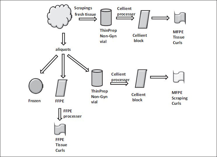
Specimen Handling
DNA Primers
Sequence Analysis, DNA
Azure Stains
Diagnostic Techniques, Urological
Diagnostic Techniques, Obstetrical and Gynecological
Mutation
Diagnostic Tests, Routine
Diagnostic Techniques, Digestive System
Molecular Sequence Data
Reverse Transcriptase Polymerase Chain Reaction
Real-Time Polymerase Chain Reaction
Diagnostic Techniques, Respiratory System
Tumor Markers, Biological
Base Sequence
Cytodiagnosis
Diagnostic Techniques, Neurological
Oligonucleotide Array Sequence Analysis
Predictive Value of Tests
Radionuclide Imaging
Gene Expression Profiling
Pedigree
Tomography, X-Ray Computed
Biopsy, Needle
Diagnostic Techniques, Endocrine
Ultrasonography
Evaluation Studies as Topic
Cluster Analysis
DNA, Ribosomal
Biopsy
Genotype
DNA
Biopsy, Fine-Needle
Microscopy
Diagnostic Techniques, Cardiovascular
Magnetic Resonance Imaging
Feces
Dog Diseases
Enzyme-Linked Immunosorbent Assay
Retrospective Studies
Mycoses
Prospective Studies
Immunoenzyme Techniques
Diagnostic Imaging
Biological Markers
Prevalence
Disease Outbreaks
Image Processing, Computer-Assisted
Fluorescent Antibody Technique
Prognosis
Characterization of telomerase activity in the human oocyte and preimplantation embryo. (1/1604)
Telomerase, a ribonucleoprotein, has been described as an essential component of highly proliferative cells as it stabilizes the telomeres and avoids cellular senescence. The objective of this study was to modify the polymerase chain reaction-based telomeric repeat amplification protocol to detect telomerase activity in the single cell and to characterize the activity expressed in the human oocyte through to the blastocyst stage embryo. A comparative evaluation of telomerase activity and developmental stage was conducted using discarded or donated human oocytes and embryos. Telomerase activity was detected in all developmental stages evaluated from immature oocytes through to blastocyst stage embryos. Immature oocytes and blastocysts had similar levels of telomerase activity; however, both groups had significantly (P < 0.05) higher activity than zygote through to pre-morula stage embryos. Seventy-five thawed zygotes were cultured to day 3, biopsied by removing 1-2 cells, and the biopsied embryos were cultured to blastocyst stage. There was no difference (P < 0.05) in telomerase activity between cells biopsied from embryos that reached the blastocyst stage and cells from those that arrested in growth. This study has shown that human oocytes through to blastocyst stage embryos express telomerase activity, but that the level of telomerase activity in biopsied blastomeres, of the day 3 cleavage stage embryo, is not predictive of embryonic growth potential. (+info)Characterization of relaxin binding in the uterus of the marmoset monkey. (2/1604)
The ovarian peptide hormone relaxin (RLX) plays an important role in the regulation of the endometrium both during the cycle and in early pregnancy. RLX interacts with specific receptors on endometrial stromal cells causing these to decidualize. In order to characterize the molecules with which RLX interacts in the primate uterus, a methodology based on a fully bioactive preparation of biotinylated porcine RLX was applied to cryosections of the uterus of female marmoset monkeys. Specific RLX binding was weakly detected in the proliferative phase in isolated endometrial stromal cells. In the secretory phase, the positively reacting cells increased in staining intensity and in number and also included some epithelial cells. Further increases occurred in pregnancy, but RLX binding in the endometrium decreased at the end of the cycle if pregnancy did not occur. The myometrium showed weak staining which did not vary through the cycle, but increased in pregnancy. Electrophoretic analysis of the RLX-binding moieties in these tissue sections indicated that a protein of approximately 40 kDa was the principal RLX-binding molecule, while minor specific bands were detectable at approximately 100 and approximately 200 kDa. The binding of biotinylated RLX could be specifically suppressed by co-incubation with unlabelled RLX, but not by insulin, IGF-I or biotin. This technique therefore allows the detection and molecular characterization of specific RLX binding in the primate uterus. In the marmoset monkey, the pattern of specific binding closely reflects the RLX-dependent physiology during implantation and early pregnancy, implying the probable involvement of a specific RLX receptor. (+info)Influence of age and gender on the clinical expression of acute intermittent porphyria based on molecular study of porphobilinogen deaminase gene among Swiss patients. (3/1604)
BACKGROUND: Acute intermittent porphyria (AIP) is an inherited disorder in the heme biosynthetic pathway caused by a partial deficiency of porphobilinogen (PBG) deaminase. Clinically, AIP is characterized as acute neurovisceral attacks that are often precipitated by exogenous factors such as drugs, hormones, and alcohol. An early detection of mutation carriers is essential for prevention of acute attacks by avoiding precipitating factors. This study was aimed at analyzing genetic defects causing AIP among Swiss families to further investigate aspects concerning the clinical expression of the disease. MATERIALS AND METHODS: The PBGD gene of index patients from 21 Swiss AIP families was systematically analyzed by denaturing gradient gel electrophoresis of polymerase chain reaction (PCR) amplified DNA fragments and direct sequencing. RESULTS: Five new mutations insA503, del L170, T190I, P241S, and R321H, as well as three known mutations (R26H, R173Q and W283X) were detected. Twelve of the 21 index patients (57%) carried the prevalent mutation W283X previously found among the Swiss AIP population. Family-specific mutations were then screened among relatives of the index patients. Among the 107 studied individuals, 58 carried a PBGD gene mutation--30 were overt AIP patients and 28 were asymptomatic carriers. The apparent rate of overt disease in the study cohort was 52%, which is significantly higher than the previously reported penetrance of 10-20%. To further examine the clinical expression of AIP, the cumulative life-time risk was calculated among 58 mutation-positive individuals after stratifying for age. The result shows a linear increase of the percentage of the symptomatic patients with age, reaching up to 75% among carriers aged over 60. Moreover, statistical analysis of the gender distribution among patients and asymptomatic carriers indicated that the disease was more frequently expressed among females than males (Fisher's exact test two sided, p= (0.001). CONCLUSIONS: This comprehensive search for genetic defects in the PBGD gene confirmed the existence of a prevalent mutation W283X among Swiss AIP patients, as well as a number of family-private mutations. Genetic analysis laid a groundwork for further studies such as the effects of gender and age on the clinical expression of AIP. (+info)BADGE, Beads Array for the Detection of Gene Expression, a high-throughput diagnostic bioassay. (4/1604)
Several methods are presently available for gene expression analysis. However, few of them are suitable for detection of moderate numbers of genes in thousands of samples with high speed and low cost. There is great demand for such a method for use in diagnostics and screening. To address this need, we have developed an assay for gene expression analysis using microspheres and a fluidic instrument made by Luminex. The assay is named Beads Array for the Detection of Gene Expression (BADGE). BADGE can monitor up to 100 genes in a single reaction, and it takes only 1 h to hybridize and <20 sec to read the results of all 100 genes in a sample for the detection process. For the genes detected in five independent replicate experiments, the standard deviation was <35% of the mean. We have monitored multiple pathogenesis-related genes simultaneously in chemical-treated and control Arabidopsis samples employing the BADGE assay. The data were compared with those obtained from an established technology, Affymetrix GeneChip. The changes in expression profiles were very similar. Our study showed that the BADGE assay was capable of profiling expression of multiple genes at affordable cost and rapid speed. (+info)Detection of mutations in the dystrophin gene via automated DHPLC screening and direct sequencing. (5/1604)
BACKGROUND: Currently molecular diagnostic laboratories focus only on the identification of large deletion and duplication mutations (spanning one exon or more) for Duchenne Muscular Dystrophy (DMD) yielding 65% of causative mutations. These mutations are detected by an existing set of multiplexed polymerase chain reaction (PCR) primer pairs. Due to the large size of the dystrophin gene (79 exons), finding point mutations (substitutions, deletions or insertions of one or several nucleotides) has been prohibitively expensive and laborious. The aim of this project was to develop an effective and convenient method of finding all, or most, mutations in the dystrophin gene with only a moderate increase in cost. RESULTS: Using denaturing high performance liquid chromatography (DHPLC) screening and direct sequencing, 86 PCR amplicons of genomic DNA from the dystrophin gene were screened for mutations in eight patients diagnosed with DMD who had tested negative for large DNA rearragements. Mutations likely to be disease-causative were found in six of the eight patients. All 86 amplicons from the two patients in whom no likely disease-causative mutations were found were completely sequenced and only polymorphisms were found. CONCLUSIONS: We have shown that it is now feasible for clinical laboratories to begin testing for both point mutations and large deletions/duplications in the dystrophin gene. The detection rate will rise from 65% to greater than 92% with only a moderate increase in cost. (+info)Rapid diagnostics: the detection of neuraminidase activity as a technology for high-specificity targets. (6/1604)
The accurate detection of influenza by clinical symptoms is challenging since multiple pathogenic viruses and bacteria mimic similar symptoms in a patient. With new and more effective influenza therapeutics available, there is a growing need for highly accurate and rapid diagnosis of influenza, particularly when the window of opportunity for proper treatment is measured in hours. A parallel technology, which is also used in the treatment of influenza, was developed for the rapid diagnosis of influenza by exploiting the enzymatic activity of influenza neuraminidase. This technology, which is called Pathozyme, offers the high specificity inherent from the conservation of the neuraminidase active site. The ZstatFlu test uses a small molecule derivative of sialic acid chemically coupled to a reporter group together with simple point-of-care reagents for directly detecting influenza from a patient specimen with high specificity. A second-generation platform technology using this neuraminidase detection system coupled with a more sensitive chemiluminescent reporter has been developed and formatted for reading on high-speed instant film. This modification resulted in a platform technology many-fold more sensitive than the former while maintaining its inherent high specificity. Preliminary data from a prototype tested during the mild 2000-2001 influenza season demonstrated that an optimized chemiluminescent test system could approach the accuracy of 14 day viral culture in a convenient 10-20 min test. This platform technology is currently being explored for the rapid detection of other pathogenic organisms where sensitivity, specificity and speed are essential in a point-of-care setting. (+info)Molecular analysis of Spinocerebellar ataxias in Koreans: frequencies and reference ranges of SCA1, SCA2, SCA3, SCA6, and SCA7. (7/1604)
Spinocerebellar ataxias (SCAs) are a heterogeneous group of neurodegenerative disorders. CAG repeat expansions in the causative genes have been identified as the basic cause of several types of SCAs, and have been used for the diagnoses and classifications of patients with ataxia. In order to assess the frequency and CAG repeat size ranges of SCAs, and to establish an effective strategy for molecular diagnosis, we performed a molecular analysis of SCA1, SCA2, SCA3, SCA6, and SCA7 in 76 patients. These patients were as follows: 32 with dominant inheritance, 39 sporadic cases, and 5 with unknown family histories. The normal and affected CAG repeat size ranges were established at five SCA loci in Koreans, which was consistent with previous reports. The total prevalence of the five types of SCAs was 39.5% in the 76 patients with ataxia, regardless of their family history. It was 75.0% in the 32 families with a dominant inheritance. The most frequent type was SCA3 (15.8%), followed by SCA2 (14.5%). Both types combined formed 76.7% of the 30 patients with CAG expansions. SCA1, SCA6, and SCA7 were less frequent, affecting 3.9%, 2.6%, and 2.6% of the cases, respectively. This mutation spectrum is quite different from a previous report concerning Koreans, but is similar to the distributions that are seen in several ethnic populations worldwide. For a correct and effective diagnosis of SCAs, we suggest that a molecular diagnosis be undertaken, even in patients without a family history, as well as those with a family history. A stepwise approach is also recommended. Patients with ataxia should be tested for SCA2 and SCA3. Individuals testing negative should be tested for SCA1, SCA6, and SCA7. (+info)Molecular diagnosis in haemophilia A. (8/1604)
Haemophilia A is the commonest cause of X-linked inherited bleeding disorder. Due to inadequate medical facility for management of the disease, the DNA based genetic diagnosis has assumed great importance. Ideally, the direct detection of mutations is the most accurate and reliable approach for carrier detection and prenatal diagnosis. However, mutation detection is possible only in limited number of cases. In majority of haemophiliacs, no common mutation is easily identifiable. The limitation has been over come by the use of linkage-based analysis using polymorphic DNA markers in the factor VIII gene. Some of these markers can be identified by restriction enzymes and are called RFLP markers. Other markers are a class of short tandem repeats sequences which result in differences in the number of CA repeats in different individuals. The combined use of these markers has made it possible to identify carriers and provide prenatal diagnosis in upto 95% of families having affected individuals. Therefore, the recurrence of the disease can be prevented to a great extent in the haemophilia A affected families. (+info)Molecular diagnostic techniques are a group of laboratory methods used to analyze biological markers in DNA, RNA, and proteins to identify specific health conditions or diseases at the molecular level. These techniques include various methods such as polymerase chain reaction (PCR), DNA sequencing, gene expression analysis, fluorescence in situ hybridization (FISH), and mass spectrometry.
Molecular diagnostic techniques are used to detect genetic mutations, chromosomal abnormalities, viral and bacterial infections, and other molecular changes associated with various diseases, including cancer, genetic disorders, infectious diseases, and neurological disorders. These techniques provide valuable information for disease diagnosis, prognosis, treatment planning, and monitoring of treatment response.
Compared to traditional diagnostic methods, molecular diagnostic techniques offer several advantages, such as higher sensitivity, specificity, and speed. They can detect small amounts of genetic material or proteins, even in early stages of the disease, and provide accurate results with a lower risk of false positives or negatives. Additionally, molecular diagnostic techniques can be automated, standardized, and performed in high-throughput formats, making them suitable for large-scale screening and research applications.
Molecular pathology is a branch of pathology that involves the study and diagnosis of diseases at the molecular level. It utilizes various molecular biology techniques such as DNA sequencing, polymerase chain reaction (PCR), and others to identify genetic mutations, gene expression changes, and protein abnormalities that underlie various diseases including cancer, genetic disorders, infectious diseases, and autoimmune conditions. The information obtained from molecular testing can help guide clinical decision-making, inform prognosis, and monitor response to therapy. Additionally, molecular pathology plays a critical role in the development of personalized medicine, which tailors treatment strategies based on an individual's unique genetic makeup and disease characteristics.
Sensitivity and specificity are statistical measures used to describe the performance of a diagnostic test or screening tool in identifying true positive and true negative results.
* Sensitivity refers to the proportion of people who have a particular condition (true positives) who are correctly identified by the test. It is also known as the "true positive rate" or "recall." A highly sensitive test will identify most or all of the people with the condition, but may also produce more false positives.
* Specificity refers to the proportion of people who do not have a particular condition (true negatives) who are correctly identified by the test. It is also known as the "true negative rate." A highly specific test will identify most or all of the people without the condition, but may also produce more false negatives.
In medical testing, both sensitivity and specificity are important considerations when evaluating a diagnostic test. High sensitivity is desirable for screening tests that aim to identify as many cases of a condition as possible, while high specificity is desirable for confirmatory tests that aim to rule out the condition in people who do not have it.
It's worth noting that sensitivity and specificity are often influenced by factors such as the prevalence of the condition in the population being tested, the threshold used to define a positive result, and the reliability and validity of the test itself. Therefore, it's important to consider these factors when interpreting the results of a diagnostic test.
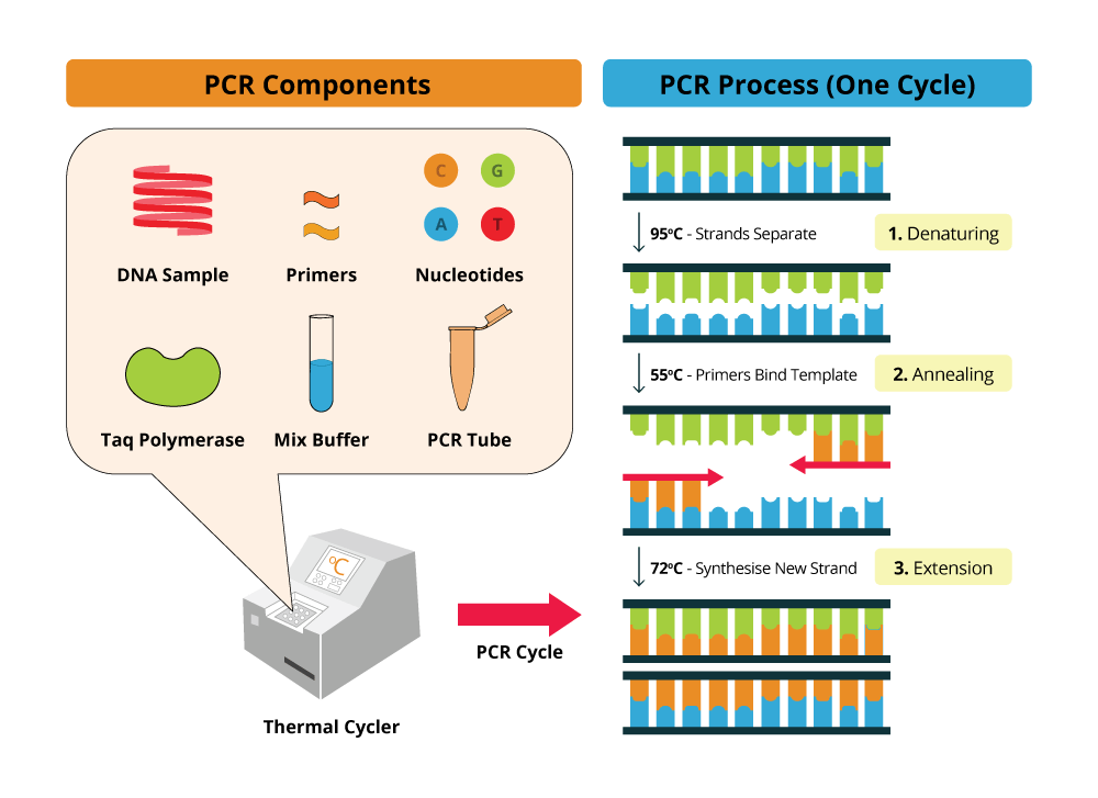
Polymerase Chain Reaction (PCR) is a laboratory technique used to amplify specific regions of DNA. It enables the production of thousands to millions of copies of a particular DNA sequence in a rapid and efficient manner, making it an essential tool in various fields such as molecular biology, medical diagnostics, forensic science, and research.
The PCR process involves repeated cycles of heating and cooling to separate the DNA strands, allow primers (short sequences of single-stranded DNA) to attach to the target regions, and extend these primers using an enzyme called Taq polymerase, resulting in the exponential amplification of the desired DNA segment.
In a medical context, PCR is often used for detecting and quantifying specific pathogens (viruses, bacteria, fungi, or parasites) in clinical samples, identifying genetic mutations or polymorphisms associated with diseases, monitoring disease progression, and evaluating treatment effectiveness.
I'm sorry for any confusion, but "pansporablastina" is not a recognized term in medical or scientific literature. It's possible that there may be a spelling error or it could be a very specialized or obscure term used only within a specific context. If you have more information or context about where this term was used, I'd be happy to help you try to clarify it further.
Diagnostic techniques and procedures are methods used by medical professionals to identify the cause of symptoms, illnesses, or diseases. These can include physical examinations, patient interviews, review of medical history, and various diagnostic tests. Diagnostic tests may involve invasive procedures such as biopsies or surgical interventions, or non-invasive imaging techniques like X-rays, CT scans, MRI scans, or ultrasounds. Functional tests, such as stress testing or electroencephalogram (EEG), can also be used to evaluate the functioning of specific organs or systems in the body. Laboratory tests, including blood tests, urine tests, and genetic tests, are also common diagnostic procedures. The choice of diagnostic technique or procedure depends on the presenting symptoms, the patient's medical history, and the suspected underlying condition.
Parasitology is a branch of biology that deals with the study of parasites, their life cycles, the relationship between parasites and their hosts, the transmission of parasitic diseases, and the development of methods for their control and elimination. It involves understanding various types of parasites including protozoa, helminths, and arthropods that can infect humans, animals, and plants. Parasitologists also study the evolution, genetics, biochemistry, and ecology of parasites to develop effective strategies for their diagnosis, treatment, and prevention.
Clinical laboratory techniques are methods and procedures used in medical laboratories to perform various tests and examinations on patient samples. These techniques help in the diagnosis, treatment, and prevention of diseases by analyzing body fluids, tissues, and other specimens. Some common clinical laboratory techniques include:
1. Clinical chemistry: It involves the analysis of bodily fluids such as blood, urine, and cerebrospinal fluid to measure the levels of chemicals, hormones, enzymes, and other substances in the body. These measurements can help diagnose various medical conditions, monitor treatment progress, and assess overall health.
2. Hematology: This technique focuses on the study of blood and its components, including red and white blood cells, platelets, and clotting factors. Hematological tests are used to diagnose anemia, infections, bleeding disorders, and other hematologic conditions.
3. Microbiology: It deals with the identification and culture of microorganisms such as bacteria, viruses, fungi, and parasites. Microbiological techniques are essential for detecting infectious diseases, determining appropriate antibiotic therapy, and monitoring the effectiveness of treatment.
4. Immunology: This technique involves studying the immune system and its response to various antigens, such as bacteria, viruses, and allergens. Immunological tests are used to diagnose autoimmune disorders, immunodeficiencies, and allergies.
5. Histopathology: It is the microscopic examination of tissue samples to identify any abnormalities or diseases. Histopathological techniques are crucial for diagnosing cancer, inflammatory conditions, and other tissue-related disorders.
6. Molecular biology: This technique deals with the study of DNA, RNA, and proteins at the molecular level. Molecular biology tests can be used to detect genetic mutations, identify infectious agents, and monitor disease progression.
7. Cytogenetics: It involves analyzing chromosomes and genes in cells to diagnose genetic disorders, cancer, and other diseases. Cytogenetic techniques include karyotyping, fluorescence in situ hybridization (FISH), and comparative genomic hybridization (CGH).
8. Flow cytometry: This technique measures physical and chemical characteristics of cells or particles as they flow through a laser beam. Flow cytometry is used to analyze cell populations, identify specific cell types, and detect abnormalities in cells.
9. Diagnostic radiology: It uses imaging technologies such as X-rays, computed tomography (CT), magnetic resonance imaging (MRI), and ultrasound to diagnose various medical conditions.
10. Clinical chemistry: This technique involves analyzing body fluids, such as blood and urine, to measure the concentration of various chemicals and substances. Clinical chemistry tests are used to diagnose metabolic disorders, electrolyte imbalances, and other health conditions.
Diagnostic techniques using radioisotopes, also known as nuclear medicine, are medical diagnostic procedures that use small amounts of radioactive material, called radioisotopes or radionuclides, to diagnose and monitor various diseases and conditions. The radioisotopes are introduced into the body through different routes (such as injection, inhalation, or ingestion) and accumulate in specific organs or tissues.
The gamma rays or photons emitted by these radioisotopes are then detected by specialized imaging devices, such as gamma cameras or PET scanners, which generate images that provide information about the structure and function of the organ or tissue being examined. This information helps healthcare professionals to make accurate diagnoses, monitor disease progression, assess treatment response, and plan appropriate therapies.
Common diagnostic techniques using radioisotopes include:
1. Radionuclide imaging (also known as scintigraphy): A gamma camera is used to produce images of specific organs or tissues after the administration of a radioisotope. Examples include bone scans, lung scans, heart scans, and brain scans.
2. Positron emission tomography (PET) scans: A PET scanner detects pairs of gamma rays emitted indirectly by a positron-emitting radionuclide, such as fluorodeoxyglucose (FDG), which is often used in oncology to assess metabolic activity and identify cancerous lesions.
3. Single-photon emission computed tomography (SPECT): A specialized gamma camera rotates around the patient, acquiring multiple images from different angles that are then reconstructed into a 3D image, providing detailed information about organ function and structure.
Diagnostic techniques using radioisotopes offer several advantages, including high sensitivity, non-invasiveness, and the ability to assess both anatomical and functional aspects of organs and tissues. However, they also involve exposure to ionizing radiation, so their use should be balanced against potential risks and benefits, and alternative diagnostic methods should be considered when appropriate.
Diagnostic techniques in otology refer to the methods and tests used by healthcare professionals to identify and diagnose various conditions related to the ear. These techniques can include:
1. Otoscopy: A visual examination of the external auditory canal and eardrum using an otoscope. This helps to identify any physical abnormalities, such as wax buildup, inflammation, or foreign objects in the ear.
2. Audiometry: A hearing test that measures a person's ability to hear different sounds, pitches, and volumes. This can help to identify any hearing loss or auditory processing issues.
3. Tympanometry: A test that measures the function of the middle ear by creating variations in air pressure in the ear canal. This can help to identify any issues with the eardrum or middle ear bones.
4. Acoustic reflex testing: A test that measures the body's involuntary response to loud sounds. This can help to identify any damage to the hearing nerves or brainstem.
5. Otoacoustic emissions (OAE) testing: A test that measures the sound waves produced by the inner ear in response to stimuli. This can help to identify any issues with the cochlea or hair cells in the inner ear.
6. Auditory brainstem response (ABR) testing: A test that measures the electrical activity of the hearing nerve and brainstem in response to sound. This can help to identify any issues with the auditory nervous system.
7. Vestibular testing: A series of tests that measure a person's balance and equilibrium. This can help to identify any issues with the vestibular system, which is responsible for maintaining balance.
These diagnostic techniques are used to diagnose various otological conditions such as hearing loss, tinnitus, vertigo, ear infections, and tumors of the ear.
Nucleic acid amplification techniques (NAATs) are medical laboratory methods used to increase the number of copies of a specific DNA or RNA sequence. These techniques are widely used in molecular biology and diagnostics, including the detection and diagnosis of infectious diseases, genetic disorders, and cancer.
The most commonly used NAAT is the polymerase chain reaction (PCR), which involves repeated cycles of heating and cooling to separate and replicate DNA strands. Other NAATs include loop-mediated isothermal amplification (LAMP), nucleic acid sequence-based amplification (NASBA), and transcription-mediated amplification (TMA).
NAATs offer several advantages over traditional culture methods for detecting pathogens, including faster turnaround times, increased sensitivity and specificity, and the ability to detect viable but non-culturable organisms. However, they also require specialized equipment and trained personnel, and there is a risk of contamination and false positive results if proper precautions are not taken.
Genetic testing is a type of medical test that identifies changes in chromosomes, genes, or proteins. The results of a genetic test can confirm or rule out a suspected genetic condition or help determine a person's chance of developing or passing on a genetic disorder. Genetic tests are performed on a sample of blood, hair, skin, amniotic fluid (the fluid that surrounds a fetus during pregnancy), or other tissue. For example, a physician may recommend genetic testing to help diagnose a genetic condition, confirm the presence of a gene mutation known to increase the risk of developing certain cancers, or determine the chance for a couple to have a child with a genetic disorder.
There are several types of genetic tests, including:
* Diagnostic testing: This type of test is used to identify or confirm a suspected genetic condition in an individual. It may be performed before birth (prenatal testing) or at any time during a person's life.
* Predictive testing: This type of test is used to determine the likelihood that a person will develop a genetic disorder. It is typically offered to individuals who have a family history of a genetic condition but do not show any symptoms themselves.
* Carrier testing: This type of test is used to determine whether a person carries a gene mutation for a genetic disorder. It is often offered to couples who are planning to have children and have a family history of a genetic condition or belong to a population that has an increased risk of certain genetic disorders.
* Preimplantation genetic testing: This type of test is used in conjunction with in vitro fertilization (IVF) to identify genetic changes in embryos before they are implanted in the uterus. It can help couples who have a family history of a genetic disorder or who are at risk of having a child with a genetic condition to conceive a child who is free of the genetic change in question.
* Pharmacogenetic testing: This type of test is used to determine how an individual's genes may affect their response to certain medications. It can help healthcare providers choose the most effective medication and dosage for a patient, reducing the risk of adverse drug reactions.
It is important to note that genetic testing should be performed under the guidance of a qualified healthcare professional who can interpret the results and provide appropriate counseling and support.
Reproducibility of results in a medical context refers to the ability to obtain consistent and comparable findings when a particular experiment or study is repeated, either by the same researcher or by different researchers, following the same experimental protocol. It is an essential principle in scientific research that helps to ensure the validity and reliability of research findings.
In medical research, reproducibility of results is crucial for establishing the effectiveness and safety of new treatments, interventions, or diagnostic tools. It involves conducting well-designed studies with adequate sample sizes, appropriate statistical analyses, and transparent reporting of methods and findings to allow other researchers to replicate the study and confirm or refute the results.
The lack of reproducibility in medical research has become a significant concern in recent years, as several high-profile studies have failed to produce consistent findings when replicated by other researchers. This has led to increased scrutiny of research practices and a call for greater transparency, rigor, and standardization in the conduct and reporting of medical research.
18S rRNA (ribosomal RNA) is the smaller subunit of the eukaryotic ribosome, which is the cellular organelle responsible for protein synthesis. The "18S" refers to the sedimentation coefficient of this rRNA molecule, which is a measure of its rate of sedimentation in a centrifuge and is expressed in Svedberg units (S).
The 18S rRNA is a component of the 40S subunit of the ribosome, and it plays a crucial role in the decoding of messenger RNA (mRNA) during protein synthesis. Specifically, the 18S rRNA helps to form the structure of the ribosome and contains several conserved regions that are involved in binding to mRNA and guiding the movement of transfer RNAs (tRNAs) during translation.
The 18S rRNA is also a commonly used molecular marker for evolutionary studies, as its sequence is highly conserved across different species and can be used to infer phylogenetic relationships between organisms. Additionally, the analysis of 18S rRNA gene sequences has been widely used in various fields such as ecology, environmental science, and medicine to study biodiversity, biogeography, and infectious diseases.
Diagnostic techniques, surgical refers to the use of surgical procedures or methods to diagnose and evaluate various medical conditions. These techniques are often used when non-invasive tests are inconclusive or when more detailed information is required. Here are some examples:
1. Biopsy: A small sample of tissue is removed from the body for examination under a microscope. This can help to confirm a diagnosis of cancer, infection, or other diseases.
2. Endoscopy: A flexible tube with a light and camera on the end is inserted into the body through a natural opening (such as the mouth or anus) or a small incision. This allows the doctor to visualize internal organs and tissues, and may also involve taking biopsy samples.
3. Imaging studies: Various imaging techniques such as X-rays, CT scans, MRI scans, and ultrasound can be used to produce detailed images of internal structures. These can help to diagnose a wide range of medical conditions, from broken bones to tumors.
4. Exploratory surgery: In some cases, a surgical incision may be made to directly visualize and examine an organ or tissue. This can help to diagnose conditions that are difficult to detect with non-invasive tests.
5. Functional testing: Some surgical techniques involve stimulating or measuring the function of an organ or system. For example, a cardiac stress test may be performed during surgery to assess heart function.
Overall, diagnostic techniques, surgical play an important role in the diagnosis and management of many medical conditions. They can provide valuable information that helps doctors to make informed decisions about treatment options and improve patient outcomes.
Specimen handling is a set of procedures and practices followed in the collection, storage, transportation, and processing of medical samples or specimens (e.g., blood, tissue, urine, etc.) for laboratory analysis. Proper specimen handling ensures accurate test results, patient safety, and data integrity. It includes:
1. Correct labeling of the specimen container with required patient information.
2. Using appropriate containers and materials to collect, store, and transport the specimen.
3. Following proper collection techniques to avoid contamination or damage to the specimen.
4. Adhering to specific storage conditions (temperature, time, etc.) before testing.
5. Ensuring secure and timely transportation of the specimen to the laboratory.
6. Properly documenting all steps in the handling process for traceability and quality assurance.
DNA primers are short single-stranded DNA molecules that serve as a starting point for DNA synthesis. They are typically used in laboratory techniques such as the polymerase chain reaction (PCR) and DNA sequencing. The primer binds to a complementary sequence on the DNA template through base pairing, providing a free 3'-hydroxyl group for the DNA polymerase enzyme to add nucleotides and synthesize a new strand of DNA. This allows for specific and targeted amplification or analysis of a particular region of interest within a larger DNA molecule.
Bacteriological techniques refer to the various methods and procedures used in the laboratory for the cultivation, identification, and study of bacteria. These techniques are essential in fields such as medicine, biotechnology, and research. Here are some common bacteriological techniques:
1. **Sterilization**: This is a process that eliminates or kills all forms of life, including bacteria, viruses, fungi, and spores. Common sterilization methods include autoclaving (using steam under pressure), dry heat (in an oven), chemical sterilants, and radiation.
2. **Aseptic Technique**: This refers to practices used to prevent contamination of sterile materials or environments with microorganisms. It includes the use of sterile equipment, gloves, and lab coats, as well as techniques such as flaming, alcohol swabbing, and using aseptic transfer devices.
3. **Media Preparation**: This involves the preparation of nutrient-rich substances that support bacterial growth. There are various types of media, including solid (agar), liquid (broth), and semi-solid (e.g., stab agar). The choice of medium depends on the type of bacteria being cultured and the purpose of the investigation.
4. **Inoculation**: This is the process of introducing a bacterial culture into a medium. It can be done using a loop, swab, or needle. The inoculum should be taken from a pure culture to avoid contamination.
5. **Incubation**: After inoculation, the bacteria are allowed to grow under controlled conditions of temperature, humidity, and atmospheric composition. This process is called incubation.
6. **Staining and Microscopy**: Bacteria are too small to be seen with the naked eye. Therefore, they need to be stained and observed under a microscope. Gram staining is a common method used to differentiate between two major groups of bacteria based on their cell wall composition.
7. **Biochemical Tests**: These are tests used to identify specific bacterial species based on their biochemical characteristics, such as their ability to ferment certain sugars, produce particular enzymes, or resist certain antibiotics.
8. **Molecular Techniques**: Advanced techniques like PCR and DNA sequencing can provide more precise identification of bacteria. They can also be used for genetic analysis and epidemiological studies.
Remember, handling microorganisms requires careful attention to biosafety procedures to prevent accidental infection or environmental contamination.
DNA Sequence Analysis is the systematic determination of the order of nucleotides in a DNA molecule. It is a critical component of modern molecular biology, genetics, and genetic engineering. The process involves determining the exact order of the four nucleotide bases - adenine (A), guanine (G), cytosine (C), and thymine (T) - in a DNA molecule or fragment. This information is used in various applications such as identifying gene mutations, studying evolutionary relationships, developing molecular markers for breeding, and diagnosing genetic diseases.
The process of DNA Sequence Analysis typically involves several steps, including DNA extraction, PCR amplification (if necessary), purification, sequencing reaction, and electrophoresis. The resulting data is then analyzed using specialized software to determine the exact sequence of nucleotides.
In recent years, high-throughput DNA sequencing technologies have revolutionized the field of genomics, enabling the rapid and cost-effective sequencing of entire genomes. This has led to an explosion of genomic data and new insights into the genetic basis of many diseases and traits.
DNA Mutational Analysis is a laboratory test used to identify genetic variations or changes (mutations) in the DNA sequence of a gene. This type of analysis can be used to diagnose genetic disorders, predict the risk of developing certain diseases, determine the most effective treatment for cancer, or assess the likelihood of passing on an inherited condition to offspring.
The test involves extracting DNA from a patient's sample (such as blood, saliva, or tissue), amplifying specific regions of interest using polymerase chain reaction (PCR), and then sequencing those regions to determine the precise order of nucleotide bases in the DNA molecule. The resulting sequence is then compared to reference sequences to identify any variations or mutations that may be present.
DNA Mutational Analysis can detect a wide range of genetic changes, including single-nucleotide polymorphisms (SNPs), insertions, deletions, duplications, and rearrangements. The test is often used in conjunction with other diagnostic tests and clinical evaluations to provide a comprehensive assessment of a patient's genetic profile.
It is important to note that not all mutations are pathogenic or associated with disease, and the interpretation of DNA Mutational Analysis results requires careful consideration of the patient's medical history, family history, and other relevant factors.
'Azure stains' is a term used in pathology to describe a histological staining technique that uses a type of dye called methyl blue, which turns the stained structures a blue-purple color. This technique is often used to stain acid mucins, which are found in various types of tissues and can be indicative of certain medical conditions.
In particular, azure stains are sometimes used to help diagnose certain types of cancer, such as mucoepidermoid carcinoma, a type of salivary gland tumor that produces acid mucins. The staining technique can help pathologists identify the presence and distribution of these mucins within the tumor cells, which can aid in making an accurate diagnosis and determining the best course of treatment.
It's worth noting that there are several different types of histological stains that use various dyes to highlight different structures or features within tissues. Azure stains are just one example of these techniques, and they are typically used in conjunction with other staining methods to provide a comprehensive picture of the tissue being examined.
Diagnostic techniques in urology are methods used to identify and diagnose various urological conditions affecting the urinary tract and male reproductive system. These techniques include:
1. Urinalysis: A laboratory examination of a urine sample to detect abnormalities such as infection, kidney stones, or other underlying medical conditions.
2. Urine Culture: A test used to identify and grow bacteria from the urine to determine the type of bacterial infection present in the urinary tract.
3. Imaging Studies: Various imaging techniques such as X-rays, ultrasound, CT scans, and MRI scans are used to visualize the internal structures of the urinary tract and identify any abnormalities.
4. Cystoscopy: A procedure that involves inserting a thin tube with a camera into the bladder through the urethra to examine the bladder and urethra for signs of disease or abnormality.
5. Urodynamics: A series of tests used to evaluate bladder function, including measuring bladder pressure and urine flow rate.
6. Biopsy: The removal and examination of tissue from the urinary tract or male reproductive system to diagnose conditions such as cancer.
7. Prostate-Specific Antigen (PSA) Test: A blood test used to screen for prostate cancer by measuring the level of PSA, a protein produced by the prostate gland.
8. Voiding Diary: A record of urinary habits, including the frequency and volume of urination, that can help diagnose conditions such as overactive bladder or urinary incontinence.
Diagnostic techniques in obstetrics and gynecology refer to the various methods used by healthcare professionals to diagnose and monitor conditions related to the female reproductive system and pregnancy. Here are some commonly used diagnostic techniques:
1. Physical examination: A thorough physical exam, including a pelvic exam, can help identify any abnormalities in the reproductive organs.
2. Medical history: A detailed medical history, including information about menstrual cycles, sexual activity, and family health, can provide valuable clues to diagnose various conditions.
3. Imaging tests: Ultrasound, CT scans, and MRIs can help healthcare professionals visualize the reproductive organs and detect any abnormalities.
4. Laboratory tests: Blood tests, urine tests, and cultures can help identify infections, hormonal imbalances, and other conditions.
5. Biopsy: A small sample of tissue is taken from the affected area and examined under a microscope to diagnose conditions such as cancer.
6. Colposcopy: This procedure involves using a special magnifying device to examine the cervix and vagina for signs of abnormalities.
7. Hysterosalpingography: This is an X-ray procedure that involves injecting a dye into the uterus and fallopian tubes to detect any blockages or other abnormalities.
8. Sonohysterography: This is an ultrasound procedure that involves injecting a fluid into the uterus to help visualize its interior and detect any abnormalities.
9. Minimally invasive surgery: Procedures such as laparoscopy and hysteroscopy can help healthcare professionals diagnose and treat various conditions related to the reproductive organs.
These diagnostic techniques can help healthcare professionals identify and manage a wide range of conditions, including infertility, pregnancy complications, infections, hormonal imbalances, and cancer.
A mutation is a permanent change in the DNA sequence of an organism's genome. Mutations can occur spontaneously or be caused by environmental factors such as exposure to radiation, chemicals, or viruses. They may have various effects on the organism, ranging from benign to harmful, depending on where they occur and whether they alter the function of essential proteins. In some cases, mutations can increase an individual's susceptibility to certain diseases or disorders, while in others, they may confer a survival advantage. Mutations are the driving force behind evolution, as they introduce new genetic variability into populations, which can then be acted upon by natural selection.
'Diagnostic tests, routine' is a medical term that refers to standard or commonly used tests that are performed to help diagnose, monitor, or manage a patient's health condition. These tests are typically simple, non-invasive, and safe, and they may be ordered as part of a regular check-up or when a patient presents with specific symptoms.
Routine diagnostic tests may include:
1. Complete Blood Count (CBC): A test that measures the number of red and white blood cells, platelets, and hemoglobin in the blood. It can help diagnose conditions such as anemia, infection, and inflammation.
2. Urinalysis: A test that examines a urine sample for signs of infection, kidney disease, or other medical conditions.
3. Blood Chemistry Tests: Also known as a chemistry panel or comprehensive metabolic panel, this test measures various chemicals in the blood such as glucose, electrolytes, and enzymes to evaluate organ function and overall health.
4. Electrocardiogram (ECG): A test that records the electrical activity of the heart, which can help diagnose heart conditions such as arrhythmias or heart attacks.
5. Chest X-ray: An imaging test that creates pictures of the structures inside the chest, including the heart, lungs, and bones, to help diagnose conditions such as pneumonia or lung cancer.
6. Fecal Occult Blood Test (FOBT): A test that checks for hidden blood in the stool, which can be a sign of colon cancer or other gastrointestinal conditions.
7. Pap Smear: A test that collects cells from the cervix to check for abnormalities that may indicate cervical cancer or other gynecological conditions.
These are just a few examples of routine diagnostic tests that healthcare providers may order. The specific tests ordered will depend on the patient's age, sex, medical history, and current symptoms.
Diagnostic techniques for the digestive system are medical tests and procedures used to diagnose and evaluate various conditions and diseases related to the gastrointestinal (GI) tract, including the esophagus, stomach, small intestine, large intestine, liver, gallbladder, pancreas, and associated organs. These techniques can be categorized into invasive and non-invasive methods.
Non-invasive diagnostic techniques:
1. Imaging tests: These include X-rays, computed tomography (CT) scans, magnetic resonance imaging (MRI), positron emission tomography (PET) scans, and ultrasounds. They help visualize the structure and function of the digestive organs without requiring any invasive procedures.
2. Laboratory tests: Blood, stool, and urine samples can be analyzed to detect signs of infection, inflammation, or other abnormalities related to digestive system disorders. Examples include complete blood count (CBC), liver function tests (LFTs), coagulation studies, and fecal occult blood test (FOBT).
3. Breath tests: These are used to diagnose conditions like lactose intolerance, small intestinal bacterial overgrowth (SIBO), or helicobacter pylori infection by analyzing the patient's exhaled air after consuming a specific substance.
Invasive diagnostic techniques:
1. Endoscopy: A thin, flexible tube with a light and camera attached to its end is inserted through the mouth or rectum to directly visualize the GI tract's inner lining. There are different types of endoscopies, such as gastroscopy (esophagus, stomach, and duodenum), colonoscopy (colon and rectum), sigmoidoscopy (lower part of the colon), and enteroscopy (small intestine).
2. Endoscopic ultrasound (EUS): This combines endoscopy with ultrasound technology to provide detailed images of the digestive organs' structure and surrounding tissues, allowing for accurate diagnosis and staging of conditions like cancer.
3. Biopsy: During an endoscopy or surgery, a small tissue sample can be taken from the affected area for further examination under a microscope to confirm a diagnosis or assess the severity of a condition.
4. Capsule endoscopy: A patient swallows a tiny camera-equipped capsule that transmits images as it passes through the GI tract, allowing doctors to diagnose conditions in the small intestine that may be difficult to reach with traditional endoscopes.
5. Imaging studies: Procedures like computed tomography (CT), magnetic resonance imaging (MRI), or positron emission tomography (PET) scans can provide detailed images of the digestive organs and help diagnose conditions like tumors, inflammation, or obstructions.
These diagnostic techniques help healthcare providers identify and manage various gastrointestinal conditions, ensuring appropriate treatment and improved patient outcomes.
Molecular sequence data refers to the specific arrangement of molecules, most commonly nucleotides in DNA or RNA, or amino acids in proteins, that make up a biological macromolecule. This data is generated through laboratory techniques such as sequencing, and provides information about the exact order of the constituent molecules. This data is crucial in various fields of biology, including genetics, evolution, and molecular biology, allowing for comparisons between different organisms, identification of genetic variations, and studies of gene function and regulation.
Bacterial DNA refers to the genetic material found in bacteria. It is composed of a double-stranded helix containing four nucleotide bases - adenine (A), thymine (T), guanine (G), and cytosine (C) - that are linked together by phosphodiester bonds. The sequence of these bases in the DNA molecule carries the genetic information necessary for the growth, development, and reproduction of bacteria.
Bacterial DNA is circular in most bacterial species, although some have linear chromosomes. In addition to the main chromosome, many bacteria also contain small circular pieces of DNA called plasmids that can carry additional genes and provide resistance to antibiotics or other environmental stressors.
Unlike eukaryotic cells, which have their DNA enclosed within a nucleus, bacterial DNA is present in the cytoplasm of the cell, where it is in direct contact with the cell's metabolic machinery. This allows for rapid gene expression and regulation in response to changing environmental conditions.
Reverse Transcriptase Polymerase Chain Reaction (RT-PCR) is a laboratory technique used in molecular biology to amplify and detect specific DNA sequences. This technique is particularly useful for the detection and quantification of RNA viruses, as well as for the analysis of gene expression.
The process involves two main steps: reverse transcription and polymerase chain reaction (PCR). In the first step, reverse transcriptase enzyme is used to convert RNA into complementary DNA (cDNA) by reading the template provided by the RNA molecule. This cDNA then serves as a template for the PCR amplification step.
In the second step, the PCR reaction uses two primers that flank the target DNA sequence and a thermostable polymerase enzyme to repeatedly copy the targeted cDNA sequence. The reaction mixture is heated and cooled in cycles, allowing the primers to anneal to the template, and the polymerase to extend the new strand. This results in exponential amplification of the target DNA sequence, making it possible to detect even small amounts of RNA or cDNA.
RT-PCR is a sensitive and specific technique that has many applications in medical research and diagnostics, including the detection of viruses such as HIV, hepatitis C virus, and SARS-CoV-2 (the virus that causes COVID-19). It can also be used to study gene expression, identify genetic mutations, and diagnose genetic disorders.
Real-Time Polymerase Chain Reaction (RT-PCR) is a laboratory technique used in molecular biology to amplify and detect specific DNA sequences in real-time. It is a sensitive and specific method that allows for the quantification of target nucleic acids, such as DNA or RNA, through the use of fluorescent reporter molecules.
The RT-PCR process involves several steps: first, the template DNA is denatured to separate the double-stranded DNA into single strands. Then, primers (short sequences of DNA) specific to the target sequence are added and allowed to anneal to the template DNA. Next, a heat-stable enzyme called Taq polymerase adds nucleotides to the annealed primers, extending them along the template DNA until a new double-stranded DNA molecule is formed.
During each amplification cycle, fluorescent reporter molecules are added that bind specifically to the newly synthesized DNA. As more and more copies of the target sequence are generated, the amount of fluorescence increases in proportion to the number of copies present. This allows for real-time monitoring of the PCR reaction and quantification of the target nucleic acid.
RT-PCR is commonly used in medical diagnostics, research, and forensics to detect and quantify specific DNA or RNA sequences. It has been widely used in the diagnosis of infectious diseases, genetic disorders, and cancer, as well as in the identification of microbial pathogens and the detection of gene expression.
Diagnostic techniques for the respiratory system are methods used to identify and diagnose various diseases and conditions affecting the lungs and breathing. Here are some commonly used diagnostic techniques:
1. Physical Examination: A healthcare provider will listen to your chest with a stethoscope to check for abnormal breath sounds, such as wheezing or crackles. They may also observe your respiratory rate and effort.
2. Chest X-ray: This imaging test can help identify abnormalities in the lungs, such as tumors, fluid accumulation, or collapsed lung sections.
3. Computed Tomography (CT) Scan: A CT scan uses X-rays to create detailed cross-sectional images of the lungs and surrounding structures. It can help detect nodules, cysts, or other abnormalities that may not be visible on a chest X-ray.
4. Pulmonary Function Tests (PFTs): These tests measure how well your lungs are working by assessing your ability to inhale and exhale air. Common PFTs include spirometry, lung volume measurement, and diffusing capacity testing.
5. Bronchoscopy: A thin, flexible tube with a camera and light is inserted through the nose or mouth into the airways to examine the lungs' interior and obtain tissue samples for biopsy.
6. Bronchoalveolar Lavage (BAL): During a bronchoscopy, fluid is introduced into a specific area of the lung and then suctioned out to collect cells and other materials for analysis.
7. Sleep Studies: These tests monitor your breathing patterns during sleep to diagnose conditions like sleep apnea or other sleep-related breathing disorders.
8. Sputum Analysis: A sample of coughed-up mucus is examined under a microscope to identify any abnormal cells, bacteria, or other organisms that may be causing respiratory issues.
9. Blood Tests: Blood tests can help diagnose various respiratory conditions by measuring oxygen and carbon dioxide levels, identifying specific antibodies or antigens, or detecting genetic markers associated with certain diseases.
10. Positron Emission Tomography (PET) Scan: A PET scan uses a small amount of radioactive material to create detailed images of the body's internal structures and functions, helping identify areas of abnormal cell growth or metabolic activity in the lungs.
Tumor markers are substances that can be found in the body and their presence can indicate the presence of certain types of cancer or other conditions. Biological tumor markers refer to those substances that are produced by cancer cells or by other cells in response to cancer or certain benign (non-cancerous) conditions. These markers can be found in various bodily fluids such as blood, urine, or tissue samples.
Examples of biological tumor markers include:
1. Proteins: Some tumor markers are proteins that are produced by cancer cells or by other cells in response to the presence of cancer. For example, prostate-specific antigen (PSA) is a protein produced by normal prostate cells and in higher amounts by prostate cancer cells.
2. Genetic material: Tumor markers can also include genetic material such as DNA, RNA, or microRNA that are shed by cancer cells into bodily fluids. For example, circulating tumor DNA (ctDNA) is genetic material from cancer cells that can be found in the bloodstream.
3. Metabolites: Tumor markers can also include metabolic products produced by cancer cells or by other cells in response to cancer. For example, lactate dehydrogenase (LDH) is an enzyme that is released into the bloodstream when cancer cells break down glucose for energy.
It's important to note that tumor markers are not specific to cancer and can be elevated in non-cancerous conditions as well. Therefore, they should not be used alone to diagnose cancer but rather as a tool in conjunction with other diagnostic tests and clinical evaluations.
A base sequence in the context of molecular biology refers to the specific order of nucleotides in a DNA or RNA molecule. In DNA, these nucleotides are adenine (A), guanine (G), cytosine (C), and thymine (T). In RNA, uracil (U) takes the place of thymine. The base sequence contains genetic information that is transcribed into RNA and ultimately translated into proteins. It is the exact order of these bases that determines the genetic code and thus the function of the DNA or RNA molecule.
Cytodiagnosis is the rapid, initial evaluation and diagnosis of a disease based on the examination of individual cells obtained from a body fluid or tissue sample. This technique is often used in cytopathology to investigate abnormalities such as lumps, bumps, or growths that may be caused by cancerous or benign conditions.
The process involves collecting cells through various methods like fine-needle aspiration (FNA), body fluids such as urine, sputum, or washings from the respiratory, gastrointestinal, or genitourinary tracts. The collected sample is then spread onto a microscope slide, stained, and examined under a microscope for abnormalities in cell size, shape, structure, and organization.
Cytodiagnosis can provide crucial information to guide further diagnostic procedures and treatment plans. It is often used as an initial screening tool due to its speed, simplicity, and cost-effectiveness compared to traditional histopathological methods that require tissue biopsy and more extensive processing. However, cytodiagnosis may not always be able to distinguish between benign and malignant conditions definitively; therefore, additional tests or follow-up evaluations might be necessary for a conclusive diagnosis.
Neurological diagnostic techniques are medical tests and examinations used to identify and diagnose conditions related to the nervous system, which includes the brain, spinal cord, nerves, and muscles. These techniques can be divided into several categories:
1. Clinical Examination: A thorough physical examination, including a neurological evaluation, is often the first step in diagnosing neurological conditions. This may involve assessing a person's mental status, muscle strength, coordination, reflexes, sensation, and gait.
2. Imaging Techniques: These are used to produce detailed images of the brain and nervous system. Common imaging techniques include:
- Computed Tomography (CT): This uses X-rays to create cross-sectional images of the brain and other parts of the body.
- Magnetic Resonance Imaging (MRI): This uses a strong magnetic field and radio waves to produce detailed images of the brain and other internal structures.
- Functional MRI (fMRI): This is a type of MRI that measures brain activity by detecting changes in blood flow.
- Positron Emission Tomography (PET): This uses small amounts of radioactive material to produce detailed images of brain function.
- Single Photon Emission Computed Tomography (SPECT): This is a type of nuclear medicine imaging that uses a gamma camera and a computer to produce detailed images of brain function.
3. Electrophysiological Tests: These are used to measure the electrical activity of the brain and nervous system. Common electrophysiological tests include:
- Electroencephalography (EEG): This measures the electrical activity of the brain.
- Evoked Potentials (EPs): These measure the electrical response of the brain and nervous system to sensory stimuli, such as sound or light.
- Nerve Conduction Studies (NCS): These measure the speed and strength of nerve impulses.
- Electromyography (EMG): This measures the electrical activity of muscles.
4. Laboratory Tests: These are used to analyze blood, cerebrospinal fluid, and other bodily fluids for signs of neurological conditions. Common laboratory tests include:
- Complete Blood Count (CBC): This measures the number and type of white and red blood cells in the body.
- Blood Chemistry Tests: These measure the levels of various chemicals in the blood.
- Lumbar Puncture (Spinal Tap): This is used to collect cerebrospinal fluid for analysis.
- Genetic Testing: This is used to identify genetic mutations associated with neurological conditions.
5. Imaging Studies: These are used to produce detailed images of the brain and nervous system. Common imaging studies include:
- Magnetic Resonance Imaging (MRI): This uses a strong magnetic field and radio waves to produce detailed images of the brain and nervous system.
- Computed Tomography (CT): This uses X-rays to produce detailed images of the brain and nervous system.
- Functional MRI (fMRI): This measures changes in blood flow in the brain during cognitive tasks.
- Diffusion Tensor Imaging (DTI): This is used to assess white matter integrity in the brain.
- Magnetic Resonance Spectroscopy (MRS): This is used to measure chemical levels in the brain.
Oligonucleotide Array Sequence Analysis is a type of microarray analysis that allows for the simultaneous measurement of the expression levels of thousands of genes in a single sample. In this technique, oligonucleotides (short DNA sequences) are attached to a solid support, such as a glass slide, in a specific pattern. These oligonucleotides are designed to be complementary to specific target mRNA sequences from the sample being analyzed.
During the analysis, labeled RNA or cDNA from the sample is hybridized to the oligonucleotide array. The level of hybridization is then measured and used to determine the relative abundance of each target sequence in the sample. This information can be used to identify differences in gene expression between samples, which can help researchers understand the underlying biological processes involved in various diseases or developmental stages.
It's important to note that this technique requires specialized equipment and bioinformatics tools for data analysis, as well as careful experimental design and validation to ensure accurate and reproducible results.
The Predictive Value of Tests, specifically the Positive Predictive Value (PPV) and Negative Predictive Value (NPV), are measures used in diagnostic tests to determine the probability that a positive or negative test result is correct.
Positive Predictive Value (PPV) is the proportion of patients with a positive test result who actually have the disease. It is calculated as the number of true positives divided by the total number of positive results (true positives + false positives). A higher PPV indicates that a positive test result is more likely to be a true positive, and therefore the disease is more likely to be present.
Negative Predictive Value (NPV) is the proportion of patients with a negative test result who do not have the disease. It is calculated as the number of true negatives divided by the total number of negative results (true negatives + false negatives). A higher NPV indicates that a negative test result is more likely to be a true negative, and therefore the disease is less likely to be present.
The predictive value of tests depends on the prevalence of the disease in the population being tested, as well as the sensitivity and specificity of the test. A test with high sensitivity and specificity will generally have higher predictive values than a test with low sensitivity and specificity. However, even a highly sensitive and specific test can have low predictive values if the prevalence of the disease is low in the population being tested.
Radionuclide imaging, also known as nuclear medicine, is a medical imaging technique that uses small amounts of radioactive material, called radionuclides or radiopharmaceuticals, to diagnose and treat various diseases and conditions. The radionuclides are introduced into the body through injection, inhalation, or ingestion and accumulate in specific organs or tissues. A special camera then detects the gamma rays emitted by these radionuclides and converts them into images that provide information about the structure and function of the organ or tissue being studied.
Radionuclide imaging can be used to evaluate a wide range of medical conditions, including heart disease, cancer, neurological disorders, gastrointestinal disorders, and bone diseases. The technique is non-invasive and generally safe, with minimal exposure to radiation. However, it should only be performed by qualified healthcare professionals in accordance with established guidelines and regulations.
Gene expression profiling is a laboratory technique used to measure the activity (expression) of thousands of genes at once. This technique allows researchers and clinicians to identify which genes are turned on or off in a particular cell, tissue, or organism under specific conditions, such as during health, disease, development, or in response to various treatments.
The process typically involves isolating RNA from the cells or tissues of interest, converting it into complementary DNA (cDNA), and then using microarray or high-throughput sequencing technologies to determine which genes are expressed and at what levels. The resulting data can be used to identify patterns of gene expression that are associated with specific biological states or processes, providing valuable insights into the underlying molecular mechanisms of diseases and potential targets for therapeutic intervention.
In recent years, gene expression profiling has become an essential tool in various fields, including cancer research, drug discovery, and personalized medicine, where it is used to identify biomarkers of disease, predict patient outcomes, and guide treatment decisions.
I must clarify that the term "pedigree" is not typically used in medical definitions. Instead, it is often employed in genetics and breeding, where it refers to the recorded ancestry of an individual or a family, tracing the inheritance of specific traits or diseases. In human genetics, a pedigree can help illustrate the pattern of genetic inheritance in families over multiple generations. However, it is not a medical term with a specific clinical definition.
X-ray computed tomography (CT or CAT scan) is a medical imaging method that uses computer-processed combinations of many X-ray images taken from different angles to produce cross-sectional (tomographic) images (virtual "slices") of the body. These cross-sectional images can then be used to display detailed internal views of organs, bones, and soft tissues in the body.
The term "computed tomography" is used instead of "CT scan" or "CAT scan" because the machines take a series of X-ray measurements from different angles around the body and then use a computer to process these data to create detailed images of internal structures within the body.
CT scanning is a noninvasive, painless medical test that helps physicians diagnose and treat medical conditions. CT imaging provides detailed information about many types of tissue including lung, bone, soft tissue and blood vessels. CT examinations can be performed on every part of the body for a variety of reasons including diagnosis, surgical planning, and monitoring of therapeutic responses.
In computed tomography (CT), an X-ray source and detector rotate around the patient, measuring the X-ray attenuation at many different angles. A computer uses this data to construct a cross-sectional image by the process of reconstruction. This technique is called "tomography". The term "computed" refers to the use of a computer to reconstruct the images.
CT has become an important tool in medical imaging and diagnosis, allowing radiologists and other physicians to view detailed internal images of the body. It can help identify many different medical conditions including cancer, heart disease, lung nodules, liver tumors, and internal injuries from trauma. CT is also commonly used for guiding biopsies and other minimally invasive procedures.
In summary, X-ray computed tomography (CT or CAT scan) is a medical imaging technique that uses computer-processed combinations of many X-ray images taken from different angles to produce cross-sectional images of the body. It provides detailed internal views of organs, bones, and soft tissues in the body, allowing physicians to diagnose and treat medical conditions.
A needle biopsy is a medical procedure in which a thin, hollow needle is used to remove a small sample of tissue from a suspicious or abnormal area of the body. The tissue sample is then examined under a microscope to check for cancer cells or other abnormalities. Needle biopsies are often used to diagnose lumps or masses that can be felt through the skin, but they can also be guided by imaging techniques such as ultrasound, CT scan, or MRI to reach areas that cannot be felt. There are several types of needle biopsy procedures, including fine-needle aspiration (FNA) and core needle biopsy. FNA uses a thin needle and gentle suction to remove fluid and cells from the area, while core needle biopsy uses a larger needle to remove a small piece of tissue. The type of needle biopsy used depends on the location and size of the abnormal area, as well as the reason for the procedure.
Diagnostic techniques in endocrinology are methods used to identify and diagnose various endocrine disorders. These techniques include:
1. Hormone measurements: Measuring the levels of hormones in blood, urine, or saliva can help identify excess or deficiency of specific hormones. This is often done through immunoassays, which use antibodies to detect and quantify hormones.
2. Provocative and suppression tests: These tests involve administering a medication that stimulates or suppresses the release of a particular hormone. Blood samples are taken before and after the medication is given to assess changes in hormone levels. Examples include the glucose tolerance test for diabetes, the ACTH stimulation test for adrenal insufficiency, and the thyroid suppression test for hyperthyroidism.
3. Imaging studies: Various imaging techniques can be used to visualize endocrine glands and identify structural abnormalities such as tumors or nodules. These include X-rays, ultrasound, computed tomography (CT), magnetic resonance imaging (MRI), and nuclear medicine scans using radioactive tracers.
4. Genetic testing: Molecular genetic tests can be used to identify genetic mutations associated with certain endocrine disorders, such as multiple endocrine neoplasia type 1 or 2, or congenital adrenal hyperplasia.
5. Biopsy: In some cases, a small sample of tissue may be removed from an endocrine gland for microscopic examination (biopsy). This can help confirm the presence of cancer or other abnormalities.
6. Functional tests: These tests assess the ability of an endocrine gland to produce and secrete hormones in response to various stimuli. Examples include the glucagon stimulation test for gastrinoma and the calcium infusion test for hyperparathyroidism.
7. Wearable monitoring devices: Continuous glucose monitoring systems (CGMS) are wearable devices that measure interstitial glucose levels continuously over several days, providing valuable information about glycemic control in patients with diabetes.
Ultrasonography, also known as sonography, is a diagnostic medical procedure that uses high-frequency sound waves (ultrasound) to produce dynamic images of organs, tissues, or blood flow inside the body. These images are captured in real-time and can be used to assess the size, shape, and structure of various internal structures, as well as detect any abnormalities such as tumors, cysts, or inflammation.
During an ultrasonography procedure, a small handheld device called a transducer is placed on the patient's skin, which emits and receives sound waves. The transducer sends high-frequency sound waves into the body, and these waves bounce back off internal structures and are recorded by the transducer. The recorded data is then processed and transformed into visual images that can be interpreted by a medical professional.
Ultrasonography is a non-invasive, painless, and safe procedure that does not use radiation like other imaging techniques such as CT scans or X-rays. It is commonly used to diagnose and monitor conditions in various parts of the body, including the abdomen, pelvis, heart, blood vessels, and musculoskeletal system.
"Evaluation studies" is a broad term that refers to the systematic assessment or examination of a program, project, policy, intervention, or product. The goal of an evaluation study is to determine its merits, worth, and value by measuring its effects, efficiency, and impact. There are different types of evaluation studies, including formative evaluations (conducted during the development or implementation of a program to provide feedback for improvement), summative evaluations (conducted at the end of a program to determine its overall effectiveness), process evaluations (focusing on how a program is implemented and delivered), outcome evaluations (assessing the short-term and intermediate effects of a program), and impact evaluations (measuring the long-term and broad consequences of a program).
In medical contexts, evaluation studies are often used to assess the safety, efficacy, and cost-effectiveness of new treatments, interventions, or technologies. These studies can help healthcare providers make informed decisions about patient care, guide policymakers in developing evidence-based policies, and promote accountability and transparency in healthcare systems. Examples of evaluation studies in medicine include randomized controlled trials (RCTs) that compare the outcomes of a new treatment to those of a standard or placebo treatment, observational studies that examine the real-world effectiveness and safety of interventions, and economic evaluations that assess the costs and benefits of different healthcare options.
Bronchoscopy is a medical procedure that involves the examination of the inside of the airways and lungs with a flexible or rigid tube called a bronchoscope. This procedure allows healthcare professionals to directly visualize the airways, take tissue samples for biopsy, and remove foreign objects or secretions. Bronchoscopy can be used to diagnose and manage various respiratory conditions such as lung infections, inflammation, cancer, and bleeding. It is usually performed under local or general anesthesia to minimize discomfort and risks associated with the procedure.
Cluster analysis is a statistical method used to group similar objects or data points together based on their characteristics or features. In medical and healthcare research, cluster analysis can be used to identify patterns or relationships within complex datasets, such as patient records or genetic information. This technique can help researchers to classify patients into distinct subgroups based on their symptoms, diagnoses, or other variables, which can inform more personalized treatment plans or public health interventions.
Cluster analysis involves several steps, including:
1. Data preparation: The researcher must first collect and clean the data, ensuring that it is complete and free from errors. This may involve removing outlier values or missing data points.
2. Distance measurement: Next, the researcher must determine how to measure the distance between each pair of data points. Common methods include Euclidean distance (the straight-line distance between two points) or Manhattan distance (the distance between two points along a grid).
3. Clustering algorithm: The researcher then applies a clustering algorithm, which groups similar data points together based on their distances from one another. Common algorithms include hierarchical clustering (which creates a tree-like structure of clusters) or k-means clustering (which assigns each data point to the nearest centroid).
4. Validation: Finally, the researcher must validate the results of the cluster analysis by evaluating the stability and robustness of the clusters. This may involve re-running the analysis with different distance measures or clustering algorithms, or comparing the results to external criteria.
Cluster analysis is a powerful tool for identifying patterns and relationships within complex datasets, but it requires careful consideration of the data preparation, distance measurement, and validation steps to ensure accurate and meaningful results.
Ribosomal DNA (rDNA) refers to the specific regions of DNA in a cell that contain the genes for ribosomal RNA (rRNA). Ribosomes are complex structures composed of proteins and rRNA, which play a crucial role in protein synthesis by translating messenger RNA (mRNA) into proteins.
In humans, there are four types of rRNA molecules: 18S, 5.8S, 28S, and 5S. These rRNAs are encoded by multiple copies of rDNA genes that are organized in clusters on specific chromosomes. In humans, the majority of rDNA genes are located on the short arms of acrocentric chromosomes 13, 14, 15, 21, and 22.
Each cluster of rDNA genes contains both transcribed and non-transcribed spacer regions. The transcribed regions contain the genes for the four types of rRNA, while the non-transcribed spacers contain regulatory elements that control the transcription of the rRNA genes.
The number of rDNA copies varies between species and even within individuals of the same species. The copy number can also change during development and in response to environmental factors. Variations in rDNA copy number have been associated with various diseases, including cancer and neurological disorders.
A biopsy is a medical procedure in which a small sample of tissue is taken from the body to be examined under a microscope for the presence of disease. This can help doctors diagnose and monitor various medical conditions, such as cancer, infections, or autoimmune disorders. The type of biopsy performed will depend on the location and nature of the suspected condition. Some common types of biopsies include:
1. Incisional biopsy: In this procedure, a surgeon removes a piece of tissue from an abnormal area using a scalpel or other surgical instrument. This type of biopsy is often used when the lesion is too large to be removed entirely during the initial biopsy.
2. Excisional biopsy: An excisional biopsy involves removing the entire abnormal area, along with a margin of healthy tissue surrounding it. This technique is typically employed for smaller lesions or when cancer is suspected.
3. Needle biopsy: A needle biopsy uses a thin, hollow needle to extract cells or fluid from the body. There are two main types of needle biopsies: fine-needle aspiration (FNA) and core needle biopsy. FNA extracts loose cells, while a core needle biopsy removes a small piece of tissue.
4. Punch biopsy: In a punch biopsy, a round, sharp tool is used to remove a small cylindrical sample of skin tissue. This type of biopsy is often used for evaluating rashes or other skin abnormalities.
5. Shave biopsy: During a shave biopsy, a thin slice of tissue is removed from the surface of the skin using a sharp razor-like instrument. This technique is typically used for superficial lesions or growths on the skin.
After the biopsy sample has been collected, it is sent to a laboratory where a pathologist will examine the tissue under a microscope and provide a diagnosis based on their findings. The results of the biopsy can help guide further treatment decisions and determine the best course of action for managing the patient's condition.
Genotype, in genetics, refers to the complete heritable genetic makeup of an individual organism, including all of its genes. It is the set of instructions contained in an organism's DNA for the development and function of that organism. The genotype is the basis for an individual's inherited traits, and it can be contrasted with an individual's phenotype, which refers to the observable physical or biochemical characteristics of an organism that result from the expression of its genes in combination with environmental influences.
It is important to note that an individual's genotype is not necessarily identical to their genetic sequence. Some genes have multiple forms called alleles, and an individual may inherit different alleles for a given gene from each parent. The combination of alleles that an individual inherits for a particular gene is known as their genotype for that gene.
Understanding an individual's genotype can provide important information about their susceptibility to certain diseases, their response to drugs and other treatments, and their risk of passing on inherited genetic disorders to their offspring.
Deoxyribonucleic acid (DNA) is the genetic material present in the cells of organisms where it is responsible for the storage and transmission of hereditary information. DNA is a long molecule that consists of two strands coiled together to form a double helix. Each strand is made up of a series of four nucleotide bases - adenine (A), guanine (G), cytosine (C), and thymine (T) - that are linked together by phosphate and sugar groups. The sequence of these bases along the length of the molecule encodes genetic information, with A always pairing with T and C always pairing with G. This base-pairing allows for the replication and transcription of DNA, which are essential processes in the functioning and reproduction of all living organisms.
A fine-needle biopsy (FNB) is a medical procedure in which a thin, hollow needle is used to obtain a sample of cells or tissue from a suspicious or abnormal area in the body, such as a lump or mass. The needle is typically smaller than that used in a core needle biopsy, and it is guided into place using imaging techniques such as ultrasound, CT scan, or MRI.
The sample obtained during an FNB can be used to diagnose various medical conditions, including cancer, infection, or inflammation. The procedure is generally considered safe and well-tolerated, with minimal risks of complications such as bleeding, infection, or discomfort. However, the accuracy of the diagnosis depends on the skill and experience of the healthcare provider performing the biopsy, as well as the adequacy of the sample obtained.
Overall, FNB is a valuable diagnostic tool that can help healthcare providers make informed decisions about treatment options and improve patient outcomes.
Microscopy is a technical field in medicine that involves the use of microscopes to observe structures and phenomena that are too small to be seen by the naked eye. It allows for the examination of samples such as tissues, cells, and microorganisms at high magnifications, enabling the detection and analysis of various medical conditions, including infections, diseases, and cellular abnormalities.
There are several types of microscopy used in medicine, including:
1. Light Microscopy: This is the most common type of microscopy, which uses visible light to illuminate and magnify samples. It can be used to examine a wide range of biological specimens, such as tissue sections, blood smears, and bacteria.
2. Electron Microscopy: This type of microscopy uses a beam of electrons instead of light to produce highly detailed images of samples. It is often used in research settings to study the ultrastructure of cells and tissues.
3. Fluorescence Microscopy: This technique involves labeling specific molecules within a sample with fluorescent dyes, allowing for their visualization under a microscope. It can be used to study protein interactions, gene expression, and cell signaling pathways.
4. Confocal Microscopy: This type of microscopy uses a laser beam to scan a sample point by point, producing high-resolution images with reduced background noise. It is often used in medical research to study the structure and function of cells and tissues.
5. Scanning Probe Microscopy: This technique involves scanning a sample with a physical probe, allowing for the measurement of topography, mechanical properties, and other characteristics at the nanoscale. It can be used in medical research to study the structure and function of individual molecules and cells.
Diagnostic techniques in cardiovascular medicine refer to the various tests and methods used to diagnose and evaluate conditions related to the heart and blood vessels. These techniques can be non-invasive or invasive and are designed to provide critical information about a patient's cardiovascular health, such as heart function, blood flow, and the presence of any abnormalities or diseases. Here are some common diagnostic techniques used in cardiovascular medicine:
1. Electrocardiogram (ECG): An ECG is a non-invasive test that records the electrical activity of the heart. It can help detect heart conditions such as arrhythmias, heart attacks, and structural abnormalities.
2. Echocardiogram: This is a non-invasive ultrasound test that produces images of the heart's structures, including the chambers, valves, and major blood vessels. It can help assess heart function, identify damage from heart attacks, and detect various cardiovascular conditions.
3. Stress testing: A stress test involves exercising on a treadmill or stationary bike while being monitored by an ECG to evaluate the heart's response to physical exertion. It can help diagnose coronary artery disease, assess exercise capacity, and determine the need for further testing or treatment.
4. Cardiac catheterization: This is an invasive procedure where a thin, flexible tube (catheter) is inserted into a blood vessel in the arm or leg and guided to the heart. It can help diagnose and treat various cardiovascular conditions, such as blocked arteries, heart valve problems, and congenital heart defects.
5. Coronary angiography: During a cardiac catheterization, a special dye is injected into the coronary arteries to visualize blood flow using X-ray imaging. This can help identify blockages or narrowing in the coronary arteries and guide treatment decisions.
6. Nuclear stress testing: This test combines the use of a radioactive tracer with exercise or pharmacological stress to evaluate heart function and blood flow. It can help diagnose coronary artery disease, assess the effectiveness of treatments, and determine the need for further interventions.
7. Cardiac magnetic resonance imaging (MRI): This non-invasive imaging technique uses a strong magnetic field and radio waves to create detailed images of the heart's structure and function. It can help diagnose various cardiovascular conditions, such as heart muscle disorders, valve problems, and congenital heart defects.
8. Transesophageal echocardiography (TEE): This is a specialized ultrasound technique where a probe is inserted through the esophagus to obtain detailed images of the heart's structure and function. It can help diagnose conditions such as blood clots, valve problems, and infective endocarditis.
9. Positron emission tomography (PET) scanning: This imaging technique uses a small amount of radioactive tracer to evaluate the metabolic activity of the heart. It can help diagnose coronary artery disease, assess the effectiveness of treatments, and determine the need for further interventions.
10. Electrophysiology studies (EPS): These are invasive procedures where catheters are inserted into the heart to study its electrical system. They can help diagnose and treat various arrhythmias, such as atrial fibrillation, ventricular tachycardia, and Wolff-Parkinson-White syndrome.
Medical Definition:
Magnetic Resonance Imaging (MRI) is a non-invasive diagnostic imaging technique that uses a strong magnetic field and radio waves to create detailed cross-sectional or three-dimensional images of the internal structures of the body. The patient lies within a large, cylindrical magnet, and the scanner detects changes in the direction of the magnetic field caused by protons in the body. These changes are then converted into detailed images that help medical professionals to diagnose and monitor various medical conditions, such as tumors, injuries, or diseases affecting the brain, spinal cord, heart, blood vessels, joints, and other internal organs. MRI does not use radiation like computed tomography (CT) scans.
Phylogeny is the evolutionary history and relationship among biological entities, such as species or genes, based on their shared characteristics. In other words, it refers to the branching pattern of evolution that shows how various organisms have descended from a common ancestor over time. Phylogenetic analysis involves constructing a tree-like diagram called a phylogenetic tree, which depicts the inferred evolutionary relationships among organisms or genes based on molecular sequence data or other types of characters. This information is crucial for understanding the diversity and distribution of life on Earth, as well as for studying the emergence and spread of diseases.
Feces are the solid or semisolid remains of food that could not be digested or absorbed in the small intestine, along with bacteria and other waste products. After being stored in the colon, feces are eliminated from the body through the rectum and anus during defecation. Feces can vary in color, consistency, and odor depending on a person's diet, health status, and other factors.
There is no medical definition for "dog diseases" as it is too broad a term. However, dogs can suffer from various health conditions and illnesses that are specific to their species or similar to those found in humans. Some common categories of dog diseases include:
1. Infectious Diseases: These are caused by viruses, bacteria, fungi, or parasites. Examples include distemper, parvovirus, kennel cough, Lyme disease, and heartworms.
2. Hereditary/Genetic Disorders: Some dogs may inherit certain genetic disorders from their parents. Examples include hip dysplasia, elbow dysplasia, progressive retinal atrophy (PRA), and degenerative myelopathy.
3. Age-Related Diseases: As dogs age, they become more susceptible to various health issues. Common age-related diseases in dogs include arthritis, dental disease, cancer, and cognitive dysfunction syndrome (CDS).
4. Nutritional Disorders: Malnutrition or improper feeding can lead to various health problems in dogs. Examples include obesity, malnutrition, and vitamin deficiencies.
5. Environmental Diseases: These are caused by exposure to environmental factors such as toxins, allergens, or extreme temperatures. Examples include heatstroke, frostbite, and toxicities from ingesting harmful substances.
6. Neurological Disorders: Dogs can suffer from various neurological conditions that affect their nervous system. Examples include epilepsy, intervertebral disc disease (IVDD), and vestibular disease.
7. Behavioral Disorders: Some dogs may develop behavioral issues due to various factors such as anxiety, fear, or aggression. Examples include separation anxiety, noise phobias, and resource guarding.
It's important to note that regular veterinary care, proper nutrition, exercise, and preventative measures can help reduce the risk of many dog diseases.
An Enzyme-Linked Immunosorbent Assay (ELISA) is a type of analytical biochemistry assay used to detect and quantify the presence of a substance, typically a protein or peptide, in a liquid sample. It takes its name from the enzyme-linked antibodies used in the assay.
In an ELISA, the sample is added to a well containing a surface that has been treated to capture the target substance. If the target substance is present in the sample, it will bind to the surface. Next, an enzyme-linked antibody specific to the target substance is added. This antibody will bind to the captured target substance if it is present. After washing away any unbound material, a substrate for the enzyme is added. If the enzyme is present due to its linkage to the antibody, it will catalyze a reaction that produces a detectable signal, such as a color change or fluorescence. The intensity of this signal is proportional to the amount of target substance present in the sample, allowing for quantification.
ELISAs are widely used in research and clinical settings to detect and measure various substances, including hormones, viruses, and bacteria. They offer high sensitivity, specificity, and reproducibility, making them a reliable choice for many applications.
Retrospective studies, also known as retrospective research or looking back studies, are a type of observational study that examines data from the past to draw conclusions about possible causal relationships between risk factors and outcomes. In these studies, researchers analyze existing records, medical charts, or previously collected data to test a hypothesis or answer a specific research question.
Retrospective studies can be useful for generating hypotheses and identifying trends, but they have limitations compared to prospective studies, which follow participants forward in time from exposure to outcome. Retrospective studies are subject to biases such as recall bias, selection bias, and information bias, which can affect the validity of the results. Therefore, retrospective studies should be interpreted with caution and used primarily to generate hypotheses for further testing in prospective studies.
Mycoses are a group of diseases caused by fungal infections. These infections can affect various parts of the body, including the skin, nails, hair, lungs, and internal organs. The severity of mycoses can range from superficial, mild infections to systemic, life-threatening conditions, depending on the type of fungus and the immune status of the infected individual. Some common types of mycoses include candidiasis, dermatophytosis, histoplasmosis, coccidioidomycosis, and aspergillosis. Treatment typically involves antifungal medications, which can be topical or systemic, depending on the location and severity of the infection.
Prospective studies, also known as longitudinal studies, are a type of cohort study in which data is collected forward in time, following a group of individuals who share a common characteristic or exposure over a period of time. The researchers clearly define the study population and exposure of interest at the beginning of the study and follow up with the participants to determine the outcomes that develop over time. This type of study design allows for the investigation of causal relationships between exposures and outcomes, as well as the identification of risk factors and the estimation of disease incidence rates. Prospective studies are particularly useful in epidemiology and medical research when studying diseases with long latency periods or rare outcomes.
Contrast media are substances that are administered to a patient in order to improve the visibility of internal body structures or processes in medical imaging techniques such as X-rays, CT scans, MRI scans, and ultrasounds. These media can be introduced into the body through various routes, including oral, rectal, or intravenous administration.
Contrast media work by altering the appearance of bodily structures in imaging studies. For example, when a patient undergoes an X-ray examination, contrast media can be used to highlight specific organs, tissues, or blood vessels, making them more visible on the resulting images. In CT and MRI scans, contrast media can help to enhance the differences between normal and abnormal tissues, allowing for more accurate diagnosis and treatment planning.
There are several types of contrast media available, each with its own specific properties and uses. Some common examples include barium sulfate, which is used as a contrast medium in X-ray studies of the gastrointestinal tract, and iodinated contrast media, which are commonly used in CT scans to highlight blood vessels and other structures.
While contrast media are generally considered safe, they can sometimes cause adverse reactions, ranging from mild symptoms such as nausea or hives to more serious complications such as anaphylaxis or kidney damage. As a result, it is important for healthcare providers to carefully evaluate each patient's medical history and individual risk factors before administering contrast media.
Diagnostic errors refer to inaccurate or delayed diagnoses of a patient's medical condition, which can lead to improper or unnecessary treatment and potentially serious harm to the patient. These errors can occur due to various factors such as lack of clinical knowledge, failure to consider all possible diagnoses, inadequate communication between healthcare providers and patients, and problems with testing or interpretation of test results. Diagnostic errors are a significant cause of preventable harm in medical care and have been identified as a priority area for quality improvement efforts.
Immunoenzyme techniques are a group of laboratory methods used in immunology and clinical chemistry that combine the specificity of antibody-antigen reactions with the sensitivity and amplification capabilities of enzyme reactions. These techniques are primarily used for the detection, quantitation, or identification of various analytes (such as proteins, hormones, drugs, viruses, or bacteria) in biological samples.
In immunoenzyme techniques, an enzyme is linked to an antibody or antigen, creating a conjugate. This conjugate then interacts with the target analyte in the sample, forming an immune complex. The presence and amount of this immune complex can be visualized or measured by detecting the enzymatic activity associated with it.
There are several types of immunoenzyme techniques, including:
1. Enzyme-linked Immunosorbent Assay (ELISA): A widely used method for detecting and quantifying various analytes in a sample. In ELISA, an enzyme is attached to either the capture antibody or the detection antibody. After the immune complex formation, a substrate is added that reacts with the enzyme, producing a colored product that can be measured spectrophotometrically.
2. Immunoblotting (Western blot): A method used for detecting specific proteins in a complex mixture, such as a protein extract from cells or tissues. In this technique, proteins are separated by gel electrophoresis and transferred to a membrane, where they are probed with an enzyme-conjugated antibody directed against the target protein.
3. Immunohistochemistry (IHC): A method used for detecting specific antigens in tissue sections or cells. In IHC, an enzyme-conjugated primary or secondary antibody is applied to the sample, and the presence of the antigen is visualized using a chromogenic substrate that produces a colored product at the site of the antigen-antibody interaction.
4. Immunofluorescence (IF): A method used for detecting specific antigens in cells or tissues by employing fluorophore-conjugated antibodies. The presence of the antigen is visualized using a fluorescence microscope.
5. Enzyme-linked immunosorbent assay (ELISA): A method used for detecting and quantifying specific antigens or antibodies in liquid samples, such as serum or culture supernatants. In ELISA, an enzyme-conjugated detection antibody is added after the immune complex formation, and a substrate is added that reacts with the enzyme to produce a colored product that can be measured spectrophotometrically.
These techniques are widely used in research and diagnostic laboratories for various applications, including protein characterization, disease diagnosis, and monitoring treatment responses.
Diagnostic imaging is a medical specialty that uses various technologies to produce visual representations of the internal structures and functioning of the body. These images are used to diagnose injury, disease, or other abnormalities and to monitor the effectiveness of treatment. Common modalities of diagnostic imaging include:
1. Radiography (X-ray): Uses ionizing radiation to produce detailed images of bones, teeth, and some organs.
2. Computed Tomography (CT) Scan: Combines X-ray technology with computer processing to create cross-sectional images of the body.
3. Magnetic Resonance Imaging (MRI): Uses a strong magnetic field and radio waves to generate detailed images of soft tissues, organs, and bones.
4. Ultrasound: Employs high-frequency sound waves to produce real-time images of internal structures, often used for obstetrics and gynecology.
5. Nuclear Medicine: Involves the administration of radioactive tracers to assess organ function or detect abnormalities within the body.
6. Positron Emission Tomography (PET) Scan: Uses a small amount of radioactive material to produce detailed images of metabolic activity in the body, often used for cancer detection and monitoring treatment response.
7. Fluoroscopy: Utilizes continuous X-ray imaging to observe moving structures or processes within the body, such as swallowing studies or angiography.
Diagnostic imaging plays a crucial role in modern medicine, allowing healthcare providers to make informed decisions about patient care and treatment plans.
A biological marker, often referred to as a biomarker, is a measurable indicator that reflects the presence or severity of a disease state, or a response to a therapeutic intervention. Biomarkers can be found in various materials such as blood, tissues, or bodily fluids, and they can take many forms, including molecular, histologic, radiographic, or physiological measurements.
In the context of medical research and clinical practice, biomarkers are used for a variety of purposes, such as:
1. Diagnosis: Biomarkers can help diagnose a disease by indicating the presence or absence of a particular condition. For example, prostate-specific antigen (PSA) is a biomarker used to detect prostate cancer.
2. Monitoring: Biomarkers can be used to monitor the progression or regression of a disease over time. For instance, hemoglobin A1c (HbA1c) levels are monitored in diabetes patients to assess long-term blood glucose control.
3. Predicting: Biomarkers can help predict the likelihood of developing a particular disease or the risk of a negative outcome. For example, the presence of certain genetic mutations can indicate an increased risk for breast cancer.
4. Response to treatment: Biomarkers can be used to evaluate the effectiveness of a specific treatment by measuring changes in the biomarker levels before and after the intervention. This is particularly useful in personalized medicine, where treatments are tailored to individual patients based on their unique biomarker profiles.
It's important to note that for a biomarker to be considered clinically valid and useful, it must undergo rigorous validation through well-designed studies, including demonstrating sensitivity, specificity, reproducibility, and clinical relevance.
In the field of medicine, "time factors" refer to the duration of symptoms or time elapsed since the onset of a medical condition, which can have significant implications for diagnosis and treatment. Understanding time factors is crucial in determining the progression of a disease, evaluating the effectiveness of treatments, and making critical decisions regarding patient care.
For example, in stroke management, "time is brain," meaning that rapid intervention within a specific time frame (usually within 4.5 hours) is essential to administering tissue plasminogen activator (tPA), a clot-busting drug that can minimize brain damage and improve patient outcomes. Similarly, in trauma care, the "golden hour" concept emphasizes the importance of providing definitive care within the first 60 minutes after injury to increase survival rates and reduce morbidity.
Time factors also play a role in monitoring the progression of chronic conditions like diabetes or heart disease, where regular follow-ups and assessments help determine appropriate treatment adjustments and prevent complications. In infectious diseases, time factors are crucial for initiating antibiotic therapy and identifying potential outbreaks to control their spread.
Overall, "time factors" encompass the significance of recognizing and acting promptly in various medical scenarios to optimize patient outcomes and provide effective care.
Prevalence, in medical terms, refers to the total number of people in a given population who have a particular disease or condition at a specific point in time, or over a specified period. It is typically expressed as a percentage or a ratio of the number of cases to the size of the population. Prevalence differs from incidence, which measures the number of new cases that develop during a certain period.
A disease outbreak is defined as the occurrence of cases of a disease in excess of what would normally be expected in a given time and place. It may affect a small and localized group or a large number of people spread over a wide area, even internationally. An outbreak may be caused by a new agent, a change in the agent's virulence or host susceptibility, or an increase in the size or density of the host population.
Outbreaks can have significant public health and economic impacts, and require prompt investigation and control measures to prevent further spread of the disease. The investigation typically involves identifying the source of the outbreak, determining the mode of transmission, and implementing measures to interrupt the chain of infection. This may include vaccination, isolation or quarantine, and education of the public about the risks and prevention strategies.
Examples of disease outbreaks include foodborne illnesses linked to contaminated food or water, respiratory infections spread through coughing and sneezing, and mosquito-borne diseases such as Zika virus and West Nile virus. Outbreaks can also occur in healthcare settings, such as hospitals and nursing homes, where vulnerable populations may be at increased risk of infection.
Computer-assisted image processing is a medical term that refers to the use of computer systems and specialized software to improve, analyze, and interpret medical images obtained through various imaging techniques such as X-ray, CT (computed tomography), MRI (magnetic resonance imaging), ultrasound, and others.
The process typically involves several steps, including image acquisition, enhancement, segmentation, restoration, and analysis. Image processing algorithms can be used to enhance the quality of medical images by adjusting contrast, brightness, and sharpness, as well as removing noise and artifacts that may interfere with accurate diagnosis. Segmentation techniques can be used to isolate specific regions or structures of interest within an image, allowing for more detailed analysis.
Computer-assisted image processing has numerous applications in medical imaging, including detection and characterization of lesions, tumors, and other abnormalities; assessment of organ function and morphology; and guidance of interventional procedures such as biopsies and surgeries. By automating and standardizing image analysis tasks, computer-assisted image processing can help to improve diagnostic accuracy, efficiency, and consistency, while reducing the potential for human error.
The Fluorescent Antibody Technique (FAT) is a type of immunofluorescence assay used in laboratory medicine and pathology for the detection and localization of specific antigens or antibodies in tissues, cells, or microorganisms. In this technique, a fluorescein-labeled antibody is used to selectively bind to the target antigen or antibody, forming an immune complex. When excited by light of a specific wavelength, the fluorescein label emits light at a longer wavelength, typically visualized as green fluorescence under a fluorescence microscope.
The FAT is widely used in diagnostic microbiology for the identification and characterization of various bacteria, viruses, fungi, and parasites. It has also been applied in the diagnosis of autoimmune diseases and certain cancers by detecting specific antibodies or antigens in patient samples. The main advantage of FAT is its high sensitivity and specificity, allowing for accurate detection and differentiation of various pathogens and disease markers. However, it requires specialized equipment and trained personnel to perform and interpret the results.
Prognosis is a medical term that refers to the prediction of the likely outcome or course of a disease, including the chances of recovery or recurrence, based on the patient's symptoms, medical history, physical examination, and diagnostic tests. It is an important aspect of clinical decision-making and patient communication, as it helps doctors and patients make informed decisions about treatment options, set realistic expectations, and plan for future care.
Prognosis can be expressed in various ways, such as percentages, categories (e.g., good, fair, poor), or survival rates, depending on the nature of the disease and the available evidence. However, it is important to note that prognosis is not an exact science and may vary depending on individual factors, such as age, overall health status, and response to treatment. Therefore, it should be used as a guide rather than a definitive forecast.
In epidemiology, the incidence of a disease is defined as the number of new cases of that disease within a specific population over a certain period of time. It is typically expressed as a rate, with the number of new cases in the numerator and the size of the population at risk in the denominator. Incidence provides information about the risk of developing a disease during a given time period and can be used to compare disease rates between different populations or to monitor trends in disease occurrence over time.
 Molecular diagnostics
SLAS Technology
Choroideremia
Syndromic testing
Focused ultrasound-mediated diagnostics
Laboratory diagnosis of viral infections
Acyl-CoA oxidase deficiency
Bio-MEMS
Campylobacter fetus
Polymerase chain reaction
Molecular pathology
Branched DNA assay
Surface plasmon resonance microscopy
Pathology
Phytoplasma
Nancy Lee Harris
Sweat diagnostics
Hot start PCR
Clinical chemistry
Centre for Genomic Regulation
Sporothrix schenckii
Brucella
Uremia
White-browed treecreeper
MALDI imaging
Orthotospovirus
Outline of photography
Antigen-antibody interaction
Weak-Link Approach
Thromboelastometry
Bio-Synthesis, Inc.
Geniom RT Analyzer
Precision diagnostics
 Cervical disease screening and detection: emerging techniques in molecular diagnostic assays | Medical Laboratory Observer
 The future of molecular diagnostics: Will isothermal amplification techniques steal PCR's crown in the close-to-patient era -...
DIAGNOSTIC TECHNIQUES OF MOLECULAR BIOLOGY AND CYTOGENETICS | Università degli studi dell'Insubria
 Anaplastic Large Cell Lymphoma: Overview, Subtypes of ALCL, Genetic-Molecular Characteristics and Their Effects
Anaplastic Large Cell Lymphoma: Overview, Subtypes of ALCL, Genetic-Molecular Characteristics and Their Effects
 Molecular Diagnostics in Colorectal Carcinoma: Advances and Applications for 2018
 Novel molecular diagnostic tools for malaria elimination: a review of options from the point of view of high-throughput and...
 Molecular Diagnostics Market - Industry Analysis, Size, Share,
Molecular diagnostics - Wikipedia
 Laboratory techniques and procedures - Latest research and news | Nature
NIOSHTIC-2 Search Results - Full View
Performance of the xTAG® gastrointestinal pathogen panel, a multiplex molecular assay for simultaneous detection of bacterial,...
 Sinusitis | About
 Advanced Search Results - Public Health Image Library(PHIL)
 Estudo da carga parasitária e dos genótipos de Toxoplasma gondii na toxoplasmose...
 ProtonDx New Rapid Multi-Viral Pathogen Detection System Receives CE-IVD Mark - PharmiWeb.com
 Master's Programs - Eastern Virginia Medical School (EVMS), Norfolk, Hampton Roads
 The Molecular Spectroscopy Group
 US Molecular Diagnostics in Infectious Disease Testing Market 2023: Expected to Grow at a high CAGR BY 2030 | Abbott, BD,...
 New diagnostic chip able to generate single-cell molecular 'fingerprints' for brain tumors
 Biological Photography and Imaging MSc 2024 entry - University of Nottingham
 COVID-19 salivary signature: diagnostic and research opportunities | Journal of Clinical Pathology
 Next-Generation Gynecological Cancer Diagnostics Market - A Global Market and Regional Analysis: Focus on Technology, Product...
Evaluation of Soil | Public Assessment & Health Consultation | ATSDR
 GEN's 30th Anniversary: Molecular Diagnostics
Molecular Diagnostics Market: Global Industry Trends, Share, Size, Growth, Opportunity and Forecast 2023-2028
WHO EMRO | COVID-19 highlights the need for a strong health laboratories foundation for infectious disease surveillance and...
Assays8
- include light microscopy, immunochromatographic lateral flow assays (known as rapid diagnostic tests, RDTs), serology, fluorescence microscopy and nucleic acid amplification techniques (NATs), such as PCR and isothermal amplification. (biomedcentral.com)
- Molecular diagnostics uses in vitro biological assays such as PCR-ELISA or Fluorescence in situ hybridization. (wikipedia.org)
- Microscale technology platforms are finding wide application in biological assays in which careful manipulation and measurement of limited sample amounts are required, and the new MIC platform is capable of making molecular measurements on small tumor samples provided by tumor resection and biopsy using as few as 1,000 to 3,000 cells, according to the researchers. (nanowerk.com)
- What are the guidelines implemented by different government bodies to regulate the approval of next-generation gynecological cancer diagnostics panels and assays? (researchandmarkets.com)
- Contrast this current state of DNA-based or molecular diagnostic assays with the GEN article written in 1983, which appears below. (genengnews.com)
- InterTech continues to partner with global leaders in in vitro diagnostics and molecular assays requiring leak-proof testing components and assured closed container integrity. (intertechdevelopment.com)
- Given the stakes and acknowledging this need, recent years have witnessed the growth of molecular diagnostic development efforts, centering primarily upon the creation of real-time PCR-based assays. (umass.edu)
- Furthermore, our assays continue to highlight the inadequacies of alternative diagnostic methods, illustrating the potential challenges and risks for misdiagnosis associated with the use of both microscopy-based diagnostics, and assays targeting less sensitive, less specific genomic regions. (umass.edu)
Diagnosis13
- Molecular diagnostics has made possible the diagnosis of the previously undetected viral nucleic acids and helped provide a deeper understanding of the cause of disease, treatment dose, and success of the treatment depending upon the case. (openpr.com)
- Increasing R&D activities focused on developing advanced tools and techniques intended to enable rapid as well as accurate diagnosis of disease will propel molecular diagnostics' market growth. (openpr.com)
- Molecular diagnostic reagents include disease-specific kits and standard reagents that are used during infectious disease diagnosis and pathogen screening. (marketresearchfuture.com)
- To compare the fitness of 2 primer sets to the SARS-CoV-2 nucleocapsid phosphoprotein gene ( NP ) in the molecular diagnosis of COVID-19, we verified the inconclusive data and confidence of high cycle threshold (Ct) values in SARS-CoV-2 detection. (allenpress.com)
- Demands for molecular diagnosis to test COVID-19 are fast growing around the world, and costs and efficiency of the reverse transcription-quantitative polymerase chain reaction (RT-qPCR) technique have been in check. (allenpress.com)
- It embraces multiple techniques to arrive at comprehensive diagnosis, molecular images and an individualized treatment regimen. (intechopen.com)
- The genetic composition of brain tumor patients plays a significant role in determining the advantages and disadvantages of surgical procedures, making molecular classification crucial for the diagnosis and management of gliomas. (coco66.com)
- [0] According to Hollon, hindrances in molecular diagnosis may lead to inadequate treatment for individuals with brain tumors, making surgical decision-making and the choice of chemoradiation regimens more complex. (coco66.com)
- Current World Health Organization recommendations for the diagnosis of soil-transmitted helminths (STH) rely on antiquated microscopy-based techniques that lack both diagnostic sensitivity and specificity. (umass.edu)
- Conclusive diagnosis of these viruses requires electron microscopic examination of stool specimens, a laboratory technique that is available only at a few large centers, including CDC. (cdc.gov)
- Application of molecular techniques to the diagnosis of microsporidial infection. (cdc.gov)
- Molecular techniques have been widely used in clinical diagnosis, e.g., diagnosing disease, predicting disease course, and identifying infectious agents. (cdc.gov)
- This basic molecular biology course series will introduce the scientific background for molecular diagnosis, the principles of molecular biology laboratory practice, and common methods. (cdc.gov)
BIOLOGY7
- Molecular diagnostics is a collection of techniques used to analyze biological markers in the genome and proteome, and how their cells express their genes as proteins, applying molecular biology to medical testing. (wikipedia.org)
- The field of molecular biology grew in the late twentieth century, as did its clinical application. (wikipedia.org)
- The industrialisation of molecular biology assay tools has made it practical to use them in clinics. (wikipedia.org)
- Its core technology builds on a decade of innovative research in diagnostics and collaboration between engineering, medicine and molecular biology carried out at Imperial College London by a dedicated multidisciplinary team. (pharmiweb.com)
- The research, which appears in the Aug. 1 issue of the journal Cancer Research , represents the teamwork of 35 co-authors from UCLA's Jonsson Comprehensive Cancer Center with expertise in surgery, pathology, cancer biology, bioinformatics and diagnostic devices. (nanowerk.com)
- Group II: Graduates of Biology, Molecular Biology and Genetics and Chemistry departments at the Faculty of Sciences. (etu.edu.tr)
- This online course is designed for public health and clinical laboratory staff and persons interested in the basic science of molecular biology. (cdc.gov)
20231
- The publisher provides an analysis of the key trends in each sub-segment of the global molecular diagnostics market report, along with forecasts at the global, regional and country level from 2023-2028. (researchandmarkets.com)
Biomarkers6
- Laboratory techniques and procedures are performed on patient specimens to detect biomarkers and diagnose diseases. (nature.com)
- Offering an in-depth assessment of laboratory processes from sequencing DNA, RNA, or cell proteins to analyzing biomarkers of potential diseases, molecular diagnostics has seen an escalating rise in demand across various medical fields, including but not limited to oncology, pharmacogenomics, and infectious diseases. (researchandmarkets.com)
- Molecular diagnostics refers to a set of laboratory tests that sequences the deoxyribonucleic acid (DNA), ribonucleic acid (RNA), or cell proteins of an individual to analyze biomarkers of potential diseases. (researchandmarkets.com)
- No matter which disease process clinical interventions based on in vitro diagnostics are aimed at - infectious agents, diabetes, cancer, biomarkers for brain, heart or other organ pathologies, etc. - there is a marked advantage of patient-friendly test methods that use easily collected samples such as saliva, exhalations, or pin prick blood samples. (intertechdevelopment.com)
- The Contrast Agent Molecular Engineering Laboratory (CAMEL) has developed molecular imaging methods that evaluate biomarkers of the tumor microenvironment. (mdanderson.org)
- CAMEL develops molecular imaging methods that evaluate biomarkers of the tumor microenvironment, including extracellular acidosis, hypoxia, enzyme activity, and vascular perfusion. (mdanderson.org)
Pharmacology3
- The MIC is essentially a cancer diagnostic chip that can generate single-cell 'molecular fingerprints' for a small quantity of pathology samples, including brain tumor tissues," said Dr. Hsian-Rong Tseng, a UCLA associate professor of molecular and medical pharmacology and one of the leaders of the research. (nanowerk.com)
- Led by Tseng and Thomas Graeber, an assistant professor of molecular and medical pharmacology, both of whom are researchers at the Crump Institute for Molecular Imaging at the David Geffen School of Medicine at UCLA and the California NanoSystems Institute (CNSI) at UCLA, the team analyzed a panel of 19 human brain tumor biopsies to show the clinical application of the MIC platform to solid tumors. (nanowerk.com)
- Molecular and medical pharmacology graduate researcher Michael Masterman-Smith approached the project as a translational cancer biologist. (nanowerk.com)
Pathology1
- in 1995, the Association for Molecular Pathology (AMP) was formed to give it structure. (wikipedia.org)
Therapeutics3
- With recent advancements, molecular diagnostics has become an essential aspect of patient-tailored interventions and therapeutics, which is escalating its demand across the globe. (researchandmarkets.com)
- The landscape of cancer therapeutics is rapidly advancing with development of new and sophisticated approaches to diagnostic testing. (intechopen.com)
- Theranostics is a term that combines diagnostics with therapeutics. (intechopen.com)
Advancements3
- The major reasons that will escalate the adoption of molecular diagnostics are a high prevalence of infectious diseases and cancers, increasing awareness and acceptance of personalized medicine and companion diagnostics, growth in the biomarker identification market, and advancements in molecular techniques. (openpr.com)
- The PCR segment occupied the largest share and is expected to maintain its dominance throughout the forecast period 2019-2025 owing to the increased usage and advancements in PCR techniques. (openpr.com)
- Increasing prevalence of infectious diseases, rising number of transplant procedures, and advancements in molecular diagnostic techniques are bolstering the market growth. (marketresearchfuture.com)
Tumor4
- We are exploring the use of the MIC for generating informative molecular fingerprints from rare populations of oncology samples - for example, tumor stem cells. (nanowerk.com)
- A team of neurosurgeons and engineers at Michigan Medicine at the University of Michigan, in collaboration with investigators from New York University, University of California, San Francisco and others, have developed an AI-based diagnostic screening system called DeepGlioma that uses rapid imaging to analyze tumor specimens taken during an operation and detect genetic mutations more rapidly. (coco66.com)
- [0] The system was tested on a group of more than 150 patients with diffuse glioma-the most common and deadly brain tumor-and was able to define the molecular subgroups of the patients as defined by the World Health Organization. (coco66.com)
- In contrast, the DeepGlioma system aims to churn out molecular analyses in less than two minutes, helping medical care providers to be more accurate in identifying the type of brain tumor. (coco66.com)
Global molecular diagnostics market3
- Based on product type the global molecular diagnostics market is bifurcated into Instruments, Reagents and services and others. (openpr.com)
- In an era where precision medicine is gaining increasing importance, the global molecular diagnostics market has a crucial role to play. (researchandmarkets.com)
- The global molecular diagnostics market size reached US$ 18.5 Billion in 2022. (researchandmarkets.com)
20223
- LONDON, UK, June 21, 2022 / B3C newswire / -- ProtonDx Ltd , an Imperial College health technology spin-out company delivering cost-effective, ultra-rapid, accurate and portable molecular testing, has received CE-IVD marking for its innovative Dragonfly™ in vitro diagnostic system. (pharmiweb.com)
- The Molecular Diagnostics for Transplant Market industry is projected to grow from USD 0.71 billion in 2022 to USD 1.40 billion by 2030, exhibiting a compound annual growth rate (CAGR) of 8.91% during the forecast period (2022 - 2030). (marketresearchfuture.com)
- March 2022 Immucor, Inc. (US) launched innovative transplant diagnostics MIA FORA NGS EXPRESS HLA Typing Software in order to provide a faster and easier NGS analysis experience. (marketresearchfuture.com)
Methods13
- PCR is still the go-to method in the molecular diagnostics industry but, with the rise of close-to-patient testing, isothermal amplification methods with their low power and potentially easier detection schemes look increasingly attractive. (ttp.com)
- Indeed, in the years since, tens of isothermal amplification methods have not only been invented, but also developed towards diagnostic applicability. (ttp.com)
- Isothermal amplification methods can thus offer the same multiplexing possibilities and ease of diagnostic detection as PCR, and, perhaps surprisingly, a further development went back to basics and re-introduced a colorimetric version of pH-dependent detection, allowing a simple visual detection. (ttp.com)
- ch 39 Because molecular diagnostics methods can detect sensitive markers, these tests are less intrusive than a traditional biopsy. (wikipedia.org)
- A combination of conventional and molecular methods was used as comparator. (nih.gov)
- The meeting will cover a variety of sample matrices, techniques, and associated data methods. (google.com)
- Our market research techniques include several methods such as surveys, focus groups, and customer observation. (express-press-release.net)
- Additionally, the recent outbreak of coronavirus disease (COVID-19) has accelerated the adoption of molecular diagnostic methods, such as polymerase chain reaction (PCR), for COVID-19 testing. (researchandmarkets.com)
- PCR testing's ability to do the equivalent of multiplying a minute sample many times over is projected to be the cornerstone of nearly all molecular diagnostic methods in the coming years. (intertechdevelopment.com)
- Current clinical diagnostics and treatment of genetic diseases and cancer will be treated as well as methods and techniques. (uu.se)
- Methods for mapping genetic diseases, genotyping with microsatellites and SNP markers and analysis of genetic variation in populations will be treated together with current techniques for the analysis of the structure and function of genes. (uu.se)
- The DPD molecular diagnostic parasitology lab, which I lead, continues to collaborate on testing of environmental samples and the development of methods to evaluate the effectiveness of interventions to eliminate infected rodents and mollusks. (cdc.gov)
- Future efforts will involve the transferring of this real-time PCR test and other molecular methods to Hawaii so testing can be performed in-state. (cdc.gov)
Brain tumors1
- The researchers noted that molecular testing for brain tumors isn't widely available and may also take days or weeks to return results. (coco66.com)
Master's program3
- This master's program teaches students the latest techniques influencing medical research, patient care, population health and drug development. (evms.edu)
- Furthermore, the program also accepts students who hold degrees from other disciplines, which allows transition to the Molecular Medicine Master's Program. (etu.edu.tr)
- The graduate courses offered within the framework of the Molecular Medicine Master's Program are listed below. (etu.edu.tr)
Genome1
- Molecular (DNA) diagnostics, targeting the genome of microorganisms, have shown that there are often more and different pathogens causing nail disease than what we currently identify via cultivation in lab. (nova.edu)
Insights1
- Dense multi-omic sampling generates a 'molecular movie' that integrates with data from wearables to reveal new insights into the dynamics of human physiology. (nature.com)
Analyze2
- The researchers also developed new bioinformatics - computational and statistical techniques and algorithms - that allowed them to process and analyze the data gleaned from the MIC platform's single-cell measurements. (nanowerk.com)
- The PCR-based technique can be used to analyze a wide variety of germ-line and somatic mutations, such as sickle-cell anemia. (brighthub.com)
20181
- In 2018, PCR dominated the global molecular diagnostics technology type market and is anticipated to generate a market size of US$ 3,508.8 million by 2025. (openpr.com)
High prevalence1
- While sufficient for providing rough estimates of infection frequency and intensity within high prevalence settings, these techniques lack the capacity to effectively estimate infection levels following successful intervention efforts, as worm burdens decline and community prevalences decrease. (umass.edu)
Prevalence1
- Thus, increasing prevalence of infectious disease has driven the demand for implementation of thorough molecular diagnostics in transplantation, thereby, driving the market. (marketresearchfuture.com)
Field of molecular1
- The group works together with the RSC, related interest groups and external organisations to deliver scientific meetings covering the latest advances in the field of Molecular Spectroscopy. (google.com)
Predict2
- This review focuses on the use of structure-based computational molecular modeling as a tool to understand and to predict the interactions between nanomaterial s and nano-biosystems. (cdc.gov)
- The ability to make these in vitro molecular measurements, or "fingerprints," marks a new advance in molecular diagnostics that could ultimately help physicians predict patient prognosis and guide personalized treatment. (nanowerk.com)
Personalized medicine2
- The concept of clinical validity and utility underpins the use of molecular diagnostic tests in the clinical laboratory, and continuous sharing of expertise between clinical and laboratory partners will enhance the appropriateness of molecular testing and interpretation, thus improving personalized medicine. (openpr.com)
- How are next-generation gynecological cancer diagnostics being utilized for the development of personalized medicine? (researchandmarkets.com)
Laboratories7
- Since its initial development in the early 1980s, PCR has been used, adapted and evolved to a point where it is now an essential part of every diagnostic laboratory as well as thousands of research laboratories around the globe. (ttp.com)
- Molecular diagnostic platforms, such as PCR and LAMP, are currently available in reference laboratories, but at a cost both financially and in turnaround time. (biomedcentral.com)
- This review describes the recent progress in developing molecular diagnostic tools in terms of their capacity for high throughput and potential for performance in non-reference laboratories for malaria elimination. (biomedcentral.com)
- Thanks to the technological advances made in molecular diagnostics, above all in next-generation sequencing (NGS) and digital polymerase chain reaction (dPCR) platforms, clinical laboratories benefit from the rapid translation of established and emerging molecular tests. (openpr.com)
- Depending on whether the candidate virus is for a seasonal or H5N1 vaccine, different laboratories, laboratory techniques, and levels of biocontainment may be used or required to perform the modifications. (who.int)
- Molecular diagnostics is widely used by hospitals, laboratories, and research institutes for early detection of diseases, coagulation, and human leukocyte antigen typing. (researchandmarkets.com)
- In fact, it already is at the heart of most immunodiagnostic and molecular assay tests in clinical laboratories worldwide today. (intertechdevelopment.com)
Sensitivity2
- Therefore, diagnostic tools with better analytical sensitivity are likely to be required to detect the low level parasitaemia associated with asymptomatic carriage of Plasmodium spp. (biomedcentral.com)
- The results of the study gave a sensitivity range of about 0.05 to 0.1% making ARMS a helpful diagnostic tool in examining human myeloproliferative disorders. (brighthub.com)
Proteins1
- This company develops lipocalins, which are endogenous low-molecular-weight human proteins typically found in blood plasma and other body fluids. (genengnews.com)
Vitro diagnostic2
- By receiving a CE-IVD Mark, the Dragonfly system complies with the European In-Vitro Diagnostic Devices Directive (IVDD 98/79/EC) which means it is commercially available as an in vitro diagnostic tool in Europe. (pharmiweb.com)
- 30 minutes sample-to-result in vitro diagnostic test, based on isothermal LAMP technology, intended for the extraction and qualitative detection of SARS-CoV-2 RNA as a single target and combined with the extraction and detection of Influenza A virus, Influenza B virus, human respiratory syncytial virus and human rhinovirus in a multiplex panel from a nasopharyngeal, nasal, throat, and buccal samples. (pharmiweb.com)
Classification4
- The molecular pathogenesis and classification of colorectal carcinoma are based on the traditional adenomaecarcinoma sequence, serrated polyp pathway, and microsatellite instability (MSI). (nih.gov)
- The potential of swift techniques in molecular classification is enormous as it can revolutionize the design of clinical trials and introduce innovative treatments to patients. (coco66.com)
- General tumour theory and tumour classification followed by cellular and molecular mechanisms important for the origin and growth of tumours as well as the body's immune defence against cancer will be discussed. (uu.se)
- It provides information that can be used by regulatory authorities intending to regulate IVDs on how the WHO applies the classification rules, and it informs manufacturers and other stakeholders of the risk-based approach undertaken by the WHO Prequalification of Diagnostics Programme to determine the stringency required for the WHO prequalification assessment of a IVD product. (who.int)
Diseases4
- The result of all these early efforts can be seen in the fact that molecular diagnostic tests are now on the market for most, if not all, of these genetic diseases. (genengnews.com)
- The value of the technique, however, is limited to those diseases in which a physical derangement of the chromosomal material is apparent. (genengnews.com)
- The course comprises genetic variation and causes behind genetic diseases as well as the cellular and molecular mechanisms behind the origin and progression of cancer. (uu.se)
- The WHO Prequalification of Diagnostics Programme determines the depth of its prequalification assessment of IVDs for priority diseases according to the risk posed by the product to the health of the public and/or an individual in WHO Member States, and to the risk of an incorrect result arising from the use of the IVD in that setting. (who.int)
Widely1
- This novel virus required new diagnostic tests to be developed, validated and made widely available quickly. (who.int)
Polymerase3
- Molecular makers with predictive and prognostic implications include quantitative multigene reverse transcriptase polymerase chain reaction assay and KRAS and BRAF mutation analysis. (nih.gov)
- Molecular diagnostics market, based on technique is segmented into Polymerase Chain Reaction (PCR), Transcription-Mediated Amplification (TMA), In Situ Hybridization, DNA Sequencing & Next-Generation Sequencing (NGS), Chips and Microarrays and others. (openpr.com)
- The Molecular Diagnostics for Transplant Market data has been segmented, based on technology, by polymerase chain reaction (PCR), sequencing, microarray, and others. (marketresearchfuture.com)
Pathogens4
- The xTAG(®) Gastrointestinal Pathogen Panel (GPP) is a multiplexed molecular test for 15 gastrointestinal pathogens. (nih.gov)
- Dragonfly™ is a portable, rapid diagnostic system able to identify multiple viral pathogens at the point of need. (pharmiweb.com)
- ProtonDx Ltd was founded in 2020 by Professor Pantelis Georgiou (CEO), Asst Professor Jesus Rodriguez-Manzano (CSO), and Dr Nicolas Moser (CTO), of Imperial College London to deliver ultra-rapid, extremely precise, molecular testing for everyone at the point-of-need to facilitate identification of infectious pathogens worldwide. (pharmiweb.com)
- Unfortunately, diagnostic technology is insufficiently developed to permit determining the disease burden of each of the known viral pathogens. (cdc.gov)
Disease12
- In medicine the technique is used to diagnose and monitor disease, detect risk, and decide which therapies will work best for individual patients,: foreword and in agricultural biosecurity similarly to monitor crop- and livestock disease, estimate risk, and decide what quarantine measures must be taken. (wikipedia.org)
- By analysing the specifics of the patient and their disease, molecular diagnostics offers the prospect of personalised medicine. (wikipedia.org)
- ch 37 In 2012, molecular diagnostic techniques for Thalassemia use genetic hybridization tests to identify the specific single nucleotide polymorphism causing an individual's disease. (wikipedia.org)
- The Molecular Spectroscopy Group are holding a one-day meeting at Burlington House to bring together scientists working on molecular spectroscopy techniques for disease diagnostics applications. (google.com)
- Global Molecular Diagnostics in Infectious Disease Testing Market report provides an overview of the Molecular Diagnostics in Infectious Disease Testing market current scenario including vital aspects of the market such as essential factors affecting the growth, challenges, limitations, and several opportunities and strategies to overcome these challenges. (express-press-release.net)
- The description acts as a guide for buyers to gain knowledge about the global Molecular Diagnostics in Infectious Disease Testing market, developing trends, product usage, and competition. (express-press-release.net)
- The detailed report has fragmented the global Molecular Diagnostics in Infectious Disease Testing market into a few segments based on type, recycling process, and regional analysis. (express-press-release.net)
- The first step centers around exhaustive primary and secondary researches, which includes an extensive collection of information on the Global Molecular Diagnostics in Infectious Disease Testing Market and the parent and peer market. (express-press-release.net)
- Witnosky goes on to point out that the two most common applications for molecular tests are: identifying and profiling infectious disease agents and monitoring patient therapeutic regimens. (genengnews.com)
- Laboratory technicians utilize specific disease diagnostic kits for earlier and more precise identification of specific pathogen strains in a patient sample. (marketresearchfuture.com)
- Additionally, it plays a crucial role in disease diagnostics and is considered to be one of the most developed molecular techniques which have a wide range of applications in the medicine field and for broad-spectrum pathogen detection and evaluation of emerging novel infections and surveillance. (marketresearchfuture.com)
- Theoretical parts and computer-based assignments will illustrate, how molecular factors behind a disease can be determined. (uu.se)
Genes1
- Genetic testing for Lynch syndrome includes MSI testing, methylator phenotype testing, BRAF mutation testing, and molecular testing for germline mutations in MMR genes. (nih.gov)
Progression1
- Now, with the progression of diagnostics out of the lab and closer to the patient, the potential advantages of isothermal amplification techniques offer an increasingly interesting alternative. (ttp.com)
Reagents1
- Standard reagents are key components of the majority of conventional molecular diagnostic procedures. (marketresearchfuture.com)
Microscopy1
- Currently available immunochromatographic lateral flow rapid diagnostic tests and field microscopy are unlikely to consistently detect infections at parasite densities less than 100 parasites/µL making them insufficiently sensitive for detecting all carriers. (biomedcentral.com)
Centers1
- The DeepGlioma may serve as a solution to the problem of limited availability of molecular testing for diffuse gliomas in certain cancer centers, and the time-consuming nature of current testing which can take weeks for the results to be obtained. (coco66.com)
Clinically2
- When the cause of mental retardation is suspected or established clinically, molecular genetic diagnostic testing is used to determine the genetic etiology. (aafp.org)
- Clinicians treating these conditions have performed molecular testing of the nails and have been surprised to find clinically significant numbers of bacterial organisms in their samples (see picture appendices 1-2). (nova.edu)
Vivo1
- or (3) in vitro and ex vivo cancer diagnostics and prognostics. (nih.gov)
Amplify2
- Notably, it explores how point-of-care testing devices and personalized interventions amplify the need for molecular diagnostics. (researchandmarkets.com)
- The technique allowed for genotype-specific primes to specifically amplify MV genotypes. (brighthub.com)
CAGR2
- The molecular diagnostics market is anticipated to gain the market worth of US$ 9.5 billion by 2025, expanding at a CAGR of 6.3% during the forecast period 2019-2025. (openpr.com)
- In 2020, the global next-generation gynecological cancer diagnostics market was valued at $1,976.5 million, which is expected to reach $5,657.7 million by 2031, growing at a CAGR of 10.07% during the forecast period 2021-2031. (researchandmarkets.com)
Genomic1
- The personalized mediator probe PCR (MP PCR) uses multiple genomic cancer cell markers in a single assay and is simpler than current techniques. (worldhealth.net)
Cancer diagnostics11
- What are the key milestones in the development of next-generation gynecological cancer diagnostics? (researchandmarkets.com)
- What is the scope of next-generation gynecological cancer diagnostics in the future? (researchandmarkets.com)
- What are the major market drivers, restraints, and opportunities in the global next-generation gynecological cancer diagnostics market? (researchandmarkets.com)
- What is the impact of COVID-19 on the next-generation gynecological cancer diagnostics market? (researchandmarkets.com)
- How will the investments by public and private companies and government organizations affect the global next-generation gynecological cancer diagnostics market? (researchandmarkets.com)
- What was the market size of the leading segments and sub-segments of the global next-generation gynecological cancer diagnostics market in 2020? (researchandmarkets.com)
- What will be the growth rate of the next-generation gynecological cancer diagnostics market during the forecast period 2021-2031? (researchandmarkets.com)
- How will each of the segments of the global next-generation gynecological cancer diagnostics market grow during the forecast period, and what will be the revenue generated by each of the segments by the end of 2031? (researchandmarkets.com)
- What is the market size of the next-generation gynecological cancer diagnostics market in different countries of the world? (researchandmarkets.com)
- Which region is expected to contribute to the highest sales of the next-generation gynecological cancer diagnostics market? (researchandmarkets.com)
- What are the reimbursement scenarios and regulatory structures for the next-generation gynecological cancer diagnostics market in different regions? (researchandmarkets.com)
Tests6
- BioHelix, purchased by Quidel in 2013, offer a hand-held molecular test based in its proprietary Helicase-Dependent Amplification (HDA) while Abbott's Alere-I offer a panel of CLIA-waiver tests based on isothermal Nicking Enzyme Amplification Reaction (NEAR) technology. (ttp.com)
- To this end, there is increasing appreciation of the extent of the asymptomatic reservoir of malaria parasites, their potential to maintain transmission and the role that sensitive diagnostic tests will need to play in providing accurate epidemiological information to guide programmatic changes. (biomedcentral.com)
- The high prices of molecular diagnostics tests and unfavorable reimbursement policies will act as the major restraints in the growth of this market. (openpr.com)
- There is a paucity of studies comparing traditional lab tests with molecular diagnostics. (nova.edu)
- New diagnostic tests for these animals also rely on biotechnology. (the-scientist.com)
- Molecular and Serological Tests for COVID-19. (mdpi.com)
Scientists2
- Advancing molecular diagnostics has been the prime agenda of biomedical scientists around the globe. (openpr.com)
- As scientists learn more about the molecular basis of cancer, the development of new tools capable of multiple, inexpensive biomarker measurements on small samples of clinical tissue will become essential to the success of genetically informed and personalized cancer therapies. (nanowerk.com)
Multiplex1
- The MRD-multiplex workflow provides a systematic and reliable way of effective MRD-MP PCR design and optimization and helps the standardization of personal diagnostics. (worldhealth.net)
Allowing researchers1
- Initially, LAMP was a pH-dependent precipitation assay, but the technique was later adapted for use with intercalating dye, allowing researchers to confirm specificity by post-reaction melting curve analysis. (ttp.com)
Crucial2
- A detailed understanding of the molecular details of interactions between nanoparticle s and biological nano-machinery - macromolecules, membranes, and intracellular organelles - is crucial for obtaining adequate information on mechanisms of action of nanomaterial s as well as a perspective on the long term effects of these materials and their possible toxicological outcomes. (cdc.gov)
- Although the creation of drugs to cure tumors is crucial, only a meager percentage of glioma patients, less than 10%, join clinical trials, which frequently restrict participation based on molecular subcategories. (coco66.com)
Isothermal5
- As early as 1990, a review of target amplification systems for diagnostic applications concluded that techniques enjoying advantages such as "isothermal reaction … may eventually gain widespread use after further development" [1]. (ttp.com)
- If we take loop-mediated isothermal amplification (LAMP) as an example, again we find much development towards compatibility with diagnostic applications over the years. (ttp.com)
- So, why have isothermal approaches still not overtaken PCR as the go-to nucleic acid diagnostic method? (ttp.com)
- The advantages of isothermal techniques do not apply everywhere. (ttp.com)
- In high-throughput labs, the speed advantages of isothermal amplification techniques can be made up for by batch operation. (ttp.com)
Biomedicine1
- Some of the most exciting applications are in the field of biomedicine - for theranostics (for combined diagnostic and therapeutic purposes) as well as for exploration of biological systems. (cdc.gov)
Biotechnology1
- Biotechnology Boom Molecular biologists find niche as new technologies are adapted to the animal health market. (the-scientist.com)
Standardized reporting1
- We propose a molecular imaging TNM system (miTNM, version 1.0) as a standardized reporting framework for PSMA-ligand PET/CT or PET/MRI. (snmjournals.org)