Microtubules
Tubulin
Microtubule-Associated Proteins
Kinesin
Spindle Apparatus
Tubulin Modulators
Nocodazole
Microtubule-Organizing Center
Centrosome
Dyneins
Colchicine
Mitosis
Kinetochores
Cytoskeleton
Paclitaxel
Stathmin
Molecular Motor Proteins
Interphase
Microscopy, Fluorescence
Cell Polarity
tau Proteins
Centrioles
Protein Binding
Metaphase
Microscopy, Electron
Benomyl
Models, Biological
Sulfanilamides
Green Fluorescent Proteins
Microscopy, Video
Fluorescent Antibody Technique
Polymerization
Guanosine Triphosphate
HeLa Cells
Polymers
Actins
Epothilones
Sperm Tail
Recombinant Fusion Proteins
Axoneme
Mutation
Cytoplasm
Molecular Sequence Data
Aurora Kinases
Biopolymers
Chromosomes
Protein Transport
Amino Acid Sequence
Xenopus Proteins
Drosophila Proteins
Protein Structure, Tertiary
Cells, Cultured
Cell Cycle
Cell Cycle Proteins
Schizosaccharomyces pombe Proteins
Luminescent Proteins
Schizosaccharomyces
Telophase
Prophase
Antineoplastic Agents, Phytogenic
Phosphorylation
Cell Nucleus
Actin Cytoskeleton
Microscopy, Confocal
Protein-Serine-Threonine Kinases
Flagella
Growth Cones
Cattle
Oocytes
Meiosis
Xenopus
Dinitrobenzenes
RNA Interference
Drosophila
Salamandridae
Alkaloids
Adenosine Triphosphatases
ran GTP-Binding Protein
Nuclear Proteins
Cilia
Drosophila melanogaster
Thiabendazole
Cytoskeletal Proteins
Cytoplasmic Dyneins
Sea Urchins
Fluorescence Recovery After Photobleaching
Prometaphase
Brain
Centromere
Carbamates
Podophyllotoxin
Movement
Organoids
Neurons
Cytoplasmic Streaming
Cell Division
Dimerization
Kymography
Chlamydomonas
Nuclear Matrix-Associated Proteins
Binding Sites
Saccharomyces cerevisiae Proteins
Embryo, Nonmammalian
Golgi Apparatus
Adenomatous Polyposis Coli Protein
Adenylyl Imidodiphosphate
Cytochalasin D
Cell Movement
Organelles
Nerve Tissue Proteins
Microfilament Proteins
Aurora Kinase B
COS Cells
Melanophores
Saccharomyces cerevisiae
Swine
RNA, Small Interfering
Cricetinae
Chromosomal Proteins, Non-Histone
Microscopy, Interference
Potoroidae
Macromolecular Substances
Models, Molecular
Cercopithecus aethiops
Biological Transport
Dipodomys
Bicyclo Compounds, Heterocyclic
Neurites
Xenopus laevis
Axonal Transport
Transfection
Sequence Homology, Amino Acid
Protein Multimerization
Thiones
Adenosine Triphosphate
Arabidopsis Proteins
Microinjections
Protein Isoforms
CHO Cells
Mad2 Proteins
Multiprotein Complexes
Brain Chemistry
Carrier Proteins
Chromosome Positioning
Immunoblotting
Cytochalasins
Phenotype
Oogenesis
Signal Transduction
Arabidopsis
Cryoelectron Microscopy
G2 Phase
Spastic Paraplegia, Hereditary
Phosphoproteins
Blotting, Western
M Phase Cell Cycle Checkpoints
Photobleaching
Morphogenesis
Caenorhabditis elegans Proteins
Caenorhabditis elegans
Cricetulus
Chlamydomonas reinhardtii
Microscopy, Phase-Contrast
CDC2 Protein Kinase
Nuclear Envelope
Neoplasm Proteins
Glycogen Synthase Kinase 3
Calcium-Binding Proteins
Electron Microscope Tomography
Vinca Alkaloids
Hypocotyl
Cell Membrane
Aspergillus nidulans
Microscopy, Electron, Transmission
Two-Hybrid System Techniques
Immunoprecipitation
Dendrites
Cyclin D-CDK subunit arrangement is dependent on the availability of competing INK4 and p21 class inhibitors. (1/7615)
The D-type cyclins and their major kinase partners CDK4 and CDK6 regulate G0-G1-S progression by contributing to the phosphorylation and inactivation of the retinoblastoma gene product, pRB. Assembly of active cyclin D-CDK complexes in response to mitogenic signals is negatively regulated by INK4 family members. Here we show that although all four INK4 proteins associate with CDK4 and CDK6 in vitro, only p16(INK4a) can form stable, binary complexes with both CDK4 and CDK6 in proliferating cells. The other INK4 family members form stable complexes with CDK6 but associate only transiently with CDK4. Conversely, CDK4 stably associates with both p21(CIP1) and p27(KIP1) in cyclin-containing complexes, suggesting that CDK4 is in equilibrium between INK4 and p21(CIP1)- or p27(KIP1)-bound states. In agreement with this hypothesis, overexpression of p21(CIP1) in 293 cells, where CDK4 is bound to p16(INK4a), stimulates the formation of ternary cyclin D-CDK4-p21(CIP1) complexes. These data suggest that members of the p21 family of proteins promote the association of D-type cyclins with CDKs by counteracting the effects of INK4 molecules. (+info)Induced expression of p16(INK4a) inhibits both CDK4- and CDK2-associated kinase activity by reassortment of cyclin-CDK-inhibitor complexes. (2/7615)
To investigate the mode of action of the p16(INK4a) tumor suppressor protein, we have established U2-OS cells in which the expression of p16(INK4a) can be regulated by addition or removal of isopropyl-beta-D-thiogalactopyranoside. As expected, induction of p16(INK4a) results in a G1 cell cycle arrest by inhibiting phosphorylation of the retinoblastoma protein (pRb) by the cyclin-dependent kinases CDK4 and CDK6. However, induction of p16(INK4a) also causes marked inhibition of CDK2 activity. In the case of cyclin E-CDK2, this is brought about by reassortment of cyclin, CDK, and CDK-inhibitor complexes, particularly those involving p27(KIP1). Size fractionation of the cellular lysates reveals that a substantial proportion of CDK4 participates in active kinase complexes of around 200 kDa. Upon induction of p16(INK4a), this complex is partly dissociated, and the majority of CDK4 is found in lower-molecular-weight fractions consistent with the formation of a binary complex with p16(INK4a). Sequestration of CDK4 by p16(INK4a) allows cyclin D1 to associate increasingly with CDK2, without affecting its interactions with the CIP/KIP inhibitors. Thus, upon the induction of p16(INK4a), p27(KIP1) appears to switch its allegiance from CDK4 to CDK2, and the accompanying reassortment of components leads to the inhibition of cyclin E-CDK2 by p27(KIP1) and p21(CIP1). Significantly, p16(INK4a) itself does not appear to form higher-order complexes, and the overwhelming majority remains either free or forms binary associations with CDK4 and CDK6. (+info)Progesterone inhibits estrogen-induced cyclin D1 and cdk4 nuclear translocation, cyclin E- and cyclin A-cdk2 kinase activation, and cell proliferation in uterine epithelial cells in mice. (3/7615)
The response of the uterine epithelium to female sex steroid hormones provides an excellent model to study cell proliferation in vivo since both stimulation and inhibition of cell proliferation can be studied. Thus, when administered to ovariectomized adult mice 17beta-estradiol (E2) stimulates a synchronized wave of DNA synthesis and cell division in the epithelial cells, while pretreatment with progesterone (P4) completely inhibits this E2-induced cell proliferation. Using a simple method to isolate the uterine epithelium with high purity, we have shown that E2 treatment induces a relocalization of cyclin D1 and, to a lesser extent, cdk4 from the cytoplasm into the nucleus and results in the orderly activation of cyclin E- and cyclin A-cdk2 kinases and hyperphosphorylation of pRb and p107. P4 pretreatment did not alter overall levels of cyclin D1, cdk4, or cdk6 nor their associated kinase activities but instead inhibited the E2-induced nuclear localization of cyclin D1 to below the control level and, to a lesser extent, nuclear cdk4 levels, with a consequent inhibition of pRb and p107 phosphorylation. In addition, it abrogated E2-induced cyclin E-cdk2 activation by dephosphorylation of cdk2, followed by inhibition of cyclin A expression and consequently of cyclin A-cdk2 kinase activity and further inhibition of phosphorylation of pRb and p107. P4 is used therapeutically to oppose the effect of E2 during hormone replacement therapy and in the treatment of uterine adenocarcinoma. This study showing a novel mechanism of cell cycle inhibition by P4 may provide the basis for the development of new antiestrogens. (+info)Functions of cyclin A1 in the cell cycle and its interactions with transcription factor E2F-1 and the Rb family of proteins. (4/7615)
Human cyclin A1, a newly discovered cyclin, is expressed in testis and is thought to function in the meiotic cell cycle. Here, we show that the expression of human cyclin A1 and cyclin A1-associated kinase activities was regulated during the mitotic cell cycle. In the osteosarcoma cell line MG63, cyclin A1 mRNA and protein were present at very low levels in cells at the G0 phase. They increased during the progression of the cell cycle and reached the highest levels in the S and G2/M phases. Furthermore, the cyclin A1-associated histone H1 kinase activity peaked at the G2/M phase. We report that cyclin A1 could bind to important cell cycle regulators: the Rb family of proteins, the transcription factor E2F-1, and the p21 family of proteins. The in vitro interaction of cyclin A1 with E2F-1 was greatly enhanced when cyclin A1 was complexed with CDK2. Associations of cyclin A1 with Rb and E2F-1 were observed in vivo in several cell lines. When cyclin A1 was coexpressed with CDK2 in sf9 insect cells, the CDK2-cyclin A1 complex had kinase activities for histone H1, E2F-1, and the Rb family of proteins. Our results suggest that the Rb family of proteins and E2F-1 may be important targets for phosphorylation by the cyclin A1-associated kinase. Cyclin A1 may function in the mitotic cell cycle in certain cells. (+info)p27 is involved in N-cadherin-mediated contact inhibition of cell growth and S-phase entry. (5/7615)
In this study the direct involvement of cadherins in adhesion-mediated growth inhibition was investigated. It is shown here that overexpression of N-cadherin in CHO cells significantly suppresses their growth rate. Interaction of these cells and two additional fibroblastic lines with synthetic beads coated with N-cadherin ligands (recombinant N-cadherin ectodomain or specific antibodies) leads to growth arrest at the G1 phase of the cell cycle. The cadherin-reactive beads inhibit the entry into S phase and the reduction in the levels of cyclin-dependent kinase (cdk) inhibitors p21 and p27, following serum-stimulation of starved cells. In exponentially growing cells these beads induce G1 arrest accompanied by elevation in p27 only. We propose that cadherin-mediated signaling is involved in contact inhibition of growth by inducing cell cycle arrest at the G1 phase and elevation of p27 levels. (+info)Interleukin-6 dependent induction of the cyclin dependent kinase inhibitor p21WAF1/CIP1 is lost during progression of human malignant melanoma. (6/7615)
Human melanoma cell lines derived from early stage primary tumors are particularly sensitive to growth arrest induced by interleukin-6 (IL-6). This response is lost in cell lines derived from advanced lesions, a phenomenon which may contribute to tumor aggressiveness. We sought to determine whether resistance to growth inhibition by IL-6 can be explained by oncogenic alterations in cell cycle regulators or relevant components of intracellular signaling. Our results show that IL-6 treatment of early stage melanoma cell lines caused G1 arrest, which could not be explained by changes in levels of G1 cyclins (D1, E), cdks (cdk4, cdk2) or by loss of cyclin/cdk complex formation. Instead, IL-6 caused a marked induction of the cdk inhibitor p21WAF1/CIP1 in three different IL-6 sensitive cell lines, two of which also showed a marked accumulation of the cdk inhibitor p27Kip1. In contrast, IL-6 failed to induce p21WAF1/CIP1 transcript and did not increase p21WAF1/CIP1 or p27kip1 proteins in any of the resistant lines. In fact, of five IL-6 resistant cell lines, only two expressed detectable levels of p21WAF1/CIP1 mRNA and protein, while in three other lines, p21WAF1/CIP1 was undetectable. IL-6 dependent upregulation of p21WAF1/CIP1 was associated with binding of both STAT3 and STAT1 to the p21WAF1/CIP1 promoter. Surprisingly, however, IL-6 stimulated STAT binding to this promoter in both sensitive and resistant cell lines (with one exception), suggesting that gross deregulation of this event is not the unifying cause of the defect in p21WAF1/CIP1 induction in IL-6 resistant cells. In somatic cell hybrids of IL-6 sensitive and resistant cell lines, the resistant phenotype was dominant and IL-6 failed to induce p21WAF1/CIP1. Thus, our results suggest that in early stage human melanoma cells, IL-6 induced growth inhibition involves induction of p21WAF1/CIP1 which is lost in the course of tumor progression presumably as a result of a dominant oncogenic event. (+info)Inducible genetic suppression of neuronal excitability. (7/7615)
Graded, reversible suppression of neuronal excitability represents a logical goal of therapy for epilepsy and intractable pain. To achieve such suppression, we have developed the means to transfer "electrical silencing" genes into neurons with sensitive control of transgene expression. An ecdysone-inducible promoter drives the expression of inwardly rectifying potassium channels in polycistronic adenoviral vectors. Infection of superior cervical ganglion neurons did not affect normal electrical activity but suppressed excitability after the induction of gene expression. These experiments demonstrate the feasibility of controlled ion channel expression after somatic gene transfer into neurons and serve as the prototype for a novel generalizable approach to modulate excitability. (+info)p27kip1: a multifunctional cyclin-dependent kinase inhibitor with prognostic significance in human cancers. (8/7615)
p27kip1 (p27) is a member of the universal cyclin-dependent kinase inhibitor (CDKI) family. p27 expression is regulated by cell contact inhibition and by specific growth factors, such as transforming growth factor (TGF)-beta. Since the cloning of the p27 gene in 1994, a host of other functions have been associated with this cell cycle protein. In addition to its role as a CDKI, p27 is a putative tumor suppressor gene, regulator of drug resistance in solid tumors, and promoter of apoptosis; acts as a safeguard against inflammatory injury; and has a role in cell differentiation. The level of p27 protein expression decreases during tumor development and progression in some epithelial, lymphoid, and endocrine tissues. This decrease occurs mainly at the post-translational level with protein degradation by the ubiquitin-proteasome pathway. A large number of studies have characterized p27 as an independent prognostic factor in various human cancers, including breast, colon, and prostate adenocarcinomas. Here we review the role of p27 in the regulation of the cell cycle and other cell functions and as a diagnostic and prognostic marker in human neoplasms. We also review studies indicating the increasingly important roles of p27, other CDKIs, and cyclins in endocrine cell hyperplasia and tumor development. (+info)Microtubules are hollow, cylindrical structures composed of tubulin proteins in the cytoskeleton of eukaryotic cells. They play crucial roles in various cellular processes such as maintaining cell shape, intracellular transport, and cell division (mitosis and meiosis). Microtubules are dynamic, undergoing continuous assembly and disassembly, which allows them to rapidly reorganize in response to cellular needs. They also form part of important cellular structures like centrioles, basal bodies, and cilia/flagella.
Tubulin is a type of protein that forms microtubules, which are hollow cylindrical structures involved in the cell's cytoskeleton. These structures play important roles in various cellular processes, including maintaining cell shape, cell division, and intracellular transport. There are two main types of tubulin proteins: alpha-tubulin and beta-tubulin. They polymerize to form heterodimers, which then assemble into microtubules. The assembly and disassembly of microtubules are dynamic processes that are regulated by various factors, including GTP hydrolysis, motor proteins, and microtubule-associated proteins (MAPs). Tubulin is an essential component of the eukaryotic cell and has been a target for anti-cancer drugs such as taxanes and vinca alkaloids.
Microtubule proteins are a class of structural proteins that make up the microtubules, which are key components of the cytoskeleton in eukaryotic cells. The main microtubule protein is tubulin, which exists in two forms: alpha-tubulin and beta-tubulin. These tubulins polymerize to form heterodimers, which then assemble into protofilaments, which in turn aggregate to form hollow microtubules. Microtubules are dynamic structures that undergo continuous assembly and disassembly, and they play crucial roles in various cellular processes, including intracellular transport, cell division, and maintenance of cell shape. Other microtubule-associated proteins (MAPs) also bind to microtubules and regulate their stability, dynamics, and interactions with other cellular structures.
Medical Definition:
Microtubule-associated proteins (MAPs) are a diverse group of proteins that bind to microtubules, which are key components of the cytoskeleton in eukaryotic cells. MAPs play crucial roles in regulating microtubule dynamics and stability, as well as in mediating interactions between microtubules and other cellular structures. They can be classified into several categories based on their functions, including:
1. Microtubule stabilizers: These MAPs promote the assembly of microtubules and protect them from disassembly by enhancing their stability. Examples include tau proteins and MAP2.
2. Microtubule dynamics regulators: These MAPs modulate the rate of microtubule polymerization and depolymerization, allowing for dynamic reorganization of the cytoskeleton during cell division and other processes. Examples include stathmin and XMAP215.
3. Microtubule motor proteins: These MAPs use energy from ATP hydrolysis to move along microtubules, transporting various cargoes within the cell. Examples include kinesin and dynein.
4. Adapter proteins: These MAPs facilitate interactions between microtubules and other cellular structures, such as membranes, organelles, or signaling molecules. Examples include MAP4 and CLASPs.
Dysregulation of MAPs has been implicated in several diseases, including neurodegenerative disorders like Alzheimer's disease (where tau proteins form abnormal aggregates called neurofibrillary tangles) and cancer (where altered microtubule dynamics can contribute to uncontrolled cell division).
Kinesin is not a medical term per se, but a term from the field of cellular biology. However, understanding how kinesins work is important in the context of medical and cellular research.
Kinesins are a family of motor proteins that play a crucial role in transporting various cargoes within cells, such as vesicles, organelles, and chromosomes. They move along microtubule filaments, using the energy derived from ATP hydrolysis to generate mechanical force and motion. This process is essential for several cellular functions, including intracellular transport, mitosis, and meiosis.
In a medical context, understanding kinesin function can provide insights into various diseases and conditions related to impaired intracellular transport, such as neurodegenerative disorders (e.g., Alzheimer's disease, Parkinson's disease, and Huntington's disease) and certain genetic disorders affecting motor neurons. Research on kinesins can potentially lead to the development of novel therapeutic strategies targeting these conditions.
The spindle apparatus is a microtubule-based structure that plays a crucial role in the process of cell division, specifically during mitosis and meiosis. It consists of three main components:
1. The spindle poles: These are organized structures composed of microtubules and associated proteins that serve as the anchoring points for the spindle fibers. In animal cells, these poles are typically formed by centrosomes, while in plant cells, they form around nucleation sites called microtubule-organizing centers (MTOCs).
2. The spindle fibers: These are dynamic arrays of microtubules that extend between the two spindle poles. They can be categorized into three types: kinetochore fibers, which connect to the kinetochores on chromosomes; astral fibers, which radiate from the spindle poles and help position the spindle within the cell; and interpolar fibers, which lie between the two spindle poles and contribute to their separation during anaphase.
3. Regulatory proteins: Various motor proteins, such as dynein and kinesin, as well as non-motor proteins like tubulin and septins, are involved in the assembly, maintenance, and dynamics of the spindle apparatus. These proteins help to generate forces that move chromosomes, position the spindle, and ultimately segregate genetic material between two daughter cells during cell division.
The spindle apparatus is essential for ensuring accurate chromosome separation and maintaining genomic stability during cell division. Dysfunction of the spindle apparatus can lead to various abnormalities, including aneuploidy (abnormal number of chromosomes) and chromosomal instability, which have been implicated in several diseases, such as cancer and developmental disorders.
Tubulin modulators are a class of drugs that target and alter the function or structure of tubulin, which is a key component of microtubules in cells. These drugs can either stabilize or destabilize microtubules by interacting with tubulin, leading to various effects on cell division and other processes that rely on microtubule dynamics.
There are two main types of tubulin modulators:
1. Microtubule stabilizers: These drugs promote the assembly and stability of microtubules by binding to tubulin, preventing its disassembly. Examples include taxanes (e.g., paclitaxel) and vinca alkaloids (e.g., vinblastine). They are primarily used as anticancer agents because they interfere with the division of cancer cells.
2. Microtubule destabilizers: These drugs inhibit the formation and stability of microtubules by binding to tubulin, promoting its disassembly. Examples include colchicine, vinca alkaloids (e.g., vinorelbine), and combretastatins. They can also be used as anticancer agents because they disrupt the mitotic spindle during cell division, leading to cancer cell death.
Tubulin modulators have various other effects on cells beyond their impact on microtubules, such as interfering with intracellular transport and signaling pathways. These diverse actions contribute to their therapeutic potential in treating diseases like cancer, but they can also lead to side effects that limit their clinical use.
Nocodazole is not a medical condition or disease, but rather a pharmacological agent used in medical research and clinical settings. It's a synthetic chemical compound that belongs to the class of drugs known as microtubule inhibitors. Nocodazole works by binding to and disrupting the dynamic assembly and disassembly of microtubules, which are important components of the cell's cytoskeleton and play a critical role in cell division.
Nocodazole is primarily used in research settings as a tool for studying cell biology and mitosis, the process by which cells divide. It can be used to synchronize cells in the cell cycle or to induce mitotic arrest, making it useful for investigating various aspects of cell division and chromosome behavior.
In clinical settings, nocodazole has been used off-label as a component of some cancer treatment regimens, particularly in combination with other chemotherapeutic agents. Its ability to disrupt microtubules can interfere with the proliferation of cancer cells and enhance the effectiveness of certain anti-cancer drugs. However, its use is not widespread due to potential side effects and the availability of alternative treatments.
A Microtubule-Organizing Center (MTOC) is a cellular structure that organizes and nucleates microtubules, which are important components of the cytoskeleton. MTOCs are involved in various cellular processes such as cell division, intracellular transport, and maintenance of cell shape. The largest and most well-known MTOC is the centrosome, which is typically located near the nucleus of animal cells. However, there are other types of MTOCs, including the basal bodies of cilia and flagella, and the microtubule-organizing centers found in plant cells called plastids. Overall, MTOCs play a crucial role in maintaining the structural integrity and organization of the cell.
A centrosome is a microtubule-organizing center found in animal cells. It consists of two barrel-shaped structures called centrioles, which are surrounded by a protein matrix called the pericentriolar material. The centrosome plays a crucial role in organizing the microtubules that form the cell's cytoskeleton and help to shape the cell, as well as in separating the chromosomes during cell division.
During mitosis, the two centrioles of the centrosome separate and move to opposite poles of the cell, where they nucleate the formation of the spindle fibers that pull the chromosomes apart. The centrosome also helps to ensure that the genetic material is equally distributed between the two resulting daughter cells.
It's worth noting that while centrioles are present in many animal cells, they are not always present in all types of cells. For example, plant cells do not have centrioles or centrosomes, and instead rely on other mechanisms to organize their microtubules.
Dyneins are a type of motor protein that play an essential role in the movement of cellular components and structures within eukaryotic cells. They are responsible for generating force and motion along microtubules, which are critical components of the cell's cytoskeleton. Dyneins are involved in various cellular processes, including intracellular transport, organelle positioning, and cell division.
There are several types of dyneins, but the two main categories are cytoplasmic dyneins and axonemal dyneins. Cytoplasmic dyneins are responsible for moving various cargoes, such as vesicles, organelles, and mRNA complexes, toward the minus-end of microtubules, which is usually located near the cell center. Axonemal dyneins, on the other hand, are found in cilia and flagella and are responsible for their movement by sliding adjacent microtubules past each other.
Dyneins consist of multiple subunits, including heavy chains, intermediate chains, light-intermediate chains, and light chains. The heavy chains contain the motor domain that binds to microtubules and hydrolyzes ATP to generate force. Dysfunction in dynein proteins has been linked to various human diseases, such as neurodevelopmental disorders, ciliopathies, and cancer.
Colchicine is a medication that is primarily used to treat gout, a type of arthritis characterized by sudden and severe attacks of pain, swelling, redness, and tenderness in the joints. It works by reducing inflammation and preventing the formation of uric acid crystals that cause gout symptoms.
Colchicine is also used to treat familial Mediterranean fever (FMF), a genetic disorder that causes recurrent fevers and inflammation in the abdomen, chest, and joints. It can help prevent FMF attacks and reduce their severity.
The medication comes in the form of tablets or capsules that are taken by mouth. Common side effects of colchicine include diarrhea, nausea, vomiting, and abdominal pain. In rare cases, it can cause more serious side effects such as muscle weakness, nerve damage, and bone marrow suppression.
It is important to follow the dosage instructions carefully when taking colchicine, as taking too much of the medication can be toxic. People with certain health conditions, such as liver or kidney disease, may need to take a lower dose or avoid using colchicine altogether.
Mitosis is a type of cell division in which the genetic material of a single cell, called the mother cell, is equally distributed into two identical daughter cells. It's a fundamental process that occurs in multicellular organisms for growth, maintenance, and repair, as well as in unicellular organisms for reproduction.
The process of mitosis can be broken down into several stages: prophase, prometaphase, metaphase, anaphase, and telophase. During prophase, the chromosomes condense and become visible, and the nuclear envelope breaks down. In prometaphase, the nuclear membrane is completely disassembled, and the mitotic spindle fibers attach to the chromosomes at their centromeres.
During metaphase, the chromosomes align at the metaphase plate, an imaginary line equidistant from the two spindle poles. In anaphase, sister chromatids are pulled apart by the spindle fibers and move toward opposite poles of the cell. Finally, in telophase, new nuclear envelopes form around each set of chromosomes, and the chromosomes decondense and become less visible.
Mitosis is followed by cytokinesis, a process that divides the cytoplasm of the mother cell into two separate daughter cells. The result of mitosis and cytokinesis is two genetically identical cells, each with the same number and kind of chromosomes as the original parent cell.
Kinetochores are specialized protein structures that form on the centromere region of a chromosome. They play a crucial role in the process of cell division, specifically during mitosis and meiosis. The primary function of kinetochores is to connect the chromosomes to the microtubules of the spindle apparatus, which is responsible for separating the sister chromatids during cell division. Through this connection, kinetochores facilitate the movement of chromosomes towards opposite poles of the cell during anaphase, ensuring equal distribution of genetic material to each resulting daughter cell.
The cytoskeleton is a complex network of various protein filaments that provides structural support, shape, and stability to the cell. It plays a crucial role in maintaining cellular integrity, intracellular organization, and enabling cell movement. The cytoskeleton is composed of three major types of protein fibers: microfilaments (actin filaments), intermediate filaments, and microtubules. These filaments work together to provide mechanical support, participate in cell division, intracellular transport, and help maintain the cell's architecture. The dynamic nature of the cytoskeleton allows cells to adapt to changing environmental conditions and respond to various stimuli.
Paclitaxel is a chemotherapeutic agent derived from the bark of the Pacific yew tree (Taxus brevifolia). It is an antimicrotubule agent that promotes the assembly and stabilization of microtubules, thereby interfering with the normal dynamic reorganization of the microtubule network that is essential for cell division.
Paclitaxel is used in the treatment of various types of cancer including ovarian, breast, lung, and pancreatic cancers. It works by inhibiting the disassembly of microtubules, which prevents the separation of chromosomes during mitosis, leading to cell cycle arrest and apoptosis (programmed cell death).
Common side effects of paclitaxel include neutropenia (low white blood cell count), anemia (low red blood cell count), alopecia (hair loss), peripheral neuropathy (nerve damage causing numbness or tingling in the hands and feet), myalgias (muscle pain), arthralgias (joint pain), and hypersensitivity reactions.
Stathmin, also known as oncoprotein 18 or OP18, is a microtubule-associated protein that plays a crucial role in the regulation of microtubule dynamics. It is involved in the destabilization of microtubules by promoting the depolymerization and inhibiting the polymerization of tubulin dimers. Stathmin has been found to be overexpressed in various types of cancer, making it a potential target for cancer therapy. Additionally, stathmin has been implicated in the regulation of cell division, differentiation, and motility, as well as in neuronal development and plasticity.
Molecular motor proteins are a type of protein that convert chemical energy into mechanical work at the molecular level. They play a crucial role in various cellular processes, such as cell division, muscle contraction, and intracellular transport. There are several types of molecular motor proteins, including myosin, kinesin, and dynein.
Myosin is responsible for muscle contraction and movement along actin filaments in the cytoplasm. Kinesin and dynein are involved in intracellular transport along microtubules, moving cargo such as vesicles, organelles, and mRNA to various destinations within the cell.
These motor proteins move in a stepwise fashion, with each step driven by the hydrolysis of adenosine triphosphate (ATP) into adenosine diphosphate (ADP) and inorganic phosphate (Pi). The directionality and speed of movement are determined by the structure and regulation of the motor proteins, as well as the properties of the tracks along which they move.
Demecolcine is a medication that belongs to the class of drugs called anticholinergics. It is derived from the plant alkaloid colchicine and has been used in medical research for its ability to arrest cells in metaphase, a specific stage of cell division. This property makes demecolcine useful in various laboratory procedures such as chromosome analysis and the production of cultured cell lines.
In clinical settings, demecolcine is not commonly used due to its narrow therapeutic index and potential for toxicity. However, it has been used off-label in some cases to treat conditions associated with uncontrolled cell division, such as certain types of cancer. Its use in these situations is typically reserved for when other treatments have failed or are not well tolerated.
It's important to note that demecolcine should only be administered under the close supervision of a healthcare professional and its use is generally avoided in pregnant women due to the risk of fetal harm.
Interphase is a phase in the cell cycle during which the cell primarily performs its functions of growth and DNA replication. It is the longest phase of the cell cycle, consisting of G1 phase (during which the cell grows and prepares for DNA replication), S phase (during which DNA replication occurs), and G2 phase (during which the cell grows further and prepares for mitosis). During interphase, the chromosomes are in their relaxed, extended form and are not visible under the microscope. Interphase is followed by mitosis, during which the chromosomes condense and separate to form two genetically identical daughter cells.
Fluorescence microscopy is a type of microscopy that uses fluorescent dyes or proteins to highlight and visualize specific components within a sample. In this technique, the sample is illuminated with high-energy light, typically ultraviolet (UV) or blue light, which excites the fluorescent molecules causing them to emit lower-energy, longer-wavelength light, usually visible light in the form of various colors. This emitted light is then collected by the microscope and detected to produce an image.
Fluorescence microscopy has several advantages over traditional brightfield microscopy, including the ability to visualize specific structures or molecules within a complex sample, increased sensitivity, and the potential for quantitative analysis. It is widely used in various fields of biology and medicine, such as cell biology, neuroscience, and pathology, to study the structure, function, and interactions of cells and proteins.
There are several types of fluorescence microscopy techniques, including widefield fluorescence microscopy, confocal microscopy, two-photon microscopy, and total internal reflection fluorescence (TIRF) microscopy, each with its own strengths and limitations. These techniques can provide valuable insights into the behavior of cells and proteins in health and disease.
Cell polarity refers to the asymmetric distribution of membrane components, cytoskeleton, and organelles in a cell. This asymmetry is crucial for various cellular functions such as directed transport, cell division, and signal transduction. The plasma membrane of polarized cells exhibits distinct domains with unique protein and lipid compositions that define apical, basal, and lateral surfaces of the cell.
In epithelial cells, for example, the apical surface faces the lumen or external environment, while the basolateral surface interacts with other cells or the extracellular matrix. The establishment and maintenance of cell polarity are regulated by various factors including protein complexes, lipids, and small GTPases. Loss of cell polarity has been implicated in several diseases, including cancer and neurological disorders.
Tau proteins are a type of microtubule-associated protein (MAP) found primarily in neurons of the central nervous system. They play a crucial role in maintaining the stability and structure of microtubules, which are essential components of the cell's cytoskeleton. Tau proteins bind to and stabilize microtubules, helping to regulate their assembly and disassembly.
In Alzheimer's disease and other neurodegenerative disorders known as tauopathies, tau proteins can become abnormally hyperphosphorylated, leading to the formation of insoluble aggregates called neurofibrillary tangles (NFTs) within neurons. These aggregates disrupt the normal function of microtubules and contribute to the degeneration and death of nerve cells, ultimately leading to cognitive decline and other symptoms associated with these disorders.
Centrioles are small, cylindrical structures found in the centrosome of animal cells. They play a crucial role in organizing the microtubules that make up the cell's cytoskeleton and are also involved in the formation of the spindle apparatus during cell division. A typical centriole is made up of nine sets of triplet microtubules arranged in a ring-like fashion around a central hub or core.
Centrioles have two main functions:
1. Microtubule Organization: Centrioles serve as the primary site for microtubule nucleation and organization within the cell. They help to form the mitotic spindle during cell division, which is responsible for separating replicated chromosomes into two identical sets that are distributed equally between the two daughter cells.
2. Formation of Cilia and Flagella: In specialized cells, centrioles can also function as basal bodies for the formation of cilia and flagella. These hair-like structures protrude from the cell surface and play a role in cell movement and the movement of extracellular fluids over the cell surface.
It is important to note that plants and fungi do not have centrioles, and their cells use alternative mechanisms for microtubule organization and cell division.
Protein binding, in the context of medical and biological sciences, refers to the interaction between a protein and another molecule (known as the ligand) that results in a stable complex. This process is often reversible and can be influenced by various factors such as pH, temperature, and concentration of the involved molecules.
In clinical chemistry, protein binding is particularly important when it comes to drugs, as many of them bind to proteins (especially albumin) in the bloodstream. The degree of protein binding can affect a drug's distribution, metabolism, and excretion, which in turn influence its therapeutic effectiveness and potential side effects.
Protein-bound drugs may be less available for interaction with their target tissues, as only the unbound or "free" fraction of the drug is active. Therefore, understanding protein binding can help optimize dosing regimens and minimize adverse reactions.
Metaphase is a phase in the cell division process (mitosis or meiosis) where the chromosomes align in the middle of the cell, also known as the metaphase plate or equatorial plane. During this stage, each chromosome consists of two sister chromatids attached to each other by a protein complex called the centromere. The spindle fibers from opposite poles of the cell attach to the centromeres of each chromosome, and through a process called congression, they align the chromosomes in the middle of the cell. This alignment allows for accurate segregation of genetic material during the subsequent anaphase stage.
Electron microscopy (EM) is a type of microscopy that uses a beam of electrons to create an image of the sample being examined, resulting in much higher magnification and resolution than light microscopy. There are several types of electron microscopy, including transmission electron microscopy (TEM), scanning electron microscopy (SEM), and reflection electron microscopy (REM).
In TEM, a beam of electrons is transmitted through a thin slice of the sample, and the electrons that pass through the sample are focused to form an image. This technique can provide detailed information about the internal structure of cells, viruses, and other biological specimens, as well as the composition and structure of materials at the atomic level.
In SEM, a beam of electrons is scanned across the surface of the sample, and the electrons that are scattered back from the surface are detected to create an image. This technique can provide information about the topography and composition of surfaces, as well as the structure of materials at the microscopic level.
REM is a variation of SEM in which the beam of electrons is reflected off the surface of the sample, rather than scattered back from it. This technique can provide information about the surface chemistry and composition of materials.
Electron microscopy has a wide range of applications in biology, medicine, and materials science, including the study of cellular structure and function, disease diagnosis, and the development of new materials and technologies.
Benomyl is a systemic fungicide that is derived from methyl 1-(butylcarbamoyl)-2-benzimidazole carbamate. It works by inhibiting the synthesis of microtubules in fungal cells, which are necessary for cell division and growth. Benomyl is used to control a wide range of fungal diseases in crops such as cereals, fruits, vegetables, and ornamental plants. However, it has been banned or restricted in many countries due to its potential toxicity to non-target organisms, including humans.
In medical contexts, benomyl is not used as a drug or therapy. It can be harmful if ingested, inhaled, or comes into contact with the skin, and may cause symptoms such as nausea, vomiting, diarrhea, abdominal pain, dizziness, headache, and respiratory difficulties. Long-term exposure to benomyl has been linked to neurological and reproductive effects in animals, but its effects on human health are not well understood.
Biological models, also known as physiological models or organismal models, are simplified representations of biological systems, processes, or mechanisms that are used to understand and explain the underlying principles and relationships. These models can be theoretical (conceptual or mathematical) or physical (such as anatomical models, cell cultures, or animal models). They are widely used in biomedical research to study various phenomena, including disease pathophysiology, drug action, and therapeutic interventions.
Examples of biological models include:
1. Mathematical models: These use mathematical equations and formulas to describe complex biological systems or processes, such as population dynamics, metabolic pathways, or gene regulation networks. They can help predict the behavior of these systems under different conditions and test hypotheses about their underlying mechanisms.
2. Cell cultures: These are collections of cells grown in a controlled environment, typically in a laboratory dish or flask. They can be used to study cellular processes, such as signal transduction, gene expression, or metabolism, and to test the effects of drugs or other treatments on these processes.
3. Animal models: These are living organisms, usually vertebrates like mice, rats, or non-human primates, that are used to study various aspects of human biology and disease. They can provide valuable insights into the pathophysiology of diseases, the mechanisms of drug action, and the safety and efficacy of new therapies.
4. Anatomical models: These are physical representations of biological structures or systems, such as plastic models of organs or tissues, that can be used for educational purposes or to plan surgical procedures. They can also serve as a basis for developing more sophisticated models, such as computer simulations or 3D-printed replicas.
Overall, biological models play a crucial role in advancing our understanding of biology and medicine, helping to identify new targets for therapeutic intervention, develop novel drugs and treatments, and improve human health.
Chromosome segregation is the process that occurs during cell division (mitosis or meiosis) where replicated chromosomes are separated and distributed equally into two daughter cells. Each chromosome consists of two sister chromatids, which are identical copies of genetic material. During chromosome segregation, these sister chromatids are pulled apart by a structure called the mitotic spindle and moved to opposite poles of the cell. This ensures that each new cell receives one copy of each chromosome, preserving the correct number and composition of chromosomes in the organism.
Sulfanilamides are a group of synthetic antibacterial agents that are chemically related to sulfanilic acid. They work by inhibiting the growth of bacteria, particularly Gram-positive cocci, and have been used in the treatment of various bacterial infections such as pneumonia, meningitis, and urinary tract infections.
Sulfanilamides are absorbed well from the gastrointestinal tract and are distributed widely throughout the body tissues. They are excreted mainly in the urine, and their action is enhanced by acidic urine. Common side effects of sulfonamides include skin rashes, nausea, vomiting, and headache. Rare but serious side effects include blood disorders, liver damage, and Stevens-Johnson syndrome.
Sulfanilamides have been largely replaced by newer antibiotics due to the emergence of drug-resistant bacteria and the availability of safer and more effective alternatives. However, they are still used in some cases, particularly for the treatment of certain parasitic infections and as topical agents for skin infections.
Green Fluorescent Protein (GFP) is not a medical term per se, but a scientific term used in the field of molecular biology. GFP is a protein that exhibits bright green fluorescence when exposed to light, particularly blue or ultraviolet light. It was originally discovered in the jellyfish Aequorea victoria.
In medical and biological research, scientists often use recombinant DNA technology to introduce the gene for GFP into other organisms, including bacteria, plants, and animals, including humans. This allows them to track the expression and localization of specific genes or proteins of interest in living cells, tissues, or even whole organisms.
The ability to visualize specific cellular structures or processes in real-time has proven invaluable for a wide range of research areas, from studying the development and function of organs and organ systems to understanding the mechanisms of diseases and the effects of therapeutic interventions.
Video microscopy is a medical technique that involves the use of a microscope equipped with a video camera to capture and display real-time images of specimens on a monitor. This allows for the observation and documentation of dynamic processes, such as cell movement or chemical reactions, at a level of detail that would be difficult or impossible to achieve with the naked eye. Video microscopy can also be used in conjunction with image analysis software to measure various parameters, such as size, shape, and motion, of individual cells or structures within the specimen.
There are several types of video microscopy, including brightfield, darkfield, phase contrast, fluorescence, and differential interference contrast (DIC) microscopy. Each type uses different optical techniques to enhance contrast and reveal specific features of the specimen. For example, fluorescence microscopy uses fluorescent dyes or proteins to label specific structures within the specimen, allowing them to be visualized against a dark background.
Video microscopy is used in various fields of medicine, including pathology, microbiology, and neuroscience. It can help researchers and clinicians diagnose diseases, study disease mechanisms, develop new therapies, and understand fundamental biological processes at the cellular and molecular level.
The Fluorescent Antibody Technique (FAT) is a type of immunofluorescence assay used in laboratory medicine and pathology for the detection and localization of specific antigens or antibodies in tissues, cells, or microorganisms. In this technique, a fluorescein-labeled antibody is used to selectively bind to the target antigen or antibody, forming an immune complex. When excited by light of a specific wavelength, the fluorescein label emits light at a longer wavelength, typically visualized as green fluorescence under a fluorescence microscope.
The FAT is widely used in diagnostic microbiology for the identification and characterization of various bacteria, viruses, fungi, and parasites. It has also been applied in the diagnosis of autoimmune diseases and certain cancers by detecting specific antibodies or antigens in patient samples. The main advantage of FAT is its high sensitivity and specificity, allowing for accurate detection and differentiation of various pathogens and disease markers. However, it requires specialized equipment and trained personnel to perform and interpret the results.
Polymerization is not exclusively a medical term, but it is widely used in the field of medical sciences, particularly in areas such as biochemistry and materials science. In a broad sense, polymerization refers to the process by which small molecules, known as monomers, chemically react and join together to form larger, more complex structures called polymers.
In the context of medical definitions:
Polymerization is the chemical reaction where multiple repeating monomer units bind together covalently (through strong chemical bonds) to create a long, chain-like molecule known as a polymer. This process can occur naturally or be induced artificially through various methods, depending on the type of monomers and desired polymer properties.
In biochemistry, polymerization plays an essential role in forming important biological macromolecules such as DNA, RNA, proteins, and polysaccharides. These natural polymers are built from specific monomer units—nucleotides for nucleic acids (DNA and RNA), amino acids for proteins, and sugars for polysaccharides—that polymerize in a highly regulated manner to create the final functional structures.
In materials science, synthetic polymers are often created through polymerization for various medical applications, such as biocompatible materials, drug delivery systems, and medical devices. These synthetic polymers can be tailored to have specific properties, such as degradation rates, mechanical strength, or hydrophilicity/hydrophobicity, depending on the desired application.
Guanosine triphosphate (GTP) is a nucleotide that plays a crucial role in various cellular processes, such as protein synthesis, signal transduction, and regulation of enzymatic activities. It serves as an energy currency, similar to adenosine triphosphate (ATP), and undergoes hydrolysis to guanosine diphosphate (GDP) or guanosine monophosphate (GMP) to release energy required for these processes. GTP is also a precursor for the synthesis of other essential molecules, including RNA and certain signaling proteins. Additionally, it acts as a molecular switch in many intracellular signaling pathways by binding and activating specific GTPase proteins.
HeLa cells are a type of immortalized cell line used in scientific research. They are derived from a cancer that developed in the cervical tissue of Henrietta Lacks, an African-American woman, in 1951. After her death, cells taken from her tumor were found to be capable of continuous division and growth in a laboratory setting, making them an invaluable resource for medical research.
HeLa cells have been used in a wide range of scientific studies, including research on cancer, viruses, genetics, and drug development. They were the first human cell line to be successfully cloned and are able to grow rapidly in culture, doubling their population every 20-24 hours. This has made them an essential tool for many areas of biomedical research.
It is important to note that while HeLa cells have been instrumental in numerous scientific breakthroughs, the story of their origin raises ethical questions about informed consent and the use of human tissue in research.
Vinblastine is an alkaloid derived from the Madagascar periwinkle plant (Catharanthus roseus) and is primarily used in cancer chemotherapy. It is classified as a vinca alkaloid, along with vincristine, vinorelbine, and others.
Medically, vinblastine is an antimicrotubule agent that binds to tubulin, a protein involved in the formation of microtubules during cell division. By binding to tubulin, vinblastine prevents the assembly of microtubules, which are essential for mitosis (cell division). This leads to the inhibition of cell division and ultimately results in the death of rapidly dividing cells, such as cancer cells.
Vinblastine is used to treat various types of cancers, including Hodgkin's lymphoma, non-Hodgkin's lymphoma, testicular cancer, breast cancer, and others. It is often administered intravenously in a healthcare setting and may be given as part of a combination chemotherapy regimen with other anticancer drugs.
As with any medication, vinblastine can have side effects, including bone marrow suppression (leading to an increased risk of infection, anemia, and bleeding), neurotoxicity (resulting in peripheral neuropathy, constipation, and jaw pain), nausea, vomiting, hair loss, and mouth sores. Regular monitoring by a healthcare professional is necessary during vinblastine treatment to manage side effects and ensure the safe and effective use of this medication.
In the context of medical definitions, polymers are large molecules composed of repeating subunits called monomers. These long chains of monomers can have various structures and properties, depending on the type of monomer units and how they are linked together. In medicine, polymers are used in a wide range of applications, including drug delivery systems, medical devices, and tissue engineering scaffolds. Some examples of polymers used in medicine include polyethylene, polypropylene, polystyrene, polyvinyl chloride (PVC), and biodegradable polymers such as polylactic acid (PLA) and polycaprolactone (PCL).
Actin is a type of protein that forms part of the contractile apparatus in muscle cells, and is also found in various other cell types. It is a globular protein that polymerizes to form long filaments, which are important for many cellular processes such as cell division, cell motility, and the maintenance of cell shape. In muscle cells, actin filaments interact with another type of protein called myosin to enable muscle contraction. Actins can be further divided into different subtypes, including alpha-actin, beta-actin, and gamma-actin, which have distinct functions and expression patterns in the body.
Antimitotic agents are a class of chemotherapeutic drugs that work by disrupting the normal mitosis (cell division) process in cells. These agents bind to and inhibit the function of specific proteins involved in the formation of the mitotic spindle, which is essential for proper chromosome separation during cell division.
By doing so, antimitotic agents prevent cancer cells from dividing and growing, ultimately leading to their death. However, these drugs can also affect normal cells that divide rapidly, such as those in the bone marrow, digestive tract, and hair follicles, which can result in side effects like anemia, nausea, vomiting, and hair loss.
Examples of antimitotic agents include vincristine, vinblastine, paclitaxel, docetaxel, and ixabepilone. They are often used to treat various types of cancer, such as leukemia, lymphoma, breast cancer, ovarian cancer, and lung cancer.
Epothilones are a type of microtubule stabilizing agent, which are a group of drugs that inhibit the depolymerization of microtubules in cells. Microtubules are important components of the cell's cytoskeleton and play a crucial role in cell division. By stabilizing the microtubules, epothilones prevent the separation of chromosomes during mitosis, leading to cell cycle arrest and apoptosis (programmed cell death).
Epothilones are naturally occurring compounds that were originally isolated from the myxobacterium Sorangium cellulosum. They have been found to have potent anticancer activity and have been developed as chemotherapeutic agents for the treatment of various types of cancer, including breast, ovarian, and lung cancer.
There are currently two epothilone drugs that have been approved by the U.S. Food and Drug Administration (FDA) for clinical use: ixabepilone and patupilone. These drugs are administered intravenously and work by binding to tubulin, a protein that makes up microtubules, thereby preventing their disassembly and interfering with cell division.
Like other chemotherapeutic agents, epothilones can have significant side effects, including neutropenia (low white blood cell count), neuropathy (nerve damage), and gastrointestinal symptoms such as nausea and vomiting. However, they are often used in combination with other drugs to improve their efficacy and reduce toxicity.
The "sperm tail" is also known as the flagellum, which is a whip-like structure that enables the sperm to move or swim through fluid. The human sperm tail is made up of nine microtubule doublets and a central pair of microtubules, which are surrounded by a mitochondrial sheath that provides energy for its movement. This complex structure allows the sperm to navigate through the female reproductive tract in order to reach and fertilize an egg.
Recombinant fusion proteins are artificially created biomolecules that combine the functional domains or properties of two or more different proteins into a single protein entity. They are generated through recombinant DNA technology, where the genes encoding the desired protein domains are linked together and expressed as a single, chimeric gene in a host organism, such as bacteria, yeast, or mammalian cells.
The resulting fusion protein retains the functional properties of its individual constituent proteins, allowing for novel applications in research, diagnostics, and therapeutics. For instance, recombinant fusion proteins can be designed to enhance protein stability, solubility, or immunogenicity, making them valuable tools for studying protein-protein interactions, developing targeted therapies, or generating vaccines against infectious diseases or cancer.
Examples of recombinant fusion proteins include:
1. Etaglunatide (ABT-523): A soluble Fc fusion protein that combines the heavy chain fragment crystallizable region (Fc) of an immunoglobulin with the extracellular domain of the human interleukin-6 receptor (IL-6R). This fusion protein functions as a decoy receptor, neutralizing IL-6 and its downstream signaling pathways in rheumatoid arthritis.
2. Etanercept (Enbrel): A soluble TNF receptor p75 Fc fusion protein that binds to tumor necrosis factor-alpha (TNF-α) and inhibits its proinflammatory activity, making it a valuable therapeutic option for treating autoimmune diseases like rheumatoid arthritis, ankylosing spondylitis, and psoriasis.
3. Abatacept (Orencia): A fusion protein consisting of the extracellular domain of cytotoxic T-lymphocyte antigen 4 (CTLA-4) linked to the Fc region of an immunoglobulin, which downregulates T-cell activation and proliferation in autoimmune diseases like rheumatoid arthritis.
4. Belimumab (Benlysta): A monoclonal antibody that targets B-lymphocyte stimulator (BLyS) protein, preventing its interaction with the B-cell surface receptor and inhibiting B-cell activation in systemic lupus erythematosus (SLE).
5. Romiplostim (Nplate): A fusion protein consisting of a thrombopoietin receptor agonist peptide linked to an immunoglobulin Fc region, which stimulates platelet production in patients with chronic immune thrombocytopenia (ITP).
6. Darbepoetin alfa (Aranesp): A hyperglycosylated erythropoiesis-stimulating protein that functions as a longer-acting form of recombinant human erythropoietin, used to treat anemia in patients with chronic kidney disease or cancer.
7. Palivizumab (Synagis): A monoclonal antibody directed against the F protein of respiratory syncytial virus (RSV), which prevents RSV infection and is administered prophylactically to high-risk infants during the RSV season.
8. Ranibizumab (Lucentis): A recombinant humanized monoclonal antibody fragment that binds and inhibits vascular endothelial growth factor A (VEGF-A), used in the treatment of age-related macular degeneration, diabetic retinopathy, and other ocular disorders.
9. Cetuximab (Erbitux): A chimeric monoclonal antibody that binds to epidermal growth factor receptor (EGFR), used in the treatment of colorectal cancer and head and neck squamous cell carcinoma.
10. Adalimumab (Humira): A fully humanized monoclonal antibody that targets tumor necrosis factor-alpha (TNF-α), used in the treatment of various inflammatory diseases, including rheumatoid arthritis, psoriasis, and Crohn's disease.
11. Bevacizumab (Avastin): A recombinant humanized monoclonal antibody that binds to VEGF-A, used in the treatment of various cancers, including colorectal, lung, breast, and kidney cancer.
12. Trastuzumab (Herceptin): A humanized monoclonal antibody that targets HER2/neu receptor, used in the treatment of breast cancer.
13. Rituximab (Rituxan): A chimeric monoclonal antibody that binds to CD20 antigen on B cells, used in the treatment of non-Hodgkin's lymphoma and rheumatoid arthritis.
14. Palivizumab (Synagis): A humanized monoclonal antibody that binds to the F protein of respiratory syncytial virus, used in the prevention of respiratory syncytial virus infection in high-risk infants.
15. Infliximab (Remicade): A chimeric monoclonal antibody that targets TNF-α, used in the treatment of various inflammatory diseases, including Crohn's disease, ulcerative colitis, rheumatoid arthritis, and ankylosing spondylitis.
16. Natalizumab (Tysabri): A humanized monoclonal antibody that binds to α4β1 integrin, used in the treatment of multiple sclerosis and Crohn's disease.
17. Adalimumab (Humira): A fully human monoclonal antibody that targets TNF-α, used in the treatment of various inflammatory diseases, including rheumatoid arthritis, psoriatic arthritis, ankylosing spondylitis, Crohn's disease, and ulcerative colitis.
18. Golimumab (Simponi): A fully human monoclonal antibody that targets TNF-α, used in the treatment of rheumatoid arthritis, psoriatic arthritis, ankylosing spondylitis, and ulcerative colitis.
19. Certolizumab pegol (Cimzia): A PEGylated Fab' fragment of a humanized monoclonal antibody that targets TNF-α, used in the treatment of rheumatoid arthritis, psoriatic arthritis, ankylosing spondylitis, and Crohn's disease.
20. Ustekinumab (Stelara): A fully human monoclonal antibody that targets IL-12 and IL-23, used in the treatment of psoriasis, psoriatic arthritis, and Crohn's disease.
21. Secukinumab (Cosentyx): A fully human monoclonal antibody that targets IL-17A, used in the treatment of psoriasis, psoriatic arthritis, and ankylosing spondylitis.
22. Ixekizumab (Taltz): A fully human monoclonal antibody that targets IL-17A, used in the treatment of psoriasis and psoriatic arthritis.
23. Brodalumab (Siliq): A fully human monoclonal antibody that targets IL-17 receptor A, used in the treatment of psoriasis.
24. Sarilumab (Kevzara): A fully human monoclonal antibody that targets the IL-6 receptor, used in the treatment of rheumatoid arthritis.
25. Tocilizumab (Actemra): A humanized monoclonal antibody that targets the IL-6 receptor, used in the treatment of rheumatoid arthritis, systemic juvenile idiopathic arthritis, polyarticular juvenile idiopathic arthritis, giant cell arteritis, and chimeric antigen receptor T-cell-induced cytokine release syndrome.
26. Siltuximab (Sylvant): A chimeric monoclonal antibody that targets IL-6, used in the treatment of multicentric Castleman disease.
27. Satralizumab (Enspryng): A humanized monoclonal antibody that targets IL-6 receptor alpha, used in the treatment of neuromyelitis optica spectrum disorder.
28. Sirukumab (Plivensia): A human monoclonal antibody that targets IL-6, used in the treatment
An axoneme is the microtubular structure that forms the core of a cilium or flagellum in eukaryotic cells. It is composed of nine pairs of peripheral microtubules, known as doublets, surrounding two central single microtubules, forming a "9+2" arrangement. The axoneme is anchored to the cell membrane through a basal body and provides the structural framework for the movement of cilia and flagella. It is composed of tubulin proteins and accessory structures such as dynein arms, which are responsible for generating the force required for ciliary or flagellar movement.
A mutation is a permanent change in the DNA sequence of an organism's genome. Mutations can occur spontaneously or be caused by environmental factors such as exposure to radiation, chemicals, or viruses. They may have various effects on the organism, ranging from benign to harmful, depending on where they occur and whether they alter the function of essential proteins. In some cases, mutations can increase an individual's susceptibility to certain diseases or disorders, while in others, they may confer a survival advantage. Mutations are the driving force behind evolution, as they introduce new genetic variability into populations, which can then be acted upon by natural selection.
A cell line is a culture of cells that are grown in a laboratory for use in research. These cells are usually taken from a single cell or group of cells, and they are able to divide and grow continuously in the lab. Cell lines can come from many different sources, including animals, plants, and humans. They are often used in scientific research to study cellular processes, disease mechanisms, and to test new drugs or treatments. Some common types of human cell lines include HeLa cells (which come from a cancer patient named Henrietta Lacks), HEK293 cells (which come from embryonic kidney cells), and HUVEC cells (which come from umbilical vein endothelial cells). It is important to note that cell lines are not the same as primary cells, which are cells that are taken directly from a living organism and have not been grown in the lab.
Cytoplasm is the material within a eukaryotic cell (a cell with a true nucleus) that lies between the nuclear membrane and the cell membrane. It is composed of an aqueous solution called cytosol, in which various organelles such as mitochondria, ribosomes, endoplasmic reticulum, Golgi apparatus, lysosomes, and vacuoles are suspended. Cytoplasm also contains a variety of dissolved nutrients, metabolites, ions, and enzymes that are involved in various cellular processes such as metabolism, signaling, and transport. It is where most of the cell's metabolic activities take place, and it plays a crucial role in maintaining the structure and function of the cell.
Molecular sequence data refers to the specific arrangement of molecules, most commonly nucleotides in DNA or RNA, or amino acids in proteins, that make up a biological macromolecule. This data is generated through laboratory techniques such as sequencing, and provides information about the exact order of the constituent molecules. This data is crucial in various fields of biology, including genetics, evolution, and molecular biology, allowing for comparisons between different organisms, identification of genetic variations, and studies of gene function and regulation.
Aurora kinases are a family of serine/threonine protein kinases that play crucial roles in the regulation of cell division. There are three members of the Aurora kinase family, designated as Aurora A, Aurora B, and Aurora C. These kinases are involved in the proper separation of chromosomes during mitosis and meiosis, and their dysregulation has been implicated in various types of cancer.
Aurora A is primarily located at the centrosomes and spindle poles during cell division, where it regulates centrosome maturation, bipolar spindle formation, and chromosome segregation. Aurora B, on the other hand, is a component of the chromosomal passenger complex (CPC) that localizes to the centromeres during prophase and moves to the spindle midzone during anaphase. It plays essential roles in kinetochore-microtubule attachment, chromosome alignment, and cytokinesis. Aurora C is most similar to Aurora B and appears to have overlapping functions with it, although its specific roles are less well understood.
Dysregulation of Aurora kinases has been associated with various types of cancer, including breast, ovarian, colon, and lung cancers. Overexpression or amplification of Aurora A is observed in many cancers, leading to chromosomal instability and aneuploidy. Inhibition of Aurora kinases has emerged as a potential therapeutic strategy for cancer treatment, with several small molecule inhibitors currently under investigation in clinical trials.
Biopolymers are large molecules composed of repeating subunits known as monomers, which are derived from living organisms or synthesized by them. They can be natural or synthetic and are often classified based on their origin and structure. Some examples of biopolymers include proteins, nucleic acids (DNA and RNA), polysaccharides (such as cellulose and starch), and some types of polyesters (such as polyhydroxyalkanoates or PHAs). Biopolymers have a wide range of applications in various industries, including medicine, food, packaging, and biotechnology.
Chromosomes are thread-like structures that exist in the nucleus of cells, carrying genetic information in the form of genes. They are composed of DNA and proteins, and are typically present in pairs in the nucleus, with one set inherited from each parent. In humans, there are 23 pairs of chromosomes for a total of 46 chromosomes. Chromosomes come in different shapes and forms, including sex chromosomes (X and Y) that determine the biological sex of an individual. Changes or abnormalities in the number or structure of chromosomes can lead to genetic disorders and diseases.
Protein transport, in the context of cellular biology, refers to the process by which proteins are actively moved from one location to another within or between cells. This is a crucial mechanism for maintaining proper cell function and regulation.
Intracellular protein transport involves the movement of proteins within a single cell. Proteins can be transported across membranes (such as the nuclear envelope, endoplasmic reticulum, Golgi apparatus, or plasma membrane) via specialized transport systems like vesicles and transport channels.
Intercellular protein transport refers to the movement of proteins from one cell to another, often facilitated by exocytosis (release of proteins in vesicles) and endocytosis (uptake of extracellular substances via membrane-bound vesicles). This is essential for communication between cells, immune response, and other physiological processes.
It's important to note that any disruption in protein transport can lead to various diseases, including neurological disorders, cancer, and metabolic conditions.
An amino acid sequence is the specific order of amino acids in a protein or peptide molecule, formed by the linking of the amino group (-NH2) of one amino acid to the carboxyl group (-COOH) of another amino acid through a peptide bond. The sequence is determined by the genetic code and is unique to each type of protein or peptide. It plays a crucial role in determining the three-dimensional structure and function of proteins.
"Xenopus proteins" refer to the proteins that are expressed or isolated from the Xenopus species, which are primarily used as model organisms in biological and biomedical research. The most commonly used Xenopus species for research are the African clawed frogs, Xenopus laevis and Xenopus tropicalis. These proteins play crucial roles in various cellular processes and functions, and they serve as valuable tools to study different aspects of molecular biology, developmental biology, genetics, and biochemistry.
Some examples of Xenopus proteins that are widely studied include:
1. Xenopus Histones: These are the proteins that package DNA into nucleosomes, which are the fundamental units of chromatin in eukaryotic cells. They play a significant role in gene regulation and epigenetic modifications.
2. Xenopus Cyclins and Cyclin-dependent kinases (CDKs): These proteins regulate the cell cycle and control cell division, differentiation, and apoptosis.
3. Xenopus Transcription factors: These proteins bind to specific DNA sequences and regulate gene expression during development and in response to various stimuli.
4. Xenopus Signaling molecules: These proteins are involved in intracellular signaling pathways that control various cellular processes, such as cell growth, differentiation, migration, and survival.
5. Xenopus Cytoskeletal proteins: These proteins provide structural support to the cells and regulate their shape, motility, and organization.
6. Xenopus Enzymes: These proteins catalyze various biochemical reactions in the cell, such as metabolic pathways, DNA replication, transcription, and translation.
Overall, Xenopus proteins are essential tools for understanding fundamental biological processes and have contributed significantly to our current knowledge of molecular biology, genetics, and developmental biology.
'Drosophila proteins' refer to the proteins that are expressed in the fruit fly, Drosophila melanogaster. This organism is a widely used model system in genetics, developmental biology, and molecular biology research. The study of Drosophila proteins has contributed significantly to our understanding of various biological processes, including gene regulation, cell signaling, development, and aging.
Some examples of well-studied Drosophila proteins include:
1. HSP70 (Heat Shock Protein 70): A chaperone protein involved in protein folding and protection from stress conditions.
2. TUBULIN: A structural protein that forms microtubules, important for cell division and intracellular transport.
3. ACTIN: A cytoskeletal protein involved in muscle contraction, cell motility, and maintenance of cell shape.
4. BETA-GALACTOSIDASE (LACZ): A reporter protein often used to monitor gene expression patterns in transgenic flies.
5. ENDOGLIN: A protein involved in the development of blood vessels during embryogenesis.
6. P53: A tumor suppressor protein that plays a crucial role in preventing cancer by regulating cell growth and division.
7. JUN-KINASE (JNK): A signaling protein involved in stress response, apoptosis, and developmental processes.
8. DECAPENTAPLEGIC (DPP): A member of the TGF-β (Transforming Growth Factor Beta) superfamily, playing essential roles in embryonic development and tissue homeostasis.
These proteins are often studied using various techniques such as biochemistry, genetics, molecular biology, and structural biology to understand their functions, interactions, and regulation within the cell.
Tertiary protein structure refers to the three-dimensional arrangement of all the elements (polypeptide chains) of a single protein molecule. It is the highest level of structural organization and results from interactions between various side chains (R groups) of the amino acids that make up the protein. These interactions, which include hydrogen bonds, ionic bonds, van der Waals forces, and disulfide bridges, give the protein its unique shape and stability, which in turn determines its function. The tertiary structure of a protein can be stabilized by various factors such as temperature, pH, and the presence of certain ions. Any changes in these factors can lead to denaturation, where the protein loses its tertiary structure and thus its function.
"Cells, cultured" is a medical term that refers to cells that have been removed from an organism and grown in controlled laboratory conditions outside of the body. This process is called cell culture and it allows scientists to study cells in a more controlled and accessible environment than they would have inside the body. Cultured cells can be derived from a variety of sources, including tissues, organs, or fluids from humans, animals, or cell lines that have been previously established in the laboratory.
Cell culture involves several steps, including isolation of the cells from the tissue, purification and characterization of the cells, and maintenance of the cells in appropriate growth conditions. The cells are typically grown in specialized media that contain nutrients, growth factors, and other components necessary for their survival and proliferation. Cultured cells can be used for a variety of purposes, including basic research, drug development and testing, and production of biological products such as vaccines and gene therapies.
It is important to note that cultured cells may behave differently than they do in the body, and results obtained from cell culture studies may not always translate directly to human physiology or disease. Therefore, it is essential to validate findings from cell culture experiments using additional models and ultimately in clinical trials involving human subjects.
The cell cycle is a series of events that take place in a cell leading to its division and duplication. It consists of four main phases: G1 phase, S phase, G2 phase, and M phase.
During the G1 phase, the cell grows in size and synthesizes mRNA and proteins in preparation for DNA replication. In the S phase, the cell's DNA is copied, resulting in two complete sets of chromosomes. During the G2 phase, the cell continues to grow and produces more proteins and organelles necessary for cell division.
The M phase is the final stage of the cell cycle and consists of mitosis (nuclear division) and cytokinesis (cytoplasmic division). Mitosis results in two genetically identical daughter nuclei, while cytokinesis divides the cytoplasm and creates two separate daughter cells.
The cell cycle is regulated by various checkpoints that ensure the proper completion of each phase before progressing to the next. These checkpoints help prevent errors in DNA replication and division, which can lead to mutations and cancer.
Cell cycle proteins are a group of regulatory proteins that control the progression of the cell cycle, which is the series of events that take place in a eukaryotic cell leading to its division and duplication. These proteins can be classified into several categories based on their functions during different stages of the cell cycle.
The major groups of cell cycle proteins include:
1. Cyclin-dependent kinases (CDKs): CDKs are serine/threonine protein kinases that regulate key transitions in the cell cycle. They require binding to a regulatory subunit called cyclin to become active. Different CDK-cyclin complexes are activated at different stages of the cell cycle.
2. Cyclins: Cyclins are a family of regulatory proteins that bind and activate CDKs. Their levels fluctuate throughout the cell cycle, with specific cyclins expressed during particular phases. For example, cyclin D is important for the G1 to S phase transition, while cyclin B is required for the G2 to M phase transition.
3. CDK inhibitors (CKIs): CKIs are regulatory proteins that bind to and inhibit CDKs, thereby preventing their activation. CKIs can be divided into two main families: the INK4 family and the Cip/Kip family. INK4 family members specifically inhibit CDK4 and CDK6, while Cip/Kip family members inhibit a broader range of CDKs.
4. Anaphase-promoting complex/cyclosome (APC/C): APC/C is an E3 ubiquitin ligase that targets specific proteins for degradation by the 26S proteasome. During the cell cycle, APC/C regulates the metaphase to anaphase transition and the exit from mitosis by targeting securin and cyclin B for degradation.
5. Other regulatory proteins: Several other proteins play crucial roles in regulating the cell cycle, such as p53, a transcription factor that responds to DNA damage and arrests the cell cycle, and the polo-like kinases (PLKs), which are involved in various aspects of mitosis.
Overall, cell cycle proteins work together to ensure the proper progression of the cell cycle, maintain genomic stability, and prevent uncontrolled cell growth, which can lead to cancer.
I'm sorry for any confusion, but "Schizosaccharomyces pombe proteins" is not a medical term or concept. Schizosaccharomyces pombe is a type of single-celled microorganism called a yeast, which is often used as a model organism in scientific research. Proteins are complex molecules that do most of the work in cells and are necessary for the structure, function, and regulation of the body's tissues and organs.
In the context of scientific research, "Schizosaccharomyces pombe proteins" would refer to the specific proteins found in or studied using this particular type of yeast. These proteins may have similarities to human proteins and can be used to help understand basic biological processes, as well as diseases that occur in humans. However, it is important to note that while research using model organisms like Schizosaccharomyces pombe has led to many important discoveries, the findings may not always translate directly to humans.
Luminescent proteins are a type of protein that emit light through a chemical reaction, rather than by absorbing and re-emitting light like fluorescent proteins. This process is called bioluminescence. The light emitted by luminescent proteins is often used in scientific research as a way to visualize and track biological processes within cells and organisms.
One of the most well-known luminescent proteins is Green Fluorescent Protein (GFP), which was originally isolated from jellyfish. However, GFP is actually a fluorescent protein, not a luminescent one. A true example of a luminescent protein is the enzyme luciferase, which is found in fireflies and other bioluminescent organisms. When luciferase reacts with its substrate, luciferin, it produces light through a process called oxidation.
Luminescent proteins have many applications in research, including as reporters for gene expression, as markers for protein-protein interactions, and as tools for studying the dynamics of cellular processes. They are also used in medical imaging and diagnostics, as well as in the development of new therapies.
Cell extracts refer to the mixture of cellular components that result from disrupting or breaking open cells. The process of obtaining cell extracts is called cell lysis. Cell extracts can contain various types of molecules, such as proteins, nucleic acids (DNA and RNA), carbohydrates, lipids, and metabolites, depending on the methods used for cell disruption and extraction.
Cell extracts are widely used in biochemical and molecular biology research to study various cellular processes and pathways. For example, cell extracts can be used to measure enzyme activities, analyze protein-protein interactions, characterize gene expression patterns, and investigate metabolic pathways. In some cases, specific cellular components can be purified from the cell extracts for further analysis or application, such as isolating pure proteins or nucleic acids.
It is important to note that the composition of cell extracts may vary depending on the type of cells, the growth conditions, and the methods used for cell disruption and extraction. Therefore, it is essential to optimize the experimental conditions to obtain representative and meaningful results from cell extract studies.
Cytokinesis is the part of the cell division process (mitosis or meiosis) in which the cytoplasm of a single eukaryotic cell divides into two daughter cells. It usually begins after telophase, and it involves the constriction of a contractile ring composed of actin filaments and myosin motor proteins that forms at the equatorial plane of the cell. This results in the formation of a cleavage furrow, which deepens and eventually leads to the physical separation of the two daughter cells. Cytokinesis is essential for cell reproduction and growth in multicellular organisms, and its failure can lead to various developmental abnormalities or diseases.
I apologize for any confusion, but "Schizosaccharomyces" is not a medical term. It is a genus name in the field of microbiology and genetics, referring to a group of budding, tear-shaped yeasts that are widely used as model organisms in scientific research. The most well-known species within this genus is Schizosaccharomyces pombe, which has been extensively studied for its cell cycle regulation, DNA repair mechanisms, and other fundamental biological processes.
If you have any questions related to medical terminology or health-related topics, I would be happy to help.
An ovum is the female reproductive cell, or gamete, produced in the ovaries. It is also known as an egg cell and is released from the ovary during ovulation. When fertilized by a sperm, it becomes a zygote, which can develop into a fetus. The ovum contains half the genetic material necessary to create a new individual.
Telophase is a phase in the cell division process (mitosis or meiosis) where the chromosomes reach their most condensed form and move to the poles of the cell. The nuclear membrane begins to reform around each set of chromosomes, and the spindle fibers that were used to separate the chromosomes break down. This phase is followed by cytokinesis, where the cytoplasm of the cell divides, resulting in two separate daughter cells. In telophase I of meiosis, crossing over between homologous chromosomes has already occurred during prophase I and sister chromatids remain together until anaphase II.
Prophase is the first phase of mitosis, the process by which eukaryotic cells divide and reproduce. During prophase, the chromosomes condense and become visible. The nuclear envelope breaks down, allowing the spindle fibers to attach to the centromeres of each chromatid in the chromosome. This is a critical step in preparing for the separation of genetic material during cell division. Prophase is also marked by the movement of the centrosomes to opposite poles of the cell, forming the mitotic spindle.
Antineoplastic agents, phytogenic, also known as plant-derived anticancer drugs, are medications that are derived from plants and used to treat cancer. These agents have natural origins and work by interfering with the growth and multiplication of cancer cells, helping to slow or stop the spread of the disease. Some examples of antineoplastic agents, phytogenic include paclitaxel (Taxol), vincristine, vinblastine, and etoposide. These drugs are often used in combination with other treatments such as surgery, radiation therapy, and other medications to provide a comprehensive approach to cancer care.
Phosphorylation is the process of adding a phosphate group (a molecule consisting of one phosphorus atom and four oxygen atoms) to a protein or other organic molecule, which is usually done by enzymes called kinases. This post-translational modification can change the function, localization, or activity of the target molecule, playing a crucial role in various cellular processes such as signal transduction, metabolism, and regulation of gene expression. Phosphorylation is reversible, and the removal of the phosphate group is facilitated by enzymes called phosphatases.
The cell nucleus is a membrane-bound organelle found in the eukaryotic cells (cells with a true nucleus). It contains most of the cell's genetic material, organized as DNA molecules in complex with proteins, RNA molecules, and histones to form chromosomes.
The primary function of the cell nucleus is to regulate and control the activities of the cell, including growth, metabolism, protein synthesis, and reproduction. It also plays a crucial role in the process of mitosis (cell division) by separating and protecting the genetic material during this process. The nuclear membrane, or nuclear envelope, surrounding the nucleus is composed of two lipid bilayers with numerous pores that allow for the selective transport of molecules between the nucleoplasm (nucleus interior) and the cytoplasm (cell exterior).
The cell nucleus is a vital structure in eukaryotic cells, and its dysfunction can lead to various diseases, including cancer and genetic disorders.
The actin cytoskeleton is a complex, dynamic network of filamentous (threadlike) proteins that provides structural support and shape to cells, allows for cell movement and division, and plays a role in intracellular transport. Actin filaments are composed of actin monomers that polymerize to form long, thin fibers. These filaments can be organized into different structures, such as stress fibers, which provide tension and support, or lamellipodia and filopodia, which are involved in cell motility. The actin cytoskeleton is constantly remodeling in response to various intracellular and extracellular signals, allowing for changes in cell shape and behavior.
Confocal microscopy is a powerful imaging technique used in medical and biological research to obtain high-resolution, contrast-rich images of thick samples. This super-resolution technology provides detailed visualization of cellular structures and processes at various depths within a specimen.
In confocal microscopy, a laser beam focused through a pinhole illuminates a small spot within the sample. The emitted fluorescence or reflected light from this spot is then collected by a detector, passing through a second pinhole that ensures only light from the focal plane reaches the detector. This process eliminates out-of-focus light, resulting in sharp images with improved contrast compared to conventional widefield microscopy.
By scanning the laser beam across the sample in a raster pattern and collecting fluorescence at each point, confocal microscopy generates optical sections of the specimen. These sections can be combined to create three-dimensional reconstructions, allowing researchers to study cellular architecture and interactions within complex tissues.
Confocal microscopy has numerous applications in medical research, including studying protein localization, tracking intracellular dynamics, analyzing cell morphology, and investigating disease mechanisms at the cellular level. Additionally, it is widely used in clinical settings for diagnostic purposes, such as analyzing skin lesions or detecting pathogens in patient samples.
An axon is a long, slender extension of a neuron (a type of nerve cell) that conducts electrical impulses (nerve impulses) away from the cell body to target cells, such as other neurons or muscle cells. Axons can vary in length from a few micrometers to over a meter long and are typically surrounded by a myelin sheath, which helps to insulate and protect the axon and allows for faster transmission of nerve impulses.
Axons play a critical role in the functioning of the nervous system, as they provide the means by which neurons communicate with one another and with other cells in the body. Damage to axons can result in serious neurological problems, such as those seen in spinal cord injuries or neurodegenerative diseases like multiple sclerosis.
Protein-Serine-Threonine Kinases (PSTKs) are a type of protein kinase that catalyzes the transfer of a phosphate group from ATP to the hydroxyl side chains of serine or threonine residues on target proteins. This phosphorylation process plays a crucial role in various cellular signaling pathways, including regulation of metabolism, gene expression, cell cycle progression, and apoptosis. PSTKs are involved in many physiological and pathological processes, and their dysregulation has been implicated in several diseases, such as cancer, diabetes, and neurodegenerative disorders.
Flagella are long, thin, whip-like structures that some types of cells use to move themselves around. They are made up of a protein called tubulin and are surrounded by a membrane. In bacteria, flagella rotate like a propeller to push the cell through its environment. In eukaryotic cells (cells with a true nucleus), such as sperm cells or certain types of algae, flagella move in a wave-like motion to achieve locomotion. The ability to produce flagella is called flagellation.
Growth cones are specialized structures found at the tips of growing neurites (axons and dendrites) during the development and regeneration of the nervous system. They were first described by Santiago Ramón y Cajal in the late 19th century. Growth cones play a crucial role in the process of neurogenesis, guiding the extension and pathfinding of axons to their appropriate targets through a dynamic interplay with environmental cues. These cues include various guidance molecules, such as netrins, semaphorins, ephrins, and slits, which bind to receptors on the growth cone membrane and trigger intracellular signaling cascades that ultimately determine the direction of axonal outgrowth.
Morphologically, a growth cone consists of three main parts: the central domain (or "C-domain"), the peripheral domain (or "P-domain"), and the transition zone connecting them. The C-domain contains microtubules and neurofilaments, which provide structural support and transport materials to the growing neurite. The P-domain is rich in actin filaments and contains numerous membrane protrusions called filopodia and lamellipodia, which explore the environment for guidance cues and facilitate motility.
The dynamic behavior of growth cones allows them to navigate complex environments, make decisions at choice points, and ultimately form precise neural circuits during development. Understanding the mechanisms that regulate growth cone function is essential for developing strategies to promote neural repair and regeneration in various neurological disorders and injuries.
"Cattle" is a term used in the agricultural and veterinary fields to refer to domesticated animals of the genus *Bos*, primarily *Bos taurus* (European cattle) and *Bos indicus* (Zebu). These animals are often raised for meat, milk, leather, and labor. They are also known as bovines or cows (for females), bulls (intact males), and steers/bullocks (castrated males). However, in a strict medical definition, "cattle" does not apply to humans or other animals.
In the context of medicine and pharmacology, "kinetics" refers to the study of how a drug moves throughout the body, including its absorption, distribution, metabolism, and excretion (often abbreviated as ADME). This field is called "pharmacokinetics."
1. Absorption: This is the process of a drug moving from its site of administration into the bloodstream. Factors such as the route of administration (e.g., oral, intravenous, etc.), formulation, and individual physiological differences can affect absorption.
2. Distribution: Once a drug is in the bloodstream, it gets distributed throughout the body to various tissues and organs. This process is influenced by factors like blood flow, protein binding, and lipid solubility of the drug.
3. Metabolism: Drugs are often chemically modified in the body, typically in the liver, through processes known as metabolism. These changes can lead to the formation of active or inactive metabolites, which may then be further distributed, excreted, or undergo additional metabolic transformations.
4. Excretion: This is the process by which drugs and their metabolites are eliminated from the body, primarily through the kidneys (urine) and the liver (bile).
Understanding the kinetics of a drug is crucial for determining its optimal dosing regimen, potential interactions with other medications or foods, and any necessary adjustments for special populations like pediatric or geriatric patients, or those with impaired renal or hepatic function.
An oocyte, also known as an egg cell or female gamete, is a large specialized cell found in the ovary of female organisms. It contains half the number of chromosomes as a normal diploid cell, as it is the product of meiotic division. Oocytes are surrounded by follicle cells and are responsible for the production of female offspring upon fertilization with sperm. The term "oocyte" specifically refers to the immature egg cell before it reaches full maturity and is ready for fertilization, at which point it is referred to as an ovum or egg.
Meiosis is a type of cell division that results in the formation of four daughter cells, each with half the number of chromosomes as the parent cell. It is a key process in sexual reproduction, where it generates gametes or sex cells (sperm and eggs).
The process of meiosis involves one round of DNA replication followed by two successive nuclear divisions, meiosis I and meiosis II. In meiosis I, homologous chromosomes pair, form chiasma and exchange genetic material through crossing over, then separate from each other. In meiosis II, sister chromatids separate, leading to the formation of four haploid cells. This process ensures genetic diversity in offspring by shuffling and recombining genetic information during the formation of gametes.
"Xenopus" is not a medical term, but it is a genus of highly invasive aquatic frogs native to sub-Saharan Africa. They are often used in scientific research, particularly in developmental biology and genetics. The most commonly studied species is Xenopus laevis, also known as the African clawed frog.
In a medical context, Xenopus might be mentioned when discussing their use in research or as a model organism to study various biological processes or diseases.
Dinitrobenzenes are a group of organic compounds that contain two nitro groups (-NO2) attached to a benzene ring. There are three isomers of dinitrobenzenes, depending on the position of the nitro groups on the benzene ring:
1. 1,2-Dinitrobenzene: This isomer has both nitro groups attached to adjacent carbon atoms on the benzene ring. It is a yellow crystalline solid with a melting point of 89-90°C and is soluble in organic solvents such as ethanol, ether, and benzene.
2. 1,3-Dinitrobenzene: This isomer has the nitro groups attached to carbon atoms separated by one carbon atom on the benzene ring. It is a white crystalline solid with a melting point of 90°C and is soluble in organic solvents such as ethanol, ether, and benzene.
3. 1,4-Dinitrobenzene: This isomer has the nitro groups attached to opposite carbon atoms on the benzene ring. It is a white crystalline solid with a melting point of 169°C and is soluble in organic solvents such as ethanol, ether, and benzene.
Dinitrobenzenes are used in chemical synthesis, particularly in the production of dyes, pharmaceuticals, and explosives. However, they are also known to be toxic and can cause skin irritation, respiratory problems, and damage to the liver and kidneys if ingested or inhaled in large quantities. Therefore, handling and use of these compounds should be done with caution and appropriate safety measures.
RNA interference (RNAi) is a biological process in which RNA molecules inhibit the expression of specific genes. This process is mediated by small RNA molecules, including microRNAs (miRNAs) and small interfering RNAs (siRNAs), that bind to complementary sequences on messenger RNA (mRNA) molecules, leading to their degradation or translation inhibition.
RNAi plays a crucial role in regulating gene expression and defending against foreign genetic elements, such as viruses and transposons. It has also emerged as an important tool for studying gene function and developing therapeutic strategies for various diseases, including cancer and viral infections.
"Drosophila" is a genus of small flies, also known as fruit flies. The most common species used in scientific research is "Drosophila melanogaster," which has been a valuable model organism for many areas of biological and medical research, including genetics, developmental biology, neurobiology, and aging.
The use of Drosophila as a model organism has led to numerous important discoveries in genetics and molecular biology, such as the identification of genes that are associated with human diseases like cancer, Parkinson's disease, and obesity. The short reproductive cycle, large number of offspring, and ease of genetic manipulation make Drosophila a powerful tool for studying complex biological processes.
Salamandridae is not a medical term, but a taxonomic designation in the field of biology. It refers to a family of amphibians commonly known as newts and salamanders. These creatures are characterized by their slender bodies, moist skin, and four legs. Some species have the ability to regenerate lost body parts, including limbs, spinal cord, heart, and more.
If you're looking for a medical term, please provide more context or check if you may have made a typo in your question.
Alkaloids are a type of naturally occurring organic compounds that contain mostly basic nitrogen atoms. They are often found in plants, and are known for their complex ring structures and diverse pharmacological activities. Many alkaloids have been used in medicine for their analgesic, anti-inflammatory, and therapeutic properties. Examples of alkaloids include morphine, quinine, nicotine, and caffeine.
Adenosine triphosphatases (ATPases) are a group of enzymes that catalyze the conversion of adenosine triphosphate (ATP) into adenosine diphosphate (ADP) and inorganic phosphate. This reaction releases energy, which is used to drive various cellular processes such as muscle contraction, transport of ions across membranes, and synthesis of proteins and nucleic acids.
ATPases are classified into several types based on their structure, function, and mechanism of action. Some examples include:
1. P-type ATPases: These ATPases form a phosphorylated intermediate during the reaction cycle and are involved in the transport of ions across membranes, such as the sodium-potassium pump and calcium pumps.
2. F-type ATPases: These ATPases are found in mitochondria, chloroplasts, and bacteria, and are responsible for generating a proton gradient across the membrane, which is used to synthesize ATP.
3. V-type ATPases: These ATPases are found in vacuolar membranes and endomembranes, and are involved in acidification of intracellular compartments.
4. A-type ATPases: These ATPases are found in the plasma membrane and are involved in various functions such as cell signaling and ion transport.
Overall, ATPases play a crucial role in maintaining the energy balance of cells and regulating various physiological processes.
Ran GTP-binding protein, also known as Ran or Ras-related nuclear protein, is a small GTPase that plays a crucial role in the regulation of nucleocytoplasmic transport in eukaryotic cells. It binds to and hydrolyzes guanosine triphosphate (GTP) and acts as a molecular switch that controls various cellular processes, including nuclear import and export, mitotic spindle assembly, and nuclear envelope formation during cell division.
Ran exists in two interconvertible forms: the GTP-bound form, which is active and can bind to importin-β and other transport factors, and the GDP-bound form, which is inactive and localized mainly in the cytoplasm. The RanGAP protein (Ran GTPase-activating protein) catalyzes the hydrolysis of GTP to GDP, while the RanGEF protein (Ran guanine nucleotide exchange factor) facilitates the exchange of GDP for GTP.
The regulation of Ran GTPase activity is critical for maintaining the proper functioning of the nuclear transport machinery and ensuring the integrity of the genome. Dysregulation of Ran GTPase has been implicated in various human diseases, including cancer, neurodegenerative disorders, and viral infections.
Nuclear proteins are a category of proteins that are primarily found in the nucleus of a eukaryotic cell. They play crucial roles in various nuclear functions, such as DNA replication, transcription, repair, and RNA processing. This group includes structural proteins like lamins, which form the nuclear lamina, and regulatory proteins, such as histones and transcription factors, that are involved in gene expression. Nuclear localization signals (NLS) often help target these proteins to the nucleus by interacting with importin proteins during active transport across the nuclear membrane.
Cilia are tiny, hair-like structures that protrude from the surface of many types of cells in the body. They are composed of a core bundle of microtubules surrounded by a protein matrix and are covered with a membrane. Cilia are involved in various cellular functions, including movement of fluid or mucus across the cell surface, detection of external stimuli, and regulation of signaling pathways.
There are two types of cilia: motile and non-motile. Motile cilia are able to move in a coordinated manner to propel fluids or particles across a surface, such as those found in the respiratory tract and reproductive organs. Non-motile cilia, also known as primary cilia, are present on most cells in the body and serve as sensory organelles that detect chemical and mechanical signals from the environment.
Defects in cilia structure or function can lead to a variety of diseases, collectively known as ciliopathies. These conditions can affect multiple organs and systems in the body, including the brain, kidneys, liver, and eyes. Examples of ciliopathies include polycystic kidney disease, Bardet-Biedl syndrome, and Meckel-Gruber syndrome.
'Drosophila melanogaster' is the scientific name for a species of fruit fly that is commonly used as a model organism in various fields of biological research, including genetics, developmental biology, and evolutionary biology. Its small size, short generation time, large number of offspring, and ease of cultivation make it an ideal subject for laboratory studies. The fruit fly's genome has been fully sequenced, and many of its genes have counterparts in the human genome, which facilitates the understanding of genetic mechanisms and their role in human health and disease.
Here is a brief medical definition:
Drosophila melanogaster (droh-suh-fih-luh meh-lon-guh-ster): A species of fruit fly used extensively as a model organism in genetic, developmental, and evolutionary research. Its genome has been sequenced, revealing many genes with human counterparts, making it valuable for understanding genetic mechanisms and their role in human health and disease.
Thiabendazole is a medication that belongs to the class of antiparasitic drugs. It works by inhibiting the growth of parasites, particularly roundworms, hookworms, and threadworms, in the body. Thiabendazole is used to treat a variety of infections caused by these parasites, including intestinal infections, skin infections, and eye infections. It may also be used to prevent certain parasitic infections in people who are at high risk.
Thiabendazole works by interfering with the metabolism of the parasite's cells, which ultimately leads to their death. The medication is available in both oral and topical forms, depending on the type of infection being treated. Thiabendazole is generally well-tolerated, but it can cause side effects such as nausea, vomiting, and diarrhea. It may also interact with other medications, so it's important to inform your healthcare provider of all medications you are taking before starting thiabendazole therapy.
It is important to note that Thiabendazole should only be used under the supervision of a healthcare professional and should not be used for self-treatment without proper medical advice.
Cytoskeletal proteins are a type of structural proteins that form the cytoskeleton, which is the internal framework of cells. The cytoskeleton provides shape, support, and structure to the cell, and plays important roles in cell division, intracellular transport, and maintenance of cell shape and integrity.
There are three main types of cytoskeletal proteins: actin filaments, intermediate filaments, and microtubules. Actin filaments are thin, rod-like structures that are involved in muscle contraction, cell motility, and cell division. Intermediate filaments are thicker than actin filaments and provide structural support to the cell. Microtubules are hollow tubes that are involved in intracellular transport, cell division, and maintenance of cell shape.
Cytoskeletal proteins are composed of different subunits that polymerize to form filamentous structures. These proteins can be dynamically assembled and disassembled, allowing cells to change their shape and move. Mutations in cytoskeletal proteins have been linked to various human diseases, including cancer, neurological disorders, and muscular dystrophies.
Cytoplasmic dyneins are a type of motor protein found in the cytoplasm of cells. They are responsible for transporting various cellular cargoes, such as vesicles, organelles, and mRNA, along microtubules toward the minus-end of the microtubule, which is typically located near the cell center or nucleus.
Cytoplasmic dyneins are large protein complexes composed of multiple subunits, including heavy chains, intermediate chains, light intermediate chains, and light chains. The heavy chains contain the motor domain that binds to microtubules and hydrolyzes ATP to generate force for movement. Different isoforms of cytoplasmic dyneins exist, which can transport different cargoes and have distinct functions in cells.
Dysfunction of cytoplasmic dyneins has been implicated in various human diseases, including neurodegenerative disorders such as motor neuron disease and Alzheimer's disease, as well as cancer and developmental abnormalities.
I'm happy to help! However, it seems there might be a bit of confusion here. "Sea urchins" is a common name that refers to a type of marine invertebrate animal, typically characterized by their round, spiny shells. They belong to the class Echinoidea within the phylum Echinodermata.
Medically speaking, there isn't a specific definition for "sea urchins." However, if you come into contact with sea urchins while swimming or diving and accidentally step on them, their spines can puncture your skin and potentially cause an infection. In this case, medical attention may be necessary to remove the embedded spines and treat any resulting infection.
If you were referring to a specific medical term related to sea urchins, could you please clarify? I'm here to help!
Griseofulvin is an antifungal medication used to treat various fungal infections, including those affecting the skin, hair, and nails. It works by inhibiting the growth of fungi, particularly dermatophytes, which cause these infections. Griseofulvin can be obtained through a prescription and is available in oral (by mouth) and topical (on the skin) forms.
The primary mechanism of action for griseofulvin involves binding to tubulin, a protein necessary for fungal cell division. This interaction disrupts the formation of microtubules, which are crucial for the fungal cell's structural integrity and growth. As a result, the fungi cannot grow and multiply, allowing the infected tissue to heal and the infection to resolve.
Common side effects associated with griseofulvin use include gastrointestinal symptoms (e.g., nausea, vomiting, diarrhea), headache, dizziness, and skin rashes. It is essential to follow the prescribing physician's instructions carefully when taking griseofulvin, as improper usage may lead to reduced effectiveness or increased risk of side effects.
It is important to note that griseofulvin has limited use in modern medicine due to the development of newer and more effective antifungal agents. However, it remains a valuable option for specific fungal infections, particularly those resistant to other treatments.
Fluorescence Recovery After Photobleaching (FRAP) is a microscopy technique used to study the mobility and diffusion of molecules in biological samples, particularly within living cells. This technique involves the use of an intense laser beam to photobleach (or permanently disable) the fluorescence of a specific region within a sample that has been labeled with a fluorescent probe or dye. The recovery of fluorescence in this bleached area is then monitored over time, as unbleached molecules from adjacent regions move into the bleached area through diffusion or active transport.
The rate and extent of fluorescence recovery can provide valuable information about the mobility, binding interactions, and dynamics of the labeled molecules within their native environment. FRAP is widely used in cell biology research to investigate various processes such as protein-protein interactions, membrane fluidity, organelle dynamics, and gene expression regulation.
Prometaphase is a stage in the cell division process called mitosis, where the nuclear membrane has broken down and the chromosomes are now moved into the center of the cell, also known as the metaphase plate. This movement is facilitated by the mitotic spindle, which attaches to specialized structures on the chromosomes called kinetochores. The prometaphase stage follows prophase and precedes metaphase in the mitosis process. It's characterized by the beginning of chromosome separation and the reorganization of the cell for the upcoming division into two daughter cells.
The brain is the central organ of the nervous system, responsible for receiving and processing sensory information, regulating vital functions, and controlling behavior, movement, and cognition. It is divided into several distinct regions, each with specific functions:
1. Cerebrum: The largest part of the brain, responsible for higher cognitive functions such as thinking, learning, memory, language, and perception. It is divided into two hemispheres, each controlling the opposite side of the body.
2. Cerebellum: Located at the back of the brain, it is responsible for coordinating muscle movements, maintaining balance, and fine-tuning motor skills.
3. Brainstem: Connects the cerebrum and cerebellum to the spinal cord, controlling vital functions such as breathing, heart rate, and blood pressure. It also serves as a relay center for sensory information and motor commands between the brain and the rest of the body.
4. Diencephalon: A region that includes the thalamus (a major sensory relay station) and hypothalamus (regulates hormones, temperature, hunger, thirst, and sleep).
5. Limbic system: A group of structures involved in emotional processing, memory formation, and motivation, including the hippocampus, amygdala, and cingulate gyrus.
The brain is composed of billions of interconnected neurons that communicate through electrical and chemical signals. It is protected by the skull and surrounded by three layers of membranes called meninges, as well as cerebrospinal fluid that provides cushioning and nutrients.
A centromere is a specialized region found on chromosomes that plays a crucial role in the separation of replicated chromosomes during cell division. It is the point where the sister chromatids (the two copies of a chromosome formed during DNA replication) are joined together. The centromere contains highly repeated DNA sequences and proteins that form a complex structure known as the kinetochore, which serves as an attachment site for microtubules of the mitotic spindle during cell division.
During mitosis or meiosis, the kinetochore facilitates the movement of chromosomes by interacting with the microtubules, allowing for the accurate distribution of genetic material to the daughter cells. Centromeres can vary in their position and structure among different species, ranging from being located near the middle of the chromosome (metacentric) to being positioned closer to one end (acrocentric). The precise location and characteristics of centromeres are essential for proper chromosome segregation and maintenance of genomic stability.
Carbamates are a group of organic compounds that contain the carbamate functional group, which is a carbon atom double-bonded to oxygen and single-bonded to a nitrogen atom (> N-C=O). In the context of pharmaceuticals and agriculture, carbamates are a class of drugs and pesticides that have carbamate as their core structure.
Carbamate insecticides work by inhibiting the enzyme acetylcholinesterase, which is responsible for breaking down the neurotransmitter acetylcholine in the synapses of the nervous system. When this enzyme is inhibited, acetylcholine accumulates in the synaptic cleft, leading to overstimulation of the nervous system and ultimately causing paralysis and death in insects.
Carbamate drugs are used for a variety of medical indications, including as anticonvulsants, muscle relaxants, and psychotropic medications. They work by modulating various neurotransmitter systems in the brain, such as GABA, glutamate, and dopamine. Carbamates can also be used as anti- parasitic agents, such as ivermectin, which is effective against a range of parasites including nematodes, arthropods, and some protozoa.
It's important to note that carbamate pesticides can be toxic to non-target organisms, including humans, if not used properly. Therefore, it's essential to follow all safety guidelines when handling or using these products.
Podophyllotoxin is a pharmaceutical agent derived from the podophyllum plant. It is an antimitotic compound that inhibits microtubule assembly, leading to cell cycle arrest and apoptosis. It is primarily used in topical form as a treatment for genital warts, caused by certain types of human papillomavirus (HPV). Podophyllotoxin works by interfering with the growth of the wart cells, eventually causing them to die off.
It's important to note that podophyllotoxin is a potent cytotoxic agent and should only be used under the supervision of a healthcare professional. It should not be taken orally or applied to open wounds, and it should be kept out of reach of children.
In the context of medicine and healthcare, "movement" refers to the act or process of changing physical location or position. It involves the contraction and relaxation of muscles, which allows for the joints to move and the body to be in motion. Movement can also refer to the ability of a patient to move a specific body part or limb, which is assessed during physical examinations. Additionally, "movement" can describe the progression or spread of a disease within the body.
Organoids are 3D tissue cultures grown from stem cells that mimic the structure and function of specific organs. They are used in research to study development, disease, and potential treatments. The term "organoid" refers to the fact that these cultures can organize themselves into structures that resemble rudimentary organs, with differentiated cell types arranged in a pattern similar to their counterparts in the body. Organoids can be derived from various sources, including embryonic stem cells, induced pluripotent stem cells (iPSCs), or adult stem cells, and they provide a valuable tool for studying complex biological processes in a controlled laboratory setting.
Neurons, also known as nerve cells or neurocytes, are specialized cells that constitute the basic unit of the nervous system. They are responsible for receiving, processing, and transmitting information and signals within the body. Neurons have three main parts: the dendrites, the cell body (soma), and the axon. The dendrites receive signals from other neurons or sensory receptors, while the axon transmits these signals to other neurons, muscles, or glands. The junction between two neurons is called a synapse, where neurotransmitters are released to transmit the signal across the gap (synaptic cleft) to the next neuron. Neurons vary in size, shape, and structure depending on their function and location within the nervous system.
Cytoplasmic streaming, also known as cyclosis, is the movement or flow of cytoplasm and organelles within a eukaryotic cell. It is a type of intracellular transport that occurs in many types of cells, but it is particularly prominent in large, single-celled organisms such as algae and fungi.
During cytoplasmic streaming, the cytoplasm moves in a coordinated and organized manner, often in circular or spiral patterns. This movement is driven by the action of motor proteins, such as myosin, which interact with filamentous structures called actin filaments. The movement of the motor proteins along the actin filaments generates force, causing the cytoplasm and organelles to move.
Cytoplasmic streaming serves several functions in cells. It helps to distribute nutrients and metabolic products throughout the cell, and it also plays a role in the movement of organelles and other cellular components to specific locations within the cell. Additionally, cytoplasmic streaming can help to maintain the structural integrity of large, single-celled organisms by ensuring that their cytoplasm is evenly distributed.
Cell division is the process by which a single eukaryotic cell (a cell with a true nucleus) divides into two identical daughter cells. This complex process involves several stages, including replication of DNA, separation of chromosomes, and division of the cytoplasm. There are two main types of cell division: mitosis and meiosis.
Mitosis is the type of cell division that results in two genetically identical daughter cells. It is a fundamental process for growth, development, and tissue repair in multicellular organisms. The stages of mitosis include prophase, prometaphase, metaphase, anaphase, and telophase, followed by cytokinesis, which divides the cytoplasm.
Meiosis, on the other hand, is a type of cell division that occurs in the gonads (ovaries and testes) during the production of gametes (sex cells). Meiosis results in four genetically unique daughter cells, each with half the number of chromosomes as the parent cell. This process is essential for sexual reproduction and genetic diversity. The stages of meiosis include meiosis I and meiosis II, which are further divided into prophase, prometaphase, metaphase, anaphase, and telophase.
In summary, cell division is the process by which a single cell divides into two daughter cells, either through mitosis or meiosis. This process is critical for growth, development, tissue repair, and sexual reproduction in multicellular organisms.
Dimerization is a process in which two molecules, usually proteins or similar structures, bind together to form a larger complex. This can occur through various mechanisms, such as the formation of disulfide bonds, hydrogen bonding, or other non-covalent interactions. Dimerization can play important roles in cell signaling, enzyme function, and the regulation of gene expression.
In the context of medical research and therapy, dimerization is often studied in relation to specific proteins that are involved in diseases such as cancer. For example, some drugs have been developed to target and inhibit the dimerization of certain proteins, with the goal of disrupting their function and slowing or stopping the progression of the disease.
Kymography is a medical imaging technique used to visualize and analyze the movement or motion of structures, such as muscles, blood vessels, or intestines, over time. It involves capturing a series of images at high temporal resolution and then displaying them in a way that emphasizes changes in intensity along a single line or region of interest.
In kymography, a moving stripe or band is created on the image display, representing the movement of the structure being studied. The resulting image shows the velocity, direction, and patterns of motion of the structure, which can be useful for diagnostic purposes or for research in physiology and biomechanics.
Kymography is often used in conjunction with other imaging techniques, such as ultrasound or fluoroscopy, to provide more detailed information about the function and behavior of different tissues and organs.
Chlamydomonas is a genus of single-celled, green algae that are widely found in freshwater and marine environments. These microorganisms are characterized by their oval or spherical shape, and each cell contains a single, large chloroplast used for photosynthesis. They also have two flagella, which are hair-like structures that enable them to move through their aquatic habitats. Chlamydomonas species are often used in scientific research due to their simple cell structure and ease of cultivation in the lab.
Nuclear matrix-associated proteins (NMAPs) are a group of structural and functional proteins that are associated with the nuclear matrix, a network of fibers within the nucleus of a eukaryotic cell. The nuclear matrix provides support to the nuclear envelope and plays a role in DNA replication, transcription, and repair. NMAPs can be categorized into several groups based on their functions, including:
1. Scaffold proteins: These proteins provide structural support to the nuclear matrix and help maintain its architecture.
2. Enzymes: These proteins are involved in various biochemical reactions, such as DNA replication and repair, RNA processing, and chromatin remodeling.
3. Transcription factors: These proteins regulate gene expression by binding to specific DNA sequences and interacting with the transcription machinery.
4. Chromatin-associated proteins: These proteins are involved in the organization and regulation of chromatin structure and function.
5. Signal transduction proteins: These proteins transmit signals from the extracellular environment to the nucleus, regulating gene expression and other nuclear functions.
NMAPs have been implicated in various cellular processes, including cell cycle regulation, differentiation, apoptosis, and oncogenesis. Therefore, understanding the structure and function of NMAPs is crucial for elucidating the mechanisms underlying these processes and developing novel therapeutic strategies for various diseases, including cancer.
In the context of medical and biological sciences, a "binding site" refers to a specific location on a protein, molecule, or cell where another molecule can attach or bind. This binding interaction can lead to various functional changes in the original protein or molecule. The other molecule that binds to the binding site is often referred to as a ligand, which can be a small molecule, ion, or even another protein.
The binding between a ligand and its target binding site can be specific and selective, meaning that only certain ligands can bind to particular binding sites with high affinity. This specificity plays a crucial role in various biological processes, such as signal transduction, enzyme catalysis, or drug action.
In the case of drug development, understanding the location and properties of binding sites on target proteins is essential for designing drugs that can selectively bind to these sites and modulate protein function. This knowledge can help create more effective and safer therapeutic options for various diseases.
Saccharomyces cerevisiae proteins are the proteins that are produced by the budding yeast, Saccharomyces cerevisiae. This organism is a single-celled eukaryote that has been widely used as a model organism in scientific research for many years due to its relatively simple genetic makeup and its similarity to higher eukaryotic cells.
The genome of Saccharomyces cerevisiae has been fully sequenced, and it is estimated to contain approximately 6,000 genes that encode proteins. These proteins play a wide variety of roles in the cell, including catalyzing metabolic reactions, regulating gene expression, maintaining the structure of the cell, and responding to environmental stimuli.
Many Saccharomyces cerevisiae proteins have human homologs and are involved in similar biological processes, making this organism a valuable tool for studying human disease. For example, many of the proteins involved in DNA replication, repair, and recombination in yeast have human counterparts that are associated with cancer and other diseases. By studying these proteins in yeast, researchers can gain insights into their function and regulation in humans, which may lead to new treatments for disease.
A nonmammalian embryo refers to the developing organism in animals other than mammals, from the fertilized egg (zygote) stage until hatching or birth. In nonmammalian species, the developmental stages and terminology differ from those used in mammals. The term "embryo" is generally applied to the developing organism up until a specific stage of development that is characterized by the formation of major organs and structures. After this point, the developing organism is referred to as a "larva," "juvenile," or other species-specific terminology.
The study of nonmammalian embryos has played an important role in our understanding of developmental biology and evolutionary developmental biology (evo-devo). By comparing the developmental processes across different animal groups, researchers can gain insights into the evolutionary origins and diversification of body plans and structures. Additionally, nonmammalian embryos are often used as model systems for studying basic biological processes, such as cell division, gene regulation, and pattern formation.
The Golgi apparatus, also known as the Golgi complex or simply the Golgi, is a membrane-bound organelle found in the cytoplasm of most eukaryotic cells. It plays a crucial role in the processing, sorting, and packaging of proteins and lipids for transport to their final destinations within the cell or for secretion outside the cell.
The Golgi apparatus consists of a series of flattened, disc-shaped sacs called cisternae, which are stacked together in a parallel arrangement. These stacks are often interconnected by tubular structures called tubules or vesicles. The Golgi apparatus has two main faces: the cis face, which is closest to the endoplasmic reticulum (ER) and receives proteins and lipids directly from the ER; and the trans face, which is responsible for sorting and dispatching these molecules to their final destinations.
The Golgi apparatus performs several essential functions in the cell:
1. Protein processing: After proteins are synthesized in the ER, they are transported to the cis face of the Golgi apparatus, where they undergo various post-translational modifications, such as glycosylation (the addition of sugar molecules) and sulfation. These modifications help determine the protein's final structure, function, and targeting.
2. Lipid modification: The Golgi apparatus also modifies lipids by adding or removing different functional groups, which can influence their properties and localization within the cell.
3. Protein sorting and packaging: Once proteins and lipids have been processed, they are sorted and packaged into vesicles at the trans face of the Golgi apparatus. These vesicles then transport their cargo to various destinations, such as lysosomes, plasma membrane, or extracellular space.
4. Intracellular transport: The Golgi apparatus serves as a central hub for intracellular trafficking, coordinating the movement of vesicles and other transport carriers between different organelles and cellular compartments.
5. Cell-cell communication: Some proteins that are processed and packaged in the Golgi apparatus are destined for secretion, playing crucial roles in cell-cell communication and maintaining tissue homeostasis.
In summary, the Golgi apparatus is a vital organelle involved in various cellular processes, including post-translational modification, sorting, packaging, and intracellular transport of proteins and lipids. Its proper functioning is essential for maintaining cellular homeostasis and overall organismal health.
Cell shape refers to the physical form or configuration of a cell, which is determined by the cytoskeleton (the internal framework of the cell) and the extracellular matrix (the external environment surrounding the cell). The shape of a cell can vary widely depending on its type and function. For example, some cells are spherical, such as red blood cells, while others are elongated or irregularly shaped. Changes in cell shape can be indicative of various physiological or pathological processes, including development, differentiation, migration, and disease.
Adenomatous polyposis coli (APC) protein is a tumor suppressor protein that plays a crucial role in regulating cell growth and division. It is encoded by the APC gene, which is located on chromosome 5. The APC protein helps to prevent excessive cell growth and division by inhibiting the activity of a protein called beta-catenin, which promotes cell growth and division when activated.
In individuals with certain genetic disorders, such as familial adenomatous polyposis (FAP), mutations in the APC gene can lead to the production of a defective APC protein or no APC protein at all. This can result in uncontrolled cell growth and division, leading to the development of numerous benign tumors called polyps in the colon and rectum. Over time, some of these polyps may become cancerous, leading to colorectal cancer if left untreated.
APC protein also has other functions in the body, including regulating cell migration and adhesion, and playing a role in maintaining the stability of the cytoskeleton. Mutations in the APC gene have been linked to other types of cancer besides colorectal cancer, including breast, lung, and ovarian cancers.
Adenylyl Imidodiphosphate (AMP-PNP) is a non-hydrolysable analog of adenosine triphosphate (ATP). ATP is a high-energy molecule that provides energy for many cellular processes, including muscle contraction, protein synthesis, and transport of molecules across cell membranes.
AMP-PNP is used in biochemical research as an ATP substitute to study various enzymatic reactions that require ATP as a substrate. Unlike ATP, AMP-PNP cannot be hydrolyzed by most enzymes, and it remains stable during the reaction, allowing researchers to observe and analyze the reaction kinetics more accurately.
AMP-PNP is also used in structural biology studies to determine the three-dimensional structures of proteins that bind to ATP. The non-hydrolyzable property of AMP-PNP makes it an ideal molecule for co-crystallization with proteins, providing valuable insights into the molecular mechanisms of ATP-dependent enzymes.
Cytochalasin D is a toxin produced by certain fungi that inhibits the polymerization and elongation of actin filaments, which are crucial components of the cytoskeleton in cells. This results in the disruption of various cellular processes such as cell division, motility, and shape maintenance. It is often used in research to study actin dynamics and cellular structure.
Cell movement, also known as cell motility, refers to the ability of cells to move independently and change their location within tissue or inside the body. This process is essential for various biological functions, including embryonic development, wound healing, immune responses, and cancer metastasis.
There are several types of cell movement, including:
1. **Crawling or mesenchymal migration:** Cells move by extending and retracting protrusions called pseudopodia or filopodia, which contain actin filaments. This type of movement is common in fibroblasts, immune cells, and cancer cells during tissue invasion and metastasis.
2. **Amoeboid migration:** Cells move by changing their shape and squeezing through tight spaces without forming protrusions. This type of movement is often observed in white blood cells (leukocytes) as they migrate through the body to fight infections.
3. **Pseudopodial extension:** Cells extend pseudopodia, which are temporary cytoplasmic projections containing actin filaments. These protrusions help the cell explore its environment and move forward.
4. **Bacterial flagellar motion:** Bacteria use a whip-like structure called a flagellum to propel themselves through their environment. The rotation of the flagellum is driven by a molecular motor in the bacterial cell membrane.
5. **Ciliary and ependymal movement:** Ciliated cells, such as those lining the respiratory tract and fallopian tubes, have hair-like structures called cilia that beat in coordinated waves to move fluids or mucus across the cell surface.
Cell movement is regulated by a complex interplay of signaling pathways, cytoskeletal rearrangements, and adhesion molecules, which enable cells to respond to environmental cues and navigate through tissues.
Organelles are specialized structures within cells that perform specific functions essential for the cell's survival and proper functioning. They can be thought of as the "organs" of the cell, and they are typically membrane-bound to separate them from the rest of the cellular cytoplasm. Examples of organelles include the nucleus (which contains the genetic material), mitochondria (which generate energy for the cell), ribosomes (which synthesize proteins), endoplasmic reticulum (which is involved in protein and lipid synthesis), Golgi apparatus (which modifies, sorts, and packages proteins and lipids for transport), lysosomes (which break down waste materials and cellular debris), peroxisomes (which detoxify harmful substances and produce certain organic compounds), and vacuoles (which store nutrients and waste products). The specific organelles present in a cell can vary depending on the type of cell and its function.
Nerve tissue proteins are specialized proteins found in the nervous system that provide structural and functional support to nerve cells, also known as neurons. These proteins include:
1. Neurofilaments: These are type IV intermediate filaments that provide structural support to neurons and help maintain their shape and size. They are composed of three subunits - NFL (light), NFM (medium), and NFH (heavy).
2. Neuronal Cytoskeletal Proteins: These include tubulins, actins, and spectrins that provide structural support to the neuronal cytoskeleton and help maintain its integrity.
3. Neurotransmitter Receptors: These are specialized proteins located on the postsynaptic membrane of neurons that bind neurotransmitters released by presynaptic neurons, triggering a response in the target cell.
4. Ion Channels: These are transmembrane proteins that regulate the flow of ions across the neuronal membrane and play a crucial role in generating and transmitting electrical signals in neurons.
5. Signaling Proteins: These include enzymes, receptors, and adaptor proteins that mediate intracellular signaling pathways involved in neuronal development, differentiation, survival, and death.
6. Adhesion Proteins: These are cell surface proteins that mediate cell-cell and cell-matrix interactions, playing a crucial role in the formation and maintenance of neural circuits.
7. Extracellular Matrix Proteins: These include proteoglycans, laminins, and collagens that provide structural support to nerve tissue and regulate neuronal migration, differentiation, and survival.
Microfilament proteins are a type of structural protein that form part of the cytoskeleton in eukaryotic cells. They are made up of actin monomers, which polymerize to form long, thin filaments. These filaments are involved in various cellular processes such as muscle contraction, cell division, and cell motility. Microfilament proteins also interact with other cytoskeletal components like intermediate filaments and microtubules to maintain the overall shape and integrity of the cell. Additionally, they play a crucial role in the formation of cell-cell junctions and cell-matrix adhesions, which are essential for tissue structure and function.
Aurora Kinase B is a type of enzyme that plays a crucial role in the regulation of cell division and mitosis. It is a member of the Aurora kinase family, which includes three different isoforms (Aurora A, B, and C). Among these, Aurora Kinase B is specifically involved in the proper alignment and separation of chromosomes during cell division.
During mitosis, Aurora Kinase B forms a complex with other proteins to form the chromosomal passenger complex (CPC), which plays a critical role in ensuring accurate chromosome segregation. The CPC is responsible for regulating various events during mitosis, including the attachment of microtubules to kinetochores (protein structures that connect chromosomes to spindle fibers), the correction of erroneous kinetochore-microtubule attachments, and the regulation of the anaphase promoting complex/cyclosome (APC/C), which targets specific proteins for degradation during mitosis.
Dysregulation of Aurora Kinase B has been implicated in various human diseases, including cancer. Overexpression or amplification of this kinase can lead to chromosomal instability and aneuploidy, contributing to tumorigenesis and cancer progression. As a result, Aurora Kinase B is considered a promising target for the development of anti-cancer therapies, with several inhibitors currently being investigated in preclinical and clinical studies.
COS cells are a type of cell line that are commonly used in molecular biology and genetic research. The name "COS" is an acronym for "CV-1 in Origin," as these cells were originally derived from the African green monkey kidney cell line CV-1. COS cells have been modified through genetic engineering to express high levels of a protein called SV40 large T antigen, which allows them to efficiently take up and replicate exogenous DNA.
There are several different types of COS cells that are commonly used in research, including COS-1, COS-3, and COS-7 cells. These cells are widely used for the production of recombinant proteins, as well as for studies of gene expression, protein localization, and signal transduction.
It is important to note that while COS cells have been a valuable tool in scientific research, they are not without their limitations. For example, because they are derived from monkey kidney cells, there may be differences in the way that human genes are expressed or regulated in these cells compared to human cells. Additionally, because COS cells express SV40 large T antigen, they may have altered cell cycle regulation and other phenotypic changes that could affect experimental results. Therefore, it is important to carefully consider the choice of cell line when designing experiments and interpreting results.
Melanophores are specialized pigment-containing cells found in various organisms, including vertebrates and some invertebrates. In humans and other mammals, melanophores are primarily located within the skin's dermal layer and are part of the larger group of chromatophores.
Melanophores contain melanosomes, which are organelles that store and transport the pigment melanin. These cells play a crucial role in determining the coloration of an individual's skin, hair, and eyes by producing, storing, and distributing melanin granules within their cytoplasm.
In response to hormonal signals or neural stimulation, melanophores can undergo changes in the distribution of melanosomes, leading to variations in color intensity. This process is known as melanin dispersion or aggregation and is responsible for various physiological responses, such as skin tanning upon exposure to sunlight or the color-changing abilities observed in some animals like chameleons and cuttlefish.
It's important to note that while humans do not have the ability to change their skin color rapidly like some other animals, melanophores still play a significant role in protecting our skin from harmful ultraviolet radiation by producing melanin, which helps absorb and dissipate this energy, reducing damage to skin cells.
"Saccharomyces cerevisiae" is not typically considered a medical term, but it is a scientific name used in the field of microbiology. It refers to a species of yeast that is commonly used in various industrial processes, such as baking and brewing. It's also widely used in scientific research due to its genetic tractability and eukaryotic cellular organization.
However, it does have some relevance to medical fields like medicine and nutrition. For example, certain strains of S. cerevisiae are used as probiotics, which can provide health benefits when consumed. They may help support gut health, enhance the immune system, and even assist in the digestion of certain nutrients.
In summary, "Saccharomyces cerevisiae" is a species of yeast with various industrial and potential medical applications.
Acetylation is a chemical process that involves the addition of an acetyl group (-COCH3) to a molecule. In the context of medical biochemistry, acetylation often refers to the post-translational modification of proteins, where an acetyl group is added to the amino group of a lysine residue in a protein by an enzyme called acetyltransferase. This modification can alter the function or stability of the protein and plays a crucial role in regulating various cellular processes such as gene expression, DNA repair, and cell signaling. Acetylation can also occur on other types of molecules, including lipids and carbohydrates, and has important implications for drug metabolism and toxicity.
"Swine" is a common term used to refer to even-toed ungulates of the family Suidae, including domestic pigs and wild boars. However, in a medical context, "swine" often appears in the phrase "swine flu," which is a strain of influenza virus that typically infects pigs but can also cause illness in humans. The 2009 H1N1 pandemic was caused by a new strain of swine-origin influenza A virus, which was commonly referred to as "swine flu." It's important to note that this virus is not transmitted through eating cooked pork products; it spreads from person to person, mainly through respiratory droplets produced when an infected person coughs or sneezes.
Small interfering RNA (siRNA) is a type of short, double-stranded RNA molecule that plays a role in the RNA interference (RNAi) pathway. The RNAi pathway is a natural cellular process that regulates gene expression by targeting and destroying specific messenger RNA (mRNA) molecules, thereby preventing the translation of those mRNAs into proteins.
SiRNAs are typically 20-25 base pairs in length and are generated from longer double-stranded RNA precursors called hairpin RNAs or dsRNAs by an enzyme called Dicer. Once generated, siRNAs associate with a protein complex called the RNA-induced silencing complex (RISC), which uses one strand of the siRNA (the guide strand) to recognize and bind to complementary sequences in the target mRNA. The RISC then cleaves the target mRNA, leading to its degradation and the inhibition of protein synthesis.
SiRNAs have emerged as a powerful tool for studying gene function and have shown promise as therapeutic agents for a variety of diseases, including viral infections, cancer, and genetic disorders. However, their use as therapeutics is still in the early stages of development, and there are challenges associated with delivering siRNAs to specific cells and tissues in the body.
Cricetinae is a subfamily of rodents that includes hamsters, gerbils, and relatives. These small mammals are characterized by having short limbs, compact bodies, and cheek pouches for storing food. They are native to various parts of the world, particularly in Europe, Asia, and Africa. Some species are popular pets due to their small size, easy care, and friendly nature. In a medical context, understanding the biology and behavior of Cricetinae species can be important for individuals who keep them as pets or for researchers studying their physiology.
Fungal proteins are a type of protein that is specifically produced and present in fungi, which are a group of eukaryotic organisms that include microorganisms such as yeasts and molds. These proteins play various roles in the growth, development, and survival of fungi. They can be involved in the structure and function of fungal cells, metabolism, pathogenesis, and other cellular processes. Some fungal proteins can also have important implications for human health, both in terms of their potential use as therapeutic targets and as allergens or toxins that can cause disease.
Fungal proteins can be classified into different categories based on their functions, such as enzymes, structural proteins, signaling proteins, and toxins. Enzymes are proteins that catalyze chemical reactions in fungal cells, while structural proteins provide support and protection for the cell. Signaling proteins are involved in communication between cells and regulation of various cellular processes, and toxins are proteins that can cause harm to other organisms, including humans.
Understanding the structure and function of fungal proteins is important for developing new treatments for fungal infections, as well as for understanding the basic biology of fungi. Research on fungal proteins has led to the development of several antifungal drugs that target specific fungal enzymes or other proteins, providing effective treatment options for a range of fungal diseases. Additionally, further study of fungal proteins may reveal new targets for drug development and help improve our ability to diagnose and treat fungal infections.
Chromosomal proteins, non-histone, are a diverse group of proteins that are associated with chromatin, the complex of DNA and histone proteins, but do not have the characteristic structure of histones. These proteins play important roles in various nuclear processes such as DNA replication, transcription, repair, recombination, and chromosome condensation and segregation during cell division. They can be broadly classified into several categories based on their functions, including architectural proteins, enzymes, transcription factors, and structural proteins. Examples of non-histone chromosomal proteins include high mobility group (HMG) proteins, poly(ADP-ribose) polymerases (PARPs), and condensins.
Interference microscopy is a type of microscopy that uses the interference of light waves to enhance contrast and visualize details in a specimen. It is often used to measure thin transparent samples, such as cells or tissues, with very high precision. One common method of interference microscopy is phase contrast microscopy, which converts differences in the optical path length of light passing through the sample into changes in amplitude and/or phase of the transmitted light. This results in enhanced contrast and visibility of details that may be difficult to see using other forms of microscopy. Other types of interference microscopy include differential interference contrast (DIC) microscopy, which uses polarized light to enhance contrast, and holographic microscopy, which records and reconstructs the wavefront of light passing through the sample to create a 3D image.
Thiazolidinediones (TZDs), also known as glitazones, are a class of drugs used in the management of type 2 diabetes. They function as insulin sensitizers, improving the body's response to insulin, particularly in muscle, fat, and liver tissues. This helps to lower blood sugar levels.
Examples of TZDs include pioglitazone (Actos) and rosiglitazone (Avandia). While effective at controlling blood sugar, these medications have been associated with serious side effects such as an increased risk of heart failure, fractures, and bladder cancer. Therefore, their use is typically reserved for patients who cannot achieve good glucose control with other medications and who do not have a history of heart failure or bladder cancer.
It's important to note that the medical community continues to evaluate and re-evaluate the risks and benefits of thiazolidinediones, and their use may change based on new research findings. As always, patients should consult with their healthcare providers for personalized medical advice regarding their diabetes treatment plan.
I'm sorry for any confusion, but "Potoroidae" is not a medical term. It is a taxonomic family within the order Diprotodontia, which includes several species of rat-kangaroos that are native to Australia. These small marsupials are known for their hopping locomotion and nocturnal behavior. If you have any questions about veterinary or medical terminology, I would be happy to help with those!
Macromolecular substances, also known as macromolecules, are large, complex molecules made up of repeating subunits called monomers. These substances are formed through polymerization, a process in which many small molecules combine to form a larger one. Macromolecular substances can be naturally occurring, such as proteins, DNA, and carbohydrates, or synthetic, such as plastics and synthetic fibers.
In the context of medicine, macromolecular substances are often used in the development of drugs and medical devices. For example, some drugs are designed to bind to specific macromolecules in the body, such as proteins or DNA, in order to alter their function and produce a therapeutic effect. Additionally, macromolecular substances may be used in the creation of medical implants, such as artificial joints and heart valves, due to their strength and durability.
It is important for healthcare professionals to have an understanding of macromolecular substances and how they function in the body, as this knowledge can inform the development and use of medical treatments.
Molecular models are three-dimensional representations of molecular structures that are used in the field of molecular biology and chemistry to visualize and understand the spatial arrangement of atoms and bonds within a molecule. These models can be physical or computer-generated and allow researchers to study the shape, size, and behavior of molecules, which is crucial for understanding their function and interactions with other molecules.
Physical molecular models are often made up of balls (representing atoms) connected by rods or sticks (representing bonds). These models can be constructed manually using materials such as plastic or wooden balls and rods, or they can be created using 3D printing technology.
Computer-generated molecular models, on the other hand, are created using specialized software that allows researchers to visualize and manipulate molecular structures in three dimensions. These models can be used to simulate molecular interactions, predict molecular behavior, and design new drugs or chemicals with specific properties. Overall, molecular models play a critical role in advancing our understanding of molecular structures and their functions.
'Cercopithecus aethiops' is the scientific name for the monkey species more commonly known as the green monkey. It belongs to the family Cercopithecidae and is native to western Africa. The green monkey is omnivorous, with a diet that includes fruits, nuts, seeds, insects, and small vertebrates. They are known for their distinctive greenish-brown fur and long tail. Green monkeys are also important animal models in biomedical research due to their susceptibility to certain diseases, such as SIV (simian immunodeficiency virus), which is closely related to HIV.
Biological transport refers to the movement of molecules, ions, or solutes across biological membranes or through cells in living organisms. This process is essential for maintaining homeostasis, regulating cellular functions, and enabling communication between cells. There are two main types of biological transport: passive transport and active transport.
Passive transport does not require the input of energy and includes:
1. Diffusion: The random movement of molecules from an area of high concentration to an area of low concentration until equilibrium is reached.
2. Osmosis: The diffusion of solvent molecules (usually water) across a semi-permeable membrane from an area of lower solute concentration to an area of higher solute concentration.
3. Facilitated diffusion: The assisted passage of polar or charged substances through protein channels or carriers in the cell membrane, which increases the rate of diffusion without consuming energy.
Active transport requires the input of energy (in the form of ATP) and includes:
1. Primary active transport: The direct use of ATP to move molecules against their concentration gradient, often driven by specific transport proteins called pumps.
2. Secondary active transport: The coupling of the movement of one substance down its electrochemical gradient with the uphill transport of another substance, mediated by a shared transport protein. This process is also known as co-transport or counter-transport.
'Dipodomys' is the genus name for kangaroo rats, which are small rodents native to North America. They are called kangaroo rats due to their powerful hind legs and long tails, which they use to hop around like kangaroos. Kangaroo rats are known for their ability to survive in arid environments, as they are able to obtain moisture from the seeds they eat and can concentrate their urine to conserve water. They are also famous for their highly specialized kidneys, which allow them to produce extremely dry urine.
A cell line that is derived from tumor cells and has been adapted to grow in culture. These cell lines are often used in research to study the characteristics of cancer cells, including their growth patterns, genetic changes, and responses to various treatments. They can be established from many different types of tumors, such as carcinomas, sarcomas, and leukemias. Once established, these cell lines can be grown and maintained indefinitely in the laboratory, allowing researchers to conduct experiments and studies that would not be feasible using primary tumor cells. It is important to note that tumor cell lines may not always accurately represent the behavior of the original tumor, as they can undergo genetic changes during their time in culture.
Maytansine is not typically defined in a medical dictionary as it is not a medical term itself, but rather a chemical compound. Maytansine is a natural product that was initially isolated from the bark of the African shrub Maytenus ovatus. It is a potent antimitotic agent, which means it interferes with cell division and has been studied for its potential use in cancer treatment.
In medical contexts, maytansine is often discussed in relation to specific drugs or therapies that utilize this compound. For example, the drug DM1 (also known as maytansinoid 1) is a derivative of maytansine and has been conjugated with monoclonal antibodies for targeted cancer therapy.
Therefore, when discussing 'Maytansine' in a medical context, it generally refers to the chemical compound or its derivatives that have potential use as anticancer agents.
Bicyclo compounds, heterocyclic, refer to a class of organic compounds that contain two rings in their structure, at least one of which is a heterocycle. A heterocycle is a cyclic compound containing atoms of at least two different elements as part of the ring structure. The term "bicyclo" indicates that there are two rings present in the molecule, with at least one common atom between them.
These compounds have significant importance in medicinal chemistry and pharmacology due to their unique structures and properties. They can be found in various natural products and are also synthesized for use as drugs, agrochemicals, and other chemical applications. The heterocyclic rings often contain nitrogen, oxygen, or sulfur atoms, which can interact with biological targets, such as enzymes and receptors, leading to pharmacological activity.
Examples of bicyclo compounds, heterocyclic, include quinolone antibiotics (e.g., ciprofloxacin), benzodiazepines (e.g., diazepam), and camptothecin-derived topoisomerase inhibitors (e.g., irinotecan). These compounds exhibit diverse biological activities, such as antibacterial, antifungal, antiviral, anxiolytic, and anticancer properties.
Neurites are extensions of a neuron (a type of cell in the nervous system) that can be either an axon or a dendrite. An axon is a thin, cable-like extension that carries signals away from the cell body, while a dendrite is a branching extension that receives signals from other neurons. Neurites play a crucial role in the communication between neurons and the formation of neural networks. They are involved in the transmission of electrical and chemical signals, as well as in the growth and development of the nervous system.
"Xenopus laevis" is not a medical term itself, but it refers to a specific species of African clawed frog that is often used in scientific research, including biomedical and developmental studies. Therefore, its relevance to medicine comes from its role as a model organism in laboratories.
In a broader sense, Xenopus laevis has contributed significantly to various medical discoveries, such as the understanding of embryonic development, cell cycle regulation, and genetic research. For instance, the Nobel Prize in Physiology or Medicine was awarded in 1963 to John R. B. Gurdon and Sir Michael J. Bishop for their discoveries concerning the genetic mechanisms of organism development using Xenopus laevis as a model system.
In the field of medicine, "time factors" refer to the duration of symptoms or time elapsed since the onset of a medical condition, which can have significant implications for diagnosis and treatment. Understanding time factors is crucial in determining the progression of a disease, evaluating the effectiveness of treatments, and making critical decisions regarding patient care.
For example, in stroke management, "time is brain," meaning that rapid intervention within a specific time frame (usually within 4.5 hours) is essential to administering tissue plasminogen activator (tPA), a clot-busting drug that can minimize brain damage and improve patient outcomes. Similarly, in trauma care, the "golden hour" concept emphasizes the importance of providing definitive care within the first 60 minutes after injury to increase survival rates and reduce morbidity.
Time factors also play a role in monitoring the progression of chronic conditions like diabetes or heart disease, where regular follow-ups and assessments help determine appropriate treatment adjustments and prevent complications. In infectious diseases, time factors are crucial for initiating antibiotic therapy and identifying potential outbreaks to control their spread.
Overall, "time factors" encompass the significance of recognizing and acting promptly in various medical scenarios to optimize patient outcomes and provide effective care.
Axonal transport is the controlled movement of materials and organelles within axons, which are the nerve fibers of neurons (nerve cells). This intracellular transport system is essential for maintaining the structural and functional integrity of axons, particularly in neurons with long axonal processes. There are two types of axonal transport: anterograde transport, which moves materials from the cell body toward the synaptic terminals, and retrograde transport, which transports materials from the synaptic terminals back to the cell body. Anterograde transport is typically slower than retrograde transport and can be divided into fast and slow components based on velocity. Fast anterograde transport moves vesicles containing neurotransmitters and their receptors, as well as mitochondria and other organelles, at speeds of up to 400 mm/day. Slow anterograde transport moves cytoskeletal elements, proteins, and RNA at speeds of 1-10 mm/day. Retrograde transport is primarily responsible for recycling membrane components, removing damaged organelles, and transmitting signals from the axon terminal to the cell body. Dysfunctions in axonal transport have been implicated in various neurodegenerative disorders, such as Alzheimer's disease, Parkinson's disease, and amyotrophic lateral sclerosis (ALS).
Transfection is a term used in molecular biology that refers to the process of deliberately introducing foreign genetic material (DNA, RNA or artificial gene constructs) into cells. This is typically done using chemical or physical methods, such as lipofection or electroporation. Transfection is widely used in research and medical settings for various purposes, including studying gene function, producing proteins, developing gene therapies, and creating genetically modified organisms. It's important to note that transfection is different from transduction, which is the process of introducing genetic material into cells using viruses as vectors.
Sequence homology, amino acid, refers to the similarity in the order of amino acids in a protein or a portion of a protein between two or more species. This similarity can be used to infer evolutionary relationships and functional similarities between proteins. The higher the degree of sequence homology, the more likely it is that the proteins are related and have similar functions. Sequence homology can be determined through various methods such as pairwise alignment or multiple sequence alignment, which compare the sequences and calculate a score based on the number and type of matching amino acids.
Cytochalasin B is a fungal metabolite that inhibits actin polymerization in cells, which can disrupt the cytoskeleton and affect various cellular processes such as cell division and motility. It is often used in research to study actin dynamics and cell shape.
Protein multimerization refers to the process where multiple protein subunits assemble together to form a complex, repetitive structure called a multimer or oligomer. This can involve the association of identical or similar protein subunits through non-covalent interactions such as hydrogen bonding, ionic bonding, and van der Waals forces. The resulting multimeric structures can have various shapes, sizes, and functions, including enzymatic activity, transport, or structural support. Protein multimerization plays a crucial role in many biological processes and is often necessary for the proper functioning of proteins within cells.
I'm sorry for any confusion, but "Thiones" is not a recognized medical term or abbreviation in physiology, pharmacology, or clinical medicine. It seems there might be a misunderstanding or a typo in your question. If you meant "thiols," I can provide a definition for that. Thiols are organic compounds containing a sulfhydryl group (-SH), which is a functional group consisting of a sulfur atom bonded to a hydrogen atom. Thiols are important in biological systems and can be found in some proteins and enzymes, where they play a crucial role in their structure and function. If you meant something else, please clarify so I can provide the most accurate information.
Adenosine Triphosphate (ATP) is a high-energy molecule that stores and transports energy within cells. It is the main source of energy for most cellular processes, including muscle contraction, nerve impulse transmission, and protein synthesis. ATP is composed of a base (adenine), a sugar (ribose), and three phosphate groups. The bonds between these phosphate groups contain a significant amount of energy, which can be released when the bond between the second and third phosphate group is broken, resulting in the formation of adenosine diphosphate (ADP) and inorganic phosphate. This process is known as hydrolysis and can be catalyzed by various enzymes to drive a wide range of cellular functions. ATP can also be regenerated from ADP through various metabolic pathways, such as oxidative phosphorylation or substrate-level phosphorylation, allowing for the continuous supply of energy to cells.
Arabidopsis proteins refer to the proteins that are encoded by the genes in the Arabidopsis thaliana plant, which is a model organism commonly used in plant biology research. This small flowering plant has a compact genome and a short life cycle, making it an ideal subject for studying various biological processes in plants.
Arabidopsis proteins play crucial roles in many cellular functions, such as metabolism, signaling, regulation of gene expression, response to environmental stresses, and developmental processes. Research on Arabidopsis proteins has contributed significantly to our understanding of plant biology and has provided valuable insights into the molecular mechanisms underlying various agronomic traits.
Some examples of Arabidopsis proteins include transcription factors, kinases, phosphatases, receptors, enzymes, and structural proteins. These proteins can be studied using a variety of techniques, such as biochemical assays, protein-protein interaction studies, and genetic approaches, to understand their functions and regulatory mechanisms in plants.
Microinjection is a medical technique that involves the use of a fine, precise needle to inject small amounts of liquid or chemicals into microscopic structures, cells, or tissues. This procedure is often used in research settings to introduce specific substances into individual cells for study purposes, such as introducing DNA or RNA into cell nuclei to manipulate gene expression.
In clinical settings, microinjections may be used in various medical and cosmetic procedures, including:
1. Intracytoplasmic Sperm Injection (ICSI): A type of assisted reproductive technology where a single sperm is injected directly into an egg to increase the chances of fertilization during in vitro fertilization (IVF) treatments.
2. Botulinum Toxin Injections: Microinjections of botulinum toxin (Botox, Dysport, or Xeomin) are used for cosmetic purposes to reduce wrinkles and fine lines by temporarily paralyzing the muscles responsible for their formation. They can also be used medically to treat various neuromuscular disorders, such as migraines, muscle spasticity, and excessive sweating (hyperhidrosis).
3. Drug Delivery: Microinjections may be used to deliver drugs directly into specific tissues or organs, bypassing the systemic circulation and potentially reducing side effects. This technique can be particularly useful in treating localized pain, delivering growth factors for tissue regeneration, or administering chemotherapy agents directly into tumors.
4. Gene Therapy: Microinjections of genetic material (DNA or RNA) can be used to introduce therapeutic genes into cells to treat various genetic disorders or diseases, such as cystic fibrosis, hemophilia, or cancer.
Overall, microinjection is a highly specialized and precise technique that allows for the targeted delivery of substances into small structures, cells, or tissues, with potential applications in research, medical diagnostics, and therapeutic interventions.
Protein isoforms are different forms or variants of a protein that are produced from a single gene through the process of alternative splicing, where different exons (or parts of exons) are included in the mature mRNA molecule. This results in the production of multiple, slightly different proteins that share a common core structure but have distinct sequences and functions. Protein isoforms can also arise from genetic variations such as single nucleotide polymorphisms or mutations that alter the protein-coding sequence of a gene. These differences in protein sequence can affect the stability, localization, activity, or interaction partners of the protein isoform, leading to functional diversity and specialization within cells and organisms.
CHO cells, or Chinese Hamster Ovary cells, are a type of immortalized cell line that are commonly used in scientific research and biotechnology. They were originally derived from the ovaries of a female Chinese hamster (Cricetulus griseus) in the 1950s.
CHO cells have several characteristics that make them useful for laboratory experiments. They can grow and divide indefinitely under appropriate conditions, which allows researchers to culture large quantities of them for study. Additionally, CHO cells are capable of expressing high levels of recombinant proteins, making them a popular choice for the production of therapeutic drugs, vaccines, and other biologics.
In particular, CHO cells have become a workhorse in the field of biotherapeutics, with many approved monoclonal antibody-based therapies being produced using these cells. The ability to genetically modify CHO cells through various methods has further expanded their utility in research and industrial applications.
It is important to note that while CHO cells are widely used in scientific research, they may not always accurately represent human cell behavior or respond to drugs and other compounds in the same way as human cells do. Therefore, results obtained using CHO cells should be validated in more relevant systems when possible.
The Mad2 (Mitotic Arrest Deficient 2) proteins are a part of the spindle assembly checkpoint (SAC), which is a crucial surveillance mechanism that ensures accurate chromosome segregation during cell division. The primary function of Mad2 proteins is to prevent the onset of anaphase until all chromosomes have achieved proper attachment and tension on the mitotic spindle.
Mad2 proteins exist in two major conformational states: open (O-Mad2) and closed (C-Mad2). The transition between these two forms plays a critical role in the regulation of the SAC. In response to unattached kinetochores, Mad2 proteins bind to and inhibit the anaphase-promoting complex/cyclosome (APC/C), thereby preventing premature chromosome separation.
There are two main isoforms of Mad2 in humans: Mad2L1 (Mad2A) and Mad2L2 (Mad2B). While both isoforms share similar functions, they exhibit distinct biochemical properties and interact with other SAC components differently. Dysregulation of the Mad2 proteins has been implicated in various diseases, including cancer and neurological disorders.
Medical Definition of "Multiprotein Complexes" :
Multiprotein complexes are large molecular assemblies composed of two or more proteins that interact with each other to carry out specific cellular functions. These complexes can range from relatively simple dimers or trimers to massive structures containing hundreds of individual protein subunits. They are formed through a process known as protein-protein interaction, which is mediated by specialized regions on the protein surface called domains or motifs.
Multiprotein complexes play critical roles in many cellular processes, including signal transduction, gene regulation, DNA replication and repair, protein folding and degradation, and intracellular transport. The formation of these complexes is often dynamic and regulated in response to various stimuli, allowing for precise control of their function.
Disruption of multiprotein complexes can lead to a variety of diseases, including cancer, neurodegenerative disorders, and infectious diseases. Therefore, understanding the structure, composition, and regulation of these complexes is an important area of research in molecular biology and medicine.
Brain chemistry refers to the chemical processes that occur within the brain, particularly those involving neurotransmitters, neuromodulators, and neuropeptides. These chemicals are responsible for transmitting signals between neurons (nerve cells) in the brain, allowing for various cognitive, emotional, and physical functions.
Neurotransmitters are chemical messengers that transmit signals across the synapse (the tiny gap between two neurons). Examples of neurotransmitters include dopamine, serotonin, norepinephrine, GABA (gamma-aminobutyric acid), and glutamate. Each neurotransmitter has a specific role in brain function, such as regulating mood, motivation, attention, memory, and movement.
Neuromodulators are chemicals that modify the effects of neurotransmitters on neurons. They can enhance or inhibit the transmission of signals between neurons, thereby modulating brain activity. Examples of neuromodulators include acetylcholine, histamine, and substance P.
Neuropeptides are small protein-like molecules that act as neurotransmitters or neuromodulators. They play a role in various physiological functions, such as pain perception, stress response, and reward processing. Examples of neuropeptides include endorphins, enkephalins, and oxytocin.
Abnormalities in brain chemistry can lead to various neurological and psychiatric conditions, such as depression, anxiety disorders, schizophrenia, Parkinson's disease, and Alzheimer's disease. Understanding brain chemistry is crucial for developing effective treatments for these conditions.
Carrier proteins, also known as transport proteins, are a type of protein that facilitates the movement of molecules across cell membranes. They are responsible for the selective and active transport of ions, sugars, amino acids, and other molecules from one side of the membrane to the other, against their concentration gradient. This process requires energy, usually in the form of ATP (adenosine triphosphate).
Carrier proteins have a specific binding site for the molecule they transport, and undergo conformational changes upon binding, which allows them to move the molecule across the membrane. Once the molecule has been transported, the carrier protein returns to its original conformation, ready to bind and transport another molecule.
Carrier proteins play a crucial role in maintaining the balance of ions and other molecules inside and outside of cells, and are essential for many physiological processes, including nerve impulse transmission, muscle contraction, and nutrient uptake.
Chromosome positioning, also known as chromosome organization or chromosome architecture, refers to the specific location and spatial arrangement of chromosomes within the nucleus of a eukaryotic cell. This complex process is critical for proper regulation of gene expression, DNA replication, and chromosomal stability during the cell cycle.
Chromosomes are not randomly positioned in the nucleus; instead, they occupy distinct territories that are non-randomly organized with respect to each other. Chromosome positioning is influenced by several factors, including the presence of nuclear bodies, such as the nucleolus and nuclear speckles, as well as by the interactions between chromatin regions and the nuclear lamina.
The spatial organization of chromosomes can have significant consequences for gene regulation, as genes that are located in close proximity to each other may be more likely to interact and influence each other's expression. Chromosome positioning has also been implicated in various diseases, including cancer, where abnormalities in chromosome organization have been associated with changes in gene expression and genomic instability.
Overall, the medical definition of 'chromosome positioning' refers to the complex and dynamic process by which chromosomes are organized within the nucleus of a cell, and how this organization influences various cellular processes and functions.
Immunoblotting, also known as western blotting, is a laboratory technique used in molecular biology and immunogenetics to detect and quantify specific proteins in a complex mixture. This technique combines the electrophoretic separation of proteins by gel electrophoresis with their detection using antibodies that recognize specific epitopes (protein fragments) on the target protein.
The process involves several steps: first, the protein sample is separated based on size through sodium dodecyl sulfate-polyacrylamide gel electrophoresis (SDS-PAGE). Next, the separated proteins are transferred onto a nitrocellulose or polyvinylidene fluoride (PVDF) membrane using an electric field. The membrane is then blocked with a blocking agent to prevent non-specific binding of antibodies.
After blocking, the membrane is incubated with a primary antibody that specifically recognizes the target protein. Following this, the membrane is washed to remove unbound primary antibodies and then incubated with a secondary antibody conjugated to an enzyme such as horseradish peroxidase (HRP) or alkaline phosphatase (AP). The enzyme catalyzes a colorimetric or chemiluminescent reaction that allows for the detection of the target protein.
Immunoblotting is widely used in research and clinical settings to study protein expression, post-translational modifications, protein-protein interactions, and disease biomarkers. It provides high specificity and sensitivity, making it a valuable tool for identifying and quantifying proteins in various biological samples.
Cytochalasins are a group of fungal metabolites that have the ability to disrupt the organization and dynamics of the cytoskeleton in eukaryotic cells. They bind to the barbed end of actin filaments, preventing the addition or loss of actin subunits, which results in the inhibition of actin polymerization and depolymerization. This can lead to changes in cell shape, motility, and cytokinesis (the process by which a cell divides into two daughter cells).
There are several different types of cytochalasins, including cytochalasin A, B, C, D, and E, among others. Each type has slightly different effects on the actin cytoskeleton and may also have other cellular targets. Cytochalasins have been widely used in research to study the role of the actin cytoskeleton in various cellular processes.
In addition to their use in research, cytochalasins have also been investigated for their potential therapeutic applications. For example, some studies have suggested that cytochalasins may have anti-cancer properties by inhibiting the proliferation and migration of cancer cells. However, more research is needed before these compounds can be developed into effective treatments for human diseases.
Antineoplastic agents are a class of drugs used to treat malignant neoplasms or cancer. These agents work by inhibiting the growth and proliferation of cancer cells, either by killing them or preventing their division and replication. Antineoplastic agents can be classified based on their mechanism of action, such as alkylating agents, antimetabolites, topoisomerase inhibitors, mitotic inhibitors, and targeted therapy agents.
Alkylating agents work by adding alkyl groups to DNA, which can cause cross-linking of DNA strands and ultimately lead to cell death. Antimetabolites interfere with the metabolic processes necessary for DNA synthesis and replication, while topoisomerase inhibitors prevent the relaxation of supercoiled DNA during replication. Mitotic inhibitors disrupt the normal functioning of the mitotic spindle, which is essential for cell division. Targeted therapy agents are designed to target specific molecular abnormalities in cancer cells, such as mutated oncogenes or dysregulated signaling pathways.
It's important to note that antineoplastic agents can also affect normal cells and tissues, leading to various side effects such as nausea, vomiting, hair loss, and myelosuppression (suppression of bone marrow function). Therefore, the use of these drugs requires careful monitoring and management of their potential adverse effects.
A phenotype is the physical or biochemical expression of an organism's genes, or the observable traits and characteristics resulting from the interaction of its genetic constitution (genotype) with environmental factors. These characteristics can include appearance, development, behavior, and resistance to disease, among others. Phenotypes can vary widely, even among individuals with identical genotypes, due to differences in environmental influences, gene expression, and genetic interactions.
Oogenesis is the biological process of formation and maturation of female gametes, or ova or egg cells, in the ovary. It begins during fetal development and continues throughout a woman's reproductive years. The process involves the division and differentiation of a germ cell (oogonium) into an immature ovum (oocyte), which then undergoes meiotic division to form a mature ovum capable of being fertilized by sperm.
The main steps in oogenesis include:
1. Multiplication phase: The oogonia divide mitotically to increase their number.
2. Growth phase: One of the oogonia becomes primary oocyte and starts to grow, accumulating nutrients and organelles required for future development.
3. First meiotic division: The primary oocyte undergoes an incomplete first meiotic division, resulting in two haploid cells - a secondary oocyte and a smaller cell called the first polar body. This division is arrested in prophase I until puberty.
4. Second meiotic division: At ovulation or just before fertilization, the secondary oocyte completes the second meiotic division, producing another small cell, the second polar body, and a mature ovum (egg) with 23 chromosomes.
5. Fertilization: The mature ovum can be fertilized by a sperm, restoring the normal diploid number of chromosomes in the resulting zygote.
Oogenesis is a complex and highly regulated process that involves various hormonal signals and cellular interactions to ensure proper development and maturation of female gametes for successful reproduction.
Signal transduction is the process by which a cell converts an extracellular signal, such as a hormone or neurotransmitter, into an intracellular response. This involves a series of molecular events that transmit the signal from the cell surface to the interior of the cell, ultimately resulting in changes in gene expression, protein activity, or metabolism.
The process typically begins with the binding of the extracellular signal to a receptor located on the cell membrane. This binding event activates the receptor, which then triggers a cascade of intracellular signaling molecules, such as second messengers, protein kinases, and ion channels. These molecules amplify and propagate the signal, ultimately leading to the activation or inhibition of specific cellular responses.
Signal transduction pathways are highly regulated and can be modulated by various factors, including other signaling molecules, post-translational modifications, and feedback mechanisms. Dysregulation of these pathways has been implicated in a variety of diseases, including cancer, diabetes, and neurological disorders.
'Arabidopsis' is a genus of small flowering plants that are part of the mustard family (Brassicaceae). The most commonly studied species within this genus is 'Arabidopsis thaliana', which is often used as a model organism in plant biology and genetics research. This plant is native to Eurasia and Africa, and it has a small genome that has been fully sequenced. It is known for its short life cycle, self-fertilization, and ease of growth, making it an ideal subject for studying various aspects of plant biology, including development, metabolism, and response to environmental stresses.
Cryo-electron microscopy (Cryo-EM) is a type of electron microscopy where the sample is studied at cryogenic temperatures, typically liquid nitrogen temperatures. This technique is used to investigate the structure and shape of biological molecules and complexes, viruses, and other nanoscale particles.
In Cryo-EM, the sample is rapidly frozen to preserve its natural structure and then imaged using a beam of electrons. The images are collected at different angles and then computationally combined to generate a 3D reconstruction of the sample. This technique allows researchers to visualize biological structures in their native environment with near-atomic resolution, providing valuable insights into their function and behavior.
Cryo-EM has become an increasingly popular tool in structural biology due to its ability to image large and complex structures that are difficult or impossible to crystallize for X-ray crystallography. It has been used to determine the structures of many important biological molecules, including membrane proteins, ribosomes, viruses, and protein complexes involved in various cellular processes.
The G2 phase, also known as the "gap 2 phase," is a stage in the cell cycle that occurs after DNA replication (S phase) and before cell division (mitosis). During this phase, the cell prepares for mitosis by completing the synthesis of proteins and organelles needed for chromosome separation. The cell also checks for any errors or damage to the DNA before entering mitosis. This phase is a critical point in the cell cycle where proper regulation ensures the faithful transmission of genetic information from one generation of cells to the next. If significant DNA damage is detected during G2, the cell may undergo programmed cell death (apoptosis) instead of dividing.
Hereditary Spastic Paraplegia (HSP) is a group of genetic disorders that affect the long motor neurons in the spinal cord, leading to lower limb spasticity and weakness. It is characterized by progressive stiffness and contraction of the leg muscles, resulting in difficulty with walking and balance.
The symptoms of HSP typically begin in childhood or early adulthood and worsen over time. The severity of the condition can vary widely, even within the same family, depending on the specific genetic mutation involved. In addition to lower limb spasticity, some individuals with HSP may also experience bladder dysfunction, sensory loss, or other neurological symptoms.
HSP is inherited in an autosomal dominant or autosomal recessive pattern, depending on the specific genetic mutation involved. There are over 70 different genes that have been identified as causing HSP, and genetic testing can be used to confirm the diagnosis and identify the specific genetic mutation responsible.
Treatment for HSP is focused on managing symptoms and maintaining mobility. Physical therapy, orthotics, and medications such as baclofen or tizanidine may be used to help reduce muscle spasticity and improve mobility. In some cases, surgery may be necessary to relieve muscle contractures or other complications.
Phosphoproteins are proteins that have been post-translationally modified by the addition of a phosphate group (-PO3H2) onto specific amino acid residues, most commonly serine, threonine, or tyrosine. This process is known as phosphorylation and is mediated by enzymes called kinases. Phosphoproteins play crucial roles in various cellular processes such as signal transduction, cell cycle regulation, metabolism, and gene expression. The addition or removal of a phosphate group can activate or inhibit the function of a protein, thereby serving as a switch to control its activity. Phosphoproteins can be detected and quantified using techniques such as Western blotting, mass spectrometry, and immunofluorescence.
Western blotting is a laboratory technique used in molecular biology to detect and quantify specific proteins in a mixture of many different proteins. This technique is commonly used to confirm the expression of a protein of interest, determine its size, and investigate its post-translational modifications. The name "Western" blotting distinguishes this technique from Southern blotting (for DNA) and Northern blotting (for RNA).
The Western blotting procedure involves several steps:
1. Protein extraction: The sample containing the proteins of interest is first extracted, often by breaking open cells or tissues and using a buffer to extract the proteins.
2. Separation of proteins by electrophoresis: The extracted proteins are then separated based on their size by loading them onto a polyacrylamide gel and running an electric current through the gel (a process called sodium dodecyl sulfate-polyacrylamide gel electrophoresis or SDS-PAGE). This separates the proteins according to their molecular weight, with smaller proteins migrating faster than larger ones.
3. Transfer of proteins to a membrane: After separation, the proteins are transferred from the gel onto a nitrocellulose or polyvinylidene fluoride (PVDF) membrane using an electric current in a process called blotting. This creates a replica of the protein pattern on the gel but now immobilized on the membrane for further analysis.
4. Blocking: The membrane is then blocked with a blocking agent, such as non-fat dry milk or bovine serum albumin (BSA), to prevent non-specific binding of antibodies in subsequent steps.
5. Primary antibody incubation: A primary antibody that specifically recognizes the protein of interest is added and allowed to bind to its target protein on the membrane. This step may be performed at room temperature or 4°C overnight, depending on the antibody's properties.
6. Washing: The membrane is washed with a buffer to remove unbound primary antibodies.
7. Secondary antibody incubation: A secondary antibody that recognizes the primary antibody (often coupled to an enzyme or fluorophore) is added and allowed to bind to the primary antibody. This step may involve using a horseradish peroxidase (HRP)-conjugated or alkaline phosphatase (AP)-conjugated secondary antibody, depending on the detection method used later.
8. Washing: The membrane is washed again to remove unbound secondary antibodies.
9. Detection: A detection reagent is added to visualize the protein of interest by detecting the signal generated from the enzyme-conjugated or fluorophore-conjugated secondary antibody. This can be done using chemiluminescent, colorimetric, or fluorescent methods.
10. Analysis: The resulting image is analyzed to determine the presence and quantity of the protein of interest in the sample.
Western blotting is a powerful technique for identifying and quantifying specific proteins within complex mixtures. It can be used to study protein expression, post-translational modifications, protein-protein interactions, and more. However, it requires careful optimization and validation to ensure accurate and reproducible results.
Benzimidazoles are a class of heterocyclic compounds containing a benzene fused to a imidazole ring. They have a wide range of pharmacological activities and are used in the treatment of various diseases. Some of the benzimidazoles are used as antiparasitics, such as albendazole and mebendazole, which are effective against a variety of worm infestations. Other benzimidazoles have antifungal properties, such as thiabendazole and fuberidazole, and are used to treat fungal infections. Additionally, some benzimidazoles have been found to have anti-cancer properties and are being investigated for their potential use in cancer therapy.
M Phase cell cycle checkpoints are control mechanisms that ensure the proper completion of the M phase (mitosis or meiosis) of the cell cycle. These checkpoints verify that certain conditions are met before the cell proceeds to the next phase of the cell cycle, thus helping to maintain genomic stability and prevent errors such as chromosomal mutations or aneuploidy.
There are two main M Phase cell cycle checkpoints:
1. The G2/M Checkpoint: This checkpoint is activated at the end of the G2 phase and verifies that all DNA has been replicated accurately, and that there are no DNA damages or other issues that could interfere with mitosis. If any problems are detected, the cell cycle is halted until they can be resolved.
2. The Mitotic Spindle Checkpoint: This checkpoint ensures that all chromosomes have attached properly to the spindle apparatus and that they will be equally distributed to the two resulting daughter cells during mitosis. If any chromosomes are not properly attached or if there is an issue with the spindle apparatus, the cell cycle is paused until these problems are corrected.
These checkpoints play a crucial role in maintaining genomic stability and preventing the development of cancer and other diseases.
Photobleaching is a process in microscopy where fluorescent molecules, used as labels to visualize specific structures or proteins within cells, lose their ability to fluoresce after exposure to high-intensity light. This can occur due to the chemical alteration of the fluorophore's structure, which causes a loss of its ability to absorb and emit light. Photobleaching is often used in fluorescence recovery after photobleaching (FRAP) experiments to measure the mobility and diffusion rates of proteins within living cells. However, it can also be a limitation in long-term imaging studies as it reduces the signal-to-noise ratio and can lead to the loss of important information.
Morphogenesis is a term used in developmental biology and refers to the process by which cells give rise to tissues and organs with specific shapes, structures, and patterns during embryonic development. This process involves complex interactions between genes, cells, and the extracellular environment that result in the coordinated movement and differentiation of cells into specialized functional units.
Morphogenesis is a dynamic and highly regulated process that involves several mechanisms, including cell proliferation, death, migration, adhesion, and differentiation. These processes are controlled by genetic programs and signaling pathways that respond to environmental cues and regulate the behavior of individual cells within a developing tissue or organ.
The study of morphogenesis is important for understanding how complex biological structures form during development and how these processes can go awry in disease states such as cancer, birth defects, and degenerative disorders.
'Caenorhabditis elegans' (C. elegans) is a type of free-living, transparent nematode (roundworm) that is often used as a model organism in scientific research. C. elegans proteins refer to the various types of protein molecules that are produced by the organism's genes and play crucial roles in maintaining its biological functions.
Proteins are complex molecules made up of long chains of amino acids, and they are involved in virtually every cellular process, including metabolism, DNA replication, signal transduction, and transportation of molecules within the cell. In C. elegans, proteins are encoded by genes, which are transcribed into messenger RNA (mRNA) molecules that are then translated into protein sequences by ribosomes.
Studying C. elegans proteins is important for understanding the basic biology of this organism and can provide insights into more complex biological systems, including humans. Because C. elegans has a relatively simple nervous system and a short lifespan, it is often used to study neurobiology, aging, and development. Additionally, because many of the genes and proteins in C. elegans have counterparts in other organisms, including humans, studying them can provide insights into human disease processes and potential therapeutic targets.
'Caenorhabditis elegans' is a species of free-living, transparent nematode (roundworm) that is widely used as a model organism in scientific research, particularly in the fields of biology and genetics. It has a simple anatomy, short lifespan, and fully sequenced genome, making it an ideal subject for studying various biological processes and diseases.
Some notable features of C. elegans include:
* Small size: Adult hermaphrodites are about 1 mm in length.
* Short lifespan: The average lifespan of C. elegans is around 2-3 weeks, although some strains can live up to 4 weeks under laboratory conditions.
* Development: C. elegans has a well-characterized developmental process, with adults developing from eggs in just 3 days at 20°C.
* Transparency: The transparent body of C. elegans allows researchers to observe its internal structures and processes easily.
* Genetics: C. elegans has a fully sequenced genome, which contains approximately 20,000 genes. Many of these genes have human homologs, making it an excellent model for studying human diseases.
* Neurobiology: C. elegans has a simple nervous system, with only 302 neurons in the hermaphrodite and 383 in the male. This simplicity makes it an ideal organism for studying neural development, function, and behavior.
Research using C. elegans has contributed significantly to our understanding of various biological processes, including cell division, apoptosis, aging, learning, and memory. Additionally, studies on C. elegans have led to the discovery of many genes associated with human diseases such as cancer, neurodegenerative disorders, and metabolic conditions.
"Cricetulus" is a genus of rodents that includes several species of hamsters. These small, burrowing animals are native to Asia and have a body length of about 8-15 centimeters, with a tail that is usually shorter than the body. They are characterized by their large cheek pouches, which they use to store food. Some common species in this genus include the Chinese hamster (Cricetulus griseus) and the Daurian hamster (Cricetulus dauuricus). These animals are often kept as pets or used in laboratory research.
Chlamydomonas reinhardtii is a species of single-celled, freshwater green algae. It is commonly used as a model organism in scientific research due to its simple unicellular structure and the ease with which it can be genetically manipulated. C. reinhardtii has a single, large chloroplast that contains both photosynthetic pigments and a nucleomorph, a remnant of a secondary endosymbiotic event where another alga was engulfed by an ancestral eukaryote. This species is capable of both phototactic and photophobic responses, allowing it to move towards or away from light sources. Additionally, C. reinhardtii has two flagella for locomotion, making it a popular subject for ciliary and flagellar research. It undergoes closed mitosis within its single, diploid nucleus, which is surrounded by a cell wall composed of glycoproteins. The genome of C. reinhardtii has been fully sequenced, providing valuable insights into the molecular mechanisms underlying photosynthesis, flagellar assembly, and other fundamental biological processes.
Tetrahymena is not a medical term itself, but it is a genus of unicellular organisms known as ciliates. They are commonly found in freshwater environments and can be studied in the field of biology and microbiology. Some species of Tetrahymena have been used in scientific research, including studies on genetics, cell division, and protein function. It is not a term that would typically be used in a medical context.
Phase-contrast microscopy is a type of optical microscopy that allows visualization of transparent or translucent specimens, such as living cells and their organelles, by increasing the contrast between areas with different refractive indices within the sample. This technique works by converting phase shifts in light passing through the sample into changes in amplitude, which can then be observed as differences in brightness and contrast.
In a phase-contrast microscope, a special condenser and objective are used to create an optical path difference between the direct and diffracted light rays coming from the specimen. The condenser introduces a phase shift for the diffracted light, while the objective contains a phase ring that compensates for this shift in the direct light. This results in the direct light appearing brighter than the diffracted light, creating contrast between areas with different refractive indices within the sample.
Phase-contrast microscopy is particularly useful for observing unstained living cells and their dynamic processes, such as cell division, motility, and secretion, without the need for stains or dyes that might affect their viability or behavior.
CDC2 protein kinase, also known as cell division cycle 2 or CDK1, is a type of enzyme that plays a crucial role in the regulation of the cell cycle. The cell cycle is the series of events that cells undergo as they grow, replicate their DNA, and divide into two daughter cells.
CDC2 protein kinase is a member of the cyclin-dependent kinase (CDK) family, which are serine/threonine protein kinases that are activated by binding to regulatory subunits called cyclins. CDC2 protein kinase is primarily associated with the regulation of the G2 phase and the entry into mitosis, the stage of the cell cycle where nuclear and cytoplasmic division occur.
CDC2 protein kinase functions by phosphorylating various target proteins, which alters their activity and contributes to the coordination of the different events that occur during the cell cycle. The activity of CDC2 protein kinase is tightly regulated through a variety of mechanisms, including phosphorylation and dephosphorylation, as well as the binding and destruction of cyclin subunits.
Dysregulation of CDC2 protein kinase has been implicated in various human diseases, including cancer, where uncontrolled cell division can lead to the formation of tumors. Therefore, understanding the regulation and function of CDC2 protein kinase is an important area of research in molecular biology and medicine.
The nuclear envelope is a complex and double-membrane structure that surrounds the eukaryotic cell's nucleus. It consists of two distinct membranes: the outer nuclear membrane, which is continuous with the endoplasmic reticulum (ER) membrane, and the inner nuclear membrane, which is closely associated with the chromatin and nuclear lamina.
The nuclear envelope serves as a selective barrier between the nucleus and the cytoplasm, controlling the exchange of materials and information between these two cellular compartments. Nuclear pore complexes (NPCs) are embedded in the nuclear envelope at sites where the inner and outer membranes fuse, forming aqueous channels that allow for the passive or active transport of molecules, such as ions, metabolites, and RNA-protein complexes.
The nuclear envelope plays essential roles in various cellular processes, including DNA replication, transcription, RNA processing, and chromosome organization. Additionally, it is dynamically regulated during the cell cycle, undergoing disassembly and reformation during mitosis to facilitate equal distribution of genetic material between daughter cells.
A neoplasm is a tumor or growth that is formed by an abnormal and excessive proliferation of cells, which can be benign or malignant. Neoplasm proteins are therefore any proteins that are expressed or produced in these neoplastic cells. These proteins can play various roles in the development, progression, and maintenance of neoplasms.
Some neoplasm proteins may contribute to the uncontrolled cell growth and division seen in cancer, such as oncogenic proteins that promote cell cycle progression or inhibit apoptosis (programmed cell death). Others may help the neoplastic cells evade the immune system, allowing them to proliferate undetected. Still others may be involved in angiogenesis, the formation of new blood vessels that supply the tumor with nutrients and oxygen.
Neoplasm proteins can also serve as biomarkers for cancer diagnosis, prognosis, or treatment response. For example, the presence or level of certain neoplasm proteins in biological samples such as blood or tissue may indicate the presence of a specific type of cancer, help predict the likelihood of cancer recurrence, or suggest whether a particular therapy will be effective.
Overall, understanding the roles and behaviors of neoplasm proteins can provide valuable insights into the biology of cancer and inform the development of new diagnostic and therapeutic strategies.
Recombinant proteins are artificially created proteins produced through the use of recombinant DNA technology. This process involves combining DNA molecules from different sources to create a new set of genes that encode for a specific protein. The resulting recombinant protein can then be expressed, purified, and used for various applications in research, medicine, and industry.
Recombinant proteins are widely used in biomedical research to study protein function, structure, and interactions. They are also used in the development of diagnostic tests, vaccines, and therapeutic drugs. For example, recombinant insulin is a common treatment for diabetes, while recombinant human growth hormone is used to treat growth disorders.
The production of recombinant proteins typically involves the use of host cells, such as bacteria, yeast, or mammalian cells, which are engineered to express the desired protein. The host cells are transformed with a plasmid vector containing the gene of interest, along with regulatory elements that control its expression. Once the host cells are cultured and the protein is expressed, it can be purified using various chromatography techniques.
Overall, recombinant proteins have revolutionized many areas of biology and medicine, enabling researchers to study and manipulate proteins in ways that were previously impossible.
Glycogen Synthase Kinase 3 (GSK-3) is a serine/threonine protein kinase that plays a crucial role in the regulation of several cellular processes, including glycogen metabolism, cell signaling, gene transcription, and apoptosis. It was initially discovered as a key enzyme involved in glycogen metabolism due to its ability to phosphorylate and inhibit glycogen synthase, an enzyme responsible for the synthesis of glycogen from glucose.
GSK-3 exists in two isoforms, GSK-3α and GSK-3β, which share a high degree of sequence similarity and are widely expressed in various tissues. Both isoforms are constitutively active under normal conditions and are regulated through inhibitory phosphorylation by several upstream signaling pathways, such as insulin, Wnt, and Hedgehog signaling.
Dysregulation of GSK-3 has been implicated in the pathogenesis of various diseases, including diabetes, neurodegenerative disorders, and cancer. In recent years, GSK-3 has emerged as an attractive therapeutic target for the development of novel drugs to treat these conditions.
Calcium-binding proteins (CaBPs) are a diverse group of proteins that have the ability to bind calcium ions (Ca^2+^) with high affinity and specificity. They play crucial roles in various cellular processes, including signal transduction, muscle contraction, neurotransmitter release, and protection against oxidative stress.
The binding of calcium ions to these proteins induces conformational changes that can either activate or inhibit their functions. Some well-known CaBPs include calmodulin, troponin C, S100 proteins, and parvalbumins. These proteins are essential for maintaining calcium homeostasis within cells and for mediating the effects of calcium as a second messenger in various cellular signaling pathways.
Electron microscope tomography (EMT) is a 3D imaging technique used in electron microscopy. It involves collecting a series of images of a sample at different tilt angles, and then using computational algorithms to reconstruct the 3D structure of the sample from these images.
In EMT, a sample is prepared and placed in an electron microscope, where it is exposed to a beam of electrons. The electrons interact with the atoms in the sample, producing contrast that allows the features of the sample to be visualized. By tilting the sample and collecting images at multiple angles, a range of perspectives can be obtained, which are then used to create a 3D reconstruction of the sample.
EMT is a powerful tool for studying the ultrastructure of cells and tissues, as it allows researchers to visualize structures that may not be visible using other imaging techniques. It has been used to study a wide range of biological systems, including viruses, bacteria, organelles, and cells.
EMT is a complex technique that requires specialized equipment and expertise to perform. However, it can provide valuable insights into the structure and function of biological systems, making it an important tool in the field of biology and medicine.
Vinca alkaloids are a group of naturally occurring chemicals derived from the Madagascar periwinkle plant, Catharanthus roseus. They are known for their antineoplastic (cancer-fighting) properties and are used in chemotherapy to treat various types of cancer. Some examples of vinca alkaloids include vinblastine, vincristine, and vinorelbine. These agents work by disrupting the normal function of microtubules, which are important components of the cell's structure and play a critical role in cell division. By binding to tubulin, a protein that makes up microtubules, vinca alkaloids prevent the formation of mitotic spindles, which are necessary for cell division. This leads to cell cycle arrest and apoptosis (programmed cell death) in cancer cells. However, vinca alkaloids can also affect normal cells, leading to side effects such as neurotoxicity, myelosuppression, and gastrointestinal disturbances.
A hypocotyl is not a medical term per se, but it is a term used in the field of botany, which is a branch of biology that deals with the study of plants. Therefore, I'd be happy to provide you with a definition of hypocotyl in a botanical context:
The hypocotyl is the portion of the embryo or seedling of a plant that lies between the cotyledons (the embryonic leaves) and the radicle (the embryonic root). In other words, it is the stem-like structure that connects the shoot and the root systems in a developing plant.
When a seed germinates, the hypocotyl elongates and pushes the cotyledons upward through the soil, allowing the young plant to emerge into the light. The hypocotyl can vary in length depending on the species of plant, and its growth is influenced by various environmental factors such as light and temperature.
While the term "hypocotyl" may not be commonly used in medical contexts, understanding basic botanical concepts like this one can still be useful for healthcare professionals who work with patients who have plant-related allergies or other health issues related to plants.
A cell membrane, also known as the plasma membrane, is a thin semi-permeable phospholipid bilayer that surrounds all cells in animals, plants, and microorganisms. It functions as a barrier to control the movement of substances in and out of the cell, allowing necessary molecules such as nutrients, oxygen, and signaling molecules to enter while keeping out harmful substances and waste products. The cell membrane is composed mainly of phospholipids, which have hydrophilic (water-loving) heads and hydrophobic (water-fearing) tails. This unique structure allows the membrane to be flexible and fluid, yet selectively permeable. Additionally, various proteins are embedded in the membrane that serve as channels, pumps, receptors, and enzymes, contributing to the cell's overall functionality and communication with its environment.
'Aspergillus nidulans' is a species of filamentous fungi that belongs to the genus Aspergillus. It is commonly found in soil, decaying vegetation, and indoor environments such as air conditioning systems and damp buildings. This fungus can produce spores that become airborne and can be inhaled, which can cause respiratory infections in individuals with weakened immune systems.
'Aspergillus nidulans' is also a widely used model organism in scientific research, particularly in the fields of genetics, molecular biology, and cell biology. Its genetic tractability, short life cycle, and ability to grow at a wide range of temperatures make it an ideal system for studying fundamental biological processes such as DNA repair, cell division, and metabolism. Additionally, this fungus is known to produce a variety of secondary metabolites, including pigments, antibiotics, and mycotoxins, which have potential applications in medicine and industry.
Transmission electron microscopy (TEM) is a type of microscopy in which an electron beam is transmitted through a ultra-thin specimen, interacting with it as it passes through. An image is formed from the interaction of the electrons with the specimen; the image is then magnified and visualized on a fluorescent screen or recorded on an electronic detector (or photographic film in older models).
TEM can provide high-resolution, high-magnification images that can reveal the internal structure of specimens including cells, viruses, and even molecules. It is widely used in biological and materials science research to investigate the ultrastructure of cells, tissues and materials. In medicine, TEM is used for diagnostic purposes in fields such as virology and bacteriology.
It's important to note that preparing a sample for TEM is a complex process, requiring specialized techniques to create thin (50-100 nm) specimens. These include cutting ultrathin sections of embedded samples using an ultramicrotome, staining with heavy metal salts, and positive staining or negative staining methods.
A two-hybrid system technique is a type of genetic screening method used in molecular biology to identify protein-protein interactions within an organism, most commonly baker's yeast (Saccharomyces cerevisiae) or Escherichia coli. The name "two-hybrid" refers to the fact that two separate proteins are being examined for their ability to interact with each other.
The technique is based on the modular nature of transcription factors, which typically consist of two distinct domains: a DNA-binding domain (DBD) and an activation domain (AD). In a two-hybrid system, one protein of interest is fused to the DBD, while the second protein of interest is fused to the AD. If the two proteins interact, the DBD and AD are brought in close proximity, allowing for transcriptional activation of a reporter gene that is linked to a specific promoter sequence recognized by the DBD.
The main components of a two-hybrid system include:
1. Bait protein (fused to the DNA-binding domain)
2. Prey protein (fused to the activation domain)
3. Reporter gene (transcribed upon interaction between bait and prey proteins)
4. Promoter sequence (recognized by the DBD when brought in proximity due to interaction)
The two-hybrid system technique has several advantages, including:
1. Ability to screen large libraries of potential interacting partners
2. High sensitivity for detecting weak or transient interactions
3. Applicability to various organisms and protein types
4. Potential for high-throughput analysis
However, there are also limitations to the technique, such as false positives (interactions that do not occur in vivo) and false negatives (lack of detection of true interactions). Additionally, the fusion proteins may not always fold or localize correctly, leading to potential artifacts. Despite these limitations, two-hybrid system techniques remain a valuable tool for studying protein-protein interactions and have contributed significantly to our understanding of various cellular processes.
Immunoprecipitation (IP) is a research technique used in molecular biology and immunology to isolate specific antigens or antibodies from a mixture. It involves the use of an antibody that recognizes and binds to a specific antigen, which is then precipitated out of solution using various methods, such as centrifugation or chemical cross-linking.
In this technique, an antibody is first incubated with a sample containing the antigen of interest. The antibody specifically binds to the antigen, forming an immune complex. This complex can then be captured by adding protein A or G agarose beads, which bind to the constant region of the antibody. The beads are then washed to remove any unbound proteins, leaving behind the precipitated antigen-antibody complex.
Immunoprecipitation is a powerful tool for studying protein-protein interactions, post-translational modifications, and signal transduction pathways. It can also be used to detect and quantify specific proteins in biological samples, such as cells or tissues, and to identify potential biomarkers of disease.
Dendrites are the branched projections of a neuron that receive and process signals from other neurons. They are typically short and highly branching, increasing the surface area for receiving incoming signals. Dendrites are covered in small protrusions called dendritic spines, which can form connections with the axon terminals of other neurons through chemical synapses. The structure and function of dendrites play a critical role in the integration and processing of information in the nervous system.
Noscapine is a natural alkaloid compound found in the opium poppy (Papaver somniferum) and some other plants. It has been used medically as a cough suppressant and antitussive agent, acting on the cough center in the brain to reduce the cough reflex. Noscapine is not habit-forming and does not have the same addictive properties as other opium derivatives like morphine or codeine. It also has some anti-inflammatory and mild pain-relieving effects, although it is not typically used for pain management. In addition, noscapine has been studied for its potential anticancer properties, particularly in relation to its ability to inhibit the formation of blood vessels that feed tumors (angiogenesis).
Spermatocytes are a type of cell that is involved in the process of spermatogenesis, which is the formation of sperm in the testes. Specifically, spermatocytes are the cells that undergo meiosis, a special type of cell division that results in the production of four haploid daughter cells, each containing half the number of chromosomes as the parent cell.
There are two types of spermatocytes: primary and secondary. Primary spermatocytes are diploid cells that contain 46 chromosomes (23 pairs). During meiosis I, these cells undergo a process called crossing over, in which genetic material is exchanged between homologous chromosomes. After crossing over, the primary spermatocytes divide into two secondary spermatocytes, each containing 23 chromosomes (but still with 23 pairs).
Secondary spermatocytes then undergo meiosis II, which results in the formation of four haploid spermatids. Each spermatid contains 23 single chromosomes and will eventually develop into a mature sperm cell through a process called spermiogenesis.
It's worth noting that spermatocytes are only found in males, as they are specific to the male reproductive system.
Intermediate filaments (IFs) are a type of cytoskeletal filament found in the cytoplasm of eukaryotic cells, including animal cells. They are called "intermediate" because they are smaller in diameter than microfilaments and larger than microtubules, two other types of cytoskeletal structures.
Intermediate filaments are composed of fibrous proteins that form long, unbranched, and flexible filaments. These filaments provide structural support to the cell and help maintain its shape. They also play a role in cell-to-cell adhesion, intracellular transport, and protection against mechanical stress.
Intermediate filaments are classified into six types based on their protein composition: Type I (acidic keratins), Type II (neutral/basic keratins), Type III (vimentin, desmin, peripherin), Type IV (neurofilaments), Type V (lamins), and Type VI (nestin). Each type of intermediate filament has a specific function and is expressed in different cell types. For example, Type I and II keratins are found in epithelial cells, while vimentin is expressed in mesenchymal cells.
Overall, intermediate filaments play an essential role in maintaining the structural integrity of cells and tissues, and their dysfunction has been implicated in various human diseases, including cancer, neurodegenerative disorders, and genetic disorders.
Cell surface extensions, also known as cellular processes or protrusions, are specialized structures that extend from the plasma membrane of a eukaryotic cell. These extensions include various types of projections such as cilia, flagella, and filopodia, as well as larger and more complex structures like lamellipodia and pseudopodia.
Cilia and flagella are hair-like structures that are involved in cell movement and the sensation of external stimuli. They are composed of a core of microtubules surrounded by the plasma membrane.
Filopodia are thin, finger-like protrusions that contain bundles of actin filaments and are involved in cell motility, sensing the environment, and establishing cell-cell contacts.
Lamellipodia are sheet-like extensions composed of a branched network of actin filaments and are involved in cell migration.
Pseudopodia are large, irregularly shaped protrusions that contain a mixture of actin filaments and other cytoskeletal elements, and are involved in phagocytosis and cell motility.
These cell surface extensions play important roles in various biological processes, including cell motility, sensing the environment, establishing cell-cell contacts, and the uptake of extracellular material.
Pseudopodia are temporary projections or extensions of the cytoplasm in certain types of cells, such as white blood cells (leukocytes) and some amoebas. They are used for locomotion and engulfing particles or other cells through a process called phagocytosis.
In simpler terms, pseudopodia are like "false feet" that some cells use to move around and interact with their environment. The term comes from the Greek words "pseudes," meaning false, and "podos," meaning foot.
Myosin Type II, also known as myosin II or heavy meromyosin, is a type of motor protein involved in muscle contraction and other cellular movements. It is a hexameric protein composed of two heavy chains and four light chains. The heavy chains have a head domain that binds to actin filaments and an tail domain that forms a coiled-coil structure, allowing the formation of filaments. Myosin II uses the energy from ATP hydrolysis to move along actin filaments, generating force and causing muscle contraction or other cell movements. It plays a crucial role in various cellular processes such as cytokinesis, cell motility, and maintenance of cell shape.
Immunoelectron microscopy (IEM) is a specialized type of electron microscopy that combines the principles of immunochemistry and electron microscopy to detect and localize specific antigens within cells or tissues at the ultrastructural level. This technique allows for the visualization and identification of specific proteins, viruses, or other antigenic structures with a high degree of resolution and specificity.
In IEM, samples are first fixed, embedded, and sectioned to prepare them for electron microscopy. The sections are then treated with specific antibodies that have been labeled with electron-dense markers, such as gold particles or ferritin. These labeled antibodies bind to the target antigens in the sample, allowing for their visualization under an electron microscope.
There are several different methods of IEM, including pre-embedding and post-embedding techniques. Pre-embedding involves labeling the antigens before embedding the sample in resin, while post-embedding involves labeling the antigens after embedding. Post-embedding techniques are generally more commonly used because they allow for better preservation of ultrastructure and higher resolution.
IEM is a valuable tool in many areas of research, including virology, bacteriology, immunology, and cell biology. It can be used to study the structure and function of viruses, bacteria, and other microorganisms, as well as the distribution and localization of specific proteins and antigens within cells and tissues.
"Cold temperature" is a relative term and its definition can vary depending on the context. In general, it refers to temperatures that are lower than those normally experienced or preferred by humans and other warm-blooded animals. In a medical context, cold temperature is often defined as an environmental temperature that is below 16°C (60.8°F).
Exposure to cold temperatures can have various physiological effects on the human body, such as vasoconstriction of blood vessels near the skin surface, increased heart rate and metabolic rate, and shivering, which helps to generate heat and maintain body temperature. Prolonged exposure to extreme cold temperatures can lead to hypothermia, a potentially life-threatening condition characterized by a drop in core body temperature below 35°C (95°F).
It's worth noting that some people may have different sensitivities to cold temperatures due to factors such as age, health status, and certain medical conditions. For example, older adults, young children, and individuals with circulatory or neurological disorders may be more susceptible to the effects of cold temperatures.
Hydrolysis is a chemical process, not a medical one. However, it is relevant to medicine and biology.
Hydrolysis is the breakdown of a chemical compound due to its reaction with water, often resulting in the formation of two or more simpler compounds. In the context of physiology and medicine, hydrolysis is a crucial process in various biological reactions, such as the digestion of food molecules like proteins, carbohydrates, and fats. Enzymes called hydrolases catalyze these hydrolysis reactions to speed up the breakdown process in the body.
Protein conformation refers to the specific three-dimensional shape that a protein molecule assumes due to the spatial arrangement of its constituent amino acid residues and their associated chemical groups. This complex structure is determined by several factors, including covalent bonds (disulfide bridges), hydrogen bonds, van der Waals forces, and ionic bonds, which help stabilize the protein's unique conformation.
Protein conformations can be broadly classified into two categories: primary, secondary, tertiary, and quaternary structures. The primary structure represents the linear sequence of amino acids in a polypeptide chain. The secondary structure arises from local interactions between adjacent amino acid residues, leading to the formation of recurring motifs such as α-helices and β-sheets. Tertiary structure refers to the overall three-dimensional folding pattern of a single polypeptide chain, while quaternary structure describes the spatial arrangement of multiple folded polypeptide chains (subunits) that interact to form a functional protein complex.
Understanding protein conformation is crucial for elucidating protein function, as the specific three-dimensional shape of a protein directly influences its ability to interact with other molecules, such as ligands, nucleic acids, or other proteins. Any alterations in protein conformation due to genetic mutations, environmental factors, or chemical modifications can lead to loss of function, misfolding, aggregation, and disease states like neurodegenerative disorders and cancer.
Electrophoresis, polyacrylamide gel (EPG) is a laboratory technique used to separate and analyze complex mixtures of proteins or nucleic acids (DNA or RNA) based on their size and electrical charge. This technique utilizes a matrix made of cross-linked polyacrylamide, a type of gel, which provides a stable and uniform environment for the separation of molecules.
In this process:
1. The polyacrylamide gel is prepared by mixing acrylamide monomers with a cross-linking agent (bis-acrylamide) and a catalyst (ammonium persulfate) in the presence of a buffer solution.
2. The gel is then poured into a mold and allowed to polymerize, forming a solid matrix with uniform pore sizes that depend on the concentration of acrylamide used. Higher concentrations result in smaller pores, providing better resolution for separating smaller molecules.
3. Once the gel has set, it is placed in an electrophoresis apparatus containing a buffer solution. Samples containing the mixture of proteins or nucleic acids are loaded into wells on the top of the gel.
4. An electric field is applied across the gel, causing the negatively charged molecules to migrate towards the positive electrode (anode) while positively charged molecules move toward the negative electrode (cathode). The rate of migration depends on the size, charge, and shape of the molecules.
5. Smaller molecules move faster through the gel matrix and will migrate farther from the origin compared to larger molecules, resulting in separation based on size. Proteins and nucleic acids can be selectively stained after electrophoresis to visualize the separated bands.
EPG is widely used in various research fields, including molecular biology, genetics, proteomics, and forensic science, for applications such as protein characterization, DNA fragment analysis, cloning, mutation detection, and quality control of nucleic acid or protein samples.
Spermatids are immature sperm cells that are produced during the process of spermatogenesis in the male testes. They are the product of the final stage of meiosis, where a diploid spermatocyte divides into four haploid spermatids. Each spermatid then undergoes a series of changes, including the development of a tail for motility and the condensation of its nucleus to form a head containing the genetic material. Once this process is complete, the spermatids are considered mature spermatozoa and are capable of fertilizing an egg.
Fibroblasts are specialized cells that play a critical role in the body's immune response and wound healing process. They are responsible for producing and maintaining the extracellular matrix (ECM), which is the non-cellular component present within all tissues and organs, providing structural support and biochemical signals for surrounding cells.
Fibroblasts produce various ECM proteins such as collagens, elastin, fibronectin, and laminins, forming a complex network of fibers that give tissues their strength and flexibility. They also help in the regulation of tissue homeostasis by controlling the turnover of ECM components through the process of remodeling.
In response to injury or infection, fibroblasts become activated and start to proliferate rapidly, migrating towards the site of damage. Here, they participate in the inflammatory response, releasing cytokines and chemokines that attract immune cells to the area. Additionally, they deposit new ECM components to help repair the damaged tissue and restore its functionality.
Dysregulation of fibroblast activity has been implicated in several pathological conditions, including fibrosis (excessive scarring), cancer (where they can contribute to tumor growth and progression), and autoimmune diseases (such as rheumatoid arthritis).
The Fluorescent Antibody Technique (FAT), Indirect is a type of immunofluorescence assay used to detect the presence of specific antigens in a sample. In this method, the sample is first incubated with a primary antibody that binds to the target antigen. After washing to remove unbound primary antibodies, a secondary fluorescently labeled antibody is added, which recognizes and binds to the primary antibody. This indirect labeling approach allows for amplification of the signal, making it more sensitive than direct methods. The sample is then examined under a fluorescence microscope to visualize the location and amount of antigen based on the emitted light from the fluorescent secondary antibody. It's commonly used in diagnostic laboratories for detection of various bacteria, viruses, and other antigens in clinical specimens.
1-Alkyl-2-acetylglycerophosphocholine esterase is an enzyme that hydrolyzes the ester bond in 1-alkyl-2-acetyl-sn-glycero-3-phosphocholine (also known as platelet-activating factor, PAF), resulting in the production of 1-alkyl-2-lyso-sn-glycero-3-phosphocholine and acetate. This enzyme is involved in the regulation of PAF levels and thus plays a role in the modulation of various physiological processes, including inflammation and allergic responses.
Immunohistochemistry (IHC) is a technique used in pathology and laboratory medicine to identify specific proteins or antigens in tissue sections. It combines the principles of immunology and histology to detect the presence and location of these target molecules within cells and tissues. This technique utilizes antibodies that are specific to the protein or antigen of interest, which are then tagged with a detection system such as a chromogen or fluorophore. The stained tissue sections can be examined under a microscope, allowing for the visualization and analysis of the distribution and expression patterns of the target molecule in the context of the tissue architecture. Immunohistochemistry is widely used in diagnostic pathology to help identify various diseases, including cancer, infectious diseases, and immune-mediated disorders.
"Eremothecium" is a genus of ascomycete fungi, which are characterized by the production of ascocarps (sexual fruiting bodies) in which ascospores (sexual spores) are produced. The genus Eremothecium includes several species that have been studied for their potential industrial and biotechnological applications.
One notable species within this genus is Eremothecium gossypii, which has been used as a model organism in molecular genetics research. This fungus has a complex life cycle that includes both sexual and asexual reproduction, and it can produce enzymes and other metabolites that are of interest for various industrial applications.
It's worth noting that the taxonomy of this genus has been subject to some revision in recent years, and some species that were previously classified as Eremothecium have since been reassigned to other genera. As such, it's important to consult up-to-date taxonomic resources when working with these organisms.
Myosin Type V is an molecular motor protein involved in the intracellular transport of various cargoes, including vesicles and organelles. It belongs to the family of myosins, which are actin-based motors that convert chemical energy into mechanical work through the hydrolysis of ATP.
Myosin V is characterized by its long tail domain, which allows it to form dimers or higher-order oligomers, and its head domain, which binds to actin filaments and hydrolyzes ATP to generate force and movement. The protein moves in a hand-over-hand manner along the actin filament, allowing it to transport cargoes over long distances within the cell.
Myosin V has been implicated in various cellular processes, including exocytosis, endocytosis, and organelle positioning. Mutations in the MYO5A gene, which encodes Myosin Type V, have been associated with several human genetic disorders, such as Griscelli syndrome type 1 and familial progressive arthro-ophthalmopathy.
I'm sorry for any confusion, but "Tradescantia" is not a medical term. It is a botanical name for a genus of flowering plants commonly known as "spiderworts." These plants are native to the Americas and have become popular ornamental plants in gardens due to their attractive flowers and ease of cultivation.
If you have any questions related to medical terminology, I would be happy to help. Please provide me with the term you're looking for.
Protein kinases are a group of enzymes that play a crucial role in many cellular processes by adding phosphate groups to other proteins, a process known as phosphorylation. This modification can activate or deactivate the target protein's function, thereby regulating various signaling pathways within the cell. Protein kinases are essential for numerous biological functions, including metabolism, signal transduction, cell cycle progression, and apoptosis (programmed cell death). Abnormal regulation of protein kinases has been implicated in several diseases, such as cancer, diabetes, and neurological disorders.
Genetically modified animals (GMAs) are those whose genetic makeup has been altered using biotechnological techniques. This is typically done by introducing one or more genes from another species into the animal's genome, resulting in a new trait or characteristic that does not naturally occur in that species. The introduced gene is often referred to as a transgene.
The process of creating GMAs involves several steps:
1. Isolation: The desired gene is isolated from the DNA of another organism.
2. Transfer: The isolated gene is transferred into the target animal's cells, usually using a vector such as a virus or bacterium.
3. Integration: The transgene integrates into the animal's chromosome, becoming a permanent part of its genetic makeup.
4. Selection: The modified cells are allowed to multiply, and those that contain the transgene are selected for further growth and development.
5. Breeding: The genetically modified individuals are bred to produce offspring that carry the desired trait.
GMAs have various applications in research, agriculture, and medicine. In research, they can serve as models for studying human diseases or testing new therapies. In agriculture, GMAs can be developed to exhibit enhanced growth rates, improved disease resistance, or increased nutritional value. In medicine, GMAs may be used to produce pharmaceuticals or other therapeutic agents within their bodies.
Examples of genetically modified animals include mice with added genes for specific proteins that make them useful models for studying human diseases, goats that produce a human protein in their milk to treat hemophilia, and pigs with enhanced resistance to certain viruses that could potentially be used as organ donors for humans.
It is important to note that the use of genetically modified animals raises ethical concerns related to animal welfare, environmental impact, and potential risks to human health. These issues must be carefully considered and addressed when developing and implementing GMA technologies.
I believe there might be a misunderstanding in your question. "Pyrones" is not a medical term, but rather a chemical term used to describe a class of organic compounds known as lactones with a characteristic eight-membered ring. These compounds are found in various natural sources such as plants and fungi, and some have been studied for their potential biological activities.
However, if you meant "pyrexia" instead of "pyrones," then I can provide the medical definition:
Pyrexia is a term used to describe an abnormally elevated body temperature, also known as fever. In adults, a core body temperature of 100.4°F (38°C) or higher is generally considered indicative of pyrexia. Fever is often a response to an infection or inflammation in the body and can be part of the immune system's effort to combat pathogens.
Eukaryota is a domain that consists of organisms whose cells have a true nucleus and complex organelles. This domain includes animals, plants, fungi, and protists. The term "eukaryote" comes from the Greek words "eu," meaning true or good, and "karyon," meaning nut or kernel. In eukaryotic cells, the genetic material is housed within a membrane-bound nucleus, and the DNA is organized into chromosomes. This is in contrast to prokaryotic cells, which do not have a true nucleus and have their genetic material dispersed throughout the cytoplasm.
Eukaryotic cells are generally larger and more complex than prokaryotic cells. They have many different organelles, including mitochondria, chloroplasts, endoplasmic reticulum, and Golgi apparatus, that perform specific functions to support the cell's metabolism and survival. Eukaryotic cells also have a cytoskeleton made up of microtubules, actin filaments, and intermediate filaments, which provide structure and shape to the cell and allow for movement of organelles and other cellular components.
Eukaryotes are diverse and can be found in many different environments, ranging from single-celled organisms that live in water or soil to multicellular organisms that live on land or in aquatic habitats. Some eukaryotes are unicellular, meaning they consist of a single cell, while others are multicellular, meaning they consist of many cells that work together to form tissues and organs.
In summary, Eukaryota is a domain of organisms whose cells have a true nucleus and complex organelles. This domain includes animals, plants, fungi, and protists, and the eukaryotic cells are generally larger and more complex than prokaryotic cells.
Post-translational protein processing refers to the modifications and changes that proteins undergo after their synthesis on ribosomes, which are complex molecular machines responsible for protein synthesis. These modifications occur through various biochemical processes and play a crucial role in determining the final structure, function, and stability of the protein.
The process begins with the translation of messenger RNA (mRNA) into a linear polypeptide chain, which is then subjected to several post-translational modifications. These modifications can include:
1. Proteolytic cleavage: The removal of specific segments or domains from the polypeptide chain by proteases, resulting in the formation of mature, functional protein subunits.
2. Chemical modifications: Addition or modification of chemical groups to the side chains of amino acids, such as phosphorylation (addition of a phosphate group), glycosylation (addition of sugar moieties), methylation (addition of a methyl group), acetylation (addition of an acetyl group), and ubiquitination (addition of a ubiquitin protein).
3. Disulfide bond formation: The oxidation of specific cysteine residues within the polypeptide chain, leading to the formation of disulfide bonds between them. This process helps stabilize the three-dimensional structure of proteins, particularly in extracellular environments.
4. Folding and assembly: The acquisition of a specific three-dimensional conformation by the polypeptide chain, which is essential for its function. Chaperone proteins assist in this process to ensure proper folding and prevent aggregation.
5. Protein targeting: The directed transport of proteins to their appropriate cellular locations, such as the nucleus, mitochondria, endoplasmic reticulum, or plasma membrane. This is often facilitated by specific signal sequences within the protein that are recognized and bound by transport machinery.
Collectively, these post-translational modifications contribute to the functional diversity of proteins in living organisms, allowing them to perform a wide range of cellular processes, including signaling, catalysis, regulation, and structural support.
Molecular cloning is a laboratory technique used to create multiple copies of a specific DNA sequence. This process involves several steps:
1. Isolation: The first step in molecular cloning is to isolate the DNA sequence of interest from the rest of the genomic DNA. This can be done using various methods such as PCR (polymerase chain reaction), restriction enzymes, or hybridization.
2. Vector construction: Once the DNA sequence of interest has been isolated, it must be inserted into a vector, which is a small circular DNA molecule that can replicate independently in a host cell. Common vectors used in molecular cloning include plasmids and phages.
3. Transformation: The constructed vector is then introduced into a host cell, usually a bacterial or yeast cell, through a process called transformation. This can be done using various methods such as electroporation or chemical transformation.
4. Selection: After transformation, the host cells are grown in selective media that allow only those cells containing the vector to grow. This ensures that the DNA sequence of interest has been successfully cloned into the vector.
5. Amplification: Once the host cells have been selected, they can be grown in large quantities to amplify the number of copies of the cloned DNA sequence.
Molecular cloning is a powerful tool in molecular biology and has numerous applications, including the production of recombinant proteins, gene therapy, functional analysis of genes, and genetic engineering.
A dose-response relationship in the context of drugs refers to the changes in the effects or symptoms that occur as the dose of a drug is increased or decreased. Generally, as the dose of a drug is increased, the severity or intensity of its effects also increases. Conversely, as the dose is decreased, the effects of the drug become less severe or may disappear altogether.
The dose-response relationship is an important concept in pharmacology and toxicology because it helps to establish the safe and effective dosage range for a drug. By understanding how changes in the dose of a drug affect its therapeutic and adverse effects, healthcare providers can optimize treatment plans for their patients while minimizing the risk of harm.
The dose-response relationship is typically depicted as a curve that shows the relationship between the dose of a drug and its effect. The shape of the curve may vary depending on the drug and the specific effect being measured. Some drugs may have a steep dose-response curve, meaning that small changes in the dose can result in large differences in the effect. Other drugs may have a more gradual dose-response curve, where larger changes in the dose are needed to produce significant effects.
In addition to helping establish safe and effective dosages, the dose-response relationship is also used to evaluate the potential therapeutic benefits and risks of new drugs during clinical trials. By systematically testing different doses of a drug in controlled studies, researchers can identify the optimal dosage range for the drug and assess its safety and efficacy.
Time-lapse imaging is a medical imaging technique where images are captured at regular intervals over a period of time and then played back at a faster rate to show the progression or changes that occur during that time frame. This technique is often used in various fields of medicine, including microbiology, pathology, and reproductive medicine. In microbiology, for example, time-lapse imaging can be used to observe bacterial growth or the movement of individual cells. In pathology, it might help track the development of a lesion or the response of a tumor to treatment. In reproductive medicine, time-lapse imaging is commonly employed in embryo culture during in vitro fertilization (IVF) procedures to assess the development and quality of embryos before implantation.
Cell size refers to the volume or spatial dimensions of a cell, which can vary widely depending on the type and function of the cell. In general, eukaryotic cells (cells with a true nucleus) tend to be larger than prokaryotic cells (cells without a true nucleus). The size of a cell is determined by various factors such as genetic makeup, the cell's role in the organism, and its environment.
The study of cell size and its relationship to cell function is an active area of research in biology, with implications for our understanding of cellular processes, evolution, and disease. For example, changes in cell size have been linked to various pathological conditions, including cancer and neurodegenerative disorders. Therefore, measuring and analyzing cell size can provide valuable insights into the health and function of cells and tissues.
In genetics, sequence alignment is the process of arranging two or more DNA, RNA, or protein sequences to identify regions of similarity or homology between them. This is often done using computational methods to compare the nucleotide or amino acid sequences and identify matching patterns, which can provide insight into evolutionary relationships, functional domains, or potential genetic disorders. The alignment process typically involves adjusting gaps and mismatches in the sequences to maximize the similarity between them, resulting in an aligned sequence that can be visually represented and analyzed.
Trifluralin is a selective, pre-emergence herbicide that is primarily used to control annual grasses and broadleaf weeds in various crops such as corn, soybeans, vegetables, fruits, and ornamentals. It works by inhibiting the germination of weed seeds and preventing their growth by disrupting the cell division process. Trifluralin is a dinitroaniline compound and its chemical formula is C12H16F3N3O4.
In a medical context, trifluralin may be relevant in cases of accidental or intentional ingestion, inhalation, or skin contact, which can result in toxicity or other adverse health effects. Symptoms of trifluralin exposure may include irritation of the eyes, skin, and respiratory tract, nausea, vomiting, diarrhea, abdominal pain, headache, dizziness, tremors, and seizures. Chronic exposure to trifluralin has been linked to reproductive and developmental toxicity in animals, but its effects on human health are not well-studied.
It is important for healthcare professionals to be aware of the potential health hazards associated with trifluralin exposure and to take appropriate measures to protect themselves and their patients. This may include using personal protective equipment (PPE) when handling trifluralin, providing proper ventilation in areas where it is used or stored, and seeking medical attention promptly in cases of suspected exposure.
Temperature, in a medical context, is a measure of the degree of hotness or coldness of a body or environment. It is usually measured using a thermometer and reported in degrees Celsius (°C), degrees Fahrenheit (°F), or kelvin (K). In the human body, normal core temperature ranges from about 36.5-37.5°C (97.7-99.5°F) when measured rectally, and can vary slightly depending on factors such as time of day, physical activity, and menstrual cycle. Elevated body temperature is a common sign of infection or inflammation, while abnormally low body temperature can indicate hypothermia or other medical conditions.
Optical tweezers, also known as optical traps or laser tweezers, refer to a scientific instrument that uses highly focused laser beams to manipulate and trap microscopic particles, typically smaller than a micron in diameter. The principle behind optical tweezers is the transfer of momentum between photons (light particles) and the particle being manipulated. When a laser beam is focused through a high numerical aperture objective lens, it creates an intense gradient force that attracts and holds the particle at the focus point, allowing researchers to precisely move and apply forces to the particle in three dimensions.
Optical tweezers have become an essential tool in various fields of biology, physics, and engineering due to their ability to manipulate and measure microscopic objects with high precision and non-invasively. In the medical field, optical tweezers are used for studying cell mechanics, molecular motors, DNA manipulation, protein folding, and other biological processes at the single-molecule level. Additionally, they have potential applications in diagnostics, therapeutics, and drug development by enabling the analysis of individual cells or biomolecules with unprecedented accuracy.
Spermatozoa are the male reproductive cells, or gametes, that are produced in the testes. They are microscopic, flagellated (tail-equipped) cells that are highly specialized for fertilization. A spermatozoon consists of a head, neck, and tail. The head contains the genetic material within the nucleus, covered by a cap-like structure called the acrosome which contains enzymes to help the sperm penetrate the female's egg (ovum). The long, thin tail propels the sperm forward through fluid, such as semen, enabling its journey towards the egg for fertilization.
Mechanical stress, in the context of physiology and medicine, refers to any type of force that is applied to body tissues or organs, which can cause deformation or displacement of those structures. Mechanical stress can be either external, such as forces exerted on the body during physical activity or trauma, or internal, such as the pressure changes that occur within blood vessels or other hollow organs.
Mechanical stress can have a variety of effects on the body, depending on the type, duration, and magnitude of the force applied. For example, prolonged exposure to mechanical stress can lead to tissue damage, inflammation, and chronic pain. Additionally, abnormal or excessive mechanical stress can contribute to the development of various musculoskeletal disorders, such as tendinitis, osteoarthritis, and herniated discs.
In order to mitigate the negative effects of mechanical stress, the body has a number of adaptive responses that help to distribute forces more evenly across tissues and maintain structural integrity. These responses include changes in muscle tone, joint positioning, and connective tissue stiffness, as well as the remodeling of bone and other tissues over time. However, when these adaptive mechanisms are overwhelmed or impaired, mechanical stress can become a significant factor in the development of various pathological conditions.
Chromosomes are thread-like structures that contain genetic material, i.e., DNA and proteins, present in the nucleus of human cells. In humans, there are 23 pairs of chromosomes, for a total of 46 chromosomes, in each diploid cell. Twenty-two of these pairs are called autosomal chromosomes, which come in identical pairs and contain genes that determine various traits unrelated to sex.
The last pair is referred to as the sex chromosomes (X and Y), which determines a person's biological sex. Females have two X chromosomes (46, XX), while males possess one X and one Y chromosome (46, XY). Chromosomes vary in size, with the largest being chromosome 1 and the smallest being the Y chromosome.
Human chromosomes are typically visualized during mitosis or meiosis using staining techniques that highlight their banding patterns, allowing for identification of specific regions and genes. Chromosomal abnormalities can lead to various genetic disorders, including Down syndrome (trisomy 21), Turner syndrome (monosomy X), and Klinefelter syndrome (XXY).
Cyclin-Dependent Kinase 5 (CDK5) is a type of protein kinase that plays crucial roles in the regulation of various cellular processes, particularly in neurons. Unlike other cyclin-dependent kinases, CDK5 is activated by associating with regulatory subunits called cyclins, specifically cyclin I and cyclin D1, but not during the cell cycle.
CDK5 activity is primarily involved in the development and functioning of the nervous system, where it regulates neuronal migration, differentiation, and synaptic plasticity. It has been implicated in several neurological disorders, including Alzheimer's disease, Parkinson's disease, and various neurodevelopmental conditions.
CDK5 activity is tightly regulated by phosphorylation and interacting partners. Dysregulation of CDK5 can lead to abnormal neuronal function and contribute to the pathogenesis of neurological disorders.
Rho GTP-binding proteins are a subfamily of the Ras superfamily of small GTPases, which function as molecular switches in various cellular signaling pathways. These proteins play crucial roles in regulating diverse cellular processes such as actin cytoskeleton dynamics, gene expression, cell cycle progression, and cell migration.
Rho GTP-binding proteins cycle between an active GTP-bound state and an inactive GDP-bound state. In the active state, they interact with various downstream effectors to regulate their respective cellular functions. Guanine nucleotide exchange factors (GEFs) activate Rho GTP-binding proteins by promoting the exchange of GDP for GTP, while GTPase-activating proteins (GAPs) inactivate them by enhancing their intrinsic GTP hydrolysis activity.
There are several members of the Rho GTP-binding protein family, including RhoA, RhoB, RhoC, Rac1, Rac2, Rac3, Cdc42, and Rnd proteins, each with distinct functions and downstream effectors. Dysregulation of Rho GTP-binding proteins has been implicated in various human diseases, including cancer, cardiovascular disease, neurological disorders, and inflammatory diseases.
I apologize for the confusion, but "Plant Epidermis" is not a medical term. Instead, it is a term used in botany to describe the outermost layer of cells in plant tissues. The epidermis serves as a protective barrier for the plant, regulating gas exchange and water loss through stomata, and producing cutin, a waxy substance that helps prevent water loss.
In summary, "Plant Epidermis" is a term related to plant biology and not medicine.
Depsipeptides are a type of naturally occurring or synthetic modified peptides that contain at least one amide bond replaced by an ester bond in their structure. These compounds exhibit diverse biological activities, including antimicrobial, antiviral, and antitumor properties. Some depsipeptides have been developed as pharmaceutical drugs for the treatment of various diseases.
Tauopathies are a group of neurodegenerative disorders that are characterized by the abnormal accumulation and aggregation of the microtubule-associated protein Tau in neurons and glial cells. These misfolded Tau proteins form insoluble inclusions, such as neurofibrillary tangles (NFTs) and neuropil threads, which are associated with the degeneration and loss of neurons in specific regions of the brain.
Tauopathies include several well-known diseases, such as Alzheimer's disease (AD), progressive supranuclear palsy (PSP), corticobasal degeneration (CBD), and frontotemporal dementia with Parkinsonism-17 (FTDP-17). The exact cause of Tauopathies remains unclear, but genetic mutations, environmental factors, or a combination of both may contribute to the development and progression of these disorders.
The accumulation of abnormal Tau aggregates is believed to play a central role in the neurodegenerative process, leading to cognitive decline, motor impairment, and other neurological symptoms associated with Tauopathies. The diagnosis of Tauopathies typically involves clinical evaluation, imaging studies, and sometimes postmortem examination of brain tissue. Currently, there are no effective disease-modifying treatments for Tauopathies, but ongoing research is focused on developing therapies that target Tau aggregation and clearance to slow down or halt the progression of these debilitating disorders.
Lactones are not a medical term per se, but they are important in the field of pharmaceuticals and medicinal chemistry. Lactones are cyclic esters derived from hydroxy acids. They can be found naturally in various plants, fruits, and some insects. In medicine, lactones have been used in the synthesis of drugs, including certain antibiotics and antifungal agents. For instance, the penicillin family of antibiotics contains a beta-lactone ring in their structure, which is essential for their antibacterial activity.
Chlorpropham is a synthetic auxin growth regulator used primarily in agriculture to regulate plant growth and development. It is also known as CIPC (iso-propyl 3-chlorophenyl carbamate) and has the chemical formula C9H10ClNO2.
Chlorpropham works by inhibiting the formation of tubers in potatoes, delaying sprouting and prolonging the storage life of the crop. It is also used to control weeds in various crops such as beans, carrots, onions, and turf. Chlorpropham is classified as a restricted use pesticide in the United States and requires a special license to purchase and apply.
It's important to note that chlorpropham is not used in medical treatments or therapies for humans or animals.
A base sequence in the context of molecular biology refers to the specific order of nucleotides in a DNA or RNA molecule. In DNA, these nucleotides are adenine (A), guanine (G), cytosine (C), and thymine (T). In RNA, uracil (U) takes the place of thymine. The base sequence contains genetic information that is transcribed into RNA and ultimately translated into proteins. It is the exact order of these bases that determines the genetic code and thus the function of the DNA or RNA molecule.
I'm sorry for any confusion, but "Paramecium" is not a medical term. It is a genus of unicellular organisms commonly found in freshwater environments. Paramecia are classified as ciliates due to the presence of hair-like structures called cilia on their surface. They use these cilia for locomotion and feeding. If you have any questions about biology or another topic, I'd be happy to try to help!
Apoptosis is a programmed and controlled cell death process that occurs in multicellular organisms. It is a natural process that helps maintain tissue homeostasis by eliminating damaged, infected, or unwanted cells. During apoptosis, the cell undergoes a series of morphological changes, including cell shrinkage, chromatin condensation, and fragmentation into membrane-bound vesicles called apoptotic bodies. These bodies are then recognized and engulfed by neighboring cells or phagocytic cells, preventing an inflammatory response. Apoptosis is regulated by a complex network of intracellular signaling pathways that involve proteins such as caspases, Bcl-2 family members, and inhibitors of apoptosis (IAPs).
Proteins are complex, large molecules that play critical roles in the body's functions. They are made up of amino acids, which are organic compounds that are the building blocks of proteins. Proteins are required for the structure, function, and regulation of the body's tissues and organs. They are essential for the growth, repair, and maintenance of body tissues, and they play a crucial role in many biological processes, including metabolism, immune response, and cellular signaling. Proteins can be classified into different types based on their structure and function, such as enzymes, hormones, antibodies, and structural proteins. They are found in various foods, especially animal-derived products like meat, dairy, and eggs, as well as plant-based sources like beans, nuts, and grains.
A zygote is the initial cell formed when a sperm fertilizes an egg, also known as an oocyte. This occurs in the process of human reproduction and marks the beginning of a new genetic identity, containing 46 chromosomes - 23 from the sperm and 23 from the egg. The zygote starts the journey of cell division and growth, eventually developing into a blastocyst, then an embryo, and finally a fetus over the course of pregnancy.
Saccharomycetales is an order of fungi that are commonly known as "true yeasts." They are characterized by their single-celled growth and ability to reproduce through budding or fission. These organisms are widely distributed in nature and can be found in a variety of environments, including soil, water, and on the surfaces of plants and animals.
Many species of Saccharomycetales are used in industrial processes, such as the production of bread, beer, and wine. They are also used in biotechnology to produce various enzymes, vaccines, and other products. Some species of Saccharomycetales can cause diseases in humans and animals, particularly in individuals with weakened immune systems. These infections, known as candidiasis or thrush, can affect various parts of the body, including the skin, mouth, and genital area.
Cell differentiation is the process by which a less specialized cell, or stem cell, becomes a more specialized cell type with specific functions and structures. This process involves changes in gene expression, which are regulated by various intracellular signaling pathways and transcription factors. Differentiation results in the development of distinct cell types that make up tissues and organs in multicellular organisms. It is a crucial aspect of embryonic development, tissue repair, and maintenance of homeostasis in the body.
Membrane proteins are a type of protein that are embedded in the lipid bilayer of biological membranes, such as the plasma membrane of cells or the inner membrane of mitochondria. These proteins play crucial roles in various cellular processes, including:
1. Cell-cell recognition and signaling
2. Transport of molecules across the membrane (selective permeability)
3. Enzymatic reactions at the membrane surface
4. Energy transduction and conversion
5. Mechanosensation and signal transduction
Membrane proteins can be classified into two main categories: integral membrane proteins, which are permanently associated with the lipid bilayer, and peripheral membrane proteins, which are temporarily or loosely attached to the membrane surface. Integral membrane proteins can further be divided into three subcategories based on their topology:
1. Transmembrane proteins, which span the entire width of the lipid bilayer with one or more alpha-helices or beta-barrels.
2. Lipid-anchored proteins, which are covalently attached to lipids in the membrane via a glycosylphosphatidylinositol (GPI) anchor or other lipid modifications.
3. Monotopic proteins, which are partially embedded in the membrane and have one or more domains exposed to either side of the bilayer.
Membrane proteins are essential for maintaining cellular homeostasis and are targets for various therapeutic interventions, including drug development and gene therapy. However, their structural complexity and hydrophobicity make them challenging to study using traditional biochemical methods, requiring specialized techniques such as X-ray crystallography, nuclear magnetic resonance (NMR) spectroscopy, and single-particle cryo-electron microscopy (cryo-EM).
Taxoids are a class of naturally occurring compounds that are derived from the bark of the Pacific yew tree (Taxus brevifolia) and other species of the genus Taxus. They are known for their antineoplastic (cancer-fighting) properties and have been used in chemotherapy to treat various types of cancer, including ovarian, breast, and lung cancer.
The most well-known taxoid is paclitaxel (also known by the brand name Taxol), which was first discovered in the 1960s and has since become a widely used cancer drug. Paclitaxel works by stabilizing microtubules, which are important components of the cell's skeleton, and preventing them from disassembling. This disrupts the normal function of the cell's mitotic spindle, leading to cell cycle arrest and ultimately apoptosis (programmed cell death).
Other taxoids that have been developed for clinical use include docetaxel (Taxotere), which is a semi-synthetic analogue of paclitaxel, and cabazitaxel (Jevtana), which is a second-generation taxoid. These drugs have similar mechanisms of action to paclitaxel but may have different pharmacokinetic properties or be effective against cancer cells that have developed resistance to other taxoids.
While taxoids have been successful in treating certain types of cancer, they can also cause significant side effects, including neutropenia (low white blood cell count), anemia (low red blood cell count), and peripheral neuropathy (nerve damage). As with all chemotherapy drugs, the use of taxoids must be carefully balanced against their potential benefits and risks.
Protein stability refers to the ability of a protein to maintain its native structure and function under various physiological conditions. It is determined by the balance between forces that promote a stable conformation, such as intramolecular interactions (hydrogen bonds, van der Waals forces, and hydrophobic effects), and those that destabilize it, such as thermal motion, chemical denaturation, and environmental factors like pH and salt concentration. A protein with high stability is more resistant to changes in its structure and function, even under harsh conditions, while a protein with low stability is more prone to unfolding or aggregation, which can lead to loss of function or disease states, such as protein misfolding diseases.
Spindle poles are specialized structures that form during cell division, specifically during mitosis and meiosis. They are the organizing centers for the microtubules of the spindle apparatus, which is responsible for separating replicated chromosomes into two identical sets in each new daughter cell.
The spindle poles are composed of two centrosomes, which consist of a pair of centrioles surrounded by a protein matrix called the pericentriolar material (PCM). The PCM provides a platform for the nucleation and organization of microtubules, which extend outward from the centrosomes to form the spindle fibers.
During cell division, the duplicated centrosomes separate and move to opposite ends of the cell, forming the two spindle poles. The microtubules that emanate from each spindle pole then attach to the kinetochores, which are protein structures on the centromeres of each chromosome.
The interaction between the microtubules and kinetochores generates forces that move the chromosomes toward the metaphase plate, where they align in the middle of the cell. Once aligned, the sister chromatids separate and move toward opposite spindle poles during anaphase, eventually leading to the formation of two genetically identical daughter cells.
In summary, spindle poles are the microtubule-organizing centers that orchestrate the movement of chromosomes during cell division, ensuring equal distribution of genetic material to each new cell.
Cell enlargement is a process in which the size of a cell increases due to various reasons. This can occur through an increase in the amount of cytoplasm, organelles, or both within the cell. Cell enlargement can be a normal physiological response to stimuli such as growth and development, or it can be a pathological change associated with certain medical conditions.
There are several mechanisms by which cells can enlarge. One way is through the process of hypertrophy, in which individual cells increase in size due to an increase in the size of their component parts, such as organelles and cytoplasm. This type of cell enlargement is often seen in response to increased functional demands on the cell, such as in the case of muscle cells that enlarge in response to exercise.
Another mechanism by which cells can enlarge is through the process of hyperplasia, in which the number of cells in a tissue or organ increases due to an increase in the rate of cell division. While this does not result in individual cells becoming larger, it can lead to an overall increase in the size of the tissue or organ.
Cell enlargement can also occur as a result of abnormal accumulations of fluids or other materials within the cell. For example, cells may become enlarged due to the accumulation of lipids, glycogen, or other storage products, or due to the accumulation of waste products that are not properly cleared from the cell.
In some cases, cell enlargement can be a sign of a medical condition or disease process. For example, certain types of cancer cells may exhibit abnormal growth and enlargement, as can cells affected by certain genetic disorders or infections. In these cases, cell enlargement may be accompanied by other symptoms or signs that can help to diagnose the underlying condition.
Diffusion, in the context of medicine and physiology, refers to the process by which molecules move from an area of high concentration to an area of low concentration until they are evenly distributed throughout a space or solution. This passive transport mechanism does not require energy and relies solely on the random motion of particles. Diffusion is a vital process in many biological systems, including the exchange of gases in the lungs, the movement of nutrients and waste products across cell membranes, and the spread of drugs and other substances throughout tissues.
Alkanes are a group of saturated hydrocarbons, which are characterized by the presence of single bonds between carbon atoms in their molecular structure. The general formula for alkanes is CnH2n+2, where n represents the number of carbon atoms in the molecule.
The simplest and shortest alkane is methane (CH4), which contains one carbon atom and four hydrogen atoms. As the number of carbon atoms increases, the length and complexity of the alkane chain also increase. For example, ethane (C2H6) contains two carbon atoms and six hydrogen atoms, while propane (C3H8) contains three carbon atoms and eight hydrogen atoms.
Alkanes are important components of fossil fuels such as natural gas, crude oil, and coal. They are also used as starting materials in the production of various chemicals and materials, including plastics, fertilizers, and pharmaceuticals. In the medical field, alkanes may be used as anesthetics or as solvents for various medical applications.
'Tumor cells, cultured' refers to the process of removing cancerous cells from a tumor and growing them in controlled laboratory conditions. This is typically done by isolating the tumor cells from a patient's tissue sample, then placing them in a nutrient-rich environment that promotes their growth and multiplication.
The resulting cultured tumor cells can be used for various research purposes, including the study of cancer biology, drug development, and toxicity testing. They provide a valuable tool for researchers to better understand the behavior and characteristics of cancer cells outside of the human body, which can lead to the development of more effective cancer treatments.
It is important to note that cultured tumor cells may not always behave exactly the same way as they do in the human body, so findings from cell culture studies must be validated through further research, such as animal models or clinical trials.
Structural models in medicine and biology are theoretical or physical representations used to explain the arrangement, organization, and relationship of various components or parts of a living organism or its systems. These models can be conceptual, graphical, mathematical, or computational and are used to understand complex biological structures and processes, such as molecular interactions, cell signaling pathways, organ system functions, and whole-body physiology. Structural models help researchers and healthcare professionals form hypotheses, design experiments, interpret data, and develop interventions for various medical conditions and diseases.
Molecular weight, also known as molecular mass, is the mass of a molecule. It is expressed in units of atomic mass units (amu) or daltons (Da). Molecular weight is calculated by adding up the atomic weights of each atom in a molecule. It is a useful property in chemistry and biology, as it can be used to determine the concentration of a substance in a solution, or to calculate the amount of a substance that will react with another in a chemical reaction.
Biomechanics is the application of mechanical laws to living structures and systems, particularly in the field of medicine and healthcare. A biomechanical phenomenon refers to a observable event or occurrence that involves the interaction of biological tissues or systems with mechanical forces. These phenomena can be studied at various levels, from the molecular and cellular level to the tissue, organ, and whole-body level.
Examples of biomechanical phenomena include:
1. The way that bones and muscles work together to produce movement (known as joint kinematics).
2. The mechanical behavior of biological tissues such as bone, cartilage, tendons, and ligaments under various loads and stresses.
3. The response of cells and tissues to mechanical stimuli, such as the way that bone tissue adapts to changes in loading conditions (known as Wolff's law).
4. The biomechanics of injury and disease processes, such as the mechanisms of joint injury or the development of osteoarthritis.
5. The use of mechanical devices and interventions to treat medical conditions, such as orthopedic implants or assistive devices for mobility impairments.
Understanding biomechanical phenomena is essential for developing effective treatments and prevention strategies for a wide range of medical conditions, from musculoskeletal injuries to neurological disorders.
Epithelial cells are types of cells that cover the outer surfaces of the body, line the inner surfaces of organs and glands, and form the lining of blood vessels and body cavities. They provide a protective barrier against the external environment, regulate the movement of materials between the internal and external environments, and are involved in the sense of touch, temperature, and pain. Epithelial cells can be squamous (flat and thin), cuboidal (square-shaped and of equal height), or columnar (tall and narrow) in shape and are classified based on their location and function.
Aurora Kinase A is a type of serine/threonine kinase that plays a crucial role in the regulation of cell division and mitosis. It is encoded by the AURKA gene in humans. This enzyme is responsible for proper chromosome alignment and segregation during mitosis, and its dysregulation has been implicated in various types of cancer. Aurora Kinase A is often overexpressed in cancer cells, leading to chromosomal instability and aneuploidy, which contribute to tumor growth and progression. Inhibitors of Aurora Kinase A are being investigated as potential cancer therapeutics.
Cell nucleus division, also known as nuclear division, is the process by which the genetic material within the cell nucleus, referred to as chromosomes, is separated into two equal sets in preparation for cell division. This process results in the formation of two daughter nuclei, each with a complete set of chromosomes.
There are two types of nuclear division: mitosis and meiosis.
Mitosis is the type of nuclear division that occurs in somatic cells (cells other than sex cells) during growth, repair, and maintenance of tissues. It results in the formation of two genetically identical daughter nuclei. The process of mitosis can be divided into several stages: prophase, prometaphase, metaphase, anaphase, and telophase.
Meiosis, on the other hand, is the type of nuclear division that occurs in sex cells (sperm and egg cells) during sexual reproduction. It results in the formation of four genetically unique daughter nuclei, each with half the number of chromosomes as the parent cell. Meiosis consists of two consecutive divisions: meiosis I and meiosis II.
Both types of nuclear division are essential for the growth, development, and reproduction of living organisms.
Quaternary protein structure refers to the arrangement and interaction of multiple folded protein molecules in a multi-subunit complex. These subunits can be identical or different forms of the same protein or distinctly different proteins that associate to form a functional complex. The quaternary structure is held together by non-covalent interactions, such as hydrogen bonds, ionic bonds, and van der Waals forces. Understanding quaternary structure is crucial for comprehending the function, regulation, and assembly of many protein complexes involved in various cellular processes.
The Mitotic Index (MI) is a measure of cell proliferation that reflects the percentage of cells in a population or sample that are undergoing mitosis, which is the process of cell division. It is often expressed as the number of mitotic figures (dividing cells) per 100 or 1,000 cells counted in a microscopic field. The Mitotic Index is used in various fields, including pathology and research, to assess the growth fraction of cells in tissues or cultures, and to monitor the effects of treatments that affect cell division, such as chemotherapy or radiation therapy.
Nephelometry and turbidimetry are methods used in clinical laboratories to measure the amount of particles, such as proteins or cells, present in a liquid sample. The main difference between these two techniques lies in how they detect and quantify the particles.
1. Nephelometry: This is a laboratory method that measures the amount of light scattered by suspended particles in a liquid medium at a 90-degree angle to the path of the incident light. When light passes through a sample containing particles, some of the light is absorbed, while some is scattered in various directions. In nephelometry, a light beam is shone into the sample, and a detector measures the intensity of the scattered light at a right angle to the light source. The more particles present in the sample, the higher the intensity of scattered light, which correlates with the concentration of particles in the sample. Nephelometry is often used to measure the levels of immunoglobulins, complement components, and other proteins in serum or plasma.
2. Turbidimetry: This is another laboratory method that measures the amount of light blocked or absorbed by suspended particles in a liquid medium. In turbidimetry, a light beam is shone through the sample, and the intensity of the transmitted light is measured. The more particles present in the sample, the more light is absorbed or scattered, resulting in lower transmitted light intensity. Turbidimetric measurements are typically reported as percent transmittance, which is the ratio of the intensity of transmitted light to that of the incident light expressed as a percentage. Turbidimetry can be used to measure various substances, such as proteins, cells, and crystals, in body fluids like urine, serum, or plasma.
In summary, nephelometry measures the amount of scattered light at a 90-degree angle, while turbidimetry quantifies the reduction in transmitted light intensity due to particle presence. Both methods are useful for determining the concentration of particles in liquid samples and are commonly used in clinical laboratories for diagnostic purposes.
In the context of medical terminology, "motion" generally refers to the act or process of moving or changing position. It can also refer to the range of movement of a body part or joint. However, there is no single specific medical definition for the term "motion." The meaning may vary depending on the context in which it is used.
Bivalvia is a class of mollusks, also known as "pelecypods," that have a laterally compressed body and two shells or valves. These valves are hinged together on one side and can be opened and closed to allow the animal to feed or withdraw into its shell for protection.
Bivalves include clams, oysters, mussels, scallops, and numerous other species. They are characterized by their simple body structure, which consists of a muscular foot used for burrowing or anchoring, a soft mantle that secretes the shell, and gills that serve both as respiratory organs and feeding structures.
Bivalves play an important role in aquatic ecosystems as filter feeders, helping to maintain water quality by removing particles and organic matter from the water column. They are also commercially important as a source of food for humans and other animals, and their shells have been used historically for various purposes such as tools, jewelry, and building materials.
Complementary DNA (cDNA) is a type of DNA that is synthesized from a single-stranded RNA molecule through the process of reverse transcription. In this process, the enzyme reverse transcriptase uses an RNA molecule as a template to synthesize a complementary DNA strand. The resulting cDNA is therefore complementary to the original RNA molecule and is a copy of its coding sequence, but it does not contain non-coding regions such as introns that are present in genomic DNA.
Complementary DNA is often used in molecular biology research to study gene expression, protein function, and other genetic phenomena. For example, cDNA can be used to create cDNA libraries, which are collections of cloned cDNA fragments that represent the expressed genes in a particular cell type or tissue. These libraries can then be screened for specific genes or gene products of interest. Additionally, cDNA can be used to produce recombinant proteins in heterologous expression systems, allowing researchers to study the structure and function of proteins that may be difficult to express or purify from their native sources.
Rhodamines are not a medical term, but rather a class of chemical compounds that are commonly used as dyes and fluorescent tracers in various fields, including biology, chemistry, and material science. They absorb light at one wavelength and emit it at another, longer wavelength, which makes them useful for tracking and visualizing processes in living cells and tissues.
In a medical context, rhodamines may be used as part of diagnostic tests or procedures, such as in fluorescence microscopy or flow cytometry, to label and detect specific cells or molecules of interest. However, they are not typically used as therapeutic agents themselves.
A computer simulation is a process that involves creating a model of a real-world system or phenomenon on a computer and then using that model to run experiments and make predictions about how the system will behave under different conditions. In the medical field, computer simulations are used for a variety of purposes, including:
1. Training and education: Computer simulations can be used to create realistic virtual environments where medical students and professionals can practice their skills and learn new procedures without risk to actual patients. For example, surgeons may use simulation software to practice complex surgical techniques before performing them on real patients.
2. Research and development: Computer simulations can help medical researchers study the behavior of biological systems at a level of detail that would be difficult or impossible to achieve through experimental methods alone. By creating detailed models of cells, tissues, organs, or even entire organisms, researchers can use simulation software to explore how these systems function and how they respond to different stimuli.
3. Drug discovery and development: Computer simulations are an essential tool in modern drug discovery and development. By modeling the behavior of drugs at a molecular level, researchers can predict how they will interact with their targets in the body and identify potential side effects or toxicities. This information can help guide the design of new drugs and reduce the need for expensive and time-consuming clinical trials.
4. Personalized medicine: Computer simulations can be used to create personalized models of individual patients based on their unique genetic, physiological, and environmental characteristics. These models can then be used to predict how a patient will respond to different treatments and identify the most effective therapy for their specific condition.
Overall, computer simulations are a powerful tool in modern medicine, enabling researchers and clinicians to study complex systems and make predictions about how they will behave under a wide range of conditions. By providing insights into the behavior of biological systems at a level of detail that would be difficult or impossible to achieve through experimental methods alone, computer simulations are helping to advance our understanding of human health and disease.
A peptide fragment is a short chain of amino acids that is derived from a larger peptide or protein through various biological or chemical processes. These fragments can result from the natural breakdown of proteins in the body during regular physiological processes, such as digestion, or they can be produced experimentally in a laboratory setting for research or therapeutic purposes.
Peptide fragments are often used in research to map the structure and function of larger peptides and proteins, as well as to study their interactions with other molecules. In some cases, peptide fragments may also have biological activity of their own and can be developed into drugs or diagnostic tools. For example, certain peptide fragments derived from hormones or neurotransmitters may bind to receptors in the body and mimic or block the effects of the full-length molecule.
Amino acid motifs are recurring patterns or sequences of amino acids in a protein molecule. These motifs can be identified through various sequence analysis techniques and often have functional or structural significance. They can be as short as two amino acids in length, but typically contain at least three to five residues.
Some common examples of amino acid motifs include:
1. Active site motifs: These are specific sequences of amino acids that form the active site of an enzyme and participate in catalyzing chemical reactions. For example, the catalytic triad in serine proteases consists of three residues (serine, histidine, and aspartate) that work together to hydrolyze peptide bonds.
2. Signal peptide motifs: These are sequences of amino acids that target proteins for secretion or localization to specific organelles within the cell. For example, a typical signal peptide consists of a positively charged n-region, a hydrophobic h-region, and a polar c-region that directs the protein to the endoplasmic reticulum membrane for translocation.
3. Zinc finger motifs: These are structural domains that contain conserved sequences of amino acids that bind zinc ions and play important roles in DNA recognition and regulation of gene expression.
4. Transmembrane motifs: These are sequences of hydrophobic amino acids that span the lipid bilayer of cell membranes and anchor transmembrane proteins in place.
5. Phosphorylation sites: These are specific serine, threonine, or tyrosine residues that can be phosphorylated by protein kinases to regulate protein function.
Understanding amino acid motifs is important for predicting protein structure and function, as well as for identifying potential drug targets in disease-associated proteins.
Cell fractionation is a laboratory technique used to separate different cellular components or organelles based on their size, density, and other physical properties. This process involves breaking open the cell (usually through homogenization), and then separating the various components using various methods such as centrifugation, filtration, and ultracentrifugation.
The resulting fractions can include the cytoplasm, mitochondria, nuclei, endoplasmic reticulum, Golgi apparatus, lysosomes, peroxisomes, and other organelles. Each fraction can then be analyzed separately to study the biochemical and functional properties of the individual components.
Cell fractionation is a valuable tool in cell biology research, allowing scientists to study the structure, function, and interactions of various cellular components in a more detailed and precise manner.
NIH 3T3 cells are a type of mouse fibroblast cell line that was developed by the National Institutes of Health (NIH). The "3T3" designation refers to the fact that these cells were derived from embryonic Swiss mouse tissue and were able to be passaged (i.e., subcultured) more than three times in tissue culture.
NIH 3T3 cells are widely used in scientific research, particularly in studies involving cell growth and differentiation, signal transduction, and gene expression. They have also been used as a model system for studying the effects of various chemicals and drugs on cell behavior. NIH 3T3 cells are known to be relatively easy to culture and maintain, and they have a stable, flat morphology that makes them well-suited for use in microscopy studies.
It is important to note that, as with any cell line, it is essential to verify the identity and authenticity of NIH 3T3 cells before using them in research, as contamination or misidentification can lead to erroneous results.
I'm sorry for any confusion, but there seems to be a misunderstanding. Mathematics is not a medical term; it is a branch of science dedicated to the study of numbers, shapes, and structures. However, mathematics does have many applications in medicine, such as in modeling disease spread, analyzing medical images, or designing clinical trials. If you have any questions related to mathematics in a medical context, I'd be happy to help clarify those for you!
Phosphoprotein phosphatases (PPPs) are a family of enzymes that play a crucial role in the regulation of various cellular processes by removing phosphate groups from serine, threonine, and tyrosine residues on proteins. Phosphorylation is a post-translational modification that regulates protein function, localization, and stability, and dephosphorylation by PPPs is essential for maintaining the balance of this regulation.
The PPP family includes several subfamilies, such as PP1, PP2A, PP2B (also known as calcineurin), PP4, PP5, and PP6. Each subfamily has distinct substrate specificities and regulatory mechanisms. For example, PP1 and PP2A are involved in the regulation of metabolism, signal transduction, and cell cycle progression, while PP2B is involved in immune response and calcium signaling.
Dysregulation of PPPs has been implicated in various diseases, including cancer, neurodegenerative disorders, and cardiovascular disease. Therefore, understanding the function and regulation of PPPs is important for developing therapeutic strategies to target these diseases.
Antibodies are proteins produced by the immune system in response to the presence of a foreign substance, such as a bacterium or virus. They are capable of identifying and binding to specific antigens (foreign substances) on the surface of these invaders, marking them for destruction by other immune cells. Antibodies are also known as immunoglobulins and come in several different types, including IgA, IgD, IgE, IgG, and IgM, each with a unique function in the immune response. They are composed of four polypeptide chains, two heavy chains and two light chains, that are held together by disulfide bonds. The variable regions of the heavy and light chains form the antigen-binding site, which is specific to a particular antigen.
Transport vesicles are membrane-bound sacs or containers within cells that are responsible for the intracellular transport of proteins, lipids, and other cargo. These vesicles form when a portion of a donor membrane buds off, enclosing the cargo inside. There are different types of transport vesicles, including:
1. Endoplasmic reticulum (ER) vesicles: These vesicles form from the ER and transport proteins to the Golgi apparatus for further processing.
2. Golgi-derived vesicles: After proteins have been processed in the Golgi, they are packaged into transport vesicles that can deliver them to their final destinations within the cell or to the plasma membrane for secretion.
3. Endocytic vesicles: These vesicles form when a portion of the plasma membrane invaginates and pinches off, engulfing extracellular material or fluid. Examples include clathrin-coated vesicles and caveolae.
4. Lysosomal vesicles: These vesicles transport materials to lysosomes for degradation.
5. Secretory vesicles: These vesicles store proteins and other molecules that will be secreted from the cell. When stimulated, these vesicles fuse with the plasma membrane, releasing their contents to the extracellular space.
The blastoderm is the layer of cells that forms on the surface of a developing embryo, during the blastula stage of embryonic development. In mammals, this layer of cells is also known as the epiblast. The blastoderm is responsible for giving rise to all of the tissues and organs of the developing organism. It is formed by the cleavage of the fertilized egg, or zygote, and is typically a single layer of cells that surrounds a fluid-filled cavity called the blastocoel. The blastoderm plays a critical role in the early stages of embryonic development, and any disruptions to its formation or function can lead to developmental abnormalities or death of the embryo.
Calcium is an essential mineral that is vital for various physiological processes in the human body. The medical definition of calcium is as follows:
Calcium (Ca2+) is a crucial cation and the most abundant mineral in the human body, with approximately 99% of it found in bones and teeth. It plays a vital role in maintaining structural integrity, nerve impulse transmission, muscle contraction, hormonal secretion, blood coagulation, and enzyme activation.
Calcium homeostasis is tightly regulated through the interplay of several hormones, including parathyroid hormone (PTH), calcitonin, and vitamin D. Dietary calcium intake, absorption, and excretion are also critical factors in maintaining optimal calcium levels in the body.
Hypocalcemia refers to low serum calcium levels, while hypercalcemia indicates high serum calcium levels. Both conditions can have detrimental effects on various organ systems and require medical intervention to correct.
DNA primers are short single-stranded DNA molecules that serve as a starting point for DNA synthesis. They are typically used in laboratory techniques such as the polymerase chain reaction (PCR) and DNA sequencing. The primer binds to a complementary sequence on the DNA template through base pairing, providing a free 3'-hydroxyl group for the DNA polymerase enzyme to add nucleotides and synthesize a new strand of DNA. This allows for specific and targeted amplification or analysis of a particular region of interest within a larger DNA molecule.
3T3 cells are a type of cell line that is commonly used in scientific research. The name "3T3" is derived from the fact that these cells were developed by treating mouse embryo cells with a chemical called trypsin and then culturing them in a flask at a temperature of 37 degrees Celsius.
Specifically, 3T3 cells are a type of fibroblast, which is a type of cell that is responsible for producing connective tissue in the body. They are often used in studies involving cell growth and proliferation, as well as in toxicity tests and drug screening assays.
One particularly well-known use of 3T3 cells is in the 3T3-L1 cell line, which is a subtype of 3T3 cells that can be differentiated into adipocytes (fat cells) under certain conditions. These cells are often used in studies of adipose tissue biology and obesity.
It's important to note that because 3T3 cells are a type of immortalized cell line, they do not always behave exactly the same way as primary cells (cells that are taken directly from a living organism). As such, researchers must be careful when interpreting results obtained using 3T3 cells and consider any potential limitations or artifacts that may arise due to their use.
Actomyosin is a contractile protein complex that consists of actin and myosin filaments. It plays an essential role in muscle contraction, cell motility, and cytokinesis (the process of cell division where the cytoplasm is divided into two daughter cells). The interaction between actin and myosin generates force and movement through a mechanism called sliding filament theory. In this process, myosin heads bind to actin filaments and then undergo a power stroke, which results in the sliding of one filament relative to the other and ultimately leads to muscle contraction or cellular movements. Actomyosin complexes are also involved in various non-muscle cellular processes such as cytoplasmic streaming, intracellular transport, and maintenance of cell shape.
Cyclin B is a type of cyclin protein that regulates the cell cycle, specifically the transition from G2 phase to mitosis (M phase) in eukaryotic cells. Cyclin B binds and activates cyclin-dependent kinase 1 (CDK1), forming the complex known as M-phase promoting factor (MPF). This complex triggers the events leading to cell division, such as chromosome condensation, nuclear envelope breakdown, and spindle formation. The levels of cyclin B increase during the G2 phase and are degraded by the anaphase-promoting complex/cyclosome (APC/C) at the onset of anaphase, allowing the cell cycle to progress into the next phase.
Guanine Nucleotide Exchange Factors (GEFs) are a group of regulatory proteins that play a crucial role in the activation of GTPases, which are enzymes that regulate various cellular processes such as signal transduction, cytoskeleton reorganization, and vesicle trafficking.
GEFs function by promoting the exchange of guanosine diphosphate (GDP) for guanosine triphosphate (GTP) on GTPases. GTP is the active form of the GTPase, and its binding to the GTPase leads to a conformational change that activates the enzyme's function.
In the absence of GEFs, GTPases remain in their inactive GDP-bound state, and cellular signaling pathways are not activated. Therefore, GEFs play a critical role in regulating the activity of GTPases and ensuring proper signal transduction in cells.
There are many different GEFs that are specific to various GTPase families, including Ras, Rho, and Arf families. Dysregulation of GEFs has been implicated in various diseases, including cancer and neurological disorders.
Cytoplasmic structures refer to the various organelles and inclusions present within the cytoplasm of a eukaryotic cell, excluding the nucleus. These structures are involved in different cellular functions, such as energy production, protein synthesis, waste management, and intracellular transport.
Some examples of cytoplasmic structures include:
1. Mitochondria - organelles that generate energy for the cell through cellular respiration.
2. Ribosomes - complexes composed of ribosomal RNA (rRNA) and proteins that facilitate protein synthesis.
3. Endoplasmic reticulum (ER) - a network of membranous tubules involved in lipid and protein synthesis, folding, and transport.
4. Golgi apparatus - a series of stacked membrane sacs responsible for modifying, sorting, and packaging proteins and lipids for transport to their destinations.
5. Lysosomes - membrane-bound organelles that contain enzymes for breaking down waste materials, cellular debris, and foreign substances.
6. Peroxisomes - single-membrane bound organelles involved in various metabolic processes, including the breakdown of fatty acids and hydrogen peroxide detoxification.
7. Vacuoles - membrane-bound compartments that store water, nutrients, waste products, or enzymes. In plant cells, vacuoles also help maintain turgor pressure.
8. Cytoskeleton - a network of protein filaments (actin microfilaments, intermediate filaments, and microtubules) responsible for maintaining cell shape, providing structural support, and enabling intracellular transport and movement.
9. Inclusions - various membrane-less structures composed of aggregated proteins or other molecules, such as lipid droplets, glycogen granules, and pigment granules (e.g., melanosomes in melanocytes).
These cytoplasmic structures contribute to the overall functioning and maintenance of a eukaryotic cell.
Drug resistance in neoplasms (also known as cancer drug resistance) refers to the ability of cancer cells to withstand the effects of chemotherapeutic agents or medications designed to kill or inhibit the growth of cancer cells. This can occur due to various mechanisms, including changes in the cancer cell's genetic makeup, alterations in drug targets, increased activity of drug efflux pumps, and activation of survival pathways.
Drug resistance can be intrinsic (present at the beginning of treatment) or acquired (developed during the course of treatment). It is a significant challenge in cancer therapy as it often leads to reduced treatment effectiveness, disease progression, and poor patient outcomes. Strategies to overcome drug resistance include the use of combination therapies, development of new drugs that target different mechanisms, and personalized medicine approaches that consider individual patient and tumor characteristics.
A protein subunit refers to a distinct and independently folding polypeptide chain that makes up a larger protein complex. Proteins are often composed of multiple subunits, which can be identical or different, that come together to form the functional unit of the protein. These subunits can interact with each other through non-covalent interactions such as hydrogen bonds, ionic bonds, and van der Waals forces, as well as covalent bonds like disulfide bridges. The arrangement and interaction of these subunits contribute to the overall structure and function of the protein.
Adenosine diphosphate (ADP) is a chemical compound that plays a crucial role in energy transfer within cells. It is a nucleotide, which consists of a adenosine molecule (a sugar molecule called ribose attached to a nitrogenous base called adenine) and two phosphate groups.
In the cell, ADP functions as an intermediate in the conversion of energy from one form to another. When a high-energy phosphate bond in ADP is broken, energy is released and ADP is converted to adenosine triphosphate (ATP), which serves as the main energy currency of the cell. Conversely, when ATP donates a phosphate group to another molecule, it is converted back to ADP, releasing energy for the cell to use.
ADP also plays a role in blood clotting and other physiological processes. In the coagulation cascade, ADP released from damaged red blood cells can help activate platelets and initiate the formation of a blood clot.
Gene knockdown techniques are methods used to reduce the expression or function of specific genes in order to study their role in biological processes. These techniques typically involve the use of small RNA molecules, such as siRNAs (small interfering RNAs) or shRNAs (short hairpin RNAs), which bind to and promote the degradation of complementary mRNA transcripts. This results in a decrease in the production of the protein encoded by the targeted gene.
Gene knockdown techniques are often used as an alternative to traditional gene knockout methods, which involve completely removing or disrupting the function of a gene. Knockdown techniques allow for more subtle and reversible manipulation of gene expression, making them useful for studying genes that are essential for cell survival or have redundant functions.
These techniques are widely used in molecular biology research to investigate gene function, genetic interactions, and disease mechanisms. However, it is important to note that gene knockdown can have off-target effects and may not completely eliminate the expression of the targeted gene, so results should be interpreted with caution.
Subcellular fractions refer to the separation and collection of specific parts or components of a cell, including organelles, membranes, and other structures, through various laboratory techniques such as centrifugation and ultracentrifugation. These fractions can be used in further biochemical and molecular analyses to study the structure, function, and interactions of individual cellular components. Examples of subcellular fractions include nuclear extracts, mitochondrial fractions, microsomal fractions (membrane vesicles), and cytosolic fractions (cytoplasmic extracts).
Ketones are organic compounds that contain a carbon atom bound to two oxygen atoms and a central carbon atom bonded to two additional carbon groups through single bonds. In the context of human physiology, ketones are primarily produced as byproducts when the body breaks down fat for energy in a process called ketosis.
Specifically, under conditions of low carbohydrate availability or prolonged fasting, the liver converts fatty acids into ketone bodies, which can then be used as an alternative fuel source for the brain and other organs. The three main types of ketones produced in the human body are acetoacetate, beta-hydroxybutyrate, and acetone.
Elevated levels of ketones in the blood, known as ketonemia, can occur in various medical conditions such as diabetes, starvation, alcoholism, and high-fat/low-carbohydrate diets. While moderate levels of ketosis are generally considered safe, severe ketosis can lead to a life-threatening condition called diabetic ketoacidosis (DKA) in people with diabetes.
I'm sorry for any confusion, but "mechanical processes" is not a specific medical term. However, in a broader scientific context, mechanical processes refer to actions or events that involve forces and the movement or conversion of energy. This could include physical movements or manipulations of the body or bodily systems, such as surgical procedures, or the functioning of mechanical medical devices like pacemakers. If you have a more specific context in mind, I'd be happy to help further!
Cell compartmentation, also known as intracellular compartmentalization, refers to the organization of cells into distinct functional and spatial domains. This is achieved through the separation of cellular components and biochemical reactions into membrane-bound organelles or compartments. Each compartment has its unique chemical composition and environment, allowing for specific biochemical reactions to occur efficiently and effectively without interfering with other processes in the cell.
Some examples of membrane-bound organelles include the nucleus, mitochondria, chloroplasts, endoplasmic reticulum, Golgi apparatus, lysosomes, peroxisomes, and vacuoles. These organelles have specific functions, such as energy production (mitochondria), protein synthesis and folding (endoplasmic reticulum and Golgi apparatus), waste management (lysosomes), and lipid metabolism (peroxisomes).
Cell compartmentation is essential for maintaining cellular homeostasis, regulating metabolic pathways, protecting the cell from potentially harmful substances, and enabling complex biochemical reactions to occur in a controlled manner. Dysfunction of cell compartmentation can lead to various diseases, including neurodegenerative disorders, cancer, and metabolic disorders.
Intracellular signaling peptides and proteins are molecules that play a crucial role in transmitting signals within cells, which ultimately lead to changes in cell behavior or function. These signals can originate from outside the cell (extracellular) or within the cell itself. Intracellular signaling molecules include various types of peptides and proteins, such as:
1. G-protein coupled receptors (GPCRs): These are seven-transmembrane domain receptors that bind to extracellular signaling molecules like hormones, neurotransmitters, or chemokines. Upon activation, they initiate a cascade of intracellular signals through G proteins and secondary messengers.
2. Receptor tyrosine kinases (RTKs): These are transmembrane receptors that bind to growth factors, cytokines, or hormones. Activation of RTKs leads to autophosphorylation of specific tyrosine residues, creating binding sites for intracellular signaling proteins such as adapter proteins, phosphatases, and enzymes like Ras, PI3K, and Src family kinases.
3. Second messenger systems: Intracellular second messengers are small molecules that amplify and propagate signals within the cell. Examples include cyclic adenosine monophosphate (cAMP), cyclic guanosine monophosphate (cGMP), diacylglycerol (DAG), inositol triphosphate (IP3), calcium ions (Ca2+), and nitric oxide (NO). These second messengers activate or inhibit various downstream effectors, leading to changes in cellular responses.
4. Signal transduction cascades: Intracellular signaling proteins often form complex networks of interacting molecules that relay signals from the plasma membrane to the nucleus. These cascades involve kinases (protein kinases A, B, C, etc.), phosphatases, and adapter proteins, which ultimately regulate gene expression, cell cycle progression, metabolism, and other cellular processes.
5. Ubiquitination and proteasome degradation: Intracellular signaling pathways can also control protein stability by modulating ubiquitin-proteasome degradation. E3 ubiquitin ligases recognize specific substrates and conjugate them with ubiquitin molecules, targeting them for proteasomal degradation. This process regulates the abundance of key signaling proteins and contributes to signal termination or amplification.
In summary, intracellular signaling pathways involve a complex network of interacting proteins that relay signals from the plasma membrane to various cellular compartments, ultimately regulating gene expression, metabolism, and other cellular processes. Dysregulation of these pathways can contribute to disease development and progression, making them attractive targets for therapeutic intervention.
Oxazepines are a class of benzodiazepine-related drugs that have a chemical structure containing an oxazepine ring. They act as antagonists or inverse agonists at the benzodiazepine binding site on GABA(A) receptors and are used in the treatment of anxiety disorders, insomnia, and seizures. Some examples of oxazepines include lorazepam, oxazepam, and temazepam. It is important to note that while they share some similarities with benzodiazepines, their mechanism of action and clinical effects can be different.
Endosomes are membrane-bound compartments within eukaryotic cells that play a critical role in intracellular trafficking and sorting of various cargoes, including proteins and lipids. They are formed by the invagination of the plasma membrane during endocytosis, resulting in the internalization of extracellular material and cell surface receptors.
Endosomes can be classified into early endosomes, late endosomes, and recycling endosomes based on their morphology, molecular markers, and functional properties. Early endosomes are the initial sorting stations for internalized cargoes, where they undergo sorting and processing before being directed to their final destinations. Late endosomes are more acidic compartments that mature from early endosomes and are responsible for the transport of cargoes to lysosomes for degradation.
Recycling endosomes, on the other hand, are involved in the recycling of internalized cargoes back to the plasma membrane or to other cellular compartments. Endosomal sorting and trafficking are regulated by a complex network of molecular interactions involving various proteins, lipids, and intracellular signaling pathways.
Defects in endosomal function have been implicated in various human diseases, including neurodegenerative disorders, developmental abnormalities, and cancer. Therefore, understanding the mechanisms underlying endosomal trafficking and sorting is of great importance for developing therapeutic strategies to treat these conditions.
Cell proliferation is the process by which cells increase in number, typically through the process of cell division. In the context of biology and medicine, it refers to the reproduction of cells that makes up living tissue, allowing growth, maintenance, and repair. It involves several stages including the transition from a phase of quiescence (G0 phase) to an active phase (G1 phase), DNA replication in the S phase, and mitosis or M phase, where the cell divides into two daughter cells.
Abnormal or uncontrolled cell proliferation is a characteristic feature of many diseases, including cancer, where deregulated cell cycle control leads to excessive and unregulated growth of cells, forming tumors that can invade surrounding tissues and metastasize to distant sites in the body.
A Structure-Activity Relationship (SAR) in the context of medicinal chemistry and pharmacology refers to the relationship between the chemical structure of a drug or molecule and its biological activity or effect on a target protein, cell, or organism. SAR studies aim to identify patterns and correlations between structural features of a compound and its ability to interact with a specific biological target, leading to a desired therapeutic response or undesired side effects.
By analyzing the SAR, researchers can optimize the chemical structure of lead compounds to enhance their potency, selectivity, safety, and pharmacokinetic properties, ultimately guiding the design and development of novel drugs with improved efficacy and reduced toxicity.
Chromatin is the complex of DNA, RNA, and proteins that make up the chromosomes in the nucleus of a cell. It is responsible for packaging the long DNA molecules into a more compact form that fits within the nucleus. Chromatin is made up of repeating units called nucleosomes, which consist of a histone protein octamer wrapped tightly by DNA. The structure of chromatin can be altered through chemical modifications to the histone proteins and DNA, which can influence gene expression and other cellular processes.
HEK293 cells, also known as human embryonic kidney 293 cells, are a line of cells used in scientific research. They were originally derived from human embryonic kidney cells and have been adapted to grow in a lab setting. HEK293 cells are widely used in molecular biology and biochemistry because they can be easily transfected (a process by which DNA is introduced into cells) and highly express foreign genes. As a result, they are often used to produce proteins for structural and functional studies. It's important to note that while HEK293 cells are derived from human tissue, they have been grown in the lab for many generations and do not retain the characteristics of the original embryonic kidney cells.
Serine is an amino acid, which is a building block of proteins. More specifically, it is a non-essential amino acid, meaning that the body can produce it from other compounds, and it does not need to be obtained through diet. Serine plays important roles in the body, such as contributing to the formation of the protective covering of nerve fibers (myelin sheath), helping to synthesize another amino acid called tryptophan, and taking part in the metabolism of fatty acids. It is also involved in the production of muscle tissues, the immune system, and the forming of cell structures. Serine can be found in various foods such as soy, eggs, cheese, meat, peanuts, lentils, and many others.
Biophysics is a interdisciplinary field that combines the principles and methods of physics with those of biology to study biological systems and phenomena. It involves the use of physical theories, models, and techniques to understand and explain the properties, functions, and behaviors of living organisms and their constituents, such as cells, proteins, and DNA.
Biophysics can be applied to various areas of biology, including molecular biology, cell biology, neuroscience, and physiology. It can help elucidate the mechanisms of biological processes at the molecular and cellular levels, such as protein folding, ion transport, enzyme kinetics, gene expression, and signal transduction. Biophysical methods can also be used to develop diagnostic and therapeutic tools for medical applications, such as medical imaging, drug delivery, and gene therapy.
Examples of biophysical techniques include X-ray crystallography, nuclear magnetic resonance (NMR) spectroscopy, electron microscopy, fluorescence microscopy, atomic force microscopy, and computational modeling. These methods allow researchers to probe the structure, dynamics, and interactions of biological molecules and systems with high precision and resolution, providing insights into their functions and behaviors.
Freeze etching is not a medical term per se, but it is a technique used in scientific research and analysis, including some medical fields such as microbiology and cell biology. Here's a brief explanation:
Freeze etching (also known as freeze-fracture replication) is a preparation technique for electron microscopy that allows the observation of biological specimens at high resolution. This method involves rapid freezing of a sample to preserve its natural structure, followed by fracturing it at low temperatures to expose internal surfaces. The exposed surface is then etched, or lightly bombarded with ions to remove thin layers of ice and reveal more detail. A layer of metal (usually platinum or gold) is then evaporated onto the surface at an oblique angle, creating a replica of the surface structure. This replica can be examined in a transmission electron microscope (TEM).
This technique is particularly useful for studying cell membranes and their associated structures, as it allows researchers to observe the distribution and organization of proteins and lipids within these membranes at high resolution.
Chromosomal instability is a term used in genetics to describe a type of genetic alteration where there are abnormalities in the number or structure of chromosomes within cells. Chromosomes are thread-like structures that contain our genetic material, and they usually exist in pairs in the nucleus of a cell.
Chromosomal instability can arise due to various factors, including errors in DNA replication or repair, problems during cell division, or exposure to environmental mutagens. This instability can lead to an increased frequency of chromosomal abnormalities, such as deletions, duplications, translocations, or changes in the number of chromosomes.
Chromosomal instability is associated with several human diseases, including cancer. In cancer cells, chromosomal instability can contribute to tumor heterogeneity, drug resistance, and disease progression. It is also observed in certain genetic disorders, such as Down syndrome, where an extra copy of chromosome 21 is present, and in some rare inherited syndromes, such as Bloom syndrome and Fanconi anemia, which are characterized by a high risk of cancer and other health problems.
Chromosomes in fungi are thread-like structures that contain genetic material, composed of DNA and proteins, present in the nucleus of a cell. Unlike humans and other eukaryotes that have a diploid number of chromosomes in their somatic cells, fungal chromosome numbers can vary widely between and within species.
Fungal chromosomes are typically smaller and fewer in number compared to those found in plants and animals. The chromosomal organization in fungi is also different from other eukaryotes. In many fungi, the chromosomes are condensed throughout the cell cycle, whereas in other eukaryotes, chromosomes are only condensed during cell division.
Fungi can have linear or circular chromosomes, depending on the species. For example, the model organism Saccharomyces cerevisiae (budding yeast) has a set of 16 small circular chromosomes, while other fungi like Neurospora crassa (red bread mold) and Aspergillus nidulans (a filamentous fungus) have linear chromosomes.
Fungal chromosomes play an essential role in the growth, development, reproduction, and survival of fungi. They carry genetic information that determines various traits such as morphology, metabolism, pathogenicity, and resistance to environmental stresses. Advances in genomic technologies have facilitated the study of fungal chromosomes, leading to a better understanding of their structure, function, and evolution.
Cytoplasmic vesicles are membrane-bound sacs or compartments within the cytoplasm of a cell. They are formed by the pinching off of a portion of the cell membrane (a process called budding) or by the breakdown of larger organelles within the cell. These vesicles can contain various substances, such as proteins, lipids, carbohydrates, and enzymes, and they play a crucial role in many cellular processes, including intracellular transport, membrane trafficking, and waste disposal.
There are several types of cytoplasmic vesicles, including:
1. Endosomes: Vesicles that form when endocytic vesicles fuse with early endosomes, which then mature into late endosomes. These vesicles are involved in the transport and degradation of extracellular molecules that have been taken up by the cell through endocytosis.
2. Lysosomes: Membrane-bound organelles that contain hydrolytic enzymes for breaking down and recycling various biomolecules, such as proteins, carbohydrates, and lipids.
3. Transport vesicles: Small, membrane-bound sacs that transport proteins and other molecules between different cellular compartments. These vesicles can be classified based on their function, such as COPI (coat protein complex I) vesicles, which are involved in retrograde transport from the Golgi apparatus to the endoplasmic reticulum, or COPII (coat protein complex II) vesicles, which are involved in anterograde transport from the endoplasmic reticulum to the Golgi apparatus.
4. Secretory vesicles: Membrane-bound sacs that store proteins and other molecules destined for secretion from the cell. These vesicles fuse with the plasma membrane, releasing their contents into the extracellular space through a process called exocytosis.
5. Autophagosomes: Double-membraned vesicles that form around cytoplasmic components during the process of autophagy, a cellular mechanism for degrading and recycling damaged organelles and protein aggregates. The autophagosome fuses with a lysosome, forming an autolysosome, where the contents are broken down and recycled.
6. Peroxisomes: Membrane-bound organelles that contain enzymes for oxidizing and detoxifying various molecules, such as fatty acids and amino acids. They also play a role in the synthesis of bile acids and plasmalogens, a type of lipid found in cell membranes.
7. Lysosomes: Membrane-bound organelles that contain hydrolytic enzymes for breaking down various biomolecules, such as proteins, carbohydrates, and lipids. They are involved in the degradation of materials delivered to them through endocytosis, phagocytosis, or autophagy.
8. Endosomes: Membrane-bound organelles that form during the process of endocytosis, where extracellular material is internalized into the cell. Early endosomes are involved in sorting and trafficking of internalized molecules, while late endosomes are acidic compartments that mature into lysosomes for degradation of their contents.
9. Golgi apparatus: Membrane-bound organelles that function as a central hub for the processing, modification, and sorting of proteins and lipids. They receive newly synthesized proteins from the endoplasmic reticulum and modify them through various enzymatic reactions before packaging them into vesicles for transport to their final destinations.
10. Endoplasmic reticulum (ER): Membrane-bound organelles that function as a site for protein synthesis, folding, and modification. The ER is continuous with the nuclear membrane and consists of two distinct domains: the rough ER, which contains ribosomes on its surface for protein synthesis, and the smooth ER, which lacks ribosomes and functions in lipid metabolism and detoxification of xenobiotics.
11. Mitochondria: Membrane-bound organelles that function as the powerhouse of the cell, generating ATP through oxidative phosphorylation. They contain their own DNA and are believed to have originated from free-living bacteria that were engulfed by a eukaryotic host cell in an ancient endosymbiotic event.
12. Nucleus: Membrane-bound organelle that contains the genetic material of the cell, including DNA and histone proteins. The nucleus is surrounded by a double membrane called the nuclear envelope, which is perforated by nuclear pores that allow for the selective transport of molecules between the nucleus and the cytoplasm.
13. Cytoskeleton: A network of protein filaments that provide structural support and organization to the cell. The cytoskeleton consists of three main types of filaments: microtubules, intermediate filaments, and actin filaments, which differ in their composition, structure, and function.
14. Plasma membrane: Membrane-bound organelle that surrounds the cell and separates it from its external environment. The plasma membrane is composed of a phospholipid bilayer with embedded proteins and carbohydrate chains, and functions as a selective barrier that regulates the exchange of molecules between the cell and its surroundings.
15. Endoplasmic reticulum (ER): Membrane-bound organelle that consists of an interconnected network of tubules and sacs that extend throughout the cytoplasm. The ER is involved in various cellular processes, including protein synthesis, lipid metabolism, and calcium homeostasis.
16. Golgi apparatus: Membrane-bound organelle that consists of a series of flattened sacs called cisternae, which are arranged in a stack-like structure. The Golgi apparatus is involved in the modification and sorting of proteins and lipids, and plays a key role in the formation of lysosomes, secretory vesicles, and the plasma membrane.
17. Lysosomes: Membrane-bound organelles that contain hydrolytic enzymes that can break down various biomolecules, including proteins, carbohydrates, lipids, and nucleic acids. Lysosomes are involved in the degradation of cellular waste, damaged organelles, and foreign particles, and play a crucial role in the maintenance of cellular homeostasis.
18. Peroxisomes: Membrane-bound organelles that contain various enzymes that are involved in oxidative metabolism, including the breakdown of fatty acids and the detoxification of harmful substances. Peroxisomes also play a role in the biosynthesis of certain lipids and hormones.
19. Mitochondria: Membrane-bound organelles that are involved in energy production, metabolism, and signaling. Mitochondria contain their own DNA and are believed to have originated from ancient bacteria that were engulfed by eukaryotic cells. They consist of an outer membrane, an inner membrane, and a matrix, and are involved in various cellular processes, including oxidative phosphorylation, the citric acid cycle, and the regulation of calcium homeostasis.
20. Nucleus: Membrane-bound organelle that contains the genetic material of the cell, including DNA and histone proteins. The nucleus is involved in various cellular processes, including gene expression, DNA replication, and RNA processing. It is surrounded by a double membrane called the nuclear envelope, which is pierced by numerous pores that allow for the exchange of molecules between the nucleus and the cytoplasm.
21. Endoplasmic reticulum (ER): Membranous network that is involved in protein synthesis, folding, and modification. The ER consists of a system of interconnected tubules and sacs that are continuous with the nuclear envelope. It is divided into two main regions: the rough ER, which is studded with ribosomes and is involved in protein synthesis, and the smooth ER, which lacks ribosomes and is involved in lipid metabolism and detoxification.
22. Golgi apparatus: Membranous organelle that is involved in the sorting, modification, and transport of proteins and lipids. The Golgi apparatus consists of a stack of flattened sacs called cisternae, which are surrounded by vesicles and tubules. It receives proteins and lipids from the ER and modifies them by adding sugar molecules or other modifications before sending them to their final destinations.
23. Lysosomes: Membrane-bound organelles that contain hydrolytic enzymes that break down and recycle cellular waste and foreign materials. Lysosomes are formed by the fusion of vesicles derived
Glycols are a type of organic compound that contain two hydroxyl (OH) groups attached to adjacent carbon atoms. They are colorless, odorless, and have a sweet taste. The most common glycols are ethylene glycol and propylene glycol. Ethylene glycol is widely used as an automotive antifreeze and in the manufacture of polyester fibers and resins, while propylene glycol is used as a food additive, in pharmaceuticals, and as a solvent in various industries. Glycols are also used as a coolant, humectant, and in the production of unsaturated polyester resins. Exposure to high levels of glycols can cause irritation to the eyes, skin, and respiratory tract, and ingestion can be harmful or fatal.
Messenger RNA (mRNA) is a type of RNA (ribonucleic acid) that carries genetic information copied from DNA in the form of a series of three-base code "words," each of which specifies a particular amino acid. This information is used by the cell's machinery to construct proteins, a process known as translation. After being transcribed from DNA, mRNA travels out of the nucleus to the ribosomes in the cytoplasm where protein synthesis occurs. Once the protein has been synthesized, the mRNA may be degraded and recycled. Post-transcriptional modifications can also occur to mRNA, such as alternative splicing and addition of a 5' cap and a poly(A) tail, which can affect its stability, localization, and translation efficiency.
Enzyme inhibitors are substances that bind to an enzyme and decrease its activity, preventing it from catalyzing a chemical reaction in the body. They can work by several mechanisms, including blocking the active site where the substrate binds, or binding to another site on the enzyme to change its shape and prevent substrate binding. Enzyme inhibitors are often used as drugs to treat various medical conditions, such as high blood pressure, abnormal heart rhythms, and bacterial infections. They can also be found naturally in some foods and plants, and can be used in research to understand enzyme function and regulation.
Scanning electron microscopy (SEM) is a type of electron microscopy that uses a focused beam of electrons to scan the surface of a sample and produce a high-resolution image. In SEM, a beam of electrons is scanned across the surface of a specimen, and secondary electrons are emitted from the sample due to interactions between the electrons and the atoms in the sample. These secondary electrons are then detected by a detector and used to create an image of the sample's surface topography. SEM can provide detailed images of the surface of a wide range of materials, including metals, polymers, ceramics, and biological samples. It is commonly used in materials science, biology, and electronics for the examination and analysis of surfaces at the micro- and nanoscale.
The hippocampus is a complex, curved formation in the brain that resembles a seahorse (hence its name, from the Greek word "hippos" meaning horse and "kampos" meaning sea monster). It's part of the limbic system and plays crucial roles in the formation of memories, particularly long-term ones.
This region is involved in spatial navigation and cognitive maps, allowing us to recognize locations and remember how to get to them. Additionally, it's one of the first areas affected by Alzheimer's disease, which often results in memory loss as an early symptom.
Anatomically, it consists of two main parts: the Ammon's horn (or cornu ammonis) and the dentate gyrus. These structures are made up of distinct types of neurons that contribute to different aspects of learning and memory.
Drug screening assays for antitumor agents are laboratory tests used to identify and evaluate the effectiveness of potential drugs or compounds that can inhibit the growth of tumor cells or induce their death. These assays are typically performed in vitro (in a test tube or petri dish) using cell cultures of various types of cancer cells.
The assays measure different parameters such as cell viability, proliferation, apoptosis (programmed cell death), and cytotoxicity to determine the ability of the drug to kill or inhibit the growth of tumor cells. The results of these assays can help researchers identify promising antitumor agents that can be further developed for clinical use in cancer treatment.
There are different types of drug screening assays for antitumor agents, including high-throughput screening (HTS) assays, which allow for the rapid and automated testing of a large number of compounds against various cancer cell lines. Other types of assays include phenotypic screening assays, target-based screening assays, and functional screening assays, each with its own advantages and limitations.
Overall, drug screening assays for antitumor agents play a critical role in the development of new cancer therapies by providing valuable information on the activity and safety of potential drugs, helping to identify effective treatments and reduce the time and cost associated with bringing new drugs to market.
A sperm head is the anterior (front) part of a spermatozoon, which contains the genetic material (DNA). It is covered by a protein layer called the acrosome, which plays a crucial role in fertilization. The sperm head is followed by the midpiece and the tail, which provide mobility to the sperm for its journey towards the egg.
RhoA (Ras Homolog Family Member A) is a small GTPase protein that acts as a molecular switch, cycling between an inactive GDP-bound state and an active GTP-bound state. It plays a crucial role in regulating various cellular processes such as actin cytoskeleton organization, gene expression, cell cycle progression, and cell migration.
RhoA GTP-binding protein becomes activated when it binds to GTP, and this activation leads to the recruitment of downstream effectors that mediate its functions. The activity of RhoA is tightly regulated by several proteins, including guanine nucleotide exchange factors (GEFs) that promote the exchange of GDP for GTP, GTPase-activating proteins (GAPs) that stimulate the intrinsic GTPase activity of RhoA to hydrolyze GTP to GDP and return it to an inactive state, and guanine nucleotide dissociation inhibitors (GDIs) that sequester RhoA in the cytoplasm and prevent its association with the membrane.
Mutations or dysregulation of RhoA GTP-binding protein have been implicated in various human diseases, including cancer, neurological disorders, and cardiovascular diseases.
A chemical model is a simplified representation or description of a chemical system, based on the laws of chemistry and physics. It is used to explain and predict the behavior of chemicals and chemical reactions. Chemical models can take many forms, including mathematical equations, diagrams, and computer simulations. They are often used in research, education, and industry to understand complex chemical processes and develop new products and technologies.
For example, a chemical model might be used to describe the way that atoms and molecules interact in a particular reaction, or to predict the properties of a new material. Chemical models can also be used to study the behavior of chemicals at the molecular level, such as how they bind to each other or how they are affected by changes in temperature or pressure.
It is important to note that chemical models are simplifications of reality and may not always accurately represent every aspect of a chemical system. They should be used with caution and validated against experimental data whenever possible.
Fluorescent dyes are substances that emit light upon excitation by absorbing light of a shorter wavelength. In a medical context, these dyes are often used in various diagnostic tests and procedures to highlight or mark certain structures or substances within the body. For example, fluorescent dyes may be used in imaging techniques such as fluorescence microscopy or fluorescence angiography to help visualize cells, tissues, or blood vessels. These dyes can also be used in flow cytometry to identify and sort specific types of cells. The choice of fluorescent dye depends on the specific application and the desired properties, such as excitation and emission spectra, quantum yield, and photostability.
A chick embryo refers to the developing organism that arises from a fertilized chicken egg. It is often used as a model system in biological research, particularly during the stages of development when many of its organs and systems are forming and can be easily observed and manipulated. The study of chick embryos has contributed significantly to our understanding of various aspects of developmental biology, including gastrulation, neurulation, organogenesis, and pattern formation. Researchers may use various techniques to observe and manipulate the chick embryo, such as surgical alterations, cell labeling, and exposure to drugs or other agents.
Chediak-Higashi Syndrome is a rare autosomal recessive disorder characterized by partial albinism, photophobia, bleeding diathesis, recurrent infections, and progressive neurological degeneration. It is caused by mutations in the LYST gene, which leads to abnormalities in lysosomes, melanosomes, and neutrophil granules. The disorder is named after two Mexican hematologists, Dr. Chediak and Dr. Higashi, who first described it in 1952.
The symptoms of Chediak-Higashi Syndrome typically appear in early childhood and include light skin and hair, blue or gray eyes, and a sensitivity to light. Affected individuals may also have bleeding problems due to abnormal platelets, and they are prone to recurrent bacterial infections, particularly of the skin, gums, and respiratory system.
The neurological symptoms of Chediak-Higashi Syndrome can include poor coordination, difficulty walking, and seizures. The disorder can also affect the immune system, leading to an accelerated phase known as the "hemophagocytic syndrome," which is characterized by fever, enlarged liver and spleen, and abnormal blood counts.
There is no cure for Chediak-Higashi Syndrome, and treatment typically focuses on managing the symptoms of the disorder. This may include antibiotics to treat infections, medications to control bleeding, and physical therapy to help with mobility issues. In some cases, bone marrow transplantation may be recommended as a potential cure for the disorder.
Heterocyclic compounds with 4 or more rings refer to a class of organic compounds that contain at least four aromatic or non-aromatic rings in their structure, where one or more of the rings contains atoms other than carbon (heteroatoms) such as nitrogen, oxygen, sulfur, or selenium. These compounds are widely found in nature and have significant importance in medicinal chemistry due to their diverse biological activities. Many natural and synthetic drugs, pigments, vitamins, and antibiotics contain heterocyclic structures with four or more rings. The properties of these compounds depend on the size, shape, and nature of the rings, as well as the presence and position of functional groups.
Enzyme activation refers to the process by which an enzyme becomes biologically active and capable of carrying out its specific chemical or biological reaction. This is often achieved through various post-translational modifications, such as proteolytic cleavage, phosphorylation, or addition of cofactors or prosthetic groups to the enzyme molecule. These modifications can change the conformation or structure of the enzyme, exposing or creating a binding site for the substrate and allowing the enzymatic reaction to occur.
For example, in the case of proteolytic cleavage, an inactive precursor enzyme, known as a zymogen, is cleaved into its active form by a specific protease. This is seen in enzymes such as trypsin and chymotrypsin, which are initially produced in the pancreas as inactive precursors called trypsinogen and chymotrypsinogen, respectively. Once they reach the small intestine, they are activated by enteropeptidase, a protease that cleaves a specific peptide bond, releasing the active enzyme.
Phosphorylation is another common mechanism of enzyme activation, where a phosphate group is added to a specific serine, threonine, or tyrosine residue on the enzyme by a protein kinase. This modification can alter the conformation of the enzyme and create a binding site for the substrate, allowing the enzymatic reaction to occur.
Enzyme activation is a crucial process in many biological pathways, as it allows for precise control over when and where specific reactions take place. It also provides a mechanism for regulating enzyme activity in response to various signals and stimuli, such as hormones, neurotransmitters, or changes in the intracellular environment.
Fertilization is the process by which a sperm cell (spermatozoon) penetrates and fuses with an egg cell (ovum), resulting in the formation of a zygote. This fusion of genetic material from both the male and female gametes initiates the development of a new organism. In human biology, fertilization typically occurs in the fallopian tube after sexual intercourse, when a single sperm out of millions is able to reach and penetrate the egg released from the ovary during ovulation. The successful fusion of these two gametes marks the beginning of pregnancy.
Computer-assisted image processing is a medical term that refers to the use of computer systems and specialized software to improve, analyze, and interpret medical images obtained through various imaging techniques such as X-ray, CT (computed tomography), MRI (magnetic resonance imaging), ultrasound, and others.
The process typically involves several steps, including image acquisition, enhancement, segmentation, restoration, and analysis. Image processing algorithms can be used to enhance the quality of medical images by adjusting contrast, brightness, and sharpness, as well as removing noise and artifacts that may interfere with accurate diagnosis. Segmentation techniques can be used to isolate specific regions or structures of interest within an image, allowing for more detailed analysis.
Computer-assisted image processing has numerous applications in medical imaging, including detection and characterization of lesions, tumors, and other abnormalities; assessment of organ function and morphology; and guidance of interventional procedures such as biopsies and surgeries. By automating and standardizing image analysis tasks, computer-assisted image processing can help to improve diagnostic accuracy, efficiency, and consistency, while reducing the potential for human error.
Cell cycle checkpoints are control mechanisms that regulate the cell cycle and ensure the accurate and timely progression through different phases of the cell cycle. These checkpoints monitor specific cellular events, such as DNA replication and damage, chromosome separation, and proper attachment of the mitotic spindle to the chromosomes. If any of these events fail to occur properly or are delayed, the cell cycle checkpoints trigger a response that can halt the cell cycle until the problem is resolved. This helps to prevent cells with damaged or incomplete genomes from dividing and potentially becoming cancerous.
There are three main types of cell cycle checkpoints:
1. G1 Checkpoint: Also known as the restriction point, this checkpoint controls the transition from the G1 phase to the S phase of the cell cycle. It monitors the availability of nutrients, growth factors, and the integrity of the genome before allowing the cell to proceed into DNA replication.
2. G2 Checkpoint: This checkpoint regulates the transition from the G2 phase to the M phase of the cell cycle. It checks for completion of DNA replication and absence of DNA damage before allowing the cell to enter mitosis.
3. Mitotic (M) Checkpoint: Also known as the spindle assembly checkpoint, this checkpoint ensures that all chromosomes are properly attached to the mitotic spindle before anaphase begins. It prevents the separation of sister chromatids until all kinetochores are correctly attached and tension is established between them.
Cell cycle checkpoints play a crucial role in maintaining genomic stability, preventing tumorigenesis, and ensuring proper cell division. Dysregulation of these checkpoints can lead to various diseases, including cancer.
Fetal proteins are a type of proteins that are produced by the fetus during pregnancy and can be detected in various biological samples, such as amniotic fluid or maternal blood. These proteins can provide valuable information about the health and development of the fetus. One commonly studied fetal protein is human chorionic gonadotropin (hCG), which is produced by the placenta and can be used as a marker for pregnancy and to detect potential complications, such as Down syndrome or spinal cord defects. Other examples of fetal proteins include alpha-fetoprotein (AFP) and human placental lactogen (hPL).
PC12 cells are a type of rat pheochromocytoma cell line, which are commonly used in scientific research. Pheochromocytomas are tumors that develop from the chromaffin cells of the adrenal gland, and PC12 cells are a subtype of these cells.
PC12 cells have several characteristics that make them useful for research purposes. They can be grown in culture and can be differentiated into a neuron-like phenotype when treated with nerve growth factor (NGF). This makes them a popular choice for studies involving neuroscience, neurotoxicity, and neurodegenerative disorders.
PC12 cells are also known to express various neurotransmitter receptors, ion channels, and other proteins that are relevant to neuronal function, making them useful for studying the mechanisms of drug action and toxicity. Additionally, PC12 cells can be used to study the regulation of cell growth and differentiation, as well as the molecular basis of cancer.
Adaptor proteins are a type of protein that play a crucial role in intracellular signaling pathways by serving as a link between different components of the signaling complex. Specifically, "signal transducing adaptor proteins" refer to those adaptor proteins that are involved in signal transduction processes, where they help to transmit signals from the cell surface receptors to various intracellular effectors. These proteins typically contain modular domains that allow them to interact with multiple partners, thereby facilitating the formation of large signaling complexes and enabling the integration of signals from different pathways.
Signal transducing adaptor proteins can be classified into several families based on their structural features, including the Src homology 2 (SH2) domain, the Src homology 3 (SH3) domain, and the phosphotyrosine-binding (PTB) domain. These domains enable the adaptor proteins to recognize and bind to specific motifs on other signaling molecules, such as receptor tyrosine kinases, G protein-coupled receptors, and cytokine receptors.
One well-known example of a signal transducing adaptor protein is the growth factor receptor-bound protein 2 (Grb2), which contains an SH2 domain that binds to phosphotyrosine residues on activated receptor tyrosine kinases. Grb2 also contains an SH3 domain that interacts with proline-rich motifs on other signaling proteins, such as the guanine nucleotide exchange factor SOS. This interaction facilitates the activation of the Ras small GTPase and downstream signaling pathways involved in cell growth, differentiation, and survival.
Overall, signal transducing adaptor proteins play a critical role in regulating various cellular processes by modulating intracellular signaling pathways in response to extracellular stimuli. Dysregulation of these proteins has been implicated in various diseases, including cancer and inflammatory disorders.
Spermatogenesis is the process by which sperm cells, or spermatozoa, are produced in male organisms. It occurs in the seminiferous tubules of the testes and involves several stages:
1. Spermatocytogenesis: This is the initial stage where diploid spermatogonial stem cells divide mitotically to produce more spermatogonia, some of which will differentiate into primary spermatocytes.
2. Meiosis: The primary spermatocytes undergo meiotic division to form haploid secondary spermatocytes, which then divide again to form haploid spermatids. This process results in the reduction of chromosome number from 46 (diploid) to 23 (haploid).
3. Spermiogenesis: The spermatids differentiate into spermatozoa, undergoing morphological changes such as the formation of a head and tail. During this stage, most of the cytoplasm is discarded, resulting in highly compacted and streamlined sperm cells.
4. Spermation: The final stage where mature sperm are released from the seminiferous tubules into the epididymis for further maturation and storage.
The entire process takes approximately 72-74 days in humans, with continuous production throughout adulthood.
Guanine nucleotides are molecules that play a crucial role in intracellular signaling, cellular regulation, and various biological processes within cells. They consist of a guanine base, a sugar (ribose or deoxyribose), and one or more phosphate groups. The most common guanine nucleotides are GDP (guanosine diphosphate) and GTP (guanosine triphosphate).
GTP is hydrolyzed to GDP and inorganic phosphate by certain enzymes called GTPases, releasing energy that drives various cellular functions such as protein synthesis, signal transduction, vesicle transport, and cell division. On the other hand, GDP can be rephosphorylated back to GTP by nucleotide diphosphate kinases, allowing for the recycling of these molecules within the cell.
In addition to their role in signaling and regulation, guanine nucleotides also serve as building blocks for RNA (ribonucleic acid) synthesis during transcription, where they pair with cytosine nucleotides via hydrogen bonds to form base pairs in the resulting RNA molecule.
Parthenogenesis is a form of asexual reproduction in which offspring develop from unfertilized eggs or ovums. It occurs naturally in some plant and insect species, as well as a few vertebrates such as reptiles and fish. Parthenogenesis does not involve the fusion of sperm and egg cells; instead, the development of offspring is initiated by some other trigger, such as a chemical or physical stimulus. This type of reproduction results in offspring that are genetically identical to the parent organism. In humans and other mammals, parthenogenesis is not a natural occurrence and would require scientific intervention to induce.
Decapodiformes is a taxonomic order of marine cephalopods, which includes squids, octopuses, and cuttlefish. The name "Decapodiformes" comes from the Greek words "deca," meaning ten, and "podos," meaning foot, referring to the fact that these animals have ten limbs.
However, it is worth noting that within Decapodiformes, octopuses are an exception as they only have eight arms. The other members of this order, such as squids and cuttlefish, have ten appendages, which are used for locomotion, feeding, and sensory perception.
Decapodiformes species are known for their complex behaviors, sophisticated communication systems, and remarkable adaptations that enable them to thrive in a variety of marine habitats. They play important ecological roles as both predators and prey in the ocean food chain.
Potassium acetate is a medication and a type of salt known as a potassium salt. It is made up of potassium ions (K+) and acetate ions (C2H3O2-). In medical contexts, it is often used as an electrolyte replenisher in intravenous fluids to maintain proper potassium levels in the body. It may also be used to treat or prevent low potassium levels (hypokalemia) and metabolic acidosis, a condition characterized by excessive acidity in the blood.
Potassium is an essential mineral that plays crucial roles in various bodily functions, including heartbeat regulation, nerve transmission, and muscle contractions. Acetate is a substance that can be converted into bicarbonate in the body, which helps neutralize acid and maintain the proper pH balance.
As with any medication or treatment, potassium acetate should be used under the supervision of a healthcare professional to ensure safe and appropriate use.
Macropodidae is not a medical term, but a taxonomic family in the order Diprotodontia, which includes large marsupials commonly known as kangaroos, wallabies, and tree-kangaroos. These animals are native to Australia and New Guinea. They are characterized by their strong hind legs, large feet adapted for leaping, and a long muscular tail used for balance. Some members of this family, particularly the larger kangaroo species, can pose a risk to humans in certain situations, such as vehicle collisions or aggressive encounters during breeding season. However, they are not typically associated with medical conditions or human health.
CDC42 is a small GTP-binding protein that belongs to the Rho family of GTPases. It acts as a molecular switch, cycling between an inactive GDP-bound state and an active GTP-bound state, and plays a critical role in regulating various cellular processes, including actin cytoskeleton organization, cell polarity, and membrane trafficking.
When CDC42 is activated by Guanine nucleotide exchange factors (GEFs), it interacts with downstream effectors to modulate the assembly of actin filaments and the formation of membrane protrusions, such as lamellipodia and filopodia. These cellular structures are essential for cell migration, adhesion, and morphogenesis.
CDC42 also plays a role in intracellular signaling pathways that regulate gene expression, cell cycle progression, and apoptosis. Dysregulation of CDC42 has been implicated in various human diseases, including cancer, neurodegenerative disorders, and immune disorders.
In summary, CDC42 is a crucial GTP-binding protein involved in regulating multiple cellular processes, and its dysfunction can contribute to the development of several pathological conditions.
Phalloidine is not a medical term, but it is often referenced in the field of toxicology and mycology. Phalloidine is a toxin found in certain species of mushrooms, including the death cap (Amanita phalloides) and the destroying angel (Amanita virosa). It is one of the most potent and deadly toxins known to affect humans.
Phalloidine is a cyclic peptide that inhibits the function of actin, a protein involved in cell movement and division. By interfering with actin's normal functioning, phalloidine causes severe damage to the liver, kidneys, and other organs, leading to symptoms such as vomiting, diarrhea, dehydration, electrolyte imbalances, and potentially fatal organ failure.
It is important to note that phalloidine poisoning can be difficult to diagnose and treat, and it often requires prompt medical attention and supportive care to manage the symptoms and prevent long-term damage or death.
Gene deletion is a type of mutation where a segment of DNA, containing one or more genes, is permanently lost or removed from a chromosome. This can occur due to various genetic mechanisms such as homologous recombination, non-homologous end joining, or other types of genomic rearrangements.
The deletion of a gene can have varying effects on the organism, depending on the function of the deleted gene and its importance for normal physiological processes. If the deleted gene is essential for survival, the deletion may result in embryonic lethality or developmental abnormalities. However, if the gene is non-essential or has redundant functions, the deletion may not have any noticeable effects on the organism's phenotype.
Gene deletions can also be used as a tool in genetic research to study the function of specific genes and their role in various biological processes. For example, researchers may use gene deletion techniques to create genetically modified animal models to investigate the impact of gene deletion on disease progression or development.
The cleavage stage of an ovum, also known as a fertilized egg, refers to the series of rapid cell divisions that occur after fertilization. During this stage, the single cell (zygote) divides into multiple cells, forming a blastomere. This process occurs in the fallopian tube and continues until the blastocyst reaches the uterus, typically around 5-6 days after fertilization. The cleavage stage is a critical period in early embryonic development, as any abnormalities during this time can lead to implantation failure or developmental defects.
I'm sorry for any confusion, but "Ustilago" is not a medical term. It is the name of a genus of fungi that includes several plant pathogens, most notably Ustilago maydis, which causes corn smut or "huitlacoche," a type of edible fungus that grows on corn.
If you have any questions related to medical terminology or health concerns, I'd be happy to try and help with those instead!
Furans are not a medical term, but a class of organic compounds that contain a four-membered ring with four atoms, usually carbon and oxygen. They can be found in some foods and have been used in the production of certain industrial chemicals. Some furan derivatives have been identified as potentially toxic or carcinogenic, but the effects of exposure to these substances depend on various factors such as the level and duration of exposure.
In a medical context, furans may be mentioned in relation to environmental exposures, food safety, or occupational health. For example, some studies have suggested that high levels of exposure to certain furan compounds may increase the risk of liver damage or cancer. However, more research is needed to fully understand the potential health effects of these substances.
It's worth noting that furans are not a specific medical condition or diagnosis, but rather a class of chemical compounds with potential health implications. If you have concerns about exposure to furans or other environmental chemicals, it's best to consult with a healthcare professional for personalized advice and recommendations.
A plant root is not a medical term per se, but it is a term from botany, which is the study of plants. However, I can certainly provide a definition for you.
Plant roots are the underground organs of a plant that typically grow downward into the soil. They serve several important functions, including:
1. Anchorage: Roots help to stabilize the plant and keep it upright in the ground.
2. Absorption: Roots absorb water and nutrients from the soil, which are essential for the plant's growth and development.
3. Conduction: Roots conduct water and nutrients up to the above-ground parts of the plant, such as the stem and leaves.
4. Vegetative reproduction: Some plants can reproduce vegetatively through their roots, producing new plants from root fragments or specialized structures called rhizomes or tubers.
Roots are composed of several different tissues, including the epidermis, cortex, endodermis, and vascular tissue. The epidermis is the outermost layer of the root, which secretes a waxy substance called suberin that helps to prevent water loss. The cortex is the middle layer of the root, which contains cells that store carbohydrates and other nutrients. The endodermis is a thin layer of cells that surrounds the vascular tissue and regulates the movement of water and solutes into and out of the root. The vascular tissue consists of xylem and phloem, which transport water and nutrients throughout the plant.
Chromatids are defined as the individual strands that make up a duplicated chromosome. They are formed during the S phase of the cell cycle, when replication occurs and each chromosome is copied, resulting in two identical sister chromatids. These chromatids are connected at a region called the centromere and are held together by cohesin protein complexes until they are separated during mitosis or meiosis.
During mitosis, the sister chromatids are pulled apart by the mitotic spindle apparatus and distributed equally to each daughter cell. In meiosis, which is a type of cell division that occurs in the production of gametes (sex cells), homologous chromosomes pair up and exchange genetic material through a process called crossing over. After crossing over, each homologous chromosome consists of two recombinant chromatids that are separated during meiosis I, and then sister chromatids are separated during meiosis II.
Chromatids play an essential role in the faithful transmission of genetic information from one generation to the next, ensuring that each daughter cell or gamete receives a complete set of chromosomes with intact and functional genes.
Calmodulin is a small, ubiquitous calcium-binding protein that plays a critical role in various intracellular signaling pathways. It functions as a calcium sensor, binding to and regulating the activity of numerous target proteins upon calcium ion (Ca^2+^) binding. Calmodulin is expressed in all eukaryotic cells and participates in many cellular processes, including muscle contraction, neurotransmitter release, gene expression, metabolism, and cell cycle progression.
The protein contains four EF-hand motifs that can bind Ca^2+^ ions. Upon calcium binding, conformational changes occur in the calmodulin structure, exposing hydrophobic surfaces that facilitate its interaction with target proteins. Calmodulin's targets include enzymes (such as protein kinases and phosphatases), ion channels, transporters, and cytoskeletal components. By modulating the activity of these proteins, calmodulin helps regulate essential cellular functions in response to changes in intracellular Ca^2+^ concentrations.
Calmodulin's molecular weight is approximately 17 kDa, and it consists of a single polypeptide chain with 148-150 amino acid residues. The protein can be found in both the cytoplasm and the nucleus of cells. In addition to its role as a calcium sensor, calmodulin has been implicated in various pathological conditions, including cancer, neurodegenerative diseases, and cardiovascular disorders.
Alzheimer's disease is a progressive disorder that causes brain cells to waste away (degenerate) and die. It's the most common cause of dementia — a continuous decline in thinking, behavioral and social skills that disrupts a person's ability to function independently.
The early signs of the disease include forgetting recent events or conversations. As the disease progresses, a person with Alzheimer's disease will develop severe memory impairment and lose the ability to carry out everyday tasks.
Currently, there's no cure for Alzheimer's disease. However, treatments can temporarily slow the worsening of dementia symptoms and improve quality of life.
Developmental gene expression regulation refers to the processes that control the activation or repression of specific genes during embryonic and fetal development. These regulatory mechanisms ensure that genes are expressed at the right time, in the right cells, and at appropriate levels to guide proper growth, differentiation, and morphogenesis of an organism.
Developmental gene expression regulation is a complex and dynamic process involving various molecular players, such as transcription factors, chromatin modifiers, non-coding RNAs, and signaling molecules. These regulators can interact with cis-regulatory elements, like enhancers and promoters, to fine-tune the spatiotemporal patterns of gene expression during development.
Dysregulation of developmental gene expression can lead to various congenital disorders and developmental abnormalities. Therefore, understanding the principles and mechanisms governing developmental gene expression regulation is crucial for uncovering the etiology of developmental diseases and devising potential therapeutic strategies.
Birefringence is a property of certain materials, such as crystals and some plastics, to split a beam of light into two separate beams with different polarization states and refractive indices when the light passes through the material. This phenomenon arises due to the anisotropic structure of these materials, where their physical properties vary depending on the direction of measurement.
When a unpolarized or partially polarized light beam enters a birefringent material, it gets separated into two orthogonally polarized beams called the ordinary and extraordinary rays. These rays propagate through the material at different speeds due to their distinct refractive indices, resulting in a phase delay between them. Upon exiting the material, the recombination of these two beams can produce various optical effects, such as double refraction or interference patterns, depending on the thickness and orientation of the birefringent material and the polarization state of the incident light.
Birefringence has numerous applications in optics, including waveplates, polarizing filters, stress analysis, and microscopy techniques like phase contrast and differential interference contrast imaging.
Macrolides are a class of antibiotics derived from natural products obtained from various species of Streptomyces bacteria. They have a large ring structure consisting of 12, 14, or 15 atoms, to which one or more sugar molecules are attached. Macrolides inhibit bacterial protein synthesis by binding to the 50S ribosomal subunit, thereby preventing peptide bond formation. Common examples of macrolides include erythromycin, azithromycin, and clarithromycin. They are primarily used to treat respiratory, skin, and soft tissue infections caused by susceptible gram-positive and gram-negative bacteria.
Chromosomes are thread-like structures located in the nucleus of cells that carry genetic information in the form of genes. A chromosome is made up of one long DNA molecule coiled tightly with proteins called histones to form a compact structure. In humans, there are 23 pairs of chromosomes, for a total of 46 chromosomes in every cell of the body, except for the sperm and egg cells which contain only 23 chromosomes each.
Chromosome structures can be described by their number, shape, size, and banding pattern. The number of chromosomes in a cell is usually constant for a species, but can vary between species. Chromosomes come in different shapes, including rod-shaped, V-shaped, or J-shaped, depending on the position of the centromere, which is the constricted region where the chromatids (the two copies of chromosome) are joined together.
The size of chromosomes also varies, with some being much larger than others. Chromosomes can be classified into several groups based on their size and banding pattern, which is a series of light and dark bands that appear when chromosomes are stained with certain dyes. The banding pattern is unique to each chromosome and can be used to identify specific regions or genes on the chromosome.
Chromosome structures can also be affected by genetic changes, such as mutations, deletions, duplications, inversions, and translocations, which can lead to genetic disorders and diseases. Understanding the structure and function of chromosomes is essential for diagnosing and treating genetic conditions, as well as for advancing our knowledge of genetics and human health.
"Chickens" is a common term used to refer to the domesticated bird, Gallus gallus domesticus, which is widely raised for its eggs and meat. However, in medical terms, "chickens" is not a standard term with a specific definition. If you have any specific medical concern or question related to chickens, such as food safety or allergies, please provide more details so I can give a more accurate answer.
Neurofibrillary tangles are a pathological hallmark of several neurodegenerative disorders, most notably Alzheimer's disease. They are intracellular inclusions composed of abnormally phosphorylated and aggregated tau protein, which forms paired helical filaments. These tangles accumulate within the neurons, leading to their dysfunction and eventual death. The presence and density of neurofibrillary tangles are strongly associated with cognitive decline and disease progression in Alzheimer's disease and other related dementias.
Hyphae (singular: hypha) are the long, branching filamentous structures of fungi that make up the mycelium. They are composed of an inner layer of cell wall materials and an outer layer of proteinaceous fibrils. Hyphae can be divided into several types based on their structure and function, including septate (with cross-walls) and coenocytic (without cross-walls) hyphae, as well as vegetative and reproductive hyphae. The ability of fungi to grow as hyphal networks allows them to explore and exploit their environment for resources, making hyphae critical to the ecology and survival of these organisms.
Gene expression is the process by which the information encoded in a gene is used to synthesize a functional gene product, such as a protein or RNA molecule. This process involves several steps: transcription, RNA processing, and translation. During transcription, the genetic information in DNA is copied into a complementary RNA molecule, known as messenger RNA (mRNA). The mRNA then undergoes RNA processing, which includes adding a cap and tail to the mRNA and splicing out non-coding regions called introns. The resulting mature mRNA is then translated into a protein on ribosomes in the cytoplasm through the process of translation.
The regulation of gene expression is a complex and highly controlled process that allows cells to respond to changes in their environment, such as growth factors, hormones, and stress signals. This regulation can occur at various stages of gene expression, including transcriptional activation or repression, RNA processing, mRNA stability, and translation. Dysregulation of gene expression has been implicated in many diseases, including cancer, genetic disorders, and neurological conditions.
Tobacco is not a medical term, but it refers to the leaves of the plant Nicotiana tabacum that are dried and fermented before being used in a variety of ways. Medically speaking, tobacco is often referred to in the context of its health effects. According to the World Health Organization (WHO), "tobacco" can also refer to any product prepared from the leaf of the tobacco plant for smoking, sucking, chewing or snuffing.
Tobacco use is a major risk factor for a number of diseases, including cancer, heart disease, stroke, lung disease, and various other medical conditions. The smoke produced by burning tobacco contains thousands of chemicals, many of which are toxic and can cause serious health problems. Nicotine, one of the primary active constituents in tobacco, is highly addictive and can lead to dependence.
I'm sorry for any confusion, but "Protozoan Proteins" is not a specific medical or scientific term. Protozoa are single-celled eukaryotic organisms, and proteins are large biological molecules consisting of one or more chains of amino acid residues. Therefore, "Protozoan Proteins" generally refers to the various types of proteins found in protozoa.
However, if you're looking for information about proteins specific to certain protozoan parasites with medical relevance (such as Plasmodium falciparum, which causes malaria), I would be happy to help! Please provide more context or specify the particular protozoan of interest.
I'm sorry for any confusion, but "T-complex genome region" is not a widely recognized or established medical or genetic term. The term "T complex" is sometimes used in the context of genetics to refer to a region on chromosome 6 in mice that contains several genes involved in development and growth. This region is also known as the "developmental regulatory gene complex" or the "T region." However, there isn't a specific medical definition associated with the term "T-complex genome region."
If you are looking for information about a specific genetic region or condition, please provide more context or details so I can give you a more accurate and helpful response.
I'm sorry for any confusion, but the "Monte Carlo method" is actually a term from the field of mathematics and computer science, not medicine. It refers to a statistical technique that allows for the modeling of complex systems by running multiple simulations with random inputs. This method is widely used in various fields such as physics, engineering, and finance, but it is not a medical concept or term.
Nucleic acid synthesis inhibitors are a class of antimicrobial, antiviral, or antitumor agents that block the synthesis of nucleic acids (DNA or RNA) by interfering with enzymes involved in their replication. These drugs can target various stages of nucleic acid synthesis, including DNA transcription, replication, and repair, as well as RNA transcription and processing.
Examples of nucleic acid synthesis inhibitors include:
1. Antibiotics like quinolones (e.g., ciprofloxacin), rifamycins (e.g., rifampin), and trimethoprim, which target bacterial DNA gyrase, RNA polymerase, or dihydrofolate reductase, respectively.
2. Antiviral drugs like reverse transcriptase inhibitors (e.g., zidovudine, lamivudine) and integrase strand transfer inhibitors (e.g., raltegravir), which target HIV replication by interfering with viral enzymes required for DNA synthesis.
3. Antitumor drugs like antimetabolites (e.g., methotrexate, 5-fluorouracil) and topoisomerase inhibitors (e.g., etoposide, doxorubicin), which interfere with DNA replication and repair in cancer cells.
These drugs have been widely used for treating various bacterial and viral infections, as well as cancers, due to their ability to selectively inhibit the growth of target cells without affecting normal cellular functions significantly. However, they may also cause side effects related to their mechanism of action or off-target effects on non-target cells.
'Gene expression regulation' refers to the processes that control whether, when, and where a particular gene is expressed, meaning the production of a specific protein or functional RNA encoded by that gene. This complex mechanism can be influenced by various factors such as transcription factors, chromatin remodeling, DNA methylation, non-coding RNAs, and post-transcriptional modifications, among others. Proper regulation of gene expression is crucial for normal cellular function, development, and maintaining homeostasis in living organisms. Dysregulation of gene expression can lead to various diseases, including cancer and genetic disorders.
A precipitin test is a type of immunodiagnostic test used to detect and measure the presence of specific antibodies or antigens in a patient's serum. The test is based on the principle of antigen-antibody interaction, where the addition of an antigen to a solution containing its corresponding antibody results in the formation of an insoluble immune complex known as a precipitin.
In this test, a small amount of the patient's serum is added to a solution containing a known antigen or antibody. If the patient has antibodies or antigens that correspond to the added reagent, they will bind and form a visible precipitate. The size and density of the precipitate can be used to quantify the amount of antibody or antigen present in the sample.
Precipitin tests are commonly used in the diagnosis of various infectious diseases, autoimmune disorders, and allergies. They can also be used in forensic science to identify biological samples. However, they have largely been replaced by more modern immunological techniques such as enzyme-linked immunosorbent assays (ELISAs) and radioimmunoassays (RIAs).
Histone deacetylases (HDACs) are a group of enzymes that play a crucial role in the regulation of gene expression. They work by removing acetyl groups from histone proteins, which are the structural components around which DNA is wound to form chromatin, the material that makes up chromosomes.
Histone acetylation is a modification that generally results in an "open" chromatin structure, allowing for the transcription of genes into proteins. When HDACs remove these acetyl groups, the chromatin becomes more compact and gene expression is reduced or silenced.
HDACs are involved in various cellular processes, including development, differentiation, and survival. Dysregulation of HDAC activity has been implicated in several diseases, such as cancer, neurodegenerative disorders, and cardiovascular diseases. As a result, HDAC inhibitors have emerged as promising therapeutic agents for these conditions.
Eukaryotic cells are complex cells that characterize the cells of all living organisms except bacteria and archaea. They are typically larger than prokaryotic cells and contain a true nucleus and other membrane-bound organelles. The nucleus houses the genetic material, DNA, which is organized into chromosomes. Other organelles include mitochondria, responsible for energy production; chloroplasts, present in plant cells and responsible for photosynthesis; endoplasmic reticulum, involved in protein synthesis; Golgi apparatus, involved in the processing and transport of proteins and lipids; lysosomes, involved in digestion and waste disposal; and vacuoles, involved in storage and waste management. Eukaryotic cells also have a cytoskeleton made up of microtubules, intermediate filaments, and actin filaments that provide structure, support, and mobility to the cell.
Thiazoles are organic compounds that contain a heterocyclic ring consisting of a nitrogen atom and a sulfur atom, along with two carbon atoms and two hydrogen atoms. They have the chemical formula C3H4NS. Thiazoles are present in various natural and synthetic substances, including some vitamins, drugs, and dyes. In the context of medicine, thiazole derivatives have been developed as pharmaceuticals for their diverse biological activities, such as anti-inflammatory, antifungal, antibacterial, and antihypertensive properties. Some well-known examples include thiazide diuretics (e.g., hydrochlorothiazide) used to treat high blood pressure and edema, and the antidiabetic drug pioglitazone.
Biophysical phenomena refer to the observable events and processes that occur in living organisms, which can be explained and studied using the principles and methods of physics. These phenomena can include a wide range of biological processes at various levels of organization, from molecular interactions to whole-organism behaviors. Examples of biophysical phenomena include the mechanics of muscle contraction, the electrical activity of neurons, the transport of molecules across cell membranes, and the optical properties of biological tissues. By applying physical theories and techniques to the study of living systems, biophysicists seek to better understand the fundamental principles that govern life and to develop new approaches for diagnosing and treating diseases.
Cell survival refers to the ability of a cell to continue living and functioning normally, despite being exposed to potentially harmful conditions or treatments. This can include exposure to toxins, radiation, chemotherapeutic drugs, or other stressors that can damage cells or interfere with their normal processes.
In scientific research, measures of cell survival are often used to evaluate the effectiveness of various therapies or treatments. For example, researchers may expose cells to a particular drug or treatment and then measure the percentage of cells that survive to assess its potential therapeutic value. Similarly, in toxicology studies, measures of cell survival can help to determine the safety of various chemicals or substances.
It's important to note that cell survival is not the same as cell proliferation, which refers to the ability of cells to divide and multiply. While some treatments may promote cell survival, they may also inhibit cell proliferation, making them useful for treating diseases such as cancer. Conversely, other treatments may be designed to specifically target and kill cancer cells, even if it means sacrificing some healthy cells in the process.
Neurofilament proteins (NFs) are type IV intermediate filament proteins that are specific to neurons. They are the major structural components of the neuronal cytoskeleton and play crucial roles in maintaining the structural integrity, stability, and diameter of axons. Neurofilaments are composed of three subunits: light (NFL), medium (NFM), and heavy (NFH) neurofilament proteins, which differ in their molecular weights. These subunits assemble into heteropolymers to form the neurofilament core, while the C-terminal tails of NFH and NFM extend outward from the core, interacting with other cellular components and participating in various neuronal functions. Increased levels of neurofilament proteins, particularly NFL, in cerebrospinal fluid (CSF) and blood are considered biomarkers for axonal damage and neurodegeneration in several neurological disorders, such as Alzheimer's disease, Parkinson's disease, amyotrophic lateral sclerosis (ALS), and multiple sclerosis (MS).
Cell adhesion refers to the binding of cells to extracellular matrices or to other cells, a process that is fundamental to the development, function, and maintenance of multicellular organisms. Cell adhesion is mediated by various cell surface receptors, such as integrins, cadherins, and immunoglobulin-like cell adhesion molecules (Ig-CAMs), which interact with specific ligands in the extracellular environment. These interactions lead to the formation of specialized junctions, such as tight junctions, adherens junctions, and desmosomes, that help to maintain tissue architecture and regulate various cellular processes, including proliferation, differentiation, migration, and survival. Disruptions in cell adhesion can contribute to a variety of diseases, including cancer, inflammation, and degenerative disorders.
Lissencephaly is a rare neurological disorder characterized by the absence or significant reduction of normal folds (gyri) and sulci (grooves) in the cerebral cortex of the brain. The cerebral cortex, which is responsible for higher brain functions such as thinking, learning, and language, usually has a smooth, flat appearance in individuals with lissencephaly. This condition results from abnormal neuronal migration during fetal development, where nerve cells fail to migrate to their proper positions in the brain.
There are several types of lissencephaly, each with distinct genetic causes and associated symptoms. The most common form is Type I (Classic) Lissencephaly, which affects both hemispheres of the brain and is characterized by a smooth brain surface with four bands of shallow grooves. Other forms include Type II (Cobblestone) Lissencephaly, Miller-Dieker Syndrome, and X-linked Lissencephaly with Ambiguous Genitalia (XLAG).
Symptoms of lissencephaly can vary but often include severe intellectual disability, developmental delays, muscle spasticity or hypotonia, seizures, difficulty swallowing, and problems with vision and hearing. The severity of the condition depends on the extent of the brain malformation. Lissencephaly is a lifelong condition, and individuals with this disorder usually require extensive care and support throughout their lives.
A mutant protein is a protein that has undergone a genetic mutation, resulting in an altered amino acid sequence and potentially changed structure and function. These changes can occur due to various reasons such as errors during DNA replication, exposure to mutagenic substances, or inherited genetic disorders. The alterations in the protein's structure and function may have no significant effects, lead to benign phenotypic variations, or cause diseases, depending on the type and location of the mutation. Some well-known examples of diseases caused by mutant proteins include cystic fibrosis, sickle cell anemia, and certain types of cancer.
Septins are a group of GTP-binding proteins that play a crucial role in the organization of cell membranes and cytoskeleton. They are involved in various cellular processes, including cell division, polarity establishment, and regulation of the actin cytoskeleton. In mammalian cells, there are 13 different septin proteins that can assemble into hetero-oligomeric complexes to form higher-order structures such as filaments and rings. Septins have been implicated in several human diseases, including cancer, neurodegenerative disorders, and infectious diseases.
Monoclonal antibodies are a type of antibody that are identical because they are produced by a single clone of cells. They are laboratory-produced molecules that act like human antibodies in the immune system. They can be designed to attach to specific proteins found on the surface of cancer cells, making them useful for targeting and treating cancer. Monoclonal antibodies can also be used as a therapy for other diseases, such as autoimmune disorders and inflammatory conditions.
Monoclonal antibodies are produced by fusing a single type of immune cell, called a B cell, with a tumor cell to create a hybrid cell, or hybridoma. This hybrid cell is then able to replicate indefinitely, producing a large number of identical copies of the original antibody. These antibodies can be further modified and engineered to enhance their ability to bind to specific targets, increase their stability, and improve their effectiveness as therapeutic agents.
Monoclonal antibodies have several mechanisms of action in cancer therapy. They can directly kill cancer cells by binding to them and triggering an immune response. They can also block the signals that promote cancer growth and survival. Additionally, monoclonal antibodies can be used to deliver drugs or radiation directly to cancer cells, increasing the effectiveness of these treatments while minimizing their side effects on healthy tissues.
Monoclonal antibodies have become an important tool in modern medicine, with several approved for use in cancer therapy and other diseases. They are continuing to be studied and developed as a promising approach to treating a wide range of medical conditions.
The Organ of Corti is the sensory organ of hearing within the cochlea of the inner ear. It is a structure in the inner spiral sulcus of the cochlear duct and is responsible for converting sound vibrations into electrical signals that are sent to the brain via the auditory nerve.
The Organ of Corti consists of hair cells, which are sensory receptors with hair-like projections called stereocilia on their apical surfaces. These stereocilia are embedded in a gelatinous matrix and are arranged in rows of different heights. When sound vibrations cause the fluid in the cochlea to move, the stereocilia bend, which opens ion channels and triggers nerve impulses that are sent to the brain.
Damage or loss of hair cells in the Organ of Corti can result in hearing loss, making it a critical structure for maintaining normal auditory function.
Cytosol refers to the liquid portion of the cytoplasm found within a eukaryotic cell, excluding the organelles and structures suspended in it. It is the site of various metabolic activities and contains a variety of ions, small molecules, and enzymes. The cytosol is where many biochemical reactions take place, including glycolysis, protein synthesis, and the regulation of cellular pH. It is also where some organelles, such as ribosomes and vesicles, are located. In contrast to the cytosol, the term "cytoplasm" refers to the entire contents of a cell, including both the cytosol and the organelles suspended within it.
Secondary protein structure refers to the local spatial arrangement of amino acid chains in a protein, typically described as regular repeating patterns held together by hydrogen bonds. The two most common types of secondary structures are the alpha-helix (α-helix) and the beta-pleated sheet (β-sheet). In an α-helix, the polypeptide chain twists around itself in a helical shape, with each backbone atom forming a hydrogen bond with the fourth amino acid residue along the chain. This forms a rigid rod-like structure that is resistant to bending or twisting forces. In β-sheets, adjacent segments of the polypeptide chain run parallel or antiparallel to each other and are connected by hydrogen bonds, forming a pleated sheet-like arrangement. These secondary structures provide the foundation for the formation of tertiary and quaternary protein structures, which determine the overall three-dimensional shape and function of the protein.
Bridged compounds are a type of organic compound where two parts of the molecule are connected by a chain of atoms, known as a bridge. This bridge can consist of one or more atoms and can be made up of carbon, oxygen, nitrogen, or other elements. The bridge can be located between two carbon atoms in a hydrocarbon, for example, creating a bridged bicyclic structure. These types of compounds are important in organic chemistry and can have unique chemical and physical properties compared to non-bridged compounds.
A plant cell is defined as a type of eukaryotic cell that makes up the structural basis of plants and other forms of multicellular plant-like organisms, such as algae and mosses. These cells are typically characterized by their rigid cell walls, which provide support and protection, and their large vacuoles, which store nutrients and help maintain turgor pressure within the cell.
Plant cells also contain chloroplasts, organelles that carry out photosynthesis and give plants their green color. Other distinctive features of plant cells include a large central vacuole, a complex system of membranes called the endoplasmic reticulum, and numerous mitochondria, which provide energy to the cell through cellular respiration.
Plant cells are genetically distinct from animal cells, and they have unique structures and functions that allow them to carry out photosynthesis, grow and divide, and respond to their environment. Understanding the structure and function of plant cells is essential for understanding how plants grow, develop, and interact with their surroundings.
Endocytosis is the process by which cells absorb substances from their external environment by engulfing them in membrane-bound structures, resulting in the formation of intracellular vesicles. This mechanism allows cells to take up large molecules, such as proteins and lipids, as well as small particles, like bacteria and viruses. There are two main types of endocytosis: phagocytosis (cell eating) and pinocytosis (cell drinking). Phagocytosis involves the engulfment of solid particles, while pinocytosis deals with the uptake of fluids and dissolved substances. Other specialized forms of endocytosis include receptor-mediated endocytosis and caveolae-mediated endocytosis, which allow for the specific internalization of molecules through the interaction with cell surface receptors.
The endoplasmic reticulum (ER) is a network of interconnected tubules and sacs that are present in the cytoplasm of eukaryotic cells. It is a continuous membranous organelle that plays a crucial role in the synthesis, folding, modification, and transport of proteins and lipids.
The ER has two main types: rough endoplasmic reticulum (RER) and smooth endoplasmic reticulum (SER). RER is covered with ribosomes, which give it a rough appearance, and is responsible for protein synthesis. On the other hand, SER lacks ribosomes and is involved in lipid synthesis, drug detoxification, calcium homeostasis, and steroid hormone production.
In summary, the endoplasmic reticulum is a vital organelle that functions in various cellular processes, including protein and lipid metabolism, calcium regulation, and detoxification.
Microscopy is a technical field in medicine that involves the use of microscopes to observe structures and phenomena that are too small to be seen by the naked eye. It allows for the examination of samples such as tissues, cells, and microorganisms at high magnifications, enabling the detection and analysis of various medical conditions, including infections, diseases, and cellular abnormalities.
There are several types of microscopy used in medicine, including:
1. Light Microscopy: This is the most common type of microscopy, which uses visible light to illuminate and magnify samples. It can be used to examine a wide range of biological specimens, such as tissue sections, blood smears, and bacteria.
2. Electron Microscopy: This type of microscopy uses a beam of electrons instead of light to produce highly detailed images of samples. It is often used in research settings to study the ultrastructure of cells and tissues.
3. Fluorescence Microscopy: This technique involves labeling specific molecules within a sample with fluorescent dyes, allowing for their visualization under a microscope. It can be used to study protein interactions, gene expression, and cell signaling pathways.
4. Confocal Microscopy: This type of microscopy uses a laser beam to scan a sample point by point, producing high-resolution images with reduced background noise. It is often used in medical research to study the structure and function of cells and tissues.
5. Scanning Probe Microscopy: This technique involves scanning a sample with a physical probe, allowing for the measurement of topography, mechanical properties, and other characteristics at the nanoscale. It can be used in medical research to study the structure and function of individual molecules and cells.
I apologize, but "video recording" is not a term that has a specific medical definition. It generally refers to the process of capturing and storing moving visual images on electronic media, which is used in various medical fields for different purposes such as surgical training, telemedicine consultations, or monitoring patient conditions. However, there is no unique medical meaning associated with this term.
Microspheres are tiny, spherical particles that range in size from 1 to 1000 micrometers in diameter. They are made of biocompatible and biodegradable materials such as polymers, glass, or ceramics. In medical terms, microspheres have various applications, including drug delivery systems, medical imaging, and tissue engineering.
In drug delivery, microspheres can be used to encapsulate drugs and release them slowly over time, improving the efficacy of the treatment while reducing side effects. They can also be used for targeted drug delivery, where the microspheres are designed to accumulate in specific tissues or organs.
In medical imaging, microspheres can be labeled with radioactive isotopes or magnetic materials and used as contrast agents to enhance the visibility of tissues or organs during imaging procedures such as X-ray, CT, MRI, or PET scans.
In tissue engineering, microspheres can serve as a scaffold for cell growth and differentiation, promoting the regeneration of damaged tissues or organs. Overall, microspheres have great potential in various medical applications due to their unique properties and versatility.
Neurofibrils are thin, thread-like structures found within the cytoplasm of nerve cells (neurons). They are primarily composed of various proteins and are involved in maintaining the structure and function of neurons. Neurofibrils include two types: neurofilaments and microtubule-associated protein tau (TAU) proteins.
Neurofilaments are intermediate filaments that provide structural support to neurons, while TAU proteins are involved in microtubule assembly, stability, and intracellular transport. Abnormal accumulation and aggregation of these proteins can lead to neurodegenerative disorders, such as Alzheimer's disease, Parkinson's disease, and amyotrophic lateral sclerosis (ALS).
Rab GTP-binding proteins, also known as Rab GTPases or simply Rabs, are a large family of small GTP-binding proteins that play a crucial role in regulating intracellular vesicle trafficking. They function as molecular switches that cycle between an active GTP-bound state and an inactive GDP-bound state.
In the active state, Rab proteins interact with various effector molecules to mediate specific membrane trafficking events such as vesicle budding, transport, tethering, and fusion. Each Rab protein is thought to have a unique function and localize to specific intracellular compartments or membranes, where they regulate the transport of vesicles and organelles within the cell.
Rab proteins are involved in several important cellular processes, including endocytosis, exocytosis, Golgi apparatus function, autophagy, and intracellular signaling. Dysregulation of Rab GTP-binding proteins has been implicated in various human diseases, such as cancer, neurodegenerative disorders, and infectious diseases.
Polarized light microscopy is a type of microscopy that uses polarized light to enhance contrast and reveal unique optical properties in specimens. In this technique, a polarizing filter is placed under the light source, which polarizes the light as it passes through. The specimen is then illuminated with this linearly polarized light. As the light travels through the specimen, its plane of polarization may be altered due to birefringence, a property of certain materials that causes the light to split into two separate rays with different refractive indices.
A second polarizing filter, called an analyzer, is placed in the light path between the objective and the eyepiece. The orientation of this filter can be adjusted to either allow or block the transmission of light through the microscope. When the polarizer and analyzer are aligned perpendicularly, no light will pass through if the specimen does not exhibit birefringence. However, if the specimen has birefringent properties, it will cause the plane of polarization to rotate, allowing some light to pass through the analyzer and create a contrasting image.
Polarized light microscopy is particularly useful for observing structures in minerals, crystals, and certain biological materials like collagen fibers, muscle proteins, and starch granules. It can also be used to study stress patterns in plastics and other synthetic materials.
Rac1 (Ras-related C3 botulinum toxin substrate 1) is a GTP-binding protein, which belongs to the Rho family of small GTPases. These proteins function as molecular switches that regulate various cellular processes such as actin cytoskeleton organization, gene expression, cell proliferation, and differentiation.
Rac1 cycles between an inactive GDP-bound state and an active GTP-bound state. When Rac1 is in its active form (GTP-bound), it interacts with various downstream effectors to modulate the actin cytoskeleton dynamics, cell adhesion, and motility. Activation of Rac1 has been implicated in several cellular responses, including cell migration, membrane ruffling, and filopodia formation.
Rac1 GTP-binding protein plays a crucial role in many physiological processes, such as embryonic development, angiogenesis, and wound healing. However, dysregulation of Rac1 activity has been associated with various pathological conditions, including cancer, inflammation, and neurological disorders.
Rho Guanine Nucleotide Exchange Factors (Rho-GEFs) are a group of proteins that play a crucial role in the regulation of intracellular signaling pathways. They function as molecular switches that activate Rho GTPases, which are important regulators of various cellular processes such as cytoskeleton reorganization, gene expression, cell cycle progression, and cell migration.
Rho-GEFs catalyze the exchange of guanosine diphosphate (GDP) for guanosine triphosphate (GTP) on Rho GTPases, leading to their activation. This process is tightly regulated and occurs in response to various extracellular signals, such as hormones, growth factors, and integrin-mediated adhesion. Once activated, Rho GTPases interact with downstream effectors to modulate cell behavior.
There are several families of Rho-GEFs, including the Dbl family, the Vav family, and the Trio family, among others. Each family has distinct structural features and regulatory mechanisms that allow for specificity in Rho GTPase activation and downstream signaling. Dysregulation of Rho-GEFs and Rho GTPases has been implicated in various human diseases, including cancer, neurological disorders, and cardiovascular disease.
Fungal genes refer to the genetic material present in fungi, which are eukaryotic organisms that include microorganisms such as yeasts and molds, as well as larger organisms like mushrooms. The genetic material of fungi is composed of DNA, just like in other eukaryotes, and is organized into chromosomes located in the nucleus of the cell.
Fungal genes are segments of DNA that contain the information necessary to produce proteins and RNA molecules required for various cellular functions. These genes are transcribed into messenger RNA (mRNA) molecules, which are then translated into proteins by ribosomes in the cytoplasm.
Fungal genomes have been sequenced for many species, revealing a diverse range of genes that encode proteins involved in various cellular processes such as metabolism, signaling, and regulation. Comparative genomic analyses have also provided insights into the evolutionary relationships among different fungal lineages and have helped to identify unique genetic features that distinguish fungi from other eukaryotes.
Understanding fungal genes and their functions is essential for advancing our knowledge of fungal biology, as well as for developing new strategies to control fungal pathogens that can cause diseases in humans, animals, and plants.
A kinase anchor protein (AKAP) is a type of scaffolding protein that plays a role in organizing and targeting various signaling molecules within cells. AKAPs are so named because they can bind to and anchor protein kinases, enzymes that add phosphate groups to other proteins, thereby modulating their activity. This allows for the localized regulation of signaling pathways and helps ensure that specific cellular responses occur in the correct location and at the right time. AKAPs can also bind to other signaling molecules, such as phosphatases, ion channels, and second messenger systems, forming large complexes that facilitate efficient communication between different parts of the cell.
There are many different AKAPs identified in various organisms, and they play crucial roles in a wide range of cellular processes, including cell division, signal transduction, and gene expression. Mutations or dysregulation of AKAPs have been implicated in several diseases, including cancer, cardiovascular disease, and neurological disorders. Therefore, understanding the structure, function, and regulation of AKAPs is an important area of research with potential therapeutic implications.
Vincristine is an antineoplastic agent, specifically a vinca alkaloid. It is derived from the Madagascar periwinkle plant (Catharanthus roseus). Vincristine binds to tubulin, a protein found in microtubules, and inhibits their polymerization, which results in disruption of mitotic spindles leading to cell cycle arrest and apoptosis (programmed cell death). It is used in the treatment of various types of cancer including leukemias, lymphomas, and solid tumors. Common side effects include peripheral neuropathy, constipation, and alopecia.
Radiation scattering is a physical process in which radiation particles or waves deviate from their original direction due to interaction with matter. This phenomenon can occur through various mechanisms such as:
1. Elastic Scattering: Also known as Thomson scattering or Rayleigh scattering, it occurs when the energy of the scattered particle or wave remains unchanged after the collision. In the case of electromagnetic radiation (e.g., light), this results in a change of direction without any loss of energy.
2. Inelastic Scattering: This type of scattering involves an exchange of energy between the scattered particle and the target medium, leading to a change in both direction and energy of the scattered particle or wave. An example is Compton scattering, where high-energy photons (e.g., X-rays or gamma rays) interact with charged particles (usually electrons), resulting in a decrease in photon energy and an increase in electron kinetic energy.
3. Coherent Scattering: In this process, the scattered radiation maintains its phase relationship with the incident radiation, leading to constructive and destructive interference patterns. An example is Bragg scattering, which occurs when X-rays interact with a crystal lattice, resulting in diffraction patterns that reveal information about the crystal structure.
In medical contexts, radiation scattering can have both beneficial and harmful effects. For instance, in diagnostic imaging techniques like computed tomography (CT) scans, radiation scattering contributes to image noise and reduces contrast resolution. However, in radiation therapy for cancer treatment, controlled scattering of therapeutic radiation beams can help ensure that the tumor receives a uniform dose while minimizing exposure to healthy tissues.
A cell-free system is a biochemical environment in which biological reactions can occur outside of an intact living cell. These systems are often used to study specific cellular processes or pathways, as they allow researchers to control and manipulate the conditions in which the reactions take place. In a cell-free system, the necessary enzymes, substrates, and cofactors for a particular reaction are provided in a test tube or other container, rather than within a whole cell.
Cell-free systems can be derived from various sources, including bacteria, yeast, and mammalian cells. They can be used to study a wide range of cellular processes, such as transcription, translation, protein folding, and metabolism. For example, a cell-free system might be used to express and purify a specific protein, or to investigate the regulation of a particular metabolic pathway.
One advantage of using cell-free systems is that they can provide valuable insights into the mechanisms of cellular processes without the need for time-consuming and resource-intensive cell culture or genetic manipulation. Additionally, because cell-free systems are not constrained by the limitations of a whole cell, they offer greater flexibility in terms of reaction conditions and the ability to study complex or transient interactions between biological molecules.
Overall, cell-free systems are an important tool in molecular biology and biochemistry, providing researchers with a versatile and powerful means of investigating the fundamental processes that underlie life at the cellular level.
Photomicrography is not a medical term per se, but it is a technique often used in the field of medicine and pathology. It refers to the process of taking photographs through a microscope, using specialized equipment and techniques to capture detailed images of specimens or structures that are too small to be seen by the naked eye. These images can be used for various purposes, such as medical research, diagnosis, education, and publication.
In summary, photomicrography is the photography of microscopic subjects, which can have many applications in the medical field.
Focal adhesions are specialized structures found in cells that act as points of attachment between the intracellular cytoskeleton and the extracellular matrix (ECM). They are composed of a complex network of proteins, including integrins, talin, vinculin, paxillin, and various others.
Focal adhesions play a crucial role in cellular processes such as adhesion, migration, differentiation, and signal transduction. They form when integrin receptors in the cell membrane bind to specific ligands within the ECM, leading to the clustering of these receptors and the recruitment of various adaptor and structural proteins. This results in the formation of a stable linkage between the cytoskeleton and the ECM, which helps maintain cell shape, provide mechanical stability, and facilitate communication between the intracellular and extracellular environments.
Focal adhesions are highly dynamic structures that can undergo rapid assembly and disassembly in response to various stimuli, allowing cells to adapt and respond to changes in their microenvironment. Dysregulation of focal adhesion dynamics has been implicated in several pathological conditions, including cancer metastasis, fibrosis, and impaired wound healing.
Stress fibers are specialized cytoskeletal structures composed primarily of actin filaments, along with myosin II and other associated proteins. They are called "stress" fibers because they are thought to provide cells with the ability to resist and respond to mechanical stresses. These structures play a crucial role in maintaining cell shape, facilitating cell migration, and mediating cell-cell and cell-matrix adhesions. Stress fibers form bundles that span the length of the cell and connect to focal adhesion complexes at their ends, allowing for the transmission of forces between the extracellular matrix and the cytoskeleton. They are dynamic structures that can undergo rapid assembly and disassembly in response to various stimuli, including changes in mechanical stress, growth factor signaling, and cellular differentiation.
I'm happy to help! However, I believe there may be a slight mistake in your question. The abbreviation "cdc" is not typically associated with genetics or genes in the context of medical definitions.
If you meant to ask for a definition of "genes," here it is:
Genes are segments of DNA (deoxyribonucleic acid) that contain the instructions for the development, function, and reproduction of all living organisms. They are the basic units of heredity, passed down from one generation to the next. Genes encode specific proteins or RNA molecules that play critical roles in the structure, function, and regulation of the body's cells, tissues, and organs.
If you had a different term in mind, please let me know, and I will be happy to provide a definition for it!
Nerve Growth Factors (NGFs) are a family of proteins that play an essential role in the growth, maintenance, and survival of certain neurons (nerve cells). They were first discovered by Rita Levi-Montalcini and Stanley Cohen in 1956. NGF is particularly crucial for the development and function of the peripheral nervous system, which connects the central nervous system to various organs and tissues throughout the body.
NGF supports the differentiation and survival of sympathetic and sensory neurons during embryonic development. In adults, NGF continues to regulate the maintenance and repair of these neurons, contributing to neuroplasticity – the brain's ability to adapt and change over time. Additionally, NGF has been implicated in pain transmission and modulation, as well as inflammatory responses.
Abnormal levels or dysfunctional NGF signaling have been associated with various medical conditions, including neurodegenerative diseases (e.g., Alzheimer's and Parkinson's), chronic pain disorders, and certain cancers (e.g., small cell lung cancer). Therefore, understanding the role of NGF in physiological and pathological processes may provide valuable insights into developing novel therapeutic strategies for these conditions.
I'm sorry for any confusion, but "grasshoppers" is not a term used in medical definitions. Grasshoppers are a type of insect that belongs to the order Orthoptera and suborder Caelifera. They are known for their long hind legs which they use for jumping, and some species can jump over 20 times their own body length. If you have any questions about medical terminology or topics, I'd be happy to help with those instead!
Calmodulin-binding proteins are a diverse group of proteins that have the ability to bind to calmodulin, a ubiquitous calcium-binding protein found in eukaryotic cells. Calmodulin plays a critical role in various cellular processes by regulating the activity of its target proteins in a calcium-dependent manner.
Calmodulin-binding proteins contain specific domains or motifs that enable them to interact with calmodulin. These domains can be classified into two main categories: IQ motifs and CaM motifs. The IQ motif is a short amino acid sequence that contains the consensus sequence IQXXXRGXXR, where X represents any amino acid. This motif binds to the C-lobe of calmodulin in a calcium-dependent manner. On the other hand, CaM motifs are longer sequences that can bind to both lobes of calmodulin with high affinity and in a calcium-dependent manner.
Calmodulin-binding proteins play crucial roles in various cellular functions, including signal transduction, gene regulation, cytoskeleton organization, and ion channel regulation. For example, calmodulin-binding proteins such as calcineurin and CaM kinases are involved in the regulation of immune responses, learning, and memory. Similarly, myosin regulatory light chains, which contain IQ motifs, play a critical role in muscle contraction by regulating the interaction between actin and myosin filaments.
In summary, calmodulin-binding proteins are a diverse group of proteins that interact with calmodulin to regulate various cellular processes. They contain specific domains or motifs that enable them to bind to calmodulin in a calcium-dependent manner, thereby modulating the activity of their target proteins.
Estramustine is an antineoplastic agent, which is a type of medication used to treat cancer. It is a chemical compound that consists of estradiol, a form of the female hormone estrogen, combined with nitrogen mustard, a type of alkylating agent that is used in chemotherapy.
Estramustine works by binding to proteins in the cells, including those involved in the division and growth of cancer cells. This helps to prevent the cancer cells from dividing and growing, which can slow down or stop the spread of the cancer.
Estramustine is used to treat prostate cancer that has not responded to other forms of treatment, such as surgery or radiation therapy. It is usually given as a tablet that is taken by mouth, and it may be given in combination with other medications as part of a treatment plan.
Like all medications, estramustine can cause side effects, including nausea, vomiting, diarrhea, and changes in sexual function. It can also increase the risk of blood clots, so it is important to be monitored by a healthcare provider while taking this medication.
I'm sorry for any confusion, but "Active Transport, Cell Nucleus" is not a widely recognized or established medical term. Active transport typically refers to the energy-dependent process by which cells move molecules across their membranes against their concentration gradient. This process is facilitated by transport proteins and requires ATP as an energy source. However, this process primarily occurs in the cell membrane and not in the cell nucleus.
The cell nucleus, on the other hand, contains genetic material (DNA) and is responsible for controlling various cellular activities such as gene expression, replication, and repair. While there are transport processes that occur within the nucleus, they do not typically involve active transport in the same way that it occurs at the cell membrane.
Therefore, a medical definition of "Active Transport, Cell Nucleus" would not be applicable or informative in this context.
Tetrahymena thermophila is not a medical term, but rather it refers to a species of ciliated protozoan that is commonly used in scientific research, including biomedical research. Here's a brief biological definition:
Tetrahymena thermophila is a free-living, freshwater ciliate protozoan found in various aquatic environments. It has a complex cell structure with two types of nuclei (a macronucleus and a micronucleus) and numerous cilia for movement. This organism is known for its ability to reproduce both sexually and asexually, making it a valuable model for studying genetic processes. Its genome has been fully sequenced, and it is widely used in research fields such as molecular biology, cell biology, and genetics due to its ease of cultivation and manipulation.
While not directly related to medical terminology, Tetrahymena thermophila has contributed significantly to our understanding of various biological processes with potential implications for medical research, including gene regulation, protein function, and DNA repair mechanisms.
Cytoplasmic granules are small, membrane-bound organelles or inclusions found within the cytoplasm of cells. They contain various substances such as proteins, lipids, carbohydrates, and genetic material. Cytoplasmic granules have diverse functions depending on their specific composition and cellular location. Some examples include:
1. Secretory granules: These are found in secretory cells and store hormones, neurotransmitters, or enzymes before they are released by exocytosis.
2. Lysosomes: These are membrane-bound organelles that contain hydrolytic enzymes for intracellular digestion of waste materials, foreign substances, and damaged organelles.
3. Melanosomes: Found in melanocytes, these granules produce and store the pigment melanin, which is responsible for skin, hair, and eye color.
4. Weibel-Palade bodies: These are found in endothelial cells and store von Willebrand factor and P-selectin, which play roles in hemostasis and inflammation.
5. Peroxisomes: These are single-membrane organelles that contain enzymes for various metabolic processes, such as β-oxidation of fatty acids and detoxification of harmful substances.
6. Lipid bodies (also called lipid droplets): These are cytoplasmic granules that store neutral lipids, such as triglycerides and cholesteryl esters. They play a role in energy metabolism and intracellular signaling.
7. Glycogen granules: These are cytoplasmic inclusions that store glycogen, a polysaccharide used for energy storage in animals.
8. Protein bodies: Found in plants, these granules store excess proteins and help regulate protein homeostasis within the cell.
9. Electron-dense granules: These are found in certain immune cells, such as mast cells and basophils, and release mediators like histamine during an allergic response.
10. Granules of unknown composition or function may also be present in various cell types.
I'm sorry for any confusion, but "thermodynamics" is not a term that has a specific medical definition. It is a branch of physics that deals with the relationships between heat and other forms of energy. However, the principles of thermodynamics can be applied to biological systems, including those in the human body, such as in the study of metabolism or muscle function. But in a medical context, "thermodynamics" would not be a term used independently as a diagnosis, treatment, or any medical condition.
Cyclin B1 is a type of cyclin protein that regulates the cell cycle, specifically the transition from G2 phase to mitosis (M phase) in eukaryotic cells. It forms a complex with and acts as a regulatory subunit of cyclin-dependent kinase 1 (CDK1), also known as CDC2. During the G2 phase, Cyclin B1 levels accumulate and upon reaching a certain threshold, it binds to CDK1 to form the maturation promoting factor (MPF). The activation of MPF triggers the onset of mitosis by promoting nuclear envelope breakdown, chromosome condensation, and other events required for cell division. After the completion of mitosis, Cyclin B1 is degraded by the ubiquitin-proteasome system, allowing the cell cycle to progress back into G1 phase.
Mutagenesis is the process by which the genetic material (DNA or RNA) of an organism is changed in a way that can alter its phenotype, or observable traits. These changes, known as mutations, can be caused by various factors such as chemicals, radiation, or viruses. Some mutations may have no effect on the organism, while others can cause harm, including diseases and cancer. Mutagenesis is a crucial area of study in genetics and molecular biology, with implications for understanding evolution, genetic disorders, and the development of new medical treatments.
Tyrosine is an non-essential amino acid, which means that it can be synthesized by the human body from another amino acid called phenylalanine. Its name is derived from the Greek word "tyros," which means cheese, as it was first isolated from casein, a protein found in cheese.
Tyrosine plays a crucial role in the production of several important substances in the body, including neurotransmitters such as dopamine, norepinephrine, and epinephrine, which are involved in various physiological processes, including mood regulation, stress response, and cognitive functions. It also serves as a precursor to melanin, the pigment responsible for skin, hair, and eye color.
In addition, tyrosine is involved in the structure of proteins and is essential for normal growth and development. Some individuals may require tyrosine supplementation if they have a genetic disorder that affects tyrosine metabolism or if they are phenylketonurics (PKU), who cannot metabolize phenylalanine, which can lead to elevated tyrosine levels in the blood. However, it is important to consult with a healthcare professional before starting any supplementation regimen.
An immunological synapse is a specialized type of junction that forms between an antigen-presenting cell (APC) and a T lymphocyte (T cell), such as a cytotoxic T cell or a helper T cell. It is a highly organized and dynamic structure that plays a critical role in the activation and regulation of the immune response.
The immunological synapse forms when the T cell receptor (TCR) on the surface of the T cell recognizes and binds to a specific antigen presented on the major histocompatibility complex (MHC) molecule of the APC. This interaction leads to the recruitment and activation of various signaling molecules, adhesion molecules, and cytoskeletal proteins, which cluster together in a bull's-eye pattern at the center of the synapse.
The immunological synapse is divided into several distinct regions, including the central supramolecular activation cluster (cSMAC), the peripheral supramolecular activation cluster (pSMAC), and the distal supramolecular activation cluster (dSMAC). The cSMAC contains the TCR, CD3, and CD28 molecules, as well as various signaling proteins. The pSMAC contains adhesion molecules such as LFA-1 and ICAM-1, which help to stabilize the synapse. The dSMAC contains actin and other cytoskeletal proteins that help to maintain the structure of the synapse.
The immunological synapse is a highly dynamic structure that can undergo rapid changes in response to various signals. For example, the size and shape of the synapse can change depending on the strength of the TCR signal, and the composition of the cSMAC can shift as different signaling molecules are recruited or released. These dynamic properties allow the immunological synapse to function as a sophisticated communication hub that regulates the activation and differentiation of T cells during an immune response.
Glycoproteins are complex proteins that contain oligosaccharide chains (glycans) covalently attached to their polypeptide backbone. These glycans are linked to the protein through asparagine residues (N-linked) or serine/threonine residues (O-linked). Glycoproteins play crucial roles in various biological processes, including cell recognition, cell-cell interactions, cell adhesion, and signal transduction. They are widely distributed in nature and can be found on the outer surface of cell membranes, in extracellular fluids, and as components of the extracellular matrix. The structure and composition of glycoproteins can vary significantly depending on their function and location within an organism.
A mammalian embryo is the developing offspring of a mammal, from the time of implantation of the fertilized egg (blastocyst) in the uterus until the end of the eighth week of gestation. During this period, the embryo undergoes rapid cell division and organ differentiation to form a complex structure with all the major organs and systems in place. This stage is followed by fetal development, which continues until birth. The study of mammalian embryos is important for understanding human development, evolution, and reproductive biology.
Contractile proteins are a type of protein found in muscle cells that are responsible for the ability of the muscle to contract and generate force. The two main types of contractile proteins are actin and myosin, which are arranged in sarcomeres, the functional units of muscle fibers. When stimulated by a nerve impulse, actin and myosin filaments slide past each other, causing the muscle to shorten and generate force. This process is known as excitation-contraction coupling. Other proteins, such as tropomyosin and troponin, regulate the interaction between actin and myosin and control muscle contraction.
I'm sorry for any confusion, but "Marsupialia" is not a medical term. It is a taxonomic category in biology, specifically an order that includes mammals known as marsupials. These are mammals that carry their young in a pouch after birth. Examples of marsupials include kangaroos, koalas, and opossums. If you have any medical questions or terms you would like defined, I'd be happy to help!
Neuropeptides are small protein-like molecules that are used by neurons to communicate with each other and with other cells in the body. They are produced in the cell body of a neuron, processed from larger precursor proteins, and then transported to the nerve terminal where they are stored in secretory vesicles. When the neuron is stimulated, the vesicles fuse with the cell membrane and release their contents into the extracellular space.
Neuropeptides can act as neurotransmitters or neuromodulators, depending on their target receptors and the duration of their effects. They play important roles in a variety of physiological processes, including pain perception, appetite regulation, stress response, and social behavior. Some neuropeptides also have hormonal functions, such as oxytocin and vasopressin, which are produced in the hypothalamus and released into the bloodstream to regulate reproductive and cardiovascular function, respectively.
There are hundreds of different neuropeptides that have been identified in the nervous system, and many of them have multiple functions and interact with other signaling molecules to modulate neural activity. Dysregulation of neuropeptide systems has been implicated in various neurological and psychiatric disorders, such as chronic pain, addiction, depression, and anxiety.
"Plant proteins" refer to the proteins that are derived from plant sources. These can include proteins from legumes such as beans, lentils, and peas, as well as proteins from grains like wheat, rice, and corn. Other sources of plant proteins include nuts, seeds, and vegetables.
Plant proteins are made up of individual amino acids, which are the building blocks of protein. While animal-based proteins typically contain all of the essential amino acids that the body needs to function properly, many plant-based proteins may be lacking in one or more of these essential amino acids. However, by consuming a variety of plant-based foods throughout the day, it is possible to get all of the essential amino acids that the body needs from plant sources alone.
Plant proteins are often lower in calories and saturated fat than animal proteins, making them a popular choice for those following a vegetarian or vegan diet, as well as those looking to maintain a healthy weight or reduce their risk of chronic diseases such as heart disease and cancer. Additionally, plant proteins have been shown to have a number of health benefits, including improving gut health, reducing inflammation, and supporting muscle growth and repair.
Down-regulation is a process that occurs in response to various stimuli, where the number or sensitivity of cell surface receptors or the expression of specific genes is decreased. This process helps maintain homeostasis within cells and tissues by reducing the ability of cells to respond to certain signals or molecules.
In the context of cell surface receptors, down-regulation can occur through several mechanisms:
1. Receptor internalization: After binding to their ligands, receptors can be internalized into the cell through endocytosis. Once inside the cell, these receptors may be degraded or recycled back to the cell surface in smaller numbers.
2. Reduced receptor synthesis: Down-regulation can also occur at the transcriptional level, where the expression of genes encoding for specific receptors is decreased, leading to fewer receptors being produced.
3. Receptor desensitization: Prolonged exposure to a ligand can lead to a decrease in receptor sensitivity or affinity, making it more difficult for the cell to respond to the signal.
In the context of gene expression, down-regulation refers to the decreased transcription and/or stability of specific mRNAs, leading to reduced protein levels. This process can be induced by various factors, including microRNA (miRNA)-mediated regulation, histone modification, or DNA methylation.
Down-regulation is an essential mechanism in many physiological processes and can also contribute to the development of several diseases, such as cancer and neurodegenerative disorders.
Site-directed mutagenesis is a molecular biology technique used to introduce specific and targeted changes to a specific DNA sequence. This process involves creating a new variant of a gene or a specific region of interest within a DNA molecule by introducing a planned, deliberate change, or mutation, at a predetermined site within the DNA sequence.
The methodology typically involves the use of molecular tools such as PCR (polymerase chain reaction), restriction enzymes, and/or ligases to introduce the desired mutation(s) into a plasmid or other vector containing the target DNA sequence. The resulting modified DNA molecule can then be used to transform host cells, allowing for the production of large quantities of the mutated gene or protein for further study.
Site-directed mutagenesis is a valuable tool in basic research, drug discovery, and biotechnology applications where specific changes to a DNA sequence are required to understand gene function, investigate protein structure/function relationships, or engineer novel biological properties into existing genes or proteins.
A "knockout" mouse is a genetically engineered mouse in which one or more genes have been deleted or "knocked out" using molecular biology techniques. This allows researchers to study the function of specific genes and their role in various biological processes, as well as potential associations with human diseases. The mice are generated by introducing targeted DNA modifications into embryonic stem cells, which are then used to create a live animal. Knockout mice have been widely used in biomedical research to investigate gene function, disease mechanisms, and potential therapeutic targets.
Aneuploidy is a medical term that refers to an abnormal number of chromosomes in a cell. Chromosomes are thread-like structures located inside the nucleus of cells that contain genetic information in the form of genes.
In humans, the normal number of chromosomes in a cell is 46, arranged in 23 pairs. Aneuploidy occurs when there is an extra or missing chromosome in one or more of these pairs. For example, Down syndrome is a condition that results from an extra copy of chromosome 21, also known as trisomy 21.
Aneuploidy can arise during the formation of gametes (sperm or egg cells) due to errors in the process of cell division called meiosis. These errors can result in eggs or sperm with an abnormal number of chromosomes, which can then lead to aneuploidy in the resulting embryo.
Aneuploidy is a significant cause of birth defects and miscarriages. The severity of the condition depends on which chromosomes are affected and the extent of the abnormality. In some cases, aneuploidy may have no noticeable effects, while in others it can lead to serious health problems or developmental delays.
'Ornithogalum' is not a medical term, but a botanical name. It refers to a genus of plants commonly known as "star-of-Bethlehem" or "snake's head." These plants belong to the family Hyacinthaceae and are native to Africa, Europe, and the Middle East. They have long, linear leaves and produce clusters of white, star-shaped flowers that bloom in the spring. While some species of Ornithogalum contain compounds that may have medicinal properties, there is no widely accepted medical definition for 'Ornithogalum' as a whole.
I'm sorry for any confusion, but "Lumicolchicines" does not appear to be a recognized term in medical science or pharmacology. It is possible that there may be a spelling error or misunderstanding of the term. If you could provide more context or clarify what you are looking for, I would be happy to help you further.
A plasmid is a small, circular, double-stranded DNA molecule that is separate from the chromosomal DNA of a bacterium or other organism. Plasmids are typically not essential for the survival of the organism, but they can confer beneficial traits such as antibiotic resistance or the ability to degrade certain types of pollutants.
Plasmids are capable of replicating independently of the chromosomal DNA and can be transferred between bacteria through a process called conjugation. They often contain genes that provide resistance to antibiotics, heavy metals, and other environmental stressors. Plasmids have also been engineered for use in molecular biology as cloning vectors, allowing scientists to replicate and manipulate specific DNA sequences.
Plasmids are important tools in genetic engineering and biotechnology because they can be easily manipulated and transferred between organisms. They have been used to produce vaccines, diagnostic tests, and genetically modified organisms (GMOs) for various applications, including agriculture, medicine, and industry.
Dimethyl Sulfoxide (DMSO) is an organosulfur compound with the formula (CH3)2SO. It is a polar aprotic solvent, which means it can dissolve both polar and nonpolar compounds. DMSO has a wide range of uses in industry and in laboratory research, including as a cryoprotectant, a solvent for pharmaceuticals, and a penetration enhancer in topical formulations.
In medicine, DMSO is used as a topical analgesic and anti-inflammatory agent. It works by increasing the flow of blood and other fluids to the site of application, which can help to reduce pain and inflammation. DMSO is also believed to have antioxidant properties, which may contribute to its therapeutic effects.
It's important to note that while DMSO has been studied for various medical uses, its effectiveness for many conditions is not well established, and it can have side effects, including skin irritation and a garlic-like taste or odor in the mouth after application. It should be used under the supervision of a healthcare provider.
Nuclear antigens are proteins or other molecules found in the nucleus of a cell that can stimulate an immune response and produce antibodies when they are recognized as foreign by the body's immune system. These antigens are normally located inside the cell and are not typically exposed to the immune system, but under certain circumstances, such as during cell death or damage, they may be released and become targets of the immune system.
Nuclear antigens can play a role in the development of some autoimmune diseases, such as systemic lupus erythematosus (SLE), where the body's immune system mistakenly attacks its own cells and tissues. In SLE, nuclear antigens such as double-stranded DNA and nucleoproteins are common targets of the abnormal immune response.
Testing for nuclear antigens is often used in the diagnosis and monitoring of autoimmune diseases. For example, a positive test for anti-double-stranded DNA antibodies is a specific indicator of SLE and can help confirm the diagnosis. However, it's important to note that not all people with SLE will have positive nuclear antigen tests, and other factors must also be considered in making a diagnosis.
Tetrahymena pyriformis is not a medical term, but rather it's a species of ciliated protozoan that is commonly used in biological research. Here's a scientific definition:
Tetrahymena pyriformis is a free-living, freshwater ciliate protozoan species with a pear-shaped (pyriform) morphology. It belongs to the genus Tetrahymena and the family Euplotidae in the phylum Ciliophora. This microorganism is widely used as a model organism in various research fields, including cell biology, genetics, and molecular biology. Its relatively large size (50-60 µm), rapid growth rate, and ease of culturing make it an ideal subject for experimental studies. Tetrahymena pyriformis has complex cellular structures, such as macronuclei and micronuclei, which are involved in its reproduction and genetic inheritance. Additionally, this species is known for its ability to undergo rapid evolutionary changes, making it a valuable tool for studying evolution and adaptation.
Intercellular junctions are specialized areas of contact between two or more adjacent cells in multicellular organisms. They play crucial roles in maintaining tissue structure and function by regulating the movement of ions, molecules, and even larger cellular structures from one cell to another. There are several types of intercellular junctions, including:
1. Tight Junctions (Zonulae Occludentes): These are the most apical structures in epithelial and endothelial cells, forming a virtually impermeable barrier to prevent the paracellular passage of solutes and water between the cells. They create a tight seal by connecting the transmembrane proteins of adjacent cells, such as occludin and claudins.
2. Adherens Junctions: These are located just below the tight junctions and help maintain cell-to-cell adhesion and tissue integrity. Adherens junctions consist of cadherin proteins that form homophilic interactions with cadherins on adjacent cells, as well as intracellular adaptor proteins like catenins, which connect to the actin cytoskeleton.
3. Desmosomes: These are another type of cell-to-cell adhesion structure, primarily found in tissues that experience mechanical stress, such as the skin and heart. Desmosomes consist of cadherin proteins (desmocadherins) that interact with each other and connect to intermediate filaments (keratin in epithelial cells) via plakoglobin and desmoplakin.
4. Gap Junctions: These are specialized channels that directly connect the cytoplasm of adjacent cells, allowing for the exchange of small molecules, ions, and second messengers. Gap junctions consist of connexin proteins that form hexameric structures called connexons in the plasma membrane of each cell. When two connexons align, they create a continuous pore or channel between the cells.
In summary, intercellular junctions are essential for maintaining tissue structure and function by regulating paracellular transport, cell-to-cell adhesion, and intercellular communication.
Plakins are a family of proteins that play important roles in maintaining the structure and function of various types of cells, particularly in epithelial tissues. They are large, multidomain proteins that interact with several other cellular components, including the cytoskeleton, cell adhesion molecules, and extracellular matrix proteins.
The name "plakin" comes from the Greek word "plax," which means "plate" or "plaque." This reflects the fact that these proteins help to form and maintain cell-cell and cell-matrix junctions, which are often referred to as "plaques" due to their plate-like appearance.
There are several different types of plakins, including:
1. BP230 (also known as BPAG1-e): This plakin is a component of hemidesmosomes, which are structures that help to anchor epithelial cells to the underlying basement membrane.
2. Plectin: This plakin is a large protein that interacts with several different components of the cytoskeleton, including intermediate filaments, microtubules, and actin filaments. It is found in many different types of cells, including epithelial cells, muscle cells, and neurons.
3. Desmoplakin: This plakin is a component of desmosomes, which are structures that help to anchor adjacent epithelial cells together.
4. Periplakin: This plakin is found in the upper layers of the skin, where it helps to form and maintain cell-cell junctions called corneodesmosomes.
5. Microtubule actin crosslinking factor 1 (MACF1): This plakin interacts with both microtubules and actin filaments, and is involved in regulating the organization and dynamics of these cytoskeletal components.
Mutations in genes encoding plakins have been associated with a variety of human diseases, including epidermolysis bullosa, a group of inherited skin disorders characterized by fragile skin and blistering.
Calcium-calmodulin-dependent protein kinases (CAMKs) are a family of enzymes that play a crucial role in intracellular signaling pathways. They are activated by the binding of calcium ions and calmodulin, a ubiquitous calcium-binding protein, to their regulatory domain.
Once activated, CAMKs phosphorylate specific serine or threonine residues on target proteins, thereby modulating their activity, localization, or stability. This post-translational modification is essential for various cellular processes, including synaptic plasticity, gene expression, metabolism, and cell cycle regulation.
There are several subfamilies of CAMKs, including CaMKI, CaMKII, CaMKIII (also known as CaMKIV), and CaMK kinase (CaMKK). Each subfamily has distinct structural features, substrate specificity, and regulatory mechanisms. Dysregulation of CAMK signaling has been implicated in various pathological conditions, such as neurodegenerative diseases, cancer, and cardiovascular disorders.
Epithelium is the tissue that covers the outer surface of the body, lines the internal cavities and organs, and forms various glands. It is composed of one or more layers of tightly packed cells that have a uniform shape and size, and rest on a basement membrane. Epithelial tissues are avascular, meaning they do not contain blood vessels, and are supplied with nutrients by diffusion from the underlying connective tissue.
Epithelial cells perform a variety of functions, including protection, secretion, absorption, excretion, and sensation. They can be classified based on their shape and the number of cell layers they contain. The main types of epithelium are:
1. Squamous epithelium: composed of flat, scalelike cells that fit together like tiles on a roof. It forms the lining of blood vessels, air sacs in the lungs, and the outermost layer of the skin.
2. Cuboidal epithelium: composed of cube-shaped cells with equal height and width. It is found in glands, tubules, and ducts.
3. Columnar epithelium: composed of tall, rectangular cells that are taller than they are wide. It lines the respiratory, digestive, and reproductive tracts.
4. Pseudostratified epithelium: appears stratified or layered but is actually made up of a single layer of cells that vary in height. The nuclei of these cells appear at different levels, giving the tissue a stratified appearance. It lines the respiratory and reproductive tracts.
5. Transitional epithelium: composed of several layers of cells that can stretch and change shape to accommodate changes in volume. It is found in the urinary bladder and ureters.
Epithelial tissue provides a barrier between the internal and external environments, protecting the body from physical, chemical, and biological damage. It also plays a crucial role in maintaining homeostasis by regulating the exchange of substances between the body and its environment.
A larva is a distinct stage in the life cycle of various insects, mites, and other arthropods during which they undergo significant metamorphosis before becoming adults. In a medical context, larvae are known for their role in certain parasitic infections. Specifically, some helminth (parasitic worm) species use larval forms to infect human hosts. These invasions may lead to conditions such as cutaneous larva migrans, visceral larva migrans, or gnathostomiasis, depending on the specific parasite involved and the location of the infection within the body.
The larval stage is characterized by its markedly different morphology and behavior compared to the adult form. Larvae often have a distinct appearance, featuring unsegmented bodies, simple sense organs, and undeveloped digestive systems. They are typically adapted for a specific mode of life, such as free-living or parasitic existence, and rely on external sources of nutrition for their development.
In the context of helminth infections, larvae may be transmitted to humans through various routes, including ingestion of contaminated food or water, direct skin contact with infective stages, or transmission via an intermediate host (such as a vector). Once inside the human body, these parasitic larvae can cause tissue damage and provoke immune responses, leading to the clinical manifestations of disease.
It is essential to distinguish between the medical definition of 'larva' and its broader usage in biology and zoology. In those fields, 'larva' refers to any juvenile form that undergoes metamorphosis before reaching adulthood, regardless of whether it is parasitic or not.
Tissue extracts refer to the substances or compounds that are extracted from various types of biological tissues, such as plants, animals, or microorganisms. These extracts contain bioactive molecules, including proteins, peptides, lipids, carbohydrates, nucleic acids, and other small molecules, which can have therapeutic or diagnostic potential. The process of tissue extraction involves homogenizing the tissue, followed by separation and purification of the desired components using various techniques such as centrifugation, filtration, chromatography, or precipitation.
In medical research and clinical settings, tissue extracts are often used to study the biochemical and molecular properties of cells and tissues, investigate disease mechanisms, develop diagnostic tests, and identify potential drug targets. Examples of tissue extracts include cell lysates, subcellular fractions, organelle preparations, plasma membrane extracts, nuclear extracts, and various types of protein or nucleic acid extracts. It is important to note that the quality and purity of tissue extracts can significantly impact the accuracy and reproducibility of experimental results, and appropriate controls and validation methods should be employed to ensure their proper use.
Molecular chaperones are a group of proteins that assist in the proper folding and assembly of other protein molecules, helping them achieve their native conformation. They play a crucial role in preventing protein misfolding and aggregation, which can lead to the formation of toxic species associated with various neurodegenerative diseases. Molecular chaperones are also involved in protein transport across membranes, degradation of misfolded proteins, and protection of cells under stress conditions. Their function is generally non-catalytic and ATP-dependent, and they often interact with their client proteins in a transient manner.
Dioscoreaceae is a family of flowering plants that includes around 750 species, mostly native to tropical and warm temperate regions. It is also known as the yam family. The plants in this family are primarily herbaceous or climbing vines, although some are small trees or shrubs. They have simple, alternate leaves and produce small flowers that are typically greenish-white or yellowish in color.
The most economically important member of Dioscoreaceae is the sweet potato (Ipomoea batatas), which is not a true yam but is a close relative. True yams, on the other hand, are the starchy, tuberous roots of several species of Dioscorea, including D. alata, D. esculenta, and D. rotundata. Yams are an important food crop in many parts of the world, particularly in Africa and Asia.
Overall, Dioscoreaceae is a diverse family of plants that includes a number of economically and culturally significant species.
'Dictyostelium' is a genus of social amoebae that are commonly found in soil and decaying organic matter. These microscopic organisms have a unique life cycle, starting as individual cells that feed on bacteria. When food becomes scarce, the cells undergo a developmental process where they aggregate together to form a multicellular slug-like structure called a pseudoplasmodium or grex. This grex then moves and differentiates into a fruiting body that can release spores for further reproduction.
Dictyostelium discoideum is the most well-studied species in this genus, serving as a valuable model organism for research in various fields such as cell biology, developmental biology, and evolutionary biology. The study of Dictyostelium has contributed significantly to our understanding of fundamental biological processes like chemotaxis, signal transduction, and cell differentiation.
Molecular structure, in the context of biochemistry and molecular biology, refers to the arrangement and organization of atoms and chemical bonds within a molecule. It describes the three-dimensional layout of the constituent elements, including their spatial relationships, bond lengths, and angles. Understanding molecular structure is crucial for elucidating the functions and reactivities of biological macromolecules such as proteins, nucleic acids, lipids, and carbohydrates. Various experimental techniques, like X-ray crystallography, nuclear magnetic resonance (NMR) spectroscopy, and cryo-electron microscopy (cryo-EM), are employed to determine molecular structures at atomic resolution, providing valuable insights into their biological roles and potential therapeutic targets.
Lysosomes are membrane-bound organelles found in the cytoplasm of eukaryotic cells. They are responsible for breaking down and recycling various materials, such as waste products, foreign substances, and damaged cellular components, through a process called autophagy or phagocytosis. Lysosomes contain hydrolytic enzymes that can break down biomolecules like proteins, nucleic acids, lipids, and carbohydrates into their basic building blocks, which can then be reused by the cell. They play a crucial role in maintaining cellular homeostasis and are often referred to as the "garbage disposal system" of the cell.
GTP (Guanosine Triphosphate) Phosphohydrolases are a group of enzymes that catalyze the hydrolysis of GTP to GDP (Guanosine Diphosphate) and inorganic phosphate. This reaction plays a crucial role in regulating various cellular processes, including signal transduction pathways, protein synthesis, and vesicle trafficking.
The human genome encodes several different types of GTP Phosphohydrolases, such as GTPase-activating proteins (GAPs), GTPase effectors, and G protein-coupled receptors (GPCRs). These enzymes share a common mechanism of action, in which they utilize the energy released from GTP hydrolysis to drive conformational changes that enable them to interact with downstream effector molecules and modulate their activity.
Dysregulation of GTP Phosphohydrolases has been implicated in various human diseases, including cancer, neurodegenerative disorders, and infectious diseases. Therefore, understanding the structure, function, and regulation of these enzymes is essential for developing novel therapeutic strategies to target these conditions.
Okadaic acid is a type of toxin that is produced by certain species of marine algae, including Dinophysis and Prorocentrum. It is a potent inhibitor of protein phosphatases 1 and 2A, which are important enzymes that help regulate cellular processes in the body.
Okadaic acid can accumulate in shellfish that feed on these algae, and consumption of contaminated seafood can lead to a serious illness known as diarrhetic shellfish poisoning (DSP). Symptoms of DSP include nausea, vomiting, diarrhea, and abdominal cramps. In severe cases, it can also cause neurological symptoms such as dizziness, disorientation, and tingling or numbness in the lips, tongue, and fingers.
It is important to note that okadaic acid is not only a marine toxin but also used in scientific research as a tool to study the role of protein phosphatases in cellular processes. However, exposure to this compound should be avoided due to its toxic effects.
Indole is not strictly a medical term, but it is a chemical compound that can be found in the human body and has relevance to medical and biological research. Indoles are organic compounds that contain a bicyclic structure consisting of a six-membered benzene ring fused to a five-membered pyrrole ring.
In the context of medicine, indoles are particularly relevant due to their presence in certain hormones and other biologically active molecules. For example, the neurotransmitter serotonin contains an indole ring, as does the hormone melatonin. Indoles can also be found in various plant-based foods, such as cruciferous vegetables (e.g., broccoli, kale), and have been studied for their potential health benefits.
Some indoles, like indole-3-carbinol and diindolylmethane, are found in these vegetables and can have anti-cancer properties by modulating estrogen metabolism, reducing inflammation, and promoting cell death (apoptosis) in cancer cells. However, it is essential to note that further research is needed to fully understand the potential health benefits and risks associated with indoles.
Brefeldin A is a fungal metabolite that inhibits protein transport from the endoplasmic reticulum to the Golgi apparatus. It disrupts the organization of the Golgi complex and causes the redistribution of its proteins to the endoplasmic reticulum. Brefeldin A is used in research to study various cellular processes, including vesicular transport, protein trafficking, and signal transduction pathways. In medicine, it has been studied as a potential anticancer agent due to its ability to induce apoptosis (programmed cell death) in certain types of cancer cells. However, its clinical use is not yet approved.
Microcystins are a type of toxin produced by certain species of blue-green algae (cyanobacteria) that can contaminate freshwater bodies. They are cyclic peptides consisting of seven amino acids, and their structure varies among different microcystin variants. These toxins can have negative effects on the liver and other organs in humans and animals upon exposure through ingestion, inhalation, or skin contact with contaminated water. They are a concern for both public health and environmental safety, particularly in relation to drinking water supplies, recreational water use, and aquatic ecosystems.
Flow cytometry is a medical and research technique used to measure physical and chemical characteristics of cells or particles, one cell at a time, as they flow in a fluid stream through a beam of light. The properties measured include:
* Cell size (light scatter)
* Cell internal complexity (granularity, also light scatter)
* Presence or absence of specific proteins or other molecules on the cell surface or inside the cell (using fluorescent antibodies or other fluorescent probes)
The technique is widely used in cell counting, cell sorting, protein engineering, biomarker discovery and monitoring disease progression, particularly in hematology, immunology, and cancer research.
Myosins are a large family of motor proteins that play a crucial role in various cellular processes, including muscle contraction and intracellular transport. They consist of heavy chains, which contain the motor domain responsible for generating force and motion, and light chains, which regulate the activity of the myosin. Based on their structural and functional differences, myosins are classified into over 35 classes, with classes II, V, and VI being the most well-studied.
Class II myosins, also known as conventional myosins, are responsible for muscle contraction in skeletal, cardiac, and smooth muscles. They form filaments called thick filaments, which interact with actin filaments to generate force and movement during muscle contraction.
Class V myosins, also known as unconventional myosins, are involved in intracellular transport and organelle positioning. They have a long tail that can bind to various cargoes, such as vesicles, mitochondria, and nuclei, and a motor domain that moves along actin filaments to transport the cargoes to their destinations.
Class VI myosins are also unconventional myosins involved in intracellular transport and organelle positioning. They have two heads connected by a coiled-coil tail, which can bind to various cargoes. Class VI myosins move along actin filaments in a unique hand-over-hand motion, allowing them to transport their cargoes efficiently.
Overall, myosins are essential for many cellular functions and have been implicated in various diseases, including cardiovascular diseases, neurological disorders, and cancer.
Mitochondria are specialized structures located inside cells that convert the energy from food into ATP (adenosine triphosphate), which is the primary form of energy used by cells. They are often referred to as the "powerhouses" of the cell because they generate most of the cell's supply of chemical energy. Mitochondria are also involved in various other cellular processes, such as signaling, differentiation, and apoptosis (programmed cell death).
Mitochondria have their own DNA, known as mitochondrial DNA (mtDNA), which is inherited maternally. This means that mtDNA is passed down from the mother to her offspring through the egg cells. Mitochondrial dysfunction has been linked to a variety of diseases and conditions, including neurodegenerative disorders, diabetes, and aging.
Intracellular membranes refer to the membrane structures that exist within a eukaryotic cell (excluding bacteria and archaea, which are prokaryotic and do not have intracellular membranes). These membranes compartmentalize the cell, creating distinct organelles or functional regions with specific roles in various cellular processes.
Major types of intracellular membranes include:
1. Nuclear membrane (nuclear envelope): A double-membraned structure that surrounds and protects the genetic material within the nucleus. It consists of an outer and inner membrane, perforated by nuclear pores that regulate the transport of molecules between the nucleus and cytoplasm.
2. Endoplasmic reticulum (ER): An extensive network of interconnected tubules and sacs that serve as a major site for protein folding, modification, and lipid synthesis. The ER has two types: rough ER (with ribosomes on its surface) and smooth ER (without ribosomes).
3. Golgi apparatus/Golgi complex: A series of stacked membrane-bound compartments that process, sort, and modify proteins and lipids before they are transported to their final destinations within the cell or secreted out of the cell.
4. Lysosomes: Membrane-bound organelles containing hydrolytic enzymes for breaking down various biomolecules (proteins, carbohydrates, lipids, and nucleic acids) in the process called autophagy or from outside the cell via endocytosis.
5. Peroxisomes: Single-membrane organelles involved in various metabolic processes, such as fatty acid oxidation and detoxification of harmful substances like hydrogen peroxide.
6. Vacuoles: Membrane-bound compartments that store and transport various molecules, including nutrients, waste products, and enzymes. Plant cells have a large central vacuole for maintaining turgor pressure and storing metabolites.
7. Mitochondria: Double-membraned organelles responsible for generating energy (ATP) through oxidative phosphorylation and other metabolic processes, such as the citric acid cycle and fatty acid synthesis.
8. Chloroplasts: Double-membraned organelles found in plant cells that convert light energy into chemical energy during photosynthesis, producing oxygen and organic compounds (glucose) from carbon dioxide and water.
9. Endoplasmic reticulum (ER): A network of interconnected membrane-bound tubules involved in protein folding, modification, and transport; it is divided into two types: rough ER (with ribosomes on the surface) and smooth ER (without ribosomes).
10. Nucleus: Double-membraned organelle containing genetic material (DNA) and associated proteins involved in replication, transcription, RNA processing, and DNA repair. The nuclear membrane separates the nucleoplasm from the cytoplasm and contains nuclear pores for transporting molecules between the two compartments.
Glutathione transferases (GSTs) are a group of enzymes involved in the detoxification of xenobiotics and endogenous compounds. They facilitate the conjugation of these compounds with glutathione, a tripeptide consisting of cysteine, glutamic acid, and glycine, which results in more water-soluble products that can be easily excreted from the body.
GSTs play a crucial role in protecting cells against oxidative stress and chemical injury by neutralizing reactive electrophilic species and peroxides. They are found in various tissues, including the liver, kidneys, lungs, and intestines, and are classified into several families based on their structure and function.
Abnormalities in GST activity have been associated with increased susceptibility to certain diseases, such as cancer, neurological disorders, and respiratory diseases. Therefore, GSTs have become a subject of interest in toxicology, pharmacology, and clinical research.
A zebrafish is a freshwater fish species belonging to the family Cyprinidae and the genus Danio. Its name is derived from its distinctive striped pattern that resembles a zebra's. Zebrafish are often used as model organisms in scientific research, particularly in developmental biology, genetics, and toxicology studies. They have a high fecundity rate, transparent embryos, and a rapid development process, making them an ideal choice for researchers. However, it is important to note that providing a medical definition for zebrafish may not be entirely accurate or relevant since they are primarily used in biological research rather than clinical medicine.
Microfibrils are tiny, thread-like structures that are found in the extracellular matrix (the material that surrounds and supports cells) of many types of biological tissues. They are made up of bundles of long, thin proteins called fibrillins, which are joined together by other proteins such as microfibril-associated glycoproteins (MAGPs).
Microfibrils play an important role in providing structural support and elasticity to tissues. They are particularly abundant in the connective tissue that surrounds blood vessels, where they help to regulate the diameter of the vessels and maintain blood pressure. Microfibrils are also found in the elastic fibers of the lungs, skin, and other tissues, where they contribute to the ability of these tissues to stretch and recoil.
In addition to their structural roles, microfibrils have been shown to play a role in regulating cell behavior and signaling. For example, they can bind to growth factors and other signaling molecules, helping to control the activity of these molecules and influence cellular processes such as proliferation, differentiation, and migration.
Abnormalities in microfibril structure or function have been linked to a number of diseases, including Marfan syndrome, Loeys-Dietz syndrome, and cutis laxa. These conditions are characterized by problems with connective tissue strength and elasticity, which can lead to a range of symptoms such as skeletal abnormalities, cardiovascular disease, and skin fragility.
Citrinin is a mycotoxin, which is a toxic compound produced by certain types of fungi. It is commonly produced by Penicillium citrinum and Aspergillus terreus. Citrinin has been found to contaminate various food and feed commodities, including cereals, fruits, vegetables, and dairy products.
Clinically, citrinin exposure can cause a range of toxic effects in humans and animals, including nephrotoxicity (kidney damage), hepatotoxicity (liver damage), genotoxicity (damage to DNA), and immunotoxicity (suppression of the immune system). Symptoms of citrinin poisoning may include nausea, vomiting, diarrhea, abdominal pain, increased urination, and kidney failure.
It is important to note that citrinin contamination in food and feed can be minimized through proper storage, handling, and processing practices. Additionally, regulatory limits have been established in many countries to control the levels of citrinin in food and feed.
Labyrinth supporting cells are specialized cells that are located in the inner ear and provide structural and functional support to the sensory hair cells within the labyrinth, which is the complex system of tubes and sacs responsible for maintaining balance and hearing. These supporting cells form a crucial part of the architecture of the inner ear and help to maintain the proper functioning of the sensory hair cells by providing mechanical support, contributing to the development and maintenance of the extracellular matrix, and playing a role in the recycling of neurotransmitters. Additionally, labyrinth supporting cells can also transform into new hair cells in certain circumstances, which has implications for potential regenerative therapies aimed at treating hearing loss and balance disorders.
Neurogenesis is the process by which new neurons (nerve cells) are generated in the brain. It occurs throughout life in certain areas of the brain, such as the hippocampus and subventricular zone, although the rate of neurogenesis decreases with age. Neurogenesis involves the proliferation, differentiation, and integration of new neurons into existing neural circuits. This process plays a crucial role in learning, memory, and recovery from brain injury or disease.
Maturation-Promoting Factor (MPF) is not a medical term per se, but it is commonly used in the field of cell biology and cancer research. MPF refers to a complex of two proteins that play a crucial role in regulating the cell cycle, specifically during the transition from the G2 phase to mitosis (M phase).
MPF is composed of a cyclin-dependent kinase (CDK1) and a regulatory subunit called cyclin B. During the late G2 phase, the levels of cyclin B increase, which leads to the activation of CDK1. Once activated, MPF triggers a series of events that promote mitosis, including chromosome condensation, nuclear envelope breakdown, and spindle formation.
In summary, Maturation-Promoting Factor (MPF) is a protein complex made up of CDK1 and cyclin B, which regulates the transition from the G2 phase to mitosis during the cell cycle.
Axonemal dyneins are motor proteins that are located in the axoneme of eukaryotic cilia and flagella. The axoneme is the internal structure of these cellular appendages, and it is composed of nine microtubule doublets arranged in a ring around two central single microtubules.
Dyneins are large protein complexes that use the energy from ATP hydrolysis to move along microtubules, generating force and motion. Axonemal dyneins are responsible for the sliding of the microtubule doublets relative to each other, which leads to the bending and movement of cilia and flagella.
There are several types of axonemal dyneins, classified based on their structure and function. The outer dynein arms are larger complexes that generate the power stroke for ciliary beating, while the inner dynein arms are smaller complexes involved in regulating the beat pattern and frequency.
Defects in axonemal dyneins can lead to a variety of genetic disorders known as ciliopathies, which affect the structure and function of cilia and flagella. These disorders can cause a range of symptoms, including respiratory problems, infertility, and developmental abnormalities.
"EF hand motifs" are structural domains found in proteins that bind calcium ions. The name "EF hand" comes from the initials of the parvalbumin protein, where these structures were first identified, and the shape of the domain, which resembles the capital letters 'E' and 'F' lying on their sides when viewed in a certain orientation.
Each EF hand motif is composed of a helix-loop-helix structure, with the calcium-binding site located in the loop region. When calcium binds to the EF hand, it causes a conformational change in the protein, which can then activate or inhibit various cellular processes.
EF hand motifs are found in many different types of proteins, including calmodulin, troponin C, and S100 proteins. They play important roles in calcium signaling pathways, muscle contraction, and other physiological processes.
Neoplasms are abnormal growths of cells or tissues in the body that serve no physiological function. They can be benign (non-cancerous) or malignant (cancerous). Benign neoplasms are typically slow growing and do not spread to other parts of the body, while malignant neoplasms are aggressive, invasive, and can metastasize to distant sites.
Neoplasms occur when there is a dysregulation in the normal process of cell division and differentiation, leading to uncontrolled growth and accumulation of cells. This can result from genetic mutations or other factors such as viral infections, environmental exposures, or hormonal imbalances.
Neoplasms can develop in any organ or tissue of the body and can cause various symptoms depending on their size, location, and type. Treatment options for neoplasms include surgery, radiation therapy, chemotherapy, immunotherapy, and targeted therapy, among others.
C57BL/6 (C57 Black 6) is an inbred strain of laboratory mouse that is widely used in biomedical research. The term "inbred" refers to a strain of animals where matings have been carried out between siblings or other closely related individuals for many generations, resulting in a population that is highly homozygous at most genetic loci.
The C57BL/6 strain was established in 1920 by crossing a female mouse from the dilute brown (DBA) strain with a male mouse from the black strain. The resulting offspring were then interbred for many generations to create the inbred C57BL/6 strain.
C57BL/6 mice are known for their robust health, longevity, and ease of handling, making them a popular choice for researchers. They have been used in a wide range of biomedical research areas, including studies of cancer, immunology, neuroscience, cardiovascular disease, and metabolism.
One of the most notable features of the C57BL/6 strain is its sensitivity to certain genetic modifications, such as the introduction of mutations that lead to obesity or impaired glucose tolerance. This has made it a valuable tool for studying the genetic basis of complex diseases and traits.
Overall, the C57BL/6 inbred mouse strain is an important model organism in biomedical research, providing a valuable resource for understanding the genetic and molecular mechanisms underlying human health and disease.
Drug resistance, also known as antimicrobial resistance, is the ability of a microorganism (such as bacteria, viruses, fungi, or parasites) to withstand the effects of a drug that was originally designed to inhibit or kill it. This occurs when the microorganism undergoes genetic changes that allow it to survive in the presence of the drug. As a result, the drug becomes less effective or even completely ineffective at treating infections caused by these resistant organisms.
Drug resistance can develop through various mechanisms, including mutations in the genes responsible for producing the target protein of the drug, alteration of the drug's target site, modification or destruction of the drug by enzymes produced by the microorganism, and active efflux of the drug from the cell.
The emergence and spread of drug-resistant microorganisms pose significant challenges in medical treatment, as they can lead to increased morbidity, mortality, and healthcare costs. The overuse and misuse of antimicrobial agents, as well as poor infection control practices, contribute to the development and dissemination of drug-resistant strains. To address this issue, it is crucial to promote prudent use of antimicrobials, enhance surveillance and monitoring of resistance patterns, invest in research and development of new antimicrobial agents, and strengthen infection prevention and control measures.
Polyploidy is a condition in which a cell or an organism has more than two sets of chromosomes, unlike the typical diploid state where there are only two sets (one from each parent). Polyploidy can occur through various mechanisms such as errors during cell division, fusion of egg and sperm cells that have an abnormal number of chromosomes, or through the reproduction process in plants.
Polyploidy is common in the plant kingdom, where it often leads to larger size, increased biomass, and sometimes hybrid vigor. However, in animals, polyploidy is less common and usually occurs in only certain types of cells or tissues, as most animals require a specific number of chromosomes for normal development and reproduction. In humans, polyploidy is typically not compatible with life and can lead to developmental abnormalities and miscarriage.
Intermediate filament proteins (IFPs) are a type of cytoskeletal protein that form the intermediate filaments (IFs), which are one of the three major components of the cytoskeleton in eukaryotic cells, along with microtubules and microfilaments. These proteins have a unique structure, characterized by an alpha-helical rod domain flanked by non-helical head and tail domains.
Intermediate filament proteins are classified into six major types based on their amino acid sequence: Type I (acidic) and Type II (basic) keratins, Type III (desmin, vimentin, glial fibrillary acidic protein, and peripherin), Type IV (neurofilaments), Type V (lamins), and Type VI (nestin). Each type of IFP has a distinct pattern of expression in different tissues and cell types.
Intermediate filament proteins play important roles in maintaining the structural integrity and mechanical strength of cells, providing resilience to mechanical stress, and regulating various cellular processes such as cell division, migration, and signal transduction. Mutations in IFP genes have been associated with several human diseases, including cancer, neurodegenerative disorders, and genetic skin fragility disorders.
Guanine Deaminase is an enzyme that catalyzes the chemical reaction in which guanine, one of the four nucleotides that make up DNA and RNA, is deaminated to form xanthine. This reaction is part of the purine catabolism pathway, which is the breakdown of purines to produce energy and eliminate nitrogenous waste. The gene that encodes this enzyme in humans is located on chromosome 2 and is called GDA. Deficiency in guanine deaminase has been associated with Lesch-Nyhan syndrome, a rare genetic disorder characterized by mental retardation, self-mutilation, spasticity, and uric acid overproduction.
Inhibitory Concentration 50 (IC50) is a measure used in pharmacology, toxicology, and virology to describe the potency of a drug or chemical compound. It refers to the concentration needed to reduce the biological or biochemical activity of a given substance by half. Specifically, it is most commonly used in reference to the inhibition of an enzyme or receptor.
In the context of infectious diseases, IC50 values are often used to compare the effectiveness of antiviral drugs against a particular virus. A lower IC50 value indicates that less of the drug is needed to achieve the desired effect, suggesting greater potency and potentially fewer side effects. Conversely, a higher IC50 value suggests that more of the drug is required to achieve the same effect, indicating lower potency.
It's important to note that IC50 values can vary depending on the specific assay or experimental conditions used, so they should be interpreted with caution and in conjunction with other measures of drug efficacy.
"Physarum" is not a term that has a specific medical definition. It is a genus of slime molds, which are single-celled organisms that can behave as multicellular entities under certain conditions. They are often studied in biological research for their unique behaviors and abilities, but they do not have direct relevance to human medicine.
Cross-linking reagents are chemical agents that are used to create covalent bonds between two or more molecules, creating a network of interconnected molecules known as a cross-linked structure. In the context of medical and biological research, cross-linking reagents are often used to stabilize protein structures, study protein-protein interactions, and develop therapeutic agents.
Cross-linking reagents work by reacting with functional groups on adjacent molecules, such as amino groups (-NH2) or sulfhydryl groups (-SH), to form a covalent bond between them. This can help to stabilize protein structures and prevent them from unfolding or aggregating.
There are many different types of cross-linking reagents, each with its own specificity and reactivity. Some common examples include glutaraldehyde, formaldehyde, disuccinimidyl suberate (DSS), and bis(sulfosuccinimidyl) suberate (BS3). The choice of cross-linking reagent depends on the specific application and the properties of the molecules being cross-linked.
It is important to note that cross-linking reagents can also have unintended effects, such as modifying or disrupting the function of the proteins they are intended to stabilize. Therefore, it is essential to use them carefully and with appropriate controls to ensure accurate and reliable results.
Cell death is the process by which cells cease to function and eventually die. There are several ways that cells can die, but the two most well-known and well-studied forms of cell death are apoptosis and necrosis.
Apoptosis is a programmed form of cell death that occurs as a normal and necessary process in the development and maintenance of healthy tissues. During apoptosis, the cell's DNA is broken down into small fragments, the cell shrinks, and the membrane around the cell becomes fragmented, allowing the cell to be easily removed by phagocytic cells without causing an inflammatory response.
Necrosis, on the other hand, is a form of cell death that occurs as a result of acute tissue injury or overwhelming stress. During necrosis, the cell's membrane becomes damaged and the contents of the cell are released into the surrounding tissue, causing an inflammatory response.
There are also other forms of cell death, such as autophagy, which is a process by which cells break down their own organelles and proteins to recycle nutrients and maintain energy homeostasis, and pyroptosis, which is a form of programmed cell death that occurs in response to infection and involves the activation of inflammatory caspases.
Cell death is an important process in many physiological and pathological processes, including development, tissue homeostasis, and disease. Dysregulation of cell death can contribute to the development of various diseases, including cancer, neurodegenerative disorders, and autoimmune diseases.
Genetically modified plants (GMPs) are plants that have had their DNA altered through genetic engineering techniques to exhibit desired traits. These modifications can be made to enhance certain characteristics such as increased resistance to pests, improved tolerance to environmental stresses like drought or salinity, or enhanced nutritional content. The process often involves introducing genes from other organisms, such as bacteria or viruses, into the plant's genome. Examples of GMPs include Bt cotton, which has a gene from the bacterium Bacillus thuringiensis that makes it resistant to certain pests, and golden rice, which is engineered to contain higher levels of beta-carotene, a precursor to vitamin A. It's important to note that genetically modified plants are subject to rigorous testing and regulation to ensure their safety for human consumption and environmental impact before they are approved for commercial use.
 Microtubule-associated protein
Microtubule-associated protein 2
Microtubule plus-end tracking protein
Microtubule associated scaffold protein 2
Neurotubule
Protein filament
Podophyllotoxin
Moroidin
MAP1B
TauRx Therapeutics
NDC80
Evan Flatow
MAP1S
Tau protein
Katanin
RGS14
Marc Kirschner
Ensconsin
CKAP2
MAP6
Tubulin beta-4A chain
TUBB1
TUBA1C
TUBA8
Translationally controlled tumor protein
SPTB
Tubulin beta-4B chain
Pregnenolone
Pregnenolone (medication)
MAP1A
Microtubule-associated protein - Wikipedia
![MAPRE3 microtubule associated protein RP/EB family member 3 [Homo sapiens (human)] - Gene - NCBI](data:image/png;base64,iVBORw0KGgoAAAANSUhEUgAAABAAAAAQCAYAAAAf8/9hAAAB1ElEQVQ4jaWSPWgTcRjGf/ehuWhobO2JxGJRY3taTTRV2yoqSpW6iIWO4iAoUsRBioNDKUWKLU7i4KA4OfhVREQnETRia03k7IdiS0LaQYKJQg3mLtfc30GySNUDn/V5nx/vy/vAf0pqad3db2xquiBJku93s2Tb2eEHdw1rTcsxol23sObTjN7oIp9KVmaU9kMdTxcLAyiqGtA0bfms+XKQULSdQG2EmnUx0q9ughAA8p/CFW0IN3Sv0vUI5p2zIMpUrd5JeP/Jii//80ZJUlrb9lyV8qn3zI5dB8A4MoBWtcITAKBmZe3eRmPzccYf9uIUsyzx6zQd7fMMAIjFdgxpkuPy4clFANbu6qa6fouybXtznxeAoqoBn0/zz5kvBqVQ5DBasJ5gXaPnDQAWFpwCkiwLZekyAMp2wTPAsqy5d8nEZcIHThPQo7jlIua9854BibdvekqKX8PouARAOn6F+c8pT4Bc7svz6U8f77O1cwDVV439PcPU4yHw8AUhhDPyOn4OfWOMuuZfBZp41INTLACorhC2/Jc2zsxMX8vl8lMcPBUHFL5mnpEZGa748sS42esKYS8WLtl2NjE22s/6fScIhtr48W2S5O0zIFwvp3vST6Z+myCvkaonAAAAAElFTkSuQmCC) MAPRE3 microtubule associated protein RP/EB family member 3 [Homo sapiens (human)] - Gene - NCBI
MAPRE3 microtubule associated protein RP/EB family member 3 [Homo sapiens (human)] - Gene - NCBI
Eml6 MGI Mouse Gene Detail - MGI:2442895 - echinoderm microtubule associated protein like 6
 SPACA9 is a lumenal protein of human ciliary singlet and doublet microtubules
 Human lung cell expressing antiviral Mx1 protein, microtubules and nuclei | Nikon's Small World
 Brain levels of microtubule-associated protein tau are elevated in Alzheimer's disease: a radioimmuno-slot-blot assay for...
 Frontiers | Transport Granules Bound with Nuclear Cap Binding Protein and Exon Junction Complex Are Associated with...
 Microtubules and associated proteins during neurodevelopment: insights from disease mouse models | Research Institute of...
The Microtubule Severing Protein Katanin Regulates Proliferation of Neuronal Progenitors in Embryonic and Adult Neurogenesis<...
 EN (en)
 Microtubules and axoplasmic transport. Inhibition of transport by podophyllotoxin: an interaction with microtubule protein. |...
KIF3A/B: a heterodimeric kinesin superfamily protein that works as a microtubule plus end-directed motor for membrane organelle...
Microtubule-associated protein tau is essential for long-term depression in the hippocampus<...
 Interactions between the Microtubule Binding Protein EB1 and F-Actin<...
 Identification of microtubule proteins in griseofulvin sensitive and resistant yeasts. - Kent Academic Repository
Microtubule-Associated Proteins | Department of Molecular Biology
 Microtubules and other axoneme proteins Illustrations
 Human MAP2(Microtubule Associated Protein 2) ELISA Kit - SR Biosystem
 Dictionary - Cell: Microtubules - The Human Protein Atlas
 Biomolecular condensation of the microtubule-associated protein tau - Biomolecular Condensates - Condensates
 Serval - Neuron-associated class III beta-tubulin isotype, microtubule-associated protein 2, and synaptophysin in human...
Mapmodulin/Leucine-rich Acidic Nuclear Protein Binds the Light Chain of Microtubule-associated Protein 1B and Modulates...
 Mouse MAPτ(Microtubule Associated Protein Tau/Tau Protein) ELISA Kit-Elabscience
 Mammalian end binding proteins control persistent microtubule growth | Documents - Universidad de Salamanca
Probing protein interactions in living mammalian cells on a microtubule bench - Université d'Évry
Microtubule acetylation and stability may explain alcohol-induced alterations in hepatic protein trafficking<...
 "Investigating the Role of Microtubule Binding Protein, NAT9, in the Dr" by Emily E. Snider
 Microtubule-associated protein tau facilitates the targeted ki... - RWTH AACHEN UNIVERSITY Institut für Angewandte...
 Virulence - Latest research and news | Nature
 The Role of Microtubule Association in Plasmodesmal Targeting of Potato mop-top virus Movement Protein TGBp1
Cytoskeleton10
- In cell biology, microtubule-associated proteins (MAPs) are proteins that interact with the microtubules of the cellular cytoskeleton. (wikipedia.org)
- While the C termini of these MAPs bind the microtubules, the N termini bind other parts of the cytoskeleton or the plasma membrane to control spacing of the microtubule within the cell. (wikipedia.org)
- Through this interaction, mRNAs and their associative proteins form messenger ribonucleoprotein particles (mRNPs) that are actively transported along the cytoskeleton to intracellular destinations. (frontiersin.org)
- TGNap1, a TGN and microtubule-binding protein, is required for defense and efficient anti-microbial protein secretion, linking secretion and cytoskeleton. (nature.com)
- Sushmita Chatterjee and a research team in nanomedicine, materials science, nanotechnology and biology at the Tel Aviv University in Israel studied the therapeutic capacity of cytoskeleton-associated protein 5 , hitherto abbreviated as CKAP5. (medicalxpress.com)
- One such protein is the cytoskeleton-associated protein 5 (CKAP5), widely expressed in a variety of cells to regulate the dynamics of microtubules in human cells. (medicalxpress.com)
- In our new pre-print, we explore two outstanding questions that have been challenging to address in plant cells: What proteins control the microtubule cytoskeleton and how do they function together to influence the polarized deposition of complex polysaccharides in the cell wall? (designerglycans.com)
- An overexpressed γ-subunit of the AP-1 complex associated with microtubules, suggesting that this subunit may mediate the interaction of AP-1 with the cytoskeleton. (huji.ac.il)
- By means of its interaction with microtubule-associated proteins, we propose novel roles for AP-1 adaptors in modulating the dynamics of the cytoskeleton, the stability and shape of coated organelles, and the loading of nascent AP-1-coated vesicles onto appropriate microtubular tracks. (huji.ac.il)
- Flagella are complex molecular machines that consists of up to 1000 different proteins, which are neatly arranged around a complex microtubule cytoskeleton consisting of nine doublet microtubules surrounding two central singlet microtubules. (lu.se)
Doublet microtubules3
- Here, we used single-particle cryo-electron microscopy methods to build atomic models of two types of human ciliary microtubule: the doublet microtubules of multiciliated respiratory cells and the distal singlet microtubules of monoflagellated human spermatozoa. (nih.gov)
- SPACA9 forms intralumenal striations in the B tubule of respiratory doublet microtubules and noncontinuous spirals in sperm singlet microtubules. (nih.gov)
- While the middle segment contains doublet microtubules (incomplete B-tubules fused to complete A-tubules), the distal segment contains only A-tubule extensions, and its existence requires coordination of microtubule length at the nanometer scale. (pasteur.fr)
Hyperphosphorylation of tau3
- Thus, the hyperphosphorylation of tau leads to massive detachment, which in turn greatly reduces the stability of microtubules in nerve cells. (wikipedia.org)
- The accumulation of these Aβ42 plaques trigger the hyperphosphorylation of tau, a microtubule associated protein, thereby destabilizing the microtubules. (udayton.edu)
- Beta -amyloid may also alter kinase and phosphatase activities in ways that eventually lead to hyperphosphorylation of tau (a protein that stabilizes microtubules) and formation of neurofibrillary tangles. (msdmanuals.com)
Kinase3
- This is accomplished through the function of the microtubule-affinity-regulating-kinase (MARK) protein. (wikipedia.org)
- The biologic function of LKB1 includes the regulation of downstream kinases, including adenosine monophosphate-activated protein kinase (AMPK) and the related kinases (microtube affinity-regulating kinase [MARK] 1 through MARK4 and brain-specific kinase/synapses of the amphid-defective kinase [Brsk/SAD]), which are involved in cellular metabolic regulation-stress response and cellular polarity, the latter through tubulin stabilization, tight junction formation, and E-cadherin localization. (medscape.com)
- Image analysis of the matched maps identified 7 proteins that were either over- or down-expressed: activated protein kinase c receptor (LACK), alpha tubulin (×2), prostaglandin f2-alpha synthase, protein disulfide isomerase, vesicular transport protein and a hypothetical protein. (who.int)
Abstract1
- ABSTRACT In order to define the protein expressional changes related to the process of meglumine antimoniate resistance in anthroponotic cutaneous leishmaniasis (CL), we performed a comparative proteomics analysis on sensitive and resistant strains of Leishmania tropica isolated from Iranian CL patients. (who.int)
Dynamic microtubules3
- Katanin, a heterodimer, consisting of catalytic (p60) and regulatory (p80) subunits severs dynamic microtubules to modulate several stages of cell division. (monash.edu)
- To analyze this process we have reconstituted dynamic microtubules in vitro and visualized the interaction of individual kinetochore components using total internal reflection fluorescence (TIRF) microscopy (Movie 1) . (uni-due.de)
- Dynamic Microtubules (red) growing in vitro from a Microtubule-organizing center (MTOC). (uni-due.de)
Stabilizes microtubules1
- A previous study suggests that NAT9 stabilizes microtubules by acetylation of tubulins, thereby inhibiting JNK signaling. (udayton.edu)
Cytoplasmic3
- The protein localizes to the cytoplasmic microtubule network and binds APCL, a homolog of the adenomatous polyposis coli tumor suppressor gene. (nih.gov)
- Microtubules are involved in mechanical support, cytoplasmic organization, and several cellular processes by interacting with diverse microtubule-associated proteins such as plus-end tracking proteins, motor proteins, and tubulin-folding cofactors. (elsevierpure.com)
- Supporting the role of Cgp1 in microtubule-related function, we demonstrate that deletion or overexpression of CGP1 alters cellular susceptibility to thiabendazole, a microtubule destabilizer and that Cgp1 is co-localized with cytoplasmic microtubules. (elsevierpure.com)
Tubulin13
- MAPs bind to the tubulin subunits that make up microtubules to regulate their stability. (wikipedia.org)
- 1] Within the cell, MAPs bind directly to the tubulin dimers of microtubules. (wikipedia.org)
- This binding can occur with either polymerized or depolymerized tubulin, and in most cases leads to the stabilization of microtubule structure, further encouraging polymerization. (wikipedia.org)
- Usually, it is the C-terminal domain of the MAP that interacts with tubulin, while the N-terminal domain can bind with cellular vesicles, intermediate filaments or other microtubules. (wikipedia.org)
- 3] A single study has suggested that MAP2 and tau bind on the inner microtubule surface on the same site in tubulin monomers as the drug Taxol, which is used in treating cancer,[4] but this study has not been confirmed. (wikipedia.org)
- The 55,000 and 52,000 dalton proteins eluted in the unbound fraction i.e. behaved as tubulin, during phosphocellulose chromatography of the copolymer. (kent.ac.uk)
- The 55,000 dalton protein co-migrated with brain β-tubulin on sodium dodecyl sulphate polyacrylamide gel electrophoresis (SDS-PAGE), however no yeast protein co-migrating with brain tubulin was observed. (kent.ac.uk)
- Neuron-associated class III beta-tubulin isotype, microtubule-associated protein 2, and synaptophysin in human retinoblastomas in situ. (unil.ch)
- A number of the cytoskeletonassociated proteins (CAPs) contain the CAP-glycine-rich (CAP-Gly) domain, which is evolutionarily conserved and generally considered to bind to a-tubulin to regulate the function of microtubules. (elsevierpure.com)
- A conceptualization of the pathogenetic sequence beginning with 2,5-hexanedione modification of tubulin consisted of the following: tubulin crosslinking leading to microtubule assembly alteration leading to shorter and more numerous microtubules, loss of sertoli cell cytoskeletal integrity, and altered microtubule associated proteins (MAPs) leading to germ cell sloughing and necrosis followed by irreversible germ cell loss. (cdc.gov)
- The presence of tubulin crosslinking and microtubule assembly alterations in the testes of 2,5-hexanedione intoxicated rats was documented. (cdc.gov)
- Fes proto-oncogene proteins also bind TUBULIN and promote MICROTUBULE assembly. (bvsalud.org)
- This structure forms an interrupted helix and binds to the inside surface of the tubulin heterodimer, the protein that forms microtubules. (lu.se)
Singlet microtubules1
- The cilium-centrosome complex contains triplet, doublet, and singlet microtubules. (nih.gov)
Stabilization3
- In this way the stabilization of microtubules by MAPs is regulated within the cell through phosphorylation. (wikipedia.org)
- Tau protein is a highly soluble microtubule-associated protein (MAPT) and promotes microtubule polymerization and stabilization. (medscape.com)
- TAILS might be involved in stabilization of the microtubules which otherwise are constantly growing and shrinking, or it might make the end piece more rigid which would yield more force in the flagellar beat. (lu.se)
Cytoskeletal integrity1
- Apo E proteins influence beta-amyloid deposition, cytoskeletal integrity, and efficiency of neuronal repair. (msdmanuals.com)
Aggregates2
- [10] Furthermore, evidence has emerged that small, non-fibrillar protein aggregates known as oligomers are toxic to the cells of an affected organ, and that amyloidogenic proteins in their fibrillar form may be relatively benign. (wikipedia.org)
- In order to determine the frequency of microtubule associated protein tau gene (coding and 3′ untranslated region (3′UTR) in a large cohort of autopsy-confirmed CBD patients (mutation in exon 13 p. aggregates of hyperphosphorylated microtubule-associated protein tau throughout the brains of these individuals [11]. (researchatlanta.org)
Alterations2
- We proposed that the ethanol-induced alterations in microtubule dynamics may explain the ethanol-induced defects in membrane trafficking that have been previously documented. (nebraska.edu)
- The protein-level grade and stage associations for all proteins profiled-along with corresponding information on phosphorylation, pathways, mRNA expression, and copy alterations-represent a resource for identifying new potential targets. (nih.gov)
Interactions9
- These include both stabilizing and destabilizing microtubules, guiding microtubules towards specific cellular locations, cross-linking microtubules and mediating the interactions of microtubules with other proteins in the cell. (wikipedia.org)
- They bind to microtubules through charge interactions, a different mechanism to many other MAPs. (wikipedia.org)
- LANP thus could play a key role in neuronal development and/or neurodegeneration by its interactions with microtubule associated proteins. (northwestern.edu)
- Here, we have developed a new method which uses these cylindrical structures as platforms to detect protein interactions in cells. (hal.science)
- This method requires only a conventional optical microscope and straightforward fluorescence image analysis for detection and quantification of protein interactions. (hal.science)
- To test the reliability of this detection scheme, we used it to probe the interactions among three mRNA-binding proteins in both fixed and living cells and compared the results to those obtained by pull-down assays. (hal.science)
- We also tested whether the molecular interactions of Cx43, a membrane protein, can be investigated with this system. (hal.science)
- Altogether, the results indicate that microtubules can be used as platforms to detect protein interactions in mammalian cells, which should provide a basis for investigating pathogenic protein interactions involved in human diseases. (hal.science)
- AP-1 in cytosolic extracts interacted with in vitro assembled microtubules, and these interactions were inhibited by ATP depletion of the extracts or in the presence of 5′-adenylylimidodiphosphate. (huji.ac.il)
Vitro2
- This analysis has already yielded some important insights: For example, the Dam1 complex, a specialized microtubule-binding component of the budding yeast kinetochore oligomerizes to form a ring around microtubules in vitro ( Figure 2 ). (uni-due.de)
- Proteomic grade correlations identified protein kinases having functional impact in vitro in uterine endometrial cancer cells, including MAP3K2, MASTL, and TTK. (nih.gov)
Regulate1
- Interestingly, no changes in transcytosis of the glycophosphatidylinositol-anchored protein, 5′-nucleotidase, were observed, suggesting that increased microtubule acetylation and stability differentially regulate internalization. (nebraska.edu)
Mitotic1
- The microtubule binding activity of AP-1 was markedly inhibited in cytosol of mitotic cells. (huji.ac.il)
Gene9
- The protein encoded by this gene is a member of the RP/EB family of genes. (nih.gov)
- Frameshift mutation of MAPRE3, a microtubule-related gene, in gastric and colorectal cancers with microsatellite instability. (nih.gov)
- The existence of mRNAs retaining both nuclear cap binding protein and EJC in the distal sites of neuronal processes suggests that some localized mRNAs have not yet undergone the "very first translation," which contribute to the spatio-temporal regulation of gene expression. (frontiersin.org)
- The protein is associated with microtubules , and Chatterjee and colleagues silenced the gene via short interfering RNA (siRNA) a molecular biological mechanism to study genes, targeting the CKAP5 encapsulated in lipid nanoparticles for in vivo delivery. (medicalxpress.com)
- For example, cystic fibrosis is caused by a defective cystic fibrosis transmembrane conductance regulator (CFTR) protein, [3] and in amyotrophic lateral sclerosis / frontotemporal lobar degeneration (FTLD), certain gene-regulating proteins inappropriately aggregate in the cytoplasm, and thus are unable to perform their normal tasks within the nucleus. (wikipedia.org)
- Progressive supranuclear palsy (PSP) is a related tauopathy that has some overlapping medical and pathologic features with CBD yet is considered a distinct disease entity [3 10 23 Microtubule connected protein tau encoded from the gene binds to microtubules and is important for keeping neuronal morphology and function. (researchatlanta.org)
- The BUBR1 protein, produced from the BUB1B gene, and the TRIP13 proteins help ensure that each copy of the duplicated chromosomes is attached to a spindle microtubule, and they prevent cell division if any remain unattached. (medlineplus.gov)
- The BUB1B gene mutations reduce the amount of functional BUBR1 protein, and TRIP13 gene mutations lead to an absence of TRIP13 protein in cells. (medlineplus.gov)
- 3R" and "4R" tau refer to the products of the alternative splicing of the MAPT gene (chromosome 17), which generates tau species with either three or four conserved ~32 amino acid repeats in the microtubule binding domain. (medscape.com)
Binds1
- This was based on the observation that LANP binds ataxin-1, the protein involved in this disease, in a glutamine repeat-dependent manner. (northwestern.edu)
Single molecule1
- This technique allows the observation of individual kinetochore complexes and microtubule-binding proteins with single-molecule sensitivity to reveal their mode of interaction with dynamic plus-ends. (uni-due.de)
Differentially2
- Cell proteins were analysed with 2-dimensional electrophoresis and differentially expressed proteins were identified by matrix-assisted laser desorption/ ionization time-of-flight mass spectrometry. (who.int)
- Our preliminary study using 2D isolates were recovered from liq- new cases throughout the world cu- gel electrophoresis in drug sensitive/ uid nitrogen, and sub-cultured in taneous leishmaniasis (CL) remains resistant strains of L. tropica showed RPMI1640 medium (Gibco/BRL) a serious public health problem in that some proteins were differentially supplemented with 10% fetal bovine numerous countries [1,2]. (who.int)
Biochemical1
- In the present study, we provide biochemical and morphological evidence that the adaptor protein 1 (AP-1) adaptor of the trans-Golgi network clathrin interacts with microtubules. (huji.ac.il)
Cytoplasm3
- LANP translocates from the nucleus to the cytoplasm during the process of neuritogenesis, interacts with the light chain of the microtubule-associated protein 1B (MAP1B), and modulates the effects of MAP1B on neurite extension. (northwestern.edu)
- Microtubules are μm-long cylinders of about 25 nm in diameter which are present in the cytoplasm of eukaryotic cells. (hal.science)
- This suggests that type 1a microtubule-associated protein may mediate the association of AP-1 with microtubules in the cytoplasm. (huji.ac.il)
Inhibition1
- Inhibition of transport by podophyllotoxin: an interaction with microtubule protein. (rupress.org)
Aggregation3
- Increasing evidence suggests that neuroinflammation promotes protein aggregation, and is involved in the etiology of neurological diseases. (phylumtech.com)
- Mutations in disrupt tau splicing and/or the binding of tau to microtubules often increase the aggregation properties of tau and lead to frontotemporal dementia with parkinsonism (FTDP-17) unequivocally demonstrating that tau dysfunction is sufficient to cause neurodegeneration [18 29 33 Though CACNA2D3 there are rare familial Repaglinide instances [13] CBD and PSP are considered sporadic disorders. (researchatlanta.org)
- The frameshift mutation results in the formation of a truncated protein which cannot fold properly, promoting aggregation, and subsequent deposition in tubular cells. (medscape.com)
Acetylation2
- We determined that exposure to 50 nM TSA for 30 minutes induced microtubule acetylation (∼3-fold increase) and stability to the same extent as did ethanol. (nebraska.edu)
- We further determined that albumin secretion was impaired in both ethanol-treated and TSA-treated cells, indicating that increased microtubule acetylation and stability also disrupted this transport step. (nebraska.edu)
Motor molecules2
- Note unidirectional, processive runs of individual motor molecules along the microtubules. (uni-due.de)
- Movement occurs when motor molecules attached to one doublet microtubule walks on the neighboring microtubule creating a bend on the flagellum. (lu.se)
Modulate1
- Our results demonstrate that IQD9, KLCR1 and TRM4 are microtubule-associated proteins that modulate seed mucilage architecture by supporting cellulose synthase movement. (designerglycans.com)
Kinases2
- Microtubule-associated protein/microtubule affinity-regulating kinases (MARKs)/PAR-1 are common regulators of cell polarity that are conserved from nematode to human. (elsevierpure.com)
- Proto-oncogene proteins fes are protein-tyrosine kinases with a central SH2 DOMAIN. (bvsalud.org)
Distal1
- We show that three conserved proteins, two of which are mutated in the ciliopathy Joubert syndrome, determine the geometry of the distal segment, by controlling the positions of specific microtubule ends. (pasteur.fr)
Subunits1
- Tau proteins stabilize microtubules, and thus shift the reaction kinetics in favor of addition of new subunits, accelerating microtubule growth. (wikipedia.org)
Spatially1
- These granules are physically associated with stabilized microtubules and are spatially segregated from eIF4E-enriched granules and P-bodies. (frontiersin.org)
Proteomics1
- The study shows the usefulness of proteomics in identifying proteins that may express differences between sensitive and resistant L. tropica isolates. (who.int)
Molecules2
- We hope not only to provide a mechanistic understanding of this process but also to explore more generally the type of features that allow translocation of molecules along microtubules. (uni-due.de)
- For example, proteins that are normally unfolded or relatively unstable as monomers (that is, as single, unbound protein molecules) are more likely to misfold into an abnormal conformation. (wikipedia.org)
Dynamics4
- We show that the MT binding protein EB1 (end-binding protein 1), a key regulator of MT dynamics, can bind directly to filamentous actin (F-actin) F-actin. (umn.edu)
- Conclusion: These results indicate that altered microtubule dynamics explain in part alcohol-induced defects in membrane trafficking. (nebraska.edu)
- Cell mitosis is an attractive target to effectively treat a variety of cancer forms, where microtubule-associated proteins (MAPs) assist cells to maintain the stability of cell dynamics. (medicalxpress.com)
- We identified the second kinesin-14 family protein Klp2 and microtubule dynamics as factors necessary for proper meiotic spindle assembly. (divide-eunetwork.org)
Neurofibrillary1
- The microtubule-associated protein tau is a principal component of neurofibrillary tangles, and has been identified as a key molecule in Alzheimer's disease and other tauopathies. (bath.ac.uk)
Different proteins1
- [9] Subsequent research has shown that many different proteins can form amyloid, and that all amyloids show birefringence in cross- polarized light after staining with the dye Congo red , as well as a fibrillar ultrastructure when viewed with an electron microscope . (wikipedia.org)
Deposition2
- Using Arabidopsis seed coat epidermal cells as a genetic model system, we discovered that IQ67 DOMAIN 9 (IQD9) and KINESIN LIGHT CHAIN-RELATED 1 (KLCR1) have overlapping roles in microtubule organization and cellulose deposition with the TONNEAU1 (TON1) RECRUITING MOTIF 4 (TRM4) protein we previously identified (Yang et al. (designerglycans.com)
- Taupathies are syndromes that occur secondary to deposition of abnormal forms of tau protein in the brain. (medscape.com)
Abnormal3
- proteinopathic adj ), or proteopathy , protein conformational disorder , or protein misfolding disease , is a class of diseases in which certain proteins become structurally abnormal, and thereby disrupt the function of cells , tissues and organs of the body. (wikipedia.org)
- Pick disease is a taupathy, with accumulation of abnormal tau protein in the brain. (medscape.com)
- Accumulation of abnormal protein leads to progressive neuronal dysfunction and loss. (medscape.com)
MAP24
- Both MAP2 and tau have been shown to stabilize microtubules by binding to the outer surface of the microtubule protofilaments. (wikipedia.org)
- Description: This is Double-antibody Sandwich Enzyme-linked immunosorbent assay for detection of Human Microtubule Associated Protein 2 (MAP2) in serum, plasma, tissue homogenates and other biological fluids. (srbiosystem.com)
- Description: Enzyme-linked immunosorbent assay based on the Double-antibody Sandwich method for detection of Human Microtubule Associated Protein 2 (MAP2) in samples from serum, plasma, tissue homogenates and other biological fluids with no significant corss-reactivity with analogues from other species. (srbiosystem.com)
- Description: A sandwich quantitative ELISA assay kit for detection of Mouse Microtubule Associated Protein 2 (MAP2) in samples from serum, plasma, tissue homogenates or other biological fluids. (srbiosystem.com)
Binding Protein4
- NMR resonance assignment and structure prediction of the C-terminal domain of the microtubule end-binding protein 3. (nih.gov)
- Here we report a class of mRNA granules in human neuronal processes that are enriched in the nuclear cap-binding protein complex (CBC) and exon junction complex (EJC) core components, Y14 and eIF4AIII. (frontiersin.org)
- Snider, Emily E., "Investigating the Role of Microtubule Binding Protein, NAT9, in the Drosophila Eye Model of Alzheimer's Disease" (2021). (udayton.edu)
- Immunopathological analysis revealed changes in localization and increased phosphorylation of microtubule binding protein tau in G2019S mice. (elsevierpure.com)
Complexes1
- The kinetochore is a complex macromolecular machine that hierarchically assembles from a set of conserved multi-protein complexes ( Figure 1 ). (uni-due.de)
Bind2
- There are "structural" MAPs which bind along the microtubules and "+TIP" MAPs which bind to the growing end of the microtubules. (wikipedia.org)
- In most, if not all proteinopathies, a change in the 3-dimensional folding conformation increases the tendency of a specific protein to bind to itself. (wikipedia.org)
Interact2
- A defining feature of kinetochores is their ability to interact with microtubule plus-ends through multiple rounds of polymerization and depolymerization. (uni-due.de)
- Purified AP-1 did not interact with purified microtubules, but interaction occurred when an isolated microtubule-associated protein fraction was added to the reaction mix. (huji.ac.il)
Molecular1
- These two proteins are high molecular weight. (wikipedia.org)
Stability1
- Related to the cellular function of microtubules, Cgp1 governs the maintenance of membrane stability and genotoxic stress responses. (elsevierpure.com)
Mitosis1
- We found that the relative concentration of the kinesin-14 family protein Pkl1 is decreased in meiosis I compared to mitosis, while the concentration of the kinesin-5 family protein Cut7 remains constant. (divide-eunetwork.org)
MRNA2
- The exon 9 missense mutations reduce the binding of tau to microtubules, enhancing 3-repeat tau assembly, while the deletion in exon 10 and mutations in intron 10 have been proven to disrupt exon 10 splicing causing decrease of 4-repeat tau mRNA transcripts and increase of 3-repeat tau. (medscape.com)
- We found that the mRNA and protein levels of inositol trisphosphate receptors (IP3Rs) were elevated in interleukin-stimulated primary human bronchial smooth muscle cells and a house dust mite-sensitized model of asthma. (bvsalud.org)
Cilia2
- By acquiring new and reanalyzing previous cryo-electron tomography data, we show that SPACA9-like intralumenal striations are common features of different microtubule types in animal cilia. (nih.gov)
- Proteins that control the geometry of microtubules at the ends of cilia. (pasteur.fr)
Adaptor3
- The classical view suggests that adaptor proteins of the clathrin coat mediate the sorting of cargo protein passengers into clathrin-coated pits and the recruitment of clathrin into budding areas in the donor membrane. (huji.ac.il)
- Adaptor protein 3-dependent microtubule-mediated movement of lytic granules to the immunological synapse. (lu.se)
- Altered trafficking of lysosomal proteins in Hermansky-Pudlak syndrome due to mutations in the beta 3A subunit of the AP-3 adaptor. (lu.se)
Metabolism1
- Pathways of interest were enriched within the grade-associated proteins across multiple cancer types, including pathways of altered metabolism, Warburg-like effects, and translation factors. (nih.gov)
Cellular2
- Microtubule severing regulates cytoskeletal rearrangement underlying various cellular functions. (monash.edu)
- Many cellular processes including cell division and cell migration require coordination between the actin and microtubule (MT) cytoskeletons. (umn.edu)
Intermediate1
- MAP changes may impact on vesicle transport and microtubule attachment to adjacent cytoskeletal elements such as intermediate filaments. (cdc.gov)
Kinesin3
- TIP MAPs are motor proteins such as kinesin, dyneins, and other MAPs. (wikipedia.org)
- KIF3A/B: a heterodimeric kinesin superfamily protein that works as a microtubule plus end-directed motor for membrane organelle transport. (rupress.org)
- Single Cik1-Kar3 Kinesin-14 motor proteins (red) moving along taxol-stabilized microtubules (blue), visualized by TIRF microscopy. (uni-due.de)
Sensitive and resistant1
- Identification of microtubule proteins in griseofulvin sensitive and resistant yeasts. (kent.ac.uk)
Human3
- Role of tau N-terminal motif in the secretion of human tau by End Binding proteins. (nih.gov)
- Description: A sandwich ELISA kit for detection of Microtubule Associated Protein 2 from Human in samples from blood, serum, plasma, cell culture fluid and other biological fluids. (srbiosystem.com)
- Doing the first cryo-electron tomography study of human spermatozoa, we found that inside the lumen of microtubules a complex structure spanned over several micrometers in the spermatozoon end piece. (lu.se)
MRNAs1
- Heterogeneous population of RNA granules serve as motile units to translocate, store, translate, and degrade mRNAs in the dendrites contain cis -elements and trans -acting factors such as RNA-binding proteins and microRNAs to convey stimulus-, transcript-specific local translation. (frontiersin.org)
Brain4
- Brain levels of microtubule-associated protein tau are elevated in Alzheimer's disease: a radioimmuno-slot-blot assay for nanograms of the protein. (alzforum.org)
- Microtubule proteins were identified from Saccharomyces cerevisiae by co-polymerisation of 3%-labelled cell extract with brain microtubule protein which was either unmodified or depleted in microtubule-associated proteins (MAPs) by assembly in dimethyl sulphoxide (DMSO). (kent.ac.uk)
- The γ-adaptin subunit of AP-1 specifically co-immunoprecipitated with a microtubule-associated protein of type 1a from rat brain cytosol. (huji.ac.il)
- Tau protein in the brain is heterogeneous, due to alternative splice forms and post-translational modifications. (medscape.com)
Nuclear3
- Electron microscopy revealed that griseofulvin did not cause the disaggregation of nuclear microtubules, however the microtubules appeared to be assembled in a disorganised manner. (kent.ac.uk)
- We had previously described the leucine-rich acidic nuclear protein (LANP) as a candidate mediator of toxicity in the polyglutamine disease, spinocerebellar ataxia type 1 (SCA1). (northwestern.edu)
- In undifferentiated neuronal cells LANP is predominantly a nuclear protein, requiring a bona fide nuclear localization signal to be imported into the nucleus. (northwestern.edu)
Amyloid4
- Micrograph of a section of the cerebral cortex from a person with Alzheimer's disease , immunostained with an antibody to amyloid beta (brown), a protein fragment that accumulates in amyloid plaques and cerebral amyloid angiopathy . (wikipedia.org)
- In 1859, Friedreich and Kekulé demonstrated that, rather than consisting of cellulose, "amyloid" actually is rich in protein. (wikipedia.org)
- [9] However, some proteinaceous lesions lack birefringence and contain few or no classical amyloid fibrils, such as the diffuse deposits of amyloid beta (Aβ) protein in the brains of people with Alzheimer's. (wikipedia.org)
- Mutations in genes for the amyloid precursor protein, presenilin I, and presenilin II may lead to autosomal dominant forms of Alzheimer disease, typically with early onset. (msdmanuals.com)
Pathogens1
- However, there has been a dearth of research on CAP-Gly proteins in fungal pathogens, including Cryptococcus neoformans, which is a global cause of fatal meningoencephalitis in immunocompromised patients. (elsevierpure.com)
Synthase1
- Yang B, Stamm G, Bürstenbinder K*, Voiniciuc C*. Microtubule-associated IQD9 guides cellulose synthase velocity to shape seed mucilage. (designerglycans.com)
Amino acid1
- STK11/LKB1 encodes a 433 amino acid ubiquitously expressed protein with a central catalytic domain and regulatory N- and C-terminal domains. (medscape.com)
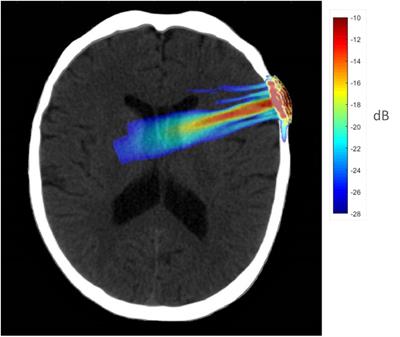

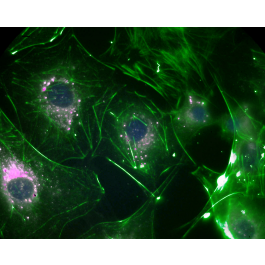

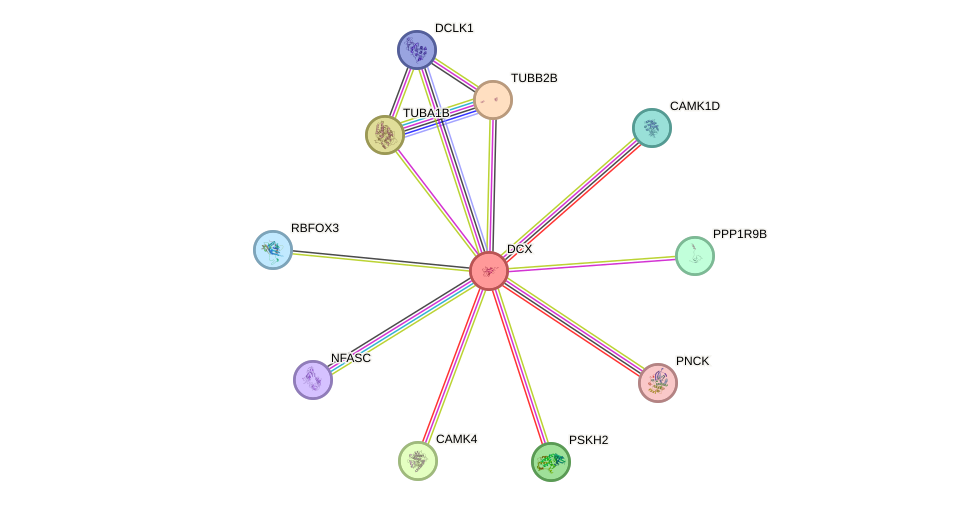


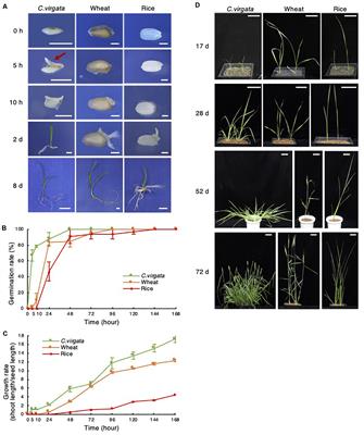


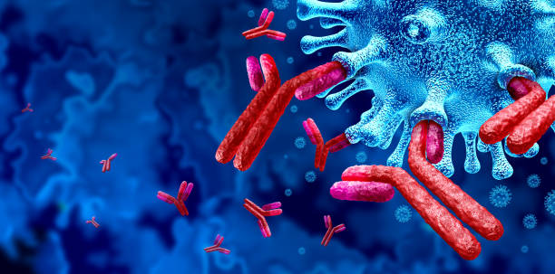


![Recombinant Anti-p53 antibody [E26] KO Tested (ab32389) | Abcam](https://www.abcam.com/ps/products/32/ab32389/Images/ab32389-389819-antip53antibodyimmunocytochemistryimmunofluorescencehap1human.jpg)


