Arterioles
Mouth Floor
Laser-Doppler Flowmetry
Venules
Microvessels
Microscopic Angioscopy
Microscopy, Video
Leukocytes
Blood Flow Velocity
Vasodilation
Splanchnic Circulation
Acridine Orange
Endothelium, Vascular
Skin
Mesocricetus
Leukocyte Rolling
Pia Mater
Vasoconstriction
Fluorescein-5-isothiocyanate
Capillary Permeability
Hemodynamics
Hemorheology
Mesentery
Nails
Corrosion Casting
Reperfusion Injury
Diagnostic Techniques, Cardiovascular
Skin Window Technique
Nitric Oxide
Fluorophotometry
Vascular Resistance
Rats, Wistar
Nitroprusside
Hyperemia
Iontophoresis
Oxygen
Vasomotor System
Erythrocytes
Blood Viscosity
Microscopy
Rheology
Models, Cardiovascular
Rats, Sprague-Dawley
Dextrans
Microscopy, Fluorescence
Acetylcholine
Hydroxyethyl Starch Derivatives
Endotoxemia
P-Selectin
Ischemia
Hematocrit
Microscopy, Polarization
Erythrocyte Deformability
Blood Gas Monitoring, Transcutaneous
Disease Models, Animal
Video Recording
Pancreatitis
Muscle, Skeletal
Tongue Diseases
Adenosine
Bradykinin
Hemodilution
Cricetinae
Mesenteric Veins
Pancreas
Capillary Leak Syndrome
Ophthalmoscopes
Microspheres
Nitroarginine
Vivisection
Transillumination
Ear
Sepsis
Oxygen Consumption
Biological Factors
Image Processing, Computer-Assisted
Shock, Septic
Ceruletide
Mice, Inbred C57BL
Endothelial Cells
Papaverine
Plasma Substitutes
Swine
Nitric Oxide Synthase
Models, Animal
Blood Vessels
Liver
Blood Substitutes
Erythrocyte Aggregation
Edema
Diagnostic Imaging
Enzyme Inhibitors
Organ Preservation
Hydronephrosis
Fluorescein Angiography
Reperfusion
Lasers
Histamine
Inflammation
Erythrocytes, Abnormal
Dose-Response Relationship, Drug
Partial Pressure
Leukocyte Count
Endothelin-1
NG-Nitroarginine Methyl Ester
Colloids
Rabbits
Blood-Brain Barrier
Mouth Mucosa
Gastric Mucosa
Capillary Resistance
Perfusion Imaging
Hemoglobins
Albumins
Microtechnology
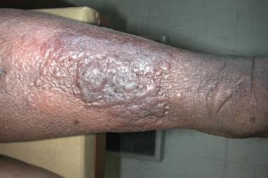
Varicose Ulcer
Muscle Tonus
Brain
Casts of hepatic blood vessels: a comparison of the microcirculation of the penguin, Pygoscelis adeliae, with some common laboratory animals. (1/5488)
Latex casts of the hepatic blood vessels of the penguin, Pygoscelis adeliae, and of some common laboratory animals were compared. There was general similarity between the different species, but the portal venous and hepatic arterial systems of the penguin were simpler than those of other species. Measurements were made of the volume and length of portal veins and it appears that the portal venous system is capable of being a more efficient blood reservoir in the penguin than in other species studied. The peribiliary plexus was especially well formed in the penguin and was drained by long veins which usually joined portal venous branches. Some of the long veins drained directly into the hepatic venous tree: these translobular veins were more prominent than in mammals. Anastomoses between hepatic artery and portal vein were not present in penguins, and the supply to the sinusoids appeared to be separate. The morphology of small hepatic veins of all the species appeared to be similar. (+info)Endometrial microvascular growth in normal and dysfunctional states. (2/5488)
As a tissue that exhibits rapid cyclical growth and shedding throughout the reproductive life of the female, human endometrium provides a good model for the study of normal physiological angiogenesis. The objective of this paper is to summarize recent data on endometrial vascular growth, present new data on regional variability in endothelial cell proliferation within the endometrium, and interpret this information in light of current knowledge of the mechanisms by which angiogenesis occurs. Conventional angiogenesis normally involves a series of steps which include endothelial cell activation, breakdown of the basement membrane, migration and proliferation of the endothelial cell, fusion of sprouts, and tube formation. Other mechanisms by which angiogenesis occurs include intussusception and vessel elongation. Using immunohistochemical techniques we have shown repeatedly that levels of endothelial cell proliferation within human endometrium do not show any consistent pattern across the different stages of the menstrual cycle, which is unexpected since significant vascular growth must occur during the proliferative phase, when the endometrium increases in thickness by up to 4-fold. There are two possible explanations for this; either there is no obligatory link between endometrial endothelial cell proliferation and new vessel formation, or there is significant variation in endothelial cell proliferation within different regions of the same uterus. Multiple samples from hysterectomy specimens subsequently demonstrated that the variability is due to real differences between individuals, as well as showing that the endothelial cell proliferation index is significantly elevated in functionalis compared with basalis. During these studies we observed that endothelial cell proliferation nearly always appeared inside existing endometrial vessels, rather than be associated with structures that could be identified as vascular sprouts. To explore further whether sprout formation occurs during endometrial angiogenesis, we investigated the immunohistochemical distribution of integrin alphavbeta3 on endometrial endothelial cells. As for endothelial cell proliferation, integrin alphavbeta3 immunostaining was seen only on endothelial cells that appeared within existing blood vessels. The results from these studies have major implications for our understanding of the mechanisms that control endometrial angiogenesis. The lack of correlation between menstrual cycle stage and endothelial cell proliferation index, or endothelial cell expression of integrin alphavbeta3, suggests that vascular growth is not under the overall control of oestrogen and progesterone. (+info)Nitric oxide modulates endothelin 1-induced Ca2+ mobilization and cytoskeletal F-actin filaments in human cerebromicrovascular endothelial cells. (3/5488)
A functional interrelation between nitric oxide (NO), the endothelial-derived vasodilating factor, and endothelin 1 (ET-1), the potent vasoconstrictive peptide, was investigated in microvascular endothelium of human brain. Nor-1 dose-dependently decreased the ET-1-stimulated mobilization of Ca2+. This response was mimicked with cGMP and abrogated by inhibitors of guanylyl cyclase or cGMP-dependent protein kinase G. These findings indicate that NO and ET-1 interactions involved in modulation of intracellular Ca2+ are mediated by cGMP/protein kinase G. In addition, Nor-1-mediated effects were associated with rearrangements of cytoskeleton F-actin filaments. The results suggest mechanisms by which NO-ET-1 interactions may contribute to regulation of microvascular function. (+info)Expression of neuropeptide Y receptors mRNA and protein in human brain vessels and cerebromicrovascular cells in culture. (4/5488)
Neuropeptide Y (NPY) has been suggested as an important regulator of CBF. However, except for the presence of Y1 receptors in large cerebral arteries, little is known about its possible sites of action on brain vessels. In this study, we sought to identify the NPY receptors present in the human cerebrovascular bed. Specific Y1 receptor binding sites, localized on the smooth muscle of human pial vessels and potently competed by NPY, polypeptide YY (PYY), and the selective Y1 receptor antagonist BIBP 3226, were identified by quantitative radioautography of the Y1 radioligand [125I]-[Leu31, Pro34]-PYY. In contrast, no specific binding of the Y2-([125I]-PYY3-36) and Y4/Y5-(125I-human pancreatic polypeptide [hPP]) radioligands could be detected. By in situ hybridization, expression of Y1 receptor mRNA was restricted to the smooth muscle layer of pial vessels, whereas no specific signals were detected for either Y2, Y4, or Y5 receptors. Similarly, using reverse transcriptase-polymerase chain reaction (RT-PCR), mRNA for Y1 but not Y2, Y4, or Y5 receptors was consistently detected in isolated human pial vessels, intracortical microvessels, and capillaries. In human brain microvascular cells in culture, PCR products for the Y1 receptors were exclusively found in the smooth muscle cells. In cultures of human brain astrocytes, a cell type that associates intimately with brain microvessels, PCR products for Y1, Y2, and Y4 but not Y5 receptors were identified. Finally, NPY significantly inhibited the forskolin-induced cAMP production in smooth muscle but not in endothelial cell cultures. We conclude that smooth muscle Y1 receptors are the primary if not exclusive NPY receptors associated with human brain extraparenchymal and intraparenchymal blood vessels, where they most likely mediate cerebral vasoconstriction. (+info)Drug-protein binding and blood-brain barrier permeability. (5/5488)
The permeability surface area (PS) product, an index of permeability of the blood-brain barrier (BBB), was measured by using the in situ perfusion method. In the cerebral circulation, the fraction of drug that permeates into the brain through the BBB is not only the unbound fraction but also the fraction dissociated from the protein in the perfusate. The sum of these two fractions, the apparent exchangeable fraction, was estimated by fitting the parameters of the BBB permeability under the condition of varying BSA concentrations in the perfusate. The unbound fraction of drugs in a buffer containing 0.5 mM BSA was measured by using the ultrafiltration method in vitro, and the apparent exchangeable fraction was measured in vivo by using the intracarotid artery injection method. The apparent exchange fraction was 100% for S-8510, 96.5% for diazepam, 90.9% for caffeine, 38.3% for S-312-d, 33.1% for propranolol, and 6.68% for (+)-S-145 Na, and each of these was higher than the corresponding unbound fraction in vitro in all drugs. The apparent exchangeable fractions, for example, were 8 times higher for diazepam and 38 times for S-312-d than the unbound fractions in vitro. The apparent exchangeable fraction of drugs was also estimated from the parameters obtained with the perfusion method. Because drugs can be infused for an arbitrary length of time in the perfusion method, substances with low permeability can be measured. The apparent exchangeable fractions obtained with this method were almost the same as those obtained with the intracarotid artery injection method. (+info)Anti-epidermal growth factor receptor antibody C225 inhibits angiogenesis in human transitional cell carcinoma growing orthotopically in nude mice. (6/5488)
Epidermal growth factor receptor (EGFR) regulates the growth and progression of human transitional cell carcinoma (TCC) of the bladder. We have shown that therapy targeting EGFR inhibited the growth of human TCC established orthotopically in nude mice. The purpose of this study was to evaluate whether EGFR-directed therapy affects angiogenesis associated with the growth and metastasis of human TCC. We determined the cytostatic effect and the effect on production of angiogenic factors after in vitro treatment of the human TCC cell line 253J B-V with MAb C225, a chimerized monoclonal anti-EGFR antibody. The 253J B-V cells were implanted orthotopically into athymic nude mice, and established tumors (4 weeks) were treated with i.p. MAb C225. Expression of the angiogenic factors vascular endothelial growth factor (VEGF), interleukin-8 (IL-8), and basic fibroblast growth factor (bFGF) was evaluated by immunohistochemistry and in situ mRNA hybridization analyses and correlated with microvessel density evaluated after immunohistochemical staining with anti-CD31. In vitro treatment with MAb C225 inhibited mRNA and protein production of VEGF, IL-8, and bFGF by 253J B-V cells in a dose-dependent manner. MAb C225 therapy of nude mice with established TCCs growing orthotopically resulted in inhibition of growth and metastasis compared with controls (P <0.0005). VEGF, IL-8, and bFGF expression was significantly lower in treated tumors than in controls. The down-regulation of these angiogenic factors preceded the involution of blood vessels. These studies indicate that therapy with anti-EGFR MAb C225 has a significant antitumor effect mediated, in part, by inhibition of angiogenesis. (+info)Microvascular loops and networks as prognostic indicators in choroidal and ciliary body melanomas. (7/5488)
BACKGROUND: Malignant melanoma of the ciliary body and choroid of the eye is a tumor that disseminates frequently, and 50% of the diagnosed patients die within 10 years. We investigated the hypothesis that, by histopathologic analysis of the arrangement of microvessels (i.e., small blood vessels) in loops and networks, we might be able to differentiate better those patients with a favorable prognosis from those with a poor prognosis. METHODS: We conducted a population-based, retrospective cohort study of melanoma-specific and all-cause mortality for 167 consecutive patients who had an eye surgically removed because of malignant choroidal or ciliary body melanoma during the period from 1972 through 1981. Microvascular loops and networks were evaluated independently by two pathologists who were unaware of patient outcome. RESULTS: Microvascular patterns could be assessed in 134 (80%) of 167 melanoma specimens. The 10-year probability of melanoma-specific survival was worse if microvascular loops (0.45 versus 0.83; two-sided P<.0001) and networks (0.41 versus 0.72, two-sided P<.0001) were present. In multivariate Cox regression analysis of melanoma-specific survival, the hazard ratios were 1.66 (95% confidence interval [CI] = 1.19-2.30) for the presence of loops and networks as a combined three-category variable, 2.36 (95% CI = 1.37-4.05) for the presence of epithelioid cells, 1.11 (95% CI = 1.03-1.19) for the largest basal tumor diameter (evaluated as a continuous variable), and 2.14 (95% CI = 1.25-3.67) for ciliary body involvement. CONCLUSIONS: Patients with malignant uveal melanoma who have a favorable prognosis can be distinguished from those with a poor prognosis by histopathologic analysis of microvascular patterns in uveal melanoma tumor specimens. (+info)Mechanical stimulation regulates voltage-gated potassium currents in cardiac microvascular endothelial cells. (8/5488)
Vascular endothelial cells are constantly exposed to mechanical forces resulting from blood flow and transmural pressure. The goal of this study was to determine whether mechanical stimulation alters the properties of endothelial voltage-gated K+ channels. Cardiac microvascular endothelial cells (CMECs) were isolated from rat ventricular muscle and cultured on thin sheets of silastic membranes. Membrane currents were measured with the use of the whole-cell arrangement of the patch-clamp technique in endothelial cells subjected to static stretch for 24 hours and compared with measurements from control, nonstretched cells. Voltage steps positive to -30 mV resulted in the activation of a time-dependent, delayed rectifier K+current (IK) in the endothelial cells. Mechanically induced increases of 97%, 355%, and 106% at +30 mV were measured in the peak amplitude of IK in cells stretched for 24 hours by 5%, 10%, and 15%, respectively. In addition, the half-maximal voltage required for IK activation was shifted from +34 mV in the nonstretched cells to -5 mV in the stretched cells. Although IK in both groups of CMECs was blocked to a similar extent by tetraethylammonium, currents in the stretched endothelial cells displayed an enhanced sensitivity to inhibition by charybdotoxin. Preincubation of the CMECs with either pertussis toxin or phorbol 12-myristate 13-acetate during the 24 hours of cell stretch did not prevent the increase in IK. The application of phorbol 12-myristate 13-acetate and static stretch stimulated the proliferation of CMECs. Stretch-induced regulation of K+ channels may be important to control the resting potential of the endothelium and may contribute to capillary growth during periods of mechanical perturbation. (+info)Microcirculation is the circulation of blood in the smallest blood vessels, including arterioles, venules, and capillaries. It's responsible for the delivery of oxygen and nutrients to the tissues and the removal of waste products. The microcirculation plays a crucial role in maintaining tissue homeostasis and is regulated by various physiological mechanisms such as autonomic nervous system activity, local metabolic factors, and hormones.
Impairment of microcirculation can lead to tissue hypoxia, inflammation, and organ dysfunction, which are common features in several diseases, including diabetes, hypertension, sepsis, and ischemia-reperfusion injury. Therefore, understanding the structure and function of the microcirculation is essential for developing new therapeutic strategies to treat these conditions.
Arterioles are small branches of arteries that play a crucial role in regulating blood flow and blood pressure within the body's circulatory system. They are the smallest type of blood vessels that have muscular walls, which allow them to contract or dilate in response to various physiological signals.
Arterioles receive blood from upstream arteries and deliver it to downstream capillaries, where the exchange of oxygen, nutrients, and waste products occurs between the blood and surrounding tissues. The contraction of arteriolar muscles can reduce the diameter of these vessels, causing increased resistance to blood flow and leading to a rise in blood pressure upstream. Conversely, dilation of arterioles reduces resistance and allows for greater blood flow at a lower pressure.
The regulation of arteriolar tone is primarily controlled by the autonomic nervous system, local metabolic factors, and various hormones. This fine-tuning of arteriolar diameter enables the body to maintain adequate blood perfusion to vital organs while also controlling overall blood pressure and distribution.
The term "mouth floor" is not a standard medical terminology. However, it might refer to the floor of the mouth, which is the part of the oral cavity located beneath the tongue and above the hyoid bone, which is a U-shaped bone in the front of the neck that helps support the tongue. The mouth floor contains several salivary glands, muscles, and nerves that are important for functions such as swallowing and speaking.
Capillaries are the smallest blood vessels in the body, with diameters that range from 5 to 10 micrometers. They form a network of tiny tubes that connect the arterioles (small branches of arteries) and venules (small branches of veins), allowing for the exchange of oxygen, carbon dioxide, nutrients, and waste products between the blood and the surrounding tissues.
Capillaries are composed of a single layer of endothelial cells that surround a hollow lumen through which blood flows. The walls of capillaries are extremely thin, allowing for easy diffusion of molecules between the blood and the surrounding tissue. This is essential for maintaining the health and function of all body tissues.
Capillaries can be classified into three types based on their structure and function: continuous, fenestrated, and sinusoidal. Continuous capillaries have a continuous layer of endothelial cells with tight junctions that restrict the passage of large molecules. Fenestrated capillaries have small pores or "fenestrae" in the endothelial cell walls that allow for the passage of larger molecules, such as proteins and lipids. Sinusoidal capillaries are found in organs with high metabolic activity, such as the liver and spleen, and have large, irregular spaces between the endothelial cells that allow for the exchange of even larger molecules.
Overall, capillaries play a critical role in maintaining the health and function of all body tissues by allowing for the exchange of nutrients, oxygen, and waste products between the blood and surrounding tissues.
Laser-Doppler flowmetry (LDF) is a non-invasive, investigative technique used to measure microcirculatory blood flow in real time. It is based on the principle of the Doppler effect, which describes the change in frequency or wavelength of light or sound waves as they encounter a moving object or reflect off a moving surface.
In LDF, a low-power laser beam is directed at the skin or other transparent tissue. The light penetrates the tissue and scatters off the moving red blood cells within the microvasculature. As the light scatters, it undergoes a slight frequency shift due to the movement of the red blood cells. This frequency shift is then detected by a photodetector, which converts it into an electrical signal. The magnitude of this signal is directly proportional to the speed and concentration of the moving red blood cells, providing a measure of microcirculatory blood flow.
LDF has various clinical applications, including the assessment of skin perfusion in patients with peripheral arterial disease, burn injuries, and flaps used in reconstructive surgery. It can also be used to study the effects of drugs or other interventions on microcirculation in research settings.
Venules are very small blood vessels that carry oxygen-depleted blood from capillaries to veins. They have a diameter of 8-50 micrometers and are an integral part of the microcirculation system in the body. Venules merge together to form veins, which then transport the low-oxygen blood back to the heart.
A "cheek" is the fleshy, muscular area of the face that forms the side of the face below the eye and above the jaw. It contains the buccinator muscle, which helps with chewing by moving food to the back teeth for grinding and also assists in speaking and forming facial expressions. The cheek also contains several sensory receptors that allow us to perceive touch, temperature, and pain in this area of the face. Additionally, there is a mucous membrane lining inside the mouth cavity called the buccal mucosa which covers the inner surface of the cheek.
Microvessels are the smallest blood vessels in the body, including capillaries, venules, and arterioles. They form a crucial part of the circulatory system, responsible for delivering oxygen and nutrients to tissues and organs while removing waste products. Capillaries, the tiniest microvessels, facilitate the exchange of substances between blood and tissue cells through their thin walls. Overall, microvessels play a vital role in maintaining proper organ function and overall health.
Microscopic angioscopy is not a widely recognized or established medical term. However, based on the individual terms, it can be interpreted as the use of a microscope with an angioscope (a type of endoscope used for visualizing the interior of blood vessels) to examine the microscopic structures of the inner walls of blood vessels. This technique would allow for detailed examination of the vasculature at a cellular level, potentially providing valuable information for research and diagnosis of various vascular diseases. However, as this is not a standard medical procedure or term, it's essential to consult the relevant literature or experts in the field for more precise information.
Video microscopy is a medical technique that involves the use of a microscope equipped with a video camera to capture and display real-time images of specimens on a monitor. This allows for the observation and documentation of dynamic processes, such as cell movement or chemical reactions, at a level of detail that would be difficult or impossible to achieve with the naked eye. Video microscopy can also be used in conjunction with image analysis software to measure various parameters, such as size, shape, and motion, of individual cells or structures within the specimen.
There are several types of video microscopy, including brightfield, darkfield, phase contrast, fluorescence, and differential interference contrast (DIC) microscopy. Each type uses different optical techniques to enhance contrast and reveal specific features of the specimen. For example, fluorescence microscopy uses fluorescent dyes or proteins to label specific structures within the specimen, allowing them to be visualized against a dark background.
Video microscopy is used in various fields of medicine, including pathology, microbiology, and neuroscience. It can help researchers and clinicians diagnose diseases, study disease mechanisms, develop new therapies, and understand fundamental biological processes at the cellular and molecular level.
Leukocytes, also known as white blood cells (WBCs), are a crucial component of the human immune system. They are responsible for protecting the body against infections and foreign substances. Leukocytes are produced in the bone marrow and circulate throughout the body in the bloodstream and lymphatic system.
There are several types of leukocytes, including:
1. Neutrophils - These are the most abundant type of leukocyte and are primarily responsible for fighting bacterial infections. They contain enzymes that can destroy bacteria.
2. Lymphocytes - These are responsible for producing antibodies and destroying virus-infected cells, as well as cancer cells. There are two main types of lymphocytes: B-lymphocytes and T-lymphocytes.
3. Monocytes - These are the largest type of leukocyte and help to break down and remove dead or damaged tissues, as well as microorganisms.
4. Eosinophils - These play a role in fighting parasitic infections and are also involved in allergic reactions and inflammation.
5. Basophils - These release histamine and other chemicals that cause inflammation in response to allergens or irritants.
An abnormal increase or decrease in the number of leukocytes can indicate an underlying medical condition, such as an infection, inflammation, or a blood disorder.
Blood flow velocity is the speed at which blood travels through a specific part of the vascular system. It is typically measured in units of distance per time, such as centimeters per second (cm/s) or meters per second (m/s). Blood flow velocity can be affected by various factors, including cardiac output, vessel diameter, and viscosity of the blood. Measuring blood flow velocity is important in diagnosing and monitoring various medical conditions, such as heart disease, stroke, and peripheral vascular disease.
Liver circulation, also known as hepatic circulation, refers to the blood flow through the liver. The liver receives blood from two sources: the hepatic artery and the portal vein.
The hepatic artery delivers oxygenated blood from the heart to the liver, accounting for about 25% of the liver's blood supply. The remaining 75% comes from the portal vein, which carries nutrient-rich, deoxygenated blood from the gastrointestinal tract, spleen, pancreas, and gallbladder to the liver.
In the liver, these two sources of blood mix in the sinusoids, small vessels with large spaces between the endothelial cells that line them. This allows for efficient exchange of substances between the blood and the hepatocytes (liver cells). The blood then leaves the liver through the hepatic veins, which merge into the inferior vena cava and return the blood to the heart.
The unique dual blood supply and extensive sinusoidal network in the liver enable it to perform various critical functions, such as detoxification, metabolism, synthesis, storage, and secretion of numerous substances, maintaining body homeostasis.
Regional blood flow (RBF) refers to the rate at which blood flows through a specific region or organ in the body, typically expressed in milliliters per minute per 100 grams of tissue (ml/min/100g). It is an essential physiological parameter that reflects the delivery of oxygen and nutrients to tissues while removing waste products. RBF can be affected by various factors such as metabolic demands, neural regulation, hormonal influences, and changes in blood pressure or vascular resistance. Measuring RBF is crucial for understanding organ function, diagnosing diseases, and evaluating the effectiveness of treatments.
Vasodilation is the widening or increase in diameter of blood vessels, particularly the involuntary relaxation of the smooth muscle in the tunica media (middle layer) of the arteriole walls. This results in an increase in blood flow and a decrease in vascular resistance. Vasodilation can occur due to various physiological and pathophysiological stimuli, such as local metabolic demands, neural signals, or pharmacological agents. It plays a crucial role in regulating blood pressure, tissue perfusion, and thermoregulation.
Splanchnic circulation refers to the blood flow to the visceral organs, including the gastrointestinal tract, pancreas, spleen, and liver. These organs receive a significant portion of the cardiac output, with approximately 25-30% of the total restingly going to the splanchnic circulation. The splanchnic circulation is regulated by a complex interplay of neural and hormonal mechanisms that help maintain adequate blood flow to these vital organs while also allowing for the distribution of blood to other parts of the body as needed.
The splanchnic circulation is unique in its ability to vasodilate and increase blood flow significantly in response to meals or other stimuli, such as stress or hormonal changes. This increased blood flow helps support the digestive process and absorption of nutrients. At the same time, the body must carefully regulate this blood flow to prevent a significant drop in blood pressure or overloading the heart with too much work.
Overall, the splanchnic circulation plays a critical role in maintaining the health and function of the body's vital organs, and dysregulation of this system can contribute to various diseases, including digestive disorders, liver disease, and cardiovascular disease.
Coronary circulation refers to the circulation of blood in the coronary vessels, which supply oxygenated blood to the heart muscle (myocardium) and drain deoxygenated blood from it. The coronary circulation system includes two main coronary arteries - the left main coronary artery and the right coronary artery - that branch off from the aorta just above the aortic valve. These arteries further divide into smaller branches, which supply blood to different regions of the heart muscle.
The left main coronary artery divides into two branches: the left anterior descending (LAD) artery and the left circumflex (LCx) artery. The LAD supplies blood to the front and sides of the heart, while the LCx supplies blood to the back and sides of the heart. The right coronary artery supplies blood to the lower part of the heart, including the right ventricle and the bottom portion of the left ventricle.
The veins that drain the heart muscle include the great cardiac vein, the middle cardiac vein, and the small cardiac vein, which merge to form the coronary sinus. The coronary sinus empties into the right atrium, allowing deoxygenated blood to enter the right side of the heart and be pumped to the lungs for oxygenation.
Coronary circulation is essential for maintaining the health and function of the heart muscle, as it provides the necessary oxygen and nutrients required for proper contraction and relaxation of the myocardium. Any disruption or blockage in the coronary circulation system can lead to serious consequences, such as angina, heart attack, or even death.
Acridine Orange is a fluorescent dye commonly used in various scientific applications, particularly in the field of cytology and microbiology. Its chemical formula is C17H19N3O.
In medical terms, Acridine Orange is often used as a supravital stain to differentiate between live and dead cells or to identify bacteria, fungi, and other microorganisms in samples. It can also be used to detect abnormalities in DNA and RNA, making it useful in the identification of certain types of cancerous cells.
When exposed to ultraviolet light, Acridine Orange exhibits a green fluorescence when bound to double-stranded DNA and a red or orange-red fluorescence when bound to single-stranded RNA. This property makes it a valuable tool in the study of cell division, gene expression, and other biological processes that involve nucleic acids.
However, it is important to note that Acridine Orange can be toxic to living cells in high concentrations or with prolonged exposure, so it must be used carefully and in accordance with established safety protocols.
Retinal vessels refer to the blood vessels that are located in the retina, which is the light-sensitive tissue that lines the inner surface of the eye. The retina contains two types of blood vessels: arteries and veins.
The central retinal artery supplies oxygenated blood to the inner layers of the retina, while the central retinal vein drains deoxygenated blood from the retina. These vessels can be visualized during a routine eye examination using an ophthalmoscope, which allows healthcare professionals to assess their health and any potential abnormalities.
Retinal vessels are essential for maintaining the health and function of the retina, and any damage or changes to these vessels can affect vision and lead to various eye conditions such as diabetic retinopathy, retinal vein occlusion, and hypertensive retinopathy.
The endothelium is a thin layer of simple squamous epithelial cells that lines the interior surface of blood vessels, lymphatic vessels, and heart chambers. The vascular endothelium, specifically, refers to the endothelial cells that line the blood vessels. These cells play a crucial role in maintaining vascular homeostasis by regulating vasomotor tone, coagulation, platelet activation, inflammation, and permeability of the vessel wall. They also contribute to the growth and repair of the vascular system and are involved in various pathological processes such as atherosclerosis, hypertension, and diabetes.
In medical terms, the skin is the largest organ of the human body. It consists of two main layers: the epidermis (outer layer) and dermis (inner layer), as well as accessory structures like hair follicles, sweat glands, and oil glands. The skin plays a crucial role in protecting us from external factors such as bacteria, viruses, and environmental hazards, while also regulating body temperature and enabling the sense of touch.
Vasodilator agents are pharmacological substances that cause the relaxation or widening of blood vessels by relaxing the smooth muscle in the vessel walls. This results in an increase in the diameter of the blood vessels, which decreases vascular resistance and ultimately reduces blood pressure. Vasodilators can be further classified based on their site of action:
1. Systemic vasodilators: These agents cause a generalized relaxation of the smooth muscle in the walls of both arteries and veins, resulting in a decrease in peripheral vascular resistance and preload (the volume of blood returning to the heart). Examples include nitroglycerin, hydralazine, and calcium channel blockers.
2. Arterial vasodilators: These agents primarily affect the smooth muscle in arterial vessel walls, leading to a reduction in afterload (the pressure against which the heart pumps blood). Examples include angiotensin-converting enzyme (ACE) inhibitors, angiotensin receptor blockers (ARBs), and direct vasodilators like sodium nitroprusside.
3. Venous vasodilators: These agents primarily affect the smooth muscle in venous vessel walls, increasing venous capacitance and reducing preload. Examples include nitroglycerin and other organic nitrates.
Vasodilator agents are used to treat various cardiovascular conditions such as hypertension, heart failure, angina, and pulmonary arterial hypertension. It is essential to monitor their use carefully, as excessive vasodilation can lead to orthostatic hypotension, reflex tachycardia, or fluid retention.
"Mesocricetus" is a genus of rodents, more commonly known as hamsters. It includes several species of hamsters that are native to various parts of Europe and Asia. The best-known member of this genus is the Syrian hamster, also known as the golden hamster or Mesocricetus auratus, which is a popular pet due to its small size and relatively easy care. These hamsters are burrowing animals and are typically solitary in the wild.
Leukocyte rolling is a crucial step in the process of leukocytes (white blood cells) migrating from the bloodstream to the site of infection or inflammation, which is known as extravasation. This phenomenon is mediated by the interaction between selectins on the surface of endothelial cells and their ligands on leukocytes.
The multi-step adhesion cascade begins with leukocyte rolling, where leukocytes move along the vessel wall in a slow, rolling motion. This is facilitated by the transient interactions between selectins (P-selectin, E-selectin, and L-selectin) on endothelial cells and their ligands (PSGL-1, CD44, and others) on leukocytes. These interactions are weak and short-lived but sufficient to reduce the leukocyte's velocity and enable it to roll along the vessel wall.
Leukocyte rolling allows the leukocytes to come in close contact with the endothelium, where they can receive further signals that promote their activation and firm adhesion. This process is critical for the immune response to infection and inflammation, as it enables the recruitment of effector cells to the site of injury or infection.
Pia Mater is the inner-most layer of the meninges, which are the protective coverings of the brain and spinal cord. It is a very thin and highly vascularized (rich in blood vessels) membrane that closely adheres to the surface of the brain. The name "Pia Mater" comes from Latin, meaning "tender mother." This layer provides nutrition and protection to the brain, and it also allows for the movement and flexibility of the brain within the skull.
Vasoconstriction is a medical term that refers to the narrowing of blood vessels due to the contraction of the smooth muscle in their walls. This process decreases the diameter of the lumen (the inner space of the blood vessel) and reduces blood flow through the affected vessels. Vasoconstriction can occur throughout the body, but it is most noticeable in the arterioles and precapillary sphincters, which control the amount of blood that flows into the capillary network.
The autonomic nervous system, specifically the sympathetic division, plays a significant role in regulating vasoconstriction through the release of neurotransmitters like norepinephrine (noradrenaline). Various hormones and chemical mediators, such as angiotensin II, endothelin-1, and serotonin, can also induce vasoconstriction.
Vasoconstriction is a vital physiological response that helps maintain blood pressure and regulate blood flow distribution in the body. However, excessive or prolonged vasoconstriction may contribute to several pathological conditions, including hypertension, stroke, and peripheral vascular diseases.
Fluorescein-5-isothiocyanate (FITC) is not a medical term per se, but a chemical compound commonly used in biomedical research and clinical diagnostics. Therefore, I will provide a general definition of this term:
Fluorescein-5-isothiocyanate (FITC) is a fluorescent dye with an absorption maximum at approximately 492-495 nm and an emission maximum at around 518-525 nm. It is widely used as a labeling reagent for various biological molecules, such as antibodies, proteins, and nucleic acids, to study their structure, function, and interactions in techniques like flow cytometry, immunofluorescence microscopy, and western blotting. The isothiocyanate group (-N=C=S) in the FITC molecule reacts with primary amines (-NH2) present in biological molecules to form a stable thiourea bond, enabling specific labeling of target molecules for detection and analysis.
Capillary permeability refers to the ability of substances to pass through the walls of capillaries, which are the smallest blood vessels in the body. These tiny vessels connect the arterioles and venules, allowing for the exchange of nutrients, waste products, and gases between the blood and the surrounding tissues.
The capillary wall is composed of a single layer of endothelial cells that are held together by tight junctions. The permeability of these walls varies depending on the size and charge of the molecules attempting to pass through. Small, uncharged molecules such as water, oxygen, and carbon dioxide can easily diffuse through the capillary wall, while larger or charged molecules such as proteins and large ions have more difficulty passing through.
Increased capillary permeability can occur in response to inflammation, infection, or injury, allowing larger molecules and immune cells to enter the surrounding tissues. This can lead to swelling (edema) and tissue damage if not controlled. Decreased capillary permeability, on the other hand, can lead to impaired nutrient exchange and tissue hypoxia.
Overall, the permeability of capillaries is a critical factor in maintaining the health and function of tissues throughout the body.
Hemodynamics is the study of how blood flows through the cardiovascular system, including the heart and the vascular network. It examines various factors that affect blood flow, such as blood volume, viscosity, vessel length and diameter, and pressure differences between different parts of the circulatory system. Hemodynamics also considers the impact of various physiological and pathological conditions on these variables, and how they in turn influence the function of vital organs and systems in the body. It is a critical area of study in fields such as cardiology, anesthesiology, and critical care medicine.
Cerebrovascular circulation refers to the network of blood vessels that supply oxygenated blood and nutrients to the brain tissue, and remove waste products. It includes the internal carotid arteries, vertebral arteries, circle of Willis, and the intracranial arteries that branch off from them.
The internal carotid arteries and vertebral arteries merge to form the circle of Willis, a polygonal network of vessels located at the base of the brain. The anterior cerebral artery, middle cerebral artery, posterior cerebral artery, and communicating arteries are the major vessels that branch off from the circle of Willis and supply blood to different regions of the brain.
Interruptions or abnormalities in the cerebrovascular circulation can lead to various neurological conditions such as stroke, transient ischemic attack (TIA), and vascular dementia.
Hemorheology is the study of the flow properties of blood and its components, including red blood cells, white blood cells, platelets, and plasma. Specifically, it examines how these components interact with each other and with the walls of blood vessels to affect the flow characteristics of blood under different conditions. Hemorheological factors can influence blood viscosity, which is a major determinant of peripheral vascular resistance and cardiac workload. Abnormalities in hemorheology have been implicated in various diseases such as atherosclerosis, hypertension, diabetes, and sickle cell disease.
The mesentery is a continuous fold of the peritoneum, the double-layered serous membrane that lines the abdominal cavity, which attaches the stomach, small intestine, large intestine (colon), and rectum to the posterior wall of the abdomen. It provides blood vessels, nerves, and lymphatic vessels to these organs.
Traditionally, the mesentery was thought to consist of separate and distinct sections along the length of the intestines. However, recent research has shown that the mesentery is a continuous organ, with a single continuous tethering point to the posterior abdominal wall. This new understanding of the anatomy of the mesentery has implications for the study of various gastrointestinal diseases and disorders.
In the context of medical terminology, "nails" primarily refer to the keratinous plates that are found at the tips of fingers and toes. These specialized structures are part of the outermost layer of the skin (epidermis) and are formed by a type of cells called keratinocytes. The nails serve to protect the delicate underlying tissues from trauma, and they also aid in tasks such as picking up small objects or scratching itches.
The medical term for fingernails and toenails is "unguis," which comes from Latin. Each nail consists of several parts:
1. Nail plate: The visible part of the nail that is hard and flat, made up of keratin.
2. Nail bed: The skin beneath the nail plate to which the nail plate is attached; it supplies blood to the nail.
3. Matrix: The area where new cells are produced for the growth of the nail plate; located under the cuticle and extends slightly onto the finger or toe.
4. Lunula: The crescent-shaped white area at the base of the nail plate, which is the visible portion of the matrix.
5. Cuticle: The thin layer of skin that overlaps the nail plate and protects the underlying tissue from infection.
6. Eponychium: The fold of skin that surrounds and covers the nail plate; also known as the "proximal nail fold."
7. Hyponychium: The area of skin between the free edge of the nail plate and the fingertip or toe tip.
8. Perionychiun: The skin surrounding the nail on all sides.
Understanding the anatomy and medical aspects of nails is essential for healthcare professionals, as various conditions can affect nail health, such as fungal infections, ingrown nails, or tumors.
Corrosion casting is a specialized technique used in anatomy and pathology to create detailed casts or molds of biological specimens, particularly vascular systems. This method is also known as "acid etching" or "corrosive casting." Here's the medical definition:
Corrosion casting is a process that involves injecting a special resin or plastic material into the vasculature or other hollow structures of a biological specimen, such as an organ or tissue. The injected material thoroughly fills the cavity and then hardens once it has set. After hardening, the surrounding tissues are corroded or dissolved using strong acids or bases, leaving behind only the cast or mold of the internal structures.
This technique results in a detailed three-dimensional representation of the complex internal networks, like blood vessels, which can be used for further study, research, and education. Corrosion casting is particularly useful in visualizing the intricate branching patterns and structural relationships within these systems.
Reperfusion injury is a complex pathophysiological process that occurs when blood flow is restored to previously ischemic tissues, leading to further tissue damage. This phenomenon can occur in various clinical settings such as myocardial infarction (heart attack), stroke, or peripheral artery disease after an intervention aimed at restoring perfusion.
The restoration of blood flow leads to the generation of reactive oxygen species (ROS) and inflammatory mediators, which can cause oxidative stress, cellular damage, and activation of the immune system. This results in a cascade of events that may lead to microvascular dysfunction, capillary leakage, and tissue edema, further exacerbating the injury.
Reperfusion injury is an important consideration in the management of ischemic events, as interventions aimed at restoring blood flow must be carefully balanced with potential harm from reperfusion injury. Strategies to mitigate reperfusion injury include ischemic preconditioning (exposing the tissue to short periods of ischemia before a prolonged ischemic event), ischemic postconditioning (applying brief periods of ischemia and reperfusion after restoring blood flow), remote ischemic preconditioning (ischemia applied to a distant organ or tissue to protect the target organ), and pharmacological interventions that scavenge ROS, reduce inflammation, or improve microvascular function.
Diagnostic techniques in cardiovascular medicine refer to the various tests and methods used to diagnose and evaluate conditions related to the heart and blood vessels. These techniques can be non-invasive or invasive and are designed to provide critical information about a patient's cardiovascular health, such as heart function, blood flow, and the presence of any abnormalities or diseases. Here are some common diagnostic techniques used in cardiovascular medicine:
1. Electrocardiogram (ECG): An ECG is a non-invasive test that records the electrical activity of the heart. It can help detect heart conditions such as arrhythmias, heart attacks, and structural abnormalities.
2. Echocardiogram: This is a non-invasive ultrasound test that produces images of the heart's structures, including the chambers, valves, and major blood vessels. It can help assess heart function, identify damage from heart attacks, and detect various cardiovascular conditions.
3. Stress testing: A stress test involves exercising on a treadmill or stationary bike while being monitored by an ECG to evaluate the heart's response to physical exertion. It can help diagnose coronary artery disease, assess exercise capacity, and determine the need for further testing or treatment.
4. Cardiac catheterization: This is an invasive procedure where a thin, flexible tube (catheter) is inserted into a blood vessel in the arm or leg and guided to the heart. It can help diagnose and treat various cardiovascular conditions, such as blocked arteries, heart valve problems, and congenital heart defects.
5. Coronary angiography: During a cardiac catheterization, a special dye is injected into the coronary arteries to visualize blood flow using X-ray imaging. This can help identify blockages or narrowing in the coronary arteries and guide treatment decisions.
6. Nuclear stress testing: This test combines the use of a radioactive tracer with exercise or pharmacological stress to evaluate heart function and blood flow. It can help diagnose coronary artery disease, assess the effectiveness of treatments, and determine the need for further interventions.
7. Cardiac magnetic resonance imaging (MRI): This non-invasive imaging technique uses a strong magnetic field and radio waves to create detailed images of the heart's structure and function. It can help diagnose various cardiovascular conditions, such as heart muscle disorders, valve problems, and congenital heart defects.
8. Transesophageal echocardiography (TEE): This is a specialized ultrasound technique where a probe is inserted through the esophagus to obtain detailed images of the heart's structure and function. It can help diagnose conditions such as blood clots, valve problems, and infective endocarditis.
9. Positron emission tomography (PET) scanning: This imaging technique uses a small amount of radioactive tracer to evaluate the metabolic activity of the heart. It can help diagnose coronary artery disease, assess the effectiveness of treatments, and determine the need for further interventions.
10. Electrophysiology studies (EPS): These are invasive procedures where catheters are inserted into the heart to study its electrical system. They can help diagnose and treat various arrhythmias, such as atrial fibrillation, ventricular tachycardia, and Wolff-Parkinson-White syndrome.
Renal circulation refers to the blood flow specifically dedicated to the kidneys. The main function of the kidneys is to filter waste and excess fluids from the blood, which then get excreted as urine. To perform this function efficiently, the kidneys receive a substantial amount of the body's total blood supply - about 20-25% in a resting state.
The renal circulation process begins when deoxygenated blood from the rest of the body returns to the right side of the heart and is pumped into the lungs for oxygenation. Oxygen-rich blood then leaves the left side of the heart through the aorta, the largest artery in the body.
A portion of this oxygen-rich blood moves into the renal arteries, which branch directly from the aorta and supply each kidney with blood. Within the kidneys, these arteries divide further into smaller vessels called afferent arterioles, which feed into a network of tiny capillaries called the glomerulus within each nephron (the functional unit of the kidney).
The filtration process occurs in the glomeruli, where waste materials and excess fluids are separated from the blood. The resulting filtrate then moves through another set of capillaries, the peritubular capillaries, which surround the renal tubules (the part of the nephron that reabsorbs necessary substances back into the bloodstream).
The now-deoxygenated blood from the kidneys' capillary network coalesces into venules and then merges into the renal veins, which ultimately drain into the inferior vena cava and return the blood to the right side of the heart. This highly specialized circulation system allows the kidneys to efficiently filter waste while maintaining appropriate blood volume and composition.
I'm sorry for any confusion, but "Skin Window Technique" is not a recognized or established term in medicine or dermatology. It may refer to a specific procedure or technique used in a particular study or clinical setting, but without more context, it's difficult to provide an accurate definition.
However, there is a similar term called "Skin as a Window Technique," which is a non-invasive method used in dermatology to evaluate the underlying structures and processes of the skin through transillumination or direct visualization. This technique can be used to diagnose various skin conditions such as hemangiomas, nevi, and other vascular anomalies.
Without more context, I cannot provide a precise definition of "Skin Window Technique." If you could provide more information about where you encountered this term or its intended meaning, I would be happy to try to help further.
Nitric oxide (NO) is a molecule made up of one nitrogen atom and one oxygen atom. In the body, it is a crucial signaling molecule involved in various physiological processes such as vasodilation, immune response, neurotransmission, and inhibition of platelet aggregation. It is produced naturally by the enzyme nitric oxide synthase (NOS) from the amino acid L-arginine. Inhaled nitric oxide is used medically to treat pulmonary hypertension in newborns and adults, as it helps to relax and widen blood vessels, improving oxygenation and blood flow.
Fluorophotometry is a medical diagnostic technique that measures the concentration of fluorescein dye in various tissues, particularly the eye. This technique utilizes a specialized instrument called a fluorophotometer which emits light at a specific wavelength that causes the fluorescein to emit light at a longer wavelength. The intensity of this emitted light is then measured and used to calculate the concentration of fluorescein in the tissue.
Fluorophotometry is often used in ophthalmology to assess the permeability of the blood-retinal barrier, which can be helpful in diagnosing and monitoring conditions such as diabetic retinopathy, age-related macular degeneration, and uveitis. It may also have applications in other medical fields for measuring the concentration of fluorescent markers in various tissues.
Cell adhesion refers to the binding of cells to extracellular matrices or to other cells, a process that is fundamental to the development, function, and maintenance of multicellular organisms. Cell adhesion is mediated by various cell surface receptors, such as integrins, cadherins, and immunoglobulin-like cell adhesion molecules (Ig-CAMs), which interact with specific ligands in the extracellular environment. These interactions lead to the formation of specialized junctions, such as tight junctions, adherens junctions, and desmosomes, that help to maintain tissue architecture and regulate various cellular processes, including proliferation, differentiation, migration, and survival. Disruptions in cell adhesion can contribute to a variety of diseases, including cancer, inflammation, and degenerative disorders.
Vascular resistance is a measure of the opposition to blood flow within a vessel or a group of vessels, typically expressed in units of mmHg/(mL/min) or sometimes as dynes*sec/cm^5. It is determined by the diameter and length of the vessels, as well as the viscosity of the blood flowing through them. In general, a decrease in vessel diameter, an increase in vessel length, or an increase in blood viscosity will result in an increase in vascular resistance, while an increase in vessel diameter, a decrease in vessel length, or a decrease in blood viscosity will result in a decrease in vascular resistance. Vascular resistance is an important concept in the study of circulation and cardiovascular physiology because it plays a key role in determining blood pressure and blood flow within the body.
"Wistar rats" are a strain of albino rats that are widely used in laboratory research. They were developed at the Wistar Institute in Philadelphia, USA, and were first introduced in 1906. Wistar rats are outbred, which means that they are genetically diverse and do not have a fixed set of genetic characteristics like inbred strains.
Wistar rats are commonly used as animal models in biomedical research because of their size, ease of handling, and relatively low cost. They are used in a wide range of research areas, including toxicology, pharmacology, nutrition, cancer, cardiovascular disease, and behavioral studies. Wistar rats are also used in safety testing of drugs, medical devices, and other products.
Wistar rats are typically larger than many other rat strains, with males weighing between 500-700 grams and females weighing between 250-350 grams. They have a lifespan of approximately 2-3 years. Wistar rats are also known for their docile and friendly nature, making them easy to handle and work with in the laboratory setting.
Blood pressure is the force exerted by circulating blood on the walls of the blood vessels. It is measured in millimeters of mercury (mmHg) and is given as two figures:
1. Systolic pressure: This is the pressure when the heart pushes blood out into the arteries.
2. Diastolic pressure: This is the pressure when the heart rests between beats, allowing it to fill with blood.
Normal blood pressure for adults is typically around 120/80 mmHg, although this can vary slightly depending on age, sex, and other factors. High blood pressure (hypertension) is generally considered to be a reading of 130/80 mmHg or higher, while low blood pressure (hypotension) is usually defined as a reading below 90/60 mmHg. It's important to note that blood pressure can fluctuate throughout the day and may be affected by factors such as stress, physical activity, and medication use.
nitroprusside (ni-troe-rus-ide)
A rapid-acting vasodilator used in the management of severe hypertension, acute heart failure, and to reduce afterload in patients undergoing cardiac surgery. It is a potent arterial and venous dilator that decreases preload and afterload, thereby reducing myocardial oxygen demand. Nitroprusside is metabolized to cyanide, which must be monitored closely during therapy to prevent toxicity.
Pharmacologic class: Peripheral vasodilators
Therapeutic class: Antihypertensives, Vasodilators
Medical Categories: Cardiovascular Drugs, Hypertension Agents
Hyperemia is a medical term that refers to an increased flow or accumulation of blood in certain capillaries or vessels within an organ or tissue, resulting in its redness and warmth. This can occur due to various reasons such as physical exertion, emotional excitement, local injury, or specific medical conditions.
There are two types of hyperemia: active and passive. Active hyperemia is a physiological response where the blood flow increases as a result of the metabolic demands of the organ or tissue. For example, during exercise, muscles require more oxygen and nutrients, leading to an increase in blood flow. Passive hyperemia, on the other hand, occurs when there is a blockage in the venous outflow, causing the blood to accumulate in the affected area. This can result from conditions like thrombosis or vasoconstriction.
It's important to note that while hyperemia itself is not a disease, it can be a symptom of various underlying medical conditions and should be evaluated by a healthcare professional if it persists or is accompanied by other symptoms.
Iontophoresis is a medical technique in which a mild electrical current is used to deliver medications through the skin. This process enhances the absorption of medication into the body, allowing it to reach deeper tissues that may not be accessible through topical applications alone. Iontophoresis is often used for local treatment of conditions such as inflammation, pain, or spasms, and is particularly useful in treating conditions affecting the hands and feet, like hyperhidrosis (excessive sweating). The medications used in iontophoresis are typically anti-inflammatory drugs, anesthetics, or corticosteroids.
Oxygen is a colorless, odorless, tasteless gas that constitutes about 21% of the earth's atmosphere. It is a crucial element for human and most living organisms as it is vital for respiration. Inhaled oxygen enters the lungs and binds to hemoglobin in red blood cells, which carries it to tissues throughout the body where it is used to convert nutrients into energy and carbon dioxide, a waste product that is exhaled.
Medically, supplemental oxygen therapy may be provided to patients with conditions such as chronic obstructive pulmonary disease (COPD), pneumonia, heart failure, or other medical conditions that impair the body's ability to extract sufficient oxygen from the air. Oxygen can be administered through various devices, including nasal cannulas, face masks, and ventilators.
The vasomotor system is a part of the autonomic nervous system that controls the diameter of blood vessels, particularly the smooth muscle in the walls of arterioles and precapillary sphincters. It regulates blood flow to different parts of the body by constricting or dilating these vessels. The vasomotor center located in the medulla oblongata of the brainstem controls the system, receiving input from various sensory receptors and modulating the sympathetic and parasympathetic nervous systems' activity. Vasoconstriction decreases blood flow, while vasodilation increases it.
Erythrocytes, also known as red blood cells (RBCs), are the most common type of blood cell in circulating blood in mammals. They are responsible for transporting oxygen from the lungs to the body's tissues and carbon dioxide from the tissues to the lungs.
Erythrocytes are formed in the bone marrow and have a biconcave shape, which allows them to fold and bend easily as they pass through narrow blood vessels. They do not have a nucleus or mitochondria, which makes them more flexible but also limits their ability to reproduce or repair themselves.
In humans, erythrocytes are typically disc-shaped and measure about 7 micrometers in diameter. They contain the protein hemoglobin, which binds to oxygen and gives blood its red color. The lifespan of an erythrocyte is approximately 120 days, after which it is broken down in the liver and spleen.
Abnormalities in erythrocyte count or function can lead to various medical conditions, such as anemia, polycythemia, and sickle cell disease.
Blood viscosity is a measure of the thickness or flow resistance of blood. It is defined as the ratio of shear stress to shear rate within the flowing blood, which reflects the internal friction or resistance to flow. Blood viscosity is primarily determined by the concentration and size of red blood cells (hematocrit), plasma proteins, and other blood constituents. An increase in any of these components can raise blood viscosity, leading to impaired blood flow, reduced oxygen delivery to tissues, and potential cardiovascular complications if not managed appropriately.
Microscopy is a technical field in medicine that involves the use of microscopes to observe structures and phenomena that are too small to be seen by the naked eye. It allows for the examination of samples such as tissues, cells, and microorganisms at high magnifications, enabling the detection and analysis of various medical conditions, including infections, diseases, and cellular abnormalities.
There are several types of microscopy used in medicine, including:
1. Light Microscopy: This is the most common type of microscopy, which uses visible light to illuminate and magnify samples. It can be used to examine a wide range of biological specimens, such as tissue sections, blood smears, and bacteria.
2. Electron Microscopy: This type of microscopy uses a beam of electrons instead of light to produce highly detailed images of samples. It is often used in research settings to study the ultrastructure of cells and tissues.
3. Fluorescence Microscopy: This technique involves labeling specific molecules within a sample with fluorescent dyes, allowing for their visualization under a microscope. It can be used to study protein interactions, gene expression, and cell signaling pathways.
4. Confocal Microscopy: This type of microscopy uses a laser beam to scan a sample point by point, producing high-resolution images with reduced background noise. It is often used in medical research to study the structure and function of cells and tissues.
5. Scanning Probe Microscopy: This technique involves scanning a sample with a physical probe, allowing for the measurement of topography, mechanical properties, and other characteristics at the nanoscale. It can be used in medical research to study the structure and function of individual molecules and cells.
Rheology is not a term that is specific to medicine, but rather it is a term used in the field of physics to describe the flow and deformation of matter. It specifically refers to the study of how materials flow or deform under various stresses or strains. This concept can be applied to various medical fields such as studying the flow properties of blood (hematology), understanding the movement of tissues and organs during surgical procedures, or analyzing the mechanical behavior of biological materials like bones and cartilages.
Cardiovascular models are simplified representations or simulations of the human cardiovascular system used in medical research, education, and training. These models can be physical, computational, or mathematical and are designed to replicate various aspects of the heart, blood vessels, and blood flow. They can help researchers study the structure and function of the cardiovascular system, test new treatments and interventions, and train healthcare professionals in diagnostic and therapeutic techniques.
Physical cardiovascular models may include artificial hearts, blood vessels, or circulation systems made from materials such as plastic, rubber, or silicone. These models can be used to study the mechanics of heart valves, the effects of different surgical procedures, or the impact of various medical devices on blood flow.
Computational and mathematical cardiovascular models use algorithms and equations to simulate the behavior of the cardiovascular system. These models may range from simple representations of a single heart chamber to complex simulations of the entire circulatory system. They can be used to study the electrical activity of the heart, the biomechanics of blood flow, or the distribution of drugs in the body.
Overall, cardiovascular models play an essential role in advancing our understanding of the human body and improving patient care.
Sprague-Dawley rats are a strain of albino laboratory rats that are widely used in scientific research. They were first developed by researchers H.H. Sprague and R.C. Dawley in the early 20th century, and have since become one of the most commonly used rat strains in biomedical research due to their relatively large size, ease of handling, and consistent genetic background.
Sprague-Dawley rats are outbred, which means that they are genetically diverse and do not suffer from the same limitations as inbred strains, which can have reduced fertility and increased susceptibility to certain diseases. They are also characterized by their docile nature and low levels of aggression, making them easier to handle and study than some other rat strains.
These rats are used in a wide variety of research areas, including toxicology, pharmacology, nutrition, cancer, and behavioral studies. Because they are genetically diverse, Sprague-Dawley rats can be used to model a range of human diseases and conditions, making them an important tool in the development of new drugs and therapies.
Dextrans are a type of complex glucose polymers that are formed by the action of certain bacteria on sucrose. They are branched polysaccharides consisting of linear chains of α-1,6 linked D-glucopyranosyl units with occasional α-1,3 branches.
Dextrans have a wide range of applications in medicine and industry. In medicine, dextrans are used as plasma substitutes, volume expanders, and anticoagulants. They are also used as carriers for drugs and diagnostic agents, and in the manufacture of immunoadsorbents for the removal of toxins and pathogens from blood.
Dextrans can be derived from various bacterial sources, but the most common commercial source is Leuconostoc mesenteroides B-512(F) or L. dextranicum. The molecular weight of dextrans can vary widely, ranging from a few thousand to several million Daltons, depending on the method of preparation and purification.
Dextrans are generally biocompatible and non-toxic, but they can cause allergic reactions in some individuals. Therefore, their use as medical products requires careful monitoring and testing for safety and efficacy.
Fluorescence microscopy is a type of microscopy that uses fluorescent dyes or proteins to highlight and visualize specific components within a sample. In this technique, the sample is illuminated with high-energy light, typically ultraviolet (UV) or blue light, which excites the fluorescent molecules causing them to emit lower-energy, longer-wavelength light, usually visible light in the form of various colors. This emitted light is then collected by the microscope and detected to produce an image.
Fluorescence microscopy has several advantages over traditional brightfield microscopy, including the ability to visualize specific structures or molecules within a complex sample, increased sensitivity, and the potential for quantitative analysis. It is widely used in various fields of biology and medicine, such as cell biology, neuroscience, and pathology, to study the structure, function, and interactions of cells and proteins.
There are several types of fluorescence microscopy techniques, including widefield fluorescence microscopy, confocal microscopy, two-photon microscopy, and total internal reflection fluorescence (TIRF) microscopy, each with its own strengths and limitations. These techniques can provide valuable insights into the behavior of cells and proteins in health and disease.
Vasoconstrictor agents are substances that cause the narrowing of blood vessels by constricting the smooth muscle in their walls. This leads to an increase in blood pressure and a decrease in blood flow. They work by activating the sympathetic nervous system, which triggers the release of neurotransmitters such as norepinephrine and epinephrine that bind to alpha-adrenergic receptors on the smooth muscle cells of the blood vessel walls, causing them to contract.
Vasoconstrictor agents are used medically for a variety of purposes, including:
* Treating hypotension (low blood pressure)
* Controlling bleeding during surgery or childbirth
* Relieving symptoms of nasal congestion in conditions such as the common cold or allergies
Examples of vasoconstrictor agents include phenylephrine, oxymetazoline, and epinephrine. It's important to note that prolonged use or excessive doses of vasoconstrictor agents can lead to rebound congestion and other adverse effects, so they should be used with caution and under the guidance of a healthcare professional.
Acetylcholine is a neurotransmitter, a type of chemical messenger that transmits signals across a chemical synapse from one neuron (nerve cell) to another "target" neuron, muscle cell, or gland cell. It is involved in both peripheral and central nervous system functions.
In the peripheral nervous system, acetylcholine acts as a neurotransmitter at the neuromuscular junction, where it transmits signals from motor neurons to activate muscles. Acetylcholine also acts as a neurotransmitter in the autonomic nervous system, where it is involved in both the sympathetic and parasympathetic systems.
In the central nervous system, acetylcholine plays a role in learning, memory, attention, and arousal. Disruptions in cholinergic neurotransmission have been implicated in several neurological disorders, including Alzheimer's disease, Parkinson's disease, and myasthenia gravis.
Acetylcholine is synthesized from choline and acetyl-CoA by the enzyme choline acetyltransferase and is stored in vesicles at the presynaptic terminal of the neuron. When a nerve impulse arrives, the vesicles fuse with the presynaptic membrane, releasing acetylcholine into the synapse. The acetylcholine then binds to receptors on the postsynaptic membrane, triggering a response in the target cell. Acetylcholine is subsequently degraded by the enzyme acetylcholinesterase, which terminates its action and allows for signal transduction to be repeated.
Hydroxyethyl starch derivatives are modified starches that are used as plasma expanders in medicine. They are created by chemically treating corn, potato, or wheat starch with hydroxylethyl groups, which makes the starch more soluble and less likely to be broken down by enzymes in the body. This results in a large molecule that can remain in the bloodstream for an extended period, increasing intravascular volume and improving circulation.
These derivatives are available in different molecular weights and substitution patterns, which affect their pharmacokinetics and pharmacodynamics. They are used to treat or prevent hypovolemia (low blood volume) due to various causes such as bleeding, burns, or dehydration. Common brand names include Hetastarch, Pentastarch, and Voluven.
It's important to note that the use of hydroxyethyl starch derivatives has been associated with adverse effects, including kidney injury, coagulopathy, and pruritus (severe itching). Therefore, their use should be carefully monitored and restricted to specific clinical situations.
Endotoxemia is a medical condition characterized by the presence of endotoxins in the bloodstream. Endotoxins are toxic substances that are found in the cell walls of certain types of bacteria, particularly gram-negative bacteria. They are released into the circulation when the bacteria die or multiply, and can cause a variety of symptoms such as fever, inflammation, low blood pressure, and organ failure.
Endotoxemia is often seen in patients with severe bacterial infections, sepsis, or septic shock. It can also occur after certain medical procedures, such as surgery or dialysis, that may allow bacteria from the gut to enter the bloodstream. In some cases, endotoxemia may be a result of a condition called "leaky gut syndrome," in which the lining of the intestines becomes more permeable, allowing endotoxins and other harmful substances to pass into the bloodstream.
Endotoxemia can be diagnosed through various tests, including blood cultures, measurement of endotoxin levels in the blood, and assessment of inflammatory markers such as c-reactive protein (CRP) and procalcitonin (PCT). Treatment typically involves antibiotics to eliminate the underlying bacterial infection, as well as supportive care to manage symptoms and prevent complications.
Coronary vessels refer to the network of blood vessels that supply oxygenated blood and nutrients to the heart muscle, also known as the myocardium. The two main coronary arteries are the left main coronary artery and the right coronary artery.
The left main coronary artery branches off into the left anterior descending artery (LAD) and the left circumflex artery (LCx). The LAD supplies blood to the front of the heart, while the LCx supplies blood to the side and back of the heart.
The right coronary artery supplies blood to the right lower part of the heart, including the right atrium and ventricle, as well as the back of the heart.
Coronary vessel disease (CVD) occurs when these vessels become narrowed or blocked due to the buildup of plaque, leading to reduced blood flow to the heart muscle. This can result in chest pain, shortness of breath, or a heart attack.
P-Selectin is a type of cell adhesion molecule, specifically a member of the selectin family, that is involved in the inflammatory response. It is primarily expressed on the surface of activated platelets and endothelial cells. P-Selectin plays a crucial role in the initial interaction between leukocytes (white blood cells) and the vascular endothelium, which is an essential step in the recruitment of leukocytes to sites of inflammation or injury. This process helps to mediate the rolling and adhesion of leukocytes to the endothelial surface, facilitating their extravasation into the surrounding tissue. P-Selectin's function is regulated by its interaction with specific ligands on the surface of leukocytes, such as PSGL-1 (P-Selectin Glycoprotein Ligand-1).
Ischemia is the medical term used to describe a lack of blood flow to a part of the body, often due to blocked or narrowed blood vessels. This can lead to a shortage of oxygen and nutrients in the tissues, which can cause them to become damaged or die. Ischemia can affect many different parts of the body, including the heart, brain, legs, and intestines. Symptoms of ischemia depend on the location and severity of the blockage, but they may include pain, cramping, numbness, weakness, or coldness in the affected area. In severe cases, ischemia can lead to tissue death (gangrene) or organ failure. Treatment for ischemia typically involves addressing the underlying cause of the blocked blood flow, such as through medication, surgery, or lifestyle changes.
Hematocrit is a medical term that refers to the percentage of total blood volume that is made up of red blood cells. It is typically measured as part of a complete blood count (CBC) test. A high hematocrit may indicate conditions such as dehydration, polycythemia, or living at high altitudes, while a low hematocrit may be a sign of anemia, bleeding, or overhydration. It is important to note that hematocrit values can vary depending on factors such as age, gender, and pregnancy status.
Polarized light microscopy is a type of microscopy that uses polarized light to enhance contrast and reveal unique optical properties in specimens. In this technique, a polarizing filter is placed under the light source, which polarizes the light as it passes through. The specimen is then illuminated with this linearly polarized light. As the light travels through the specimen, its plane of polarization may be altered due to birefringence, a property of certain materials that causes the light to split into two separate rays with different refractive indices.
A second polarizing filter, called an analyzer, is placed in the light path between the objective and the eyepiece. The orientation of this filter can be adjusted to either allow or block the transmission of light through the microscope. When the polarizer and analyzer are aligned perpendicularly, no light will pass through if the specimen does not exhibit birefringence. However, if the specimen has birefringent properties, it will cause the plane of polarization to rotate, allowing some light to pass through the analyzer and create a contrasting image.
Polarized light microscopy is particularly useful for observing structures in minerals, crystals, and certain biological materials like collagen fibers, muscle proteins, and starch granules. It can also be used to study stress patterns in plastics and other synthetic materials.
Erythrocyte deformability refers to the ability of red blood cells (erythrocytes) to change shape and bend without rupturing, which is crucial for their efficient movement through narrow blood vessels. This deformability is influenced by several factors including the cell membrane structure, hemoglobin concentration, and intracellular viscosity. A decrease in erythrocyte deformability can negatively impact blood flow and oxygen delivery to tissues, potentially contributing to various pathological conditions such as sickle cell disease, diabetes, and cardiovascular diseases.
Transcutaneous blood gas monitoring (TcBGM) is a non-invasive method to measure the partial pressure of oxygen (pO2) and carbon dioxide (pCO2) in the blood. This technique uses heated sensors placed on the skin, typically on the ear lobe or the soles of the feet, to estimate the gas tensions in the capillary blood.
The sensors contain a electrochemical or optical sensor that measures the pO2 and pCO2 levels in the tiny amount of gas that diffuses through the skin from the underlying capillaries. The measurements are then adjusted to reflect the actual blood gas values based on calibration curves and other factors, such as the patient's age, temperature, and skin perfusion.
TcBGM is commonly used in neonatal intensive care units (NICUs) to monitor oxygenation and ventilation in premature infants, who may have immature lungs or other respiratory problems that make invasive blood gas sampling difficult or risky. It can also be used in adults with conditions such as chronic obstructive pulmonary disease (COPD), sleep apnea, or neuromuscular disorders, where frequent blood gas measurements are needed to guide therapy and monitor response to treatment.
Overall, TcBGM provides a safe, painless, and convenient way to monitor blood gases in real-time, without the need for repeated arterial punctures or other invasive procedures. However, it is important to note that TcBGM may not always provide accurate measurements in certain situations, such as when the skin perfusion is poor or when there are significant differences between the capillary and arterial blood gases. Therefore, clinical judgment and other diagnostic tests should be used in conjunction with TcBGM to ensure appropriate patient management.
Animal disease models are specialized animals, typically rodents such as mice or rats, that have been genetically engineered or exposed to certain conditions to develop symptoms and physiological changes similar to those seen in human diseases. These models are used in medical research to study the pathophysiology of diseases, identify potential therapeutic targets, test drug efficacy and safety, and understand disease mechanisms.
The genetic modifications can include knockout or knock-in mutations, transgenic expression of specific genes, or RNA interference techniques. The animals may also be exposed to environmental factors such as chemicals, radiation, or infectious agents to induce the disease state.
Examples of animal disease models include:
1. Mouse models of cancer: Genetically engineered mice that develop various types of tumors, allowing researchers to study cancer initiation, progression, and metastasis.
2. Alzheimer's disease models: Transgenic mice expressing mutant human genes associated with Alzheimer's disease, which exhibit amyloid plaque formation and cognitive decline.
3. Diabetes models: Obese and diabetic mouse strains like the NOD (non-obese diabetic) or db/db mice, used to study the development of type 1 and type 2 diabetes, respectively.
4. Cardiovascular disease models: Atherosclerosis-prone mice, such as ApoE-deficient or LDLR-deficient mice, that develop plaque buildup in their arteries when fed a high-fat diet.
5. Inflammatory bowel disease models: Mice with genetic mutations affecting intestinal barrier function and immune response, such as IL-10 knockout or SAMP1/YitFc mice, which develop colitis.
Animal disease models are essential tools in preclinical research, but it is important to recognize their limitations. Differences between species can affect the translatability of results from animal studies to human patients. Therefore, researchers must carefully consider the choice of model and interpret findings cautiously when applying them to human diseases.
In the field of medicine, "time factors" refer to the duration of symptoms or time elapsed since the onset of a medical condition, which can have significant implications for diagnosis and treatment. Understanding time factors is crucial in determining the progression of a disease, evaluating the effectiveness of treatments, and making critical decisions regarding patient care.
For example, in stroke management, "time is brain," meaning that rapid intervention within a specific time frame (usually within 4.5 hours) is essential to administering tissue plasminogen activator (tPA), a clot-busting drug that can minimize brain damage and improve patient outcomes. Similarly, in trauma care, the "golden hour" concept emphasizes the importance of providing definitive care within the first 60 minutes after injury to increase survival rates and reduce morbidity.
Time factors also play a role in monitoring the progression of chronic conditions like diabetes or heart disease, where regular follow-ups and assessments help determine appropriate treatment adjustments and prevent complications. In infectious diseases, time factors are crucial for initiating antibiotic therapy and identifying potential outbreaks to control their spread.
Overall, "time factors" encompass the significance of recognizing and acting promptly in various medical scenarios to optimize patient outcomes and provide effective care.
I apologize, but "video recording" is not a term that has a specific medical definition. It generally refers to the process of capturing and storing moving visual images on electronic media, which is used in various medical fields for different purposes such as surgical training, telemedicine consultations, or monitoring patient conditions. However, there is no unique medical meaning associated with this term.
Pancreatitis is a medical condition characterized by inflammation of the pancreas, a gland located in the abdomen that plays a crucial role in digestion and regulating blood sugar levels. The inflammation can be acute (sudden and severe) or chronic (persistent and recurring), and it can lead to various complications if left untreated.
Acute pancreatitis often results from gallstones or excessive alcohol consumption, while chronic pancreatitis may be caused by long-term alcohol abuse, genetic factors, autoimmune conditions, or metabolic disorders like high triglyceride levels. Symptoms of acute pancreatitis include severe abdominal pain, nausea, vomiting, fever, and increased heart rate, while chronic pancreatitis may present with ongoing abdominal pain, weight loss, diarrhea, and malabsorption issues due to impaired digestive enzyme production. Treatment typically involves supportive care, such as intravenous fluids, pain management, and addressing the underlying cause. In severe cases, hospitalization and surgery may be necessary.
Skeletal muscle, also known as striated or voluntary muscle, is a type of muscle that is attached to bones by tendons or aponeuroses and functions to produce movements and support the posture of the body. It is composed of long, multinucleated fibers that are arranged in parallel bundles and are characterized by alternating light and dark bands, giving them a striped appearance under a microscope. Skeletal muscle is under voluntary control, meaning that it is consciously activated through signals from the nervous system. It is responsible for activities such as walking, running, jumping, and lifting objects.
Tongue diseases refer to various medical conditions that affect the structure, function, or appearance of the tongue. These conditions can be categorized into several types, including:
1. Infections: Bacterial, viral, or fungal infections can cause tongue inflammation (glossitis), pain, and ulcers. Common causes include streptococcus, herpes simplex, and candida albicans.
2. Traumatic injuries: These can result from accidental bites, burns, or irritation caused by sharp teeth, dental appliances, or habitual habits like tongue thrusting or chewing.
3. Neoplasms: Both benign and malignant growths can occur on the tongue, such as papillomas, fibromas, and squamous cell carcinoma.
4. Congenital disorders: Some individuals may be born with abnormalities of the tongue, like ankyloglossia (tongue-tie) or macroglossia (enlarged tongue).
5. Neurological conditions: Certain neurological disorders can affect tongue movement and sensation, such as Bell's palsy, stroke, or multiple sclerosis.
6. Systemic diseases: Various systemic conditions can have symptoms that manifest on the tongue, like diabetes mellitus (which can cause dryness and furring), iron deficiency anemia (which may lead to atrophic glossitis), or Sjögren's syndrome (which can result in xerostomia).
7. Idiopathic: In some cases, the cause of tongue symptoms remains unknown, leading to a diagnosis of idiopathic glossitis or burning mouth syndrome.
Proper diagnosis and treatment of tongue diseases require a thorough examination by a healthcare professional, often involving a dental or medical specialist such as an oral pathologist, otolaryngologist, or dermatologist.
The forearm is the region of the upper limb between the elbow and the wrist. It consists of two bones, the radius and ulna, which are located side by side and run parallel to each other. The forearm is responsible for movements such as flexion, extension, supination, and pronation of the hand and wrist.
Adenosine is a purine nucleoside that is composed of a sugar (ribose) and the base adenine. It plays several important roles in the body, including serving as a precursor for the synthesis of other molecules such as ATP, NAD+, and RNA.
In the medical context, adenosine is perhaps best known for its use as a pharmaceutical agent to treat certain cardiac arrhythmias. When administered intravenously, it can help restore normal sinus rhythm in patients with paroxysmal supraventricular tachycardia (PSVT) by slowing conduction through the atrioventricular node and interrupting the reentry circuit responsible for the arrhythmia.
Adenosine can also be used as a diagnostic tool to help differentiate between narrow-complex tachycardias of supraventricular origin and those that originate from below the ventricles (such as ventricular tachycardia). This is because adenosine will typically terminate PSVT but not affect the rhythm of VT.
It's worth noting that adenosine has a very short half-life, lasting only a few seconds in the bloodstream. This means that its effects are rapidly reversible and generally well-tolerated, although some patients may experience transient symptoms such as flushing, chest pain, or shortness of breath.
Bradykinin is a naturally occurring peptide in the human body, consisting of nine amino acids. It is a potent vasodilator and increases the permeability of blood vessels, causing a local inflammatory response. Bradykinin is formed from the breakdown of certain proteins, such as kininogen, by enzymes called kininases or proteases, including kallikrein. It plays a role in several physiological processes, including pain transmission, blood pressure regulation, and the immune response. In some pathological conditions, such as hereditary angioedema, bradykinin levels can increase excessively, leading to symptoms like swelling, redness, and pain.
Hemodilution is a medical term that refers to the reduction in the concentration of certain components in the blood, usually referring to red blood cells (RBCs) or hemoglobin. This occurs when an individual's plasma volume expands due to the infusion of intravenous fluids or the body's own production of fluid, such as during severe infection or inflammation. As a result, the number of RBCs per unit of blood decreases, leading to a lower hematocrit and hemoglobin level. It is important to note that while hemodilution reduces the concentration of RBCs in the blood, it does not necessarily indicate anemia or blood loss.
Cricetinae is a subfamily of rodents that includes hamsters, gerbils, and relatives. These small mammals are characterized by having short limbs, compact bodies, and cheek pouches for storing food. They are native to various parts of the world, particularly in Europe, Asia, and Africa. Some species are popular pets due to their small size, easy care, and friendly nature. In a medical context, understanding the biology and behavior of Cricetinae species can be important for individuals who keep them as pets or for researchers studying their physiology.
Perfusion, in medical terms, refers to the process of circulating blood through the body's organs and tissues to deliver oxygen and nutrients and remove waste products. It is a measure of the delivery of adequate blood flow to specific areas or tissues in the body. Perfusion can be assessed using various methods, including imaging techniques like computed tomography (CT) scans, magnetic resonance imaging (MRI), and perfusion scintigraphy.
Perfusion is critical for maintaining proper organ function and overall health. When perfusion is impaired or inadequate, it can lead to tissue hypoxia, acidosis, and cell death, which can result in organ dysfunction or failure. Conditions that can affect perfusion include cardiovascular disease, shock, trauma, and certain surgical procedures.
The mesenteric veins are a set of blood vessels that are responsible for draining deoxygenated blood from the small and large intestines. There are two main mesenteric veins: the superior mesenteric vein and the inferior mesenteric vein. The superior mesenteric vein drains blood from the majority of the small intestine, as well as the ascending colon and proximal two-thirds of the transverse colon. The inferior mesenteric vein drains blood from the distal third of the transverse colon, descending colon, sigmoid colon, and rectum. These veins ultimately drain into the portal vein, which carries the blood to the liver for further processing.
The pancreas is a glandular organ located in the abdomen, posterior to the stomach. It has both exocrine and endocrine functions. The exocrine portion of the pancreas consists of acinar cells that produce and secrete digestive enzymes into the duodenum via the pancreatic duct. These enzymes help in the breakdown of proteins, carbohydrates, and fats in food.
The endocrine portion of the pancreas consists of clusters of cells called islets of Langerhans, which include alpha, beta, delta, and F cells. These cells produce and secrete hormones directly into the bloodstream, including insulin, glucagon, somatostatin, and pancreatic polypeptide. Insulin and glucagon are critical regulators of blood sugar levels, with insulin promoting glucose uptake and storage in tissues and glucagon stimulating glycogenolysis and gluconeogenesis to raise blood glucose when it is low.
Resuscitation is a medical term that refers to the process of reversing cardiopulmonary arrest or preventing further deterioration of someone in cardiac or respiratory arrest. It involves a series of interventions aimed at restoring spontaneous blood circulation and breathing, thereby preventing or minimizing tissue damage due to lack of oxygen.
The most common form of resuscitation is cardiopulmonary resuscitation (CPR), which combines chest compressions to manually pump blood through the body with rescue breaths to provide oxygen to the lungs. In a hospital setting, more advanced techniques such as defibrillation, medication administration, and intubation may also be used as part of the resuscitation process.
The goal of resuscitation is to stabilize the patient's condition and prevent further harm while treating the underlying cause of the arrest. Successful resuscitation can lead to a full recovery or, in some cases, result in varying degrees of neurological impairment depending on the severity and duration of the cardiac or respiratory arrest.
Capillary leak syndrome (CLS) is a rare, but serious condition characterized by the abnormal leakage of plasma from the bloodstream into surrounding tissues. This occurs due to increased permeability of the capillary walls, which are the smallest blood vessels in the body that connect arterioles and venules, allowing for the exchange of nutrients, waste products, and gases between the blood and the tissues.
In CLS, the leakage of plasma leads to a rapid loss of intravascular volume, resulting in hypotension (low blood pressure), hemoconcentration (increased concentration of red blood cells due to reduced plasma volume), and edema (swelling) in various parts of the body. The fluid shift from the bloodstream to the tissues can also cause organ dysfunction and failure if not promptly treated.
The exact causes of capillary leak syndrome are not fully understood, but it can be associated with certain medical conditions, such as infections, autoimmune disorders, medications, or cancer. In some cases, CLS may occur without an identifiable underlying cause, known as idiopathic capillary leak syndrome.
Treatment for capillary leak syndrome typically involves supportive care to maintain blood pressure, replace lost fluids and electrolytes, and manage any organ dysfunction. Medications such as corticosteroids, immunoglobulins, or vasopressors may be used depending on the severity of the condition and the presence of underlying causes. In severe cases, extracorporeal membrane oxygenation (ECMO) or other intensive care interventions might be necessary to support organ function and ensure adequate blood flow.
An ophthalmoscope is a medical device used by healthcare professionals to examine the interior structures of the eye, including the retina, optic disc, and vitreous humor. It consists of a handle with a battery-powered light source and a head that contains lenses for focusing. When placed in contact with the patient's dilated pupil, the ophthalmoscope allows the examiner to visualize the internal structures of the eye and assess their health. Ophthalmoscopes are commonly used in routine eye examinations, as well as in the diagnosis and management of various eye conditions and diseases.
Microspheres are tiny, spherical particles that range in size from 1 to 1000 micrometers in diameter. They are made of biocompatible and biodegradable materials such as polymers, glass, or ceramics. In medical terms, microspheres have various applications, including drug delivery systems, medical imaging, and tissue engineering.
In drug delivery, microspheres can be used to encapsulate drugs and release them slowly over time, improving the efficacy of the treatment while reducing side effects. They can also be used for targeted drug delivery, where the microspheres are designed to accumulate in specific tissues or organs.
In medical imaging, microspheres can be labeled with radioactive isotopes or magnetic materials and used as contrast agents to enhance the visibility of tissues or organs during imaging procedures such as X-ray, CT, MRI, or PET scans.
In tissue engineering, microspheres can serve as a scaffold for cell growth and differentiation, promoting the regeneration of damaged tissues or organs. Overall, microspheres have great potential in various medical applications due to their unique properties and versatility.
Contrast media are substances that are administered to a patient in order to improve the visibility of internal body structures or processes in medical imaging techniques such as X-rays, CT scans, MRI scans, and ultrasounds. These media can be introduced into the body through various routes, including oral, rectal, or intravenous administration.
Contrast media work by altering the appearance of bodily structures in imaging studies. For example, when a patient undergoes an X-ray examination, contrast media can be used to highlight specific organs, tissues, or blood vessels, making them more visible on the resulting images. In CT and MRI scans, contrast media can help to enhance the differences between normal and abnormal tissues, allowing for more accurate diagnosis and treatment planning.
There are several types of contrast media available, each with its own specific properties and uses. Some common examples include barium sulfate, which is used as a contrast medium in X-ray studies of the gastrointestinal tract, and iodinated contrast media, which are commonly used in CT scans to highlight blood vessels and other structures.
While contrast media are generally considered safe, they can sometimes cause adverse reactions, ranging from mild symptoms such as nausea or hives to more serious complications such as anaphylaxis or kidney damage. As a result, it is important for healthcare providers to carefully evaluate each patient's medical history and individual risk factors before administering contrast media.
Pulmonary circulation refers to the process of blood flow through the lungs, where blood picks up oxygen and releases carbon dioxide. This is a vital part of the overall circulatory system, which delivers nutrients and oxygen to the body's cells while removing waste products like carbon dioxide.
In pulmonary circulation, deoxygenated blood from the systemic circulation returns to the right atrium of the heart via the superior and inferior vena cava. The blood then moves into the right ventricle through the tricuspid valve and gets pumped into the pulmonary artery when the right ventricle contracts.
The pulmonary artery divides into smaller vessels called arterioles, which further branch into a vast network of tiny capillaries in the lungs. Here, oxygen from the alveoli diffuses into the blood, binding to hemoglobin in red blood cells, while carbon dioxide leaves the blood and is exhaled through the nose or mouth.
The now oxygenated blood collects in venules, which merge to form pulmonary veins. These veins transport the oxygen-rich blood back to the left atrium of the heart, where it enters the systemic circulation once again. This continuous cycle enables the body's cells to receive the necessary oxygen and nutrients for proper functioning while disposing of waste products.
Nitro-L-arginine or Nitroarginine is not a medical term per se, but it is a chemical compound that is sometimes used in medical research and experiments. It is a salt of nitric acid and L-arginine, an amino acid that is important for the functioning of the body.
Nitroarginine is known to inhibit the production of nitric oxide, a molecule that plays a role in various physiological processes such as blood flow regulation, immune response, and neurotransmission. As a result, nitroarginine has been used in research to study the effects of reduced nitric oxide levels on different systems in the body.
It's worth noting that nitroarginine is not approved for use as a medication in humans, and its use is generally limited to laboratory settings.
Vivisection is defined in the medical context as the act of cutting into or dissecting a living animal, usually for scientific research or educational purposes. The term is often used to refer to experiments that cause pain or distress to the animal. It's important to note that the use of animals in research and education is a highly debated topic, with strong opinions on both sides.
Transillumination is a medical procedure that involves the passage of bright light through a body structure, typically fluid-filled or hollow organs, to assess their size, location, or presence of abnormalities. This technique is often used to examine structures such as the breasts, lungs, or extremities in both adults and children. The transmission of light can help identify any irregularities like tumors, cysts, or other lesions based on the differences in light transmission through normal and abnormal tissues. It's a non-invasive, relatively simple, and quick method to gain preliminary information about certain medical conditions. However, transillumination is not commonly used as a primary diagnostic tool and often serves as an adjunct to other imaging techniques or clinical examinations.
The ear is the sensory organ responsible for hearing and maintaining balance. It can be divided into three parts: the outer ear, middle ear, and inner ear. The outer ear consists of the pinna (the visible part of the ear) and the external auditory canal, which directs sound waves toward the eardrum. The middle ear contains three small bones called ossicles that transmit sound vibrations from the eardrum to the inner ear. The inner ear contains the cochlea, a spiral-shaped organ responsible for converting sound vibrations into electrical signals that are sent to the brain, and the vestibular system, which is responsible for maintaining balance.
Sepsis is a life-threatening condition that arises when the body's response to an infection injures its own tissues and organs. It is characterized by a whole-body inflammatory state (systemic inflammation) that can lead to blood clotting issues, tissue damage, and multiple organ failure.
Sepsis happens when an infection you already have triggers a chain reaction throughout your body. Infections that lead to sepsis most often start in the lungs, urinary tract, skin, or gastrointestinal tract.
Sepsis is a medical emergency. If you suspect sepsis, seek immediate medical attention. Early recognition and treatment of sepsis are crucial to improve outcomes. Treatment usually involves antibiotics, intravenous fluids, and may require oxygen, medication to raise blood pressure, and corticosteroids. In severe cases, surgery may be required to clear the infection.
Oxygen consumption, also known as oxygen uptake, is the amount of oxygen that is consumed or utilized by the body during a specific period of time, usually measured in liters per minute (L/min). It is a common measurement used in exercise physiology and critical care medicine to assess an individual's aerobic metabolism and overall health status.
In clinical settings, oxygen consumption is often measured during cardiopulmonary exercise testing (CPET) to evaluate cardiovascular function, pulmonary function, and exercise capacity in patients with various medical conditions such as heart failure, chronic obstructive pulmonary disease (COPD), and other respiratory or cardiac disorders.
During exercise, oxygen is consumed by the muscles to generate energy through a process called oxidative phosphorylation. The amount of oxygen consumed during exercise can provide important information about an individual's fitness level, exercise capacity, and overall health status. Additionally, measuring oxygen consumption can help healthcare providers assess the effectiveness of treatments and rehabilitation programs in patients with various medical conditions.
Biological factors are the aspects related to living organisms, including their genes, evolution, physiology, and anatomy. These factors can influence an individual's health status, susceptibility to diseases, and response to treatments. Biological factors can be inherited or acquired during one's lifetime and can interact with environmental factors to shape a person's overall health. Examples of biological factors include genetic predisposition, hormonal imbalances, infections, and chronic medical conditions.
Computer-assisted image processing is a medical term that refers to the use of computer systems and specialized software to improve, analyze, and interpret medical images obtained through various imaging techniques such as X-ray, CT (computed tomography), MRI (magnetic resonance imaging), ultrasound, and others.
The process typically involves several steps, including image acquisition, enhancement, segmentation, restoration, and analysis. Image processing algorithms can be used to enhance the quality of medical images by adjusting contrast, brightness, and sharpness, as well as removing noise and artifacts that may interfere with accurate diagnosis. Segmentation techniques can be used to isolate specific regions or structures of interest within an image, allowing for more detailed analysis.
Computer-assisted image processing has numerous applications in medical imaging, including detection and characterization of lesions, tumors, and other abnormalities; assessment of organ function and morphology; and guidance of interventional procedures such as biopsies and surgeries. By automating and standardizing image analysis tasks, computer-assisted image processing can help to improve diagnostic accuracy, efficiency, and consistency, while reducing the potential for human error.
Septic shock is a serious condition that occurs as a complication of an infection that has spread throughout the body. It's characterized by a severe drop in blood pressure and abnormalities in cellular metabolism, which can lead to organ failure and death if not promptly treated.
In septic shock, the immune system overreacts to an infection, releasing an overwhelming amount of inflammatory chemicals into the bloodstream. This leads to widespread inflammation, blood vessel dilation, and leaky blood vessels, which can cause fluid to leak out of the blood vessels and into surrounding tissues. As a result, the heart may not be able to pump enough blood to vital organs, leading to organ failure.
Septic shock is often caused by bacterial infections, but it can also be caused by fungal or viral infections. It's most commonly seen in people with weakened immune systems, such as those who have recently undergone surgery, have chronic medical conditions, or are taking medications that suppress the immune system.
Prompt diagnosis and treatment of septic shock is critical to prevent long-term complications and improve outcomes. Treatment typically involves aggressive antibiotic therapy, intravenous fluids, vasopressors to maintain blood pressure, and supportive care in an intensive care unit (ICU).
Blood circulation, also known as cardiovascular circulation, refers to the process by which blood is pumped by the heart and circulated throughout the body through a network of blood vessels, including arteries, veins, and capillaries. This process ensures that oxygen and nutrients are delivered to cells and tissues, while waste products and carbon dioxide are removed.
The circulation of blood can be divided into two main parts: the pulmonary circulation and the systemic circulation. The pulmonary circulation involves the movement of blood between the heart and the lungs, where it picks up oxygen and releases carbon dioxide. The systemic circulation refers to the movement of blood between the heart and the rest of the body, delivering oxygen and nutrients to cells and tissues while picking up waste products for removal.
The heart plays a central role in blood circulation, acting as a pump that contracts and relaxes to move blood through the body. The contraction of the heart's left ventricle pushes oxygenated blood into the aorta, which then branches off into smaller arteries that carry blood throughout the body. The blood then flows through capillaries, where it exchanges oxygen and nutrients for waste products and carbon dioxide with surrounding cells and tissues. The deoxygenated blood is then collected in veins, which merge together to form larger vessels that eventually return the blood back to the heart's right atrium. From there, the blood is pumped into the lungs to pick up oxygen and release carbon dioxide, completing the cycle of blood circulation.
Arteries are blood vessels that carry oxygenated blood away from the heart to the rest of the body. They have thick, muscular walls that can withstand the high pressure of blood being pumped out of the heart. Arteries branch off into smaller vessels called arterioles, which further divide into a vast network of tiny capillaries where the exchange of oxygen, nutrients, and waste occurs between the blood and the body's cells. After passing through the capillary network, deoxygenated blood collects in venules, then merges into veins, which return the blood back to the heart.
"Optical processes" is not a specific medical term, but rather a general term that refers to various phenomena and techniques involving the use of light in physics and engineering, which can have applications in medicine. Here are some examples of optical processes that may be relevant to medical fields:
1. Optical imaging: This refers to the use of light to create images of structures within the body. Examples include endoscopy, microscopy, and ophthalmoscopy.
2. Optical spectroscopy: This involves analyzing the interaction between light and matter to identify the chemical composition or physical properties of a sample. It can be used in medical diagnostics to detect abnormalities in tissues or fluids.
3. Laser therapy: Lasers are highly concentrated beams of light that can be used for a variety of medical applications, including surgery, pain relief, and skin treatments.
4. Optogenetics: This is a technique that involves using light to control the activity of specific cells in living organisms. It has potential applications in neuroscience and other fields of medicine.
5. Photodynamic therapy: This is a treatment that uses light to activate a photosensitizing agent, which then produces a chemical reaction that can destroy abnormal cells or tissues.
Overall, optical processes are an important part of many medical technologies and techniques, enabling doctors and researchers to diagnose and treat diseases with greater precision and effectiveness.
Ceruletide is a synthetic analog of the natural hormone cholecystokinin (CCK). It is a decapeptide with the following sequence: cyclo(D-Asp-Tic-Phe-Ser-Leu-Hand-Ala-Lys-Thr-Nle-NH2).
Ceruletide has several pharmacological actions, including stimulation of the release of digestive enzymes from the pancreas, contraction of the gallbladder and sphincter of Oddi, and inhibition of gastric acid secretion. It is used in clinical medicine for diagnostic purposes to test the motor function of the biliary tract and to diagnose gastrointestinal motility disorders.
Ceruletide has also been investigated as a potential treatment for certain conditions such as pancreatitis, gallstones, and intestinal obstruction, but its use is limited due to its side effects, which include nausea, vomiting, abdominal cramps, and diarrhea.
C57BL/6 (C57 Black 6) is an inbred strain of laboratory mouse that is widely used in biomedical research. The term "inbred" refers to a strain of animals where matings have been carried out between siblings or other closely related individuals for many generations, resulting in a population that is highly homozygous at most genetic loci.
The C57BL/6 strain was established in 1920 by crossing a female mouse from the dilute brown (DBA) strain with a male mouse from the black strain. The resulting offspring were then interbred for many generations to create the inbred C57BL/6 strain.
C57BL/6 mice are known for their robust health, longevity, and ease of handling, making them a popular choice for researchers. They have been used in a wide range of biomedical research areas, including studies of cancer, immunology, neuroscience, cardiovascular disease, and metabolism.
One of the most notable features of the C57BL/6 strain is its sensitivity to certain genetic modifications, such as the introduction of mutations that lead to obesity or impaired glucose tolerance. This has made it a valuable tool for studying the genetic basis of complex diseases and traits.
Overall, the C57BL/6 inbred mouse strain is an important model organism in biomedical research, providing a valuable resource for understanding the genetic and molecular mechanisms underlying human health and disease.
Endothelial cells are the type of cells that line the inner surface of blood vessels, lymphatic vessels, and heart chambers. They play a crucial role in maintaining vascular homeostasis by controlling vasomotor tone, coagulation, platelet activation, and inflammation. Endothelial cells also regulate the transport of molecules between the blood and surrounding tissues, and contribute to the maintenance of the structural integrity of the vasculature. They are flat, elongated cells with a unique morphology that allows them to form a continuous, nonthrombogenic lining inside the vessels. Endothelial cells can be isolated from various tissues and cultured in vitro for research purposes.
Papaverine is defined as a smooth muscle relaxant and a non-narcotic alkaloid derived from the opium poppy. It works by blocking the phosphodiesterase enzyme, leading to an increase in cyclic adenosine monophosphate (cAMP) levels within the cells, which in turn results in muscle relaxation.
It is used medically for its vasodilatory effects to treat conditions such as cerebral or peripheral vascular spasms and occlusive diseases, Raynaud's phenomenon, and priapism. Papaverine can also be used as an anti-arrhythmic agent in the management of certain types of cardiac arrhythmias.
It is important to note that papaverine has a narrow therapeutic index, and its use should be closely monitored due to the potential for adverse effects such as hypotension, reflex tachycardia, and gastrointestinal disturbances.
Plasma substitutes are fluids that are used to replace the plasma volume in conditions such as hypovolemia (low blood volume) or plasma loss, for example due to severe burns, trauma, or major surgery. They do not contain cells or clotting factors, but they help to maintain intravascular volume and tissue perfusion. Plasma substitutes can be divided into two main categories: crystalloids and colloids.
Crystalloid solutions contain small molecules that can easily move between intracellular and extracellular spaces. Examples include normal saline (0.9% sodium chloride) and lactated Ringer's solution. They are less expensive and have a lower risk of allergic reactions compared to colloids, but they may require larger volumes to achieve the same effect due to their rapid distribution in the body.
Colloid solutions contain larger molecules that tend to stay within the intravascular space for longer periods, thus increasing the oncotic pressure and helping to maintain fluid balance. Examples include albumin, fresh frozen plasma, and synthetic colloids such as hydroxyethyl starch (HES) and gelatin. Colloids may be more effective in restoring intravascular volume, but they carry a higher risk of allergic reactions and anaphylaxis, and some types have been associated with adverse effects such as kidney injury and coagulopathy.
The choice of plasma substitute depends on various factors, including the patient's clinical condition, the underlying cause of plasma loss, and any contraindications or potential side effects of the available products. It is important to monitor the patient's hemodynamic status, electrolyte balance, and coagulation profile during and after the administration of plasma substitutes to ensure appropriate resuscitation and avoid complications.
"Swine" is a common term used to refer to even-toed ungulates of the family Suidae, including domestic pigs and wild boars. However, in a medical context, "swine" often appears in the phrase "swine flu," which is a strain of influenza virus that typically infects pigs but can also cause illness in humans. The 2009 H1N1 pandemic was caused by a new strain of swine-origin influenza A virus, which was commonly referred to as "swine flu." It's important to note that this virus is not transmitted through eating cooked pork products; it spreads from person to person, mainly through respiratory droplets produced when an infected person coughs or sneezes.
Nitric Oxide Synthase (NOS) is a group of enzymes that catalyze the production of nitric oxide (NO) from L-arginine. There are three distinct isoforms of NOS, each with different expression patterns and functions:
1. Neuronal Nitric Oxide Synthase (nNOS or NOS1): This isoform is primarily expressed in the nervous system and plays a role in neurotransmission, synaptic plasticity, and learning and memory processes.
2. Inducible Nitric Oxide Synthase (iNOS or NOS2): This isoform is induced by various stimuli such as cytokines, lipopolysaccharides, and hypoxia in a variety of cells including immune cells, endothelial cells, and smooth muscle cells. iNOS produces large amounts of NO, which functions as a potent effector molecule in the immune response, particularly in the defense against microbial pathogens.
3. Endothelial Nitric Oxide Synthase (eNOS or NOS3): This isoform is constitutively expressed in endothelial cells and produces low levels of NO that play a crucial role in maintaining vascular homeostasis by regulating vasodilation, inhibiting platelet aggregation, and preventing smooth muscle cell proliferation.
Overall, NOS plays an essential role in various physiological processes, including neurotransmission, immune response, cardiovascular function, and respiratory regulation. Dysregulation of NOS activity has been implicated in several pathological conditions such as hypertension, atherosclerosis, neurodegenerative diseases, and inflammatory disorders.
An animal model in medicine refers to the use of non-human animals in experiments to understand, predict, and test responses and effects of various biological and chemical interactions that may also occur in humans. These models are used when studying complex systems or processes that cannot be easily replicated or studied in human subjects, such as genetic manipulation or exposure to harmful substances. The choice of animal model depends on the specific research question being asked and the similarities between the animal's and human's biological and physiological responses. Examples of commonly used animal models include mice, rats, rabbits, guinea pigs, and non-human primates.
Blood vessels are the part of the circulatory system that transport blood throughout the body. They form a network of tubes that carry blood to and from the heart, lungs, and other organs. The main types of blood vessels are arteries, veins, and capillaries. Arteries carry oxygenated blood away from the heart to the rest of the body, while veins return deoxygenated blood back to the heart. Capillaries connect arteries and veins and facilitate the exchange of oxygen, nutrients, and waste materials between the blood and the body's tissues.
The liver is a large, solid organ located in the upper right portion of the abdomen, beneath the diaphragm and above the stomach. It plays a vital role in several bodily functions, including:
1. Metabolism: The liver helps to metabolize carbohydrates, fats, and proteins from the food we eat into energy and nutrients that our bodies can use.
2. Detoxification: The liver detoxifies harmful substances in the body by breaking them down into less toxic forms or excreting them through bile.
3. Synthesis: The liver synthesizes important proteins, such as albumin and clotting factors, that are necessary for proper bodily function.
4. Storage: The liver stores glucose, vitamins, and minerals that can be released when the body needs them.
5. Bile production: The liver produces bile, a digestive juice that helps to break down fats in the small intestine.
6. Immune function: The liver plays a role in the immune system by filtering out bacteria and other harmful substances from the blood.
Overall, the liver is an essential organ that plays a critical role in maintaining overall health and well-being.
Blood substitutes, also known as artificial blood or blood surrogates, are fluids that are designed to mimic some of the properties and functions of human blood. They are used as a replacement for blood transfusions in situations where blood is not available or when it is not safe to use. Blood substitutes can be divided into two main categories: oxygen-carrying and non-oxygen-carrying.
Oxygen-carrying blood substitutes contain artificial molecules called hemoglobin-based oxygen carriers (HBOCs) that are designed to carry oxygen from the lungs to the body's tissues. These HBOCs can be derived from human or animal hemoglobin, or they can be synthetically produced.
Non-oxygen-carrying blood substitutes, on the other hand, do not contain hemoglobin and are used primarily to restore intravascular volume and maintain blood pressure in cases of hypovolemia (low blood volume) caused by bleeding or dehydration. These products include crystalloids, such as saline solution and lactated Ringer's solution, and colloids, such as albumin and hydroxyethyl starch solutions.
It is important to note that while blood substitutes can be useful in certain situations, they are not a perfect substitute for human blood. They do not provide all of the functions of blood, such as immune defense and clotting, and their use is associated with some risks, including allergic reactions, kidney damage, and increased oxygen free radical production. Therefore, they should only be used when there is no suitable alternative available.
Ophthalmoscopy is a medical examination technique used by healthcare professionals to observe the interior structures of the eye, including the retina, optic disc, and vitreous humor. This procedure typically involves using an ophthalmoscope, a handheld device that consists of a light and magnifying lenses. The healthcare provider looks through the ophthalmoscope and directly observes the internal structures of the eye by illuminating them.
There are several types of ophthalmoscopy, including direct ophthalmoscopy, indirect ophthalmoscopy, and slit-lamp biomicroscopy. Each type has its own advantages and disadvantages, and they may be used in different situations depending on the specific clinical situation and the information needed.
Ophthalmoscopy is an important diagnostic tool for detecting and monitoring a wide range of eye conditions, including diabetic retinopathy, glaucoma, age-related macular degeneration, and other retinal disorders. It can also provide valuable information about the overall health of the individual, as changes in the appearance of the retina or optic nerve may indicate the presence of systemic diseases such as hypertension or diabetes.
Erythrocyte aggregation, also known as rouleaux formation, is the clumping together of red blood cells (erythrocytes) in a way that resembles a stack of coins. This phenomenon is typically observed under low-shear conditions, such as those found in small blood vessels and capillaries.
The aggregation of erythrocytes is influenced by several factors, including the concentration of plasma proteins, the charge and shape of the red blood cells, and the flow characteristics of the blood. One of the most important proteins involved in this process is fibrinogen, a large plasma protein that can bridge between adjacent red blood cells and cause them to stick together.
Erythrocyte aggregation can have significant effects on blood flow and rheology (the study of how blood flows), particularly in diseases such as diabetes, sickle cell disease, and certain types of anemia. Increased erythrocyte aggregation can lead to reduced oxygen delivery to tissues, increased blood viscosity, and impaired microcirculatory flow, all of which can contribute to tissue damage and organ dysfunction.
Edema is the medical term for swelling caused by excess fluid accumulation in the body tissues. It can affect any part of the body, but it's most commonly noticed in the hands, feet, ankles, and legs. Edema can be a symptom of various underlying medical conditions, such as heart failure, kidney disease, liver disease, or venous insufficiency.
The swelling occurs when the capillaries leak fluid into the surrounding tissues, causing them to become swollen and puffy. The excess fluid can also collect in the cavities of the body, leading to conditions such as pleural effusion (fluid around the lungs) or ascites (fluid in the abdominal cavity).
The severity of edema can vary from mild to severe, and it may be accompanied by other symptoms such as skin discoloration, stiffness, and pain. Treatment for edema depends on the underlying cause and may include medications, lifestyle changes, or medical procedures.
Diagnostic imaging is a medical specialty that uses various technologies to produce visual representations of the internal structures and functioning of the body. These images are used to diagnose injury, disease, or other abnormalities and to monitor the effectiveness of treatment. Common modalities of diagnostic imaging include:
1. Radiography (X-ray): Uses ionizing radiation to produce detailed images of bones, teeth, and some organs.
2. Computed Tomography (CT) Scan: Combines X-ray technology with computer processing to create cross-sectional images of the body.
3. Magnetic Resonance Imaging (MRI): Uses a strong magnetic field and radio waves to generate detailed images of soft tissues, organs, and bones.
4. Ultrasound: Employs high-frequency sound waves to produce real-time images of internal structures, often used for obstetrics and gynecology.
5. Nuclear Medicine: Involves the administration of radioactive tracers to assess organ function or detect abnormalities within the body.
6. Positron Emission Tomography (PET) Scan: Uses a small amount of radioactive material to produce detailed images of metabolic activity in the body, often used for cancer detection and monitoring treatment response.
7. Fluoroscopy: Utilizes continuous X-ray imaging to observe moving structures or processes within the body, such as swallowing studies or angiography.
Diagnostic imaging plays a crucial role in modern medicine, allowing healthcare providers to make informed decisions about patient care and treatment plans.
Enzyme inhibitors are substances that bind to an enzyme and decrease its activity, preventing it from catalyzing a chemical reaction in the body. They can work by several mechanisms, including blocking the active site where the substrate binds, or binding to another site on the enzyme to change its shape and prevent substrate binding. Enzyme inhibitors are often used as drugs to treat various medical conditions, such as high blood pressure, abnormal heart rhythms, and bacterial infections. They can also be found naturally in some foods and plants, and can be used in research to understand enzyme function and regulation.
Organ preservation is a medical technique used to maintain the viability and functionality of an organ outside the body for a certain period, typically for transplantation purposes. This process involves cooling the organ to slow down its metabolic activity and prevent tissue damage, while using specialized solutions that help preserve the organ's structure and function. Commonly preserved organs include hearts, livers, kidneys, lungs, and pancreases. The goal of organ preservation is to ensure that the transplanted organ remains in optimal condition until it can be successfully implanted into a recipient.
Hydronephrosis is a medical condition characterized by the swelling of one or both kidneys due to the accumulation of urine. This occurs when the flow of urine from the kidney to the bladder is obstructed, causing urine to back up into the kidney. The obstruction can be caused by various factors such as kidney stones, tumors, or congenital abnormalities. If left untreated, hydronephrosis can lead to serious complications including kidney damage and infection. It is typically diagnosed through imaging tests such as ultrasound, CT scan, or MRI.
Fluorescein angiography is a medical diagnostic procedure used in ophthalmology to examine the blood flow in the retina and choroid, which are the inner layers of the eye. This test involves injecting a fluorescent dye, Fluorescein, into a patient's arm vein. As the dye reaches the blood vessels in the eye, a specialized camera takes rapid sequences of photographs to capture the dye's circulation through the retina and choroid.
The images produced by fluorescein angiography can help doctors identify any damage to the blood vessels, leakage, or abnormal growth of new blood vessels. This information is crucial in diagnosing and managing various eye conditions such as age-related macular degeneration, diabetic retinopathy, retinal vein occlusions, and inflammatory eye diseases.
It's important to note that while fluorescein angiography is a valuable diagnostic tool, it does carry some risks, including temporary side effects like nausea, vomiting, or allergic reactions to the dye. In rare cases, severe adverse reactions can occur, so patients should discuss these potential risks with their healthcare provider before undergoing the procedure.
Reperfusion, in medical terms, refers to the restoration of blood flow to tissues or organs that have been deprived of adequate oxygen supply, usually as a result of ischemia (lack of blood flow). This process is often initiated through therapeutic interventions such as thrombolysis (breaking up blood clots), angioplasty (opening narrowed or blocked blood vessels using a balloon or stent), or surgical procedures.
Reperfusion aims to salvage the affected tissues and prevent further damage; however, it can also lead to reperfusion injury. This injury occurs when the return of oxygen-rich blood to previously ischemic tissues results in the overproduction of free radicals and inflammatory mediators, which can cause additional cellular damage and organ dysfunction.
Managing reperfusion injury involves using various strategies such as antioxidants, anti-inflammatory agents, and other protective treatments to minimize its negative impact on the recovering tissues or organs.
A laser is not a medical term per se, but a physical concept that has important applications in medicine. The term "LASER" stands for "Light Amplification by Stimulated Emission of Radiation." It refers to a device that produces and amplifies light with specific characteristics, such as monochromaticity (single wavelength), coherence (all waves moving in the same direction), and high intensity.
In medicine, lasers are used for various therapeutic and diagnostic purposes, including surgery, dermatology, ophthalmology, and dentistry. They can be used to cut, coagulate, or vaporize tissues with great precision, minimizing damage to surrounding structures. Additionally, lasers can be used to detect and measure physiological parameters, such as blood flow and oxygen saturation.
It's important to note that while lasers are powerful tools in medicine, they must be used by trained professionals to ensure safe and effective treatment.
Histamine is defined as a biogenic amine that is widely distributed throughout the body and is involved in various physiological functions. It is derived primarily from the amino acid histidine by the action of histidine decarboxylase. Histamine is stored in granules (along with heparin and proteases) within mast cells and basophils, and is released upon stimulation or degranulation of these cells.
Once released into the tissues and circulation, histamine exerts a wide range of pharmacological actions through its interaction with four types of G protein-coupled receptors (H1, H2, H3, and H4 receptors). Histamine's effects are diverse and include modulation of immune responses, contraction and relaxation of smooth muscle, increased vascular permeability, stimulation of gastric acid secretion, and regulation of neurotransmission.
Histamine is also a potent mediator of allergic reactions and inflammation, causing symptoms such as itching, sneezing, runny nose, and wheezing. Antihistamines are commonly used to block the actions of histamine at H1 receptors, providing relief from these symptoms.
Inflammation is a complex biological response of tissues to harmful stimuli, such as pathogens, damaged cells, or irritants. It is characterized by the following signs: rubor (redness), tumor (swelling), calor (heat), dolor (pain), and functio laesa (loss of function). The process involves the activation of the immune system, recruitment of white blood cells, and release of inflammatory mediators, which contribute to the elimination of the injurious stimuli and initiation of the healing process. However, uncontrolled or chronic inflammation can also lead to tissue damage and diseases.
Skin temperature is the measure of heat emitted by the skin, which can be an indicator of the body's core temperature. It is typically lower than the body's internal temperature and varies depending on factors such as environmental temperature, blood flow, and physical activity. Skin temperature is often used as a vital sign in medical settings and can be measured using various methods, including thermal scanners, digital thermometers, or mercury thermometers. Changes in skin temperature may also be associated with certain medical conditions, such as inflammation, infection, or nerve damage.
Abnormal erythrocytes refer to red blood cells that have an abnormal shape, size, or other characteristics. This can include various types of abnormalities such as:
1. Anisocytosis: Variation in the size of erythrocytes.
2. Poikilocytosis: Variation in the shape of erythrocytes, including but not limited to teardrop-shaped cells (dacrocytes), crescent-shaped cells (sickle cells), and spherical cells (spherocytes).
3. Anemia: A decrease in the total number of erythrocytes or a reduction in hemoglobin concentration, which can result from various underlying conditions such as iron deficiency, chronic disease, or blood loss.
4. Hemoglobinopathies: Abnormalities in the structure or function of hemoglobin, the protein responsible for carrying oxygen in erythrocytes, such as sickle cell anemia and thalassemia.
5. Inclusion bodies: Abnormal structures within erythrocytes, such as Heinz bodies (denatured hemoglobin) or Howell-Jolly bodies (nuclear remnants).
These abnormalities can be detected through a complete blood count (CBC) and peripheral blood smear examination. The presence of abnormal erythrocytes may indicate an underlying medical condition, and further evaluation is often necessary to determine the cause and appropriate treatment.
Omega-N-Methylarginine (also known as NG, NG-dimethyl-L-arginine) is not a commonly used medical term and it's not a well-known compound in medicine. However, it is a form of methylated arginine that can be found in the body.
Methylated arginines are a group of compounds that are generated through the post-translational modification of proteins by enzymes called protein arginine methyltransferases (PRMTs). These modifications play important roles in various cellular processes, including gene expression and signal transduction.
Omega-N-Methylarginine is a specific type of methylated arginine that has two methyl groups attached to the nitrogen atom at the end of the side chain (omega position) of the amino acid arginine. It can be formed by the action of PRMTs on proteins, and it may have various biological functions in the body. However, its specific medical significance is not well-established, and more research is needed to fully understand its role in health and disease.
A dose-response relationship in the context of drugs refers to the changes in the effects or symptoms that occur as the dose of a drug is increased or decreased. Generally, as the dose of a drug is increased, the severity or intensity of its effects also increases. Conversely, as the dose is decreased, the effects of the drug become less severe or may disappear altogether.
The dose-response relationship is an important concept in pharmacology and toxicology because it helps to establish the safe and effective dosage range for a drug. By understanding how changes in the dose of a drug affect its therapeutic and adverse effects, healthcare providers can optimize treatment plans for their patients while minimizing the risk of harm.
The dose-response relationship is typically depicted as a curve that shows the relationship between the dose of a drug and its effect. The shape of the curve may vary depending on the drug and the specific effect being measured. Some drugs may have a steep dose-response curve, meaning that small changes in the dose can result in large differences in the effect. Other drugs may have a more gradual dose-response curve, where larger changes in the dose are needed to produce significant effects.
In addition to helping establish safe and effective dosages, the dose-response relationship is also used to evaluate the potential therapeutic benefits and risks of new drugs during clinical trials. By systematically testing different doses of a drug in controlled studies, researchers can identify the optimal dosage range for the drug and assess its safety and efficacy.
In the context of medicine, and specifically in physiology and respiratory therapy, partial pressure (P or p) is a measure of the pressure exerted by an individual gas in a mixture of gases. It's commonly used to describe the concentrations of gases in the body, such as oxygen (PO2), carbon dioxide (PCO2), and nitrogen (PN2).
The partial pressure of a specific gas is calculated as the fraction of that gas in the total mixture multiplied by the total pressure of the mixture. This concept is based on Dalton's law, which states that the total pressure exerted by a mixture of gases is equal to the sum of the pressures exerted by each individual gas.
For example, in room air at sea level, the partial pressure of oxygen (PO2) is approximately 160 mmHg (mm of mercury), which represents about 21% of the total barometric pressure (760 mmHg). This concept is crucial for understanding gas exchange in the lungs and how gases move across membranes, such as from alveoli to blood and vice versa.
Blood volume refers to the total amount of blood present in an individual's circulatory system at any given time. It is the combined volume of both the plasma (the liquid component of blood) and the formed elements (such as red and white blood cells and platelets) in the blood. In a healthy adult human, the average blood volume is approximately 5 liters (or about 1 gallon). However, blood volume can vary depending on several factors, including age, sex, body weight, and overall health status.
Blood volume plays a critical role in maintaining proper cardiovascular function, as it affects blood pressure, heart rate, and the delivery of oxygen and nutrients to tissues throughout the body. Changes in blood volume can have significant impacts on an individual's health and may be associated with various medical conditions, such as dehydration, hemorrhage, heart failure, and liver disease. Accurate measurement of blood volume is essential for diagnosing and managing these conditions, as well as for guiding treatment decisions in clinical settings.
A leukocyte count, also known as a white blood cell (WBC) count, is a laboratory test that measures the number of leukocytes in a sample of blood. Leukocytes are a vital part of the body's immune system and help fight infection and inflammation. A high or low leukocyte count may indicate an underlying medical condition, such as an infection, inflammation, or a bone marrow disorder. The normal range for a leukocyte count in adults is typically between 4,500 and 11,000 cells per microliter (mcL) of blood. However, the normal range can vary slightly depending on the laboratory and the individual's age and sex.
Endothelin-1 is a small peptide (21 amino acids) and a potent vasoconstrictor, which means it narrows blood vessels. It is primarily produced by the endothelial cells that line the interior surface of blood vessels. Endothelin-1 plays a crucial role in regulating vascular tone, cell growth, and inflammation. Its dysregulation has been implicated in various cardiovascular diseases, such as hypertension and heart failure. It exerts its effects by binding to specific G protein-coupled receptors (ETA and ETB) on the surface of target cells.
NG-Nitroarginine Methyl Ester (L-NAME) is not a medication, but rather a research chemical used in scientific studies. It is an inhibitor of nitric oxide synthase, an enzyme that synthesizes nitric oxide, a molecule involved in the relaxation of blood vessels.
Therefore, L-NAME is often used in experiments to investigate the role of nitric oxide in various physiological and pathophysiological processes. It is important to note that the use of L-NAME in humans is not approved for therapeutic purposes due to its potential side effects, which can include hypertension, decreased renal function, and decreased cerebral blood flow.
Colloids are a type of mixture that contains particles that are intermediate in size between those found in solutions and suspensions. These particles range in size from about 1 to 1000 nanometers in diameter, which is smaller than what can be seen with the naked eye, but larger than the molecules in a solution.
Colloids are created when one substance, called the dispersed phase, is dispersed in another substance, called the continuous phase. The dispersed phase can consist of particles such as proteins, emulsified fats, or finely divided solids, while the continuous phase is usually a liquid, but can also be a gas or a solid.
Colloids are important in many areas of medicine and biology, including drug delivery, diagnostic imaging, and tissue engineering. They are also found in nature, such as in milk, blood, and fog. The properties of colloids can be affected by factors such as pH, temperature, and the presence of other substances, which can influence their stability and behavior.
Acepromazine is a medication that belongs to a class of drugs called phenothiazine derivatives. It acts as a tranquilizer and is commonly used in veterinary medicine to control anxiety, aggression, and excitable behavior in animals. It also has antiemetic properties and is sometimes used to prevent vomiting. In addition, it can be used as a pre-anesthetic medication to help calm and relax animals before surgery.
Acepromazine works by blocking the action of dopamine, a neurotransmitter in the brain that helps regulate movement, emotion, and cognition. This leads to sedation, muscle relaxation, and reduced anxiety. It is available in various forms, including tablets, injectable solutions, and transdermal gels, and is typically given to dogs, cats, and horses.
As with any medication, acepromazine can have side effects, including drowsiness, low blood pressure, decreased heart rate, and respiratory depression. It should be used with caution in animals with certain medical conditions, such as heart disease or liver disease, and should not be given to animals that are pregnant or lactating. It is important to follow the dosing instructions provided by a veterinarian carefully and to monitor the animal for any signs of adverse reactions.
An acute disease is a medical condition that has a rapid onset, develops quickly, and tends to be short in duration. Acute diseases can range from minor illnesses such as a common cold or flu, to more severe conditions such as pneumonia, meningitis, or a heart attack. These types of diseases often have clear symptoms that are easy to identify, and they may require immediate medical attention or treatment.
Acute diseases are typically caused by an external agent or factor, such as a bacterial or viral infection, a toxin, or an injury. They can also be the result of a sudden worsening of an existing chronic condition. In general, acute diseases are distinct from chronic diseases, which are long-term medical conditions that develop slowly over time and may require ongoing management and treatment.
Examples of acute diseases include:
* Acute bronchitis: a sudden inflammation of the airways in the lungs, often caused by a viral infection.
* Appendicitis: an inflammation of the appendix that can cause severe pain and requires surgical removal.
* Gastroenteritis: an inflammation of the stomach and intestines, often caused by a viral or bacterial infection.
* Migraine headaches: intense headaches that can last for hours or days, and are often accompanied by nausea, vomiting, and sensitivity to light and sound.
* Myocardial infarction (heart attack): a sudden blockage of blood flow to the heart muscle, often caused by a buildup of plaque in the coronary arteries.
* Pneumonia: an infection of the lungs that can cause coughing, chest pain, and difficulty breathing.
* Sinusitis: an inflammation of the sinuses, often caused by a viral or bacterial infection.
It's important to note that while some acute diseases may resolve on their own with rest and supportive care, others may require medical intervention or treatment to prevent complications and promote recovery. If you are experiencing symptoms of an acute disease, it is always best to seek medical attention to ensure proper diagnosis and treatment.
I believe there may be some confusion in your question. "Rabbits" is a common name used to refer to the Lagomorpha species, particularly members of the family Leporidae. They are small mammals known for their long ears, strong legs, and quick reproduction.
However, if you're referring to "rabbits" in a medical context, there is a term called "rabbit syndrome," which is a rare movement disorder characterized by repetitive, involuntary movements of the fingers, resembling those of a rabbit chewing. It is also known as "finger-chewing chorea." This condition is usually associated with certain medications, particularly antipsychotics, and typically resolves when the medication is stopped or adjusted.
The Blood-Brain Barrier (BBB) is a highly specialized, selective interface between the central nervous system (CNS) and the circulating blood. It is formed by unique endothelial cells that line the brain's capillaries, along with tight junctions, astrocytic foot processes, and pericytes, which together restrict the passage of substances from the bloodstream into the CNS. This barrier serves to protect the brain from harmful agents and maintain a stable environment for proper neural function. However, it also poses a challenge in delivering therapeutics to the CNS, as most large and hydrophilic molecules cannot cross the BBB.
The mouth mucosa refers to the mucous membrane that lines the inside of the mouth, also known as the oral mucosa. It covers the tongue, gums, inner cheeks, palate, and floor of the mouth. This moist tissue is made up of epithelial cells, connective tissue, blood vessels, and nerve endings. Its functions include protecting the underlying tissues from physical trauma, chemical irritation, and microbial infections; aiding in food digestion by producing enzymes; and providing sensory information about taste, temperature, and texture.
Gastric mucosa refers to the innermost lining of the stomach, which is in contact with the gastric lumen. It is a specialized mucous membrane that consists of epithelial cells, lamina propria, and a thin layer of smooth muscle. The surface epithelium is primarily made up of mucus-secreting cells (goblet cells) and parietal cells, which secrete hydrochloric acid and intrinsic factor, and chief cells, which produce pepsinogen.
The gastric mucosa has several important functions, including protection against self-digestion by the stomach's own digestive enzymes and hydrochloric acid. The mucus layer secreted by the epithelial cells forms a physical barrier that prevents the acidic contents of the stomach from damaging the underlying tissues. Additionally, the bicarbonate ions secreted by the surface epithelial cells help neutralize the acidity in the immediate vicinity of the mucosa.
The gastric mucosa is also responsible for the initial digestion of food through the action of hydrochloric acid and pepsin, an enzyme that breaks down proteins into smaller peptides. The intrinsic factor secreted by parietal cells plays a crucial role in the absorption of vitamin B12 in the small intestine.
The gastric mucosa is constantly exposed to potential damage from various factors, including acid, pepsin, and other digestive enzymes, as well as mechanical stress due to muscle contractions during digestion. To maintain its integrity, the gastric mucosa has a remarkable capacity for self-repair and regeneration. However, chronic exposure to noxious stimuli or certain medical conditions can lead to inflammation, erosions, ulcers, or even cancer of the gastric mucosa.
Capillary resistance, in the context of physiology and medicine, refers to the resistance to blood flow that is offered by the small capillaries in the circulatory system. Capillaries are tiny blood vessels that connect the arteries and veins, and they play a critical role in the exchange of oxygen, nutrients, and waste products between the blood and the body's tissues.
The resistance provided by the capillaries is determined by several factors, including the diameter and length of the capillaries, as well as the viscosity of the blood that flows through them. Capillary resistance is an important factor in regulating blood flow and blood pressure throughout the body. In general, an increase in capillary resistance can lead to a decrease in blood flow and an increase in blood pressure, while a decrease in capillary resistance can have the opposite effect.
It's worth noting that the term "capillary resistance" is not commonly used in medical literature or clinical practice. Instead, physicians and researchers may use more specific terms to describe the resistance provided by different parts of the circulatory system, such as "total peripheral resistance," which refers to the resistance provided by all of the body's blood vessels excluding the heart and lungs.
Perfusion imaging is a medical imaging technique used to evaluate the blood flow or perfusion in various organs and tissues of the body. It is often utilized in conjunction with computed tomography (CT), magnetic resonance imaging (MRI), or single-photon emission computed tomography (SPECT) scans.
During a perfusion imaging procedure, a contrast agent is introduced into the patient's bloodstream, and a series of images are captured to track the flow and distribution of the contrast agent over time. This information helps medical professionals assess tissue viability, identify areas of reduced or blocked blood flow, and detect various pathological conditions such as stroke, heart attack, pulmonary embolism, and tumors.
In summary, perfusion imaging is a valuable diagnostic tool for evaluating the circulatory function of different organs and tissues in the body.
Hemoglobin (Hb or Hgb) is the main oxygen-carrying protein in the red blood cells, which are responsible for delivering oxygen throughout the body. It is a complex molecule made up of four globin proteins and four heme groups. Each heme group contains an iron atom that binds to one molecule of oxygen. Hemoglobin plays a crucial role in the transport of oxygen from the lungs to the body's tissues, and also helps to carry carbon dioxide back to the lungs for exhalation.
There are several types of hemoglobin present in the human body, including:
* Hemoglobin A (HbA): This is the most common type of hemoglobin, making up about 95-98% of total hemoglobin in adults. It consists of two alpha and two beta globin chains.
* Hemoglobin A2 (HbA2): This makes up about 1.5-3.5% of total hemoglobin in adults. It consists of two alpha and two delta globin chains.
* Hemoglobin F (HbF): This is the main type of hemoglobin present in fetal life, but it persists at low levels in adults. It consists of two alpha and two gamma globin chains.
* Hemoglobin S (HbS): This is an abnormal form of hemoglobin that can cause sickle cell disease when it occurs in the homozygous state (i.e., both copies of the gene are affected). It results from a single amino acid substitution in the beta globin chain.
* Hemoglobin C (HbC): This is another abnormal form of hemoglobin that can cause mild to moderate hemolytic anemia when it occurs in the homozygous state. It results from a different single amino acid substitution in the beta globin chain than HbS.
Abnormal forms of hemoglobin, such as HbS and HbC, can lead to various clinical disorders, including sickle cell disease, thalassemia, and other hemoglobinopathies.
Albumins are a type of protein found in various biological fluids, including blood plasma. The most well-known albumin is serum albumin, which is produced by the liver and is the most abundant protein in blood plasma. Serum albumin plays several important roles in the body, such as maintaining oncotic pressure (which helps to regulate fluid balance in the body), transporting various substances (such as hormones, fatty acids, and drugs), and acting as an antioxidant.
Albumins are soluble in water and have a molecular weight ranging from 65,000 to 69,000 daltons. They are composed of a single polypeptide chain that contains approximately 585 amino acid residues. The structure of albumin is characterized by a high proportion of alpha-helices and beta-sheets, which give it a stable, folded conformation.
In addition to their role in human physiology, albumins are also used as diagnostic markers in medicine. For example, low serum albumin levels may indicate liver disease, malnutrition, or inflammation, while high levels may be seen in dehydration or certain types of kidney disease. Albumins may also be used as a replacement therapy in patients with severe protein loss, such as those with nephrotic syndrome or burn injuries.
Annexin A1 is a protein that belongs to the annexin family, which are calcium-dependent phospholipid-binding proteins. This protein is found in various tissues, including the human body, and has multiple functions, such as anti-inflammatory, anti-proliferative, and pro-resolving activities. It plays a crucial role in regulating cellular processes like apoptosis (programmed cell death), membrane organization, and signal transduction.
Annexin A1 is also known to interact with other proteins and receptors, such as the formyl peptide receptor 2 (FPR2), which contributes to its immunomodulatory functions. In addition, it has been implicated in several pathophysiological conditions, including cancer, inflammation, and autoimmune diseases.
Modulating Annexin A1 levels or activity may provide therapeutic benefits for various medical conditions; however, further research is required to fully understand its potential as a drug target.
I'm sorry for any confusion, but "Microtechnology" is not a term that has a specific medical definition. Microtechnology generally refers to the development and application of technologies on a microscopic or tiny scale. It is used in various fields including engineering, physics, electronics, and materials science.
In the context of medicine, microtechnologies can be used in the development of medical devices, diagnostic tools, drug delivery systems, and other healthcare applications. For example, microfabrication techniques are used to create microfluidic devices for lab-on-a-chip applications, which can perform complex biochemical analyses for disease diagnosis or drug screening.
However, it's important to note that the application of microtechnologies in medicine is constantly evolving, and new developments and techniques are being explored all the time.
A varicose ulcer is a type of chronic wound that typically occurs on the lower leg, often as a result of poor circulation and venous insufficiency. These ulcers form when there is increased pressure in the veins, leading to damage and leakage of fluids into the surrounding tissues. Over time, this can cause the skin to break down and form an open sore or ulcer.
Varicose ulcers are often associated with varicose veins, which are swollen and twisted veins that are visible just beneath the surface of the skin. These veins have weakened walls and valves, which can lead to the pooling of blood and fluid in the lower legs. This increased pressure can cause damage to the surrounding tissues, leading to the formation of an ulcer.
Varicose ulcers are typically slow to heal and may require extensive treatment, including compression therapy, wound care, and sometimes surgery. Risk factors for developing varicose ulcers include obesity, smoking, sedentary lifestyle, and a history of deep vein thrombosis or other circulatory problems.
Hemorrhagic shock is a type of shock that occurs when there is significant blood loss leading to inadequate perfusion of tissues and organs. It is characterized by hypovolemia (low blood volume), hypotension (low blood pressure), tachycardia (rapid heart rate), and decreased urine output. Hemorrhagic shock can be classified into four stages based on the amount of blood loss and hemodynamic changes. In severe cases, it can lead to multi-organ dysfunction and death if not treated promptly and effectively.
Cerebral veins are the blood vessels that carry deoxygenated blood from the brain to the dural venous sinuses, which are located between the layers of tissue covering the brain. The largest cerebral vein is the superior sagittal sinus, which runs along the top of the brain. Other major cerebral veins include the straight sinus, transverse sinus, sigmoid sinus, and cavernous sinus. These veins receive blood from smaller veins called venules that drain the surface and deep structures of the brain. The cerebral veins play an important role in maintaining normal circulation and pressure within the brain.
Muscle tonus, also known as muscle tone, refers to the continuous and passive partial contraction of the muscles, which helps to maintain posture and stability. It is the steady state of slight tension that is present in resting muscles, allowing them to quickly respond to stimuli and move. This natural state of mild contraction is maintained by the involuntary activity of the nervous system and can be affected by factors such as injury, disease, or exercise.
It's important to note that muscle tone should not be confused with muscle "tone" in the context of physical appearance or body sculpting, which refers to the amount of muscle definition and leanness seen in an individual's physique.
The brain is the central organ of the nervous system, responsible for receiving and processing sensory information, regulating vital functions, and controlling behavior, movement, and cognition. It is divided into several distinct regions, each with specific functions:
1. Cerebrum: The largest part of the brain, responsible for higher cognitive functions such as thinking, learning, memory, language, and perception. It is divided into two hemispheres, each controlling the opposite side of the body.
2. Cerebellum: Located at the back of the brain, it is responsible for coordinating muscle movements, maintaining balance, and fine-tuning motor skills.
3. Brainstem: Connects the cerebrum and cerebellum to the spinal cord, controlling vital functions such as breathing, heart rate, and blood pressure. It also serves as a relay center for sensory information and motor commands between the brain and the rest of the body.
4. Diencephalon: A region that includes the thalamus (a major sensory relay station) and hypothalamus (regulates hormones, temperature, hunger, thirst, and sleep).
5. Limbic system: A group of structures involved in emotional processing, memory formation, and motivation, including the hippocampus, amygdala, and cingulate gyrus.
The brain is composed of billions of interconnected neurons that communicate through electrical and chemical signals. It is protected by the skull and surrounded by three layers of membranes called meninges, as well as cerebrospinal fluid that provides cushioning and nutrients.
 Microcirculation