Metacarpal Bones
Carpal Bones
Bone and Bones
Phosphorus, Dietary
Bone Density
Horses
Bone Remodeling
Trapeziometacarpal subluxation predisposes to incident trapeziometacarpal osteoarthritis (OA): the Framingham Study. (1/90)
OBJECTIVE: Osteoarthritis (OA) of the thumb carpo-metacarpal joint is a common condition that can lead to substantial pain, instability, deformity, and loss of motion. It has been hypothesized that instability of the trapeziometacarpal joint combined with strenuous use can potentially lead to OA. However, as yet there have been no longitudinal evaluations to determine if this hypothesis is true. We examined the relation of radial subluxation to the risk of radiographic OA at trapeziometacarpal joint. METHODS: We conducted a nested case-control study. We restricted our evaluation of cases to subjects with no radiographic trapeziometacarpal OA at baseline (1967). We defined incident trapeziometacarpal OA as the development of a modified Kellgren and Lawrence grade>or=2 in that joint at a later examination (1992-1993). Radial subluxation of the base of the first metacarpal off the trapezium and the amount of the base of the first metacarpal covering the articulating surface of the trapezium were measured using a digital calculation caliper. We examined the relation of gender-specific quartile groups of radial subluxation to the risk of trapeziometacarpal OA using a conditional logistic regression model. RESULTS: We assessed 203 men and 431 women. After adjusting for age, handedness, number of other joints with OA, and grip strength, the odds ratios for the risk of trapeziometacarpal OA in men were 1.0, 1.8, 2.7, and 3.1 from the lowest quartile of radial subluxation to the highest quartile, respectively (P for trend=0.015). There was no significant relationship between radial subluxation quartiles and incident trapeziometacarpal OA in women. CONCLUSION: This study provides evidence that radial subluxation predisposes to subsequent OA of the trapeziometacarpal joint in men. (+info)Ultrasonographic monitoring of nuclear transferred fetal weight during the final stage of gestation in Holstein cows. (2/90)
Dystocia or stillbirth accompanied by Large Offspring Syndrome (LOS) occurs rather frequently in Holstein nuclear transferred calves. In regard to prophylaxes, nuclear transferred Holstein fetuses were monitored with ultrasonography during the final stage of gestation. Fetal weight was estimated weekly based on the fetal metacarpal width using ultrasonography. Fourteen Holstein cows pregnant with Holstein nuclear transferred fetuses were the subjects of this experiment. The fetal weight was estimated by measuring the fetal metacarpal width during the last month of gestation according to the expected date of parturition. Measurements were performed on a weekly basis. The ultrasound-estimated metacarpal width and body weight of 13 of the fetuses in the last week of gestation (30.2+/-2.2 mm, 50.0+/-4.7 kg) were similar to the actual measurement immediately after birth (30.0+/-2.1 mm, 51.2+/-5.5 kg). These results indicate that ultrasonographic monitoring within a week of parturition to is accurate for estimating fetal weight. Prediction of LOS with ultrasonography contributes to reliable a diagnostic method that minimizes syndrome-related gyneco-obstetric complications at parturition with the aid of appropriate treatments. (+info)Functional trade-offs in the limb bones of dogs selected for running versus fighting. (3/90)
The physical demands of rapid and economical running differ from the demands of fighting in ways that may prevent the simultaneous evolution of optimal performance in these two behaviors. Here, we test an hypothesis of functional trade-off in limb bones by measuring mechanical properties of limb bones in two breeds of domestic dog (Canis lupus familiaris L.) that have undergone intense artificial selection for running (greyhound) and fighting (pit bull) performance. The bones were loaded to fracture in three-point static bending. To correct for the effect of shear, we estimated the shear stress in the cross section and added energy due to shear stress to the tensile energy. The proximal limb bones of the pit bulls differed from those of the greyhounds in having relatively larger second moments of area of mid-diaphyseal cross sections and in having more circular cross-sectional shape. The pit bulls exhibited lower stresses at yield, had lower elastic moduli and failed at much higher levels of work. The stiffness of the tissue of the humerus, radius, femur and tibia was 1.5-2.4-fold greater in the greyhounds than in the pit bulls. These bones from the pit bulls absorbed 1.9-2.6-fold more energy before failure than did those of the greyhounds. These differences between breeds were not observed in the long bones of the feet, metacarpals and metatarsals. Nevertheless, the results of this analysis suggest that selection for high-speed running is associated with the evolution of relatively stiff, brittle limb bones, whereas selection for fighting performance leads to the evolution of limb bones with relatively high resistance to failure. (+info)Association of radiological hand osteoarthritis with bone mineral mass: a population study. (4/90)
OBJECTIVES: A number of previous studies have reported an inverse relationship between osteoarthritis and osteoporosis. However, the association has remained controversial because osteoarthritis in hand joints seems to associate differently from osteoarthritis in weight-bearing joints with bone mineral mass. We studied osteoarthritis in distal interphalangeal (DIP) joints and osteoarthritis in the base of the thumb (CMC-1) for their cross-sectional associations with metacarpal cortical bone mineral mass, and for their prediction of calcaneal broadband ultrasound attenuation. METHODS: A population sample of 8000 Finns aged 30 yr and over was invited to a comprehensive health examination in 1978-1980; 90% complied. Hand radiographs were taken from 3568 participants to diagnose osteoarthritis in various hand joints, and to determine two indicators of cortical bone mineral mass, the combined cortical thickness (CCT) and the metacarpal index (MCI). Calcaneal broadband ultrasound attenuation was measured 20 yr later in 340 of these participants with the Sahara sonometer. RESULTS: In the cross-sectional setting, osteoarthritis in the DIP joints and osteoarthritis in the base of the thumb (CMC-1) were significantly associated with low CCT and low MCI. These associations were proportional to the radiological severity of osteoarthritis. In the follow-up setting, symmetrical DIP osteoarthritis adjusted for age, sex, body mass index, smoking, education, workload and MCI significantly predicted low values of broadband ultrasound attenuation. CONCLUSIONS: Our results indicate a direct relation of both radiological DIP osteoarthritis and CMC-1 osteoarthritis with low cortical bone mineral mass, in proportion to the severity of osteoarthritis. The presence of symmetrical DIP osteoarthritis, a possible indicator of generalized osteoarthritis, suggests an increased risk of osteoporosis over time. (+info)Disseminated tuberculosis presenting with finger swelling in a patient with tuberculous osteomyelitis: a case report. (5/90)
BACKGROUND: Extrapulmonary manifestations of tuberculosis have become increasingly important in the era of HIV/AIDS. CASE PRESENTATION: We describe a case of tuberculosis (TB) dactylitis in a patient with AIDS who originated from the Ivory Coast. The diagnosis was established by direct visualization of acid-fast bacilli on joint fluid and bone biopsy of the proximal phalanx. Imaging of the chest revealed multiple bilateral nodules. Confirmation of the diagnosis was made by isolation of Mycobacterium tuberculosis from sputum and bone cultures. CONCLUSION: Tuberculosis should be considered in patients with unusual soft tissue or skeletal lesions, especially when an immunosuppressive condition is present. Ziehl-Neelsen staining and culture of tissue obtained via surgical biopsy offer the most direct approach to diagnosis. (+info)Basal thumb metacarpal osteotomy for trapeziometacarpal osteoarthritis. (6/90)
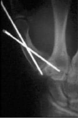
PURPOSE: To review the subjective and functional results of basal thumb metacarpal osteotomy for the treatment of trapeziometacarpal osteoarthritis. METHODS: Between July 1993 and November 1998, 35 thumb osteotomies without internal fixation were performed on 33 patients in the Christchurch Hospital, New Zealand. Records of 28 thumbs (13 right and 15 left) of 26 patients (17 women and 9 men) were available for review. Patients were reviewed using strength testing and the Michigan Hand Outcomes Questionnaire. RESULTS: The mean age of the 26 patients was 54 years (range, 30-69 years). Of the 28 thumbs, 22 (21 patients) had good or excellent results, 2 fair, one poor. The remaining 3 thumbs (3 patients) required further revision and were classified as failures. The mean follow-up period of the 25 thumbs (24 patients) not requiring revision was 34 months (range, 12-73 months). Good thumb motion was present in all hands with no trapeziometacarpal instability seen. Compared with the normative data, the strengths of key pinch, pulp pinch, and tripod pinch of our patients were significantly lower (22-32% lower), but not the grip strength. Michigan Hand Outcomes Questionnaire scores increased 28 (range, 1-56) points after surgery, with significant improvement especially in pain (+44 points), activities of daily living (one-handed tasks, +41 points), and satisfaction (+35 points). CONCLUSION: Basal thumb metacarpal osteotomy is a straightforward, conservative procedure that should be considered for grades II and III trapeziometacarpal osteoarthritis. (+info)Piezoelectric osteotomy in hand surgery: first experiences with a new technique. (7/90)
BACKGROUND: In hand and spinal surgery nerve lesions are feared complications with the use of standard oscillating saws. Oral surgeons have started using a newly developed ultrasound bone scalpel when performing precise osteotomies. By using a frequency of 25-29 kHz only mineralized tissue is cut, sparing the soft tissue. This reduces the risk of nerve lesions. As there is a lack of experience with this technique in the field of orthopaedic bone surgery, we performed the first ultrasound osteotomy in hand surgery. METHOD: While performing a correctional osteotomy of the 5th metacarpal bone we used the Piezosurgery Device from Mectron [Italy] instead of the usual oscillating saw. We will report on our experience with one case, with a follow up time of one year. RESULTS: The cut was highly precise and there were no vibrations of the bone. The time needed for the operation was slightly longer than the time needed while using the usual saw. Bone healing was good and at no point were there any neurovascular disturbances. CONCLUSION: The Piezosurgery Device is useful for small long bone osteotomies. Using the fine tip enables curved cutting and provides an opportunity for new osteotomy techniques. As the device selectively cuts bone we feel that this device has great potential in the field of hand- and spinal surgery. (+info)Parosteal osteosarcoma of the thumb metacarpal: a case report. (8/90)
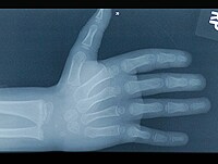
A 60-year-old man presented with increasing swelling of his right thumb, duration one year. Imaging studies demonstrated a bone-forming lesion extending from the dorsal cortex of the thumb metacarpal and involving the underlying medullary canal. Incisional biopsy yielded the diagnosis of parosteal osteosarcoma. The differential diagnosis for and rarity of parosteal osteosarcoma arising in the tubular bones of the hand are discussed. (+info)The metacarpal bones are the long slender bones that make up the middle part of the hand, located between the carpals (wrist bones) and the phalanges (finger bones). There are five metacarpal bones in total, with one for each finger and thumb. Each bone has a base attached to the carpals, a shaft, and a head that connects to the phalanges. The metacarpal bones play a crucial role in hand function, providing stability and support during gripping and manipulation movements.
The metacarpus is the medical term for the part of the hand located between the carpus (wrist) and the digits (fingers). It consists of five bones, known as the metacarpal bones, which are numbered 1 to 5 from the thumb side to the little finger side. Each metacarpal bone has a base, a shaft, and a head. The bases of the metacarpal bones articulate with the carpal bones to form the wrist joint, while the heads of the metacarpal bones form the knuckles at the back of the hand.
The metacarpus plays an essential role in hand function as it provides stability and support for the movement of the fingers and thumb. Injuries or conditions affecting the metacarpus can significantly impact hand function, causing pain, stiffness, weakness, or deformity.
Carpal bones are the eight small bones that make up the wrist joint in humans and other primates. These bones are arranged in two rows, with four bones in each row. The proximal row includes the scaphoid, lunate, triquetral, and pisiform bones, while the distal row includes the trapezium, trapezoid, capitate, and hamate bones.
The carpal bones play an essential role in the function of the wrist joint by providing stability, support, and mobility. They allow for a wide range of movements, including flexion, extension, radial deviation, ulnar deviation, and circumduction. The complex structure of the carpal bones also helps to absorb shock and distribute forces evenly across the wrist during activities such as gripping or lifting objects.
Injuries to the carpal bones, such as fractures or dislocations, can be painful and may require medical treatment to ensure proper healing and prevent long-term complications. Additionally, degenerative conditions such as arthritis can affect the carpal bones, leading to pain, stiffness, and decreased mobility in the wrist joint.
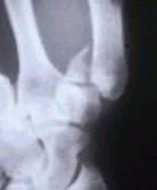
The carpometacarpal (CMC) joints are the articulations between the carpal bones of the wrist and the metacarpal bones of the hand. There are five CMC joints in total, with one located at the base of each finger and thumb. The CMC joint of the thumb, also known as the first CMC joint or trapeziometacarpal joint, is the most commonly affected by osteoarthritis. These joints play a crucial role in hand function and movement, allowing for various grips and grasping motions.
"Bone" is the hard, dense connective tissue that makes up the skeleton of vertebrate animals. It provides support and protection for the body's internal organs, and serves as a attachment site for muscles, tendons, and ligaments. Bone is composed of cells called osteoblasts and osteoclasts, which are responsible for bone formation and resorption, respectively, and an extracellular matrix made up of collagen fibers and mineral crystals.
Bones can be classified into two main types: compact bone and spongy bone. Compact bone is dense and hard, and makes up the outer layer of all bones and the shafts of long bones. Spongy bone is less dense and contains large spaces, and makes up the ends of long bones and the interior of flat and irregular bones.
The human body has 206 bones in total. They can be further classified into five categories based on their shape: long bones, short bones, flat bones, irregular bones, and sesamoid bones.
Dietary Phosphorus is a mineral that is an essential nutrient for human health. It is required for the growth, maintenance, and repair of body tissues, including bones and teeth. Phosphorus is also necessary for the production of energy, the formation of DNA and RNA, and the regulation of various physiological processes.
In the diet, phosphorus is primarily found in protein-containing foods such as meat, poultry, fish, dairy products, legumes, and nuts. It can also be found in processed foods that contain additives such as phosphoric acid, which is used to enhance flavor or as a preservative.
The recommended daily intake of phosphorus for adults is 700 milligrams (mg) per day. However, it's important to note that excessive intake of phosphorus, particularly from supplements and fortified foods, can lead to health problems such as kidney damage and calcification of soft tissues. Therefore, it's recommended to obtain phosphorus primarily from whole foods rather than supplements.
Bone density refers to the amount of bone mineral content (usually measured in grams) in a given volume of bone (usually measured in cubic centimeters). It is often used as an indicator of bone strength and fracture risk. Bone density is typically measured using dual-energy X-ray absorptiometry (DXA) scans, which provide a T-score that compares the patient's bone density to that of a young adult reference population. A T-score of -1 or above is considered normal, while a T-score between -1 and -2.5 indicates osteopenia (low bone mass), and a T-score below -2.5 indicates osteoporosis (porous bones). Regular exercise, adequate calcium and vitamin D intake, and medication (if necessary) can help maintain or improve bone density and prevent fractures.
I'm sorry for any confusion, but "horses" is not a medical term. It is the common name given to the species *Equus ferus caballus*, which are large domesticated mammals used for transportation, work, and recreation. If you have any questions about horses or a related topic that you would like a medical perspective on, please let me know and I'd be happy to help!
Bone remodeling is the normal and continuous process by which bone tissue is removed from the skeleton (a process called resorption) and new bone tissue is formed (a process called formation). This ongoing cycle allows bones to repair microdamage, adjust their size and shape in response to mechanical stress, and maintain mineral homeostasis. The cells responsible for bone resorption are osteoclasts, while the cells responsible for bone formation are osteoblasts. These two cell types work together to maintain the structural integrity and health of bones throughout an individual's life.
During bone remodeling, the process can be divided into several stages:
1. Activation: The initiation of bone remodeling is triggered by various factors such as microdamage, hormonal changes, or mechanical stress. This leads to the recruitment and activation of osteoclast precursor cells.
2. Resorption: Osteoclasts attach to the bone surface and create a sealed compartment called a resorption lacuna. They then secrete acid and enzymes that dissolve and digest the mineralized matrix, creating pits or cavities on the bone surface. This process helps remove old or damaged bone tissue and releases calcium and phosphate ions into the bloodstream.
3. Reversal: After resorption is complete, the osteoclasts undergo apoptosis (programmed cell death), and mononuclear cells called reversal cells appear on the resorbed surface. These cells prepare the bone surface for the next stage by cleaning up debris and releasing signals that attract osteoblast precursors.
4. Formation: Osteoblasts, derived from mesenchymal stem cells, migrate to the resorbed surface and begin producing a new organic matrix called osteoid. As the osteoid mineralizes, it forms a hard, calcified structure that gradually replaces the resorbed bone tissue. The osteoblasts may become embedded within this newly formed bone as they differentiate into osteocytes, which are mature bone cells responsible for maintaining bone homeostasis and responding to mechanical stress.
5. Mineralization: Over time, the newly formed bone continues to mineralize, becoming stronger and more dense. This process helps maintain the structural integrity of the skeleton and ensures adequate calcium storage.
Throughout this continuous cycle of bone remodeling, hormones, growth factors, and mechanical stress play crucial roles in regulating the balance between resorption and formation. Disruptions to this delicate equilibrium can lead to various bone diseases, such as osteoporosis, where excessive resorption results in weakened bones and increased fracture risk.
The metatarsus is the region in the foot between the tarsal bones (which form the hindfoot and midfoot) and the phalanges (toes). It consists of five long bones called the metatarsals, which articulate with the tarsal bones proximally and the phalanges distally. The metatarsus plays a crucial role in weight-bearing, support, and propulsion during walking and running. Any abnormalities or injuries to this region may result in various foot conditions, such as metatarsalgia, Morton's neuroma, or hammertoes.
 Metacarpal bones
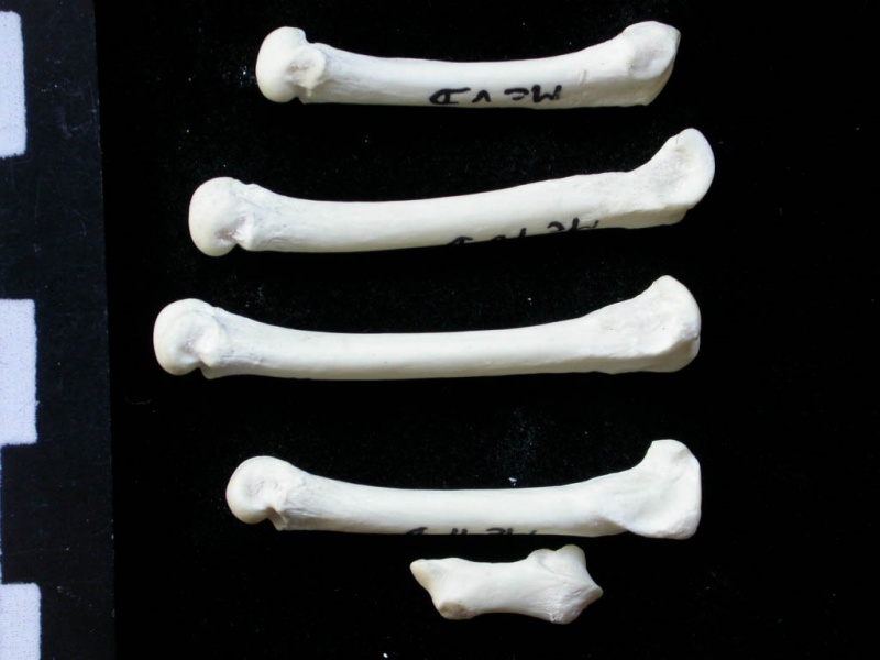
 Mallard Duck Metacarpal - OsteoID Bone Identification
 metacarpal bone of ox Archives » AnatomyLearner || The Place to Learn Veterinary Anatomy Online
 S62.363A Nondisplaced fracture of neck of third metacarpal bone, left hand, initial encounter for closed fracture | eORIF
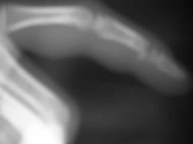
 Bennett Fracture: Practice Essentials, Anatomy, Pathophysiology and Etiology
 Metacarpal Stress Fracture Is Not an Uncommon Condition in Adolescent Racket Athletes
 cat injury - PoC - our relationship with cats and other animals
 Berberis Vulgaris modalities etc - ABC Homeopathy
 Browse subject: Sheep -- Anatomy | The Online Books Page
 Boxer's Fracture - Health Encyclopedia - University of Rochester Medical Center
 Incident Reports - Informal Investigations
 Formula 1: Daniel Ricciardo out of Dutch Grand Prix after injuring hand in practice crash
 Anatomy of the Hand | Johns Hopkins Medicine
 Denosumab, cortical bone and bone erosions in rheumatoid arthritis | Annals of the Rheumatic Diseases
 Two cases of acrometastasis to the hands and review of the literature
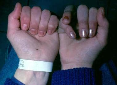
 Thrombocytopenia with absent radius syndrome | Radiology Reference Article | Radiopaedia.org
 Adventures in Behavioral Neurology-Or-What Neurology Can Tell Us About Human Nature | Edge.org
 Frontiers | Deep Learning-Based Classification of Inflammatory Arthritis by Identification of Joint Shape Patterns-How Neural...
 Opponens digiti minimi
 CT angiography and MRI of hand vascular lesions: technical considerations and spectrum of imaging findings | Insights into...
 thumb-pad - definition and meaning
 Earliest modern human-like hand bone from a new |1.84-million-year-old site at Olduvai in Tanzania | Nature Communications
 Graduate Design 2009 - ICON Magazine
 Brian M. Christie, MD