Meningeal Carcinomatosis
Meningeal Neoplasms
Krukenberg Tumor
Fatal Outcome
Hyperthermia, Induced
Chemotherapy, Cancer, Regional Perfusion
Lymphangitis
Carcinoma
Pseudomyxoma Peritonei
Oligoclonal Bands
Encyclopedias as Topic
Multiple Sclerosis
Cerebrospinal Fluid Proteins
Serum
Prognostic factors and outcomes in patients with leptomeningeal melanomatosis. (1/40)
(+info)Postradiation lumbosacral radiculopathy with spinal root cavernomas mimicking carcinomatous meningitis. (2/40)
(+info)Atypical teratoid/rhabdoid tumor arising from the third cranial nerve. (3/40)
(+info)Neoplastic meningitis. (4/40)
(+info)Leptomeningeal carcinomatosis: prognostic implications of clinical and cerebrospinal fluid features. (5/40)
(+info)Leptomeningeal carcinomatosis and sensorineural hearing loss: correlation of labyrinthine enhancement patterns with symptoms. (6/40)
(+info)Intradural squamous cell carcinoma in the sacrum. (7/40)
(+info)Efficacy and safety of liposomal cytarabine in lymphoma patients with central nervous system involvement from lymphoma. (8/40)
(+info)Meningeal carcinomatosis, also known as leptomeningeal metastasis or neoplastic meningitis, is a medical condition characterized by the spread of cancer cells to the meninges, which are the thin layers of tissue that cover and protect the brain and spinal cord.
In this condition, cancer cells from a primary tumor or metastatic cancer elsewhere in the body invade the cerebrospinal fluid (CSF) and spread throughout the meningeal spaces, causing inflammation and damage to the surrounding tissues. This can result in various neurological symptoms such as headache, nausea, vomiting, seizures, confusion, weakness, or paralysis, depending on the location of the cancer cells in the meninges.
Meningeal carcinomatosis is a serious and often life-threatening complication of advanced cancer, with a poor prognosis and limited treatment options. It can occur in various types of cancer, including lung, breast, melanoma, and hematological malignancies such as leukemia and lymphoma. Early diagnosis and prompt treatment are crucial to improve the quality of life and prolong survival in affected patients.
Meningeal neoplasms, also known as malignant meningitis or leptomeningeal carcinomatosis, refer to cancerous tumors that originate in the meninges, which are the membranes covering the brain and spinal cord. These tumors can arise primarily from the meningeal cells themselves, although they more commonly result from the spread (metastasis) of cancer cells from other parts of the body, such as breast, lung, or melanoma.
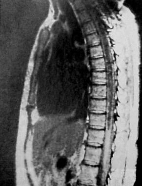
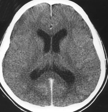
Meningeal neoplasms can cause a variety of symptoms, including headaches, nausea and vomiting, mental status changes, seizures, and focal neurological deficits. Diagnosis typically involves imaging studies (such as MRI) and analysis of cerebrospinal fluid obtained through a spinal tap. Treatment options may include radiation therapy, chemotherapy, or surgery, depending on the type and extent of the tumor. The prognosis for patients with meningeal neoplasms is generally poor, with a median survival time of several months to a year.
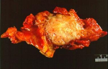
A Krukenberg tumor is a type of metastatic cancer that primarily originates from the stomach (70% of cases) but can also arise from other organs such as the colon, ovary, or breast. It is characterized by the presence of signet-ring cells, which are a specific type of malignant cell with abundant mucin displacing the nucleus to the periphery.
Krukenberg tumors typically involve both ovaries and often present with bilateral ovarian enlargement. They can cause various symptoms such as abdominal pain, bloating, or irregular menstruation. The prognosis for patients with Krukenberg tumors is generally poor due to the advanced stage of the disease at diagnosis.
A fatal outcome is a term used in medical context to describe a situation where a disease, injury, or illness results in the death of an individual. It is the most severe and unfortunate possible outcome of any medical condition, and is often used as a measure of the severity and prognosis of various diseases and injuries. In clinical trials and research, fatal outcome may be used as an endpoint to evaluate the effectiveness and safety of different treatments or interventions.
Peritoneal neoplasms refer to tumors or cancerous growths that develop in the peritoneum, which is the thin, transparent membrane that lines the inner wall of the abdomen and covers the organs within it. These neoplasms can be benign (non-cancerous) or malignant (cancerous). Malignant peritoneal neoplasms are often associated with advanced stages of gastrointestinal, ovarian, or uterine cancers and can spread (metastasize) to other parts of the abdomen.
Peritoneal neoplasms can cause various symptoms such as abdominal pain, bloating, nausea, vomiting, loss of appetite, and weight loss. Diagnosis typically involves imaging tests like CT scans or MRIs, followed by a biopsy to confirm the presence of cancerous cells. Treatment options may include surgery, chemotherapy, radiation therapy, or a combination of these approaches, depending on the type, stage, and location of the neoplasm.
Hyperthermia, induced, is a medically controlled increase in core body temperature beyond the normal range (36.5-37.5°C or 97.7-99.5°F) to a target temperature typically between 38-42°C (100.4-107.6°F). This therapeutic intervention is used in various medical fields, including oncology and critical care medicine. Induced hyperthermia can be achieved through different methods such as whole-body heating or localized heat application, often combined with chemotherapy or radiation therapy to enhance treatment efficacy.
In the context of oncology, hyperthermia is used as a sensitizer for cancer treatments by increasing blood flow to tumors, enhancing drug delivery, and directly damaging cancer cells through protein denaturation and apoptosis at higher temperatures. In critical care settings, induced hyperthermia may be applied in therapeutic hypothermia protocols to protect the brain after cardiac arrest or other neurological injuries by decreasing metabolic demand and reducing oxidative stress.
It is essential to closely monitor patients undergoing induced hyperthermia for potential adverse effects, including cardiovascular instability, electrolyte imbalances, and infections, and manage these complications promptly to ensure patient safety during the procedure.
Regional perfusion chemotherapy for cancer is a medical treatment in which a specific area or region of the body is infused with high concentrations of cancer-killing (cytotoxic) drugs via a temporary isolation and perfusion of that region. This technique is typically used to treat isolated areas of cancer that are locally advanced, recurrent, or cannot be removed surgically.
The procedure involves isolating the regional blood circulation by cannulating the artery and vein that supply blood to the target area, often the limbs (such as in melanoma or sarcoma) or the liver (for liver tumors). The chemotherapeutic drugs are then introduced into the isolated arterial circulation, allowing for a high concentration of the drug to be delivered directly to the cancerous tissue while minimizing systemic exposure and toxicity.
After the infusion, the region is rinsed with a blood-substitute solution to remove any residual chemotherapeutic agents before reconnecting the circulation. This procedure can be repeated multiple times if necessary.
Regional perfusion chemotherapy has been shown to improve local control and potentially increase survival rates in certain types of cancer, while reducing systemic side effects compared to traditional intravenous chemotherapy. However, it is a complex and invasive procedure that requires specialized medical expertise and facilities.
Lymphangitis is a medical condition characterized by the inflammation and infection of the lymphatic vessels, which are the tubular structures that transport lymph fluid from various tissues to the bloodstream. This condition typically occurs as a complication of a bacterial or fungal skin infection that spreads to the nearby lymphatic vessels.
The inflammation in lymphangitis can cause symptoms such as red streaks along the affected lymphatic vessels, swelling, warmth, and pain. Fever, chills, and fatigue may also accompany these localized symptoms. In severe cases, lymphangitis can lead to more widespread infection, sepsis, or abscess formation if left untreated.
The diagnosis of lymphangitis typically involves a physical examination and laboratory tests such as blood cultures or skin lesion cultures to identify the causative organism. Treatment usually consists of antibiotics or antifungal medications to eradicate the infection, along with supportive care such as warm compresses, elevation, and pain management. In some cases, surgical intervention may be necessary to drain any abscesses that have formed.
Carcinoma is a type of cancer that develops from epithelial cells, which are the cells that line the inner and outer surfaces of the body. These cells cover organs, glands, and other structures within the body. Carcinomas can occur in various parts of the body, including the skin, lungs, breasts, prostate, colon, and pancreas. They are often characterized by the uncontrolled growth and division of abnormal cells that can invade surrounding tissues and spread to other parts of the body through a process called metastasis. Carcinomas can be further classified based on their appearance under a microscope, such as adenocarcinoma, squamous cell carcinoma, and basal cell carcinoma.
Pseudomyxoma Peritonei (PMP) is a rare, slow-growing, and invasive cancer that typically starts in the appendix as a low-grade mucinous neoplasm, although it can also arise from other organs of the abdominal cavity. The primary characteristic of PMP is the accumulation of copious amounts of gelatinous ascites (peritoneal fluid containing mucin) within the peritoneal cavity, causing progressive abdominal distension and discomfort.
The condition is classified into three main histological subtypes: disseminated peritoneal adenomucinosis (DPAM), peritoneal mucinous carcinomatosis (PMCA), and hybrid tumors. DPAM is the least aggressive form, while PMCA is more invasive and has a worse prognosis.
The primary treatment for Pseudomyxoma Peritonei involves cytoreductive surgery (CRS) combined with hyperthermic intraperitoneal chemotherapy (HIPEC). This approach aims to remove all visible tumors and destroy any remaining cancer cells within the abdominal cavity. Early diagnosis and aggressive treatment can significantly improve the prognosis for patients with PMP, although long-term survival rates remain variable due to the disease's rarity and heterogeneity.
Oligoclonal bands (OB) are a pattern of immunoglobulin (antibody) proteins found in the cerebrospinal fluid (CSF) when it is analyzed using a technique called electrophoresis. This pattern shows a limited number (oligo) of distinct protein bands, which are clonally expanded (clonal), indicating the presence of an intr Theatreaterathecal immunoglobulin synthesis, typically in response to some sort of central nervous system (CNS) antigenic stimulation or immune response.
The detection of oligoclonal bands is often associated with inflammatory conditions affecting the CNS, such as multiple sclerosis (MS), neuromyelitis optica spectrum disorder (NMOSD), and other infectious or autoimmune diseases. However, it's important to note that their presence alone does not confirm a specific diagnosis, but rather serves as a supportive finding in conjunction with other clinical and diagnostic data.
An encyclopedia is a comprehensive reference work containing articles on various topics, usually arranged in alphabetical order. In the context of medicine, a medical encyclopedia is a collection of articles that provide information about a wide range of medical topics, including diseases and conditions, treatments, tests, procedures, and anatomy and physiology. Medical encyclopedias may be published in print or electronic formats and are often used as a starting point for researching medical topics. They can provide reliable and accurate information on medical subjects, making them useful resources for healthcare professionals, students, and patients alike. Some well-known examples of medical encyclopedias include the Merck Manual and the Stedman's Medical Dictionary.
Multiple Sclerosis (MS) is a chronic autoimmune disease that affects the central nervous system (CNS), which includes the brain, spinal cord, and optic nerves. In MS, the immune system mistakenly attacks the protective covering of nerve fibers, called myelin, leading to damage and scarring (sclerosis). This results in disrupted communication between the brain and the rest of the body, causing a variety of neurological symptoms that can vary widely from person to person.
The term "multiple" refers to the numerous areas of scarring that occur throughout the CNS in this condition. The progression, severity, and specific symptoms of MS are unpredictable and may include vision problems, muscle weakness, numbness or tingling, difficulty with balance and coordination, cognitive impairment, and mood changes. There is currently no cure for MS, but various treatments can help manage symptoms, modify the course of the disease, and improve quality of life for those affected.
Blood protein electrophoresis (BPE) is a laboratory test that separates and measures the different proteins in the blood, such as albumin, alpha-1 globulins, alpha-2 globulins, beta globulins, and gamma globulins. This test is often used to help diagnose or monitor conditions related to abnormal protein levels, such as multiple myeloma, macroglobulinemia, and other plasma cell disorders.
In this test, a sample of the patient's blood is placed on a special gel and an electric current is applied. The proteins in the blood migrate through the gel based on their electrical charge and size, creating bands that can be visualized and measured. By comparing the band patterns to reference ranges, doctors can identify any abnormal protein levels or ratios, which may indicate underlying medical conditions.
It's important to note that while BPE is a useful diagnostic tool, it should be interpreted in conjunction with other clinical findings and laboratory tests for accurate diagnosis and management of the patient's condition.
Cerebrospinal fluid (CSF) proteins refer to the proteins present in the cerebrospinal fluid, which is a clear, colorless fluid that surrounds and protects the brain and spinal cord. The protein concentration in the CSF is much lower than that in the blood, and it contains a specific set of proteins that are produced by the brain, spinal cord, and associated tissues.
The normal range for CSF protein levels is typically between 15-45 mg/dL, although this can vary slightly depending on the laboratory's reference range. An elevation in CSF protein levels may indicate the presence of neurological disorders such as meningitis, encephalitis, multiple sclerosis, or Guillain-Barre syndrome. Additionally, certain conditions such as spinal cord injury, brain tumors, or neurodegenerative diseases can also cause an increase in CSF protein levels.
Therefore, measuring CSF protein levels is an important diagnostic tool for neurologists to evaluate various neurological disorders and monitor disease progression. However, it's essential to interpret the results of CSF protein tests in conjunction with other clinical findings and laboratory test results to make an accurate diagnosis.
Serum, in the context of clinical and medical laboratory science, refers to the fluid that is obtained after blood coagulation. It is the yellowish, straw-colored liquid fraction of whole blood that remains after the clotting factors have been removed. Serum contains various proteins, electrolytes, hormones, antibodies, antigens, and other substances, which can be analyzed to help diagnose and monitor a wide range of medical conditions. It is commonly used for various clinical tests such as chemistry panels, immunological assays, drug screening, and infectious disease testing.
Immunoglobulin G (IgG) is a type of antibody, which is a protective protein produced by the immune system in response to foreign substances like bacteria or viruses. IgG is the most abundant type of antibody in human blood, making up about 75-80% of all antibodies. It is found in all body fluids and plays a crucial role in fighting infections caused by bacteria, viruses, and toxins.
IgG has several important functions:
1. Neutralization: IgG can bind to the surface of bacteria or viruses, preventing them from attaching to and infecting human cells.
2. Opsonization: IgG coats the surface of pathogens, making them more recognizable and easier for immune cells like neutrophils and macrophages to phagocytose (engulf and destroy) them.
3. Complement activation: IgG can activate the complement system, a group of proteins that work together to help eliminate pathogens from the body. Activation of the complement system leads to the formation of the membrane attack complex, which creates holes in the cell membranes of bacteria, leading to their lysis (destruction).
4. Antibody-dependent cellular cytotoxicity (ADCC): IgG can bind to immune cells like natural killer (NK) cells and trigger them to release substances that cause target cells (such as virus-infected or cancerous cells) to undergo apoptosis (programmed cell death).
5. Immune complex formation: IgG can form immune complexes with antigens, which can then be removed from the body through various mechanisms, such as phagocytosis by immune cells or excretion in urine.
IgG is a critical component of adaptive immunity and provides long-lasting protection against reinfection with many pathogens. It has four subclasses (IgG1, IgG2, IgG3, and IgG4) that differ in their structure, function, and distribution in the body.
 Oligoclonal band
Carcinosis
Leptomeningeal cancer
Causes of cancer pain
Brain tumor
Watershed stroke
Index of oncology articles
International Classification of Diseases for Oncology
Subarachnoid hemorrhage
Oligoclonal band - Wikipedia
 Syringomyelia: Background, Pathophysiology, Etiology
Magnetic resonance imaging of a choroid plexus carcinoma and meningeal carcinomatosis in a dog | Advanced Veterinary Medical...
 Page 1 | Search Results | Acta Cytologica | Karger Publishers
3PC-013 Formulation and elaboration of intrathecal trastuzumab for the treatment of a meningeal carcinomatosis | European...
Application of 3D Fast Spin-Echo T1 Black-Blood Imaging in the Diagnosis and Prognostic Prediction of Patients with...
Pancreatic Cancer: Practice Essentials, Pathophysiology, Etiology
AKR1B10 | Cancer Genetics Web
 A Phase 1b/2, Open-Label Umbrella Study To Evaluate Safety And Efficacy Of Elacestrant In Various Combinations In Patients With...
 Sixth Cranial (Abducens) Nerve Palsy - Neurologic Disorders - MSD Manual Professional Edition
HuGE Navigator|Genopedia|PHGKB
Cantore, W. A.<...
Meningitis Differential Diagnoses
 Autoimmune GFAP astrocytopathy | MedLink Neurology
VEGF165 Protein - ACROBiosystems
Accuracy of the Spinal Tap for MS - Live Disease Free
 Brain tumors, other
 Fiona Goode (AHS Coven) | Femmes de séries
 DeCS
DeCS SOAP Note for a Patient Diagnosed with Bacterial Meningitis | Essay Example
Role of temozolomide in the treatment of cancers involving the central nervous system - Fingerprint - Johns Hopkins...
 A Case of Isolated Temporal Bone Metastasis in a Lung Cancer Patient with a History of Otitis Media
 UCSD Neoplasms Trial → E7386 in Combination With Other Anticancer Drug in Participants With Solid Tumor
Clinical Trial: NCT04172597 - My Cancer Genome
 A Clinical Reasoning Exercise: Dysmetria, Nausea, Vomiting, Trouble to Speak - Prodiagnosis / Foro Osler
 UCSF Neoplasms Trial → CYT-0851 in B-Cell Malignancies and Advanced Solid Tumors
UCSF Breast Cancer Trial → Umbrella Study To Evaluate Safety And Efficacy Of Elacestrant In Various Combination In Patients...
 Búsqueda | BVS Bolivia
 Scott Lambert Carter, Ph.D. | Harvard Catalyst Profiles | Harvard Catalyst
Leptomeningeal Carcinomatosis7
- Isolated leptomeningeal carcinomatosis and possible fungal meningitis as late sequelae of oesophageal adenocarcinoma. (ox.ac.uk)
- Cytological analysis of a CSF sample revealed a cellular infiltrate consistent with leptomeningeal carcinomatosis (adenocarcinoma), with the previous oesophageal malignancy the likely primary. (ox.ac.uk)
- This term is coined as leptomeningeal carcinomatosis. (journalmc.org)
- Cerebral spinal fluid obtained from a lumbar puncture supported a diagnosis of leptomeningeal carcinomatosis. (journalmc.org)
- Leptomeningeal carcinomatosis (LC) is a form of cancer involving the nervous system, mainly arachnoid and pia mater. (journalmc.org)
- A leptomeningeal tumor - also called leptomeningeal carcinomatosis, leptomeningeal metastasis, neoplastic meningitis, meningeal metastasis and meningeal carcinomatosis - refers to cancer that has spread from the original tumor to the meninges, the thin layers of tissue that cover and protect the brain and spinal cord, causing the meninges to become inflamed. (adventhealthcancerinstitute.com)
- This is called leptomeningeal carcinomatosis. (kaiserpermanente.org)
Metastases2
- Aim and Objectives To define a formulation of IT trastuzumab (in combination with hydrocortisone and methotrexate), for a woman with metastatic breast cancer, HER2+, with meningeal carcinomatosis and brain metastases. (bmj.com)
- Active or newly diagnosed CNS metastases, including meningeal carcinomatosis. (gwdocs.com)
Neck-stiffness1
- The medical oncologist made a short differential between encephalitis , but no fever was measured, viral or bacterial meningitis , but there was no neck stiffness or meningeal carcinomatosis. (prodiagnosis.org)
Patients3
- 04 ). Given the prodromal symptoms and meningeal involvement, patients are frequently diagnosed speculatively with "test negative" meningitis of infectious etiology. (medlink.com)
- Acute myelopathy in patients with cancer can also be caused by irradiation, paraneoplastic necrotising myelitis, ruptured intervertebral disc and meningeal carcinomatosis with spinal cord involvement. (patient.info)
- Malignant plasma cells in meningeal MM out of 2000 patients with bone marrow produce an immunoglobulin, MM, was reported was by Schluterman et al. (who.int)
Diagnosis2
- Histopathologically the diagnosis was a choroid plexus carcinoma with meningeal carcinomatosis. (avmi.net)
- Meningeal carcinomatosis is a very well known diagnosis for a medical oncologist. (prodiagnosis.org)
Involvement1
- however, involvement of MM in the cerebrospinal fluid and lepto meningeal infiltration is considered rare. (who.int)
Leptomeningeal carcinomatosis5
- 5. Leptomeningeal carcinomatosis: review of the literature. (nih.gov)
- 10. Leptomeningeal carcinomatosis originating from advanced gastric cancer--a report of three cases and review of the literatures. (nih.gov)
- 11. Leptomeningeal carcinomatosis. (nih.gov)
- 15. [Intrathecal methotrexate chemotherapy for leptomeningeal carcinomatosis]. (nih.gov)
- Also called leptomeningeal carcinomatosis or meningeal carcinomatosis. (cancer.ca)
Symptoms are the first manifestations1
- Meningeal symptoms are the first manifestations in some patients. (medscape.com)
Lumbar puncture4
- Intracranial hypotension, which is most commonly caused by lumbar puncture, can lead to intense meningeal enhancement, which resolves on its own once the intracranial hypotension has been corrected. (johnshopkins.edu)
- The characteristic clinical presentation of severe postural headaches with a low opening CSF pressure on subsequent lumbar puncture and a history of prior dural puncture should alert one to the diagnosis, thus avoiding an extensive workup for carcinomatosis or infection. (johnshopkins.edu)
- Bourekas, EC , Lewin, JS & Lanzieri, CF 1995, ' Postcontrast meningeal mr enhancement secondary to intracranial hypotension caused by lumbar puncture ', Journal of computer assisted tomography , vol. 19, no. 2, pp. 299-301. (johnshopkins.edu)
- MRI ought to be performed before lumbar puncture because meningeal Salirasib discomfort because of the puncture could produce fake positive MRI outcomes. (mindunwindart.com)
Adenocarcinoma1
- The objective of this research is to investigate the clinical application value of cerebrospinal fluid (CSF) cytology and circulating tumor DNA (ctDNA) in lung adenocarcinoma (LUAD) meningeal metastasis-meningeal carcinomatosis (MC), and to further explore the possible molecular mechanisms and drug treatment targets of LUAD meningeal metastasis by next-generation sequencing (NGS). (nih.gov)
Metastasis1
- 2. [Four cases of meningeal metastasis originating from breast cancer]. (nih.gov)
Subarachnoid space1
- Primary meningeal diseases, such as the granulomatous meningitides or meningeal carcinomatosis, may on occasion cause bleeding into the subarachnoid space. (jamanetwork.com)
Lung1
- Solitary meningeal carcinomatosis from lung cancer with 8-year disease-free interval. (minervamedica.it)
Brain1
- When cancer travels to the lining of the brain, it's called meningeal carcinomatosis, and it's a serious and potentially life-threatening complication," he says. (everydayhealth.com)
Neck1
- Spinal root involvement is caused by either meningeal irritation, presenting with nuchal rigidity (15%) and neck and back pain (rare), or invasion of the spinal roots. (medscape.com)
Patients1
- 4. Meningeal carcinomatosis in patients with breast carcinoma. (nih.gov)