Mandibular Condyle
Temporomandibular Joint
Temporomandibular Joint Disorders
Radiography, Panoramic
Jaw Fixation Techniques
Mandibular Diseases
Pterygoid Muscles
Osteochondroma
Temporomandibular Joint Disc
Temporal Muscle
Mandible
Tomography, Spiral Computed
Calcification, Physiologic
Dislocations
Cartilage, Articular
Cartilage
Imaging, Three-Dimensional
Tomography, X-Ray Computed
Comparison of cephalometric analysis using a non-radiographic sonic digitizer (DigiGraph Workstation) with conventional radiography. (1/314)
Cephalometric analysis conventionally requires radiographic exposure which may not be compatible with the growing concern over radiation hazards. Recently, the Dolphin Workstation Imaging System introduced to the dental profession a non-radiographic system, called the DigiGraph Workstation which may be an alternative to cephalometric radiography. The aims of this study were to compare the validity and reproducibility of cephalometric measurements obtained from the DigiGraph Workstation with conventional cephalometric radiographs. The sample consisted of 30 human dry skulls. Two replicated sets of lateral cephalograms were obtained with steel ball markers placed at the majority of the cephalometric landmarks. Duplicate tracings prepared from each radiograph were digitized to obtain cephalometric measurements using the computer software, Dentofacial Planner. For the DigiGraph Workstation, double sonic digitizations were repeated twice for each skull, on two occasions. Fifteen angular and one linear measurements were obtained from both methods and these findings compared using ANOVA, paired t-tests and F-tests. All, except one, cephalometric measurement showed significant differences between the two methods (P < 0.0001). The DigiGraph Workstation consistently produced higher values in 11 measurements (mean differences +0.5 to +15.7 degrees or mm) and lower values in four measurements (mean differences -0.2 to -3.5 degrees). The standard deviations of the differences between readings of both methods were large (0.4-5.8 degrees or mm). The reproducibility of the DigiGraph Workstation measurements was lower than that of the radiographic measurements. The method error of the DigiGraph Workstation ranged from 7 to 70 per cent, while that of radiographic tracings was less than 2 per cent. It was concluded that measurements obtained with the DigiGraph Workstation should be interpreted with caution. (+info)Development of the human temporomandibular joint. (2/314)
A great deal of research has been published on the development of the human temporomandibularjoint (TMJ). However, there is some discordance about its morphological timing. The most controversial aspects concern the moment of the initial organization of the condyle and the squamous part of the temporal bone, the articular disc and capsule and also the cavitation and onset of condylar chondrogenesis. Serial sections of 70 human specimens between weeks 7 and 17 of development were studied by optical microscopy (25 embryos and 45 fetuses). All specimens were obtained from collections of the Institute of Embryology of the Complutense University of Madrid and the Department of Morphological Sciences of the University of Granada. Three phases in the development of the TMJ were identified. The first is the blastematic stage (weeks 7-8 of development), which corresponds with the onset of the organization of the condyle and the articular disc and capsule. During week 8 intramembranous ossification of the temporal squamous bone begins. The second stage is the cavitation stage (weeks 9-11 of development), corresponding to the initial formation of the inferior joint cavity (week 9) and the start condylar chondrogenesis. Week 11 marks the initiation of organization of the superior joint cavity. And the third stage is the maturation stage (after week 12 of development). This work establishes three phases in TMJ development: 1) the blastematic stage (weeks 7-8 of development); 2) the cavitation stage (weeks 9-11 of development); and 3) the maturation stage (after week 12 of development). This study identifies the critical period of TMJ morphogenesis as occurring between weeks 7 and 11 of development. (+info)Mandibular shape and skeletal divergency. (3/314)
Pre-treatment lateral cephalograms of 41 skeletal Class I girls aged 11 to 15 were divided according to MP-SN angle: lower than 28 degrees (hypodivergent, 10 girls), between 31 and 34 degrees (normodivergent, 18 girls), or larger than 37 degrees (hyperdivergent, 13 girls). The mandibular outlines were traced and digitized, and differences in shape were quantified using the elliptic Fourier series. Size differences were measured from the areas enclosed by the mandibular outlines. Shape differences were assessed by calculating a morphological distance (MD) between the size-independent mean mathematical reconstructions of the mandibular outlines of the three divergency classes. Mandibular shape was different in the three classes: large variations were found in hyperdivergent girls versus normodivergent girls (MD = 4.61), while smaller differences were observed in hypodivergent girls (MD versus normodivergent 2.91). Mean size-independent mandibular shapes were superimposed on an axis passing through the centres of gravity of the condyle and of the chin. Normodivergent and hyperdivergent mandibles differed mostly at gonion, the coronoid process, sigmoid notch, alveolar process, posterior border of the ramus, and along the mandibular plane. A significant size effect was also found, with smaller mandibles in the hyperdivergent girls. (+info)The functional shift of the mandible in unilateral posterior crossbite and the adaptation of the temporomandibular joints: a pilot study. (4/314)
Changes in the functional shift of the mandibular midline and the condyles were studied during treatment of unilateral posterior crossbite in six children, aged 7-11 years. An expansion plate with covered occlusal surfaces was used as a reflex-releasing stabilizing splint during an initial diagnostic phase (I) in order to determine the structural (i.e. non-guided) position of the mandible. The same plate was used for expansion and retention (phase II), followed by a post-retention phase (III) without the appliance. Before and after each phase, the functional shift was determined kinesiographically and on transcranial radiographs by concurrent recordings with and without the splint. Transverse mandibular position was also recorded on cephalometric radiographs. Prior to phase I, the mandibular midline deviated more than 2 mm and, in occlusion (ICP), the condyles showed normally centred positions in the sagittal plane. With the splint, the condyle on the crossbite side was displaced 2.4 mm (P < 0.05) forwards compared with the ICP, while the position of the condyle on the non-crossbite side was unaltered. After phase III, the deviation of the midline had been eliminated. Sagittal condylar positions in the ICP still did not deviate from the normal, and the splint position was now obtained by symmetrical forward movement of both condyles (1.3 and 1.4 mm). These findings suggest that the TMJs adapted to displacements of the mandible by condylar growth or surface modelling of the fossa. The rest position remained directly caudal to the ICP during treatment. Thus, the splint position, rather than the rest position should be used to determine the therapeutic position of the mandible. (+info)Incremental growth charts for condylar growth between 6 and 16 years of age. (5/314)
This study provides sex specific reference data for the incremental growth of the mandibular condyle. The results pertain to a mixed-longitudinal sample of 113 males and 108 females followed annually between 6 and 16 years of age (total of 1647 observations). Growth of condylion was evaluated using naturally stable mandibular reference structures. The mean growth curves were estimated by multilevel models using iterative least squares procedures; between subject variation was estimated based on the sample's percentile distributions. Mean yearly velocities of condylar growth for males ranged between 2.1 and 3.1 mm/year. Growth rates decreased during childhood, increased during adolescence, and attained a maximum of 3.1 mm/year at approximately 14.3 years of age. Females showed a more constant rate of condylar growth during childhood (2.0-2.7 mm/year), a smaller adolescent peak (2.3 mm/year) at approximately 12.2 years and rapid deceleration after the peak. These reference data offer orthodontists an objective means of evaluating growth potential and treatment outcome in individual patients. Charts are provided for evaluating condylar growth of individual patients. (+info)Thin-plate spline analysis of treatment effects of rapid maxillary expansion and face mask therapy in early Class III malocclusions. (6/314)
An effective morphometric method (thin-plate spline analysis) was applied to evaluate shape changes in the craniofacial configuration of a sample of 23 children with Class III malocclusions in the early mixed dentition treated with rapid maxillary expansion and face mask therapy, and compared with a sample of 17 children with untreated Class III malocclusions. Significant treatment-induced changes involved both the maxilla and the mandible. Major deformations consisted of forward displacement of the maxillary complex from the pterygoid region and of anterior morphogenetic rotation of the mandible, due to a significant upward and forward direction of growth of the mandibular condyle. Significant differences in size changes due to reduced increments in mandibular dimensions were associated with significant shape changes in the treated group. (+info)Differential responses to parathyroid hormone-related protein (PTHrP) deficiency in the various craniofacial cartilages. (7/314)
PTHrP null mutant mice exhibit skeletal abnormalities both in the craniofacial region and limbs. In the growth plate cartilage of the null mutant, a diminished number of proliferating chondrocytes and accelerated chondrocytic differentiation are observed. In order to examine the effect of PTHrP deficiency on the craniofacial morphology and highlight the differential feature of the composing cartilages, we examined the various cartilages in the craniofacial region of neonatal PTHrP deficient mice. The major part of the cartilaginous anterior cranial base appeared to be normal in the homozygous PTHrP deficient mice. However, acceleration of chondrocytic differentiation and endochondral bone formation was observed in the posterior part of the anterior cranial base and in the cranial base synchondroses. Ectopic bone formation was observed in the soft tissue-running mid-portion of the Meckel's cartilage, where the cartilage degenerates and converts to ligament in the course of normal development. The zonal structure of the mandibular condylar cartilage was scarcely affected, but the whole condyle was reduced in size. These results suggest the effect of PTHrP deficiency varies widely between the craniofacial cartilages, according to the differential features of each cartilage. (+info)In situ hybridisation study of type I, II, X collagens and aggrecan mRNas in the developing condylar cartilage of fetal mouse mandible. (8/314)
The aim of this study was to investigate the developmental characteristics of the mandibular condyle in sequential phases at the gene level using in situ hybridisation. At d 14.5 of gestation, although no expression of type II collagen mRNA was observed, aggrecan mRNA was detected with type I collagen mRNA in the posterior region of the mesenchymal cell aggregation continuous with the ossifying mandibular bone anlage prior to chondrogenesis. At d 15.0 of gestation, the first cartilaginous tissue appeared at the posterior edge of the ossifying mandibular bone anlage. The primarily formed chondrocytes in the cartilage matrix had already shown the appearance of hypertrophy and expressed types I, II and X collagens and aggrecan mRNAs simultaneously. At d 16.0 of gestation, the condylar cartilage increased in size due to accumulation of hypertrophic chondrocytes characterised by the expression of type X collagen mRNA, whereas the expression of type I collagen mRNA had been reduced in the hypertrophic chondrocytes and was confined to the periosteal osteogenic cells surrounding the cartilaginous tissue. At d 18.0 of gestation before birth, cartilage-characteristic gene expression had been reduced in the chondrocytes of the lower half of the hypertrophic cell layer. The present findings demonstrate that the initial chondrogenesis for the mandibular condyle starts continuous with the posterior edge of the mandibular periosteum and that chondroprogenitor cells for the condylar cartilage rapidly differentiate into hypertrophic chondrocytes. Further, it is indicated that sequential rapid changes and reductions of each mRNA might be closely related to the construction of the temporal mandibular ramus in the fetal stage. (+info)The mandibular condyle is a part of the temporomandibular joint (TMJ) in the human body. It is a rounded eminence at the end of the mandible (lower jawbone) that articulates with the glenoid fossa of the temporal bone in the skull, allowing for movements such as opening and closing the mouth, chewing, speaking, and swallowing. The mandibular condyle has both a fibrocartilaginous articular surface and a synovial joint capsule surrounding it, which provides protection and lubrication during these movements.
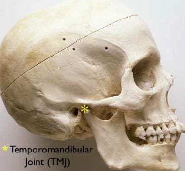
The temporomandibular joint (TMJ) is the articulation between the mandible (lower jaw) and the temporal bone of the skull. It's a complex joint that involves the movement of two bones, several muscles, and various ligaments. The TMJ allows for movements like rotation and translation, enabling us to open and close our mouth, chew, speak, and yawn. Dysfunction in this joint can lead to temporomandibular joint disorders (TMD), which can cause pain, discomfort, and limited jaw movement.
A mandibular fracture is a break or crack in the lower jaw (mandible) bone. It can occur at any point along the mandible, but common sites include the condyle (the rounded end near the ear), the angle (the curved part of the jaw), and the symphysis (the area where the two halves of the jaw meet in the front). Mandibular fractures are typically caused by trauma, such as a direct blow to the face or a fall. Symptoms may include pain, swelling, bruising, difficulty chewing or speaking, and malocclusion (misalignment) of the teeth. Treatment usually involves immobilization with wires or screws to allow the bone to heal properly.
Jaw abnormalities, also known as maxillofacial abnormalities, refer to any structural or functional deviations from the normal anatomy and physiology of the jaw bones (mandible and maxilla) and the temporomandibular joint (TMJ). These abnormalities can be present at birth (congenital) or acquired later in life due to various factors such as trauma, infection, tumors, or degenerative diseases.
Examples of jaw abnormalities include:
1. Micrognathia: a condition where the lower jaw is underdeveloped and appears recessed or small.
2. Prognathism: a condition where the lower jaw protrudes forward beyond the normal position.
3. Maxillary hypoplasia/aplasia: a condition where the upper jaw is underdeveloped or absent.
4. Mandibular hypoplasia/aplasia: a condition where the lower jaw is underdeveloped or absent.
5. Condylar hyperplasia: a condition where one or both of the condyles (the rounded ends of the mandible that articulate with the skull) continue to grow abnormally, leading to an asymmetrical jaw and facial deformity.
6. TMJ disorders: conditions affecting the temporomandibular joint, causing pain, stiffness, and limited movement.
7. Jaw tumors or cysts: abnormal growths that can affect the function and structure of the jaw bones.
Jaw abnormalities can cause various problems, including difficulty with chewing, speaking, breathing, and swallowing, as well as aesthetic concerns. Treatment options may include orthodontic treatment, surgery, or a combination of both, depending on the severity and nature of the abnormality.
Facial asymmetry refers to a condition in which the facial features are not identical or proportionate on both sides of a vertical line drawn down the middle of the face. This can include differences in the size, shape, or positioning of facial features such as the eyes, ears, nose, cheeks, and jaw. Facial asymmetry can be mild and barely noticeable, or it can be more severe and affect a person's appearance and/or functionality of the mouth and jaw.
Facial asymmetry can be present at birth (congenital) or can develop later in life due to various factors such as injury, surgery, growth disorders, nerve damage, or tumors. In some cases, facial asymmetry may not cause any medical problems and may only be of cosmetic concern. However, in other cases, it may indicate an underlying medical condition that requires treatment.
Depending on the severity and cause of the facial asymmetry, treatment options may include cosmetic procedures such as fillers or surgery, orthodontic treatment, physical therapy, or medication to address any underlying conditions.
Temporomandibular Joint Disorders (TMD) refer to a group of conditions that cause pain and dysfunction in the temporomandibular joint (TMJ) and the muscles that control jaw movement. The TMJ is the hinge joint that connects the lower jaw (mandible) to the skull (temporal bone) in front of the ear. It allows for movements required for activities such as eating, speaking, and yawning.
TMD can result from various causes, including:
1. Muscle tension or spasm due to clenching or grinding teeth (bruxism), stress, or jaw misalignment
2. Dislocation or injury of the TMJ disc, which is a small piece of cartilage that acts as a cushion between the bones in the joint
3. Arthritis or other degenerative conditions affecting the TMJ
4. Bite problems (malocclusion) leading to abnormal stress on the TMJ and its surrounding muscles
5. Stress, which can exacerbate existing TMD symptoms by causing muscle tension
Symptoms of Temporomandibular Joint Disorders may include:
- Pain or tenderness in the jaw, face, neck, or shoulders
- Limited jaw movement or locking of the jaw
- Clicking, popping, or grating sounds when moving the jaw
- Headaches, earaches, or dizziness
- Difficulty chewing or biting
- Swelling on the side of the face
Treatment for TMD varies depending on the severity and cause of the condition. It may include self-care measures (like eating soft foods, avoiding extreme jaw movements, and applying heat or cold packs), physical therapy, medications (such as muscle relaxants, pain relievers, or anti-inflammatory drugs), dental work (including bite adjustments or orthodontic treatment), or even surgery in severe cases.
Panoramic radiography is a specialized type of dental X-ray imaging that captures a panoramic view of the entire mouth, including the teeth, upper and lower jaws, and surrounding structures. It uses a special machine that rotates around the head, capturing images as it moves. This technique provides a two-dimensional image that is helpful in diagnosing and planning treatment for various dental conditions such as impacted teeth, bone abnormalities, and jaw disorders.
The panoramic radiograph can also be used to assess the development and positioning of wisdom teeth, detect cysts or tumors in the jaws, and evaluate the effects of trauma or injury to the mouth. It is a valuable tool for dental professionals as it allows them to see a comprehensive view of the oral structures, which may not be visible with traditional X-ray techniques.
It's important to note that while panoramic radiography provides valuable information, it should be used in conjunction with other diagnostic tools and clinical examinations to ensure accurate diagnosis and treatment planning.
Jaw fixation techniques, also known as maxillomandibular fixation (MMF), are procedures used in dental and oral surgery to hold the jaw in a specific position. This is typically done by wiring the upper and lower teeth together or using elastic bands and other devices to keep the jaws aligned. The technique is often used after surgical procedures on the jaw, such as corrective jaw surgery (orthognathic surgery) or fracture repair, to help promote proper healing and alignment of the bones. It may also be used in the management of temporomandibular joint disorders or other conditions affecting the jaw. The duration of jaw fixation can vary depending on the specific procedure and individual patient needs, but it typically lasts several weeks.
Mandibular neoplasms refer to abnormal growths or tumors that develop in the mandible, which is the lower jawbone. These growths can be benign (non-cancerous) or malignant (cancerous). Benign neoplasms are typically slow-growing and rarely spread to other parts of the body, while malignant neoplasms can invade surrounding tissues and may metastasize (spread) to distant sites.
Mandibular neoplasms can have various causes, including genetic mutations, exposure to certain chemicals or radiation, and infection with certain viruses. The symptoms of mandibular neoplasms may include swelling or pain in the jaw, difficulty chewing or speaking, numbness in the lower lip or chin, loose teeth, and/or a lump or mass in the mouth or neck.
The diagnosis of mandibular neoplasms typically involves a thorough clinical examination, imaging studies such as X-rays, CT scans, or MRI scans, and sometimes a biopsy to confirm the type and extent of the tumor. Treatment options depend on the type, stage, and location of the neoplasm, and may include surgery, radiation therapy, chemotherapy, or a combination of these approaches. Regular follow-up care is essential to monitor for recurrence or metastasis.
Mandibular diseases refer to conditions that affect the mandible, or lower jawbone. These diseases can be classified as congenital (present at birth) or acquired (developing after birth). They can also be categorized based on the tissues involved, such as bone, muscle, or cartilage. Some examples of mandibular diseases include:
1. Mandibular fractures: These are breaks in the lower jawbone that can result from trauma or injury.
2. Osteomyelitis: This is an infection of the bone and surrounding tissues, which can affect the mandible.
3. Temporomandibular joint (TMJ) disorders: These are conditions that affect the joint that connects the jawbone to the skull, causing pain and limited movement.
4. Mandibular tumors: These are abnormal growths that can be benign or malignant, and can develop in any of the tissues of the mandible.
5. Osteonecrosis: This is a condition where the bone tissue dies due to lack of blood supply, which can affect the mandible.
6. Cleft lip and palate: This is a congenital deformity that affects the development of the face and mouth, including the lower jawbone.
7. Mandibular hypoplasia: This is a condition where the lower jawbone does not develop properly, leading to a small or recessed chin.
8. Developmental disorders: These are conditions that affect the growth and development of the mandible, such as condylar hyperplasia or hemifacial microsomia.
The pterygoid muscles are a pair of muscles located in the deep part of the lateral aspect of the nasopharynx, in the human head. They are part of the group of muscles known as the muscles of mastication, which are involved in the chewing process.
There are two sets of pterygoid muscles: the medial and lateral pterygoids. The medial pterygoids are located deep within the jaw, near the temporomandibular joint (TMJ). They originate from the medial surface of the lateral pterygoid plate of the sphenoid bone and insert onto the inner aspect of the angle of the mandible (lower jawbone). The main function of the medial pterygoids is to assist in closing the jaw and moving it forward during chewing.
The lateral pterygoids, on the other hand, are located more superficially than the medial pterygoids and are situated near the TMJ. They have two heads: the upper head originates from the greater wing of the sphenoid bone, while the lower head arises from the lateral surface of the lateral pterygoid plate. The lateral pterygoids insert onto the front part of the neck of the mandible and the disc of the TMJ. Their main function is to assist in opening the jaw and moving it sideways during chewing.
Together, the pterygoid muscles play a crucial role in the movement and function of the jaw, allowing us to chew food effectively and speak clearly.
A comminuted fracture is a type of bone break where the bone is shattered into three or more pieces. This type of fracture typically occurs after high-energy trauma, such as a car accident or a fall from a great height. Commminuted fractures can also occur in bones that are weakened by conditions like osteoporosis or cancer. Because of the severity and complexity of comminuted fractures, they often require extensive treatment, which may include surgery to realign and stabilize the bone fragments using metal screws, plates, or rods.
Osteochondroma is a benign (noncancerous) bone tumor that typically develops during childhood or adolescent growth years. It usually forms near the end of long bones, such as those in the arms and legs, but can also occur in other bones. An osteochondroma may have a cartilage cap covering its surface.
This type of tumor often grows slowly and typically stops growing once the person has stopped growing. In many cases, an osteochondroma doesn't cause any symptoms and doesn't require treatment. However, if it continues to grow or causes problems such as pain, restricted movement, or bone deformity, surgical removal may be necessary.
Most osteochondromas are solitary (occurring singly), but some people can develop multiple tumors, a condition known as multiple hereditary exostoses or diaphyseal aclasis. This genetic disorder is associated with a higher risk of developing sarcoma, a type of cancerous tumor that can arise from osteochondromas.
It's essential to have regular follow-ups with your healthcare provider if you have an osteochondroma to monitor its growth and any potential complications.
The temporomandibular joint (TMJ) disc is a small, thin piece of fibrocartilaginous tissue located within the TMJ, which is the joint that connects the mandible (jawbone) to the temporal bone of the skull. The disc acts as a cushion and allows for smooth movement of the jaw during activities such as eating, speaking, and yawning. It divides the joint into two compartments: the upper and lower compartments.
The TMJ disc is composed of several types of tissue, including collagen fibers, elastin fibers, and a small number of cells called fibroblasts. The disc's unique structure allows it to withstand the forces generated during jaw movement and helps to distribute these forces evenly across the joint.
The TMJ disc can become damaged or displaced due to various factors such as trauma, teeth grinding (bruxism), or degenerative joint diseases like osteoarthritis. This can lead to temporomandibular disorders (TMDs) characterized by pain, stiffness, and limited jaw movement.
The temporalis muscle is a fan-shaped muscle located in the lateral aspect of the head, in the temporal fossa region. It belongs to the group of muscles known as muscles of mastication, responsible for chewing movements. The temporalis muscle has its origin at the temporal fossa and inserts into the coronoid process and ramus of the mandible. Its main function is to retract the mandible and assist in closing the jaw.
Mastication is the medical term for the process of chewing food. It's the first step in digestion, where food is broken down into smaller pieces by the teeth, making it easier to swallow and further digest. The act of mastication involves not only the physical grinding and tearing of food by the teeth but also the mixing of the food with saliva, which contains enzymes that begin to break down carbohydrates. This process helps to enhance the efficiency of digestion and nutrient absorption in the subsequent stages of the digestive process.
Bite force refers to the amount of force or pressure that can be exerted by the teeth and jaw when biting down or clenching together. It is a measure of an individual's maximum biting strength, typically expressed in units such as pounds (lb) or newtons (N). Bite force is an important factor in various biological and medical contexts, including oral health, nutrition, and the study of animal behavior and evolution.
In humans, bite force can vary widely depending on factors such as age, sex, muscle strength, and dental health. On average, a healthy adult human male may have a maximum bite force of around 150-200 pounds (670-890 newtons), while an adult female may have a bite force of around 100-130 pounds (445-578 newtons). However, these values can vary significantly from person to person.
Abnormalities in bite force can be indicative of various medical conditions or injuries, such as temporomandibular joint disorders (TMD), muscle weakness, or neurological disorders affecting the facial muscles. Assessing and measuring bite force may also be useful in evaluating the effectiveness of dental treatments or appliances, such as dentures or orthodontic devices.
The mandible, also known as the lower jaw, is the largest and strongest bone in the human face. It forms the lower portion of the oral cavity and plays a crucial role in various functions such as mastication (chewing), speaking, and swallowing. The mandible is a U-shaped bone that consists of a horizontal part called the body and two vertical parts called rami.
The mandible articulates with the skull at the temporomandibular joints (TMJs) located in front of each ear, allowing for movements like opening and closing the mouth, protrusion, retraction, and side-to-side movement. The mandible contains the lower teeth sockets called alveolar processes, which hold the lower teeth in place.
In medical terminology, the term "mandible" refers specifically to this bone and its associated structures.
Spiral Computed Tomography (CT), also known as Helical CT, is a type of computed tomography scan in which the X-ray tube and detector rotate around the patient in a spiral path, capturing data as the table moves the patient through the scanner. This continuous spiral motion allows for faster and more detailed volumetric imaging of internal organs and structures, reducing the need for multiple slices and providing improved image reconstruction. It is commonly used to diagnose and monitor various medical conditions, including cancer, heart disease, and trauma injuries.
Physiologic calcification is the normal deposit of calcium salts in body tissues and organs. It is a natural process that occurs as part of the growth and development of the human body, as well as during the repair and remodeling of tissues.
Calcium is an essential mineral that plays a critical role in many bodily functions, including bone formation, muscle contraction, nerve impulse transmission, and blood clotting. In order to maintain proper levels of calcium in the body, excess calcium that is not needed for these functions may be deposited in various tissues as a normal part of the aging process.
Physiologic calcification typically occurs in areas such as the walls of blood vessels, the lungs, and the heart valves. While these calcifications are generally harmless, they can sometimes lead to complications, particularly if they occur in large amounts or in sensitive areas. For example, calcification of the coronary arteries can increase the risk of heart disease, while calcification of the lung tissue can cause respiratory symptoms.
It is important to note that pathologic calcification, on the other hand, refers to the abnormal deposit of calcium salts in tissues and organs, which can be caused by various medical conditions such as chronic kidney disease, hyperparathyroidism, and certain infections. Pathologic calcification is not a normal process and can lead to serious health complications if left untreated.
A dislocation is a condition in which a bone slips out of its normal position in a joint. This can happen as a result of trauma or injury, such as a fall or direct blow to the body. Dislocations can cause pain, swelling, and limited mobility in the affected area. In some cases, a dislocation may also damage surrounding tissues, such as ligaments, tendons, and nerves.
Dislocations are typically treated by reducing the dislocation, which means putting the bone back into its normal position. This is usually done with the help of medication to relieve pain and relaxation techniques to help the person stay still during the reduction. In some cases, surgery may be necessary to repair damaged tissues or if the dislocation cannot be reduced through other methods. After the dislocation has been reduced, the joint may be immobilized with a splint or sling to allow it to heal properly.
It is important to seek medical attention promptly if you suspect that you have a dislocation. If left untreated, a dislocation can lead to further complications, such as joint instability and chronic pain.
Articular cartilage is the smooth, white tissue that covers the ends of bones where they come together to form joints. It provides a cushion between bones and allows for smooth movement by reducing friction. Articular cartilage also absorbs shock and distributes loads evenly across the joint, protecting the bones from damage. It is avascular, meaning it does not have its own blood supply, and relies on the surrounding synovial fluid for nutrients. Over time, articular cartilage can wear down or become damaged due to injury or disease, leading to conditions such as osteoarthritis.
Cartilage is a type of connective tissue that is found throughout the body in various forms. It is made up of specialized cells called chondrocytes, which are embedded in a firm, flexible matrix composed of collagen fibers and proteoglycans. This unique structure gives cartilage its characteristic properties of being both strong and flexible.
There are three main types of cartilage in the human body: hyaline cartilage, elastic cartilage, and fibrocartilage.
1. Hyaline cartilage is the most common type and is found in areas such as the articular surfaces of bones (where they meet to form joints), the nose, trachea, and larynx. It has a smooth, glassy appearance and provides a smooth, lubricated surface for joint movement.
2. Elastic cartilage contains more elastin fibers than hyaline cartilage, which gives it greater flexibility and resilience. It is found in structures such as the external ear and parts of the larynx and epiglottis.
3. Fibrocartilage has a higher proportion of collagen fibers and fewer chondrocytes than hyaline or elastic cartilage. It is found in areas that require high tensile strength, such as the intervertebral discs, menisci (found in joints like the knee), and the pubic symphysis.
Cartilage plays a crucial role in supporting and protecting various structures within the body, allowing for smooth movement and providing a cushion between bones to absorb shock and prevent wear and tear. However, cartilage has limited capacity for self-repair and regeneration, making damage or degeneration of cartilage tissue a significant concern in conditions such as osteoarthritis.
Three-dimensional (3D) imaging in medicine refers to the use of technologies and techniques that generate a 3D representation of internal body structures, organs, or tissues. This is achieved by acquiring and processing data from various imaging modalities such as X-ray computed tomography (CT), magnetic resonance imaging (MRI), ultrasound, or confocal microscopy. The resulting 3D images offer a more detailed visualization of the anatomy and pathology compared to traditional 2D imaging techniques, allowing for improved diagnostic accuracy, surgical planning, and minimally invasive interventions.
In 3D imaging, specialized software is used to reconstruct the acquired data into a volumetric model, which can be manipulated and viewed from different angles and perspectives. This enables healthcare professionals to better understand complex anatomical relationships, detect abnormalities, assess disease progression, and monitor treatment response. Common applications of 3D imaging include neuroimaging, orthopedic surgery planning, cancer staging, dental and maxillofacial reconstruction, and interventional radiology procedures.
Cephalometry is a medical term that refers to the measurement and analysis of the skull, particularly the head face relations. It is commonly used in orthodontics and maxillofacial surgery to assess and plan treatment for abnormalities related to the teeth, jaws, and facial structures. The process typically involves taking X-ray images called cephalograms, which provide a lateral view of the head, and then using various landmarks and reference lines to make measurements and evaluate skeletal and dental relationships. This information can help clinicians diagnose problems, plan treatment, and assess treatment outcomes.
X-ray computed tomography (CT or CAT scan) is a medical imaging method that uses computer-processed combinations of many X-ray images taken from different angles to produce cross-sectional (tomographic) images (virtual "slices") of the body. These cross-sectional images can then be used to display detailed internal views of organs, bones, and soft tissues in the body.
The term "computed tomography" is used instead of "CT scan" or "CAT scan" because the machines take a series of X-ray measurements from different angles around the body and then use a computer to process these data to create detailed images of internal structures within the body.
CT scanning is a noninvasive, painless medical test that helps physicians diagnose and treat medical conditions. CT imaging provides detailed information about many types of tissue including lung, bone, soft tissue and blood vessels. CT examinations can be performed on every part of the body for a variety of reasons including diagnosis, surgical planning, and monitoring of therapeutic responses.
In computed tomography (CT), an X-ray source and detector rotate around the patient, measuring the X-ray attenuation at many different angles. A computer uses this data to construct a cross-sectional image by the process of reconstruction. This technique is called "tomography". The term "computed" refers to the use of a computer to reconstruct the images.
CT has become an important tool in medical imaging and diagnosis, allowing radiologists and other physicians to view detailed internal images of the body. It can help identify many different medical conditions including cancer, heart disease, lung nodules, liver tumors, and internal injuries from trauma. CT is also commonly used for guiding biopsies and other minimally invasive procedures.
In summary, X-ray computed tomography (CT or CAT scan) is a medical imaging technique that uses computer-processed combinations of many X-ray images taken from different angles to produce cross-sectional images of the body. It provides detailed internal views of organs, bones, and soft tissues in the body, allowing physicians to diagnose and treat medical conditions.
The occipital bone is the single, posterior cranial bone that forms the base of the skull and encloses the brain. It articulates with the parietal bones anteriorly and the temporal bones laterally. The occipital bone also contains several important structures such as the foramen magnum, through which the spinal cord connects to the brain, and the external and internal occipital protuberances, which serve as attachment points for neck muscles.
 Condylar hypoplasia
Theories of craniofacial growth
Ectopic tooth
Mandibular fracture
Isocetus
Frey's syndrome
Megaloceros
Pseudocyst
Pterygoid fovea
Odontogenic cyst
Cyst
Aneurysmal bone cyst
Hemotympanum
Taikicetus
Dislocation of jaw
Catopsbaatar
Entelodontidae
Articular tubercle
Diplobune
Avascular necrosis
Tetragonostylops
Polonosuchus
Dysosteosclerosis
Condylar hyperplasia
Tapirus mesopotamicus
Mandibular setback surgery
Fibrous ankylosis
Activator appliance
Occlusion (dentistry)
Battle's sign
Mechanical significance of the trabecular microstructure of the human mandibular condyle - Vrije Universiteit Amsterdam
 Fracture of Condyle Mandibular in Children: Functional Orthopedic Tretment
Effects of estrogen deficiency combined with chronic alcohol consumption on rat mandibular condyle | Brazilian Journal...
 October 1977 - Volume 60 - Issue 4 : Plastic and Reconstructive Surgery
 Study of open versus closed reduction of mandibular condyle fractures
| International Surgery Journal
Study of open versus closed reduction of mandibular condyle fractures
| International Surgery Journal
 Relationship between posterior permanent dentition pattern and radiographic changes of the mandibular condyle
Dislocation of the intact mandibular condyle into the middle cranial fossa: a case report | Pocket Dentistry
Condylar hypoplasia - Wikipedia
 Osteonecrosis (Bone Infarction) Imaging: Practice Essentials, Radiography, Computed Tomography
Bifid mandibular condyles: report of four cases. - Fingerprint - Manipal Academy of Higher Education, Manipal, India
 Open Surgery Versus Closed Treatment of Unilateral Mandibular Condyle Fractures. | J Craniofac Surg;31(2): 484-487, 2020. |...
 Open Reduction With K-Wire Stabilization of Fracture Dislocations of the Mandibular Condyle: A Retrospective Review - NJ Facial...
Aneurysmal bone cyst of mandibular condyle: A case report and review of the literature<...
 A rare case of bilateral pseudoaneurysm secondary to mandibular condyle fracture-a case report with review of literature. - myAO
Effects of lateral pterygoid muscle hyperactivity on differentiation of mandibular condyles in rats - Fingerprint - Kyushu...
 Required Submission of Safety and Effectiveness Information for Certain Class III Devices | FDA
 Cone beam computed tomography-based models versus multislice spiral computed tomography-based models for assessing condylar...
Ancient whale fossil points to new species - News - Nature Middle East
 Advanced Search Results - Public Health Image Library(PHIL)
 1,487 Humerus Bone PPTs View free & download | PowerShow.com
Advanced Search Results - Public Health Image Library(PHIL)
 The Journal of Contemporary Dental Practice
 10 Benefits of Grape Seed Extract, Based on Science
 Nutrients | Free Full-Text | The Impact of Diet, Nutrition and Nutraceuticals on Oral and Periodontal Health
Guimarães AS - Search Results - PubMed
Análisis del Cóndilo, Fosa Articular y Rama Mandibular de Sujetos con Hiperplasia Condilar Activa
 Could Your Lateral Pterygoid Muscle Be Causing TMJ Pain? | Colgate®
250087009 - Joint deformity - SNOMED CT
 Environmental impacts of different methods of coffee preparation | Request PDF
 TMJ and Tinnitus: Should we explore the ligament chain from the cervical spine through the neck to the jaw to the ear? - Caring...
Mandible3
- The module discusses fractures of the mandible and condyle using a variety of techniques which underpin the totality of facial trauma management. (qmul.ac.uk)
- The term "condylar" in the name of the condition refers to the mandibular condyle, which is the upper portion of the mandible that forms part of the TMJ. (medlineplus.gov)
- Class III BMCs were observed uni- the mandible from the mandibular fora- in 4200 panoramic radiographs of Syr- laterally in eight cases (19.5%), five on men to the mental foramen, involving ian subjects (1899 women and 2301 the right side and three on the left side. (who.int)
Fracture7
- Choi K, Yang J, Chung H, Cho B. Current Concepts in the Mandibular Condyle Fracture Management Part I: Overview of Condylar Fracture. (ijsurgery.com)
- Initially, a closed reduction was attempted as there was no condylar fracture, but this failed to reduce the condyle. (pocketdentistry.com)
- The aim of this study was to determine the feasibility of direct transcortical stabilization of fracture dislocations of the mandibular condyle (FDMCs) using narrow-diameter non-threaded Kirschner wire (K-wire). (njfacialsurgery.com)
- Postoperative parameters of relevance included infection, facial nerve function, hardware removal, mandibular range of motion, and radiographic determination of fracture union. (njfacialsurgery.com)
- Fracture dislocation of the mandibular condyle (FDMC) extending into the joint capsule can add to the degree of complexity. (njfacialsurgery.com)
- A rare case of bilateral pseudoaneurysm secondary to mandibular condyle fracture-a case report with review of literature. (myao.app)
- It was later determined Mudd suffered a fractured skull, mandibular condyle fracture, and subdural hematoma as a result of the altercation, leaving him with paralysis in his lower extremities and suffering from dementia. (findlaw.com)
Ramus4
- Fractures of the peri-condylar region of the mandibular ramus are a unique surgical challenge. (njfacialsurgery.com)
- An aneurysmal bone cyst (ABC) in the right mandibular condyle and ramus of a 37-year-old woman was surgically resected and immediately reconstructed with a costochondral graft. (huji.ac.il)
- Linear measurements were taken of the condylar morphology on the sagittal and coronal planes, establishing the size of the articular fossa, mandibular ramus, and other aspects. (scielo.cl)
- The purpose of this study was to perform a systematic review of morphological alterations in the condyles after orthographic surgery involving a sagittal split ramus osteotomy (SSRO), with or without surgery on the maxilla. (unesp.br)
Dislocation7
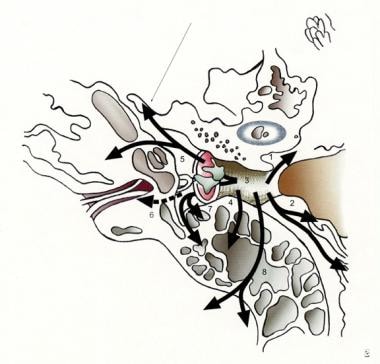
- Dislocation of the mandibular condyle into the middle cranial fossa is extremely rare. (pocketdentistry.com)
- The authors present a case of superior dislocation of left condyle into the middle cranial fossa. (pocketdentistry.com)
- Complete dislocation of the mandibular condyle from the glenoid fossa caused by trauma is commonly seen in clinics, but dislocation of the condyle into the middle cranial fossa is extremely rare. (pocketdentistry.com)
- Preoperative CT scans showed superior dislocation of mandibular condyle into the middle cranial fossa. (pocketdentistry.com)
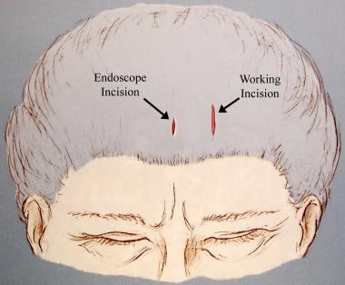
- A temporal musculofascial flap and titanium network was used to repair the defect in the middle cranial fossa to prevent re-dislocation of the condyle ( Fig. 2 ). (pocketdentistry.com)
- A CT scan revealed fractures of the left clavicle and the jaw bone (mandibular condyles, bilaterally), and dislocation of the shoulder (acromioclavicular) joint. (gjel.com)
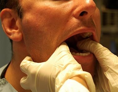
- With dislocation, the mandibular condyles may be palpated anterior to the articular eminence. (medscape.com)
Glenoid fossa2
- In rare cases, the scroll-shaped mandibular condyle penetrates the middle cranial fossa though the central part of the glenoid fossa, which is the weakest part. (pocketdentistry.com)
- Cone beam computed tomography images revealed the absence of the sphenoid, frontal, and maxillary sinus, flattening of the condyles and glenoid fossa, and bilateral hypoplasia of the mandibular condyles. (wjgnet.com)
Fossa6
- The dislocated condyle was reduced successfully and then a flap of temporal muscle and a thin titanium network were used to repair the defect in the middle cranial fossa. (pocketdentistry.com)
- The CT scan showed that the entire head of the left condyle was dislocated into the temporal fossa through the fractured glenoid cavity ( Fig. 1 ). (pocketdentistry.com)
- This also failed to reduce the condyle because almost the whole condylar head had dislocated into the fossa. (pocketdentistry.com)
- Postoperative CT scans showed reduction of the condyle and repair of the cranial fossa. (pocketdentistry.com)
- p>Basically, the shape of the articular disc changes depending on the position of the condyle in the mandibular fossa and forces applied to it. (glidewelldental.com)
- Spasm of the masseter, temporalis, and internal pterygoid muscles results in trismus, preventing return of the condyle to the temporal fossa. (medscape.com)
Bilaterally1
- Lateral mandibular motion was restricted bilaterally. (pocketdentistry.com)
Radiographic1
- Relationship between posterior permanent dentition pattern and radiographic changes of the mandibular condyle / Esfehani, M. (uniroma1.it)
Unilateral5
- Andersson J, Hallmer F, Eriksson L. Unilateral mandibular condylar fractures: a 31-year follow-up of non-surgical treatment. (ijsurgery.com)
- Clinical signs include restriction of lateral mandibular motion and mouth opening, unilateral open bite with contralateral cross-bite. (pocketdentistry.com)
- Open Surgery Versus Closed Treatment of Unilateral Mandibular Condyle Fractures. (bvsalud.org)
- The aim of this meta-analysis was to evaluate the efficacy of open surgery and closed treatment for unilateral moderately displaced mandibular condyle fractures. (bvsalud.org)
- Compared with closed treatment , open surgery has significant advantages in improving mouth opening and mandibular movement , and reducing the incidence of temporomandibular joint pain , provided that open surgery was a promising application in treatment of unilateral moderately displaced mandibular condyle fractures. (bvsalud.org)
Bifid4
- Bifid mandibular condyles: report of four cases. (manipal.edu)
- Dive into the research topics of 'Bifid mandibular condyles: report of four cases. (manipal.edu)
- ABSTRACT The objective of this study was to classify the different routes of the bifid mandibular canals (BMCs) on 2400 panoramic radiographs in a Syrian population. (who.int)
- eral bifid canals that extend to the servers and the classification proposed mandibular third molar area or the by Langlias et al. (who.int)
Lateral4
- No significant differences were observed (p=0.155) in the relation of the mean lateral distance (coronal image) of hyperplastic and non-hyperplastic condyles. (scielo.cl)
- No se observó diferencias significativas (p=0,155) en la relación de distancia medio lateral (imagen coronal) de cóndilos hiperplásico y no hiperplásicos. (scielo.cl)
- Both of the heads of your lateral pterygoid attach to the mandibular condyle. (colgate.com)
- It could involve a dislocated jaw, displaced disc, or injury to the condyle (which the lateral pterygoid attaches to). (colgate.com)
Prevalence2
- The mandibular condyle fractures are the most frequent of the mandibular bone, having a prevalence 34 - 45%, and generally occur in young adults. (bvsalud.org)
- The prevalence of mandibular dysfunction. (thejcdp.com)
Anatomic1
- With innovations in surgical technique and imaging technology, classification schemes have further subdivided fractures of the head of the condyle that compromise the anatomic and functional integrity of the TMJ. (njfacialsurgery.com)
Reduction3
- This study provides a comparative evaluation of open and closed reduction of the mandibular condyle. (ijsurgery.com)
- Nussbaum ML, Laskin DM, Best AM. Closed Versus Open Reduction of Mandibular Condylar Fractures in Adults: A Meta-Analysis. (ijsurgery.com)
- Spinzia A, Patrone R, Belli E. Open reduction and internal fixation of extracapsular mandibular condyle fractures: a long-term clinical and radiological follow-up of 25 patients. (ijsurgery.com)
Hypoplasia4
- Condylar hypoplasia is known as underdevelopment of the mandibular condyle. (wikipedia.org)
- Congenitally (primary) caused condylar hypoplasia leads to underdeveloped condyle at birth. (wikipedia.org)
- Congenital condylar hypoplasia happens when a person is born with smaller condyle than normal. (wikipedia.org)
- Acquired condylar hypoplasia happens when a person is not born with a small condyle but they sustain an injury during their growth which leads to this condition. (wikipedia.org)
Versus closed treatment1
- Open versus closed treatment of fractures of the mandibular condylar process-a prospective randomized multi-centre study. (ijsurgery.com)
Dysfunction1
- However, several authors have noted a lack of correlation between MRI findings of disk displacement and the extent of pain and dysfunction of the TMJ in patients with painful limitation of mandibular opening. (jcda.ca)
Bone3
- The condyles were analyzed histologically, histomorphometrically, and immunohistochemically using the antibodies for bone sialoprotein (BSP), osteocalcin (OCC) and receptor activator of nuclear factor kappa-B ligand (RANKL). (unicamp.br)
- Subsequently, Walker 8 investigated surgically created FDMC and its effect on growth in young Macaca rhesus monkeys by comparing bone remodeling in anatomically reduced, fixated condyles with condyles that were not reduced. (njfacialsurgery.com)
- Internal derangement is defined as a disruption within the internal aspects of the TMJ whereby the disk is displaced from its normal functional relationship with the mandibular condyle and the articular portion of the temporal bone. (jcda.ca)
Limitation1
- Pain, tenderness, and limitation of mandibular motion occur. (msdmanuals.com)
Rats1
- Aim: To investigate the effects of estrogen deficiency associated with chronic alcohol consumption on the mandibular condyle in rats. (unicamp.br)
Advancement2
- The most popular dental appliance category for snoring and sleep apnea is the mandibular advancement device (MAD) . (glidewelldental.com)
- An often-mentioned side effect of mandibular advancement is morning malocclusion. (glidewelldental.com)
Morphological1
- The purpose of this investigation was to describe and compare the morphological characteristics of hyperplastic condyles to their non-hyperplastic contralateral side. (scielo.cl)
Oral2
- Treatment of this condyle usually requires a multi-team approach involving an oral surgeon, an orthodontist and a plastic surgeon. (wikipedia.org)
- it absorbs fluid due to the reduced pressure as the condyle is held down and forward by the oral appliance. (glidewelldental.com)
Cysts1
- X-rays or cone beam CT may show flattening (eg, subchondral cysts, erosions and lipping of the condyle, suggestive of dysfunctional change, most likely due to excessive loading of the joint). (msdmanuals.com)
Submandibular1
- The chorda tympani nerve enters the mandibular arch and terminates just proximal to the submandibular ganglion, near a branch of the trigeminal nerve that will become the lingual nerve. (medscape.com)
Posterior1
- Displacement of the posterior band in relation to the condyle was quantified as mild or significant. (jcda.ca)
Nerve1
- The chorda tympani nerve exits rostrally and courses ventrally to the first pharyngeal pouch to enter the mandibular arch. (medscape.com)
Estrogen1
- Salgado MCM, Marchin AMP da S, Tera T de M, Rocha RF da, Marchini L. Effects of estrogen deficiency combined with chronic alcohol consumption on rat mandibular condyle. (unicamp.br)
Suggests1
- BMAL1 knockout mice exhibited premature aging of the mandibular condyle, which suggests that circadian rhythms affect mandibular condyle morphology. (go.jp)
Facial1
- La asimetría facial es relativamente común, transformándose en enfermedad cuando se asocia hiperplásia condilar. (scielo.cl)
Coronal1
- Se determinaron mediciones lineales de la morfología condilar en vista coronal y sagital, estableciendo tamaños de la fosa articular, rama mandibular, entre otras. (scielo.cl)
Contralateral1
- 2008), taking on a variety of morphologies, all of which manifest an increase in the size of condyle, causing the jaw to deviate towards the contralateral side to a greater or lesser degree (Nitzan et al. (scielo.cl)
Painful1
- see the image below) is a painful condition that occurs when the mandibular condyle becomes fixed in the anterosuperior aspect of the articular eminence. (medscape.com)
Review2
- Fridrich K, Pena-Velasco G, Olson R. Changing trends with mandibular fractures: A review of 1,067 cases. (ijsurgery.com)
- Zachariades N, Mezitis M, Mourouzis C, Papadakis D, Spanou A. Fractures of the mandibular condyle: A review of 466 cases. (ijsurgery.com)
Patterns1
- configuration and patterns of duplica- The drawings included the condyles, of sex and affected side of the jaw on tion [3-6]. (who.int)
Scan1
- Three-dimensional surface models of 146 condyles were constructed from each scan modality. (nih.gov)
Case1
- An analysis of 319 case reports of mandibular fractures. (ijsurgery.com)
Range of mot1
- Oscillatory joint mobilization in TMD may be effective in increasing mandibular range of motion and opening of the mouth, as well as in reducing TMJ pain and masticatory musculature. (bvsalud.org)
Mouth1
- Depending on the presence or absence of the condyle, an individual may have limited opening of the mouth. (wikipedia.org)
Results1
- Results: Histological analysis of the mandibular condyles showed that Ovx and Sham groups presented almost the same characteristics. (unicamp.br)
Small1
- The small condyle can be present either one or both sides of the lower jaw. (wikipedia.org)
Left1
- The left mandibular angle was approached and then held by Kocher forceps. (pocketdentistry.com)