Mass Screening
Early Detection of Cancer
Breast
Breast Self-Examination
Radiology
Radiographic Image Interpretation, Computer-Assisted
Palpation
False Positive Reactions
Sensitivity and Specificity
United States
Carcinoma, Intraductal, Noninfiltrating
Health Knowledge, Attitudes, Practice
New Hampshire
X-Ray Intensifying Screens
Mobile Health Units
Health Services Accessibility
Carcinoma, Ductal, Breast
Reminder Systems
Reproducibility of Results
Logistic Models
Phantoms, Imaging
Health Behavior
Physical Examination
Age Factors
Radiation Dosage
Observer Variation
Socioeconomic Factors
Medicare
Questionnaires
Technology, Radiologic
Washington
Papanicolaou Test
African Americans
Vermont
Biopsy
Vaginal Smears
Carcinoma in Situ
Prepaid Health Plans
ROC Curve
Health Care Surveys
Predictive Value of Tests
Women's Health Services
Risk Factors
Retrospective Studies
Magnetic Resonance Imaging
Biopsy, Needle
Incidence
Registries
North Carolina
Carcinoma, Lobular
Gamma Cameras
Hispanic Americans
Cost-Benefit Analysis
Tomography
Mammography and 99mTc-MIBI scintimammography in suspected breast cancer. (1/2964)
The aim of this work has been to evaluate whether a diagnostic protocol based on the joint use of mammography and 99mTc-methoxyisobutyl isonitrile (MIBI) scintimammography is capable of reducing the number of biopsies required in patients with suspected breast cancer. METHODS: We performed prone scintimammography in 90 patients with suspected breast cancer, involving 97 lesions. In all patients, the diagnosis was established by way of biopsy. On mammography, we evaluated the degree of suspicion of malignancy and the size of the lesion (smaller or larger than 1 cm in diameter). RESULTS: The results of only 41 of the biopsies indicated malignancy. On mammography, 20 lesions (of which 1 was breast cancer) were considered to be of low suspicion of malignancy, 31 (of which 4 were breast cancer) as indeterminate and 46 (of which 36 were breast cancer) as high. Fourteen lesions (2 low probability, 2 indeterminate and 10 high) were smaller than 1 cm, whereas 83 (18 low probability, 29 indeterminate and 36 high) were larger. The sensitivity, specificity, positive predictive value and negative predictive value of scintimammography were 85%, 79%, 74% and 88%, respectively. Scintimammography was positive in all cases of breast cancer that initially had a low or indeterminate suspicion of malignancy according to mammography, as well as in 30 cases of breast cancer that initially were highly suspicious. Six false-negative scintimammography studies were obtained in lesions with a high suspicion of malignancy. CONCLUSION: We propose a diagnostic protocol with a biopsy performed on lesions that have a high suspicion of malignancy as well as those with low or indeterminate suspicion that are smaller than 1 cm or with positive scintimammography results. This would have reduced the total number of biopsies performed by 34%. More importantly, there would have been a 65% reduction in number of biopsies performed in the low and indeterminate mammographic suspicion groups. All 41 cases of breast cancer would have been detected. (+info)The effect of the antiscatter grid on full-field digital mammography phantom images. (2/2964)
Computer Analysis of Mammography Phantom Images (CAMPI) is a method for making quantitative measurements of image quality. This article reports on a recent application of this method to a prototype full-field digital mammography (FFDM) machine. Images of a modified ACR phantom were acquired on the General Electric Diagnostic Molybdenum Rhodium (GE-DMR) FFDM machine at a number of x-ray techniques, both with and without the scatter reduction grid. The techniques were chosen so that one had sets of grid and non-grid images with matched doses (200 mrads) and matched gray-scale values (1500). A third set was acquired at constant 26 kVp and varying mAs for both grid conditions. Analyses of the images yielded signal-to-noise-ratio (SNR), contrast and noise corresponding to each target object, and a non-uniformity measure. The results showed that under conditions of equal gray-scale value the grid images were markedly superior, albeit at higher doses than the non-grid images. Under constant dose conditions, the non-grid images were slightly superior in SNR (7%) but markedly less uniform (60%). Overall, the grid images had substantially greater contrast and superior image uniformity. These conclusions applied to the whole kVp range studied for the Mo-Mo target filter combination and 4 cm of breast equivalent material of average composition. These results suggest that use of the non-grid technique in digital mammography with the GE-DMR-FFDM unit, is presently not warranted. With improved uniformity correction procedure, this conclusion would change and one should be able to realize a 14% reduction in patient dose at the same SNR by using a non-grid technique. (+info)Screening Mammography Program of British Columbia: pattern of use and health care system costs. (3/2964)
BACKGROUND: The use of mammography for screening asymptomatic women has increased dramatically in the past decade. This report describes the changes that have occurred in the use of bilateral mammography in British Columbia since the provincial breast cancer screening program began in 1988. METHODS: Using province-wide databases from both the breast cancer screening program and the provincial health insurance plan in BC, the authors determined the number and costs of bilateral mammography services for women aged 40 years or older between Apr. 1, 1986, and Mar. 31, 1997. Unilateral mammography was excluded because it is used for investigating symptomatic disease and screening abnormalities, and for follow-up of women who have undergone mastectomy for cancer. RESULTS: As the provincial breast cancer screening program expanded from 1 site in 1988 to 23 in 1997, it provided an increasing proportion of the bilateral mammographic examinations carried out each year in BC. In fiscal year 1996/97, 65% of bilateral mammographic examinations were performed through the screening program. The cost per examination within the screening program dropped as volume increased. Thirty percent more bilateral mammography examinations were done in 1996/97 than in 1991/92, but health care system expenditures for these services increased by only 4% during the same period. In calendar year 1996, 21% of new breast cancers were diagnosed as a result of a screening program visit. INTERPRETATION: Substantial increases in health care expenditures have been avoided by shifting bilateral mammography services to the provincial screening program, which has a lower cost per screening visit. (+info)Moderate physical activity in relation to mammographic patterns. (4/2964)
High-risk mammographic patterns may be used as a surrogate end point for breast cancer in etiologic research as well as in prevention studies. Physical activity may be one of the few modifiable risk factors for breast cancer. We examined the relationship between physical activity and mammographic patterns among 2720 Norwegian women, ages 40-56 years, who participated in both the Second and Third Tromso studies. Epidemiologic data were obtained through questionnaires. Two questions from the Second Tromso study and five questions from the Third elicited information on physical activity. The mammograms were categorized into five groups based on anatomical-mammographic correlations. For analysis, patterns I through III were combined into a low-risk group and patterns IV and V into a high-risk group. Odds ratios that were adjusted for age, education, menopausal status, body mass index, parity, age at menarche, oral contraceptive use, and alcohol intake, with 95% confidence intervals, were estimated using logistic regression. Women who reported moderate physical activity, i.e., more than 2 h/week, were 20% less likely (odds ratio, 0.8; 95% confidence interval, 0.6-1.1) to have high-risk mammographic patterns compared with those who reported being inactive. This relationship remains consistent when stratified by menopausal status, parity, and tertiles of body mass index. However, all of the associations between various measures of physical activity and high-risk patterns found in this study are weak with confidence intervals that include 1.0. Thus, chance is a reasonable explanation for the weak associations found. The relationship between physical activity and high-risk patterns should be examined further as a means to explore the biologic mechanisms relating physical activity to breast cancer risk. (+info)Macronutrient intake and change in mammographic density at menopause: results from a randomized trial. (5/2964)
To examine the effects of dietary fat intake on breast cancer risk, we are conducting a randomized trial of dietary intervention in women with extensive areas of radiologically dense breast tissue on mammography, a risk factor for breast cancer. Early results show that after 2 years on a low-fat, high-carbohydrate diet there is a significant reduction in area of density, particularly in women going through menopause. In women who went through menopause during the 2-year follow-up, the mean decreases in area of density and percentage of density in the intervention group were 11.0 cm2 and 11.0%, respectively, whereas the control group decreased 4.5 cm2 and 5.2%. The purpose of this analysis was to determine whether changes in intake of specific macronutrients could account for the observed reduction in breast density in these women. Differences between 2-year and baseline values of macronutrients (averaged over 3 nonconsecutive days of food intake) were calculated. We examined the effect of dietary variables, adjusted for changes in total calorie intake and weight and for family history of breast cancer, on changes in area of density and percentage of density using linear regression. Reduction in total or saturated fat intake or cholesterol intake was significantly associated with decreased dense area (p < or = .004). The most significant dietary variable associated with reduction in percentage of density was reduction in dietary cholesterol intake (P = 0.001), although reducing saturated fat intake was of borderline significance (P = 0.05). The effect of the membership in the intervention and control groups on change in area of density or percentage of density was reduced by models that included changes in intake of any fat, or cholesterol, or carbohydrates. The observation of an effect of diet at menopause on breast density, a marker of increased risk of breast cancer, may be an indication that exposures at this time have an enhanced effect on subsequent risk. (+info)'Should a mammographic screening programme carry the warning: screening can damage your health!'? (6/2964)
The balanced presentation afforded by convening a Citizens' Jury when considering a major question such as the introduction of a breast screening programme is advocated. This method would enable account to be taken of all the costs, both human and financial, to all those affected, both participating and organizing, as well as the benefits. Provision of such a democratic opportunity enables consideration to be given to a broad range of factors, by selection of an appropriate range of witnesses, with the advantage of involving the lay public in this decision-making process. Attendance by health correspondents, medical journalists and other media representatives enables publicization of a democracy in action whilst helping to inform the wider debate. Such an exercise could inform whether the NHS BSP should continue in its current form. (+info)American Society of Clinical Oncology 1998 update of recommended breast cancer surveillance guidelines. (7/2964)
OBJECTIVE: To determine an effective, evidence-based, postoperative surveillance strategy for the detection and treatment of recurrent breast cancer. Tests are recommended only if they have an impact on the outcomes specified by American Society of Clinical Oncology (ASCO) for clinical practice guidelines. POTENTIAL INTERVENTION: All tests described in the literature for postoperative monitoring were considered. In addition, the data were critically evaluated to determine the optimal frequency of monitoring. OUTCOME: Outcomes of interest include overall and disease-free survival, quality of life, toxicity reduction, and secondarily cost-effectiveness. EVIDENCE: A search was performed to determine all relevant articles published over the past 20 years on the efficacy of surveillance testing for breast cancer recurrence. These publications comprised both retrospective and prospective studies. VALUES: Levels of evidence and guideline grades were rated by a standard process. More weight was given to studies that tested a hypothesis directly relating testing to one of the primary outcomes in a randomized design. BENEFITS, HARMS, AND COSTS: The possible consequences of false-positive and -negative tests were considered in evaluating a preference for one of two tests providing similar information. Cost alone was not a determining factor. RECOMMENDATIONS: The attached guidelines and text summarize the updated recommendations of the ASCO breast cancer expert panel. Data are sufficient to recommend monthly breast self-examination, annual mammography of the preserved and contralateral breast, and a careful history and physical examination every 3 to 6 months for 3 years, then every 6 to 12 months for 2 years, then annually. Data are not sufficient to recommend routine bone scans, chest radiographs, hematologic blood counts, tumor markers (carcinoembryonic antigen, cancer antigen [CA] 15-5, and CA 27.29), liver ultrasonograms, or computed tomography scans. VALIDATION: The recommendations of the breast cancer expert panel were evaluated and supported by the ASCO Health Services Research Committee reviewers and the ASCO Board of Directors. (+info)Double-phase 99mTc-sestamibi scintimammography and trans-scan in diagnosing breast cancer. (8/2964)
The goal of our study was to assess the value of both scintimammography with 99mTc-sestamibi (SMM) and trans-scan (T-scan) in detecting breast cancer. METHODS: A total of 121 women were evaluated by palpation, mammography, SMM and T-scan. SMM was performed in the prone, breast dependent position. Immediate and delayed views (double-phase) were obtained. T-scan is a new breast imaging method that maps noninvasively the distribution of tissue electrical impedance and capacitance. RESULTS: SMM had 88.9% sensitivity, 88.4% specificity and 88.4% accuracy in detecting breast cancer. SMM had 100% sensitivity in detecting breast tumors >1 cm and only 66% sensitivity in detecting tumors <1 cm. T-scan had 72.2% sensitivity and 67% specificity in detecting breast cancer. It detected one more breast cancer than SMM, at the expense of 27 additional false-positive results. CONCLUSION: Double-phase SMM was sensitive and specific in detecting breast cancer. This method may reduce the rate of negative breast biopsies in tumors >1 cm. T-scan was only moderately accurate in detecting breast cancer. Its addition to SMM did not improve significantly the rate of breast cancer detection. However, because of its complete noninvasiveness, large-scale applicability and low cost, T-scan deserves further refining. (+info)Mammography is defined as a specialized medical imaging technique used to create detailed X-ray images of the breast tissue. It's primarily used as a screening tool to detect early signs of breast cancer in women who have no symptoms or complaints, as well as a diagnostic tool for further evaluation of abnormalities detected by other imaging techniques or during a clinical breast exam.
There are two primary types of mammography: film-screen mammography and digital mammography. Film-screen mammography uses traditional X-ray films to capture the images, while digital mammography utilizes digital detectors to convert X-rays into electronic signals, which are then displayed on a computer screen. Digital mammography offers several advantages over film-screen mammography, including lower radiation doses, improved image quality, and the ability to manipulate and enhance the images for better interpretation.
Mammography plays a crucial role in reducing breast cancer mortality by enabling early detection and treatment of this disease. Regular mammography screenings are recommended for women over a certain age (typically starting at age 40 or 50, depending on individual risk factors) to increase the chances of detecting breast cancer at an early stage when it is most treatable.
Breast neoplasms refer to abnormal growths in the breast tissue that can be benign or malignant. Benign breast neoplasms are non-cancerous tumors or growths, while malignant breast neoplasms are cancerous tumors that can invade surrounding tissues and spread to other parts of the body.
Breast neoplasms can arise from different types of cells in the breast, including milk ducts, milk sacs (lobules), or connective tissue. The most common type of breast cancer is ductal carcinoma, which starts in the milk ducts and can spread to other parts of the breast and nearby structures.
Breast neoplasms are usually detected through screening methods such as mammography, ultrasound, or MRI, or through self-examination or clinical examination. Treatment options for breast neoplasms depend on several factors, including the type and stage of the tumor, the patient's age and overall health, and personal preferences. Treatment may include surgery, radiation therapy, chemotherapy, hormone therapy, or targeted therapy.
Medical mass screening, also known as population screening, is a public health service that aims to identify and detect asymptomatic individuals in a given population who have or are at risk of a specific disease. The goal is to provide early treatment, reduce morbidity and mortality, and prevent the spread of diseases within the community.
A mass screening program typically involves offering a simple, quick, and non-invasive test to a large number of people in a defined population, regardless of their risk factors or symptoms. Those who test positive are then referred for further diagnostic tests and appropriate medical interventions. Examples of mass screening programs include mammography for breast cancer detection, PSA (prostate-specific antigen) testing for prostate cancer, and fecal occult blood testing for colorectal cancer.
It is important to note that mass screening programs should be evidence-based, cost-effective, and ethically sound, with clear benefits outweighing potential harms. They should also consider factors such as the prevalence of the disease in the population, the accuracy and reliability of the screening test, and the availability and effectiveness of treatment options.
Early detection of cancer refers to the identification of malignant cells or tumors in their initial stages, before they have had a chance to grow and spread. This is typically achieved through various screening methods and tests that are designed to detect specific types of cancers. The goal of early detection is to increase the chances of successful treatment and improve the overall prognosis for patients.
Some common methods used for early cancer detection include:
1. Regular screenings such as mammograms, colonoscopies, and Pap tests, which can help identify precancerous or cancerous cells in their earliest stages.
2. Imaging tests like CT scans, MRIs, and PET scans, which can help detect tumors that may not be visible through other screening methods.
3. Blood tests that look for specific biomarkers or tumor markers, which can indicate the presence of cancer in the body.
4. Genetic testing to identify individuals who may be at higher risk of developing certain types of cancer due to inherited genetic mutations.
It's important to note that while early detection is an important tool in the fight against cancer, it is not a guarantee of successful treatment or cure. However, it can significantly improve the odds of successful treatment and increase the chances of survival for many patients.
An X-ray film, also known as radiograph, is a medical imaging tool that uses X-rays to create images of the body's internal structures. The film itself is a light-sensitive material that reacts to the X-rays passing through the body and records the resulting shadows and patterns on its surface.
The process involves exposing the patient to a controlled amount of X-ray radiation, which passes through the body and is absorbed differently by various tissues and structures. Denser materials such as bone absorb more X-rays and appear white or light gray on the film, while less dense materials such as soft tissues absorb fewer X-rays and appear darker.
Once the X-ray exposure is complete, the film is developed using a chemical process that produces a visible image of the internal structures. This image can then be analyzed by medical professionals to diagnose injuries, diseases, or other conditions affecting the body's internal structures.
It's worth noting that in modern medical imaging, digital X-ray sensors have largely replaced traditional X-ray film, offering several advantages such as reduced radiation exposure, faster image processing, and easier storage and retrieval of images.
The breast is the upper ventral region of the human body in females, which contains the mammary gland. The main function of the breast is to provide nutrition to infants through the production and secretion of milk, a process known as lactation. The breast is composed of fibrous connective tissue, adipose (fatty) tissue, and the mammary gland, which is made up of 15-20 lobes that are arranged in a radial pattern. Each lobe contains many smaller lobules, where milk is produced during lactation. The milk is then transported through a network of ducts to the nipple, where it can be expressed by the infant.
In addition to its role in lactation, the breast also has important endocrine and psychological functions. It contains receptors for hormones such as estrogen and progesterone, which play a key role in sexual development and reproduction. The breast is also a source of sexual pleasure and can be an important symbol of femininity and motherhood.
It's worth noting that males also have breast tissue, although it is usually less developed than in females. Male breast tissue consists mainly of adipose tissue and does not typically contain functional mammary glands. However, some men may develop enlarged breast tissue due to conditions such as gynecomastia, which can be caused by hormonal imbalances or certain medications.
Breast self-examination (BSE) is a procedure in which an individual manually checks their own breasts for any changes or abnormalities. The goal of BSE is to detect breast cancer or other breast abnormalities as early as possible. It involves looking at and feeling the breasts for any lumps, thickenings, or other changes in size, shape, or appearance.
The American Cancer Society recommends that women become familiar with how their breasts normally look and feel and report any changes to their healthcare provider. However, they do not recommend regular monthly BSE as a routine screening tool for breast cancer, as it has not been shown to reduce the risk of dying from breast cancer or improve survival rates. Instead, they recommend regular mammograms and clinical breast exams as the most effective ways to detect breast cancer early.
It's important to note that while BSE can help women become more familiar with their breasts and detect changes early, it should not replace regular medical check-ups and mammograms. Any concerns or changes in the breasts should be reported to a healthcare provider as soon as possible for further evaluation.
Radiology is a medical specialty that uses imaging technologies to diagnose and treat diseases. These imaging technologies include X-rays, computed tomography (CT) scans, magnetic resonance imaging (MRI) scans, positron emission tomography (PET) scans, ultrasound, and mammography. Radiologists are medical doctors who have completed specialized training in interpreting these images to diagnose medical conditions and guide treatment plans. They also perform image-guided procedures such as biopsies and tumor ablations. The goal of radiology is to provide accurate and timely information to help physicians make informed decisions about patient care.
Computer-assisted radiographic image interpretation is the use of computer algorithms and software to assist and enhance the interpretation and analysis of medical images produced by radiography, such as X-rays, CT scans, and MRI scans. The computer-assisted system can help identify and highlight certain features or anomalies in the image, such as tumors, fractures, or other abnormalities, which may be difficult for the human eye to detect. This technology can improve the accuracy and speed of diagnosis, and may also reduce the risk of human error. It's important to note that the final interpretation and diagnosis is always made by a qualified healthcare professional, such as a radiologist, who takes into account the computer-assisted analysis in conjunction with their clinical expertise and knowledge.
Breast diseases refer to a wide range of conditions that affect the breast tissue. These can be broadly categorized into non-cancerous and cancerous conditions.
Non-cancerous breast diseases include:
1. Fibrocystic breast changes: This is a common condition where the breast tissue becomes lumpy, tender, and sometimes painful. It is caused by hormonal changes and is most common in women aged 20 to 50.
2. Mastitis: This is an infection of the breast tissue, usually occurring in breastfeeding women. Symptoms include redness, swelling, warmth, and pain in the affected area.
3. Breast abscess: This is a collection of pus in the breast tissue, often caused by bacterial infection. It can be painful and may require surgical drainage.
4. Fibroadenomas: These are benign tumors made up of glandular and fibrous tissue. They are usually round, firm, and mobile, and can be removed if they cause discomfort.
5. Intraductal papillomas: These are small, wart-like growths that occur in the milk ducts. They may cause nipple discharge, which can be bloody or clear.
Cancerous breast diseases include:
1. Breast cancer: This is a malignant tumor that starts in the breast tissue. It can spread to other parts of the body if left untreated. There are several types of breast cancer, including ductal carcinoma, lobular carcinoma, and inflammatory breast cancer.
2. Paget's disease of the nipple: This is a rare form of breast cancer that affects the skin of the nipple and areola. It can cause symptoms such as redness, itching, burning, and flaking of the nipple skin.
3. Phyllodes tumors: These are rare breast tumors that can be benign or malignant. They usually grow quickly and may require surgical removal.
It is important to note that not all breast lumps are cancerous, and many non-cancerous conditions can cause breast changes. However, any new or unusual breast symptoms should be evaluated by a healthcare professional to rule out serious conditions such as breast cancer.
Palpation is a medical examination technique in which a healthcare professional uses their hands to feel the size, shape, and consistency of body parts, including organs, tissues, and bones. It is used to assess the patient's overall health, identify any abnormalities or areas of pain, monitor healing and disease progression, and guide diagnostic and treatment decisions.
During palpation, the healthcare professional applies gentle pressure with their fingers or hands to specific areas of the body, feeling for any changes in texture, temperature, moisture, or movement. The technique can be used to assess various bodily systems, including the cardiovascular, respiratory, gastrointestinal, musculoskeletal, and nervous systems.
Palpation is a valuable tool in physical examinations because it is non-invasive, relatively quick, and cost-effective. It can provide important information that helps healthcare professionals make accurate diagnoses and develop effective treatment plans for their patients.
A "false positive reaction" in medical testing refers to a situation where a diagnostic test incorrectly indicates the presence of a specific condition or disease in an individual who does not actually have it. This occurs when the test results give a positive outcome, while the true health status of the person is negative or free from the condition being tested for.
False positive reactions can be caused by various factors including:
1. Presence of unrelated substances that interfere with the test result (e.g., cross-reactivity between similar molecules).
2. Low specificity of the test, which means it may detect other conditions or irrelevant factors as positive.
3. Contamination during sample collection, storage, or analysis.
4. Human errors in performing or interpreting the test results.
False positive reactions can have significant consequences, such as unnecessary treatments, anxiety, and increased healthcare costs. Therefore, it is essential to confirm any positive test result with additional tests or clinical evaluations before making a definitive diagnosis.
Sensitivity and specificity are statistical measures used to describe the performance of a diagnostic test or screening tool in identifying true positive and true negative results.
* Sensitivity refers to the proportion of people who have a particular condition (true positives) who are correctly identified by the test. It is also known as the "true positive rate" or "recall." A highly sensitive test will identify most or all of the people with the condition, but may also produce more false positives.
* Specificity refers to the proportion of people who do not have a particular condition (true negatives) who are correctly identified by the test. It is also known as the "true negative rate." A highly specific test will identify most or all of the people without the condition, but may also produce more false negatives.
In medical testing, both sensitivity and specificity are important considerations when evaluating a diagnostic test. High sensitivity is desirable for screening tests that aim to identify as many cases of a condition as possible, while high specificity is desirable for confirmatory tests that aim to rule out the condition in people who do not have it.
It's worth noting that sensitivity and specificity are often influenced by factors such as the prevalence of the condition in the population being tested, the threshold used to define a positive result, and the reliability and validity of the test itself. Therefore, it's important to consider these factors when interpreting the results of a diagnostic test.
Patient acceptance of health care refers to the willingness and ability of a patient to follow and engage in a recommended treatment plan or healthcare regimen. This involves understanding the proposed medical interventions, considering their potential benefits and risks, and making an informed decision to proceed with the recommended course of action.
The factors that influence patient acceptance can include:
1. Patient's understanding of their condition and treatment options
2. Trust in their healthcare provider
3. Personal beliefs and values related to health and illness
4. Cultural, linguistic, or socioeconomic barriers
5. Emotional responses to the diagnosis or proposed treatment
6. Practical considerations, such as cost, time commitment, or potential side effects
Healthcare providers play a crucial role in facilitating patient acceptance by clearly communicating information, addressing concerns and questions, and providing support throughout the decision-making process. Encouraging shared decision-making and tailoring care plans to individual patient needs and preferences can also enhance patient acceptance of health care.
I'm sorry for any confusion, but "United States" is a geopolitical entity, specifically the name of the country consisting of 50 states, and it is not a medical term or concept. Therefore, I can't provide a medical definition for it. If you have any questions related to health, medicine, or biology, I would be happy to try to help answer those!
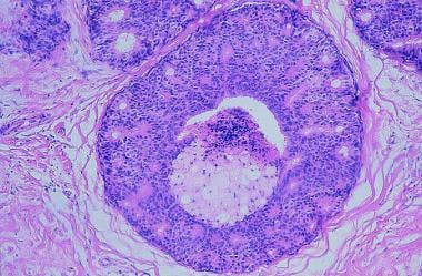
Intraductal carcinoma, noninfiltrating is a medical term used to describe a type of breast cancer that is confined to the milk ducts of the breast. It is also sometimes referred to as ductal carcinoma in situ (DCIS). Noninfiltrating means that the cancer cells have not spread beyond the ducts into the surrounding breast tissue or elsewhere in the body.
In this type of cancer, abnormal cells line the milk ducts and fill the inside of the ducts. These abnormal cells may look like cancer cells under a microscope, but they have not grown through the walls of the ducts into the surrounding breast tissue. However, if left untreated, noninfiltrating intraductal carcinoma can progress to an invasive form of breast cancer where the cancer cells spread beyond the milk ducts and invade the surrounding breast tissue.
It is important to note that while noninfiltrating intraductal carcinoma is considered a precancerous condition, it still requires medical treatment to prevent the development of invasive breast cancer. Treatment options may include surgery, radiation therapy, or hormone therapy, depending on the size and location of the tumor and other individual factors.
"Health Knowledge, Attitudes, and Practices" (HKAP) is a term used in public health to refer to the knowledge, beliefs, assumptions, and behaviors that individuals possess or engage in that are related to health. Here's a brief definition of each component:
1. Health Knowledge: Refers to the factual information and understanding that individuals have about various health-related topics, such as anatomy, physiology, disease processes, and healthy behaviors.
2. Attitudes: Represent the positive or negative evaluations, feelings, or dispositions that people hold towards certain health issues, practices, or services. These attitudes can influence their willingness to adopt and maintain healthy behaviors.
3. Practices: Encompass the specific actions or habits that individuals engage in related to their health, such as dietary choices, exercise routines, hygiene practices, and use of healthcare services.
HKAP is a multidimensional concept that helps public health professionals understand and address various factors influencing individual and community health outcomes. By assessing and addressing knowledge gaps, negative attitudes, or unhealthy practices, interventions can be designed to promote positive behavior change and improve overall health status.
I'm sorry for any confusion, but "New Hampshire" is not a medical term or concept. It is one of the 50 states in the United States of America, located in the New England region. If you have any questions related to medical topics, I would be happy to try and help answer those for you!
Patient compliance, also known as medication adherence or patient adherence, refers to the degree to which a patient's behavior matches the agreed-upon recommendations from their healthcare provider. This includes taking medications as prescribed (including the correct dosage, frequency, and duration), following dietary restrictions, making lifestyle changes, and attending follow-up appointments. Poor patient compliance can negatively impact treatment outcomes and lead to worsening of symptoms, increased healthcare costs, and development of drug-resistant strains in the case of antibiotics. It is a significant challenge in healthcare and efforts are being made to improve patient education, communication, and support to enhance compliance.
X-ray intensifying screens are medical imaging devices that contain phosphorescent materials, which emit light in response to the absorption of X-ray radiation. They are used in conjunction with X-ray film to enhance the visualization of radiographic images by converting X-rays into visible light. The screens are placed inside a cassette, along with the X-ray film, and exposed to X-rays during medical imaging procedures such as radiography or fluoroscopy.
The phosphorescent materials in the intensifying screens absorb most of the X-ray energy and re-emit it as visible light, which then exposes the X-ray film. This process increases the efficiency of the X-ray exposure, reducing the amount of radiation required to produce a diagnostic image. The use of intensifying screens can significantly improve the quality and detail of radiographic images while minimizing patient exposure to ionizing radiation.
Mobile Health Units (MHUs) are specialized vehicles or transportable facilities that deliver healthcare services in a flexible and accessible manner. They are equipped with medical equipment, supplies, and staff to provide a range of health care services, including preventive care, primary care, dental care, mental health services, and diagnostic screenings. MHUs can be deployed to various locations such as rural areas, underserved communities, disaster-stricken regions, and community events to increase access to healthcare for those who may not have easy access to medical facilities. They are an innovative solution to address health disparities and improve overall population health.
Women's health is a branch of healthcare that focuses on the unique health needs, conditions, and concerns of women throughout their lifespan. It covers a broad range of topics including menstruation, fertility, pregnancy, menopause, breast health, sexual health, mental health, and chronic diseases that are more common in women such as osteoporosis and autoimmune disorders. Women's health also addresses issues related to gender-based violence, socioeconomic factors, and environmental impacts on women's health. It is aimed at promoting and maintaining the physical, emotional, and reproductive well-being of women, and preventing and treating diseases and conditions that disproportionately affect them.
Health services accessibility refers to the degree to which individuals and populations are able to obtain needed health services in a timely manner. It includes factors such as physical access (e.g., distance, transportation), affordability (e.g., cost of services, insurance coverage), availability (e.g., supply of providers, hours of operation), and acceptability (e.g., cultural competence, language concordance).
According to the World Health Organization (WHO), accessibility is one of the key components of health system performance, along with responsiveness and fair financing. Improving accessibility to health services is essential for achieving universal health coverage and ensuring that everyone has access to quality healthcare without facing financial hardship. Factors that affect health services accessibility can vary widely between and within countries, and addressing these disparities requires a multifaceted approach that includes policy interventions, infrastructure development, and community engagement.
Carcinoma, ductal, breast is a type of breast cancer that begins in the milk ducts (the tubes that carry milk from the lobules of the breast to the nipple). It is called "ductal" because it starts in the cells that line the milk ducts. Ductal carcinoma can be further classified as either non-invasive or invasive, based on whether the cancer cells are confined to the ducts or have spread beyond them into the surrounding breast tissue.
Non-invasive ductal carcinoma (also known as intraductal carcinoma or ductal carcinoma in situ) is a condition where abnormal cells have been found in the lining of the milk ducts, but they have not spread outside of the ducts. These cells have the potential to become invasive and spread to other parts of the breast or body if left untreated.
Invasive ductal carcinoma (IDC) is a type of breast cancer that starts in a milk duct and then grows into the surrounding breast tissue. From there, it can spread to other parts of the body through the bloodstream and lymphatic system. IDC is the most common form of breast cancer, accounting for about 80% of all cases.
Symptoms of ductal carcinoma may include a lump or thickening in the breast, changes in the size or shape of the breast, dimpling or puckering of the skin on the breast, nipple discharge (especially if it is clear or bloody), and/or redness or scaling of the nipple or breast skin. However, many cases of ductal carcinoma are detected through mammography before any symptoms develop.
Treatment for ductal carcinoma depends on several factors, including the stage and grade of the cancer, as well as the patient's overall health and personal preferences. Treatment options may include surgery (such as a lumpectomy or mastectomy), radiation therapy, chemotherapy, hormone therapy, and/or targeted therapies.
An "attitude to health" is a set of beliefs, values, and behaviors that an individual holds regarding their own health and well-being. It encompasses their overall approach to maintaining good health, preventing illness, seeking medical care, and managing any existing health conditions.
A positive attitude to health typically includes:
1. A belief in the importance of self-care and taking responsibility for one's own health.
2. Engaging in regular exercise, eating a balanced diet, getting enough sleep, and avoiding harmful behaviors such as smoking and excessive alcohol consumption.
3. Regular check-ups and screenings to detect potential health issues early on.
4. Seeking medical care when necessary and following recommended treatment plans.
5. A willingness to learn about and implement new healthy habits and lifestyle changes.
6. Developing a strong support network of family, friends, and healthcare professionals.
On the other hand, a negative attitude to health may involve:
1. Neglecting self-care and failing to take responsibility for one's own health.
2. Engaging in unhealthy behaviors such as sedentary lifestyle, poor diet, lack of sleep, smoking, and excessive alcohol consumption.
3. Avoidance of regular check-ups and screenings, leading to delayed detection and treatment of potential health issues.
4. Resistance to seeking medical care or following recommended treatment plans.
5. Closed-mindedness towards new healthy habits and lifestyle changes.
6. Lack of a support network or reluctance to seek help from others.
Overall, an individual's attitude to health can significantly impact their physical and mental well-being, as well as their ability to manage and overcome any health challenges that may arise.
A reminder system in a medical context is a tool or service that helps individuals or healthcare providers remember and adhere to certain health-related tasks or appointments. These systems can be manual, such as written reminders or calendar alerts, or automated, such as electronic messaging services, mobile apps, or wearable devices.
Reminder systems are often used to help patients remember to take their medications at the right time and dose, keep track of medical appointments, perform self-care activities, or monitor their health status. They can also be used by healthcare providers to remind patients about upcoming appointments, follow-up care, or test results.
Effective reminder systems have been shown to improve medication adherence, reduce missed appointments, and enhance overall patient outcomes.
Reproducibility of results in a medical context refers to the ability to obtain consistent and comparable findings when a particular experiment or study is repeated, either by the same researcher or by different researchers, following the same experimental protocol. It is an essential principle in scientific research that helps to ensure the validity and reliability of research findings.
In medical research, reproducibility of results is crucial for establishing the effectiveness and safety of new treatments, interventions, or diagnostic tools. It involves conducting well-designed studies with adequate sample sizes, appropriate statistical analyses, and transparent reporting of methods and findings to allow other researchers to replicate the study and confirm or refute the results.
The lack of reproducibility in medical research has become a significant concern in recent years, as several high-profile studies have failed to produce consistent findings when replicated by other researchers. This has led to increased scrutiny of research practices and a call for greater transparency, rigor, and standardization in the conduct and reporting of medical research.
Logistic models, specifically logistic regression models, are a type of statistical analysis used in medical and epidemiological research to identify the relationship between the risk of a certain health outcome or disease (dependent variable) and one or more independent variables, such as demographic factors, exposure variables, or other clinical measurements.
In contrast to linear regression models, logistic regression models are used when the dependent variable is binary or dichotomous in nature, meaning it can only take on two values, such as "disease present" or "disease absent." The model uses a logistic function to estimate the probability of the outcome based on the independent variables.
Logistic regression models are useful for identifying risk factors and estimating the strength of associations between exposures and health outcomes, adjusting for potential confounders, and predicting the probability of an outcome given certain values of the independent variables. They can also be used to develop clinical prediction rules or scores that can aid in decision-making and patient care.
In the field of medical imaging, "phantoms" refer to physical objects that are specially designed and used for calibration, quality control, and evaluation of imaging systems. These phantoms contain materials with known properties, such as attenuation coefficients or spatial resolution, which allow for standardized measurement and comparison of imaging parameters across different machines and settings.
Imaging phantoms can take various forms depending on the modality of imaging. For example, in computed tomography (CT), a common type of phantom is the "water-equivalent phantom," which contains materials with similar X-ray attenuation properties as water. This allows for consistent measurement of CT dose and image quality. In magnetic resonance imaging (MRI), phantoms may contain materials with specific relaxation times or magnetic susceptibilities, enabling assessment of signal-to-noise ratio, spatial resolution, and other imaging parameters.
By using these standardized objects, healthcare professionals can ensure the accuracy, consistency, and reliability of medical images, ultimately contributing to improved patient care and safety.
Health behavior can be defined as a series of actions and decisions that individuals take to protect, maintain or promote their health and well-being. These behaviors can include activities such as engaging in regular exercise, eating a healthy diet, getting sufficient sleep, practicing safe sex, avoiding tobacco and excessive alcohol consumption, and managing stress.
Health behaviors are influenced by various factors, including knowledge and attitudes towards health, beliefs and values, cultural norms, social support networks, environmental factors, and individual genetic predispositions. Understanding health behaviors is essential for developing effective public health interventions and promoting healthy lifestyles to prevent chronic diseases and improve overall quality of life.
A physical examination is a methodical and systematic process of evaluating a patient's overall health status. It involves inspecting, palpating, percussing, and auscultating different parts of the body to detect any abnormalities or medical conditions. The primary purpose of a physical examination is to gather information about the patient's health, identify potential health risks, diagnose medical conditions, and develop an appropriate plan for prevention, treatment, or further evaluation.
During a physical examination, a healthcare provider may assess various aspects of a patient's health, including their vital signs (such as blood pressure, heart rate, temperature, and respiratory rate), height, weight, body mass index (BMI), and overall appearance. They may also examine different organ systems, such as the cardiovascular, respiratory, gastrointestinal, neurological, musculoskeletal, and genitourinary systems, to identify any signs of disease or abnormalities.
Physical examinations are an essential part of preventive healthcare and are typically performed during routine check-ups, annual physicals, and when patients present with symptoms or concerns about their health. The specific components of a physical examination may vary depending on the patient's age, sex, medical history, and presenting symptoms.
"Age factors" refer to the effects, changes, or differences that age can have on various aspects of health, disease, and medical care. These factors can encompass a wide range of issues, including:
1. Physiological changes: As people age, their bodies undergo numerous physical changes that can affect how they respond to medications, illnesses, and medical procedures. For example, older adults may be more sensitive to certain drugs or have weaker immune systems, making them more susceptible to infections.
2. Chronic conditions: Age is a significant risk factor for many chronic diseases, such as heart disease, diabetes, cancer, and arthritis. As a result, age-related medical issues are common and can impact treatment decisions and outcomes.
3. Cognitive decline: Aging can also lead to cognitive changes, including memory loss and decreased decision-making abilities. These changes can affect a person's ability to understand and comply with medical instructions, leading to potential complications in their care.
4. Functional limitations: Older adults may experience physical limitations that impact their mobility, strength, and balance, increasing the risk of falls and other injuries. These limitations can also make it more challenging for them to perform daily activities, such as bathing, dressing, or cooking.
5. Social determinants: Age-related factors, such as social isolation, poverty, and lack of access to transportation, can impact a person's ability to obtain necessary medical care and affect their overall health outcomes.
Understanding age factors is critical for healthcare providers to deliver high-quality, patient-centered care that addresses the unique needs and challenges of older adults. By taking these factors into account, healthcare providers can develop personalized treatment plans that consider a person's age, physical condition, cognitive abilities, and social circumstances.
Radiation dosage, in the context of medical physics, refers to the amount of radiation energy that is absorbed by a material or tissue, usually measured in units of Gray (Gy), where 1 Gy equals an absorption of 1 Joule of radiation energy per kilogram of matter. In the clinical setting, radiation dosage is used to plan and assess the amount of radiation delivered to a patient during treatments such as radiotherapy. It's important to note that the biological impact of radiation also depends on other factors, including the type and energy level of the radiation, as well as the sensitivity of the irradiated tissues or organs.
Calcinosis is a medical condition characterized by the abnormal deposit of calcium salts in various tissues of the body, commonly under the skin or in the muscles and tendons. These calcium deposits can form hard lumps or nodules that can cause pain, inflammation, and restricted mobility. Calcinosis can occur as a complication of other medical conditions, such as autoimmune disorders, kidney disease, and hypercalcemia (high levels of calcium in the blood). In some cases, the cause of calcinosis may be unknown. Treatment for calcinosis depends on the underlying cause and may include medications to manage calcium levels, physical therapy, and surgical removal of large deposits.
Observer variation, also known as inter-observer variability or measurement agreement, refers to the difference in observations or measurements made by different observers or raters when evaluating the same subject or phenomenon. It is a common issue in various fields such as medicine, research, and quality control, where subjective assessments are involved.
In medical terms, observer variation can occur in various contexts, including:
1. Diagnostic tests: Different radiologists may interpret the same X-ray or MRI scan differently, leading to variations in diagnosis.
2. Clinical trials: Different researchers may have different interpretations of clinical outcomes or adverse events, affecting the consistency and reliability of trial results.
3. Medical records: Different healthcare providers may document medical histories, physical examinations, or treatment plans differently, leading to inconsistencies in patient care.
4. Pathology: Different pathologists may have varying interpretations of tissue samples or laboratory tests, affecting diagnostic accuracy.
Observer variation can be minimized through various methods, such as standardized assessment tools, training and calibration of observers, and statistical analysis of inter-rater reliability.
Socioeconomic factors are a range of interconnected conditions and influences that affect the opportunities and resources a person or group has to maintain and improve their health and well-being. These factors include:
1. Economic stability: This includes employment status, job security, income level, and poverty status. Lower income and lack of employment are associated with poorer health outcomes.
2. Education: Higher levels of education are generally associated with better health outcomes. Education can affect a person's ability to access and understand health information, as well as their ability to navigate the healthcare system.
3. Social and community context: This includes factors such as social support networks, discrimination, and community safety. Strong social supports and positive community connections are associated with better health outcomes, while discrimination and lack of safety can negatively impact health.
4. Healthcare access and quality: Access to affordable, high-quality healthcare is an important socioeconomic factor that can significantly impact a person's health. Factors such as insurance status, availability of providers, and cultural competency of healthcare systems can all affect healthcare access and quality.
5. Neighborhood and built environment: The physical conditions in which people live, work, and play can also impact their health. Factors such as housing quality, transportation options, availability of healthy foods, and exposure to environmental hazards can all influence health outcomes.
Socioeconomic factors are often interrelated and can have a cumulative effect on health outcomes. For example, someone who lives in a low-income neighborhood with limited access to healthy foods and safe parks may also face challenges related to employment, education, and healthcare access that further impact their health. Addressing socioeconomic factors is an important part of promoting health equity and reducing health disparities.
Medicare is a social insurance program in the United States, administered by the Centers for Medicare & Medicaid Services (CMS), that provides health insurance coverage to people who are aged 65 and over; or who have certain disabilities; or who have End-Stage Renal Disease (permanent kidney failure requiring dialysis or a transplant).
The program consists of four parts:
1. Hospital Insurance (Part A), which helps pay for inpatient care in hospitals, skilled nursing facilities, hospices, and home health care.
2. Medical Insurance (Part B), which helps pay for doctors' services, outpatient care, medical supplies, and preventive services.
3. Medicare Advantage Plans (Part C), which are private insurance plans that provide all of your Part A and Part B benefits, and may include additional benefits like dental, vision, and hearing coverage.
4. Prescription Drug Coverage (Part D), which helps pay for medications doctors prescribe for treatment.
Medicare is funded by payroll taxes, premiums paid by beneficiaries, and general revenue. Beneficiaries typically pay a monthly premium for Part B and Part D coverage, while Part A is generally free for those who have worked and paid Medicare taxes for at least 40 quarters.
A questionnaire in the medical context is a standardized, systematic, and structured tool used to gather information from individuals regarding their symptoms, medical history, lifestyle, or other health-related factors. It typically consists of a series of written questions that can be either self-administered or administered by an interviewer. Questionnaires are widely used in various areas of healthcare, including clinical research, epidemiological studies, patient care, and health services evaluation to collect data that can inform diagnosis, treatment planning, and population health management. They provide a consistent and organized method for obtaining information from large groups or individual patients, helping to ensure accurate and comprehensive data collection while minimizing bias and variability in the information gathered.
Radiologic technology is a medical term that refers to the use of imaging technologies to diagnose and treat diseases. It involves the application of various forms of radiation, such as X-rays, magnetic fields, sound waves, and radioactive substances, to create detailed images of the internal structures of the body.
Radiologic technologists are healthcare professionals who operate the imaging equipment and work closely with radiologists, who are medical doctors specializing in interpreting medical images. Radiologic technology includes various imaging modalities such as:
1. X-ray radiography: produces images of internal structures by passing X-rays through the body onto a detector.
2. Computed tomography (CT): uses X-rays to create detailed cross-sectional images of the body.
3. Magnetic resonance imaging (MRI): uses magnetic fields and radio waves to produce detailed images of internal structures without using radiation.
4. Ultrasound: uses high-frequency sound waves to create images of internal structures, such as fetuses during pregnancy or organs like the heart and liver.
5. Nuclear medicine: uses small amounts of radioactive substances to diagnose and treat diseases by creating detailed images of the body's internal structures and functions.
Radiologic technology plays a crucial role in modern medicine, enabling healthcare providers to make accurate diagnoses, plan treatments, and monitor patient progress.
I'm sorry for any confusion, but "Washington" is not a medical term. It is a place name, referring to the U.S. state of Washington or the city of Washington, D.C., the capital of the United States. If you have any questions about medical terms or concepts, I would be happy to help answer those for you!
The Papanicolaou (Pap) test, also known as the Pap smear, is a screening procedure for detecting precancerous and cancerous cells in the cervix. It involves collecting cells from the cervix and examining them under a microscope to look for any abnormalities. The test is typically recommended for women aged 21-65 as part of routine pelvic exams, with the frequency depending on age and risk factors.
The Pap test was developed by Georgios Papanikolaou in the early 20th century and has since become a widely used and important tool in preventing cervical cancer. The test is usually performed in a healthcare provider's office and takes only a few minutes to complete. It is a relatively simple, safe, and painless procedure that can help detect cervical abnormalities at an early stage, when they are most treatable.
I'm sorry for any confusion, but "Women" is not a medical term that can be defined in a straightforward way. In medical contexts, "women" is often used to refer to adult human females, based on their biological sex characteristics. However, it's important to note that there are individuals who may be biologically male but identify as women, and they are also considered part of the female population in many medical and societal contexts.
In general, gender identity is a personal sense of being male, female, or something else. It's separate from biological sex, which refers to physical characteristics like chromosomes, hormone levels, and reproductive organs. Some people identify with the gender that matches their biological sex, while others may identify as the opposite gender, or as neither male nor female.
Therefore, it's important to consider both the biological and personal aspects of an individual's identity when discussing medical issues related to women.
African Americans are defined as individuals who have ancestry from any of the black racial groups of Africa. This term is often used to describe people living in the United States who have total or partial descent from enslaved African peoples. The term does not refer to a single ethnicity but is a broad term that includes various ethnic groups with diverse cultures, languages, and traditions. It's important to note that some individuals may prefer to identify as Black or of African descent rather than African American, depending on their personal identity and background.
I'm sorry for any confusion, but "Vermont" is not a medical term. It is a geographical location, being the name of a state in the United States. If you have any questions about medical conditions or terminology, I would be happy to try and help answer those!
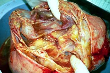
A biopsy is a medical procedure in which a small sample of tissue is taken from the body to be examined under a microscope for the presence of disease. This can help doctors diagnose and monitor various medical conditions, such as cancer, infections, or autoimmune disorders. The type of biopsy performed will depend on the location and nature of the suspected condition. Some common types of biopsies include:
1. Incisional biopsy: In this procedure, a surgeon removes a piece of tissue from an abnormal area using a scalpel or other surgical instrument. This type of biopsy is often used when the lesion is too large to be removed entirely during the initial biopsy.
2. Excisional biopsy: An excisional biopsy involves removing the entire abnormal area, along with a margin of healthy tissue surrounding it. This technique is typically employed for smaller lesions or when cancer is suspected.
3. Needle biopsy: A needle biopsy uses a thin, hollow needle to extract cells or fluid from the body. There are two main types of needle biopsies: fine-needle aspiration (FNA) and core needle biopsy. FNA extracts loose cells, while a core needle biopsy removes a small piece of tissue.
4. Punch biopsy: In a punch biopsy, a round, sharp tool is used to remove a small cylindrical sample of skin tissue. This type of biopsy is often used for evaluating rashes or other skin abnormalities.
5. Shave biopsy: During a shave biopsy, a thin slice of tissue is removed from the surface of the skin using a sharp razor-like instrument. This technique is typically used for superficial lesions or growths on the skin.
After the biopsy sample has been collected, it is sent to a laboratory where a pathologist will examine the tissue under a microscope and provide a diagnosis based on their findings. The results of the biopsy can help guide further treatment decisions and determine the best course of action for managing the patient's condition.
A vaginal smear, also known as a Pap test or Pap smear, is a medical procedure in which a sample of cells is collected from the cervix (the lower part of the uterus that opens into the vagina) and examined under a microscope. The purpose of this test is to detect abnormal cells, including precancerous changes, that may indicate the presence of cervical cancer or other conditions such as infections or inflammation.
During the procedure, a speculum is inserted into the vagina to allow the healthcare provider to visualize the cervix. A spatula or brush is then used to gently scrape cells from the surface of the cervix. The sample is spread onto a microscope slide and sent to a laboratory for analysis.
Regular Pap smears are recommended for women as part of their routine healthcare, as they can help detect abnormalities at an early stage when they are more easily treated. The frequency of Pap smears may vary depending on age, medical history, and other factors. It is important to follow the recommendations of a healthcare provider regarding the timing and frequency of Pap smears.
Carcinoma in situ is a medical term used to describe the earliest stage of cancer, specifically a type of cancer that begins in the epithelial tissue, which is the tissue that lines the outer surfaces of organs and body structures. In this stage, the cancer cells are confined to the layer of cells where they first developed and have not spread beyond that layer into the surrounding tissues or organs.
Carcinoma in situ can occur in various parts of the body, including the skin, cervix, breast, lung, prostate, bladder, and other areas. It is often detected through routine screening tests, such as Pap smears for cervical cancer or mammograms for breast cancer.
While carcinoma in situ is not invasive, it can still be a serious condition because it has the potential to develop into an invasive cancer if left untreated. Treatment options for carcinoma in situ may include surgery, radiation therapy, or other forms of treatment, depending on the location and type of cancer. It is important to consult with a healthcare provider to determine the best course of action for each individual case.
Preventive health services refer to measures taken to prevent diseases or injuries rather than curing them or treating their symptoms. These services include screenings, vaccinations, and counseling aimed at preventing or identifying illnesses in their earliest stages. Examples of preventive health services include:
1. Screenings for various types of cancer (e.g., breast, cervical, colorectal)
2. Vaccinations against infectious diseases (e.g., influenza, pneumococcal pneumonia, human papillomavirus)
3. Counseling on lifestyle modifications to reduce the risk of chronic diseases (e.g., smoking cessation, diet and exercise counseling, alcohol misuse screening and intervention)
4. Screenings for cardiovascular disease risk factors (e.g., cholesterol levels, blood pressure, body mass index)
5. Screenings for mental health conditions (e.g., depression)
6. Preventive medications (e.g., aspirin for primary prevention of cardiovascular disease in certain individuals)
Preventive health services are an essential component of overall healthcare and play a critical role in improving health outcomes, reducing healthcare costs, and enhancing quality of life.
A Prepaid Health Plan (PHP), also known as a Health Maintenance Organization (HMO) or Point of Service (POS) plan, is a type of health insurance in which the insured pays a fixed, prepaid fee for access to specific healthcare services. These plans typically have a network of healthcare providers with whom they have contracts to provide services at reduced rates. The insured must choose a primary care physician (PCP) from within the network who will coordinate their care and refer them to specialists as needed, also within the network. Prepaid health plans may not cover services received outside of the designated network, except in emergency situations.
A Receiver Operating Characteristic (ROC) curve is a graphical representation used in medical decision-making and statistical analysis to illustrate the performance of a binary classifier system, such as a diagnostic test or a machine learning algorithm. It's a plot that shows the tradeoff between the true positive rate (sensitivity) and the false positive rate (1 - specificity) for different threshold settings.
The x-axis of an ROC curve represents the false positive rate (the proportion of negative cases incorrectly classified as positive), while the y-axis represents the true positive rate (the proportion of positive cases correctly classified as positive). Each point on the curve corresponds to a specific decision threshold, with higher points indicating better performance.
The area under the ROC curve (AUC) is a commonly used summary measure that reflects the overall performance of the classifier. An AUC value of 1 indicates perfect discrimination between positive and negative cases, while an AUC value of 0.5 suggests that the classifier performs no better than chance.
ROC curves are widely used in healthcare to evaluate diagnostic tests, predictive models, and screening tools for various medical conditions, helping clinicians make informed decisions about patient care based on the balance between sensitivity and specificity.
Health care surveys are research tools used to systematically collect information from a population or sample regarding their experiences, perceptions, and knowledge of health services, health outcomes, and various other health-related topics. These surveys typically consist of standardized questionnaires that cover specific aspects of healthcare, such as access to care, quality of care, patient satisfaction, health disparities, and healthcare costs. The data gathered from health care surveys are used to inform policy decisions, improve healthcare delivery, identify best practices, allocate resources, and monitor the health status of populations. Health care surveys can be conducted through various modes, including in-person interviews, telephone interviews, mail-in questionnaires, or online platforms.
The Predictive Value of Tests, specifically the Positive Predictive Value (PPV) and Negative Predictive Value (NPV), are measures used in diagnostic tests to determine the probability that a positive or negative test result is correct.
Positive Predictive Value (PPV) is the proportion of patients with a positive test result who actually have the disease. It is calculated as the number of true positives divided by the total number of positive results (true positives + false positives). A higher PPV indicates that a positive test result is more likely to be a true positive, and therefore the disease is more likely to be present.
Negative Predictive Value (NPV) is the proportion of patients with a negative test result who do not have the disease. It is calculated as the number of true negatives divided by the total number of negative results (true negatives + false negatives). A higher NPV indicates that a negative test result is more likely to be a true negative, and therefore the disease is less likely to be present.
The predictive value of tests depends on the prevalence of the disease in the population being tested, as well as the sensitivity and specificity of the test. A test with high sensitivity and specificity will generally have higher predictive values than a test with low sensitivity and specificity. However, even a highly sensitive and specific test can have low predictive values if the prevalence of the disease is low in the population being tested.
Women's health services refer to medical services that are specifically designed, focused on, or tailored to the unique physiological and psychological needs of women, throughout various stages of their lives. These services encompass a wide range of healthcare areas including:
1. Gynecology and obstetrics - covering routine preventive care, family planning, prenatal and postnatal care, as well as management of gynecological conditions like menstrual disorders, sexually transmitted infections (STIs), and reproductive system cancers (e.g., cervical, ovarian, and endometrial cancer).
2. Breast health - including breast cancer screening, diagnostics, treatment, and survivorship care, as well as education on breast self-examination and risk reduction strategies.
3. Mental health - addressing women's mental health concerns such as depression, anxiety, post-traumatic stress disorder (PTSD), eating disorders, and perinatal mood disorders, while also considering the impact of hormonal changes, life events, and societal expectations on emotional wellbeing.
4. Sexual health - providing care for sexual concerns, dysfunctions, and sexually transmitted infections (STIs), as well as offering education on safe sexual practices and promoting healthy relationships.
5. Cardiovascular health - addressing women's specific cardiovascular risks, such as pregnancy-related complications, and managing conditions like hypertension and high cholesterol to prevent heart disease, the leading cause of death for women in many countries.
6. Bone health - focusing on prevention, diagnosis, and management of osteoporosis and other bone diseases that disproportionately affect women, particularly after menopause.
7. Menopause care - providing support and treatment for symptoms related to menopause, such as hot flashes, sleep disturbances, and mood changes, while also addressing long-term health concerns like bone density loss and heart disease risk.
8. Preventive care - offering routine screenings and vaccinations specific to women's health needs, including cervical cancer screening (Pap test), breast cancer screening (mammography), human papillomavirus (HPV) testing, and osteoporosis screening.
9. Education and counseling - empowering women with knowledge about their bodies, sexual and reproductive health, and overall wellbeing through evidence-based resources and support.
10. Integrative care - addressing the whole person, including mental, emotional, and spiritual wellbeing, by incorporating complementary therapies like acupuncture, mindfulness, and yoga into treatment plans as appropriate.
A nipple is a small projection or tubular structure located at the center of the areola, which is the darker circle of skin surrounding the nipple on the breast. The primary function of the nipple is to provide a pathway for milk flow from the mammary glands during lactation in females.
The nipple contains smooth muscle fibers that contract and cause the nipple to become erect when stimulated, such as during sexual arousal or cold temperatures. Nipples can come in various shapes, sizes, and colors, and some individuals may have inverted or flat nipples. It is essential to monitor any changes in the appearance or sensation of the nipples, as these could be indicative of underlying medical conditions, such as breast cancer.
Medical Definition:
"Risk factors" are any attribute, characteristic or exposure of an individual that increases the likelihood of developing a disease or injury. They can be divided into modifiable and non-modifiable risk factors. Modifiable risk factors are those that can be changed through lifestyle choices or medical treatment, while non-modifiable risk factors are inherent traits such as age, gender, or genetic predisposition. Examples of modifiable risk factors include smoking, alcohol consumption, physical inactivity, and unhealthy diet, while non-modifiable risk factors include age, sex, and family history. It is important to note that having a risk factor does not guarantee that a person will develop the disease, but rather indicates an increased susceptibility.
Retrospective studies, also known as retrospective research or looking back studies, are a type of observational study that examines data from the past to draw conclusions about possible causal relationships between risk factors and outcomes. In these studies, researchers analyze existing records, medical charts, or previously collected data to test a hypothesis or answer a specific research question.
Retrospective studies can be useful for generating hypotheses and identifying trends, but they have limitations compared to prospective studies, which follow participants forward in time from exposure to outcome. Retrospective studies are subject to biases such as recall bias, selection bias, and information bias, which can affect the validity of the results. Therefore, retrospective studies should be interpreted with caution and used primarily to generate hypotheses for further testing in prospective studies.
Medical Definition:
Magnetic Resonance Imaging (MRI) is a non-invasive diagnostic imaging technique that uses a strong magnetic field and radio waves to create detailed cross-sectional or three-dimensional images of the internal structures of the body. The patient lies within a large, cylindrical magnet, and the scanner detects changes in the direction of the magnetic field caused by protons in the body. These changes are then converted into detailed images that help medical professionals to diagnose and monitor various medical conditions, such as tumors, injuries, or diseases affecting the brain, spinal cord, heart, blood vessels, joints, and other internal organs. MRI does not use radiation like computed tomography (CT) scans.
A needle biopsy is a medical procedure in which a thin, hollow needle is used to remove a small sample of tissue from a suspicious or abnormal area of the body. The tissue sample is then examined under a microscope to check for cancer cells or other abnormalities. Needle biopsies are often used to diagnose lumps or masses that can be felt through the skin, but they can also be guided by imaging techniques such as ultrasound, CT scan, or MRI to reach areas that cannot be felt. There are several types of needle biopsy procedures, including fine-needle aspiration (FNA) and core needle biopsy. FNA uses a thin needle and gentle suction to remove fluid and cells from the area, while core needle biopsy uses a larger needle to remove a small piece of tissue. The type of needle biopsy used depends on the location and size of the abnormal area, as well as the reason for the procedure.
In epidemiology, the incidence of a disease is defined as the number of new cases of that disease within a specific population over a certain period of time. It is typically expressed as a rate, with the number of new cases in the numerator and the size of the population at risk in the denominator. Incidence provides information about the risk of developing a disease during a given time period and can be used to compare disease rates between different populations or to monitor trends in disease occurrence over time.
A registry in the context of medicine is a collection or database of standardized information about individuals who share a certain condition or attribute, such as a disease, treatment, exposure, or demographic group. These registries are used for various purposes, including:
* Monitoring and tracking the natural history of diseases and conditions
* Evaluating the safety and effectiveness of medical treatments and interventions
* Conducting research and generating hypotheses for further study
* Providing information to patients, clinicians, and researchers
* Informing public health policy and decision-making
Registries can be established for a wide range of purposes, including disease-specific registries (such as cancer or diabetes registries), procedure-specific registries (such as joint replacement or cardiac surgery registries), and population-based registries (such as birth defects or cancer registries). Data collected in registries may include demographic information, clinical data, laboratory results, treatment details, and outcomes.
Registries can be maintained by a variety of organizations, including hospitals, clinics, academic medical centers, professional societies, government agencies, and industry. Participation in registries is often voluntary, although some registries may require informed consent from participants. Data collected in registries are typically de-identified to protect the privacy of individuals.
I'm sorry for any confusion, but "North Carolina" is a geographical location and not a medical term or condition. It is a state located in the southeastern region of the United States. If you have any questions about medical terms or conditions, I'd be happy to help with those!
Carcinoma, lobular is a type of breast cancer that begins in the milk-producing glands (lobules) of the breast. It can be either invasive or non-invasive (in situ). Invasive lobular carcinoma (ILC) occurs when the cancer cells break through the wall of the lobule and invade the surrounding breast tissue, and can potentially spread to other parts of the body. Non-invasive lobular carcinoma (LCIS), on the other hand, refers to the presence of abnormal cells within the lobule that have not invaded nearby breast tissue.
ILC is usually detected as a mass or thickening in the breast, and it may not cause any symptoms or show up on mammograms until it has grown quite large. It tends to grow more slowly than some other types of breast cancer, but it can still be serious and require extensive treatment. LCIS does not typically cause any symptoms and is usually found during a biopsy performed for another reason.
Treatment options for carcinoma, lobular depend on several factors, including the stage of the cancer, the patient's overall health, and their personal preferences. Treatment may include surgery, radiation therapy, chemotherapy, hormone therapy, or targeted therapy. Regular follow-up care is essential to monitor for recurrence or the development of new cancers.
A gamma camera, also known as a scintillation camera, is a device used in nuclear medicine to image gamma-emitting radionuclides in the body. It detects gamma radiation emitted by radioisotopes that have been introduced into the body, usually through injection or ingestion. The camera consists of a large flat crystal (often sodium iodide) that scintillates when struck by gamma rays, producing light flashes that are detected by an array of photomultiplier tubes.
The resulting signals are then processed by a computer to generate images that reflect the distribution and concentration of the radionuclide in the body. Gamma cameras are used in a variety of medical imaging procedures, including bone scans, lung scans, heart scans (such as myocardial perfusion imaging), and brain scans. They can help diagnose conditions such as cancer, heart disease, and neurological disorders.
Hispanic Americans, also known as Latino Americans, are individuals in the United States who are of Spanish-speaking origin or whose ancestors came from Spain, Mexico, Cuba, the Caribbean, Central and South America. This group includes various cultures, races, and nationalities. It is important to note that "Hispanic" refers to a cultural and linguistic affiliation rather than a racial category. Therefore, Hispanic Americans can be of any race, including White, Black, Asian, Native American, or mixed races.
Cost-benefit analysis (CBA) is a systematic process used to compare the costs and benefits of different options to determine which one provides the greatest net benefit. In a medical context, CBA can be used to evaluate the value of medical interventions, treatments, or policies by estimating and monetizing all the relevant costs and benefits associated with each option.
The costs included in a CBA may include direct costs such as the cost of the intervention or treatment itself, as well as indirect costs such as lost productivity or time away from work. Benefits may include improved health outcomes, reduced morbidity or mortality, and increased quality of life.
Once all the relevant costs and benefits have been identified and quantified, they are typically expressed in monetary terms to allow for a direct comparison. The option with the highest net benefit (i.e., the difference between total benefits and total costs) is considered the most cost-effective.
It's important to note that CBA has some limitations and can be subject to various biases and assumptions, so it should be used in conjunction with other evaluation methods to ensure a comprehensive understanding of the value of medical interventions or policies.
Tomography is a medical imaging technique used to produce cross-sectional images or slices of specific areas of the body. This technique uses various forms of radiation (X-rays, gamma rays) or sound waves (ultrasound) to create detailed images of the internal structures, such as organs, bones, and tissues. Common types of tomography include Computerized Tomography (CT), Positron Emission Tomography (PET), and Magnetic Resonance Imaging (MRI). The primary advantage of tomography is its ability to provide clear and detailed images of internal structures, allowing healthcare professionals to accurately diagnose and monitor a wide range of medical conditions.
Prospective studies, also known as longitudinal studies, are a type of cohort study in which data is collected forward in time, following a group of individuals who share a common characteristic or exposure over a period of time. The researchers clearly define the study population and exposure of interest at the beginning of the study and follow up with the participants to determine the outcomes that develop over time. This type of study design allows for the investigation of causal relationships between exposures and outcomes, as well as the identification of risk factors and the estimation of disease incidence rates. Prospective studies are particularly useful in epidemiology and medical research when studying diseases with long latency periods or rare outcomes.