Lysosomes
Lysosome-Associated Membrane Glycoproteins
Endosomes
Acid Phosphatase
Endocytosis
Phagosomes
Cathepsin D
Autophagy
Lysosomal-Associated Membrane Protein 2
Microscopy, Electron
Cathepsins
Protein Transport
Vacuoles
Chediak-Higashi Syndrome
beta-N-Acetylhexosaminidases
Receptor, IGF Type 2
Cell Fractionation
Golgi Apparatus
Biological Transport
Chloroquine
rab GTP-Binding Proteins
Subcellular Fractions
Organelles
Glucuronidase
Mucolipidoses
Organoids
Lysosomal Storage Diseases
Cathepsin B
Cell Compartmentation
Intracellular Membranes
Hydrogen-Ion Concentration
Liver
Androstenes
Cell Membrane
Hydrolases
Arylsulfatases
Histocytochemistry
Cells, Cultured
Acetylglucosaminidase
Multivesicular Bodies
Ammonium Chloride
Microscopy, Fluorescence
Horseradish Peroxidase
Membrane Fusion
Macrophages
Hexosaminidases
Vesicular Transport Proteins
Microscopy, Immunoelectron
Cathepsin L
Asialoglycoproteins
Fibroblasts
Antigens, CD63
Leupeptins
Cystinosis
Phagocytosis
Microscopy, Confocal
Macrolides
Sulfatases
Pepstatins
Exocytosis
Membrane Proteins
Fetuins
rab5 GTP-Binding Proteins
HeLa Cells
Centrifugation, Density Gradient
Cytoplasm
Saposins
Endoplasmic Reticulum
Niemann-Pick Diseases
Endosomal Sorting Complexes Required for Transport
Proteins
Cricetinae
Vacuolar Proton-Translocating ATPases
Dextrans
Proteolysis
Cathepsin C
Molecular Sequence Data
Amino Acid Sequence
Transport Vesicles
Monensin
Cytoplasmic Vesicles
Clathrin
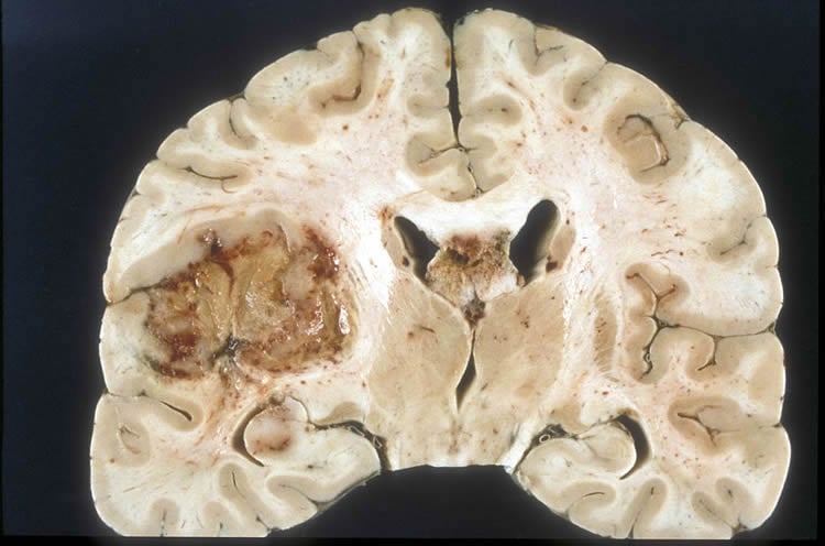
A cytomegalovirus glycoprotein re-routes MHC class I complexes to lysosomes for degradation. (1/6965)
Mouse cytomegalovirus (MCMV) early gene expression interferes with the major histocompatibility complex class I (MHC class I) pathway of antigen presentation. Here we identify a 48 kDa type I transmembrane glycoprotein encoded by the MCMV early gene m06, which tightly binds to properly folded beta2-microglobulin (beta2m)-associated MHC class I molecules in the endoplasmic reticulum (ER). This association is mediated by the lumenal/transmembrane part of the protein. gp48-MHC class I complexes are transported out of the ER, pass the Golgi, but instead of being expressed on the cell surface, they are redirected to the endocytic route and rapidly degraded in a Lamp-1(+) compartment. As a result, m06-expressing cells are impaired in presenting antigenic peptides to CD8(+) T cells. The cytoplasmic tail of gp48 contains two di-leucine motifs. Mutation of the membrane-proximal di-leucine motif of gp48 restored surface expression of MHC class I, while mutation of the distal one had no effect. The results establish a novel viral mechanism for downregulation of MHC class I molecules by directly binding surface-destined MHC complexes and exploiting the cellular di-leucine sorting machinery for lysosomal degradation. (+info)Identification of low density lipoprotein receptor-related protein-2/megalin as an endocytic receptor for seminal vesicle secretory protein II. (2/6965)
The low density lipoprotein receptor-related protein-2/megalin (LRP-2) is an endocytic receptor that is expressed on the apical surfaces of epithelial cells lining specific regions of the male and female reproductive tracts. In the present study, immunohistochemical staining revealed that LRP-2 is also expressed by epithelial cells lining the ductal region and the ampulla of the rat seminal vesicle. To identify LRP-2 ligands in the seminal vesicle, we probed seminal vesicle fluid with 125I-labeled LRP-2 in a gel-blot overlay assay. A 100-kDa protein (under non-reducing conditions) was found to bind the radiolabeled receptor. The protein was isolated and subjected to protease digestion, and the proteolytic fragments were subjected to mass spectroscopic sequence analysis. As a result, the 100-kDa protein was identified as the seminal vesicle secretory protein II (SVS-II), a major constituent of the seminal coagulum. Using purified preparations of SVS-II and LRP-2, solid-phase binding assays were used to show that the SVS-II bound to the receptor with high affinity (Kd = 5.6 nM). The binding of SVS-II to LRP-2 was inhibited using a known antagonist of LRP-2 function, the 39-kDa receptor-associated protein RAP. Using a series of recombinant subfragments of SVS-II, the LRP-2 binding site was mapped to a stretch of repeated 13-residue modules located in the central portion of the SVS-II polypeptide. To evaluate the ability of LRP-2 to mediate 125I-SVS-II endocytosis and lysosomal degradation, ligand clearance assays were performed using differentiated mouse F9 cells, which express high levels of LRP-2. Radiolabeled SVS-II was internalized and degraded by the cells, and both processes were inhibited by antibodies to LRP-2 or by RAP. The results indicate that LRP-2 binds SVS-II and can mediate its endocytosis leading to lysosomal degradation. (+info)Purification of gibberellic acid-induced lysosomes from wheat aleurone cells. (3/6965)
Using isopycnic density gradient centrifugation, lysosomes were concentrated in a single region of a sucrose-Ficoll gradient (p = 1-10 g cm-3), well separated from most other cell organelles. Gibberellic acid-induced lysosomes were found to be rich in alpha-amylase and protease but not ribonuclease. The lysosomal band also contained a majority of the NADH2-cytochrome c reductase, a marker enzyme for endoplasmic reticulum, found in the gradient. Examination of electron micrographs revealed that a purified band of lyosomes contained at least 3 vesicle types, ranging in size from 0-1 to 0-5 mum. The significance of these findings to proposed mechanisms of action of gibberellic acid is discussed. (+info)Impaired lysosomal processing of beta2-microglobulin by infiltrating macrophages in dialysis amyloidosis. (4/6965)
BACKGROUND: Macrophages may participate in amyloid fibril formation by processing the protein precursor. Although this theory seems to apply for amyloidosis, in which proteolytic cleavage is a prerequisite for amyloid fibril formation, it has not been demonstrated for beta2-microglobulin (beta2m) amyloidosis. We aimed to establish the role played by macrophages in beta2m amyloidosis. METHODS: We used a double immunogold electron microscopy technique, including mouse antihuman CD68, rabbit antihuman beta2m, amyloid P component, and lysosome-associated membrane protein (LAMP-1) antibodies. Differential density labeling studies of beta2m and amyloid P component were performed extra- and intracellularly to assess protein processing by macrophages. RESULTS: The cells surrounding amyloid fibrils were found to be mostly CD68 positive, suggesting that they were of monocyte-macrophage lineage. Intracellular accumulation of amyloid fibrils was also observed; these fibrils were constantly surrounded by LAMP-1-linked gold particles, demonstrating that intracellular beta2m was almost exclusively lysosomal. The rough-surface endoplasmic reticulum was not labeled by beta2m antibody, suggesting that there was no active synthesis of beta2m by the cells. As a marker of endocytosis, protruded cytoplasmic processes in close relation with the intracellular accumulations of beta2m amyloid fibrils were observed. No difference in density labeling (extracellular vs. intracellular) was observed for beta2m, whereas intracellular P component labeling was significantly decreased. CONCLUSIONS: All of these data are strongly suggestive of phagocytosis and not synthesis of amyloid fibrils by macrophages. Further, they demonstrate an impaired lysosomal processing specific for beta2m, as other compounds of the amyloid fibrils (P component) are significantly cleared. (+info)5'-Nucleotidase activity of mouse peritoneal macrophages. II. Cellular distribution and effects of endocytosis. (5/6965)
The diazonium salt of sulfanilic acid (DASA) can inactivate about 80% of the total 5'-nucleotidase of viable macrophages. The remaining 20% can be inactivated if the cells are first lysed in detergent, and presumably represents an intracellular pool of 5'-nucleotidase. The bulk of this pool may represent cytoplasmic vesicles derived from plasma membrane by endocytosis. This internal compartment is expanded up to threefold immediately after the cells have ingested a large latex load. This is consistent with previous observations on the internalization of 5'-nucleotidase in latex phagosomes. In latex-filled cells this intracellular pool of enzyme is inactivated over a few hours, and the cells then slowly increase their enzyme activity to nearly normal levels. However, 24 h after latex ingestion the metabolism of 5'-nucleotidase in these recovered cells is abnormal, as the rate of enzyme degradation is about twice the normal rate, and the DASA-insensitive enzyme pool in these cells is strikingly diminished. This may reflect effects of the accumulated indigestible particles on the fate of incoming pinocytic vesicles or on newly synthesized plasma membrane precursor. Another endocytic stimulus, concanavalin A, also reduces the total cell 5'-nucleotidase activity. This effect, which is time and temperature dependent, can be prevented by the competitive sugar alpha-methyl mannose. The concanavalin A inhibition can be reversed in the absence of new protein synthesis or in cells cultivated in serum-free conditions. It is not known whether the effect of concanavalin A on 5'-nucleotidase depends upon the interiorizaiton of plasma membrane or is strictly associated with events at the cell surface. (+info)Macrophage plasminogen activator: induction by asbestos is blocked by anti-inflammatory steroids. (6/6965)
Intraperitoneal injection of asbestos fibres into mice induces the formation of exudates containing macrophages that produce plasminogen activator. Like-wise, in vitro addition of asbestos to macrophage cultures stimulates plasminogen activator secretion; the synthesis and secretion of lysozyme and lysosomal enzymes are not changed under these conditions. The enhanced secretion of plasminogen activator by macrophages exposed to asbestos is suppressed by low concentrations of anti-inflammatory steroids. (+info)Opposing motor activities of dynein and kinesin determine retention and transport of MHC class II-containing compartments. (7/6965)
MHC class II molecules exert their function at the cell surface by presenting to T cells antigenic fragments that are generated in the endosomal pathway. The class II molecules are targetted to early lysosomal structures, termed MIIC, where they interact with antigenic fragments and are subsequently transported to the cell surface. We previously visualised vesicular transport of MHC class II-containing early lysosomes from the microtubule organising centre (MTOC) region towards the cell surface in living cells. Here we show that the MIIC move bidirectionally in a 'stop-and-go' fashion. Overexpression of a motor head-deleted kinesin inhibited MIIC motility, showing that kinesin is the motor that drives its plus end transport towards the cell periphery. Cytoplasmic dynein mediates the return of vesicles to the MTOC area and effectively retains the vesicles at this location, as assessed by inactivation of dynein by overexpression of dynamitin. Our data suggest a retention mechanism that determines the perinuclear accumulation of MIIC, which is the result of dynein activity being superior over kinesin activity. The bidirectional nature of MIIC movement is the result of both kinesin and dynein acting reciprocally on the MIIC during its transport. The motors may be the ultimate targets of regulatory kinases since the protein kinase inhibitor staurosporine induces a massive release of lysosomal vesicles from the MTOC region that is morphologically similar to that observed after inactivation of the dynein motor. (+info)Endometrial lysosomal enzyme activity in normal cycling endometrium. (8/6965)
The objective of this study was to evaluate the possible role of four lysosomal enzymes in endometrial function and remodelling during the normal menstrual cycle by fluorimetric measurement (acid phosphatase, N-acetyl-beta-D-glucosaminidase, alpha-L-fucosidase and alpha-D-mannosidase). A prospective study was conducted of 45 endometrial biopsies obtained from women with normal menstrual cycles. Activity of all four enzymes was identified in human endometrium. Activity of acid phosphatase and N-acetyl-beta-D-glucosaminidase was relatively high, whilst that of alpha-L-fucosidase and alpha-D-mannosidase was low. There was no significant change in the activity of any of the four enzymes from the proliferative to the secretory phase of the cycle. This study suggests that the activity of these enzymes remains constant throughout a major portion of the normal cycle. (+info)Lysosomes are membrane-bound organelles found in the cytoplasm of eukaryotic cells. They are responsible for breaking down and recycling various materials, such as waste products, foreign substances, and damaged cellular components, through a process called autophagy or phagocytosis. Lysosomes contain hydrolytic enzymes that can break down biomolecules like proteins, nucleic acids, lipids, and carbohydrates into their basic building blocks, which can then be reused by the cell. They play a crucial role in maintaining cellular homeostasis and are often referred to as the "garbage disposal system" of the cell.
Lysosome-Associated Membrane Glycoproteins (LAMPs) are a group of proteins found in the membrane of lysosomes, which are cellular organelles responsible for breaking down and recycling various biomolecules. LAMPs play a crucial role in maintaining the integrity and function of the lysosomal membrane.
There are two major types of LAMPs: LAMP-1 and LAMP-2. Both proteins share structural similarities, including a large heavily glycosylated domain that faces the lumen of the lysosome and a short hydrophobic region that anchors them to the membrane.
The primary function of LAMPs is to protect the lysosomal membrane from degradation by hydrolytic enzymes present inside the lysosome. They also participate in the process of autophagy, a cellular recycling mechanism, by fusing with autophagosomes (double-membraned vesicles formed during autophagy) to form autolysosomes, where the contents are degraded.
Moreover, LAMPs have been implicated in several cellular processes, such as antigen presentation, cholesterol homeostasis, and intracellular signaling. Mutations in LAMP-2 have been associated with certain genetic disorders, including Danon disease, a rare X-linked dominant disorder characterized by heart problems, muscle weakness, and intellectual disability.
Endosomes are membrane-bound compartments within eukaryotic cells that play a critical role in intracellular trafficking and sorting of various cargoes, including proteins and lipids. They are formed by the invagination of the plasma membrane during endocytosis, resulting in the internalization of extracellular material and cell surface receptors.
Endosomes can be classified into early endosomes, late endosomes, and recycling endosomes based on their morphology, molecular markers, and functional properties. Early endosomes are the initial sorting stations for internalized cargoes, where they undergo sorting and processing before being directed to their final destinations. Late endosomes are more acidic compartments that mature from early endosomes and are responsible for the transport of cargoes to lysosomes for degradation.
Recycling endosomes, on the other hand, are involved in the recycling of internalized cargoes back to the plasma membrane or to other cellular compartments. Endosomal sorting and trafficking are regulated by a complex network of molecular interactions involving various proteins, lipids, and intracellular signaling pathways.
Defects in endosomal function have been implicated in various human diseases, including neurodegenerative disorders, developmental abnormalities, and cancer. Therefore, understanding the mechanisms underlying endosomal trafficking and sorting is of great importance for developing therapeutic strategies to treat these conditions.
Acid phosphatase is a type of enzyme that is found in various tissues and organs throughout the body, including the prostate gland, red blood cells, bone, liver, spleen, and kidneys. This enzyme plays a role in several biological processes, such as bone metabolism and the breakdown of molecules like nucleotides and proteins.
Acid phosphatase is classified based on its optimum pH level for activity. Acid phosphatases have an optimal activity at acidic pH levels (below 7.0), while alkaline phosphatases have an optimal activity at basic or alkaline pH levels (above 7.0).
In clinical settings, measuring the level of acid phosphatase in the blood can be useful as a tumor marker for prostate cancer. Elevated acid phosphatase levels may indicate the presence of metastatic prostate cancer or disease progression. However, it is important to note that acid phosphatase is not specific to prostate cancer and can also be elevated in other conditions, such as bone diseases, liver disorders, and some benign conditions. Therefore, acid phosphatase should be interpreted in conjunction with other diagnostic tests and clinical findings for a more accurate diagnosis.
Endocytosis is the process by which cells absorb substances from their external environment by engulfing them in membrane-bound structures, resulting in the formation of intracellular vesicles. This mechanism allows cells to take up large molecules, such as proteins and lipids, as well as small particles, like bacteria and viruses. There are two main types of endocytosis: phagocytosis (cell eating) and pinocytosis (cell drinking). Phagocytosis involves the engulfment of solid particles, while pinocytosis deals with the uptake of fluids and dissolved substances. Other specialized forms of endocytosis include receptor-mediated endocytosis and caveolae-mediated endocytosis, which allow for the specific internalization of molecules through the interaction with cell surface receptors.
A phagosome is a type of membrane-bound organelle that forms around a particle or microorganism following its engulfment by a cell, through the process of phagocytosis. This results in the formation of a vesicle containing the ingested material, which then fuses with another organelle called a lysosome to form a phago-lysosome. The lysosome contains enzymes that digest and break down the contents of the phagosome, allowing the cell to neutralize and dispose of potentially harmful substances or pathogens.
In summary, phagosomes are important organelles involved in the immune response, helping to protect the body against infection and disease.
Cathepsin D is a lysosomal aspartic protease that plays a role in intracellular protein degradation and turnover. It is produced as an inactive precursor and is activated by cleavage into two subunits within the acidic environment of the lysosome. Cathepsin D is also known to be secreted by certain cells, where it can contribute to extracellular matrix remodeling and tissue degradation. In addition, abnormal levels or activity of cathepsin D have been implicated in various diseases, including cancer, neurodegenerative disorders, and infectious diseases.
Autophagy is a fundamental cellular process that involves the degradation and recycling of damaged or unnecessary cellular components, such as proteins and organelles. The term "autophagy" comes from the Greek words "auto" meaning self and "phagy" meaning eating. It is a natural process that occurs in all types of cells and helps maintain cellular homeostasis by breaking down and recycling these components.
There are several different types of autophagy, including macroautophagy, microautophagy, and chaperone-mediated autophagy (CMA). Macroautophagy is the most well-known form and involves the formation of a double-membraned vesicle called an autophagosome, which engulfs the cellular component to be degraded. The autophagosome then fuses with a lysosome, an organelle containing enzymes that break down and recycle the contents of the autophagosome.
Autophagy plays important roles in various cellular processes, including adaptation to starvation, removal of damaged organelles, clearance of protein aggregates, and regulation of programmed cell death (apoptosis). Dysregulation of autophagy has been implicated in a number of diseases, including cancer, neurodegenerative disorders, and infectious diseases.
Lysosome-Associated Membrane Protein 2 (LAMP-2) is a type of transmembrane protein that is primarily found in the membranes of lysosomes, which are organelles within cells responsible for breaking down and recycling various cellular components. LAMP-2 plays a crucial role in maintaining the structural integrity and stability of the lysosomal membrane. It also participates in the process of autophagy, where damaged or unnecessary cellular components are engulfed by membranes to form vesicles called autophagosomes, which then fuse with lysosomes for degradation. Mutations in the LAMP-2 gene have been associated with certain genetic disorders, such as Danon disease, a rare X-linked condition characterized by heart problems, muscle weakness, and intellectual disability.
Electron microscopy (EM) is a type of microscopy that uses a beam of electrons to create an image of the sample being examined, resulting in much higher magnification and resolution than light microscopy. There are several types of electron microscopy, including transmission electron microscopy (TEM), scanning electron microscopy (SEM), and reflection electron microscopy (REM).
In TEM, a beam of electrons is transmitted through a thin slice of the sample, and the electrons that pass through the sample are focused to form an image. This technique can provide detailed information about the internal structure of cells, viruses, and other biological specimens, as well as the composition and structure of materials at the atomic level.
In SEM, a beam of electrons is scanned across the surface of the sample, and the electrons that are scattered back from the surface are detected to create an image. This technique can provide information about the topography and composition of surfaces, as well as the structure of materials at the microscopic level.
REM is a variation of SEM in which the beam of electrons is reflected off the surface of the sample, rather than scattered back from it. This technique can provide information about the surface chemistry and composition of materials.
Electron microscopy has a wide range of applications in biology, medicine, and materials science, including the study of cellular structure and function, disease diagnosis, and the development of new materials and technologies.
Cathepsins are a type of proteolytic enzymes, which are found in lysosomes and are responsible for breaking down proteins inside the cell. They are classified as papain-like cysteine proteases and play important roles in various physiological processes, including tissue remodeling, antigen presentation, and apoptosis (programmed cell death). There are several different types of cathepsins, including cathepsin B, C, D, F, H, K, L, S, V, and X/Z, each with distinct substrate specificities and functions.
Dysregulation of cathepsins has been implicated in various pathological conditions, such as cancer, neurodegenerative diseases, and inflammatory disorders. For example, overexpression or hyperactivation of certain cathepsins has been shown to contribute to tumor invasion and metastasis, while their inhibition has been explored as a potential therapeutic strategy in cancer treatment. Similarly, abnormal levels of cathepsins have been linked to the progression of neurodegenerative diseases like Alzheimer's and Parkinson's, making them attractive targets for drug development.
Protein transport, in the context of cellular biology, refers to the process by which proteins are actively moved from one location to another within or between cells. This is a crucial mechanism for maintaining proper cell function and regulation.
Intracellular protein transport involves the movement of proteins within a single cell. Proteins can be transported across membranes (such as the nuclear envelope, endoplasmic reticulum, Golgi apparatus, or plasma membrane) via specialized transport systems like vesicles and transport channels.
Intercellular protein transport refers to the movement of proteins from one cell to another, often facilitated by exocytosis (release of proteins in vesicles) and endocytosis (uptake of extracellular substances via membrane-bound vesicles). This is essential for communication between cells, immune response, and other physiological processes.
It's important to note that any disruption in protein transport can lead to various diseases, including neurological disorders, cancer, and metabolic conditions.
Vacuoles are membrane-bound organelles found in the cells of most eukaryotic organisms. They are essentially fluid-filled sacs that store various substances, such as enzymes, waste products, and nutrients. In plants, vacuoles often contain water, ions, and various organic compounds, while in fungi, they may store lipids or pigments. Vacuoles can also play a role in maintaining the turgor pressure of cells, which is critical for cell shape and function.
In animal cells, vacuoles are typically smaller and less numerous than in plant cells. Animal cells have lysosomes, which are membrane-bound organelles that contain digestive enzymes and break down waste materials, cellular debris, and foreign substances. Lysosomes can be considered a type of vacuole, but they are more specialized in their function.
Overall, vacuoles are essential for maintaining the health and functioning of cells by providing a means to store and dispose of various substances.
Chediak-Higashi Syndrome is a rare autosomal recessive disorder characterized by partial albinism, photophobia, bleeding diathesis, recurrent infections, and progressive neurological degeneration. It is caused by mutations in the LYST gene, which leads to abnormalities in lysosomes, melanosomes, and neutrophil granules. The disorder is named after two Mexican hematologists, Dr. Chediak and Dr. Higashi, who first described it in 1952.
The symptoms of Chediak-Higashi Syndrome typically appear in early childhood and include light skin and hair, blue or gray eyes, and a sensitivity to light. Affected individuals may also have bleeding problems due to abnormal platelets, and they are prone to recurrent bacterial infections, particularly of the skin, gums, and respiratory system.
The neurological symptoms of Chediak-Higashi Syndrome can include poor coordination, difficulty walking, and seizures. The disorder can also affect the immune system, leading to an accelerated phase known as the "hemophagocytic syndrome," which is characterized by fever, enlarged liver and spleen, and abnormal blood counts.
There is no cure for Chediak-Higashi Syndrome, and treatment typically focuses on managing the symptoms of the disorder. This may include antibiotics to treat infections, medications to control bleeding, and physical therapy to help with mobility issues. In some cases, bone marrow transplantation may be recommended as a potential cure for the disorder.
Beta-N-Acetylhexosaminidases are a group of enzymes that play a role in the breakdown and recycling of complex carbohydrates in the body. Specifically, they help to break down gangliosides, which are a type of molecule found in cell membranes.
There are several different isoforms of beta-N-Acetylhexosaminidases, including A, B, and S. These isoforms are formed by different combinations of subunits, which can affect their activity and substrate specificity.
Mutations in the genes that encode for these enzymes can lead to a variety of genetic disorders, including Tay-Sachs disease and Sandhoff disease. These conditions are characterized by an accumulation of gangliosides in the brain, which can cause progressive neurological deterioration and death.
Treatment for these conditions typically involves managing symptoms and providing supportive care, as there is currently no cure. Enzyme replacement therapy has been explored as a potential treatment option, but its effectiveness varies depending on the specific disorder and the age of the patient.
IGF-2 (Insulin-like Growth Factor 2) receptor is a type of transmembrane protein that plays a role in cell growth, differentiation, and survival. Unlike other receptors in the insulin and IGF family, IGF-2 receptor does not mediate the activation of intracellular signaling pathways upon binding to its ligand (IGF-2). Instead, it acts as a clearance receptor that facilitates the removal of IGF-2 from circulation by transporting it to lysosomes for degradation.
The IGF-2 receptor is also known as cation-independent mannose-6-phosphate receptor (CI-M6PR) because it can also bind and transport mannose-6-phosphate-containing enzymes to lysosomes for degradation.
Mutations in the IGF-2 receptor gene have been associated with certain types of cancer, as well as developmental disorders such as Beckwith-Wiedemann syndrome.
Cell fractionation is a laboratory technique used to separate different cellular components or organelles based on their size, density, and other physical properties. This process involves breaking open the cell (usually through homogenization), and then separating the various components using various methods such as centrifugation, filtration, and ultracentrifugation.
The resulting fractions can include the cytoplasm, mitochondria, nuclei, endoplasmic reticulum, Golgi apparatus, lysosomes, peroxisomes, and other organelles. Each fraction can then be analyzed separately to study the biochemical and functional properties of the individual components.
Cell fractionation is a valuable tool in cell biology research, allowing scientists to study the structure, function, and interactions of various cellular components in a more detailed and precise manner.
The Golgi apparatus, also known as the Golgi complex or simply the Golgi, is a membrane-bound organelle found in the cytoplasm of most eukaryotic cells. It plays a crucial role in the processing, sorting, and packaging of proteins and lipids for transport to their final destinations within the cell or for secretion outside the cell.
The Golgi apparatus consists of a series of flattened, disc-shaped sacs called cisternae, which are stacked together in a parallel arrangement. These stacks are often interconnected by tubular structures called tubules or vesicles. The Golgi apparatus has two main faces: the cis face, which is closest to the endoplasmic reticulum (ER) and receives proteins and lipids directly from the ER; and the trans face, which is responsible for sorting and dispatching these molecules to their final destinations.
The Golgi apparatus performs several essential functions in the cell:
1. Protein processing: After proteins are synthesized in the ER, they are transported to the cis face of the Golgi apparatus, where they undergo various post-translational modifications, such as glycosylation (the addition of sugar molecules) and sulfation. These modifications help determine the protein's final structure, function, and targeting.
2. Lipid modification: The Golgi apparatus also modifies lipids by adding or removing different functional groups, which can influence their properties and localization within the cell.
3. Protein sorting and packaging: Once proteins and lipids have been processed, they are sorted and packaged into vesicles at the trans face of the Golgi apparatus. These vesicles then transport their cargo to various destinations, such as lysosomes, plasma membrane, or extracellular space.
4. Intracellular transport: The Golgi apparatus serves as a central hub for intracellular trafficking, coordinating the movement of vesicles and other transport carriers between different organelles and cellular compartments.
5. Cell-cell communication: Some proteins that are processed and packaged in the Golgi apparatus are destined for secretion, playing crucial roles in cell-cell communication and maintaining tissue homeostasis.
In summary, the Golgi apparatus is a vital organelle involved in various cellular processes, including post-translational modification, sorting, packaging, and intracellular transport of proteins and lipids. Its proper functioning is essential for maintaining cellular homeostasis and overall organismal health.
Biological transport refers to the movement of molecules, ions, or solutes across biological membranes or through cells in living organisms. This process is essential for maintaining homeostasis, regulating cellular functions, and enabling communication between cells. There are two main types of biological transport: passive transport and active transport.
Passive transport does not require the input of energy and includes:
1. Diffusion: The random movement of molecules from an area of high concentration to an area of low concentration until equilibrium is reached.
2. Osmosis: The diffusion of solvent molecules (usually water) across a semi-permeable membrane from an area of lower solute concentration to an area of higher solute concentration.
3. Facilitated diffusion: The assisted passage of polar or charged substances through protein channels or carriers in the cell membrane, which increases the rate of diffusion without consuming energy.
Active transport requires the input of energy (in the form of ATP) and includes:
1. Primary active transport: The direct use of ATP to move molecules against their concentration gradient, often driven by specific transport proteins called pumps.
2. Secondary active transport: The coupling of the movement of one substance down its electrochemical gradient with the uphill transport of another substance, mediated by a shared transport protein. This process is also known as co-transport or counter-transport.
Chloroquine is an antimalarial and autoimmune disease drug. It works by increasing the pH or making the environment less acidic in the digestive vacuoles of malaria parasites, which inhibits the polymerization of heme and the formation of hemozoin. This results in the accumulation of toxic levels of heme that are harmful to the parasite. Chloroquine is also used as an anti-inflammatory agent in the treatment of rheumatoid arthritis, discoid or systemic lupus erythematosus, and photodermatitis.
The chemical name for chloroquine is 7-chloro-4-(4-diethylamino-1-methylbutylamino)quinoline, and it has a molecular formula of C18H26ClN3. It is available in the form of phosphate or sulfate salts for oral administration as tablets or solution.
Chloroquine was first synthesized in 1934 by Bayer scientists, and it has been widely used since the 1940s as a safe and effective antimalarial drug. However, the emergence of chloroquine-resistant strains of malaria parasites has limited its use in some areas. Chloroquine is also being investigated for its potential therapeutic effects on various viral infections, including COVID-19.
Rab GTP-binding proteins, also known as Rab GTPases or simply Rabs, are a large family of small GTP-binding proteins that play a crucial role in regulating intracellular vesicle trafficking. They function as molecular switches that cycle between an active GTP-bound state and an inactive GDP-bound state.
In the active state, Rab proteins interact with various effector molecules to mediate specific membrane trafficking events such as vesicle budding, transport, tethering, and fusion. Each Rab protein is thought to have a unique function and localize to specific intracellular compartments or membranes, where they regulate the transport of vesicles and organelles within the cell.
Rab proteins are involved in several important cellular processes, including endocytosis, exocytosis, Golgi apparatus function, autophagy, and intracellular signaling. Dysregulation of Rab GTP-binding proteins has been implicated in various human diseases, such as cancer, neurodegenerative disorders, and infectious diseases.
Subcellular fractions refer to the separation and collection of specific parts or components of a cell, including organelles, membranes, and other structures, through various laboratory techniques such as centrifugation and ultracentrifugation. These fractions can be used in further biochemical and molecular analyses to study the structure, function, and interactions of individual cellular components. Examples of subcellular fractions include nuclear extracts, mitochondrial fractions, microsomal fractions (membrane vesicles), and cytosolic fractions (cytoplasmic extracts).
I'm sorry for any confusion, but "Mannosephosphates" is not a widely recognized or established term in medicine or biochemistry. It seems that this term may be a combination of "mannose," which is a type of sugar (monosaccharide), and "phosphates," which are compounds containing phosphorus. However, without more context, it's difficult to provide an accurate medical definition for this term.
In biochemistry, mannose can be linked to phosphate groups in various ways, such as in the context of mannose-1-phosphate or mannose-6-phosphate, which are involved in different metabolic pathways. If you could provide more information about where you encountered this term, I might be able to give a more precise definition or explanation.
Organelles are specialized structures within cells that perform specific functions essential for the cell's survival and proper functioning. They can be thought of as the "organs" of the cell, and they are typically membrane-bound to separate them from the rest of the cellular cytoplasm. Examples of organelles include the nucleus (which contains the genetic material), mitochondria (which generate energy for the cell), ribosomes (which synthesize proteins), endoplasmic reticulum (which is involved in protein and lipid synthesis), Golgi apparatus (which modifies, sorts, and packages proteins and lipids for transport), lysosomes (which break down waste materials and cellular debris), peroxisomes (which detoxify harmful substances and produce certain organic compounds), and vacuoles (which store nutrients and waste products). The specific organelles present in a cell can vary depending on the type of cell and its function.
Glucuronidase is an enzyme that catalyzes the hydrolysis of glucuronic acid from various substrates, including molecules that have been conjugated with glucuronic acid as part of the detoxification process in the body. This enzyme plays a role in the breakdown and elimination of certain drugs, toxins, and endogenous compounds, such as bilirubin. It is found in various tissues and organisms, including humans, bacteria, and insects. In clinical contexts, glucuronidase activity may be measured to assess liver function or to identify the presence of certain bacterial infections.
Mucolipidoses are a group of inherited metabolic disorders characterized by the accumulation of complex carbohydrates (muco-) and fatty substances (lipids) in various tissues and cells (-oses). This is due to deficiency in enzymes that help break down these substances within lysosomes, which are organelles responsible for recycling and breaking down waste materials inside the cell.
There are four main types of mucolipidoses (I, II, III, and IV), each resulting from specific genetic mutations affecting different enzymes or proteins involved in the lysosomal degradation pathway. The symptoms, severity, and age of onset can vary widely among these types, ranging from mild to severe and including developmental delays, bone abnormalities, vision and hearing loss, heart problems, and coarse facial features.
Mucolipidoses are typically inherited in an autosomal recessive manner, meaning that an individual must inherit two copies of the mutated gene (one from each parent) to develop the condition. However, mucolipidosis II is caused by X-linked inheritance, where a single copy of the mutated gene on the X chromosome is enough to cause the disorder.
Early diagnosis and management of mucolipidoses can help improve quality of life and slow disease progression. Treatment options include physical therapy, occupational therapy, speech therapy, medications for symptom management, and in some cases, enzyme replacement therapy or bone marrow transplantation.
Organoids are 3D tissue cultures grown from stem cells that mimic the structure and function of specific organs. They are used in research to study development, disease, and potential treatments. The term "organoid" refers to the fact that these cultures can organize themselves into structures that resemble rudimentary organs, with differentiated cell types arranged in a pattern similar to their counterparts in the body. Organoids can be derived from various sources, including embryonic stem cells, induced pluripotent stem cells (iPSCs), or adult stem cells, and they provide a valuable tool for studying complex biological processes in a controlled laboratory setting.
Lysosomal storage diseases (LSDs) are a group of rare inherited metabolic disorders caused by defects in lysosomal function. Lysosomes are membrane-bound organelles within cells that contain enzymes responsible for breaking down and recycling various biomolecules, such as proteins, lipids, and carbohydrates. In LSDs, the absence or deficiency of specific lysosomal enzymes leads to the accumulation of undigested substrates within the lysosomes, resulting in cellular dysfunction and organ damage.
These disorders can affect various organs and systems in the body, including the brain, nervous system, bones, skin, and visceral organs. Symptoms may include developmental delays, neurological impairment, motor dysfunction, bone abnormalities, coarse facial features, hepatosplenomegaly (enlarged liver and spleen), and recurrent infections.
Examples of LSDs include Gaucher disease, Tay-Sachs disease, Niemann-Pick disease, Fabry disease, Pompe disease, and mucopolysaccharidoses (MPS). Treatment options for LSDs may include enzyme replacement therapy, substrate reduction therapy, or bone marrow transplantation. Early diagnosis and intervention can help improve the prognosis and quality of life for affected individuals.
Cathepsin B is a lysosomal cysteine protease that plays a role in various physiological processes, including intracellular protein degradation, antigen presentation, and extracellular matrix remodeling. It is produced as an inactive precursor (procathepsin B) and activated upon cleavage of the propeptide by other proteases or autocatalytically. Cathepsin B has a wide range of substrates, including collagen, elastin, and various intracellular proteins. Its dysregulation has been implicated in several pathological conditions, such as cancer, neurodegenerative diseases, and inflammatory disorders.
A cell line is a culture of cells that are grown in a laboratory for use in research. These cells are usually taken from a single cell or group of cells, and they are able to divide and grow continuously in the lab. Cell lines can come from many different sources, including animals, plants, and humans. They are often used in scientific research to study cellular processes, disease mechanisms, and to test new drugs or treatments. Some common types of human cell lines include HeLa cells (which come from a cancer patient named Henrietta Lacks), HEK293 cells (which come from embryonic kidney cells), and HUVEC cells (which come from umbilical vein endothelial cells). It is important to note that cell lines are not the same as primary cells, which are cells that are taken directly from a living organism and have not been grown in the lab.
Cell compartmentation, also known as intracellular compartmentalization, refers to the organization of cells into distinct functional and spatial domains. This is achieved through the separation of cellular components and biochemical reactions into membrane-bound organelles or compartments. Each compartment has its unique chemical composition and environment, allowing for specific biochemical reactions to occur efficiently and effectively without interfering with other processes in the cell.
Some examples of membrane-bound organelles include the nucleus, mitochondria, chloroplasts, endoplasmic reticulum, Golgi apparatus, lysosomes, peroxisomes, and vacuoles. These organelles have specific functions, such as energy production (mitochondria), protein synthesis and folding (endoplasmic reticulum and Golgi apparatus), waste management (lysosomes), and lipid metabolism (peroxisomes).
Cell compartmentation is essential for maintaining cellular homeostasis, regulating metabolic pathways, protecting the cell from potentially harmful substances, and enabling complex biochemical reactions to occur in a controlled manner. Dysfunction of cell compartmentation can lead to various diseases, including neurodegenerative disorders, cancer, and metabolic disorders.
Intracellular membranes refer to the membrane structures that exist within a eukaryotic cell (excluding bacteria and archaea, which are prokaryotic and do not have intracellular membranes). These membranes compartmentalize the cell, creating distinct organelles or functional regions with specific roles in various cellular processes.
Major types of intracellular membranes include:
1. Nuclear membrane (nuclear envelope): A double-membraned structure that surrounds and protects the genetic material within the nucleus. It consists of an outer and inner membrane, perforated by nuclear pores that regulate the transport of molecules between the nucleus and cytoplasm.
2. Endoplasmic reticulum (ER): An extensive network of interconnected tubules and sacs that serve as a major site for protein folding, modification, and lipid synthesis. The ER has two types: rough ER (with ribosomes on its surface) and smooth ER (without ribosomes).
3. Golgi apparatus/Golgi complex: A series of stacked membrane-bound compartments that process, sort, and modify proteins and lipids before they are transported to their final destinations within the cell or secreted out of the cell.
4. Lysosomes: Membrane-bound organelles containing hydrolytic enzymes for breaking down various biomolecules (proteins, carbohydrates, lipids, and nucleic acids) in the process called autophagy or from outside the cell via endocytosis.
5. Peroxisomes: Single-membrane organelles involved in various metabolic processes, such as fatty acid oxidation and detoxification of harmful substances like hydrogen peroxide.
6. Vacuoles: Membrane-bound compartments that store and transport various molecules, including nutrients, waste products, and enzymes. Plant cells have a large central vacuole for maintaining turgor pressure and storing metabolites.
7. Mitochondria: Double-membraned organelles responsible for generating energy (ATP) through oxidative phosphorylation and other metabolic processes, such as the citric acid cycle and fatty acid synthesis.
8. Chloroplasts: Double-membraned organelles found in plant cells that convert light energy into chemical energy during photosynthesis, producing oxygen and organic compounds (glucose) from carbon dioxide and water.
9. Endoplasmic reticulum (ER): A network of interconnected membrane-bound tubules involved in protein folding, modification, and transport; it is divided into two types: rough ER (with ribosomes on the surface) and smooth ER (without ribosomes).
10. Nucleus: Double-membraned organelle containing genetic material (DNA) and associated proteins involved in replication, transcription, RNA processing, and DNA repair. The nuclear membrane separates the nucleoplasm from the cytoplasm and contains nuclear pores for transporting molecules between the two compartments.
Hydrogen-ion concentration, also known as pH, is a measure of the acidity or basicity of a solution. It is defined as the negative logarithm (to the base 10) of the hydrogen ion activity in a solution. The standard unit of measurement is the pH unit. A pH of 7 is neutral, less than 7 is acidic, and greater than 7 is basic.
In medical terms, hydrogen-ion concentration is important for maintaining homeostasis within the body. For example, in the stomach, a high hydrogen-ion concentration (low pH) is necessary for the digestion of food. However, in other parts of the body such as blood, a high hydrogen-ion concentration can be harmful and lead to acidosis. Conversely, a low hydrogen-ion concentration (high pH) in the blood can lead to alkalosis. Both acidosis and alkalosis can have serious consequences on various organ systems if not corrected.
The liver is a large, solid organ located in the upper right portion of the abdomen, beneath the diaphragm and above the stomach. It plays a vital role in several bodily functions, including:
1. Metabolism: The liver helps to metabolize carbohydrates, fats, and proteins from the food we eat into energy and nutrients that our bodies can use.
2. Detoxification: The liver detoxifies harmful substances in the body by breaking them down into less toxic forms or excreting them through bile.
3. Synthesis: The liver synthesizes important proteins, such as albumin and clotting factors, that are necessary for proper bodily function.
4. Storage: The liver stores glucose, vitamins, and minerals that can be released when the body needs them.
5. Bile production: The liver produces bile, a digestive juice that helps to break down fats in the small intestine.
6. Immune function: The liver plays a role in the immune system by filtering out bacteria and other harmful substances from the blood.
Overall, the liver is an essential organ that plays a critical role in maintaining overall health and well-being.
Androstenes are a group of steroidal compounds that are produced and released by the human body. They are classified as steroids because they contain a characteristic carbon skeleton, called the sterane ring, which consists of four fused rings arranged in a specific structure. Androstenes are derived from cholesterol and are synthesized in the gonads (testes and ovaries), adrenal glands, and other tissues.
The term "androstene" refers specifically to compounds that contain a double bond between the 5th and 6th carbon atoms in the sterane ring. This double bond gives these compounds their characteristic chemical properties and distinguishes them from other steroidal compounds.
Androstenes are important in human physiology because they serve as precursors to the synthesis of sex hormones, such as testosterone and estrogen. They also have been found to play a role in the regulation of various bodily functions, including sexual behavior, mood, and cognition.
Some examples of androstenes include androstenedione, which is a precursor to both testosterone and estrogen; androstenediol, which can be converted into either testosterone or estrogen; and androsterone, which is a weak androgen that is produced in the body as a metabolite of testosterone.
It's worth noting that androstenes are sometimes referred to as "pheromones" because they have been found to play a role in chemical communication between individuals of the same species. However, this use of the term "pheromone" is controversial and not universally accepted, as it has been difficult to demonstrate conclusively that humans communicate using chemical signals in the same way that many other animals do.
A cell membrane, also known as the plasma membrane, is a thin semi-permeable phospholipid bilayer that surrounds all cells in animals, plants, and microorganisms. It functions as a barrier to control the movement of substances in and out of the cell, allowing necessary molecules such as nutrients, oxygen, and signaling molecules to enter while keeping out harmful substances and waste products. The cell membrane is composed mainly of phospholipids, which have hydrophilic (water-loving) heads and hydrophobic (water-fearing) tails. This unique structure allows the membrane to be flexible and fluid, yet selectively permeable. Additionally, various proteins are embedded in the membrane that serve as channels, pumps, receptors, and enzymes, contributing to the cell's overall functionality and communication with its environment.
Hydrolases are a class of enzymes that help facilitate the breakdown of various types of chemical bonds through a process called hydrolysis, which involves the addition of water. These enzymes catalyze the cleavage of bonds in substrates by adding a molecule of water, leading to the formation of two or more smaller molecules.
Hydrolases play a crucial role in many biological processes, including digestion, metabolism, and detoxification. They can act on a wide range of substrates, such as proteins, lipids, carbohydrates, and nucleic acids, breaking them down into smaller units that can be more easily absorbed or utilized by the body.
Examples of hydrolases include:
1. Proteases: enzymes that break down proteins into smaller peptides or amino acids.
2. Lipases: enzymes that hydrolyze lipids, such as triglycerides, into fatty acids and glycerol.
3. Amylases: enzymes that break down complex carbohydrates, like starches, into simpler sugars, such as glucose.
4. Nucleases: enzymes that cleave nucleic acids, such as DNA or RNA, into smaller nucleotides or oligonucleotides.
5. Phosphatases: enzymes that remove phosphate groups from various substrates, including proteins and lipids.
6. Esterases: enzymes that hydrolyze ester bonds in a variety of substrates, such as those found in some drugs or neurotransmitters.
Hydrolases are essential for maintaining proper cellular function and homeostasis, and their dysregulation can contribute to various diseases and disorders.
Arylsulfatases are a group of enzymes that play a role in the breakdown and recycling of complex molecules in the body. Specifically, they catalyze the hydrolysis of sulfate ester bonds in certain types of large sugar molecules called glycosaminoglycans (GAGs).
There are several different types of arylsulfatases, each of which targets a specific type of sulfate ester bond. For example, arylsulfatase A is responsible for breaking down sulfate esters in a GAG called cerebroside sulfate, while arylsulfatase B targets a different GAG called dermatan sulfate.
Deficiencies in certain arylsulfatases can lead to genetic disorders. For example, a deficiency in arylsulfatase A can cause metachromatic leukodystrophy, a progressive neurological disorder that affects the nervous system and causes a range of symptoms including muscle weakness, developmental delays, and cognitive decline. Similarly, a deficiency in arylsulfatase B can lead to Maroteaux-Lamy syndrome, a rare genetic disorder that affects the skeleton, eyes, ears, heart, and other organs.
Histochemistry is the branch of pathology that deals with the microscopic localization of cellular or tissue components using specific chemical reactions. It involves the application of chemical techniques to identify and locate specific biomolecules within tissues, cells, and subcellular structures. This is achieved through the use of various staining methods that react with specific antigens or enzymes in the sample, allowing for their visualization under a microscope. Histochemistry is widely used in diagnostic pathology to identify different types of tissues, cells, and structures, as well as in research to study cellular and molecular processes in health and disease.
"Cells, cultured" is a medical term that refers to cells that have been removed from an organism and grown in controlled laboratory conditions outside of the body. This process is called cell culture and it allows scientists to study cells in a more controlled and accessible environment than they would have inside the body. Cultured cells can be derived from a variety of sources, including tissues, organs, or fluids from humans, animals, or cell lines that have been previously established in the laboratory.
Cell culture involves several steps, including isolation of the cells from the tissue, purification and characterization of the cells, and maintenance of the cells in appropriate growth conditions. The cells are typically grown in specialized media that contain nutrients, growth factors, and other components necessary for their survival and proliferation. Cultured cells can be used for a variety of purposes, including basic research, drug development and testing, and production of biological products such as vaccines and gene therapies.
It is important to note that cultured cells may behave differently than they do in the body, and results obtained from cell culture studies may not always translate directly to human physiology or disease. Therefore, it is essential to validate findings from cell culture experiments using additional models and ultimately in clinical trials involving human subjects.
Acetylglucosaminidase (ACG) is an enzyme that catalyzes the hydrolysis of N-acetyl-beta-D-glucosaminides, which are found in glycoproteins and glycolipids. This enzyme plays a crucial role in the degradation and recycling of these complex carbohydrates within the body.
Deficiency or malfunction of Acetylglucosaminidase can lead to various genetic disorders, such as mucolipidosis II (I-cell disease) and mucolipidosis III (pseudo-Hurler polydystrophy), which are characterized by the accumulation of glycoproteins and glycolipids in lysosomes, resulting in cellular dysfunction and progressive damage to multiple organs.
Multivesicular bodies (MVBs) are membrane-bound organelles found within eukaryotic cells, including animal and human cells. They are involved in the transport and disposal of cellular components, such as proteins and lipids. MVBs are characterized by the presence of multiple intraluminal vesicles (ILVs) contained within a larger compartment. These ILVs form through the inward budding of the limiting membrane, creating a complex internal structure.
MVBs play a crucial role in the process of autophagy, where they help to degrade damaged organelles and protein aggregates by fusing with lysosomes. Additionally, MVBs are essential for the downregulation of cell surface receptors through a process called endocytosis. In this pathway, activated receptors on the plasma membrane are internalized into early endosomes, which then mature into late endosomes or multivesicular bodies. The ILVs within MVBs contain these receptors along with other cellular components, and upon fusion of MVBs with lysosomes, the contents are degraded by hydrolytic enzymes.
In summary, multivesicular bodies (MVBs) are membrane-bound organelles containing multiple intraluminal vesicles that participate in autophagy and endocytosis for the disposal of cellular components and downregulation of surface receptors.
Ammonium chloride is an inorganic compound with the formula NH4Cl. It is a white crystalline salt that is highly soluble in water and can be produced by combining ammonia (NH3) with hydrochloric acid (HCl). Ammonium chloride is commonly used as a source of hydrogen ions in chemical reactions, and it has a variety of industrial and medical applications.
In the medical field, ammonium chloride is sometimes used as a expectorant to help thin and loosen mucus in the respiratory tract, making it easier to cough up and clear from the lungs. It may also be used to treat conditions such as metabolic alkalosis, a condition characterized by an excess of base in the body that can lead to symptoms such as confusion, muscle twitching, and irregular heartbeat.
However, it is important to note that ammonium chloride can have side effects, including stomach upset, nausea, vomiting, and diarrhea. It should be used under the guidance of a healthcare professional and should not be taken in large amounts or for extended periods of time without medical supervision.
Fluorescence microscopy is a type of microscopy that uses fluorescent dyes or proteins to highlight and visualize specific components within a sample. In this technique, the sample is illuminated with high-energy light, typically ultraviolet (UV) or blue light, which excites the fluorescent molecules causing them to emit lower-energy, longer-wavelength light, usually visible light in the form of various colors. This emitted light is then collected by the microscope and detected to produce an image.
Fluorescence microscopy has several advantages over traditional brightfield microscopy, including the ability to visualize specific structures or molecules within a complex sample, increased sensitivity, and the potential for quantitative analysis. It is widely used in various fields of biology and medicine, such as cell biology, neuroscience, and pathology, to study the structure, function, and interactions of cells and proteins.
There are several types of fluorescence microscopy techniques, including widefield fluorescence microscopy, confocal microscopy, two-photon microscopy, and total internal reflection fluorescence (TIRF) microscopy, each with its own strengths and limitations. These techniques can provide valuable insights into the behavior of cells and proteins in health and disease.
Horseradish peroxidase (HRP) is not a medical term, but a type of enzyme that is derived from the horseradish plant. In biological terms, HRP is defined as a heme-containing enzyme isolated from the roots of the horseradish plant (Armoracia rusticana). It is widely used in molecular biology and diagnostic applications due to its ability to catalyze various oxidative reactions, particularly in immunological techniques such as Western blotting and ELISA.
HRP catalyzes the conversion of hydrogen peroxide into water and oxygen, while simultaneously converting a variety of substrates into colored or fluorescent products that can be easily detected. This enzymatic activity makes HRP a valuable tool in detecting and quantifying specific biomolecules, such as proteins and nucleic acids, in biological samples.
Membrane fusion is a fundamental biological process that involves the merging of two initially separate lipid bilayers, such as those surrounding cells or organelles, to form a single continuous membrane. This process plays a crucial role in various physiological events including neurotransmitter release, hormone secretion, fertilization, viral infection, and intracellular trafficking of proteins and lipids. Membrane fusion is tightly regulated and requires the participation of specific proteins called SNAREs (Soluble NSF Attachment Protein REceptors) and other accessory factors that facilitate the recognition, approximation, and merger of the membranes. The energy required to overcome the repulsive forces between the negatively charged lipid headgroups is provided by these proteins, which undergo conformational changes during the fusion process. Membrane fusion is a highly specific and coordinated event, ensuring that the correct membranes fuse at the right time and place within the cell.
Macrophages are a type of white blood cell that are an essential part of the immune system. They are large, specialized cells that engulf and destroy foreign substances, such as bacteria, viruses, parasites, and fungi, as well as damaged or dead cells. Macrophages are found throughout the body, including in the bloodstream, lymph nodes, spleen, liver, lungs, and connective tissues. They play a critical role in inflammation, immune response, and tissue repair and remodeling.
Macrophages originate from monocytes, which are a type of white blood cell produced in the bone marrow. When monocytes enter the tissues, they differentiate into macrophages, which have a larger size and more specialized functions than monocytes. Macrophages can change their shape and move through tissues to reach sites of infection or injury. They also produce cytokines, chemokines, and other signaling molecules that help coordinate the immune response and recruit other immune cells to the site of infection or injury.
Macrophages have a variety of surface receptors that allow them to recognize and respond to different types of foreign substances and signals from other cells. They can engulf and digest foreign particles, bacteria, and viruses through a process called phagocytosis. Macrophages also play a role in presenting antigens to T cells, which are another type of immune cell that helps coordinate the immune response.
Overall, macrophages are crucial for maintaining tissue homeostasis, defending against infection, and promoting wound healing and tissue repair. Dysregulation of macrophage function has been implicated in a variety of diseases, including cancer, autoimmune disorders, and chronic inflammatory conditions.
Hexosaminidases are a group of enzymes that play a crucial role in the breakdown of complex carbohydrates, specifically glycoproteins and glycolipids, in the human body. These enzymes are responsible for cleaving the terminal N-acetyl-D-glucosamine (GlcNAc) residues from these molecules during the process of glycosidase digestion.
There are several types of hexosaminidases, including Hexosaminidase A and Hexosaminidase B, which are encoded by different genes and have distinct functions. Deficiencies in these enzymes can lead to serious genetic disorders, such as Tay-Sachs disease and Sandhoff disease, respectively. These conditions are characterized by the accumulation of undigested glycolipids and glycoproteins in various tissues, leading to progressive neurological deterioration and other symptoms.
Vesicular transport proteins are specialized proteins that play a crucial role in the intracellular trafficking and transportation of various biomolecules, such as proteins and lipids, within eukaryotic cells. These proteins facilitate the formation, movement, and fusion of membrane-bound vesicles, which are small, spherical structures that carry cargo between different cellular compartments or organelles.
There are several types of vesicular transport proteins involved in this process:
1. Coat Proteins (COPs): These proteins form a coat around the vesicle membrane and help shape it into its spherical form during the budding process. They also participate in selecting and sorting cargo for transportation. Two main types of COPs exist: COPI, which is involved in transport between the Golgi apparatus and the endoplasmic reticulum (ER), and COPII, which mediates transport from the ER to the Golgi apparatus.
2. SNARE Proteins: These proteins are responsible for the specific recognition and docking of vesicles with their target membranes. They form complexes that bring the vesicle and target membranes close together, allowing for fusion and the release of cargo into the target organelle. There are two types of SNARE proteins: v-SNAREs (vesicle SNAREs) and t-SNAREs (target SNAREs), which interact to form a stable complex during membrane fusion.
3. Rab GTPases: These proteins act as molecular switches that regulate the recruitment of coat proteins, motor proteins, and SNAREs during vesicle transport. They cycle between an active GTP-bound state and an inactive GDP-bound state, controlling the various stages of vesicular trafficking, such as budding, transport, tethering, and fusion.
4. Tethering Proteins: These proteins help to bridge the gap between vesicles and their target membranes before SNARE-mediated fusion occurs. They play a role in ensuring specificity during vesicle docking and may also contribute to regulating the timing of membrane fusion events.
5. Soluble N-ethylmaleimide-sensitive factor Attachment Protein Receptors (SNAREs): These proteins are involved in intracellular transport, particularly in the trafficking of vesicles between organelles. They consist of a family of coiled-coil domain-containing proteins that form complexes to mediate membrane fusion events.
Overall, these various classes of proteins work together to ensure the specificity and efficiency of vesicular transport in eukaryotic cells. Dysregulation or mutation of these proteins can lead to various diseases, including neurodegenerative disorders and cancer.
Immunoelectron microscopy (IEM) is a specialized type of electron microscopy that combines the principles of immunochemistry and electron microscopy to detect and localize specific antigens within cells or tissues at the ultrastructural level. This technique allows for the visualization and identification of specific proteins, viruses, or other antigenic structures with a high degree of resolution and specificity.
In IEM, samples are first fixed, embedded, and sectioned to prepare them for electron microscopy. The sections are then treated with specific antibodies that have been labeled with electron-dense markers, such as gold particles or ferritin. These labeled antibodies bind to the target antigens in the sample, allowing for their visualization under an electron microscope.
There are several different methods of IEM, including pre-embedding and post-embedding techniques. Pre-embedding involves labeling the antigens before embedding the sample in resin, while post-embedding involves labeling the antigens after embedding. Post-embedding techniques are generally more commonly used because they allow for better preservation of ultrastructure and higher resolution.
IEM is a valuable tool in many areas of research, including virology, bacteriology, immunology, and cell biology. It can be used to study the structure and function of viruses, bacteria, and other microorganisms, as well as the distribution and localization of specific proteins and antigens within cells and tissues.
Cathepsin L is a lysosomal cysteine protease that plays a role in various physiological processes, including protein degradation, antigen presentation, and extracellular matrix remodeling. It is produced as an inactive precursor and activated by cleavage of its propeptide domain. Cathepsin L has a broad specificity for peptide bonds and can cleave both intracellular and extracellular proteins, making it an important player in various pathological conditions such as cancer, neurodegenerative diseases, and infectious diseases. Inhibition of cathepsin L has been explored as a potential therapeutic strategy for these conditions.
Asialoglycoproteins are glycoproteins that have lost their terminal sialic acid residues. In the body, these molecules are typically recognized and removed from circulation by hepatic lectins, such as the Ashwell-Morrell receptor, found on liver cells. This process is a part of the normal turnover and clearance of glycoproteins in the body.
Cytoplasmic granules are small, membrane-bound organelles or inclusions found within the cytoplasm of cells. They contain various substances such as proteins, lipids, carbohydrates, and genetic material. Cytoplasmic granules have diverse functions depending on their specific composition and cellular location. Some examples include:
1. Secretory granules: These are found in secretory cells and store hormones, neurotransmitters, or enzymes before they are released by exocytosis.
2. Lysosomes: These are membrane-bound organelles that contain hydrolytic enzymes for intracellular digestion of waste materials, foreign substances, and damaged organelles.
3. Melanosomes: Found in melanocytes, these granules produce and store the pigment melanin, which is responsible for skin, hair, and eye color.
4. Weibel-Palade bodies: These are found in endothelial cells and store von Willebrand factor and P-selectin, which play roles in hemostasis and inflammation.
5. Peroxisomes: These are single-membrane organelles that contain enzymes for various metabolic processes, such as β-oxidation of fatty acids and detoxification of harmful substances.
6. Lipid bodies (also called lipid droplets): These are cytoplasmic granules that store neutral lipids, such as triglycerides and cholesteryl esters. They play a role in energy metabolism and intracellular signaling.
7. Glycogen granules: These are cytoplasmic inclusions that store glycogen, a polysaccharide used for energy storage in animals.
8. Protein bodies: Found in plants, these granules store excess proteins and help regulate protein homeostasis within the cell.
9. Electron-dense granules: These are found in certain immune cells, such as mast cells and basophils, and release mediators like histamine during an allergic response.
10. Granules of unknown composition or function may also be present in various cell types.
Fibroblasts are specialized cells that play a critical role in the body's immune response and wound healing process. They are responsible for producing and maintaining the extracellular matrix (ECM), which is the non-cellular component present within all tissues and organs, providing structural support and biochemical signals for surrounding cells.
Fibroblasts produce various ECM proteins such as collagens, elastin, fibronectin, and laminins, forming a complex network of fibers that give tissues their strength and flexibility. They also help in the regulation of tissue homeostasis by controlling the turnover of ECM components through the process of remodeling.
In response to injury or infection, fibroblasts become activated and start to proliferate rapidly, migrating towards the site of damage. Here, they participate in the inflammatory response, releasing cytokines and chemokines that attract immune cells to the area. Additionally, they deposit new ECM components to help repair the damaged tissue and restore its functionality.
Dysregulation of fibroblast activity has been implicated in several pathological conditions, including fibrosis (excessive scarring), cancer (where they can contribute to tumor growth and progression), and autoimmune diseases (such as rheumatoid arthritis).
CD63 is a type of protein found on the surface of certain cells, including platelets and some immune cells. It is also known as granulophysin and is a member of the tetraspanin family of proteins. CD63 is often used as a marker for activated immune cells, particularly those involved in the immune response to viruses and other pathogens.
In the context of antigens, CD63 may be referred to as a target antigen, which is a molecule on the surface of a cell that can be recognized by the immune system. In this case, CD63 may be targeted by antibodies produced by the immune system in response to an infection or other stimulus.
It's important to note that while CD63 is often used as a marker for activated immune cells, it is not itself an antigen in the sense of being a foreign molecule that can elicit an immune response. Rather, it is a protein that can be targeted by the immune system in certain contexts.
Leupeptins are a type of protease inhibitors, which are substances that can inhibit the activity of enzymes called proteases. Proteases play a crucial role in breaking down proteins into smaller peptides or individual amino acids. Leupeptins are naturally occurring compounds found in some types of bacteria and are often used in laboratory research to study various cellular processes that involve protease activity.
Leupeptins can inhibit several different types of proteases, including serine proteases, cysteine proteases, and some metalloproteinases. They work by binding to the active site of these enzymes and preventing them from cleaving their protein substrates. Leupeptins have been used in various research applications, such as studying protein degradation, signal transduction pathways, and cell death mechanisms.
It is important to note that leupeptins are not typically used as therapeutic agents in clinical medicine due to their potential toxicity and lack of specificity for individual proteases. Instead, they are primarily used as research tools in basic science investigations.
Cystinosis is a rare, inherited metabolic disorder that affects primarily the eyes, kidneys, and liver. It is characterized by an abnormal accumulation of the amino acid cystine within lysosomes (cellular organelles responsible for breaking down and recycling waste products) due to a defect in the gene CTNS that encodes for a protein called cystinosin. This leads to the formation of crystals, which can cause cell damage and multi-organ dysfunction.
There are three main types of cystinosis:
1. Nephropathic or infantile cystinosis: This is the most severe form, with symptoms appearing within the first year of life. It primarily affects the kidneys, leading to Fanconi syndrome (a condition characterized by excessive loss of nutrients in urine), growth failure, and kidney dysfunction. If left untreated, it can progress to end-stage renal disease (ESRD) around the age of 10.
2. Intermediate cystinosis: This form presents during childhood with milder kidney involvement but can still lead to ESRD in adolescence or early adulthood. Eye and central nervous system abnormalities may also be present.
3. Non-nephropathic or ocular cystinosis: This is the mildest form, primarily affecting the eyes. Symptoms include photophobia (sensitivity to light), corneal opacities, and decreased vision. Kidney function remains normal in this type.
Treatment for cystinosis typically involves a combination of medications to manage symptoms and slow disease progression. Cysteamine therapy, which helps remove excess cystine from cells, is the primary treatment for all types of cystinosis. Regular monitoring and management of complications are essential to maintain quality of life and prolong survival.
Phagocytosis is the process by which certain cells in the body, known as phagocytes, engulf and destroy foreign particles, bacteria, or dead cells. This mechanism plays a crucial role in the immune system's response to infection and inflammation. Phagocytes, such as neutrophils, monocytes, and macrophages, have receptors on their surface that recognize and bind to specific molecules (known as antigens) on the target particles or microorganisms.
Once attached, the phagocyte extends pseudopodia (cell extensions) around the particle, forming a vesicle called a phagosome that completely encloses it. The phagosome then fuses with a lysosome, an intracellular organelle containing digestive enzymes and other chemicals. This fusion results in the formation of a phagolysosome, where the engulfed particle is broken down by the action of these enzymes, neutralizing its harmful effects and allowing for the removal of cellular debris or pathogens.
Phagocytosis not only serves as a crucial defense mechanism against infections but also contributes to tissue homeostasis by removing dead cells and debris.
Confocal microscopy is a powerful imaging technique used in medical and biological research to obtain high-resolution, contrast-rich images of thick samples. This super-resolution technology provides detailed visualization of cellular structures and processes at various depths within a specimen.
In confocal microscopy, a laser beam focused through a pinhole illuminates a small spot within the sample. The emitted fluorescence or reflected light from this spot is then collected by a detector, passing through a second pinhole that ensures only light from the focal plane reaches the detector. This process eliminates out-of-focus light, resulting in sharp images with improved contrast compared to conventional widefield microscopy.
By scanning the laser beam across the sample in a raster pattern and collecting fluorescence at each point, confocal microscopy generates optical sections of the specimen. These sections can be combined to create three-dimensional reconstructions, allowing researchers to study cellular architecture and interactions within complex tissues.
Confocal microscopy has numerous applications in medical research, including studying protein localization, tracking intracellular dynamics, analyzing cell morphology, and investigating disease mechanisms at the cellular level. Additionally, it is widely used in clinical settings for diagnostic purposes, such as analyzing skin lesions or detecting pathogens in patient samples.
Macrolides are a class of antibiotics derived from natural products obtained from various species of Streptomyces bacteria. They have a large ring structure consisting of 12, 14, or 15 atoms, to which one or more sugar molecules are attached. Macrolides inhibit bacterial protein synthesis by binding to the 50S ribosomal subunit, thereby preventing peptide bond formation. Common examples of macrolides include erythromycin, azithromycin, and clarithromycin. They are primarily used to treat respiratory, skin, and soft tissue infections caused by susceptible gram-positive and gram-negative bacteria.
Sulfatases are a group of enzymes that play a crucial role in the metabolism of sulfated steroids, glycosaminoglycans (GAGs), and other sulfated molecules. These enzymes catalyze the hydrolysis of sulfate groups from these substrates, converting them into their respective unsulfated forms.
The human genome encodes for several different sulfatases, each with specificity towards particular types of sulfated substrates. For instance, some sulfatases are responsible for removing sulfate groups from steroid hormones and neurotransmitters, while others target GAGs like heparan sulfate, dermatan sulfate, and keratan sulfate.
Defects in sulfatase enzymes can lead to various genetic disorders, such as multiple sulfatase deficiency (MSD), X-linked ichthyosis, and mucopolysaccharidosis (MPS) type IIIC (Sanfilippo syndrome type C). These conditions are characterized by the accumulation of sulfated molecules in different tissues, resulting in progressive damage to multiple organs and systems.
Pepstatins are a group of naturally occurring cyclic peptides that inhibit aspartic proteases, a type of enzyme that breaks down proteins. They are isolated from various actinomycete species of Streptomyces and Actinosynnema. Pepstatins are often used in laboratory research to study the function of aspartic proteases and as tools to probe the mechanism of action of these enzymes. In addition, pepstatins have been explored for their potential therapeutic use in various diseases, including cancer, viral infections, and cardiovascular disease. However, they have not yet been approved for clinical use.
Exocytosis is the process by which cells release molecules, such as hormones or neurotransmitters, to the extracellular space. This process involves the transport of these molecules inside vesicles (membrane-bound sacs) to the cell membrane, where they fuse and release their contents to the outside of the cell. It is a crucial mechanism for intercellular communication and the regulation of various physiological processes in the body.
Membrane proteins are a type of protein that are embedded in the lipid bilayer of biological membranes, such as the plasma membrane of cells or the inner membrane of mitochondria. These proteins play crucial roles in various cellular processes, including:
1. Cell-cell recognition and signaling
2. Transport of molecules across the membrane (selective permeability)
3. Enzymatic reactions at the membrane surface
4. Energy transduction and conversion
5. Mechanosensation and signal transduction
Membrane proteins can be classified into two main categories: integral membrane proteins, which are permanently associated with the lipid bilayer, and peripheral membrane proteins, which are temporarily or loosely attached to the membrane surface. Integral membrane proteins can further be divided into three subcategories based on their topology:
1. Transmembrane proteins, which span the entire width of the lipid bilayer with one or more alpha-helices or beta-barrels.
2. Lipid-anchored proteins, which are covalently attached to lipids in the membrane via a glycosylphosphatidylinositol (GPI) anchor or other lipid modifications.
3. Monotopic proteins, which are partially embedded in the membrane and have one or more domains exposed to either side of the bilayer.
Membrane proteins are essential for maintaining cellular homeostasis and are targets for various therapeutic interventions, including drug development and gene therapy. However, their structural complexity and hydrophobicity make them challenging to study using traditional biochemical methods, requiring specialized techniques such as X-ray crystallography, nuclear magnetic resonance (NMR) spectroscopy, and single-particle cryo-electron microscopy (cryo-EM).
Fetuins are a group of proteins that are produced by the liver and found in circulation in the blood. The most well-known fetuin, fetuin-A, is a 64 kDa glycoprotein that is synthesized in the liver and secreted into the bloodstream. Fetuin-A plays a role in several physiological processes, including inhibition of tissue calcification, regulation of insulin sensitivity, and modulation of immune responses.
Fetuin-B is another member of the fetuin family that shares some structural similarities with fetuin-A but has distinct functions. Fetuin-B is also produced by the liver and secreted into the bloodstream, where it plays a role in regulating lipid metabolism and insulin sensitivity.
It's worth noting that while both fetuins have been studied for their roles in various physiological processes, there is still much to be learned about their functions and regulation.
Rab5 GTP-binding proteins are a subfamily of Rab (Ras-related in brain) proteins that function as molecular switches in the regulation of intracellular membrane trafficking. They play a crucial role in the early stages of endocytosis, including the formation and movement of early endosomes.
Rab5 GTP-binding proteins cycle between an active GTP-bound state and an inactive GDP-bound state. In their active form, they interact with various effector proteins to regulate vesicle transport, tethering, and fusion. Specifically, Rab5 GTP-binding proteins are involved in the homotypic fusion of early endosomes, promoting the maturation of early endosomes into late endosomes.
There are multiple isoforms of Rab5 GTP-binding proteins (Rab5A, Rab5B, and Rab5C) that share a high degree of sequence similarity but may have distinct functions in different cellular contexts. Dysregulation of Rab5 GTP-binding proteins has been implicated in various human diseases, including cancer and neurodegenerative disorders.
Adaptor Protein Complex 3 (APC3), also known as AP-3, is a type of adaptor protein complex that plays a crucial role in the sorting and trafficking of proteins within cells. It is composed of four subunits: delta, beta3A, mu3, and sigma3A. APC3 is primarily involved in the transport of proteins from the early endosomes to the lysosomes or to the plasma membrane. It also plays a role in the biogenesis of lysosome-related organelles such as melanosomes and platelet-dense granules. Mutations in the genes encoding for APC3 subunits have been associated with several genetic disorders, including Hermansky-Pudlak syndrome and Chediak-Higashi syndrome.
In the context of medicine and pharmacology, "kinetics" refers to the study of how a drug moves throughout the body, including its absorption, distribution, metabolism, and excretion (often abbreviated as ADME). This field is called "pharmacokinetics."
1. Absorption: This is the process of a drug moving from its site of administration into the bloodstream. Factors such as the route of administration (e.g., oral, intravenous, etc.), formulation, and individual physiological differences can affect absorption.
2. Distribution: Once a drug is in the bloodstream, it gets distributed throughout the body to various tissues and organs. This process is influenced by factors like blood flow, protein binding, and lipid solubility of the drug.
3. Metabolism: Drugs are often chemically modified in the body, typically in the liver, through processes known as metabolism. These changes can lead to the formation of active or inactive metabolites, which may then be further distributed, excreted, or undergo additional metabolic transformations.
4. Excretion: This is the process by which drugs and their metabolites are eliminated from the body, primarily through the kidneys (urine) and the liver (bile).
Understanding the kinetics of a drug is crucial for determining its optimal dosing regimen, potential interactions with other medications or foods, and any necessary adjustments for special populations like pediatric or geriatric patients, or those with impaired renal or hepatic function.
HeLa cells are a type of immortalized cell line used in scientific research. They are derived from a cancer that developed in the cervical tissue of Henrietta Lacks, an African-American woman, in 1951. After her death, cells taken from her tumor were found to be capable of continuous division and growth in a laboratory setting, making them an invaluable resource for medical research.
HeLa cells have been used in a wide range of scientific studies, including research on cancer, viruses, genetics, and drug development. They were the first human cell line to be successfully cloned and are able to grow rapidly in culture, doubling their population every 20-24 hours. This has made them an essential tool for many areas of biomedical research.
It is important to note that while HeLa cells have been instrumental in numerous scientific breakthroughs, the story of their origin raises ethical questions about informed consent and the use of human tissue in research.
Centrifugation, Density Gradient is a medical laboratory technique used to separate and purify different components of a mixture based on their size, density, and shape. This method involves the use of a centrifuge and a density gradient medium, such as sucrose or cesium chloride, to create a stable density gradient within a column or tube.
The sample is carefully layered onto the top of the gradient and then subjected to high-speed centrifugation. During centrifugation, the particles in the sample move through the gradient based on their size, density, and shape, with heavier particles migrating faster and further than lighter ones. This results in the separation of different components of the mixture into distinct bands or zones within the gradient.
This technique is commonly used to purify and concentrate various types of biological materials, such as viruses, organelles, ribosomes, and subcellular fractions, from complex mixtures. It allows for the isolation of pure and intact particles, which can then be collected and analyzed for further study or use in downstream applications.
In summary, Centrifugation, Density Gradient is a medical laboratory technique used to separate and purify different components of a mixture based on their size, density, and shape using a centrifuge and a density gradient medium.
Cytoplasm is the material within a eukaryotic cell (a cell with a true nucleus) that lies between the nuclear membrane and the cell membrane. It is composed of an aqueous solution called cytosol, in which various organelles such as mitochondria, ribosomes, endoplasmic reticulum, Golgi apparatus, lysosomes, and vacuoles are suspended. Cytoplasm also contains a variety of dissolved nutrients, metabolites, ions, and enzymes that are involved in various cellular processes such as metabolism, signaling, and transport. It is where most of the cell's metabolic activities take place, and it plays a crucial role in maintaining the structure and function of the cell.
Membrane glycoproteins are proteins that contain oligosaccharide chains (glycans) covalently attached to their polypeptide backbone. They are integral components of biological membranes, spanning the lipid bilayer and playing crucial roles in various cellular processes.
The glycosylation of these proteins occurs in the endoplasmic reticulum (ER) and Golgi apparatus during protein folding and trafficking. The attached glycans can vary in structure, length, and composition, which contributes to the diversity of membrane glycoproteins.
Membrane glycoproteins can be classified into two main types based on their orientation within the lipid bilayer:
1. Type I (N-linked): These glycoproteins have a single transmembrane domain and an extracellular N-terminus, where the oligosaccharides are predominantly attached via asparagine residues (Asn-X-Ser/Thr sequon).
2. Type II (C-linked): These glycoproteins possess two transmembrane domains and an intracellular C-terminus, with the oligosaccharides linked to tryptophan residues via a mannose moiety.
Membrane glycoproteins are involved in various cellular functions, such as:
* Cell adhesion and recognition
* Receptor-mediated signal transduction
* Enzymatic catalysis
* Transport of molecules across membranes
* Cell-cell communication
* Immunological responses
Some examples of membrane glycoproteins include cell surface receptors (e.g., growth factor receptors, cytokine receptors), adhesion molecules (e.g., integrins, cadherins), and transporters (e.g., ion channels, ABC transporters).
Saposins are a group of naturally occurring lipid-binding proteins that play an essential role in the metabolism of lipids within cells. They are named after a skin disease called "Niemann-Pick disease," where defects in saposin function lead to an accumulation of lipids in various tissues, including the brain.
There are four types of saposins (SapA, SapB, SapC, and SapD) that are produced by the cleavage of a larger precursor protein called prosaposin. These proteins help to facilitate the breakdown of lipids in lysosomes, which are specialized organelles within cells that break down and recycle various materials.
Saposins play an important role in activating certain enzymes that are involved in breaking down lipids, such as sphingolipids and gangliosides. They do this by binding to these enzymes and presenting them with their lipid substrates in a way that allows the enzymes to efficiently break them down.
Defects in saposin function can lead to a variety of diseases, including Niemann-Pick disease, Gaucher disease, and Krabbe disease, which are characterized by an accumulation of lipids in various tissues and neurological symptoms.
The endoplasmic reticulum (ER) is a network of interconnected tubules and sacs that are present in the cytoplasm of eukaryotic cells. It is a continuous membranous organelle that plays a crucial role in the synthesis, folding, modification, and transport of proteins and lipids.
The ER has two main types: rough endoplasmic reticulum (RER) and smooth endoplasmic reticulum (SER). RER is covered with ribosomes, which give it a rough appearance, and is responsible for protein synthesis. On the other hand, SER lacks ribosomes and is involved in lipid synthesis, drug detoxification, calcium homeostasis, and steroid hormone production.
In summary, the endoplasmic reticulum is a vital organelle that functions in various cellular processes, including protein and lipid metabolism, calcium regulation, and detoxification.
Niemann-Pick diseases are a group of inherited metabolic disorders characterized by the accumulation of lipids, particularly sphingomyelin and cholesterol, within cells due to deficiencies in certain enzymes. These diseases are caused by mutations in the SMPD1, NPC1, or NPC2 genes, among others. There are four main types of Niemann-Pick disease (Types A, B, C, and D), each with varying severity and symptoms.
Type A and Type B diseases, also known as Acid Sphingomyelinase Deficiency or ASMD, result from mutations in the SMPD1 gene leading to a deficiency of acid sphingomyelinase enzyme. This causes excessive accumulation of sphingomyelin in various tissues, particularly in the liver, spleen, lungs, and brain.
Type A is the most severe form, typically presenting in infancy with symptoms such as developmental delay, feeding difficulties, enlarged liver and spleen, lung infection, and progressive neurological degeneration, which often leads to early death, usually before age 3.
Type B has a broader range of severity and onset, from infancy to adulthood. Symptoms may include enlarged liver and spleen, lung disease, poor growth, and varying degrees of neurological impairment. Type B patients can survive into adolescence or adulthood, depending on the severity of their symptoms.
Type C and Type D diseases, also known as Niemann-Pick Type C Disease (NPC), are caused by mutations in either the NPC1 or NPC2 genes, leading to defective intracellular lipid transport. This results in excessive accumulation of cholesterol and other lipids within cells, particularly in the brain, liver, spleen, and lungs.
Type C typically presents in childhood but can also manifest in adolescence or adulthood. Symptoms include progressive neurological degeneration, ataxia, seizures, dementia, problems with speech and swallowing, and yellowish skin (jaundice) at birth or during infancy due to liver involvement. Type C patients usually have a shorter life expectancy, often surviving into their teens, twenties, or thirties.
Type D is a subtype of NPC that affects people of Nova Scotian descent and has similar symptoms to Type C but with an earlier onset and faster progression.
Endosomal Sorting Complexes Required for Transport (ESCRT) are a set of protein complexes found in the endosomal membrane of eukaryotic cells. They play a crucial role in the sorting and trafficking of proteins and lipids between various cellular compartments, particularly in the formation of vesicles and the budding of viruses.
The ESCRT system is composed of several distinct complexes (ESCRT-0, -I, -II, and -III) that work together in a coordinated manner to carry out their functions. ESCRT-0 recognizes and binds to ubiquitinated proteins on the endosomal membrane, initiating the sorting process. ESCRT-I and -II then help to deform the membrane and recruit ESCRT-III, which forms a tight spiral around the neck of the budding vesicle. Finally, the AAA+ ATPase Vps4 disassembles the ESCRT-III complex, allowing for the release of the vesicle into the lumen of the endosome or extracellular space.
Defects in the ESCRT system have been linked to a variety of human diseases, including neurological disorders, cancer, and viral infections.
Proteins are complex, large molecules that play critical roles in the body's functions. They are made up of amino acids, which are organic compounds that are the building blocks of proteins. Proteins are required for the structure, function, and regulation of the body's tissues and organs. They are essential for the growth, repair, and maintenance of body tissues, and they play a crucial role in many biological processes, including metabolism, immune response, and cellular signaling. Proteins can be classified into different types based on their structure and function, such as enzymes, hormones, antibodies, and structural proteins. They are found in various foods, especially animal-derived products like meat, dairy, and eggs, as well as plant-based sources like beans, nuts, and grains.
Cricetinae is a subfamily of rodents that includes hamsters, gerbils, and relatives. These small mammals are characterized by having short limbs, compact bodies, and cheek pouches for storing food. They are native to various parts of the world, particularly in Europe, Asia, and Africa. Some species are popular pets due to their small size, easy care, and friendly nature. In a medical context, understanding the biology and behavior of Cricetinae species can be important for individuals who keep them as pets or for researchers studying their physiology.
Vacuolar Proton-Translocating ATPases (V-ATPases) are complex enzyme systems that are found in the membranes of various intracellular organelles, such as vacuoles, endosomes, lysosomes, and Golgi apparatus. They play a crucial role in the establishment and maintenance of electrochemical gradients across these membranes by actively pumping protons (H+) from the cytosol to the lumen of the organelles.
The V-ATPases are composed of two major components: a catalytic domain, known as V1, which contains multiple subunits and is responsible for ATP hydrolysis; and a membrane-bound domain, called V0, which consists of several subunits and facilitates proton translocation. The energy generated from ATP hydrolysis in the V1 domain is used to drive conformational changes in the V0 domain, resulting in the vectorial transport of protons across the membrane.
These electrochemical gradients established by V-ATPases are essential for various cellular processes, including secondary active transport, maintenance of organellar pH, protein sorting and trafficking, and regulation of cell volume. Dysfunction in V-ATPases has been implicated in several human diseases, such as neurodegenerative disorders, renal tubular acidosis, and certain types of cancer.
Dextrans are a type of complex glucose polymers that are formed by the action of certain bacteria on sucrose. They are branched polysaccharides consisting of linear chains of α-1,6 linked D-glucopyranosyl units with occasional α-1,3 branches.
Dextrans have a wide range of applications in medicine and industry. In medicine, dextrans are used as plasma substitutes, volume expanders, and anticoagulants. They are also used as carriers for drugs and diagnostic agents, and in the manufacture of immunoadsorbents for the removal of toxins and pathogens from blood.
Dextrans can be derived from various bacterial sources, but the most common commercial source is Leuconostoc mesenteroides B-512(F) or L. dextranicum. The molecular weight of dextrans can vary widely, ranging from a few thousand to several million Daltons, depending on the method of preparation and purification.
Dextrans are generally biocompatible and non-toxic, but they can cause allergic reactions in some individuals. Therefore, their use as medical products requires careful monitoring and testing for safety and efficacy.
Proteolysis is the biological process of breaking down proteins into smaller polypeptides or individual amino acids by the action of enzymes called proteases. This process is essential for various physiological functions, including digestion, protein catabolism, cell signaling, and regulation of numerous biological activities. Dysregulation of proteolysis can contribute to several pathological conditions, such as cancer, neurodegenerative diseases, and inflammatory disorders.
Cathepsin C is a lysosomal cysteine protease that plays a role in intracellular protein degradation and activation of other proteases. It is also known as dipeptidyl peptidase I (DPP I) because of its ability to remove dipeptides from the N-terminus of polypeptides. Cathepsin C is widely expressed in many tissues, including immune cells, and has been implicated in various physiological and pathological processes such as antigen presentation, bone resorption, and tumor cell invasion. Defects in the gene encoding cathepsin C have been associated with several genetic disorders, including Papillon-Lefèvre syndrome and Haim-Munk syndrome, which are characterized by severe periodontal disease and skin abnormalities.
Molecular sequence data refers to the specific arrangement of molecules, most commonly nucleotides in DNA or RNA, or amino acids in proteins, that make up a biological macromolecule. This data is generated through laboratory techniques such as sequencing, and provides information about the exact order of the constituent molecules. This data is crucial in various fields of biology, including genetics, evolution, and molecular biology, allowing for comparisons between different organisms, identification of genetic variations, and studies of gene function and regulation.
Melanosomes are membrane-bound organelles found in melanocytes, the pigment-producing cells in the skin, hair, and eyes. They contain the pigment melanin, which is responsible for giving color to these tissues. Melanosomes are produced in the melanocyte and then transferred to surrounding keratinocytes in the epidermis via a process called cytocrinesis. There are four stages of melanosome development: stage I (immature), stage II (developing), stage III (mature), and stage IV (degrading). The amount and type of melanin in the melanosomes determine the color of an individual's skin, hair, and eyes. Mutations in genes involved in melanosome biogenesis or function can lead to various pigmentation disorders, such as albinism.
An amino acid sequence is the specific order of amino acids in a protein or peptide molecule, formed by the linking of the amino group (-NH2) of one amino acid to the carboxyl group (-COOH) of another amino acid through a peptide bond. The sequence is determined by the genetic code and is unique to each type of protein or peptide. It plays a crucial role in determining the three-dimensional structure and function of proteins.
Transport vesicles are membrane-bound sacs or containers within cells that are responsible for the intracellular transport of proteins, lipids, and other cargo. These vesicles form when a portion of a donor membrane buds off, enclosing the cargo inside. There are different types of transport vesicles, including:
1. Endoplasmic reticulum (ER) vesicles: These vesicles form from the ER and transport proteins to the Golgi apparatus for further processing.
2. Golgi-derived vesicles: After proteins have been processed in the Golgi, they are packaged into transport vesicles that can deliver them to their final destinations within the cell or to the plasma membrane for secretion.
3. Endocytic vesicles: These vesicles form when a portion of the plasma membrane invaginates and pinches off, engulfing extracellular material or fluid. Examples include clathrin-coated vesicles and caveolae.
4. Lysosomal vesicles: These vesicles transport materials to lysosomes for degradation.
5. Secretory vesicles: These vesicles store proteins and other molecules that will be secreted from the cell. When stimulated, these vesicles fuse with the plasma membrane, releasing their contents to the extracellular space.
Monensin is a type of antibiotic known as a polyether ionophore, which is used primarily in the veterinary field for the prevention and treatment of coccidiosis, a parasitic disease caused by protozoa in animals. It works by selectively increasing the permeability of cell membranes to sodium ions, leading to disruption of the ion balance within the cells of the parasite and ultimately causing its death.
In addition to its use as an animal antibiotic, monensin has also been studied for its potential effects on human health, including its ability to lower cholesterol levels and improve insulin sensitivity in type 2 diabetes. However, it is not currently approved for use in humans due to concerns about toxicity and potential side effects.
Cytoplasmic vesicles are membrane-bound sacs or compartments within the cytoplasm of a cell. They are formed by the pinching off of a portion of the cell membrane (a process called budding) or by the breakdown of larger organelles within the cell. These vesicles can contain various substances, such as proteins, lipids, carbohydrates, and enzymes, and they play a crucial role in many cellular processes, including intracellular transport, membrane trafficking, and waste disposal.
There are several types of cytoplasmic vesicles, including:
1. Endosomes: Vesicles that form when endocytic vesicles fuse with early endosomes, which then mature into late endosomes. These vesicles are involved in the transport and degradation of extracellular molecules that have been taken up by the cell through endocytosis.
2. Lysosomes: Membrane-bound organelles that contain hydrolytic enzymes for breaking down and recycling various biomolecules, such as proteins, carbohydrates, and lipids.
3. Transport vesicles: Small, membrane-bound sacs that transport proteins and other molecules between different cellular compartments. These vesicles can be classified based on their function, such as COPI (coat protein complex I) vesicles, which are involved in retrograde transport from the Golgi apparatus to the endoplasmic reticulum, or COPII (coat protein complex II) vesicles, which are involved in anterograde transport from the endoplasmic reticulum to the Golgi apparatus.
4. Secretory vesicles: Membrane-bound sacs that store proteins and other molecules destined for secretion from the cell. These vesicles fuse with the plasma membrane, releasing their contents into the extracellular space through a process called exocytosis.
5. Autophagosomes: Double-membraned vesicles that form around cytoplasmic components during the process of autophagy, a cellular mechanism for degrading and recycling damaged organelles and protein aggregates. The autophagosome fuses with a lysosome, forming an autolysosome, where the contents are broken down and recycled.
6. Peroxisomes: Membrane-bound organelles that contain enzymes for oxidizing and detoxifying various molecules, such as fatty acids and amino acids. They also play a role in the synthesis of bile acids and plasmalogens, a type of lipid found in cell membranes.
7. Lysosomes: Membrane-bound organelles that contain hydrolytic enzymes for breaking down various biomolecules, such as proteins, carbohydrates, and lipids. They are involved in the degradation of materials delivered to them through endocytosis, phagocytosis, or autophagy.
8. Endosomes: Membrane-bound organelles that form during the process of endocytosis, where extracellular material is internalized into the cell. Early endosomes are involved in sorting and trafficking of internalized molecules, while late endosomes are acidic compartments that mature into lysosomes for degradation of their contents.
9. Golgi apparatus: Membrane-bound organelles that function as a central hub for the processing, modification, and sorting of proteins and lipids. They receive newly synthesized proteins from the endoplasmic reticulum and modify them through various enzymatic reactions before packaging them into vesicles for transport to their final destinations.
10. Endoplasmic reticulum (ER): Membrane-bound organelles that function as a site for protein synthesis, folding, and modification. The ER is continuous with the nuclear membrane and consists of two distinct domains: the rough ER, which contains ribosomes on its surface for protein synthesis, and the smooth ER, which lacks ribosomes and functions in lipid metabolism and detoxification of xenobiotics.
11. Mitochondria: Membrane-bound organelles that function as the powerhouse of the cell, generating ATP through oxidative phosphorylation. They contain their own DNA and are believed to have originated from free-living bacteria that were engulfed by a eukaryotic host cell in an ancient endosymbiotic event.
12. Nucleus: Membrane-bound organelle that contains the genetic material of the cell, including DNA and histone proteins. The nucleus is surrounded by a double membrane called the nuclear envelope, which is perforated by nuclear pores that allow for the selective transport of molecules between the nucleus and the cytoplasm.
13. Cytoskeleton: A network of protein filaments that provide structural support and organization to the cell. The cytoskeleton consists of three main types of filaments: microtubules, intermediate filaments, and actin filaments, which differ in their composition, structure, and function.
14. Plasma membrane: Membrane-bound organelle that surrounds the cell and separates it from its external environment. The plasma membrane is composed of a phospholipid bilayer with embedded proteins and carbohydrate chains, and functions as a selective barrier that regulates the exchange of molecules between the cell and its surroundings.
15. Endoplasmic reticulum (ER): Membrane-bound organelle that consists of an interconnected network of tubules and sacs that extend throughout the cytoplasm. The ER is involved in various cellular processes, including protein synthesis, lipid metabolism, and calcium homeostasis.
16. Golgi apparatus: Membrane-bound organelle that consists of a series of flattened sacs called cisternae, which are arranged in a stack-like structure. The Golgi apparatus is involved in the modification and sorting of proteins and lipids, and plays a key role in the formation of lysosomes, secretory vesicles, and the plasma membrane.
17. Lysosomes: Membrane-bound organelles that contain hydrolytic enzymes that can break down various biomolecules, including proteins, carbohydrates, lipids, and nucleic acids. Lysosomes are involved in the degradation of cellular waste, damaged organelles, and foreign particles, and play a crucial role in the maintenance of cellular homeostasis.
18. Peroxisomes: Membrane-bound organelles that contain various enzymes that are involved in oxidative metabolism, including the breakdown of fatty acids and the detoxification of harmful substances. Peroxisomes also play a role in the biosynthesis of certain lipids and hormones.
19. Mitochondria: Membrane-bound organelles that are involved in energy production, metabolism, and signaling. Mitochondria contain their own DNA and are believed to have originated from ancient bacteria that were engulfed by eukaryotic cells. They consist of an outer membrane, an inner membrane, and a matrix, and are involved in various cellular processes, including oxidative phosphorylation, the citric acid cycle, and the regulation of calcium homeostasis.
20. Nucleus: Membrane-bound organelle that contains the genetic material of the cell, including DNA and histone proteins. The nucleus is involved in various cellular processes, including gene expression, DNA replication, and RNA processing. It is surrounded by a double membrane called the nuclear envelope, which is pierced by numerous pores that allow for the exchange of molecules between the nucleus and the cytoplasm.
21. Endoplasmic reticulum (ER): Membranous network that is involved in protein synthesis, folding, and modification. The ER consists of a system of interconnected tubules and sacs that are continuous with the nuclear envelope. It is divided into two main regions: the rough ER, which is studded with ribosomes and is involved in protein synthesis, and the smooth ER, which lacks ribosomes and is involved in lipid metabolism and detoxification.
22. Golgi apparatus: Membranous organelle that is involved in the sorting, modification, and transport of proteins and lipids. The Golgi apparatus consists of a stack of flattened sacs called cisternae, which are surrounded by vesicles and tubules. It receives proteins and lipids from the ER and modifies them by adding sugar molecules or other modifications before sending them to their final destinations.
23. Lysosomes: Membrane-bound organelles that contain hydrolytic enzymes that break down and recycle cellular waste and foreign materials. Lysosomes are formed by the fusion of vesicles derived
Clathrin is a type of protein that plays a crucial role in the formation of coated vesicles within cells. These vesicles are responsible for transporting materials between different cellular compartments, such as from the plasma membrane to the endoplasmic reticulum or Golgi apparatus. Clathrin molecules form a lattice-like structure that curves around the vesicle, providing stability and shape to the coated vesicle. This process is known as clathrin-mediated endocytosis.
The formation of clathrin-coated vesicles begins with the recruitment of clathrin proteins to specific sites on the membrane, where they assemble into a polygonal lattice structure. As more clathrin molecules join the assembly, the lattice curves and eventually pinches off from the membrane, forming a closed vesicle. The clathrin coat then disassembles, releasing the vesicle to continue with its intracellular transport mission.
Disruptions in clathrin-mediated endocytosis can lead to various cellular dysfunctions and diseases, including neurodegenerative disorders and certain types of cancer.
 Lysosome
 Plk1 promotes renal tubulointerstitial fibrosis by targeting autophagy/lysosome axis | Cell Death & Disease
 Regulation and Function of Mitochondria-Lysosome Membrane Contact Sites in Cellular Homeostasis
 lysosome
 Lysosome function, dysfunction and human disease | Memorial Sloan Kettering Cancer Center
 Lysosomes May Contribute to Alzheimer's Disease
 Revealing the secrets of the lysosome - Linköping University
 Plus it
 Lysosomes mCherry SIM capture copy - 3i
 Cell Component | Lysosome And Related Organelles
 pH of endosome and lysosome - Eukaryotes - BNID 117078
 Lysosomes, cholesterol and atherosclerosis - Andrew Steele
 Monitoring phagocytic uptake of amyloid beta into glial cell lysosomes in real time
-
UCL Discovery
Monitoring phagocytic uptake of amyloid beta into glial cell lysosomes in real time
-
UCL Discovery KAKEN - Research Projects | Characterization of AEP/Legumain-deficient mice as a new model of lysosome disease (KAKENHI-PROJECT...
 LYSOSOMES IN RAT THORACIC DUCT LYMPHOCYTES FRACTIONATED BY ZONAL CENTRIFUGATION | Journal of Cell Biology | Rockefeller...
 lysosome Archives - Baylor College of Medicine Blog Network
 miR-352 participates in the regulation of trypsinogen activation in pancreatic acinar cells by influencing the function of...
 Lysosomes 3 | Digital Histology
 Lysosomes labelled with CFP fluorescent marker
 Capturing Metabolism-Dependent Solvent Dynamics in the Lumen of a Trafficking Lysosome
 Stalled lysosomes prime cell proliferation | Sphingolipid Biology