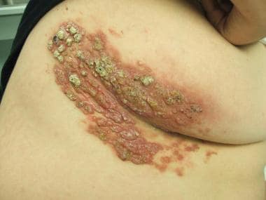
Lymphangioma
Lymphangioma, Cystic
Picibanil
Mesenteric Cyst
Mesentery
Abdomen, Acute
Klippel-Trenaunay-Weber Syndrome
Kasabach-Merritt Syndrome
Macroglossia
Hemangioendothelioma
Incidental Findings
Retroperitoneal Neoplasms
Tomography, X-Ray Computed
Ranula
Lymphatic Abnormalities
Encyclopedias as Topic
Lymphangioma of the spleen in an elderly patient. (1/83)
Splenic cystic lymphangioma is a very rare condition, and is classified among cystic proliferations of the spleen. It is considered to be the result of a developmental malformation of the lymphatic system and can involve the spleen alone or be a part of multiorgan disease. It is usually seen in children, often found incidentally. We describe a case of cystic lymphangioma of the spleen in an elderly woman putting emphasis on the rarity of the case in old age, and on the problems of differential diagnosis with the other cystic proliferations of the spleen, in particular hydatid disease, in the absence of histologic information. (+info)Rapid determination of zygosity and common aneuploidies from amniotic fluid cells using quantitative fluorescent polymerase chain reaction following genetic amniocentesis in multiple pregnancies. (2/83)
Following second-trimester twin amniocentesis, we used quantitative fluorescent polymerase chain reaction (QF-PCR) assays and polymorphic small tandem repeats (STR) for rapid determination of zygosity and common aneuploidies from amniotic fluid (AF) cells in four pregnancies with like-sex twins, fused placentae and inconclusive chorionicity. The first and the second cases were suspected to have inadvertent sampling of the same amniotic cavity twice. The first case showed a dizygotic (DZ) pattern and repeat amniocentesis was thus avoided. The second case was monozygotic (MZ) and was complicated by discordant fetal growth and twin-twin transfusion syndrome. The third case was associated with a co-twin malformation, occipital encephalocele. DNA studies revealed MZ twinning with a discordant structural defect. The fourth case was associated with co-twin abnormalities of cystic hygroma and hydrops fetalis. DNA studies showed DZ twinning with discordant structural and chromosomal defects. The QF-PCR assay with STR has the advantages of rapid determination of zygosity and common aneuploidies in AF cells. This simple test appears to be useful in the instances of possible inadvertent puncture of the same amniotic cavity twice during amniocentesis and of discordant fetal structural and/or chromosomal abnormalities following genetic amniocentesis in multiple pregnancies with uncertain chorionicity. (+info)Management of orbital lymphangioma using intralesional injection of OK-432. (3/83)
AIM: To treat orbital lymphangioma with an intralesional injection of OK-432 (group A Streptococcus pyogenes of human origin). METHOD: A 14 year old boy had a right orbital cystic lymphangioma. The visual acuity in the eye was 20/28. In an initial treatment, 0.02 mg of OK-432, was injected into the tumour after aspiration of the fluid contents, but no effect was seen. The second treatment was performed with 0.04 mg of OK-432. RESULT: 4 months later, the lesion had totally shrunk to fibrous tissue. The side effects were fever, a local inflammatory reaction lasting 3 days, and increased intraocular pressure, which was managed by draining the fluid contents. Visual acuity improved to 20/15, and the visual field defect and restriction of eye movement seen before treatment disappeared. No recurrence was noted 1 year after treatment. CONCLUSION: An intralesional injection of OK-432 shrunk the lymphangioma without functional disturbance and scar in the facial skin. OK-432 may be useful for orbital lymphangioma, but further studies are still warranted to determine efficacy, complications, and the optimal dose for safe treatment. (+info)Cystic lymphangioma of the small-bowel mesentery: case report and a review of the literature. (4/83)
Cystic lymphangioma of the small-bowel mesentery is a rare manifestation of an intraabdominal tumor in elderly patients. We present a case of a small-bowel mesentery lymphangioma, causing fever and chills and present clinical and pathologic features. Furthermore, etiology and differential diagnosis of this tumor are discussed. (+info)Prognostic value of ultrasound findings of fetal cystic hygroma detected in early pregnancy by transvaginal sonography. (5/83)
OBJECTIVE: To assess the prognostic value of first and early second trimester transvaginal ultrasound findings of fetal cystic hygroma such as volume, presence of septa and associated fetal anomalies or malformations. DESIGN: A prospective study of fetal cystic hygroma volume detected at 10-15 weeks of gestation by transvaginal scan. SUBJECTS: The study comprised a series of 33 cystic hygromas detected throughout the period March 1994 to March 1998 in 1918 pregnant women. METHODS: The volume of the hygroma and the presence of septa and other associated fetal anomalies or malformations were evaluated and correlated with fetal karyotype, persistence of the hygroma and pregnancy outcome. RESULTS: A volume equal to or greater than 75 mm3 revealed a sensitivity of 66.7% [eight of 12 cases; 95% confidence interval (CI), 34.9-90.1%] for the identification of abnormal fetal karyotype, 72.7% (eight of 11 cases; 95% CI, 39.0-90.4%) for the identification of persistence of the hygroma and 90% (nine of 10 cases; 95% CI, 55.5-99.7%) for identification of an unfavorable outcome of pregnancy. Furthermore, the prognostic value of ultrasound in determining pregnancy and fetal outcome were improved by combining data on the volume of the hygroma and the presence of associated anomalies or malformations. CONCLUSIONS: Measuring the volume of a cystic hygroma proves to be a useful ultrasound prognostic indicator in determining the risk of an associated karyotypic abnormality and adverse fetal and pregnancy outcome. However, due to limited sample size, caution is required in interpretation of the data and further studies are needed. (+info)The progression of mediastinal lymphangioma in utero. (6/83)
We describe a case of mediastinal lymphangioma that suddenly progressed in utero. The initial sonographic impression was one of pericardial effusion while the follow-up examination revealed a mediastinal cystic mass. At 35 weeks of gestation preterm labor occurred. An ultrasound examination performed at this time demonstrated the mediastinal cyst with internal hemorrhage and fetal hydrops. The cystic mass was resected 5 days after birth. The diagnosis of lymphangioma was made from histological examination. The infant was followed up for 18 months and remained well. We recommend frequent prenatal sonographic follow-up when a localized fluid collection is, shown in the mediastinum. (+info)Five cases of the lymphangioma of the mediastinum in adult. (7/83)
Mediastinal lymphangioma is rare disease. Above all cavernous type of mediastinal lymphangioma is very rare. We report 5 cases of mediastinal lymphangioma including cavernous type. CT was performed in all and revealed that they were smoothly marginated and cystic. All were surgically resected and specimens were classified pathologically into cystic type (3 cases), cavernous type (1 case) and mixed type of the two (1 case). MRI was performed in the cavernous type and suggested that the mass was lymphangioma because of pathognomonic lesion. Despite preoperative diagnosis of mediastinal lymphangioma is difficult, MRI is able to useful examination. In the follow-ups there has been no recurrence in our series. (+info)Sonographic screening for fetal aneuploidy: first trimester. (8/83)
OBJECTIVES: Screening for fetal aneuploidy is now possible during the first trimester using sonographic and biochemical markers. The aim of this review was to summarize the efficacy and use of nuchal translucency in screening for fetal aneuploidy, especially fetal Down syndrome, and other anomalies. METHODS: We reviewed available literature regarding first-trimester screening. This includes more than 16 studies of nuchal translucency as a marker for fetal aneuploidy published since 1995. RESULTS: Although early studies showed wide variation in detection of fetal Down syndrome when using nuchal translucency, more recent studies showed sensitivities of approximately 70% to 80%, for a 5% false-positive rate. Increased nuchal translucency has also been found to be a marker for other aneuploidies, including trisomy 18, trisomy 13, and Turner syndrome. Maternal serum biochemical screening can be used as a test for aneuploidy during the first trimester The 2 maternal serum markers that appear to be most useful in the late first trimester are the free beta subunit of human chorionic gonadotropin and pregnancy-associated plasma protein A. Together with maternal age, these markers yield a detection rate for trisomy 21 of approximately 60%, for a 5% false-positive rate. Because sonographic and biochemical markers appear to be largely independent, their combined risk results in improved detection rates compared with either method alone. As a result, the combination of nuchal translucency, biochemical markers, and maternal age has achieved a detection rate of approximately 85%, for a 5% false-positive level for detection of trisomy 21. A newly proposed "integrated" approach using a panel of first- and second-trimester markers suggests that further improvement in the screening performance is possible. A number of questions regarding first-trimester screening remain. We address some of these questions: is first-trimester screening more effective than second-trimester screening? How to account for intrauterine lethality? Is earlier diagnosis important, and will it be accepted by patients? Is first-trimester screening cost-effective? How should first-trimester screening be interpreted with second-trimester tests? CONCLUSIONS: Despite encouraging data and general enthusiasm for first-trimester screening for fetal Down syndrome and other aneuploidies, a number of questions remain about its implementation in the United States. Multicenter studies currently under way should help answer some of these questions. (+info)Lymphangioma is a benign (noncancerous) tumor or malformation that occurs due to the abnormal development of the lymphatic system, a part of the immune system that helps fight infection and eliminate waste products. Lymphangiomas are typically composed of dilated lymphatic vessels filled with clear fluid called lymph. These masses can occur in various parts of the body but are most commonly found in the head, neck, and axilla (armpit) regions.
There are three main types of lymphangiomas:
1. Capillary lymphangioma: Also known as "lymphangiectasia" or "lymphangiomatosis," this is the most superficial and least aggressive type, often presenting as small vesicles or blisters on the skin.
2. Cavernous lymphangioma: This type consists of larger, more dilated lymphatic spaces and can involve deeper tissues. It usually appears as a soft, compressible mass beneath the skin.
3. Cystic hygroma: A subtype of cavernous lymphangioma, cystic hygromas are typically found in the neck or axilla regions and present as large, fluid-filled sacs or cysts.
Lymphangiomas can cause various symptoms depending on their size and location, including swelling, pain, infection, difficulty swallowing, or breathing problems if they compress vital structures such as airways or blood vessels. Treatment options may include surgical excision, sclerotherapy (injection of a substance to shrink the lesion), or observation, depending on the individual case and patient's preferences.
Cystic lymphangioma is a benign (noncancerous) tumor that develops in the lymphatic system, which is a part of the immune system. It is typically present at birth or appears in early childhood. The tumor is caused by the abnormal development of lymphatic vessels, resulting in the formation of cystic spaces filled with lymph fluid.
Cystic lymphangioma can occur anywhere in the body but are most commonly found in the head and neck region, particularly in the tongue, mouth, and throat. They may also appear in the armpits or groin. The tumor usually grows slowly and can cause various symptoms depending on its location. For example, a cystic lymphangioma in the throat can cause difficulty breathing, swallowing, or speaking.
Treatment options for cystic lymphangioma include surgical removal of the tumor, sclerotherapy (injection of a solution that causes the cysts to harden and shrink), or observation if the tumor is not causing any symptoms. The choice of treatment depends on various factors, including the size and location of the tumor, as well as the patient's age and overall health.
Picibanil is not a commonly used medical term, and it may be more familiar as the brand name for a specific preparation of Group A Streptococcus OK-432. It is an immunotherapeutic agent that has been used in Japan for the treatment of certain types of cancer, such as nasopharyngeal carcinoma and soft tissue sarcoma.
Group A Streptococcus OK-432 is a weakened form of a bacterium that causes strep throat. When administered, it stimulates the immune system to produce cytokines, which are substances that help regulate the immune response. This can enhance the body's ability to fight off cancer cells and potentially slow or stop tumor growth.
It is important to note that Picibanil/OK-432 is not approved for use in the United States and its effectiveness as a cancer treatment has not been extensively studied outside of Japan.
A Mesenteric Cyst is a rare, benign abdominal mass that forms within the mesentery, which is the fold of membrane that attaches the intestine to the abdominal wall and contains blood vessels, lymphatic vessels, and nerves. These cysts can vary in size from a few centimeters to several inches in diameter. They are typically asymptomatic but can cause symptoms such as abdominal pain, bloating, or a palpable mass, depending on their size and location. The exact cause of mesenteric cysts is not well understood, but they may be congenital or acquired due to trauma, inflammation, or surgery. Treatment usually involves surgical removal of the cyst.
Mediastinal neoplasms refer to abnormal growths or tumors located in the mediastinum, which is the central compartment of the thoracic cavity that lies between the lungs and contains various vital structures such as the heart, esophagus, trachea, blood vessels, lymph nodes, and nerves. Mediastinal neoplasms can be benign (non-cancerous) or malignant (cancerous), and they can arise from any of the tissues or organs within the mediastinum.
Benign mediastinal neoplasms may include thymomas, lipomas, neurofibromas, or teratomas, among others. These tumors are typically slow-growing and rarely spread to other parts of the body. However, they can still cause symptoms or complications by compressing adjacent structures within the mediastinum, such as the airways, blood vessels, or nerves.
Malignant mediastinal neoplasms are cancerous tumors that can invade and destroy surrounding tissues and may spread (metastasize) to other parts of the body. Common types of malignant mediastinal neoplasms include thymic carcinomas, lymphomas, germ cell tumors, and neuroendocrine tumors. These tumors often require aggressive treatment, such as surgery, radiation therapy, and chemotherapy, to control their growth and spread.
It is important to note that mediastinal neoplasms can present with various symptoms depending on their location, size, and type. Some patients may be asymptomatic, while others may experience cough, chest pain, difficulty breathing, hoarseness, or swallowing difficulties. A thorough diagnostic workup, including imaging studies and biopsies, is necessary to confirm the diagnosis and determine the best course of treatment for mediastinal neoplasms.
Intestinal volvulus is a serious medical condition that occurs when a segment of the intestine twists around itself, cutting off its blood supply. This can lead to tissue death and perforation of the intestine if not promptly treated. Intestinal volvulus can occur in any part of the intestine but is most common in the colon, particularly in the sigmoid colon.
Volvulus can be caused by a variety of factors, including congenital abnormalities, adhesions from previous surgeries, and conditions that cause the intestines to become mobile or elongated. Symptoms of intestinal volvulus may include severe abdominal pain, nausea, vomiting, bloating, and constipation. In some cases, a physical examination or imaging tests such as X-rays or CT scans may be used to diagnose the condition.
Treatment for intestinal volvulus typically involves surgery to untwist the intestine and restore blood flow. In some cases, a portion of the intestine may need to be removed if it has been damaged beyond repair. Preventative measures such as avoiding constipation and seeking prompt medical attention for abdominal pain can help reduce the risk of developing intestinal volvulus.
Splenic neoplasms refer to abnormal growths or tumors in the spleen, which can be benign (non-cancerous) or malignant (cancerous). These growths can arise from various cell types present within the spleen, including hematopoietic cells (red and white blood cells, platelets), stromal cells (supporting tissue), or lymphoid cells (part of the immune system).
There are several types of splenic neoplasms:
1. Hematologic malignancies: These are cancers that affect the blood and bone marrow, such as leukemias, lymphomas, and multiple myeloma. They often involve the spleen, causing enlargement (splenomegaly) and neoplastic infiltration of splenic tissue.
2. Primary splenic tumors: These are rare and include benign lesions like hemangiomas, lymphangiomas, and hamartomas, as well as malignant tumors such as angiosarcoma, littoral cell angiosarcoma, and primary splenic lymphoma.
3. Metastatic splenic tumors: These occur when cancer cells from other primary sites spread (metastasize) to the spleen. Common sources of metastasis include lung, breast, colon, and ovarian cancers, as well as melanomas and sarcomas.
Symptoms of splenic neoplasms may vary depending on the type and extent of the disease but often include abdominal pain or discomfort, fatigue, weight loss, and anemia. Diagnosis typically involves imaging studies (such as ultrasound, CT, or MRI scans) and sometimes requires a biopsy for confirmation. Treatment options depend on the type of neoplasm and may include surgery, chemotherapy, radiation therapy, targeted therapy, or immunotherapy.
The mesentery is a continuous fold of the peritoneum, the double-layered serous membrane that lines the abdominal cavity, which attaches the stomach, small intestine, large intestine (colon), and rectum to the posterior wall of the abdomen. It provides blood vessels, nerves, and lymphatic vessels to these organs.
Traditionally, the mesentery was thought to consist of separate and distinct sections along the length of the intestines. However, recent research has shown that the mesentery is a continuous organ, with a single continuous tethering point to the posterior abdominal wall. This new understanding of the anatomy of the mesentery has implications for the study of various gastrointestinal diseases and disorders.
Jejunal neoplasms refer to abnormal growths or tumors in the jejunum, which is the middle section of the small intestine. These neoplasms can be benign (non-cancerous) or malignant (cancerous). Malignant jejunal neoplasms are often aggressive and can spread to other parts of the body, making them potentially life-threatening.
There are several types of jejunal neoplasms, including:
1. Adenocarcinomas: These are cancerous tumors that develop from the glandular cells lining the jejunum. They are the most common type of jejunal neoplasm.
2. Carcinoid tumors: These are slow-growing neuroendocrine tumors that arise from the hormone-producing cells in the jejunum. While they are usually benign, some can become malignant and spread to other parts of the body.
3. Gastrointestinal stromal tumors (GISTs): These are rare tumors that develop from the connective tissue cells in the jejunum. They can be benign or malignant.
4. Lymphomas: These are cancerous tumors that develop from the immune system cells in the jejunum. They are less common than adenocarcinomas but can be aggressive and spread to other parts of the body.
5. Sarcomas: These are rare cancerous tumors that develop from the connective tissue cells in the jejunum. They can be aggressive and spread to other parts of the body.
Symptoms of jejunal neoplasms may include abdominal pain, bloating, diarrhea, weight loss, and bleeding in the stool. Treatment options depend on the type and stage of the neoplasm but may include surgery, chemotherapy, radiation therapy, or a combination of these approaches.
"Acute abdomen" is a medical term used to describe a sudden and severe abdominal pain that requires immediate medical attention. This condition can be caused by various factors such as inflammation, infection, obstruction, or perforation of the abdominal organs. Common causes of acute abdomen include appendicitis, cholecystitis, diverticulitis, intestinal obstruction, and perforated ulcers.
The symptoms of acute abdomen may include severe and localized or generalized abdominal pain, tenderness, rigidity, rebound tenderness, fever, nausea, vomiting, and loss of appetite. The diagnosis of acute abdomen is usually made based on the patient's history, physical examination, laboratory tests, and imaging studies such as X-rays, ultrasound, or CT scan.
Treatment of acute abdomen depends on the underlying cause and may include antibiotics, intravenous fluids, pain management, and surgery in severe cases. Delayed diagnosis and treatment of acute abdomen can lead to serious complications such as sepsis, peritonitis, and even death.
Klippel-Trenaunay-Weber Syndrome (KTWS) is a rare and complex congenital vascular disorder that affects the development of blood vessels, soft tissues, and bones. It is also known as Klippel-Trenaunay syndrome or KTS.
The medical definition of KTWS includes the following features:
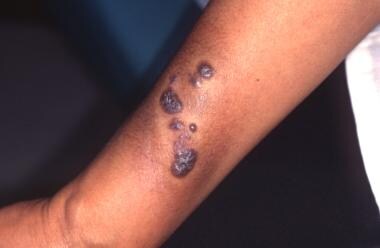
1. Port-wine stain (capillary malformation): A red or purple birthmark caused by an abnormal collection of blood vessels in the skin, often present at birth and usually affecting one limb or part of the body.
2. Venous and lymphatic abnormalities: Varicose veins, dilated veins, or abnormal vein patterns may be present, along with lymphatic malformations that can cause swelling in the affected area.
3. Soft tissue and bone hypertrophy: Overgrowth of soft tissues and bones in the affected limb or region, leading to asymmetry and sometimes functional impairment.
4. Other possible features: May include skin abnormalities, such as increased hair growth or changes in texture; joint deformities; and orthopedic problems, like scoliosis or hip dysplasia.
It is important to note that the severity of KTWS can vary significantly from person to person, ranging from mild symptoms to severe cases with significant functional impairment. The condition is not typically life-threatening but may require ongoing medical management and surveillance to address potential complications, such as infections, bleeding, or deep vein thrombosis.
Kasabach-Merritt syndrome (KMS) is a rare but serious condition characterized by the combination of a large hemangioma or tufted angioma (benign vascular tumors) with severe thrombocytopenia (low platelet count) and consumptive coagulopathy (a disorder of blood clotting).
The syndrome is named after the two physicians who first described it in 1940. It primarily affects infants, with about 70% of cases diagnosed before the age of one month.
In KMS, the hemangioma or tufted angioma grows rapidly and becomes a consumptive coagulopathy due to platelet trapping within the lesion, leading to profound thrombocytopenia. This can result in bleeding complications, which can be life-threatening if not promptly treated.
The treatment of KMS typically involves a combination of medical management (such as corticosteroids, interferon, and vincristine) and surgical intervention to remove the hemangioma or tufted angioma. In some cases, embolization of the lesion may also be considered.
Macroglossia is a medical term that refers to an abnormally large tongue in relation to the size of the oral cavity. It can result from various conditions, including certain genetic disorders (such as Down syndrome and Beckwith-Wiedemann syndrome), hormonal disorders (such as acromegaly), inflammatory diseases (such as amyloidosis), tumors or growths on the tongue, or neurological conditions. Macroglossia can cause difficulties with speaking, swallowing, and breathing, particularly during sleep. Treatment depends on the underlying cause but may include corticosteroids, radiation therapy, surgery, or a combination of these approaches.
Orbital neoplasms refer to abnormal growths or tumors that develop in the orbit, which is the bony cavity that contains the eyeball, muscles, nerves, fat, and blood vessels. These neoplasms can be benign (non-cancerous) or malignant (cancerous), and they can arise from various types of cells within the orbit.
Orbital neoplasms can cause a variety of symptoms depending on their size, location, and rate of growth. Common symptoms include protrusion or displacement of the eyeball, double vision, limited eye movement, pain, swelling, and numbness in the face. In some cases, orbital neoplasms may not cause any noticeable symptoms, especially if they are small and slow-growing.
There are many different types of orbital neoplasms, including:
1. Optic nerve glioma: a rare tumor that arises from the optic nerve's supportive tissue.
2. Orbital meningioma: a tumor that originates from the membranes covering the brain and extends into the orbit.
3. Lacrimal gland tumors: benign or malignant growths that develop in the lacrimal gland, which produces tears.
4. Orbital lymphangioma: a non-cancerous tumor that arises from the lymphatic vessels in the orbit.
5. Rhabdomyosarcoma: a malignant tumor that develops from the skeletal muscle cells in the orbit.
6. Metastatic tumors: cancerous growths that spread to the orbit from other parts of the body, such as the breast, lung, or prostate.
The diagnosis and treatment of orbital neoplasms depend on several factors, including the type, size, location, and extent of the tumor. Imaging tests, such as CT scans and MRI, are often used to visualize the tumor and determine its extent. A biopsy may also be performed to confirm the diagnosis and determine the tumor's type and grade. Treatment options include surgery, radiation therapy, chemotherapy, or a combination of these approaches.
"Intralesional injection" is a medical term that refers to the administration of a medication directly into a lesion or skin abnormality, such as a tumor, cyst, or blister. This technique is used to deliver the medication directly to the site of action, allowing for higher local concentrations and potentially reducing systemic side effects. Common examples include the injection of corticosteroids into inflamed tissues to reduce swelling and pain, or the injection of chemotherapeutic agents directly into tumors to shrink them.
Hemangioendothelioma is a rare type of vascular tumor, which means it arises from the endothelial cells that line the blood vessels. It can occur in various parts of the body, but it most commonly involves the soft tissues and bones. Hemangioendotheliomas are often classified as borderline malignant tumors because they can behave either indolently (like a benign tumor) or aggressively (like a malignant tumor), depending on their specific type and location.
There are several subtypes of hemangioendothelioma, including:
1. Epithelioid hemangioendothelioma: This subtype typically affects young adults and can involve various organs, such as the liver, lungs, or soft tissues. It tends to have a more indolent course but can metastasize in some cases.
2. Kaposiform hemangioendothelioma: This is an aggressive subtype that usually occurs in infants and children. It often involves the skin and soft tissues, causing local invasion and consumptive coagulopathy (Kasabach-Merritt phenomenon).
3. Retiform hemangioendothelioma: A rare and low-grade malignant tumor that typically affects the skin and subcutaneous tissue of adults. It has a favorable prognosis with a low risk of metastasis.
4. Papillary intralymphatic angioendothelioma (PILA): This is a rare, slow-growing tumor that usually occurs in the head and neck region of children and young adults. It has an excellent prognosis with no reported cases of metastasis or recurrence after complete surgical resection.
Treatment for hemangioendotheliomas typically involves surgical excision when possible. Other treatment options, such as radiation therapy, chemotherapy, or targeted therapies, may be considered depending on the tumor's location, size, and behavior. Regular follow-up is essential to monitor for potential recurrence or metastasis.
Incidental findings are diagnoses or conditions that are discovered unintentionally while evaluating a patient for a different condition or symptom. These findings are not related to the primary reason for the medical examination, investigation, or procedure. They can occur in various contexts such as radiology studies, laboratory tests, or physical examinations.
Incidental findings can sometimes lead to further evaluation and management, depending on their nature and potential clinical significance. However, they also pose challenges related to communication, informed consent, and potential patient anxiety or harm. Therefore, it is essential to have clear guidelines for managing incidental findings in clinical practice.
Genital neoplasms in males refer to abnormal growths or tumors that develop in the male reproductive organs. These can be benign (non-cancerous) or malignant (cancerous).
Malignant genital neoplasms are often referred to as genital cancers. The most common types of male genital cancers include:
1. Penile Cancer: This occurs when cancer cells form in the tissues of the penis.
2. Testicular Cancer: This forms in the testicles (testes), which are located inside the scrotum.
3. Prostate Cancer: This is a common cancer in men, forming in the prostate gland, which is part of the male reproductive system that helps make semen.
4. Scrotal Cancer: This is a rare form of cancer that forms in the skin or tissue of the scrotum.
5. Penile Intraepithelial Neoplasia (PeIN): This is not cancer, but it is considered a pre-cancerous condition of the penis.
Early detection and treatment of genital neoplasms can significantly improve the prognosis. Regular self-examinations and medical check-ups are recommended, especially for individuals with risk factors such as smoking, HIV infection, or a family history of these cancers.
Retroperitoneal neoplasms refer to abnormal growths or tumors that develop in the retroperitoneal space. This is the area located behind the peritoneum, which is the membrane that lines the abdominal cavity and covers the abdominal organs. The retroperitoneal space contains several vital structures such as the kidneys, adrenal glands, pancreas, aorta, and lymphatic vessels.
Retroperitoneal neoplasms can be benign or malignant (cancerous). Malignant retroperitoneal neoplasms are often aggressive and can invade surrounding tissues and organs, leading to various complications. Common types of retroperitoneal neoplasms include lymphomas, sarcomas, and metastatic tumors from other primary sites. Symptoms may vary depending on the size and location of the tumor but can include abdominal or back pain, weight loss, and swelling in the legs. Diagnosis typically involves imaging studies such as CT scans or MRI, followed by a biopsy to determine the type and grade of the tumor. Treatment options may include surgery, radiation therapy, chemotherapy, or a combination of these approaches.
Vulvar neoplasms refer to abnormal growths or tumors in the vulvar region, which is the exterior female genital area including the mons pubis, labia majora, labia minora, clitoris, and the vaginal vestibule. These neoplasms can be benign (non-cancerous) or malignant (cancerous).
Benign vulvar neoplasms may include conditions such as vulvar cysts, fibromas, lipomas, or condylomas (genital warts). They are typically slow-growing and less likely to spread or invade surrounding tissues.
Malignant vulvar neoplasms, on the other hand, are cancers that can invade nearby tissues and potentially metastasize (spread) to distant parts of the body. The most common types of malignant vulvar neoplasms are squamous cell carcinoma, vulvar melanoma, and adenocarcinoma.
Early detection and treatment of vulvar neoplasms are essential for improving prognosis and reducing the risk of complications or recurrence. Regular gynecological examinations, self-examinations, and prompt attention to any unusual symptoms or changes in the vulvar area can help ensure timely diagnosis and management.
Peritoneal neoplasms refer to tumors or cancerous growths that develop in the peritoneum, which is the thin, transparent membrane that lines the inner wall of the abdomen and covers the organs within it. These neoplasms can be benign (non-cancerous) or malignant (cancerous). Malignant peritoneal neoplasms are often associated with advanced stages of gastrointestinal, ovarian, or uterine cancers and can spread (metastasize) to other parts of the abdomen.
Peritoneal neoplasms can cause various symptoms such as abdominal pain, bloating, nausea, vomiting, loss of appetite, and weight loss. Diagnosis typically involves imaging tests like CT scans or MRIs, followed by a biopsy to confirm the presence of cancerous cells. Treatment options may include surgery, chemotherapy, radiation therapy, or a combination of these approaches, depending on the type, stage, and location of the neoplasm.
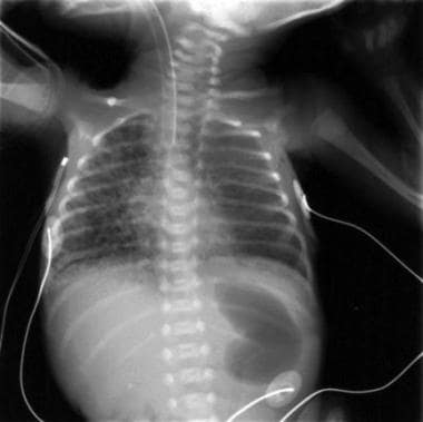
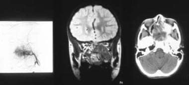
X-ray computed tomography (CT or CAT scan) is a medical imaging method that uses computer-processed combinations of many X-ray images taken from different angles to produce cross-sectional (tomographic) images (virtual "slices") of the body. These cross-sectional images can then be used to display detailed internal views of organs, bones, and soft tissues in the body.
The term "computed tomography" is used instead of "CT scan" or "CAT scan" because the machines take a series of X-ray measurements from different angles around the body and then use a computer to process these data to create detailed images of internal structures within the body.
CT scanning is a noninvasive, painless medical test that helps physicians diagnose and treat medical conditions. CT imaging provides detailed information about many types of tissue including lung, bone, soft tissue and blood vessels. CT examinations can be performed on every part of the body for a variety of reasons including diagnosis, surgical planning, and monitoring of therapeutic responses.
In computed tomography (CT), an X-ray source and detector rotate around the patient, measuring the X-ray attenuation at many different angles. A computer uses this data to construct a cross-sectional image by the process of reconstruction. This technique is called "tomography". The term "computed" refers to the use of a computer to reconstruct the images.
CT has become an important tool in medical imaging and diagnosis, allowing radiologists and other physicians to view detailed internal images of the body. It can help identify many different medical conditions including cancer, heart disease, lung nodules, liver tumors, and internal injuries from trauma. CT is also commonly used for guiding biopsies and other minimally invasive procedures.
In summary, X-ray computed tomography (CT or CAT scan) is a medical imaging technique that uses computer-processed combinations of many X-ray images taken from different angles to produce cross-sectional images of the body. It provides detailed internal views of organs, bones, and soft tissues in the body, allowing physicians to diagnose and treat medical conditions.
A ranula is a type of mucocele, which is a mucus-containing cyst that forms in the mouth. Specifically, a ranula is a mucocele that develops in the floor of the mouth, usually as a result of a blocked salivary gland duct. It appears as a smooth, dome-shaped swelling that is bluish or transparent in color. Ranulas can cause discomfort, particularly when speaking, eating, or swallowing, and they may interfere with normal oral function if they become large enough. Treatment typically involves surgical removal of the cyst, along with any affected salivary gland tissue.
Lymphatic abnormalities refer to conditions or defects that affect the lymphatic system, which is a part of the immune and circulatory systems. The lymphatic system includes a network of vessels, tissues, and organs that help rid the body of waste and toxins, fight infections, and maintain fluid balance.
Lymphatic abnormalities can occur due to genetic mutations, infections, inflammation, or cancer. These abnormalities may affect various components of the lymphatic system, including:
1. Lymph vessels: Abnormalities in lymph vessels can lead to a buildup of lymph fluid in certain parts of the body, causing swelling known as lymphedema.
2. Lymph nodes: Enlarged or abnormally shaped lymph nodes (lymphadenopathy) may indicate an infection, inflammation, or cancer.
3. Spleen: An enlarged spleen (splenomegaly) can be a sign of various conditions, such as infections, blood disorders, or cancer.
4. Thymus: Abnormalities in the thymus gland, which is part of the immune system, can lead to immunodeficiency disorders.
5. Tonsils and adenoids: Enlarged tonsils and adenoids can cause breathing and swallowing difficulties, especially in children.
6. Aggregated lymphatic tissue: Abnormalities in aggregated lymphatic tissue, such as Peyer's patches in the small intestine or the appendix, can increase the risk of infections and autoimmune disorders.
Lymphatic abnormalities can present with various symptoms, including swelling, pain, recurrent infections, and fatigue. Treatment depends on the underlying cause and may involve medications, surgery, or lifestyle changes.
An encyclopedia is a comprehensive reference work containing articles on various topics, usually arranged in alphabetical order. In the context of medicine, a medical encyclopedia is a collection of articles that provide information about a wide range of medical topics, including diseases and conditions, treatments, tests, procedures, and anatomy and physiology. Medical encyclopedias may be published in print or electronic formats and are often used as a starting point for researching medical topics. They can provide reliable and accurate information on medical subjects, making them useful resources for healthcare professionals, students, and patients alike. Some well-known examples of medical encyclopedias include the Merck Manual and the Stedman's Medical Dictionary.
The lymphatic system is a complex network of organs, tissues, vessels, and cells that work together to defend the body against infectious diseases and also play a crucial role in the immune system. It is made up of:
1. Lymphoid Organs: These include the spleen, thymus, lymph nodes, tonsils, adenoids, and Peyer's patches (in the intestines). They produce and mature immune cells.
2. Lymphatic Vessels: These are thin tubes that carry clear fluid called lymph towards the heart.
3. Lymph: This is a clear-to-white fluid that contains white blood cells, mainly lymphocytes, which help fight infections.
4. Other tissues and cells: These include bone marrow where immune cells are produced, and lymphocytes (T cells and B cells) which are types of white blood cells that help protect the body from infection and disease.
The primary function of the lymphatic system is to transport lymph throughout the body, collecting waste products, bacteria, viruses, and other foreign substances from the tissues, and filtering them out through the lymph nodes. The lymphatic system also helps in the absorption of fats and fat-soluble vitamins from food in the digestive tract.
A subdural effusion is an abnormal accumulation of fluid in the potential space between the dura mater (the outermost layer of the meninges that covers the brain and spinal cord) and the arachnoid membrane (one of the three layers of the meninges that surround the brain and spinal cord) in the subdural space.
Subdural effusions can occur due to various reasons, including head trauma, infection, or complications from neurosurgical procedures. The fluid accumulation may result from bleeding (subdural hematoma), inflammation, or increased cerebrospinal fluid pressure. Depending on the underlying cause and the amount of fluid accumulated, subdural effusions can cause various symptoms, such as headaches, altered mental status, or neurological deficits.
Subdural effusions are often asymptomatic and may resolve independently; however, in some cases, medical intervention might be necessary to alleviate the pressure on the brain or address the underlying condition. Imaging techniques like computed tomography (CT) or magnetic resonance imaging (MRI) scans are typically used to diagnose and monitor subdural effusions.
 Cystic hygroma
Vascular malformation
Lymphangioma
Cystic tumour of the atrioventricular nodal region
Lymphangioleiomyomatosis
List of diseases (A)
Lymphohemangioma
List of skin conditions
Neoplasm
Lymphangiomatosis
International Classification of Diseases for Oncology
List of diseases (C)
Picibanil
List of MeSH codes (C04)
Angioma
 Lymphangioma, Cystic | Harvard Catalyst Profiles | Harvard Catalyst
Cystic lymphangioma of the vallecula<...
 cystic lymphangioma Archives - Jennifer Michelle Greenberg
 Congenital Malformations of the Neck: Embryology, Branchial Malformations, Thyroglossal Duct Cysts/Ectopic Thyroid
 View of Rare case of retroperitoneal cystic lymphangioma in an adult
Abdominal distention caused by cystic lymphangioma in a neonate<...
Cystic hygroma - Wikipedia
 Broad ligament cystic lymphangioma: A case report | Journal of Medical Case Reports | Peer Review
 Cystic hygroma: MedlinePlus Medical Encyclopedia
 Cystic Lymphangioma of the Adrenal Gland: Case Report | Zemheri | World Journal of Nephrology and Urology
 Ranjini Kudva | Department of Pathology | KMC Manipal, Manipal Academy of Higher Education
Single-port laparoscopic-assisted resection for a large abdominal cystic lymphangioma: a case report | Surgical Case Reports |...
 Bilateral ovarian cystic lymphangioma with chylous ascitis in pregnancy - a rare case report with review
| International...
Bilateral ovarian cystic lymphangioma with chylous ascitis in pregnancy - a rare case report with review
| International...
 Hussaini S. Ndakwo - Articles - Scientific Research Publishing
 Pathology Outlines - WHO reporting system for pancreaticobiliary cytopathology
Oral Hemangiomas: Practice Essentials, Pathophysiology, Etiology
Tongue lymphangiomas treated with picibanil (OK432). Experience with Mexican children
 Birthmark Support Group
Keïta Mamadou - Articles - Scientific Research Publishing
Istituto di Analisi dei Sistemi ed Informatica "Antonio Ruberti" (Publications of G. Boldrini)
 Webpathology.com: A Collection of Surgical Pathology Images
 Book - Essentials of Pediatric Surgery
| Bentham Science
Book - Essentials of Pediatric Surgery
| Bentham Science
 Infections
Cord
 Saiama Waqar - Research output
- Research Profiles at Washington University School of Medicine
Saiama Waqar - Research output
- Research Profiles at Washington University School of Medicine
Internet Scientific Publications
 An adolescent with an axillary mass | Pediatric Oncall Journal
 Laparoscopic splenectomy for nodular and cystic lesions in the spleen - SAGES Abstract Archives
 Lymphatic Malformations - Dermatologic Disorders - MSD Manual Professional Edition
Malformation5
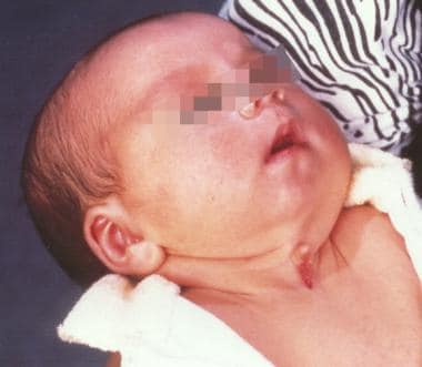
- Also known as cystic lymphangioma and macrocystic lymphatic malformation, the growth is often a congenital lymphatic lesion of many small cavities (multiloculated) that can arise anywhere, but is classically found in the left posterior triangle of the neck and armpits. (wikipedia.org)
- Currently, the medical field prefers to use the term lymphatic malformation, because the term cystic hygroma means water tumor. (wikipedia.org)
- Detection of a cystic malformation may prompt further investigation, such as amniocentesis, to evaluate for genetic abnormalities in the fetus. (wikipedia.org)
- The mass was evaluated with computed tomography (CT) revealing a 9.6 (craniocaudad) x 4.4 (transverse) x 5.7 (anterior-posterior) cm multiloculated cystic mass within the upper right lateral chest wall and axilla, with very faint peripheral and septal enhancement most consistent with a large lymphatic malformation or cystic hygroma. (pediatriconcall.com)
- Lymphangioma is a malformation composed of a mass of dilated lymph vessels typically found in the cervical region in children. (edu.pl)
Cyst3
- Traumatic cervical cyst lymphangioma in an adult. (medigraphic.com)
- Axial magnetic resonance imaging (MRI) revealed a multiloculated, multiseptated cystic mass with a distinct fluid-fluid level indicative of recent bleeding into the cyst. (pediatriconcall.com)
- Final diagnosis was confirmed pathologically as hemangiomas in 3, SANT in 3, inflammatory pseudotumor in 2, malignant lymphoma in one, lymphangioma in one, simple cyst in one and pseudocyst in one patient. (sages.org)
Hygromas9
- Cystic hygromas are benign, but can be disfiguring. (wikipedia.org)
- Cystic hygromas occur when the lymphatic vessels that make up the lymphatic system are not formed properly. (wikipedia.org)
- Cystic hygromas are increasingly diagnosed by prenatal ultrasonography. (wikipedia.org)
- Cystic hygromas can grow very large and may affect breathing and swallowing. (wikipedia.org)
- Cystic hygromas are also often seen in Turner's syndrome, although a patient who does not have the syndrome can present with this condition. (wikipedia.org)
- Cystic hygromas that develop in the third trimester, after 30 weeks' gestation, or in the postnatal period are usually not associated with chromosome abnormalities. (wikipedia.org)
- However, cystic hygromas can often grow, making it impossible to remove all of the tissue. (medlineplus.gov)
- A non-profit organisation to support children, their parents and adults with lymphatic malformations (cystic hygromas or lymphangiomas). (birthmarksupportgroup.org.uk)
- Cystic lymphangiomas, once referred to as cystic hygromas, are congenital lymphatic malformations constituting 6% of all benign lesions of infancy and childhood. (pediatriconcall.com)
Malformations2
- lymphangiomas are malformations of the lymphatic system that can appear as multilobulated cavities in the neck (75%) and axilla (20%) with very few cases of extensive lymphangioma reported in the literature. (uis.edu.co)
- Son pronostic dépend essentiellement des autres malformations congénitales cardiaques associées, des arythmies et des troubles de la conduction, ainsi que de la fonction systolique du ventricule droit en position systémique. (bvsalud.org)
Retroperitoneal cystic lymphangioma1
- Laparoscopic excision of an adult retroperitoneal cystic lymphangioma coexisting with an esophageal hiatus hernia. (harvard.edu)
Abdominal2
- Lin, SC , Lun, HH & Chen, PH 2013, ' Abdominal distention caused by cystic lymphangioma in a neonate ', Journal of Experimental and Clinical Medicine(Taiwan) , vol. 5, no. 4, pp. 148-149. (tmu.edu.tw)
- Abdominal CT scan revealed a septate cystic lesion adherent to the ileum. (webpathology.com)
Hygroma in children2
- Ogita S, Tsuto T, TokiwaK, Takahasi T. Intracystic injection of OK-432 a new esclerosing therapy for cystic hygroma in children. (medigraphic.com)
- The hemangioma, Lymphangiomas and cystic hygroma in children, are the widespread benign neoplasm. (benthamscience.com)
Lesion5
- Bilateral lymphangioma of the ovary being an extremely rare lesion, with chylous ascites and full term pregnancy, this is the first ever case in the world to be reported. (ijrcog.org)
- The specimen shows a 10 cm. thin-walled cystic lesion arising from the mesentery of ileum . (webpathology.com)
- Twelve patients with splenic nodular or cystic lesions who underwent laparoscopic splenectomy between April 2003 and June 2018 were retrospectively reviewed, in which patient factors (age, sex), lesion factors (diagnosis, size, number) and surgical factors (procedures, operation time, blood loss, postoperative complication, postoperative hospital stay) were assessed. (sages.org)
- Laparoscopic splenectomy for the nodular and cystic lesion is safe and feasible. (sages.org)
- The stomach X-ray has no place within the analysis of cystic lymphangioma, it only reveals the shadow of the lesion as a water tone opacity comparatively properly restricted backing the digestive gasoline in periphery (6) [url=http://www.mhcurling.com/dcs/buy-ditropan/] gastritis diet peanut butter ditropan 2.5 mg without prescription[/url]. (ehd.org)
Cervical2
- Antoniades K, Kiziridou A, Psimopoulou M. Traumatic cervical cystic hygroma. (medigraphic.com)
- Katsumo S, Ezawa S, Minemura T. Excision of cervical cystic lymphangioma using injection of hidrocolloid dental impression material. (medigraphic.com)
Lesions2
- However, the feasibility and safety of laparoscopic splenectomy for nodular and cystic splenic lesions are yet to be elucidated. (sages.org)
- Splenectomy was performed by either pure laparoscopy (n = 3), hand - assisted laparoscopic surgery (HALS) (n = 5) or single incision laparoscopic surgery (SILS) (n = 4), for solid lesions in 8 and cystic lesions in 4 patients. (sages.org)
Mediastinal lymphangioma6
- Anterior mediastinal lymphangioma in an infant: diagnosis and surgical management. (harvard.edu)
- Mediastinal lymphangioma is a rare condition and accounts for 0.01% to 4.5% of all mediastinal tumors. (edu.pl)
- Only 4 cases of mediastinal lymphangioma involving the heart and great vessels in adults have been described in the available literature. (edu.pl)
- 2] Park J.G., Aubry M.C., Godfrey J.A., Midthun D.E. Mediastinal Lymphangioma: Mayo Clinic Experience of 25 Cases. (edu.pl)
- 6] McLoughlin G.S., Nuchtern J.G., Dauser R.C., Sciubba D.M., Gokaslan Z.L., Wolinsky J.P. Mediastinal lymphangioma presenting as an acute epidural hematoma. (edu.pl)
- Mediastinal lymphangioma. (edu.pl)
Cavernous Lymphangioma1
- 11] Teramoto K., Suzumura Y. Mediastinal cavernous lymphangioma in an adult. (edu.pl)
Removal of cystic1
- Several years previously, the patient underwent surgical removal of cystic lymphangiomas from the left ovary, both fallopian tubes and small intestine. (edu.pl)
Nuchal2
- First-trimester cystic hygroma: relationship of nuchal translucency thickness and outcomes. (harvard.edu)
- Cystic hygroma can be associated with a nuchal lymphangioma or a fetal hydrops. (wikipedia.org)
Fetal1
- Retrospective analysis of genetic etiology and obstetric outcome of fetal cystic hygroma: A single-center study. (harvard.edu)
Atypical2
- This case illustrates an atypical adolescent presentation of cystic lymphangioma, manifest after trauma and masquerading as a soft tissue tumor. (pediatriconcall.com)
- 3] Yildirim E., Dural K., Kaplan T., Sakinci U. Cystic lymphangioma: report of two atypical cases. (edu.pl)
Benign1
- Introduction: Lymphangiomas are benign lymph vessels tumors that affect, in general, head and neck. (bvsalud.org)
Mediastinum3
- Asymptomatic lymphangioma involving the spleen and mediastinum in adults. (harvard.edu)
- But when lymphangiomas are located in the mediastinum diagnosis is late because symptoms are subjective. (medric.or.kr)
- 10] Oshikiri T., Morikawa T., Jinushi E., Kawakami Y., Katoh H. Five Cases of Lymphangioma of Mediastinum in Adult. (edu.pl)
Malignant2
- Malignant Lymphangioma of ovary. (ijrcog.org)
- Kroemer L. Malignant lymphangioma of the ovary. (ijrcog.org)
Immunohistochemical characterization2
- Evans A, Lytwyn A, Urbach G, Chapman W. Bi lateral lymphangioma of the ovary immunohistochemical characterization. (ijrcog.org)
- 12] Sinzelle E., Van Huyen J.P.D, Breiteneder-Geleff S., Braunberger E., Deloche A., Kerjaschki D., Bruneval P. Intrapericardial lymphangioma with podoplanin immunohistochemical characterization of lymphatic endothelial cells. (edu.pl)
Neck4
- A cystic hygroma is an abnormal growth that usually appears on a baby's neck or head. (wikipedia.org)
- A cystic hygroma is a growth that often occurs in the head and neck area. (medlineplus.gov)
- Cystic Hygroma of the head and neck a long term follow-up of 44 cases. (medigraphic.com)
- Long term results of intratumorous bleomycin-A5 injection for head and neck lymphangioma. (medigraphic.com)
Multiple cystic2
- Verification of the diagnosis may require more testing, as multiple cystic masses can arise in children. (wikipedia.org)
- A CT scan with contrast of the patient's abdomen showed multiple cystic formations in the liver, spleen, kidneys, and left parapelvic region. (edu.pl)
Lymph vessels1
- Lymphangioma - A structure consisting of a collection of blood vessels and lymph vessels that are overgrown and clumped together. (en-academic.com)
Resection1
- Surgical resection and histo-pathological findings were compatible with cystic lymphangioma of the adrenal gland. (wjnu.org)
Diagnosis4
- We discuss pathological differential diagnosis for cystic adrenal lymhangioma with a review of related literature on this unusual case. (wjnu.org)
- The main differential diagnosis is an adenomatoid tumour which can be differentiated from the lymphangioma by immunohistochemical studies. (ijrcog.org)
- A diagnosis of cystic lymphangioma was made based on imaging. (pediatriconcall.com)
- Conclusion: lymphangioma is an uncommon condition whose early diagnosis and adequate prenatal counseling helps to manage and improve newborn neonatal prognosis (MÉD.UIS.2012;25(2):149-54). (uis.edu.co)
Adult2
- Wiggs WJ, Sismanis A. Cystic hygroma in the adult: Two cases report. (medigraphic.com)
- Cystic hygroma in an adult: a case report. (medigraphic.com)
Neoplasms1
- Cystic pancreatic neoplasms: imaging features and management strategy. (harvard.edu)
Asymptomatic1
- Lymphangioma is usually asymptomatic and unilateral, presenting as an incidental finding during routine gynaecologic procedures. (ijrcog.org)
Ovary5
- Lymphangioma of the ovary accompanied by chylous ascites. (ijrcog.org)
- Heinig J, Beckmann V, Bialas T, Diallo R. Lymphangioma of the ovary after irradiation due to Wilms tumour in childhood. (ijrcog.org)
- Lymphangiomas involving the ovary. (ijrcog.org)
- Logani KB, Agarwal K. Lymphangioma of the ovary. (ijrcog.org)
- Lymphangioma of the ovary. (ijrcog.org)
Cleft palate1
- A lethal version of this condition exists, known as Cowchock-Wapner-Kurtz syndrome, that, in addition to cystic hygroma, includes cleft palate and lymphedema, a condition of localized edema and tissue swelling caused by a compromised lymphatic system. (wikipedia.org)
Lymphatic tissue1
- A cystic growth originating from lymphatic tissue. (harvard.edu)
Congenital1
- 2) Cystic lymphangiomas arise as the result of congenital malformatian-of the lymphatic system and are considered to be a form of lymphatic hamartoma(1). (medric.or.kr)
Tumour1
- 8] Bilgin M., Akçali Y., Oguzkaya F., Öktem T. Mediastinal Cystic Lymphangioma: A Rare Mediastinal Tumour. (edu.pl)
Traumatic1
- Imaging revealed a multi-loculated cystic mass with an internal fluid-fluid level, and enhancement characteristics consistent with cystic lymphangioma and recent traumatic hemorrhage. (pediatriconcall.com)
Surgical removal1
- A chance exists of recurrence after surgical removal of the cystic hygroma. (wikipedia.org)
Case2
- Rare case of ovarian cystic lymphangioma managed at laparoscopy. (ijrcog.org)
- VELITCHKOV N, FILIPOV A, LOSANOFF J, KJOSSEV K, KATROV E. Mesenteric cystic lymphangioma as a cause of acute intestinal obstruction: Report of a case and a brief review of its status. (clinicalsurgeryjournal.com)
Pregnancy1
- Sometimes, a cystic hygroma is seen using a pregnancy ultrasound when the baby is still in the womb. (medlineplus.gov)
MeSH1
- Lymphangioma, Cystic" is a descriptor in the National Library of Medicine's controlled vocabulary thesaurus, MeSH (Medical Subject Headings) . (harvard.edu)
Patient2
- 1) A clinical investigation was made 6 cases of cystic lymphangioma, treated at the Kwang Ju Christian hospital as in the patient. (medric.or.kr)
- After application of chi-square test at 5% of significance, we noted a relationship between patient gender and location of the lymphangiomas (p = 0.0003). (bvsalud.org)
Findings2
- Park C, Lee JW, Kim J. Sonographic Findings of Prenatal Torsion of Ovarian Lymphangioma. (ijrcog.org)
- Gross and microscopic findings were diagnostic of lymphangioma. (webpathology.com)
Picibanil2
- Tongue lymphangiomas treated with picibanil (OK432). (medigraphic.com)
- The effectiveness of intralesional sclerotherapy of lymphangioma with OK-432 (Picibanil) has been proved in several clinical studies. (medigraphic.com)
Prenatally1
- A baby with a prenatally diagnosed cystic hygroma should be delivered in a major medical center equipped to deal with neonatal complications, such as a neonatal intensive care unit. (wikipedia.org)
Literature1
- And above all, only 20 cases of ovarian lymphangioma are reported in the literature so far. (ijrcog.org)
Diagnostic1
- Clinical and diagnostic work-up revealed a cystic mass at left suprarenal space. (wjnu.org)
Cases3
- In cases where complete removal is not possible, the cystic hygroma commonly returns. (medlineplus.gov)
- Objective and Employee Methods: The aim of this study was to make a survey of the cases of lymphangioma recorded in the Pathology Laboratories of the Institute of Biological Sciences, University of Passo Fundo (ICB/ UPF) and the Hospital of Saint Vincent Paul Passo Fundo/ RS (HSVP) since the year 1987 until 2012. (bvsalud.org)
- Results: In all, 78 cases of lymphangioma were found. (bvsalud.org)
Children1
- Ogita S, Tsuto T, TokiwaK, Takahashi T. Intracystic injection of OK-432 therapy for lymphangiomas in children: why and how does it work? (medigraphic.com)
Infection1
- Furthermore, in lymphangiomas studied we noted the presence of local infection and the treatment was by surgery. (bvsalud.org)
Occur2
- If resolution of the cystic hygroma does not occur before birth, a pediatric surgeon should be consulted. (wikipedia.org)
- Extremely rarely, lymphangiomas occur as a generalized lymphangiomatosis. (edu.pl)