Lymphangiectasis, Intestinal
Protein-Losing Enteropathies
Hypoproteinemia
Malabsorption Syndromes
Encyclopedias as Topic
Very late onset small intestinal B cell lymphoma associated with primary intestinal lymphangiectasia and diffuse cutaneous warts. (1/35)
As only a handful of lymphoma cases have been reported in conjunction with primary intestinal lymphangiectasia, it is not yet clear if this association is merely fortuitous or related to primary intestinal lymphangiectasia induced immune deficiency. We report on two female patients, 50 and 58 years old, who developed small intestinal high grade B cell lymphoma a long time (45 and 40 years, respectively) after the initial clinical manifestations of primary intestinal lymphangiectasia. They presented with a longstanding history of fluctuating protein losing enteropathy, multiple cutaneous plane warts, and markedly dilated mucosal and submucosal lymphatic channels in duodenal biopsies. One had a large ulcerated tumour of the proximal ileum and the other diffuse ileal infiltration. In both, histological examination showed centroblastic high grade B cell lymphoma associated with duodenojejuno-ileal mucosal and submucosal lymphangiectasia. They were subsequently successfully treated with surgery and postoperative chemotherapy (AVmCP: adriamycin, cyclophosphamide, Vm26, and prednisolone), and chemotherapy alone (PACOB: adriamycin, cyclophosphamide, vincristine, bleomycine, and prednisolone), respectively. A three year follow up in both cases showed persistent diffuse lymphangiectasia without evidence of lymphoma. The present findings support the hypothesis that primary intestinal lymphangiectasia is associated with lymphoma development. (+info)Protracted diarrhea: results of the five-year survey in a tertiary hospital in Korea. (2/35)
The syndrome of protracted diarrhea (PD) includes several diseases with diverse etiologies. This study was conducted to characterize the spectrum of causes, clinical manifestations, and the outcomes of PD. A retrospective analysis of the clinical and pathological findings was performed on 25 patients with diarrhea starting within the first 2 yr of life and a requirement of parenteral nutrition (PN). According to the intestinal histopathology, patients were classified into four groups: immune enteropathy (12 cases), lymphangiectasia (6 cases), epithelial dysplasia (5 cases), and unclassified (2 cases). All patients with epithelial dysplasia had earlier onset of diarrhea and longer duration of PN than those in the other groups. Three patients (12%) had an evidence of a familial condition. Five patients (three with microvillous inclusion disease and two with immune enteropathy) died. Sixteen patients recovered, and three (two with primary lymphangiectasia and one with microvillous inclusion disease) still had diarrhea. One patient underwent intestinal transplantation for tufting enteropathy. In conclusion, infants with PD should be referred to specialized centers where advanced diagnostic and therapeutic facilities are available, because histological analysis is critical for the diagnosis of PD, and PN or intestinal transplantation is the only therapeutic option in a subset of cases. (+info)Surgical resection of duodenal lymphangiectasia: a case report. (3/35)
Intestinal lymphangiectasia, characterized by dilatation of intestinal lacteals, is rare. The major treatment for primary intestinal lymphangiectasia is dietary modification. Surgery to relieve symptoms and to clarify the etiology should be considered when medical treatment failed. This article reports a 49-year-old woman of solitary duodenal lymphangiectasia, who presented with epigastralgia and anemia. Her symptoms persisted with medical treatment. Surgery was finally performed to relieve the symptoms and to exclude the existence of underlying etiologies, with satisfactory effect. In conclusion, duodenal lymphangiectasia can present clinically as epigastralgia and chronic blood loss. Surgical resection may be resorted to relieve pain, control bleeding, and exclude underlying diseases in some patients. (+info)Successful treatment of protein-losing enteropathy induced by intestinal lymphangiectasia in a liver cirrhosis patient with octreotide: a case report. (4/35)
A 47-yr-old man with hepatitis B virus associated liver cirrhosis was admitted to our hospital with diarrhea and generalized edema and diagnosed as protein-losing enteropathy due to intestinal lymphangiectasia by intestinal biopsy and 99m Tc albumin scan. During hospitalization, he received subcutaneous octreotide therapy. After 2 weeks of octreotide therapy, follow-up albumin scan showed no albumin leakage, and the serum albumin level was sustained. We speculate that liver cirrhosis can be a cause of intestinal lymphangiectasia and administration of octreotide should be considered for patients with intestinal lymphangiectasia whose clinical and bio-chemical abnormalities do not respond to a low-fat diet. (+info)Case study in canine intestinal lymphangiectasia. (5/35)
A 9.52 kg, 9-year-old, spayed female beagle was presented with the chief complaint of abdominal distention of 1 week's duration. A presumptive diagnosis of canine intestinal lymphangectasia was arrived at by exclusion of other causes for the patient's ascites. The patient was successfully treated with dietary modification and immunosuppressive therapy. (+info)Coeliac disease and lymphangiectasia. (6/35)
Two out of 74 children with coeliac disease demonstrated severe intestinal protein loss. In both children a serial small bowel biopsy specimen showed intestinal lymphangiectasia to be also present. Intestinal lymphangiectasia is another disorder that may be associated with coeliac disease. (+info)Intestinal lymphangiectasia associated with angiofollicular lymph node hyperplasia (Castleman's disease). (7/35)
A patient presenting with predominantly gastrointestinal symptoms and a history of myocardial infarction was found to have ascites, hepatosplenomegaly, para-aortic lymphadenopathy, thrombocytosis, and a paraproteinaemia. A jejunal biopsy specimen showed lymphangiectasia and histology of the spleen and lymph nodes showed angiofollicular hyperplasia or Castleman's disease of the hyaline vascular type. This association has not previously been described and, moreover, systemic symptoms are unusual in this variant of Castleman's disease. (+info)A primary intestinal lymphangiectasia patient diagnosed by capsule endoscopy and confirmed at surgery: a case report. (8/35)
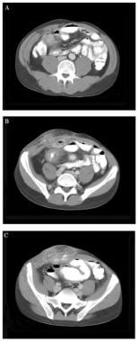
Intestinal lymphangiectasia (IL) is a rare disease characterized by dilated lymphatic vessles in the intestinal wall and small bowel mesentery which induce loss of protein and lymphocytes into bowel lumen. Because it most often occurs in the intestine and cannot be detected by upper gastroendoscopy or colonoscopy, and the value of common image examinations such as X-ray and computerized tomography (CT) are limited, the diagnosis of IL is difficult, usually needing the help of surgery. Capsule endoscopy is useful in diagnosing intestinal diseases, such as IL. We here report a case of IL in a female patient who was admitted for the complaint of recurrent edema accompanied with diarrhea and abdominal pain over the last twenty years, and aggravated ten days ago. She was diagnosed by M2A capsule endoscopy as a primary IL and confirmed by surgical and pathological examination. (+info)Lymphangiectasis is a medical condition characterized by the dilation and abnormal expansion of lymphatic vessels, which are responsible for transporting lymph fluid throughout the body. These dilated lymphatic vessels can be found in various tissues and organs, including the intestines, lungs, or other parts of the body.
In the case of intestinal lymphangiectasis (also known as Waldmann's disease), the lymphatic vessels in the small intestine become enlarged, leading to impaired absorption of nutrients and lymph fluid. This can result in protein-losing enteropathy, malnutrition, diarrhea, and edema (swelling) due to the loss of proteins and lymphatic fluids into the gastrointestinal tract.
Pulmonary lymphangiectasis is a rare congenital disorder where the lymphatic vessels in the lungs are abnormally developed and dilated, causing respiratory distress, recurrent lung infections, and chylous effusions (accumulation of milky lymph fluid in the pleural space surrounding the lungs).
Treatment for lymphangiectasis depends on the underlying cause and severity of the condition. It may involve dietary modifications, medications to manage symptoms, or surgical interventions in some cases.
Intestinal lymphangiectasis is a rare condition characterized by the dilation and dysfunction of the lacteals (lymphatic vessels) within the intestinal villi. This results in the leakage of lymphatic fluid into the gastrointestinal lumen, leading to chronic protein loss, malabsorption of nutrients, and various other complications.
The condition can be primary (congenital), which is usually caused by genetic mutations affecting lymphatic development, or secondary, resulting from acquired conditions that obstruct or damage the intestinal lymphatics. Secondary intestinal lymphangiectasis may occur due to various causes such as abdominal surgeries, radiation therapy, inflammatory bowel disease, or tumors compressing the lymphatic vessels.
Symptoms of intestinal lymphangiectasis include diarrhea, steatorrhea (fatty stools), weight loss, edema (swelling), and hypoproteinemia (low protein levels in the blood). The diagnosis typically involves imaging techniques like lymphangiography or magnetic resonance imaging (MRI) to visualize the dilated lymphatic vessels. Treatment often focuses on dietary modifications, such as a low-fat, high-protein, and medium-chain triglyceride diet, along with managing any underlying conditions contributing to the development of the disease. In some cases, medications or surgical interventions may be necessary to alleviate symptoms and improve quality of life.
Lung diseases refer to a broad category of disorders that affect the lungs and other structures within the respiratory system. These diseases can impair lung function, leading to symptoms such as coughing, shortness of breath, chest pain, and wheezing. They can be categorized into several types based on the underlying cause and nature of the disease process. Some common examples include:
1. Obstructive lung diseases: These are characterized by narrowing or blockage of the airways, making it difficult to breathe out. Examples include chronic obstructive pulmonary disease (COPD), asthma, bronchiectasis, and cystic fibrosis.
2. Restrictive lung diseases: These involve stiffening or scarring of the lungs, which reduces their ability to expand and take in air. Examples include idiopathic pulmonary fibrosis, sarcoidosis, and asbestosis.
3. Infectious lung diseases: These are caused by bacteria, viruses, fungi, or parasites that infect the lungs. Examples include pneumonia, tuberculosis, and influenza.
4. Vascular lung diseases: These affect the blood vessels in the lungs, impairing oxygen exchange. Examples include pulmonary embolism, pulmonary hypertension, and chronic thromboembolic pulmonary hypertension (CTEPH).
5. Neoplastic lung diseases: These involve abnormal growth of cells within the lungs, leading to cancer. Examples include small cell lung cancer, non-small cell lung cancer, and mesothelioma.
6. Other lung diseases: These include interstitial lung diseases, pleural effusions, and rare disorders such as pulmonary alveolar proteinosis and lymphangioleiomyomatosis (LAM).
It is important to note that this list is not exhaustive, and there are many other conditions that can affect the lungs. Proper diagnosis and treatment of lung diseases require consultation with a healthcare professional, such as a pulmonologist or respiratory therapist.
Protein-losing enteropathies (PLE) refer to a group of conditions characterized by excessive loss of proteins from the gastrointestinal tract into the intestinal lumen and ultimately into the stool. This results in hypoproteinemia, which is a decrease in the concentration of proteins in the bloodstream, particularly albumin.
The protein loss can occur due to various reasons such as increased permeability of the intestinal mucosa, lymphatic obstruction, or inflammatory processes affecting the gastrointestinal tract. Common causes of PLE include conditions such as inflammatory bowel disease, intestinal lymphangiectasia, celiac disease, Whipple's disease, and menetrier's disease.
Symptoms of PLE may include edema, ascites, weight loss, diarrhea, and fatigue. The diagnosis of PLE typically involves measuring the concentration of proteins in the stool, as well as other diagnostic tests to determine the underlying cause. Treatment of PLE depends on the underlying cause and may involve dietary modifications, medications, or surgical interventions.
Hypoproteinemia is a medical condition characterized by abnormally low levels of protein, particularly albumin, in the blood. This can occur due to various reasons such as malnutrition, liver disease, kidney disease, or gastrointestinal disorders that affect protein absorption. It can lead to edema (swelling), especially in the legs and abdomen, and other complications. It's important to note that while albumin is the most abundant protein in blood serum, other proteins such as immunoglobulins and enzymes can also be affected in hypoproteinemia.
Malabsorption syndromes refer to a group of disorders in which the small intestine is unable to properly absorb nutrients from food, leading to various gastrointestinal and systemic symptoms. This can result from a variety of underlying conditions, including:
1. Mucosal damage: Conditions such as celiac disease, inflammatory bowel disease (IBD), or bacterial overgrowth that cause damage to the lining of the small intestine, impairing nutrient absorption.
2. Pancreatic insufficiency: A lack of digestive enzymes produced by the pancreas can lead to poor breakdown and absorption of fats, proteins, and carbohydrates. Examples include chronic pancreatitis or cystic fibrosis.
3. Bile acid deficiency: Insufficient bile acids, which are necessary for fat emulsification and absorption, can result in steatorrhea (fatty stools) and malabsorption. This may occur due to liver dysfunction, gallbladder removal, or ileal resection.
4. Motility disorders: Abnormalities in small intestine motility can affect nutrient absorption, as seen in conditions like gastroparesis, intestinal pseudo-obstruction, or scleroderma.
5. Structural abnormalities: Congenital or acquired structural defects of the small intestine, such as short bowel syndrome, may lead to malabsorption.
6. Infections: Certain bacterial, viral, or parasitic infections can cause transient malabsorption by damaging the intestinal mucosa or altering gut flora.
Symptoms of malabsorption syndromes may include diarrhea, steatorrhea, bloating, abdominal cramps, weight loss, and nutrient deficiencies. Diagnosis typically involves a combination of clinical evaluation, laboratory tests, radiologic imaging, and sometimes endoscopic procedures to identify the underlying cause. Treatment is focused on addressing the specific etiology and providing supportive care to manage symptoms and prevent complications.
An encyclopedia is a comprehensive reference work containing articles on various topics, usually arranged in alphabetical order. In the context of medicine, a medical encyclopedia is a collection of articles that provide information about a wide range of medical topics, including diseases and conditions, treatments, tests, procedures, and anatomy and physiology. Medical encyclopedias may be published in print or electronic formats and are often used as a starting point for researching medical topics. They can provide reliable and accurate information on medical subjects, making them useful resources for healthcare professionals, students, and patients alike. Some well-known examples of medical encyclopedias include the Merck Manual and the Stedman's Medical Dictionary.
Staphylococcal toxoid is a modified form of a toxin produced by the Staphylococcus aureus bacterium, which has been made less toxic through chemical treatment or irradiation. It is used in vaccines to stimulate an immune response and provide protection against staphylococcal infections. The toxoid induces the production of antibodies that recognize and neutralize the harmful effects of the original toxin, without causing the adverse reactions associated with the live toxin. This type of vaccine is used to prevent diseases such as staphylococcal scalded skin syndrome and toxic shock syndrome.
 Lymphangiectasia - Wikipedia
 22q11 Deletion Syndrome | Harvard Catalyst Profiles | Harvard Catalyst
 DeCS
DeCS lymphangiectasis
 MeSH Browser
 Namespace
 Ascites - Medic Journal
 A Short Note on Generalized Lymphatic Anomaly: A Lymphatic Disorder
 Hennekam lymphangiectasia-lymphedema syndrome 1 (Concept Id: C4012050)
- MedGen - NCBI
Hennekam lymphangiectasia-lymphedema syndrome 1 (Concept Id: C4012050)
- MedGen - NCBI cystic hygroma ultrasound 11 weeks
 Diagnostic Pathology Nonneoplastic Pediatrics, 2nd edition - Angelica R. Putnam - 1020 - ELSEVIER HEALTH SCIENCES -...
 Ulcerative Colitis Imaging: Practice Essentials, Radiography, Computed Tomography
 Top 205 Journal of Parasitology papers published in 1984
 EurekaMag PDF full texts Chapter 60754
 Intestinal lymphangiectasia, lymphedema, mental retardation, and typical face: confirmation of the Hennekam syndrome - PubMed
 Hennekam syndrome - About the Disease - Genetic and Rare Diseases Information Center
 Hierarchy
 Pesquisa | Biblioteca Virtual em Saúde - BRASIL