Lipofuscin
Pyridinium Compounds
Retinoids
Pigment Epithelium of Eye
Ceroid
Retinal Pigment Epithelium
Macular Degeneration
Retinal Pigments
Vitamin E Deficiency
Fluorescence
Cytophotometry
Retinaldehyde
Fundus Oculi
Aging
Rod Cell Outer Segment
Lysosomal Storage Diseases, Nervous System
Lysosomes
Bruch Membrane
Retinal Drusen
Diarylheptanoids
Inhibition of lysosomal degradative functions in RPE cells by a retinoid component of lipofuscin. (1/296)
PURPOSE: To investigate the effect of the lipofuscin component N-retinylidene-N-retinylethanolamine (A2-E) on degradative functions of lysosomes in human retinal pigment epithelial (RPE) cells and to evaluate its mechanism of action. METHODS: A2-E was coupled to low-density lipoprotein (LDL). Human RPE cell cultures were loaded with the A2-E/LDL complex, and controls were run with medium containing LDL alone. To determine whether A2-E accumulated in lysosomes, cells were fractionated in a Percoll gradient, and protein degradation was determined by metabolic labeling and measurement of the release of low-molecular-weight radioactivity. Lysosomal degradation was distinguished from nonlysosomal degradation by inclusion of NH4Cl in the medium. The metabolism of sulfated glycosaminoglycans was studied by radiosulfate incorporation in pulse-chase experiments. Intralysosomal pH was determined using a fluorescent lysosomotropic pH indicator. RESULTS: A2-E accumulated almost exclusively in the lysosomal compartment. Lysosomal protein degradation was reduced in a dose-dependent fashion in A2-E-treated cells. The selectivity of A2-E on lysosomal function was demonstrated by its lack of effect on degradation of extralysosomal protein. Lysosomal glycosaminoglycan catabolism of RPE cells was also strongly inhibited by A2-E. Lysosomal pH was increased by A2-E. CONCLUSIONS: The findings indicate that accumulation of A2-E in RPE cells interferes with lysosomal functions as exemplified by its inhibitory effect on protein and glycosaminoglycan catabolic pathways. The quaternary amine character of the A2-E apparently causes a perturbation of the acidic intralysosomal milieu, resulting in diminished hydrolase action and consequent accumulation of undegraded material. Such mechanism could be operative in retinal diseases associated with excessive lipofuscin accumulation including age-related macular degeneration. (+info)Melanosomes of retinal pigment epithelium--distribution, shape, and acid phosphatase activity. (2/296)
The distribution and shape of melanosomes of the retinal pigment epithelium (RPE), and acid phosphatase activity in melanosomes were studied in rabbits. The rabbit eyes were observed using electron microscopy and enzyme cytochemical electron microscopy. The majority of melanosomes were located near the apical region of the RPE. Melanosomes in the RPE were classified as two shapes, elliptical and spherical or oval. Elliptical melanosomes were located parallel to the apical process and spherical or oval melanosomes were arranged vertically or obliquely to the apical process. We think that the distribution and shape of melanosomes contributes to the effective absorption and blocking of light coming from all directions. Almost all of the mature and immature melanosomes we identified showed positive in acid phosphatase reaction, indicating that melanosomes are commonly incorporated into the lysosomal system of the RPE. However, a few melanosomes showed negative in acid phosphatase reaction, suggesting that some melanosomes are stable and inert. The observed premelanosome showed negative reaction. Two types of melanosome-related complex granules were identified; melanosomes with a cortex of enzyme-reactive material (melanolysosome) and melanosomes with a cortex of lipofuscin (melanolipofuscin). These findings indicate tha a relationship between melanosomes and the lysosomal system of the RPE exists, and suggest that melanosomes may undergo modification or degradation in the cytoplasm. Also, the observation of a premelanosome and the positive acid phosphatase activity in mature and immature melanosomes indicates that melanosomes of the RPE may continue to be synthesized at a low rate in adult eyes. (+info)Reproducibility of fundus autofluorescence measurements obtained using a confocal scanning laser ophthalmoscope. (3/296)
AIM: To evaluate the reproducibility of the background fundus autofluorescence measurements obtained using a confocal scanning laser ophthalmoscope. METHODS: 10 normal volunteers and 10 patients with retinal disease were included in the study. One eye per subject was chosen randomly. Five images of the same eye of each individual were obtained, after pupillary dilatation, by two investigators using a confocal scanning laser ophthalmoscope. Background fundus autofluorescence was measured at 7 degrees temporal to the fovea in normal volunteers and between 7 and 15 degrees temporal to the fovea in patients. Within session reproducibility of the measurements obtained by each investigator and interobserver reproducibility were evaluated. RESULTS: For investigator 1 the median values of fundus autofluorescence obtained were 31.9 units for normal volunteers and 27.3 units for patients. The median largest differences in readings in normal volunteers was 5.7 units (range 1.4-13.5 units) and in patients 4.2 units (1.5-15.1 units). For investigator 2 the median values of fundus autofluorescence obtained were 28.9 units for normal volunteers and 27.4 units for patients. The median largest difference in readings in normal volunteers was 3.6 units (2.7-11.7 units), and in patients 4.1 units (1.5-9.3 units). The median interobserver difference in readings in normal volunteers was 3.3 units and for patients 6.6 units. The median greatest interobserver difference in measurements obtained for normal volunteers was 8.8 units (8.4-23.0 units) and for patients 11.1 units (7.1-40.8 units). CONCLUSION: Within session reproducibility of the measurements of background fundus autofluorescence was satisfactory. Although interobserver reproducibility was moderate, the variability of the measurements of fundus autofluorescence between observers appears to be small when compared with variation in fundus autofluorescence with age and disease. (+info)The photoreactivity of the retinal age pigment lipofuscin. (4/296)
The presence of the age pigment lipofuscin is associated with numerous age-related diseases. In the retina lipofuscin is located within the pigment epithelium where it is exposed to high oxygen and visible light, a prime environment for the generation of reactive oxygen species. Although we, and others, have demonstrated that retinal lipofuscin is a photoinducible generator of reactive oxygen species it is unclear how this may translate into cell damage. The position of lipofuscin within the lysosome infers that irradiated lipofuscin is liable to cause oxidative damage to either the lysosomal membrane or the lysosomal enzymes. We have found that illumination of lipofuscin with visible light is capable of extragranular lipid peroxidation, enzyme inactivation, and protein oxidation. These effects, which were pH-dependent, were significantly reduced by the addition of the antioxidants, superoxide dismutase and 1,4-diazabicyclo(2,2,2)-octane, confirming a role for both the superoxide anion and singlet oxygen. We postulate that lipofuscin may compromise retinal cell function by causing loss of lysosomal integrity and that this may be a major contributory factor to the pathology associated with retinal light damage and diseases such as age-related macular degeneration. (+info)ECL cell morphology. (5/296)
Using immunohistochemistry at the conventional light, confocal and electron microscopic levels, we have demonstrated that rat stomach ECL cells store histamine and pancreastatin in granules and secretory vesicles, while histidine decarboxylase occurs in the cytosol. Furthermore the ECL cells display immunoreactivity for vesicular monoamine transporter type 2 (VMAT-2), synaptophysin, synaptotagmin III, vesicle-associated membrane protein-2, cysteine string protein, synaptosomal-associated protein of 25 kDa, syntaxin and Munc-18. Using electron microscopy in combination with stereological methods, we have evidence to suggest the existence of both an exocytotic and a crinophagic pathway in the ECL cells. The process of exocytosis in the ECL cells seems to involve a class of proteins that promote or participate in the fusion between the granule/vesicle membrane and the plasma membrane. The granules take up histamine by VMAT-2 from the cytosol during transport from the Golgi zone to the more peripheral parts of the cells. As a result, they turn into secretory vesicles. As a consequence of stimulation (e.g., by gastrin), the secretory vesicles fuse with the cell membrane to release their contents by exocytosis. The crinophagic pathway was studied in hypergastrinemic rats. In the ECL cells of such animals, the secretory vesicles were found to fuse not only with the cell membrane but also with each other to form vacuoles. Subsequent lysosomal degradation of the vacuoles and their contents resulted in the development of lipofuscin bodies. (+info)A2E, a lipofuscin fluorophore, in human retinal pigmented epithelial cells in culture. (6/296)
PURPOSE: To study A2E, a component of retinal pigmented epithelial (RPE) cell lipofuscin, after its internalization by cultured human RPE cells. METHODS: A2E was synthesized and incubated with an adult RPE cell line devoid of native lipofuscin. To investigate the cellular compartmentalization of A2E, cells were incubated simultaneously with A2E and a fluorescent acidotropic probe, (Lysotracker Red DND-99; Molecular Probes, Eugene, OR). Plasma membrane integrity was evaluated by assaying for leakage of the cytoplasmic enzyme lactate dehydrogenase (LDH), by fluorescence nuclear staining with a membrane-impermeant dye and by morphologic criteria. The emission spectrum of internalized A2E was also determined. The levels of A2E accumulated by the cultured cells were quantified by high-performance liquid chromatography and compared with amounts present in RPE isolated from human eyes. RESULTS: Internalization of A2E by the RPE cells was evidenced by the acquisition of intracellular granules detectable by fluorescence confocal imaging. Internalized A2E had an emission maxima of 565 to 570 nm. The levels of A2E accumulating in cells incubated with 10 to 25 microM A2E were comparable to the amounts of A2E present in equal numbers of RPE cells harvested from human eyes. Colocalization of A2E and the Lysotracker probe revealed a preferential accumulation in acidic organelles. The elevated LDH levels that were measured after exposure to 50 and 100 microM A2E were attributable to membrane damage in a subpopulation of the A2E-accumulating cells, determined by fluorescence nuclear labeling. CONCLUSIONS: Internalized A2E has an affinity for acidic organelles. The membrane damage exhibited by A2E-accumulating RPE is dependent on the concentration of A2E and reflects the ability of this amphiphilic compound to exert detergent-like effects. (+info)Oxidative damage and age-related macular degeneration. (7/296)
This article provides current information on the potential role of oxidation in relation to age-related macular degeneration (AMD). The emphasis is placed on the generation of oxidants and free radicals and the protective effects of antioxidants in the outer retina, with specific emphasis on the photoreceptor cells, the retinal pigment epithelium and the choriocapillaris. The starting points include a discussion and a definition of what radicals are, their endogenous sources, how they react, and what damage they may cause. The photoreceptor/pigment epithelium complex is exposed to sunlight, is bathed in a near-arterial level of oxygen, and membranes in this complex contain high concentrations of polyunsaturated fatty acids, all considered to be potential factors leading to oxidative damage. Actions of antioxidants such as glutathione, vitamin C, superoxide dismutase, catalase, vitamin E and the carotenoids are discussed in terms of their mechanisms of preventing oxidative damage. The phototoxicity of lipofuscin, a group of complex autofluorescent lipid/protein aggregates that accumulate in the retinal pigment epithelium, is described and evidence is presented suggesting that intracellular lipofuscin is toxic to these cells, thus supporting a role for lipofuscin in aging and AMD. The theory that AMD is primarily due to a photosensitizing injury to the choriocapillaris is evaluated. Results are presented showing that when protoporphyric mice are exposed to blue light there is an induction in the synthesis of Type IV collagen synthesis by the choriocapillary endothelium, which leads to a thickened Bruch's membrane and to the appearance of sub-retinal pigment epithelial fibrillogranular deposits, which are similar to basal laminar deposits. The hypothesis that AMD may result from oxidative injury to the retinal pigment epithelium is further evaluated in experiments designed to test the protective effects of glutathione in preventing damage to cultured human pigment epithelial cells exposed to an oxidant. Experiments designed to increase the concentration of glutathione in pigment epithelial cells using dimethylfumarate, a monofunctional inducer, are described in relation to the ability of these cells to survive an oxidative challenge. While all these models provide undisputed evidence of oxidative damage to the retinal pigment epithelium and the choriocapillaris that is both light- and oxygen-dependent, it nevertheless is still unclear at this time what the precise linkage is between oxidation-induced events and the onset and progression of AMD. (+info)Senescence of the retinal pigment epithelium. (8/296)
Senescence of human cells has largely been studied as an in vitro phenomenon resulting from replicative exhaustion. The literature contains many studies of retinal pigment epithelium (RPE) cells which document replicative senescence. Several studies by Burke and others illustrate the relationship between donor age and replicative lifespan, the relationship between geographical location of RPE in the posterior pole and replicative lifespan, and the phenomena of altered cellular morphology and decreased culture saturation density for senescent RPE cells. Other studies have focused on the alterations of the expression of specific genes or the alteration of enzymatic activities during the senescence of RPE cells in vitro. Recently, a technique utilizing a histochemical staining procedure for beta galactosidase has been developed which identifies senescent cells. Normal beta galactosidase histochemistry which identifies the lysosomal form of the enzyme is performed at pH 4.0, while senescence-associated beta galactosidase activity is observed at pH 6.0 and is observed in the cytoplasm. We have studied the replicative senescence of human RPE cells in vitro using this procedure and have also measured the length of chromosomal telomeres to identify the aging of cultures in vitro. Our results show that RPE cultures accumulate beta galactosidase positive cells as a function of the number of population doublings and that these data correlate with the shortening of chromosomal telomeres to a functional limit observed for many human cell types at senescence. We have also recently extended this work to the development of a senescence-associated beta galactosidase procedure for observing senescent RPE cells in vivo. Basically, the same histochemical procedure is used with a post-staining bleaching step to clearly visualize staining in the RPE. Our first studies were performed on globes from Rhesus monkeys at a variety of ages from 1 year to 29 years of age. The results show the accumulation of beta galactosidase positive cells in the older monkey eyes. We have also examined several human eyes in an attempt to observe whether any relationship exists between beta galactosidase staining and age, pathology (diabetes, basal laminar deposits), and geographical location (macula vrs. periphery). These studies represent a first effort to determine if senescent RPE are present in vivo. It will be important to extend these studies so that these data might be expressed on a quantitative bases. (+info)Lipofuscin is a type of pigment that accumulates in the lysosomes (membrane-bound organelles found inside cells) of various tissues, particularly in nerve cells and heart muscle cells. It consists of cross-linked proteins and lipids that are resistant to degradation by enzymes. The accumulation of lipofuscin is a normal part of aging but can also be associated with certain diseases such as neurodegenerative disorders.
It's often referred to as "age pigment" because it tends to increase in amount with age, and its presence in tissues has been linked to oxidative stress and cellular damage caused by free radicals. Lipofuscin is autofluorescent, meaning that it emits light when excited by certain wavelengths of light, which can be useful for its detection and quantification in research and diagnostic settings.
Pyridinium compounds are organic salts that contain a positively charged pyridinium ion. Pyridinium is a type of cation that forms when pyridine, a basic heterocyclic organic compound, undergoes protonation. The nitrogen atom in the pyridine ring accepts a proton (H+) and becomes positively charged, forming the pyridinium ion.
Pyridinium compounds have the general structure of C5H5NH+X-, where X- is an anion or negatively charged ion. These compounds are often used in research and industry, including as catalysts, intermediates in chemical synthesis, and in pharmaceuticals. Some pyridinium compounds have been studied for their potential therapeutic uses, such as in the treatment of bacterial infections or cancer. However, it is important to note that some pyridinium compounds can also be toxic or reactive, so they must be handled with care.
Retinoids are a class of chemical compounds that are derivatives of vitamin A. They are widely used in dermatology for the treatment of various skin conditions, including acne, psoriasis, and photoaging. Retinoids can help to reduce inflammation, improve skin texture and tone, and stimulate collagen production.
Retinoids work by binding to specific receptors in the skin cells, which triggers a series of biochemical reactions that regulate gene expression and promote cell differentiation and turnover. This can help to unclog pores, reduce the appearance of fine lines and wrinkles, and improve the overall health and appearance of the skin.
There are several different types of retinoids used in skincare products, including retinoic acid, retinaldehyde, and retinol. Retinoic acid is the most potent form of retinoid and is available by prescription only. Retinaldehyde and retinol are weaker forms of retinoid that can be found in over-the-counter skincare products.
While retinoids can be highly effective for treating various skin conditions, they can also cause side effects such as dryness, irritation, and sensitivity to the sun. It is important to use retinoids as directed by a healthcare professional and to follow proper sun protection measures when using these products.
The pigment epithelium of the eye, also known as the retinal pigment epithelium (RPE), is a layer of cells located between the photoreceptor cells of the retina and the choroid, which is the vascular layer of the eye. The RPE plays a crucial role in maintaining the health and function of the photoreceptors by providing them with nutrients, removing waste products, and helping to regulate the light that enters the eye.
The RPE cells contain pigment granules that absorb excess light, preventing it from scattering within the eye and improving visual acuity. They also help to create a barrier between the retina and the choroid, which is important for maintaining the proper functioning of the photoreceptors. Additionally, the RPE plays a role in the regeneration of visual pigments in the photoreceptor cells, allowing us to see in different light conditions.
Damage to the RPE can lead to various eye diseases and conditions, including age-related macular degeneration (AMD), which is a leading cause of vision loss in older adults.
Ceroid is a term used in pathology to describe a type of inclusion body that can be found in various tissues and cells in the body. These inclusions are composed of a protein called alpha-synuclein that has become aggregated and tangled, as well as lipids and other substances. Ceroids are often seen in neurons, but they can also be found in other types of cells such as glial cells.
Ceroid deposits are associated with several neurodegenerative disorders, including Parkinson's disease, dementia with Lewy bodies, and multiple system atrophy. These conditions are characterized by the accumulation of abnormal protein aggregates in the brain, which can lead to neuronal dysfunction and death. The exact role that ceroids play in these diseases is not fully understood, but they are thought to contribute to the toxicity and degeneration of nerve cells.
It's worth noting that ceroid is sometimes used interchangeably with other terms such as "lipofuscin" or "age pigment," although there are some differences between these substances at a molecular level. Nonetheless, they all refer to the accumulation of lipid-rich inclusion bodies in cells and tissues over time.
The retinal pigment epithelium (RPE) is a single layer of cells located between the photoreceptor cells of the retina and the choroid, which is a part of the eye containing blood vessels. The RPE plays a crucial role in maintaining the health and function of the photoreceptors by providing them with nutrients, removing waste products, and helping to regulate the light-sensitive visual pigments within the photoreceptors.
The RPE cells contain pigment granules that absorb excess light to prevent scattering within the eye and improve visual acuity. They also help to form the blood-retina barrier, which restricts the movement of certain molecules between the retina and the choroid, providing an important protective function for the retina.
Damage to the RPE can lead to a variety of eye conditions, including age-related macular degeneration (AMD), which is a leading cause of vision loss in older adults.
Biological pigments are substances produced by living organisms that absorb certain wavelengths of light and reflect others, resulting in the perception of color. These pigments play crucial roles in various biological processes such as photosynthesis, vision, and protection against harmful radiation. Some examples of biological pigments include melanin, hemoglobin, chlorophyll, carotenoids, and flavonoids.
Melanin is a pigment responsible for the color of skin, hair, and eyes in animals, including humans. Hemoglobin is a protein found in red blood cells that contains a porphyrin ring with an iron atom at its center, which gives blood its red color and facilitates oxygen transport. Chlorophyll is a green pigment found in plants, algae, and some bacteria that absorbs light during photosynthesis to convert carbon dioxide and water into glucose and oxygen. Carotenoids are orange, yellow, or red pigments found in fruits, vegetables, and some animals that protect against oxidative stress and help maintain membrane fluidity. Flavonoids are a class of plant pigments with antioxidant properties that have been linked to various health benefits.
Macular degeneration, also known as age-related macular degeneration (AMD), is a medical condition that affects the central part of the retina, called the macula. The macula is responsible for sharp, detailed vision, which is necessary for activities such as reading, driving, and recognizing faces.
In AMD, there is a breakdown or deterioration of the macula, leading to gradual loss of central vision. There are two main types of AMD: dry (atrophic) and wet (exudative). Dry AMD is more common and progresses more slowly, while wet AMD is less common but can cause rapid and severe vision loss if left untreated.
The exact causes of AMD are not fully understood, but risk factors include age, smoking, family history, high blood pressure, obesity, and exposure to sunlight. While there is no cure for AMD, treatments such as vitamin supplements, laser therapy, and medication injections can help slow its progression and reduce the risk of vision loss.
Retinal pigments refer to the light-sensitive chemicals found in the retina, specifically within the photoreceptor cells called rods and cones. The main types of retinal pigments are rhodopsin (also known as visual purple) in rods and iodopsins in cones. These pigments play a crucial role in the process of vision by absorbing light and initiating a series of chemical reactions that ultimately trigger nerve impulses, which are then transmitted to the brain and interpreted as visual images. Rhodopsin is more sensitive to lower light levels and is responsible for night vision, while iodopsins are sensitive to specific wavelengths of light and contribute to color vision.
Vitamin E deficiency is a condition that occurs when there is a lack of sufficient vitamin E in the body. Vitamin E is a fat-soluble antioxidant that plays an essential role in maintaining the health of cell membranes, protecting them from damage caused by free radicals. It also helps to support the immune system and promotes healthy blood vessels and nerves.
Vitamin E deficiency can occur due to several reasons, including malnutrition, malabsorption disorders such as cystic fibrosis or celiac disease, premature birth, or genetic defects affecting the alpha-tocopherol transfer protein (alpha-TTP), which is responsible for transporting vitamin E from the liver to other tissues.
Symptoms of vitamin E deficiency may include:
* Neurological problems such as peripheral neuropathy, ataxia (loss of coordination), and muscle weakness
* Retinopathy (damage to the retina) leading to vision loss
* Increased susceptibility to oxidative stress and inflammation
* Impaired immune function
Vitamin E deficiency is rare in healthy individuals who consume a balanced diet, but it can occur in people with certain medical conditions or those who have undergone bariatric surgery. In these cases, supplementation may be necessary to prevent or treat vitamin E deficiency.
Fluorescence is not a medical term per se, but it is widely used in the medical field, particularly in diagnostic tests, medical devices, and research. Fluorescence is a physical phenomenon where a substance absorbs light at a specific wavelength and then emits light at a longer wavelength. This process, often referred to as fluorescing, results in the emission of visible light that can be detected and measured.
In medical terms, fluorescence is used in various applications such as:
1. In-vivo imaging: Fluorescent dyes or probes are introduced into the body to highlight specific structures, cells, or molecules during imaging procedures. This technique can help doctors detect and diagnose diseases such as cancer, inflammation, or infection.
2. Microscopy: Fluorescence microscopy is a powerful tool for visualizing biological samples at the cellular and molecular level. By labeling specific proteins, nucleic acids, or other molecules with fluorescent dyes, researchers can observe their distribution, interactions, and dynamics within cells and tissues.
3. Surgical guidance: Fluorescence-guided surgery is a technique where surgeons use fluorescent markers to identify critical structures such as blood vessels, nerves, or tumors during surgical procedures. This helps ensure precise and safe surgical interventions.
4. Diagnostic tests: Fluorescence-based assays are used in various diagnostic tests to detect and quantify specific biomarkers or analytes. These assays can be performed using techniques such as enzyme-linked immunosorbent assay (ELISA), polymerase chain reaction (PCR), or flow cytometry.
In summary, fluorescence is a physical process where a substance absorbs and emits light at different wavelengths. In the medical field, this phenomenon is harnessed for various applications such as in-vivo imaging, microscopy, surgical guidance, and diagnostic tests.
Cytophotometry is a medical analytical technique that involves the measurement of light intensity or absorbance by individual cells, allowing for the quantitative analysis of cellular components such as DNA, RNA, and proteins. This method can be used to study cell cycle phase distribution, chromosome abnormalities, and changes in nuclear structure associated with various pathological conditions, including cancer and genetic disorders.
In cytophotometry, cells are typically stained with a fluorescent dye that specifically binds to the target molecule of interest, such as DNA or RNA. The cells are then placed on a microscope slide and illuminated with light at an appropriate wavelength for the dye used. A photodetector is used to measure the intensity of the emitted fluorescent light, which is proportional to the amount of target molecule present in each cell.
The resulting data can be analyzed using specialized software to generate histograms or other visual representations of the distribution of target molecules within a population of cells. This information can be used to identify abnormalities or changes in cellular composition, providing valuable insights into the underlying biology of various diseases and conditions.
Retinaldehyde, also known as retinal, is a form of vitamin A that is essential for vision. It is the aldehyde form of retinol (vitamin A alcohol) and is involved in the visual cycle, where it plays a crucial role in the process of converting light into electrical signals that are sent to the brain.
When light hits the retina, it activates a protein called rhodopsin, which contains retinaldehyde as one of its components. This activation causes a chemical change in retinaldehyde, leading to the generation of an electrical signal that is transmitted to the brain via the optic nerve.
Retinaldehyde is also involved in other physiological processes, including the regulation of gene expression and cell growth and differentiation. It can be synthesized in the body from beta-carotene, a pigment found in fruits and vegetables, or obtained directly from animal sources such as liver, fish liver oil, and dairy products.
"Fundus Oculi" is a medical term that refers to the back part of the interior of the eye, including the optic disc, macula, fovea, retinal vasculature, and peripheral retina. It is the area where light is focused and then transmitted to the brain via the optic nerve, forming visual images. Examinations of the fundus oculi are crucial for detecting various eye conditions such as diabetic retinopathy, macular degeneration, glaucoma, and other retinal diseases. The examination is typically performed using an ophthalmoscope or a specialized camera called a retinal camera.
Aging is a complex, progressive and inevitable process of bodily changes over time, characterized by the accumulation of cellular damage and degenerative changes that eventually lead to increased vulnerability to disease and death. It involves various biological, genetic, environmental, and lifestyle factors that contribute to the decline in physical and mental functions. The medical field studies aging through the discipline of gerontology, which aims to understand the underlying mechanisms of aging and develop interventions to promote healthy aging and extend the human healthspan.
A rod cell outer segment is a specialized structure in the retina of the eye that is responsible for photoreception, or the conversion of light into electrical signals. Rod cells are one of the two types of photoreceptor cells in the retina, with the other type being cone cells. Rod cells are more sensitive to light than cone cells and are responsible for low-light vision and peripheral vision.
The outer segment of a rod cell is a long, thin structure that contains stacks of discs filled with the visual pigment rhodopsin. When light hits the rhodopsin molecules in the discs, it causes a chemical reaction that leads to the activation of a signaling pathway within the rod cell. This ultimately results in the generation of an electrical signal that is transmitted to the brain via the optic nerve.
The outer segment of a rod cell is constantly being regenerated and broken down through a process called shedding and renewal. The tips of the outer segments are shed and phagocytosed by cells called retinal pigment epithelial (RPE) cells, which help to maintain the health and function of the rod cells.
Lysosomal storage diseases (LSDs) are a group of rare inherited metabolic disorders caused by defects in lysosomal function. These diseases affect many different organ systems, including the nervous system. Lysosomes are membrane-bound organelles found inside cells that break down and recycle various types of cellular waste materials through the action of enzymes. In LSDs, a genetic mutation leads to a deficiency or complete lack of a specific lysosomal enzyme, resulting in the accumulation of undigested substrates within the lysosomes. This accumulation can cause progressive damage to cells and tissues throughout the body, including those in the nervous system.
There are more than 50 different types of LSDs, some of which primarily affect the nervous system:
1. Tay-Sachs disease: A severe neurological disorder caused by a deficiency of the enzyme hexosaminidase A (HEXA). The accumulation of ganglioside GM2 in neurons leads to progressive neurodegeneration, resulting in motor and cognitive decline, blindness, and early death.
2. Sandhoff disease: Similar to Tay-Sachs disease but caused by a deficiency in both HEXA and hexosaminidase B (HEXB) enzymes. This disorder affects multiple organ systems, including the nervous system, with symptoms similar to Tay-Sachs disease but often more severe and rapid progression.
3. GM1 gangliosidosis: A condition caused by a deficiency of the enzyme β-galactosidase (GLB1), leading to the accumulation of GM1 ganglioside in neurons. Symptoms include developmental delay, motor and cognitive decline, seizures, and progressive neurological deterioration.
4. Gaucher disease: A disorder caused by a deficiency of the enzyme glucocerebrosidase (GBA), resulting in the accumulation of glucocerebroside in various tissues, including the nervous system. There are three main types of Gaucher disease, with type 2 and 3 having neurological involvement.
5. Niemann-Pick disease types A and B: These disorders are caused by a deficiency of the enzyme acid sphingomyelinase (SMPD1), leading to the accumulation of sphingomyelin in various tissues, including the nervous system. Type A primarily affects the nervous system, while type B mainly involves visceral organs.
6. Fabry disease: An X-linked disorder caused by a deficiency of the enzyme α-galactosidase A (GLA), resulting in the accumulation of globotriaosylceramide (Gb3) in various tissues, including the nervous system. Symptoms include pain, gastrointestinal issues, skin lesions, and progressive renal, cardiac, and cerebrovascular complications.
7. Metachromatic leukodystrophy: A disorder caused by a deficiency of the enzyme arylsulfatase A (ARSA), leading to the accumulation of sulfatides in the white matter of the brain. Symptoms include motor and cognitive decline, seizures, and progressive neurological deterioration.
8. Krabbe disease: An autosomal recessive disorder caused by a deficiency of the enzyme galactocerebrosidase (GALC), resulting in the accumulation of psychosine in the nervous system. Symptoms include motor and cognitive decline, seizures, and progressive neurological deterioration.
9. Mucopolysaccharidoses: A group of disorders caused by deficiencies of various enzymes involved in the breakdown of glycosaminoglycans (GAGs), leading to their accumulation in tissues throughout the body, including the nervous system. Symptoms vary depending on the specific disorder and include skeletal abnormalities, cardiac complications, vision and hearing loss, and progressive neurological decline.
10. Neuronal ceroid lipofuscinoses: A group of neurodegenerative disorders caused by mutations in various genes involved in lysosomal function, leading to the accumulation of lipopigments in neurons and other cells. Symptoms include seizures, motor and cognitive decline, vision loss, and progressive neurological deterioration.
11. Peroxisomal biogenesis disorders: A group of disorders caused by mutations in genes involved in peroxisome biogenesis, leading to the accumulation of very long-chain fatty acids, phytanic acid, and pipecolic acid in tissues throughout the body, including the nervous system. Symptoms vary depending on the specific disorder and include developmental delay, hypotonia, seizures, vision loss, hearing impairment, and progressive neurological decline.
12. Congenital disorders of glycosylation: A group of disorders caused by mutations in genes involved in N-glycosylation, leading to abnormal protein folding, trafficking, and function. Symptoms vary depending on the specific disorder and include developmental delay, hypotonia, seizures, vision loss, hearing impairment, and progressive neurological decline.
13. Leukodystrophies: A group of disorders characterized by abnormalities in the white matter of the brain due to defects in myelin formation or maintenance. Symptoms vary depending on the specific disorder and include developmental delay, hypotonia, seizures, vision loss, hearing impairment, and progressive neurological decline.
14. Mitochondrial disorders: A group of disorders caused by mutations in genes involved in mitochondrial function, leading to energy production deficits and oxidative stress. Symptoms vary depending on the specific disorder and include developmental delay, hypotonia, seizures, vision loss, hearing impairment, and progressive neurological decline.
15. Neurodegenerative disorders: A group of disorders characterized by progressive degeneration of the nervous system, leading to cognitive decline, motor dysfunction, and ultimately death. Examples include Alzheimer's disease, Parkinson's disease, Huntington's disease, and amyotrophic lateral sclerosis (ALS).
16. Neurodevelopmental disorders: A group of disorders characterized by impairments in cognitive, social, and motor development, including autism spectrum disorder, attention deficit hyperactivity disorder (ADHD), intellectual disability, and specific learning disorders.
17. Epilepsy: A group of disorders characterized by recurrent seizures due to abnormal electrical activity in the brain. Epilepsy can be caused by various genetic and environmental factors, including structural brain abnormalities, infections, trauma, and metabolic imbalances.
18. Neuroinflammatory disorders: A group of disorders characterized by inflammation of the nervous system, leading to damage and dysfunction. Examples include multiple sclerosis, neuromyelitis optica, and autoimmune encephalitis.
19. Infectious diseases of the nervous system: A group of disorders caused by infectious agents such as viruses, bacteria, fungi, or parasites that affect the nervous system. Examples include meningitis, encephalitis, and HIV-associated neurological disorders.
20. Neurotoxic disorders: A group of disorders caused by exposure to neurotoxic substances such as heavy metals, pesticides, solvents, or drugs that damage the nervous system. Examples include lead poisoning, organophosphate poisoning, and methanol toxicity.
21. Neurooncological disorders: A group of disorders characterized by tumors of the nervous system, including primary brain tumors, metastatic brain tumors, and spinal cord tumors.
22. Vascular disorders of the nervous system: A group of disorders caused by disruption of blood flow to the nervous system, leading to ischemia or hemorrhage. Examples include stroke, transient ischemic attack, and subarachnoid hemorrhage.
23. Degenerative disorders of the nervous system: A group of disorders characterized by progressive degeneration of nerve cells and their supporting structures, leading to functional impairment. Examples include Alzheimer's disease, Parkinson's disease, and Huntington's disease.
24. Neurodevelopmental disorders: A group of disorders that affect the development of the nervous system, leading to cognitive, behavioral, or motor impairments. Examples include autism spectrum disorder, attention deficit hyperactivity disorder, and intellectual disability.
25. Epilepsy and seizure disorders: A group of disorders characterized by recurrent seizures, which are abnormal electrical discharges in the brain that can cause a variety of symptoms such as convulsions, altered consciousness, or sensory disturbances.
26. Neurogenetic disorders: A group of disorders caused by genetic mutations that affect the structure or function of the nervous system. Examples include fragile X syndrome, tuberous sclerosis complex, and neurofibromatosis type 1.
27. Neuromuscular
Lysosomes are membrane-bound organelles found in the cytoplasm of eukaryotic cells. They are responsible for breaking down and recycling various materials, such as waste products, foreign substances, and damaged cellular components, through a process called autophagy or phagocytosis. Lysosomes contain hydrolytic enzymes that can break down biomolecules like proteins, nucleic acids, lipids, and carbohydrates into their basic building blocks, which can then be reused by the cell. They play a crucial role in maintaining cellular homeostasis and are often referred to as the "garbage disposal system" of the cell.
The Bruch membrane is a thin, layered structure that separates the retina from the choroid in the eye. It is composed of five layers: the basement membrane of the retinal pigment epithelium (RPE), the inner collagenous layer, the elastic layer, the outer collagenous layer, and the basement membrane of the choriocapillaris. The Bruch membrane provides structural support to the RPE and serves as a barrier between the retina and the choroid, allowing for the selective transport of nutrients and waste products. It also plays a role in maintaining the health of the photoreceptors in the retina. Damage to the Bruch membrane is associated with age-related macular degeneration (AMD), a leading cause of vision loss in older adults.
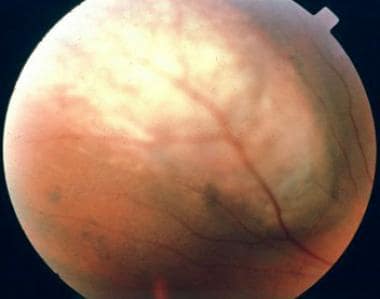
Retinal drusen are yellow-white, deposits of extracellular material that accumulate beneath the retina, most commonly in the macula. They are a common age-related finding and can also be seen in various other conditions such as inherited retinal diseases. Drusen can vary in size and number, and their presence is often associated with an increased risk of developing age-related macular degeneration (AMD), a leading cause of vision loss in older adults. However, not all individuals with drusen will develop AMD, and the significance of drusen depends on factors such as size, number, and location. It's important to monitor drusen and have regular eye examinations to assess any changes or progression that may indicate a higher risk for developing AMD.
Photofluorography is not a widely used medical term, but it generally refers to a radiographic technique that uses fluorescent screens to produce images. It was historically used for mass screening of pulmonary diseases such as tuberculosis. The patient would be exposed to a low-dose X-ray, and the resulting image would be captured on a special film or sensor that is sensitive to light emitted by the fluorescent screen.
However, it's worth noting that photofluorography has largely been replaced by digital radiography and other modern imaging techniques in clinical practice.
Diarylheptanoids are a type of organic compound characterized by a chemical structure consisting of two aromatic rings (diaryl) linked by a seven-carbon chain (heptane). They are commonly found in various plants and have been reported to exhibit a range of biological activities, including anti-inflammatory, antioxidant, and anticancer effects. Some well-known diarylheptanoids include curcumin, a component of turmeric, and gingerol, a compound found in ginger. Medical professionals may refer to diarylheptanoids when discussing the potential therapeutic benefits of these compounds for various health conditions.
Cis-trans isomeres are molecules that have the same molecular formula and skeletal structure, but differ in the arrangement of their atoms around a double bond. In a cis isomer, the two larger groups or atoms are on the same side of the double bond, while in a trans isomer, they are on opposite sides.
Cis-trans isomerases are enzymes that catalyze the interconversion between cis and trans isomers of various molecules, such as fatty acids, steroids, and retinals. These enzymes play important roles in various biological processes, including membrane fluidity, vision, and the biosynthesis of hormones and other signaling molecules.
Examples of cis-trans isomerases include:
* Fatty acid desaturases, which introduce double bonds into fatty acids and can convert trans isomers to cis isomers
* Retinal isomerases, which interconvert the cis and trans isomers of retinal, a molecule involved in vision
* Steroid isomerases, which catalyze the interconversion of various steroids, including cholesterol and its derivatives.
Melanin is a pigment that determines the color of skin, hair, and eyes in humans and animals. It is produced by melanocytes, which are specialized cells found in the epidermis (the outer layer of the skin) and the choroid (the vascular coat of the eye). There are two main types of melanin: eumelanin and pheomelanin. Eumelanin is a black or brown pigment, while pheomelanin is a red or yellow pigment. The amount and type of melanin produced by an individual can affect their skin and hair color, as well as their susceptibility to certain diseases, such as skin cancer.
 Lipofuscin
Sweat gland
Senescence
Pantothenate kinase-associated neurodegeneration
Melanosis coli
Sarcophaga bullata
Stargardt disease
Ageing
Neuron
Chromhidrosis
Advanced glycation end-product
Nasal cavity
Granulin
PI4K2A
Emixustat
Cyclodextrin
Hepatocyte
Adrenal gland
Vitelliform macular dystrophy
Neuronal ceroid lipofuscinosis
Leydig cell
Miriam Cnop
Octopus pallidus
Sparicotyle chrysophrii
Autofluorescence
Prussian blue
Eye color
Kang Zhang
Choroidal nevus
Bestrophin 1
Lipofuscin - Wikipedia
Half Life Of Lipofuscin | One Radio Network
 Prosaposin maintains lipid homeostasis in dopamine neurons and counteracts experimental parkinsonism in rodents | Nature...
Re: P57 Lipofuscin and carotenoids
 Lipofuscin Granule Accumulation Requires Autophagy Activation
 Conceptual Nanomedical Lipofuscin Removal Strategy - transhumanity.net
 GEMINI record about TN249-10 - Lipofuscin and Euphausiid Aging
 Preliminary identification and quantification of the age-pigment lipofuscin in the brain of farfantepenaeus paulensis ...
![Snyder A[au] - Search Results - PubMed](data:image/png;base64,iVBORw0KGgoAAAANSUhEUgAAABAAAAAQCAMAAAAoLQ9TAAAARVBMVEVHcEwoU45gYmYAUpQAUpRPYGVgYmZLXnJgYmYAUZUAUpRJXnIAUpQAUpRgYmYAUpRgYmZgYmZhYmYAUpQAUpQAUpRgYmaDiPJuAAAAFXRSTlMADOJ+6QewGO8/uTRqtH7GdFJ11p1bCL3TAAAAZUlEQVQYlV2PVw7AIAxDTeney7n/UcsoldX3E+VJOAboEi7MBpHWMs1ADlG8u7UYWauwyZFeRQVPOhG2o+aiwhByJxUx91Jxhje3iJSqGfHuLKI0+0TpXvY1twCOPlFh5pa/++MB0vIOBm+1zaoAAAAASUVORK5CYII=) Snyder A[au] - Search Results - PubMed
Snyder A[au] - Search Results - PubMed
 TrueBlack® Background Reducers - Biotium
 Homocysteine:blood<...
 A2E clearance Archives - SENS Research Foundation
 Ependymoma Pathology: Definition, Epidemiology, Etiology
 Free Histology Flashcards about Histology Spec Stain
 Pigmented ependymoma, a tumor with predilection for the middle-aged adult: case report with methylation classification and...
Plus it
 Differences in the surface and subsurface reflection characteristics of facial skin by age group | Request PDF
 Survival Mode That Protects Cells When Oxygen Is Low Also Slows Aging | ScienceDaily
 Involvement of Nrf2 in Ocular Diseases
 Albedextrin
 Frontiers | Dystrophin Distribution and Expression in Human and Experimental Temporal Lobe Epilepsy
Atom Bergstrom | Being Adaptable and Versatile Explains Our Survival | July 20, 2022 | One Radio Network
 Can your sweat be different colors? | HowStuffWorks
The Signaling of Cellular Senescence in Diabetic Nephropathy
Molecular interrogation of hypothalamic organization reveals distinct dopamine neuronal subtypes | Nature Neuroscience
 Age-Related Macular Degeneration - Naturopathic Doctor News and Review
Frontiers | Caffeine Protects Against Retinal Inflammation
 Search Results For Health And Wellness: Okra Results found: 10
 ucla team Archives - ScienceBlog.com
 Leber Congenital Amaurosis - American Academy of Ophthalmology
Fluorescence6
- Meanwhile, autophagosome formation is attenuated in cells experiencing oxidative stress induced by a doxorubicin pulse and chase, and lipofuscin fluorescence granules seldom manifest in the cytoplasm. (molcells.org)
- Lipofuscin granules fluoresce brightly in all channels used for fluorescence microscopy (Figure 1). (biotium.com)
- Consequently, immunofluorescence in many human tissues or aged animal tissues can be virtually impossible unless lipofuscin fluorescence is masked. (biotium.com)
- In untreated tissue (top row), lipofuscin appeared as fluorescent granules that fluoresced in all fluorescence channels. (biotium.com)
- TrueBlack® pre-treatment quenched lipofuscin fluorescence without affecting specific staining (right). (biotium.com)
- I should note in passing that the material whose accumulation causes macular degeneration is often called lipofuscin but really should not be, because the only thing it has in common with bonafide lipofuscin is its subcellular location (the lysosome) and its fluorescence properties: its molecular composition is entirely different. (nextbigfuture.com)
Ceroid lipofuscin3
- HA can react with MDA to form stable products, a non-fluorescent enamine (product 1) and a fluorescent 1,4-dihydropyridine (product 2) which are ceroid/lipofuscin-related adducts. (nel.edu)
- The end product of the damage MDA produces in the body is a pigment called ceroid lipofuscin. (naturalnews.com)
- Pulmonary fibrosis, granulomatous colitis, cardiomyopathy, and renal failure are due to the lysosomal accumulation of ceroid lipofuscin. (lu.se)
Granules9
- Lipofuscin is the name given to fine yellow-brown pigment granules composed of lipid-containing residues of lysosomal digestion. (wikipedia.org)
- Through traditional microscopy, lipofuscins are readily observed as fluorescent granules thought to accumulate in lysosomes. (molcells.org)
- Our results substantiate that in glucose-starved or replicative senescence cells, where elevated oxidative stress levels activate autophagy, lipofuscins predominately appear as granules that co-localize with autolysosomes due to lysosomal acidity or impairment. (molcells.org)
- The nanomechanical segmentation or disassembly of individual lipofuscin granules at atomic/molecular resolution may be possible employing arrays of diamondoid "debriders" to reduce lipofuscin to its most elemental and harmless fractions. (transhumanity.net)
- The detection of lipofuscin granules in 5 month old F. paulensis indicates that lipofuscin deposition probably takes place even earlier in the juvenile phase. (furg.br)
- Our results suggested that the amount of granules in the F. paulensis OLCM is related to age, but further studies are necessary to evaluate the relationship between lipofuscin content and the age of captive F. paulensis. (furg.br)
- Lipofuscin consists of highly autofluorescent granules of oxidized proteins and lipids that build up in the lysosomes of aging cells in a variety of tissues. (biotium.com)
- In untreated tissue, lipofuscin granules appeared as white speckles (left). (biotium.com)
- Autofluorescent lipofuscin granules in untreated tissue are marked with arrows. (biotium.com)
Accumulation of lipofuscin5
- The accumulation of lipofuscin-like material may be the result of an imbalance between formation and disposal mechanisms. (wikipedia.org)
- In the peripheral nervous system, abnormal accumulation of lipofuscin known as lipofuscinosis is associated with a family of neurodegenerative disorders - neuronal ceroid lipofuscinoses, the most common of these is Batten disease. (wikipedia.org)
- Accumulation of lipofuscin in the colon is the cause of the condition melanosis coli. (wikipedia.org)
- The nootropic drug piracetam appears to significantly reduce accumulation of lipofuscin in the brain tissue of rats. (wikipedia.org)
- The age-related accumulation of lipofuscin within the nervous cells is now well known, so the action of Gerovital-H3 ® was studied on rats under 6 to 18 months old. (antiaging-systems.com)
Autophagy8
- Apoptosis and autophagy are the preferred way of eliminating lipofuscin. (oneradionetwork.com)
- As Torin-1 treatment activates autophagy, granular lipofuscins intensify and dominate, indicating that autophagy activation triggers their accumulation. (molcells.org)
- Our results suggest that high oxidative stress activates autophagy but fails in lipofuscin removal, leaving an abundance of lipofuscin-filled impaired autolysosomes, referred to as residual bodies. (molcells.org)
- Therefore, future endeavors in treating lipofuscin pathology-associated diseases and dysfunctions through autophagy activation demand meticulous consideration. (molcells.org)
- Recently, autophagy-mediated lipofuscin granule degradation has garnered attention. (molcells.org)
- Thus, some theorize that autophagy flux impairment and lipofuscin accumulation are correlated ( Brunk and Terman, 2002 ). (molcells.org)
- However, the association between lipofuscin accumulation and autophagy is not definitive. (molcells.org)
- Nonetheless, autophagy activation may be better for preventing lipofuscin accumulation rather than their removal. (molcells.org)
Granule2
- However, lipofuscin granule formation and accumulation in senescent cells are poorly understood. (molcells.org)
- Once a lipofuscin granule has been captured it would proceed to be drawn into the core, where it would be digested by potent encapsulated enzymes or nanomechanically minced into a liquid state and subsequently purged from the outlet port. (transhumanity.net)
Retinal4
- Soraprazan (remofuscin) has been found to remove lipofuscin from retinal pigment epithelial cells in animals. (wikipedia.org)
- Similarly, remofuscin reversed lipofuscin accumulation in older human retinal pigment epithelium cells and restored retinal degeneration in a pathologic Stargardt disease mouse model. (molcells.org)
- As a result, N-retinylidene-PE combines with another substance to produce a fatty yellow pigment called lipofuscin, which builds up in retinal cells. (medlineplus.gov)
- Toxic constituents of lipofuscin are generated as byproducts of the visual cycle, a complex chemical pathway that is required for the maintenance of the light-gathering components of the eye called retinal photoreceptors. (harvard.edu)
Eliminate lipofuscin2
- Necrosis, oncosis, and pyroptosis eliminate lipofuscin but generate its production in surrounding cells via inflammation - not a good idea! (oneradionetwork.com)
- Q: How difficult would it be to eliminate lipofuscin (the cellular junk that particularly accumulates in neurons and heart muscle cells) compared to eliminating 7KC (an oxidized derivative of cholesterol that accumulates in atherosclerotic plaques) or A2E (a substance accumulating in the retina with age that causes macular degeneration and blindness) as a lysoSENS project? (nextbigfuture.com)
Myocardial lipofuscin2
- On the other hand, myocardial lipofuscin accumulation more directly reflects chronological ageing rather than human cardiac pathology. (wikipedia.org)
- Cumulative Disorder of Myocardial Lipofuscin after Long-Term Heart Transplantation: A Study Based on Endomyocardial Biopsies. (bvsalud.org)
Deposition2
- In addition, hepatocytes are larger, have increased lipofuscin deposition, more frequent nuclear morphological anomalies, decreased mitochondria number, and increased mitochondrial diameter compared to wild-type mice. (nih.gov)
- [ 17 ] This may reflect increased photo-oxidation and cellular toxicity with lipofuscin deposition in the retina. (medscape.com)
Lysosomes2
- Lipofuscin appears to be the product of the oxidation of unsaturated fatty acids and may be symptomatic of membrane damage, or damage to mitochondria and lysosomes. (wikipedia.org)
- Lipofuscin is a waxy heterogeneous substance that is comprised of aggregates of metabolic cellular waste end products, which reside within the lysosomes and cytosols of particular types of human cells. (transhumanity.net)
Macular degeneration6
- Lipofuscin accumulation in the eye, is a major risk factor implicated in macular degeneration, a degenerative disease, and Stargardt disease, an inherited juvenile form of macular degeneration. (wikipedia.org)
- Other possible treatments: Centrophenoxine Acetyl-L-carnitine Ginkgo biloba Dimethylethanolamine Curcumin Wet macular degeneration can be treated using selective photothermolysis where a pulsed unfocused laser predominantly heats and kills lipofuscin-rich cells, leaving untouched healthy cells to multiply and fill in the gaps. (wikipedia.org)
- The buildup of lipofuscin is toxic to the cells of the retina and causes progressive vision loss in people with Stargardt macular degeneration. (medlineplus.gov)
- Age-related macular degeneration is associated with carotenoid lipofuscin , primarily the pyridinium bisretinoid A2E. (fightaging.org)
- We also discovered that soil fungi, plants, and some bacteria possess peroxidase and carotenoid cleavage oxygenase enzymes that effectively destroy with varied degrees of efficiency and selectivity the carotenoid lipofuscin found in macular degeneration. (fightaging.org)
- Lipofuscin accumulation appears to be a major risk factor for macular degeneration, including the age-related type," Rando said. (harvard.edu)
Lysosomal2
- The prevailing thought is that the major source of lipofuscin is derived from the incomplete lysosomal degradation of cell membrane lipids and damaged mitochondria. (transhumanity.net)
- Samples of his brain revealed he was missing a protein Lipofuscin, a not so well understood compound which contains lipid residues of lysosomal digestion that accumulates in the brain liver kidney, heart muscle, retina, adrenals, nerve cells, and ganglion cells. (lifeboat.com)
Fluorescent2
- Age determination in crustaceans relies on quantification of a fluorescent age pigment, lipofuscin, that accumulates in eye stalks of krill over time. (ucar.edu)
- The only mounting medium that quenches lipofuscin background and preserves fluorescent signal. (biotium.com)
Autofluorescence quencher1
- Biotium developed TrueBlack® Lipofuscin Autofluorescence Quencher as a superior alternative to Sudan Black B to quench autofluorescence with much lower background. (biotium.com)
Lipid1
- Lipofuscins are oxidized lipid and protein complexes that accumulate during cellular senescence and tissue aging, regarded as markers for cellular oxidative damage, tissue aging, and certain aging-associated diseases. (molcells.org)
TrueBlack3
- TrueBlack® effectively eliminates lipofuscin autofluorescence in tissues like human brain and retina. (biotium.com)
- TrueBlack (bottom row) masked lipofuscin with minimal increase in background. (biotium.com)
- TrueBlack® can reduce non-lipofuscin autofluorescence. (biotium.com)
Toxic3
- They also found that animals lacking VHL-1 were resistant to the toxic proteins known to cause Alzheimer's and Huntington's diseases, and that their cells accumulated less of an age-pigment called lipofuscin. (sciencedaily.com)
- These toxic pigments, called lipofuscin, are responsible for the visual loss in patients with Stargardt's disease. (scienceblog.com)
- Rando's approach is to prevent toxic substances called lipofuscins from forming in the eye. (harvard.edu)
Quantification1
- Lipofuscin quantification is used for age determination in various crustaceans such as lobsters and spiny lobsters. (wikipedia.org)
Autofluorescent1
- The pigment was negative for Fontana-Masson, positive for Periodic-acid Schiff and autofluorescent, which coincide with characteristics of lipofuscin. (uni-muenster.de)
Tissues2
Nootropic1
- For example, centrophenoxine is a cholinergic nootropic used for treating senile dementia that notably decreased or reversed lipofuscin accumulation in post-mitotic cells over two weeks ( Terman and Welander, 1999 ). (molcells.org)
Heterogeneous1
- Lipofuscin is very heterogeneous in its molecular composition, and moreover it is mainly made of proteins, so it is hard to distinguish from material that we don't want to break down. (nextbigfuture.com)
Protein1
- The ratio of lipofuscin to protein concentration is the Lipofuscin Index and is calculated for each individual organism. (ucar.edu)
Constituents1
- Lipofuscin potentially interferes with the autophagic process, thereby preventing cellular renewal and accumulating compromised cellular constituents. (molcells.org)
Cellular1
- While lipofuscin accumulation characterizes cellular and tissue aging, it is onerous for cellular function and viability. (molcells.org)
Cells2
- It is conceivable that the future development of nanomedical robotics, as described in the upcoming book: Nanomedical Device and Systems Design: Challenges, Possibilities, Visions (FJ Boehm - Editor), to be published on Nov/22/13 by CRC Press (Taylor & Francis) might enable the capacity for the therapeutic removal of lipofuscin from individual cells in massively parallel fashion without the initial use of UV transparency . (transhumanity.net)
- Histologically and histochemically, the number of entirely lipofuscin-loaded pyramidal and Purkinje cells from the brain cortex and cerebellum was much lower (19.4 in treated vs. control animals- 72.8) (11,12). (antiaging-systems.com)
Detection1
- Although entirely conceptual and seemingly impracticable currently (2013), advanced AI [Artificial Intelligence] involvement in the design/optimization of sophisticated nanomedical devices working in conjunction with mature nanomanufacturing may indeed bring about much more complex, albeit far more rapid and efficient lipofuscin detection and removal. (transhumanity.net)
Shrimp1
- Shrimp will be aged using the pigment lipofuscin and data used to develop a predictive index and define a threshold at which treatment to control these pests is necessary. (usda.gov)
Crustaceans1
- Several studies have indicated that quantifying the amount of lipofuscin present in the eye-stalks of various crustaceans can give an index of their age. (wikipedia.org)
Substance1
- Lipofuscin is indeed harder, but what makes it harder is not the aging-versus-disease distinction but simply the nature of the substance. (nextbigfuture.com)
Junk1
- Lipofuscin is not passive junk, it's aggressive. (oneradionetwork.com)
Adult1
- Lipofuscin autofluorescence in methanol-fixed adult human brain tissue sections. (biotium.com)
Deposits1
- Within the aging human brain, deposits of lipofuscin are not uniformly distributed, but rather, are concentrated at specific sites of functional interest. (transhumanity.net)
Content1
- A preliminary study was done on the age-pigment lipofuscin content in the brains of captive Farfantepenaeus paulensis juveniles (5 months old) and wild adults (estimated age of 12-15 months). (furg.br)
Reduce1
- Calorie restriction, vitamin E, and increased glutathione appear to reduce or halt the production of lipofuscin. (wikipedia.org)
Make1
- How much difference do you think elimination of lipofuscin would make in terms of rejuvenation? (nextbigfuture.com)
Highly2
- as potential electrodes for highly localized hyperthermic interventions, following insertion into the lipofuscin mass, or hollow nanosyringes… for the injection of powerful cleaving enzymes. (transhumanity.net)
- It becomes more problematic when the sweat is brown or black because, although the sweat is colored by lipofuscins, they've been highly oxidized when they reach that pigment and don't fluoresce under a black light. (howstuffworks.com)
Months1
- after a period of three months, the levels of the lipofuscin-like material return to normal, indicating the action of a significant disposal mechanism. (wikipedia.org)
Thought1
- Lipofuscin is thought to be one indicator of an animal's health during aging. (sciencedaily.com)
Levels1
- Concurrently, certain chemicals are ostensibly effective in lowering lipofuscin levels. (molcells.org)
Removal1
- therefore, developing lipofuscin suppression and removal strategies is imperative. (molcells.org)
Field1
- This ability to selectively target lipofuscin has opened up research opportunities in the field of anti-aging medicine. (wikipedia.org)
Laser1
- 2) The two radical species produced following laser flash photolysis of lipofuscin are not quenched by oxygen impying that they are not neutral carbon centred radicals or radical anions. (netsci-journal.com)