Lingual Nerve
Lingual Nerve Injuries
Mandibular Nerve
Foramen Ovale
Tongue
Trigeminal Nerve Injuries
Cranial Fossa, Middle
Pterygoid Muscles
Paresthesia
Nerve Compression Syndromes
Sphenoid Bone
Hypoglossal Nerve
Tongue Diseases
Lingual Thyroid
Sciatic Nerve
Peripheral Nerves
Optic Nerve
Nerve Fibers
Cats
Effects of reducing submandibular blood flow on secretory responses to parasympathetic stimulation in anaesthetized cats. (1/48)
Submandibular secretory responses to stimulation of the parasympathetic chorda-lingual nerve were investigated in five anaesthetized cats before, during and after withdrawal of blood (ca 20 ml kg-1) in order to investigate the consequences of a reduced blood flow through the gland. Stimulation at different frequencies (2, 4, 6 and 8 Hz) evoked a frequency-dependent increase in the flow of submandibular saliva, sodium concentration, electrolyte and protein output. When the blood pressure was reduced (by about 50%) there was a significant reduction in submandibular blood flow and the secretion of both saliva and protein during stimulation. Under each set of conditions the flow of saliva was linearly related to the blood flow through the gland. It is concluded that submandibular secretory responses to electrical stimulation of the parasympathetic innervation can be significantly attenuated when the blood flow through the gland is reduced under the conditions employed in this study. (+info)P2X purinoceptor-mediated excitation of trigeminal lingual nerve terminals in an in vitro intra-arterially perfused rat tongue preparation. (2/48)
A novel in vitro intra-arterially perfused adult rat tongue-nerve preparation was used to explore the possible actions of P2X purinoceptor agonists (ATP and alpha,beta-methylene ATP (alpha, beta-meATP)) on sensory nerve terminals innervating the rat tongue. We made whole-nerve recordings of the trigeminal branch of the lingual nerve (LN), which conducts general sensory information (pain, temperature, touch, etc.), and the chorda tympani (CT), which conducts taste information. Changes in LN and CT activity following intra-arterial application of P2X agonists were compared. In seven preparations, bolus close-arterial injection of ATP (30-3000 microM, 0.1 ml) or alpha,beta-meATP (10-300 microM, 0.1 ml) induced a rapid (< 1 s after injection), dose-related increase in LN activity that decayed within a few seconds. The minimal concentration of ATP (100 microM) required to elicit a response was about 10-fold higher than that of alpha,beta-meATP (10 microM). Bolus injection of ATP or alpha,beta-meATP induced a moderate decrease in firing frequency in three of seven CT preparations. LN responses to P2X agonists showed signs of rapid desensitisation with the peak frequency of discharge being smaller when the agonists were applied at short intervals. Suramin (200 microM) or PPADS (200 microM) applied by intra-arterial perfusion each antagonised the rapid increase in LN activity following application of alpha,beta-meATP (100 microM). Capsaicin (10 microM, 0.1 ml, n = 5 preparations) was injected intra-arterially to desensitise nociceptive fibres. This was found to block (n = 2) or greatly reduce (n = 3) the excitatory effects of alpha,beta-meATP (100 microM, 0.1 ml) on LN activity, implying that only capsaicin-sensitive nociceptive fibres in LN were responsive to P2X agonists. In contrast to the consistent excitatory responses in LN activity following fast application of P2X agonists as bolus, a variable and moderate change in discharge rate of LN and no change in CT activity (n = 5) was observed after applying ATP (100-300 microM, n = 21) or alpha,beta-meATP (100-300 microM, n = 14) by intra-arterial perfusion. The variable responses in LN activity to slow perfusion in contrast to close-arterial bolus injection are consistent with activation of the rapidly desensitising P2X3 receptors. In summary, ATP and alpha,beta-meATP preferentially activate general sensory afferent fibres (LN) but not taste fibres (CT). We suggest that the increase in whole-nerve activity of LN following application of P2X agonists represents activation of nociceptive fibres which possess P2X3 receptors. Our data indicate that ATP and P2X3 receptors may play a role in nociception, rather than taste sensation in the tongue. (+info)Enhancement of gustatory neural response to salts following adaptation of frog tongue to quinine-HCI. (3/48)
After a frog tongue was adapted to 0.001 M quinine-HCl(Q-HCl), a change in the gustatory neural responses to salts was investigated. The initial phasic response to a variety of salt solutions such as 0.1 M NaCl, KCl, LiCl, MgCl2 and CaCl2 was greatly potentiated as a result of the Q-HCl adaptation. A weaker enhancement of the response to salts was observed after the tongue was adapted to deionized water, compared with the control response to salts during Ringer adaptation. Therefore, the Q-HCl-induced enhancement of salt responses is due to the summated effect of Q-HCl solute and water solvent. Concerning the enhancing mechanism of Q-HCl, it is postulated that the membrane potential of some salt-sensitive taste cells will be displaced in the hyperpolarizing direction during the Q-HCl adaptation, and that large depolarization, which may be related to the enhanced nerve response, will be produced by applying salts after Q-HCl. (+info)Submandibular secretory and vascular responses to stimulation of the parasympathetic innervation in anesthetized cats. (4/48)
Submandibular secretory responses to stimulation of the parasympathetic chorda-lingual nerve in anaesthetized cats have been investigated before, during, and after intracarotid infusion of endothelin-1 (ET-1), which reduced blood flow through the gland by 64+/-7%. Stimulation at different frequencies (2, 4, 8, and 16 Hz) evoked a frequency-dependent increase in the flow of submandibular saliva, sodium concentration and output, and output of both potassium and protein. The reduction in submandibular blood flow, which occurred in response to the infusion of ET-1, was associated with a decreased flow of saliva and a diminished output of both sodium and protein. The flow of saliva was linearly related to submandibular blood flow both in the presence and absence of ET-1. It is concluded that submandibular secretory responses to electrical stimulation of the parasympathetic innervation can be significantly attenuated by reducing the blood flow through the gland by ET-1 infusion, just as it is when the blood flow is reduced by hypotension. (+info)Two phases of chorda-lingual induced vasodilatation in the cat's submandibular gland during prolonged perfusion with Locke solution. (5/48)
The effect of stimulation of the chorda-lingual nerve on the venous flow has been studied in cat submandibular glands perfused with Locke solution for 2-4 hr. When trains of pulses at 25 Hz were given for 1-5 sec, two distinct phases of vasodilatation were observed: a rapid initial phase of high amplitude and a slower developing more prolonged phase of smaller amplitude. Repeated stimulations did not lead to a reduction of the vasodilatory response. A close relationship was found between the duration and magnitude of the second phase of vasodilatation and the duration and magnitude of the post-stimulatory, active reuptake of potassium. When the active reuptake of potassium was prevented either by ouabain (which inhibits active transport) or by atropine (which abolishes the stimulation induced loss of potassium) the second phase of vasodilatation was severely reduced, while the initial phase remained virtually normal. It is concluded that the initial phase of vasodilatation probably is mediated by vasodilator nerve fibres. The second phase is perhaps causally related to the post-stimulatory, active transport of cations. An involvement of bradykinin formation is highly unlikely under the given experimental conditions. (+info)Oral and pharyngeal reflexes in the mammalian nervous system: their diverse range in complexity and the pivotal role of the tongue. (6/48)
The oral cavity and pharynx are anatomically separate but functionally integrated regions of the head. The two regions are involved in complex motor responses that include feeding, chewing, swallowing, speech, and respiration. The multiple sensory receptors that innervate these two regions provide the first link in reflexes that control muscles of the entire head, upper gastrointestinal tract, and airway. Most of the reflexes affect the diversity of muscles that compose the tongue, which is vital to all stages of feeding and which continually affects the patency of the airway. Oral-pharyngeal reflexes are evident in the mammalian fetus and continually emerge as the animal or human matures. Some of the first reflexes in the oral region are geared toward nourishment. As the central nervous system matures and the oral and pharyngeal regions develop morphologically, new reflexes develop. Many of these reflexes are protective both of the tissue in the oral cavity, such as the tongue, and of the upper airway in preventing aspiration. While simple reflexes can be evoked in isolation, most reflexes combine with more complex oral and pharyngeal responses such as chewing and vocalization. Oral-pharyngeal reflexes demonstrate a range in complexity. Some sensory stimuli will evoke a series of responses, as is often evident in the infant, and other stimuli will evoke a complex multiple-level recruitment of muscles in a sequence, as in pharyngeal swallowing. Certain sensory inputs evoke an entire motor behavior pattern, such as taste avoidance or facial expression. The oral-pharyngeal reflexes are critical to maintaining life and ultimately serve functions that the oral and pharyngeal regions have in common, such as communication, feeding, and breathing. (+info)Effects of prolonged reduction in blood flow on submandibular secretory function in anesthetized sheep. (7/48)
Submandibular vascular and secretory responses to parasympathetic chorda-lingual (C-L) stimulation were investigated in anesthetized sheep before, during, and after an intracarotid (ic) infusion of endothelin-1 (ET-1). Stimulation of the peripheral end of the C-L nerve at 4 and 8 Hz produced a frequency-dependent reduction in submandibular vascular resistance (SVR) associated with a frequency-dependent increase in submandibular blood flow, salivary flow, and Na+, K+, and protein output from the gland. During stimulation at 4 Hz, ic ET-1 significantly increased SVR (P < 0.01), without significantly affecting either the aortic blood pressure or heart rate. Submandibular blood flow (SBF) was reduced by 48 +/- 4% and the flow of saliva by 50 +/- 1%. The effect on blood and salivary flow persisted for at least 30 min after the infusion of ET-1. The reduction in SBF was associated with a diminution in the output of Na+,K+, and protein in the saliva (P < 0.01). These effects persisted for 30 min after the infusion of ET-1 had been discontinued and were linearly related to the flow of plasma throughout. (+info)Branch of mylohyoid and lingual nerves on submandibular and submental triangles. (8/48)
The mylohyoid nerve (MN) displays several branches in the posterior, intermediate, and anterior region of the mylohyoid muscle (MM) as it courses on the internal surface of the mandibular body. Branches in the intermediate region were found in 66% of the cases (272 out of 413 sides). In the submandibular triangle, one or two large branches of the MN communicated with the lingual nerve at submandibular triangle and submental triangle in 1.45% of the cases (6 out of 413 sides). These distributions of nerve supply are an important in the operations of radical neck dissection on the submandibular triangle. (+info)The lingual nerve is a branch of the mandibular division of the trigeminal nerve (cranial nerve V). It provides general sensory innervation to the anterior two-thirds of the tongue, including taste sensation from the same region. It also supplies sensory innervation to the floor of the mouth and the lingual gingiva (gum tissue). The lingual nerve is closely associated with the submandibular and sublingual salivary glands and their ducts.
A lingual nerve injury refers to damage or trauma to the lingual nerve, which is a branch of the mandibular nerve (itself a branch of the trigeminal nerve). The lingual nerve provides sensation to the anterior two-thirds of the tongue and the floor of the mouth. It also contributes to taste perception on the front two-thirds of the tongue through its connection with the chorda tympani nerve.
Lingual nerve injuries can result from various causes, such as surgical procedures (e.g., dental extractions, implant placements, or third molar surgeries), pressure from tumors or cysts, or direct trauma to the mouth and tongue area. The injury may lead to symptoms like numbness, altered taste sensation, pain, or difficulty speaking and swallowing. Treatment for lingual nerve injuries typically involves a combination of symptom management and possible surgical intervention, depending on the severity and cause of the injury.
The mandibular nerve is a branch of the trigeminal nerve (the fifth cranial nerve), which is responsible for sensations in the face and motor functions such as biting and chewing. The mandibular nerve provides both sensory and motor innervation to the lower third of the face, below the eye and nose down to the chin.
More specifically, it carries sensory information from the lower teeth, lower lip, and parts of the oral cavity, as well as the skin over the jaw and chin. It also provides motor innervation to the muscles of mastication (chewing), which include the masseter, temporalis, medial pterygoid, and lateral pterygoid muscles.
Damage to the mandibular nerve can result in numbness or loss of sensation in the lower face and mouth, as well as weakness or difficulty with chewing and biting.
The foramen ovale is a fetal cardiovascular structure that usually closes after birth. It's a flap-like opening between the right and left atria (the upper chambers) of the heart. This opening allows oxygen-rich blood from the mother to bypass the fetal lungs and go directly to the fetal brain and body.
After birth, when the newborn starts breathing and blood pressure in the lungs increases, the pressure in the left atrium also rises, causing the flap to close and seal the foramen ovale. In about 25% of adults, this flap doesn't close completely, resulting in a condition known as a patent foramen ovale (PFO), which is usually asymptomatic but can rarely lead to complications such as stroke or migraine with aura.
In medical terms, the tongue is a muscular organ in the oral cavity that plays a crucial role in various functions such as taste, swallowing, and speech. It's covered with a mucous membrane and contains papillae, which are tiny projections that contain taste buds to help us perceive different tastes - sweet, salty, sour, and bitter. The tongue also assists in the initial process of digestion by moving food around in the mouth for chewing and mixing with saliva. Additionally, it helps in forming words and speaking clearly by shaping the sounds produced in the mouth.
Trigeminal nerve injuries refer to damages or traumas affecting the trigeminal nerve, also known as the fifth cranial nerve. This nerve is responsible for sensations in the face and motor functions such as biting and chewing. Trigeminal nerve injuries can result in various symptoms depending on the severity and location of the injury, including:
1. Loss or reduction of sensation in the face, lips, gums, teeth, or tongue.
2. Pain, often described as burning, aching, or stabbing, in the affected areas.
3. Numbness or tingling sensations.
4. Difficulty with biting, chewing, or performing other motor functions.
5. Impaired taste sensation.
6. Headaches or migraines.
7. Eye dryness or excessive tearing.
Trigeminal nerve injuries can occur due to various reasons, such as trauma during facial surgeries, accidents, tumors, infections, or neurological conditions like multiple sclerosis. Treatment options depend on the cause and severity of the injury and may include medication, physical therapy, surgical intervention, or pain management strategies.
The middle cranial fossa is a depression or hollow in the skull that forms the upper and central portion of the cranial cavity. It is located between the anterior cranial fossa (which lies anteriorly) and the posterior cranial fossa (which lies posteriorly). The middle cranial fossa contains several important structures, including the temporal lobes of the brain, the pituitary gland, the optic chiasm, and the cavernous sinuses. It is also where many of the cranial nerves pass through on their way to the brain.
The middle cranial fossa can be further divided into two parts: the anterior and posterior fossae. The anterior fossa contains the optic chiasm and the pituitary gland, while the posterior fossa contains the temporal lobes of the brain and the cavernous sinuses.
The middle cranial fossa is formed by several bones of the skull, including the sphenoid bone, the temporal bone, and the parietal bone. The shape and size of the middle cranial fossa can vary from person to person, and abnormalities in its structure can be associated with various medical conditions, such as pituitary tumors or aneurysms.
The pterygoid muscles are a pair of muscles located in the deep part of the lateral aspect of the nasopharynx, in the human head. They are part of the group of muscles known as the muscles of mastication, which are involved in the chewing process.
There are two sets of pterygoid muscles: the medial and lateral pterygoids. The medial pterygoids are located deep within the jaw, near the temporomandibular joint (TMJ). They originate from the medial surface of the lateral pterygoid plate of the sphenoid bone and insert onto the inner aspect of the angle of the mandible (lower jawbone). The main function of the medial pterygoids is to assist in closing the jaw and moving it forward during chewing.
The lateral pterygoids, on the other hand, are located more superficially than the medial pterygoids and are situated near the TMJ. They have two heads: the upper head originates from the greater wing of the sphenoid bone, while the lower head arises from the lateral surface of the lateral pterygoid plate. The lateral pterygoids insert onto the front part of the neck of the mandible and the disc of the TMJ. Their main function is to assist in opening the jaw and moving it sideways during chewing.
Together, the pterygoid muscles play a crucial role in the movement and function of the jaw, allowing us to chew food effectively and speak clearly.
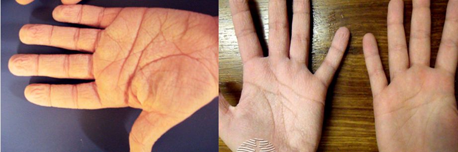
Paresthesia is a medical term that describes an abnormal sensation such as tingling, numbness, prickling, or burning, usually in the hands, feet, arms, or legs. These sensations can occur without any obvious cause, often described as "pins and needles" or falling asleep in a limb. However, persistent paresthesia can be a sign of an underlying medical condition, such as nerve damage, diabetes, multiple sclerosis, or a vitamin deficiency. It is important to consult with a healthcare professional if experiencing persistent paresthesia to determine the cause and appropriate treatment.
Nerve compression syndromes refer to a group of conditions characterized by the pressure or irritation of a peripheral nerve, causing various symptoms such as pain, numbness, tingling, and weakness in the affected area. This compression can occur due to several reasons, including injury, repetitive motion, bone spurs, tumors, or swelling. Common examples of nerve compression syndromes include carpal tunnel syndrome, cubital tunnel syndrome, radial nerve compression, and ulnar nerve entrapment at the wrist or elbow. Treatment options may include physical therapy, splinting, medications, injections, or surgery, depending on the severity and underlying cause of the condition.
The sphenoid bone is a complex, irregularly shaped bone located in the middle cranial fossa and forms part of the base of the skull. It articulates with several other bones, including the frontal, parietal, temporal, ethmoid, palatine, and zygomatic bones. The sphenoid bone has two main parts: the body and the wings.
The body of the sphenoid bone is roughly cuboid in shape and contains several important structures, such as the sella turcica, which houses the pituitary gland, and the sphenoid sinuses, which are air-filled cavities within the bone. The greater wings of the sphenoid bone extend laterally from the body and form part of the skull's lateral walls. They contain the superior orbital fissure, through which important nerves and blood vessels pass between the cranial cavity and the orbit of the eye.
The lesser wings of the sphenoid bone are thin, blade-like structures that extend anteriorly from the body and form part of the floor of the anterior cranial fossa. They contain the optic canal, which transmits the optic nerve and ophthalmic artery between the brain and the orbit of the eye.
Overall, the sphenoid bone plays a crucial role in protecting several important structures within the skull, including the pituitary gland, optic nerves, and ophthalmic arteries.
The hypoglossal nerve, also known as the 12th cranial nerve (CN XII), is primarily responsible for innervating the muscles of the tongue, allowing for its movement and function. These muscles include the intrinsic muscles that alter the shape of the tongue and the extrinsic muscles that position it in the oral cavity. The hypoglossal nerve also has some minor contributions to the innervation of two muscles in the neck: the sternocleidomastoid and the trapezius. These functions are related to head turning and maintaining head position. Any damage to this nerve can lead to weakness or paralysis of the tongue, causing difficulty with speech, swallowing, and tongue movements.
In medical terms, a "lip" refers to the thin edge or border of an organ or other biological structure. However, when people commonly refer to "the lip," they are usually talking about the lips on the face, which are part of the oral cavity. The lips are a pair of soft, fleshy tissues that surround the mouth and play a crucial role in various functions such as speaking, eating, drinking, and expressing emotions.
The lips are made up of several layers, including skin, muscle, blood vessels, nerves, and mucous membrane. The outer surface of the lips is covered by skin, while the inner surface is lined with a moist mucous membrane. The muscles that make up the lips allow for movements such as pursing, puckering, and smiling.
The lips also contain numerous sensory receptors that help detect touch, temperature, pain, and other stimuli. Additionally, they play a vital role in protecting the oral cavity from external irritants and pathogens, helping to keep the mouth clean and healthy.
The sublingual glands are a pair of salivary glands located in the floor of the mouth, beneath the tongue. They are the smallest of the major salivary glands and produce around 5-10% of the total saliva in the mouth. The sublingual glands secrete saliva containing electrolytes, enzymes (such as amylase), and antibacterial compounds that help in digestion, lubrication, and protection against microorganisms.
The sublingual glands' secretions are released through multiple small ducts called the ducts of Rivinus or minor sublingual ducts, as well as a larger duct called the duct of Wharton, which is a common excretory duct for both sublingual and submandibular glands.
Sublingual gland dysfunction can lead to conditions such as dry mouth (xerostomia), dental caries, or oral infections.
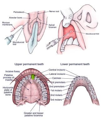
A third molar is the most posterior of the three molars present in an adult human dental arch. They are also commonly known as wisdom teeth, due to their late eruption period which usually occurs between the ages of 17-25, a time traditionally associated with gaining maturity and wisdom.
Anatomically, third molars have four cusps, making them the largest of all the teeth. However, not everyone develops third molars; some people may have one, two, three or no third molars at all. In many cases, third molars do not have enough space to fully erupt and align properly with the rest of the teeth, leading to impaction, infection, or other dental health issues. As a result, third molars are often extracted if they cause problems or if there is a risk they will cause problems in the future.
Tooth extraction is a dental procedure in which a tooth that is damaged or poses a threat to oral health is removed from its socket in the jawbone. This may be necessary due to various reasons such as severe tooth decay, gum disease, fractured teeth, crowded teeth, or for orthodontic treatment purposes. The procedure is performed by a dentist or an oral surgeon, under local anesthesia to numb the area around the tooth, ensuring minimal discomfort during the extraction process.
Tongue diseases refer to various medical conditions that affect the structure, function, or appearance of the tongue. These conditions can be categorized into several types, including:
1. Infections: Bacterial, viral, or fungal infections can cause tongue inflammation (glossitis), pain, and ulcers. Common causes include streptococcus, herpes simplex, and candida albicans.
2. Traumatic injuries: These can result from accidental bites, burns, or irritation caused by sharp teeth, dental appliances, or habitual habits like tongue thrusting or chewing.
3. Neoplasms: Both benign and malignant growths can occur on the tongue, such as papillomas, fibromas, and squamous cell carcinoma.
4. Congenital disorders: Some individuals may be born with abnormalities of the tongue, like ankyloglossia (tongue-tie) or macroglossia (enlarged tongue).
5. Neurological conditions: Certain neurological disorders can affect tongue movement and sensation, such as Bell's palsy, stroke, or multiple sclerosis.
6. Systemic diseases: Various systemic conditions can have symptoms that manifest on the tongue, like diabetes mellitus (which can cause dryness and furring), iron deficiency anemia (which may lead to atrophic glossitis), or Sjögren's syndrome (which can result in xerostomia).
7. Idiopathic: In some cases, the cause of tongue symptoms remains unknown, leading to a diagnosis of idiopathic glossitis or burning mouth syndrome.
Proper diagnosis and treatment of tongue diseases require a thorough examination by a healthcare professional, often involving a dental or medical specialist such as an oral pathologist, otolaryngologist, or dermatologist.
A lingual thyroid is a type of ectopic thyroid gland, which means that it is located in an abnormal position outside its usual location in the neck. In the case of a lingual thyroid, the gland is found on the base of the tongue. This condition is present at birth and occurs when the thyroid gland fails to migrate to its normal position during fetal development.
While some individuals with a lingual thyroid may not experience any symptoms, others may have problems such as difficulty swallowing, speaking, or breathing, depending on the size of the gland. In some cases, a lingual thyroid may also cause symptoms related to hypothyroidism if it is not functioning properly. Treatment for a lingual thyroid may include surgery to remove the gland or radioactive iodine therapy to destroy it.
The sciatic nerve is the largest and longest nerve in the human body, running from the lower back through the buttocks and down the legs to the feet. It is formed by the union of the ventral rami (branches) of the L4 to S3 spinal nerves. The sciatic nerve provides motor and sensory innervation to various muscles and skin areas in the lower limbs, including the hamstrings, calf muscles, and the sole of the foot. Sciatic nerve disorders or injuries can result in symptoms such as pain, numbness, tingling, or weakness in the lower back, hips, legs, and feet, known as sciatica.
The lingual frenum is a small fold of mucous membrane that attaches the tongue to the floor of the mouth. It contains muscle fibers and can vary in length, thickness, and attachment level. In some individuals, the lingual frenum may be too short or tight, restricting tongue movement, which is known as being "tongue-tied" or having ankyloglossia. This condition can potentially impact speech, feeding, and oral hygiene, although in many cases, it does not cause any significant problems.
Peripheral nerves are nerve fibers that transmit signals between the central nervous system (CNS, consisting of the brain and spinal cord) and the rest of the body. These nerves convey motor, sensory, and autonomic information, enabling us to move, feel, and respond to changes in our environment. They form a complex network that extends from the CNS to muscles, glands, skin, and internal organs, allowing for coordinated responses and functions throughout the body. Damage or injury to peripheral nerves can result in various neurological symptoms, such as numbness, weakness, or pain, depending on the type and severity of the damage.
The optic nerve, also known as the second cranial nerve, is the nerve that transmits visual information from the retina to the brain. It is composed of approximately one million nerve fibers that carry signals related to vision, such as light intensity and color, from the eye's photoreceptor cells (rods and cones) to the visual cortex in the brain. The optic nerve is responsible for carrying this visual information so that it can be processed and interpreted by the brain, allowing us to see and perceive our surroundings. Damage to the optic nerve can result in vision loss or impairment.
Nerve fibers are specialized structures that constitute the long, slender processes (axons) of neurons (nerve cells). They are responsible for conducting electrical impulses, known as action potentials, away from the cell body and transmitting them to other neurons or effector organs such as muscles and glands. Nerve fibers are often surrounded by supportive cells called glial cells and are grouped together to form nerve bundles or nerves. These fibers can be myelinated (covered with a fatty insulating sheath called myelin) or unmyelinated, which influences the speed of impulse transmission.
"Cat" is a common name that refers to various species of small carnivorous mammals that belong to the family Felidae. The domestic cat, also known as Felis catus or Felis silvestris catus, is a popular pet and companion animal. It is a subspecies of the wildcat, which is found in Europe, Africa, and Asia.
Domestic cats are often kept as pets because of their companionship, playful behavior, and ability to hunt vermin. They are also valued for their ability to provide emotional support and therapy to people. Cats are obligate carnivores, which means that they require a diet that consists mainly of meat to meet their nutritional needs.
Cats are known for their agility, sharp senses, and predatory instincts. They have retractable claws, which they use for hunting and self-defense. Cats also have a keen sense of smell, hearing, and vision, which allow them to detect prey and navigate their environment.
In medical terms, cats can be hosts to various parasites and diseases that can affect humans and other animals. Some common feline diseases include rabies, feline leukemia virus (FeLV), feline immunodeficiency virus (FIV), and toxoplasmosis. It is important for cat owners to keep their pets healthy and up-to-date on vaccinations and preventative treatments to protect both the cats and their human companions.
 Lingual nerve
Lingual branches of glossopharyngeal nerve
Medial pterygoid muscle
Mandibular canal
Maxillary artery
Submandibular duct
Dental extraction
Paul Coulthard
Neck-tongue syndrome
Pterygospinous ligament
Infratemporal fossa
Alveoloplasty
Mandibular setback surgery
Transverse muscle of tongue
External carotid artery
Lingual artery
Hypoglossal nerve
Inferior alveolar nerve anaesthesia
Local anesthetic
Inferior alveolar artery
Submandibular ganglion
Sensory loss
Superior pharyngeal constrictor muscle
Lingual branch
Brown Dog affair
Sialogogue
Advanced airway management
Sublingual space
Impacted wisdom teeth
Inferior alveolar nerve
Lingual nerve - Wikipedia
 Lingual nerve
 Oral Nerve Block: Overview, Indications, Contraindications
 Negligent Wisdom Tooth Extractions Can Result in Lingual Nerve Damage - Wagner Reese
 How can a visit to the dentist lead to lingual nerve damage? | MeehanLaw, LLC
 Interventions for iatrogenic inferior alveolar and lingual nerve injury: Cochrane systematic review | Cochrane Abstracts
 Blogborygmi: 02/01/2007 - 03/01/2007
"Nerve to mylohyoid branched from the lingual nerve: previously undescr" by Joe Iwanaga, Shogo Kikuta et al.
Blogborygmi: bye bye baby
Wisdom tooth extraction causing lingual nerve and styloglossus muscle damage: a mimic of multiple cranial nerve palsies |...
 CBD for lingual nerve pain. Delta 8 Delta 9 Oil/gummies. Quick reply for a viewer - JMAXONE PROUD AMERICAN
A case report of a long-term abandoned torn lingual nerve injury repaired by collagen nerve graft induced by lower third molar...
Oral Nerve Block: Overview, Indications, Contraindications
 A new treatment for lingual nerve injury: an anatomical feasibility study for using a buccal nerve pedicle graft. | Surg...
Complex Tongue Laceration: Overview, Indications, Contraindications
Facial Nerve Embryology: Overview, The Mature Facial Nerve, Overview of Hindbrain Development
 Bassett Collection - Lane Medical Library - Stanford University School of Medicine
Drooling (Sialorrhea): Practice Essentials, Problem, Epidemiology
After my wisdom teeth were extracted, I can't feel my tongue - Cosmetic Dentistry Blog
 IndexCat
 Can the Flu and Other Viruses Cause Neurodegeneration? | The Scientist Magazine®
 Best Portland, Oregon Lawyers | Best Lawyers
 Atlas of Oral and Maxillofacial Surgery - Elsevier E-Book on VitalSource, 2nd Edition - 9780323789653
 Frontiers | Time of Day Influences Psychophysical Measures in Women With Burning Mouth Syndrome
 Colonic Metastasis of Adenoid Cystic Carcinoma 19 Years after the Primary Tumor Resection
Plus it
 Can wisdom teeth cause permanent damage? (Explained)
 TRPM8: The Cold and Menthol Receptor - TRP Ion Channel Function in Sensory Transduction and Cellular Signaling Cascades - NCBI...
 About us | Clinical Dentistry | The University of Sheffield
Inferior alveolar20
- while passing between these two muscles, it is joined by the chorda tympani, and often by a communicating branch from the inferior alveolar nerve. (wikipedia.org)
- Here, the lingual nerve is anterior and somewhat medial (deep) to the inferior alveolar nerve. (wikipedia.org)
- Lingual and inferior alveolar nerve. (wikipedia.org)
- Cochrane Abstracts , Evidence Central , evidence.unboundmedicine.com/evidence/view/Cochrane/433705/all/Interventions_for_iatrogenic_inferior_alveolar_and_lingual_nerve_injury:_Cochrane_systematic_review. (unboundmedicine.com)
- It descends medial and anterior to the inferior alveolar nerve through the pterygomandibular space, runs by the lingual plate and lingual crest at the lower third molar closely, and supplies sensory fibers to the anterior two-thirds of the tongue. (providence.org)
- The inferior alveolar nerve gives rise to the nerve to mylohyoid just before entering the mandibular foramen, which supplies the mylohyoid and anterior belly of the digastric muscle. (providence.org)
- The inferior alveolar nerve provides sensation to the lip and chin and the lingual nerve provides sensation to the tongue. (sheffield.ac.uk)
- The courses of the inferior alveolar, facial, and lingual arteries and their branches are reviewed. (allenpress.com)
- The mandibular division exits through the foramen ovale (FO) and divides into the buccal, lingual, inferior alveolar, and auriculotemporal nerves. (asra.com)
- Radiographic signs, detectable on an orthopantomogram (OPG) indicating the presence of close relationship between the inferior alveolar nerve (IAN) and lower third molar requires further investigation to better understand its relevant course. (dentalnews.com)
- Coronectomy has been proposed as a valid treatment option to reduce the risk of inferior alveolar nerve (IAN) injury in selected cases. (dentalnews.com)
- This paper presents and highlights the shortcoming of panoramic imaging and elaborates the importance of cone beam CT as an important tool in visualising the course of inferior alveolar nerve in relation to lower third molars. (dentalnews.com)
- The inferior alveolar nerve (IAN) is a branch of the trigeminal nerve and plays a vital role in providing sensation to the lower lip. (reachmd.com)
- The most common dental nerve injuries are to the lingual nerve or the inferior alveolar nerve and occur during surgical procedures. (dentalandpodiatricmalpractice.com)
- The inferior alveolar nerve runs alongside the outside of the mouth. (dentalandpodiatricmalpractice.com)
- These 3D images allow clinicians to assess vital anatomical structures, such as the inferior alveolar nerve, mental foramen, lingual concavity of the posterior mandible, incisive canal, shape and pathological changes of the maxillary sinus, and the endodontic status of adjacent teeth. (dimensionsofdentalhygiene.com)
- Injury to the inferior alveolar nerve after removal of third molars occurs in 0.4 to 8.4% of cases, less than 1% permanent. (bvsalud.org)
- An inferior alveolar nerve block, the most common dental nerve block, anesthetizes the ipsilateral hemi-mandible (including teeth and bone), as well as the lateral (buccal) mucosa over the lower incisors, canine, and first premolar, and, cutaneously, the ipsilateral lower lip and chin. (msdmanuals.com)
- A buccal block (of the long buccal nerve) is often done as part of the inferior alveolar nerve block procedure, if anesthetization of the lateral (buccal) gingiva and mucosa of the lower molars and second premolar is needed. (msdmanuals.com)
- the inferior alveolar dental nerve block is the method most commonly used by endodontists to achieve local anesthesia during treatments. (bvsalud.org)
Experienced lingual nerve damage1
- If you experienced lingual nerve damage, you may want to consider your legal options. (ctdentalmalpractice.com)
Hypoglossal nerve3
- Avoiding lingual access when undertaking wisdom tooth surgery will also avoid unnecessary lingual nerve injury Lingual branches of hypoglossal nerve Mandible of human embryo 24 mm. long. (wikipedia.org)
- Hypoglossal nerve, cervical plexus, and their branches. (wikipedia.org)
- This then joins the sublingual vein and passes with the hypoglossal nerve between hypoglossus and mylohyoid muscles to drain into the internal jugular, facial, or lingual vein. (ispub.com)
Mandibular10
- It contains fibres from both the mandibular division of the trigeminal nerve (CN V3) and from the facial nerve (CN VII). (wikipedia.org)
- The lingual nerve arises from the posterior trunk of mandibular nerve (CN V3) within the infratemporal fossa. (wikipedia.org)
- citation needed] The lingual nerve supplies general somatic afferent (i.e. general sensory) innervation to the mucous membrane of the anterior two-thirds of the tongue (i.e. body of tongue) (whereas the posterior one-third (i.e. root of tongue) is innervated via the glossopharyngeal nerve (CN IX)[citation needed]), the floor of the oral cavity, and the mandibular/inferior lingual gingiva. (wikipedia.org)
- Mandibular division of the trifacial nerve. (wikipedia.org)
- Mandibular division of trifacial nerve, seen from the middle line. (wikipedia.org)
- Lingual nerve Lingual nerve Mandibular nerve and bone. (wikipedia.org)
- A sensory branch of the mandibular nerve , which is part of the trigeminal (5th cranial) nerve. (lookfordiagnosis.com)
- The lingual nerve is a branch of the mandibular division of the trigeminal nerve. (providence.org)
- It soon combines with the larger lingual nerve , a branch of the mandibular nerve (cranial nerve V 3 ). (wikidoc.org)
- Distribution of the maxillary and mandibular nerves, and the submaxillary ganglion. (wikidoc.org)
Trigeminal8
- The fibres from the trigeminal nerve are for touch, pain and temperature (general sensation), and the ones from the facial nerve are for taste (special sensation). (wikipedia.org)
- The sensory root (nervus intermedius) consists of (1) central projections of neurons located in the geniculate ganglion (general somatic fibers that synapse in the spinal nucleus of the trigeminal nerve and special afferent fibers that synapse in the nucleus solitarius) and (2) axons of parasympathetic neurons from the superior salivatory (lacrimal) nucleus. (medscape.com)
- The University of Sheffield Charles Clifford Dental Hospital is a world-leading centre for the repair of trigeminal nerves. (sheffield.ac.uk)
- Damage to the trigeminal nerve can be distressing and in some cases extremely painful. (sheffield.ac.uk)
- The function of the trigeminal nerve is to provide sensation and motor functions to the mouth and face. (sheffield.ac.uk)
- TN is characterized by recurrent short episodes of sharp, electrical shock like pain, typically abrupt in onset and termination, along the distribution of one or more divisions of the trigeminal nerve. (asra.com)
- The trigeminal nerve supplies the sensory innervation to the face as well as the sensory and motor innervation to the mastication muscles. (asra.com)
- My most significant settlements and awards have involved cases involving injuries to the Trigeminal nerve. (frankjriccio.com)
Tongue22
- The lingual nerve carries sensory innervation from the anterior two-thirds of the tongue. (wikipedia.org)
- it finally runs from laterally to medially inferiorly crossing the duct of the submandibular gland, and along the tongue to its tip becoming the sublingual nerve, lying immediately beneath the mucous membrane. (wikipedia.org)
- The lingual nerve also comes to convey fibres of the chorda tympani (which are derived from the facial nerve (CN VII)), which providee special sensation (taste) to the anterior two-thirds of the tongue as well as parasympathetic and sympathetic innervation. (wikipedia.org)
- This nerve runs through the tongue and controls the touch, temperature, and taste for the front two-thirds of the tongue. (wagnerreese.com)
- The lingual nerve-a nerve which is responsible for sensation in the lower lip, tongue and chin-can be damaged in a variety of different ways. (ctdentalmalpractice.com)
- Therefore, injury of this nerve is occasionally induced by wisdom tooth extraction and could lead to paralysis of the tongue. (providence.org)
- The combination of tongue hemianaesthesia, dysgeusia, dysarthria and dysphagia suggests the involvement of multiple cranial nerves. (bmj.com)
- Jenny, The lingual nerve, which is the nerve that goes to your tongue, runs along the inside of your jaw close to your wisdom tooth. (mynewsmile.com)
- If your entire tongue is numb, that means that both lingual nerves were damaged. (mynewsmile.com)
- Dear Doctor Hall, I had a root canal treatment which I was given anesthetic that caused me lingual nerve damage 2 months ago , my nerve is healing but now I am suffering from dry mouth feeling especially my tongue with no other symptoms at all , I did test for diabetes and thyroid and all came out normal, so please can you tell what is happening? (mynewsmile.com)
- My name is Mary too and I am suffering exactly like you, my tongue is healing after nerve damage during root canal treatment but I have dry mouth especially my tongue and no one is understanding what is going on. (mynewsmile.com)
- These include sore throat, laryngeal nerve palsy, lingual nerve palsy, alteration of taste/swallowing/ speech, rarely tongue cyanosis or tongue cyanosis with swelling. (ispub.com)
- The venous drainage of the tongue is via two main routes - dorsal lingual and deep lingual vein. (ispub.com)
- The dorsal lingual vein drains the dorsum and lateral aspects of the tongue and joins the lingual vein along side the lingual artery and finally drains into the internal jugular vein at or near the greater cornu of the hyoid bone. (ispub.com)
- The deep lingual vein commences at the tip of the tongue passes along the ventral surface just beneath the mucosa. (ispub.com)
- The lingual nerve transmits sensation from the floor of the mouth and most of the tongue. (dentalandpodiatricmalpractice.com)
- Because there are two branches of the lingual nerve, loss of sensation may be experienced on only one side of the tongue or mouth. (dentalandpodiatricmalpractice.com)
- Chorda tympani is a branch of the facial nerve (the seventh cranial nerve) that serves the taste buds in the front of the tongue , runs through the middle ear , and carries taste messages to the brain. (wikidoc.org)
- The chorda tympani appears to exert a particularly strong inhibitory influence on other taste nerves, as well as on pain fibers in the tongue. (wikidoc.org)
- Special sensory (taste) fibers also extend from the chorda tympani to the anterior 2/3rds of the tongue via the lingual nerve. (wikidoc.org)
- The case I worked on at the ATLA Damages seminar was a dental malpractice case where an oral surgeon severed my client's lingual nerve while extracting a lower wisdom tooth, leaving half of his tongue permanently numb. (trialguides.com)
- The lingual nerve lies nearby and is usually blocked incidentally, anesthetizing the ipsilateral floor of the mouth, medial (lingual) gingiva, and anterior two thirds of the tongue. (msdmanuals.com)
Injuries9
- citation needed] The most common cause of lingual nerve injuries is third molar (wisdom tooth) surgery, less commonly the lingual nerve can be injured by local anaesthetic dental injections (particularly inferior dental block injections) and sublingual or submandibular surgery. (wikipedia.org)
- Patients should be routinely warned about lingual nerve injuries prior to wisdom tooth and floor of mouth surgery. (wikipedia.org)
- failed verification] Infiltration dentistry is a technique that may reduce the possibility of lingual nerve injuries by avoiding deep injections. (wikipedia.org)
- Because of the importance of the lingual nerve, injuries to this nerve can have a significant impact on patients. (ctdentalmalpractice.com)
- Peripheral nerve injuries are generally associated with incomplete restoration of motor function. (vinomis.com)
- Peripheral nerve injuries represent a significant source of patient morbidity and disability (Asplund et al. (vinomis.com)
- This paper supports the routine use of CBCT as a preoperative decision-making tool for the removal of lower third molars to prevent IAN nerve injuries. (dentalnews.com)
- Coronectomy is an alternative procedure increasingly accepted world-wide to reduce the risk of nerve injuries 3,4,5 . (dentalnews.com)
- Besides, there are surgical complications like bleeding, nerve damage, injuries to adjacent teeth, fracture of maxillary tuberosity, displacement of the tooth to other anatomical structures and fracture of the dental apex 1 . (bvsalud.org)
Injury16
- Any injury to sensory nerves can result in pain, altered sensation and/or numbness, but usually a combination of all three symptoms arises. (wikipedia.org)
- Warning patients of nerve injury prior to administration of deep dental injections has a risk of injury in approximately 1:14,000 with 25% of these remaining persistent. (wikipedia.org)
- This reflects good practice recommended by the Royal College of Anaesthetists (prior warning of potential nerve injury in relation to spinal and epidural blocks 1 on 24-57,000 risk). (wikipedia.org)
- We present a case with sudden onset of these symptoms immediately following wisdom tooth extraction and highlight the clinical features that allowed localisation of the lesion to a focal, iatrogenic injury of the lingual nerve and adjacent styloglossus muscle. (bmj.com)
- A new treatment for lingual nerve injury: an anatomical feasibility study for using a buccal nerve pedicle graft. (bvsalud.org)
- Citation: Ding Z, Cao J, Shen Y, Zou Y, Yang X, Zhou W, Guo Q and Huang C (2018) Resveratrol Promotes Nerve Regeneration via Activation of p300 Acetyltransferase-Mediated VEGF Signaling in a Rat Model of Sciatic Nerve Crush Injury. (vinomis.com)
- The slow rate of nerve regeneration after injury may account for this. (vinomis.com)
- Although many benefits of resveratrol have been shown in the nervous system, it is not clear whether resveratrol could promote fast nerve regeneration and motor repair after peripheral nerve injury. (vinomis.com)
- This study showed that the motor deficits caused by sciatic nerve crush injury were alleviated by daily systematic resveratrol treatment within 10 days. (vinomis.com)
- Inactivation of p300 acetyltransferase reversed the resveratrol-induced expression of VEGFs and motor repair in rats that had undergone sciatic nerve crush injury. (vinomis.com)
- 2010). Although axons in peripheral nerves have the capacity to regenerate after injury, a number of clinical reports and studies in recent years have indicated that functional recovery, especially motor function, is far from satisfactory even with advances in surgical procedures (Ruijs et al. (vinomis.com)
- 2014). In vivo, VEGFs are expressed after peripheral nerve injury (Li et al. (vinomis.com)
- In another case, laryngeal nerve injury caused by LMA has been reported 2 . (ispub.com)
- This finding suggests that the human body has the potential for self-repair and compensation in cases of nerve injury. (reachmd.com)
- These findings provide valuable insights into the complex processes underlying sensory recovery after peripheral nerve injury. (reachmd.com)
- The implications of this study are significant for patients undergoing oral and maxillofacial surgery, as well as for the broader field of nerve injury regeneration and offers hope for patients who have experienced sensory loss in the lower lip due to IAN sacrifice or damage during mandibulectomy. (reachmd.com)
Buccal nerve2
- To our knowledge , there has been no study using a (long) buccal nerve (BN) graft as a donor for LN repair. (bvsalud.org)
- The experiments revealed the collateral compensation of the ipsilateral buccal nerve, which played a crucial role in the sensory innervation of the lower lip. (reachmd.com)
Palsy1
- Lingual nerve (LN) palsy is a serious complication in dentistry and repaired by direct suture or a free graft technique . (bvsalud.org)
Branch1
- Management of hemorrhage from a branch of the lingual or facial arteries may require an extraoral approach for ligation, because the mylohyoid, sublingual, and submental arteries can anastomose and be anatomically variable as well. (allenpress.com)
Damage23
- One of the most prominent risks during wisdom tooth extraction is damage to the lingual nerve. (wagnerreese.com)
- People who suffer lingual nerve damage during wisdom tooth extraction often suffer from permanent loss of sensation in their tongues, which can affect their ability to eat, drink, and speak. (wagnerreese.com)
- Unfortunately, some patients who have had the surgery now face a lifetime of lost sensation, taste, and control over their tongues due to nerve damage caused by negligence. (wagnerreese.com)
- If you or someone you love suffered lingual nerve damage during wisdom tooth extraction, we want to hear from you. (wagnerreese.com)
- How can a visit to the dentist lead to lingual nerve damage? (ctdentalmalpractice.com)
- In some cases, this could be a sign that your dental visit did serious damage to the nerves in your jaw. (ctdentalmalpractice.com)
- What commonly causes lingual nerve damage? (ctdentalmalpractice.com)
- Unfortunately, this leads wisdom tooth removal to be one of the most common reasons for lingual nerve damage, with up to 2% of wisdom tooth surgeries resulting in long-term or permanent damage. (ctdentalmalpractice.com)
- While less common than damage due to wisdom tooth removal, anesthetic injections can also injure the nerves of the lower jaw. (ctdentalmalpractice.com)
- The needles used for anesthesia can, in some cases, do damage to the nerves and cause bleeding, scarring or inflammation . (ctdentalmalpractice.com)
- How can lingual nerve damage impact your life? (ctdentalmalpractice.com)
- Nerve damage can limit patients' sense of taste or make it difficult for them to eat or speak. (ctdentalmalpractice.com)
- In my understanding lingual nerve damage usually effects either one side or the other. (mynewsmile.com)
- Lingual nerve damage after the extraction of a wisdom tooth can be either because the nerve is severed by the incision of the dentist, or because it is compressed and traumatized either during the surgery or as a result of the swelling afterward. (mynewsmile.com)
- However, a literature review of complication following the use of LMA's found reports of damage not only to recurrent laryngeal nerve but also to other adjacent nerves namely the hypoglossal and lingual nerve 3 . (ispub.com)
- If you have suffered dental nerve damage due to negligence, you may well be entitled to substantial compensation. (dentalandpodiatricmalpractice.com)
- Nerve damage during dental procedures is not as rare an occurrence as one might hope, and can negatively impact patients for the rest of their lives. (dentalandpodiatricmalpractice.com)
- While some types of minor nerve damage may heal within weeks or months, others result in permanent damage. (dentalandpodiatricmalpractice.com)
- Because dental nerve damage causes the sensation of an electrical shock, as well as numbness and pain, such nerve damage requires immediate treatment. (dentalandpodiatricmalpractice.com)
- Though in most cases these symptoms will gradually subside, if they do not, you may well be a victim of nerve damage caused by a negligent dentist. (dentalandpodiatricmalpractice.com)
- If you believe this may be true, get in touch with Lance Ehrenberg promptly to find out whether you are entitled to compensation for medical costs, lost income, pain and suffering, and other costs related to your nerve damage. (dentalandpodiatricmalpractice.com)
- Lingual nerve damage ranges from 0 to 23% 3 . (bvsalud.org)
- Hearing loss that occurs when sound enters the ear normally, but because of damage to the inner ear or the hearing nerve, sound isn't organized in a way that the brain can understand. (cdc.gov)
Artery3
- Uncontrolledbleeding from the lingual artery,if left unchecked, may cause anexpanding ecchymosis that could compromisethe airway and/or blood volumeand may result in fatality.REVIEW OF THE ANATOMYLingual arteryThe lingual artery arises from the externalcarotid artery between the superiorthyroid and facial arteries (Figures1, 2, and 3). (allenpress.com)
- We believe that in our case the laryngeal mask airway was occluding the patients' lingual artery bilaterally. (ispub.com)
- The cause of compression of the lingual artery may be due to malpositioning, size of LMA it self, or the cuff may also be a factor. (ispub.com)
Mandible2
- The nerve then comes to pass inferoanteriorly upon the medial pterygoid muscle towards the medial aspect of the ramus of mandible, eventually meeting the mandible at the junction of the ramus and body of mandible. (wikipedia.org)
- This article discusses what procedures to perform to obtund bleeding from1 of these arteries and the technique of performing an emergency tracheotomy.INTRODUCTIONhree arteries that providethe major blood supply tothe mandible are importantfor dental implantology.These are the lingual, facial,and inferior alveolararteries. (allenpress.com)
Medial1
- After exiting the internal auditory canal, the facial nerve enters the middle ear, where it bends posteriorly (first, or medial, genu) and courses horizontally through the middle ear. (medscape.com)
Afferent1
- At the level of the primary afferent nerve, the site at which thermal stimuli are converted into neuronal activity, temperature-sensitive members of the TRP channel family are found. (nih.gov)
Sensory nerves1
- In the early to mid-twentieth century, a number of laboratories began to observe cold-induced electrical impulses when recording from mammalian sensory nerves. (nih.gov)
Chorda tympani5
- The chorda tympani is a nerve that branches from the facial nerve (cranial nerve VII) inside the facial canal , just before the facial nerve exits the skull via the stylomastoid foramen . (wikidoc.org)
- The chorda tympani is part of one of three cranial nerves that are involved in taste. (wikidoc.org)
- When the chorda tympani is damaged, its inhibitory function is disrupted, leading to less inhibited activity in the other nerves. (wikidoc.org)
- Rather than leave the skull with the facial nerve, the chorda tympani travels through the middle ear , where it runs from posterior to anterior across the tympanic membrane . (wikidoc.org)
- The fibers of the chorda tympani travel with the lingual nerve to the submandibular ganglion . (wikidoc.org)
Infratemporal fossa1
- The nerve continues through the petrotympanic fissure , after which it emerges from the skull into the infratemporal fossa . (wikidoc.org)
Submandibular ganglion1
- citation needed] The submandibular ganglion is suspended by two nerve filaments from the lingual nerve. (wikipedia.org)
Facial nerve11
- In order to appreciate the complex embryology of the facial nerve, one has to have a basic understanding of cranial embryology as a whole. (medscape.com)
- This may help prepare the otolaryngologist to comprehend and anticipate variations encountered in clinical practice, such as anticipating facial nerve anomalies in congenital stapes fixation. (medscape.com)
- [ 1 ] However, the main objective of this article is to outline the embryology of the facial nerve and its common clinical implications. (medscape.com)
- The reader is referred to Embryology and Anomalies of the Facial Nerve and Their Surgical Implications, 2nd Ed for a more comprehensive review of the development of the facial nerve and the associated development of the ear (see table 1). (medscape.com)
- The surgical anatomy and landmarks of the facial nerve. (medscape.com)
- Schematic illustration shows the facial nerve and its peripheral connections. (medscape.com)
- While studying the embryology of the facial nerve, keep in mind the mature course and structure that is the end result of developmental events. (medscape.com)
- The motor nucleus of the facial nerve is located in the reticular formation of the caudal pons. (medscape.com)
- Just anterior to the lateral aspect of the horizontal semicircular canal, the facial nerve curves gently (the second genu) to form the vertical, or mastoid, segment that exits via the stylomastoid foramen. (medscape.com)
- The development of the hindbrain (rhombencephalon) and the subsequent delamination of the neural crest cells are interrelated processes that need to be understood to appreciate the development of the branchiomotor cranial nerves in general and the facial nerve in particular. (medscape.com)
- The course and connections of the facial nerve in the temporal bone. (wikidoc.org)
Extraction1
- The lingual nerve can be damaged during wisdom tooth extraction by dental tools or even the sharp surface of the tooth. (wagnerreese.com)
Anesthetic2
- Mary - Your lingual nerve would have been scratched by the needle that was used for your local anesthetic injection. (mynewsmile.com)
- A nerve block may be preferred to local anesthetic infiltration when accurate approximation of wound edges is important (eg, skin or lip repair), because a nerve block does not distort the tissue as does local infiltration. (msdmanuals.com)
Cranial nerve2
- Note the interconnections of cranial nerve (CN) VII with CN V, CN IX, and CN X. (medscape.com)
- Upon leaving the motor nucleus, axons extend dorsally and medially, cranially and superficially, to bend around the abducens (sixth cranial nerve) nucleus. (medscape.com)
Commonly1
- This article highlights the indications, equipment, contraindications, complications, and approach to performing the more commonly used oral nerve blocks. (medscape.com)
Anatomical1
- We present an extremely rare anatomical variation where the nerve to mylohyoid arose from the lingual nerve near the submandibular duct during routine oral dissection. (providence.org)
Surgical procedures1
- These nerves may be injured during surgical procedures such as wisdom tooth removal or corrective jaw surgery. (sheffield.ac.uk)
Mucosa1
- la articaína mostró mayor efecto anestésico en mucosa vestibular (88,9%) y punta de lengua (55,6%), en comparación con la lidocaína. (bvsalud.org)
Middle ear1
- Three surgical approaches by an otologist can help to decrease salivary flow, including removal of the salivary glands, ligation of the salivary ducts, and sectioning of the nerves in the middle ear, which are responsible for salivary production. (medscape.com)
Patients1
- Younger patients showed a faster recovery time, while the preservation of ipsilateral buccal and lingual nerves was associated with more favorable outcomes. (reachmd.com)
Anatomy1
- An oral nerve block is a simple and effective way to manage orofacial pain without distorting the anatomy of a wound and without the use of narcotics. (medscape.com)
Systematic1
- The above results indicated that daily systematic resveratrol treatment promoted nerve regeneration and led to rapid motor repair. (vinomis.com)
Involvement1
- The primary tumor was 4 cm in size, with multiple positive margins and lingual nerve involvement. (hindawi.com)
Relation1
- Paramolars are rudimentary and are located in the lingual or vestibular region in relation to the alignment of the molars 8 . (bvsalud.org)
Lower lip1
- The expression of ApoD was significantly up-regulated in the lower lip after nerve transection and was found to promote axonal elongation and neurite formation. (reachmd.com)
Morbidity1
- However, this approach has limitations, including donor site morbidity and limited nerve supply, resulting in unsatisfactory recovery outcomes. (reachmd.com)
Spinal1
- Resveratrol activated p300 acetyltransferase-mediated VEGF signaling in the affected ventral spinal cord, which may have thus contributed to the acceleration of nerve regeneration and motor repair. (vinomis.com)