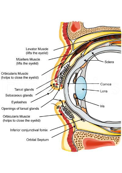
Limbus Corneae
Vittaforma
Encephalitozoon
Arcus Senilis
Microsporidia
Microsporida
Epithelium, Corneal
Cornea
Conjunctiva
Sclera
Corneal Transplantation
Burns, Chemical
Corneal Stroma
Keratin-12
Corneal Neovascularization
Stem Cells
Adenocarcinoma, Sebaceous
Amnion
Fluorescent Antibody Technique, Indirect
Uvea
Iris
Stem Cell Niche
Eye
Cochlea
Ciliary Body
Stria Vascularis
Conjunctival-limbal autografts for primary and recurrent pterygia: technique and results. (1/345)
Our technique of pterygium excision with conjunctival-limbal autografting is described and the safety and efficacy of the procedure in India is analysed. Case records of 51 consecutive patients (53 eyes) who underwent surgery at our institute between November 1992 and September 1994 were retrospectively analysed. Recurrence was defined as fibrovascular tissue crossing the corneoscleral limbus onto clear cornea in the area of previous pterygium excision. 2 (3.8%) of the 53 pterygia (primary 36; recurrent 17) recurred, after a mean follow up of 18.9 +/- 12.1 months (range: 1.5-43 months). Both recurrences occurred within a year of follow up, in patients who were < or = 40 years of age. No major operative or postoperative complications were encountered. The inclusion of limbal tissue in conjunctival autografts following pterygium excision appears to be essential to ensure low recurrence rates. The technique is safe, simple and inexpensive and is recommended for the management of both primary and recurrent pterygia in Indian eyes. (+info)Expression of cell adhesion molecules on limbal and neovascular endothelium in corneal inflammatory neovascularization. (2/345)
PURPOSE: To investigate the expression of cell-adhesion molecules on corneolimbal and neovascular endothelium and the associated leukocyte infiltration in an experimental model of inflammatory corneal neovascularization (NV). METHODS: Corneal NV was induced in BALB/c mice by placement of nylon sutures. Interleukin-1 receptor antagonist (IL-1ra) was used topically to determine whether suppression of IL-1 could affect adhesion molecule expression and leukocytic infiltration. At set time points, corneal samples were analyzed immunohistochemically for expression of P-selectin, E-selectin, intercellular adhesion molecule (ICAM)-1, vascular adhesion molecule (VCAM)-1, and platelet- endothelial adhesion molecule (PECAM)-1. Leukocytic infiltration at different time points was quantified histologically. In companion experiments mice deficient in ICAM-1 were investigated to determine the functional relevance of this molecule in corneal leukocyte infiltration. RESULTS: Significant enhanced expression of ICAM-1 was detected on the corneolimbal vascular endothelium as early as 8 hours and on the newly formed corneal NV by day 3, and treatment with IL-1ra led to significant suppression of this expression. IL-1ra-induced suppression of ICAM-1 expression was accompanied by a profound decrease in corneal leukocytic infiltration by 44.6% at day 1 (P < 0.003), 71.8% at day 3 (P < 0.001), 60.1% at day 7 (P < 0.001), and 63.8% at day 14 (P < 0.001), compared with control corneas. Similarly, in ICAM-1 knockout mice, the corneal leukocytic infiltration was 50.3%, 52.9%, and 36.4%, compared with wild-type control animals on day 1 (P < 0.001), day 7 (P < 0.005), and day 14 (P < 0.001), respectively. Expression of PECAM-1 was constitutively present on perilimbal vascular endothelium and had no response to IL-1ra treatment. No significant expression of P-selectin, E-selectin, or VCAM-1 was detected in this experimental model. CONCLUSIONS: These results suggest that leukocytic infiltration in this model of inflammatory corneal NV is closely associated with ICAM-1 expression, and that topical IL-1ra displays corneal anti-inflammatory effects, largely by suppressing ICAM-1 expression on vascular endothelial cells. (+info)Evidence of long-term survival of donor-derived cells after limbal allograft transplantation. (3/345)
PURPOSE: Severe destruction of the corneal limbus causes conjunctival invasion and subsequent visual loss. Limbal allograft transplantation (LAT) was recently proposed for the treatment of these disorders. However, whether the method functions as a stem cell transplantation of the corneal epithelium remains unclear. This study provided evidence that donor-derived corneal epithelial cells survive long after LAT. METHODS: Epithelial cells on the paracentral cornea in patients who have undergone LAT were subjected to fluorescence in situ hybridization (FISH) and polymerase chain reaction restriction fragment length polymorphism (RFLP) analysis. X and Y chromosomes were detected using sex chromosome-specific probes in the FISH analysis, and HLA-DPBI antigens were examined in the RFLP analysis. Eyes receiving conventional penetrating keratoplasty (PKP) served as controls. RESULTS: Donor-derived epithelial cells were detected in three of five eyes (60.0%) in the FISH analysis and in seven of nine eyes (77.8%) in the RFLP analysis. Among these eyes, one and three eyes in the FISH and RFLP analysis, respectively, had both donor- and recipient-derived cells. In control PKP eyes, none of the eyes in the FISH analysis and one of eight eyes (12.5%) in the RFLP analysis had donor-derived cells. CONCLUSIONS: These results suggest that donor-derived cells survive much longer after LAT than those after PKP, and that LAT may function as stem cell transplantation of the corneal epithelium. (+info)Inhibition of vascular endothelial cell morphogenesis in cultures by limbal epithelial cells. (4/345)
PURPOSE: To study the in vitro angiogenic activity of human conjunctival and limbal epithelial cells and conjunctival, limbal, and corneal fibroblasts in a three-cell-type coculture model. METHODS: Human umbilical vein endothelial cells (EC) were cocultured with epithelial cells, fibroblasts, or epithelial cells and fibroblasts to test their effect on EC morphogenesis. Neutralizing antibodies to some known angiogenic factors were added to the culture to see whether the EC morphogenesis may be blocked by a particular antibody. RESULTS: Conjunctival and limbal epithelial cells exhibited very little or no stimulatory effect on EC tube formation when examined in an EC- epithelial cell coculture system. In contrast, conjunctival, limbal, and corneal fibroblasts all promoted EC morphogenesis when examined under the same culture conditions. Fibroblast-induced EC morphogenesis was inhibited by addition of anti-vascular endothelial growth factor (VEGF) and/or anti-basic fibroblast growth factor (bFGF) antibodies to the culture medium. In the three-cell-type coculture system consisting of ECs, fibroblasts, and epithelial cells, limbal epithelial cells (but not conjunctival epithelial cells) exhibited a strong inhibitory effect on fibroblast-induced EC tube formation. CONCLUSIONS: The proangiogenic activity of ocular surface fibroblasts is probably mediated through a paracrine mechanism by VEGF and bFGF. Limbal epithelial cells, but not conjunctival epithelial cells, inhibit fibroblast-stimulated angiogenesis. (+info)Allo-limbal transplantation in patients with limbal stem cell deficiency. (5/345)
AIM: To report the outcome of a series of patients with stem cell deficiency who underwent allo-limbal transplantation and to describe a technique for this procedure. METHODS: Six consecutive patients underwent allo-limbal stem cell transplantation. The primary diagnosis included alkali burn (n = 2), trachoma (n = 1), chronic rosacea blepharitis and kerato-conjunctivitis (n = 1), aniridia (n = 1), and Stevens-Johnson syndrome (n = 1). The limbal rim consisted of peripheral cornea and perilimbal sclera. FK-506 was used postoperatively for immunosuppression. RESULTS: The length of follow up ranged from 3 to 24 months (mean follow up 11.8 (SD 9.3) months). The outcome was considered satisfactory in five of six cases. The corneal surface was completely epithelialised within 2 weeks, and there was a substantial improvement in vision and symptoms. One patient had recurrent epithelial defects related to eyelid abnormalities. No side effects associated with systemic immunosuppression were noted. CONCLUSION: Allo-limbal transplantation, with systemic immunosuppression with FK-506 is useful in reconstruction of the ocular surface with improvement in vision in patients with severe stem cell deficiency. (+info)Expression of a single pair of desmosomal glycoproteins renders the corneal epithelium unique amongst stratified epithelia. (6/345)
PURPOSE: To determine desmosomal glycoprotein isoform expression in bovine corneal, limbal, and conjunctival epithelium and desmosomal profile and distribution during corneal re-epithelialization. METHODS: Immunofluorescence (IF) for desmosomal components on cryostat sections of fresh epithelia was supported by immunoblot analysis of tissue lysates. Wounded corneas maintained in organ culture were examined by IF at times up to full re-epithelialization (96 hours). RESULT: Immunofluorescence for desmoplakin confirmed desmosome presence throughout all three epithelia. Plakoglobin was also ubiquitous. Of the desmosomal glycoproteins, desmocollin 2 (Dsc2) and desmoglein 2 (Dsg2) were expressed throughout, but Dsc3 and Dsg3 were confined to the limbus and conjunctiva, and Dscl and Dsgl were absent. Dsc2 and Dsg2 IFs were stronger in superficial layers, but Dsc3 and Dsg3 were stronger basally, fading suprabasally. Glycoprotein expression in cornea and conjunctiva was confirmed by immunoblot analysis. No change in glycoprotein expression occurred during re-epithelialization. CONCLUSIONS: Uniquely among stratified epithelia, cornea expresses only a single pair of desmosomal glycoproteins, Dsc2 and Dsg2. Expression of Dsc3 and Dsg3 in limbus and conjunctiva coincides with their association with cell proliferation in other epithelia, but corneal epithelial cells did not express Dsc3 or Dsg3 during re-epithelialization. Absence of Dscl and Dsgl correlates with lack of keratinization in ocular epithelia. These expression patterns may have significance for the specific properties and differentiation patterns of the epithelia. Presence of desmosomes throughout re-epithelialization raises the question of how migrating cells mutually re-position. (+info)Detection of gonioscopically occludable angles and primary angle closure glaucoma by estimation of limbal chamber depth in Asians: modified grading scheme. (7/345)
AIM: To evaluate the performance of limbal chamber depth estimation as a means of detecting occludable drainage angles and primary angle closure, with or without glaucoma, in an east Asian population, and determine whether an augmented grading scheme would enhance test performance. METHOD: A two phase, cross sectional, community based study was conducted on rural and urban areas of Hovsgol and Omnogobi provinces, Mongolia. 1800 subjects aged 40 to 93 years were selected and 1717 (95%) of these were examined. Depth of the anterior chamber at the temporal limbus was graded as a percentage fraction of peripheral corneal thickness. An "occludable" angle was one in which the trabecular meshwork was seen in less than 90 degrees of the angle circumference by gonioscopy. Primary angle closure (PAC) was diagnosed in subjects with an occludable angle and either raised pressure or peripheral anterior synechiae. PAC with glaucoma (PACG) was diagnosed in cases with an occludable angle combined with glaucomatous optic neuropathy and consistent visual morbidity. RESULTS: Occludable angles were identified in 140 subjects, 28 of these had PACG. The 15% grade (equivalent to the traditional "grade 1") yielded sensitivity and specificity of 84% and 86% respectively for the detection of occludable angles. The 5% grade gave sensitivity of 91% and specificity of 93% for the detection of PACG. The interobserver agreement for this augmented grading scheme was good (weighted kappa 0.76). CONCLUSIONS: The traditional limbal chamber depth grading scheme offers good performance for detecting occludable drainage angles in this population. The augmented scheme gives enhanced performance in detection of established PACG. The augmented scheme has potential for good interobserver agreement. (+info)Autologous limbal transplantation in patients with unilateral corneal stem cell deficiency. (8/345)
AIM: To describe a surgical technique for autologous limbal stem cell transplantation and the outcome of a series of patients with unilateral stem cell deficiency. METHODS: A report of six consecutive patients who underwent autologous limbal stem cell transplantation is presented. The primary diagnosis included alkali burn (n=3), conjunctival intraepithelial neoplasia (CIN) (n=1), recurrent pterygium (n=1), and contact lens induced keratopathy (n=1). The autologous transplanted tissue consisted of peripheral cornea, limbus, and conjunctiva obtained from the contralateral eye. Three of the above patients underwent penetrating keratoplasty in association with auto-limbal transplantation. A significant modification to established techniques was the close monitoring of conjunctival epithelial migration in the immediate postoperative period. If conjunctival epithelium threatened to migrate on to the corneal surface, it was mechanically removed at the slit lamp and prevented from crossing the limbus. This was required in three patients. RESULTS: The mean follow up was 18.8 months. The outcome was satisfactory in all cases: a stable corneal surface was restored and there was a substantial improvement in vision and symptoms. One patient had a primary failure of the corneal allograft associated with glaucoma, and 6 months later developed a retinal detachment. No complications were noted in the donor eye with the exception of one patient who developed filamentary keratitis along the edge of the donor site. CONCLUSION: Autologous limbal transplantation with corneal, limbal, and conjunctival carriers was found to be useful for ocular surface reconstruction, over a mid-term follow up, in patients with unilateral stem cell deficiency. Close monitoring of the migration of conjunctival epithelium in the immediate postoperative period, and preventing it from crossing the limbus, ensured that the corneal surface was re-epithelialised exclusively from epithelial cells derived from the transplanted limbal tissue. This approach should improve the success of this procedure. (+info)The limbus cornea, also known as the corneoscleral junction, is the border between the transparent cornea and the opaque sclera in the eye. It's a circular, narrow region that contains cells called limbal stem cells, which are essential for maintaining the health and clarity of the cornea. These stem cells continuously regenerate and differentiate into corneal epithelial cells, replacing the outermost layer of the cornea. Any damage or disorder in this area can lead to vision impairment or loss.
I'm sorry for any confusion, but "Vittaforma" is not a term commonly used in medical definitions. It appears to be a genus of ciliates, which are protozoan organisms with hair-like structures called cilia. If you have any more context or information about where you encountered this term, I'd be happy to help further!
Encephalitozoon is a genus of intracellular parasites belonging to the phylum Microspora. The two species that are most relevant to human health are Encephalitozoon cuniculi and Encephalitozoon intestinalis (previously known as Septata intestinalis). These microscopic organisms are capable of infecting a wide range of hosts, including humans, and are often associated with opportunistic infections in immunocompromised individuals.
E. cuniculi is well-known for causing encephalitozoonosis, a disease that can lead to various symptoms depending on the infected organ. In humans, it primarily affects the central nervous system (CNS), leading to neurological issues such as seizures, cognitive impairment, and motor function loss. E. intestinalis, on the other hand, tends to infect the gastrointestinal tract, causing diarrhea and wasting syndrome.
Transmission of these parasites typically occurs through the ingestion of spores present in contaminated food, water, or soil. Once inside a host, the spores germinate and invade various cells, including intestinal epithelial cells, hepatocytes, and endothelial cells. The subsequent infection can lead to a range of clinical manifestations, from asymptomatic to severe, life-threatening disease.
Effective treatment for encephalitozoonosis involves the administration of antiparasitic drugs such as albendazole or nitazoxanide. In immunocompromised patients, improving immune function through appropriate therapy is also crucial to prevent recurrence and manage the infection effectively.
Arcus senilis is a medical term that refers to the gray or white discoloration that forms around the outer edge (periphery) of the cornea, which is the clear, dome-shaped surface at the front of the eye. This condition is caused by the accumulation of cholesterol and other lipids in the corneal tissue, and it is more commonly seen in older adults over the age of 60.
Arcus senilis itself does not typically affect vision or cause any symptoms, but it can be a sign of underlying health issues such as high cholesterol levels or coronary artery disease. In some cases, the presence of arcus senilis may prompt doctors to recommend further testing to assess the patient's cardiovascular health.
It is important to note that arcus senilis should not be confused with arcus juvenilis, which is a similar condition that affects younger people and can be a sign of high cholesterol levels or other medical issues.
Microsporidia are a group of small, obligate intracellular parasites that belong to the kingdom Fungi. They are characterized by their spore stage, which contains a unique infection apparatus called the polar tube or coiled filament. These spores can infect a wide range of hosts, including humans, animals, and insects.
In humans, Microsporidia can cause chronic diarrhea and other gastrointestinal symptoms, particularly in individuals with weakened immune systems, such as those with HIV/AIDS. They can also infect various other tissues, including the eye, muscle, and kidney, leading to a variety of clinical manifestations.
Microsporidia were once considered to be protozoa but are now classified as fungi based on genetic and biochemical evidence. There are over 1,300 species of Microsporidia, with at least 14 species known to infect humans.
Microsporidiosis is an infection caused by microscopic, single-celled parasites belonging to the phylum Microspora. These parasites are primarily intracellular and can infect various organisms, including humans. Infection typically occurs through ingestion of spores present in contaminated food, water, or soil, or through inhalation of spores. Once inside a host, the spores germinate, releasing the infective sporoplasm that invades host cells and multiplies within them.
In humans, microsporidiosis can cause various symptoms depending on the species involved and the immune status of the host. In immunocompetent individuals, it may present as self-limiting diarrhea or mild gastrointestinal disturbances. However, in immunocompromised patients (e.g., those with HIV/AIDS, organ transplants, or using immunosuppressive medications), microsporidiosis can lead to severe and chronic diarrhea, wasting, and potentially life-threatening complications affecting various organs such as the eyes, kidneys, and respiratory system.
Diagnosis of microsporidiosis typically involves detecting the parasites in stool or tissue samples using specialized staining techniques (e.g., chromotrope stains) or molecular methods (e.g., PCR). Treatment usually includes antiparasitic drugs such as albendazole, which has activity against many microsporidian species. In severe cases or when the infection involves multiple organs, additional supportive care and management of underlying immunodeficiencies may be necessary.
Microsporidia are a group of small, spore-forming, obligate intracellular parasites that were once considered to be primitive protozoans but are now classified within the fungi. They are characterized by a unique infection mechanism called "polysporous invasion," where a single spore can infect multiple host cells and produce numerous progeny spores.
Microsporidia infect a wide range of hosts, including insects, fish, birds, and mammals, including humans. In humans, microsporidiosis is an opportunistic infection that primarily affects immunocompromised individuals, such as those with HIV/AIDS, organ transplant recipients, and those undergoing chemotherapy.
The most common Microsporidia species that infect humans are Enterocytozoon bieneusi and Encephalitozoon intestinalis, which can cause gastrointestinal symptoms such as diarrhea, abdominal pain, and weight loss. Other species can infect various organs, including the eyes, muscles, and respiratory system, causing a range of clinical manifestations.
Microsporidia have a complex life cycle that involves several developmental stages, including spores, meronts, and sporonts. The spores are highly resistant to environmental stresses and can survive for long periods outside the host, facilitating their transmission. Once inside the host cell, the spore releases its infectious contents, including a coiled tubular structure called the polar filament, which penetrates the host cell membrane and injects the parasite's genetic material into the host cytoplasm. The parasite then undergoes rapid multiplication, eventually producing numerous progeny spores that are released into the environment upon host cell lysis.
Microsporidia have been identified as potential bioterrorism agents due to their high infectivity, environmental resistance, and ability to cause severe disease in immunocompromised hosts. However, there are currently no effective vaccines or specific antimicrobial therapies available for microsporidiosis, and treatment is mainly supportive, focusing on managing symptoms and improving immune function.
The corneal epithelium is the outermost layer of the cornea, which is the clear, dome-shaped surface at the front of the eye. It is a stratified squamous epithelium, consisting of several layers of flat, scale-like cells that are tightly packed together. The corneal epithelium serves as a barrier to protect the eye from microorganisms, dust, and other foreign particles. It also provides a smooth surface for the refraction of light, contributes to the maintenance of corneal transparency, and plays a role in the eye's sensitivity to touch and pain. The corneal epithelium is constantly being renewed through the process of cell division and shedding, with new cells produced by stem cells located at the limbus, the border between the cornea and the conjunctiva.
The cornea is the clear, dome-shaped surface at the front of the eye. It plays a crucial role in focusing vision. The cornea protects the eye from harmful particles and microorganisms, and it also serves as a barrier against UV light. Its transparency allows light to pass through and get focused onto the retina. The cornea does not contain blood vessels, so it relies on tears and the fluid inside the eye (aqueous humor) for nutrition and oxygen. Any damage or disease that affects its clarity and shape can significantly impact vision and potentially lead to blindness if left untreated.
The conjunctiva is the mucous membrane that lines the inner surface of the eyelids and covers the front part of the eye, also known as the sclera. It helps to keep the eye moist and protected from irritants. The conjunctiva can become inflamed or infected, leading to conditions such as conjunctivitis (pink eye).
The sclera is the tough, white, fibrous outer coating of the eye in humans and other vertebrates, covering about five sixths of the eyeball's surface. It provides protection for the delicate inner structures of the eye and maintains its shape. The sclera is composed mainly of collagen and elastic fiber, making it strong and resilient. Its name comes from the Greek word "skleros," which means hard.
Corneal diseases are a group of disorders that affect the cornea, which is the clear, dome-shaped surface at the front of the eye. The cornea plays an important role in focusing vision, and any damage or disease can cause significant visual impairment or loss. Some common types of corneal diseases include:
1. Keratoconus: A progressive disorder in which the cornea thins and bulges outward into a cone shape, causing distorted vision.
2. Fuchs' dystrophy: A genetic disorder that affects the inner layer of the cornea called the endothelium, leading to swelling, cloudiness, and decreased vision.
3. Dry eye syndrome: A condition in which the eyes do not produce enough tears or the tears evaporate too quickly, causing discomfort, redness, and blurred vision.
4. Corneal ulcers: Open sores on the cornea that can be caused by infection, trauma, or other factors.
5. Herpes simplex keratitis: A viral infection of the cornea that can cause recurrent episodes of inflammation, scarring, and vision loss.
6. Corneal dystrophies: Inherited disorders that affect the structure and clarity of the cornea, leading to visual impairment or blindness.
7. Bullous keratopathy: A condition in which the endothelium fails to pump fluid out of the cornea, causing it to swell and form blisters.
8. Corneal trauma: Injury to the cornea caused by foreign objects, chemicals, or other factors that can lead to scarring, infection, and vision loss.
Treatment for corneal diseases varies depending on the specific condition and severity of the disease. Options may include eyedrops, medications, laser surgery, corneal transplantation, or other treatments.
Eye burns typically refer to injuries or damage to the eyes caused by exposure to harmful substances, extreme temperatures, or radiation. This can result in a variety of symptoms, including redness, pain, tearing, swelling, and blurred vision.
Chemical eye burns can occur when the eyes come into contact with strong acids, alkalis, or other irritants. These substances can cause damage to the cornea, conjunctiva, and other structures of the eye. The severity of the burn will depend on the type and concentration of the chemical, as well as the length of time it was in contact with the eye.
Thermal eye burns can result from exposure to hot or cold temperatures, such as steam, flames, or extreme cold. These types of burns can cause damage to the surface of the eye and may require medical attention to prevent further complications.
Radiation eye burns can occur after exposure to high levels of ultraviolet (UV) light, such as from welding torches, sun lamps, or tanning beds. Prolonged exposure to these sources can cause damage to the cornea and other structures of the eye, leading to symptoms like pain, redness, and sensitivity to light.
If you experience symptoms of an eye burn, it is important to seek medical attention as soon as possible. Treatment may include flushing the eyes with water or saline solution, administering medication to relieve pain and inflammation, or in severe cases, surgery to repair damaged tissue.
Corneal transplantation, also known as keratoplasty, is a surgical procedure in which all or part of a damaged or diseased cornea is replaced with healthy corneal tissue from a deceased donor. The cornea is the clear, dome-shaped surface at the front of the eye that plays an important role in focusing vision. When it becomes cloudy or misshapen due to injury, infection, or inherited conditions, vision can become significantly impaired.
During the procedure, the surgeon carefully removes a circular section of the damaged cornea and replaces it with a similarly sized piece of donor tissue. The new cornea is then stitched into place using very fine sutures that are typically removed several months after surgery.
Corneal transplantation has a high success rate, with more than 90% of procedures resulting in improved vision. However, as with any surgical procedure, there are risks involved, including infection, rejection of the donor tissue, and bleeding. Regular follow-up care is essential to monitor for any signs of complications and ensure proper healing.
Chemical burns are a type of tissue injury that results from exposure to strong acids, bases, or other corrosive chemicals. These substances can cause damage by reacting chemically with the skin or other tissues, leading to destruction of cells and potentially serious harm. The severity of a chemical burn depends on several factors, including the type and concentration of the chemical, the duration of exposure, and the amount of body surface area affected.
Chemical burns can occur through direct contact with the skin or eyes, inhalation of toxic fumes, or ingestion of harmful substances. Symptoms may include redness, pain, blistering, swelling, and irritation at the site of contact. In severe cases, chemical burns can lead to scarring, disability, or even death.
Immediate medical attention is required for chemical burns, as they can continue to cause damage until the source of the injury is removed, and appropriate first aid measures are taken. Treatment typically involves thorough cleaning and irrigation of the affected area, followed by administration of pain medication and other supportive care as needed. In some cases, skin grafting or other surgical interventions may be required to promote healing and minimize scarring.
The corneal stroma, also known as the substantia propria, is the thickest layer of the cornea, which is the clear, dome-shaped surface at the front of the eye. The cornea plays a crucial role in focusing vision.
The corneal stroma makes up about 90% of the cornea's thickness and is composed of parallel bundles of collagen fibers that are arranged in regular, repeating patterns. These fibers give the cornea its strength and transparency. The corneal stroma also contains a small number of cells called keratocytes, which produce and maintain the collagen fibers.
Disorders that affect the corneal stroma can cause vision loss or other eye problems. For example, conditions such as keratoconus, in which the cornea becomes thin and bulges outward, can distort vision and make it difficult to see clearly. Other conditions, such as corneal scarring or infection, can also affect the corneal stroma and lead to vision loss or other eye problems.
Keratin-1
"Biological dressings" refer to materials used in wound healing that are derived from biological sources, such as living cells, tissues, or extracellular matrix components. These dressings can be used to promote the regeneration and repair of damaged or injured tissues by providing a supportive environment for cell growth, differentiation, and tissue formation.
Biological dressings may be derived from various sources, including:
1. Autografts: Tissue harvested from the same individual who will receive the graft.
2. Allografts: Tissue harvested from a donor of the same species.
3. Xenografts: Tissue harvested from a donor of a different species.
4. Decellularized tissue matrices: Tissues that have had their cellular components removed, leaving behind an intact extracellular matrix scaffold.
5. Engineered tissues: Tissues created in the lab through the cultivation and assembly of cells on biocompatible scaffolds or hydrogels.
Examples of biological dressings include skin substitutes, amniotic membranes, and platelet-rich plasma (PRP) preparations. These dressings can help to reduce inflammation, prevent infection, and enhance the healing process in various types of wounds, including chronic wounds, burns, and surgical incisions.
It is important to note that while biological dressings offer several advantages over traditional wound dressings, they may also carry risks such as immune rejection or disease transmission, depending on their source and preparation. Therefore, careful consideration should be given to the selection of appropriate biological dressing materials for each individual patient and application.
Corneal neovascularization is a medical condition that refers to the growth of new, abnormal blood vessels in the cornea, which is the clear, dome-shaped surface at the front of the eye. The cornea typically receives its nutrients from tears and oxygen in the air, so it does not have its own blood vessels. However, when the cornea is damaged or inflamed, it may trigger the growth of new blood vessels from the surrounding tissue into the cornea to promote healing.
Corneal neovascularization can occur due to various eye conditions such as infection, injury, inflammation, degenerative diseases, or contact lens wear. Excessive growth of blood vessels in the cornea can interfere with vision, cause scarring, and increase the risk of corneal transplant rejection. Treatment for corneal neovascularization depends on the underlying cause and may include topical medications, surgery, or other therapies to reduce inflammation, prevent further growth of blood vessels, and preserve vision.
According to the National Institutes of Health (NIH), stem cells are "initial cells" or "precursor cells" that have the ability to differentiate into many different cell types in the body. They can also divide without limit to replenish other cells for as long as the person or animal is still alive.
There are two main types of stem cells: embryonic stem cells, which come from human embryos, and adult stem cells, which are found in various tissues throughout the body. Embryonic stem cells have the ability to differentiate into all cell types in the body, while adult stem cells have more limited differentiation potential.
Stem cells play an essential role in the development and repair of various tissues and organs in the body. They are currently being studied for their potential use in the treatment of a wide range of diseases and conditions, including cancer, diabetes, heart disease, and neurological disorders. However, more research is needed to fully understand the properties and capabilities of these cells before they can be used safely and effectively in clinical settings.
Adenocarcinoma, sebaceous is a type of cancer that develops from the sebaceous glands, which are glands in the skin that produce an oily substance called sebum. This type of cancer is a malignant tumor that forms in the glandular cells and can spread to other parts of the body. It most commonly occurs in the glands found in the eyelids (known as meibomian glands), but it can also occur in other areas of the body such as the genitals, breasts, and skin.
Sebaceous adenocarcinoma is a rare type of cancer, accounting for less than 1% of all skin cancers. It typically affects older adults and has been linked to exposure to radiation and certain genetic mutations. Treatment usually involves surgical removal of the tumor, along with radiation therapy or chemotherapy in some cases.
It is important to note that while I strive to provide accurate and up-to-date information, this definition may not be complete or fully comprehensive. If you have any concerns about your health or a medical condition, it is always best to consult with a qualified healthcare professional for personalized advice and treatment.
The amnion is the innermost fetal membrane in mammals, forming a sac that contains and protects the developing embryo and later the fetus within the uterus. It is one of the extraembryonic membranes that are derived from the outer cell mass of the blastocyst during early embryonic development. The amnion is filled with fluid (amniotic fluid) that allows for the freedom of movement and protection of the developing fetus.
The primary function of the amnion is to provide a protective environment for the growing fetus, allowing for expansion and preventing physical damage from outside forces. Additionally, the amniotic fluid serves as a medium for the exchange of waste products and nutrients between the fetal membranes and the placenta. The amnion also contributes to the formation of the umbilical cord and plays a role in the initiation of labor during childbirth.
Keratitis is a medical condition that refers to inflammation of the cornea, which is the clear, dome-shaped surface at the front of the eye. The cornea plays an essential role in focusing vision, and any damage or infection can cause significant visual impairment. Keratitis can result from various causes, including bacterial, viral, fungal, or parasitic infections, as well as trauma, allergies, or underlying medical conditions such as dry eye syndrome. Symptoms of keratitis may include redness, pain, tearing, sensitivity to light, blurred vision, and a feeling of something foreign in the eye. Treatment for keratitis depends on the underlying cause but typically includes antibiotics, antivirals, or anti-fungal medications, as well as measures to alleviate symptoms and promote healing.
The Fluorescent Antibody Technique (FAT), Indirect is a type of immunofluorescence assay used to detect the presence of specific antigens in a sample. In this method, the sample is first incubated with a primary antibody that binds to the target antigen. After washing to remove unbound primary antibodies, a secondary fluorescently labeled antibody is added, which recognizes and binds to the primary antibody. This indirect labeling approach allows for amplification of the signal, making it more sensitive than direct methods. The sample is then examined under a fluorescence microscope to visualize the location and amount of antigen based on the emitted light from the fluorescent secondary antibody. It's commonly used in diagnostic laboratories for detection of various bacteria, viruses, and other antigens in clinical specimens.
The Uvea, also known as the uveal tract or vascular tunic, is the middle layer of the eye between the sclera (the white, protective outer coat) and the retina (the light-sensitive inner layer). It consists of three main parts: the iris (the colored part of the eye), the ciliary body (structures that control the lens shape and produce aqueous humor), and the choroid (a layer of blood vessels that provides oxygen and nutrients to the retina). Inflammation of the uvea is called uveitis.
In medical terms, the iris refers to the colored portion of the eye that surrounds the pupil. It is a circular structure composed of thin, contractile muscle fibers (radial and circumferential) arranged in a regular pattern. These muscles are controlled by the autonomic nervous system and can adjust the size of the pupil in response to changes in light intensity or emotional arousal. By constricting or dilating the iris, the amount of light entering the eye can be regulated, which helps maintain optimal visual acuity under various lighting conditions.
The color of the iris is determined by the concentration and distribution of melanin pigments within the iris stroma. The iris also contains blood vessels, nerves, and connective tissue that support its structure and function. Anatomically, the iris is continuous with the ciliary body and the choroid, forming part of the uveal tract in the eye.
A stem cell niche is a specific microenvironment in which stem cells reside, interact with surrounding cells and receive molecular signals that regulate their self-renewal, proliferation, differentiation, and survival. This specialized niche provides the necessary conditions for maintaining the undifferentiated state of stem cells and controlling their fate decisions. The components of a stem cell niche typically include various cell types (such as supporting cells, immune cells, and blood vessels), extracellular matrix proteins, signaling molecules, and physical factors like oxygen tension and mechanical stress. Together, these elements create a unique microenvironment that helps to preserve the functional integrity and potential of stem cells for tissue repair, regeneration, and homeostasis.
Ophthalmologic surgical procedures refer to various types of surgeries performed on the eye and its surrounding structures by trained medical professionals called ophthalmologists. These procedures aim to correct or improve vision, diagnose and treat eye diseases or injuries, and enhance the overall health and functionality of the eye. Some common examples of ophthalmologic surgical procedures include:
1. Cataract Surgery: This procedure involves removing a cloudy lens (cataract) from the eye and replacing it with an artificial intraocular lens (IOL).
2. LASIK (Laser-Assisted In Situ Keratomileusis): A type of refractive surgery that uses a laser to reshape the cornea, correcting nearsightedness, farsightedness, and astigmatism.
3. Glaucoma Surgery: Several surgical options are available for treating glaucoma, including laser trabeculoplasty, traditional trabeculectomy, and various drainage device implantations. These procedures aim to reduce intraocular pressure (IOP) and prevent further optic nerve damage.
4. Corneal Transplant: This procedure involves replacing a damaged or diseased cornea with a healthy donor cornea to restore vision and improve the eye's appearance.
5. Vitreoretinal Surgery: These procedures focus on treating issues within the vitreous humor (gel-like substance filling the eye) and the retina, such as retinal detachment, macular holes, or diabetic retinopathy.
6. Strabismus Surgery: This procedure aims to correct misalignment of the eyes (strabismus) by adjusting the muscles responsible for eye movement.
7. Oculoplastic Surgery: These procedures involve reconstructive, cosmetic, and functional surgeries around the eye, such as eyelid repair, removal of tumors, or orbital fracture repairs.
8. Pediatric Ophthalmologic Procedures: Various surgical interventions are performed on children to treat conditions like congenital cataracts, amblyopia (lazy eye), or blocked tear ducts.
These are just a few examples of ophthalmic surgical procedures. The specific treatment plan will depend on the individual's condition and overall health.
The eye is the organ of sight, primarily responsible for detecting and focusing on visual stimuli. It is a complex structure composed of various parts that work together to enable vision. Here are some of the main components of the eye:
1. Cornea: The clear front part of the eye that refracts light entering the eye and protects the eye from harmful particles and microorganisms.
2. Iris: The colored part of the eye that controls the amount of light reaching the retina by adjusting the size of the pupil.
3. Pupil: The opening in the center of the iris that allows light to enter the eye.
4. Lens: A biconvex structure located behind the iris that further refracts light and focuses it onto the retina.
5. Retina: A layer of light-sensitive cells (rods and cones) at the back of the eye that convert light into electrical signals, which are then transmitted to the brain via the optic nerve.
6. Optic Nerve: The nerve that carries visual information from the retina to the brain.
7. Vitreous: A clear, gel-like substance that fills the space between the lens and the retina, providing structural support to the eye.
8. Conjunctiva: A thin, transparent membrane that covers the front of the eye and the inner surface of the eyelids.
9. Extraocular Muscles: Six muscles that control the movement of the eye, allowing for proper alignment and focus.
The eye is a remarkable organ that allows us to perceive and interact with our surroundings. Various medical specialties, such as ophthalmology and optometry, are dedicated to the diagnosis, treatment, and management of various eye conditions and diseases.
The cochlea is a part of the inner ear that is responsible for hearing. It is a spiral-shaped structure that looks like a snail shell and is filled with fluid. The cochlea contains hair cells, which are specialized sensory cells that convert sound vibrations into electrical signals that are sent to the brain.
The cochlea has three main parts: the vestibular canal, the tympanic canal, and the cochlear duct. Sound waves enter the inner ear and cause the fluid in the cochlea to move, which in turn causes the hair cells to bend. This bending motion stimulates the hair cells to generate electrical signals that are sent to the brain via the auditory nerve.
The brain then interprets these signals as sound, allowing us to hear and understand speech, music, and other sounds in our environment. Damage to the hair cells or other structures in the cochlea can lead to hearing loss or deafness.
The ciliary body is a part of the eye's internal structure that is located between the choroid and the iris. It is composed of muscle tissue and is responsible for adjusting the shape of the lens through a process called accommodation, which allows the eye to focus on objects at varying distances. Additionally, the ciliary body produces aqueous humor, the clear fluid that fills the anterior chamber of the eye and helps to nourish the eye's internal structures. The ciliary body is also responsible for maintaining the shape and position of the lens within the eye.
Eyelids are the thin folds of skin that cover and protect the front surface (cornea) of the eye when closed. They are composed of several layers, including the skin, muscle, connective tissue, and a mucous membrane called the conjunctiva. The upper and lower eyelids meet at the outer corner of the eye (lateral canthus) and the inner corner of the eye (medial canthus).
The main function of the eyelids is to protect the eye from foreign particles, light, and trauma. They also help to distribute tears evenly over the surface of the eye through blinking, which helps to keep the eye moist and healthy. Additionally, the eyelids play a role in facial expressions and non-verbal communication.
Stria vascularis is a highly vascularized (rich in blood vessels) structure located in the cochlea of the inner ear. It plays a crucial role in the process of hearing by maintaining the endocochlear potential, which is essential for the conversion of sound waves into electrical signals that can be interpreted by the brain. The stria vascularis is composed of three layers: the marginal cells, intermediate cells, and basal cells, which work together to maintain the ionic balance and generate the endocochlear potential. Damage to the stria vascularis can result in hearing loss.
 Cornea
Human eye
Anterior chamber paracentesis
Corneal limbus
Neovascularization
Globe rupture
Limbal stem cell
Ochronosis
Limbal nodule
Pre-Descemet's endothelial keratoplasty
Pterygium (eye)
Pinguecula
Corneal ulcer
Limbal ring
Vitamin A deficiency
Anterior chamber of eyeball
Arcus senilis
Peripheral light focusing
Limbus sign
Phlyctenular keratoconjunctivitis
Ocular melanosis
Couching (ophthalmology)
Corneal opacity
Trabectome
Dorairajan Balasubramanian
ABCB5
List of MeSH codes (A09)
Chronic superficial keratitis
Cataract surgery
Manual small incision cataract surgery
Retina - Wikipedia
 Congenital Clouding of the Cornea: Background, Pathophysiology, Epidemiology
Cornea - Wikipedia
Conjunctival Melanoma: Terminology, Introduction, Etiology
 2020-2021 BCSC Basic and Clinical Science Course™
In vivo confocal microscopic features of normal limbus | British Journal of Ophthalmology
 Advanced Search Results - Public Health Image Library(PHIL)
Emergency Care of Corneal Abrasion: Overview, Clinical Evaluation, ED Treatment Considerations
 Confocal Imaging | Materials Science and Engineering | The University of Sheffield
 Bassett Collection - Lane Medical Library - Stanford University School of Medicine
Plus it
Acute Conjunctivitis (Pink Eye): Overview, Clinical Evaluation, Bacterial Conjunctivitis
 SciELO - Brazil - |i|In vivo|/i| confocal microstructural analysis of corneas presenting Kayser-Fleischer rings in patients...
 BBC NEWS | Health | Cell transplant restores vision
 Vernal conjunctivitis: MedlinePlus Medical Encyclopedia
Clinical Research | The Wu Lab | Stanford Medicine
Scleral Incision is a Good Option for Secondary Iris Claw Lens Implantation - American Academy of Ophthalmology
 DailyMed - DURYSTA- bimatoprost implant
 RegeneRx gets FDA orphan drug status for Tß4 for treatment of neurotrophic keratopathy - Pharmaceutical Technology
GP Lens Management Guide - 10 - GPLI
 Epithelial-Stromal and Stromal Corneal Dystrophies
 Characterisation and evaluation of the impact of microfabricated pockets on the performance of limbal epithelial stem cells in...
 Molecular Vision: Down-regulation of Notch signaling during
corneal epithelial proliferation
Molecular Vision: Down-regulation of Notch signaling during
corneal epithelial proliferation
Challenging Cataract Cases - American Academy of Ophthalmology
 Descemet's Membrane Biomimetic Microtopography Differentiate... : Cornea
 CRSToday | Corectopia With Glare After ACIOL
 Corneal Transplantation - Eye Disorders - Merck Manuals Professional Edition
Molecular Vision: Protective effects of Dunaliella salina - a
carotenoids-rich alga - against ultraviolet B-induced...
 Biomedicines | Free Full-Text | In Vivo CaV3 Channel Inhibition Promotes Maturation of Glucose-Dependent Ca2+ Signaling in...
Sclera14
- On clinical evaluation, patients with partial sclerocornea have a peripheral, white, vascularized, 1- to 2-mm corneal rim that blends with the sclera, obliterating the limbus. (medscape.com)
- The human cornea borders with the sclera at the corneal limbus. (wikipedia.org)
- In lampreys, the cornea is solely an extension of the sclera, and is separate from the skin above it, but in more advanced vertebrates it is always fused with the skin to form a single structure, albeit one composed of multiple layers. (wikipedia.org)
- This tissue firmly adheres to the sclera at the limbus, where it meets the cornea. (medscape.com)
- The limbus is the thin area that connects the cornea and the sclera, the white part of the eye. (bbc.co.uk)
- Image: The cornea, as demarcated from the sclera by the corneal limbus. (pharmaceutical-technology.com)
- These are located in the limbus which is the area between the cornea and sclera. (whiterose.ac.uk)
- Vessels should be categorized based on depth (superficial vessels branch in tree fashion and can be seen crossing the limbus, deeper vessels possess a brush type border and emerge from the sclera). (vin.com)
- There was staphyloma in the superior three-quar�ter portion of the sclera around the right limbus and part of it at 11 o'clock meridian was quite prominent with the uveal tissue visible behind. (ijo.in)
- LRIs, on the other hand, are made at or near the limbus, which is the border between the cornea and the sclera (the white part of the eye). (trustedlasiksurgeons.com)
- Bovine corneoscleral buttons were clamped in a specially designed chamber through the sclera outside the limbus. (lancs.ac.uk)
- The junction between cornea and sclera is called the limbus. (medcell.org)
- An annular transitional zone, approximately 1 mm wide, between the cornea and the bulbar conjunctiva and sclera. (bvsalud.org)
- It is ophthalmologically significant in that it appears on the outer surface of the eyeball as a slight furrow, marking the line between the clear cornea and the sclera. (bvsalud.org)
Conjunctiva7
- The eyes should be opened with the lids retracted to get a full look at the cornea as well as the conjunctiva. (medscape.com)
- However such data is not available especially for the ocular surface, which consists of the cornea, limbus, and conjunctiva. (stanford.edu)
- Tumors of the conjunctiva and cornea. (meduniver.com)
- 12. Nordentoft B, Andersen SR. Juvenile xanthogranuloma of the cornea and conjunctiva. (meduniver.com)
- Five Japanese Black cows with synchronized estrus were subjected to temperature measurements in five regions of the ocular surface, including the nasal conjunctiva, nasal limbus, center cornea, temporal limbus, and temporal conjunctiva, twice a day (0800 h and 1600 h) during the experimental period. (go.jp)
- To implant the device, Dr. Marzette said, a partial-thickness scleral flap is made around the limbus of the cornea (just as in a traditional trabeculectomy) through the conjunctiva and into the subconjunctival space. (ophthalmologytimes.com)
- Retrobulbar block also provides sensory anesthesia of the cornea, uvea, and conjunctiva by blocking the ciliary nerves. (medscape.com)
Near the limbus2
- LRIs are placed in the peripheral cornea, near the limbus. (aao.org)
- Conjunctival injection usually located near the limbus may also be present. (medscape.com)
Thickened stroma1
- dua's layer : the fourth layer of cornea is dua's layer which is nothing but a thickened stroma in the poster most part of the stroma this was recently declared as the newly added structure, thus making the cornea a 6 layered structure rather than just 5 layer structure. (wikipedia.org)
Epithelial7
- The goal of the cornea ageing and rejuvenation project is to analyze the difference between a young adult versus an old adult in terms of their markers involved in ageing, senescence, inflammation, mitochrondria dysfunction and epithelial homeostasis. (stanford.edu)
- We intend to harness the inherent epigenetic memory of the ocular epithelial cells for superior and quicker protocol to regenerate cornea. (stanford.edu)
- Cornea is constantly regenerated from a small population of limbal epithelial stem cells via transiently amplifying cell (TAC) states. (stanford.edu)
- Corneal epithelial-stromal and stromal dystrophies are a group of inherited disorders of the cornea that are caused by progressive accumulation of deposits within the layers of the cornea. (uiowa.edu)
- Although cultured limbal epithelial cells have been used to regenerate scarred corneas for more than 15 years, the culture strategies do not deliver cells under the physiological conditions they experience in vivo. (whiterose.ac.uk)
- UVB irradiation caused significant damage to the corneas, including apparent corneal ulcer and severe epithelial exfoliation, leading to decrease in the activities of SOD, catalase, GSH-Px, GSH-Rd, and GSH content in cornea, whereas there was increased corneal MDA content as compared with the control group. (molvis.org)
- The organ culture system maintained the epithelium, the putative epithelial stem cells in the limbus, the stroma, and the endothelium in good condition for the 10-day period during which the system was evaluated. (lancs.ac.uk)
Pupil5
- The cornea (PL: corneas or corneae) is the transparent front part of the eye that covers the iris, pupil, and anterior chamber. (wikipedia.org)
- The cornea is the clear part of the outer layer of the eye that covers the iris and the pupil. (bbc.co.uk)
- In current usage, it is the name of the rim of the cornea where the pupil meets the white of the eye. (calarts.edu)
- Pterygium extends to cover the cornea, which can cause significant irregular corneal astigmatism, pupil occlusion and affect visual acuity. (researchsquare.com)
- Features, which included ED, corneal limbus (L), pupil (P), stromal infiltrate (SI), white blood cell (WBC) infiltration at the SI edge, and hypopyon (H), were annotated independently by 2 physicians on SLP images. (duke.edu)
Corneoscleral limbus1
- Juvenile xanthogranuloma of the corneoscleral limbus. (meduniver.com)
Epithelium4
- The identified features demonstrate quantitative changes in the basal epithelium between the limbus and the central cornea and morphological differences between pigmented or non-pigmented studied subjects. (bmj.com)
- The expression of Notch1 and Jagged1 was higher in the human limbal epithelium while the expression of Hes1 and Hes5 was higher in the central cornea. (molvis.org)
- The cornea is covered by a stratified squamous epithelium that functions to maintain transparency as well as provide the first line of defense against pathogens and the penetration of noxious agents. (molvis.org)
- The size of the pig eyes is quite similar to those of humans and regeneration of the corneal epithelium can be studied because the cells of the cornea are alive during the culture. (icmab.es)
Keratitis3
- If close to the limbus, primary inflammatory lesions should be considered (especially in dogs, e.g., fibrous histiocytoma, chronic superficial keratitis, pannus). (vin.com)
- Severe injury to the eye can involve the cornea ( keratitis ) or result in penetration of the orbit. (dermnetnz.org)
- To evaluate the reliability of manual annotation when quantifying cornea anatomical and microbial keratitis (MK) morphological features on slit-lamp photography (SLP) images.Prospectively enrolled patients with MK underwent SLP at initial encounter at 2 academic eye hospitals. (duke.edu)
Anterior chamber2
- Along with the anterior chamber and lens, the cornea refracts light, accounting for approximately two-thirds of the eye's total optical power. (wikipedia.org)
- To develop and evaluate a three-dimensional organ culture system of the cornea anterior chamber that could replicate the in vivo processes occurring during corneal wound healing and corneal transplantation. (lancs.ac.uk)
Sclerocorneal1
- A sclerocorneal tunnel extending 1.5 millimeters to the clear cornea was created, a keratotomy was performed, and finally the iris claw lens was inserted and the iris locked into the claw. (aao.org)
Glaucoma1
- A common reason for congenital clouding of the cornea is congenital glaucoma . (medscape.com)
Incisions5
- These incisions are made on the cornea, which is the transparent front surface of the eye. (trustedlasiksurgeons.com)
- Arcuate Incisions are typically created on the steep axis of the cornea to flatten that meridian, thereby reducing astigmatism. (trustedlasiksurgeons.com)
- These incisions are designed to change the shape of the cornea by altering its curvature and reducing astigmatism. (trustedlasiksurgeons.com)
- These incisions are usually deeper and longer than LRIs, penetrating into the corneal stroma (the middle layer of the cornea). (trustedlasiksurgeons.com)
- It's important to note that the choice between Arcuate Incisions and Limbal Relaxing Incisions will depend on the specific characteristics of your astigmatism, the shape, thickness and curvature of your cornea, your eye health, the surgeon's expertise, and other factors that may influence the best surgical approach. (trustedlasiksurgeons.com)
Stem cells3
- It is the home of infinitely generative stem cells that cross the limbus to maintain and replenish the cornea. (calarts.edu)
- The production, preparation, and use of bacterial nanocellulose as corneal bandages could be the key to help delicate stem cells to migrate to the cornea and heal the eye from a range of ocular disorders. (icmab.es)
- A therapeutic answer can be found in limbal stem cells (LSC), which are located in the corneal limbus and replace lost cells in the cornea. (icmab.es)
Ciliary1
- The ciliary body is located in the anterior portion of the uveal layer, right behind the limbus. (medcell.org)
Vivo2
- Aim To describe in vivo confocal microscopy (IVCM) features of the limbus in normal eyes as related to the palisades of Vogt's. (bmj.com)
- Results indicate that the micropockets enhance the migration of cells from limbal explants and cells transfer readily from the membranes to the ex vivo cornea model. (whiterose.ac.uk)
Scarring2
- Scarring of the cornea affects thousands of people every year, significantly reducing the quality of life and potentially leading to corneal blindness. (whiterose.ac.uk)
- The consequences of these ocular disorders (like opacification, vascularization, and scarring of the cornea) can cause a partial or total loss of vision. (icmab.es)
Descemet1
- In patients with Fuchs corneal dystrophy involving the central cornea only, another corneal transplant technique called Descemet stripping only (DSO, not a true transplant because nothing is transplanted) has been used. (merckmanuals.com)
Opacification2
- Congenital clouding or opacification of the normally clear cornea can result from various genetic, metabolic, developmental, and idiopathic causes. (medscape.com)
- Reis-Bücklers, formerly known as Granular corneal dystrophy type III or Corneal Dystrophy of Bowman's type I, typically present with normal corneas at birth but develop painful recurrent erosions, opacification, and progressive vision loss within the first decade of life (1). (uiowa.edu)
Refractive2
- In humans, the refractive power of the cornea is approximately 43 dioptres. (wikipedia.org)
- In fish, and aquatic vertebrates in general, the cornea plays no role in focusing light, since it has virtually the same refractive index as water. (wikipedia.org)
Vessels5
- Because transparency is of prime importance, the healthy cornea does not have or need blood vessels within it. (wikipedia.org)
- The blood vessels must invade the cornea beyond the limbal arcade superiorly to be considered pannus. (uiowa.edu)
- Slit-lamp examination of the patient's cornea revealed new blood vessels passing through the gray zone into the cornea superiorly. (uiowa.edu)
- The gray area at the superior limbus is an essential zone in deciding whether vessels in that area meet the requirement for being called part of a pannus. (uiowa.edu)
- If the vessels are seen to loop back without entering the cornea beyond the gray zone, they are part of the normal limbal arcade and not part of a pannus. (uiowa.edu)
Aqueous1
- At times acute ectasia develops due to rupture of Descemet's membrane leading to imbibition of aqueous with swelling and opacifica�tion of the cornea. (ijo.in)
Confocal1
- To evaluate microstructural differences between corneas with and without Kayser-Fleischer rings in age-matched subjects with Wilson's disease with neurological symptoms, using confocal laser scanning microscopy. (scielo.br)
Flatten1
- The interface does not flatten or applanate the cornea but rather bathes the ocular surface in saline solution, so there is a minimal elevation in IOP. (crstoday.com)
Stromal1
- Other rarer causes of congenital clouding or opacity of the cornea include the following: corneal keloids, congenital corneal ectasia, congenital hereditary stromal dystrophy, posterior polymorphous dystrophy , and Fryns syndrome. (medscape.com)
Humans1
- In humans, the cornea has a diameter of about 11.5 mm and a thickness of 0.5-0.6 mm in the center and 0.6-0.8 mm at the periphery. (wikipedia.org)
Opacities2
Tears1
- Instead, oxygen dissolves in tears and then diffuses throughout the cornea to keep it healthy. (wikipedia.org)
Conclusions1
- Conclusions Laser IVCM can be used to establish the features of the normal limbus. (bmj.com)
Primarily2
- The cornea is innervated primarily by the ophthalmic division of the trigeminal nerve and the oculomotor nerve. (medscape.com)
- Severe corneal damage in trachoma patients is due primarily to the constant rubbing of the cornea by errant, bristle-like lashes. (uiowa.edu)
O'clock1
- Figure 3-6 Marking the 6 o'clock axis of the limbus while the patient is sitting upright and looking straight ahead. (aao.org)
Thickness1
- If transplantation involves the full thickness of the cornea (as in penetrating keratoplasty, or PKP), achievement of full visual potential may take up to 18 months because of changing refraction with wound healing and after suture removal. (merckmanuals.com)
Outer1
- Opening on Tuesday, Sept. 18, the exhibition - titled LIMBUS - will present a selection of older and newer works demonstrating the artist's poetic approach to photography and process of layering images to capture both an inner and outer experience. (calarts.edu)
Posterior1
- From the anterior to posterior the layers of the human cornea are: 1. (wikipedia.org)
Central2
- The central cornea is generally normal. (medscape.com)
- The central portion of the cornea is mainly affected which thins out con�siderably. (ijo.in)
Examination1
- Slit lamp examination of both eyes revealed the presence of linear scars and thinning of cornea at the apex. (ijo.in)
Cells5
- Transparency, avascularity, the presence of immature resident immune cells, and immunologic privilege makes the cornea a very special tissue. (wikipedia.org)
- In this study we introduced micropockets into biodegradable microfabricated membranes and explored the potential contribution of these structures to limbal cell migration and their ability to deliver cells to a 3D cornea model. (whiterose.ac.uk)
- 1 If the number of corneal endothelial cells (CECs) falls below a certain threshold because of traumatic injury, disease, or normal aging processes, functionality of the CE is decreased, and the cornea swells and becomes milky, leading to eventual vision loss. (lww.com)
- A transplant of this type of cells into the affected area could heal the cornea, but that is a complex process because LSC are very delicate. (icmab.es)
- To ensure efficient transplantation of cells, an appropriate substrate is required to affix the LSC to the cornea - a very specific combination of properties have to be achieved for a successful transplant. (icmab.es)
Consists1
- It consists of a thick, superficial infiltration of the cornea by young, highly vascularized connective tissue, i.e, granulation tissue. (uiowa.edu)
Decrease1
- 0.05) ameliorate corneal damage and increase the activities of SOD, catalase, GSH-Px, GSH-Rd, and GSH content, and decrease the MDA content in corneas when compared with the UVB-treated group. (molvis.org)
Tissue1
- A pannus is young vascularized connective tissue (granulation tissue) growing into the cornea beyond the gray zone with its normal limbal arcade. (uiowa.edu)
Eyelid1
- a touch of the cornea causes an involuntary reflex to close the eyelid. (wikipedia.org)
Protein2
- The most abundant soluble protein in mammalian cornea is albumin. (wikipedia.org)
- When a mutation in the TGFβI gene occurs, the keratoepithelin structure is abnormal and accumulation of the insoluble protein or its proteolytic fragments occurs in the cornea (1, 3). (uiowa.edu)
Infection1
- These deposits are not caused by inflammation, infection, or trauma, but by genetic mutations that lead to transcription of aberrant proteins resulting in the accumulation of insoluble material within the cornea. (uiowa.edu)
Normal1
- The control corneas presented a normal microstructure apart from dust-like granular deposits in the periphery. (scielo.br)
Clear1
- Suture traction was placed in the clear cornea and a scleral incision was made 2 millimeters from the limbus. (aao.org)
Edema1
- The cornea can become hazy if there is edema due to the abrasion. (medscape.com)
Border1
- Further expounding upon the show's title, Sternberg created a limbus for each piece in the show, tracing within each image to cross the limbus border. (calarts.edu)
Area1
- The area around the cornea where the white of the eye and the cornea meet (limbus) may become rough and swollen. (medlineplus.gov)
Layers2
- The human cornea has six layers Corneas of other primates have five known layers. (wikipedia.org)
- these were universal and similar in appearance in all affected corneas and all cornea layers. (scielo.br)
Study1
- The authors believe this model will be useful for basic investigations into the cornea, such as study of the response of the cornea to surgery, wound healing, toxins, and therapeutic agents. (lancs.ac.uk)