Limb Salvage
Salvage Therapy
Ischemia
Tibial Arteries
Popliteal Artery
Lower Extremity
Arterial Occlusive Diseases
Inguinal Canal
Limb Buds
Polytetrafluoroethylene
Treatment Outcome
Life Tables
Reoperation
Peripheral Vascular Diseases
Blood Vessel Prosthesis
Reconstructive Surgical Procedures
Atherectomy
Retrospective Studies
Angioplasty, Balloon
Osteosarcoma
Intermittent Claudication
Blood Vessel Prosthesis Implantation
Arteriosclerosis Obliterans
Foot
Surgical Flaps
Vascular Grafting
Follow-Up Studies
Diabetic Foot
Femoral Neoplasms
Peripheral Arterial Disease
Sarcoma
Iliac Artery
Foot Ulcer
Chondrosarcoma
Postoperative Complications
Limb Deformities, Congenital
Blood Gas Monitoring, Transcutaneous
Fibula
Tibia
Debridement
Leg Bones
Chemotherapy, Cancer, Regional Perfusion
Anastomosis, Surgical
Angioplasty
Ultrasonography, Doppler, Duplex
Leg Ulcer
Transplantation, Autologous
Arteriovenous Shunt, Surgical
Axillary Artery
Kaplan-Meier Estimate
Aneurysm
Phantom Limb
Pelvic Bones
Constriction, Pathologic
Endovascular Procedures
Operative Blood Salvage
Tibial Fractures
Treatment Failure
Intermittent Pneumatic Compression Devices
Prostheses and Implants
Popliteal Vein
Embolectomy
Hindlimb
Survival Rate
Femoral Vein
Fractures, Spontaneous
Prosthesis-Related Infections
Neoplasm Recurrence, Local
Polyethylene Terephthalates
Free Tissue Flaps
Risk Assessment
Soft Tissue Neoplasms
Stents
Recovery of Function
Ankle Brachial Index
Combined Modality Therapy
Prospective Studies
Orthopedic Procedures
Histiocytoma, Benign Fibrous
Proportional Hazards Models
Thromboangiitis Obliterans
Risk Factors
Vascular Access Devices
Endarterectomy
Thrombectomy
Survival Analysis
Chi-Square Distribution
Severity of Illness Index
Wounds, Nonpenetrating
Patient Selection
External Fixators
Graft Survival
Prosthesis Failure
Tissue and Organ Harvesting
Sarcoma, Ewing
Blood Transfusion, Autologous
Prognosis
Disease-Free Survival
Angiography, Digital Subtraction
Embolism
Diabetes Complications
Measuring physical activity in patients after surgery for a malignant tumour in the leg. The reliability and validity of a continuous ambulatory activity monitor. (1/528)
A continuous ambulatory activity monitor allows objective measurement of the amount and intensity of physical activity. We examined the reliability and validity of this device in the assessment of seven aspects of function over a period of 24 hours in 20 patients who had undergone limb salvage or amputation for a tumour in the leg. The test-retest reliability was determined by undertaking identical assessments on two separate days. The measurements were compared with other indicators of functional status and quality of life in order to determine the validity of the monitor. Its reliability was satisfactory, with intraclass correlation coefficients ranging from 0.65 to 0.91. Significant correlations were seen between the 'time spent walking' and the Musculoskeletal Tumor Society rating scales and the Rand-36 physical functioning score. There was also a significant association between the 'movement intensity during walking' and the Musculoskeletal Tumor Society score. The satisfactory reliability and validity of the monitor shows considerable promise for its use as a device for measuring physical activity objectively in patients after surgery for limb-salvage or an amputation. (+info)The use of impedance index in the surveillance of PTFE femorodistal grafts. (2/528)
BACKGROUND: Impedance is the equivalent in pulsatile flow of resistance in steady flow. The impedance index has been used successfully in the surveillance of vein grafts, but its use has not been reported in the context of PTFE femorodistal grafts. METHODS: Twenty-eight patients (median age 68 years (IQR 59-73 years) and 20 men) undergoing 28 PTFE femorodistal grafts with a vein cuff were evaluated prospectively comparing the impedance index with standard duplex graft surveillance. All grafts were performed for critical ischaemia. At risk grafts were identified and treated appropriately after angiography. RESULTS: The primary patencies at 1 and 2 years were 82% and 50% respectively. Duplex identified 11 at risk grafts of which 9 had an identifiable correctable lesion. Impedance analysis overpredicted at risk status when compared with duplex in the immediate postoperative phase and was unsuccessful in detecting inflow disease or low flow relating to cardiac failure. Using a threshold index of 0.5, impedance analysis has a sensitivity of 87%, specificity of 88%, with positive and negative predictive values of 76% and 94% respectively. CONCLUSIONS: Impedance index is a non-invasive method of graft surveillance which is applicable to PTFE femorodistal bypasses and may be a useful alternative to duplex although formal validation studies will be required. (+info)Modular prosthetic replacement of the proximal femur after resection of a bone tumour a long-term follow-up. (3/528)
We describe 25 patients who were treated for a tumour of the proximal femur by resection and replacement with an uncemented, bipolar, modular prosthesis. When followed up after more than ten years four prostheses (16%) had required revision. Two joints showed wear and another necrosis of the acetabulum. One patient with loosening of the stem had been treated by radiotherapy to the femur. Articular cartilage seemed to be a reliable barrier to acetabular wear. Very few signs of the formation of particulate debris were observed. The most obvious feature in the bone-stem relationship was stress shielding, seen as osteoporosis of the proximal part of the femur around the stem in 68%. Functional activity was satisfactory in 68% of the patients. A better system of reattachment of the soft tissues is needed to avoid pain and a persistent limp. (+info)Partial resection of pelvis and salvage of the lower limb in the treatment of malignant pelvic tumours. (4/528)
Malignant pelvic tumours often present late, hence a high index of suspicion should be maintain in order to arrive at the diagnosis. This is particularly true for those who have unusual symptoms. A proper planning and staging strategies is required to save the limb, and the limb salvage surgery is at present the surgery of choice to achieve local control and restoring optimum functions of the lower limbs as being illustrated by our three cases. (+info)Usefulness of autogenous bypass grafts originating distal to the groin. (5/528)
PURPOSE: Infrainguinal bypass grafting with a proximal anastomosis distal to the groin has been used increasingly to conserve available conduit and reduce wound morbidity and recovery time. The usefulness of the liberalized use of distal origin grafts (DOGs) is unknown. METHODS: Consecutive autogenous DOG procedures that were performed between 1978 and 2000 were reviewed retrospectively with a computerized registry. Procedures performed as revisions to earlier infrainguinal bypass grafting procedures and for popliteal aneurysm were excluded. RESULTS: In the 22-year study period, 249 autogenous DOG procedures were performed in 217 patients. Comparison of the 159 DOGs in patients with diabetes mellitus (+DM) with the 90 grafts in patients without diabetes mellitus (-DM) revealed more associated renal disease (33% vs 9%), preoperative foot necrosis (80% vs 52%), distal popliteal artery graft origins (49% vs 37%), and non-greater saphenous conduits used (30% vs 19%) among the +DM subgroup than the -DM subgroup (P <.05). The operative mortality rate was 2.0%, the major morbidity rate was 8.8%, the early graft failure rate was 6.4%, and the early amputation rate was 2.4%, with no differences related to diabetes mellitus. Follow-up was complete in 92% of patients in a mean interval of 27 months. At 5 years, cumulative primary graft patency rates were 62% overall, 73% for the +DM subgroup, and 45% for the -DM subgroup (P <.001). The overall limb salvage rate after DOG procedures for critical ischemia was 79%, and it was 84% for the +DM subgroup and 69% for the -DM subgroup (P <.04). The overall patient survival rate was 45%, with no difference related to diabetes mellitus. CONCLUSION: Outcome after autogenous DOG revascularization is satisfactory overall. Graft patency and limb salvage after DOG for critical ischemia are significantly better among patients with diabetes mellitus than patients without diabetes mellitus, despite significantly more bypass grafting procedures performed for foot necrosis. DOG revascularization appears to be an appropriate preference for patients with diabetes mellitus with good inflow below the groin; it should be used less liberally among patients without diabetes mellitus. (+info)Long-term outcome of revised lower-extremity bypass grafts. (6/528)
PURPOSE: Reversed lower-extremity vein grafts (LEVGs) frequently require operative revisions to maintain patency. Identifying grafts that are at risk, however, requires an intensive duplex scanning-based surveillance program. Excellent 5-year graft patency and limb-salvage rates have previously been reported in patients undergoing graft revisions, but results beyond 5 years are essentially unknown, a factor that is of importance in an increasingly aging population. This study was performed to determine the results of surgical revisions of LEVGs after a follow-up as long as 10 years. METHODS: All patients undergoing placement of a LEVG were observed in a program of duplex scanning-based surveillance as long as the patient remained a candidate for graft revision. Grafts were considered for revision on the basis of the presence of focal areas of increased velocity, a prestenotic to intrastenotic velocity ratio more than 3.0, or uniformly low velocities throughout the graft. All lesions were confirmed with preoperative arteriography before revision. Assisted primary patency, limb-salvage, and survival rates were determined by means of Kaplan-Meier analysis in all patients who underwent LEVG revision from January 1990 to December 2000. RESULTS: A total of 1498 LEVG procedures were performed during the study period. A total of 330 surgical graft revisions were performed on 259 extremities in 245 patients. The median follow-up period was 38 months. The assisted primary patency rate of all grafts, the limb-salvage rate for patients undergoing surgery for limb-salvage indications, and the survival rate of all patients were 87.4%, 88.7%, and 72.4%, respectively, 5 years after the original bypass grafting procedure, 85.7%, 83.4%, and 67.8%, respectively, 7 years after the original bypass grafting procedure, and 80.4%, 75.4%, and 53.4%, respectively, 10 years after the original bypass grafting procedure. A total of 180 revisions (55%) were performed during the first year, 110 (33%) between the first year and the fifth year, and 40 revisions (12%) were performed on grafts older than 5 years. LEVGs revised within the first year after bypass grafting had lesions within the graft in 78%, in the native arterial inflow in 10%, and in the native arterial outflow in 12%. This differed significantly from the location of lesions in revisions performed between 1 and 5 years and after 5 years (graft, 63% and 62%; inflow, 20% and 19%; outflow, 17% and 19%; P >.05, Chi-square). CONCLUSION: Excellent assisted primary patency and limb-salvage rates can be achieved for as long as 10 years in LEVGs that require revision, with only a 7% drop in overall patency and limb-salvage rates between the fifth and 10th years. Although most revisions were required within the first year, 34% were performed between the first year and the fifth year, and 11% after 5 years. These data support the growing body of evidence that favors an aggressive regimen of duplex scanning surveillance of LEVGs for the life of the graft. Revised grafts have excellent patency through 10 years. (+info)Comparative decades of experience with glutaraldehyde-tanned human umbilical cord vein graft for lower limb revascularization: an analysis of 1275 cases. (7/528)
PURPOSE: Biological material has been used as an alternative to autogenous vein since the first lower extremity revascularization procedures were performed. Our experience with glutaraldehyde-tanned human umbilical cord vein graft (UVg), which spanned a period of 28 years, forms the basis of this report, with an emphasis on comparative results between the two decades from 1975 to 1985 and from 1990 to 2000. METHODS: Between 1990 and 2000, 283 lower extremity bypass grafting procedures were performed in 230 patients (264 limbs), with UVg used as the predominant, or sole, graft material. Our experience with 907 reconstructions in the decade from 1975 to 1985 has been previously documented and now serves as a baseline comparison with the past decade of experience with UVg. Each reconstruction was classified on the basis of the distal anastomotic site with or without distal arteriovenous fistulas (dAVFs). The primary and secondary graft patency rates were determined for each category as was cumulative palliation, which combines the end points of graft failure, amputation, and death. RESULTS: The results from the second decade (1990 to 2000) showed a continuation of improving patency rates for UVg grafts in lower extremity revascularization. Comparison results of complications showed no changes in the low incidence rates of infection, stenosis, dissection, and pseudoaneurysm. The original series results showed a 2.9% requirement for aneurysm surgery, with an incidence rate of biodegradation of 57% (36% aneurysms, 21% dilation), whereas the current series results have shown no aneurysms to date. The comparative 6-year secondary patency rates for past and current popliteal and crural bypass grafts (with or without dAVF) were: popliteal, 53% versus 67%, P <.05; and crural, 26% without dAVF versus 47% with dAVF, P <.05. The limb salvage rates for the two series at 6 years showed no significant changes between the decades and the types of bypass grafts. Thrombolysis was performed during the decade from 1990 to 2000 in 27 UVg cases, with lysis achieved in 23 cases (85%) and limb salvage achieved in 20 cases (74%). Since 1996, associated endovascular procedures (fluoroscopy, angioplasty) have assumed increasing importance in the reduction of perioperative graft closure and in the enhancement of patency. CONCLUSION: Our continuing experience with UVg confirms that favorable results can be obtained with this biologic alternative to autologous vein for lower limb revascularization. Concern regarding biodegradation and aneurysm formation even after 5 years are unfounded at this time. Improved patency and limb salvage rates can be achieved in concert with lower nonthrombotic failure rates, increasing performance of associated endovascular procedures, use of tourniquets, and the addition of dAVF for crural bypass grafting. Prospective randomized studies are still necessary for the assessment of the comparative role of all graft materials, a project that continues to evade our specialty. (+info)Modular megaprosthesis for distal femoral tumors. (8/528)
We treated 48 patients with distal femoral tumors by resection and limb salvage with an uncemented megaprosthesis (Howmedica Modular Replacement System). Diagnoses included: 32 osteosarcomas, five chondrosarcomas, six giant cell tumors of the bone, three fibrosarcomas, and two Ewing's sarcomas. The mean follow-up was 5.6 years (2-10 years). The overall complication rate was 39%. Seven patients died of their disease, but none from complications related to the surgery. Five patients were revised to arthrodesis, and one required amputation because of complications. The mean postoperative Musculoskeletal Tumor Society score (MSTS) score was 21 (6-28) for the remaining 35 patients. The most frequent complications were infection (14.6%) and aseptic loosening (4.8%). Ten-year survival of the prosthesis was 65%. (+info)Limb salvage is a medical term used to describe the surgical procedures and treatments aimed at preserving and restoring the functionality of a severely injured or diseased limb, rather than amputating it. The goal of limb salvage is to improve the patient's quality of life by maintaining their mobility, independence, and overall well-being.
Limb salvage may involve various surgical techniques such as vascular reconstruction, bone realignment, muscle flap coverage, and external fixation. These procedures aim to restore blood flow, stabilize bones, cover exposed tissues, and prevent infection. Additionally, adjuvant therapies like hyperbaric oxygen treatment, physical therapy, and pain management may be employed to support the healing process and improve functional outcomes.
Limb salvage is typically considered when a limb is threatened by conditions such as severe trauma, tumors, infections, or peripheral arterial disease. The decision to pursue limb salvage over amputation depends on factors like the patient's overall health, age, and personal preferences, as well as the extent of the injury or disease, potential for recovery, and likelihood of successful rehabilitation.
Amputation is defined as the surgical removal of all or part of a limb or extremity such as an arm, leg, foot, hand, toe, or finger. This procedure is typically performed to remove damaged or dead tissue due to various reasons like severe injury, infection, tumors, or chronic conditions that impair circulation, such as diabetes or peripheral arterial disease. The goal of amputation is to alleviate pain, prevent further complications, and improve the patient's quality of life. Following the surgery, patients may require rehabilitation and prosthetic devices to help them adapt to their new physical condition.
Salvage therapy, in the context of medical oncology, refers to the use of treatments that are typically considered less desirable or more aggressive, often due to greater side effects or lower efficacy, when standard treatment options have failed. These therapies are used to attempt to salvage a response or delay disease progression in patients with refractory or relapsed cancers.
In other words, salvage therapy is a last-resort treatment approach for patients who have not responded to first-line or subsequent lines of therapy. It may involve the use of different drug combinations, higher doses of chemotherapy, immunotherapy, targeted therapy, or radiation therapy. The goal of salvage therapy is to extend survival, improve quality of life, or achieve disease stabilization in patients with limited treatment options.
Ischemia is the medical term used to describe a lack of blood flow to a part of the body, often due to blocked or narrowed blood vessels. This can lead to a shortage of oxygen and nutrients in the tissues, which can cause them to become damaged or die. Ischemia can affect many different parts of the body, including the heart, brain, legs, and intestines. Symptoms of ischemia depend on the location and severity of the blockage, but they may include pain, cramping, numbness, weakness, or coldness in the affected area. In severe cases, ischemia can lead to tissue death (gangrene) or organ failure. Treatment for ischemia typically involves addressing the underlying cause of the blocked blood flow, such as through medication, surgery, or lifestyle changes.
In medical terms, the leg refers to the lower portion of the human body that extends from the knee down to the foot. It includes the thigh (femur), lower leg (tibia and fibula), foot, and ankle. The leg is primarily responsible for supporting the body's weight and enabling movements such as standing, walking, running, and jumping.
The leg contains several important structures, including bones, muscles, tendons, ligaments, blood vessels, nerves, and joints. These structures work together to provide stability, support, and mobility to the lower extremity. Common medical conditions that can affect the leg include fractures, sprains, strains, infections, peripheral artery disease, and neurological disorders.
The tibial arteries are three major arteries that supply blood to the lower leg and foot. They are branches of the popliteal artery, which is a continuation of the femoral artery. The three tibial arteries are:
1. Anterior tibial artery: This artery runs down the front of the leg and supplies blood to the muscles in the anterior compartment of the leg, as well as to the foot. It becomes the dorsalis pedis artery as it approaches the ankle.
2. Posterior tibial artery: This artery runs down the back of the leg and supplies blood to the muscles in the posterior compartment of the leg. It then branches into the fibular (peroneal) artery and the medial and lateral plantar arteries, which supply blood to the foot.
3. Fibular (peroneal) artery: This artery runs down the outside of the leg and supplies blood to the muscles in the lateral compartment of the leg. It also provides branches that anastomose with the anterior and posterior tibial arteries, forming a network of vessels that helps ensure adequate blood flow to the foot.
Together, these arteries play a critical role in providing oxygenated blood and nutrients to the lower leg and foot, helping to maintain their health and function.
Vascular patency is a term used in medicine to describe the state of a blood vessel (such as an artery or vein) being open, unobstructed, and allowing for the normal flow of blood. It is an important concept in the treatment and management of various cardiovascular conditions, such as peripheral artery disease, coronary artery disease, and deep vein thrombosis.
Maintaining vascular patency can help prevent serious complications like tissue damage, organ dysfunction, or even death. This may involve medical interventions such as administering blood-thinning medications to prevent clots, performing procedures to remove blockages, or using devices like stents to keep vessels open. Regular monitoring of vascular patency is also crucial for evaluating the effectiveness of treatments and adjusting care plans accordingly.
The popliteal artery is the continuation of the femoral artery that passes through the popliteal fossa, which is the area behind the knee. It is the major blood vessel that supplies oxygenated blood to the lower leg and foot. The popliteal artery divides into the anterior tibial artery and the tibioperoneal trunk at the lower border of the popliteus muscle. Any damage or blockage to this artery can result in serious health complications, including reduced blood flow to the leg and foot, which may lead to pain, cramping, numbness, or even tissue death (gangrene) if left untreated.
The term "lower extremity" is used in the medical field to refer to the portion of the human body that includes the structures below the hip joint. This includes the thigh, lower leg, ankle, and foot. The lower extremities are responsible for weight-bearing and locomotion, allowing individuals to stand, walk, run, and jump. They contain many important structures such as bones, muscles, tendons, ligaments, nerves, and blood vessels.
The femoral artery is the major blood vessel that supplies oxygenated blood to the lower extremity of the human body. It is a continuation of the external iliac artery and becomes the popliteal artery as it passes through the adductor hiatus in the adductor magnus muscle of the thigh.
The femoral artery is located in the femoral triangle, which is bound by the sartorius muscle anteriorly, the adductor longus muscle medially, and the biceps femoris muscle posteriorly. It can be easily palpated in the groin region, making it a common site for taking blood samples, measuring blood pressure, and performing surgical procedures such as femoral artery catheterization and bypass grafting.
The femoral artery gives off several branches that supply blood to the lower limb, including the deep femoral artery, the superficial femoral artery, and the profunda femoris artery. These branches provide blood to the muscles, bones, skin, and other tissues of the leg, ankle, and foot.
Arterial occlusive diseases are medical conditions characterized by the blockage or narrowing of the arteries, which can lead to a reduction in blood flow to various parts of the body. This reduction in blood flow can cause tissue damage and may result in serious complications such as tissue death (gangrene), organ dysfunction, or even death.
The most common cause of arterial occlusive diseases is atherosclerosis, which is the buildup of plaque made up of fat, cholesterol, calcium, and other substances in the inner lining of the artery walls. Over time, this plaque can harden and narrow the arteries, restricting blood flow. Other causes of arterial occlusive diseases include blood clots, emboli (tiny particles that travel through the bloodstream and lodge in smaller vessels), inflammation, trauma, and certain inherited conditions.
Symptoms of arterial occlusive diseases depend on the location and severity of the blockage. Common symptoms include:
* Pain, cramping, or fatigue in the affected limb, often triggered by exercise and relieved by rest (claudication)
* Numbness, tingling, or weakness in the affected limb
* Coldness or discoloration of the skin in the affected area
* Slow-healing sores or wounds on the toes, feet, or legs
* Erectile dysfunction in men
Treatment for arterial occlusive diseases may include lifestyle changes such as quitting smoking, exercising regularly, and eating a healthy diet. Medications to lower cholesterol, control blood pressure, prevent blood clots, or manage pain may also be prescribed. In severe cases, surgical procedures such as angioplasty, stenting, or bypass surgery may be necessary to restore blood flow.
Veins are blood vessels that carry deoxygenated blood from the tissues back to the heart. They have a lower pressure than arteries and contain valves to prevent the backflow of blood. Veins have a thin, flexible wall with a larger lumen compared to arteries, allowing them to accommodate more blood volume. The color of veins is often blue or green due to the absorption characteristics of light and the reduced oxygen content in the blood they carry.
Vascular surgical procedures are operations that are performed to treat conditions and diseases related to the vascular system, which includes the arteries, veins, and capillaries. These procedures can be invasive or minimally invasive and are often used to treat conditions such as peripheral artery disease, carotid artery stenosis, aortic aneurysms, and venous insufficiency.
Some examples of vascular surgical procedures include:
* Endarterectomy: a procedure to remove plaque buildup from the inside of an artery
* Bypass surgery: creating a new path for blood to flow around a blocked or narrowed artery
* Angioplasty and stenting: using a balloon to open a narrowed artery and placing a stent to keep it open
* Aneurysm repair: surgically repairing an aneurysm, a weakened area in the wall of an artery that has bulged out and filled with blood
* Embolectomy: removing a blood clot from a blood vessel
* Thrombectomy: removing a blood clot from a vein
These procedures are typically performed by vascular surgeons, who are trained in the diagnosis and treatment of vascular diseases.
The term "extremities" in a medical context refers to the most distant parts of the body, including the hands and feet (both fingers and toes), as well as the arms and legs. These are the farthest parts from the torso and head. Medical professionals may examine a patient's extremities for various reasons, such as checking circulation, assessing nerve function, or looking for injuries or abnormalities.
The inguinal canal is a narrow passage in the lower abdominal wall. In males, it allows for the spermatic cord and blood vessels to travel from the abdomen to the scrotum. In females, it provides a pathway for the round ligament of the uterus to pass through. The inguinal canal is located in the groin region, and an inguinal hernia occurs when a portion of the intestine protrudes through this canal.
Limb buds are embryological structures that develop in the early stages of fetal growth and give rise to future limbs. In humans, they appear around the 4th week of gestation as thickenings on the sides of the body trunk. These buds consist of a core of mesenchymal tissue surrounded by ectoderm. The mesenchyme will later differentiate into bones, muscles, tendons, ligaments, and cartilages, while the ectoderm will form the skin and nervous tissues, including sensory organs in the limbs.
The development of limb buds is regulated by a complex interplay of genetic and molecular factors that control their outgrowth, patterning, and differentiation into specific limb components. Abnormalities during this process can lead to various congenital limb defects or deformations.
Polytetrafluoroethylene (PTFE) is not inherently a medical term, but it is a chemical compound with significant uses in the medical field. Medically, PTFE is often referred to by its brand name, Teflon. It is a synthetic fluoropolymer used in various medical applications due to its unique properties such as high resistance to heat, electrical and chemical interaction, and exceptional non-reactivity with body tissues.
PTFE can be found in medical devices like catheters, where it reduces friction, making insertion easier and minimizing trauma. It is also used in orthopedic and dental implants, drug delivery systems, and sutures due to its biocompatibility and non-adhesive nature.
Treatment outcome is a term used to describe the result or effect of medical treatment on a patient's health status. It can be measured in various ways, such as through symptoms improvement, disease remission, reduced disability, improved quality of life, or survival rates. The treatment outcome helps healthcare providers evaluate the effectiveness of a particular treatment plan and make informed decisions about future care. It is also used in clinical research to compare the efficacy of different treatments and improve patient care.
Life tables are statistical tools used in actuarial science, demography, and public health to estimate the mortality rate and survival rates of a population. They provide a data-driven representation of the probability that individuals of a certain age will die before their next birthday (the death rate) or live to a particular age (the survival rate).
Life tables are constructed using data on the number of deaths and the size of the population in specific age groups over a given period. These tables typically include several columns representing different variables, such as:
1. Age group or interval: The age range for which the data is being presented (e.g., 0-1 year, 1-5 years, 5-10 years, etc.).
2. Number of people in the population: The size of the population within each age group.
3. Number of deaths: The number of individuals who died during the study period within each age group.
4. Death rate: The probability that an individual in a given age group will die before their next birthday. It is calculated as the number of deaths divided by the size of the population for that age group.
5. Survival rate: The probability that an individual in a given age group will survive to a specific age or older. It is calculated using the death rates from earlier age groups.
6. Life expectancy: The average number of years a person is expected to live, based on their current age and mortality rates for each subsequent age group.
Life tables are essential in various fields, including insurance, pension planning, social security administration, and healthcare policy development. They help researchers and policymakers understand the health status and demographic trends of populations, allowing them to make informed decisions about resource allocation, program development, and public health interventions.
A reoperation is a surgical procedure that is performed again on a patient who has already undergone a previous operation for the same or related condition. Reoperations may be required due to various reasons, such as inadequate initial treatment, disease recurrence, infection, or complications from the first surgery. The nature and complexity of a reoperation can vary widely depending on the specific circumstances, but it often carries higher risks and potential complications compared to the original operation.
Gangrene is a serious and potentially life-threatening condition that occurs when there is a loss of blood flow to a specific area of the body, resulting in tissue death. It can be caused by various factors such as bacterial infections, trauma, diabetes, vascular diseases, and smoking. The affected tissues may become discolored, swollen, and emit a foul odor due to the accumulation of bacteria and toxins.
Gangrene can be classified into two main types: dry gangrene and wet (or moist) gangrene. Dry gangrene develops slowly and is often associated with peripheral arterial disease, which reduces blood flow to the extremities. The affected area turns black and shriveled as it dries out. Wet gangrene, on the other hand, progresses rapidly due to bacterial infections that cause tissue breakdown and pus formation. This type of gangrene can spread quickly throughout the body, leading to severe complications such as sepsis and organ failure if left untreated.
Treatment for gangrene typically involves surgical removal of the dead tissue (debridement), antibiotics to control infections, and sometimes revascularization procedures to restore blood flow to the affected area. In severe cases where the infection has spread or the damage is irreversible, amputation of the affected limb may be necessary to prevent further complications and save the patient's life.
Peripheral Vascular Diseases (PVD) refer to a group of medical conditions that affect the blood vessels outside of the heart and brain. These diseases are characterized by a narrowing or blockage of the peripheral arteries, which can lead to reduced blood flow to the limbs, particularly the legs.
The primary cause of PVD is atherosclerosis, a buildup of fats, cholesterol, and other substances in and on the walls of the arteries, forming plaques that restrict blood flow. Other risk factors include smoking, diabetes, hypertension, high cholesterol levels, and a family history of vascular disease.
Symptoms of PVD can vary depending on the severity of the condition but may include leg pain or cramping during exercise (claudication), numbness or tingling in the legs, coldness or discoloration of the feet, sores or wounds that heal slowly or not at all, and in severe cases, gangrene.
PVD can increase the risk of heart attack and stroke, so it is essential to diagnose and treat the condition as early as possible. Treatment options include lifestyle changes such as quitting smoking, exercising regularly, and maintaining a healthy diet, medications to control symptoms and reduce the risk of complications, and surgical procedures such as angioplasty or bypass surgery to restore blood flow.
A blood vessel prosthesis is a medical device that is used as a substitute for a damaged or diseased natural blood vessel. It is typically made of synthetic materials such as polyester, Dacron, or ePTFE (expanded polytetrafluoroethylene) and is designed to mimic the function of a native blood vessel by allowing the flow of blood through it.
Blood vessel prostheses are used in various surgical procedures, including coronary artery bypass grafting, peripheral arterial reconstruction, and the creation of arteriovenous fistulas for dialysis access. The choice of material and size of the prosthesis depends on several factors, such as the location and diameter of the vessel being replaced, the patient's age and overall health status, and the surgeon's preference.
It is important to note that while blood vessel prostheses can be effective in restoring blood flow, they may also carry risks such as infection, thrombosis (blood clot formation), and graft failure over time. Therefore, careful patient selection, surgical technique, and postoperative management are crucial for the success of these procedures.
The saphenous vein is a term used in anatomical description to refer to the great or small saphenous veins, which are superficial veins located in the lower extremities of the human body.
The great saphenous vein (GSV) is the longest vein in the body and originates from the medial aspect of the foot, ascending along the medial side of the leg and thigh, and drains into the femoral vein at the saphenofemoral junction, located in the upper third of the thigh.
The small saphenous vein (SSV) is a shorter vein that originates from the lateral aspect of the foot, ascends along the posterior calf, and drains into the popliteal vein at the saphenopopliteal junction, located in the popliteal fossa.
These veins are often used as conduits for coronary artery bypass grafting (CABG) surgery due to their consistent anatomy and length.
Reconstructive surgical procedures are a type of surgery aimed at restoring the form and function of body parts that are defective or damaged due to various reasons such as congenital abnormalities, trauma, infection, tumors, or disease. These procedures can involve the transfer of tissue from one part of the body to another, manipulation of bones, muscles, and tendons, or use of prosthetic materials to reconstruct the affected area. The goal is to improve both the physical appearance and functionality of the body part, thereby enhancing the patient's quality of life. Examples include breast reconstruction after mastectomy, cleft lip and palate repair, and treatment of severe burns.
Atherectomy is a medical procedure in which the accumulated plaque or deposits in the inner lining of the artery (the endothelium) are removed using a specialized catheter with a cutting device on its tip. The goal of this procedure is to improve blood flow through the artery by physically removing the obstruction, as opposed to other procedures like angioplasty and stenting which use balloons and/or metal scaffolds to open up the artery.
There are several types of atherectomy devices available, including:
1. Directional atherectomy (DA): A rotating blade cuts and removes plaque from the artery wall into a collection chamber within the catheter.
2. Rotational atherectomy (RA): A high-speed burr-like device abrades and pulverizes the plaque, which is then carried away by blood flow.
3. Laser atherectomy: A laser beam vaporizes the plaque, turning it into gas that is absorbed or removed through irrigation.
4. Orbital atherectomy: A high-speed spinning diamond-coated crown abrades and removes plaque while minimizing the risk of damaging the artery wall.
Atherectomy can be an effective treatment option for peripheral arterial disease (PAD) and coronary artery disease (CAD), particularly in cases where angioplasty and stenting are not feasible or have failed. However, like any medical procedure, atherectomy carries certain risks, such as bleeding, infection, perforation of the artery, and distal embolization (the release of plaque particles downstream). Proper patient selection, careful technique, and close follow-up are essential for successful outcomes.
Bone neoplasms are abnormal growths or tumors that develop in the bone. They can be benign (non-cancerous) or malignant (cancerous). Benign bone neoplasms do not spread to other parts of the body and are rarely a threat to life, although they may cause problems if they grow large enough to press on surrounding tissues or cause fractures. Malignant bone neoplasms, on the other hand, can invade and destroy nearby tissue and may spread (metastasize) to other parts of the body.
There are many different types of bone neoplasms, including:
1. Osteochondroma - a benign tumor that develops from cartilage and bone
2. Enchondroma - a benign tumor that forms in the cartilage that lines the inside of the bones
3. Chondrosarcoma - a malignant tumor that develops from cartilage
4. Osteosarcoma - a malignant tumor that develops from bone cells
5. Ewing sarcoma - a malignant tumor that develops in the bones or soft tissues around the bones
6. Giant cell tumor of bone - a benign or occasionally malignant tumor that develops from bone tissue
7. Fibrosarcoma - a malignant tumor that develops from fibrous tissue in the bone
The symptoms of bone neoplasms vary depending on the type, size, and location of the tumor. They may include pain, swelling, stiffness, fractures, or limited mobility. Treatment options depend on the type and stage of the tumor but may include surgery, radiation therapy, chemotherapy, or a combination of these treatments.
Retrospective studies, also known as retrospective research or looking back studies, are a type of observational study that examines data from the past to draw conclusions about possible causal relationships between risk factors and outcomes. In these studies, researchers analyze existing records, medical charts, or previously collected data to test a hypothesis or answer a specific research question.
Retrospective studies can be useful for generating hypotheses and identifying trends, but they have limitations compared to prospective studies, which follow participants forward in time from exposure to outcome. Retrospective studies are subject to biases such as recall bias, selection bias, and information bias, which can affect the validity of the results. Therefore, retrospective studies should be interpreted with caution and used primarily to generate hypotheses for further testing in prospective studies.
Angioplasty, balloon refers to a medical procedure used to widen narrowed or obstructed blood vessels, particularly the coronary arteries that supply blood to the heart muscle. This procedure is typically performed using a catheter-based technique, where a thin, flexible tube called a catheter is inserted into an artery, usually through the groin or wrist, and guided to the site of the narrowing or obstruction in the coronary artery.
Once the catheter reaches the affected area, a small balloon attached to the tip of the catheter is inflated, which compresses the plaque against the artery wall and stretches the artery, thereby restoring blood flow. The balloon is then deflated and removed, along with the catheter.
Balloon angioplasty is often combined with the placement of a stent, a small metal mesh tube that helps to keep the artery open and prevent it from narrowing again. This procedure is known as percutaneous coronary intervention (PCI) or coronary angioplasty and stenting.
Overall, balloon angioplasty is a relatively safe and effective treatment for coronary artery disease, although complications such as bleeding, infection, or re-narrowing of the artery can occur in some cases.
Osteosarcoma is defined as a type of cancerous tumor that arises from the cells that form bones (osteoblasts). It's the most common primary bone cancer, and it typically develops in the long bones of the body, such as the arms or legs, near the growth plates. Osteosarcoma can metastasize (spread) to other parts of the body, including the lungs, making it a highly malignant form of cancer. Symptoms may include bone pain, swelling, and fractures. Treatment usually involves a combination of surgery, chemotherapy, and/or radiation therapy.
Graft occlusion in the context of vascular surgery refers to the complete or partial blockage of a blood vessel that has been surgically replaced or repaired with a graft. The graft can be made from either synthetic materials or autologous tissue (taken from another part of the patient's body).
Graft occlusion can occur due to various reasons, including:
1. Thrombosis: Formation of a blood clot within the graft, which can obstruct blood flow.
2. Intimal hyperplasia: Overgrowth of the inner lining (intima) of the graft or the adjacent native vessel, causing narrowing of the lumen and reducing blood flow.
3. Atherosclerosis: Deposition of cholesterol and other substances in the walls of the graft, leading to hardening and narrowing of the vessel.
4. Infection: Bacterial or fungal infection of the graft can cause inflammation, weakening, and ultimately occlusion of the graft.
5. Mechanical factors: Kinking, twisting, or compression of the graft can lead to obstruction of blood flow.
Graft occlusion is a significant complication following vascular surgery, as it can result in reduced perfusion to downstream tissues and organs, leading to ischemia (lack of oxygen supply) and potential tissue damage or loss.
Intermittent claudication is a medical condition characterized by pain or cramping in the legs, usually in the calf muscles, that occurs during exercise or walking and is relieved by rest. This symptom is caused by insufficient blood flow to the working muscles due to peripheral artery disease (PAD), a narrowing or blockage of the arteries in the limbs. As the individual walks, the muscle demands for oxygen and nutrients increase, but the restricted blood supply cannot meet these demands, leading to ischemia (lack of oxygen) and pain. The pain typically subsides after a few minutes of rest, as the muscle's demand for oxygen decreases, allowing the limited blood flow to compensate. Regular exercise and medications may help improve symptoms and reduce the risk of complications associated with PAD.
Blood vessel prosthesis implantation is a surgical procedure in which an artificial blood vessel, also known as a vascular graft or prosthetic graft, is inserted into the body to replace a damaged or diseased native blood vessel. The prosthetic graft can be made from various materials such as Dacron (polyester), PTFE (polytetrafluoroethylene), or bovine/human tissue.
The implantation of a blood vessel prosthesis is typically performed to treat conditions that cause narrowing or blockage of the blood vessels, such as atherosclerosis, aneurysms, or traumatic injuries. The procedure may be used to bypass blocked arteries in the legs (peripheral artery disease), heart (coronary artery bypass surgery), or neck (carotid endarterectomy). It can also be used to replace damaged veins for hemodialysis access in patients with kidney failure.
The success of blood vessel prosthesis implantation depends on various factors, including the patient's overall health, the location and extent of the vascular disease, and the type of graft material used. Possible complications include infection, bleeding, graft thrombosis (clotting), and graft failure, which may require further surgical intervention or endovascular treatments.
Arteriosclerosis obliterans (ASO) is a specific type of arteriosclerosis, which is a hardening and narrowing of the arteries. ASO is also known as peripheral artery disease (PAD). It mainly affects the arteries that supply blood to the legs, but it can also affect the arms, head, and stomach.
In ASO, fatty deposits called plaques build up in the inner lining of the arterial walls, causing them to become thickened and less flexible. This leads to a decrease in blood flow, which can cause symptoms such as leg pain or cramping when walking (claudication), numbness, weakness, and coldness in the legs or feet. In severe cases, ASO can lead to tissue damage, gangrene, and even amputation if left untreated.
ASO is typically caused by risk factors such as smoking, high blood pressure, diabetes, high cholesterol, and a family history of the disease. Treatment may include lifestyle changes, medication, or surgery to improve blood flow.
In medical terms, the foot is the part of the lower limb that is distal to the leg and below the ankle, extending from the tarsus to the toes. It is primarily responsible for supporting body weight and facilitating movement through push-off during walking or running. The foot is a complex structure made up of 26 bones, 33 joints, and numerous muscles, tendons, ligaments, and nerves that work together to provide stability, balance, and flexibility. It can be divided into three main parts: the hindfoot, which contains the talus and calcaneus (heel) bones; the midfoot, which includes the navicular, cuboid, and cuneiform bones; and the forefoot, which consists of the metatarsals and phalanges that form the toes.
A surgical flap is a specialized type of surgical procedure where a section of living tissue (including skin, fat, muscle, and/or blood vessels) is lifted from its original site and moved to another location, while still maintaining a blood supply through its attached pedicle. This technique allows the surgeon to cover and reconstruct defects or wounds that cannot be closed easily with simple suturing or stapling.
Surgical flaps can be classified based on their vascularity, type of tissue involved, or method of transfer. The choice of using a specific type of surgical flap depends on the location and size of the defect, the patient's overall health, and the surgeon's expertise. Some common types of surgical flaps include:
1. Random-pattern flaps: These flaps are based on random blood vessels within the tissue and are typically used for smaller defects in areas with good vascularity, such as the face or scalp.
2. Axial pattern flaps: These flaps are designed based on a known major blood vessel and its branches, allowing them to cover larger defects or reach distant sites. Examples include the radial forearm flap and the anterolateral thigh flap.
3. Local flaps: These flaps involve tissue adjacent to the wound and can be further classified into advancement, rotation, transposition, and interpolation flaps based on their movement and orientation.
4. Distant flaps: These flaps are harvested from a distant site and then transferred to the defect after being tunneled beneath the skin or through a separate incision. Examples include the groin flap and the latissimus dorsi flap.
5. Free flaps: In these flaps, the tissue is completely detached from its original blood supply and then reattached at the new site using microvascular surgical techniques. This allows for greater flexibility in terms of reach and placement but requires specialized expertise and equipment.
Surgical flaps play a crucial role in reconstructive surgery, helping to restore form and function after trauma, tumor removal, or other conditions that result in tissue loss.
Vascular grafting is a surgical procedure where a vascular graft, which can be either a natural or synthetic tube, is used to replace or bypass a damaged or diseased portion of a blood vessel. The goal of this procedure is to restore normal blood flow to the affected area, thereby preventing tissue damage or necrosis due to insufficient oxygen and nutrient supply.
The vascular graft can be sourced from various locations in the body, such as the saphenous vein in the leg, or it can be made of synthetic materials like polytetrafluoroethylene (PTFE) or Dacron. The choice of graft depends on several factors, including the size and location of the damaged vessel, the patient's overall health, and the surgeon's preference.
Vascular grafting is commonly performed to treat conditions such as atherosclerosis, peripheral artery disease, aneurysms, and vasculitis. This procedure carries risks such as bleeding, infection, graft failure, and potential complications related to anesthesia. However, with proper postoperative care and follow-up, vascular grafting can significantly improve the patient's quality of life and overall prognosis.
Follow-up studies are a type of longitudinal research that involve repeated observations or measurements of the same variables over a period of time, in order to understand their long-term effects or outcomes. In medical context, follow-up studies are often used to evaluate the safety and efficacy of medical treatments, interventions, or procedures.
In a typical follow-up study, a group of individuals (called a cohort) who have received a particular treatment or intervention are identified and then followed over time through periodic assessments or data collection. The data collected may include information on clinical outcomes, adverse events, changes in symptoms or functional status, and other relevant measures.
The results of follow-up studies can provide important insights into the long-term benefits and risks of medical interventions, as well as help to identify factors that may influence treatment effectiveness or patient outcomes. However, it is important to note that follow-up studies can be subject to various biases and limitations, such as loss to follow-up, recall bias, and changes in clinical practice over time, which must be carefully considered when interpreting the results.
Angiography is a medical procedure in which an x-ray image is taken to visualize the internal structure of blood vessels, arteries, or veins. This is done by injecting a radiopaque contrast agent (dye) into the blood vessel using a thin, flexible catheter. The dye makes the blood vessels visible on an x-ray image, allowing doctors to diagnose and treat various medical conditions such as blockages, narrowing, or malformations of the blood vessels.
There are several types of angiography, including:
* Cardiac angiography (also called coronary angiography) - used to examine the blood vessels of the heart
* Cerebral angiography - used to examine the blood vessels of the brain
* Peripheral angiography - used to examine the blood vessels in the limbs or other parts of the body.
Angiography is typically performed by a radiologist, cardiologist, or vascular surgeon in a hospital setting. It can help diagnose conditions such as coronary artery disease, aneurysms, and peripheral arterial disease, among others.
In the field of medicine, "time factors" refer to the duration of symptoms or time elapsed since the onset of a medical condition, which can have significant implications for diagnosis and treatment. Understanding time factors is crucial in determining the progression of a disease, evaluating the effectiveness of treatments, and making critical decisions regarding patient care.
For example, in stroke management, "time is brain," meaning that rapid intervention within a specific time frame (usually within 4.5 hours) is essential to administering tissue plasminogen activator (tPA), a clot-busting drug that can minimize brain damage and improve patient outcomes. Similarly, in trauma care, the "golden hour" concept emphasizes the importance of providing definitive care within the first 60 minutes after injury to increase survival rates and reduce morbidity.
Time factors also play a role in monitoring the progression of chronic conditions like diabetes or heart disease, where regular follow-ups and assessments help determine appropriate treatment adjustments and prevent complications. In infectious diseases, time factors are crucial for initiating antibiotic therapy and identifying potential outbreaks to control their spread.
Overall, "time factors" encompass the significance of recognizing and acting promptly in various medical scenarios to optimize patient outcomes and provide effective care.
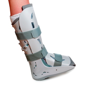
The term "diabetic foot" refers to a condition that affects the feet of people with diabetes, particularly when the disease is not well-controlled. It is characterized by a combination of nerve damage (neuropathy) and poor circulation (peripheral artery disease) in the feet and lower legs.
Neuropathy can cause numbness, tingling, or pain in the feet, making it difficult for people with diabetes to feel injuries, cuts, blisters, or other foot problems. Poor circulation makes it harder for wounds to heal and increases the risk of infection.
Diabetic foot ulcers are a common complication of diabetic neuropathy and can lead to serious infections, hospitalization, and even amputation if not treated promptly and effectively. Preventive care, including regular foot exams, proper footwear, and good blood glucose control, is essential for people with diabetes to prevent or manage diabetic foot problems.
Femoral neoplasms refer to abnormal growths or tumors that develop in the femur, which is the long thigh bone in the human body. These neoplasms can be benign (non-cancerous) or malignant (cancerous). Benign femoral neoplasms are slow-growing and rarely spread to other parts of the body, while malignant neoplasms are aggressive and can invade nearby tissues and organs, as well as metastasize (spread) to distant sites.
There are various types of femoral neoplasms, including osteochondromas, enchondromas, chondrosarcomas, osteosarcomas, and Ewing sarcomas, among others. The specific type of neoplasm is determined by the cell type from which it arises and its behavior.
Symptoms of femoral neoplasms may include pain, swelling, stiffness, or weakness in the thigh, as well as a palpable mass or limited mobility. Diagnosis typically involves imaging studies such as X-rays, CT scans, or MRI, as well as biopsy to determine the type and grade of the tumor. Treatment options may include surgery, radiation therapy, chemotherapy, or a combination of these approaches, depending on the type, size, location, and stage of the neoplasm.
Peripheral Arterial Disease (PAD) is a medical condition characterized by the narrowing or blockage of arteries that supply blood to the extremities, most commonly the legs. This results in reduced blood flow, leading to symptoms such as leg pain, cramping, numbness, or weakness during physical activity, and in severe cases, tissue damage or gangrene. PAD is often indicative of widespread atherosclerosis, which is the hardening and narrowing of arteries due to the buildup of fatty deposits called plaques. It's important to note that early detection and management can help prevent serious complications.
Sarcoma is a type of cancer that develops from certain types of connective tissue (such as muscle, fat, fibrous tissue, blood vessels, or nerves) found throughout the body. It can occur in any part of the body, but it most commonly occurs in the arms, legs, chest, and abdomen.
Sarcomas are classified into two main groups: bone sarcomas and soft tissue sarcomas. Bone sarcomas develop in the bones, while soft tissue sarcomas develop in the soft tissues of the body, such as muscles, tendons, ligaments, fat, blood vessels, and nerves.
Sarcomas can be further classified into many subtypes based on their specific characteristics, such as the type of tissue they originate from, their genetic makeup, and their appearance under a microscope. The different subtypes of sarcoma have varying symptoms, prognoses, and treatment options.
Overall, sarcomas are relatively rare cancers, accounting for less than 1% of all cancer diagnoses in the United States each year. However, they can be aggressive and may require intensive treatment, such as surgery, radiation therapy, and chemotherapy.
In medical terms, the arm refers to the upper limb of the human body, extending from the shoulder to the wrist. It is composed of three major bones: the humerus in the upper arm, and the radius and ulna in the lower arm. The arm contains several joints, including the shoulder joint, elbow joint, and wrist joint, which allow for a wide range of motion. The arm also contains muscles, blood vessels, nerves, and other soft tissues that are essential for normal function.
The iliac arteries are major branches of the abdominal aorta, the large artery that carries oxygen-rich blood from the heart to the rest of the body. The iliac arteries divide into two branches, the common iliac arteries, which further bifurcate into the internal and external iliac arteries.
The internal iliac artery supplies blood to the lower abdomen, pelvis, and the reproductive organs, while the external iliac artery provides blood to the lower extremities, including the legs and feet. Together, the iliac arteries play a crucial role in circulating blood throughout the body, ensuring that all tissues and organs receive the oxygen and nutrients they need to function properly.
A foot ulcer is a wound or sore on the foot that occurs most commonly in people with diabetes, but can also affect other individuals with poor circulation or nerve damage. These ulcers can be challenging to heal and are prone to infection, making it essential for individuals with foot ulcers to seek medical attention promptly.
Foot ulcers typically develop due to prolonged pressure on bony prominences of the foot, leading to breakdown of the skin and underlying tissues. The development of foot ulcers can be attributed to several factors, including:
1. Neuropathy (nerve damage): This condition causes a loss of sensation in the feet, making it difficult for individuals to feel pain or discomfort associated with pressure points, leading to the formation of ulcers.
2. Peripheral artery disease (PAD): Reduced blood flow to the lower extremities can impair wound healing and make the body more susceptible to infection.
3. Deformities: Structural foot abnormalities, such as bunions or hammertoes, can cause increased pressure on specific areas of the foot, increasing the risk of ulcer formation.
4. Poorly fitting shoes: Shoes that are too tight, narrow, or ill-fitting can create friction and pressure points, contributing to the development of foot ulcers.
5. Trauma: Injuries or trauma to the feet can lead to the formation of ulcers, particularly in individuals with neuropathy who may not feel the initial pain associated with the injury.
6. Foot care neglect: Failure to inspect and care for the feet regularly can result in undetected wounds or sores that progress into ulcers.
Foot ulcers are classified based on their depth, severity, and extent of tissue involvement. Proper assessment, treatment, and prevention strategies are crucial in managing foot ulcers and minimizing the risk of complications such as infection, gangrene, and amputation.
Chondrosarcoma is a type of cancer that develops in the cartilaginous tissue, which is the flexible and smooth connective tissue found in various parts of the body such as the bones, ribs, and nose. It is characterized by the production of malignant cartilage cells that can invade surrounding tissues and spread to other parts of the body (metastasis).
Chondrosarcomas are typically slow-growing tumors but can be aggressive in some cases. They usually occur in adults over the age of 40, and men are more commonly affected than women. The most common sites for chondrosarcoma development include the bones of the pelvis, legs, and arms.
Treatment for chondrosarcoma typically involves surgical removal of the tumor, along with radiation therapy or chemotherapy in some cases. The prognosis for chondrosarcoma depends on several factors, including the size and location of the tumor, the grade of malignancy, and whether it has spread to other parts of the body.
Postoperative complications refer to any unfavorable condition or event that occurs during the recovery period after a surgical procedure. These complications can vary in severity and may include, but are not limited to:
1. Infection: This can occur at the site of the incision or inside the body, such as pneumonia or urinary tract infection.
2. Bleeding: Excessive bleeding (hemorrhage) can lead to a drop in blood pressure and may require further surgical intervention.
3. Blood clots: These can form in the deep veins of the legs (deep vein thrombosis) and can potentially travel to the lungs (pulmonary embolism).
4. Wound dehiscence: This is when the surgical wound opens up, which can lead to infection and further complications.
5. Pulmonary issues: These include atelectasis (collapsed lung), pneumonia, or respiratory failure.
6. Cardiovascular problems: These include abnormal heart rhythms (arrhythmias), heart attack, or stroke.
7. Renal failure: This can occur due to various reasons such as dehydration, blood loss, or the use of certain medications.
8. Pain management issues: Inadequate pain control can lead to increased stress, anxiety, and decreased mobility.
9. Nausea and vomiting: These can be caused by anesthesia, opioid pain medication, or other factors.
10. Delirium: This is a state of confusion and disorientation that can occur in the elderly or those with certain medical conditions.
Prompt identification and management of these complications are crucial to ensure the best possible outcome for the patient.
Congenital limb deformities refer to abnormalities in the structure, position, or function of the arms or legs that are present at birth. These deformities can vary greatly in severity and may affect any part of the limb, including the bones, muscles, joints, and nerves.
Congenital limb deformities can be caused by genetic factors, exposure to certain medications or chemicals during pregnancy, or other environmental factors. Some common types of congenital limb deformities include:
1. Clubfoot: A condition in which the foot is twisted out of shape, making it difficult to walk normally.
2. Polydactyly: A condition in which a person is born with extra fingers or toes.
3. Radial clubhand: A rare condition in which the radius bone in the forearm is missing or underdeveloped, causing the hand to turn inward and the wrist to bend.
4. Amniotic band syndrome: A condition in which strands of the amniotic sac wrap around a developing limb, restricting its growth and leading to deformities.
5. Agenesis: A condition in which a limb or part of a limb is missing at birth.
Treatment for congenital limb deformities may include surgery, bracing, physical therapy, or other interventions depending on the severity and nature of the deformity. In some cases, early intervention and treatment can help to improve function and reduce the impact of the deformity on a person's daily life.
Leg injuries refer to damages or harm caused to any part of the lower extremity, including the bones, muscles, tendons, ligaments, blood vessels, and other soft tissues. These injuries can result from various causes such as trauma, overuse, or degenerative conditions. Common leg injuries include fractures, dislocations, sprains, strains, contusions, and cuts. Symptoms may include pain, swelling, bruising, stiffness, weakness, or difficulty walking. The specific treatment for a leg injury depends on the type and severity of the injury.
In medical terms, the "groin" refers to the area where the lower abdomen meets the thigh. It is located on both sides of the body, in front of the upper part of each leg. The groin contains several important structures such as the inguinal canal, which contains blood vessels and nerves, and the femoral artery and vein, which supply blood to and from the lower extremities. Issues in this region, such as pain or swelling, may indicate a variety of medical conditions, including muscle strains, hernias, or infections.
Transcutaneous blood gas monitoring (TcBGM) is a non-invasive method to measure the partial pressure of oxygen (pO2) and carbon dioxide (pCO2) in the blood. This technique uses heated sensors placed on the skin, typically on the ear lobe or the soles of the feet, to estimate the gas tensions in the capillary blood.
The sensors contain a electrochemical or optical sensor that measures the pO2 and pCO2 levels in the tiny amount of gas that diffuses through the skin from the underlying capillaries. The measurements are then adjusted to reflect the actual blood gas values based on calibration curves and other factors, such as the patient's age, temperature, and skin perfusion.
TcBGM is commonly used in neonatal intensive care units (NICUs) to monitor oxygenation and ventilation in premature infants, who may have immature lungs or other respiratory problems that make invasive blood gas sampling difficult or risky. It can also be used in adults with conditions such as chronic obstructive pulmonary disease (COPD), sleep apnea, or neuromuscular disorders, where frequent blood gas measurements are needed to guide therapy and monitor response to treatment.
Overall, TcBGM provides a safe, painless, and convenient way to monitor blood gases in real-time, without the need for repeated arterial punctures or other invasive procedures. However, it is important to note that TcBGM may not always provide accurate measurements in certain situations, such as when the skin perfusion is poor or when there are significant differences between the capillary and arterial blood gases. Therefore, clinical judgment and other diagnostic tests should be used in conjunction with TcBGM to ensure appropriate patient management.
The fibula is a slender bone located in the lower leg of humans and other vertebrates. It runs parallel to the larger and more robust tibia, and together they are known as the bones of the leg or the anterior tibial segment. The fibula is the lateral bone in the leg, positioned on the outside of the tibia.
In humans, the fibula extends from the knee joint proximally to the ankle joint distally. Its proximal end, called the head of the fibula, articulates with the lateral condyle of the tibia and forms part of the inferior aspect of the knee joint. The narrowed portion below the head is known as the neck of the fibula.
The shaft of the fibula, also called the body of the fibula, is a long, thin structure that descends from the neck and serves primarily for muscle attachment rather than weight-bearing functions. The distal end of the fibula widens to form the lateral malleolus, which is an important bony landmark in the ankle region. The lateral malleolus articulates with the talus bone of the foot and forms part of the ankle joint.
The primary functions of the fibula include providing attachment sites for muscles that act on the lower leg, ankle, and foot, as well as contributing to the stability of the ankle joint through its articulation with the talus bone. Fractures of the fibula can occur due to various injuries, such as twisting or rotational forces applied to the ankle or direct trauma to the lateral aspect of the lower leg.
A critical illness is a serious condition that has the potential to cause long-term or permanent disability, or even death. It often requires intensive care and life support from medical professionals. Critical illnesses can include conditions such as:
1. Heart attack
2. Stroke
3. Organ failure (such as kidney, liver, or lung)
4. Severe infections (such as sepsis)
5. Coma or brain injury
6. Major trauma
7. Cancer that has spread to other parts of the body
These conditions can cause significant physical and emotional stress on patients and their families, and often require extensive medical treatment, rehabilitation, and long-term care. Critical illness insurance is a type of insurance policy that provides financial benefits to help cover the costs associated with treating these serious medical conditions.
The tibia, also known as the shin bone, is the larger of the two bones in the lower leg and part of the knee joint. It supports most of the body's weight and is a major insertion point for muscles that flex the foot and bend the leg. The tibia articulates with the femur at the knee joint and with the fibula and talus bone at the ankle joint. Injuries to the tibia, such as fractures, are common in sports and other activities that put stress on the lower leg.
Debridement is a medical procedure that involves the removal of dead, damaged, or infected tissue to improve the healing process or prevent further infection. This can be done through various methods such as surgical debridement (removal of tissue using scalpel or scissors), mechanical debridement (use of wound irrigation or high-pressure water jet), autolytic debridement (using the body's own enzymes to break down and reabsorb dead tissue), and enzymatic debridement (application of topical enzymes to dissolve necrotic tissue). The goal of debridement is to promote healthy tissue growth, reduce the risk of infection, and improve overall wound healing.
'Leg bones' is a general term that refers to the bones in the leg portion of the lower extremity. In humans, this would specifically include:
1. Femur: This is the thigh bone, the longest and strongest bone in the human body. It connects the hip bone to the knee.
2. Patella: This is the kneecap, a small triangular bone located at the front of the knee joint.
3. Tibia and Fibula: These are the bones of the lower leg. The tibia, or shin bone, is the larger of the two and bears most of the body's weight. It connects the knee to the ankle. The fibula, a slender bone, runs parallel to the tibia on its outside.
Please note that in medical terminology, 'leg bones' doesn't include the bones of the foot (tarsal bones, metatarsal bones, and phalanges), which are often collectively referred to as the 'foot bones'.
Regional perfusion chemotherapy for cancer is a medical treatment in which a specific area or region of the body is infused with high concentrations of cancer-killing (cytotoxic) drugs via a temporary isolation and perfusion of that region. This technique is typically used to treat isolated areas of cancer that are locally advanced, recurrent, or cannot be removed surgically.
The procedure involves isolating the regional blood circulation by cannulating the artery and vein that supply blood to the target area, often the limbs (such as in melanoma or sarcoma) or the liver (for liver tumors). The chemotherapeutic drugs are then introduced into the isolated arterial circulation, allowing for a high concentration of the drug to be delivered directly to the cancerous tissue while minimizing systemic exposure and toxicity.
After the infusion, the region is rinsed with a blood-substitute solution to remove any residual chemotherapeutic agents before reconnecting the circulation. This procedure can be repeated multiple times if necessary.
Regional perfusion chemotherapy has been shown to improve local control and potentially increase survival rates in certain types of cancer, while reducing systemic side effects compared to traditional intravenous chemotherapy. However, it is a complex and invasive procedure that requires specialized medical expertise and facilities.
Surgical anastomosis is a medical procedure that involves the connection of two tubular structures, such as blood vessels or intestines, to create a continuous passage. This technique is commonly used in various types of surgeries, including vascular, gastrointestinal, and orthopedic procedures.
During a surgical anastomosis, the ends of the two tubular structures are carefully prepared by removing any damaged or diseased tissue. The ends are then aligned and joined together using sutures, staples, or other devices. The connection must be secure and leak-free to ensure proper function and healing.
The success of a surgical anastomosis depends on several factors, including the patient's overall health, the location and condition of the structures being joined, and the skill and experience of the surgeon. Complications such as infection, bleeding, or leakage can occur, which may require additional medical intervention or surgery.
Proper postoperative care is also essential to ensure the success of a surgical anastomosis. This may include monitoring for signs of complications, administering medications to prevent infection and promote healing, and providing adequate nutrition and hydration.
Angioplasty is a medical procedure used to open narrowed or blocked blood vessels, often referred to as coronary angioplasty when it involves the heart's blood vessels (coronary arteries). The term "angio" refers to an angiogram, which is a type of X-ray image that reveals the inside of blood vessels.
The procedure typically involves the following steps:
1. A thin, flexible catheter (tube) is inserted into a blood vessel, usually through a small incision in the groin or arm.
2. The catheter is guided to the narrowed or blocked area using real-time X-ray imaging.
3. Once in place, a tiny balloon attached to the tip of the catheter is inflated to widen the blood vessel and compress any plaque buildup against the artery walls.
4. A stent (a small mesh tube) may be inserted to help keep the blood vessel open and prevent it from narrowing again.
5. The balloon is deflated, and the catheter is removed.
Angioplasty helps improve blood flow, reduce symptoms such as chest pain or shortness of breath, and lower the risk of heart attack in patients with blocked arteries. It's important to note that angioplasty is not a permanent solution for coronary artery disease, and lifestyle changes, medications, and follow-up care are necessary to maintain long-term cardiovascular health.
Ultrasonography, Doppler, and Duplex are diagnostic medical techniques that use sound waves to create images of internal body structures and assess their function. Here are the definitions for each:
1. Ultrasonography: Also known as ultrasound, this is a non-invasive imaging technique that uses high-frequency sound waves to produce images of internal organs and tissues. A small handheld device called a transducer is placed on the skin surface, which emits and receives sound waves. The returning echoes are then processed to create real-time visual images of the internal structures.
2. Doppler: This is a type of ultrasound that measures the velocity and direction of blood flow in the body by analyzing the frequency shift of the reflected sound waves. It can be used to assess blood flow in various parts of the body, such as the heart, arteries, and veins.
3. Duplex: Duplex ultrasonography is a combination of both gray-scale ultrasound and Doppler ultrasound. It provides detailed images of internal structures, as well as information about blood flow velocity and direction. This technique is often used to evaluate conditions such as deep vein thrombosis, carotid artery stenosis, and peripheral arterial disease.
In summary, ultrasonography is a diagnostic imaging technique that uses sound waves to create images of internal structures, Doppler is a type of ultrasound that measures blood flow velocity and direction, and duplex is a combination of both techniques that provides detailed images and information about blood flow.
A leg ulcer is a chronic wound that occurs on the lower extremities, typically on the inner or outer ankle. It's often caused by poor circulation, venous insufficiency, or diabetes. Leg ulcers can also result from injury, infection, or inflammatory diseases such as rheumatoid arthritis or lupus. These ulcers can be painful, and they may take a long time to heal, making them prone to infection. Proper diagnosis, treatment, and wound care are essential for healing leg ulcers and preventing complications.
Autologous transplantation is a medical procedure where cells, tissues, or organs are removed from a person, stored and then returned back to the same individual at a later time. This is different from allogeneic transplantation where the tissue or organ is obtained from another donor. The term "autologous" is derived from the Greek words "auto" meaning self and "logos" meaning study.
In autologous transplantation, the patient's own cells or tissues are used to replace or repair damaged or diseased ones. This reduces the risk of rejection and eliminates the need for immunosuppressive drugs, which are required in allogeneic transplants to prevent the body from attacking the foreign tissue.
Examples of autologous transplantation include:
* Autologous bone marrow or stem cell transplantation, where stem cells are removed from the patient's blood or bone marrow, stored and then reinfused back into the same individual after high-dose chemotherapy or radiation therapy to treat cancer.
* Autologous skin grafting, where a piece of skin is taken from one part of the body and transplanted to another area on the same person.
* Autologous chondrocyte implantation, where cartilage cells are harvested from the patient's own knee, cultured in a laboratory and then implanted back into the knee to repair damaged cartilage.
The forefoot is the front part of the human foot that contains the toes and the associated bones, muscles, ligaments, and tendons. It is made up of five long bones called metatarsals and fourteen phalanges, which are the bones in the toes. The forefoot plays a crucial role in weight-bearing, balance, and propulsion during walking and running. The joints in the forefoot allow for flexion, extension, abduction, and adduction of the toes, enabling us to maintain our footing on various surfaces and adapt to different terrain.
The humerus is the long bone in the upper arm that extends from the shoulder joint (glenohumeral joint) to the elbow joint. It articulates with the glenoid cavity of the scapula to form the shoulder joint and with the radius and ulna bones at the elbow joint. The proximal end of the humerus has a rounded head that provides for movement in multiple planes, making it one of the most mobile joints in the body. The greater and lesser tubercles are bony prominences on the humeral head that serve as attachment sites for muscles that move the shoulder and arm. The narrow shaft of the humerus provides stability and strength for weight-bearing activities, while the distal end forms two articulations: one with the ulna (trochlea) and one with the radius (capitulum). Together, these structures allow for a wide range of motion in the shoulder and elbow joints.
An arteriovenous shunt is a surgically created connection between an artery and a vein. This procedure is typically performed to reroute blood flow or to provide vascular access for various medical treatments. In a surgical setting, the creation of an arteriovenous shunt involves connecting an artery directly to a vein, bypassing the capillary network in between.
There are different types of arteriovenous shunts used for specific medical purposes:
1. Arteriovenous Fistula (AVF): This is a surgical connection created between an artery and a vein, usually in the arm or leg. The procedure involves dissecting both the artery and vein, then suturing them directly together. Over time, the increased blood flow to the vein causes it to dilate and thicken, making it suitable for repeated needle punctures during hemodialysis treatments for patients with kidney failure.
2. Arteriovenous Graft (AVG): An arteriovenous graft is a synthetic tube used to connect an artery and a vein when a direct AVF cannot be created due to insufficient vessel size or poor quality. The graft can be made of various materials, such as polytetrafluoroethylene (PTFE) or Dacron. Grafts are more prone to infection and clotting compared to native AVFs but remain an essential option for patients requiring hemodialysis access.
3. Central Venous Catheter (CVC): A central venous catheter is a flexible tube inserted into a large vein, often in the neck or groin, and advanced towards the heart. CVCs can be used as temporary arteriovenous shunts for patients who require immediate hemodialysis access but do not have time to wait for an AVF or AVG to mature. However, they are associated with higher risks of infection and thrombosis compared to native AVFs and AVGs.
In summary, a surgical arteriovenous shunt is a connection between an artery and a vein established through a medical procedure. The primary purpose of these shunts is to provide vascular access for hemodialysis in patients with end-stage renal disease or to serve as temporary access when native AVFs or AVGs are not feasible.
Arteries are blood vessels that carry oxygenated blood away from the heart to the rest of the body. They have thick, muscular walls that can withstand the high pressure of blood being pumped out of the heart. Arteries branch off into smaller vessels called arterioles, which further divide into a vast network of tiny capillaries where the exchange of oxygen, nutrients, and waste occurs between the blood and the body's cells. After passing through the capillary network, deoxygenated blood collects in venules, then merges into veins, which return the blood back to the heart.
The axillary artery is a major blood vessel in the upper limb. It is the continuation of the subclavian artery and begins at the lateral border of the first rib, where it becomes the brachial artery. The axillary artery supplies oxygenated blood to the upper extremity, chest wall, and breast.
The axillary artery is divided into three parts based on the surrounding structures:
1. First part: From its origin at the lateral border of the first rib to the medial border of the pectoralis minor muscle. It lies deep to the clavicle and is covered by the scalene muscles, the anterior and middle scalene being the most important. The branches arising from this portion are the superior thoracic artery and the thyrocervical trunk.
2. Second part: Behind the pectoralis minor muscle. The branches arising from this portion are the lateral thoracic artery and the subscapular artery.
3. Third part: After leaving the lower border of the pectoralis minor muscle, it becomes the brachial artery. The branches arising from this portion are the anterior circumflex humeral artery and the posterior circumflex humeral artery.
The axillary artery is a common site for surgical interventions such as angioplasty and stenting to treat peripheral arterial disease, as well as for bypass grafting in cases of severe atherosclerosis or occlusion.
The Kaplan-Meier estimate is a statistical method used to calculate the survival probability over time in a population. It is commonly used in medical research to analyze time-to-event data, such as the time until a patient experiences a specific event like disease progression or death. The Kaplan-Meier estimate takes into account censored data, which occurs when some individuals are lost to follow-up before experiencing the event of interest.
The method involves constructing a survival curve that shows the proportion of subjects still surviving at different time points. At each time point, the survival probability is calculated as the product of the conditional probabilities of surviving from one time point to the next. The Kaplan-Meier estimate provides an unbiased and consistent estimator of the survival function, even when censoring is present.
In summary, the Kaplan-Meier estimate is a crucial tool in medical research for analyzing time-to-event data and estimating survival probabilities over time while accounting for censored observations.
An aneurysm is a localized, balloon-like bulge in the wall of a blood vessel. It occurs when the pressure inside the vessel causes a weakened area to swell and become enlarged. Aneurysms can develop in any blood vessel, but they are most common in arteries at the base of the brain (cerebral aneurysm) and the main artery carrying blood from the heart to the rest of the body (aortic aneurysm).
Aneurysms can be classified as saccular or fusiform, depending on their shape. A saccular aneurysm is a round or oval bulge that projects from the side of a blood vessel, while a fusiform aneurysm is a dilated segment of a blood vessel that is uniform in width and involves all three layers of the arterial wall.
The size and location of an aneurysm can affect its risk of rupture. Generally, larger aneurysms are more likely to rupture than smaller ones. Aneurysms located in areas with high blood pressure or where the vessel branches are also at higher risk of rupture.
Ruptured aneurysms can cause life-threatening bleeding and require immediate medical attention. Symptoms of a ruptured aneurysm may include sudden severe headache, neck stiffness, nausea, vomiting, blurred vision, or loss of consciousness. Unruptured aneurysms may not cause any symptoms and are often discovered during routine imaging tests for other conditions.
Treatment options for aneurysms depend on their size, location, and risk of rupture. Small, unruptured aneurysms may be monitored with regular imaging tests to check for growth or changes. Larger or symptomatic aneurysms may require surgical intervention, such as clipping or coiling, to prevent rupture and reduce the risk of complications.
Phantom limb is a condition where an individual experiences sensations in a limb or appendage that has been amputated. These sensations can include feelings of pain, warmth, cold, itching, or tingling in the area where the limb used to be. The exact cause of phamtom limb is not fully understood, but it's believed to be related to mixed signals from the brain and nervous system.
Phantom limb sensations are relatively common among amputees, with some studies suggesting that up to 80% of individuals who have undergone an amputation may experience these sensations to some degree. While phantom limb can be a challenging condition to live with, there are various treatments available that can help manage the symptoms and improve quality of life. These may include medications, physical therapy, and alternative therapies such as acupuncture or mirror box therapy.
The pelvic bones, also known as the hip bones, are a set of three irregularly shaped bones that connect to form the pelvic girdle in the lower part of the human body. They play a crucial role in supporting the spine and protecting the abdominal and pelvic organs.
The pelvic bones consist of three bones:
1. The ilium: This is the largest and uppermost bone, forming the majority of the hip bone and the broad, flaring part of the pelvis known as the wing of the ilium or the iliac crest, which can be felt on the side of the body.
2. The ischium: This is the lower and back portion of the pelvic bone that forms part of the sitting surface or the "sit bones."
3. The pubis: This is the front part of the pelvic bone, which connects to the other side at the pubic symphysis in the midline of the body.
The pelvic bones are joined together at the acetabulum, a cup-shaped socket that forms the hip joint and articulates with the head of the femur (thigh bone). The pelvic bones also have several openings for the passage of blood vessels, nerves, and reproductive and excretory organs.
The shape and size of the pelvic bones differ between males and females due to their different roles in childbirth and locomotion. Females typically have a wider and shallower pelvis than males to accommodate childbirth, while males usually have a narrower and deeper pelvis that is better suited for weight-bearing and movement.
Pathological constriction refers to an abnormal narrowing or tightening of a body passage or organ, which can interfere with the normal flow of blood, air, or other substances through the area. This constriction can occur due to various reasons such as inflammation, scarring, or abnormal growths, and can affect different parts of the body, including blood vessels, airways, intestines, and ureters. Pathological constriction can lead to a range of symptoms and complications depending on its location and severity, and may require medical intervention to correct.
Endovascular procedures are minimally invasive medical treatments that involve accessing and repairing blood vessels or other interior parts of the body through small incisions or punctures. These procedures typically use specialized catheters, wires, and other tools that are inserted into the body through an artery or vein, usually in the leg or arm.
Endovascular procedures can be used to treat a wide range of conditions, including aneurysms, atherosclerosis, peripheral artery disease, carotid artery stenosis, and other vascular disorders. Some common endovascular procedures include angioplasty, stenting, embolization, and thrombectomy.
The benefits of endovascular procedures over traditional open surgery include smaller incisions, reduced trauma to surrounding tissues, faster recovery times, and lower risks of complications such as infection and bleeding. However, endovascular procedures may not be appropriate for all patients or conditions, and careful evaluation and consideration are necessary to determine the best treatment approach.
Wound healing is a complex and dynamic process that occurs after tissue injury, aiming to restore the integrity and functionality of the damaged tissue. It involves a series of overlapping phases: hemostasis, inflammation, proliferation, and remodeling.
1. Hemostasis: This initial phase begins immediately after injury and involves the activation of the coagulation cascade to form a clot, which stabilizes the wound and prevents excessive blood loss.
2. Inflammation: Activated inflammatory cells, such as neutrophils and monocytes/macrophages, infiltrate the wound site to eliminate pathogens, remove debris, and release growth factors that promote healing. This phase typically lasts for 2-5 days post-injury.
3. Proliferation: In this phase, various cell types, including fibroblasts, endothelial cells, and keratinocytes, proliferate and migrate to the wound site to synthesize extracellular matrix (ECM) components, form new blood vessels (angiogenesis), and re-epithelialize the wounded area. This phase can last up to several weeks depending on the size and severity of the wound.
4. Remodeling: The final phase of wound healing involves the maturation and realignment of collagen fibers, leading to the restoration of tensile strength in the healed tissue. This process can continue for months to years after injury, although the tissue may never fully regain its original structure and function.
It is important to note that wound healing can be compromised by several factors, including age, nutrition, comorbidities (e.g., diabetes, vascular disease), and infection, which can result in delayed healing or non-healing chronic wounds.
Operative blood salvage, also known as intraoperative blood recovery or cell salvage, is a medical procedure that involves the collection, washing, and reinfusion of a patient's own blood during surgery. The blood is collected from the surgical site using a suction device and then processed to remove any debris, clots, and free hemoglobin. The resulting red blood cells are then washed and suspended in a sterile solution before being returned to the patient through a transfusion.
This technique is commonly used during surgeries where significant blood loss is expected, such as orthopedic, cardiovascular, and major cancer surgeries. It offers several advantages over allogeneic (donor) blood transfusions, including reduced exposure to potential transfusion reactions, decreased risk of infectious disease transmission, and lower costs. However, it may not be appropriate for all patients or surgical procedures, and its use should be carefully considered based on the individual patient's medical history and condition.
A tibial fracture is a medical term that refers to a break in the shin bone, which is called the tibia. The tibia is the larger of the two bones in the lower leg and is responsible for supporting much of your body weight. Tibial fractures can occur in various ways, such as from high-energy trauma like car accidents or falls, or from low-energy trauma in individuals with weakened bones due to osteoporosis or other medical conditions.
Tibial fractures can be classified into different types based on the location, pattern, and severity of the break. Some common types of tibial fractures include:
1. Transverse fracture: A straight break that goes across the bone.
2. Oblique fracture: A diagonal break that slopes across the bone.
3. Spiral fracture: A break that spirals around the bone, often caused by twisting or rotational forces.
4. Comminuted fracture: A break where the bone is shattered into multiple pieces.
5. Open fracture: A break in which the bone pierces through the skin, increasing the risk of infection.
6. Closed fracture: A break in which the bone does not pierce through the skin.
Tibial fractures can cause symptoms such as pain, swelling, bruising, deformity, and difficulty walking or bearing weight on the affected leg. Treatment for tibial fractures may include immobilization with a cast or brace, surgery to realign and stabilize the bone with plates, screws, or rods, and rehabilitation to restore strength, mobility, and function to the injured limb.
Arm injuries refer to any damage or harm sustained by the structures of the upper limb, including the bones, muscles, tendons, ligaments, nerves, and blood vessels. These injuries can occur due to various reasons such as trauma, overuse, or degenerative conditions. Common arm injuries include fractures, dislocations, sprains, strains, tendonitis, and nerve damage. Symptoms may include pain, swelling, bruising, limited mobility, numbness, or weakness in the affected area. Treatment varies depending on the type and severity of the injury, and may include rest, ice, compression, elevation, physical therapy, medication, or surgery.
Treatment failure is a term used in medicine to describe the situation when a prescribed treatment or intervention is not achieving the desired therapeutic goals or objectives. This may occur due to various reasons, such as:
1. Development of drug resistance by the pathogen or disease being treated.
2. Inadequate dosage or frequency of the medication.
3. Poor adherence or compliance to the treatment regimen by the patient.
4. The presence of underlying conditions or comorbidities that may affect the efficacy of the treatment.
5. The severity or progression of the disease despite appropriate treatment.
When treatment failure occurs, healthcare providers may need to reassess the patient's condition and modify the treatment plan accordingly, which may include adjusting the dosage, changing the medication, adding new medications, or considering alternative treatments.
Prosthesis design is a specialized field in medical device technology that involves creating and developing artificial substitutes to replace a missing body part, such as a limb, tooth, eye, or internal organ. The design process typically includes several stages: assessment of the patient's needs, selection of appropriate materials, creation of a prototype, testing and refinement, and final fabrication and fitting of the prosthesis.
The goal of prosthesis design is to create a device that functions as closely as possible to the natural body part it replaces, while also being comfortable, durable, and aesthetically pleasing for the patient. The design process may involve collaboration between medical professionals, engineers, and designers, and may take into account factors such as the patient's age, lifestyle, occupation, and overall health.
Prosthesis design can be highly complex, particularly for advanced devices such as robotic limbs or implantable organs. These devices often require sophisticated sensors, actuators, and control systems to mimic the natural functions of the body part they replace. As a result, prosthesis design is an active area of research and development in the medical field, with ongoing efforts to improve the functionality, comfort, and affordability of these devices for patients.
Intermittent Pneumatic Compression (IPC) devices are medical devices that use inflatable garments to apply controlled pressure in a rhythmic, intermittent manner to the extremities of the body, most commonly used on the legs. These devices are designed to help improve venous and lymphatic flow, reduce edema (swelling), and prevent the formation of blood clots (deep vein thrombosis) in patients who are at risk.
The IPC device typically consists of a pump, hoses, and an air-filled garment that covers the affected limb. The pump regulates the pressure and inflation pattern, while the garment applies pressure to the limb. The compression cycle usually starts with low pressure and gradually increases to a peak pressure before decreasing again. This process is repeated at regular intervals, providing intermittent compression that mimics natural muscle contractions and helps promote blood flow.
IPC devices are often used in clinical settings such as hospitals and rehabilitation centers, but they can also be prescribed for home use. They are commonly recommended for patients who have undergone surgery, experienced trauma, or have conditions that increase their risk of developing blood clots, such as prolonged immobilization, varicose veins, or certain medical disorders.
It is essential to follow the healthcare provider's instructions when using IPC devices and report any discomfort, pain, or unusual symptoms during treatment.
Prostheses: Artificial substitutes or replacements for missing body parts, such as limbs, eyes, or teeth. They are designed to restore the function, appearance, or mobility of the lost part. Prosthetic devices can be categorized into several types, including:
1. External prostheses: Devices that are attached to the outside of the body, like artificial arms, legs, hands, and feet. These may be further classified into:
a. Cosmetic or aesthetic prostheses: Primarily designed to improve the appearance of the affected area.
b. Functional prostheses: Designed to help restore the functionality and mobility of the lost limb.
2. Internal prostheses: Implanted artificial parts that replace missing internal organs, bones, or tissues, such as heart valves, hip joints, or intraocular lenses.
Implants: Medical devices or substances that are intentionally placed inside the body to replace or support a missing or damaged biological structure, deliver medication, monitor physiological functions, or enhance bodily functions. Examples of implants include:
1. Orthopedic implants: Devices used to replace or reinforce damaged bones, joints, or cartilage, such as knee or hip replacements.
2. Cardiovascular implants: Devices that help support or regulate heart function, like pacemakers, defibrillators, and artificial heart valves.
3. Dental implants: Artificial tooth roots that are placed into the jawbone to support dental prostheses, such as crowns, bridges, or dentures.
4. Neurological implants: Devices used to stimulate nerves, brain structures, or spinal cord tissues to treat various neurological conditions, like deep brain stimulators for Parkinson's disease or cochlear implants for hearing loss.
5. Ophthalmic implants: Artificial lenses that are placed inside the eye to replace a damaged or removed natural lens, such as intraocular lenses used in cataract surgery.
The popliteal vein is the continuation of the tibial and fibular (or anterior and posterior tibial) veins, forming in the lower leg's back portion or popliteal fossa. It carries blood from the leg towards the heart. The popliteal vein is located deep within the body and is accompanied by the popliteal artery, which supplies oxygenated blood to the lower leg. This venous structure is a crucial part of the venous system in the lower extremities and is often assessed during physical examinations for signs of venous insufficiency or deep vein thrombosis (DVT).
An embolectomy is a surgical procedure to remove an embolus, which is a blockage in a blood vessel caused by a clot or air bubble that has traveled from another part of the body. During an embolectomy, the surgeon makes an incision in the affected blood vessel and removes the embolus using specialized surgical instruments. This procedure is often performed as an emergency treatment to restore blood flow and prevent tissue damage in the affected area of the body.
Artificial limbs, also known as prosthetics, are artificial substitutes that replace a part or all of an absent extremity or limb. They are designed to restore the function, mobility, and appearance of the lost limb as much as possible. Artificial limbs can be made from various materials such as wood, plastic, metal, or carbon fiber, and they can be custom-made to fit the individual's specific needs and measurements.
Prosthetic limbs can be categorized into two main types: cosmetic and functional. Cosmetic prosthetics are designed to look like natural limbs and are primarily used to improve the appearance of the person. Functional prosthetics, on the other hand, are designed to help the individual perform specific tasks and activities. They may include features such as hooks, hands, or specialized feet that can be used for different purposes.
Advances in technology have led to the development of more sophisticated artificial limbs, including those that can be controlled by the user's nervous system, known as bionic prosthetics. These advanced prosthetic devices can provide a greater degree of mobility and control for the user, allowing them to perform complex movements and tasks with ease.
Replantation is a surgical procedure in which a body part that has been completely detached or amputated is reattached to the body. This procedure involves careful reattachment of bones, muscles, tendons, nerves, and blood vessels to restore function and sensation to the greatest extent possible. The success of replantation depends on various factors such as the level of injury, the condition of the amputated part, and the patient's overall health.
A hindlimb, also known as a posterior limb, is one of the pair of extremities that are located distally to the trunk in tetrapods (four-legged vertebrates) and include mammals, birds, reptiles, and amphibians. In humans and other primates, hindlimbs are equivalent to the lower limbs, which consist of the thigh, leg, foot, and toes.
The primary function of hindlimbs is locomotion, allowing animals to move from one place to another. However, they also play a role in other activities such as balance, support, and communication. In humans, the hindlimbs are responsible for weight-bearing, standing, walking, running, and jumping.
In medical terminology, the term "hindlimb" is not commonly used to describe human anatomy. Instead, healthcare professionals use terms like lower limbs or lower extremities to refer to the same region of the body. However, in comparative anatomy and veterinary medicine, the term hindlimb is still widely used to describe the corresponding structures in non-human animals.
Medical survival rate is a statistical measure used to determine the percentage of patients who are still alive for a specific period of time after their diagnosis or treatment for a certain condition or disease. It is often expressed as a five-year survival rate, which refers to the proportion of people who are alive five years after their diagnosis. Survival rates can be affected by many factors, including the stage of the disease at diagnosis, the patient's age and overall health, the effectiveness of treatment, and other health conditions that the patient may have. It is important to note that survival rates are statistical estimates and do not necessarily predict an individual patient's prognosis.
The femoral vein is the large vein that runs through the thigh and carries oxygen-depleted blood from the lower limbs back to the heart. It is located in the femoral triangle, along with the femoral artery and nerve. The femoral vein begins at the knee as the popliteal vein, which then joins with the deep vein of the thigh to form the femoral vein. As it moves up the leg, it is joined by several other veins, including the great saphenous vein, before it becomes the external iliac vein at the inguinal ligament in the groin.
Spontaneous fractures are bone breaks that occur without any identifiable trauma or injury. They are typically caused by underlying medical conditions that weaken the bones, making them more susceptible to breaking under normal stress or weight. The most common cause of spontaneous fractures is osteoporosis, a condition characterized by weak and brittle bones. Other potential causes include various bone diseases, certain cancers, long-term use of corticosteroids, and genetic disorders affecting bone strength.
It's important to note that while the term "spontaneous" implies that the fracture occurred without any apparent cause, it is usually the result of an underlying medical condition. Therefore, if you experience a spontaneous fracture, seeking medical attention is crucial to diagnose and manage the underlying cause to prevent future fractures and related complications.
Prosthesis-related infections, also known as prosthetic joint infections (PJIs), are infections that occur around or within a prosthetic device, such as an artificial joint. These infections can be caused by bacteria, fungi, or other microorganisms and can lead to serious complications if not treated promptly and effectively.
Prosthesis-related infections can occur soon after the implantation of the prosthetic device (early infection) or months or even years later (late infection). Early infections are often caused by bacteria that enter the surgical site during the procedure, while late infections may be caused by hematogenous seeding (i.e., when bacteria from another source spread through the bloodstream and settle in the prosthetic device) or by contamination during a subsequent medical procedure.
Symptoms of prosthesis-related infections can include pain, swelling, redness, warmth, and drainage around the affected area. In some cases, patients may also experience fever, chills, or fatigue. Diagnosis typically involves a combination of clinical evaluation, laboratory tests (such as blood cultures, joint fluid analysis, and tissue biopsy), and imaging studies (such as X-rays, CT scans, or MRI).
Treatment of prosthesis-related infections usually involves a combination of antibiotics and surgical intervention. The specific treatment approach will depend on the type and severity of the infection, as well as the patient's overall health status. In some cases, it may be necessary to remove or replace the affected prosthetic device.
Local neoplasm recurrence is the return or regrowth of a tumor in the same location where it was originally removed or treated. This means that cancer cells have survived the initial treatment and started to grow again in the same area. It's essential to monitor and detect any local recurrence as early as possible, as it can affect the prognosis and may require additional treatment.
I'm sorry for any confusion, but "Polyethylene Terephthalates" is not a medical term. It is a type of polymer used in the manufacturing of various products, such as plastic bottles and textile fibers. Medically, you might encounter the abbreviation "PET" or "PET scan," which stands for "Positron Emission Tomography." A PET scan is a type of medical imaging that provides detailed pictures of the body's interior. If you have any medical terms you would like defined, I'd be happy to help!
A free tissue flap in medical terms refers to a surgical procedure where living tissue, along with its own blood supply, is lifted from its original location and moved to another part of the body for reconstruction or repair. The term "free" indicates that the tissue is completely detached from its original blood vessels and then reattached to new blood vessels at the recipient site using microvascular surgical techniques.
Free tissue flaps can be composed of various tissues, such as skin, muscle, fascia (the connective tissue beneath the skin), or bone. They are often used in reconstructive surgery following trauma, tumor removal, or for treating complex wounds that cannot heal on their own. The advantages of free tissue flaps include increased flexibility in choosing the type and size of tissue to be transferred, as well as improved blood flow to the transplanted tissue, which can enhance healing and overall surgical success.
Prosthesis implantation is a surgical procedure where an artificial device or component, known as a prosthesis, is placed inside the body to replace a missing or damaged body part. The prosthesis can be made from various materials such as metal, plastic, or ceramic and is designed to perform the same function as the original body part.
The implantation procedure involves making an incision in the skin to create a pocket where the prosthesis will be placed. The prosthesis is then carefully positioned and secured in place using screws, cement, or other fixation methods. In some cases, tissue from the patient's own body may be used to help anchor the prosthesis.
Once the prosthesis is in place, the incision is closed with sutures or staples, and the area is bandaged. The patient will typically need to undergo rehabilitation and physical therapy to learn how to use the new prosthesis and regain mobility and strength.
Prosthesis implantation is commonly performed for a variety of reasons, including joint replacement due to arthritis or injury, dental implants to replace missing teeth, and breast reconstruction after mastectomy. The specific procedure and recovery time will depend on the type and location of the prosthesis being implanted.
Risk assessment in the medical context refers to the process of identifying, evaluating, and prioritizing risks to patients, healthcare workers, or the community related to healthcare delivery. It involves determining the likelihood and potential impact of adverse events or hazards, such as infectious diseases, medication errors, or medical devices failures, and implementing measures to mitigate or manage those risks. The goal of risk assessment is to promote safe and high-quality care by identifying areas for improvement and taking action to minimize harm.
"Biological dressings" refer to materials used in wound healing that are derived from biological sources, such as living cells, tissues, or extracellular matrix components. These dressings can be used to promote the regeneration and repair of damaged or injured tissues by providing a supportive environment for cell growth, differentiation, and tissue formation.
Biological dressings may be derived from various sources, including:
1. Autografts: Tissue harvested from the same individual who will receive the graft.
2. Allografts: Tissue harvested from a donor of the same species.
3. Xenografts: Tissue harvested from a donor of a different species.
4. Decellularized tissue matrices: Tissues that have had their cellular components removed, leaving behind an intact extracellular matrix scaffold.
5. Engineered tissues: Tissues created in the lab through the cultivation and assembly of cells on biocompatible scaffolds or hydrogels.
Examples of biological dressings include skin substitutes, amniotic membranes, and platelet-rich plasma (PRP) preparations. These dressings can help to reduce inflammation, prevent infection, and enhance the healing process in various types of wounds, including chronic wounds, burns, and surgical incisions.
It is important to note that while biological dressings offer several advantages over traditional wound dressings, they may also carry risks such as immune rejection or disease transmission, depending on their source and preparation. Therefore, careful consideration should be given to the selection of appropriate biological dressing materials for each individual patient and application.
Soft tissue neoplasms refer to abnormal growths or tumors that develop in the soft tissues of the body. Soft tissues include muscles, tendons, ligaments, fascia, nerves, blood vessels, fat, and synovial membranes (the thin layer of cells that line joints and tendons). Neoplasms can be benign (non-cancerous) or malignant (cancerous), and their behavior and potential for spread depend on the specific type of neoplasm.
Benign soft tissue neoplasms are typically slow-growing, well-circumscribed, and rarely spread to other parts of the body. They can often be removed surgically with a low risk of recurrence. Examples of benign soft tissue neoplasms include lipomas (fat tumors), schwannomas (nerve sheath tumors), and hemangiomas (blood vessel tumors).
Malignant soft tissue neoplasms, on the other hand, can grow rapidly, invade surrounding tissues, and may metastasize (spread) to distant parts of the body. They are often more difficult to treat than benign neoplasms and require a multidisciplinary approach, including surgery, radiation therapy, and chemotherapy. Examples of malignant soft tissue neoplasms include sarcomas, such as rhabdomyosarcoma (arising from skeletal muscle), leiomyosarcoma (arising from smooth muscle), and angiosarcoma (arising from blood vessels).
It is important to note that soft tissue neoplasms can occur in any part of the body, and their diagnosis and treatment require a thorough evaluation by a healthcare professional with expertise in this area.
A stent is a small mesh tube that's used to treat narrow or weak arteries. Arteries are blood vessels that carry blood away from your heart to other parts of your body. A stent is placed in an artery as part of a procedure called angioplasty. Angioplasty restores blood flow through narrowed or blocked arteries by inflating a tiny balloon inside the blocked artery to widen it.
The stent is then inserted into the widened artery to keep it open. The stent is usually made of metal, but some are coated with medication that is slowly and continuously released to help prevent the formation of scar tissue in the artery. This can reduce the chance of the artery narrowing again.
Stents are also used in other parts of the body, such as the neck (carotid artery) and kidneys (renal artery), to help maintain blood flow and prevent blockages. They can also be used in the urinary system to treat conditions like ureteropelvic junction obstruction or narrowing of the urethra.
"Recovery of function" is a term used in medical rehabilitation to describe the process in which an individual regains the ability to perform activities or tasks that were previously difficult or impossible due to injury, illness, or disability. This can involve both physical and cognitive functions. The goal of recovery of function is to help the person return to their prior level of independence and participation in daily activities, work, and social roles as much as possible.
Recovery of function may be achieved through various interventions such as physical therapy, occupational therapy, speech-language therapy, and other rehabilitation strategies. The specific approach used will depend on the individual's needs and the nature of their impairment. Recovery of function can occur spontaneously as the body heals, or it may require targeted interventions to help facilitate the process.
It is important to note that recovery of function does not always mean a full return to pre-injury or pre-illness levels of ability. Instead, it often refers to the person's ability to adapt and compensate for any remaining impairments, allowing them to achieve their maximum level of functional independence and quality of life.
In medical terms, "heel" generally refers to the posterior and largest part of the foot, specifically the calcaneus bone. The heel is the first part of the foot to make contact with the ground during walking or running, and it plays a crucial role in supporting the body's weight and absorbing shock during movement.
The term "heel" can also be used to describe a structure or device that is attached to the back of a shoe or boot to provide additional height, support, or protection to the wearer's heel. These types of heels are often worn for fashion purposes or to compensate for differences in leg length.
The Ankle-Brachial Index (ABI) is a medical test used to diagnose and evaluate peripheral artery disease (PAD), a condition characterized by narrowing or blockage of the blood vessels outside of the heart. The ABI measures the ratio of blood pressure in the ankles to the blood pressure in the arms, which can indicate whether there is reduced blood flow to the legs due to PAD.
To perform the test, healthcare professionals measure the blood pressure in both arms and ankles using a blood pressure cuff and a Doppler ultrasound device. The systolic blood pressure (the higher number) is used for the calculation. The ABI value is obtained by dividing the highest ankle pressure by the highest arm pressure.
In healthy individuals, the ABI values typically range from 0.9 to 1.3. Values below 0.9 suggest that there may be narrowed or blocked blood vessels in the legs, indicating PAD. The lower the ABI value, the more severe the blockage is likely to be. Additionally, an ABI of 1.4 or higher may indicate calcification of the arteries, which can also affect blood flow.
In summary, the Ankle-Brachial Index (ABI) is a medical test that measures the ratio of blood pressure in the ankles to the blood pressure in the arms, providing valuable information about peripheral artery disease and overall circulatory health.
Combined modality therapy (CMT) is a medical treatment approach that utilizes more than one method or type of therapy simultaneously or in close succession, with the goal of enhancing the overall effectiveness of the treatment. In the context of cancer care, CMT often refers to the combination of two or more primary treatment modalities, such as surgery, radiation therapy, and systemic therapies (chemotherapy, immunotherapy, targeted therapy, etc.).
The rationale behind using combined modality therapy is that each treatment method can target cancer cells in different ways, potentially increasing the likelihood of eliminating all cancer cells and reducing the risk of recurrence. The specific combination and sequence of treatments will depend on various factors, including the type and stage of cancer, patient's overall health, and individual preferences.
For example, a common CMT approach for locally advanced rectal cancer may involve preoperative (neoadjuvant) chemoradiation therapy, followed by surgery to remove the tumor, and then postoperative (adjuvant) chemotherapy. This combined approach allows for the reduction of the tumor size before surgery, increases the likelihood of complete tumor removal, and targets any remaining microscopic cancer cells with systemic chemotherapy.
It is essential to consult with a multidisciplinary team of healthcare professionals to determine the most appropriate CMT plan for each individual patient, considering both the potential benefits and risks associated with each treatment method.
Prospective studies, also known as longitudinal studies, are a type of cohort study in which data is collected forward in time, following a group of individuals who share a common characteristic or exposure over a period of time. The researchers clearly define the study population and exposure of interest at the beginning of the study and follow up with the participants to determine the outcomes that develop over time. This type of study design allows for the investigation of causal relationships between exposures and outcomes, as well as the identification of risk factors and the estimation of disease incidence rates. Prospective studies are particularly useful in epidemiology and medical research when studying diseases with long latency periods or rare outcomes.
Orthopedic procedures are surgical or nonsurgical methods used to treat musculoskeletal conditions, including injuries, deformities, or diseases of the bones, joints, muscles, ligaments, and tendons. These procedures can range from simple splinting or casting to complex surgeries such as joint replacements, spinal fusions, or osteotomies (cutting and repositioning bones). The primary goal of orthopedic procedures is to restore function, reduce pain, and improve the quality of life for patients.
Benign fibrous histiocytoma (BFH) is a common benign tumor of the skin and superficial soft tissues. It primarily affects middle-aged adults and is more prevalent in men than women. The exact cause of BFH is unknown, but it's thought to arise from dermal fibroblasts or histiocytes.
Medical Definition: Benign Fibrous Histiocytoma (BFH) is a benign, slowly growing, solitary cutaneous or subcutaneous nodular tumor predominantly composed of a mixture of fibroblastic and histiocytic-like cells. The tumor typically presents as a well-circumscribed, firm, dome-shaped papule or nodule, ranging in size from a few millimeters to several centimeters. Histologically, BFH is characterized by the proliferation of spindle-shaped fibroblasts and histiocytes arranged in a storiform pattern, along with variable amounts of collagen deposition, multinucleated giant cells, and hemosiderin deposits. The lesion usually has a pushing border with no invasion into the surrounding tissues. BFH generally follows a benign clinical course, with local recurrence being uncommon following complete surgical excision.
Recurrence, in a medical context, refers to the return of symptoms or signs of a disease after a period of improvement or remission. It indicates that the condition has not been fully eradicated and may require further treatment. Recurrence is often used to describe situations where a disease such as cancer comes back after initial treatment, but it can also apply to other medical conditions. The likelihood of recurrence varies depending on the type of disease and individual patient factors.
Proportional hazards models are a type of statistical analysis used in medical research to investigate the relationship between covariates (predictor variables) and survival times. The most common application of proportional hazards models is in the Cox regression model, which is named after its developer, Sir David Cox.
In a proportional hazards model, the hazard rate or risk of an event occurring at a given time is assumed to be proportional to the hazard rate of a reference group, after adjusting for the covariates. This means that the ratio of the hazard rates between any two individuals remains constant over time, regardless of their survival times.
Mathematically, the hazard function h(t) at time t for an individual with a set of covariates X can be expressed as:
h(t|X) = h0(t) \* exp(β1X1 + β2X2 + ... + βpXp)
where h0(t) is the baseline hazard function, X1, X2, ..., Xp are the covariates, and β1, β2, ..., βp are the regression coefficients that represent the effect of each covariate on the hazard rate.
The assumption of proportionality is crucial in the interpretation of the results from a Cox regression model. If the assumption is violated, then the estimated regression coefficients may be biased and misleading. Therefore, it is important to test for the proportional hazards assumption before interpreting the results of a Cox regression analysis.
Thromboangiitis obliterans, also known as Buerger's disease, is a rare inflammatory disease that affects the small and medium-sized arteries and veins, most commonly in the legs and feet but sometimes in the arms and hands. The condition is characterized by the formation of blood clots (thrombi) and inflammation in the affected blood vessels, leading to their obstruction and damage.
The exact cause of thromboangiitis obliterans is not known, but it is strongly associated with tobacco use, particularly smoking. The condition primarily affects young men, although women can also develop the disease. The symptoms include pain and cramping in the affected limbs, especially during exercise, skin discoloration, ulcers, and in severe cases, gangrene.
The diagnosis of thromboangiitis obliterans is based on a combination of clinical presentation, medical history, laboratory tests, and imaging studies. There is no cure for the disease, but quitting smoking and other tobacco products can help slow its progression and reduce the risk of complications. Treatment typically involves medications to manage symptoms, improve blood flow, and prevent further clotting. In severe cases, surgery may be necessary to remove damaged tissue or bypass blocked blood vessels.
Bone transplantation, also known as bone grafting, is a surgical procedure in which bone or bone-like material is transferred from one part of the body to another or from one person to another. The graft may be composed of cortical (hard outer portion) bone, cancellous (spongy inner portion) bone, or a combination of both. It can be taken from different sites in the same individual (autograft), from another individual of the same species (allograft), or from an animal source (xenograft). The purpose of bone transplantation is to replace missing bone, provide structural support, and stimulate new bone growth. This procedure is commonly used in orthopedic, dental, and maxillofacial surgeries to repair bone defects caused by trauma, tumors, or congenital conditions.
Medical Definition:
"Risk factors" are any attribute, characteristic or exposure of an individual that increases the likelihood of developing a disease or injury. They can be divided into modifiable and non-modifiable risk factors. Modifiable risk factors are those that can be changed through lifestyle choices or medical treatment, while non-modifiable risk factors are inherent traits such as age, gender, or genetic predisposition. Examples of modifiable risk factors include smoking, alcohol consumption, physical inactivity, and unhealthy diet, while non-modifiable risk factors include age, sex, and family history. It is important to note that having a risk factor does not guarantee that a person will develop the disease, but rather indicates an increased susceptibility.
Angioscopy is a medical diagnostic procedure that uses a small fiber-optic scope, called an angioscope, to directly visualize the interior of blood vessels. The angioscope is inserted into the vessel through a small incision or catheter and allows physicians to examine the vessel walls for abnormalities such as plaque buildup, inflammation, or damage. This procedure can be used to diagnose and monitor conditions such as coronary artery disease, peripheral artery disease, and vasculitis. It can also be used during surgical procedures to assist with the placement of stents or other devices in the blood vessels.
Vascular access devices (VADs) are medical devices that are used to gain access to a patient's vascular system for the purpose of administering treatments, monitoring vital signs, or obtaining diagnostic samples. These devices can be categorized into short-term and long-term based on their intended duration of use.
Short-term VADs include peripheral intravenous catheters (PIVs), midline catheters, and peripherally inserted central catheters (PICCs). PIVs are thin, flexible tubes that are inserted into a vein in the arm or hand for short-term use. Midlines are similar to PIVs but are longer and can be used for up to 4 weeks. PICCs are inserted into a vein in the upper arm and threaded through to the larger veins near the heart, allowing for long-term access.
Long-term VADs include tunneled central venous catheters (CVCs), non-tunneled CVCs, and implanted ports. Tunneled CVCs are inserted into a large vein in the neck or chest and then threaded under the skin to an exit site, reducing the risk of infection. Non-tunneled CVCs are similar but do not have a tunnel, making them more prone to infection. Implanted ports are small devices that are surgically implanted under the skin, usually in the chest or arm, and connected to a catheter that is inserted into a large vein.
VADs can be used for various medical treatments such as chemotherapy, antibiotic therapy, parenteral nutrition, dialysis, and blood transfusions. Proper care and maintenance of VADs are essential to prevent complications such as infection, thrombosis, and catheter-related bloodstream infections (CRBSI).
Endarterectomy is a surgical procedure in which the inner lining of an artery (the endothelium) that has become thickened, damaged, or narrowed due to the buildup of fatty deposits, called plaques, is removed. This process helps restore normal blood flow through the artery and reduces the risk of serious complications such as stroke or limb loss.
The procedure typically involves making an incision in the affected artery, carefully removing the plaque and inner lining, and then closing the artery with sutures or a patch graft. Endarterectomy is most commonly performed on the carotid arteries in the neck, but it can also be done on other arteries throughout the body, including the femoral artery in the leg and the iliac artery in the pelvis.
Endarterectomy is usually recommended for patients with significant narrowing of their arteries who are experiencing symptoms such as pain, numbness, or weakness in their limbs, or who have a high risk of stroke due to carotid artery disease. The procedure is generally safe and effective, but like any surgery, it carries risks such as bleeding, infection, and damage to nearby nerves or tissues.
A thrombectomy is a medical procedure that involves the removal of a blood clot (thrombus) from a blood vessel. This is typically performed to restore blood flow in cases where the clot is causing significant blockage, which can lead to serious complications such as tissue damage or organ dysfunction.
During a thrombectomy, a surgeon makes an incision and accesses the affected blood vessel, often with the help of imaging guidance. Specialized tools are then used to extract the clot, after which the blood vessel is usually repaired. Thrombectomies can be performed on various blood vessels throughout the body, including those in the brain, heart, lungs, and limbs.
This procedure may be recommended for patients with deep vein thrombosis (DVT), pulmonary embolism (PE), or certain types of stroke, depending on the specific circumstances and the patient's overall health. It is generally considered when anticoagulation therapy or clot-dissolving medications are not sufficient or appropriate to treat the blood clot.
Survival analysis is a branch of statistics that deals with the analysis of time to event data. It is used to estimate the time it takes for a certain event of interest to occur, such as death, disease recurrence, or treatment failure. The event of interest is called the "failure" event, and survival analysis estimates the probability of not experiencing the failure event until a certain point in time, also known as the "survival" probability.
Survival analysis can provide important information about the effectiveness of treatments, the prognosis of patients, and the identification of risk factors associated with the event of interest. It can handle censored data, which is common in medical research where some participants may drop out or be lost to follow-up before the event of interest occurs.
Survival analysis typically involves estimating the survival function, which describes the probability of surviving beyond a certain time point, as well as hazard functions, which describe the instantaneous rate of failure at a given time point. Other important concepts in survival analysis include median survival times, restricted mean survival times, and various statistical tests to compare survival curves between groups.
Regional blood flow (RBF) refers to the rate at which blood flows through a specific region or organ in the body, typically expressed in milliliters per minute per 100 grams of tissue (ml/min/100g). It is an essential physiological parameter that reflects the delivery of oxygen and nutrients to tissues while removing waste products. RBF can be affected by various factors such as metabolic demands, neural regulation, hormonal influences, and changes in blood pressure or vascular resistance. Measuring RBF is crucial for understanding organ function, diagnosing diseases, and evaluating the effectiveness of treatments.
The Chi-square distribution is a continuous probability distribution that is often used in statistical hypothesis testing. It is the distribution of a sum of squares of k independent standard normal random variables. The resulting quantity follows a chi-square distribution with k degrees of freedom, denoted as χ²(k).
The probability density function (pdf) of the Chi-square distribution with k degrees of freedom is given by:
f(x; k) = (1/ (2^(k/2) * Γ(k/2))) \* x^((k/2)-1) \* e^(-x/2), for x > 0 and 0, otherwise.
Where Γ(k/2) is the gamma function evaluated at k/2. The mean and variance of a Chi-square distribution with k degrees of freedom are k and 2k, respectively.
The Chi-square distribution has various applications in statistical inference, including testing goodness-of-fit, homogeneity of variances, and independence in contingency tables.
A Severity of Illness Index is a measurement tool used in healthcare to assess the severity of a patient's condition and the risk of mortality or other adverse outcomes. These indices typically take into account various physiological and clinical variables, such as vital signs, laboratory values, and co-morbidities, to generate a score that reflects the patient's overall illness severity.
Examples of Severity of Illness Indices include the Acute Physiology and Chronic Health Evaluation (APACHE) system, the Simplified Acute Physiology Score (SAPS), and the Mortality Probability Model (MPM). These indices are often used in critical care settings to guide clinical decision-making, inform prognosis, and compare outcomes across different patient populations.
It is important to note that while these indices can provide valuable information about a patient's condition, they should not be used as the sole basis for clinical decision-making. Rather, they should be considered in conjunction with other factors, such as the patient's overall clinical presentation, treatment preferences, and goals of care.
Nonpenetrating wounds are a type of trauma or injury to the body that do not involve a break in the skin or underlying tissues. These wounds can result from blunt force trauma, such as being struck by an object or falling onto a hard surface. They can also result from crushing injuries, where significant force is applied to a body part, causing damage to internal structures without breaking the skin.
Nonpenetrating wounds can cause a range of injuries, including bruising, swelling, and damage to internal organs, muscles, bones, and other tissues. The severity of the injury depends on the force of the trauma, the location of the impact, and the individual's overall health and age.
While nonpenetrating wounds may not involve a break in the skin, they can still be serious and require medical attention. If you have experienced blunt force trauma or suspect a nonpenetrating wound, it is important to seek medical care to assess the extent of the injury and receive appropriate treatment.
Patient selection, in the context of medical treatment or clinical research, refers to the process of identifying and choosing appropriate individuals who are most likely to benefit from a particular medical intervention or who meet specific criteria to participate in a study. This decision is based on various factors such as the patient's diagnosis, stage of disease, overall health status, potential risks, and expected benefits. The goal of patient selection is to ensure that the selected individuals will receive the most effective and safe care possible while also contributing to meaningful research outcomes.
An external fixator is a type of orthopedic device used in the treatment of severe fractures or deformities of bones. It consists of an external frame that is attached to the bone with pins or wires that pass through the skin and into the bone. This provides stability to the injured area while allowing for alignment and adjustment of the bone during the healing process.
External fixators are typically used in cases where traditional casting or internal fixation methods are not feasible, such as when there is extensive soft tissue damage, infection, or when a limb needs to be gradually stretched or shortened. They can also be used in reconstructive surgery for bone defects or deformities.
The external frame of the fixator is made up of bars and clamps that are adjustable, allowing for precise positioning and alignment of the bones. The pins or wires that attach to the bone are carefully inserted through small incisions in the skin, and are held in place by the clamps on the frame.
External fixators can be used for a period of several weeks to several months, depending on the severity of the injury and the individual's healing process. During this time, the patient may require regular adjustments and monitoring by an orthopedic surgeon or other medical professional. Once the bone has healed sufficiently, the external fixator can be removed in a follow-up procedure.
Graft survival, in medical terms, refers to the success of a transplanted tissue or organ in continuing to function and integrate with the recipient's body over time. It is the opposite of graft rejection, which occurs when the recipient's immune system recognizes the transplanted tissue as foreign and attacks it, leading to its failure.
Graft survival depends on various factors, including the compatibility between the donor and recipient, the type and location of the graft, the use of immunosuppressive drugs to prevent rejection, and the overall health of the recipient. A successful graft survival implies that the transplanted tissue or organ has been accepted by the recipient's body and is functioning properly, providing the necessary physiological support for the recipient's survival and improved quality of life.
A forelimb is a term used in animal anatomy to refer to the upper limbs located in the front of the body, primarily involved in movement and manipulation of the environment. In humans, this would be equivalent to the arms, while in quadrupedal animals (those that move on four legs), it includes the structures that are comparable to both the arms and legs of humans, such as the front legs of dogs or the forepaws of cats. The bones that make up a typical forelimb include the humerus, radius, ulna, carpals, metacarpals, and phalanges.
In medical terms, the knee is referred to as the largest and one of the most complex joints in the human body. It is a hinge joint that connects the thigh bone (femur) to the shin bones (tibia and fibula), enabling movements like flexion, extension, and a small amount of rotation. The knee also contains several other components such as menisci, ligaments, tendons, and bursae, which provide stability, cushioning, and protection during movement.
Prosthesis failure is a term used to describe a situation where a prosthetic device, such as an artificial joint or limb, has stopped functioning or failed to meet its intended purpose. This can be due to various reasons, including mechanical failure, infection, loosening of the device, or a reaction to the materials used in the prosthesis.
Mechanical failure can occur due to wear and tear, manufacturing defects, or improper use of the prosthetic device. Infection can also lead to prosthesis failure, particularly in cases where the prosthesis is implanted inside the body. The immune system may react to the presence of the foreign material, leading to inflammation and infection.
Loosening of the prosthesis can also cause it to fail over time, as the device becomes less stable and eventually stops working properly. Additionally, some people may have a reaction to the materials used in the prosthesis, leading to tissue damage or other complications that can result in prosthesis failure.
In general, prosthesis failure can lead to decreased mobility, pain, and the need for additional surgeries or treatments to correct the problem. It is important for individuals with prosthetic devices to follow their healthcare provider's instructions carefully to minimize the risk of prosthesis failure and ensure that the device continues to function properly over time.
The femur is the medical term for the thigh bone, which is the longest and strongest bone in the human body. It connects the hip bone to the knee joint and plays a crucial role in supporting the weight of the body and allowing movement during activities such as walking, running, and jumping. The femur is composed of a rounded head, a long shaft, and two condyles at the lower end that articulate with the tibia and patella to form the knee joint.
Tissue and organ harvesting is the surgical removal of healthy tissues or organs from a living or deceased donor for the purpose of transplantation into another person in need of a transplant. This procedure is performed with great care, adhering to strict medical standards and ethical guidelines, to ensure the safety and well-being of both the donor and the recipient.
In the case of living donors, the harvested tissue or organ is typically removed from a site that can be safely spared, such as a kidney, a portion of the liver, or a segment of the lung. The donor must undergo extensive medical evaluation to ensure they are physically and psychologically suitable for the procedure.
For deceased donors, tissue and organ harvesting is performed in a manner that respects their wishes and those of their family, as well as adheres to legal and ethical requirements. Organs and tissues must be recovered promptly after death to maintain their viability for transplantation.
Tissue and organ harvesting is an essential component of the transplant process, allowing individuals with terminal illnesses or severe injuries to receive life-saving or life-enhancing treatments. It is a complex and highly regulated medical practice that requires specialized training, expertise, and coordination among healthcare professionals, donor families, and recipients.
Penetrating wounds are a type of traumatic injury that occurs when an object pierces through the skin and underlying tissues, creating a hole or cavity in the body. These wounds can vary in severity, depending on the size and shape of the object, as well as the location and depth of the wound.
Penetrating wounds are typically caused by sharp objects such as knives, bullets, or glass. They can damage internal organs, blood vessels, nerves, and bones, leading to serious complications such as bleeding, infection, organ failure, and even death if not treated promptly and properly.
The management of penetrating wounds involves a thorough assessment of the wound and surrounding tissues, as well as the identification and treatment of any associated injuries or complications. This may include wound cleaning and closure, antibiotics to prevent infection, pain management, and surgery to repair damaged structures. In some cases, hospitalization and close monitoring may be necessary to ensure proper healing and recovery.
A surgical wound infection, also known as a surgical site infection (SSI), is defined by the Centers for Disease Control and Prevention (CDC) as an infection that occurs within 30 days after surgery (or within one year if an implant is left in place) and involves either:
1. Purulent drainage from the incision;
2. Organisms isolated from an aseptically obtained culture of fluid or tissue from the incision;
3. At least one of the following signs or symptoms of infection: pain or tenderness, localized swelling, redness, or heat; and
4. Diagnosis of surgical site infection by the surgeon or attending physician.
SSIs can be classified as superficial incisional, deep incisional, or organ/space infections, depending on the depth and extent of tissue involvement. They are a common healthcare-associated infection and can lead to increased morbidity, mortality, and healthcare costs.
Ewing sarcoma is a type of cancer that originates in bones or the soft tissues surrounding them, such as muscles and tendons. It primarily affects children and adolescents, although it can occur in adults as well. The disease is characterized by small, round tumor cells that typically grow quickly and are prone to metastasize (spread) to other parts of the body, most commonly the lungs, bones, and bone marrow.
Ewing sarcoma is caused by a genetic abnormality, specifically a chromosomal translocation that results in the fusion of two genes, EWSR1 and FLI1. This gene fusion leads to the formation of an abnormal protein that disrupts normal cell growth and division processes, ultimately resulting in cancer.
Symptoms of Ewing sarcoma can vary depending on the location and size of the tumor but may include pain or swelling in the affected area, fever, fatigue, and weight loss. Diagnosis typically involves imaging studies such as X-rays, CT scans, or MRI scans to locate the tumor, followed by a biopsy to confirm the presence of cancer cells. Treatment may involve surgery, radiation therapy, chemotherapy, or a combination of these approaches, depending on the stage and location of the disease.
Autologous blood transfusion is a medical procedure in which a patient receives their own blood that has been collected and stored prior to surgery or a medical treatment that may cause significant blood loss. The blood is drawn from the patient, typically in the days or weeks leading up to the scheduled procedure, and then stored until it is needed during or after the surgery.
The primary advantage of autologous blood transfusion is that it eliminates the risk of transfusion reactions, infectious disease transmission, and immunomodulation associated with allogeneic (donor) blood transfusions. However, not all patients are candidates for this type of transfusion due to various factors such as medical conditions, low hemoglobin levels, or insufficient time to collect and store the blood before the procedure.
Autologous blood transfusion can be performed using several methods, including preoperative blood donation, acute normovolemic hemodilution, intraoperative cell salvage, and postoperative blood collection. The choice of method depends on various factors, such as the patient's medical condition, the type and extent of surgery, and the availability of resources.
In summary, autologous blood transfusion is a safe and effective way to reduce the need for allogeneic blood transfusions during or after surgical procedures, but it may not be suitable for all patients.
Thrombosis is the formation of a blood clot (thrombus) inside a blood vessel, obstructing the flow of blood through the circulatory system. When a clot forms in an artery, it can cut off the supply of oxygen and nutrients to the tissues served by that artery, leading to damage or tissue death. If a thrombus forms in the heart, it can cause a heart attack. If a thrombus breaks off and travels through the bloodstream, it can lodge in a smaller vessel, causing blockage and potentially leading to damage in the organ that the vessel supplies. This is known as an embolism.
Thrombosis can occur due to various factors such as injury to the blood vessel wall, abnormalities in blood flow, or changes in the composition of the blood. Certain medical conditions, medications, and lifestyle factors can increase the risk of thrombosis. Treatment typically involves anticoagulant or thrombolytic therapy to dissolve or prevent further growth of the clot, as well as addressing any underlying causes.
Prognosis is a medical term that refers to the prediction of the likely outcome or course of a disease, including the chances of recovery or recurrence, based on the patient's symptoms, medical history, physical examination, and diagnostic tests. It is an important aspect of clinical decision-making and patient communication, as it helps doctors and patients make informed decisions about treatment options, set realistic expectations, and plan for future care.
Prognosis can be expressed in various ways, such as percentages, categories (e.g., good, fair, poor), or survival rates, depending on the nature of the disease and the available evidence. However, it is important to note that prognosis is not an exact science and may vary depending on individual factors, such as age, overall health status, and response to treatment. Therefore, it should be used as a guide rather than a definitive forecast.
I'm not aware of any medical definition for the term "Boston." It is a city in the state of Massachusetts, USA, and is widely known for its cultural institutions, such as Boston Symphony Orchestra, Boston Ballet, and The Museum of Fine Arts. Additionally, it is home to many renowned medical institutions, including Massachusetts General Hospital, Brigham and Women's Hospital, and Dana-Farber Cancer Institute. However, "Boston" does not have a specific meaning or definition in the medical field.
Disease-free survival (DFS) is a term used in medical research and clinical practice, particularly in the field of oncology. It refers to the length of time after primary treatment for a cancer during which no evidence of the disease can be found. This means that the patient shows no signs or symptoms of the cancer, and any imaging studies or other tests do not reveal any tumors or other indications of the disease.
DFS is often used as an important endpoint in clinical trials to evaluate the effectiveness of different treatments for cancer. By measuring the length of time until the cancer recurs or a new cancer develops, researchers can get a better sense of how well a particular treatment is working and whether it is improving patient outcomes.
It's important to note that DFS is not the same as overall survival (OS), which refers to the length of time from primary treatment until death from any cause. While DFS can provide valuable information about the effectiveness of cancer treatments, it does not necessarily reflect the impact of those treatments on patients' overall survival.
Digital subtraction angiography (DSA) is a medical imaging technique used to visualize the blood vessels and blood flow within the body. It combines the use of X-ray technology with digital image processing to produce detailed images of the vascular system.
In DSA, a contrast agent is injected into the patient's bloodstream through a catheter, which is typically inserted into an artery in the leg and guided to the area of interest using fluoroscopy. As the contrast agent flows through the blood vessels, X-ray images are taken at multiple time points.
The digital subtraction process involves taking a baseline image without contrast and then subtracting it from subsequent images taken with contrast. This allows for the removal of background structures and noise, resulting in clearer images of the blood vessels. DSA can be used to diagnose and evaluate various vascular conditions, such as aneurysms, stenosis, and tumors, and can also guide interventional procedures such as angioplasty and stenting.
An embolism is a medical condition that occurs when a substance, such as a blood clot or an air bubble, blocks a blood vessel. This can happen in any part of the body, but it is particularly dangerous when it affects the brain (causing a stroke) or the lungs (causing a pulmonary embolism). Embolisms can cause serious harm by preventing oxygen and nutrients from reaching the tissues and organs that need them. They are often the result of underlying medical conditions, such as heart disease or deep vein thrombosis, and may require immediate medical attention to prevent further complications.
Diabetes complications refer to a range of health issues that can develop as a result of poorly managed diabetes over time. These complications can affect various parts of the body and can be classified into two main categories: macrovascular and microvascular.
Macrovascular complications include:
* Cardiovascular disease (CVD): People with diabetes are at an increased risk of developing CVD, including coronary artery disease, peripheral artery disease, and stroke.
* Peripheral arterial disease (PAD): This condition affects the blood vessels that supply oxygen and nutrients to the limbs, particularly the legs. PAD can cause pain, numbness, or weakness in the legs and may increase the risk of amputation.
Microvascular complications include:
* Diabetic neuropathy: This is a type of nerve damage that can occur due to prolonged high blood sugar levels. It commonly affects the feet and legs, causing symptoms such as numbness, tingling, or pain.
* Diabetic retinopathy: This condition affects the blood vessels in the eye and can cause vision loss or blindness if left untreated.
* Diabetic nephropathy: This is a type of kidney damage that can occur due to diabetes. It can lead to kidney failure if not managed properly.
Other complications of diabetes include:
* Increased risk of infections, particularly skin and urinary tract infections.
* Slow healing of wounds, which can increase the risk of infection and amputation.
* Gum disease and other oral health problems.
* Hearing impairment.
* Sexual dysfunction.
Preventing or managing diabetes complications involves maintaining good blood sugar control, regular monitoring of blood glucose levels, following a healthy lifestyle, and receiving routine medical care.
Aortic diseases refer to conditions that affect the aorta, which is the largest and main artery in the body. The aorta carries oxygenated blood from the heart to the rest of the body. Aortic diseases can weaken or damage the aorta, leading to various complications. Here are some common aortic diseases with their medical definitions:
1. Aortic aneurysm: A localized dilation or bulging of the aortic wall, which can occur in any part of the aorta but is most commonly found in the abdominal aorta (abdominal aortic aneurysm) or the thoracic aorta (thoracic aortic aneurysm). Aneurysms can increase the risk of rupture, leading to life-threatening bleeding.
2. Aortic dissection: A separation of the layers of the aortic wall due to a tear in the inner lining, allowing blood to flow between the layers and potentially cause the aorta to rupture. This is a medical emergency that requires immediate treatment.
3. Aortic stenosis: A narrowing of the aortic valve opening, which restricts blood flow from the heart to the aorta. This can lead to shortness of breath, chest pain, and other symptoms. Severe aortic stenosis may require surgical or transcatheter intervention to replace or repair the aortic valve.
4. Aortic regurgitation: Also known as aortic insufficiency, this condition occurs when the aortic valve does not close properly, allowing blood to leak back into the heart. This can lead to symptoms such as fatigue, shortness of breath, and palpitations. Treatment may include medication or surgical repair or replacement of the aortic valve.
5. Aortitis: Inflammation of the aorta, which can be caused by various conditions such as infections, autoimmune diseases, or vasculitides. Aortitis can lead to aneurysms, dissections, or stenosis and may require medical treatment with immunosuppressive drugs or surgical intervention.
6. Marfan syndrome: A genetic disorder that affects the connective tissue, including the aorta. People with Marfan syndrome are at risk of developing aortic aneurysms and dissections, and may require close monitoring and prophylactic surgery to prevent complications.
A feasibility study is a preliminary investigation or analysis conducted to determine the viability of a proposed project, program, or product. In the medical field, feasibility studies are often conducted before implementing new treatments, procedures, equipment, or facilities. These studies help to assess the practicality and effectiveness of the proposed intervention, as well as its potential benefits and risks.
Feasibility studies in healthcare typically involve several steps:
1. Problem identification: Clearly define the problem that the proposed project, program, or product aims to address.
2. Objectives setting: Establish specific, measurable, achievable, relevant, and time-bound (SMART) objectives for the study.
3. Literature review: Conduct a thorough review of existing research and best practices related to the proposed intervention.
4. Methodology development: Design a methodology for data collection and analysis that will help answer the research questions and achieve the study's objectives.
5. Resource assessment: Evaluate the availability and adequacy of resources, including personnel, time, and finances, required to carry out the proposed intervention.
6. Risk assessment: Identify potential risks and challenges associated with the implementation of the proposed intervention and develop strategies to mitigate them.
7. Cost-benefit analysis: Estimate the costs and benefits of the proposed intervention, including direct and indirect costs, as well as short-term and long-term benefits.
8. Stakeholder engagement: Engage relevant stakeholders, such as patients, healthcare providers, administrators, and policymakers, to gather their input and support for the proposed intervention.
9. Decision-making: Based on the findings of the feasibility study, make an informed decision about whether or not to proceed with the proposed project, program, or product.
Feasibility studies are essential in healthcare as they help ensure that resources are allocated efficiently and effectively, and that interventions are evidence-based, safe, and beneficial for patients.
 David G. Armstrong
Limb-sparing techniques
Epithelioid sarcoma
Lawrence B. Harkless
Femoropopliteal bypass
Bone tumor
Rotationplasty
Technoblade
Ewing sarcoma
Ischemia
Angioplasty
Acute limb ischaemia
Healthcare in Ahmedabad
Mary I. O'Connor
Gustilo open fracture classification
Mayilvahanan Natarajan
Percutaneous intentional extraluminal revascularization
Crush injury
Podiatrist
Gait deviations
Tata Memorial Centre
Metaltech: Earthsiege
Andrew Nicolaides
Friday the 13th (franchise)
Open fracture
Hemipelvectomy
John Terrell
Rajiv Parakh
Podiatric medical school
Hakim Sanaullah Cancer Centre
 Limb Salvage Program in Charleston | Trident Health System
 Musculoskeletal Tumor Imaging for Staging and Treatment Planning: Overview, Surgical Staging of Bone Tumors, Limb Salvage...
 Microsurgical Techniques for Limb Salvage
Medical Education on ReachMD
Microsurgical Techniques for Limb Salvage
Medical Education on ReachMD
 Extremity Reconstruction and Limb Salvage Surgery - Los Angeles, CA | Cedars-Sinai
Tibia Fracture in Staged Limb Salvage Using External Ring Fixation and Intramedullary Nailing: A Report of Two Cases in:...
 Limb Salvage/Preservation | Department of Plastic Surgery
 Limb Preservation and Salvage - ABMSP
 Desert Foot - Multi-Disciplinary Limb Salvage and Wound Care Conference, 13-16 Dec 2023 | Conference Locate (Clocate)
 Limb Salvage
 Limb Salvage - InsightHack
 Limb salvage - Modus Health
 "Limb Salvage for Diabetic Patients with Peripheral Arterial Disease" by Jamie Jesse Johnson
 Diabetic Limb Salvage conference - Blog
 SLATME - ISOLS - International Society Of Limb Salvage
 XL-Infinity Customized Limb Salvage Systems - XLO
 Diabetic Limb Salvage | The South Bend Clinic
 DLS 2024 Archives - Diabetic Limb Salvage 2022
 Limb Salvage Surgery Frequent Questions | Family Foot & Ankle
Limb Salvage Webinar Video Landing Page 1 - Medline Corius
 Limb Salvage
 Podiatry & Limb Salvage Surgery, Best Diabetic Foot & Podiatrist - Jioforme.com
 Limb Salvage Surgery Hospital in Thane - Limb Surgeons, Doctors | Jupiter Hospital
The Penn Orthoplastics and Limb Salvage (OLS) Fellowship Offers Critical Microvascular Surgery Training from Leaders in the...
 Limb Salvage Plates | Plates - DCP & Locking | Internal Fixation | Catalogue | Veterinary Instrumentation Ltd
 Megaprosthesis Limb Salvage Surgery: A Triumph of Expertise & Collaboration - Republic News Today
 Conditions & Treatments | University Hospitals
 Med Journal 360 | Flap reconstruction for foot and ankle STS essential for limb salvage
Multidisciplinary Diabetic Limb Salvage service promotes faster healing, fewer amputations and reduced wound recurrences
David G. Armstrong - Wikipedia
 Vascular Surgery and Intervention Services | Sutter Health
Ischemia7
- The review of literature analyzed studies that compared vascularization procedures to determine whether early diagnosis and intervention provide benefit to reduce lower extremity amputation in diabetic patients with PAD and critical limb ischemia (CLI), and to determine cost effectiveness. (und.edu)
- The program transforms the standard of care for "no option" critical limb ischemia (CLI) patients at risk of amputation. (uhhospitals.org)
- The summery of the right leg includes a femoral above knee popliteal bypass with reversed greater saphenous vein for critical limb ischemia that resolved the wound over several months. (annexpublishers.co)
- In patients with limb-threatening ischemia who lack suitable tibial vessels, but have adequate PA outflow, a peroneal bypass is the preferred method of revascularization for limb salvage. (savs.org)
- We present a case of chronic limb threatening ischemia (CLTI) in a patient with prior bypass, and multiple failed attempts with endovascular treatment, who underwent a distal popliteal bypass for limb salvage. (savs.org)
- The US Food and Drug Administration (FDA) has approved an endovascular device for patients with chronic limb-threatening ischemia (CLTI) at risk for major amputation who are poor candidates for traditional revascularization, manufacturer LimFlow SA has announced . (medscape.com)
- The patient had severe cardiac dysfunction and atrial fibrillation and presented with acute ischemia in the right lower limb 24 h after receiving an inferior vena cava filter. (medscape.com)
Surgery18
- Limb reconstruction surgery can help restore or improve the function and utilization of upper and lower limbs. (vumc.org)
- Upon diagnosis and determining that limb salvage is the appropriate treatment, the podiatrist may enlist the help of a physical and/or occupational therapist to prepare the patient for surgery by introducing various muscle-strengthening, walking, and range of motion exercises. (footdoctorteaneck.com)
- Physical activity levels after limb salvage surgery are not related to clinical scores-objective activity assessment in 22 patients after malignant bone tumor treatment with modular prostheses. (modushealth.com)
- When should I choose limb salvage surgery? (yourfamilyfootcare.com)
- By contrast, limb salvage surgery may offer the chance to preserve a greater degree of limb function, but because it may involve several complex procedures (such as bone and skin grafts, synthetic implants, vascular treatments, and reconstructive surgery) there is a longer road to recovery, with a much higher chance of failure or complications. (yourfamilyfootcare.com)
- While we can't make any guarantees, our practice has a passion for limb salvage and will do everything in our power to provide the best possible life for you after your surgery, whatever you decide. (yourfamilyfootcare.com)
- Unlike the traditional procedure of amputation, in which the whole limb is removed, limb salvage surgery helps to preserve the affected limb by targeting only the affected area. (jupiterhospital.com)
- Penn Orthopaedics also offers an Orthoplastics and Limb Salvage (OLS) Fellowship - a microvascular surgery program uniquely focused on traumatic reconstruction. (pennmedicine.org)
- In addition to advanced training at the PGY-6 level or higher, the Penn Orthoplastics and Limb Salvage Surgery Fellowship provides training in extremity reconstructive surgery, including trauma. (pennmedicine.org)
- The Orthoplastics and Limb Salvage Surgery fellow also becomes an integral member of the Hand Transplantation Service and is expected to participate in research, resulting in at least one publication or formal presentation alongside our faculty members, who are heavily involved in clinical and basic research. (pennmedicine.org)
- It was challenging to consider limb salvage surgery in a club foot woman. (republicnewstoday.com)
- The collaborative efforts of Dr. Karthik K Prasad and Dr. Lokesh H at Health India Hospital, Bengaluru, demonstrated the power of teamwork and expertise in performing limb salvage surgery. (republicnewstoday.com)
- He is Professor of Surgery with Tenure and director of the Southwestern Academic Limb Salvage Alliance (SALSA) at the Keck School of Medicine of the University of Southern California and has produced more than 650 peer reviewed manuscripts and more than 110 book chapters. (wikipedia.org)
- Through a multidisciplinary care team comprising orthopaedic surgery, intervention radiology, plastic surgery, podiatry, physiotherapy and dietetics, the SKH DLS programme has demonstrated high positive patient outcomes, such as higher healing rates and reduced limb amputations and wound recurrences among patients with diabetic foot complications. (healthxchange.sg)
- Patient's second wound had a Society for Vascular Surgery Lower Extremity Threatened Limb (SVS WIfI) classification of WIfI 232. (annexpublishers.co)
- The patient was referred to plastic surgery by his vascular surgeon in an attempt to salvage his foot and cover the exposed bypass graft to maintain patency. (annexpublishers.co)
- The surgery was performed according to the general principles of limb salvage surgery and modular segmental replacement was used. (ijoro.org)
- Generally, sophisticated surgical procedures such as vascular surgery and reconstruction are used to salvage diseased limbs. (bvsalud.org)
Reconstruction8
- Our doctors specialise in the reconstruction of the functional limb using metal implants, bone-grafts or a combination of both. (jupiterhospital.com)
- Directed by Drs. L. Scott Levin , Stephen J. Kovach and Samir Mehta , the program allows fellows to design a more advanced clinical and research experience in managing extremity reconstruction and salvage. (pennmedicine.org)
- And in 2016, I took part in a bilateral hand transplantation at Penn - and from that moment on, I knew that hand microsurgery and limb reconstruction were what I wanted to do. (pennmedicine.org)
- The Fellowship focuses on both upper and lower limb extremity, allowing the fellow and the institution to design a more advanced clinical and research experience in managing extremity reconstruction and salvage. (pennmedicine.org)
- Due to the involvement of half of the femur length by cancer, the surgical team faced the challenge of planning a proximal femur reconstruction to ensure a cancer cure, maintain limb length, and preserve functionality. (republicnewstoday.com)
- Flap reconstruction following resection of a foot and ankle sarcoma is essential for limb salvage, according to a study. (medjournal360.com)
- In this case study role of brachioradialis and latissimus dorsi muscles in reconstruction of complex upper limb defect is evaluated. (journalcra.com)
- Brachioradialis and latissimus dorsi muscle flaps and split skin grafting were used for this complex reconstruction of upper limb Conclusion: The brachioradialis muscle flap is reliable option for coverage of the anterior defects of the elbow. (journalcra.com)
Amputation prevention3
- He provides wellness exams of diabetic feet, amputation prevention, limb salvage, ulceration treatment, and sports medicine foot care for casual and professional athletes. (insighthack.com)
- We specialize in amputation prevention and limb salvage for patients who suffer from peripheral arterial disease (PAD). (insighthack.com)
- Joseph L. Mills, M.D. is a board certified vascular surgeon in Houston, Texas specializing in diabetic foot amputation prevention and limb salvage. (bcm.edu)
Preservation7
- Limb preservation specializes in the management of complex extremity problems and treatment of conditions and diseases that place people at risk of losing an extremity. (tridenthealthsystem.com)
- The ABMSP designation of Certified in Limb Preservation and Salvage (CLPS) recognizes individuals with advanced knowledge, competence, and skill in their field. (abmsp.org)
- Certification in Limb Preservation and Salvage will be awarded by way of a portfolio which demonstrates the candidate's competence in the practice of lower limb preservation and salvage. (abmsp.org)
- Each experience will be awarded a number of points, the sum of which must be at least 75 points in order to earn the ABMSP Certified in Limb Preservation and Salvage (CLPS). (abmsp.org)
- One of the nation's few facilities of its kind, the Advanced Diabetic Foot Clinic at Apollo hospitals brings a uniquely integrated and highly effective approach to limb preservation. (jioforme.com)
- All patients presenting to our multidisciplinary diabetic limb preservation service (January 2012-June 2016) were enrolled in a prospective database. (nih.gov)
- In particular, the therapeutic improvement of tissue perfusion by local angio- and vasculogenesis by means of structured gait training, the influence (positive/negative) of local therapeutics in the context of wound healing (e.g. hemostyptics, antiseptics) as well as objective prognostic and diagnostic parameters and methods (scores, modern imaging, biomarkers) in the context of modern wound management and "limb salvage" strategies (limb preservation) are in the foreground of our research group. (uniklinik-duesseldorf.de)
Peripheral4
- This conversation covers some of the latest advances in limb salvage techniques that might benefit trauma patients, as well as those with diabetes or peripheral vascular disease. (reachmd.com)
- He specializes in limb salvage in patients with peripheral arterial disease, using both minimally invasive and open surgical techniques. (ohsu.edu)
- Described as "deliberately challenging", the new time-to-treatment targets published by the Vascular Society of Great Britain and Ireland ( VSGBI ), as part of the Peripheral Arterial Disease Quality Improvement Framework, have prompted a number of different measures in the UK for the treatment of patients with chronic limb-threatening ischaemia (CLTI). (vascularnews.com)
- He has special interest in limb salvage and the treatment of amputation pain with targeted muscle reinnervation (TMR) and regenerative peripheral nerve interface (RPNI). (uwhealth.org)
Approach to limb salvage1
- One approach to limb salvage is the ortho-plastic technique, which involves integrated soft tissue and bone. (reachmd.com)
20232
- SINGAPORE, 13 OCTOBER 2023 - More patients with diabetic foot conditions have been able to resume their independence and mobility with the Diabetic Limb Salvage (DLS) service offered by Sengkang General Hospital (SKH). (healthxchange.sg)
- Aug. 17 , 2022) - Three industry-leading wound care organizations are uniting for a conference in April 2023, focusing on wound prevention and management, research, and limb salvage. (hmpglobal.com)
Wound care2
- Our limb-saving program features a unique approach involving specialists from various disciplines, from diabetes and cardiovascular care to wound care and diagnostics. (tridenthealthsystem.com)
- The Desert Foot - Multi-Disciplinary Limb Salvage and Wound Care Conference is dedicated to limb salvage techniques, advanced wound care skills, and treatment of surgical and medical disorders of the lower limb. (clocate.com)
Arterial disease1
- VAC's scope of practice includes complete dialysis access maintenance services, limb salvage, venous and arterial disease treatment, and other related work. (insighthack.com)
CLTI3
- Speaking at the VSGBI's annual meeting (27-29 November, Manchester, UK) , Andrew Nickinson (University of Leicester, Leicester, UK) spoke about the potential benefits offered by a specialist, vascular limb salvage (VaLS) clinic, as well as missed opportunities for the timely recognition of CLTI within primary care. (vascularnews.com)
- Focusing on the work done in Leicester for patients with CLTI, Nickinson discussed how "a nurse-led, open access, outpatient limb salvage clinic" has attempted to provide a one-stop assessment for patients. (vascularnews.com)
- Following his presentation on the success of a specialist limb salvage clinic, Nickinson turned his attention to what may be happening before patients reach the clinic and, specifically, whether or not a timely diagnosis of CLTI is being made in primary care. (vascularnews.com)
Amputations4
- The goal of Trident Health System's limb-saving program is to provide patients with every option available to prevent unnecessary amputations. (tridenthealthsystem.com)
- There was a 10-year limb salvage rate of 84% and no amputations as a result of flap failure were needed. (medjournal360.com)
- and Diabetic Limb Salvage (DLS) is the premier annual limb salvage education event focused on wound healing and preventing amputations. (hmpglobal.com)
- This often results in the need for minor or major amputations (partial or total limb loss), prolonged hospitalization, and greatly increased morbidity and mortality. (uniklinik-duesseldorf.de)
Preserve2
- At the State of the Art Advanced Diabetic Foot Clinics as part of SUGAR - Apollo's initiatives to address the growing epidemic of Diabetes & Diabetic Foot, we partner with physicians and diabetic patients to save limbs, heal wounds, and optimally preserve limb function. (jioforme.com)
- Two decades later, the two friends put their minds together to preserve the limb and functionality of the limb and relieve her of cancer. (republicnewstoday.com)
Venous1
- The LimFlow System for Transcatheter Arterialization of Deep Veins (TADV), as the company calls it, can redirect flow from leg arteries into the venous system serving the distal limb and supply oxygen-rich arterial blood, for example, toa badly ischemic foot. (medscape.com)
Arthrodesis1
- From cut to close, VOI specializes in implants for TPLO, CBLO, TTA, Arthrodesis and limb salvage from toy to large breed including canine, feline and equine patients. (insighthack.com)
Outcomes4
- Outcomes After Severe Distal Tibia, Ankle, and/or Foot Trauma: Comparison of Limb Salvage Versus Transtibial Amputation (OUTLET). (modushealth.com)
- In order to establish whether or not this limb salvage service can help to meet time-to-treatment targets, Nickinson explained that an investigation evaluating one-year amputation outcomes was conducted. (vascularnews.com)
- This study evaluated the effectiveness of a multi-disciplinary diabetic limb salvage programme in improving clinical outcomes and optimising healthcare utilisation in 406 patients aged ≥80 years with diabetic foot ulcers (DFUs), compared to 2392 younger patients enrolled from June 2020 to June 2021 and against 1716 historical controls using one-to-one propensity score matching. (bvsalud.org)
- This study aimed to report the limb salvage outcomes in patients requiring multiple flap procedures for two or more concurrent extremity injuries. (usuhs.edu)
Reconstructive Procedures1
- A total of 359 limb salvage reconstructive procedures were performed, consisting of 311 cases of single extremity salvage and 48 cases of multiple flap or multiple extremity salvage. (usuhs.edu)
Tree limb2
Upper limb2
- Frontal radiograph in a 10-year-old boy with pain in the left upper limb shows a well-defined, loculated, centrally located lesion in the left humeral metaphysis. (medscape.com)
- Case Study: A fifty year old patient presented with severe crush injury to right upper limb which was salvaged with three staged surgical procedures. (journalcra.com)
Intervention3
- American Endovascular has brought together top Intervention Radiologists & Endovascular Specialists focused on limb salvage for one reason: to save lives. (insighthack.com)
- Here, your patients receive intervention and treatment from an interdisciplinary team that includes Board-certified Podiatric surgeons, Vascular specialists and Endocrinologists who specialize in limb salvage. (jioforme.com)
- The surgical approach presented is feasible as a limb-salvaging intervention. (savs.org)
Flap4
- Number of single and multiple flap limb salvages, Injury Severity Score, success rates, and complications were analyzed. (usuhs.edu)
- Primary flap failure rate was 9 percent in single and 12 percent in multiple limb salvage cases (p = 0.390). (usuhs.edu)
- The subgroup flap failure rate in the multiple limb salvage cohort was 8 percent, 7 percent, and 25 percent for pedicle flaps, pedicle/free flaps, and free flaps, respectively (p = 0.361). (usuhs.edu)
- Limb salvage requiring multiple flap procedures in the polyextremity-injured patent is safe and equally effective when compared with a single-limb-injured cohort despite a significantly higher injury severity score. (usuhs.edu)
Graft2
- This case study shows the promising use of Integra when applied over an exposed vascular graft in the lower extremity for limb salvage. (annexpublishers.co)
- Dr. Mills has authored nearly 300 peer-reviewed journal articles and book chapters, focused on his clinical and research interests in noninvasive diagnosis, vein graft stenosis, intimal hyperplasia and limb-salvage in patients with diabetes mellitus. (bcm.edu)
Multidisciplinary2
- We believe a multidisciplinary team approach is critical to limb saving. (tridenthealthsystem.com)
- Research suggests that limb salvage is cost efficient with early detection, proper patient compliance and use of a multidisciplinary approach. (und.edu)
20181
- To do this, we interrogated our prospectively maintained clinic database and included all patients who were managed with chronic limb threatening ischaemia or diabetic foot ulceration over a one-year period from the inception of the clinic (February 2018 to February 2019)," commented the speaker. (vascularnews.com)
Procedure3
- Limb salvage is a procedure that involves saving a lower extremity from amputation. (footdoctorteaneck.com)
- Limb salvage is typically the preferred choice of procedure over amputation, as the procedure preserves both the patient's appearance and allows for the greatest possible degree of function in the affected limb. (footdoctorteaneck.com)
- Her course was complicated by poor response to chemotherapy, and a limb salvage procedure was performed 3 months after diagnosis. (cdc.gov)
Chronic2
- In total, we assessed 294 patients in the first year of the clinic, of which 222 had chronic limb threatening ischaemia or neuropathic diabetic foot ulceration. (vascularnews.com)
- Our research group focuses predominantly on the overarching themes of "Acute & Chronic Wound Healing and Tissue Regeneration", "Tissue Perfusion, (Neo-)Angiogenesis and Microcirculation" as well as "Limb Salvage", with several research approaches involving overlap of focal themes. (uniklinik-duesseldorf.de)
Prevention2
- Premier annual limb salvage meeting co-locates with the leading meeting dedicated to the research, management, treatment, and prevention of wounds in April. (hmpglobal.com)
- Through this collaboration, educational opportunities at the conference will focus on every aspect of wound research, prevention, and healing along with a focus on limb salvage," said Dr. Steinberg. (hmpglobal.com)
Distal1
- Four weeks after explantation and clinical resolution of infection, a third bypass with ringed ePTFE (Propaten) from right external iliac to a vein patched distal posterior tibial bypass was created to salvage his limb. (annexpublishers.co)
Surgeons1
- Both surgeons navigated the challenges posed by the bulky disease, carefully preserving major vessels and nerves of the limb. (republicnewstoday.com)
Complications1
- If left untreated, diabetic foot ulcers and related complications can lead to limb amputation, drastically affecting the quality of life and livelihood for those afflicted. (healthxchange.sg)
Lower4
- The fundamental goal of limb salvage is to restore and maintain stability and movement of the affected lower extremity. (footdoctorteaneck.com)
- Reliability of StepWatch Activity Monitor to Measure Locomotor Activity in Youth With Lower Limb Salvage. (modushealth.com)
- Risk Factors for Loss to Follow-Up in the Lower Extremity Limb Salvage Population. (amedeo.com)
- A 26-year-old man presented with a history of injury to left lower limb following a collision of the motorcycle he was riding with a car. (njps.org)
Fractures1
- these fractures can heal and limb salvage can be performed when indicated. (lu.se)
Tissue3
- In cases where limb can be salvaged, exposed vital structures are prone to desiccation if prompt soft tissue cover is not provided. (njps.org)
- Due to the high relevance, close integration and interdisciplinarity of the topics "wound healing/tissue regeneration", "perfusion" and "limb salvage" with complex facets on all levels of research (experimental-biomolecular, practice-oriented clinical, health care research, etc.), our research group strives for a translational, synergistic research approach across the levels of the different practice and research areas. (uniklinik-duesseldorf.de)
- Extremity battlefield injuries from Operation Iraq and Enduring Freedom (Afghanistan) requiring multiple limbs salvaged with tissue transfers in the same patient are an understudied population. (usuhs.edu)
Care2
- We work closely with our Emergency Department to streamline the delivery of limb salvage care. (jioforme.com)
- Since 2019, DLS care team at SKH cared for more than 250 patients with complex diabetic foot ulcers, most of whom were on the verge of losing their limbs. (healthxchange.sg)
Podiatrist1
- Join expert surgical podiatrist John Steinberg, DPM FACFAS , Professor at Georgetown University School of Medicine, as he shares ways to build the ideal limb salvage program and what to do when conservative treatments fail. (medlinecorius.com)
Logger3
Treatment2
- Trauma patients with injury to one or more limb often have additional systemic injuries that need to be considered during treatment. (reachmd.com)
- With advancements in surgical techniques and adjuvant therapies, limb salvage (LS) has replaced amputation as the dominant treatment model for malignancies involving the extremities. (waocp.com)
Loss2
- Depending on severity, viability may be in question and may result in limb loss. (njps.org)
- With LimFlow, we now have an option for the sickest patients who were previously consigned to limb loss and the downward spiral that accompanies it," states Daniel G. Clair, MD, in the company's press release. (medscape.com)
Restore1
- Sengkang General Hospital's Diabetic Limb Salvage service maximises number of shoe-able patients with diabetic foot ulcers to restore their mobility and livelihood. (healthxchange.sg)
Severe1
- When you have a severe diabetic wound that has become infected and is spreading, you may be faced with only two options: amputation or limb salvage . (yourfamilyfootcare.com)