Lens Cortex, Crystalline
Lens, Crystalline
Cataract
Crystallins
Cerebral Cortex
Lenses
Prefrontal Cortex
Visual Cortex
Motor Cortex
Auditory Cortex
Contact Lenses, Hydrophilic
Somatosensory Cortex
Lens Capsule, Crystalline
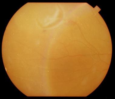
Human lens epithelial layer in cortical cataract. (1/122)
Normal and cataractous human eye lenses were studied by morphology and protein analysis. A marked decrease in protein sulfhydryl (PSH) and nonprotein sulfhydryl (NSPH) was observed in nuclear and cortical cataractous epithelia. Moreover, decrease in PSH contents and an increase in insoluble proteins were found to be correlated only in cortical cataractous epithelium which is also accompanied by various morphological abnormalities. In nuclear cataractous epithelium, however, there was very little insolubilisation of proteins. The epithelial morphology in nuclear cataracts was almost similar to normal lens epithelium. Hence, it is assumed that the protein insolubilisation and various morphological abnormalities are characteristics of cortical cataractous epithelium. This leads us to believe that opacification in cortical cataract might initiate in the epithelial layer. (+info)Conservation of gene expression during embryonic lens formation and cornea-lens transdifferentiation in Xenopus laevis. (2/122)
Few molecular comparisons have been made between the processes of embryogenesis and regeneration or transdifferentiation that lead to the formation of the same structures. In the amphibian, Xenopus laevis, the cornea can undergo transdifferentiation to form a lens when the original lens is removed during tadpole larval stages. Unlike the process of embryonic lens induction, cornea-lens transdifferentiation is elicited via a single inductive interaction involving factors produced by the neural retina. In this study, we compared the expression of a number of genes known to be activated during various phases of embryonic lens formation, during the process of cornea-lens transdifferentiation. mRNA expression was monitored via in situ hybridization using digoxigenin-labeled riboprobes of pax-6, Xotx2, xSOX3, XProx1, and gamma6-cry. We found that all of the genes studied are expressed during both embryogenesis and cornea-lens transdifferentiation, though in some cases their relative temporal sequences are not maintained. The reiterated expression of these genes suggests that a large suite of genes activated during embryonic lens formation are also involved in cornea-lens transdifferentiation. Ultimately functional tests will be required to determine whether they actually play similar roles in these processes. It is significant that the single inductive event responsible for initiating cornea-lens transdifferentiation triggers the expression of genes activated during both the early and late phases of embryonic lens induction. These findings have significant implications in terms of our current understanding of the "multistep" process of lens induction. Dev Dyn 1999;215:308-318. (+info)Endogenous casein kinase I catalyzes the phosphorylation of the lens fiber cell connexin49. (3/122)
The lens fiber cell-specific gap junction protein connexin49 is a substrate for a membrane-associated Ser/Thr protein kinase that can be extracted from lens cell membranes by 0.6 M KCl. However, the identity of this protein kinase has not been defined. In this report, evidence is presented indicating that it is casein kinase I. Thus, connexin49 was shown to be a substrate for purified casein kinase I but not for casein kinase II; the endogenous connexin49 protein kinase activity extracted from lens membranes with KCl was inhibited by the casein kinase I-specific inhibitor, N-(2-aminoethyl)-5-chloroisoquinoline-8-sulfonamide (CKI-7); the connexin49 protein kinase activity in the lens membrane KCl extract, which could be partially purified by gel filtration and affinity purification with a casein-Sepharose 4B column, copurified with casein kinase activity; phosphopeptide analysis showed that casein kinase I and the connexin49 protein kinase activity in the lens membrane KCl extract probably share the same phosphorylation sites in connexin49. Reverse transcription-PCR using total ovine lens RNA and casein kinase I isoform-specific oligonucleotide primers resulted in the amplification of cDNAs encoding casein kinase I-alpha and -gamma, while an in-gel casein kinase assay indicated casein kinase activity in the lens membrane KCl extract was associated with a major 39.2-kDa species, which is consistent with the 36 to 40-kDa size of casein kinase I-alpha in other animal species. These results demonstrate that the protein kinase activity present in the lens membrane 0.6 M KCl extract that catalyzes the phosphorylation of connexin49 is casein kinase I, probably the alpha isoform. (+info)Direct evidence for immiscible cholesterol domains in human ocular lens fiber cell plasma membranes. (4/122)
The molecular structure of human ocular lens fiber cell plasma membranes was examined directly using small angle x-ray diffraction approaches. A distinct biochemical feature of these membranes is their high relative levels of free cholesterol; the mole ratio of cholesterol to phospholipid (C/P) measured in these membranes ranges from 1 to 4. The organization of cholesterol in this membrane system is not well understood, however. In this study, the structure of plasma membrane samples isolated from nuclear (3.3 C/P) and cortical (2.4 C/P) regions of human lenses was evaluated with x-ray diffraction approaches. Meridional diffraction patterns obtained from the oriented membrane samples demonstrated the presence of an immiscible cholesterol domain with a unit cell periodicity of 34.0 A, consistent with a cholesterol monohydrate bilayer. The dimensions of the sterol-rich domains remained constant over a broad range of temperatures (5-20 degrees C) and relative humidity levels (31-97%). In contrast, dimensions of the surrounding sterol-poor phase were significantly affected by experimental conditions. Similar structural features were observed in membranes reconstituted from fiber cell plasma membrane lipid extracts. The results of this study indicate that the lens fiber cell plasma membrane is a complex structure consisting of separate sterol-rich and -poor domains. Maintenance of these separate domains may be required for the normal function of lens fiber cell plasma membrane and may interfere with the cataractogenic aggregation of soluble lens proteins at the membrane surface. (+info)Galectin-3 is associated with the plasma membrane of lens fiber cells. (5/122)
PURPOSE: To discover proteins that have the potential to contribute to the tight packing of fiber cells in the lens. METHODS: Crude fiber cell membranes were isolated from ovine lens cortex. Proteins were separated by two-dimensional gel electrophoresis, and selected protein spots identified by micro-sequencing. The identification of galectin-3 was confirmed by immunoblotting with a specific antibody. The association of galectin-3 with the fiber cell plasma membrane was investigated using immunofluorescence microscopy, solubilization trials with selected reagents, and immunoprecipitation to identify candidate ligands. RESULTS: A cluster of three protein spots with an apparent molecular weight of 31,000 and isoelectric points ranging between 7 and 8.5 were resolved and identified as galectin-3. This protein was associated peripherally with the fiber cell plasma membrane and interacted with MP20, an abundant intrinsic membrane protein that had been identified previously as a component of membrane junctions between fiber cells. CONCLUSIONS: The detection of galectin-3 in the lens is a novel result and adds to the growing list of lens proteins with adhesive properties. Its location at the fiber cell membrane and its association with the junction-forming MP20 is consistent with a potential role in the development or maintenance of the tightly packed lens tissue architecture. (+info)Risk factors for cortical, nuclear, and posterior subcapsular cataracts: the POLA study. Pathologies Oculaires Liees a l'Age. (6/122)
The POLA (Pathologies Oculaires Liees a L'Age) Study is a population-based study of cataract and age-related macular degeneration and their risk factors being carried out among 2,584 residents of Sete, southern France, aged 60-95 years. Recruitment took place between June 1995 and July 1997. Cataract classification was based on a standardized lens examination by slit lamp, according to Lens Opacities Classification System III. This paper presents results obtained from cross-sectional analysis of the first phase of the study. In polytomous logistic regression analyses, an increased risk of cataract was found for female sex (cataract surgery: odds ratio (OR) = 3.03; cortical cataract: OR = 1.67), brown irises (cortical, nuclear, and mixed cataracts: OR = 1.61), smoking (cataract surgery: OR = 2.34 for current smokers and OR = 3.75 for former smokers), known diabetes of 10 or more years' duration (posterior subcapsular, cortical, and mixed cataracts and cataract surgery: OR = 2.72), use of oral corticosteroids for at least 5 years (posterior subcapsular cataract: OR = 3.25), asthma or chronic bronchitis (cataract surgery: OR = 2.04), cancer (posterior subcapsular cataract: OR = 1.92), and cardiovascular disease (cortical cataract: OR = 1.96). Decreased risk of cataract was found with higher education (all types of cataract and cataract surgery: OR = 0.59), hypertension (cataract surgery: OR = 0.57), and high plasma retinol levels (nuclear and mixed cataracts and cataract surgery: OR = 0.75 for a 1-standard-deviation increase). Most of the risk factors identified in this study confirm the findings of other studies. The association of cataract with plasma retinol level requires further investigation. (+info)A human lens model of cortical cataract: Ca2+-induced protein loss, vimentin cleavage and opacification. (7/122)
PURPOSE: Cortical cataract in humans is associated with Ca2+ overload and protein loss, and although animal models of cataract have implicated Ca2+-activated proteases in this process, it remains to be determined whether the human lens responds in this manner to conditions of Ca2+ overload. The purpose of these experiments was to investigate Ca2+-induced opacification and proteolysis in the organ-cultured human lens. METHODS: Donor human lenses were cultured in Eagle's minimum essential medium (EMEM) for up to 14 days. The Ca2+ ionophore ionomycin was used to induce a Ca2+ overload. Lenses were loaded with [3H]-amino acids for 48 hours. After a 24-hour control efflux period, lenses were cultured in control EMEM (Ca2+ 1.8 mM), EMEM + 5 microM ionomycin, or EMEM + 5 microM ionomycin + 5 mM EGTA (Ca2+ < 1 microM). Efflux of proteins and transparency were monitored daily. Protein distribution and cytoskeletal proteolysis were analyzed at the end of the experiment. Cytoskeletal proteins were isolated and separated by sodium dodecyl sulfate-polyacrylamide gel electrophoresis (SDS-PAGE). Western blot analyses were probed with anti-vimentin antibody (clone V9) and detected by enhanced chemiluminescence. RESULTS: Lenses cultured under control conditions remained transparent for 14 days in EMEM with no added supplements or serum. The lenses synthesized proteins and had a low rate of protein efflux throughout the experimental period. Ionomycin treatment resulted in cortical opacification, which was inhibited when external Ca2+ was chelated with EGTA. Exposure to ionomycin also led to an efflux of [3H]-labeled protein, amounting to 41% of the labeled protein over the 7-day experimental period, compared with 12% in ionomycin + EGTA-treated lenses. Efflux was accounted for by loss from the lens soluble protein (crystallin) fraction. Western blot analysis of the cytoskeletal protein vimentin (56 kDa) revealed a distinct breakdown product of 48 kDa in ionomycin-treated lenses that was not present when Ca2+ was chelated with EGTA. In addition, high-molecular-weight proteins (approximately 115 kDa and 235 kDa) that cross-reacted with the vimentin antibody were observed in ionomycin-treated lenses. The Ca2+-induced changes were not age dependent. CONCLUSIONS: Human lenses can be successfully maintained in vitro, remaining transparent for extended periods. Increased intracellular Ca2+ induces cortical opacification in the human lens. Ca2+-dependent cleavage and cross-linking of vimentin supports possible roles for calpain and transglutaminase in the opacification process. This human lens calcium-induced opacification (HLCO) model enables investigation of the molecular mechanisms of opacification, and the data help to explain the loss of protein observed in human cortical cataractous lenses in vivo. (+info)Polymorphic glutathione S-transferases as genetic risk factors for senile cortical cataract in Estonians. (8/122)
PURPOSE: To investigate the possible association between glutathione S-transferase GSTM1, GSTM3, GSTT1, and GSTP1 polymorphism and the occurrence of age-related cataracts in Estonian patients. METHODS: Patients with cortical (155), nuclear (77), posterior subcapsular (120), mixed type (151) of senile cataract and control individuals (202) were phenotyped for GSTM1 and GSTT1 by enzyme-linked immunosorbent assay and genotyped for GSTM3 and GSTP1 by polymerase chain reaction. RESULTS: The frequency of the GSTM1-positive phenotype was significantly higher in the cortical cataract group (60.6%) than in the controls (45.0%) with odds ratio of 1.88 (95% CI, 1.23-2.94; P = 0.004). The cortical cataract risk associated with the GSTM1-positive phenotype was increased in carriers of the combined GSTM1-positive/GSTT1-positive phenotype (OR = 1.99; 95% CI, 1.30-3.11; P = 0.002) and the GSTM1-positive/GSTM3 AA genotype (OR = 2.28; 95% CI, 1.51-3.73; P < 0.001). The highest risk of cortical cataract was observed in patients having all three susceptible genotypes (OR = 2.56; 95% CI, 1.59-4.11; P < 0.001). Also, a significant interaction between the presence of the GSTP1* A allele and cortical cataract was found with prevalence of the GSTP1* A allele among the cortical cataract cases compared with the controls. Ninety-five percent of subjects with cortical cataract had the GSTP1 (AA, AB, or AC) genotype, whereas in controls 87% of persons had a genotype with GSTP1*A allele (OR = 3.1; 95% CI, 1.31-7.35; P = 0.007). In contrast to the GSTP1*A allele, the presence of the GSTP1*B allele in one or two copies leads to decreased cortical cataract risk (OR = 0.09 for GSTP1 BB genotype). CONCLUSIONS. The GSTM1-positive phenotype as well as the presence of the GSTP1*A allele may be a genetic risk factor for development of cortical cataract. (+info)The crystalline lens in the eye is composed of three main parts: the capsule, the cortex, and the nucleus. The lens cortex is the outer layer of the lens, located between the capsule and the nucleus. It is made up of proteins and water, and its primary function is to help refract (bend) light rays as they pass through the eye, contributing to the focusing power of the eye.
The cortex is more flexible than the central nucleus, allowing it to change shape and adjust the focus of the eye for different distances. However, with age, the lens cortex can become less elastic, leading to presbyopia, a common age-related condition that affects the ability to focus on close objects. Additionally, changes in the lens cortex have been associated with cataracts, a clouding of the lens that can impair vision.
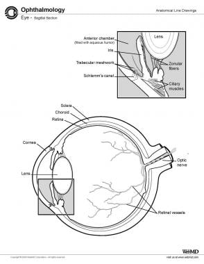
The crystalline lens is a biconvex transparent structure in the eye that helps to refract (bend) light rays and focus them onto the retina. It is located behind the iris and pupil and is suspended by small fibers called zonules that connect it to the ciliary body. The lens can change its shape to accommodate and focus on objects at different distances, a process known as accommodation. With age, the lens may become cloudy or opaque, leading to cataracts.
The lens nucleus, also known as the crystalline lens nucleus, is the central part of the crystalline lens in the eye. The crystalline lens is a biconvex structure located behind the iris and pupil, which helps to refract (bend) light rays and focus them onto the retina.
The lens nucleus is composed of densely packed lens fibers that have lost their nuclei and cytoplasm during differentiation. It is surrounded by the lens cortex, which consists of younger lens fiber cells that are still metabolically active. The lens nucleus is relatively avascular and receives its nutrients through diffusion from the aqueous humor in the anterior chamber of the eye.
The lens nucleus plays an important role in the accommodation process, which allows the eye to focus on objects at different distances. During accommodation, the ciliary muscles contract and release tension on the lens zonules, allowing the lens to become thicker and increase its curvature. This results in a decrease in the focal length of the lens and enables the eye to focus on nearby objects. The lens nucleus is more rigid than the cortex and helps maintain the shape of the lens during accommodation.
Changes in the lens nucleus are associated with several age-related eye conditions, including cataracts and presbyopia. Cataracts occur when the lens becomes cloudy or opaque, leading to a decrease in vision clarity. Presbyopia is a condition that affects the ability to focus on near objects and is caused by a hardening of the lens nucleus and a loss of elasticity in the lens fibers.
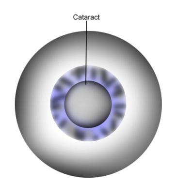
A cataract is a clouding of the natural lens in the eye that affects vision. This clouding can cause vision to become blurry, faded, or dim, making it difficult to see clearly. Cataracts are a common age-related condition, but they can also be caused by injury, disease, or medication use. In most cases, cataracts develop gradually over time and can be treated with surgery to remove the cloudy lens and replace it with an artificial one.
Crystallins are the major proteins found in the lens of the eye in vertebrates. They make up about 90% of the protein content in the lens and are responsible for maintaining the transparency and refractive properties of the lens, which are essential for clear vision. There are two main types of crystallins, alpha (α) and beta/gamma (β/γ), which are further divided into several subtypes. These proteins are highly stable and have a long half-life, which allows them to remain in the lens for an extended period of time. Mutations in crystallin genes have been associated with various eye disorders, including cataracts and certain types of glaucoma.
The cerebral cortex is the outermost layer of the brain, characterized by its intricate folded structure and wrinkled appearance. It is a region of great importance as it plays a key role in higher cognitive functions such as perception, consciousness, thought, memory, language, and attention. The cerebral cortex is divided into two hemispheres, each containing four lobes: the frontal, parietal, temporal, and occipital lobes. These areas are responsible for different functions, with some regions specializing in sensory processing while others are involved in motor control or associative functions. The cerebral cortex is composed of gray matter, which contains neuronal cell bodies, and is covered by a layer of white matter that consists mainly of myelinated nerve fibers.
In the context of medical terminology, "lenses" generally refers to optical lenses used in various medical devices and instruments. These lenses are typically made of glass or plastic and are designed to refract (bend) light in specific ways to help magnify, focus, or redirect images. Here are some examples:
1. In ophthalmology and optometry, lenses are used in eyeglasses, contact lenses, and ophthalmic instruments to correct vision problems like myopia (nearsightedness), hypermetropia (farsightedness), astigmatism, or presbyopia.
2. In surgical microscopes, lenses are used to provide a magnified and clear view of the operating field during microsurgical procedures like ophthalmic, neurosurgical, or ENT (Ear, Nose, Throat) surgeries.
3. In endoscopes and laparoscopes, lenses are used to transmit light and images from inside the body during minimally invasive surgical procedures.
4. In ophthalmic diagnostic instruments like slit lamps, lenses are used to examine various structures of the eye in detail.
In summary, "lenses" in medical terminology refer to optical components that help manipulate light to aid in diagnosis, treatment, or visual correction.
Contact lenses are thin, curved plastic or silicone hydrogel devices that are placed on the eye to correct vision, replace a missing or damaged cornea, or for cosmetic purposes. They rest on the surface of the eye, called the cornea, and conform to its shape. Contact lenses are designed to float on a thin layer of tears and move with each blink.
There are two main types of contact lenses: soft and rigid gas permeable (RGP). Soft contact lenses are made of flexible hydrophilic (water-absorbing) materials that allow oxygen to pass through the lens to the cornea. RGP lenses are made of harder, more oxygen-permeable materials.
Contact lenses can be used to correct various vision problems, including nearsightedness, farsightedness, astigmatism, and presbyopia. They come in different shapes, sizes, and powers to suit individual needs and preferences. Proper care, handling, and regular check-ups with an eye care professional are essential for maintaining good eye health and preventing complications associated with contact lens wear.
The prefrontal cortex is the anterior (frontal) part of the frontal lobe in the brain, involved in higher-order cognitive processes such as planning complex cognitive behavior, personality expression, decision making, and moderating social behavior. It also plays a significant role in working memory and executive functions. The prefrontal cortex is divided into several subregions, each associated with specific cognitive and emotional functions. Damage to the prefrontal cortex can result in various impairments, including difficulties with planning, decision making, and social behavior regulation.
The visual cortex is the part of the brain that processes visual information. It is located in the occipital lobe, which is at the back of the brain. The visual cortex is responsible for receiving and interpreting signals from the retina, which are then transmitted through the optic nerve and optic tract.
The visual cortex contains several areas that are involved in different aspects of visual processing, such as identifying shapes, colors, and movements. These areas work together to help us recognize and understand what we see. Damage to the visual cortex can result in various visual impairments, such as blindness or difficulty with visual perception.
The motor cortex is a region in the frontal lobe of the brain that is responsible for controlling voluntary movements. It is involved in planning, initiating, and executing movements of the limbs, body, and face. The motor cortex contains neurons called Betz cells, which have large cell bodies and are responsible for transmitting signals to the spinal cord to activate muscles. Damage to the motor cortex can result in various movement disorders such as hemiplegia or paralysis on one side of the body.
The auditory cortex is the region of the brain that is responsible for processing and analyzing sounds, including speech. It is located in the temporal lobe of the cerebral cortex, specifically within the Heschl's gyrus and the surrounding areas. The auditory cortex receives input from the auditory nerve, which carries sound information from the inner ear to the brain.
The auditory cortex is divided into several subregions that are responsible for different aspects of sound processing, such as pitch, volume, and location. These regions work together to help us recognize and interpret sounds in our environment, allowing us to communicate with others and respond appropriately to our surroundings. Damage to the auditory cortex can result in hearing loss or difficulty understanding speech.
Hydrophilic contact lenses are a type of contact lens that is designed to absorb and retain water. These lenses are made from materials that have an affinity for water, which helps them to remain moist and comfortable on the eye. The water content of hydrophilic contact lenses can vary, but typically ranges from 30-80% by weight.
Hydrophilic contact lenses are often used to correct refractive errors such as myopia (nearsightedness), hyperopia (farsightedness), and astigmatism. They can be made in a variety of materials, including soft hydrogel and silicone hydrogel.
One advantage of hydrophilic contact lenses is that they tend to be more comfortable to wear than other types of contacts, as they retain moisture and conform closely to the shape of the eye. However, they may also be more prone to deposits and buildup, which can lead to protein accumulation and discomfort over time. Proper care and cleaning are essential to maintain the health of the eyes when wearing hydrophilic contact lenses.
Intraocular lenses (IOLs) are artificial lens implants that are placed inside the eye during ophthalmic surgery, such as cataract removal. These lenses are designed to replace the natural lens of the eye that has become clouded or damaged, thereby restoring vision impairment caused by cataracts or other conditions.
There are several types of intraocular lenses available, including monofocal, multifocal, toric, and accommodative lenses. Monofocal IOLs provide clear vision at a single fixed distance, while multifocal IOLs offer clear vision at multiple distances. Toric IOLs are designed to correct astigmatism, and accommodative IOLs can change shape and position within the eye to allow for a range of vision.
The selection of the appropriate type of intraocular lens depends on various factors, including the patient's individual visual needs, lifestyle, and ocular health. The implantation procedure is typically performed on an outpatient basis and involves minimal discomfort or recovery time. Overall, intraocular lenses have become a safe and effective treatment option for patients with vision impairment due to cataracts or other eye conditions.
The somatosensory cortex is a part of the brain located in the postcentral gyrus of the parietal lobe, which is responsible for processing sensory information from the body. It receives and integrates tactile, proprioceptive, and thermoception inputs from the skin, muscles, joints, and internal organs, allowing us to perceive and interpret touch, pressure, pain, temperature, vibration, position, and movement of our body parts. The somatosensory cortex is organized in a map-like manner, known as the sensory homunculus, where each body part is represented according to its relative sensitivity and density of innervation. This organization allows for precise localization and discrimination of tactile stimuli across the body surface.
The crystalline lens of the eye is covered by a transparent, elastic capsule known as the lens capsule. This capsule is made up of collagen and forms the continuous outer layer of the lens. It is highly resistant to both physical and chemical insults, which allows it to protect the lens fibers within. The lens capsule is important for maintaining the shape and transparency of the lens, which are essential for proper focusing of light onto the retina.
Lens diseases refer to conditions that affect the lens of the eye, which is a transparent structure located behind the iris and pupil. The main function of the lens is to focus light onto the retina, enabling clear vision. Here are some examples of lens diseases:
1. Cataract: A cataract is a clouding of the lens that affects vision. It is a common age-related condition, but can also be caused by injury, disease, or medication.
2. Presbyopia: This is not strictly a "disease," but rather an age-related change in the lens that causes difficulty focusing on close objects. It typically becomes noticeable in people over the age of 40.
3. Lens dislocation: This occurs when the lens slips out of its normal position, usually due to trauma or a genetic disorder. It can cause vision problems and may require surgical intervention.
4. Lens opacity: This refers to any clouding or opacification of the lens that is not severe enough to be considered a cataract. It can cause visual symptoms such as glare or blurred vision.
5. Anterior subcapsular cataract: This is a type of cataract that forms in the front part of the lens, often as a result of injury or inflammation. It can cause significant visual impairment.
6. Posterior subcapsular cataract: This is another type of cataract that forms at the back of the lens, often as a result of diabetes or certain medications. It can also cause significant visual impairment.
Overall, lens diseases can have a significant impact on vision and quality of life, and may require medical intervention to manage or treat.
 List of MeSH codes (A09)
Lenticonus
Lens (vertebrate anatomy)
Refractive index
Visual impairment
Advanced glycation end-product
Neuroprosthetics
Beluga whale
Visual system
Phacoemulsification
Eye
Cataract
Retinal implant
List of dog diseases
Cataract surgery
Accommodation (vertebrate eye)
Croonian Medal
Mandarin (character)
Glossary of bird terms
Adult development
Glossary of botanical terms
List of MeSH codes (A09) - Wikipedia
Viscoelastic properties of fresh human lenses under 40 years of age: implications for the aetiology of presbyopia | British...
MESH TREE NUMBER CHANGES - 2008 MeSH
MESH TREE NUMBER CHANGES - 2008 MeSH
MESH TREE NUMBER CHANGES - 2008 MeSH
MESH TREE NUMBER CHANGES - 2008 MeSH
MESH TREE NUMBER CHANGES - 2008 MeSH
MESH TREE NUMBER CHANGES - 2008 MeSH
MESH TREE NUMBER CHANGES - 2008 MeSH
MESH TREE NUMBER CHANGES - 2008 MeSH
MESH TREE NUMBER CHANGES - 2008 MeSH
MESH TREE NUMBER CHANGES - 2008 MeSH
 MESH TREE NUMBER CHANGES - 2008 MeSH
 Beta crystallin a chain. Medical search
Cataract extraction. Medical search
 Lens, Crystalline | Harvard Catalyst Profiles | Harvard Catalyst
 Presbiopia
 Erowid LSD (Acid) Vault : Material Safety Data Sheet
 Ophthalmic Research Group - Fingerprint
- Aston Research Explorer
Ophthalmic Research Group - Fingerprint
- Aston Research Explorer
Cataract | Miyoshi eye clinic
 Eyes
Eyes
 Eye - AbsoluteAstronomy.com
Seeing Through Binoculars - See December 2023's Top Rated Pick
 Metamorphopsia - Definition, Causes, Symptoms and Treatment
 Molecular Expressions Microscopy Primer: Physics of Light and Color
 US Patent for Laser methods and systems for addressing, mitigating and reversing presbyopia Patent (Patent # 11,607,339 issued...
Eye Globe Anatomy: Overview, Extraocular Structures, Intraocular Structures
Eye Globe Anatomy: Overview, Extraocular Structures, Intraocular Structures
 AMD
AMD
 CRSTG | Europe Edition | Phacoemulsification in the Vitrectomized Eye
Capsule9
- Partial or complete opacity on or in the lens or capsule of one or both eyes, impairing vision or causing blindness. (lookformedical.com)
- The making of a continuous circular tear in the anterior capsule during cataract surgery in order to allow expression or phacoemulsification of the nucleus of the lens. (lookformedical.com)
- A partial or complete opacity of the lens and/or its capsule. (ackcsc.org)
- Cataract formation can also result after lens touch with intraocular instruments, in response to the introduction of intraocular tamponading agents such as silicone oil and gas, and if crystallization on the anterior hyaloid or posterior capsule results in posterior capsular lens feathering and inflammation. (crstodayeurope.com)
- It is a cataract-removal surgery that involves removing the front portion of the lens from the eye while retaining the posterior capsule. (scopeheal.com)
- After making the incisions, the crystalline lens capsule must be opened so that the opacified nucleus can be removed through this opening. (scopeheal.com)
- Once the posterior lens cortex and capsule have been hydrodissected, the surgeon can rotate the nucleus within the capsular bag to facilitate removal by phacoemulsification. (scopeheal.com)
- In my hands, prechopping the lens permits the safe use of the highest aspiration level, and the laser chop instrument helps me to manipulate pieces to the central safety zone and protects the posterior capsule during removal of the last portions of the lens. (crstoday.com)
- If, instead, I find a large, 5-mm-thick crystalline lens, I might make the opposite choice, because I would expect the lens to sit farther back in that capsule. (crstoday.com)
Nucleus4
- Conclusions The observed age-related increase in tissue stiffness of the lens nucleus, ∼0.4 Pa/year, is too small to account for the 10 dioptre decline in accommodative amplitude in this age group. (bmj.com)
- In the Cavalier King Charles Spaniel, onset is at an early age (less than 6 months), affecting the cortex and nucleus with rapid progression to complete cataract, resulting in blindness. (ackcsc.org)
- Drooped nucleus and retained lens fragment is a complication not so rare in clinical practice. (eophtha.com)
- Definisi Katarak adalah adanya kekeruhan pada lensa, bisa terjadi pada cortex, nucleus, atau kapsul posterior. (scribd.com)
RETINA15
- The transparent, semigelatinous substance that fills the cavity behind the CRYSTALLINE LENS of the EYE and in front of the RETINA. (lookformedical.com)
- Microphthalmia is a congenital defect characterized by a small eye often associated with other ocular malformations, including defects of the cornea, anterior chamber, lens and/or retina. (ackcsc.org)
- The crystalline lens is a transparent, biconvex structure in the eye that, along with the cornea, helps to refract light to be focused on the retina. (absoluteastronomy.com)
- Light that enters the eye is focused on the retina by the lens, a flexible biconvex crystal-like structure. (allhealthsite.com)
- Inside the retina, the light waves are transformed into electrical signals then transported to the occipital cortex through the optic nerve. (allhealthsite.com)
- Generally, sight occurs when light enters the eye through the cornea 101 and pupil, then proceeds through the ocular lens 103 through the vitreous 110 along the visual axis 104 , strikes the retina 105 at the back of the eye, forming an image at the macula 106 that is transferred by the optic nerve 107 to the brain. (justia.com)
- The space between the cornea 101 and the retina 105 is filled with a liquid called the aqueous 117 in the anterior chamber 109 and the vitreous 110 , a gel-like clear substance, in the chamber posterior to the lens. (justia.com)
- Within the layers of the retina, photons trigger a series of electrical and chemical reactions, ultimately sending electrical signals by way of the optic nerve, along with visual pathway to the occipital cortex. (medscape.com)
- By applying our model to the large-scale visual system model, it is expected to be able to evaluate the phenomena, for example, age dependent perception on visual illusions, blur compensation in retina or visual cortex, and so on. (biomedcentral.com)
- The lens changes shape to assist the cornea in focusing and directing light onto the retina. (myoptometristcalgary.ca)
- Light is focused by the cornea and through the lens to reach the retina, which is the highly sensitive inner lining of the eye. (myoptometristcalgary.ca)
- The receptors throughout the retina convert the images through the optic nerve, which transmits the image signals through the processing parts of the vision system to land at the visual cortex, the portion of our brain that is dedicated to the sense of sight. (myoptometristcalgary.ca)
- After passing through the cornea, the light is bent again by the lens for refinement and focused onto the retina in a process called refraction. (springhouseeye.net)
- The lens focuses the light on the retina. (emeraldparkeyecare.com)
- This is achieved by the ciliary muscles in the eye, changing the shape of the lens, bending or flattening it to focus the light rays on the retina. (emeraldparkeyecare.com)
Intraocular lenses7
- PURPOSE: To evaluate the visual outcome, light distortion index (LDI), and quality of life (QoL) of patients implanted with two complementary intraocular lenses (IOLs) to treat cataract and presbyopia. (bvsalud.org)
- Currently, the advanced multifocal intraocular lenses Minifell Lady ® (SIFI) and Technics Symphony ® Opti Blue (AMO) are called Extended depth of focus intraocular lens (EDOF) and have depth of focus. (miyoshi-eye-clinic.com)
- EDOF intraocular lenses have lower contrast sensitivity (quality of appearance), halo, glare (glare), while acquiring a far wider clear vision range (range of focus) compared to single focus intraocular lenses, ghost (you can see something like a shadow), and so on. (miyoshi-eye-clinic.com)
- EDOF intraocular lens is a lens that can be used more reliably than many traditional multifocal intraocular lenses, with fewer secondary symptoms, although the near vision will be slightly reduced. (miyoshi-eye-clinic.com)
- Especially Miniwell Lady ® (SIFI) thinks that it is one of the most superior intraocular lenses today with fewer side effects than Tecniques Symphony ® Opti Blue (AMO). (miyoshi-eye-clinic.com)
- Multifocal intraocular lenses are expensive due to their own burden, but patients who wish to reduce the frequency of use of eyeglasses after cataract surgery become more and more year by year, and more people who choose multifocal intraocular lens are increasing. (miyoshi-eye-clinic.com)
- Because multifocal intraocular lenses are highly functional, precise surgery and examination are required, and depending on the patient's eyes, personality and life, it may be worse than monofocal intraocular lenses. (miyoshi-eye-clinic.com)
Cerebral Cortex1
- Virtual histology of multi-modal magnetic resonance imaging of cerebral cortex in young men. (cardiff.ac.uk)
Aqueous humor3
- The space in the eye, filled with aqueous humor, bounded anteriorly by the cornea and a small portion of the sclera and posteriorly by a small portion of the ciliary body, the iris, and that part of the crystalline lens which presents through the pupil. (lookformedical.com)
- Light rays are bent in the eye as they encounter the cornea, aqueous humor, lens and vitreous humor. (allhealthsite.com)
- We do not know whether they, like the crystalline lens and aqueous humor of the eye, have the optical properties necessary for the functions we would assign to them. (emergentpublications.com)
Cataract surgery6
- A procedure for removal of the crystalline lens in cataract surgery in which an anterior capsulectomy is performed by means of a needle inserted through a small incision at the temporal limbus, allowing the lens contents to fall through the dilated pupil into the anterior chamber where they are broken up by the use of ultrasound and aspirated out of the eye through the incision. (lookformedical.com)
- An intraocular lens is an artificial lens that is implanted in the eye instead of the lens extracted by cataract surgery. (miyoshi-eye-clinic.com)
- The intraocular lens affects the most visible operation (quality of appearance, quality of life) of various surgical items and devices related to cataract surgery. (miyoshi-eye-clinic.com)
- If you would like a multifocal intraocular lens during cataract surgery, if you are interested, please do not hesitate to contact your doctor or staff. (miyoshi-eye-clinic.com)
- The Toric intraocular lens, instead of the lens cloudy in cataract surgery, intraocular lens to be implanted one of the, correct astigmatism you can. (miyoshi-eye-clinic.com)
- A significant advantage of the femtosecond laser-and one that was possibly not anticipated in the early days of laser cataract surgery-is that the incorporated OCT imaging gives the surgeon much more information about the anatomy of the eye, the anterior chamber, and the lens thickness. (crstoday.com)
Retinal2
- Therefore, we have developed an eye optics model which can calculate the blurred retinal image based on known physiological evidences to provide a more realistic input to retinal and visual cortex model. (biomedcentral.com)
- En face OCT reflectance images which accompany OCTA studies offer a glimpse of the macrophage-like cellular activity above the retinal surface which responds to systemically instigated vascular events below. (stanford.edu)
Cataractous1
- The removal of a cataractous CRYSTALLINE LENS from the eye. (lookformedical.com)
Presbyopia4
- BACKGROUND: As the trend of refractive lens exchange for presbyopia continues to grow, our case report shows the first occurrence of an acute bilateral outer retinopathy following uncomplicated sequential clear lens extraction in an otherwise healthy individual. (bvsalud.org)
- CASE PRESENTATION: A 54-year-old male without significant medical history benefited from a sequential bilateral lens exchange for presbyopia. (bvsalud.org)
- The present invention relates to systems and methods for treating the structure of the natural human crystalline lens with a laser to address a variety of medical conditions, such as presbyopia and refractive error. (justia.com)
- This condition is called presbyopia, and results from the crystalline lens being less flexible, and therefore less able to bend light. (emeraldparkeyecare.com)
Artificial lens2
- Insertion of an artificial lens to replace the natural CRYSTALLINE LENS after CATARACT EXTRACTION or to supplement the natural lens which is left in place. (lookformedical.com)
- After this, the cataract surgeon inserts a new artificial lens called as intraocular lens (IOL) and the procedure is called intraocular lens implantation.Mrs. Khan showed tremendous improvement in her vision post-surgery. (rnreyecare.com)
Iris5
- The ocular or natural crystalline lens 103 , a more detailed picture of which is shown in FIG. 8 A , (utilizing similar reference numbers for similar structures) is located just posterior to the iris 102 . (justia.com)
- Light is further converged by the crystalline lens located posterior to the iris. (medscape.com)
- Located behind the iris, is our natural lens. (myoptometristcalgary.ca)
- The crystalline lens is located behind the iris, controlling the focusing ability of the eye. (myoptometristcalgary.ca)
- After passing through the pupil (the aperture in the iris diaphragm) light is further refracted by the crystalline lens . (answersingenesis.org)
Scleral Lenses2
- Scleral Lenses: Current Practice and Future Directions. (harvard.edu)
- This presentation used two imaging methods to measure ocular sagittal height to fit scleral lenses in eyes with and without keratoconus. (coetf.ca)
Avian1
- Alpha, beta, and delta crystallins occur in avian and reptilian lenses, while alpha, beta, and gamma crystallins occur in all other lenses. (lookformedical.com)
Optic nerve1
- Millions of impulses travel along the nerve fibres of the optic nerve at the back of the eye, eventually arriving at the visual cortex (and other areas) of the brain, located at the back of the head. (emeraldparkeyecare.com)
Refractive2
- A class of crystallins that provides refractive power and translucency to the lens (LENS, CRYSTALLINE) in VERTEBRATES. (lookformedical.com)
- Stroud has pointed out that if we had only one such substance to work with, we could still build a lens into our receptors to secure differential sensitivity to different wavelengths, and that the cones with their refractive globules may do just that. (emergentpublications.com)
Visual7
- Ocular Impression-Based Scleral Lens With Wavefront-Guided Optics for Visual Improvement in Keratoconus. (harvard.edu)
- Generally, the ocular lens changes shape through the action of the ciliary muscle 108 to allow for focusing of a visual image. (justia.com)
- Within the occipital cortices, these electrical signals are processed and interpreted, (ie, "seen") by the brain as a visual image. (medscape.com)
- When these signals reach the visual cortex located in the back of the brain, they are deciphered and translated, so we can make sense of the visual information. (springhouseeye.net)
- 9 - 11 The goal of prevention is supported by (1) prospective studies of animal models demonstrating that abnormal visual experience can lead to abnormal development of synaptic circuitry in visual cortex, 12 - 18 and (2) the hypothesis that providing "normal" visual experience to human infants will prevent the development of clinical abnormality. (arvojournals.org)
- On the basis of destructive changes in certain layers of the geniculate produced by raising monkeys in a red light, and on the basis of the destruction of their layers by toxins producing a loss of sensitivity to blue light, Le Gros Clark thinks that colors are separated into separate channels in the lateral geniculate, and so, in their projection to the visual cortex, notably the area striata. (emergentpublications.com)
- Upon detailed eye examination, which began with Visual Acuity, it showed that vision loss was because of cloudy lens i.e. she had developed moderate cataract (Anterior capsular cataract along with Nuclear Sclerosis Grade II) mainly due to her age. (rnreyecare.com)
Cataracts5
- Cataracts are a disease in which the crystalline lens becomes clouded. (miyoshi-eye-clinic.com)
- Cataracts may involve the lens completely (diffuse) or in a localized region. (ackcsc.org)
- Well-developed cataracts appear as gray, white, or yellow-brown opacities in the lens. (msdmanuals.com)
- Dense white cataracts tend to be in older patients and on slit lamp examination there is a yellow to brown hue to the central portion of the crystalline lens. (cataractcoach.com)
- These white intumescent cataracts pose a challenge during capsulorrhexis creation because the intra-lenticular pressure increases as the lens cortex liquefies. (cataractcoach.com)
Capsular4
- In a routine cataract, the lens material is solid and the pressure within the capsular bag is lower than the pressure in the anterior chamber, making capsulorrhexis creation straightforward. (cataractcoach.com)
- But with the white intumescent cataract, the liquefied cortex increases the intracapsular pressure and forces the capsular bag to rip uncontrollably once it is opened. (cataractcoach.com)
- Is there fluid from decomposed lens proteins within the capsular bag? (cataractcoach.com)
- The dreaded Argentinian flag sign is due to pressure within the capsular bag due to the liquified cortex (intumescent cataract). (cataractcoach.com)
Proteins5
- A heterogeneous family of water-soluble structural proteins found in cells of the vertebrate lens. (lookformedical.com)
- The presence of these proteins accounts for the transparency of the lens. (lookformedical.com)
- Alpha-crystallins also act as molecular chaperones that bind to denatured proteins, keep them in solution and thereby maintain the translucency of the lens. (lookformedical.com)
- This presentation reviews what will happen when confocal images are used to model and simulate contractions of proteins in the chicken lens. (coetf.ca)
- The lens is a transparent eye structure made mostly of proteins that plays a role in bending and focusing light. (springhouseeye.net)
Ocular3
- The terms ocular lens, natural crystalline lens, natural lens, natural human crystalline lens, and lens (when referring to the prior terms) are used interchangeably herein and refer to the same anatomical structure of the human eye. (justia.com)
- A neural feedback mechanism from the brain allows the ciliary muscle 108 , acting through the attachment of the zonules 111 , to change the shape of the ocular lens. (justia.com)
- An ocular cataract is the clouding of the natural lens of the eye, which causes considerable loss of vision, especially in people over 40 years of age. (scopeheal.com)
Fragment1
- An ultrasonic probe is used to fragment the opacified crystalline lens, and the resulting material is extracted through a minimal incision using a suction system always under the control of the surgeon. (scopeheal.com)
Ciliary2
- The lens is located in the ciliary body. (allhealthsite.com)
- The lens is held upright in the eye by suspensory ligaments attached to the ciliary body. (allhealthsite.com)
Cortical1
- On the other hand, a cortical cataract begins at the periphery of the lens and spreads toward the center. (scopeheal.com)
Anterior chamber2
- After placing Healon (Abbott Medical Optics) in the anterior chamber, I direct a 30-gauge cannula on a Healon syringe into the center of the lens and inject the OVD (Figure 1). (crstoday.com)
- Knowing how big the crystalline lens is in relation to the anterior chamber helps me prepare for surgery and may eventually allow me to more accurately predict the effective lens position (ELP). (crstoday.com)
Extraction3
- Absence of the crystalline lens resulting from cataract extraction. (lookformedical.com)
- Presence of an intraocular lens after cataract extraction. (lookformedical.com)
- Predictors of long-term intraocular pressure control after lens extraction in primary angle closure glaucoma: results from the EAGLE trial. (harvard.edu)
Determines1
- I base emulsification settings on the grade of cataract, which determines how I want to chop the lens, the grid pattern I want to use, and how much energy I want to put into softening the lens. (crstoday.com)
Nuclear2
- Clouding at the back of the lens is called a subscapular cataract, while cloudiness at the core of the lens is called a nuclear cataract. (scopeheal.com)
- central nuclear sclerosis with yellow tint, with periphery of cataract showing less opacity indicating intact (non-liquid) cortex. (cataractcoach.com)
Opacity of the lens1
- A cataract is a congenital or degenerative opacity of the lens. (msdmanuals.com)
Procedure1
- In my opinion, the greatest benefits of the procedure are seen in the new information provided by the laser's imaging capabilities, lens fragmentation, and the creation of uniform, precise capsulotomies. (crstoday.com)
Viscoelastic2
- Aim To determine the viscoelastic properties of fresh human lenses obtained from cadavers under 40 years of age. (bmj.com)
- The viscoelastic properties of the lens nuclei and 16 of the lens cortices were quantified within 42±10 h of death using a controlled-strain, linear, simple-shear rheometer. (bmj.com)
Diseases1
- Lens diseases refer to conditions that affect the lens of the eye, impairing vision and potentially leading to blindness if left untreated. (lookformedical.com)
Eye's2
- Light enters the human eye via the transparent cornea, the eye's front window, which acts as a powerful convex lens. (answersingenesis.org)
- The working of eye's lens is similar to that of camera's lens, which is to focus light properly so that image can be viewed clearly. (rnreyecare.com)
Ultrasonic1
- A device emits ultrasonic energy on the cloudy lens that breaks the lens into small pieces, which are then removed by suction. (rnreyecare.com)
Contrast1
- Conventional multifocal intraocular lens suffered from problems such as reduction in contrast sensitivity and secondary symptoms such as halo, glare, ghost, etc., in order to obtain high visibility capability. (miyoshi-eye-clinic.com)
Outer1
- The thin noncellular outer covering of the CRYSTALLINE LENS composed mainly of COLLAGEN TYPE IV and GLYCOSAMINOGLYCANS. (lookformedical.com)
Contact Lenses1
- Since changing the apparent refraction of the eye is relatively easy through the use of corrective spectacle or contact lenses, many of the conditions that contribute to unclear vision can be readily corrected. (emeraldparkeyecare.com)
Vision3
- By the ligament-like structure supporting the lens, the eye can change focus between close and far vision. (myoptometristcalgary.ca)
- he evaluated her eyes and explained that the hazy vision in her eyes was because of the cloudy lens. (rnreyecare.com)
- Mrs. Khan opted for a multifocal lens (a lens which allow clear vision for near as well as far without glasses).In Phacoemulsification, a micro incision is made on the side of the cornea. (rnreyecare.com)
Fluid1
- Following capsulorhexis, fluid irrigation or hydrodissection is performed gently to separate the lens capsule's cortex. (scopeheal.com)
Methods2
- Methods 52 intact clear, human lenses were obtained from 26 donors (mean age of 27.5±9.2 years) within 9±4 h of death. (bmj.com)
- Systems, methods and laser delivery patterns and operations for structural pillars in the lens of the eye to permit deformation of laser effected areas of the lens that are adjacent to the pillars. (justia.com)
Patients1
- Depending on the size of retained lens fragments, Patients present with varying degrees of inflammation. (eophtha.com)
Incision2
- If a rigid IOL is implanted thereafter, enlargement of the incision and suturing the lens to the bag may be required. (crstodayeurope.com)
- Another incision is made in the membrane surrounding the cataract so that it can be easily separated from the cortex using a stream of water. (scopeheal.com)
Affects1
- It affects the lens of the eyes and is usually seen in people over the age of 40. (rnreyecare.com)
Spectacle1
- The wavefront aberration is defined by Zernike Coefficients values (measured by wavefront aberrometer such as OPD-Scan or Shack-Hartman Sensor systems) or SCA values (power of sphero-cylindrical lens with its axis for spectacle lens or contact lens prescriptions). (biomedcentral.com)
Back2
- The image produce at the back of the lens is upside-down. (allhealthsite.com)
- If VR back up is available at the hospital it is best to remove the fragments/ lens in the same sitting. (eophtha.com)
Image2
- OTF (Optical transfer function) calculated from the wavefront aberration with spectral transmittance of the lens is used for image processing filter. (biomedcentral.com)
- The system uses sophisticated algorithms to process volumetric OCT image data, map the surfaces of the cornea and lens, and create safety zones, which I can then move or customize if necessary. (crstoday.com)