Lateral Sinus Thrombosis
Sinus Thrombosis, Intracranial
Cranial Sinuses
Sagittal Sinus Thrombosis
Cavernous Sinus Thrombosis
Mastoiditis
Mastoid air sinus abnormalities associated with lateral venous sinus thrombosis: cause or consequence? (1/10)
BACKGROUND: Mastoiditis is a known cause of lateral venous sinus thrombosis (LST). We have encountered patients with LST associated with mastoid abnormality on MRI without any clinical signs of infection; the significance of these abnormalities is uncertain. This study examines the relationship of LST and mastoid air sinus abnormalities systematically. SUMMARY OF REPORT: We performed a retrospective clinical and radiological review of a series of 26 patients with cerebral venous thrombosis. Mastoid abnormalities were detected ipsilateral to 9 of 23 thrombosed lateral sinuses (39%) and 0 of 29 unaffected lateral sinuses (P<0.001). No patient had clinical evidence of mastoiditis. Eight of 9 patients with mastoid abnormalities were treated without antibiotics; all made uneventful clinical recoveries. Repeated MRI in 1 patient revealed reversal of the mastoid changes. CONCLUSIONS: The mastoid changes observed are likely to be due to venous congestion as a consequence of LST, not mastoiditis. (+info)MR venography in idiopathic intracranial hypertension: unappreciated and misunderstood. (2/10)
BACKGROUND: Venous sinus disease must be excluded before diagnosing idiopathic intracranial hypertension but is found only rarely in typical cases. Magnetic resonance venography (MRV) is the technique of choice for investigating this, and provides images that are diagnostic and easy to interpret. However, recent work using more invasive techniques has documented pressure gradients and stenoses in the lateral venous sinuses in many cases of idiopathic intracranial hypertension. OBJECTIVE: To examine the reason for this discrepancy and to establish whether there are characteristic appearances on MRV in idiopathic intracranial hypertension that are routinely overlooked in clinical practice. METHODS: MRVs from 20 patients with idiopathic intracranial hypertension were reviewed, unblinded, by two neuroradiologists, and their appearances rated for focal narrowings and signal gaps. A control group of 40 asymptomatic volunteers, matched for age and sex with the patient group, was recruited prospectively for MRV, and their scans rated in the same way. RESULTS: The lateral sinuses presented a range of appearances with quite different distributions in the two groups (p<0.001). Bilateral lateral sinus flow gaps were seen in 13 of 20 patients with idiopathic intracranial hypertension and in none of 40 controls. CONCLUSIONS: A historical failure to use normal healthy controls to establish the boundaries between imaging artefact, normal anatomical variant, and disease means that the pathological significance of the different appearances of the lateral sinuses on MRV has not so far been appreciated. (+info)Lateral sinus thrombosis. (3/10)
We describe four cases of lateral sinus thrombosis secondary to otitis media. They presented with low-grade fever, headache, nausea, vomiting and ear discharge. One patient had facial nerve palsy. CT scan was helpful in managing these patients. They were treated with antibiotics followed by surgery. Two patients had intracranial abscesses and were treated accordingly. (+info)Lateral sinus thrombosis as a complication of acute mastoiditis. (4/10)
Lateral sinus thrombosis is a rare complication of middle ear diseases: in children, it is usually related to acute otitis media, but it is also found in adults with chronic otitis. It was more frequent in the pre-antibiotic era and mortality was high. The Authors present a paediatric case of lateral sinus thrombosis in which they describe the clinical approach and related literature. (+info)Isolated lateral sinus thrombosis: a series of 62 patients. (5/10)
(+info)Lateral sinus thrombosis as a complication of otitis media: 10-year experience at the children's hospital of Philadelphia. (6/10)
(+info)Stenting of a cerebral venous thrombosis. (7/10)
(+info)Cortical subarachnoid hemorrhage caused by cerebral venous thrombosis. (8/10)
Patients with non-traumatic, non-aneurysmal, and non-perimesencephalic subarachnoid hemorrhage (SAH) tend to have clots circumscribed along the cortical convexity, a condition referred to as acute cortical SAH. Cerebral venous thrombosis (CVT) is a potential cause of cortical SAH. The study tried to establish the diagnosis and management of cortical SAH caused by CVT. Retrospective review of 145 patients with non-traumatic SAH identified 15 patients with no ruptured aneurysm. Clinical features were investigated with a specific focus on patients with SAH caused by CVT. Eight of the 15 patients had perimesencephalic SAH, and 7 had cortical SAH. SAH caused by CVT was diagnosed in 4 of the 7 patients with cortical SAH. The cortical SAH involved the unilateral convexity or sylvian cistern and spared the basal cistern on computed tomography in all 4 patients. CVT occurred in the transverse sinus and cortical vein (1 patient), insular vein (1 patient), and cortical vein (2 patients). Identification of thrombosed veins or sinuses was established directly by T(2)*-weighted and diffusion-weighted magnetic resonance (MR) imaging in the acute stage and diffusion-weighted and T(1)-weighted MR imaging in the subacute stage. All patients had cortical swelling without findings of venous hemorrhagic infarction on T(2)*-weighted MR imaging. None of the 4 patients received active treatment, and all had favorable outcomes. CVT in patients with non-traumatic cortical SAH should be first excluded as a potential hemorrhagic cause by MR imaging for thrombosed veins or sinuses before initiating antifibrinolytic therapy. (+info)Lateral sinus thrombosis, also known as sigmoid sinus thrombosis, is a medical condition characterized by the formation of a blood clot (thrombus) in the lateral or sigmoid sinus, which are venous structures located in the skull that help drain blood from the brain.
The lateral sinuses are situated near the mastoid process of the temporal bone and can become thrombosed due to various reasons such as infection (often associated with ear or mastoid infections), trauma, tumors, or other underlying medical conditions that increase the risk of blood clot formation.
Symptoms of lateral sinus thrombosis may include headache, fever, neck stiffness, altered mental status, and signs of increased intracranial pressure such as papilledema (swelling of the optic nerve disc). Diagnosis is typically made with the help of imaging studies like CT or MRI scans, and treatment often involves anticoagulation therapy to prevent clot expansion and potential complications. In some cases, surgical intervention may be necessary to remove the clot or manage any underlying conditions.
Intracranial sinus thrombosis is a medical condition characterized by the formation of a blood clot (thrombus) within the intracranial venous sinuses, which are responsible for draining blood from the brain. The condition can lead to various neurological symptoms and complications, such as increased intracranial pressure, headaches, seizures, visual disturbances, and altered consciousness. Intracranial sinus thrombosis may result from various factors, including hypercoagulable states, infections, trauma, and malignancies. Immediate medical attention is necessary for proper diagnosis and treatment to prevent potential long-term neurological damage or even death.
Cranial sinuses are a part of the venous system in the human head. They are air-filled spaces located within the skull and are named according to their location. The cranial sinuses include:
1. Superior sagittal sinus: It runs along the top of the brain, inside the skull, and drains blood from the scalp and the veins of the brain.
2. Inferior sagittal sinus: It runs along the bottom of the brain and drains into the straight sinus.
3. Straight sinus: It is located at the back of the brain and receives blood from the inferior sagittal sinus and great cerebral vein.
4. Occipital sinuses: They are located at the back of the head and drain blood from the scalp and skull.
5. Cavernous sinuses: They are located on each side of the brain, near the temple, and receive blood from the eye and surrounding areas.
6. Sphenoparietal sinus: It is a small sinus that drains blood from the front part of the brain into the cavernous sinus.
7. Petrosquamosal sinuses: They are located near the ear and drain blood from the scalp and skull.
The cranial sinuses play an essential role in draining blood from the brain and protecting it from injury.
Sagittal sinus thrombosis is a medical condition that refers to the formation of a blood clot (thrombus) in the sagittal sinus, which is a venous structure located in the brain. The sagittal sinus runs along the midline of the brain and receives blood from the superficial veins of the brain.
Sagittal sinus thrombosis can occur as a result of various conditions, such as head trauma, infection, cancer, or certain medical disorders that cause hypercoagulability (an increased tendency to form blood clots). The formation of a blood clot in the sagittal sinus can obstruct the flow of blood from the brain, leading to symptoms such as headache, seizures, altered consciousness, and focal neurological deficits.
Diagnosis of sagittal sinus thrombosis typically involves imaging studies such as computed tomography (CT) or magnetic resonance imaging (MRI) scans, which can show the presence of a blood clot in the sagittal sinus. Treatment may involve administering anticoagulant medications to prevent further growth of the blood clot and reduce the risk of complications such as pulmonary embolism or cerebral infarction. In some cases, surgical intervention may be necessary to remove the blood clot or alleviate pressure on the brain.
Cavernous sinus thrombosis is a medical condition that refers to the formation of a blood clot (thrombus) in the cavernous sinuses, which are located near the base of the brain and are important for draining blood from the face and brain. This condition can occur as a complication of an infection in the facial area or sinuses, or it can be associated with other medical conditions such as cancer or trauma.
Symptoms of cavernous sinus thrombosis may include headache, fever, eye pain, swelling or bulging of the eyes, double vision, and decreased vision. If left untreated, this condition can lead to serious complications such as meningitis, brain abscess, or even death. Treatment typically involves administering antibiotics to treat any underlying infection and anticoagulants to prevent further clot formation. In some cases, surgery may be necessary to remove the clot.
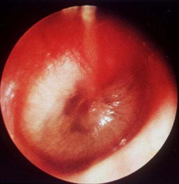
Mastoiditis is a medical condition characterized by an infection and inflammation of the mastoid process, which is the bony prominence located behind the ear. The mastoid process contains air cells that are connected to the middle ear, and an infection in the middle ear (otitis media) can spread to the mastoid process, resulting in mastoiditis.
The symptoms of mastoiditis may include:
* Pain and tenderness behind the ear
* Swelling or redness of the skin behind the ear
* Ear drainage or discharge
* Fever and headache
* Hearing loss or difficulty hearing
Mastoiditis is a serious condition that requires prompt medical attention. Treatment typically involves antibiotics to eliminate the infection, as well as possible surgical intervention if the infection does not respond to medication or if it has caused significant damage to the mastoid process. If left untreated, mastoiditis can lead to complications such as meningitis, brain abscess, or even death.
Intracranial thrombosis refers to the formation of a blood clot (thrombus) within the intracranial vessels, which supply blood to the brain. This condition can occur in any of the cerebral arteries or veins and can lead to serious complications such as ischemic stroke, transient ischemic attack (TIA), or venous sinus thrombosis.
The formation of an intracranial thrombus can be caused by various factors, including atherosclerosis, cardiac embolism, vasculitis, sickle cell disease, hypercoagulable states, and head trauma. Symptoms may vary depending on the location and extent of the thrombosis but often include sudden onset of headache, weakness or numbness in the face or limbs, difficulty speaking or understanding speech, vision changes, and loss of balance or coordination.
Diagnosis of intracranial thrombosis typically involves imaging studies such as computed tomography (CT) angiography, magnetic resonance angiography (MRA), or digital subtraction angiography (DSA). Treatment options may include anticoagulation therapy, thrombolysis, endovascular intervention, or surgical intervention, depending on the underlying cause and severity of the condition.
Cavernous sinus7
- Cavernous sinus thrombosis is amongst them, thus making it one of the main causes that need to be ruled in or out when a patient first presents to the ED with complaints of headache. (iem-student.org)
- They include the transverse , sigmoid and cavernous sinus, the superior sagittal sinus , inferior sagittal sinus and the straight sinus . (resus.com.au)
- There are reports on various other risks like rhinoliquorrhea, brain damage, fistulas between sinus-cavernosus and carotid artery, aneurysms and thrombosis of the cavernous sinus. (egms.de)
- It moves forward in the middle cranial fossa and enters into the cavernous sinus along with the internal carotid artery. (brainmadesimple.com)
- This whole ensheathed structure runs through the cavernous sinus. (brainmadesimple.com)
- Several mechanisms responsible for the headache have been suggested and they include irritation of the meninges and the superior division of the trigeminal nerve within the cavernous sinus , compression of the dura mater and sellar wall enlargement. (symptoma.com)
- In addition, other cranial nerves that course through the cavernous sinus can also be affected, in particular, cranial nerves III, IV, and VI with resulting diplopia . (symptoma.com)
Dural venous5
- The proximity of the middle ear and mastoid air cells to the dural venous sinuses predisposes them to thrombosis and thrombophlebitis secondary to infection and inflammation in the middle ear and mastoid. (medscape.com)
- The purpose of this study was to review MR venograms to elucidate developmental patterns and diameters of the major dural venous sinuses from 0 to 20 years of age. (ajnr.org)
- Patient age at the time of image acquisition was noted, and measurements were taken of the diameters of the major dural venous sinuses. (ajnr.org)
- All dural venous sinuses demonstrated a maximal growth rate from 0 to 7 years of age and reached maximal diameters around 5-10 years of age. (ajnr.org)
- Dural venous sinuses demonstrate maximal growth between 0 and 7 years of age and reach adult size around 5-10 years of age. (ajnr.org)
Abscess2
- The infection may decompress through a perforation in the tympanic membrane or extend through the lateral mastoid cortex, forming a postauricular subperiosteal abscess. (msdmanuals.com)
- Rarely, it extends centrally, causing a temporal lobe abscess or a septic thrombosis of the lateral sinus. (msdmanuals.com)
Vein7
- Five patients had a thrombosis in the jugular vein. (bvsalud.org)
- Prognosis of cerebral vein and dural sinus thrombosis: Results of the International Study on Cerebral Vein and Dural Sinus Thrombosis (ISCVT). (ac.ir)
- Cerebral vein thrombosis: Clinical manifestation and diagnosis. (ac.ir)
- Venous blood from cerebral veins drains into the major dural sinuses and the internal jugular vein. (iem-student.org)
- Pregnant woman carrying a fetus harboring a vein of Galen malformation in whom the straight sinus or falcine sinus draining the prosencephalic varix measures 8 mm or more on fetal MRI (medio-lateral diameter measured at the narrowest point of the sinus along the rostral-caudal axis, assessed on a T2-weighted coronal slice). (childrenshospital.org)
- Medical disease requiring current anticoagulation including maternal deep vein thrombosis. (childrenshospital.org)
- CXR may reveal evidence of septic pulmonary emboli (propagation of the thrombus into the inferior petrosal sinus and jugular vein). (hku.hk)
Right lateral sinus3
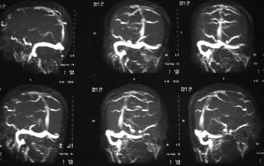
- This MRI study shows abnormal heterogeneous T2 high signals in the region of superior sagittal sinus and right lateral sinus, loss of normal T2 flow voids implies to thrombosis. (neuroradiologycases.com)
- We report a patient is 37 years with no particular history in which MRI led to the diagnosis of cerebral venous thrombosis on T1 and T2 sequences in the right lateral sinus and veno MRI in the acute phase. (issr-journals.org)
- One month later a check showed recanalization of the right lateral sinus. (issr-journals.org)
Thrombophlebitis4
- Lateral sinus thrombophlebitis as a complication of otogenic infection may still pose a serious threat that warrants immediate attention and care(1). (arquivosdeorl.org.br)
- Large series of lateral sinus thrombophlebitis (LST) have been reported from South Africa(3,4) and Iran(5), where access to health care is limited, but LST is rarely seen in western developed countries(6-10). (arquivosdeorl.org.br)
- Emaciation and anaemia were once considered common findings in sinus thrombophlebitis. (arquivosdeorl.org.br)
- The clinical manifestations of suppurative intracranial thrombophlebitis depend on the sinus involved, the involvement of anatomical structures within the sinus, and coexisting central nervous system infection. (hku.hk)
Septic thrombosis1
- Occasionally, findings compatible with frank meningitis may be present in patients with septic thrombosis of the superior sagittal sinus. (hku.hk)
Transverse sinus5
- Dominance patterns of the transverse sinus system were determined. (ajnr.org)
- The prevalence of persistent prenatal sinuses and transverse sinus-dominance patterns was compared across ages. (ajnr.org)
- This study consisted of the following 4 objectives: 1) to elucidate the growth patterns of each dural venous sinus from birth to 20 years of age, 2) to compare the mean size of each dural venous sinus among ages, 3) to compare the prevalence of persistent prenatal sinuses among ages, and 4) to determine the prevalence of transverse sinus-dominance patterns among ages. (ajnr.org)
- The dural sinuses consist of the superior sagittal sinus, straight sinus, and transverse sinus. (iem-student.org)
- Transverse sinus thrombosis and IVIg treatment: a case report and discussion of risk-benefit assessment for immunoglobulin treatment. (ox.ac.uk)
Mastoiditis1
- A retrospective study by Schneider et al suggested that pediatric patients with otogenic lateral sinus thrombosis secondary to acute otitis media and mastoiditis may have an underlying thrombophilic condition. (medscape.com)
Acute3
- Before the advent of antibiotics, which brought about a decline in this condition, most lateral sinus thrombosis was attributable to acute otitis media. (medscape.com)
- Lateral sinus thrombosis was ranked second to meningitis in the preantibiotic era as the most frequent fatal complication of otitis media and lateral sinus thrombosis occurred largely as a complication of acute otitis media. (medscape.com)
- The infection results from rapid spread of fungi from the paranasal sinuses to the adjacent orbits and central nervous system with hyphal invasion of blood vessels, vasculitis with thrombosis, haemorrhage, tissue infarction and acute neutrophilic infiltrates with a necrotising pathological reaction 5 . (actaitalica.it)
Diagnosis6
- Lateral sinus thrombosis is a potentially fatal condition in which early diagnosis may be difficult because of previous antibiotic therapy. (medscape.com)
- Post-traumatic cerebral venous sinus thrombosis is one of the several causes of cerebral venous thrombosis, but its early diagnosis and management are still difficult in this traumatic context. (bvsalud.org)
- Although the list of differentials is long, cerebral venous thrombosis should definitely be kept amongst the top 3, as early diagnosis is key. (iem-student.org)
- Due to its clinical polymorphism diagnosis of cerebral venous thrombosis is often hidden. (issr-journals.org)
- The prognosis is usually reserved compared to arterial thrombosis diagnosis but deserves to be placed early to avoid disabling sequelae. (issr-journals.org)
- Cerebral venous thrombosis is a rare diagnosis, but a very important diagnosis to make. (resus.com.au)
Paranasal sinuses1
- The most important anatomic structures below the anterior cranial fossa are the orbits and the paranasal sinuses. (medscape.com)
Cerebral veins3
- Stam J. Thrombosis of the cerebral veins and sinuses. (ac.ir)
- CVT is the formation of a clot in the cerebral veins and the dural sinuses. (iem-student.org)
- Cerebral veins include the dural sinus and cerebral veins. (resus.com.au)
Epidural1
- The retrospective review studied cases over a five-year period (2003-2008) involving complications in patients ranging from newborns to 18 years old, and found that complications continue to occur, with most cases involving lateral sinus thrombosis and epidural abscesses. (scienceblog.com)
Complication2
- Otogenic lateral sinus thrombosis is a well-known intracranial complication of otitis media . (medscape.com)
- Lateral sinus thrombosis usually develops as a complication of chronic otitis media caused by the direct dissemination of the infection through the neighboring eroded bone. (medscape.com)
Intracranial hypertension3
- Lateral sinus thrombosis and intracranial hypertension associated with primary hypothyroidism: case report. (nel.edu)
- Chen Q, Yao Z, Zhou D, Zheng H, Shang H. Lateral sinus thrombosis and intracranial hypertension associated with primary hypothyroidism: case report. (nel.edu)
- Biousse V, Ameri A, Bousser MG. Isolated intracranial hypertension as the only sign of cerebral venous thrombosis. (ac.ir)
Falcine sinus2
- The presence of embryonic sinuses including the persistent falcine sinus and the occipital sinus was noted. (ajnr.org)
- Fetus with VOGM in whom the straight sinus or falcine sinus draining the prosencephalic varix measures less than 8 mm on fetal MRI (T2-weighted coronal slice, medio-lateral diameter measured at the narrowest point of the sinus along the anterior-posterior axis), fitting fetal MRI criteria for likely evolution into the IT cohort. (childrenshospital.org)
Hemorrhage3
- The majority of DVAs are found incidentally and never cause symptoms, although there are isolated reports of patients with syndromes attributed to DVAs (eg, secondary to hemorrhage or thrombosis). (medscape.com)
- While some believe that DVAs can hemorrhage on their own, most notably after venous infarction from spontaneous DVA thrombosis, most instances of hemorrhage with DVAs have been in patients with combined vascular malformations. (medscape.com)
- This is generally the result of sudden hemorrhage and necrosis in the lateral pituitary fossa, leading to the displacement of the oculomotor nerves. (symptoma.com)
Sigmoid2
- the sigmoid sinus was affected in 6 patients. (bvsalud.org)
- Chronic thrombosis of superior sagittal and right transverse sigmoid sinus with partial recanalization. (neuroradiologycases.com)
Sagittal5
- Sagittal T1 for cross sectional view of sinus, sinus is not very bulky iso intense to adjacent parenchyma. (neuroradiologycases.com)
- The superficial system mainly drains into the superior sagittal sinus and the lateral sinus. (iem-student.org)
- A 54 year old woman presented with symptoms resulting from a thrombosis of the lateral transverse and sagittal sinuses the day after an infusion of intravenous immunoglobulin (IVIg) replacement treatment. (ox.ac.uk)
- The foramen cecum sits between the frontal crest and the prominent crista galli and is a site of communication between the draining veins of the nasal cavity and the superior sagittal sinus. (medscape.com)
- The posterior wall is thin and adjacent to the superior sagittal sinus and frontal lobe dura. (medscape.com)
Headache4
- Lateral sinus thrombosis should be suspected in patients who have persistent fever, otorrhea, and headache despite adequate antibiotic treatment. (medscape.com)
- Severe generalized fronto occipital headache, earache, nausea, vomiting, diplopia, sixth nerve palsy, loss of visual acuity, hemiparesis and picket fence fevers, were the major signs and symptoms described in cases of lateral sinus thrombosis before the advent of antibiotics(12,13). (arquivosdeorl.org.br)
- Crassard I, Bousser MG. Headache in patients with cerebral venous thrombosis. (ac.ir)
- There appears to be no association between the location of the headache and the site of venous thrombosis. (resus.com.au)
Rectus muscle2
- Inferior petrosal sinus: ipsilateral facial pain and lateral rectus muscle involvement. (hku.hk)
- Here, it moves towards the lateral wall of the orbit and supplies the lateral rectus muscle. (brainmadesimple.com)
Papilledema2
- We analyzed the frequency of symptoms and risk factors of cerebral venous thrombosis and the intensity of papilledema as time passed, as also the frequency of the involved sinus, in two groups of patients with and without papilledema. (ac.ir)
- Factors Influencing the Incidence of Papilledema in Patients with Cerebral Venous Thrombosis', Advanced Biomedical Research , 2017(December), pp. 1-5. (ac.ir)
Recanalization1
- A follow-up of MRI or CT scan at 3 months revealed complete sinus recanalization in three patients. (bvsalud.org)
Sphenoid sinus1
- The anterior clinoid processes and the planum sphenoidale, which forms the roof of the sphenoid sinus, mark the posterior limit. (medscape.com)
Inferior1
- These muscles include four recti (lateral, medial, superior, and inferior) and two obliques (superior and inferior) and are involved in eyeball movements. (brainmadesimple.com)
Cerebrovascular3
- The role of the dural venous sinus system in cerebrovascular pathology and the understanding of normal developmental patterns and sizes of the dural venous sinus system continue to expand. (ajnr.org)
- Evidence continues to accumulate supporting the idea that the dural venous sinus (DVS) system is a plastic, active player in cerebrovascular pathology rather than a fixed and immutable entity. (ajnr.org)
- Cerebral venous thrombosis (CVT) is an uncommon cerebrovascular disease with a wide spectrum of symptoms and severity. (ac.ir)
Otogenic2
- An image depicting otogenic lateral sinus thrombosis can be seen below. (medscape.com)
- Similarly, a retrospective study by Scorpecci et al found that out of 25 pediatric patients with otogenic lateral sinus thrombosis, 24 (96%) demonstrated a thrombophilia-related genetic abnormality. (medscape.com)
Brain1
- Post-traumatic cerebral venous sinus thrombosis in the intensive care department remains underdiagnosed because of the common clinical presentation of traumatic brain injury. (bvsalud.org)
Cranial1
- The petro-occipital fissure subdivides the middle cranial fossa into 1 central component and 2 lateral components. (medscape.com)
Carotid1
- amongst them cerebral venous thrombosis and carotid artery dissection. (resus.com.au)
Venogram2
- MR venogram that shows nonfilling of the lateral sinus on the left side. (medscape.com)
- Eccentric T2 flow voids in the region of sinuses are the partially recanlised channels, which show poor flow related signals on 2D TOF MR Venogram implies to chronic thrombosis. (neuroradiologycases.com)
Ethmoid2
- Chronic IFRS is an indolent infection with a slow destructive process that most commonly affects the ethmoid and sphenoid sinuses, but may involve any paranasal sinus 6 , 7 . (actaitalica.it)
- The cribriform plate may be more than 1 cm lower than the roof of the ethmoid cavity (fovea ethmoidalis), and it is made of extremely thin bone compared with the relatively thick bone of the lateral fovea ethmoidalis. (medscape.com)
Symptoms1
- In the antibiotic era, the presentation of lateral sinus thrombosis has changed from pronounced signs and symptoms to vague and nonspecific symptoms. (medscape.com)
Chronic1
- Those suffering from headaches, jaw pain (TMJ), neck pain, chronic sinus or nasal congestion and difficulty breathing often experience significant relief after the treatment. (painreliefhq.com)
Frontal sinus1
- The anterior limit of the anterior skull base is the posterior wall of the frontal sinus. (medscape.com)
Pathology1
- Three decades later, the pathology of lateral sinus thrombosis was first described by Lebert. (medscape.com)
Strokes1
- Cerebral Venous Thrombosis(CVT) represents approximately 1% of strokes(1), and tends to occur in the younger population ie. (resus.com.au)
Facial1
- Pressure or distortion of the bones of the skull may manifest as any number of conditions like migraine headaches, sinus issues, jaw disorders (TMJ), facial asymmetry, ear infections and more. (painreliefhq.com)
Endoscopic2
- This retrospective monocentric study included 17 patients who underwent endoscopic sinus surgery evaluated by paranasal computed tomography and magnetic resonance imaging. (actaitalica.it)
- This single-centre retrospective study included 17 patients affected by IFRS who underwent endoscopic sinus surgery (ESS) at the ENT Department in San Luigi Gonzaga Hospital, Turin, Italy between January 2016 and January 2020. (actaitalica.it)
Superior2
- Radiological studies including CT and MRI are superior to conventional sinus radiographs. (hku.hk)
- All the extraocular muscles are innervated by the oculomotor nerve (CN III) except the superior oblique and lateral rectus muscles, which are innervated by the trochlear nerve (CN IV) and abducent nerve (CN VI), respectively. (brainmadesimple.com)
Disease1
- Lateral sinus thrombosis accounts for 6% of all intracranial complications in the era of antibiotic treatment of suppurative ear disease. (medscape.com)
Orbital2
- As the name shows, the lateral rectus is a small straight muscle present on the lateral side of the eyeball in the orbital cavity. (brainmadesimple.com)
- The lateral portion of the IOF is an important surgical landmark for positioning lateral orbital osteotomies during anterior skull base resections. (medscape.com)