Selectins
P-Selectin
E-Selectin
L-Selectin
Fucosyltransferases
Leukocyte Rolling
Oligosaccharides
Antigens, CD15
Ligands
Fucose
Leukocytes
Cell Adhesion Molecules
Polysaccharides
Platelet Membrane Glycoproteins
Neutrophils
Leukocyte-Adhesion Deficiency Syndrome
N-Acetylglucosaminyltransferases
Mannosides
Mucins
Sialyltransferases
Endothelium, Vascular
Cell Movement
Sulfoglycosphingolipids
Carbohydrate Sequence
HL-60 Cells
Venules
Lewis Blood-Group System
Intercellular Adhesion Molecule-1
Antigens, CD18
Glycosyltransferases
Receptors, Lymphocyte Homing
Integrin alpha4
Chemotaxis, Leukocyte
N-Acetylneuraminic Acid
Amino Sugars
Vascular Cell Adhesion Molecule-1
Antigens, CD44
Flow Cytometry
Shear Strength
Integrin alpha4beta1
CHO Cells
Mice, Inbred C57BL
Mice, Knockout
Inflammation
Mannose
Carbohydrate Metabolism
Stress, Mechanical
Skin
Neutrophil Infiltration
Molecular Sequence Data
Glycosylation
Integrins
Cell Communication
Protein Binding
Cricetinae
Peritonitis
Microspheres
Lectins
Endothelium
Gangliosides
T-Lymphocytes
Neoplasm Metastasis
Blood Platelets
Leukocyte Count
Antigens, CD
Mice, Inbred BALB C
Hypersensitivity, Delayed
Antigens, Differentiation, T-Lymphocyte
Th1 Cells
Cells, Cultured
Peroxidase
Cricetulus
Models, Biological
Reperfusion Injury
Tumor Necrosis Factor-alpha
Monocytes
Transfection
Up-Regulation
Disease Models, Animal
Cytokines
Antibodies
Glycoproteins
Endothelial Cells
Amino Acid Sequence
Recombinant Fusion Proteins
Immunohistochemistry
Lymphocytes
RNA, Messenger
Antigens, Neoplasm
Biological Markers
Mutagenesis
Lipopolysaccharides
Binding Sites
Lung
Inhibition of L-selectin-mediated leukocyte rolling by synthetic glycoprotein mimics. (1/1317)
Synthetic carbohydrate and glycoprotein mimics displaying sulfated saccharide residues have been assayed for their L-selectin inhibitory properties under static and flow conditions. Polymers displaying the L-selectin recognition epitopes 3',6-disulfo Lewis x(Glc) (3-O-SO3-Galbeta1alpha4(Fucalpha1alpha3)-6-O-SO3-Glcbeta+ ++-OR) and 3',6'-disulfo Lewis x(Glc) (3, 6-di-O-SO3-Galbeta1alpha4(Fucalpha1alpha3)Glcbeta-OR) both inhibit L-selectin binding to heparin under static, cell-free binding conditions with similar efficacies. Under conditions of shear flow, however, only the polymer displaying 3',6-disulfo Lewis x(Glc) inhibits the rolling of L-selectin-transfected cells on the glycoprotein ligand GlyCAM-1. Although it has been shown to more effective than sialyl Lewis x at blocking the L-selectin-GlyCAM-1 interaction in static binding studies, the corresponding monomer had no effect in the dynamic assay. These data indicate that multivalent ligands are far more effective inhibitors of L-selectin-mediated rolling than their monovalent counterparts and that the inhibitory activities are dependent on the specific sulfation pattern of the recognition epitope. Importantly, our results indicate the L-selectin specificity for one ligand over another found in static, cell-free binding assays is not necessarily retained under the conditions of shear flow. The results suggest that monovalent or polyvalent carbohydrate or glycoprotein mimetics that inhibit selectin binding in static assays may not block the more physiologically relevant process of selectin-mediated rolling. (+info)Modulation of VLA-4 and L-selectin expression on normal CD34+ cells during mobilization with G-CSF. (2/1317)
We have evaluated the immunophenotype, functional activity and clonogenic potential of CD34+ cells from peripheral blood (PB) of normal donors before and after 4 and 6 days of G-CSF administration. The percentage and absolute number of CD34+ cells significantly increased at days 4 and 6 of G-CSF administration, compared to the steady-state level (P < 0.0001). Two-colour fluorescence analysis showed, at days 4 and 6, a lower proportion of CD34+/c-kit+ compared to the steady-state level (P < 0.0001), but a similar expression of CD13, CD33, CD38, HLA-DR and Thy-1 antigens on CD34+ cells. The expression of adhesion molecules on CD34+ cells revealed a significant reduction of CD11a (P = 0.009), CD18, CD49d and CD62L (P < 0.0001) at days 4 and 6, compared to the baseline level. Three-colour staining showed a reduction of the more immature compartment (34+/DR-/13-) and an increase of the more differentiated compartment (34+/DR+/13+). Downregulation of VLA-4 during mobilisation was seen almost exclusively on more committed cells (34+/13+); downregulation of CD62L, on the contrary, was observed on both early progenitors (34+/13-) and more committed cells (34+/13+). The expression of 34+/VLA-4+ decreased on both c-kit+ and c-kit- cells, while the expression of 34+/62L+ decreased on the c-kit+ cells only. In vivo administration of G-CSF reduced the adherence capacity of CD34+ cells to normal BM stroma; in vitro incubation with SCF or IL-3 enhanced the expression of CD49d on CD34+ cells, while GM-CSF reduced the expression of CD62L. SCF was the only cytokine able to induce a significant increase of CD34+ cell adherence to preformed stroma. Pre-incubation with the blocking beta2 integrin monoclonal antibody caused a reduction of CD34+ cell adherence. In conclusion, the decrease of CD49d expression on mobilized CD34+ cells correlates with a poor adhesion to BM stroma; CD34+ cells incubated in vitro with SCF showed, conversely, a higher expression of CD49d and a greater adherence capacity on normal preformed stroma. (+info)Identification and characterization of ligands for L-selectin in the kidney. II. Expression of chondroitin sulfate and heparan sulfate proteoglycans reactive with L-selectin. (3/1317)
Ligands for the leukocyte adhesion molecule L-selectin are expressed not only in lymph node high endothelial venules (HEV) but also in the renal distal tubuli. Here we report that L-selectin-reactive molecules in the kidney are chondroitin sulfate and heparan sulfate proteoglycans of 500-1000 kDa, unlike those in HEV bearing sialyl Lewis X-like carbohydrates. Binding of L-selectin to these molecules was mediated by the lectin domain of L-selectin and required divalent cations. Binding was inhibited by chondroitinase and/or heparitinase but not sialidase. Thus, L-selectin can recognize chondroitin sulfate and heparan sulfate glycosaminoglycans structurally distinct from sialyl Lewis X-like carbohydrates. (+info)Targeted disruption of SMAD3 results in impaired mucosal immunity and diminished T cell responsiveness to TGF-beta. (4/1317)
SMAD3 is one of the intracellular mediators that transduces signals from transforming growth factor-beta (TGF-beta) and activin receptors. We show that SMAD3 mutant mice generated by gene targeting die between 1 and 8 months due to a primary defect in immune function. Symptomatic mice exhibit thymic involution, enlarged lymph nodes, and formation of bacterial abscesses adjacent to mucosal surfaces. Mutant T cells exhibit an activated phenotype in vivo, and are not inhibited by TGF-beta1 in vitro. Mutant neutrophils are also impaired in their chemotactic response toward TGF-beta. Chronic intestinal inflammation is infrequently associated with colonic adenocarcinoma in mice older than 6 months of age. These data suggest that SMAD3 has an important role in TGF-beta-mediated regulation of T cell activation and mucosal immunity, and that the loss of these functions is responsible for chronic infection and the lethality of Smad3-null mice. (+info)Expression of the cell adhesion molecules on leukocytes that demarginate during acute maximal exercise. (5/1317)
The pulmonary vascular bed is an important reservoir for the marginated pool of leukocytes that can be mobilized by exercise or catecholamines. This study was designed to determine the phenotypic characteristics of leukocytes that are mobilized into the circulation during exercise. Twenty healthy volunteers performed incremental exercise to exhaustion [maximal O2 consumption (VO2 max)] on a cycle ergometer. Blood was collected at baseline, at 3-min intervals during exercise, at VO2 max, and 30 min after exercise. Total white cell, polymorphonuclear leukocyte (PMN), and lymphocyte counts increased with exercise to VO2 max (P < 0.05). Flow cytometric analysis showed that the mean fluorescence intensity of L-selectin on PMN (from 14.9 +/- 1 at baseline to 9.5 +/- 1.6 at VO2 max, P < 0.05) and lymphocytes (from 11.7 +/- 1.2 at baseline to 8 +/- 0.8 at VO2 max, P < 0.05) decreased with exercise. Mean fluorescence intensity of CD11b on PMN increased with exercise (from 10.2 +/- 0.6 at baseline to 25 +/- 2.5 at VO2 max, P < 0.002) but remained unchanged on lymphocytes. Myeloperoxidase levels in PMN did not change with exercise. In vitro studies showed that neither catecholamines nor plasma collected at VO2 max during exercise changed leukocyte L-selectin or CD11b levels. We conclude that PMN released from the marginated pool during exercise express low levels of L-selectin and high levels of CD11b. (+info)Morphine preconditioning attenuates neutrophil activation in rat models of myocardial infarction. (6/1317)
Previous results from our laboratory have suggested that morphine can attenuate neutrophil activation in patients with acute myocardial infarction. To elucidate if morphine preconditioning (PC) has the same effects via activation of neutrophil endopeptidase 24.11 (NEP), we measured serum levels of intercellular adhesion molecule-1 (ICAM-1), gp100MEL14 and NEP in adult Wistar rats subjected to ten different protocols (n = 10 for each) at baseline, immediately after and 2 h after morphine PC. All groups were subjected to 30 min of occlusion and 2 h of reperfusion. Similarly, morphine-induced PC was elicited by 3-min drug infusions (100 micrograms/kg) interspersed with 5-min drug-free periods before the prolonged 30-min occlusion. Infarct size (IS), as a percentage of the area at risk (AAR), was determined by triphenyltetrazolium staining. Pretreatment with morphine increased NEP activities (9.86 +/- 1.98 vs. 5.12 +/- 1.10 nmol/mg protein in control group; p < 0.001). Naloxone (mu-opioid receptor antagonist) (4.82 +/- 1.02 nmol/mg protein) and phosphoramidon (NEP inhibitor) (4.66 +/- 1.00 nmol/mg protein) inhibited morphine-activated NEP, whereas glibenclamide (ATP-sensitive potassium channel antagonist) and chelerythrine (protein kinase C inhibitor) had no effects. The ICAM-1 and gp100MEL14 of the third sampling were lowest for those with morphine PC (280 +/- 30 ng/ml and 2.2 +/- 0.7 micrograms/ml; p < 0.001), but naloxone (372 +/- 38 ng/ml and 3.8 +/- 0.9 micrograms/ml) and phosphoramidon (382 +/- 40 ng/ml and 4.2 +/- 1.1 micrograms/ml) abolished the above phenomenon. IS/AAR were definitely lowest for those with morphine PC (24 +/- 7%; p < 0.05). Morphine preconditioning increases NEP activities to attenuate shedding of gp100MEL14 and to ICAM-1 and, thus, provides myocardial protection. (+info)B cell response after MMTV infection: extrafollicular plasmablasts represent the main infected population and can transmit viral infection. (7/1317)
The immune response to mouse mammary tumor virus (MMTV) relies on the presentation of an MMTV-encoded superantigen by infected B cells to superantigen-specific T cells. The initial extrafollicular B cell differentiation involved the generation of B cells expressing low levels of B220. These B220low B cells corresponded to plasmablasts that expressed high levels of CD43 and syndecan-1 and were CD62 ligand- and IgD-. Viral DNA was detected nearly exclusively in these B220low B cells by PCR, and retroviral type-A particles were observed in their cytoplasm by electron microscopy. An MMTV transmission to the offspring was also achieved after transfer of B220low CD62 ligand- CD43+ plasmablasts into noninfected females. These data suggest that B220low plasmablasts, representing the bulk of infected B cells, are capable of sustaining viral replication and may be involved in the transmission of MMTV. (+info)Overlapping roles for L-selectin and P-selectin in antigen-induced immune responses in the microvasculature. (8/1317)
Although L-selectin mediates lymphocyte attachment to endothelial venules of peripheral lymph nodes, its role in leukocyte recruitment into tissues following Ag challenge is less well established. The objective of this study was to systematically examine the role of L-selectin in leukocyte rolling in the peripheral microvasculature during the first 24 h of an immune response. A type I hypersensitivity response was elicited in wild-type (C57BL/6) and L-selectin-deficient mice by systemic (i.p.) sensitization and intrascrotal challenge with chicken egg OVA. The cremaster microcirculation was observed in untreated and sensitized mice 4, 8, and 24 h post-Ag challenge by intravital microscopy. Leukocyte recruitment in L-selectin-deficient mice and wild-type mice treated with an L-selectin function-blocking mAb was examined at each time point. Ag challenge induced a significant increase in leukocyte rolling (60 cells/min/venule to approximately 300 cells/min/venule) in wild-type mice at 4-24 h. This response was reduced by approximately 60-70% in L-selectin-deficient mice and in wild-type mice treated with an L-selectin-blocking mAb. P-selectin blockade by Ab completely inhibited leukocyte rolling at 4-24 h in wild-type animals and also blocked the residual rolling seen in L-selectin-deficient mice. Blocking E-selectin function had no effect on leukocyte rolling flux at any time point in wild-type or L-selectin-deficient mice. Despite reduced rolling, leukocyte adhesion and emigration were not measurably reduced in the L-selectin-deficient mice in this vascular bed. In conclusion, leukocyte rolling is L-selectin-dependent post-Ag challenge with L-selectin and P-selectin sharing overlapping functions. (+info)Selectins are a type of cell adhesion molecule that play a crucial role in the inflammatory response. They are involved in the initial attachment and rolling of white blood cells (such as neutrophils) along the walls of blood vessels, which is an essential step in the extravasation process that allows these cells to migrate from the bloodstream into surrounding tissues in order to respond to infection or injury.
There are three main types of selectins: E-selectin (expressed on endothelial cells), P-selectin (expressed on both endothelial cells and platelets), and L-selectin (expressed on leukocytes). These proteins recognize specific carbohydrate structures on the surface of white blood cells, allowing them to bind together and initiate the inflammatory cascade. Selectins have been implicated in various inflammatory diseases, including atherosclerosis, asthma, and rheumatoid arthritis, making them potential targets for therapeutic intervention.
P-Selectin is a type of cell adhesion molecule, specifically a member of the selectin family, that is involved in the inflammatory response. It is primarily expressed on the surface of activated platelets and endothelial cells. P-Selectin plays a crucial role in the initial interaction between leukocytes (white blood cells) and the vascular endothelium, which is an essential step in the recruitment of leukocytes to sites of inflammation or injury. This process helps to mediate the rolling and adhesion of leukocytes to the endothelial surface, facilitating their extravasation into the surrounding tissue. P-Selectin's function is regulated by its interaction with specific ligands on the surface of leukocytes, such as PSGL-1 (P-Selectin Glycoprotein Ligand-1).
E-Selectin, also known as Endothelial Leukocyte Adhesion Molecule 1 (ELAM-1), is a type of cell adhesion molecule mainly expressed on the surface of endothelial cells in response to inflammatory cytokines. It plays a crucial role in the initial recruitment and attachment of leukocytes (white blood cells) to the site of inflammation or injury, facilitating their transendothelial migration into the surrounding tissue. E-Selectin recognizes specific carbohydrate structures on the surface of leukocytes, contributing to the specificity of this adhesive interaction during the inflammatory response.
L-Selectin, also known as LECAM-1 (Leukocyte Cell Adhesion Molecule 1), is a type of cell adhesion molecule that is found on the surface of leukocytes (white blood cells). It plays an important role in the immune system by mediating the initial attachment and rolling of leukocytes along the endothelial lining of blood vessels, which is a critical step in the process of inflammation and immune response.
L-Selectin recognizes specific sugar structures called sialyl Lewis x (sLeX) and related structures on the surface of endothelial cells, allowing leukocytes to bind to them. This interaction helps to slow down the leukocytes and facilitate their extravasation from the blood vessels into the surrounding tissues, where they can carry out their immune functions.
L-Selectin is involved in a variety of immunological processes, including the recruitment of leukocytes to sites of infection or injury, the homing of lymphocytes to lymphoid organs, and the regulation of immune cell trafficking under homeostatic conditions.
Fucosyltransferases (FUTs) are a group of enzymes that catalyze the transfer of fucose, a type of sugar, to specific acceptor molecules, such as proteins and lipids. This transfer results in the addition of a fucose residue to these molecules, creating structures known as fucosylated glycans. These structures play important roles in various biological processes, including cell-cell recognition, inflammation, and cancer metastasis.
There are several different types of FUTs, each with its own specificity for acceptor molecules and the linkage type of fucose it adds. For example, FUT1 and FUT2 add fucose to the terminal position of glycans in a alpha-1,2 linkage, while FUT3 adds fucose in an alpha-1,3 or alpha-1,4 linkage. Mutations in genes encoding FUTs have been associated with various diseases, including congenital disorders of glycosylation and cancer.
In summary, Fucosyltransferases are enzymes that add fucose to acceptor molecules, creating fucosylated glycans that play important roles in various biological processes.
Leukocyte rolling is a crucial step in the process of leukocytes (white blood cells) migrating from the bloodstream to the site of infection or inflammation, which is known as extravasation. This phenomenon is mediated by the interaction between selectins on the surface of endothelial cells and their ligands on leukocytes.
The multi-step adhesion cascade begins with leukocyte rolling, where leukocytes move along the vessel wall in a slow, rolling motion. This is facilitated by the transient interactions between selectins (P-selectin, E-selectin, and L-selectin) on endothelial cells and their ligands (PSGL-1, CD44, and others) on leukocytes. These interactions are weak and short-lived but sufficient to reduce the leukocyte's velocity and enable it to roll along the vessel wall.
Leukocyte rolling allows the leukocytes to come in close contact with the endothelium, where they can receive further signals that promote their activation and firm adhesion. This process is critical for the immune response to infection and inflammation, as it enables the recruitment of effector cells to the site of injury or infection.
Oligosaccharides are complex carbohydrates composed of relatively small numbers (3-10) of monosaccharide units joined together by glycosidic linkages. They occur naturally in foods such as milk, fruits, vegetables, and legumes. In the body, oligosaccharides play important roles in various biological processes, including cell recognition, signaling, and protection against pathogens.
There are several types of oligosaccharides, classified based on their structures and functions. Some common examples include:
1. Disaccharides: These consist of two monosaccharide units, such as sucrose (glucose + fructose), lactose (glucose + galactose), and maltose (glucose + glucose).
2. Trisaccharides: These contain three monosaccharide units, like maltotriose (glucose + glucose + glucose) and raffinose (galactose + glucose + fructose).
3. Oligosaccharides found in human milk: Human milk contains unique oligosaccharides that serve as prebiotics, promoting the growth of beneficial bacteria in the gut. These oligosaccharides also help protect infants from pathogens by acting as decoy receptors and inhibiting bacterial adhesion to intestinal cells.
4. N-linked and O-linked glycans: These are oligosaccharides attached to proteins in the body, playing crucial roles in protein folding, stability, and function.
5. Plant-derived oligosaccharides: Fructooligosaccharides (FOS) and galactooligosaccharides (GOS) are examples of plant-derived oligosaccharides that serve as prebiotics, promoting the growth of beneficial gut bacteria.
Overall, oligosaccharides have significant impacts on human health and disease, particularly in relation to gastrointestinal function, immunity, and inflammation.
CD15 is a type of antigen that is found on the surface of certain types of white blood cells called neutrophils and monocytes. It is also expressed on some types of cancer cells, including myeloid leukemia cells and some lymphomas. CD15 antigens are part of a group of molecules known as carbohydrate antigens because they contain sugar-like substances called carbohydrates.
CD15 antigens play a role in the immune system's response to infection and disease. They can be recognized by certain types of immune cells, such as natural killer (NK) cells and cytotoxic T cells, which can then target and destroy cells that express CD15 antigens. In cancer, the presence of CD15 antigens on the surface of cancer cells can make them more visible to the immune system, potentially triggering an immune response against the cancer.
CD15 antigens are also used as a marker in laboratory tests to help identify and classify different types of white blood cells and cancer cells. For example, CD15 staining is often used in the diagnosis of acute myeloid leukemia (AML) to distinguish it from other types of leukemia.
A ligand, in the context of biochemistry and medicine, is a molecule that binds to a specific site on a protein or a larger biomolecule, such as an enzyme or a receptor. This binding interaction can modify the function or activity of the target protein, either activating it or inhibiting it. Ligands can be small molecules, like hormones or neurotransmitters, or larger structures, like antibodies. The study of ligand-protein interactions is crucial for understanding cellular processes and developing drugs, as many therapeutic compounds function by binding to specific targets within the body.
Fucose is a type of sugar molecule that is often found in complex carbohydrates known as glycans, which are attached to many proteins and lipids in the body. It is a hexose sugar, meaning it contains six carbon atoms, and is a type of L-sugar, which means that it rotates plane-polarized light in a counterclockwise direction.
Fucose is often found at the ends of glycan chains and plays important roles in various biological processes, including cell recognition, signaling, and interaction. It is also a component of some blood group antigens and is involved in the development and function of the immune system. Abnormalities in fucosylation (the addition of fucose to glycans) have been implicated in various diseases, including cancer, inflammation, and neurological disorders.
Cell adhesion refers to the binding of cells to extracellular matrices or to other cells, a process that is fundamental to the development, function, and maintenance of multicellular organisms. Cell adhesion is mediated by various cell surface receptors, such as integrins, cadherins, and immunoglobulin-like cell adhesion molecules (Ig-CAMs), which interact with specific ligands in the extracellular environment. These interactions lead to the formation of specialized junctions, such as tight junctions, adherens junctions, and desmosomes, that help to maintain tissue architecture and regulate various cellular processes, including proliferation, differentiation, migration, and survival. Disruptions in cell adhesion can contribute to a variety of diseases, including cancer, inflammation, and degenerative disorders.
Sialomucins are a type of glycoprotein mucins that contain high amounts of sialic acid, which is a family of negatively charged sugars found on the surface of many cell types. These mucins are produced by the major salivary glands and are a major component of saliva. They play an important role in lubricating and protecting the oral cavity, as well as contributing to the mouth's ability to resist infection and damage.
Sialomucins have also been shown to have various biological functions, such as regulating cell adhesion, modulating immune responses, and serving as receptors for certain viruses and bacteria. Abnormalities in sialomucin expression or structure have been implicated in several diseases, including cancer, autoimmune disorders, and infectious diseases.
Leukocytes, also known as white blood cells (WBCs), are a crucial component of the human immune system. They are responsible for protecting the body against infections and foreign substances. Leukocytes are produced in the bone marrow and circulate throughout the body in the bloodstream and lymphatic system.
There are several types of leukocytes, including:
1. Neutrophils - These are the most abundant type of leukocyte and are primarily responsible for fighting bacterial infections. They contain enzymes that can destroy bacteria.
2. Lymphocytes - These are responsible for producing antibodies and destroying virus-infected cells, as well as cancer cells. There are two main types of lymphocytes: B-lymphocytes and T-lymphocytes.
3. Monocytes - These are the largest type of leukocyte and help to break down and remove dead or damaged tissues, as well as microorganisms.
4. Eosinophils - These play a role in fighting parasitic infections and are also involved in allergic reactions and inflammation.
5. Basophils - These release histamine and other chemicals that cause inflammation in response to allergens or irritants.
An abnormal increase or decrease in the number of leukocytes can indicate an underlying medical condition, such as an infection, inflammation, or a blood disorder.
Cell adhesion molecules (CAMs) are a type of protein found on the surface of cells that mediate the attachment or adhesion of cells to either other cells or to the extracellular matrix (ECM), which is the network of proteins and carbohydrates that provides structural and biochemical support to surrounding cells.
CAMs play crucial roles in various biological processes, including tissue development, differentiation, repair, and maintenance of tissue architecture and function. They are also involved in cell signaling, migration, and regulation of the immune response.
There are several types of CAMs, classified based on their structure and function, such as immunoglobulin-like CAMs (IgCAMs), cadherins, integrins, and selectins. Dysregulation of CAMs has been implicated in various diseases, including cancer, inflammation, and neurological disorders.
Polysaccharides are complex carbohydrates consisting of long chains of monosaccharide units (simple sugars) bonded together by glycosidic linkages. They can be classified based on the type of monosaccharides and the nature of the bonds that connect them.
Polysaccharides have various functions in living organisms. For example, starch and glycogen serve as energy storage molecules in plants and animals, respectively. Cellulose provides structural support in plants, while chitin is a key component of fungal cell walls and arthropod exoskeletons.
Some polysaccharides also have important roles in the human body, such as being part of the extracellular matrix (e.g., hyaluronic acid) or acting as blood group antigens (e.g., ABO blood group substances).
Platelet membrane glycoproteins are specialized proteins found on the surface of platelets, which are small blood cells responsible for clotting. These glycoproteins play crucial roles in various processes related to hemostasis and thrombosis, including platelet adhesion, activation, and aggregation.
There are several key platelet membrane glycoproteins, such as:
1. Glycoprotein (GP) Ia/IIa (also known as integrin α2β1): This glycoprotein mediates the binding of platelets to collagen fibers in the extracellular matrix, facilitating platelet adhesion and activation.
2. GP IIb/IIIa (also known as integrin αIIbβ3): This is the most abundant glycoprotein on the platelet surface and functions as a receptor for fibrinogen, von Willebrand factor, and other adhesive proteins. Upon activation, GP IIb/IIIa undergoes conformational changes that enable it to bind these ligands, leading to platelet aggregation and clot formation.
3. GPIb-IX-V: This glycoprotein complex is involved in the initial tethering and adhesion of platelets to von Willebrand factor (vWF) in damaged blood vessels. It consists of four subunits: GPIbα, GPIbβ, GPIX, and GPV.
4. GPVI: This glycoprotein is essential for platelet activation upon contact with collagen. It associates with the Fc receptor γ-chain (FcRγ) to form a signaling complex that triggers intracellular signaling pathways, leading to platelet activation and aggregation.
Abnormalities in these platelet membrane glycoproteins can lead to bleeding disorders or thrombotic conditions. For example, mutations in GPIIb/IIIa can result in Glanzmann's thrombasthenia, a severe bleeding disorder characterized by impaired platelet aggregation. On the other hand, increased expression or activation of these glycoproteins may contribute to the development of arterial thrombosis and cardiovascular diseases.
Neutrophils are a type of white blood cell that are part of the immune system's response to infection. They are produced in the bone marrow and released into the bloodstream where they circulate and are able to move quickly to sites of infection or inflammation in the body. Neutrophils are capable of engulfing and destroying bacteria, viruses, and other foreign substances through a process called phagocytosis. They are also involved in the release of inflammatory mediators, which can contribute to tissue damage in some cases. Neutrophils are characterized by the presence of granules in their cytoplasm, which contain enzymes and other proteins that help them carry out their immune functions.
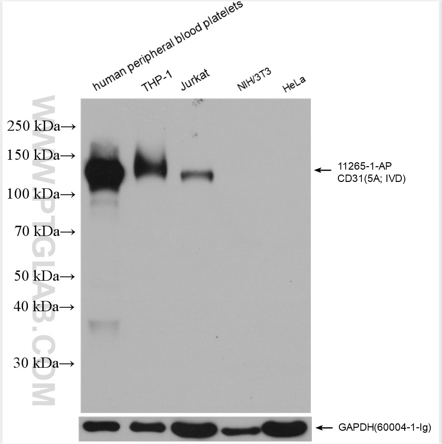
Leukocyte Adhesion Deficiency Syndrome (LAD) is a group of rare inherited disorders that affect the ability of white blood cells, specifically neutrophils, to adhere to and migrate into tissues, particularly those involved in immune responses. This results in recurrent bacterial and fungal infections starting in infancy.
There are three types of LAD, each caused by different genetic mutations:
1. LAD I: This is the most common and severe form, caused by a deficiency in the CD18 protein which is crucial for neutrophil adhesion. Symptoms include delayed separation of the umbilical cord, severe periodontal disease, and recurrent skin, lung and gastrointestinal infections.
2. LAD II: Also known as congenital disorder of glycosylation, type Ib, it is caused by a deficiency in the enzyme glucosyltransferase, leading to abnormal sugar chains on cell surfaces. Symptoms are similar to LAD I but less severe, and also include mental retardation and impaired growth.
3. LAD III: This is the least common form, caused by a defect in the integrin-linked kinase (ILK) gene. It results in a more complex phenotype with muscular and cardiac abnormalities, in addition to immune dysfunction.
Treatment typically involves prophylactic antibiotics, granulocyte-colony stimulating factor (G-CSF) to increase neutrophil counts, and sometimes bone marrow transplantation.
N-Acetylglucosaminyltransferases (GlcNAc transferases) are a group of enzymes that play a crucial role in the post-translational modification of proteins by adding N-acetylglucosamine (GlcNAc) to specific amino acids in a protein sequence. These enzymes catalyze the transfer of GlcNAc from a donor molecule, typically UDP-GlcNAc, to acceptor proteins, which can be other glycoproteins or proteins without any prior glycosylation.
The addition of N-acetylglucosamine by these enzymes is an essential step in the formation of complex carbohydrate structures called N-linked glycans, which are attached to asparagine residues within the protein sequence. The process of adding GlcNAc can occur in different ways, leading to various types of N-glycan structures, such as oligomannose, hybrid, and complex types.
There are several classes of N-Acetylglucosaminyltransferases (GnTs) based on their substrate specificity and the type of glycosidic linkage they form:
1. GnT I (MGAT1): Transfers GlcNAc to the α1,6 position of the mannose residue in the chitobiose core of N-linked glycans, initiating the formation of complex-type structures.
2. GnT II (MGAT2): Adds a second GlcNAc residue to the β1,4 position of the mannose residue at the non-reducing end of the chitobiose core, forming bi-antennary N-glycans.
3. GnT III (MGAT3): Transfers GlcNAc to the β1,4 position of the mannose residue in the chitobiose core, creating a branching point for further glycosylation and leading to tri- or tetra-antennary N-glycans.
4. GnT IV (MGAT4): Adds GlcNAc to the β1,4 position of the mannose residue at the non-reducing end of antennae, forming multi-branched complex-type structures.
5. GnT V (MGAT5): Transfers GlcNAc to the β1,6 position of the mannose residue in the chitobiose core, leading to hybrid and complex-type N-glycans with bisecting GlcNAc.
6. GnT VI (MGAT6): Adds GlcNAc to the α1,3 position of the mannose residue at the non-reducing end of antennae, forming a-linked poly-N-acetyllactosamine structures.
7. GnT VII (MGAT7): Transfers GlcNAc to the β1,6 position of the N-acetylglucosamine residue in complex-type N-glycans, forming i-antigen structures.
8. GnT VIII (MGAT8): Adds GlcNAc to the α1,3 position of the mannose residue at the non-reducing end of antennae, forming a-linked poly-N-acetyllactosamine structures.
9. GnT IX (MGAT9): Transfers GlcNAc to the β1,6 position of the N-acetylglucosamine residue in complex-type N-glycans, forming i-antigen structures.
10. GnT X (MGAT10): Adds GlcNAc to the α1,3 position of the mannose residue at the non-reducing end of antennae, forming a-linked poly-N-acetyllactosamine structures.
11. GnT XI (MGAT11): Transfers GlcNAc to the β1,6 position of the N-acetylglucosamine residue in complex-type N-glycans, forming i-antigen structures.
12. GnT XII (MGAT12): Adds GlcNAc to the α1,3 position of the mannose residue at the non-reducing end of antennae, forming a-linked poly-N-acetyllactosamine structures.
13. GnT XIII (MGAT13): Transfers GlcNAc to the β1,6 position of the N-acetylglucosamine residue in complex-type N-glycans, forming i-antigen structures.
14. GnT XIV (MGAT14): Adds GlcNAc to the α1,3 position of the mannose residue at the non-reducing end of antennae, forming a-linked poly-N-acetyllactosamine structures.
15. GnT XV (MGAT15): Transfers GlcNAc to the β1,6 position of the N-acetylglucosamine residue in complex-type N-glycans, forming i-antigen structures.
16. GnT XVI (MGAT16): Adds GlcNAc to the α1,3 position of the mannose residue at the non-reducing end of antennae, forming a-linked poly-N-acetyllactosamine structures.
17. GnT XVII (MGAT17): Transfers GlcNAc to the β1,6 position of the N-acetylglucosamine residue in complex-type N-glycans, forming i-antigen structures.
18. GnT XVIII (MGAT18): Adds GlcNAc to the α1,3 position of the mannose residue at the non-reducing end of antennae, forming a-linked poly-N-acetyllactosamine structures.
19. GnT XIX (MGAT19): Transfers GlcNAc to the β1,6 position of the N-acetylglucosamine residue in complex-type N-glycans, forming i-antigen structures.
20. GnT XX (MGAT20): Adds GlcNAc to the α1,3 position of the mannose residue at the non-reducing end of antennae, forming a-linked poly-N-acetyllactosamine structures.
21. GnT XXI (MGAT21): Transfers GlcNAc to the β1,6 position of the N-acetylglucosamine residue in complex-type N-glycans, forming i-antigen structures.
22. GnT XXII (MGAT22): Adds GlcNAc to the α1,3 position of the mannose residue at the non-reducing end of antennae, forming a-linked poly-N-acetyllactosamine structures.
23. GnT XXIII (MGAT23): Transfers GlcNAc to the β1,6 position of the N-acetylglucosamine residue in complex-type N-glycans, forming i-antigen structures.
24. GnT XXIV (MGAT24): Adds GlcNAc to the α1,3 position of the mannose residue at the non-reducing end of antennae, forming a-linked poly-N-acetyllactosamine structures.
25. GnT XXV (MGAT25): Transfers GlcNAc to the β1,6 position of the N-acetylglucosamine residue in complex-type N-glycans, forming i-antigen structures.
26. GnT XXVI (MGAT26): Adds GlcNAc to the α1,3 position of the mannose residue at the non-reducing end of antennae, forming a-linked poly-N-acetyllactosamine structures.
27. GnT XXVII (MGAT27): Transfers GlcNAc to the β1,6 position of the N-acetylglucosamine residue in complex-type N-glycans, forming i-antigen structures.
28. GnT XXVIII (MGAT28): Adds GlcNAc to the α1,3 position of the mannose residue at the non-reducing end of antennae, forming a-linked poly-N-acetyllactosamine structures.
29. GnT XXIX (MGAT29): Transfers GlcNAc to the β1,6 position of the N-acetylglucosamine residue in complex-type N-glycans, forming i-antigen structures.
30. GnT XXX (MG
Mannosides are glycosylated compounds that consist of a mannose sugar molecule (a type of monosaccharide) linked to another compound, often a protein or lipid. They are formed when an enzyme called a glycosyltransferase transfers a mannose molecule from a donor substrate, such as a nucleotide sugar (like GDP-mannose), to an acceptor molecule.
Mannosides can be found on the surface of many types of cells and play important roles in various biological processes, including cell recognition, signaling, and protein folding. They are also involved in the immune response and have been studied as potential therapeutic targets for a variety of diseases, including infectious diseases and cancer.
It's worth noting that mannosides can be further classified based on the specific linkage between the mannose molecule and the acceptor compound. For example, an N-linked mannoside is one in which the mannose is linked to a nitrogen atom on the acceptor protein, while an O-linked mannoside is one in which the mannose is linked to an oxygen atom on the acceptor protein.
Mucins are high molecular weight, heavily glycosylated proteins that are the major components of mucus. They are produced and secreted by specialized epithelial cells in various organs, including the respiratory, gastrointestinal, and urogenital tracts, as well as the eyes and ears.
Mucins have a characteristic structure consisting of a protein backbone with numerous attached oligosaccharide side chains, which give them their gel-forming properties and provide a protective barrier against pathogens, environmental insults, and digestive enzymes. They also play important roles in lubrication, hydration, and cell signaling.
Mucins can be classified into two main groups based on their structure and function: secreted mucins and membrane-bound mucins. Secreted mucins are released from cells and form a physical barrier on the surface of mucosal tissues, while membrane-bound mucins are integrated into the cell membrane and participate in cell adhesion and signaling processes.
Abnormalities in mucin production or function have been implicated in various diseases, including chronic inflammation, cancer, and cystic fibrosis.
Sialyltransferases are a group of enzymes that play a crucial role in the biosynthesis of sialic acids, which are a type of sugar molecule found on the surface of many cell types. These enzymes catalyze the transfer of sialic acid from a donor molecule (usually CMP-sialic acid) to an acceptor molecule, such as a glycoprotein or glycolipid.
The addition of sialic acids to these molecules can affect their function and properties, including their recognition by other cells and their susceptibility to degradation. Sialyltransferases are involved in various biological processes, including cell-cell recognition, inflammation, and cancer metastasis.
There are several different types of sialyltransferases, each with specific substrate preferences and functions. For example, some sialyltransferases add sialic acids to the ends of N-linked glycans, while others add them to O-linked glycans or glycolipids.
Abnormalities in sialyltransferase activity have been implicated in various diseases, including cancer, inflammatory disorders, and neurological conditions. Therefore, understanding the function and regulation of these enzymes is an important area of research with potential implications for disease diagnosis and treatment.
The endothelium is a thin layer of simple squamous epithelial cells that lines the interior surface of blood vessels, lymphatic vessels, and heart chambers. The vascular endothelium, specifically, refers to the endothelial cells that line the blood vessels. These cells play a crucial role in maintaining vascular homeostasis by regulating vasomotor tone, coagulation, platelet activation, inflammation, and permeability of the vessel wall. They also contribute to the growth and repair of the vascular system and are involved in various pathological processes such as atherosclerosis, hypertension, and diabetes.
Cell movement, also known as cell motility, refers to the ability of cells to move independently and change their location within tissue or inside the body. This process is essential for various biological functions, including embryonic development, wound healing, immune responses, and cancer metastasis.
There are several types of cell movement, including:
1. **Crawling or mesenchymal migration:** Cells move by extending and retracting protrusions called pseudopodia or filopodia, which contain actin filaments. This type of movement is common in fibroblasts, immune cells, and cancer cells during tissue invasion and metastasis.
2. **Amoeboid migration:** Cells move by changing their shape and squeezing through tight spaces without forming protrusions. This type of movement is often observed in white blood cells (leukocytes) as they migrate through the body to fight infections.
3. **Pseudopodial extension:** Cells extend pseudopodia, which are temporary cytoplasmic projections containing actin filaments. These protrusions help the cell explore its environment and move forward.
4. **Bacterial flagellar motion:** Bacteria use a whip-like structure called a flagellum to propel themselves through their environment. The rotation of the flagellum is driven by a molecular motor in the bacterial cell membrane.
5. **Ciliary and ependymal movement:** Ciliated cells, such as those lining the respiratory tract and fallopian tubes, have hair-like structures called cilia that beat in coordinated waves to move fluids or mucus across the cell surface.
Cell movement is regulated by a complex interplay of signaling pathways, cytoskeletal rearrangements, and adhesion molecules, which enable cells to respond to environmental cues and navigate through tissues.
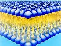
Membrane glycoproteins are proteins that contain oligosaccharide chains (glycans) covalently attached to their polypeptide backbone. They are integral components of biological membranes, spanning the lipid bilayer and playing crucial roles in various cellular processes.
The glycosylation of these proteins occurs in the endoplasmic reticulum (ER) and Golgi apparatus during protein folding and trafficking. The attached glycans can vary in structure, length, and composition, which contributes to the diversity of membrane glycoproteins.
Membrane glycoproteins can be classified into two main types based on their orientation within the lipid bilayer:
1. Type I (N-linked): These glycoproteins have a single transmembrane domain and an extracellular N-terminus, where the oligosaccharides are predominantly attached via asparagine residues (Asn-X-Ser/Thr sequon).
2. Type II (C-linked): These glycoproteins possess two transmembrane domains and an intracellular C-terminus, with the oligosaccharides linked to tryptophan residues via a mannose moiety.
Membrane glycoproteins are involved in various cellular functions, such as:
* Cell adhesion and recognition
* Receptor-mediated signal transduction
* Enzymatic catalysis
* Transport of molecules across membranes
* Cell-cell communication
* Immunological responses
Some examples of membrane glycoproteins include cell surface receptors (e.g., growth factor receptors, cytokine receptors), adhesion molecules (e.g., integrins, cadherins), and transporters (e.g., ion channels, ABC transporters).
Sulfoglycosphingolipids are a type of glycosphingolipid that contain a sulfate ester group in their carbohydrate moiety. They are important components of animal cell membranes and play a role in various biological processes, including cell recognition, signal transduction, and cell adhesion.
The most well-known sulfoglycosphingolipids are the sulfatides, which contain a 3'-sulfate ester on the galactose residue of the glycosphingolipid GalCer (galactosylceramide). Sulfatides are abundant in the nervous system and have been implicated in various neurological disorders.
Other sulfoglycosphingolipids include the seminolipids, which contain a 3'-sulfate ester on the galactose residue of lactosylceramide (Galβ1-4Glcβ1-Cer), and are found in high concentrations in the testis.
Abnormalities in sulfoglycosphingolipid metabolism have been associated with several genetic disorders, such as metachromatic leukodystrophy (MLD) and globoid cell leukodystrophy (GLD), which are characterized by progressive neurological deterioration.
A "carbohydrate sequence" refers to the specific arrangement or order of monosaccharides (simple sugars) that make up a carbohydrate molecule, such as a polysaccharide or an oligosaccharide. Carbohydrates are often composed of repeating units of monosaccharides, and the sequence in which these units are arranged can have important implications for the function and properties of the carbohydrate.
For example, in glycoproteins (proteins that contain carbohydrate chains), the specific carbohydrate sequence can affect how the protein is processed and targeted within the cell, as well as its stability and activity. Similarly, in complex carbohydrates like starch or cellulose, the sequence of glucose units can determine whether the molecule is branched or unbranched, which can have implications for its digestibility and other properties.
Therefore, understanding the carbohydrate sequence is an important aspect of studying carbohydrate structure and function in biology and medicine.
HL-60 cells are a type of human promyelocytic leukemia cell line that is commonly used in scientific research. They are named after the hospital where they were first isolated, the Hospital of the University of Pennsylvania (HUP) and the 60th culture attempt to grow these cells.
HL-60 cells have the ability to differentiate into various types of blood cells, such as granulocytes, monocytes, and macrophages, when exposed to certain chemical compounds or under specific culturing conditions. This makes them a valuable tool for studying the mechanisms of cell differentiation, proliferation, and apoptosis (programmed cell death).
HL-60 cells are also often used in toxicity studies, drug discovery and development, and research on cancer, inflammation, and infectious diseases. They can be easily grown in the lab and have a stable genotype, making them ideal for use in standardized experiments and comparisons between different studies.
Venules are very small blood vessels that carry oxygen-depleted blood from capillaries to veins. They have a diameter of 8-50 micrometers and are an integral part of the microcirculation system in the body. Venules merge together to form veins, which then transport the low-oxygen blood back to the heart.
The Lewis blood-group system is one of the human blood group systems, which is based on the presence or absence of two antigens: Lea and Leb. These antigens are carbohydrate structures that can be found on the surface of red blood cells (RBCs) as well as other cells and in various body fluids.
The Lewis system is unique because its antigens are not normally present at birth, but instead develop during early childhood or later in life due to the action of certain enzymes in the digestive tract. The production of Lea and Leb antigens depends on the activity of two genes, FUT3 (also known as Lewis gene) and FUT2 (also known as Secretor gene).
There are four main phenotypes or blood types in the Lewis system:
1. Le(a+b-): This is the most common phenotype, where individuals have both Lea and Leb antigens on their RBCs.
2. Le(a-b+): In this phenotype, individuals lack the Lea antigen but have the Leb antigen on their RBCs.
3. Le(a-b-): This is a rare phenotype where neither Lea nor Leb antigens are present on the RBCs.
4. Le(a+b+): In this phenotype, individuals have both Lea and Leb antigens on their RBCs due to the simultaneous expression of FUT3 and FUT2 genes.
The Lewis blood-group system is not typically associated with transfusion reactions or hemolytic diseases, unlike other blood group systems such as ABO and Rh. However, the presence or absence of Lewis antigens can still have implications for certain medical conditions and tests, including:
* Infectious diseases: Some bacteria and viruses can use the Lewis antigens as receptors to attach to and infect host cells. For example, Helicobacter pylori, which causes gastritis and peptic ulcers, binds to Lea antigens in the stomach.
* Autoimmune disorders: In some cases, autoantibodies against Lewis antigens have been found in patients with autoimmune diseases such as rheumatoid arthritis and systemic lupus erythematosus (SLE).
* Pregnancy: The Lewis antigens can be expressed on the surface of placental cells, and changes in their expression have been linked to pregnancy complications such as preeclampsia and fetal growth restriction.
* Blood typing: Although not a primary factor in blood transfusion compatibility, the Lewis blood-group system is still considered when determining the best match for patients who require frequent transfusions or organ transplants.
Thioglycolates are a group of chemical compounds that contain a thiol (sulfhydryl) group (-SH) bonded to a glycolate group. In the context of medical and cosmetic use, the term "thioglycolates" often refers to salts of thioglycolic acid, which are used as depilatories or hair-curling agents.
Thioglycolates work by breaking the disulfide bonds in keratin, the protein that makes up hair and nails. When applied to hair, thioglycolates reduce the disulfide bonds into sulfhydryl groups, making the hair more flexible and easier to shape or remove. This property is exploited in hair-curling products and depilatories (hair removal creams).
It's important to note that thioglycolates can cause skin irritation, allergic reactions, and respiratory issues in some individuals. Therefore, they should be used with caution, following the manufacturer's instructions, and in a well-ventilated area.
Intercellular Adhesion Molecule-1 (ICAM-1), also known as CD54, is a transmembrane glycoprotein expressed on the surface of various cell types including endothelial cells, fibroblasts, and immune cells. ICAM-1 plays a crucial role in the inflammatory response and the immune system by mediating the adhesion of leukocytes (white blood cells) to the endothelium, allowing them to migrate into surrounding tissues during an immune response or inflammation.
ICAM-1 contains five immunoglobulin-like domains in its extracellular region and binds to several integrins present on leukocytes, such as LFA-1 (lymphocyte function-associated antigen 1) and Mac-1 (macrophage-1 antigen). This interaction facilitates the firm adhesion of leukocytes to the endothelium, which is a critical step in the extravasation process.
In addition to its role in inflammation and immunity, ICAM-1 has been implicated in several pathological conditions, including atherosclerosis, cancer, and autoimmune diseases. Increased expression of ICAM-1 on endothelial cells is associated with the recruitment of immune cells to sites of injury or infection, making it an important target for therapeutic interventions in various inflammatory disorders.
CD18 is a type of protein called an integrin that is found on the surface of many different types of cells in the human body, including white blood cells (leukocytes). It plays a crucial role in the immune system by helping these cells to migrate through blood vessel walls and into tissues where they can carry out their various functions, such as fighting infection and inflammation.
CD18 forms a complex with another protein called CD11b, and together they are known as Mac-1 or CR3 (complement receptor 3). This complex is involved in the recognition and binding of various molecules, including bacterial proteins and fragments of complement proteins, which help to trigger an immune response.
CD18 has been implicated in a number of diseases, including certain types of cancer, inflammatory bowel disease, and rheumatoid arthritis. Mutations in the gene that encodes CD18 can lead to a rare disorder called leukocyte adhesion deficiency (LAD) type 1, which is characterized by recurrent bacterial infections and impaired wound healing.
Glycosyltransferases are a group of enzymes that play a crucial role in the synthesis of glycoconjugates, which are complex carbohydrate structures found on the surface of cells and in various biological fluids. These enzymes catalyze the transfer of a sugar moiety from an activated donor molecule to an acceptor molecule, resulting in the formation of a glycosidic bond.
The donor molecule is typically a nucleotide sugar, such as UDP-glucose or CMP-sialic acid, which provides the energy required for the transfer reaction. The acceptor molecule can be a wide range of substrates, including proteins, lipids, and other carbohydrates.
Glycosyltransferases are highly specific in their activity, with each enzyme recognizing a particular donor and acceptor pair. This specificity allows for the precise regulation of glycan structures, which have been shown to play important roles in various biological processes, including cell recognition, signaling, and adhesion.
Defects in glycosyltransferase function can lead to a variety of genetic disorders, such as congenital disorders of glycosylation (CDG), which are characterized by abnormal glycan structures and a wide range of clinical manifestations, including developmental delay, neurological impairment, and multi-organ dysfunction.
Lymphocyte homing receptors are specialized molecules found on the surface of lymphocytes (white blood cells that include T-cells and B-cells), which play a crucial role in the immune system's response to infection and disease. These receptors facilitate the targeted migration and trafficking of lymphocytes from the bloodstream to specific secondary lymphoid organs, such as lymph nodes, spleen, and Peyer's patches in the intestines, where they can encounter antigens and mount an immune response.
The homing receptors consist of two main components: adhesion molecules and chemokine receptors. Adhesion molecules, such as selectins and integrins, mediate the initial attachment and rolling of lymphocytes along the endothelial cells that line the blood vessels in lymphoid organs. Chemokine receptors, on the other hand, interact with chemokines (a type of cytokine) that are secreted by the endothelial cells and stromal cells within the lymphoid organs. This interaction triggers a signaling cascade that activates integrins, leading to their firm adhesion to the endothelium and subsequent transmigration into the lymphoid tissue.
The specificity of this homing process is determined by the unique combination of adhesion molecules and chemokine receptors expressed on different subsets of lymphocytes, which allows them to home to distinct anatomical locations in response to various chemokine gradients. This targeted migration ensures that the immune system can effectively mount a rapid and localized response against pathogens while minimizing unnecessary inflammation in other parts of the body.
Dermatitis is a general term that describes inflammation of the skin. It is often characterized by redness, swelling, itching, and tenderness. There are many different types of dermatitis, including atopic dermatitis (eczema), contact dermatitis, seborrheic dermatitis, and nummular dermatitis.
Atopic dermatitis is a chronic skin condition that often affects people with a family history of allergies, such as asthma or hay fever. It typically causes dry, scaly patches on the skin that can be extremely itchy.
Contact dermatitis occurs when the skin comes into contact with an irritant or allergen, such as poison ivy or certain chemicals. This type of dermatitis can cause redness, swelling, and blistering.
Seborrheic dermatitis is a common condition that causes a red, itchy rash, often on the scalp, face, or other areas of the body where oil glands are located. It is thought to be related to an overproduction of oil by the skin's sebaceous glands.
Nummular dermatitis is a type of eczema that causes round, coin-shaped patches of dry, scaly skin. It is more common in older adults and often occurs during the winter months.
Treatment for dermatitis depends on the underlying cause and severity of the condition. In some cases, over-the-counter creams or lotions may be sufficient to relieve symptoms. Prescription medications, such as corticosteroids or immunosuppressants, may be necessary in more severe cases. Avoiding triggers and irritants can also help prevent flare-ups of dermatitis.
Integrin α4 (also known as CD49d or ITGA4) is a subunit of integrin proteins, which are heterodimeric transmembrane receptors that mediate cell-cell and cell-extracellular matrix interactions. Integrin α4 typically pairs with β1 (CD29 or ITGB1) or β7 (ITGB7) subunits to form integrins α4β1 and α4β7, respectively.
Integrin α4β1, also known as very late antigen-4 (VLA-4), is widely expressed on various hematopoietic cells, including lymphocytes, monocytes, eosinophils, and basophils. It plays crucial roles in the adhesion, migration, and homing of these cells to secondary lymphoid organs, as well as in the recruitment of immune cells to inflammatory sites. Integrin α4β1 binds to its ligands, vascular cell adhesion molecule-1 (VCAM-1) and fibronectin, via the arginine-glycine-aspartic acid (RGD) motif.
Integrin α4β7, on the other hand, is primarily expressed on gut-homing lymphocytes and interacts with mucosal addressin cell adhesion molecule-1 (MAdCAM-1), a protein mainly found in the high endothelial venules of intestinal Peyer's patches and mesenteric lymph nodes. This interaction facilitates the trafficking of immune cells to the gastrointestinal tract, where they participate in immune responses against pathogens and maintain gut homeostasis.
In summary, Integrin α4 is a crucial subunit of integrins that mediates cell adhesion, migration, and homing to specific tissues through its interactions with various ligands. Dysregulation of integrin α4 has been implicated in several pathological conditions, including inflammatory diseases, autoimmune disorders, and cancer metastasis.
Chemotaxis, Leukocyte is the movement of leukocytes (white blood cells) towards a higher concentration of a particular chemical substance, known as a chemotactic factor. This process plays a crucial role in the immune system's response to infection and injury.
When there is an infection or tissue damage, certain cells release chemotactic factors, which are small molecules or proteins that can attract leukocytes to the site of inflammation. Leukocytes have receptors on their surface that can detect these chemotactic factors and move towards them through a process called chemotaxis.
Once they reach the site of inflammation, leukocytes can help eliminate pathogens or damaged cells by phagocytosis (engulfing and destroying) or releasing toxic substances that kill the invading microorganisms. Chemotaxis is an essential part of the immune system's defense mechanisms and helps to maintain tissue homeostasis and prevent the spread of infection.
N-Acetylneuraminic Acid (Neu5Ac) is an organic compound that belongs to the family of sialic acids. It is a common terminal sugar found on many glycoproteins and glycolipids on the surface of animal cells. Neu5Ac plays crucial roles in various biological processes, including cell recognition, signaling, and intercellular interactions. It is also involved in the protection against pathogens by serving as a barrier to prevent their attachment to host cells. Additionally, Neu5Ac has been implicated in several disease conditions, such as cancer and inflammation, due to its altered expression and metabolism.
Leukocytosis is a condition characterized by an increased number of leukocytes (white blood cells) in the peripheral blood. A normal white blood cell count ranges from 4,500 to 11,000 cells per microliter of blood in adults. Leukocytosis is typically considered present when the white blood cell count exceeds 11,000 cells/µL. However, the definition might vary slightly depending on the laboratory and clinical context.
Leukocytosis can be a response to various underlying conditions, including bacterial or viral infections, inflammation, tissue damage, leukemia, and other hematological disorders. It is essential to investigate the cause of leukocytosis through further diagnostic tests, such as blood smears, differential counts, and additional laboratory and imaging studies, to guide appropriate treatment.
Carbohydrate conformation refers to the three-dimensional shape and structure of a carbohydrate molecule. Carbohydrates, also known as sugars, can exist in various conformational states, which are determined by the rotation of their component bonds and the spatial arrangement of their functional groups.
The conformation of a carbohydrate molecule can have significant implications for its biological activity and recognition by other molecules, such as enzymes or antibodies. Factors that can influence carbohydrate conformation include the presence of intramolecular hydrogen bonds, steric effects, and intermolecular interactions with solvent molecules or other solutes.
In some cases, the conformation of a carbohydrate may be stabilized by the formation of cyclic structures, in which the hydroxyl group at one end of the molecule forms a covalent bond with the carbonyl carbon at the other end, creating a ring structure. The most common cyclic carbohydrates are monosaccharides, such as glucose and fructose, which can exist in various conformational isomers known as anomers.
Understanding the conformation of carbohydrate molecules is important for elucidating their biological functions and developing strategies for targeting them with drugs or other therapeutic agents.
Amino sugars, also known as glycosamine or hexosamines, are sugar molecules that contain a nitrogen atom as part of their structure. The most common amino sugars found in nature are glucosamine and galactosamine, which are derived from the hexose sugars glucose and galactose, respectively.
Glucosamine is an essential component of the structural polysaccharide chitin, which is found in the exoskeletons of arthropods such as crustaceans and insects, as well as in the cell walls of fungi. It is also a precursor to the glycosaminoglycans (GAGs), which are long, unbranched polysaccharides that are important components of the extracellular matrix in animals.
Galactosamine, on the other hand, is a component of some GAGs and is also found in bacterial cell walls. It is used in the synthesis of heparin and heparan sulfate, which are important anticoagulant molecules.
Amino sugars play a critical role in many biological processes, including cell signaling, inflammation, and immune response. They have also been studied for their potential therapeutic uses in the treatment of various diseases, such as osteoarthritis and cancer.
Vascular Cell Adhesion Molecule-1 (VCAM-1) is a glycoprotein expressed on the surface of endothelial cells that plays a crucial role in the inflammatory response. It is involved in the recruitment and adhesion of leukocytes to the site of inflammation. VCAM-1 interacts with integrins on the surface of leukocytes, particularly very late antigen-4 (VLA-4), to facilitate this adhesion process. This interaction leads to the activation of signaling pathways that promote the migration of leukocytes across the endothelial barrier and into the surrounding tissue, where they can contribute to the immune response and resolution of inflammation. Increased expression of VCAM-1 has been associated with various inflammatory diseases, including atherosclerosis, rheumatoid arthritis, and multiple sclerosis.
CD44 is a type of protein found on the surface of some cells in the human body. It is a cell adhesion molecule and is involved in various biological processes such as cell-cell interaction, lymphocyte activation, and migration of cells. CD44 also acts as a receptor for hyaluronic acid, a component of the extracellular matrix.
As an antigen, CD44 can be recognized by certain immune cells, including T cells and B cells, and can play a role in the immune response. There are several isoforms of CD44 that exist due to alternative splicing of its mRNA, leading to differences in its structure and function.
CD44 has been studied in the context of cancer, where it can contribute to tumor growth, progression, and metastasis. In some cases, high levels of CD44 have been associated with poor prognosis in certain types of cancer. However, CD44 also has potential roles in tumor suppression and immune surveillance, making its overall role in cancer complex and context-dependent.
Monoclonal antibodies are a type of antibody that are identical because they are produced by a single clone of cells. They are laboratory-produced molecules that act like human antibodies in the immune system. They can be designed to attach to specific proteins found on the surface of cancer cells, making them useful for targeting and treating cancer. Monoclonal antibodies can also be used as a therapy for other diseases, such as autoimmune disorders and inflammatory conditions.
Monoclonal antibodies are produced by fusing a single type of immune cell, called a B cell, with a tumor cell to create a hybrid cell, or hybridoma. This hybrid cell is then able to replicate indefinitely, producing a large number of identical copies of the original antibody. These antibodies can be further modified and engineered to enhance their ability to bind to specific targets, increase their stability, and improve their effectiveness as therapeutic agents.
Monoclonal antibodies have several mechanisms of action in cancer therapy. They can directly kill cancer cells by binding to them and triggering an immune response. They can also block the signals that promote cancer growth and survival. Additionally, monoclonal antibodies can be used to deliver drugs or radiation directly to cancer cells, increasing the effectiveness of these treatments while minimizing their side effects on healthy tissues.
Monoclonal antibodies have become an important tool in modern medicine, with several approved for use in cancer therapy and other diseases. They are continuing to be studied and developed as a promising approach to treating a wide range of medical conditions.
Flow cytometry is a medical and research technique used to measure physical and chemical characteristics of cells or particles, one cell at a time, as they flow in a fluid stream through a beam of light. The properties measured include:
* Cell size (light scatter)
* Cell internal complexity (granularity, also light scatter)
* Presence or absence of specific proteins or other molecules on the cell surface or inside the cell (using fluorescent antibodies or other fluorescent probes)
The technique is widely used in cell counting, cell sorting, protein engineering, biomarker discovery and monitoring disease progression, particularly in hematology, immunology, and cancer research.
Shear strength is a property of a material that describes its ability to withstand forces that cause internal friction and sliding of one portion of the material relative to another. In the context of human tissues, shear strength is an important factor in understanding how tissues respond to various stresses and strains, such as those experienced during physical activities or injuries.
For example, in the case of bones, shear strength is a critical factor in determining their ability to resist fractures under different types of loading conditions. Similarly, in soft tissues like ligaments and tendons, shear strength plays a crucial role in maintaining the integrity of these structures during movement and preventing excessive deformation or injury.
It's worth noting that measuring the shear strength of human tissues can be challenging due to their complex structure and anisotropic properties. As such, researchers often use specialized techniques and equipment to quantify these properties under controlled conditions in the lab.
Integrin α4β1, also known as Very Late Antigen-4 (VLA-4), is a heterodimeric transmembrane receptor protein composed of two subunits, α4 and β1. It is involved in various cellular activities such as adhesion, migration, and signaling. This integrin plays a crucial role in the immune system by mediating the interaction between leukocytes (white blood cells) and the endothelial cells that line blood vessels. The activation of Integrin α4β1 allows leukocytes to roll along and then firmly adhere to the endothelium, followed by their migration into surrounding tissues, particularly during inflammation and immune responses. Additionally, Integrin α4β1 also interacts with extracellular matrix proteins such as fibronectin and helps regulate cell survival, proliferation, and differentiation in various cell types.
CHO cells, or Chinese Hamster Ovary cells, are a type of immortalized cell line that are commonly used in scientific research and biotechnology. They were originally derived from the ovaries of a female Chinese hamster (Cricetulus griseus) in the 1950s.
CHO cells have several characteristics that make them useful for laboratory experiments. They can grow and divide indefinitely under appropriate conditions, which allows researchers to culture large quantities of them for study. Additionally, CHO cells are capable of expressing high levels of recombinant proteins, making them a popular choice for the production of therapeutic drugs, vaccines, and other biologics.
In particular, CHO cells have become a workhorse in the field of biotherapeutics, with many approved monoclonal antibody-based therapies being produced using these cells. The ability to genetically modify CHO cells through various methods has further expanded their utility in research and industrial applications.
It is important to note that while CHO cells are widely used in scientific research, they may not always accurately represent human cell behavior or respond to drugs and other compounds in the same way as human cells do. Therefore, results obtained using CHO cells should be validated in more relevant systems when possible.
C57BL/6 (C57 Black 6) is an inbred strain of laboratory mouse that is widely used in biomedical research. The term "inbred" refers to a strain of animals where matings have been carried out between siblings or other closely related individuals for many generations, resulting in a population that is highly homozygous at most genetic loci.
The C57BL/6 strain was established in 1920 by crossing a female mouse from the dilute brown (DBA) strain with a male mouse from the black strain. The resulting offspring were then interbred for many generations to create the inbred C57BL/6 strain.
C57BL/6 mice are known for their robust health, longevity, and ease of handling, making them a popular choice for researchers. They have been used in a wide range of biomedical research areas, including studies of cancer, immunology, neuroscience, cardiovascular disease, and metabolism.
One of the most notable features of the C57BL/6 strain is its sensitivity to certain genetic modifications, such as the introduction of mutations that lead to obesity or impaired glucose tolerance. This has made it a valuable tool for studying the genetic basis of complex diseases and traits.
Overall, the C57BL/6 inbred mouse strain is an important model organism in biomedical research, providing a valuable resource for understanding the genetic and molecular mechanisms underlying human health and disease.
A "knockout" mouse is a genetically engineered mouse in which one or more genes have been deleted or "knocked out" using molecular biology techniques. This allows researchers to study the function of specific genes and their role in various biological processes, as well as potential associations with human diseases. The mice are generated by introducing targeted DNA modifications into embryonic stem cells, which are then used to create a live animal. Knockout mice have been widely used in biomedical research to investigate gene function, disease mechanisms, and potential therapeutic targets.
Inflammation is a complex biological response of tissues to harmful stimuli, such as pathogens, damaged cells, or irritants. It is characterized by the following signs: rubor (redness), tumor (swelling), calor (heat), dolor (pain), and functio laesa (loss of function). The process involves the activation of the immune system, recruitment of white blood cells, and release of inflammatory mediators, which contribute to the elimination of the injurious stimuli and initiation of the healing process. However, uncontrolled or chronic inflammation can also lead to tissue damage and diseases.
Mannose is a simple sugar (monosaccharide) that is similar in structure to glucose. It is a hexose, meaning it contains six carbon atoms. Mannose is a stereoisomer of glucose, meaning it has the same chemical formula but a different structural arrangement of its atoms.
Mannose is not as commonly found in foods as other simple sugars, but it can be found in some fruits, such as cranberries, blueberries, and peaches, as well as in certain vegetables, like sweet potatoes and turnips. It is also found in some dietary fibers, such as those found in beans and whole grains.
In the body, mannose can be metabolized and used for energy, but it is also an important component of various glycoproteins and glycolipids, which are molecules that play critical roles in many biological processes, including cell recognition, signaling, and adhesion.
Mannose has been studied as a potential therapeutic agent for various medical conditions, including urinary tract infections (UTIs), because it can inhibit the attachment of certain bacteria to the cells lining the urinary tract. Additionally, mannose-binding lectins have been investigated for their potential role in the immune response to viral and bacterial infections.
Disaccharides are a type of carbohydrate that is made up of two monosaccharide units bonded together. Monosaccharides are simple sugars, such as glucose, fructose, or galactose. When two monosaccharides are joined together through a condensation reaction, they form a disaccharide.
The most common disaccharides include:
* Sucrose (table sugar), which is composed of one glucose molecule and one fructose molecule.
* Lactose (milk sugar), which is composed of one glucose molecule and one galactose molecule.
* Maltose (malt sugar), which is composed of two glucose molecules.
Disaccharides are broken down into their component monosaccharides during digestion by enzymes called disaccharidases, which are located in the brush border of the small intestine. These enzymes catalyze the hydrolysis of the glycosidic bond that links the two monosaccharides together, releasing them to be absorbed into the bloodstream and used for energy.
Disorders of disaccharide digestion and absorption can lead to various symptoms, such as bloating, diarrhea, and abdominal pain. For example, lactose intolerance is a common condition in which individuals lack sufficient levels of the enzyme lactase, leading to an inability to properly digest lactose and resulting in gastrointestinal symptoms.
Carbohydrate metabolism is the process by which the body breaks down carbohydrates into glucose, which is then used for energy or stored in the liver and muscles as glycogen. This process involves several enzymes and chemical reactions that convert carbohydrates from food into glucose, fructose, or galactose, which are then absorbed into the bloodstream and transported to cells throughout the body.
The hormones insulin and glucagon regulate carbohydrate metabolism by controlling the uptake and storage of glucose in cells. Insulin is released from the pancreas when blood sugar levels are high, such as after a meal, and promotes the uptake and storage of glucose in cells. Glucagon, on the other hand, is released when blood sugar levels are low and signals the liver to convert stored glycogen back into glucose and release it into the bloodstream.
Disorders of carbohydrate metabolism can result from genetic defects or acquired conditions that affect the enzymes or hormones involved in this process. Examples include diabetes, hypoglycemia, and galactosemia. Proper management of these disorders typically involves dietary modifications, medication, and regular monitoring of blood sugar levels.
Mechanical stress, in the context of physiology and medicine, refers to any type of force that is applied to body tissues or organs, which can cause deformation or displacement of those structures. Mechanical stress can be either external, such as forces exerted on the body during physical activity or trauma, or internal, such as the pressure changes that occur within blood vessels or other hollow organs.
Mechanical stress can have a variety of effects on the body, depending on the type, duration, and magnitude of the force applied. For example, prolonged exposure to mechanical stress can lead to tissue damage, inflammation, and chronic pain. Additionally, abnormal or excessive mechanical stress can contribute to the development of various musculoskeletal disorders, such as tendinitis, osteoarthritis, and herniated discs.
In order to mitigate the negative effects of mechanical stress, the body has a number of adaptive responses that help to distribute forces more evenly across tissues and maintain structural integrity. These responses include changes in muscle tone, joint positioning, and connective tissue stiffness, as well as the remodeling of bone and other tissues over time. However, when these adaptive mechanisms are overwhelmed or impaired, mechanical stress can become a significant factor in the development of various pathological conditions.
In medical terms, the skin is the largest organ of the human body. It consists of two main layers: the epidermis (outer layer) and dermis (inner layer), as well as accessory structures like hair follicles, sweat glands, and oil glands. The skin plays a crucial role in protecting us from external factors such as bacteria, viruses, and environmental hazards, while also regulating body temperature and enabling the sense of touch.
Neutrophil infiltration is a pathological process characterized by the accumulation of neutrophils, a type of white blood cell, in tissue. It is a common feature of inflammation and occurs in response to infection, injury, or other stimuli that trigger an immune response. Neutrophils are attracted to the site of tissue damage by chemical signals called chemokines, which are released by damaged cells and activated immune cells. Once they reach the site of inflammation, neutrophils help to clear away damaged tissue and microorganisms through a process called phagocytosis. However, excessive or prolonged neutrophil infiltration can also contribute to tissue damage and may be associated with various disease states, including cancer, autoimmune disorders, and ischemia-reperfusion injury.
Molecular sequence data refers to the specific arrangement of molecules, most commonly nucleotides in DNA or RNA, or amino acids in proteins, that make up a biological macromolecule. This data is generated through laboratory techniques such as sequencing, and provides information about the exact order of the constituent molecules. This data is crucial in various fields of biology, including genetics, evolution, and molecular biology, allowing for comparisons between different organisms, identification of genetic variations, and studies of gene function and regulation.
Glycosylation is the enzymatic process of adding a sugar group, or glycan, to a protein, lipid, or other organic molecule. This post-translational modification plays a crucial role in modulating various biological functions, such as protein stability, trafficking, and ligand binding. The structure and composition of the attached glycans can significantly influence the functional properties of the modified molecule, contributing to cell-cell recognition, signal transduction, and immune response regulation. Abnormal glycosylation patterns have been implicated in several disease states, including cancer, diabetes, and neurodegenerative disorders.
Integrins are a type of cell-adhesion molecule that play a crucial role in cell-cell and cell-extracellular matrix (ECM) interactions. They are heterodimeric transmembrane receptors composed of non-covalently associated α and β subunits, which form more than 24 distinct integrin heterodimers in humans.
Integrins bind to specific ligands, such as ECM proteins (e.g., collagen, fibronectin, laminin), cell surface molecules, and soluble factors, through their extracellular domains. The intracellular domains of integrins interact with the cytoskeleton and various signaling proteins, allowing them to transduce signals from the ECM into the cell (outside-in signaling) and vice versa (inside-out signaling).
These molecular interactions are essential for numerous biological processes, including cell adhesion, migration, proliferation, differentiation, survival, and angiogenesis. Dysregulation of integrin function has been implicated in various pathological conditions, such as cancer, fibrosis, inflammation, and autoimmune diseases.
Acetylglucosamine is a type of sugar that is commonly found in the body and plays a crucial role in various biological processes. It is a key component of glycoproteins and proteoglycans, which are complex molecules made up of protein and carbohydrate components.
More specifically, acetylglucosamine is an amino sugar that is formed by the addition of an acetyl group to glucosamine. It can be further modified in the body through a process called acetylation, which involves the addition of additional acetyl groups.
Acetylglucosamine is important for maintaining the structure and function of various tissues in the body, including cartilage, tendons, and ligaments. It also plays a role in the immune system and has been studied as a potential therapeutic target for various diseases, including cancer and inflammatory conditions.
In summary, acetylglucosamine is a type of sugar that is involved in many important biological processes in the body, and has potential therapeutic applications in various diseases.
Cell communication, also known as cell signaling, is the process by which cells exchange and transmit signals between each other and their environment. This complex system allows cells to coordinate their functions and maintain tissue homeostasis. Cell communication can occur through various mechanisms including:
1. Autocrine signaling: When a cell releases a signal that binds to receptors on the same cell, leading to changes in its behavior or function.
2. Paracrine signaling: When a cell releases a signal that binds to receptors on nearby cells, influencing their behavior or function.
3. Endocrine signaling: When a cell releases a hormone into the bloodstream, which then travels to distant target cells and binds to specific receptors, triggering a response.
4. Synaptic signaling: In neurons, communication occurs through the release of neurotransmitters that cross the synapse and bind to receptors on the postsynaptic cell, transmitting electrical or chemical signals.
5. Contact-dependent signaling: When cells physically interact with each other, allowing for the direct exchange of signals and information.
Cell communication is essential for various physiological processes such as growth, development, differentiation, metabolism, immune response, and tissue repair. Dysregulation in cell communication can contribute to diseases, including cancer, diabetes, and neurological disorders.
Protein binding, in the context of medical and biological sciences, refers to the interaction between a protein and another molecule (known as the ligand) that results in a stable complex. This process is often reversible and can be influenced by various factors such as pH, temperature, and concentration of the involved molecules.
In clinical chemistry, protein binding is particularly important when it comes to drugs, as many of them bind to proteins (especially albumin) in the bloodstream. The degree of protein binding can affect a drug's distribution, metabolism, and excretion, which in turn influence its therapeutic effectiveness and potential side effects.
Protein-bound drugs may be less available for interaction with their target tissues, as only the unbound or "free" fraction of the drug is active. Therefore, understanding protein binding can help optimize dosing regimens and minimize adverse reactions.
Cricetinae is a subfamily of rodents that includes hamsters, gerbils, and relatives. These small mammals are characterized by having short limbs, compact bodies, and cheek pouches for storing food. They are native to various parts of the world, particularly in Europe, Asia, and Africa. Some species are popular pets due to their small size, easy care, and friendly nature. In a medical context, understanding the biology and behavior of Cricetinae species can be important for individuals who keep them as pets or for researchers studying their physiology.
Peritonitis is a medical condition characterized by inflammation of the peritoneum, which is the serous membrane that lines the inner wall of the abdominal cavity and covers the abdominal organs. The peritoneum has an important role in protecting the abdominal organs and providing a smooth surface for them to move against each other.
Peritonitis can occur as a result of bacterial or fungal infection, chemical irritation, or trauma to the abdomen. The most common cause of peritonitis is a rupture or perforation of an organ in the abdominal cavity, such as the appendix, stomach, or intestines, which allows bacteria from the gut to enter the peritoneal cavity.
Symptoms of peritonitis may include abdominal pain and tenderness, fever, nausea and vomiting, loss of appetite, and decreased bowel movements. In severe cases, peritonitis can lead to sepsis, a life-threatening condition characterized by widespread inflammation throughout the body.
Treatment for peritonitis typically involves antibiotics to treat the infection, as well as surgical intervention to repair any damage to the abdominal organs and remove any infected fluid or tissue from the peritoneal cavity. In some cases, a temporary or permanent drain may be placed in the abdomen to help remove excess fluid and promote healing.
Microspheres are tiny, spherical particles that range in size from 1 to 1000 micrometers in diameter. They are made of biocompatible and biodegradable materials such as polymers, glass, or ceramics. In medical terms, microspheres have various applications, including drug delivery systems, medical imaging, and tissue engineering.
In drug delivery, microspheres can be used to encapsulate drugs and release them slowly over time, improving the efficacy of the treatment while reducing side effects. They can also be used for targeted drug delivery, where the microspheres are designed to accumulate in specific tissues or organs.
In medical imaging, microspheres can be labeled with radioactive isotopes or magnetic materials and used as contrast agents to enhance the visibility of tissues or organs during imaging procedures such as X-ray, CT, MRI, or PET scans.
In tissue engineering, microspheres can serve as a scaffold for cell growth and differentiation, promoting the regeneration of damaged tissues or organs. Overall, microspheres have great potential in various medical applications due to their unique properties and versatility.
Lectins are a type of proteins that bind specifically to carbohydrates and have been found in various plant and animal sources. They play important roles in biological recognition events, such as cell-cell adhesion, and can also be involved in the immune response. Some lectins can agglutinate certain types of cells or precipitate glycoproteins, while others may have a more direct effect on cellular processes. In some cases, lectins from plants can cause adverse effects in humans if ingested, such as digestive discomfort or allergic reactions.
The endothelium is the thin, delicate tissue that lines the interior surface of blood vessels and lymphatic vessels. It is a single layer of cells called endothelial cells that are in contact with the blood or lymph fluid. The endothelium plays an essential role in maintaining vascular homeostasis by regulating blood flow, coagulation, platelet activation, immune function, and angiogenesis (the formation of new blood vessels). It also acts as a barrier between the vessel wall and the circulating blood or lymph fluid. Dysfunction of the endothelium has been implicated in various cardiovascular diseases, diabetes, inflammation, and cancer.
Gangliosides are a type of complex lipid molecule known as sialic acid-containing glycosphingolipids. They are predominantly found in the outer leaflet of the cell membrane, particularly in the nervous system. Gangliosides play crucial roles in various biological processes, including cell recognition, signal transduction, and cell adhesion. They are especially abundant in the ganglia (nerve cell clusters) of the peripheral and central nervous systems, hence their name.
Gangliosides consist of a hydrophobic ceramide portion and a hydrophilic oligosaccharide chain that contains one or more sialic acid residues. The composition and structure of these oligosaccharide chains can vary significantly among different gangliosides, leading to the classification of various subtypes, such as GM1, GD1a, GD1b, GT1b, and GQ1b.
Abnormalities in ganglioside metabolism or expression have been implicated in several neurological disorders, including Parkinson's disease, Alzheimer's disease, and various lysosomal storage diseases like Tay-Sachs and Gaucher's diseases. Additionally, certain bacterial toxins, such as botulinum neurotoxin and tetanus toxin, target gangliosides to gain entry into neuronal cells, causing their toxic effects.
T-lymphocytes, also known as T-cells, are a type of white blood cell that plays a key role in the adaptive immune system's response to infection. They are produced in the bone marrow and mature in the thymus gland. There are several different types of T-cells, including CD4+ helper T-cells, CD8+ cytotoxic T-cells, and regulatory T-cells (Tregs).
CD4+ helper T-cells assist in activating other immune cells, such as B-lymphocytes and macrophages. They also produce cytokines, which are signaling molecules that help coordinate the immune response. CD8+ cytotoxic T-cells directly kill infected cells by releasing toxic substances. Regulatory T-cells help maintain immune tolerance and prevent autoimmune diseases by suppressing the activity of other immune cells.
T-lymphocytes are important in the immune response to viral infections, cancer, and other diseases. Dysfunction or depletion of T-cells can lead to immunodeficiency and increased susceptibility to infections. On the other hand, an overactive T-cell response can contribute to autoimmune diseases and chronic inflammation.
Neoplasm metastasis is the spread of cancer cells from the primary site (where the original or primary tumor formed) to other places in the body. This happens when cancer cells break away from the original (primary) tumor and enter the bloodstream or lymphatic system. The cancer cells can then travel to other parts of the body and form new tumors, called secondary tumors or metastases.
Metastasis is a key feature of malignant neoplasms (cancers), and it is one of the main ways that cancer can cause harm in the body. The metastatic tumors may continue to grow and may cause damage to the organs and tissues where they are located. They can also release additional cancer cells into the bloodstream or lymphatic system, leading to further spread of the cancer.
The metastatic tumors are named based on the location where they are found, as well as the type of primary cancer. For example, if a patient has a primary lung cancer that has metastasized to the liver, the metastatic tumor would be called a liver metastasis from lung cancer.
It is important to note that the presence of metastases can significantly affect a person's prognosis and treatment options. In general, metastatic cancer is more difficult to treat than cancer that has not spread beyond its original site. However, there are many factors that can influence a person's prognosis and response to treatment, so it is important for each individual to discuss their specific situation with their healthcare team.
Blood platelets, also known as thrombocytes, are small, colorless cell fragments in our blood that play an essential role in normal blood clotting. They are formed in the bone marrow from large cells called megakaryocytes and circulate in the blood in an inactive state until they are needed to help stop bleeding. When a blood vessel is damaged, platelets become activated and change shape, releasing chemicals that attract more platelets to the site of injury. These activated platelets then stick together to form a plug, or clot, that seals the wound and prevents further blood loss. In addition to their role in clotting, platelets also help to promote healing by releasing growth factors that stimulate the growth of new tissue.
A leukocyte count, also known as a white blood cell (WBC) count, is a laboratory test that measures the number of leukocytes in a sample of blood. Leukocytes are a vital part of the body's immune system and help fight infection and inflammation. A high or low leukocyte count may indicate an underlying medical condition, such as an infection, inflammation, or a bone marrow disorder. The normal range for a leukocyte count in adults is typically between 4,500 and 11,000 cells per microliter (mcL) of blood. However, the normal range can vary slightly depending on the laboratory and the individual's age and sex.
CD (cluster of differentiation) antigens are cell-surface proteins that are expressed on leukocytes (white blood cells) and can be used to identify and distinguish different subsets of these cells. They are important markers in the field of immunology and hematology, and are commonly used to diagnose and monitor various diseases, including cancer, autoimmune disorders, and infectious diseases.
CD antigens are designated by numbers, such as CD4, CD8, CD19, etc., which refer to specific proteins found on the surface of different types of leukocytes. For example, CD4 is a protein found on the surface of helper T cells, while CD8 is found on cytotoxic T cells.
CD antigens can be used as targets for immunotherapy, such as monoclonal antibody therapy, in which antibodies are designed to bind to specific CD antigens and trigger an immune response against cancer cells or infected cells. They can also be used as markers to monitor the effectiveness of treatments and to detect minimal residual disease (MRD) after treatment.
It's important to note that not all CD antigens are exclusive to leukocytes, some can be found on other cell types as well, and their expression can vary depending on the activation state or differentiation stage of the cells.
BALB/c is an inbred strain of laboratory mouse that is widely used in biomedical research. The strain was developed at the Institute of Cancer Research in London by Henry Baldwin and his colleagues in the 1920s, and it has since become one of the most commonly used inbred strains in the world.
BALB/c mice are characterized by their black coat color, which is determined by a recessive allele at the tyrosinase locus. They are also known for their docile and friendly temperament, making them easy to handle and work with in the laboratory.
One of the key features of BALB/c mice that makes them useful for research is their susceptibility to certain types of tumors and immune responses. For example, they are highly susceptible to developing mammary tumors, which can be induced by chemical carcinogens or viral infection. They also have a strong Th2-biased immune response, which makes them useful models for studying allergic diseases and asthma.
BALB/c mice are also commonly used in studies of genetics, neuroscience, behavior, and infectious diseases. Because they are an inbred strain, they have a uniform genetic background, which makes it easier to control for genetic factors in experiments. Additionally, because they have been bred in the laboratory for many generations, they are highly standardized and reproducible, making them ideal subjects for scientific research.
Delayed hypersensitivity, also known as type IV hypersensitivity, is a type of immune response that takes place several hours to days after exposure to an antigen. It is characterized by the activation of T cells (a type of white blood cell) and the release of various chemical mediators, leading to inflammation and tissue damage. This reaction is typically associated with chronic inflammatory diseases, such as contact dermatitis, granulomatous disorders (e.g. tuberculosis), and certain autoimmune diseases.
The reaction process involves the following steps:
1. Sensitization: The first time an individual is exposed to an antigen, T cells are activated and become sensitized to it. This process can take several days.
2. Memory: Some of the activated T cells differentiate into memory T cells, which remain in the body and are ready to respond quickly if the same antigen is encountered again.
3. Effector phase: Upon subsequent exposure to the antigen, the memory T cells become activated and release cytokines, which recruit other immune cells (e.g. macrophages) to the site of inflammation. These cells cause tissue damage through various mechanisms, such as phagocytosis, degranulation, and the release of reactive oxygen species.
4. Chronic inflammation: The ongoing immune response can lead to chronic inflammation, which may result in tissue destruction and fibrosis (scarring).
Examples of conditions associated with delayed hypersensitivity include:
* Contact dermatitis (e.g. poison ivy, nickel allergy)
* Tuberculosis
* Leprosy
* Sarcoidosis
* Rheumatoid arthritis
* Type 1 diabetes mellitus
* Multiple sclerosis
* Inflammatory bowel disease (e.g. Crohn's disease, ulcerative colitis)
Antigens are substances (usually proteins) on the surface of cells, viruses, fungi, or bacteria that the immune system recognizes as foreign and mounts a response against.
Differentiation in the context of T-lymphocytes refers to the process by which immature T-cells mature and develop into different types of T-cells with specific functions, such as CD4+ helper T-cells or CD8+ cytotoxic T-cells.
T-lymphocytes, also known as T-cells, are a type of white blood cell that plays a central role in cell-mediated immunity. They are produced in the bone marrow and mature in the thymus gland. Once mature, they circulate throughout the body in search of foreign antigens to attack and destroy.
Therefore, 'Antigens, Differentiation, T-Lymphocyte' refers to the process by which T-lymphocytes mature and develop the ability to recognize and respond to specific foreign antigens.
Th1 cells, or Type 1 T helper cells, are a subset of CD4+ T cells that play a crucial role in the cell-mediated immune response. They are characterized by the production of specific cytokines, such as interferon-gamma (IFN-γ), tumor necrosis factor-alpha (TNF-α), and interleukin-2 (IL-2). Th1 cells are essential for protecting against intracellular pathogens, including viruses, bacteria, and parasites. They activate macrophages to destroy ingested microorganisms, stimulate the differentiation of B cells into plasma cells that produce antibodies, and recruit other immune cells to the site of infection. Dysregulation of Th1 cell responses has been implicated in various autoimmune diseases, such as multiple sclerosis, rheumatoid arthritis, and type 1 diabetes.
"Cells, cultured" is a medical term that refers to cells that have been removed from an organism and grown in controlled laboratory conditions outside of the body. This process is called cell culture and it allows scientists to study cells in a more controlled and accessible environment than they would have inside the body. Cultured cells can be derived from a variety of sources, including tissues, organs, or fluids from humans, animals, or cell lines that have been previously established in the laboratory.
Cell culture involves several steps, including isolation of the cells from the tissue, purification and characterization of the cells, and maintenance of the cells in appropriate growth conditions. The cells are typically grown in specialized media that contain nutrients, growth factors, and other components necessary for their survival and proliferation. Cultured cells can be used for a variety of purposes, including basic research, drug development and testing, and production of biological products such as vaccines and gene therapies.
It is important to note that cultured cells may behave differently than they do in the body, and results obtained from cell culture studies may not always translate directly to human physiology or disease. Therefore, it is essential to validate findings from cell culture experiments using additional models and ultimately in clinical trials involving human subjects.
Peroxidase is a type of enzyme that catalyzes the chemical reaction in which hydrogen peroxide (H2O2) is broken down into water (H2O) and oxygen (O2). This enzymatic reaction also involves the oxidation of various organic and inorganic compounds, which can serve as electron donors.
Peroxidases are widely distributed in nature and can be found in various organisms, including bacteria, fungi, plants, and animals. They play important roles in various biological processes, such as defense against oxidative stress, breakdown of toxic substances, and participation in metabolic pathways.
The peroxidase-catalyzed reaction can be represented by the following chemical equation:
H2O2 + 2e- + 2H+ → 2H2O
In this reaction, hydrogen peroxide is reduced to water, and the electron donor is oxidized. The peroxidase enzyme facilitates the transfer of electrons between the substrate (hydrogen peroxide) and the electron donor, making the reaction more efficient and specific.
Peroxidases have various applications in medicine, industry, and research. For example, they can be used for diagnostic purposes, as biosensors, and in the treatment of wastewater and medical wastes. Additionally, peroxidases are involved in several pathological conditions, such as inflammation, cancer, and neurodegenerative diseases, making them potential targets for therapeutic interventions.
"Cricetulus" is a genus of rodents that includes several species of hamsters. These small, burrowing animals are native to Asia and have a body length of about 8-15 centimeters, with a tail that is usually shorter than the body. They are characterized by their large cheek pouches, which they use to store food. Some common species in this genus include the Chinese hamster (Cricetulus griseus) and the Daurian hamster (Cricetulus dauuricus). These animals are often kept as pets or used in laboratory research.
Biological models, also known as physiological models or organismal models, are simplified representations of biological systems, processes, or mechanisms that are used to understand and explain the underlying principles and relationships. These models can be theoretical (conceptual or mathematical) or physical (such as anatomical models, cell cultures, or animal models). They are widely used in biomedical research to study various phenomena, including disease pathophysiology, drug action, and therapeutic interventions.
Examples of biological models include:
1. Mathematical models: These use mathematical equations and formulas to describe complex biological systems or processes, such as population dynamics, metabolic pathways, or gene regulation networks. They can help predict the behavior of these systems under different conditions and test hypotheses about their underlying mechanisms.
2. Cell cultures: These are collections of cells grown in a controlled environment, typically in a laboratory dish or flask. They can be used to study cellular processes, such as signal transduction, gene expression, or metabolism, and to test the effects of drugs or other treatments on these processes.
3. Animal models: These are living organisms, usually vertebrates like mice, rats, or non-human primates, that are used to study various aspects of human biology and disease. They can provide valuable insights into the pathophysiology of diseases, the mechanisms of drug action, and the safety and efficacy of new therapies.
4. Anatomical models: These are physical representations of biological structures or systems, such as plastic models of organs or tissues, that can be used for educational purposes or to plan surgical procedures. They can also serve as a basis for developing more sophisticated models, such as computer simulations or 3D-printed replicas.
Overall, biological models play a crucial role in advancing our understanding of biology and medicine, helping to identify new targets for therapeutic intervention, develop novel drugs and treatments, and improve human health.
Microcirculation is the circulation of blood in the smallest blood vessels, including arterioles, venules, and capillaries. It's responsible for the delivery of oxygen and nutrients to the tissues and the removal of waste products. The microcirculation plays a crucial role in maintaining tissue homeostasis and is regulated by various physiological mechanisms such as autonomic nervous system activity, local metabolic factors, and hormones.
Impairment of microcirculation can lead to tissue hypoxia, inflammation, and organ dysfunction, which are common features in several diseases, including diabetes, hypertension, sepsis, and ischemia-reperfusion injury. Therefore, understanding the structure and function of the microcirculation is essential for developing new therapeutic strategies to treat these conditions.
Reperfusion injury is a complex pathophysiological process that occurs when blood flow is restored to previously ischemic tissues, leading to further tissue damage. This phenomenon can occur in various clinical settings such as myocardial infarction (heart attack), stroke, or peripheral artery disease after an intervention aimed at restoring perfusion.
The restoration of blood flow leads to the generation of reactive oxygen species (ROS) and inflammatory mediators, which can cause oxidative stress, cellular damage, and activation of the immune system. This results in a cascade of events that may lead to microvascular dysfunction, capillary leakage, and tissue edema, further exacerbating the injury.
Reperfusion injury is an important consideration in the management of ischemic events, as interventions aimed at restoring blood flow must be carefully balanced with potential harm from reperfusion injury. Strategies to mitigate reperfusion injury include ischemic preconditioning (exposing the tissue to short periods of ischemia before a prolonged ischemic event), ischemic postconditioning (applying brief periods of ischemia and reperfusion after restoring blood flow), remote ischemic preconditioning (ischemia applied to a distant organ or tissue to protect the target organ), and pharmacological interventions that scavenge ROS, reduce inflammation, or improve microvascular function.
Tumor Necrosis Factor-alpha (TNF-α) is a cytokine, a type of small signaling protein involved in immune response and inflammation. It is primarily produced by activated macrophages, although other cell types such as T-cells, natural killer cells, and mast cells can also produce it.
TNF-α plays a crucial role in the body's defense against infection and tissue injury by mediating inflammatory responses, activating immune cells, and inducing apoptosis (programmed cell death) in certain types of cells. It does this by binding to its receptors, TNFR1 and TNFR2, which are found on the surface of many cell types.
In addition to its role in the immune response, TNF-α has been implicated in the pathogenesis of several diseases, including autoimmune disorders such as rheumatoid arthritis, inflammatory bowel disease, and psoriasis, as well as cancer, where it can promote tumor growth and metastasis.
Therapeutic agents that target TNF-α, such as infliximab, adalimumab, and etanercept, have been developed to treat these conditions. However, these drugs can also increase the risk of infections and other side effects, so their use must be carefully monitored.
A cell line is a culture of cells that are grown in a laboratory for use in research. These cells are usually taken from a single cell or group of cells, and they are able to divide and grow continuously in the lab. Cell lines can come from many different sources, including animals, plants, and humans. They are often used in scientific research to study cellular processes, disease mechanisms, and to test new drugs or treatments. Some common types of human cell lines include HeLa cells (which come from a cancer patient named Henrietta Lacks), HEK293 cells (which come from embryonic kidney cells), and HUVEC cells (which come from umbilical vein endothelial cells). It is important to note that cell lines are not the same as primary cells, which are cells that are taken directly from a living organism and have not been grown in the lab.
In the context of medicine and pharmacology, "kinetics" refers to the study of how a drug moves throughout the body, including its absorption, distribution, metabolism, and excretion (often abbreviated as ADME). This field is called "pharmacokinetics."
1. Absorption: This is the process of a drug moving from its site of administration into the bloodstream. Factors such as the route of administration (e.g., oral, intravenous, etc.), formulation, and individual physiological differences can affect absorption.
2. Distribution: Once a drug is in the bloodstream, it gets distributed throughout the body to various tissues and organs. This process is influenced by factors like blood flow, protein binding, and lipid solubility of the drug.
3. Metabolism: Drugs are often chemically modified in the body, typically in the liver, through processes known as metabolism. These changes can lead to the formation of active or inactive metabolites, which may then be further distributed, excreted, or undergo additional metabolic transformations.
4. Excretion: This is the process by which drugs and their metabolites are eliminated from the body, primarily through the kidneys (urine) and the liver (bile).
Understanding the kinetics of a drug is crucial for determining its optimal dosing regimen, potential interactions with other medications or foods, and any necessary adjustments for special populations like pediatric or geriatric patients, or those with impaired renal or hepatic function.
Monocytes are a type of white blood cell that are part of the immune system. They are large cells with a round or oval shape and a nucleus that is typically indented or horseshoe-shaped. Monocytes are produced in the bone marrow and then circulate in the bloodstream, where they can differentiate into other types of immune cells such as macrophages and dendritic cells.
Monocytes play an important role in the body's defense against infection and tissue damage. They are able to engulf and digest foreign particles, microorganisms, and dead or damaged cells, which helps to clear them from the body. Monocytes also produce cytokines, which are signaling molecules that help to coordinate the immune response.
Elevated levels of monocytes in the bloodstream can be a sign of an ongoing infection, inflammation, or other medical conditions such as cancer or autoimmune disorders.
Transfection is a term used in molecular biology that refers to the process of deliberately introducing foreign genetic material (DNA, RNA or artificial gene constructs) into cells. This is typically done using chemical or physical methods, such as lipofection or electroporation. Transfection is widely used in research and medical settings for various purposes, including studying gene function, producing proteins, developing gene therapies, and creating genetically modified organisms. It's important to note that transfection is different from transduction, which is the process of introducing genetic material into cells using viruses as vectors.
Recombinant proteins are artificially created proteins produced through the use of recombinant DNA technology. This process involves combining DNA molecules from different sources to create a new set of genes that encode for a specific protein. The resulting recombinant protein can then be expressed, purified, and used for various applications in research, medicine, and industry.
Recombinant proteins are widely used in biomedical research to study protein function, structure, and interactions. They are also used in the development of diagnostic tests, vaccines, and therapeutic drugs. For example, recombinant insulin is a common treatment for diabetes, while recombinant human growth hormone is used to treat growth disorders.
The production of recombinant proteins typically involves the use of host cells, such as bacteria, yeast, or mammalian cells, which are engineered to express the desired protein. The host cells are transformed with a plasmid vector containing the gene of interest, along with regulatory elements that control its expression. Once the host cells are cultured and the protein is expressed, it can be purified using various chromatography techniques.
Overall, recombinant proteins have revolutionized many areas of biology and medicine, enabling researchers to study and manipulate proteins in ways that were previously impossible.
Up-regulation is a term used in molecular biology and medicine to describe an increase in the expression or activity of a gene, protein, or receptor in response to a stimulus. This can occur through various mechanisms such as increased transcription, translation, or reduced degradation of the molecule. Up-regulation can have important functional consequences, for example, enhancing the sensitivity or response of a cell to a hormone, neurotransmitter, or drug. It is a normal physiological process that can also be induced by disease or pharmacological interventions.
Animal disease models are specialized animals, typically rodents such as mice or rats, that have been genetically engineered or exposed to certain conditions to develop symptoms and physiological changes similar to those seen in human diseases. These models are used in medical research to study the pathophysiology of diseases, identify potential therapeutic targets, test drug efficacy and safety, and understand disease mechanisms.
The genetic modifications can include knockout or knock-in mutations, transgenic expression of specific genes, or RNA interference techniques. The animals may also be exposed to environmental factors such as chemicals, radiation, or infectious agents to induce the disease state.
Examples of animal disease models include:
1. Mouse models of cancer: Genetically engineered mice that develop various types of tumors, allowing researchers to study cancer initiation, progression, and metastasis.
2. Alzheimer's disease models: Transgenic mice expressing mutant human genes associated with Alzheimer's disease, which exhibit amyloid plaque formation and cognitive decline.
3. Diabetes models: Obese and diabetic mouse strains like the NOD (non-obese diabetic) or db/db mice, used to study the development of type 1 and type 2 diabetes, respectively.
4. Cardiovascular disease models: Atherosclerosis-prone mice, such as ApoE-deficient or LDLR-deficient mice, that develop plaque buildup in their arteries when fed a high-fat diet.
5. Inflammatory bowel disease models: Mice with genetic mutations affecting intestinal barrier function and immune response, such as IL-10 knockout or SAMP1/YitFc mice, which develop colitis.
Animal disease models are essential tools in preclinical research, but it is important to recognize their limitations. Differences between species can affect the translatability of results from animal studies to human patients. Therefore, researchers must carefully consider the choice of model and interpret findings cautiously when applying them to human diseases.
Cytokines are a broad and diverse category of small signaling proteins that are secreted by various cells, including immune cells, in response to different stimuli. They play crucial roles in regulating the immune response, inflammation, hematopoiesis, and cellular communication.
Cytokines mediate their effects by binding to specific receptors on the surface of target cells, which triggers intracellular signaling pathways that ultimately result in changes in gene expression, cell behavior, and function. Some key functions of cytokines include:
1. Regulating the activation, differentiation, and proliferation of immune cells such as T cells, B cells, natural killer (NK) cells, and macrophages.
2. Coordinating the inflammatory response by recruiting immune cells to sites of infection or tissue damage and modulating their effector functions.
3. Regulating hematopoiesis, the process of blood cell formation in the bone marrow, by controlling the proliferation, differentiation, and survival of hematopoietic stem and progenitor cells.
4. Modulating the development and function of the nervous system, including neuroinflammation, neuroprotection, and neuroregeneration.
Cytokines can be classified into several categories based on their structure, function, or cellular origin. Some common types of cytokines include interleukins (ILs), interferons (IFNs), tumor necrosis factors (TNFs), chemokines, colony-stimulating factors (CSFs), and transforming growth factors (TGFs). Dysregulation of cytokine production and signaling has been implicated in various pathological conditions, such as autoimmune diseases, chronic inflammation, cancer, and neurodegenerative disorders.
Antibodies are proteins produced by the immune system in response to the presence of a foreign substance, such as a bacterium or virus. They are capable of identifying and binding to specific antigens (foreign substances) on the surface of these invaders, marking them for destruction by other immune cells. Antibodies are also known as immunoglobulins and come in several different types, including IgA, IgD, IgE, IgG, and IgM, each with a unique function in the immune response. They are composed of four polypeptide chains, two heavy chains and two light chains, that are held together by disulfide bonds. The variable regions of the heavy and light chains form the antigen-binding site, which is specific to a particular antigen.
Glycoproteins are complex proteins that contain oligosaccharide chains (glycans) covalently attached to their polypeptide backbone. These glycans are linked to the protein through asparagine residues (N-linked) or serine/threonine residues (O-linked). Glycoproteins play crucial roles in various biological processes, including cell recognition, cell-cell interactions, cell adhesion, and signal transduction. They are widely distributed in nature and can be found on the outer surface of cell membranes, in extracellular fluids, and as components of the extracellular matrix. The structure and composition of glycoproteins can vary significantly depending on their function and location within an organism.
Endothelial cells are the type of cells that line the inner surface of blood vessels, lymphatic vessels, and heart chambers. They play a crucial role in maintaining vascular homeostasis by controlling vasomotor tone, coagulation, platelet activation, and inflammation. Endothelial cells also regulate the transport of molecules between the blood and surrounding tissues, and contribute to the maintenance of the structural integrity of the vasculature. They are flat, elongated cells with a unique morphology that allows them to form a continuous, nonthrombogenic lining inside the vessels. Endothelial cells can be isolated from various tissues and cultured in vitro for research purposes.
An amino acid sequence is the specific order of amino acids in a protein or peptide molecule, formed by the linking of the amino group (-NH2) of one amino acid to the carboxyl group (-COOH) of another amino acid through a peptide bond. The sequence is determined by the genetic code and is unique to each type of protein or peptide. It plays a crucial role in determining the three-dimensional structure and function of proteins.
Recombinant fusion proteins are artificially created biomolecules that combine the functional domains or properties of two or more different proteins into a single protein entity. They are generated through recombinant DNA technology, where the genes encoding the desired protein domains are linked together and expressed as a single, chimeric gene in a host organism, such as bacteria, yeast, or mammalian cells.
The resulting fusion protein retains the functional properties of its individual constituent proteins, allowing for novel applications in research, diagnostics, and therapeutics. For instance, recombinant fusion proteins can be designed to enhance protein stability, solubility, or immunogenicity, making them valuable tools for studying protein-protein interactions, developing targeted therapies, or generating vaccines against infectious diseases or cancer.
Examples of recombinant fusion proteins include:
1. Etaglunatide (ABT-523): A soluble Fc fusion protein that combines the heavy chain fragment crystallizable region (Fc) of an immunoglobulin with the extracellular domain of the human interleukin-6 receptor (IL-6R). This fusion protein functions as a decoy receptor, neutralizing IL-6 and its downstream signaling pathways in rheumatoid arthritis.
2. Etanercept (Enbrel): A soluble TNF receptor p75 Fc fusion protein that binds to tumor necrosis factor-alpha (TNF-α) and inhibits its proinflammatory activity, making it a valuable therapeutic option for treating autoimmune diseases like rheumatoid arthritis, ankylosing spondylitis, and psoriasis.
3. Abatacept (Orencia): A fusion protein consisting of the extracellular domain of cytotoxic T-lymphocyte antigen 4 (CTLA-4) linked to the Fc region of an immunoglobulin, which downregulates T-cell activation and proliferation in autoimmune diseases like rheumatoid arthritis.
4. Belimumab (Benlysta): A monoclonal antibody that targets B-lymphocyte stimulator (BLyS) protein, preventing its interaction with the B-cell surface receptor and inhibiting B-cell activation in systemic lupus erythematosus (SLE).
5. Romiplostim (Nplate): A fusion protein consisting of a thrombopoietin receptor agonist peptide linked to an immunoglobulin Fc region, which stimulates platelet production in patients with chronic immune thrombocytopenia (ITP).
6. Darbepoetin alfa (Aranesp): A hyperglycosylated erythropoiesis-stimulating protein that functions as a longer-acting form of recombinant human erythropoietin, used to treat anemia in patients with chronic kidney disease or cancer.
7. Palivizumab (Synagis): A monoclonal antibody directed against the F protein of respiratory syncytial virus (RSV), which prevents RSV infection and is administered prophylactically to high-risk infants during the RSV season.
8. Ranibizumab (Lucentis): A recombinant humanized monoclonal antibody fragment that binds and inhibits vascular endothelial growth factor A (VEGF-A), used in the treatment of age-related macular degeneration, diabetic retinopathy, and other ocular disorders.
9. Cetuximab (Erbitux): A chimeric monoclonal antibody that binds to epidermal growth factor receptor (EGFR), used in the treatment of colorectal cancer and head and neck squamous cell carcinoma.
10. Adalimumab (Humira): A fully humanized monoclonal antibody that targets tumor necrosis factor-alpha (TNF-α), used in the treatment of various inflammatory diseases, including rheumatoid arthritis, psoriasis, and Crohn's disease.
11. Bevacizumab (Avastin): A recombinant humanized monoclonal antibody that binds to VEGF-A, used in the treatment of various cancers, including colorectal, lung, breast, and kidney cancer.
12. Trastuzumab (Herceptin): A humanized monoclonal antibody that targets HER2/neu receptor, used in the treatment of breast cancer.
13. Rituximab (Rituxan): A chimeric monoclonal antibody that binds to CD20 antigen on B cells, used in the treatment of non-Hodgkin's lymphoma and rheumatoid arthritis.
14. Palivizumab (Synagis): A humanized monoclonal antibody that binds to the F protein of respiratory syncytial virus, used in the prevention of respiratory syncytial virus infection in high-risk infants.
15. Infliximab (Remicade): A chimeric monoclonal antibody that targets TNF-α, used in the treatment of various inflammatory diseases, including Crohn's disease, ulcerative colitis, rheumatoid arthritis, and ankylosing spondylitis.
16. Natalizumab (Tysabri): A humanized monoclonal antibody that binds to α4β1 integrin, used in the treatment of multiple sclerosis and Crohn's disease.
17. Adalimumab (Humira): A fully human monoclonal antibody that targets TNF-α, used in the treatment of various inflammatory diseases, including rheumatoid arthritis, psoriatic arthritis, ankylosing spondylitis, Crohn's disease, and ulcerative colitis.
18. Golimumab (Simponi): A fully human monoclonal antibody that targets TNF-α, used in the treatment of rheumatoid arthritis, psoriatic arthritis, ankylosing spondylitis, and ulcerative colitis.
19. Certolizumab pegol (Cimzia): A PEGylated Fab' fragment of a humanized monoclonal antibody that targets TNF-α, used in the treatment of rheumatoid arthritis, psoriatic arthritis, ankylosing spondylitis, and Crohn's disease.
20. Ustekinumab (Stelara): A fully human monoclonal antibody that targets IL-12 and IL-23, used in the treatment of psoriasis, psoriatic arthritis, and Crohn's disease.
21. Secukinumab (Cosentyx): A fully human monoclonal antibody that targets IL-17A, used in the treatment of psoriasis, psoriatic arthritis, and ankylosing spondylitis.
22. Ixekizumab (Taltz): A fully human monoclonal antibody that targets IL-17A, used in the treatment of psoriasis and psoriatic arthritis.
23. Brodalumab (Siliq): A fully human monoclonal antibody that targets IL-17 receptor A, used in the treatment of psoriasis.
24. Sarilumab (Kevzara): A fully human monoclonal antibody that targets the IL-6 receptor, used in the treatment of rheumatoid arthritis.
25. Tocilizumab (Actemra): A humanized monoclonal antibody that targets the IL-6 receptor, used in the treatment of rheumatoid arthritis, systemic juvenile idiopathic arthritis, polyarticular juvenile idiopathic arthritis, giant cell arteritis, and chimeric antigen receptor T-cell-induced cytokine release syndrome.
26. Siltuximab (Sylvant): A chimeric monoclonal antibody that targets IL-6, used in the treatment of multicentric Castleman disease.
27. Satralizumab (Enspryng): A humanized monoclonal antibody that targets IL-6 receptor alpha, used in the treatment of neuromyelitis optica spectrum disorder.
28. Sirukumab (Plivensia): A human monoclonal antibody that targets IL-6, used in the treatment
Immunohistochemistry (IHC) is a technique used in pathology and laboratory medicine to identify specific proteins or antigens in tissue sections. It combines the principles of immunology and histology to detect the presence and location of these target molecules within cells and tissues. This technique utilizes antibodies that are specific to the protein or antigen of interest, which are then tagged with a detection system such as a chromogen or fluorophore. The stained tissue sections can be examined under a microscope, allowing for the visualization and analysis of the distribution and expression patterns of the target molecule in the context of the tissue architecture. Immunohistochemistry is widely used in diagnostic pathology to help identify various diseases, including cancer, infectious diseases, and immune-mediated disorders.
Lymphocytes are a type of white blood cell that is an essential part of the immune system. They are responsible for recognizing and responding to potentially harmful substances such as viruses, bacteria, and other foreign invaders. There are two main types of lymphocytes: B-lymphocytes (B-cells) and T-lymphocytes (T-cells).
B-lymphocytes produce antibodies, which are proteins that help to neutralize or destroy foreign substances. When a B-cell encounters a foreign substance, it becomes activated and begins to divide and differentiate into plasma cells, which produce and secrete large amounts of antibodies. These antibodies bind to the foreign substance, marking it for destruction by other immune cells.
T-lymphocytes, on the other hand, are involved in cell-mediated immunity. They directly attack and destroy infected cells or cancerous cells. T-cells can also help to regulate the immune response by producing chemical signals that activate or inhibit other immune cells.
Lymphocytes are produced in the bone marrow and mature in either the bone marrow (B-cells) or the thymus gland (T-cells). They circulate throughout the body in the blood and lymphatic system, where they can be found in high concentrations in lymph nodes, the spleen, and other lymphoid organs.
Abnormalities in the number or function of lymphocytes can lead to a variety of immune-related disorders, including immunodeficiency diseases, autoimmune disorders, and cancer.
Metalloendopeptidases are a type of enzymes that cleave peptide bonds in proteins, specifically at interior positions within the polypeptide chain. They require metal ions as cofactors for their catalytic activity, typically zinc (Zn2+) or cobalt (Co2+). These enzymes play important roles in various biological processes such as protein degradation, processing, and signaling. Examples of metalloendopeptidases include thermolysin, matrix metalloproteinases (MMPs), and neutrophil elastase.
Messenger RNA (mRNA) is a type of RNA (ribonucleic acid) that carries genetic information copied from DNA in the form of a series of three-base code "words," each of which specifies a particular amino acid. This information is used by the cell's machinery to construct proteins, a process known as translation. After being transcribed from DNA, mRNA travels out of the nucleus to the ribosomes in the cytoplasm where protein synthesis occurs. Once the protein has been synthesized, the mRNA may be degraded and recycled. Post-transcriptional modifications can also occur to mRNA, such as alternative splicing and addition of a 5' cap and a poly(A) tail, which can affect its stability, localization, and translation efficiency.
Neoplasm antigens, also known as tumor antigens, are substances that are produced by cancer cells (neoplasms) and can stimulate an immune response. These antigens can be proteins, carbohydrates, or other molecules that are either unique to the cancer cells or are overexpressed or mutated versions of normal cellular proteins.
Neoplasm antigens can be classified into two main categories: tumor-specific antigens (TSAs) and tumor-associated antigens (TAAs). TSAs are unique to cancer cells and are not expressed by normal cells, while TAAs are present at low levels in normal cells but are overexpressed or altered in cancer cells.
TSAs can be further divided into viral antigens and mutated antigens. Viral antigens are produced when cancer is caused by a virus, such as human papillomavirus (HPV) in cervical cancer. Mutated antigens are the result of genetic mutations that occur during cancer development and are unique to each patient's tumor.
Neoplasm antigens play an important role in the immune response against cancer. They can be recognized by the immune system, leading to the activation of immune cells such as T cells and natural killer (NK) cells, which can then attack and destroy cancer cells. However, cancer cells often develop mechanisms to evade the immune response, allowing them to continue growing and spreading.
Understanding neoplasm antigens is important for the development of cancer immunotherapies, which aim to enhance the body's natural immune response against cancer. These therapies include checkpoint inhibitors, which block proteins that inhibit T cell activation, and therapeutic vaccines, which stimulate an immune response against specific tumor antigens.
A biological marker, often referred to as a biomarker, is a measurable indicator that reflects the presence or severity of a disease state, or a response to a therapeutic intervention. Biomarkers can be found in various materials such as blood, tissues, or bodily fluids, and they can take many forms, including molecular, histologic, radiographic, or physiological measurements.
In the context of medical research and clinical practice, biomarkers are used for a variety of purposes, such as:
1. Diagnosis: Biomarkers can help diagnose a disease by indicating the presence or absence of a particular condition. For example, prostate-specific antigen (PSA) is a biomarker used to detect prostate cancer.
2. Monitoring: Biomarkers can be used to monitor the progression or regression of a disease over time. For instance, hemoglobin A1c (HbA1c) levels are monitored in diabetes patients to assess long-term blood glucose control.
3. Predicting: Biomarkers can help predict the likelihood of developing a particular disease or the risk of a negative outcome. For example, the presence of certain genetic mutations can indicate an increased risk for breast cancer.
4. Response to treatment: Biomarkers can be used to evaluate the effectiveness of a specific treatment by measuring changes in the biomarker levels before and after the intervention. This is particularly useful in personalized medicine, where treatments are tailored to individual patients based on their unique biomarker profiles.
It's important to note that for a biomarker to be considered clinically valid and useful, it must undergo rigorous validation through well-designed studies, including demonstrating sensitivity, specificity, reproducibility, and clinical relevance.
Mutagenesis is the process by which the genetic material (DNA or RNA) of an organism is changed in a way that can alter its phenotype, or observable traits. These changes, known as mutations, can be caused by various factors such as chemicals, radiation, or viruses. Some mutations may have no effect on the organism, while others can cause harm, including diseases and cancer. Mutagenesis is a crucial area of study in genetics and molecular biology, with implications for understanding evolution, genetic disorders, and the development of new medical treatments.
Lipopolysaccharides (LPS) are large molecules found in the outer membrane of Gram-negative bacteria. They consist of a hydrophilic polysaccharide called the O-antigen, a core oligosaccharide, and a lipid portion known as Lipid A. The Lipid A component is responsible for the endotoxic activity of LPS, which can trigger a powerful immune response in animals, including humans. This response can lead to symptoms such as fever, inflammation, and septic shock, especially when large amounts of LPS are introduced into the bloodstream.
An epitope is a specific region on the surface of an antigen (a molecule that can trigger an immune response) that is recognized by an antibody, B-cell receptor, or T-cell receptor. It is also commonly referred to as an antigenic determinant. Epitopes are typically composed of linear amino acid sequences or conformational structures made up of discontinuous amino acids in the antigen. They play a crucial role in the immune system's ability to differentiate between self and non-self molecules, leading to the targeted destruction of foreign substances like viruses and bacteria. Understanding epitopes is essential for developing vaccines, diagnostic tests, and immunotherapies.
In the context of medical and biological sciences, a "binding site" refers to a specific location on a protein, molecule, or cell where another molecule can attach or bind. This binding interaction can lead to various functional changes in the original protein or molecule. The other molecule that binds to the binding site is often referred to as a ligand, which can be a small molecule, ion, or even another protein.
The binding between a ligand and its target binding site can be specific and selective, meaning that only certain ligands can bind to particular binding sites with high affinity. This specificity plays a crucial role in various biological processes, such as signal transduction, enzyme catalysis, or drug action.
In the case of drug development, understanding the location and properties of binding sites on target proteins is essential for designing drugs that can selectively bind to these sites and modulate protein function. This knowledge can help create more effective and safer therapeutic options for various diseases.
A lung is a pair of spongy, elastic organs in the chest that work together to enable breathing. They are responsible for taking in oxygen and expelling carbon dioxide through the process of respiration. The left lung has two lobes, while the right lung has three lobes. The lungs are protected by the ribcage and are covered by a double-layered membrane called the pleura. The trachea divides into two bronchi, which further divide into smaller bronchioles, leading to millions of tiny air sacs called alveoli, where the exchange of gases occurs.
In the field of medicine, "time factors" refer to the duration of symptoms or time elapsed since the onset of a medical condition, which can have significant implications for diagnosis and treatment. Understanding time factors is crucial in determining the progression of a disease, evaluating the effectiveness of treatments, and making critical decisions regarding patient care.
For example, in stroke management, "time is brain," meaning that rapid intervention within a specific time frame (usually within 4.5 hours) is essential to administering tissue plasminogen activator (tPA), a clot-busting drug that can minimize brain damage and improve patient outcomes. Similarly, in trauma care, the "golden hour" concept emphasizes the importance of providing definitive care within the first 60 minutes after injury to increase survival rates and reduce morbidity.
Time factors also play a role in monitoring the progression of chronic conditions like diabetes or heart disease, where regular follow-ups and assessments help determine appropriate treatment adjustments and prevent complications. In infectious diseases, time factors are crucial for initiating antibiotic therapy and identifying potential outbreaks to control their spread.
Overall, "time factors" encompass the significance of recognizing and acting promptly in various medical scenarios to optimize patient outcomes and provide effective care.
A cell line that is derived from tumor cells and has been adapted to grow in culture. These cell lines are often used in research to study the characteristics of cancer cells, including their growth patterns, genetic changes, and responses to various treatments. They can be established from many different types of tumors, such as carcinomas, sarcomas, and leukemias. Once established, these cell lines can be grown and maintained indefinitely in the laboratory, allowing researchers to conduct experiments and studies that would not be feasible using primary tumor cells. It is important to note that tumor cell lines may not always accurately represent the behavior of the original tumor, as they can undergo genetic changes during their time in culture.
 Selectin
P-selectin
L-selectin
E-selectin
P-selectin glycoprotein ligand-1
Cell junction
Thromboxane receptor
Isaac Witz
CD34
Catch bond
C5a receptor
Nanogel
Cell adhesion
Leukocyte extravasation
HP59
PODXL2
LAMP2
Proteases in angiogenesis
SNX27
Addressin
Cell-cell recognition
Alpha granule
Implantation (embryology)
Gut-specific homing
Cimetidine
SNX17
Versican
CD44
Murine respirovirus
Wnt signaling pathway
Selectin - Wikipedia
 dict.cc | selectins | English Dictionary
 E-Selectin/CD62E Products: R&D Systems
 Re: UAG prompts user password again after selectin... - VMware Technology Network VMTN
 Selectin of bracelets | Byard Art
 P-selectin glycoprotein ligand-1 - wikidoc
 E-Selectin Human Recombinant | SELE Protein | ProSpec
 Gene silencing of E-selectin block recruitment of endothelial progenitor cell to vascular endothelium under flow
 Human sE-Selectin ELISA Kit, 1 - BioConcept
 Brilliant Violet 650 anti-human CD62P P-Selectin Antibody, CD62P, AK4
 Anti-Human E-Selectin - Leinco Technologies
 Platelet P-selectin as a biomarker of inflammation in psoriasis
 Paper: Targeting E-Selectin with GMI-1271 Overcomes Microenvironment-Mediated Resistance to Venetoclax/HMA Therapy
 IHC-plus™ SELP / P-Selectin / CD62P Monoclonal Antibody Mouse | LSBio
 P-selectin
LEGENDplex™ Human Thrombosis Panel (10-plex) with V-bottom Plate, IL-6, P-selectin, D-Dimer, PSGL1, tPa, sCD40L, PAI-1, Tissue...
 Mimetics of Sialyl Lewisx: The Pre-Organization of the Carboxylic Acid is Essential for Binding to Selectins
| CHIMIA
Mimetics of Sialyl Lewisx: The Pre-Organization of the Carboxylic Acid is Essential for Binding to Selectins
| CHIMIA
 PSGL-1 (P-selectin glycoprotein ligand 1, Selectin P ligand, CD162 antigen) | BioVendor R&D
 Mouse Endothelial Leukocyte Adhesion Molecule 1 (E-Selectin) ELISA Kit - Innovative Research
 P-selectin Glycoprotein Ligand-1 Mediates L-Selectin-dependent Leukocyte Rolling in Venules | Journal of Experimental Medicine...
P-selectin, a granule membrane protein of platelets and endothelial cells, follows the regulated secretory pathway in AtT-20...
![Erratum to: The effect of soluble E-selectin on tumor progression and metastasis [BMC Cancer. 2016;16:331] Doi 10.1186/s12885...](data:image/png;base64,iVBORw0KGgoAAAANSUhEUgAAABAAAAAQCAMAAAAoLQ9TAAAAMFBMVEW2uMfHydKlp7f49/gFBAaYmKnY2ODp6u9DRVh2eY2GiZs5OUJkaIJWWHMZHR8nJykthivoAAAAiUlEQVQYlV2OSxJEIQgDQQEBf/e/7QRn91hY1V0hSNN9rxWp2lojzIeZfO8Vkal/ZtrLjxz3e6OYCfmQiTxeCKOKlyBastkgUktUv0gJ9CvCtT+l2SDcU/HXBzEg2hNgu8IQ6G/ixSRzjA5BT5j54dEhSJdIaMxt4I56zayf2yju7x7ywMf9y/0H9BQFFx53yyMAAAAASUVORK5CYII=) Erratum to: The effect of soluble E-selectin on tumor progression and metastasis [BMC Cancer. 2016;16:331] Doi 10.1186/s12885...
Erratum to: The effect of soluble E-selectin on tumor progression and metastasis [BMC Cancer. 2016;16:331] Doi 10.1186/s12885...
 hsCRP and E-Selectin as Markers of Endothelial Dysfunction in Children with Type 1 Diabetes Mellitus<...
 Pediatric Complement Receptor Deficiency: Background, Diseases Related to the Complement System, Additional Leukocyte Adhesion...
 select - In The House Of Health
 Rabbit SELE(Selectin, Endothelium) ELISA Kit - Bio EMM
 Lutron RA2 Select - In-line Switch - Cable Smart Ireland
rabbit soluble P-selectin,sP-selectin ELISA Kit - Science PRO
 CD62L (L-Selectin) Monoclonal Antibody (MEL-14), Brilliant Ultra Viole | Cytek Biosciences
 Human PSGL1(P-Selectin Glycoprotein Ligand 1) ELISA Kit - Molecular Post Gazette
Adhesion27
- The selectins (cluster of differentiation 62 or CD62) are a family of cell adhesion molecules (or CAMs). (wikipedia.org)
- Selectins bind to sugar moieties and so are considered to be a type of lectin, cell adhesion proteins that bind sugar polymers. (wikipedia.org)
- The low-affinity nature of selectins is what allows the characteristic "rolling" action attributed to leukocytes during the leukocyte adhesion cascade. (wikipedia.org)
- Selectins are part of the broader family of cell adhesion molecules . (wikidoc.org)
- However, inflammation causes the expression of cell adhesion molecules (CAM) such as P-selectin on the surface of the blood vessel wall. (wikidoc.org)
- 1997). "Isolated P-selectin glycoprotein ligand-1 dynamic adhesion to P- and E-selectin" . (wikidoc.org)
- E-selectin (SELE) is a part of a family of divalent cation-dependent carbohydrate-binding glycoproteins or adhesion molecules. (prospecbio.com)
- We demonstrated for the first time that gene silencing of endothelial E-selectin using siRNA transfection in human umbilical vein endothelial cells (HUVECs) causes inhibition of EPC adhesion under flow conditions. (scirp.org)
- Our experiments have shown the importance of E-selectin in EPC adhesion to HUVECs and the potential utility of gene silencing of E-selectin in EPC recruitment. (scirp.org)
- Yoshida, M. and Gimbrone, M.A. Jr. (1997) Novel roles for E-selectin in endothelial-leukocyte adhesion. (scirp.org)
- The vascular adhesion molecule, E-selectin (E-sel) is responsible for the tethering and rolling of leukocytes on perivascular endothelial BM niche cells (EC). (confex.com)
- This Mouse Endothelial Leukocyte Adhesion Molecule 1 (E-Selectin) ELISA Kit from Innovative Research is intended for quantitative detection of mouse soluble E-Selectin in cell culture supernates, serum and plasma (heparin, citrate). (innov-research.com)
- Expression system for standard: E-selectin, also called endothelial-leukocyte adhesion molecule-1(ELAM-1), is a cell surface glycoprotein expressed by cytokine-activated endothelium that mediates the adhesion of blood neutrophils. (innov-research.com)
- Description: P selectin glycoprotein ligand 1 (PSGL-1), also called CD162 glycoprotein, functions as a high affinity counter-receptor for the cell adhesion molecules P-, E- and L- selectin expressed on myeloid cells and stimulated T lymphocytes. (molepostgazette.com)
- Methods: We tested this hypothesis prospectively using P/E-selectin adhesion molecule deficient mice that mimic the human syndrome leukocyte adhesion deficiency II. (nyu.edu)
- Stability of soluble adhesion molecules, selectins, and C-reactive protein at various temperatures: implications for epidemiological and large-scale clinical studies. (ox.ac.uk)
- BACKGROUND: We assessed the impact of sample storage conditions on soluble vascular cell adhesion molecules (sVCAM), soluble intracellular adhesion molecules (sICAM-1), soluble (s)E-selectin, C-reactive protein (CRP), and sP-selectin. (ox.ac.uk)
- The adhesion molecule L-selectin expressed on most leukocytes mediates tethering and rolling of leukocytes on activated endothelia and initiates the extravasation of leukocytes into inflamed tissues. (sfb765.de)
- Since previous studies on cellular L-selectin have indicated that glycosylation influences adhesive interactions of the adhesion molecule, we have examined whether the binding activity of L-selectin-Fcγ is affected by sialylation. (sfb765.de)
- P-selectin, a cell adhesion molecule, is an important member of the selectin family. (ijbs.com)
- Furthermore, our previous study demonstrated that P-selectin deletion inhibited melanoma metastasis by suppressing platelet adhesion and that P-selectin-mediated platelet adhesion promoted intestinal tumor growth in Apc Min/+ mice [ 1 , 2 ]. (ijbs.com)
- Systemic psoriasis treatment is correlated with E-selectin and vascular cell adhesion molecule 1 (VCAM-1) plasma concentrations and may be associated with the risk for cardiovascular disease, according to a study in Medicina . (dermatologyadvisor.com)
- Heparins exhibit blockade of P‐selectin‐mediated adhesion. (functionalfluidics.com)
- Selectins contribute to both SSRBC and neutrophil adhesion (PMN) in vitro and in SCD mouse models. (functionalfluidics.com)
- In this study, a standardized flow adhesion assay was used to evaluate the effects of sevuparin on the adhesion of sickle whole blood to cultured endothelial cells and vascular cell adhesion molecule‐1 (VCAM‐1), and sickle‐leucocyte rolling adhesion on L‐selectin. (functionalfluidics.com)
- During inflammation, binding of selectins to cell-surface glycoconjugates mediates rolling adhesion of leukocytes on vascular surfaces. (ashpublications.org)
- Variants of leukocyte adhesion deficiency have also been reported, including fully expressed but nonfunctional CD18 and an E selectin that is expressed but rapidly cleaved from the cell surface (only present in soluble form). (medscape.com)
Glycoprotein ligand7
- The best-characterized ligand for the three selectins is P-selectin glycoprotein ligand-1 (PSGL-1), which is a mucin-type glycoprotein expressed on all white blood cells. (wikipedia.org)
- P-selectin glycoprotein ligand-1 ( PSGL-1 ) is a glycoprotein found on white blood cells and endothelial cells that binds to P-selectin (P stands for platelet ), which is one of a family of selectins that includes E-selectin (endothelial) and L-selectin (leukocyte). (wikidoc.org)
- The mucin-like glycoprotein PSGL-1 (P-selectin Glycoprotein Ligand-1) has been cloned and sequenced. (biovendor.com)
- Leukocyte rolling in postcapillary venules of inflamed tissues is reduced in L-selectin-deficient mice and mice treated with L-selectin blocking antibodies, but the glycoprotein ligand for L-selectin in inflamed venules is unknown. (rupress.org)
- Here, we show that L-selectin-dependent rolling after P-selectin blockade is completely absent in P-selectin glycoprotein ligand-1 (PSGL-1) −/− mice or wild-type mice treated with a PSGL-1 blocking monoclonal antibody. (rupress.org)
- Description: A competitive ELISA for quantitative measurement of Human P selectin glycoprotein ligand 1 in samples from blood, plasma, serum, cell culture supernatant and other biological fluids. (molepostgazette.com)
- Description: A competitive ELISA for quantitative measurement of Canine P selectin glycoprotein ligand 1 in samples from blood, plasma, serum, cell culture supernatant and other biological fluids. (molepostgazette.com)
Ligand for L-selectin1
- However, PSGL-1 does not seem to be the primary ligand for L-selectin. (biovendor.com)
Soluble E-selectin1
- Erratum to: The effect of soluble E-selectin on tumor progression and metastasis [BMC Cancer. (nus.edu.sg)
Ligands for selectins2
- It was shown that glycoproteins represent the biological relevant ligands for selectins. (biovendor.com)
- It is a defect in the expression of ligands for selectins due to lack of enzymes required for expression of selectin ligands. (medscape.com)
Member of the selectin family1
- Description: The MEL-14 monoclonal antibody reacts with mouse CD62L, a 76 kDa member of the selectin family. (cytekbio.com)
Sialyl5
- Selectins bind to the sialyl Lewis X (SLex) determinant "NeuAcα2-3Galβ1-4(Fucα1-3)GlcNAc. (wikipedia.org)
- This mini-review describes selectin antagonists, where the N-acetylneuraminic acid moiety of the natural ligand sialyl Lewisx is replaced by mimetics containing the essential carboxylic acid function. (chimia.ch)
- The data show that sialylation of L-selectin-Fcγ reduces binding activity to ligand epitopes containing sialyl Lewis x and sulfated tyrosine residues. (sfb765.de)
- Murine leukocytes are thought to express α2-3-sialylated and α1-3-fucosylated selectin ligands such as sialyl Lewis x (sLe x ), although monoclonal antibodies (mAbs) to sLe x or Le x reportedly do not bind to murine leukocytes. (ashpublications.org)
- This disease is a defect in fucose metabolism (lack of fucosylation of the carbohydrate selectin ligands) that results in failure to express the ligand for E and P selectin, sialyl Lewis-X (CD15s) expressed on leukocytes and endothelial cells. (medscape.com)
Vascular4
- There are three subsets of selectins: E-selectin (in endothelial cells) L-selectin (in leukocytes) P-selectin (in platelets and endothelial cells) L-selectin is the smallest of the vascular selectins, expressed on all granulocytes and monocytes and on most lymphocytes, can be found in most leukocytes. (wikipedia.org)
- E-Selectin, also known as CD62E, is expressed on vascular endothelial cells in response to IL-1 beta and TNF-alpha. (rndsystems.com)
- Nishiwaki, Y., Yoshida, M., Masuda, H. and Isobe, M. (2004) Recruitment of bone marrow-derived endothelial progenitor cells to vascular endothelium involves E-selectin dependent mechanism. (scirp.org)
- The aim of this study is to evaluate serum level biomarkers of atherosclerosis lipoprotein-associated phospholipase A2 and E-selectin in patients with atherosclerotic carotid stenosis with different clinical manifestation in associated with vascular risk factors. (uran.ua)
Enzyme-Linke2
- Description: This is Double-antibody Sandwich Enzyme-linked immunosorbent assay for detection of Rabbit Selectin, Endothelium (SELE) in serum, plasma and other biological fluids. (bioemm.com)
- VCAM-1 and E-selectin levels were measured with an enzyme-linked immunosorbent assay at baseline and after 12 weeks. (dermatologyadvisor.com)
Neutrophils6
- Neutrophils and eosinophils bind to E-selectin. (wikipedia.org)
- Eosinophils, like neutrophils, use sialylated, protease-resistant structures to bind to E-selectin, although the eosinophil expresses much lower levels of these structures on its surface. (wikipedia.org)
- Ligands for P-selectin on eosinophils and neutrophils are similar sialylated, protease-sensitive, endo-beta-galactosidase-resistant structures, clearly different from those reported for E-selectin, and suggest disparate roles for P-selectin and E-selectin during recruitment during inflammatory responses. (wikipedia.org)
- Lselectin is constitutively expressed on neutrophils, P-selectin is found on platelets and is stored in Weibel-Palade bodies from where it is transported to the cell surface upon endothelial activation. (biovendor.com)
- Upon stimulation of these cells, P-selectin is translocated to the plasma membrane where it functions as a receptor for monocytes and neutrophils. (rupress.org)
- We observed that P- and E-selectin bound to pronase-sensitive ligands on murine monocytic WEHI-3 cells and murine neutrophils, indicating that the ligands for both selectins are glycoproteins. (ashpublications.org)
Glycoproteins2
- All selectins are single-chain transmembrane glycoproteins that share similar properties to C-type lectins due to a related amino terminus and calcium-dependent binding. (wikipedia.org)
- Although a number of important interactions by proteins such as selectins, galectins, and sialic acid-binding immunoglobulin-like lectins are thought to mainly rely on sulfated O-glycans, our insight into the sulfotransferases that modify these glycoproteins, and in particular GalNAc-type O-glycoproteins, is limited. (lu.se)
Recombinant4
- The standard product used in this kit is recombinant mouse E-Selectin, excluding intercellular E-Selectin and transmembrane domain. (innov-research.com)
- Recombinant L-selectin-Fcγ is widely used both as a tool to study this key step of inflammation and as an anti-inflammatory compound in animal models of inflammation. (sfb765.de)
- Different forms of recombinant human L-selectin-Fcγ were expressed in CHO and K-562 cells and were purified by affinity chromatography using Protein A-Sepharose. (sfb765.de)
- For the production of biologically active L-selectin-Fcγ conditions should be chosen that favor the generation of non-sialylated or of scarcely sialylated forms of the recombinant glycoprotein. (sfb765.de)
Known as P-selectin2
- CD62P is a 140 kD type I transmembrane glycoprotein also known as P-selectin, platelet activation-dependent granule membrane protein (PADGEM), and GMP-140. (biolegend.com)
- Recognizes the CD62P cell surface antigen, a 140kD glycoprotein also known as P-selectin. (lsbio.com)
Antibody5
- CD62P antibody LS-B3656 is an unconjugated mouse monoclonal antibody to human CD62P (SELP / P-Selectin). (lsbio.com)
- A monoclonal antibody from rat specific for E-Selectin has been precoated onto 96-well plates. (innov-research.com)
- Standards(NSO, W22-P557) and test samples are added to the wells, a biotinylated detection polyclonal antibody from goat specific for E-Selectin is added subsequently and then followed by washing with PBS or TBS buffer. (innov-research.com)
- Immunoelectron microscopy using a polyclonal anti-P-selectin antibody demonstrated immunogold localization in dense granules, morphologically indistinguishable from the ACTH granules. (rupress.org)
- Binding experiments with radiolabeled monoclonal antibody to P-selectin indicated that there was also surface expression of P-selectin on the AtT-20 cells. (rupress.org)
Family of selectins1
- The family of selectins consists of three structurally and functionally related molecules. (biovendor.com)
Lectin-like domain2
- As the leukocyte rolls along the blood vessel wall, the distal lectin-like domain of the selectin binds to certain carbohydrate groups presented on proteins (such as PSGL-1) on the leukocyte, which slows the cell and allows it to leave the blood vessel and enter the site of infection. (wikipedia.org)
- Due to a common structural element, the amino-terminal lectin-like domain, the selectins are able to bind to carbohydrate ligands. (biovendor.com)
Platelets and endothelial cells1
- P-selectin, a granule membrane protein of platelets and endothelial cells, follows the regulated secretory pathway in AtT-20 cells. (rupress.org)
Mediates3
- Each selectin has a carbohydrate recognition domain that mediates binding to specific glycans on apposing cells. (wikipedia.org)
- E-Selectin mediates the initial attachment of circulating leukocytes to the blood vessel wall during inflammation. (rndsystems.com)
- E-selectin is expressed on the surface of endothelial cells and mediates the interaction of leukocytes and platelets with endothelial cells during an inflammatory response. (prospecbio.com)
CD62P1
- Thrombin-activated human peripheral blood platelets were stained with CD62P (P-Selectin) (clone AK4) Brilliant Violet 650™ (filled histogram) or mouse IgG1, κ Brilliant Violet 650™ isotype control (open histogram). (biolegend.com)
CD62E2
- E-Selectin/CD62E " has 39 results in Products. (rndsystems.com)
- Simple Plex Human E-Selectin/CD62E assay kit for use on Ella instrument. (rndsystems.com)
Cluster of differentiat1
- Selectin P ligand , also known as SELPLG or CD162 ( cluster of differentiation 162), is a human gene . (wikidoc.org)
CD621
- P-selectin (PADGEM, GMP-140, CD62) is a transmembrane protein specific to alpha granules of platelets and Weibel-Palade bodies of endotheial cells. (rupress.org)
Endothelium3
- An increased expression of E-selectin has been observed in the arterial endothelium interacting with lymphocytes and macrophages in human atherosclerotic lesions. (innov-research.com)
- Description: A sandwich ELISA kit for detection of Selectin, Endothelium from Rabbit in samples from blood, serum, plasma, cell culture fluid and other biological fluids. (bioemm.com)
- Description: A sandwich quantitative ELISA assay kit for detection of Rat Selectin, Endothelium (SELE) in samples from serum, plasma, tissue homogenates, cell lysates, cell culture supernates or other biological fluids. (bioemm.com)
Atherosclerosis2
- Selectins are involved in constitutive lymphocyte homing, and in chronic and acute inflammation processes, including post-ischemic inflammation in muscle, kidney and heart, skin inflammation, atherosclerosis, glomerulonephritis and lupus erythematosus and cancer metastasis. (wikipedia.org)
- Researchers analyzed VCAM-1, E-selectin serum concentrations, and atherosclerosis severity in patients with plaque psoriasis and sought to determine the effects of methotrexate and adalimumab treatment for 12 weeks on the plasma levels of these molecules. (dermatologyadvisor.com)
Antigen1
- One of the reported ligands for E-selectin is the sialylated Lewis X antigen (SLex). (wikipedia.org)
Expression6
- Fluorescence immunobinding assay analysis showed that significant reduction of E-selectin surface expression in HUVECs (activated with IL-1β (10 U/mL) for 4 h) transfected with siRNA against E-selectin, but not in HUVECs transfected with LacZ siRNA (control). (scirp.org)
- The expression of P-selectin in platelets from 46 psoriasis patients, 41 healthy controls and 100 other patients with inflammatory skin disease was studied. (accessdermatology.com)
- In contrast, the surface expression of P-selectin transfected into CHO cells, which do not have a regulated pathway of secretion, did not change with 8-Br-cAMP treatment. (rupress.org)
- Furthermore, we showed that P-selectin deficiency significantly decreased tissue stiffness by inhibiting lysyl oxidase (LOX) expression. (ijbs.com)
- These results indicate that P-selectin deletion significantly decreases tumor stiffness in Rip1-Tag2 mice by inhibiting LOX expression. (ijbs.com)
- Further study demonstrated that P-selectin-mediated platelet accumulation increases tissue stiffness mainly by increasing LOX expression and thus promotes tumor growth. (ijbs.com)
Serum3
- At baseline, the serum levels of VCAM-1 and E-selectin were significantly higher in patients with an estimated SCORE risk of 10% or higher compared with patients with a risk of less than 1% ( P = .02 and P = .012, respectively). (dermatologyadvisor.com)
- No association the serum level of lipoprotein-associated phospholipase A2 and E-selectin with common stroke risk factor such as hypercholesterinemia, smoking and body mass index were found, but positive correlation of lipoprotein-associated phospholipase A2 with E-selectin was significant (p=0.00085). (uran.ua)
- ABSTRACT This study assessed changes in serum levels of cytokines IFN and IL-10 (biomarkers of in ammatory changes) and soluble biomarkers sICAM-1 and sE-selectin (biomarkers of endothe- lial dysfunction) in diabetic patients with and without nephropathy. (who.int)
Molecules1
- These parts of the selectin molecules are responsible for their targeting to different compartments: P-selectin to secretory granules, E-selectin to the plasma membrane, and L-selectin to the tips of microfolds on leukocytes. (wikipedia.org)
Binds3
- Analysis of this homology has revealed that the lectin domain, which binds sugars, is most conserved, suggesting that the three selectins bind similar sugar structures. (wikipedia.org)
- PSGL-1 can bind to all three members of the family but binds best (with the highest affinity) to P-selectin. (wikidoc.org)
- L-selectin, expressed on leukocytes, binds to ligands on some endothelial cells and on other leukocytes. (ashpublications.org)
Metastasis1
- Many studies have reported that P-selectin has significant effects on the growth and metastasis of many cancers including melanoma, lung cancer, and colorectal carcinoma. (ijbs.com)
Biomarkers1
- However, the biomarkers sICAM-1 and sE-selectin (as mechanism of this immune dysregulation markers of endothelial dysfunction) in dia- is still unclear. (who.int)
Integrins1
- CAMs have been classified into: cadherins, the immunoglobulin superfamily, integrins and selectins. (bvsalud.org)
ELISA2
- This mouse E-Selectin ELISA Kit was based on standard sandwich enzyme-linked immune-sorbent assay technology. (innov-research.com)
- Lipoprotein-associated phospholipase A2 and E-selectin was measured using commercially available (ELISA) kit. (uran.ua)
Deficiency1
- Importantly, we found that P-selectin deficiency inhibited insulinoma progression using Rip1-Tag2 transgenic mice and Rip1-Tag2;P-sel -/- mice [ 3 ]. (ijbs.com)
PSGL4
- SELPLG codes for PSGL-1, the high affinity counter-receptor for P-selectin on myeloid cells and stimulated T lymphocytes. (wikidoc.org)
- The first step in this interaction process is carried out by PSGL-1 interacting with P-selectin and/or E-selectin on endothelial cells and adherent platelets. (wikidoc.org)
- This soluble isoform of PSGL-1 is still capable of binding to P-selectin, thus representing a competitor for cellular PSGL-1 through which regulation in many physiological and pathological processes can take place. (biovendor.com)
- To investigate whether leukocyte-expressed PSGL-1 is mediating L-selectin-dependent rolling, we reconstituted lethally irradiated wild-type mice with PSGL-1 −/− bone marrow cells. (rupress.org)
Markers1
- The values of hsCRP were higher (p = 0.002) in the T1DM-ED group (0.36 ± 0.2 mg/L) relative to control (0.15 ± 0.1 mg/L) and T1DM (0.19 ± 0.2 mg/L). The results suggest that E-selectin and hsCRP can be useful markers of ED in children with T1DM. (unl.pt)
Tumor progression1
- To explore the role of P-selectin in tissue stiffness, we demonstrated that tumor progression in Rip1-Tag2 mice was correlated with tissue stiffness using immunofluorescence and histological staining. (ijbs.com)
Mechanism2
- To investigate whether the mechanism of targeting of P-selectin to granules is specific for megakaryocytes and endothelial cells and/or dependent on von Willebrand factor, a soluble adhesive protein that is stored in the same granules, we have expressed the cDNA for P-selectin in AtT-20 cells. (rupress.org)
- We also found that large numbers of platelets accumulated in Rip1-Tag2 mouse insulinomas via a P-selectin-dependent mechanism [ 3 ]. (ijbs.com)
Assay1
- sP-selectin is not stable and therefore requires immediate assay. (ox.ac.uk)
Cytokines2
- E-selectin is not expressed under baseline conditions, except in skin microvessels, but is rapidly induced by inflammatory cytokines. (wikipedia.org)
- Cytokines such as TNF-alpha stimulate transcription and translation of E-selectin and additional P-selectin, which account for the delay of several hours. (wikipedia.org)
High affinity1
- Different putative ligand structures have been identified for which the selectins show high affinity. (biovendor.com)
Inflammation1
- As such, this protein plays a critical role in leukocyte trafficking during inflammation by tethering of leukocytes to activated platelets or endothelia expressing selectins. (molepostgazette.com)
Human2
- E-selectin is present in single copy in the human genome and contains 14 exons spanning about 13 kb of DNA. (prospecbio.com)
- Therefore, P-selectin may be an effective therapeutic targeting for treating human insulinomas. (ijbs.com)
Inflammatory3
- During an inflammatory response, P-selectin is expressed on endothelial cells first, followed by E-selectin later. (wikipedia.org)
- Selectins play a key role in leukocyte trafficking during the inflammatory response of the organism, i.e. the recruitment and extravasation of leukocytes from the blood stream into inflamed tissue. (chimia.ch)
- Antagonizing the interaction of selectins with their physiological ligands was shown to be a validated approach for the treatment of inflammatory disorders like rheumatoid arthritis, stroke or reperfusion injuries. (chimia.ch)
Gene2
Tissue1
- E-selectin plays a critical role in mediating tissue-specific homing of T cells into skin, and of primitive hematopoietic progenitor cells(HPCs) into bone marrow(BM). (innov-research.com)