Kidney Tubules
Kidney Tubules, Proximal
Kidney
Kidney Tubules, Collecting
Kidney Cortex
Kidney Tubules, Distal
Kidney Medulla
Glutamine
Malpighian Tubules
Sodium-Potassium-Exchanging ATPase
Epithelium
Gluconeogenesis
Tissue Distribution
Microscopy, Electron
Epithelial Cells
Dogs
Immunohistochemistry
Acute Kidney Injury
RNA, Messenger
Molecular Sequence Data
Kidney Failure, Chronic
Kidney Glomerulus
Polycystic Kidney Diseases
Kidney Function Tests
Kidney Calculi
Polycystic Kidney, Autosomal Dominant
Kidney Concentrating Ability
Glomerular Filtration Rate
Renal Insufficiency
Uremia
Hepatic Insufficiency
Acute renal failure caused by nephrotoxins. (1/3275)
Renal micropuncture studies have greatly changed our views on the pathophysiology of acute renal failure caused by nephrotoxins. Formerly, this type of renal insufficiency was attributed to a direct effect of the nephrotoxins on tubule epithelial permeability. According to that theory, glomerular filtration was not greatly diminished, the filtrate formed being absorbed almost quantitatively and nonselectively across damaged tubule epithelium. Studies in a wide variety of rat models have now shown glomerular filtration to be reduced to a level which will inevitably cause renal failure in and of itself. Passive backflow of filtrate across tubular epithelium is either of minor degree or nonexistent even in models where frank tubular necrosis has occurred. This failure of filtration cannot be attributed to tubular obstruction since proximal tubule pressure is distinctly subnormal in most models studied. Instead, filtration failure appears best attributed to intrarenal hemodynamic alterations. While certain facts tend to incriminate the renin-angiotensin system as the cause of the hemodynamic aberrations, others argue to the contrary. The issue is underactive investigation. (+info)Methoxyflurane nephropathy. (2/3275)
Investigations of methoxyflurane-induced nephrotoxicity in man have been extensively aided by the use of an animal model. To be of value the animal model must share similar metabolic pathways with man and have the same clinical manifestations of the diseases process. The Fischer 344 rat appears to meet these criteria. The predominant factors in the production of methoxyflurane nephrotoxicity appear to be high methoxyflurane dosage and serum inorganic fluoride concentration. It is likely that secondary factors include: (1) a high rate of methoxyflurane metabolism and sepsitivity of the kidney to inorganic fluoride toxicity: (2) concurrent treatment with other nephrotoxic drugs; (3) preexisting renal disease; (4) surgery of the urogenital tract, aorta, or renal vasculative; (5) repeat administration of methoxyflurane due to accumulation of inorganic fluoride and, perhaps, methoxyflurane induction of its own metabolism: and (6) concurrent treatment with enzyme-inducing drugs such as phenobarbital. (+info)Renal function tests: what do they mean? A review of renal anatomy, biochemistry, and physiology. (3/3275)
Renal physiology, biochemistry, and anatomy are reviewed. For the most part, those aspects of these disciplines will be discussed which relate directly to the question of the evaluation of nephrotoxicity. In addition, emphasis is placed on those procedures and techniques which are useful in the evaluation of nephrotoxicity. A detailed discussion of histological and anatomical considerations is not given, since this is probably the least useful criterion for evaluation of renal damage. This information is intended as background for the remainder of the symposium which will be directed toward an understanding of specific nephrotoxicity phenomena. (+info)The surface ectoderm is essential for nephric duct formation in intermediate mesoderm. (4/3275)
The nephric duct is the first epithelial tubule to differentiate from intermediate mesoderm that is essential for all further urogenital development. In this study we identify the domain of intermediate mesoderm that gives rise to the nephric duct and demonstrate that the surface ectoderm is required for its differentiation. Removal of the surface ectoderm resulted in decreased levels of Sim-1 and Pax-2 mRNA expression in mesenchymal nephric duct progenitors, and caused inhibition of nephric duct formation and subsequent kidney development. The surface ectoderm expresses BMP-4 and we show that it is required for the maintenance of high-level BMP-4 expression in lateral plate mesoderm. Addition of a BMP-4-coated bead to embryos lacking the surface ectoderm restored normal levels of Sim-1 and Pax-2 mRNA expression in nephric duct progenitors, nephric duct formation and the initiation of nephrogenesis. Thus, BMP-4 signaling can substitute for the surface ectoderm in supporting nephric duct morphogenesis. Collectively, these data suggest that inductive interactions between the surface ectoderm, lateral mesoderm and intermediate mesoderm are essential for nephric duct formation and the initiation of urogenital development. (+info)Decreased expression of the pro-apoptotic protein Par-4 in renal cell carcinoma. (5/3275)
Par-4 is a widely expressed leucine zipper protein that confers sensitization to apoptosis induced by exogenous insults. Because the expression of genes that promote apoptosis may be down-regulated during tumorigenesis, we sought to examine the expression of Par-4 in human tumors. We present here evidence that Par-4 protein levels were severely decreased in human renal cell carcinoma specimens relative to normal tubular cells. Replenishment of Par-4 protein levels in renal cell carcinoma cell lines conferred sensitivity to apoptosis. Because apoptosis may serve as a defense mechanism against malignant transformation or progression, decreased expression of Par-4 may contribute to the pathophysiology of renal cell carcinoma. (+info)T lymphocyte adhesion mechanisms within inflamed human kidney: studies with a Stamper-Woodruff assay. (6/3275)
Renal inflammatory conditions are characterized by mononuclear cell recruitment to sites of inflammation. We have developed a modified Stamper-Woodruff assay system to analyze mechanisms of functional T cell adhesion to cryostat sections of renal biopsy material from patients with vasculitic glomerulonephritis (GN) and acute allograft rejection. Peripheral blood T cells adhered to intraglomerular, periglomerular, and tubulointerstitial regions of the cortex. Blocking monoclonal antibodies against tissue expressed ICAM-1, VCAM-1, and the CS-1 domain of fibronectin (CS-1Fn) differentially attenuated T cell adhesion. Glomerular adhesion in vasculitic GN and tubulointerstitial adhesion in acute rejection were particularly sensitive to both anti-ICAM-1 and anti-VCAM-1 antibodies, indicating a prominent role for ICAM-1 and VCAM-1 at glomerular sites in vasculitis and at tubulointerstitial sites in rejection. Furthermore, using KL/4 cells (LFA-1 expressing) and Jurkat cells (VLA-4 expressing), we demonstrated specific LFA-1/ICAM-1- and VLA-4/VCAM-1-mediated interactions within glomerular and tubulointerstitial compartments. Jurkat cells also adhered to VCAM-1-free sites, and binding was inhibitable by anti-CS-1Fn antibody, thereby demonstrating a role for VLA-4/fibronectin interactions especially at intraglomerular sites in acute rejection where VCAM-1 is notably absent. We therefore propose a prominent functional role for ICAM-1, VCAM-1, and CS-1 domain fibronectin in T cell recruitment to the inflamed kidney. (+info)Recovery following relief of unilateral ureteral obstruction in the neonatal rat. (7/3275)
BACKGROUND: Obstructive nephropathy is a primary cause of renal insufficiency in infants and children. This study was designed to distinguish the reversible and irreversible cellular consequences of temporary unilateral ureteral obstruction (UUO) on the developing kidney. METHODS: Rats were subjected to UUO or sham operation in the first 48 hours of life, and the obstruction was removed five days later (or was left in place). Kidneys were removed for study 14 or 28 days later. In additional groups, kidneys were removed at the end of five days of obstruction. Immunoreactive distribution of renin was determined in arterioles, and the distribution of epidermal growth factor, transforming growth factor-beta1, clusterin, vimentin, and alpha-smooth muscle actin was determined in tubules and/or interstitium. The number of glomeruli, glomerular maturation, tubular atrophy, and interstitial collagen deposition was determined by morphometry. Renal cellular proliferation and apoptosis were measured by proliferating cell nuclear antigen and the TdT uridine-nick-end-label technique, respectively. The glomerular filtration rate was measured by inulin clearance. RESULTS: Renal microvascular renin maintained a fetal distribution with persistent UUO; this was partially reversed by the relief of obstruction. Although glomerular maturation was also delayed and glomerular volume was reduced by UUO, the relief of obstruction prevented the reduction in glomerular volume. Although relief of obstruction did not reverse a 40% reduction in the number of nephrons, the glomerular filtration rate of the postobstructed kidney was normal. The relief of obstruction did not improve tubular cell proliferation and only partially reduced apoptosis induced by UUO. This was associated with a persistent reduction in the tubular epidermal growth factor. In addition, the relief of obstruction reduced but did not normalize tubular expression of transforming growth factor-beta1, clusterin, and vimentin, all of which are evidence of persistent tubular injury. The relief of obstruction significantly reduced interstitial fibrosis and expression of alpha-smooth muscle actin by interstitial fibroblasts, but not to normal levels. CONCLUSIONS: The relief of obstruction in the neonatal rat attenuates, but does not reverse, renal vascular, glomerular, tubular, and interstitial injury resulting from five days of UUO. Hyperfiltration by remaining nephrons and residual tubulointerstitial injury in the postobstructed kidney are likely to lead to deterioration of renal function later in life. (+info)Proteinuria induces tubular cell turnover: A potential mechanism for tubular atrophy. (8/3275)
BACKGROUND: Proteinuria and tubular atrophy have both been closely linked with progressive renal failure. We hypothesized that apoptosis may be induced by tubular cell exposure to heavy proteinuria, potentially leading to tubular atrophy. Apoptosis was studied in a rat model of "pure" proteinuria, which does not induce renal impairment, namely protein-overload proteinuria. METHODS: Adult female Lewis rats underwent intraperitoneal injection of 2 g of bovine serum albumin (BSA, N = 16) or sham saline injections (controls, N = 8) daily for seven days. Apoptosis was assessed at day 7 in tissue sections using in situ end labeling (ISEL) and electron microscopy. ISEL-positive nuclei (apoptotic particles) were counted in blinded fashion using image analysis with NIH Image. Cell proliferation was assessed by detection of mRNA for histone by in situ hybridization, followed by counting of positive cells using NIH Image. RESULTS: Animals injected with saline showed very low levels of apoptosis on image analysis. BSA-injected rats had heavy proteinuria and showed both cortical and medullary apoptosis on ISEL. This was predominantly seen in the tubules and, to a lesser extent, in the interstitial compartment. Overall, the animals injected with BSA showed a significant 30-fold increase in the number of cortical apoptotic particles. Electron microscopy of tubular cells in a BSA-injected animal showed a progression of ultrastructural changes consistent with tubular cell apoptosis. The BSA-injected animals also displayed a significant increase in proximal tubular cell proliferation. This increased proliferation was less marked than the degree of apoptosis. CONCLUSION: Protein-overload proteinuria in rats induces tubular cell apoptosis. This effect is only partially balanced by proliferation and potentially provides a direct mechanism whereby heavy proteinuria can induce tubular atrophy and progressive renal failure. (+info)Kidney tubules are the structural and functional units of the kidney responsible for reabsorption, secretion, and excretion of various substances. They are part of the nephron, which is the basic unit of the kidney's filtration and reabsorption process.
There are three main types of kidney tubules:
1. Proximal tubule: This is the initial segment of the kidney tubule that receives the filtrate from the glomerulus. It is responsible for reabsorbing approximately 65% of the filtrate, including water, glucose, amino acids, and electrolytes.
2. Loop of Henle: This U-shaped segment of the tubule consists of a thin descending limb, a thin ascending limb, and a thick ascending limb. The loop of Henle helps to concentrate urine by creating an osmotic gradient that allows water to be reabsorbed in the collecting ducts.
3. Distal tubule: This is the final segment of the kidney tubule before it empties into the collecting duct. It is responsible for fine-tuning the concentration of electrolytes and pH balance in the urine by selectively reabsorbing or secreting substances such as sodium, potassium, chloride, and hydrogen ions.
Overall, kidney tubules play a critical role in maintaining fluid and electrolyte balance, regulating acid-base balance, and removing waste products from the body.
The proximal kidney tubule is the initial portion of the renal tubule in the nephron of the kidney. It is located in the renal cortex and is called "proximal" because it is closer to the glomerulus, compared to the distal tubule. The proximal tubule plays a crucial role in the reabsorption of water, electrolytes, and nutrients from the filtrate that has been formed by the glomerulus. It also helps in the secretion of waste products and other substances into the urine.
The proximal tubule is divided into two segments: the pars convoluta and the pars recta. The pars convoluta is the curved portion that receives filtrate from the Bowman's capsule, while the pars recta is the straight portion that extends deeper into the renal cortex.
The proximal tubule is lined with a simple cuboidal epithelium, and its cells are characterized by numerous mitochondria, which provide energy for active transport processes. The apical surface of the proximal tubular cells has numerous microvilli, forming a brush border that increases the surface area for reabsorption.
In summary, the proximal kidney tubule is a critical site for the reabsorption of water, electrolytes, and nutrients from the glomerular filtrate, contributing to the maintenance of fluid and electrolyte balance in the body.
A kidney, in medical terms, is one of two bean-shaped organs located in the lower back region of the body. They are essential for maintaining homeostasis within the body by performing several crucial functions such as:
1. Regulation of water and electrolyte balance: Kidneys help regulate the amount of water and various electrolytes like sodium, potassium, and calcium in the bloodstream to maintain a stable internal environment.
2. Excretion of waste products: They filter waste products from the blood, including urea (a byproduct of protein metabolism), creatinine (a breakdown product of muscle tissue), and other harmful substances that result from normal cellular functions or external sources like medications and toxins.
3. Endocrine function: Kidneys produce several hormones with important roles in the body, such as erythropoietin (stimulates red blood cell production), renin (regulates blood pressure), and calcitriol (activated form of vitamin D that helps regulate calcium homeostasis).
4. pH balance regulation: Kidneys maintain the proper acid-base balance in the body by excreting either hydrogen ions or bicarbonate ions, depending on whether the blood is too acidic or too alkaline.
5. Blood pressure control: The kidneys play a significant role in regulating blood pressure through the renin-angiotensin-aldosterone system (RAAS), which constricts blood vessels and promotes sodium and water retention to increase blood volume and, consequently, blood pressure.
Anatomically, each kidney is approximately 10-12 cm long, 5-7 cm wide, and 3 cm thick, with a weight of about 120-170 grams. They are surrounded by a protective layer of fat and connected to the urinary system through the renal pelvis, ureters, bladder, and urethra.
Collecting kidney tubules, also known as collecting ducts, are the final portion of the renal tubule in the nephron of the kidney. They collect filtrate from the distal convoluted tubules and glomeruli and are responsible for the reabsorption of water and electrolytes back into the bloodstream under the influence of antidiuretic hormone (ADH) and aldosterone. The collecting ducts then deliver the remaining filtrate to the ureter, which transports it to the bladder for storage until urination.
The kidney cortex is the outer region of the kidney where most of the functional units called nephrons are located. It plays a crucial role in filtering blood and regulating water, electrolyte, and acid-base balance in the body. The kidney cortex contains the glomeruli, proximal tubules, loop of Henle, and distal tubules, which work together to reabsorb necessary substances and excrete waste products into the urine.
A nephron is the basic structural and functional unit of the kidney. It is responsible for filtering blood, reabsorbing necessary substances, and excreting waste products into the urine. Each human kidney contains approximately one million nephrons.
The structure of a nephron includes a glomerulus, which is a tuft of capillaries surrounded by Bowman's capsule. The glomerulus filters blood, allowing small molecules like water and solutes to pass through while keeping larger molecules like proteins and blood cells within the capillaries.
The filtrate then passes through the tubular portion of the nephron, which includes the proximal convoluted tubule, loop of Henle, distal convoluted tubule, and collecting duct. The tubular portion reabsorbs necessary substances like water, glucose, amino acids, and electrolytes back into the bloodstream while excreting waste products like urea and creatinine into the urine.
Overall, nephrons play a critical role in maintaining fluid and electrolyte balance, regulating blood pressure, and removing waste products from the body.
Distal kidney tubules are the final segment of the renal tubule in the nephron of the kidney. The nephron is the basic unit of the kidney that filters blood and produces urine. After the filtrate leaves the glomerulus, it enters the proximal tubule where most of the reabsorption of water, electrolytes, and nutrients occurs.
The filtrate then moves into the loop of Henle, which is divided into a thin and thick descending limb and a thin and thick ascending limb. The loop of Henle helps to establish a concentration gradient in the medullary interstitium, allowing for the reabsorption of water in the collecting ducts.
The distal tubule is the last segment of the renal tubule before the filtrate enters the collecting duct. It is a relatively short structure that receives filtrate from the thick ascending limb of the loop of Henle. The distal tubule plays an important role in regulating electrolyte and water balance by actively transporting ions such as sodium, potassium, and chloride.
The distal tubule also contains specialized cells called principal cells and intercalated cells that are responsible for secreting or reabsorbing hydrogen and potassium ions to maintain acid-base balance. Additionally, the distal tubule is a site of action for several hormones, including aldosterone, which stimulates sodium reabsorption and potassium excretion, and vasopressin (antidiuretic hormone), which promotes water reabsorption in the collecting ducts.
The kidney medulla is the inner portion of the renal pyramids in the kidney, consisting of multiple conical structures found within the kidney. It is composed of loops of Henle and collecting ducts responsible for concentrating urine by reabsorbing water and producing a hyperosmotic environment. The kidney medulla has a unique blood supply and is divided into an inner and outer zone, with the inner zone having a higher osmolarity than the outer zone. This region of the kidney helps regulate electrolyte and fluid balance in the body.
Glutamine is defined as a conditionally essential amino acid in humans, which means that it can be produced by the body under normal circumstances, but may become essential during certain conditions such as stress, illness, or injury. It is the most abundant free amino acid found in the blood and in the muscles of the body.
Glutamine plays a crucial role in various biological processes, including protein synthesis, energy production, and acid-base balance. It serves as an important fuel source for cells in the intestines, immune system, and skeletal muscles. Glutamine has also been shown to have potential benefits in wound healing, gut function, and immunity, particularly during times of physiological stress or illness.
In summary, glutamine is a vital amino acid that plays a critical role in maintaining the health and function of various tissues and organs in the body.
Malpighian tubules are specialized excretory structures found in the circulatory system of many arthropods, including insects. They are named after Marcello Malpighi, an Italian physician and biologist who was one of the first to describe them. These tubules play a crucial role in eliminating waste products and maintaining water and ion balance within the insect's body.
Functionally, Malpighian tubules are analogous to the vertebrate kidneys as they filter the hemolymph (insect blood) and reabsorb necessary substances while excreting waste materials. The main waste product excreted by these tubules is uric acid, which is a less toxic form of nitrogenous waste compared to urea or ammonia, making it more suitable for terrestrial arthropods.
Malpighian tubules originate from the midgut epithelium and extend into the hemocoel (insect body cavity). They are lined with a single layer of epithelial cells that contain microvilli, increasing their surface area for efficient filtration. The tubules receive nutrient-rich hemolymph from the hemocoel through open-ended or blind-ended structures called ostia.
The filtrate formed by Malpighian tubules passes through a series of cellular transport processes involving both active and passive transport mechanisms. These processes help in reabsorbing water, ions, and nutrients back into the hemolymph while concentrating waste products for excretion. The final waste-laden fluid is then released into the hindgut, where it gets mixed with fecal material before being eliminated from the body through the anus.
In summary, Malpighian tubules are vital excretory organs in arthropods that filter hemolymph, reabsorb essential substances, and excrete waste products to maintain homeostasis within their bodies.
Sodium-Potassium-Exchanging ATPase (also known as Na+/K+ ATPase) is a type of active transporter found in the cell membrane of many types of cells. It plays a crucial role in maintaining the electrochemical gradient and membrane potential of animal cells by pumping sodium ions (Na+) out of the cell and potassium ions (K+) into the cell, using energy derived from ATP hydrolysis.
This transporter is composed of two main subunits: a catalytic α-subunit that contains the binding sites for Na+, K+, and ATP, and a regulatory β-subunit that helps in the proper targeting and functioning of the pump. The Na+/K+ ATPase plays a critical role in various physiological processes, including nerve impulse transmission, muscle contraction, and kidney function.
In summary, Sodium-Potassium-Exchanging ATPase is an essential membrane protein that uses energy from ATP to transport sodium and potassium ions across the cell membrane, thereby maintaining ionic gradients and membrane potentials necessary for normal cellular function.
Epithelium is the tissue that covers the outer surface of the body, lines the internal cavities and organs, and forms various glands. It is composed of one or more layers of tightly packed cells that have a uniform shape and size, and rest on a basement membrane. Epithelial tissues are avascular, meaning they do not contain blood vessels, and are supplied with nutrients by diffusion from the underlying connective tissue.
Epithelial cells perform a variety of functions, including protection, secretion, absorption, excretion, and sensation. They can be classified based on their shape and the number of cell layers they contain. The main types of epithelium are:
1. Squamous epithelium: composed of flat, scalelike cells that fit together like tiles on a roof. It forms the lining of blood vessels, air sacs in the lungs, and the outermost layer of the skin.
2. Cuboidal epithelium: composed of cube-shaped cells with equal height and width. It is found in glands, tubules, and ducts.
3. Columnar epithelium: composed of tall, rectangular cells that are taller than they are wide. It lines the respiratory, digestive, and reproductive tracts.
4. Pseudostratified epithelium: appears stratified or layered but is actually made up of a single layer of cells that vary in height. The nuclei of these cells appear at different levels, giving the tissue a stratified appearance. It lines the respiratory and reproductive tracts.
5. Transitional epithelium: composed of several layers of cells that can stretch and change shape to accommodate changes in volume. It is found in the urinary bladder and ureters.
Epithelial tissue provides a barrier between the internal and external environments, protecting the body from physical, chemical, and biological damage. It also plays a crucial role in maintaining homeostasis by regulating the exchange of substances between the body and its environment.
Kidney disease, also known as nephropathy or renal disease, refers to any functional or structural damage to the kidneys that impairs their ability to filter blood, regulate electrolytes, produce hormones, and maintain fluid balance. This damage can result from a wide range of causes, including diabetes, hypertension, glomerulonephritis, polycystic kidney disease, lupus, infections, drugs, toxins, and congenital or inherited disorders.
Depending on the severity and progression of the kidney damage, kidney diseases can be classified into two main categories: acute kidney injury (AKI) and chronic kidney disease (CKD). AKI is a sudden and often reversible loss of kidney function that occurs over hours to days, while CKD is a progressive and irreversible decline in kidney function that develops over months or years.
Symptoms of kidney diseases may include edema, proteinuria, hematuria, hypertension, electrolyte imbalances, metabolic acidosis, anemia, and decreased urine output. Treatment options depend on the underlying cause and severity of the disease and may include medications, dietary modifications, dialysis, or kidney transplantation.
Gluconeogenesis is a metabolic pathway that occurs in the liver, kidneys, and to a lesser extent in the small intestine. It involves the synthesis of glucose from non-carbohydrate precursors such as lactate, pyruvate, glycerol, and certain amino acids. This process becomes particularly important during periods of fasting or starvation when glucose levels in the body begin to drop, and there is limited carbohydrate intake to replenish them.
Gluconeogenesis helps maintain blood glucose homeostasis by providing an alternative source of glucose for use by various tissues, especially the brain, which relies heavily on glucose as its primary energy source. It is a complex process that involves several enzymatic steps, many of which are regulated to ensure an adequate supply of glucose while preventing excessive production, which could lead to hyperglycemia.
Kidney transplantation is a surgical procedure where a healthy kidney from a deceased or living donor is implanted into a patient with end-stage renal disease (ESRD) or permanent kidney failure. The new kidney takes over the functions of filtering waste and excess fluids from the blood, producing urine, and maintaining the body's electrolyte balance.
The transplanted kidney is typically placed in the lower abdomen, with its blood vessels connected to the recipient's iliac artery and vein. The ureter of the new kidney is then attached to the recipient's bladder to ensure proper urine flow. Following the surgery, the patient will require lifelong immunosuppressive therapy to prevent rejection of the transplanted organ by their immune system.
Tissue distribution, in the context of pharmacology and toxicology, refers to the way that a drug or xenobiotic (a chemical substance found within an organism that is not naturally produced by or expected to be present within that organism) is distributed throughout the body's tissues after administration. It describes how much of the drug or xenobiotic can be found in various tissues and organs, and is influenced by factors such as blood flow, lipid solubility, protein binding, and the permeability of cell membranes. Understanding tissue distribution is important for predicting the potential effects of a drug or toxin on different parts of the body, and for designing drugs with improved safety and efficacy profiles.
Electron microscopy (EM) is a type of microscopy that uses a beam of electrons to create an image of the sample being examined, resulting in much higher magnification and resolution than light microscopy. There are several types of electron microscopy, including transmission electron microscopy (TEM), scanning electron microscopy (SEM), and reflection electron microscopy (REM).
In TEM, a beam of electrons is transmitted through a thin slice of the sample, and the electrons that pass through the sample are focused to form an image. This technique can provide detailed information about the internal structure of cells, viruses, and other biological specimens, as well as the composition and structure of materials at the atomic level.
In SEM, a beam of electrons is scanned across the surface of the sample, and the electrons that are scattered back from the surface are detected to create an image. This technique can provide information about the topography and composition of surfaces, as well as the structure of materials at the microscopic level.
REM is a variation of SEM in which the beam of electrons is reflected off the surface of the sample, rather than scattered back from it. This technique can provide information about the surface chemistry and composition of materials.
Electron microscopy has a wide range of applications in biology, medicine, and materials science, including the study of cellular structure and function, disease diagnosis, and the development of new materials and technologies.
Epithelial cells are types of cells that cover the outer surfaces of the body, line the inner surfaces of organs and glands, and form the lining of blood vessels and body cavities. They provide a protective barrier against the external environment, regulate the movement of materials between the internal and external environments, and are involved in the sense of touch, temperature, and pain. Epithelial cells can be squamous (flat and thin), cuboidal (square-shaped and of equal height), or columnar (tall and narrow) in shape and are classified based on their location and function.
I believe there might be a misunderstanding in your question. "Dogs" is not a medical term or condition. It is the common name for a domesticated carnivore of the family Canidae, specifically the genus Canis, which includes wolves, foxes, and other extant and extinct species of mammals. Dogs are often kept as pets and companions, and they have been bred in a wide variety of forms and sizes for different purposes, such as hunting, herding, guarding, assisting police and military forces, and providing companionship and emotional support.
If you meant to ask about a specific medical condition or term related to dogs, please provide more context so I can give you an accurate answer.
Immunohistochemistry (IHC) is a technique used in pathology and laboratory medicine to identify specific proteins or antigens in tissue sections. It combines the principles of immunology and histology to detect the presence and location of these target molecules within cells and tissues. This technique utilizes antibodies that are specific to the protein or antigen of interest, which are then tagged with a detection system such as a chromogen or fluorophore. The stained tissue sections can be examined under a microscope, allowing for the visualization and analysis of the distribution and expression patterns of the target molecule in the context of the tissue architecture. Immunohistochemistry is widely used in diagnostic pathology to help identify various diseases, including cancer, infectious diseases, and immune-mediated disorders.
Acute kidney injury (AKI), also known as acute renal failure, is a rapid loss of kidney function that occurs over a few hours or days. It is defined as an increase in the serum creatinine level by 0.3 mg/dL within 48 hours or an increase in the creatinine level to more than 1.5 times baseline, which is known or presumed to have occurred within the prior 7 days, or a urine volume of less than 0.5 mL/kg per hour for six hours.
AKI can be caused by a variety of conditions, including decreased blood flow to the kidneys, obstruction of the urinary tract, exposure to toxic substances, and certain medications. Symptoms of AKI may include decreased urine output, fluid retention, electrolyte imbalances, and metabolic acidosis. Treatment typically involves addressing the underlying cause of the injury and providing supportive care, such as dialysis, to help maintain kidney function until the injury resolves.
A cell line is a culture of cells that are grown in a laboratory for use in research. These cells are usually taken from a single cell or group of cells, and they are able to divide and grow continuously in the lab. Cell lines can come from many different sources, including animals, plants, and humans. They are often used in scientific research to study cellular processes, disease mechanisms, and to test new drugs or treatments. Some common types of human cell lines include HeLa cells (which come from a cancer patient named Henrietta Lacks), HEK293 cells (which come from embryonic kidney cells), and HUVEC cells (which come from umbilical vein endothelial cells). It is important to note that cell lines are not the same as primary cells, which are cells that are taken directly from a living organism and have not been grown in the lab.
Messenger RNA (mRNA) is a type of RNA (ribonucleic acid) that carries genetic information copied from DNA in the form of a series of three-base code "words," each of which specifies a particular amino acid. This information is used by the cell's machinery to construct proteins, a process known as translation. After being transcribed from DNA, mRNA travels out of the nucleus to the ribosomes in the cytoplasm where protein synthesis occurs. Once the protein has been synthesized, the mRNA may be degraded and recycled. Post-transcriptional modifications can also occur to mRNA, such as alternative splicing and addition of a 5' cap and a poly(A) tail, which can affect its stability, localization, and translation efficiency.
Molecular sequence data refers to the specific arrangement of molecules, most commonly nucleotides in DNA or RNA, or amino acids in proteins, that make up a biological macromolecule. This data is generated through laboratory techniques such as sequencing, and provides information about the exact order of the constituent molecules. This data is crucial in various fields of biology, including genetics, evolution, and molecular biology, allowing for comparisons between different organisms, identification of genetic variations, and studies of gene function and regulation.
Chronic kidney failure, also known as chronic kidney disease (CKD) stage 5 or end-stage renal disease (ESRD), is a permanent loss of kidney function that occurs gradually over a period of months to years. It is defined as a glomerular filtration rate (GFR) of less than 15 ml/min, which means the kidneys are filtering waste and excess fluids at less than 15% of their normal capacity.
CKD can be caused by various underlying conditions such as diabetes, hypertension, glomerulonephritis, polycystic kidney disease, and recurrent kidney infections. Over time, the damage to the kidneys can lead to a buildup of waste products and fluids in the body, which can cause a range of symptoms including fatigue, weakness, shortness of breath, nausea, vomiting, and confusion.
Treatment for chronic kidney failure typically involves managing the underlying condition, making lifestyle changes such as following a healthy diet, and receiving supportive care such as dialysis or a kidney transplant to replace lost kidney function.
A kidney glomerulus is a functional unit in the nephron of the kidney. It is a tuft of capillaries enclosed within a structure called Bowman's capsule, which filters waste and excess fluids from the blood. The glomerulus receives blood from an afferent arteriole and drains into an efferent arteriole.
The process of filtration in the glomerulus is called ultrafiltration, where the pressure within the glomerular capillaries drives plasma fluid and small molecules (such as ions, glucose, amino acids, and waste products) through the filtration membrane into the Bowman's space. Larger molecules, like proteins and blood cells, are retained in the blood due to their larger size. The filtrate then continues down the nephron for further processing, eventually forming urine.
Polycystic Kidney Disease (PKD) is a genetic disorder characterized by the growth of multiple cysts in the kidneys. These cysts are fluid-filled sacs that can vary in size and can multiply, leading to enlarged kidneys. The increased size and number of cysts can result in reduced kidney function, high blood pressure, and eventually kidney failure.
There are two main types of PKD: Autosomal Dominant Polycystic Kidney Disease (ADPKD) and Autosomal Recessive Polycystic Kidney Disease (ARPKD). ADPKD is the most common form, affecting approximately 1 in every 500 people. It typically develops in adulthood. On the other hand, ARPKD is a rarer form, affecting about 1 in every 20,000 children, and it often presents in infancy or early childhood.
In addition to kidney problems, PKD can also affect other organs, such as the liver and the heart. It's important to note that while there is no cure for PKD, various treatments can help manage symptoms and slow down the progression of the disease.
Kidney neoplasms refer to abnormal growths or tumors in the kidney tissues that can be benign (non-cancerous) or malignant (cancerous). These growths can originate from various types of kidney cells, including the renal tubules, glomeruli, and the renal pelvis.
Malignant kidney neoplasms are also known as kidney cancers, with renal cell carcinoma being the most common type. Benign kidney neoplasms include renal adenomas, oncocytomas, and angiomyolipomas. While benign neoplasms are generally not life-threatening, they can still cause problems if they grow large enough to compromise kidney function or if they undergo malignant transformation.
Early detection and appropriate management of kidney neoplasms are crucial for improving patient outcomes and overall prognosis. Regular medical check-ups, imaging studies, and urinalysis can help in the early identification of these growths, allowing for timely intervention and treatment.
Kidney function tests (KFTs) are a group of diagnostic tests that evaluate how well your kidneys are functioning by measuring the levels of various substances in the blood and urine. The tests typically assess the glomerular filtration rate (GFR), which is an indicator of how efficiently the kidneys filter waste from the blood, as well as the levels of electrolytes, waste products, and proteins in the body.
Some common KFTs include:
1. Serum creatinine: A waste product that's produced by normal muscle breakdown and is excreted by the kidneys. Elevated levels may indicate reduced kidney function.
2. Blood urea nitrogen (BUN): Another waste product that's produced when protein is broken down and excreted by the kidneys. Increased BUN levels can suggest impaired kidney function.
3. Estimated glomerular filtration rate (eGFR): A calculation based on serum creatinine, age, sex, and race that estimates the GFR and provides a more precise assessment of kidney function than creatinine alone.
4. Urinalysis: An examination of a urine sample to detect abnormalities such as protein, blood, or bacteria that may indicate kidney disease.
5. Electrolyte levels: Measurement of sodium, potassium, chloride, and bicarbonate in the blood to ensure they're properly balanced, which is essential for normal kidney function.
KFTs are often ordered as part of a routine check-up or when kidney disease is suspected based on symptoms or other diagnostic tests. Regular monitoring of kidney function can help detect and manage kidney disease early, potentially preventing or slowing down its progression.
Kidney calculi, also known as kidney stones, are hard deposits made of minerals and salts that form inside your kidneys. They can range in size from a grain of sand to a golf ball. When they're small enough, they can be passed through your urine without causing too much discomfort. However, larger stones may block the flow of urine, causing severe pain and potentially leading to serious complications such as urinary tract infections or kidney damage if left untreated.
The formation of kidney calculi is often associated with factors like dehydration, high levels of certain minerals in your urine, family history, obesity, and certain medical conditions such as gout or inflammatory bowel disease. Symptoms of kidney stones typically include severe pain in the back, side, lower abdomen, or groin; nausea and vomiting; fever and chills if an infection is present; and blood in the urine. Treatment options depend on the size and location of the stone but may include medications to help pass the stone, shock wave lithotripsy to break up the stone, or surgical removal of the stone in severe cases.
Autosomal Dominant Polycystic Kidney Disease (ADPKD) is a genetic disorder characterized by the growth of multiple cysts in the kidneys. These cysts are fluid-filled sacs that can vary in size and can multiply, leading to enlarged kidneys. The increased size and number of cysts can eventually result in reduced kidney function, high blood pressure, and potentially kidney failure.
ADPKD is an autosomal dominant disorder, meaning it only requires one copy of the altered gene (from either the mother or father) to have the disease. Each child of an affected individual has a 50% chance of inheriting the mutated gene. The two genes most commonly associated with ADPKD are PKD1 and PKD2, located on chromosomes 16 and 4, respectively.
Symptoms can vary widely among individuals with ADPKD, but they often include high blood pressure, back or side pain, headaches, increased abdominal size due to enlarged kidneys, blood in the urine, and kidney failure. Other complications may include cysts in the liver, pancreas, and/or brain (berries aneurysms).
Early diagnosis and management of ADPKD can help slow down disease progression and improve quality of life. Treatment typically includes controlling high blood pressure, managing pain, monitoring kidney function, and addressing complications as they arise. In some cases, dialysis or a kidney transplant may be necessary if kidney failure occurs.
Kidney concentrating ability refers to the capacity of the kidneys to increase the concentration of solutes, such as urea and minerals, and remove waste products while reabsorbing water to maintain fluid balance in the body. This is primarily regulated by the hormone vasopressin (ADH), which signals the collecting ducts in the nephrons of the kidneys to absorb more water, resulting in the production of concentrated urine. A decreased kidney concentrating ability may indicate a variety of renal disorders or diseases, such as diabetes insipidus or chronic kidney disease.
Glomerular filtration rate (GFR) is a test used to check how well the kidneys are working. Specifically, it estimates how much blood passes through the glomeruli each minute. The glomeruli are the tiny fibers in the kidneys that filter waste from the blood. A lower GFR number means that the kidneys aren't working properly and may indicate kidney disease.
The GFR is typically calculated using a formula that takes into account the patient's serum creatinine level, age, sex, and race. The most commonly used formula is the CKD-EPI (Chronic Kidney Disease Epidemiology Collaboration) equation. A normal GFR is usually above 90 mL/min/1.73m2, but this can vary depending on the individual's age and other factors.
Renal insufficiency, also known as kidney failure, is a medical condition in which the kidneys are unable to properly filter waste products and excess fluids from the blood. This results in a buildup of these substances in the body, which can cause a variety of symptoms such as weakness, shortness of breath, and fluid retention. Renal insufficiency can be acute, meaning it comes on suddenly, or chronic, meaning it develops over time. It is typically diagnosed through blood tests, urine tests, and imaging studies. Treatment may include medications to control symptoms, dietary changes, and in severe cases, dialysis or a kidney transplant.
Uremia is not a disease itself, but rather it's a condition that results from the buildup of waste products in the blood due to kidney failure. The term "uremia" comes from the word "urea," which is one of the waste products that accumulate when the kidneys are not functioning properly.
In uremia, the kidneys are unable to effectively filter waste and excess fluids from the blood, leading to a variety of symptoms such as nausea, vomiting, fatigue, itching, mental confusion, and ultimately, if left untreated, can lead to coma and death. It is a serious condition that requires immediate medical attention, often involving dialysis or a kidney transplant to manage the underlying kidney dysfunction.
Hepatic insufficiency, also known as liver insufficiency, refers to the reduced ability of the liver to perform its vital functions due to damage or disease. The liver plays a crucial role in metabolism, detoxification, synthesis, storage, and secretion. When it becomes insufficient, it can lead to various complications such as:
1. Impaired metabolism of carbohydrates, fats, and proteins
2. Buildup of toxic substances in the blood due to reduced detoxification capacity
3. Decreased synthesis of essential proteins, including clotting factors
4. Reduced glycogen storage and impaired glucose regulation
5. Fluid accumulation in the abdomen (ascites) and legs (edema) due to decreased production of albumin and increased pressure in the portal vein
6. Impaired immune function, making the individual more susceptible to infections
7. Hormonal imbalances leading to various symptoms such as changes in appetite, weight loss, and sexual dysfunction
Hepatic insufficiency can range from mild to severe, and if left untreated, it may progress to liver failure, a life-threatening condition requiring immediate medical attention.
Central nervous system (CNS) diseases refer to medical conditions that primarily affect the brain and spinal cord. The CNS is responsible for controlling various functions in the body, including movement, sensation, cognition, and behavior. Therefore, diseases of the CNS can have significant impacts on a person's quality of life and overall health.
There are many different types of CNS diseases, including:
1. Infectious diseases: These are caused by viruses, bacteria, fungi, or parasites that infect the brain or spinal cord. Examples include meningitis, encephalitis, and polio.
2. Neurodegenerative diseases: These are characterized by progressive loss of nerve cells in the brain or spinal cord. Examples include Alzheimer's disease, Parkinson's disease, and Huntington's disease.
3. Structural diseases: These involve damage to the physical structure of the brain or spinal cord, such as from trauma, tumors, or stroke.
4. Functional diseases: These affect the function of the nervous system without obvious structural damage, such as multiple sclerosis and epilepsy.
5. Genetic disorders: Some CNS diseases are caused by genetic mutations, such as spinal muscular atrophy and Friedreich's ataxia.
Symptoms of CNS diseases can vary widely depending on the specific condition and the area of the brain or spinal cord that is affected. They may include muscle weakness, paralysis, seizures, loss of sensation, difficulty with coordination and balance, confusion, memory loss, changes in behavior or mood, and pain. Treatment for CNS diseases depends on the specific condition and may involve medications, surgery, rehabilitation therapy, or a combination of these approaches.
 Nephron
Glycosuria
Tubular fluid
Cauxin
DNA-binding protein from starved cells
Isosthenuria
Fanconi syndrome
Drug delivery
Induced stem cells
Hypertensive kidney disease
OK cells
Stable cell
Emile Boulpaep
Monovalent cation:proton antiporter-1
Proximal tubule
Homeostasis
CLDN17
Nephridiopore
Acetazolamide
WNT9B
Glucocorticoid remediable aldosteronism
Nephrogenic adenoma
Friedrich Gustav Jakob Henle
Inflammatory cytokine
Cladoselache
Trichlormethiazide
Epithelium
Glucose uptake
Amikacin
Serum free light-chain measurement
Distal3
- These include an increase of salt reabsorption via the Na(+),Cl(-)-cotransporter (NCC) of the distal convoluted tubule (DCT), which minimizes electroneutral K(+) loss in downstream nephron segments. (mdc-berlin.de)
- Approximately 20-25% of filtered calcium is reabsorbed in the ascending limb of the loop of Henle, whereas the remaining 10% is reabsorbed under the influence of PTH and vitamin D in the distal tubule. (medscape.com)
- Using tissue microarrays and full tissue sections of normal and 448 neoplastic tissues, HE4 immunoreactivity was found in normal glandular epithelium of the female genital tract and breast, the epididymis and vas deferens, respiratory epithelium, distal renal tubules, colonic mucosa, and salivary glands, consistent with HE4 gene expression. (nature.com)
Polycystic kidney5
- The autosomal dominant form of polycystic kidney disease (ADPKD) is the most common life-threatening monogenic disease, affecting 12 million people worldwide. (basicmedicalkey.com)
- The autosomal recessive form of polycystic kidney disease (ARPKD) is rarer but affects the pediatric population. (basicmedicalkey.com)
- Topology of autosomal dominant polycystic kidney disease (ADPKD) and autosomal recessive polycystic kidney disease (ARPKD) proteins polycystin-1, polycystin-2, and fibrocystin/polyductin (FPC) are shown. (basicmedicalkey.com)
- renal tubular diseases include acute tubular necrosis and polycystic kidney disease. (wikipedia.org)
- ANCA vasculitis) and autoimmune diseases (e.g., lupus), as well as genetic conditions like polycystic kidney disease. (maxhealthcare.in)
Glomerular3
- Glomerular filtration rate (GFR) is a test used to check how well the kidneys are working. (medlineplus.gov)
- Krishnan A, Levin A. Laboratory assessment of kidney disease: glomerular filtration rate, urinalysis, and proteinuria. (medlineplus.gov)
- 10 µg/dL, increase the risk of chronic kidney disease (CKD), with decreased estimated glomerular filtration rate (eGFR) and creatinine clearance. (medscape.com)
Epithelial4
- Etiology and Pathogenesis ( Fig. 339-1 ) ADPKD is characterized by progressive formation of epithelial-lined cysts in the kidney. (basicmedicalkey.com)
- To investigate the role of HIF-1 in hypoxia-induced renal epithelial cell death, we generated mice that allow inactivation of HIF-1alpha by tetracycline-inducible Cre-loxP-mediated recombination in primary renal proximal tubule cells (PRPTC), resulting in a suppression of HIF-1-mediated gene transcription during oxygen deprivation. (nih.gov)
- These data resolve long-standing questions concerning the role of PCP signaling in the developing kidney and, moreover, establish rosette-based intercalation as a deeply conserved cellular engine for epithelial morphogenesis. (elsevierpure.com)
- The capsule and tubule are connected and are composed of epithelial cells with a lumen. (wikipedia.org)
Reabsorb1
- In a normal kidney, the tubules reabsorb practically all of this filtered protein. (vin.com)
Nephrology3
- Nephrology is the study of the normal functioning of kidneys as well as treatment or diseases related to it. (maxhealthcare.in)
- Nephrology deals with the diagnosis as well as treatment of kidney diseases, including hypertension and electrolyte disturbances. (maxhealthcare.in)
- Patients are referred to nephrology experts after a urinalysis, for multiple reasons, such as chronic kidney disease, acute kidney failure, hematuria, kidney stones , proteinuria hypertension, and disorders of electrolytes or acid/base. (maxhealthcare.in)
Urine8
- No one knows right now if stone prevention treatments will also prevent these plugs, but since the plugs form at the very ends of the renal tubules, where the final urine exits into the renal pelvis, one would think that whatever reduces crystal formation in the urine will reduce plugging. (uchicago.edu)
- At the end of the tubule, the remaining fluid-urine-exits: it is composed of water, metabolic waste, and toxins. (wikipedia.org)
- The creatinine clearance test , which involves a 24-hour urine collection, can also provide an estimate of kidney function. (medlineplus.gov)
- The urine passes from each kidney through a long tube called a ureter into the bladder . (hoacny.com)
- An inset shows the renal tubules and urine. (hoacny.com)
- Urine is made in the renal tubules and collects in the renal pelvis of each kidney. (hoacny.com)
- The urine flows from the kidneys through the ureters to the bladder. (hoacny.com)
- Cancer that starts in the ureters or the renal pelvis (the part of the kidney that collects urine and drains it to the ureters) is different from renal cell cancer. (hoacny.com)
Acute Kidney6
- We address this question by pharmacologic and genetic blockade of autophagy using mouse models of cisplatin- and ischemia-reperfusion-induced acute kidney injury. (elsevierpure.com)
- Chloroquine, a pharmacological inhibitor of autophagy, blocked autophagic flux and enhanced acute kidney injury in both models. (elsevierpure.com)
- Rapamycin, however, activated autophagy and protected against cisplatin-induced acute kidney injury. (elsevierpure.com)
- Compared with wild-type littermates, these knockout mice were markedly more sensitive to cisplatin-induced acute kidney injury as indicated by renal functional loss, tissue damage, and apoptosis. (elsevierpure.com)
- Mechanistically, these knockout mice had heightened activation of p53 and c-Jun N terminal kinase, the signaling pathways contributing to cisplatin acute kidney injury. (elsevierpure.com)
- Thus, our results establish a renoprotective role of tubular cell autophagy in acute kidney injury where it may interfere with cell killing mechanisms. (elsevierpure.com)
Chronic8
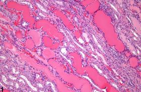
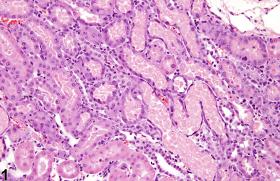
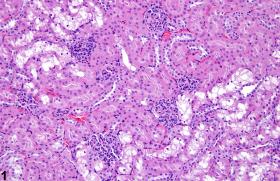
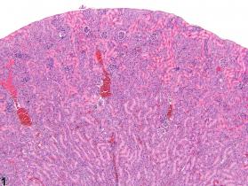
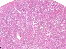
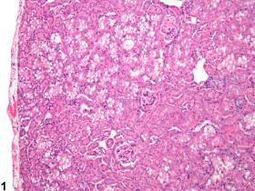
- Kidney, Renal tubule - Normal in a male B6C3F1 mouse from a chronic study. (nih.gov)
- Hypokalemia contributes to the progression of chronic kidney disease, while a definitive pathophysiogical theory to explain this remains to be established. (mdc-berlin.de)
- The GFR test is recommended for people with chronic kidney disease. (medlineplus.gov)
- Levels below 60 mL/min/1.73 m2 for 3 or more months are a sign of chronic kidney disease. (medlineplus.gov)
- In chronic renal failure, excessive production of renin by the kidney can lead to severe high blood pressure ( hypertension ), and the effects of this may even dominate the clinical picture. (britannica.com)
- The association between lead exposure and GFR was evaluated in North American children with CKD in the Chronic Kidney Disease in Children (CKiD) study. (medscape.com)
- Hardening of the KIDNEY due to infiltration by fibrous connective tissue (FIBROSIS), usually caused by renovascular diseases or chronic HYPERTENSION. (mcw.edu)
- With the exception of a very small proportion of patients with chronic kidney disease (CKD) who waste magnesium through their tubules due to medication toxicity, such as amphotericin, the vast majority of CKD patients will retain magnesium to some small and usually clinically insignificant extent. (medscape.com)
Mouse kidney2
- The light and, to a lesser extent, the dark cells of the cortical collecting tubules in mouse kidney contain a great number of granules which according to histochemical tests are composed of phospholipids and proteins. (rupress.org)
- In a mouse kidney cold storage/transplantation model, we detected p53 accumulation in proximal tubules in a cold storage time-dependent manner, which correlated with tubular injury and cell death. (elsevierpure.com)
Reabsorption4
- In the kidney, the first portion of the nephron, called the proximal tubule (PT), performs the majority of solute reabsorption including about two-thirds of calcium. (ku.edu)
- Reabsorption occurs in the renal tubules and is either passive, due to diffusion, or active, due to pumping against a concentration gradient. (wikipedia.org)
- Some of the hormones which signal the tubules to alter the reabsorption or secretion rate, and thereby maintain homeostasis, include (along with the substance affected) antidiuretic hormone (water), aldosterone (sodium, potassium), parathyroid hormone (calcium, phosphate), atrial natriuretic peptide (sodium) and brain natriuretic peptide (sodium). (wikipedia.org)
- Sixty-five percent of the calcium filtered through the glomeruli is reabsorbed in the proximal tubule by a process linked to sodium reabsorption. (medscape.com)
Stimulates1
- 6. Stimulates absorption of water from the kidney tubule. (wikieducator.org)
Collecting ducts1
- the connecting tubule, and the last part of nephron the collecting ducts. (wikipedia.org)
Glomeruli3
- Some diseases of the nephron predominantly affect either the glomeruli or the tubules. (wikipedia.org)
- Glomeruli are the tiny filters in the kidneys that filter waste from the blood. (medlineplus.gov)
- The two most common causes are pyelonephritis and glomerulonephritis (kidney inflammation involving the structures around the renal pelvis or the glomeruli), and other common causes are renal damage from the effects of high blood pressure and renal damage from obstructive conditions of the lower urinary tract. (britannica.com)
Disorders5
- The polycystic kidney diseases are a group of genetically heterogeneous disorders and a leading cause of kidney failure. (basicmedicalkey.com)
- and a number of primary disorders of the kidney tubules. (britannica.com)
- Some diseases affecting the kidney are systemic disorders, which means, they are not limited to the organ itself and may require special treatment. (maxhealthcare.in)
- Nephrologists are kidney doctors who specialise in the care and treatment of renal or kidney related disorders. (maxhealthcare.in)
- This can be caused by urinary tract infections (UTIs), autoimmune disorders, sickle cell disease, diabetes, kidney transplant rejection, or some medicines. (teenshealth.org)
Glomerulus1
- About 95% of free -2-m is filtered by the branous and proliferative being the most normal glomerulus and a normal kidney is common [ 2 ]. (who.int)
Diabetic nephropathy1
- Broadly, the diseases that are treated by them include autoimmune diseases, kidney cancers, diabetic nephropathy, blood pressure and several others. (maxhealthcare.in)
Tissues4
- As such, it is maintained within a narrow range through the actions of gastrointestinal, bone, kidney, and endocrine tissues. (ku.edu)
- Although cysts only occur in 5% of the tubules in the kidney, the enormous growth of these cysts ultimately leads to the loss of normal surrounding tissues and loss of renal function. (basicmedicalkey.com)
- Also, cisplatin increased ERK1/2 phosphorylation and NF-κB expression, which subsequently increased mRNA expression of TNF-α, IL-6, KIM-1, NGAL, and Bax/Bcl-2 ratio as well as decreased mRNA expression of IL-10 in kidney tissues. (thieme-connect.com)
- Childhood kidney tumors are diseases in which malignant (cancer) cells form in the tissues of the kidney. (instituteofliving.org)
Urinary2
- Anatomy of the male urinary system (left panel) and female urinary system (right panel) showing the kidneys, ureters, bladder, and urethra. (hoacny.com)
- Postrenal proteinuria is due to plasma proteins from hemorrhage or inflammation in the urinary tract (kidneys, ureters, bladder, urethra, and/or accessory sex glands). (vin.com)
Human kidney4
- Next, I tested the hypothesis that deletion of the claudin-2 gene Cldn2 in mice causes nephrocalcinosis similar to human kidney stone disease using micro-computed tomography (micro-CT) and histological analyses. (ku.edu)
- My findings indicate that this papillary pattern of nephrocalcinosis shares striking similarities to human kidney stone disease. (ku.edu)
- Our colleagues subsequently identified multiple SNPs in the CLDN2 locus that associate with human kidney stone disease. (ku.edu)
- These findings shed light on the functional and regulatory aspects of SOX9 activation in the human kidney during injury and regeneration. (lu.se)
Nephron4
- The nephron is the minute or microscopic structural and functional unit of the kidney. (wikipedia.org)
- The nephron is the functional unit of the kidney. (wikipedia.org)
- This means that each separate nephron is where the main work of the kidney is performed. (wikipedia.org)
- A nephron is made of two parts: a renal corpuscle, which is the initial filtering component, and a renal tubule that processes and carries away the filtered fluid. (wikipedia.org)
Cisplatin-induced9
- We also established a renal proximal tubule-specific autophagy-related gene 7-knockout mouse model shown to be defective in both basal and cisplatin-induced autophagy in kidneys. (elsevierpure.com)
- Objective In addition to oxidative stress, inflammation and apoptosis have an important role in the pathogenesis of cisplatin-induced kidney damage. (thieme-connect.com)
- This study aimed to investigate the molecular mechanisms of protective effects of curcumin against cisplatin-induced kidney inflammation and apoptosis in rats. (thieme-connect.com)
- Conclusions These data indicate that curcumin has nephroprotective properties against cisplatin-induced kidney damage in rats and this effect is associated with its anti-inflammatory and anti-apoptosis profiles, in addition to its antioxidant. (thieme-connect.com)
- Over the last few decades, it has been studied that the mechanisms of cisplatin-induced kidney damage are complex and involved numerous cellular and molecular processes including inflammation, apoptosis, accumulation of cisplatin in renal tubular cells via renal drug transporters, Ctr1 and OCT2, and involvement of mitogen-activated protein kinases (MAPK) pathways [ 3 ] [ 4 ]. (thieme-connect.com)
- Even though various treatment strategies namely saline hydration and diuresis have been suggested for prevention of cisplatin-induced kidney damage, but its prevalence is still high. (thieme-connect.com)
- In fact, the prevalence of cisplatin-induced kidney damage was 34% after fourth cycles and 52% after six cycles of cisplatin chemotherapy in adult cancer patients treated with cisplatin at a dose of ≥60 mg/m 2 at Dharmais National Cancer Hospital, Jakarta, Indonesia [ 6 ]. (thieme-connect.com)
- However, the molecular mechanisms behind the anti-inflammatory and anti-apoptotic effects of curcumin in the cisplatin-induced kidney damage have not been explored. (thieme-connect.com)
- Therefore, in this study, we aimed to investigate the possible molecular mechanisms of anti-inflammatory and anti-apoptotic effects of curcumin in cisplatin-induced kidney damage. (thieme-connect.com)
Inflammation4
- Fortunately kidney function appears to remain intact, but there is cell injury and inflammation. (uchicago.edu)
- In particular, extracellular-regulated kinase (ERK) 1/2, one of the MAPK pathway is considered as an important mediator of signal transduction processes, namely cell survival, cell division, gene expression, and cell metabolism that plays role in injury, death, and inflammation of kidney tubular cells due to cisplatin administration [ 5 ]. (thieme-connect.com)
- BACKGROUND: Inflammation may affect long-term kidney function. (cdc.gov)
- We hypothesized that proinflammatory diets increase the risk of progression to kidney failure with replacement therapy (KFRT), and systemic inflammation is a mediator of the effect of diet on progression to KFRT. (cdc.gov)
Diseases2
- Kidney cysts are often seen in a wide range of syndromic diseases. (basicmedicalkey.com)
- Cystic kidney diseases are a global public health burden, affecting over 12 million people 1 . (elsevierpure.com)
Damage7
- Histopathological examination furthers confirmed the kidney damage protection effect of curcumin. (thieme-connect.com)
- Hence, curcumin may be useful for preventing kidney damage against cisplatin administration. (thieme-connect.com)
- However, it has many severe side effects that limit the therapeutic use of cisplatin, one of them is kidney damage [ 1 ] [ 2 ]. (thieme-connect.com)
- Proximal tubules obtained in bulk by nonenzymatic procedures were resistant to cell damage by levels of chloroform (67663) and carbon-tetrachloride (56235) likely to be encountered in the work place. (cdc.gov)
- Drug treatment of the rabbits followed by tubule isolation and exposure to these solvent s in-vitro did not enhance cell damage. (cdc.gov)
- Tubule cells propagated in-vitro were more susceptible to cell damage caused by these organic solvent s. (cdc.gov)
- In a study of ultrasound and laboratory findings in Wilms tumor survivors with a solitary kidney, signs of kidney damage were seen in 22 of 53 patients (41.5%) on ultrasonography. (medscape.com)
Cells14
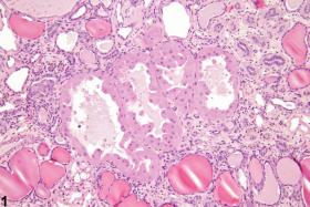
- Numerous clear cytoplasmic vacuoles are present in renal tubule cells. (nih.gov)
- Increased cAMP promotes protein kinase A activity, among other effectors, and, in turn, leads to cyst growth by promoting proliferation and fluid secretion of cyst-lining cells through chloride and aquaporin channels in ADPKD kidneys. (basicmedicalkey.com)
- Similar effects were shown by the ablation of p53 from proximal tubule cells. (elsevierpure.com)
- in vitro, cold storage followed by rewarming induced cell death in cultured proximal tubule cells, which was accompanied by p53 activation and suppressed by pifithrin-α and dominant-negative p53. (elsevierpure.com)
- first with the interstitial fluid outside the tubules, and then into the plasma in the adjacent peritubular capillaries through the endothelial cells lining that capillary. (wikipedia.org)
- Lead is absorbed by the proximal tubular cells of the renal tubules, where it binds to specific lead-binding proteins. (medscape.com)
- Renal cell cancer is a disease in which malignant (cancer) cells form in tubules of the kidney. (hoacny.com)
- Renal cell cancer (also called kidney cancer or renal cell adenocarcinoma ) is a disease in which malignant (cancer) cells are found in the lining of tubules (very small tubes) in the kidney. (hoacny.com)
- The underlying mechanism is most probably a blockade of anion conductance in the plasma membrane at nanomolar concentrations of OTA with subsequent disturbance of cellular acid-base homeostasis as shown in cultured kidney cells. (karger.com)
- Disturbance of cellular pH homeostasis is probably also involved in OTA-induced transformation of cultured kidney cells. (karger.com)
- Induction of SRY box transcription factor 9 (SOX9) has been shown to occur in response to kidney injury in rodents, where SOX9-positive cells proliferate and regenerate the proximal tubules of injured kidneys. (lu.se)
- SOX9 expression was found to colocalize with a proportion of so-called scattered tubular cells in the uninjured kidney, a cell population previously shown to be involved in kidney injury and regeneration. (lu.se)
- Moreover, a kidney explant model was used to demonstrate that only SOX9-positive cells survive the massive injury associated with kidney ischemia and that the surviving SOX9-positive cells spread and repopulate the tubules. (lu.se)
- It is produced by Idiopathic nephrotic syndrome (INS) a variety of cells, including monocytes and accounts for 90% of nephrosis in child- mesangial cells in the kidney [ 9 ]. (who.int)
Mice1
- Notably, pifithrin-α also ameliorated kidney injury and improved the function of transplanted kidneys in 6 days when it became the sole life-supporting kidney in recipient mice. (elsevierpure.com)
Bladder1
- The ureter is a long tube that connects the kidney to the bladder. (instituteofliving.org)
Calcium7
- My work suggests that proximal delivery of calcium to the loops of Henle is important in the pathogenesis of nephrocalcinosis and kidney stone formation. (ku.edu)
- In addition, claudin-2 expression is an important mediator of calcium transport that is associated with kidney stone disease in humans. (ku.edu)
- This new post shows very new research done over the past decade or so, mainly by us, which shows that the tiny tubules of the kidneys can become plugged with calcium phosphate crystals. (uchicago.edu)
- When the diseased kidney cannot excrete phosphorus, it builds up in the body and also affects calcium absorption. (healthhype.com)
- The major regulators of calcium levels are parathyroid hormone (PTH) and vitamin D, which target the bones, intestine, and kidney to increase serum calcium. (medscape.com)
- Calcitonin, a more minor player in regulation, decreases serum calcium by its effects on bone and kidney. (medscape.com)
- The kidney serves as the rapid regulator of calcium fluxes but has limited capacity to handle large swings in the serum calcium levels. (medscape.com)
Nephrotoxicity2
- Nephrotoxicity results from lead exposure because the kidney is the main route by which lead is eliminated. (medscape.com)
- A study was conducted to examine the validity of the isolated rabbit tubule model as a tool in the study of nephrotoxicants and to determine the validity of the rabbit tubule model as a predictor of nephrotoxicity in humans. (cdc.gov)
Tumors1
- In most cases, there will be a solitary tumor in one kidney, but 5-13% of children have bilateral tumors and 10% have multifocal tumors in a single kidney. (medscape.com)
Mechanisms1
- Although much is known about the genetics of kidney development and disease, the cellular mechanisms driving normal kidney tubule elongation remain unclear 2,3 . (elsevierpure.com)
Disease12
- FIGURE 339-1 Scheme of the primary cilium and cystic kidney disease proteins. (basicmedicalkey.com)
- It may also be done to see how far kidney disease has progressed. (medlineplus.gov)
- What is a kidney disease diet? (healthhype.com)
- A kidney disease diet is more correctly known as renal diet . (healthhype.com)
- It includes changes in eating habits that aims to reduce the strain on the kidneys and minimize the imbalances that arise with kidney disease. (healthhype.com)
- Always speak to your doctor and if necessary consult with a registered dietitian or nutritionist who can assist with an appropriate eating plan for kidney disease. (healthhype.com)
- For example there may be specific amendments to the renal diet for people with heart disease, diabetes and obesity in addition to the kidney problems. (healthhype.com)
- Toxic nephropathies are estimated to cause fewer than 1% of all cases of end-stage kidney disease. (medscape.com)
- Untreated renal (REE-nul) tubular acidosis can affect a child's growth, cause kidney stones , and other problems like bone or kidney disease . (teenshealth.org)
- Dapagliflozin is not recommended for use in patients with moderate to severe kidney disease . (medicinenet.com)
- Background: Access to renal replacement therapy by the increasing population of patients with end-stage kidney disease across Sub-Saharan Africa, including Nigeria, has become a major public health challenge. (bvsalud.org)
- While complications of both hypo- and hypermagnesemia exist, their relative infrequency among persons with kidney disease has reinforced a very limited practice of monitoring magnesium levels. (medscape.com)
Proteinuria1
- However, the diseased tubules are unable to metabolize these proteins and tubular proteinuria ensues. (vin.com)
Tiny1
- The kidneys' main job is to remove waste - including acid - and extra water from the blood through tiny tubes called tubules. (teenshealth.org)
Dysfunction1
- Limit sodium , phosphorus and potassium intake since kidney dysfunction may cause high levels of these electrolytes. (healthhype.com)
Exposure2
- However, dapagliflozin is secreted in the milk of lactating rats, and exposure showed risk to the developing kidneys in the rat fetus. (medicinenet.com)
- Renal hemodynamics and the secretory function of the proximal tubule are affected by OTA after prolonged but not by acute exposure. (karger.com)
Unclear1
- p53 contributes to both ischemic and nephrotoxic kidney injury, but its involvement in kidney cold storage/transplantation is unclear. (elsevierpure.com)
Bone1
- It is unknown if blood or bone lead levels are more consistently associated with kidney effects. (medscape.com)
Vitro1
- Kidney proximal tubules were isolated from control and phenobarbital induced rabbits by nonenzymatic sieving techniques and then studied in-vitro. (cdc.gov)
Sodium2
- However, when the kidney is diseased it cannot properly eliminate excess sodium. (healthhype.com)
- 1 liter of filtrate in kidney tubules contains around 140 mEq of sodium. (medscape.com)
Function3
- Kidney injury associated with cold storage/transplantation is a primary factor for delayed graft function and poor outcome of renal transplants. (elsevierpure.com)
- Here, we report that p53 in kidney proximal tubules plays a critical role in cold storage/transplantation kidney injury and inhibition of p53 can effectively improve the histology and function of transplanted kidneys. (elsevierpure.com)
- Kidney function must be assessed before starting treatment. (medicinenet.com)
Topology1
- In contrast to findings in Drosophila, however, non-canonical Wnt and planar cell polarity (PCP) signaling is required to control rosette topology and orientation during vertebrate kidney tubule elongation. (elsevierpure.com)
Normal2
- According to the National Kidney Foundation, normal results range from 90 to 120 mL/min/1.73 m 2 . (medlineplus.gov)
- Here, we characterized the role of SOX9 in normal and injured human kidneys. (lu.se)
Hypertension1
- 4] The hypothesis was that decreased synthesis of eicosanoids might contribute to hypertension and make the kidney more vulnerable to drugs that reduce the synthesis of locally produced vasodilators (eg, nonsteroidal anti-inflammatory drugs). (medscape.com)
Renal tubular2
- Renal tubular acidosis is an illness that happens when the kidneys are damaged and can't remove a waste, called acid, from the blood. (teenshealth.org)
- But with renal tubular acidosis, the kidney's tubules are damaged, so they can't remove the acid. (teenshealth.org)