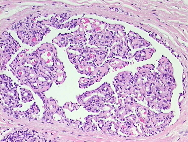
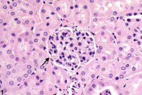
Kidney Glomerulus
Podocytes
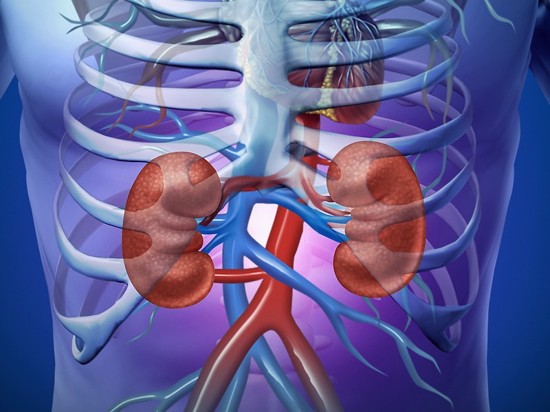
Kidney
Glomerular Mesangium
Basement Membrane
Membrane Proteins
Kidney Tubules
Glomerulonephritis
Kidney Cortex
Olfactory Bulb
Acute Kidney Injury
Olfactory Pathways
Kidney Failure, Chronic
Kidney Tubules, Proximal
Olfactory Receptor Neurons
Glomerulosclerosis, Focal Segmental
Nephrosis
Kidney Medulla
Polycystic Kidney Diseases
Kidney Function Tests
T lymphocyte adhesion mechanisms within inflamed human kidney: studies with a Stamper-Woodruff assay. (1/4509)
Renal inflammatory conditions are characterized by mononuclear cell recruitment to sites of inflammation. We have developed a modified Stamper-Woodruff assay system to analyze mechanisms of functional T cell adhesion to cryostat sections of renal biopsy material from patients with vasculitic glomerulonephritis (GN) and acute allograft rejection. Peripheral blood T cells adhered to intraglomerular, periglomerular, and tubulointerstitial regions of the cortex. Blocking monoclonal antibodies against tissue expressed ICAM-1, VCAM-1, and the CS-1 domain of fibronectin (CS-1Fn) differentially attenuated T cell adhesion. Glomerular adhesion in vasculitic GN and tubulointerstitial adhesion in acute rejection were particularly sensitive to both anti-ICAM-1 and anti-VCAM-1 antibodies, indicating a prominent role for ICAM-1 and VCAM-1 at glomerular sites in vasculitis and at tubulointerstitial sites in rejection. Furthermore, using KL/4 cells (LFA-1 expressing) and Jurkat cells (VLA-4 expressing), we demonstrated specific LFA-1/ICAM-1- and VLA-4/VCAM-1-mediated interactions within glomerular and tubulointerstitial compartments. Jurkat cells also adhered to VCAM-1-free sites, and binding was inhibitable by anti-CS-1Fn antibody, thereby demonstrating a role for VLA-4/fibronectin interactions especially at intraglomerular sites in acute rejection where VCAM-1 is notably absent. We therefore propose a prominent functional role for ICAM-1, VCAM-1, and CS-1 domain fibronectin in T cell recruitment to the inflamed kidney. (+info)Angiotensin receptor subtype 1 mediates angiotensin II enhancement of isoproterenol-induced cyclic AMP production in preglomerular microvascular smooth muscle cells. (2/4509)
In a previous study, we found that angiotensin (Ang) II enhances beta-adrenoceptor-induced cAMP production in cultured preglomerular microvascular smooth muscle cells (PMVSMCs) obtained from spontaneously hypertensive rats. The purpose of the present investigation was to identify the Ang receptor subtypes that mediate this effect. In our first study, we compared the ability of Ang II, Ang III, Ang (3-8), and Ang (1-7) to increase cAMP production in isoproterenol (1 microM)-treated PMVSMCs. Each peptide was tested at 0.1, 1, 10, 100, and 1000 nM. Both Ang II and Ang III increased intracellular (EC50s, 1 and 11 nM, respectively) and extracellular (EC50s, 2 and 14 nM, respectively) cAMP levels in a concentration-dependent fashion. In contrast, Ang (3-8) and Ang (1-7) did not enhance either intracellular or extracellular cAMP levels at any concentration tested. In our second study, we examined the ability of L 158809 [a selective Ang receptor subtype 1 (AT1) receptor antagonist] to inhibit Ang II (100 nM) and Ang III (100 nM) enhancement of isoproterenol (1 microM)-induced cAMP production in PMVSMCs. L 158809 (10 nM) abolished or nearly abolished (p <.001) Ang II and Ang III enhancement of isoproterenol-induced intracellular and extracellular cAMP levels. In contrast, PD 123319 (300 nM; a selective AT2 receptor antagonist) did not significantly alter Ang II enhancement of isoproterenol-induced intracellular or extracellular cAMP levels. We conclude that AT1 receptors, but not AT2, Ang (3-8), nor Ang (1-7) receptors mediate Ang II and Ang III enhancement of beta-adrenoceptor-induced cAMP production in cultured PMVSMCs. (+info)Recovery following relief of unilateral ureteral obstruction in the neonatal rat. (3/4509)
BACKGROUND: Obstructive nephropathy is a primary cause of renal insufficiency in infants and children. This study was designed to distinguish the reversible and irreversible cellular consequences of temporary unilateral ureteral obstruction (UUO) on the developing kidney. METHODS: Rats were subjected to UUO or sham operation in the first 48 hours of life, and the obstruction was removed five days later (or was left in place). Kidneys were removed for study 14 or 28 days later. In additional groups, kidneys were removed at the end of five days of obstruction. Immunoreactive distribution of renin was determined in arterioles, and the distribution of epidermal growth factor, transforming growth factor-beta1, clusterin, vimentin, and alpha-smooth muscle actin was determined in tubules and/or interstitium. The number of glomeruli, glomerular maturation, tubular atrophy, and interstitial collagen deposition was determined by morphometry. Renal cellular proliferation and apoptosis were measured by proliferating cell nuclear antigen and the TdT uridine-nick-end-label technique, respectively. The glomerular filtration rate was measured by inulin clearance. RESULTS: Renal microvascular renin maintained a fetal distribution with persistent UUO; this was partially reversed by the relief of obstruction. Although glomerular maturation was also delayed and glomerular volume was reduced by UUO, the relief of obstruction prevented the reduction in glomerular volume. Although relief of obstruction did not reverse a 40% reduction in the number of nephrons, the glomerular filtration rate of the postobstructed kidney was normal. The relief of obstruction did not improve tubular cell proliferation and only partially reduced apoptosis induced by UUO. This was associated with a persistent reduction in the tubular epidermal growth factor. In addition, the relief of obstruction reduced but did not normalize tubular expression of transforming growth factor-beta1, clusterin, and vimentin, all of which are evidence of persistent tubular injury. The relief of obstruction significantly reduced interstitial fibrosis and expression of alpha-smooth muscle actin by interstitial fibroblasts, but not to normal levels. CONCLUSIONS: The relief of obstruction in the neonatal rat attenuates, but does not reverse, renal vascular, glomerular, tubular, and interstitial injury resulting from five days of UUO. Hyperfiltration by remaining nephrons and residual tubulointerstitial injury in the postobstructed kidney are likely to lead to deterioration of renal function later in life. (+info)Up-regulation of glomerular extracellular matrix and transforming growth factor-beta expression in RF/J mice. (4/4509)
BACKGROUND: RF/J mice were first reported as a murine model of spontaneous glomerulosclerosis by Gude and Lupton in 1960, but the precise histologic characteristics and immunopathological background of this mouse have not been investigated further. METHODS: Measurements of serum levels of immunoglobulins, anti-single strand DNA (anti-ss-DNA) antibody, complement (C3), and circulating immune complex (IC) were performed. Analyses of glomerular histological and immunopathological lesions in association with the detection of mRNA expression of collagen IV, TGF-beta, matrix protein turnover related enzymes, matrix metalloproteinase-2 (MMP-2), tissue inhibitor of metalloproteinase-2 (TIMP-2) and platelet-derived growth factor (PDGF) were also performed in young (10-week-old) and elderly (60-week-old) RF/J mice with age-matched BALB/C mice as the controls. RESULTS: High levels of serum IgA and IgG from as early as 20 weeks of age were noted in the RF/J mice. Serum anti-ss-DNA antibody of aged RF/J mice increased up to 23% of that of aged MRL-lpr/lpr mice, and serum C3 concentration significantly decreased with age, reaching lower levels than that of BALB/c mice. IgA-IC levels were significantly high compared to BALB/C mice both in the early and late stages of life, whereas IgG-IC levels were high only in mice younger than 20 weeks. Semiquantitative and quantitative analyzes of renal histopathological findings revealed significantly marked and age-related mesangial matrix expansion in RF/J mice, with increasing frequency of global glomerular sclerosis and tubulointerstitial damage. On the other hand, although precise measurements of glomerular cell numbers also showed an apparent augmentation in both young and old RF/J mice compared to BALB/C mice, glomerular cellularity decreased with age in RF/J mice. Immunohistochemical study revealed massive immunoglobulin deposition from a young age in association with significantly higher accumulation of matrix proteins, such as types I and IV collagen and laminin from the early stage of life. In addition, in these glomeruli, transforming growth factor-beta1 (TGF-beta1) was highly expressed both in young and old mice. The mRNA expression of MMP-2 was up-regulated only in the early stage of life. Although PDGF mRNA of RF/J mice was significantly up-regulated in the early stage of life, the differences between the mice disappeared in the late stage of life. CONCLUSIONS: These findings suggest that in RF/J mice, an immunopathological background inducing high serum immunoglobulin and IC levels from the early stage of life is closely related to mesangioproliferative glomerular lesions mediated by PDGF, and that development of massive extracellular matrix accumulation in glomeruli was induced by up-regulated expression of TGF-beta with inappropriate regulation of protein turnover-related enzyme production. (+info)Mycophenolate mofetil prevents the progressive renal failure induced by 5/6 renal ablation in rats. (5/4509)
BACKGROUND: Extensive renal ablation is associated with progressive sclerosis of the remnant kidney. Because lymphocytes and monocytes accumulate in the remnant kidney, it is likely that they play a role in the renal scarring. Therefore, we treated rats with 5/6 nephrectomy (5/6Nx) with mycophenolate mofetil (MMF), a drug that has an antiproliferative effect and that suppresses the expression of intercellular adhesion molecules. METHODS: Sprague-Dawley rats with 5/6Nx received MMF (30 mg. kg-1. day-1 by daily gastric gavage, N = 15) or vehicle (N = 16). Ten additional rats were sham operated. All rats were fed a 30% protein diet. Body weight, serum creatinine, and urinary protein excretion were determined weekly. Lipid peroxidation, as a measure of oxidative stress observed by urinary malondialdehyde determinations, was performed every two weeks. Histologic studies were done in the remnant kidney four weeks (9 rats from the vehicle-treated group, 7 rats from the MMF group, and 5 sham-operated rats) and eight weeks after surgery (the remaining rats). Glomerular volume, sclerosis in glomeruli (segmental and global) and interstitium (semiquantitative scale), infiltrating lymphocytes and macrophages (CD43- and ED1-positive cells), and expression of adhesion molecules (CD54, CD18, and CD11b) were analyzed. RESULTS: MMF treatment prevented the progressive increment in serum creatinine and the proteinuria observed in the 5/6 nephrectomized rats during the eight weeks of observation (P < 0.01). Weight gain was comparable in the MMF-treated and sham-operated rats, whereas weight gain was decreased in untreated 5/6 nephrectomized rats. Excretion of malondialdehyde increased after surgery but returned sooner to control levels in the MMF-treated rats. Increments in glomerular size and mean arterial blood pressure induced by renal ablation were not modified by MMF treatment. Eight weeks after surgery, segmental sclerosis was present in 48.4 +/- 8.35% (+/- sd) glomeruli in the vehicle-treated group versus 25 +/- 10.5% in the MMF-treated group (P < 0.001). Interstitial fibrosis was reduced significantly with MMF treatment (P < 0.001). Infiltration with CD43- and ED1-positive cells in glomeruli and interstitium was two to five times lower in MMF-treated rats (P < 0.01). Expression of adhesion molecules CD18 and CD11b was similarly reduced. CONCLUSION: MMF ameliorates the progressive renal damage in the remnant kidney after 5/6Nx. This effect is associated with a reduction in the infiltration of lymphocytes and monocytes, whereas glomerular hypertrophy and systemic hypertension are unchanged. (+info)Glomerular size-selective dysfunction in NIDDM is not ameliorated by ACE inhibition or by calcium channel blockade. (6/4509)
BACKGROUND: In patients with insulin-dependent diabetes mellitus (IDDM) and overt nephropathy glomerular barrier size-selectivity progressively deteriorates with time and is effectively improved by angiotensin converting enzyme (ACE) inhibition. Whether similar glomerular functional changes develop in proteinuric patients with non-insulin-dependent diabetes mellitus (NIDDM), and whether antihypertensive agents can favorably affect glomerular filtration of macromolecules in these patients, has not been documented yet. METHODS: We investigated renal hemodynamics and fractional clearance of neutral dextrans of graded sizes, in nine proteinuric patients with NIDDM and renal biopsy findings of typical diabetic glomerulopathy. Six healthy volunteers served as controls. We also investigated the effects of an ACE inhibitor and of a calcium channel blocker, both given in doses targeted to achieve a comparable level of systemic blood pressure control, on glomerular hemodynamics and sieving function. Theoretical analysis of glomerular macromolecule transport was adopted to evaluate intrinsic glomerular membrane permeability properties. RESULTS: Fractional clearance of large macromolecules (42 to 66 A in radius) was significantly higher in diabetic patients than in controls, and the distribution of membrane pore radii was calculated to be shifted towards larger pore sizes in diabetics (mean radius increased from 55 to 60 A). Despite effective blood pressure control, neither antihypertensive affected glomerular hemodynamics to any significant extent. Fractional clearance of dextrans, as well as of albumin and IgG, and total urinary proteins were not modified by either treatments. CONCLUSIONS: These data indicate that patients with NIDDM and overt nephropathy develop abnormalities in size-selective function of the glomerular barrier and, at variance to IDDM, such changes were not ameliorated either by ACE inhibition or calcium channel blockade. (+info)T cell subsets in experimental lupus nephritis: modulation by bacterial superantigen. (7/4509)
Chronic graft-vs-host disease (GvH), induced by injection of DBA/2 lymphocytes into (C57BL/6 x DBA/2)F1 hybrids, is a murine model for lupus nephritis, associated with a Th2-dependent polyclonal B cell activation. The development of glomerulosclerosis in this model is preceded by a glomerular influx of LFA-1+ T cells. We investigated whether exposure to bacterial superantigen would modulate the course of this autoimmune syndrome. Injection of the bacterial superantigen staphylococcal enterotoxin B (SEB) in mice has been shown to induce the activation of TcRVbeta8+ T cells. Within 2 weeks after GvH induction, mice were injected twice with 20 microg of SEB and the following parameters were examined: cytokine and Ig profile, proteinuria and renal pathology. The second SEB injection induced in GvH mice an increased release of both interferon-gamma (IFN-gamma) and interleukin-10 (IL-10) as compared with control F1 mice. No differences were observed in IL-2 production. SEB-treated GvH mice demonstrated a delayed onset of proteinuria. Histological analysis of the kidney showed that SEB-challenged GvH mice displayed significantly more interstitial inflammation and mesangial proliferation together with more IgG2a deposits in glomeruli than non-injected GvH mice. From these results, we conclude that GvH mice are more responsive to SEB in terms of cytokine production and that bacterial infection can modulate the course of this renal disease from a membranous to a more proliferative type of nephropathy. (+info)Adoptive transfer of genetically modified macrophages elucidated TGF-beta-mediated 'self-defence' of the glomerulus against local action of macrophages. (8/4509)
TGF-beta has several anti-inflammatory properties which may be relevant to prevention of or recovery from acute glomerular inflammation. Using genetically modified mesangial cells and a technique for in vivo macrophage transfer, this article provides evidence for TGF-beta-mediated 'self-defence' of the glomerulus against macrophages. Rat mesangial cells stably transfected with TGF-beta1 showed a blunted response to the macrophage-derived, proinflammatory cytokine IL-1beta. In contrast, mesangial cells expressing the dominant-interfering TGF-beta receptor showed an enhanced response to IL-1. Similarly, externally added TGF-beta1 inhibited the cytokine response of normal glomeruli, and isolated nephritic glomeruli producing active TGF-beta1 showed a depressed response to IL-1beta, compared to normal glomeruli. Consistent with these in vitro results, in vivo transfer of activated macrophages revealed that the TGF-beta-producing glomeruli are insensitive to the effector action of macrophages. These results indicate that TGF-beta1 functions as an endogenous 'defender' that counteracts local action of activated macrophages in the glomerulus. (+info)A kidney glomerulus is a functional unit in the nephron of the kidney. It is a tuft of capillaries enclosed within a structure called Bowman's capsule, which filters waste and excess fluids from the blood. The glomerulus receives blood from an afferent arteriole and drains into an efferent arteriole.
The process of filtration in the glomerulus is called ultrafiltration, where the pressure within the glomerular capillaries drives plasma fluid and small molecules (such as ions, glucose, amino acids, and waste products) through the filtration membrane into the Bowman's space. Larger molecules, like proteins and blood cells, are retained in the blood due to their larger size. The filtrate then continues down the nephron for further processing, eventually forming urine.
Podocytes are specialized cells that make up the visceral epithelial layer of the glomerular basement membrane in the kidney. They have long, interdigitating foot processes that wrap around the capillaries of the glomerulus and play a crucial role in maintaining the filtration barrier of the kidney. The slit diaphragms between the foot processes allow for the passage of small molecules while retaining larger proteins in the bloodstream. Podocytes also contribute to the maintenance and regulation of the glomerular filtration rate, making them essential for normal renal function. Damage or loss of podocytes can lead to proteinuria and kidney disease.
A kidney, in medical terms, is one of two bean-shaped organs located in the lower back region of the body. They are essential for maintaining homeostasis within the body by performing several crucial functions such as:
1. Regulation of water and electrolyte balance: Kidneys help regulate the amount of water and various electrolytes like sodium, potassium, and calcium in the bloodstream to maintain a stable internal environment.
2. Excretion of waste products: They filter waste products from the blood, including urea (a byproduct of protein metabolism), creatinine (a breakdown product of muscle tissue), and other harmful substances that result from normal cellular functions or external sources like medications and toxins.
3. Endocrine function: Kidneys produce several hormones with important roles in the body, such as erythropoietin (stimulates red blood cell production), renin (regulates blood pressure), and calcitriol (activated form of vitamin D that helps regulate calcium homeostasis).
4. pH balance regulation: Kidneys maintain the proper acid-base balance in the body by excreting either hydrogen ions or bicarbonate ions, depending on whether the blood is too acidic or too alkaline.
5. Blood pressure control: The kidneys play a significant role in regulating blood pressure through the renin-angiotensin-aldosterone system (RAAS), which constricts blood vessels and promotes sodium and water retention to increase blood volume and, consequently, blood pressure.
Anatomically, each kidney is approximately 10-12 cm long, 5-7 cm wide, and 3 cm thick, with a weight of about 120-170 grams. They are surrounded by a protective layer of fat and connected to the urinary system through the renal pelvis, ureters, bladder, and urethra.
The glomerular mesangium is a part of the nephron in the kidney. It is the region located in the middle of the glomerular tuft, where the capillary loops of the glomerulus are surrounded by a network of extracellular matrix and mesangial cells. These cells and matrix play an important role in maintaining the structure and function of the filtration barrier in the glomerulus, which helps to filter waste products from the blood.
The mesangial cells have contractile properties and can regulate the flow of blood through the capillaries by constricting or dilating the diameter of the glomerular capillary loops. They also play a role in immune responses, as they can phagocytize immune complexes and release cytokines and growth factors that modulate inflammation and tissue repair.
Abnormalities in the mesangium can lead to various kidney diseases, such as glomerulonephritis, mesangial proliferative glomerulonephritis, and diabetic nephropathy.
The basement membrane is a thin, specialized layer of extracellular matrix that provides structural support and separates epithelial cells (which line the outer surfaces of organs and blood vessels) from connective tissue. It is composed of two main layers: the basal lamina, which is produced by the epithelial cells, and the reticular lamina, which is produced by the connective tissue. The basement membrane plays important roles in cell adhesion, migration, differentiation, and survival.
The basal lamina is composed mainly of type IV collagen, laminins, nidogens, and proteoglycans, while the reticular lamina contains type III collagen, fibronectin, and other matrix proteins. The basement membrane also contains a variety of growth factors and cytokines that can influence cell behavior.
Defects in the composition or organization of the basement membrane can lead to various diseases, including kidney disease, eye disease, and skin blistering disorders.
Membrane proteins are a type of protein that are embedded in the lipid bilayer of biological membranes, such as the plasma membrane of cells or the inner membrane of mitochondria. These proteins play crucial roles in various cellular processes, including:
1. Cell-cell recognition and signaling
2. Transport of molecules across the membrane (selective permeability)
3. Enzymatic reactions at the membrane surface
4. Energy transduction and conversion
5. Mechanosensation and signal transduction
Membrane proteins can be classified into two main categories: integral membrane proteins, which are permanently associated with the lipid bilayer, and peripheral membrane proteins, which are temporarily or loosely attached to the membrane surface. Integral membrane proteins can further be divided into three subcategories based on their topology:
1. Transmembrane proteins, which span the entire width of the lipid bilayer with one or more alpha-helices or beta-barrels.
2. Lipid-anchored proteins, which are covalently attached to lipids in the membrane via a glycosylphosphatidylinositol (GPI) anchor or other lipid modifications.
3. Monotopic proteins, which are partially embedded in the membrane and have one or more domains exposed to either side of the bilayer.
Membrane proteins are essential for maintaining cellular homeostasis and are targets for various therapeutic interventions, including drug development and gene therapy. However, their structural complexity and hydrophobicity make them challenging to study using traditional biochemical methods, requiring specialized techniques such as X-ray crystallography, nuclear magnetic resonance (NMR) spectroscopy, and single-particle cryo-electron microscopy (cryo-EM).
Kidney disease, also known as nephropathy or renal disease, refers to any functional or structural damage to the kidneys that impairs their ability to filter blood, regulate electrolytes, produce hormones, and maintain fluid balance. This damage can result from a wide range of causes, including diabetes, hypertension, glomerulonephritis, polycystic kidney disease, lupus, infections, drugs, toxins, and congenital or inherited disorders.
Depending on the severity and progression of the kidney damage, kidney diseases can be classified into two main categories: acute kidney injury (AKI) and chronic kidney disease (CKD). AKI is a sudden and often reversible loss of kidney function that occurs over hours to days, while CKD is a progressive and irreversible decline in kidney function that develops over months or years.
Symptoms of kidney diseases may include edema, proteinuria, hematuria, hypertension, electrolyte imbalances, metabolic acidosis, anemia, and decreased urine output. Treatment options depend on the underlying cause and severity of the disease and may include medications, dietary modifications, dialysis, or kidney transplantation.
Kidney tubules are the structural and functional units of the kidney responsible for reabsorption, secretion, and excretion of various substances. They are part of the nephron, which is the basic unit of the kidney's filtration and reabsorption process.
There are three main types of kidney tubules:
1. Proximal tubule: This is the initial segment of the kidney tubule that receives the filtrate from the glomerulus. It is responsible for reabsorbing approximately 65% of the filtrate, including water, glucose, amino acids, and electrolytes.
2. Loop of Henle: This U-shaped segment of the tubule consists of a thin descending limb, a thin ascending limb, and a thick ascending limb. The loop of Henle helps to concentrate urine by creating an osmotic gradient that allows water to be reabsorbed in the collecting ducts.
3. Distal tubule: This is the final segment of the kidney tubule before it empties into the collecting duct. It is responsible for fine-tuning the concentration of electrolytes and pH balance in the urine by selectively reabsorbing or secreting substances such as sodium, potassium, chloride, and hydrogen ions.
Overall, kidney tubules play a critical role in maintaining fluid and electrolyte balance, regulating acid-base balance, and removing waste products from the body.
Kidney transplantation is a surgical procedure where a healthy kidney from a deceased or living donor is implanted into a patient with end-stage renal disease (ESRD) or permanent kidney failure. The new kidney takes over the functions of filtering waste and excess fluids from the blood, producing urine, and maintaining the body's electrolyte balance.
The transplanted kidney is typically placed in the lower abdomen, with its blood vessels connected to the recipient's iliac artery and vein. The ureter of the new kidney is then attached to the recipient's bladder to ensure proper urine flow. Following the surgery, the patient will require lifelong immunosuppressive therapy to prevent rejection of the transplanted organ by their immune system.
Glomerulonephritis is a medical condition that involves inflammation of the glomeruli, which are the tiny blood vessel clusters in the kidneys that filter waste and excess fluids from the blood. This inflammation can impair the kidney's ability to filter blood properly, leading to symptoms such as proteinuria (protein in the urine), hematuria (blood in the urine), edema (swelling), hypertension (high blood pressure), and eventually kidney failure.
Glomerulonephritis can be acute or chronic, and it may occur as a primary kidney disease or secondary to other medical conditions such as infections, autoimmune disorders, or vasculitis. The diagnosis of glomerulonephritis typically involves a combination of medical history, physical examination, urinalysis, blood tests, and imaging studies, with confirmation often requiring a kidney biopsy. Treatment depends on the underlying cause and severity of the disease but may include medications to suppress inflammation, control blood pressure, and manage symptoms.
The kidney cortex is the outer region of the kidney where most of the functional units called nephrons are located. It plays a crucial role in filtering blood and regulating water, electrolyte, and acid-base balance in the body. The kidney cortex contains the glomeruli, proximal tubules, loop of Henle, and distal tubules, which work together to reabsorb necessary substances and excrete waste products into the urine.
The olfactory bulb is the primary center for the sense of smell in the brain. It's a structure located in the frontal part of the brain, specifically in the anterior cranial fossa, and is connected to the nasal cavity through tiny holes called the cribriform plates. The olfactory bulb receives signals from olfactory receptors in the nose that detect different smells, processes this information, and then sends it to other areas of the brain for further interpretation and perception of smell.
Proteinuria is a medical term that refers to the presence of excess proteins, particularly albumin, in the urine. Under normal circumstances, only small amounts of proteins should be found in the urine because the majority of proteins are too large to pass through the glomeruli, which are the filtering units of the kidneys.
However, when the glomeruli become damaged or diseased, they may allow larger molecules such as proteins to leak into the urine. Persistent proteinuria is often a sign of kidney disease and can indicate damage to the glomeruli. It is usually detected through a routine urinalysis and may be confirmed with further testing.
The severity of proteinuria can vary, and it can be a symptom of various underlying conditions such as diabetes, hypertension, glomerulonephritis, and other kidney diseases. Treatment for proteinuria depends on the underlying cause and may include medications to control blood pressure, manage diabetes, or reduce protein loss in the urine.
Acute kidney injury (AKI), also known as acute renal failure, is a rapid loss of kidney function that occurs over a few hours or days. It is defined as an increase in the serum creatinine level by 0.3 mg/dL within 48 hours or an increase in the creatinine level to more than 1.5 times baseline, which is known or presumed to have occurred within the prior 7 days, or a urine volume of less than 0.5 mL/kg per hour for six hours.
AKI can be caused by a variety of conditions, including decreased blood flow to the kidneys, obstruction of the urinary tract, exposure to toxic substances, and certain medications. Symptoms of AKI may include decreased urine output, fluid retention, electrolyte imbalances, and metabolic acidosis. Treatment typically involves addressing the underlying cause of the injury and providing supportive care, such as dialysis, to help maintain kidney function until the injury resolves.
The olfactory pathways refer to the neural connections and structures involved in the sense of smell. The process begins with odor molecules that are inhaled through the nostrils, where they bind to specialized receptor cells located in the upper part of the nasal cavity, known as the olfactory epithelium.
These receptor cells then transmit signals via the olfactory nerve (cranial nerve I) to the olfactory bulb, a structure at the base of the brain. Within the olfactory bulb, the signals are processed and relayed through several additional structures, including the olfactory tract, lateral olfactory striae, and the primary olfactory cortex (located within the piriform cortex).
From there, information about odors is further integrated with other sensory systems and cognitive functions in higher-order brain regions, such as the limbic system, thalamus, and hippocampus. This complex network of olfactory pathways allows us to perceive and recognize various scents and plays a role in emotional responses, memory formation, and feeding behaviors.
Chronic kidney failure, also known as chronic kidney disease (CKD) stage 5 or end-stage renal disease (ESRD), is a permanent loss of kidney function that occurs gradually over a period of months to years. It is defined as a glomerular filtration rate (GFR) of less than 15 ml/min, which means the kidneys are filtering waste and excess fluids at less than 15% of their normal capacity.
CKD can be caused by various underlying conditions such as diabetes, hypertension, glomerulonephritis, polycystic kidney disease, and recurrent kidney infections. Over time, the damage to the kidneys can lead to a buildup of waste products and fluids in the body, which can cause a range of symptoms including fatigue, weakness, shortness of breath, nausea, vomiting, and confusion.
Treatment for chronic kidney failure typically involves managing the underlying condition, making lifestyle changes such as following a healthy diet, and receiving supportive care such as dialysis or a kidney transplant to replace lost kidney function.
A nephron is the basic structural and functional unit of the kidney. It is responsible for filtering blood, reabsorbing necessary substances, and excreting waste products into the urine. Each human kidney contains approximately one million nephrons.
The structure of a nephron includes a glomerulus, which is a tuft of capillaries surrounded by Bowman's capsule. The glomerulus filters blood, allowing small molecules like water and solutes to pass through while keeping larger molecules like proteins and blood cells within the capillaries.
The filtrate then passes through the tubular portion of the nephron, which includes the proximal convoluted tubule, loop of Henle, distal convoluted tubule, and collecting duct. The tubular portion reabsorbs necessary substances like water, glucose, amino acids, and electrolytes back into the bloodstream while excreting waste products like urea and creatinine into the urine.
Overall, nephrons play a critical role in maintaining fluid and electrolyte balance, regulating blood pressure, and removing waste products from the body.
The proximal kidney tubule is the initial portion of the renal tubule in the nephron of the kidney. It is located in the renal cortex and is called "proximal" because it is closer to the glomerulus, compared to the distal tubule. The proximal tubule plays a crucial role in the reabsorption of water, electrolytes, and nutrients from the filtrate that has been formed by the glomerulus. It also helps in the secretion of waste products and other substances into the urine.
The proximal tubule is divided into two segments: the pars convoluta and the pars recta. The pars convoluta is the curved portion that receives filtrate from the Bowman's capsule, while the pars recta is the straight portion that extends deeper into the renal cortex.
The proximal tubule is lined with a simple cuboidal epithelium, and its cells are characterized by numerous mitochondria, which provide energy for active transport processes. The apical surface of the proximal tubular cells has numerous microvilli, forming a brush border that increases the surface area for reabsorption.
In summary, the proximal kidney tubule is a critical site for the reabsorption of water, electrolytes, and nutrients from the glomerular filtrate, contributing to the maintenance of fluid and electrolyte balance in the body.
Olfactory receptor neurons (ORNs) are specialized sensory nerve cells located in the olfactory epithelium, a patch of tissue inside the nasal cavity. These neurons are responsible for detecting and transmitting information about odors to the brain. Each ORN expresses only one type of olfactory receptor protein, which is specific to certain types of odor molecules. When an odor molecule binds to its corresponding receptor, it triggers a signal transduction pathway that generates an electrical impulse in the neuron. This impulse is then transmitted to the brain via the olfactory nerve, where it is processed and interpreted as a specific smell. ORNs are continuously replaced throughout an individual's lifetime due to their exposure to environmental toxins and other damaging agents.
Focal segmental glomerulosclerosis (FSGS) is a pattern of kidney injury that involves scarring or sclerosis in some (segmental) areas of some (focal) glomeruli. Glomeruli are the tiny blood vessel clusters within the kidneys that filter waste and excess fluids from the blood.
In FSGS, the scarring occurs due to damage to the glomerular basement membrane, which can be caused by various factors such as genetic mutations, viral infections, or immune system disorders. The damage leads to the accumulation of extracellular matrix proteins and the formation of scar tissue, impairing the kidney's ability to filter blood effectively.
FSGS is characterized by proteinuria (protein in the urine), hematuria (blood in the urine), hypertension (high blood pressure), and declining kidney function, which can lead to end-stage renal disease if left untreated. The focal and segmental nature of the scarring means that not all glomeruli are affected, and only some areas of each affected glomerulus are damaged, making FSGS a highly variable condition with different clinical presentations and outcomes.
Nephrosis is an older term that was used to describe a group of kidney diseases, primarily characterized by the damage and loss of function in the glomeruli - the tiny filtering units within the kidneys. This results in the leakage of large amounts of protein (primarily albumin) into the urine, a condition known as proteinuria.
The term "nephrosis" was often used interchangeably with "minimal change nephropathy," which is a specific type of kidney disorder that demonstrates little to no changes in the glomeruli under a microscope, despite significant protein leakage. However, current medical terminology and classifications prefer the use of more precise terms to describe various kidney diseases, such as minimal change disease, focal segmental glomerulosclerosis, or membranous nephropathy, among others.
It is important to consult with a healthcare professional or refer to updated medical resources for accurate and current information regarding kidney diseases and their specific diagnoses.
Nephritis is a medical term that refers to inflammation of the kidneys, specifically affecting the glomeruli - the tiny filtering units inside the kidneys. The condition can cause damage to the glomeruli, leading to impaired kidney function and the leakage of protein and blood into the urine.
Nephritis can result from a variety of causes, including infections, autoimmune disorders, and exposure to certain medications or toxins. Depending on the severity and underlying cause, nephritis may be treated with medications, dietary modifications, or other therapies aimed at reducing inflammation and preserving kidney function. In severe cases, hospitalization and more intensive treatments may be necessary.
The kidney medulla is the inner portion of the renal pyramids in the kidney, consisting of multiple conical structures found within the kidney. It is composed of loops of Henle and collecting ducts responsible for concentrating urine by reabsorbing water and producing a hyperosmotic environment. The kidney medulla has a unique blood supply and is divided into an inner and outer zone, with the inner zone having a higher osmolarity than the outer zone. This region of the kidney helps regulate electrolyte and fluid balance in the body.
Polycystic Kidney Disease (PKD) is a genetic disorder characterized by the growth of multiple cysts in the kidneys. These cysts are fluid-filled sacs that can vary in size and can multiply, leading to enlarged kidneys. The increased size and number of cysts can result in reduced kidney function, high blood pressure, and eventually kidney failure.
There are two main types of PKD: Autosomal Dominant Polycystic Kidney Disease (ADPKD) and Autosomal Recessive Polycystic Kidney Disease (ARPKD). ADPKD is the most common form, affecting approximately 1 in every 500 people. It typically develops in adulthood. On the other hand, ARPKD is a rarer form, affecting about 1 in every 20,000 children, and it often presents in infancy or early childhood.
In addition to kidney problems, PKD can also affect other organs, such as the liver and the heart. It's important to note that while there is no cure for PKD, various treatments can help manage symptoms and slow down the progression of the disease.
Kidney function tests (KFTs) are a group of diagnostic tests that evaluate how well your kidneys are functioning by measuring the levels of various substances in the blood and urine. The tests typically assess the glomerular filtration rate (GFR), which is an indicator of how efficiently the kidneys filter waste from the blood, as well as the levels of electrolytes, waste products, and proteins in the body.
Some common KFTs include:
1. Serum creatinine: A waste product that's produced by normal muscle breakdown and is excreted by the kidneys. Elevated levels may indicate reduced kidney function.
2. Blood urea nitrogen (BUN): Another waste product that's produced when protein is broken down and excreted by the kidneys. Increased BUN levels can suggest impaired kidney function.
3. Estimated glomerular filtration rate (eGFR): A calculation based on serum creatinine, age, sex, and race that estimates the GFR and provides a more precise assessment of kidney function than creatinine alone.
4. Urinalysis: An examination of a urine sample to detect abnormalities such as protein, blood, or bacteria that may indicate kidney disease.
5. Electrolyte levels: Measurement of sodium, potassium, chloride, and bicarbonate in the blood to ensure they're properly balanced, which is essential for normal kidney function.
KFTs are often ordered as part of a routine check-up or when kidney disease is suspected based on symptoms or other diagnostic tests. Regular monitoring of kidney function can help detect and manage kidney disease early, potentially preventing or slowing down its progression.
Kidney neoplasms refer to abnormal growths or tumors in the kidney tissues that can be benign (non-cancerous) or malignant (cancerous). These growths can originate from various types of kidney cells, including the renal tubules, glomeruli, and the renal pelvis.
Malignant kidney neoplasms are also known as kidney cancers, with renal cell carcinoma being the most common type. Benign kidney neoplasms include renal adenomas, oncocytomas, and angiomyolipomas. While benign neoplasms are generally not life-threatening, they can still cause problems if they grow large enough to compromise kidney function or if they undergo malignant transformation.
Early detection and appropriate management of kidney neoplasms are crucial for improving patient outcomes and overall prognosis. Regular medical check-ups, imaging studies, and urinalysis can help in the early identification of these growths, allowing for timely intervention and treatment.
 Glomerulus (kidney)