Hip Dysplasia, Canine
Hip Dislocation, Congenital
Hip Joint
Acetabulum
Osteoarthritis, Hip
Bone Diseases, Developmental
Hip
Femur Head
Dogs
Pelvic Bones
Fibrous Dysplasia of Bone
Hip Fractures
Dog Diseases
Spina Bifida Occulta
Ectodermal Dysplasia
Bronchopulmonary Dysplasia
Arthrography
Mucopolysaccharidosis I
Range of Motion, Articular
Uterine Cervical Dysplasia
Fibromuscular Dysplasia
Breeding
Follow-Up Studies
Fibrous Dysplasia, Polyostotic
Recovery of Function
Retrospective Studies
Treatment Outcome
Osteoarthritis
Cleidocranial Dysplasia
Retinal Dysplasia
Pedigree
Cartilage, Articular
Thanatophoric Dysplasia
Site-specific variation in femoral head cartilage composition in dogs at high and low risk for development of osteoarthritis: insights into cartilage degeneration. (1/44)
OBJECTIVE: To determine which characteristics of cartilage lesion pathology are detected in dogs at high risk to develop osteoarthritis prior to diagnosis by standard radiographs or macroscopic cartilage abnormality on necropsy. METHODS: Fourteen disease-free dogs were assigned to risk groups based on hip distraction index. For seven dogs, three dimensional images of hip joints from computed tomography were available. At necropsy, ligamentum capitis femoris volumes were measured and articular cartilage was harvested and analyzed for percent water, swelling, glycosaminoglycan, and fibronectin. Comparisons were made with nine dogs with macroscopic cartilage lesions (OA group). RESULTS: Ligament volumes were greater in the high distraction index group (P=0.000). Water content was elevated in the lesion area in both low and high risk dogs (P=0.000); no additional increase was noted in the high risk group. Glycosaminoglycan content was slightly elevated in the surrounding area in both groups (P< 0.02) but loss was noted histologically in the lesion area of the high risk group. Fibronectin was increased in the lesion area and in the high risk group (P=0.000). The magnitude of this increase was greatest in the lesion area (P=0.000) in explants. Computed tomography indicated dorsal acetabular rim impingement on the lesion area in high risk dogs. CONCLUSIONS: Water content and swelling suggest matrix structure is weaker at the site of lesion predilection in all dogs regardless of risk status. Computed tomography imaging is consistent with site specific initiation of lesions by mechanical factors. (+info)Estimates of genetic parameters for hip and elbow dysplasia in Finnish Rottweilers. (2/44)
Data from 2,764 Rottweiler dogs born from 1987 to 1996 were analyzed with a Restricted Maximum Likelihood procedure using a mixed linear animal model to obtain variance component estimates for hip and elbow dysplasia. The data included 2,764 hip dysplasia and 2,278 elbow dysplasia records. Hip joints were scored as normal (0), borderline (1), slight (2), moderate (3), and severe (4, 4.5, and 5) hip dysplasia. Elbow joints were graded normal or borderline (0), slight (1), moderate (2), and severe (3) elbow dysplasia. The mean for the hip scores was 1.07 and for the elbow scores .60. Environmental effects influencing hip dysplasia were age, birth year, birth year x season interaction, and experience of the veterinarian responsible for x-raying the dog. For elbow dysplasia, statistically significant effects were age, birth year, sex of the dog, and panelist responsible for each screening. Estimates of heritability for hip and elbow dysplasia were .58 +/- .04 and .31 +/- .04, respectively, with a genetic correlation of .37 +/- .08 between the traits. Genetic improvement of almost one genetic standard deviation was observed in both traits during the 10 yr covered by the data. (+info)Biomarkers of joint tissue metabolism in canine osteoarthritic and arthritic joint disorders. (3/44)
OBJECTIVE: To explore the levels of matrix metalloprotease-3 (MMP-3), tissue inhibitor of metalloproteases-1 (TIMP-1), 5D4 keratan sulfate, and two 3B3 chondroitin-sulfate epitopes in several canine osteoarthritic and inflammatory arthropathies. METHODS: Blood and synovial fluid were obtained from 103 dogs with rupture of the anterior cruciate ligament (ACLR), osteochondritis dissecans (OCD), fragmented coronoid process (FPC), patella luxation (PL), hip dysplasia (HD) or infectious arthritis. Dogs with non-musculosceletal disorders were used as controls. The biomarkers were measured by immunoassays. RESULTS: Median levels of synovial MMP-3, TIMP-1 and molar ratios of MMP/TIMP-1 were significantly higher in the arthritis than in the control group. The release of 5D4 keratan sulfate epitope and serum 3B3 neoepitope was reduced in arthritis patients. Increases in synovial TIMP-1 in OA were less pronounced and the molar ratio of MMP-3/TIMP-1 remained far below 1.0, demonstrating a surplus of the protease inhibitor. In osteoarthritic patients median levels of synovial 5D4 keratan sulfate were up-regulated after ACLR and PL and were inversely correlated with increasing duration of lameness. Serum TIMP-1 levels were significantly reduced in the joint disorder group when compared with the control group. CONCLUSION: Our observations present the TIMP-1 serum level as a potential marker for the detection of degenerative changes in cartilage and also indicate that in canine OA, the MMP-3 mediated matrix destruction is not of major importance. However MMP-3 seems to be a sensitive marker for the local inflammation in canine arthritis. (+info)Changes of hip joint congruity after triple pelvic osteotomy in the dog with hip dysplasia. (4/44)
Changes in hip joint congruity was evaluated in dogs with hip dysplasia before and after triple pelvic osteotomy by computed tomography examination in the standing position. Lateral center edge angle significantly increased, and center distance (CD) significantly decreased after surgery compared to the values before surgery, respectively. There was an inverse proportion between the postoperative period and the change in the ratio of CD. These results suggested that joint laxity was improved with time after surgery, providing evidence of the clinical usefulness of this surgery. (+info)Genetic structure of susceptibility traits for hip dysplasia and microsatellite informativeness of an outcrossed canine pedigree. (5/44)
An outcrossed canine pedigree was developed for quantitative trait locus (QTL) mapping of hip dysplasia by breeding dysplastic Labrador retrievers to trait-free greyhounds. Measured susceptibility traits included age at onset of femoral capital chondroepiphyseal ossification (OSS), maximum hip distraction (laxity) index (DI), and the dorsolateral subluxation (DLS) score. The pedigrees consisted of 147 dogs representing four generations. For 59 dogs genotyped with 65 microsatellites, the median heterozygosity and polymorphic information content (PIC) values of the F(1) generation were 0.82 and 0.68, respectively. Seventy-seven percent of microsatellites had a PIC greater than 0.59 in the F(1)s. Ninety-six percent of alleles showed Mendelian inheritance. Based on marker informativeness, approximately 350 randomly selected markers would be required for genome-wide screening to obtain an average interval between informative markers of 10 cM. Heritability was estimated as 0.43, 0.5, and 0.61 for OSS, DI, and the DLS score, respectively. Biometric estimates of the mean (+/- variance) effective number of segregating QTLs was 1.2 (+/- 0.05), 0.8 (+/- 0.02), and 1.0 (+/- 0.03) for OSS, DI, and the DLS score, respectively. The distributions of simulated backcross trait data suggested that the loci controlling these traits acted additively and that the DI may be controlled by a major locus. When combined with previous power and quantitative genetic analyses, these estimates indicate that this pedigree is informative for QTL mapping of hip dysplasia traits. (+info)Bilaterally asymmetric effects of quantitative trait loci (QTLs): QTLs that affect laxity in the right versus left coxofemoral (hip) joints of the dog (Canis familiaris). (6/44)
In dogs hip joint laxity that can lead to degenerative joint disease (DJD) is frequent and heritable, providing a genetic model for some aspects of the human disease. We have used Portuguese water dogs (PWDs) to identify Quantitative trait loci (QTLs) that regulate laxity in the hip joint. A population of 286 PWDs, each characterized by ca. 500 molecular genetic markers, was analyzed for subluxation of the hip joint as measured by the Norberg angle, a quantitative radiographic measure of laxity. A significant directed asymmetry was observed, such that greater laxity was observed in the left than the right hip. This asymmetry was not heritable. However, the average Norberg angle was highly heritable as were the Norberg angles of either the right or left hips. After correction for pedigree effects, two QTLs were identified using the metrics of the left and right hips as separate data sets. Both are on canine chromosome 1 (CFA1), separated by about 95 Mb. One QTL, associated with the SSR marker FH2524 was significant for the left, but not the right hip. The other, associated with FH2598, was significant for the right but not the left hip. For both QTLs, some extreme phenotypes were best explained by specific interactions between haplotypes. (+info)Laxity of canine hip joint in two positions with computed tomography. (7/44)
Computed tomographic (CT) examination of 20 canine hip joints was carried out in two positions, normal-standing and weight-bearing. In normal (dorsal acetabular rim angle: DARA < 15 degrees ) or slightly abnormal (DARA, 15 degrees to 20 degrees ) hip joints, the values of parameters to laxity were evaluated as more severe in the weight-bearing position. Comparisons of results using various indicators, including the center distance (CD) index, dorsolateral subluxation score, and lateral center edge angle, revealed that the CD index may be a useful marker of functional laxity in the canine hip joint under CT scanning. Further, CT scanning in the weight-bearing position was more sensitive than in the normal-standing position for the detection of laxity in hips with normal or only slightly abnormal DARA. (+info)Total hip replacement in a dog. (8/44)
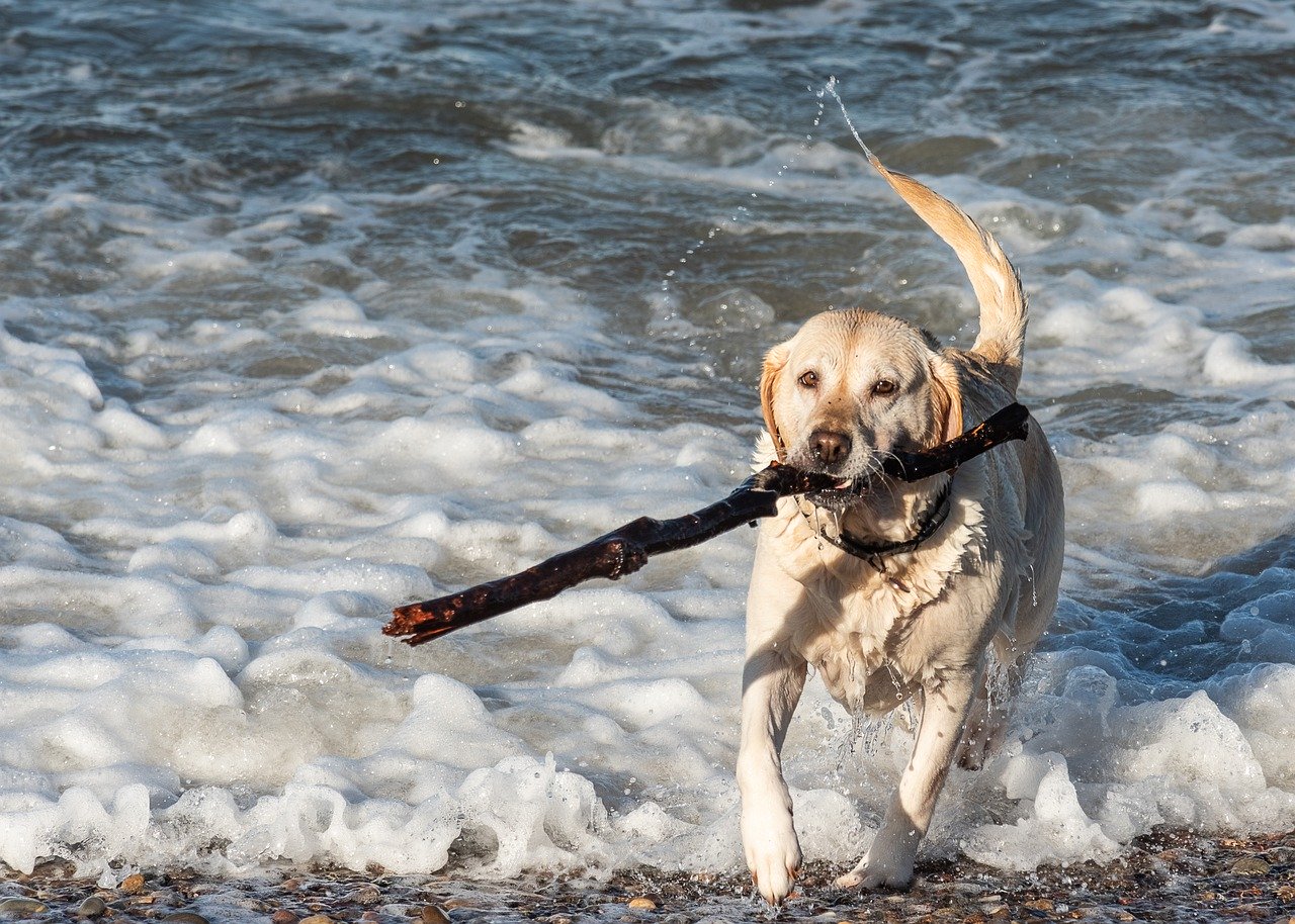
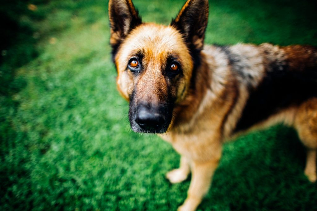
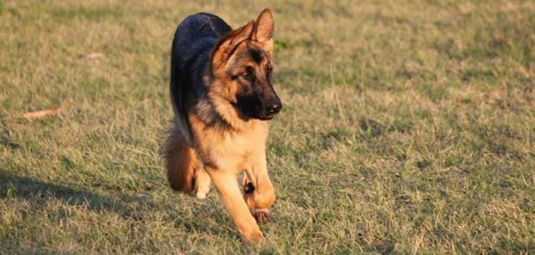
An intact male German shepherd dog (4 y, body wt. 35.5 kg) was referred to the Animal Medical Center, Chonbuk National University with severe lameness, pain and gait abnormality on the right hind limb. Survey radiographs of the pelvis revealed dysplasia of the right coxofemoral joint with subluxation. The dog was surgically treated performing total hip replacement (THR) using Modular Biolox Canine Modular THR System. The dog began to bear weight and slowly returned to a normal exercise pattern 2 months after surgery. THR resulted in satisfactory clinical functions with 6 months follow up and no complications were observed. Canine modular THR could be a successful modality for the management of disabling conditions of the coxofemoral joint. (+info)Canine hip dysplasia (CHD) is a common skeletal disorder in dogs, particularly in large and giant breeds, characterized by the abnormal development and degeneration of the coxofemoral joint - the joint where the head of the femur (thigh bone) meets the acetabulum (hip socket) of the pelvis. This condition is often caused by a combination of genetic and environmental factors that lead to laxity (looseness) of the joint, which can result in osteoarthritis (OA), pain, and decreased mobility over time.
In a healthy hip joint, the femoral head fits snugly into the acetabulum, allowing smooth and stable movement. However, in dogs with CHD, the following abnormalities may occur:
1. Shallow acetabulum: The hip socket may not be deep enough to provide adequate coverage of the femoral head, leading to joint instability.
2. Flared acetabulum: The rim of the acetabulum may become stretched and flared due to excessive forces exerted on it by the lax joint.
3. Misshapen or malformed femoral head: The femoral head may not have a normal round shape, further contributing to joint instability.
4. Laxity of the joint: The ligament that holds the femoral head in place within the acetabulum (ligamentum teres) can become stretched, allowing for excessive movement and abnormal wear of the joint surfaces.
These changes can lead to the development of osteoarthritis, which is characterized by the breakdown and loss of cartilage within the joint, as well as the formation of bone spurs (osteophytes) and thickening of the joint capsule. This results in pain, stiffness, and decreased range of motion, making it difficult for affected dogs to perform everyday activities such as walking, running, or climbing stairs.
Canine hip dysplasia is typically diagnosed through a combination of physical examination, medical history, and imaging techniques such as radiographs (X-rays). Treatment options may include conservative management, such as weight management, exercise modification, joint supplements, and pain medication, or surgical intervention, such as total hip replacement. The choice of treatment depends on the severity of the disease, the age and overall health of the dog, and the owner's financial resources.
Preventing canine hip dysplasia is best achieved through selective breeding practices that aim to eliminate affected animals from breeding populations. Additionally, maintaining a healthy weight, providing appropriate exercise, and ensuring proper nutrition throughout a dog's life can help reduce the risk of developing this debilitating condition.
Congenital hip dislocation, also known as developmental dysplasia of the hip (DDH), is a condition where the hip joint fails to develop normally in utero or during early infancy. In a healthy hip, the head of the femur (thigh bone) fits snugly into the acetabulum (hip socket). However, in congenital hip dislocation, the femoral head is not held firmly in place within the acetabulum due to abnormal development or laxity of the ligaments that support the joint.
There are two types of congenital hip dislocations:
1. Teratologic dislocation: This type is present at birth and occurs due to abnormalities in the development of the hip joint during fetal growth. The femoral head may be completely outside the acetabulum or partially dislocated.
2. Developmental dysplasia: This type develops after birth, often within the first few months of life, as a result of ligamentous laxity and shallow acetabulum. In some cases, it can progress to a complete hip dislocation if left untreated.
Risk factors for congenital hip dislocation include family history, breech presentation during delivery, and female gender. Early diagnosis and treatment are crucial to prevent long-term complications such as pain, limited mobility, and osteoarthritis. Treatment options may include bracing, closed reduction, or surgical intervention, depending on the severity and age of the child at diagnosis.
The hip joint, also known as the coxal joint, is a ball-and-socket type synovial joint that connects the femur (thigh bone) to the pelvis. The "ball" is the head of the femur, while the "socket" is the acetabulum, a concave surface on the pelvic bone.
The hip joint is surrounded by a strong fibrous capsule and is reinforced by several ligaments, including the iliofemoral, ischiofemoral, and pubofemoral ligaments. The joint allows for flexion, extension, abduction, adduction, medial and lateral rotation, and circumduction movements, making it one of the most mobile joints in the body.
The hip joint is also supported by various muscles, including the gluteus maximus, gluteus medius, gluteus minimus, iliopsoas, and other hip flexors and extensors. These muscles provide stability and strength to the joint, allowing for weight-bearing activities such as walking, running, and jumping.
A hip dislocation is a medical emergency that occurs when the head of the femur (thighbone) slips out of its socket in the pelvis. This can happen due to high-energy trauma, such as a car accident or a severe fall. Hip dislocations can also occur in people with certain health conditions that make their hips more prone to displacement, such as developmental dysplasia of the hip.
There are two main types of hip dislocations: posterior and anterior. In a posterior dislocation, the femur head moves out of the back of the socket, which is the most common type. In an anterior dislocation, the femur head moves out of the front of the socket. Both types of hip dislocations can cause severe pain, swelling, and difficulty moving the affected leg.
Immediate medical attention is necessary for a hip dislocation to realign the bones and prevent further damage. Treatment typically involves sedation or anesthesia to relax the muscles around the joint, followed by a closed reduction procedure to gently guide the femur head back into the socket. In some cases, surgery may be required to repair any associated injuries, such as fractures or damaged ligaments. After treatment, physical therapy and rehabilitation are usually necessary to restore strength, mobility, and function to the affected hip joint.
The acetabulum is the cup-shaped cavity in the pelvic bone (specifically, the os coxa) where the head of the femur bone articulates to form the hip joint. It provides a stable and flexible connection between the lower limb and the trunk, allowing for a wide range of movements such as flexion, extension, abduction, adduction, rotation, and circumduction. The acetabulum is lined with articular cartilage, which facilitates smooth and frictionless movement of the hip joint. Its stability is further enhanced by various ligaments, muscles, and the labrum, a fibrocartilaginous rim that deepens the socket and increases its contact area with the femoral head.
Osteoarthritis (OA) of the hip is a degenerative joint disease that affects the articular cartilage and subchondral bone of the hip joint. It is characterized by the progressive loss of cartilage, remodeling of bone, osteophyte formation (bone spurs), cysts, and mild to moderate inflammation. The degenerative process can lead to pain, stiffness, limited range of motion, and crepitus (grating or crackling sound) during movement.
In the hip joint, OA typically affects the femoral head and acetabulum. As the articular cartilage wears away, the underlying bone becomes exposed and can lead to bone-on-bone contact, which is painful. The body responds by attempting to repair the damage through remodeling of the subchondral bone and formation of osteophytes. However, these changes can further limit joint mobility and exacerbate symptoms.
Risk factors for OA of the hip include age, obesity, genetics, previous joint injury or surgery, and repetitive stress on the joint. Treatment options may include pain management (such as NSAIDs, physical therapy, and injections), lifestyle modifications (such as weight loss and exercise), and, in severe cases, surgical intervention (such as hip replacement).
Developmental bone diseases are a group of medical conditions that affect the growth and development of bones. These diseases are present at birth or develop during childhood and adolescence, when bones are growing rapidly. They can result from genetic mutations, hormonal imbalances, or environmental factors such as poor nutrition.
Some examples of developmental bone diseases include:
1. Osteogenesis imperfecta (OI): Also known as brittle bone disease, OI is a genetic disorder that affects the body's production of collagen, a protein necessary for healthy bones. People with OI have fragile bones that break easily and may also experience other symptoms such as blue sclerae (whites of the eyes), hearing loss, and joint laxity.
2. Achondroplasia: This is the most common form of dwarfism, caused by a genetic mutation that affects bone growth. People with achondroplasia have short limbs and a large head relative to their body size.
3. Rickets: A condition caused by vitamin D deficiency or an inability to absorb or use vitamin D properly. This leads to weak, soft bones that can bow or bend easily, particularly in children.
4. Fibrous dysplasia: A rare bone disorder where normal bone is replaced with fibrous tissue, leading to weakened bones and deformities.
5. Scoliosis: An abnormal curvature of the spine that can develop during childhood or adolescence. While not strictly a developmental bone disease, scoliosis can be caused by various underlying conditions such as cerebral palsy, muscular dystrophy, or spina bifida.
Treatment for developmental bone diseases varies depending on the specific condition and its severity. Treatment may include medication, physical therapy, bracing, or surgery to correct deformities and improve function. Regular follow-up with a healthcare provider is essential to monitor growth, manage symptoms, and prevent complications.
Osteotomy is a surgical procedure in which a bone is cut to shorten, lengthen, or change its alignment. It is often performed to correct deformities or to realign bones that have been damaged by trauma or disease. The bone may be cut straight across (transverse osteotomy) or at an angle (oblique osteotomy). After the bone is cut, it can be realigned and held in place with pins, plates, or screws until it heals. This procedure is commonly performed on bones in the leg, such as the femur or tibia, but can also be done on other bones in the body.
In medical terms, the hip is a ball-and-socket joint where the rounded head of the femur (thigh bone) fits into the cup-shaped socket, also known as the acetabulum, of the pelvis. This joint allows for a wide range of movement in the lower extremities and supports the weight of the upper body during activities such as walking, running, and jumping. The hip joint is surrounded by strong ligaments, muscles, and tendons that provide stability and enable proper functioning.
Hip arthroplasty, also known as hip replacement surgery, is a medical procedure where the damaged or diseased joint surfaces of the hip are removed and replaced with artificial components. These components typically include a metal or ceramic ball that replaces the head of the femur (thigh bone), and a polyethylene or ceramic socket that replaces the acetabulum (hip socket) in the pelvis.
The goal of hip arthroplasty is to relieve pain, improve joint mobility, and restore function to the hip joint. This procedure is commonly performed in patients with advanced osteoarthritis, rheumatoid arthritis, hip fractures, or other conditions that cause significant damage to the hip joint.
There are several types of hip replacement surgeries, including traditional total hip arthroplasty, partial (hemi) hip arthroplasty, and resurfacing hip arthroplasty. The choice of procedure depends on various factors, such as the patient's age, activity level, overall health, and the extent of joint damage.
After surgery, patients typically require rehabilitation to regain strength, mobility, and function in the affected hip. With proper care and follow-up, most patients can expect significant pain relief and improved quality of life following hip arthroplasty.
The femoral head is the rounded, ball-like top portion of the femur (thigh bone) that fits into the hip socket (acetabulum) to form the hip joint. It has a smooth, articular cartilage surface that allows for smooth and stable articulation with the pelvis. The femoral head is connected to the femoral neck, which is a narrower section of bone that angles downward and leads into the shaft of the femur. Together, the femoral head and neck provide stability and range of motion to the hip joint.
I believe there might be a misunderstanding in your question. "Dogs" is not a medical term or condition. It is the common name for a domesticated carnivore of the family Canidae, specifically the genus Canis, which includes wolves, foxes, and other extant and extinct species of mammals. Dogs are often kept as pets and companions, and they have been bred in a wide variety of forms and sizes for different purposes, such as hunting, herding, guarding, assisting police and military forces, and providing companionship and emotional support.
If you meant to ask about a specific medical condition or term related to dogs, please provide more context so I can give you an accurate answer.
A hip prosthesis, also known as a total hip replacement, is a surgical implant designed to replace the damaged or diseased components of the human hip joint. The procedure involves replacing the femoral head (the ball at the top of the thigh bone) and the acetabulum (the socket in the pelvis) with artificial parts, typically made from materials such as metal, ceramic, or plastic.
The goal of a hip prosthesis is to relieve pain, improve joint mobility, and restore function, allowing patients to return to their normal activities and enjoy an improved quality of life. The procedure is most commonly performed in individuals with advanced osteoarthritis, rheumatoid arthritis, or other degenerative conditions that have caused significant damage to the hip joint.
There are several different types of hip prostheses available, each with its own unique design and set of benefits and risks. The choice of prosthesis will depend on a variety of factors, including the patient's age, activity level, overall health, and specific medical needs. In general, however, all hip prostheses are designed to provide a durable, long-lasting solution for patients suffering from debilitating joint pain and stiffness.
The pelvic bones, also known as the hip bones, are a set of three irregularly shaped bones that connect to form the pelvic girdle in the lower part of the human body. They play a crucial role in supporting the spine and protecting the abdominal and pelvic organs.
The pelvic bones consist of three bones:
1. The ilium: This is the largest and uppermost bone, forming the majority of the hip bone and the broad, flaring part of the pelvis known as the wing of the ilium or the iliac crest, which can be felt on the side of the body.
2. The ischium: This is the lower and back portion of the pelvic bone that forms part of the sitting surface or the "sit bones."
3. The pubis: This is the front part of the pelvic bone, which connects to the other side at the pubic symphysis in the midline of the body.
The pelvic bones are joined together at the acetabulum, a cup-shaped socket that forms the hip joint and articulates with the head of the femur (thigh bone). The pelvic bones also have several openings for the passage of blood vessels, nerves, and reproductive and excretory organs.
The shape and size of the pelvic bones differ between males and females due to their different roles in childbirth and locomotion. Females typically have a wider and shallower pelvis than males to accommodate childbirth, while males usually have a narrower and deeper pelvis that is better suited for weight-bearing and movement.
Fibrous Dysplasia of Bone is a rare, benign bone disorder that is characterized by the replacement of normal bone tissue with fibrous (scar-like) and immature bone tissue. This results in weakened bones that are prone to fractures, deformities, and pain. The condition can affect any bone in the body but most commonly involves the long bones of the legs, arms, and skull. It can occur as an isolated finding or as part of a genetic disorder called McCune-Albright syndrome. The exact cause of fibrous dysplasia is not fully understood, but it is believed to result from a genetic mutation that occurs during early bone development. There is no cure for fibrous dysplasia, and treatment typically focuses on managing symptoms and preventing complications.
A hip fracture is a medical condition referring to a break in the upper part of the femur (thigh) bone, which forms the hip joint. The majority of hip fractures occur due to falls or direct trauma to the area. They are more common in older adults, particularly those with osteoporosis, a condition that weakens bones and makes them more prone to breaking. Hip fractures can significantly impact mobility and quality of life, often requiring surgical intervention and rehabilitation.
There is no medical definition for "dog diseases" as it is too broad a term. However, dogs can suffer from various health conditions and illnesses that are specific to their species or similar to those found in humans. Some common categories of dog diseases include:
1. Infectious Diseases: These are caused by viruses, bacteria, fungi, or parasites. Examples include distemper, parvovirus, kennel cough, Lyme disease, and heartworms.
2. Hereditary/Genetic Disorders: Some dogs may inherit certain genetic disorders from their parents. Examples include hip dysplasia, elbow dysplasia, progressive retinal atrophy (PRA), and degenerative myelopathy.
3. Age-Related Diseases: As dogs age, they become more susceptible to various health issues. Common age-related diseases in dogs include arthritis, dental disease, cancer, and cognitive dysfunction syndrome (CDS).
4. Nutritional Disorders: Malnutrition or improper feeding can lead to various health problems in dogs. Examples include obesity, malnutrition, and vitamin deficiencies.
5. Environmental Diseases: These are caused by exposure to environmental factors such as toxins, allergens, or extreme temperatures. Examples include heatstroke, frostbite, and toxicities from ingesting harmful substances.
6. Neurological Disorders: Dogs can suffer from various neurological conditions that affect their nervous system. Examples include epilepsy, intervertebral disc disease (IVDD), and vestibular disease.
7. Behavioral Disorders: Some dogs may develop behavioral issues due to various factors such as anxiety, fear, or aggression. Examples include separation anxiety, noise phobias, and resource guarding.
It's important to note that regular veterinary care, proper nutrition, exercise, and preventative measures can help reduce the risk of many dog diseases.
Spina Bifida Occulta is a type of spinal dysraphism, which is a birth defect involving incomplete closure of the spine. In Spina Bifida Occulta, the spinal bones (vertebrae) do not fully form and close around the spinal cord during fetal development, leaving a small gap or split in the lower back region. However, the spinal cord and nerves usually develop normally and are not exposed or damaged, unlike in more severe forms of spina bifida.
In many cases, individuals with Spina Bifida Occulta do not experience any symptoms and may not even know they have the condition unless it is discovered during an imaging test for another reason. In some instances, people with this condition might develop late-onset neurological symptoms or complications such as back pain, muscle weakness, or changes in bladder or bowel function.
It's essential to note that while Spina Bifida Occulta is generally less severe than other forms of spina bifida, it can still pose risks and may require medical evaluation and monitoring to ensure proper development and address any potential issues.
Ectodermal dysplasia (ED) is a group of genetic disorders that affect the development and formation of ectodermal tissues, which include the skin, hair, nails, teeth, and sweat glands. The condition is usually present at birth or appears in early infancy.
The symptoms of ED can vary widely depending on the specific type and severity of the disorder. Common features may include:
* Sparse or absent hair
* Thin, wrinkled, or rough skin
* Abnormal or missing teeth
* Nail abnormalities
* Absent or reduced sweat glands, leading to heat intolerance and problems regulating body temperature
* Ear abnormalities, which can result in hearing loss
* Eye abnormalities
ED is caused by mutations in genes that are involved in the development of ectodermal tissues. Most cases of ED are inherited in an autosomal dominant or autosomal recessive pattern, meaning that a child can inherit the disorder even if only one parent (dominant) or both parents (recessive) carry the mutated gene.
There is no cure for ED, but treatment is focused on managing the symptoms and improving quality of life. This may include measures to maintain body temperature, such as cooling vests or frequent cool baths; dental treatments to replace missing teeth; hearing aids for hearing loss; and skin care regimens to prevent dryness and irritation.
Bronchopulmonary dysplasia (BPD) is a chronic lung disease that primarily affects premature infants. It is defined as the need for supplemental oxygen at 28 days of life or beyond, due to abnormal development and injury to the lungs.
The condition was first described in the 1960s, following the introduction of mechanical ventilation and high concentrations of oxygen therapy for premature infants with respiratory distress syndrome (RDS). These treatments, while lifesaving, can also cause damage to the delicate lung tissue, leading to BPD.
The pathogenesis of BPD is complex and involves an interplay between genetic factors, prenatal exposures, and postnatal injury from mechanical ventilation and oxygen toxicity. Inflammation, oxidative stress, and impaired lung development contribute to the development of BPD.
Infants with BPD typically have abnormalities in their airways, alveoli (air sacs), and blood vessels in the lungs. These changes can lead to symptoms such as difficulty breathing, wheezing, coughing, and poor growth. Treatment may include oxygen therapy, bronchodilators, corticosteroids, diuretics, and other medications to support lung function and minimize complications.
The prognosis for infants with BPD varies depending on the severity of the disease and associated medical conditions. While some infants recover completely, others may have long-term respiratory problems that require ongoing management.
Arthrography is a medical imaging technique used to diagnose problems within joints. It involves the injection of a contrast agent, such as a radiopaque dye or air, into the joint space, followed by the use of fluoroscopy or X-ray imaging to visualize the internal structures of the joint. This can help to identify injuries, tears, or other abnormalities in the cartilage, ligaments, tendons, or bones within the joint.
The procedure is typically performed on an outpatient basis and may be used to diagnose conditions such as shoulder dislocations, rotator cuff tears, meniscal tears in the knee, or hip labral injuries. It is a relatively safe and minimally invasive procedure, although there may be some temporary discomfort or swelling at the injection site. Patients are usually advised to avoid strenuous activity for a day or two following the procedure to allow the contrast agent to fully dissipate from the joint.
Mucopolysaccharidosis I (MPS I) is a rare genetic disorder caused by the deficiency of an enzyme called alpha-L-iduronidase. This enzyme is responsible for breaking down complex sugars called glycosaminoglycans (GAGs), also known as mucopolysaccharides, in the body.
When the enzyme is deficient, GAGs accumulate in various tissues and organs, leading to a range of symptoms that can affect different parts of the body, including the skeletal system, heart, respiratory system, eyes, and central nervous system. There are three subtypes of MPS I: Hurler syndrome (the most severe form), Hurler-Scheie syndrome (an intermediate form), and Scheie syndrome (the least severe form).
The symptoms and severity of MPS I can vary widely depending on the specific subtype, with Hurler syndrome typically causing more significant health problems and a shorter life expectancy than the other two forms. Treatment options for MPS I include enzyme replacement therapy, bone marrow transplantation, and various supportive therapies to manage symptoms and improve quality of life.
The femur is the medical term for the thigh bone, which is the longest and strongest bone in the human body. It connects the hip bone to the knee joint and plays a crucial role in supporting the weight of the body and allowing movement during activities such as walking, running, and jumping. The femur is composed of a rounded head, a long shaft, and two condyles at the lower end that articulate with the tibia and patella to form the knee joint.
Articular Range of Motion (AROM) is a term used in physiotherapy and orthopedics to describe the amount of movement available in a joint, measured in degrees of a circle. It refers to the range through which synovial joints can actively move without causing pain or injury. AROM is assessed by measuring the degree of motion achieved by active muscle contraction, as opposed to passive range of motion (PROM), where the movement is generated by an external force.
Assessment of AROM is important in evaluating a patient's functional ability and progress, planning treatment interventions, and determining return to normal activities or sports participation. It is also used to identify any restrictions in joint mobility that may be due to injury, disease, or surgery, and to monitor the effectiveness of rehabilitation programs.
Hip injuries refer to damages or harm caused to the hip joint or its surrounding structures, including bones, muscles, tendons, ligaments, and cartilage. These injuries can occur due to various reasons such as falls, accidents, sports-related activities, or degenerative conditions. Common hip injuries include fractures, dislocations, strains, sprains, bursitis, and labral tears. Symptoms may include pain, swelling, bruising, stiffness, limited mobility, and inability to bear weight on the affected leg. Proper diagnosis and treatment are crucial to ensure optimal recovery and prevent long-term complications.
Uterine cervical dysplasia is a condition characterized by abnormal cell growth on the lining of the cervix, which is the lower part of the uterus that connects to the vagina. It is also known as cervical intraepithelial neoplasia (CIN).
Cervical dysplasia can be caused by certain strains of human papillomavirus (HPV), a common sexually transmitted infection. The abnormal cells may develop into cancerous cells over time, although not all cases of cervical dysplasia will progress to cancer.
Cervical dysplasia is typically detected through a Pap test or HPV test, which are screening tests used to detect precancerous changes in the cervix. Depending on the severity and extent of the abnormal cells, treatment options may include close monitoring, surgical removal of the affected tissue, or more extensive surgery.
It is important for women to receive regular Pap tests and HPV tests as recommended by their healthcare provider to detect and treat cervical dysplasia early, before it has a chance to progress to cancer.
Fibromuscular dysplasia (FMD) is a rare condition that affects the arterial walls, primarily in the medium and large-sized arteries. According to the American Heart Association, FMD is characterized by uneven growth or damage to the cells in the artery wall, leading to the formation of fibrous tissue and areas with narrowing (stenosis) or ballooning (aneurysm) of the artery.
FMD most commonly affects the renal (kidney) and carotid (neck) arteries but can also occur in other arteries, such as those in the abdomen, arms, and legs. The exact cause of FMD is unknown, but genetic factors and hormonal influences are believed to play a role.
Symptoms of FMD depend on which arteries are affected and may include high blood pressure, headaches, neck pain, dizziness, visual disturbances, or kidney problems. Diagnosis typically involves imaging tests like ultrasound, CT angiography, or magnetic resonance angiography (MRA). Treatment options for FMD include medications to manage symptoms and control high blood pressure, as well as various interventions such as angioplasty or stenting to open narrowed arteries.
In medical terms, "breeding" is not a term that is commonly used. It is more frequently used in the context of animal husbandry to refer to the process of mating animals in order to produce offspring with specific desired traits or characteristics. In human medicine, the term is not typically applied to people and instead, related concepts such as reproduction, conception, or pregnancy are used.
Osteochondrodysplasias are a group of genetic disorders that affect the development of bones and cartilage. These conditions can result in dwarfism or short stature, as well as other skeletal abnormalities. Osteochondrodysplasias can be caused by mutations in genes that regulate bone and cartilage growth, and they are often characterized by abnormalities in the shape, size, and/or structure of the bones and cartilage.
There are many different types of osteochondrodysplasias, each with its own specific symptoms and patterns of inheritance. Some common examples include achondroplasia, thanatophoric dysplasia, and spondyloepiphyseal dysplasia. These conditions can vary in severity, and some may be associated with other health problems, such as respiratory difficulties or neurological issues.
Treatment for osteochondrodysplasias typically focuses on managing the symptoms and addressing any related health concerns. This may involve physical therapy, bracing or surgery to correct skeletal abnormalities, and treatment for any associated medical conditions. In some cases, genetic counseling may also be recommended for individuals with osteochondrodysplasias and their families.
Fibrous dysplasia, monostotic is a benign bone disorder that affects a single bone (monostotic) and is characterized by the replacement of normal bone tissue with fibrous (scar-like) tissue. This results in the formation of abnormal bone that is weakened and more susceptible to fractures. The lesions can cause deformities, pain, and decreased mobility, depending on their size and location. Monostotic fibrous dysplasia is the most common form of fibrous dysplasia, accounting for approximately 70-80% of all cases. It typically manifests during childhood or adolescence and may stabilize or progress slowly over time. In some cases, it can be associated with endocrine disorders such as precocious puberty, hyperthyroidism, or growth hormone excess.
Follow-up studies are a type of longitudinal research that involve repeated observations or measurements of the same variables over a period of time, in order to understand their long-term effects or outcomes. In medical context, follow-up studies are often used to evaluate the safety and efficacy of medical treatments, interventions, or procedures.
In a typical follow-up study, a group of individuals (called a cohort) who have received a particular treatment or intervention are identified and then followed over time through periodic assessments or data collection. The data collected may include information on clinical outcomes, adverse events, changes in symptoms or functional status, and other relevant measures.
The results of follow-up studies can provide important insights into the long-term benefits and risks of medical interventions, as well as help to identify factors that may influence treatment effectiveness or patient outcomes. However, it is important to note that follow-up studies can be subject to various biases and limitations, such as loss to follow-up, recall bias, and changes in clinical practice over time, which must be carefully considered when interpreting the results.
Fibrous Dysplasia, Polyostotic is a rare genetic disorder that affects the bone tissue. It is characterized by the replacement of normal bone tissue with fibrous (scar-like) tissue, leading to weak and fragile bones that are prone to fractures and deformities. The term "polyostotic" refers to the involvement of multiple bones in the body.
In this condition, there is an abnormal development of the bone during fetal growth or early childhood due to a mutation in the GNAS gene. This results in the formation of fibrous tissue instead of normal bone tissue, leading to the characteristic features of Fibrous Dysplasia, Polyostotic.
The symptoms of this condition can vary widely depending on the severity and location of the affected bones. Common symptoms include:
* Bone pain and tenderness
* Bone deformities (such as bowing of the legs)
* Increased risk of fractures
* Skin pigmentation changes (cafe-au-lait spots)
* Hearing loss or other hearing problems (if the skull is affected)
Fibrous Dysplasia, Polyostotic can also be associated with endocrine disorders such as precocious puberty and hyperthyroidism. Treatment typically involves a combination of medications to manage pain and prevent fractures, as well as surgical intervention to correct bone deformities or stabilize fractures.
"Recovery of function" is a term used in medical rehabilitation to describe the process in which an individual regains the ability to perform activities or tasks that were previously difficult or impossible due to injury, illness, or disability. This can involve both physical and cognitive functions. The goal of recovery of function is to help the person return to their prior level of independence and participation in daily activities, work, and social roles as much as possible.
Recovery of function may be achieved through various interventions such as physical therapy, occupational therapy, speech-language therapy, and other rehabilitation strategies. The specific approach used will depend on the individual's needs and the nature of their impairment. Recovery of function can occur spontaneously as the body heals, or it may require targeted interventions to help facilitate the process.
It is important to note that recovery of function does not always mean a full return to pre-injury or pre-illness levels of ability. Instead, it often refers to the person's ability to adapt and compensate for any remaining impairments, allowing them to achieve their maximum level of functional independence and quality of life.
Retrospective studies, also known as retrospective research or looking back studies, are a type of observational study that examines data from the past to draw conclusions about possible causal relationships between risk factors and outcomes. In these studies, researchers analyze existing records, medical charts, or previously collected data to test a hypothesis or answer a specific research question.
Retrospective studies can be useful for generating hypotheses and identifying trends, but they have limitations compared to prospective studies, which follow participants forward in time from exposure to outcome. Retrospective studies are subject to biases such as recall bias, selection bias, and information bias, which can affect the validity of the results. Therefore, retrospective studies should be interpreted with caution and used primarily to generate hypotheses for further testing in prospective studies.
Treatment outcome is a term used to describe the result or effect of medical treatment on a patient's health status. It can be measured in various ways, such as through symptoms improvement, disease remission, reduced disability, improved quality of life, or survival rates. The treatment outcome helps healthcare providers evaluate the effectiveness of a particular treatment plan and make informed decisions about future care. It is also used in clinical research to compare the efficacy of different treatments and improve patient care.
Osteoarthritis (OA) is a type of joint disease that is characterized by the breakdown and eventual loss of cartilage - the tissue that cushions the ends of bones where they meet in the joints. This breakdown can cause the bones to rub against each other, causing pain, stiffness, and loss of mobility. OA can occur in any joint, but it most commonly affects the hands, knees, hips, and spine. It is often associated with aging and can be caused or worsened by obesity, injury, or overuse.
The medical definition of osteoarthritis is: "a degenerative, non-inflammatory joint disease characterized by the loss of articular cartilage, bone remodeling, and the formation of osteophytes (bone spurs). It is often associated with pain, stiffness, and decreased range of motion in the affected joint."
Cleidocranial dysplasia is a genetic skeletal disorder that affects the development of bones and teeth. The condition is characterized by the underdevelopment or absence of the collarbones (clavicles), which can result in shoulder joints that are abnormally close together. This may allow the person to bring their shoulders around to touch or even overlap in front of their body.
People with cleidocranial dysplasia also often have a delayed closure of the fontanels (soft spots) on the skull, as well as an abnormal shape and size of the head. The facial bones may be underdeveloped, leading to a sunken appearance in the middle of the face and a prominent forehead. Dental abnormalities are also common, such as missing or delayed eruption of teeth, extra teeth, and misaligned teeth.
Cleidocranial dysplasia is caused by mutations in the CBFA1/RUNX2 gene and is inherited in an autosomal dominant manner, meaning that a child has a 50% chance of inheriting the condition if one of their parents is affected. However, many cases result from new mutations in the gene and occur in people with no family history of the disorder. Treatment typically involves surgical procedures to correct skeletal abnormalities and dental issues, as well as orthodontic treatment to align teeth.
Retinal dysplasia is a developmental abnormality of the retina, which is the light-sensitive tissue located at the back of the eye. This condition is characterized by the presence of folds or rosettes (round clusters) in the retinal structure, resulting from improper or disorganized growth of the retinal cells during fetal development.
Retinal dysplasia can be classified into two types:
1. Focal or localized retinal dysplasia: This type is limited to a small area of the retina and usually does not significantly affect vision. It may present as mild folds or rosettes in the retinal structure.
2. Generalized or severe retinal dysplasia: This type involves widespread disorganization of the retinal layers, leading to more significant visual impairment. In extreme cases, it can result in complete detachment of the retina from the underlying tissue, causing blindness.
Retinal dysplasia can be an isolated finding or associated with various genetic disorders, infections, or environmental factors during pregnancy. Depending on the severity and underlying cause, management may include monitoring for visual development, corrective lenses, or treatment of associated conditions.
I must clarify that the term "pedigree" is not typically used in medical definitions. Instead, it is often employed in genetics and breeding, where it refers to the recorded ancestry of an individual or a family, tracing the inheritance of specific traits or diseases. In human genetics, a pedigree can help illustrate the pattern of genetic inheritance in families over multiple generations. However, it is not a medical term with a specific clinical definition.
Articular cartilage is the smooth, white tissue that covers the ends of bones where they come together to form joints. It provides a cushion between bones and allows for smooth movement by reducing friction. Articular cartilage also absorbs shock and distributes loads evenly across the joint, protecting the bones from damage. It is avascular, meaning it does not have its own blood supply, and relies on the surrounding synovial fluid for nutrients. Over time, articular cartilage can wear down or become damaged due to injury or disease, leading to conditions such as osteoarthritis.
Thnanatophoric Dysplasia is a severe skeletal disorder characterized by extreme short limbs, a narrow chest, and large head. It is one of the most common types of short-limbed dwarfism. The name "thanatophoric" comes from the Greek word thanatos, meaning death, as this condition is often lethal in the newborn period or shortly thereafter due to respiratory distress.
The disorder is caused by mutations in the FGFR3 gene, which provides instructions for making a protein that is part of a group of proteins called fibroblast growth factor receptors. These receptors play critical roles in many important processes during embryonic development, such as controlling bone growth.
There are two major types of thanatophoric dysplasia: type I and type II. Type I is characterized by curved thigh bones (femurs) and a clover-leaf shaped skull. Type II is characterized by straight femurs and an unossified (not fully developed) vertebral column.
The diagnosis of thanatophoric dysplasia can be made prenatally through ultrasound examination or postnatally through physical examination, X-rays, and genetic testing. Unfortunately, due to the severity of the condition, there is no cure for thanatophoric dysplasia and management is supportive in nature, focusing on providing comfort and addressing any complications that may arise.
In the field of medicine, "time factors" refer to the duration of symptoms or time elapsed since the onset of a medical condition, which can have significant implications for diagnosis and treatment. Understanding time factors is crucial in determining the progression of a disease, evaluating the effectiveness of treatments, and making critical decisions regarding patient care.
For example, in stroke management, "time is brain," meaning that rapid intervention within a specific time frame (usually within 4.5 hours) is essential to administering tissue plasminogen activator (tPA), a clot-busting drug that can minimize brain damage and improve patient outcomes. Similarly, in trauma care, the "golden hour" concept emphasizes the importance of providing definitive care within the first 60 minutes after injury to increase survival rates and reduce morbidity.
Time factors also play a role in monitoring the progression of chronic conditions like diabetes or heart disease, where regular follow-ups and assessments help determine appropriate treatment adjustments and prevent complications. In infectious diseases, time factors are crucial for initiating antibiotic therapy and identifying potential outbreaks to control their spread.
Overall, "time factors" encompass the significance of recognizing and acting promptly in various medical scenarios to optimize patient outcomes and provide effective care.
A newborn infant is a baby who is within the first 28 days of life. This period is also referred to as the neonatal period. Newborns require specialized care and attention due to their immature bodily systems and increased vulnerability to various health issues. They are closely monitored for signs of well-being, growth, and development during this critical time.
Canine distemper virus (CDV) is a single-stranded RNA virus that belongs to the family Paramyxoviridae and causes a contagious and serious disease in dogs and other animals. The virus primarily affects the respiratory, gastrointestinal, and central nervous systems of infected animals.
The symptoms of canine distemper can vary widely depending on the age and immune status of the animal, as well as the strain of the virus. Initial signs may include fever, lethargy, loss of appetite, and discharge from the eyes and nose. As the disease progresses, affected animals may develop vomiting, diarrhea, pneumonia, and neurological symptoms such as seizures, muscle twitching, and paralysis.
Canine distemper is highly contagious and can be spread through direct contact with infected animals or their respiratory secretions. The virus can also be transmitted through contaminated objects such as food bowls, water dishes, and bedding.
Prevention of canine distemper is achieved through vaccination, which is recommended for all dogs as a core vaccine. It is important to keep dogs up-to-date on their vaccinations and to avoid contact with unfamiliar or unvaccinated animals. There is no specific treatment for canine distemper, and therapy is generally supportive, focusing on managing symptoms and preventing complications.
 Hip dysplasia (canine)
Chow Chow
Shetland Sheepdog
Samoyed dog
Dalmatian (dog)
English Cocker Spaniel
Dog breeding
PennHIP
Canine physical therapy
Akita (dog)
Australian Shepherd
Guiding Eyes for the Blind
Pumi dog
Dog health
Femoral head ostectomy
Treatment of equine lameness
Orthopedic Foundation for Animals
Welsh Springer Spaniel
German Shepherd
Persian cat
Tibetan Terrier
Robert L. Rooks
Fred Lanting
Tornjak
Dislocation of hip in animals
Organ replacement in animals
Buckles (comics)
Greater Swiss Mountain Dog
St. Bernard (dog)
Labradoodle
Hip dysplasia (canine) - Wikipedia
 Hip Dysplasia In Dogs: Causes, Symptoms, and Treatment
 Best CBD Oils For Dogs With Hip Dysplasia | mindbodygreen
 Canine Hip Dysplasia Treatment | Management of Hip Dysplasia in Dogs
 Dog Hip Dysplasia
 supplements for hip dysplasia in dogs
 PennHip|sup|TM|/sup|: A Collaborative Effort to Reduce the Incidence of Canine Hip Dysplasia
 Hip Dysplasia in Dogs: Causes, Symptoms, & Treatment
 Maremma Sheepdog - breeding decisions with canine hip dysplasia
 CLINICAL AND RADIOGRAPHIC EVALUATION OF INTERTROCHANTERIC OSTEOTOMY FIXATION WITH KÜNTSCHER NAIL FOR THE TREATMENT OF CANINE...
 Be on the lookout for Canine Hip Dysplasia - The Dogington Post
 Mapping of Genetic Risk Factors for Canine Hip Dysplasia - PWD Foundation, Inc.
 Your Guide to Hip Dysplasia in Dogs | ElleVet Sciences
 Hip Dysplasia in Dogs - Musculoskeletal System - MSD Veterinary Manual
Hip Dysplasia, Canine | Colorado PROFILES
 Unexplained Canine Hip Dysplasia Occurrences
 Hip Dysplasia Dog Tag
 dogs and hip dysplasia
 Hip dysplasia in dogs - VetDojo
 Glucosamine - Hip Dysplasia in Dogs
 Hip Dysplasia | The Balanced Dog
 Hip Dysplasia in Dogs - Alpha Feeds
 Hip Dysplasia In Dogs
- Vital Vet
Hip Dysplasia In Dogs
- Vital Vet
 Hip Dysplasia and Dog Agility - AgilityFusion.com
 Heritability of hip dysplasia - The Institute of Canine Biology
 Dog Hip Dysplasia Surgery | Bonita Springs Vets
Dogs And Puppies : Hip and Elbow Dysplasia
Exercise for dogs with hip dysplasia - Barkercise
Best Dog Bed For Hip Dysplasia - My Wordpress
 Hip Dysplasia & Dog Beds: Big Barker Case Study
Risk of hip dysplasia3
- What else can I do to reduce the risk of hip dysplasia? (sevenoakspet.com)
- It also makes it easier for them to gain excess fat around their hindquarters if they're not active enough, which increases the risk of hip dysplasia even further! (hempforhounds.org)
- Maintaining a healthy weight: Being overweight puts extra strain on your dog's joints, increasing the risk of hip dysplasia. (myhipdysplasiaindogs.com)
Develop hip dysplasia6
- Neutering a dog, especially before the dog has reached an age of full developmental maturity, has been shown to almost double the chance he or she will develop hip dysplasia versus intact dogs or dogs that were neutered after reaching adulthood. (wikipedia.org)
- What Breeds Are Most Likely To Develop Hip Dysplasia? (ellevetsciences.com)
- Smaller dogs can still develop hip dysplasia, with basset hounds, french bulldogs, and pugs being among the most likely small breeds. (ellevetsciences.com)
- While breeds like the Siberian husky are far less likely than other breeds to develop hip dysplasia, they're far more likely to develop epilepsy. (ellevetsciences.com)
- Also, if an animal lives in a culture, too harsh conditions or too much physical activity at a young age could make it more likely to develop hip dysplasia. (woofysh.com)
- instituteofcaninebiology.org states, ' Puppies are born with perfect hips, and if the hips do not develop laxity the dog does not develop hip dysplasia (Riser 1985). (pethangout.com)
Arthritis29
- In dogs, hip dysplasia is an abnormal formation of the hip socket that, in its more severe form, can eventually cause lameness and arthritis of the joints. (wikipedia.org)
- It is common in many dog breeds, particularly the larger breeds, and is the most common single cause of arthritis of the hips. (wikipedia.org)
- Normal hip function can be affected by congenital conditions such as dysplasia, trauma, and by acquired diseases such as osteoarthritis and rheumatoid arthritis. (wikipedia.org)
- In mild to moderate dysplasia it is often the secondary effects of abnormal wear and tear or arthritis, rather than dysplasia itself, which is the direct causes of visible problems. (wikipedia.org)
- If your veterinarian diagnoses your dog with arthritis, glucosamine will likely be part of a comprehensive treatment plan. (akc.org)
- You can also purchase supplements with these ingredients for dogs that might be prone to developing arthritis and hip dysplasia down the line. (akc.org)
- Exercise and mental stimulation are an important part of development of puppies but over-exercising at a young age can prevent the proper growth of bones, muscles and joints, leading to joint problems like as hip dysplasia and arthritis. (dogshealth.com)
- If your dog already suffers from hip dysplasia or arthritis, a safe and proven supplement for treating your pet is Winston's Joint System , a combination of three, totally-natural whole food supplements developed by a Naturopathic Doctor for his own dog. (dogshealth.com)
- Our staff specializes in hip dysplasia, arthritis and all joint, pain and mobility issues. (dogshealth.com)
- ArthroSoothe for Pets - ArthroSoothe is the most comprehensive arthritis, hip and joint supplement available for dogs. (askariel.com)
- Some dog parents confuse arthritis with hip dysplasia but, unlike arthritis, hip dysplasia in dogs develops when a dog is young. (hillspet.co.za)
- A dog can develop muscular health issues or arthritis due to hip dysplasia. (ellevetsciences.com)
- Surgical treatments include pectineal myotenectomy to reduce pain, triple pelvic osteotomy to prevent subluxation, pubic fusion to prevent subluxation, joint capsule denervation to reduce pain, dorsal acetabulum reinforcement to reduce subluxation, femoral head and neck resection to reduce arthritis, and total hip replacement for optimal restoration of joint and limb functions. (msdvetmanual.com)
- Later on, dogs with hip dysplasia develop arthritis which is characterised radiographically. (vetdojo.com)
- Canine Arthritis: Is Glucosamine or Chondroitin Safe? (myhipdysplasiaindogs.com)
- Hip dysplasia causes pain, swelling, stiffness and eventually arthritis so it is essential that you recognise the symptoms early, before your dog experiences the pain and discomfort that unfortunately accompanies the condition. (alphafeeds.com)
- Dogs with mild hip dysplasia on x-ray may develop minimal arthritis without clinical signs until they are older. (sevenoakspet.com)
- This disease should not be confused with hip arthritis. (durationcares.com)
- Rather, it is the most common cause of arthritis in the hips. (durationcares.com)
- 1.hip-extended ventrodorsal view x-ray - It provides a frontal view of the pelvis and hip-joints and best assesses the degree of severity of arthritis present. (durationcares.com)
- Prophylactic surgery is done to prevent the progression af arthritis while therapeutic surgery aims to treat already arthritic hips. (durationcares.com)
- This procedure is not recommended for mild cases of arthritis and is generally effective only on smaller, well-muscled dogs. (durationcares.com)
- Consider physical therapy options to build up the supportive muscles of the hip and rear legs, and learn about the various methods to help ease the symptoms associated with arthritis as it develops. (hhvh.net)
- It also increases the likelihood that your dog will develop arthritis later in life. (hempforhounds.org)
- The vet can also observe the presence of any arthritis in the hip socket as well as signs that suggest degenerative joint disease. (hempforhounds.org)
- It is caused by the abnormal development of the hip joint, which can lead to joint instability and arthritis. (pawsometips.com)
- Hip dysplasia is a common orthopedic condition that affects many dogs, leading to pain, lameness, and arthritis. (woofysh.com)
- The exam will check for signs of bone disease and arthritis, which can be caused by dysplasia. (dogcontrol.ca)
- In addition, radiographs (x-rays) may or may not be taken that can show the need for Osteoarthritis and Rheumatoid Arthritis medication.In short, if untreated dysplastic hips lead to osteoarthritis and rheumatoid arthritis, then how much worse could dysplastic hip be? (dogcontrol.ca)
Joints39
- Obesity puts a lot of stress on your dog's joints, which can exacerbate a pre-existing condition such as hip dysplasia or even cause hip dysplasia. (akc.org)
- The common condition occurs when hip joints don't properly develop, which causes more wear-and-tear on the joints. (mindbodygreen.com)
- The condition occurs when a dog's hip joints don't develop right, causing the hips to partially dislocated and leads to early development of degenerative joint disease. (pethealthnetwork.com)
- It is caused by a partial dislocation of bones in the dog's hip joints, leaving them misaligned but still in contact with each other, which causes erosion of the tissues that keep the joint moving smoothly. (dogshealth.com)
- If your dog is overweight , you'll want to make a weight-loss plan right away, as reducing the weight-bearing load and friction upon the joints will likely improve your dog's mobility and comfort. (hillspet.co.za)
- To determine whether intertrochanteric osteotomy (ITO) can prevent the progression of degenerative joint disease in dysplastic hip joints. (vin.com)
- Canine hip dysplasia is a condition that affects a dog's hip joints while they're growing. (ellevetsciences.com)
- A hereditary disease of the hip joints in dogs. (ucdenver.edu)
- If you have a breed that is considered "at risk" and you have done the research to find a reputable breeder and have done all the preventative measures to keep your dogs joints healthy, you are not guaranteed that your dog will not develop this condition classified as a disease. (agilityfusion.com)
- When we got him to the vet they took x-rays and found he had horrid joints and his hip had dislocated (It popped back when they did the x-ray). (agilityfusion.com)
- Put simply, the hip joints are designed to fit together perfectly to enable easy movement. (alphafeeds.com)
- When these joints don't fit together as they should, the hips become unstable and hip dysplasia becomes apparent. (alphafeeds.com)
- If you notice the symptoms of hip dysplasia in your dog, you should seek advice from your vet, who is likely to suggest some daily management measures such as weight management (to reduce strain on joints), Anti - inflammatory medication, rest and controlled exercise, meaning their life as a working dog may be questionable. (alphafeeds.com)
- Since excess weight puts undue stress on the hip joints, weight loss is strongly recommended in overweight dogs. (sevenoakspet.com)
- This body shape puts extra strain on all of the weight-bearing joints so that even mild hip dysplasia can lead to severe osteoarthritis and difficulty with normal daily activities. (petietec.com)
- Brachycephalic, or short-faced, dogs (like English Bulldogs and French Bulldogs ) often have poorly fitting joints including the elbow, knee, and hip. (petietec.com)
- Giant dogs have special needs during their growth and development stages in order to have healthy, strong bones and joints. (petietec.com)
- While you can't prevent your dog from developing hip dysplasia, you can take steps to protect their joints and relieve their pain. (petietec.com)
- A healthy weight protects your dog's hips and joints and reduces the excess stress the body puts on the joints. (petietec.com)
- Hip dysplasia is a debilitating condition that affects the hip joints of dogs. (healthextension.com)
- During regular checkups, your vet will examine your dog and sometimes find this condition just based on the range of motion, looseness of joints and/or any grinding of the joints that may be present. (healthextension.com)
- Enriched with Glucosamine and Chondroitin, the key ingredients for healthy hips and joints. (healthextension.com)
- Obesity puts an abnormal amount of stress on your pup's joints and may aggravate pre-existing hip dysplasia or even cause the condition. (swfvs.com)
- It is important that a dog with hip dysplasia maintain a good weight as being overweight will put more stress on the sore joints. (barkercise.com)
- Low impact exercise lessens the weight that is applied to the joints making it more comfortable for a dog with joint pain. (barkercise.com)
- This not only helps to keep the joints mobile but provides an opportunity for mental stimulation for your dog by enjoying the sights, sounds, and smells. (barkercise.com)
- Healthy hips joints are lubricated, and movement is not painful. (petinsuranceu.com)
- After a thorough physical exam, which was normal and didn't detect any evidence of pain in the hip joints, Dakota was sedated and positioned for her radiographs. (hhvh.net)
- The images revealed significant laxity in both hip joints which is consistent with a diagnosis of hip dysplasia. (hhvh.net)
- A healthy body helps to keep joints strong, flexible, and free of disease-all good things when it comes to preventing hip dysplasia. (hempforhounds.org)
- Your dog's hip joints work like a ball and socket. (carrieranimalhospital.com)
- If your dog has trouble working out because of age or pain, swimming is a great way for them to stay in shape and get a good workout without putting too much stress on their joints. (woofysh.com)
- Your dog's hip joints work similarly to a ball and socket. (ranchovillagevet.com)
- It is essential to regularly inspect and monitor your dog's hips and joints, as hip dysplasia can often go undetected. (monkeskateclothing.com)
- Hip Dysplasia is a malformation of the hip where the ball and socket joints do not meet properly. (pethangout.com)
- This will show any issues with how the hip and ball joints are developing and how they fit together. (pethangout.com)
- Older dogs may be able to pace more comfortably in a crate or on a bed, which may help prevent injuries to the joints. (dogcontrol.ca)
- METHODS: Radiographic and ultrasonographic evaluations were performed on 108 hip joints of 54 dogs. (bvsalud.org)
- Additionally, changes in joint capsule thickness can be identified in B-mode in young and adult dogs with dysplastic joints, which contributes to the diagnosis of hip dysplasia. (bvsalud.org)
Development of hip dysplasia4
- Several factors lead to the development of hip dysplasia in dogs, beginning with genetics. (akc.org)
- Advances in nutritional research have shown that diet plays an important role in the development of hip dysplasia. (sevenoakspet.com)
- Genes, nutrition, and the environment all play a role in the development of hip dysplasia. (woofysh.com)
- Joint laxity is the primary factor that predisposes a dog to the development of hip dysplasia. (pethangout.com)
Genetics10
- The American College of Veterinary Surgeons explains that there are two primary causes of hip dysplasia in dogs: genetics and nutrition. (hillspet.co.za)
- While genetics determine whether a dog develops hip dysplasia, nutritional research shows that consuming food that's too high in calcium or calories also plays a role in the condition's development. (hillspet.co.za)
- If you haven't read the section on genetics of dysplasia, read that section now to assist you in working out why breeders need to make informed breeding choices. (maremmano.com)
- Breakthroughs in other breeds can be utilized to better understand the genetics of this condition in Portuguese Water dogs. (pwdfoundation.org)
- There are two primary causes of hip dysplasia, genetics, and diet . (sevenoakspet.com)
- In dogs, hip dysplasia is predominantly a hereditary condition, with genetics being the leading contributor to the development of the condition. (swfvs.com)
- Generally, hip dysplasia is a hereditary condition, with genetics being the most common cause of this condition in dogs. (carrieranimalhospital.com)
- One of the main causes of canine hip dysplasia is genetics. (pawsometips.com)
- One of the most common reasons behind hip dysplasia is genetics. (ranchovillagevet.com)
- Although biomechanical and environmental factors associated with certain body conformation and size are considered the main cause of canine hip dysplasia, genetics also play a role. (yourvetonline.com)
Veterinarian17
- This list isn't comprehensive - there are a variety of other procedures and recommendations, and your veterinarian will be the best resource in determining what is right for a dog diagnosed with hip dysplasia. (pethealthnetwork.com)
- When your veterinarian examines your dog's hips, they'll watch for indicators of pain or resistance to pressure. (hillspet.co.za)
- Be sure to ask your veterinarian about dog foods that are specially formulated for mobility health that contain many of the same joint health nutrients that these supplements contain. (hillspet.co.za)
- Your veterinarian can help determine the best method, including a proper nutrition plan (including potentially switching to a dog food formulated for getting your dog to the proper weight) and exercise as long as your dog can tolerate it with the stress on their hips. (hillspet.co.za)
- Dog-specific anti-inflammatory medications should be purchased from a licensed veterinarian. (vetdojo.com)
- Sometimes this exam is enough for your veterinarian to suspect hip dysplasia. (vitalvet.org)
- Your veterinarian will take radiographs of your dog's hips and pelvis to look for signs of hip dysplasia. (healthextension.com)
- When choosing pet food for your dog, it is best to check with your veterinarian to make sure you feed a product that will support your specific pup's needs. (healthextension.com)
- A dysplastic dog can be diagnosed at a very young age by a veterinarian experienced in canine orthopedics. (vetetnous.com)
- In 1983 Dr. Gail Smith, a veterinarian at the University of Pennsylvania Veterinary School began researching better methods to determine which dogs were most susceptible, and in 1993 he established PennHIP. (hhvh.net)
- Your veterinarian will examine your dog and do a thorough physical exam. (hempforhounds.org)
- Your veterinarian may recommend X-rays to get a better understanding of the shape and position of your dog's hip socket. (hempforhounds.org)
- Remember that every dog is different and what works for one may not work for another, so please consult with your veterinarian before starting any treatment regimen. (pawsometips.com)
- The first step is to talk to your veterinarian about the best course of action for your dog. (pawsometips.com)
- No matter what route you choose, working with your veterinarian is the best way to ensure that your dog gets the care they need to live a happy and healthy life. (pawsometips.com)
- If your dog is showing any signs of difficulty or hesitancy when climbing stairs or demonstrates any form of pain or distress during physical activity, it is important to have your pet evaluated by a veterinarian for the potential of hip dysplasia. (monkeskateclothing.com)
- If you have left untreated dysplastic hips, the dogs pain and mobility will worsen over time, eventually leading to lameness and a shortened life.The best way to ensure a dogs health is to have his hips examined by a veterinarian. (dogcontrol.ca)
Treat hip dysplasia4
- There are several surgical procedures available to treat hip dysplasia. (sevenoakspet.com)
- Numerous treatments are possible to surgically treat hip dysplasia. (vetetnous.com)
- The best way not to have to treat hip dysplasia is to select dogs that do not have it. (vetetnous.com)
- It's possible to treat hip dysplasia early and possibly even help prevent it. (petinsuranceu.com)
Joint laxity7
- Hip dysplasia is a multifactorial abnormal development of the coxofemoral joint in dogs that is characterized by joint laxity and subsequent degenerative joint disease. (msdvetmanual.com)
- Standard ventrodorsal views of sedated or anesthetized animals can be graded by the Orthopedic Foundation for Animals , or stress radiographs performed and joint laxity measured (Penn Hip). (msdvetmanual.com)
- Early signs of hip dysplasia, such as joint laxity (or looseness of the hips) are visible in patients as young as 4 months old thanks to tests such as Barden's sign and the Ortolani test. (vetdojo.com)
- A key sign is hip joint laxity, or a loose fit of the head of the femur in the hip socket. (the-balanced-dog.com)
- Clinical signs and palpable joint laxity may also indicate hip dysplasia. (sevenoakspet.com)
- Dogs with genetically determined joint laxity are at increased risk for developing osteoarthritis, especially if they also have excessive weight gain and/or do not exercise adequately. (hempforhounds.org)
- Some of the most important environmental factors include excessive weight gain during puppyhood, inadequate nutrition during growth periods (when bones are growing), injury to the hips from jumping off tables or other high places (particularly when combined with heavy body weight), inadequate exercise leading to overweight status in adulthood, and poorly constructed hips inherited from parents with hip dysplasia due to congenital joint laxity (inherited joint laxity). (hempforhounds.org)
Cause of canine hip dyspl1
- Another cause of canine hip dysplasia is obesity. (pawsometips.com)
Lameness9
- Lameness resulting from hip dysplasia in dogs is usually chronic and progresses slowly. (hillspet.co.za)
- Your dog likely doesn't have hip dysplasia if their lameness begins suddenly or progresses rapidly, the lameness is severe, they can't move well on their rear legs or if they have an uncoordinated gait. (hillspet.co.za)
- The results of the physical examination and radiographic evaluation at the preoperative examination and at the two follow-ups differ significantly for pain, lameness and radiographic evidence of OA, where pain and lameness were improved after surgery and OA had progressed by more than one degree in all dogs. (vin.com)
- It leads to debilitating hip osteoarthritis, lameness and pain. (hound101.com)
- The degree of lameness that occurs is dependent on the extent of these arthritic changes and may not be correlated with the appearance of the hip joint on x-rays. (sevenoakspet.com)
- Some pets with significant signs of hip dysplasia or osteoarthritis on x-rays may not exhibit any clinical signs while others with minimal changes may experience severe pain and lameness. (sevenoakspet.com)
- It results from abnormal development of the hip anatomy, resulting in pain and lameness. (woofysh.com)
- If you notice any signs of lameness, limping, or an unwillingness to exercise, that may be a sign of dog hip dysplasia. (monkeskateclothing.com)
- The severity of pain, lameness progression, and the need for intermittent pain medication will vary between individual dogs.The best way to live with a dog with hip dysplasia is to provide optimal pain management. (dogcontrol.ca)
Prevent Hip Dysplasia in Dogs3
- What Can You Do to Prevent Hip Dysplasia in Dogs? (hempforhounds.org)
- Prevent Hip Dysplasia in Dogs with Proper Harness - Shop Now! (myhipdysplasiaindogs.com)
- How Can You Prevent Hip Dysplasia in Dogs? (myhipdysplasiaindogs.com)
Diagnosing Hip Dysplasia in Dogs1
- Diagnosing hip dysplasia in dogs can often be a difficult process. (woofysh.com)
Susceptible9
- Large and giant breeds are most susceptible to hip dysplasia (possibly due to the body mass index (BMI) of the individual animal), though many other breeds can suffer from it. (wikipedia.org)
- Some breeders are irresponsible and will breed a dog knowing it is susceptible to hip dysplasia, or will breed a dog too young to be properly tested, and the result is an increased occurrence of the disease. (dogshealth.com)
- It's also worth noting that every dog breed will be susceptible to certain conditions. (ellevetsciences.com)
- Some breeds of dogs are more susceptible to developing hip dysplasia than others. (healthextension.com)
- Breeds that commonly suffer from hip dysplasia include large and giant dogs such as mastiffs, St. Bernards, Rottweilers, retrievers, and bulldogs, but several smaller breeds such as French bulldogs and pugs may also be susceptible. (swfvs.com)
- Hip dysplasia is seen more frequently in giant breeds and large dogs such as Rottweilers, Mastiffs, St. Bernards, retrievers, and bulldogs, although a handful of smaller breeds such as French bulldogs and pugs are also susceptible. (carrieranimalhospital.com)
- Dogs are susceptible to hip dysplasia, just as humans are. (pawsometips.com)
- Dogs that are not getting the proper nutrients can be more susceptible to the condition. (pawsometips.com)
- Some breeds are more susceptible to certain dog breed related health conditions , so it's important to be well-informed before making a decision. (monkeskateclothing.com)
Show signs of hip dysplasia4
- Some dogs begin to show signs of hip dysplasia when they are as young as four months of age. (akc.org)
- Although it's most common in mature dogs and larger dogs, some will begin to show signs of hip dysplasia while they're still puppies. (ellevetsciences.com)
- This is because some dogs, with apparently normal hips at the age of 6 or 12 months, show signs of hip dysplasia later in life. (vetetnous.com)
- Some pets start to show signs of hip dysplasia at the beginning of the early stage, and others can show it in conjunction with osteoarthritis as the dog age. (thevetexpert.com)
Osteoarthritis6
- The result is laxity (looseness) of the joint, followed by degenerative joint disease (DJD) or osteoarthritis (OA), which is the body's attempt to stabilize the loose hip joint. (wikipedia.org)
- What develops over time is pain from the osteoarthritis caused by a poorly fitted hip joint. (petietec.com)
- Hip dysplasia may also be compounded by other painful conditions such as osteoarthritis in senior dogs. (swfvs.com)
- Hip dysplasia is a polygenic hereditary disease (transmitted by many genes) characterized by instability of the coxofemoral articulation (hip joint) that can lead to a hip subluxation (partial separation of the joint), and lead later to osteoarthritis (articular structure degeneration). (vetetnous.com)
- For the adult, arthroplasty or excision of the femoral head and neck allows control of the pain related to osteoarthritis, but it modifies the mechanical function of the hip and generally, the dog becomes tired faster. (vetetnous.com)
- Hip dysplasia could be compounded by other painful conditions such as osteoarthritis in senior dogs. (carrieranimalhospital.com)
Diagnosis8
- While an x-ray test is the only accepted method of accurate diagnosis of the mild case of canine hip dysplasia, the breeder should decide whether the radiographic deviation from the perfection of the hip joint is a characteristic so undesirable to the breed involved that, it should not be bred, regardless of the other desirable traits of the dog. (trysincere.com)
- The first step to treating your dog's hip dysplasia at home is to confirm the diagnosis of hip dysplasia because clinical signs can be similar to other knee problems, such as cruciate ligament ruptures, and spinal diseases. (vetdojo.com)
- As such, the veterinary focus has been directed towards early diagnosis, intervention and treatment of affected dogs, and encouraging selective breeding to decrease the risk of passing the genes along to offspring. (hhvh.net)
- Depending on the severity of the issue, a dog with hip dysplasia can have a good quality of life for years with proper diagnosis and treatment. (woofysh.com)
- Missing an early diagnosis of hip dysplasia - Note: it can be detected as early as 4 - 6 months of age. (pethangout.com)
- The diagnosis is made by radiographic examinations that evaluate bone alterations associated with hip dysplasia. (bvsalud.org)
- This study aimed to evaluate the accuracy of B-mode ultrasonography and acoustic radiation force impulse (ARFI) elastography in assessing the joint capsule and periarticular muscles of dogs using the Orthopedic Foundation of Animals (OFA) classification and the distraction index (DI) in the early and late diagnosis of hip dysplasia. (bvsalud.org)
- OBJECTIVES: The paper gives an overview of current radiological possibilities of diagnosis with focus on congenital hip dysplasia supplemented by insights into therapeutic options. (bvsalud.org)
Breeds of Dogs5
- And while it tends to affect large breed dogs more frequently, all breeds of dogs are at risk. (pethealthnetwork.com)
- Hip dysplasia appears to be hereditary and is more common in larger breeds of dogs. (ellevetsciences.com)
- It is believed that canine hip dysplasia has increased since large breeds of dogs evolved into companion dogs from the hunting or guard functions for which they were bred and kept in the past. (trysincere.com)
- Hip dysplasia is commonly found to be a hereditary condition and mostly found in large and giant breeds of dogs like mastiffs, St. Bernards, retrievers, Rottweilers and bulldogs, but many smaller breeds like pugs or French bulldogs may also commonly experience this condition. (ranchovillagevet.com)
- Hip Dysplasia is a developmental disorder that is more common in large and giant breeds of dogs. (thevetexpert.com)
Live with hip dysplasia4
- How Long Can A Dog Live With Hip Dysplasia? (woofysh.com)
- In this article, we will explore how long can a dog live with hip dysplasia, factors that affect its prognosis, and tips on improving its quality of life. (woofysh.com)
- So, How Long Can A Dog Live With Hip Dysplasia? (woofysh.com)
- However, it is a degenerative disease, so how long a dog may live with hip dysplasia varies case by case. (woofysh.com)
Treatment of hip dysplasia2
- In many cases, total hip replacement is the best choice for the surgical treatment of hip dysplasia in dogs since it is typically the most effective. (swfvs.com)
- The treatment of hip dysplasia may vary widely depending on the age of the dogs, the pathologic condition of the joint, the expectation of the function, and the surgical skill of your clinician. (thevetexpert.com)
Greater risk for hip dysplasia1
- However, basic radiographs, and OFA (Orthopedic Foundation of America) images of the hips have failed to accurately diagnose and predict which dogs are at greater risk for hip dysplasia. (hhvh.net)
Overweight7
- Other environmental influences include overweight condition, injury at a young age, overexertion on the hip joint at a young age, ligament tear at a young age, or repetitive motion on forming joint (e.g. jogging with puppy under the age of 1 year). (wikipedia.org)
- Overweight adolescent dogs are more likely to suffer from hip dysplasia as are dogs that don't receive sufficient amounts of calcium and other nutrients during their adolescent stage. (dogshealth.com)
- Dogs who have been over-exercised when young, dogs that grow at a rapid rate and also overweight dogs can experience worse symptoms of the condition. (alphafeeds.com)
- Studies show that being overweight worsens hip dysplasia symptoms including pain, reluctance to exercise, and difficulty getting up. (petietec.com)
- Overweight dogs have more difficulty moving around than their thinner counterparts, which puts more stress on the hips when they do move around. (hempforhounds.org)
- Dogs that are overweight are more likely to develop the condition. (pawsometips.com)
- Similarly, dogs that are overweight or don't get enough exercise are more likely to develop the condition. (myhipdysplasiaindogs.com)
Painful17
- The quality of life of dogs with this painful condition can be drastically reduced. (petietec.com)
- Surgical repair of abnormal and painful hips in dogs is only available in the most severe cases. (petietec.com)
- Hip dysplasia can be a very painful and frightening experience for dogs and their owners. (swfvs.com)
- Hip dysplasia is a condition most commonly seen in giant or large-breed dogs, however smaller breeds can also suffer from this painful joint condition. (swfvs.com)
- It involves the removal of the top of the femur which then eliminates the painful grinding at the hip joint. (durationcares.com)
- Hip dysplasia (CHD) is a debilitating and painful condition that affects the hips of millions of dogs (and some cats! (hhvh.net)
- This painful condition is something that you should pay close attention to if you notice any symptoms of it in your dog at all! (hempforhounds.org)
- While hip dysplasia is most commonly seen in giant or large breed dogs, smaller breeds can also suffer from this painful condition. (carrieranimalhospital.com)
- However, the condition can be painful and debilitating, so it is important to seek veterinary advice if you think your dog may be affected. (pawsometips.com)
- Hip dysplasia is a painful and debilitating condition that can significantly affect your dog's quality of life. (myhipdysplasiaindogs.com)
- Owners must stay vigilant to ensure their beloved dogs are monitored regularly for any signs of early onset due to the disorder potentially leading to painful bone chips. (woofysh.com)
- Hip dysplasia is a concern for many dog owners as it can be painful and debilitating and often shorten their lifespan. (woofysh.com)
- Sometimes the hips of a dog may not form properly while developing, this can lead to painful conditions such as hip dysplasia. (ranchovillagevet.com)
- If your pup is diagnosed with hip dysplasia, the ball and socket that make up their hip haven't developed properly and aren't functioning the way they are supposed to.This can cause the ball and socket to not function as intended causing a number of painful complications and conditions that will worsen over time. (ranchovillagevet.com)
- As you can imagine, this disease can be quite painful for your dog. (pethangout.com)
- It is a painful condition, and Hip Dysplasia may occur in any size or breed of dogs. (thevetexpert.com)
- Your dog can live a normal life until painful bones cause all activity to end. (dogcontrol.ca)
Laxity4
- Studies have shown that images can be taken in dogs as young as 16 weeks of age, and there is consistent correlation with their hip laxity at 2 years and 3 years of age. (hhvh.net)
- The resulting images allow calculations to determine the laxity of the hip joint. (hhvh.net)
- An x-ray can help your vet determine if there is an abnormal slip in the hip joint, too much laxity within the joint structure, or dislocation of one side over another (luxation). (hempforhounds.org)
- Rapid growth and inappropriate exercise can result in instability or laxity of the hip joint. (yourvetonline.com)
Onset of hip dysplasia2
- A study done on Labrador retrievers showed that obesity increases the onset of hip dysplasia. (dogshealth.com)
- The onset of hip dysplasia can be delayed in many dogs with a genetic predisposition by preventing excessive weight gain during the early months and by making sure that the puppy does not place undue stress on the hips. (durationcares.com)
Prone6
- Every breeder who breeds dogs genetically prone to hip dysplasia should have their dogs checked before breeding. (dogshealth.com)
- Since there is a genetic influence in developing hip dysplasia , it is perhaps understandable that some dog breeds are more prone to hip dysplasia than others. (petietec.com)
- Both Labrador and Golden Retrievers have strong genetic risks for hip dysplasia and are prone to weight gain. (petietec.com)
- To minimise the risk of a dog being prone to hip dysplasia, it is recommended to only select pups from breed lines where PennHIP scores are performed but the distraction factor is less than or very close to the average for that breed. (yourvetonline.com)
- Which Breeds are Prone to Canine Hip Dysplasia? (thevetexpert.com)
- Hip dysplasia is more prone to bigger dogs because of their size, and the condition may affect dogs of all sizes. (thevetexpert.com)
Cure for hip dysplasia3
- Because the condition is inherited, there's no cure for hip dysplasia. (pethealthnetwork.com)
- There is no cure for hip dysplasia but there are treatments available to help improve your dog's quality of life. (healthextension.com)
- Even though there is no cure for hip dysplasia, treatments like surgery and physical therapy can help dogs who have it live with it for a long time. (woofysh.com)
Occurrence of hip dysplasia1
- A study of the canine hip dysplasia breed frequency figures published by the Orthopedic Foundation for Animals shows a genuine relationship between calm disposition and occurrence of hip dysplasia. (trysincere.com)
Owners should know about hip dysplasia1
- Learn what all owners should know about hip dysplasia in dogs, including the symptoms, treatment options, and preventative measures you can take to keep your dog healthy, happy, and active. (akc.org)
Radiographs4
- A dog may have good radiographs and yet be in pain, or may have very poor radiographs and have no apparent pain issues. (wikipedia.org)
- Dr. Robin Riedinger is one of only a few veterinarians in the greater Seattle area who is certified to perform PennHIP radiographs , the definitive diagnostic tool for evaluating whether a dog's hips are at increased risk of developing hip dysplasia. (hhvh.net)
- We appreciate Dakota's owner being willing to share Dakota's radiographs with the public and helping others to understand more about hip dysplasia. (hhvh.net)
- Radiographs are taken under heavy sedation or anesthesia to ensure proper positioning and to eliminate any pain to those dogs who already have hip changes. (hhvh.net)
Degenerative joint d1
- In adult dogs, the findings of degenerative joint disease on ultrasound were associated with a diseased OFA classification (P (bvsalud.org)
Treatments7
- If your dog responds well to these treatments at home, they may not require surgery. (alphafeeds.com)
- Today, our Bonita Springs vets discuss hip dysplasia in dogs as well as the surgery options available as treatments. (swfvs.com)
- Your vet may recommend simple changes in lifestyle such as diet and exercise, or more intensive treatments such as pain meds or orthopedic surgery for your dog. (swfvs.com)
- This blog post will explore the 8 best possible treatments for canine hip dysplasia. (pawsometips.com)
- It can be hard to see early signs of hip dysplasia in dogs, but it's important to know what they might be so you can get your dog treated and decide on possible treatments. (woofysh.com)
- Most treatments for hip dysplasia are easier and more successful in younger dogs. (pethangout.com)
- Learn about hip dysplasia in dogs including how you can help prevent this common skeletal abnormality from developing as well as treatments. (yourvetonline.com)
Develops hip dysplasia1
- When the hip joint does not develop correctly the dog develops hip dysplasia. (healthextension.com)
Manage hip dysplasia3
- You can successfully manage hip dysplasia with a well-rounded approach and a vigilant eye. (ellevetsciences.com)
- The good news is that there are ways to manage hip dysplasia in dogs, and one of the most effective is by using a harness. (myhipdysplasiaindogs.com)
- A harness can be an effective way to manage hip dysplasia in dogs. (myhipdysplasiaindogs.com)
Discuss hip dysplasia1
- It is unfortunate that we even have to discuss hip dysplasia in dog agility, but we do as it is more common than you may realize. (agilityfusion.com)
Indicate hip dysplasia1
- These symptoms can indicate hip dysplasia, but they can also point to other conditions. (ellevetsciences.com)
Left untreated3
- If hip dysplasia is left untreated it can drastically reduce your dog's quality of life, as the condition causes significant pain and reduces your dog's ability to move normally. (swfvs.com)
- If hip dysplasia is left untreated in the early stages, it will likely continue to worsen with age and affect both hips. (swfvs.com)
- If left untreated, the infection can spread and cause further damage to the hip joint. (pawsometips.com)
Signs19
- Instead, dogs may show signs of exercise intolerance or weakness. (hillspet.co.za)
- In 31 dogs, palpation caused signs of pain. (vin.com)
- What Are the Signs and Symptoms of Hip Dysplasia? (ellevetsciences.com)
- The clinical signs and symptoms of hip dysplasia can appear at any age. (ellevetsciences.com)
- What are the first signs of hip dysplasia in dogs? (vetdojo.com)
- Early signs of hip dysplasia include a reluctance to exercise and bunny hopping gait. (vetdojo.com)
- Some patients with hip dysplasia, characterised by radiographic changes, show no clinical signs, whereas some are extremely debilitated and struggle to even walk. (vetdojo.com)
- The vet told us to be on the look out for signs of hip dysplasia. (myunentitledlife.com)
- There are many signs of hip dysplasia in dogs including, limping, difficulty getting up, refusing jumps, bunny hop run and refusal to sit. (agilityfusion.com)
- So, what is it and what are the signs that your dog has it? (alphafeeds.com)
- We do not recommend that any pet showing any signs of hip dysplasia be bred. (sevenoakspet.com)
- Whenever a dog comes in for an examination your vet will check for signs that point to hip dysplasia. (swfvs.com)
- However, this is easier said than done, because not all dogs with hip dysplasia show signs of the disease. (durationcares.com)
- On the other hand, to be sure that a dog is not dysplastic, one must wait until the dog is 2 years old (age determined by the OFA - Orthopedic Foundation for Animals from the statistics on the age at which the radiographic signs of dysplasia appear). (vetetnous.com)
- medications, weight loss, activity control) Many dogs that are minimally dysplastic will only show minor signs as they get older. (vetetnous.com)
- What are the common signs of dog hip dysplasia? (ranchovillagevet.com)
- Dogs suffering from hip dysplasia can often exhibit signs of skipping or hopping when walking. (monkeskateclothing.com)
- Signs of dog hip dysplasia (HD) vary depending on the age and severity of the issue. (monkeskateclothing.com)
- In saying that, studies in closed populations of German Shepherd dogs did show that breeding dogs that showed no radiographic signs of hip dysplasia at 1 year of age, resulted in a decrease in the incidence of hip dysplasia from 37.9% to less than 17% after 3.5 years of breeding. (yourvetonline.com)
Incidence3
- According to the Cornell University College of Veterinary Medicine , hip dysplasia is the most common orthopaedic condition in medium and large breed dogs, with an incidence rate of more than 70 percent in some pure breeds. (hillspet.co.za)
- The identification of genetic risk elements will allow the development of genetic tests that can be used in breeding programs to control the disease incidence, as well as further studies regarding the possible role of diet and exercise in hip dysplasia development. (pwdfoundation.org)
- Subsequent to this, although boxers were popular in the city of Boston, the incidence of diagnosed hip dysplasia cases declined in this breed. (trysincere.com)
Mild2
- Some cases of hip dysplasia are very mild and the dog may show few symptoms. (barkercise.com)
- You can see in the distraction view just how far out of the socket Dakota's hips are with mild distraction. (hhvh.net)
Dislocation of the hip2
- This can result in either partial or total hip luxation , meaning the dislocation of the hip. (vetdojo.com)
- Congenital dislocation of the hip generally includes subluxation of the femoral head, acetabular dysplasia, and complete dislocation of the femoral head from the true acetabulum. (bvsalud.org)
Dog's regular checkup1
- Your vet will likely screen for hip dysplasia during your dog's regular checkup. (hillspet.co.za)
Severe4
- This is an option in dogs with especially severe hip dysplasia and can really increase quality of life if successful. (pethealthnetwork.com)
- However, a sling may be helpful to support dogs either experiencing severe hip dysplasia or in the postoperative period. (vetdojo.com)
- In 1998, Tucker was diagnosed with severe hip dysplasia at eight months of age and due to the severity of his condition, the board certified orthopedic veterinary surgeon at Angell Animal Medical Center in Boston was adamant that he would require a hip replacement after he was fully grown. (caninerehabhub.com)
- Front and Back Support: Some harnesses provide support for both the front and back legs, which can be beneficial for dogs with severe hip dysplasia . (myhipdysplasiaindogs.com)
Elbow dysplasia3
- These foods help prevent excessive growth, which can lead to skeletal disorders such as hip dysplasia, along with elbow dysplasia and other joint conditions. (akc.org)
- It is quite common for puppies to have genetically acquired hip dysplasia and/or elbow dysplasia. (trysincere.com)
- If your puppy has been diagnosed as having hip or elbow dysplasia, there are a few ways in which you can make his life a lot more comfortable. (trysincere.com)
Dysplastic2
- It is possible to palpate hip instability in a dysplastic dog as early as the age of 3 to 4 months. (vetetnous.com)
- Don't breed a dog who has dysplastic brothers and/or sisters. (vetetnous.com)
Ball and so16
- The hip joint functions as a ball and socket. (akc.org)
- In dogs with hip dysplasia, the ball and socket do not fit or develop properly, and they rub and grind instead of sliding smoothly. (akc.org)
- The hip joint is a "ball and socket" joint. (sevenoakspet.com)
- The hip joint is a ball and socket joint where the head of the thighbone (femur) fits into the socket (acetabulum). (healthextension.com)
- Your dog's hip joint works as a ball and socket. (swfvs.com)
- If your pooch is diagnosed with hip dysplasia, the ball and socket that make up their hip have not developed properly and are not functioning as they are supposed to. (swfvs.com)
- Instead, the ball and socket grind and rub against each other, leading to continued breakdown, pain, and eventual loss in the function of the affected hip. (swfvs.com)
- Hip dysplasia is a condition that occurs when the "ball and socket" hip joint doesn't function properly. (petinsuranceu.com)
- In dogs with hip dysplasia, the ball and socket don't fit as they should. (petinsuranceu.com)
- When dogs are diagnosed with hip dysplasia, the ball and socket that makes up their hip hasn't developed properly and are not working as they are supposed to. (carrieranimalhospital.com)
- Instead, the ball and socket rub and grind against each other, causing a breakdown over time and potentially causing an eventual loss of function in the impacted hip joint. (carrieranimalhospital.com)
- Canine hip dysplasia can be caused by developmental abnormalities, which can prevent the ball and socket joint from forming correctly. (pawsometips.com)
- Simply put, it's a condition where the ball and socket joint of the hip fail to develop properly. (myhipdysplasiaindogs.com)
- This condition develops when the ball and socket joint of the hip does not fit correctly, leading to inflammation and gradual damage over time. (woofysh.com)
- The hip joint forms the attachment of the hind leg to the body and is called a ball and socket joint (coxofemoral joint). (yourvetonline.com)
- The hip joint of dogs is like a ball and socket joint, and it occurs during growth. (thevetexpert.com)
Discomfort7
- Recommended by Veterinarians , our supplements for hip dysplasia in dogs have been proven to help reduce pain and discomfort, promote healing and repair of the cartilage, reduce atrophy and improve mobility. (askariel.com)
- Although hip dysplasia can go undetected in dogs who are fortunate enough to experience a milder form of the condition, it can present itself quite early on in a dog's life and cause considerable discomfort too. (alphafeeds.com)
- In other cases, it's up to owners to let veterinarians know that when dogs are experiencing discomfort. (vitalvet.org)
- THR involves using plastic and metal implants to replace the entire hip joint, bringing hip function back to a more normal range and eliminating most hip dysplasia-related discomfort. (swfvs.com)
- Hip dysplasia can occur in any breed of dog and is an abnormal formation of one or both of a dog's hips, this can lead to them experiencing pain or discomfort whenever they exercise or change position. (carrieranimalhospital.com)
- This type of surgery entails removing the femoral head (ball) of the hip joint, allowing the body to create a "false" joint, which decreases the discomfort related to hip dysplasia. (ranchovillagevet.com)
- Difficulty going up or down stairs is especially indicative of hip dysplasia, as the movement of the legs stretches the hip joint, increasing the likelihood of pain or discomfort. (monkeskateclothing.com)
Heritable3
- The causes of hip dysplasia are considered heritable, but new research conclusively suggests that environment also plays a role. (wikipedia.org)
- Hip dysplasia is heritable, meaning there is a genetic basis in both likelihood and severity of the disease. (petietec.com)
- The best measure of prevention is of course careful breeding since hip dysplasia is a heritable condition. (durationcares.com)
Malformation of the hip1
- Surgery can help to correct the malformation of the hip joint and relieve pain. (pawsometips.com)
Obesity1
- Weight management is often recommended for dogs with hip dysplasia as obesity can worsen the condition. (healthextension.com)
Abnormal development1
- Hip dysplasia is an abnormal development of one or both of your pup's hips. (carrieranimalhospital.com)
Exercise28
- Improper nutrition can also influence a dog's likelihood of developing hip dysplasia, as can giving a dog too much or too little exercise . (akc.org)
- Talk to your vet about the best diet for your dog and the appropriate amount of exercise your dog needs each day to keep them in good physical condition. (akc.org)
- However, there are several things you can do , such as proper nutrition and exercise, that will reduce the early onset and intensity of hip dysplasia in your dog. (dogshealth.com)
- Running and swimming are excellent ways to exercise a young dog, but avoid activities that involve a lot of jumping, like Frisbee, until your puppy is a few years old. (dogshealth.com)
- By selecting a dog from a responsible, professional breeder, feeding your puppy and adult dog a healthy diet, and reducing high-impact exercise, occurrences of the disease can be reduced. (dogshealth.com)
- Abundant research on hip dysplasia in dogs indicates that good nutrition, routine exercise and using the best supplements for hip dysplasia in dogs can improve their mobility, especially if started at an early age. (askariel.com)
- I would add that another thing breeders can do to help prevent hip dysplasia in their bloodlines is to give high quality information to their puppy buyers on how to raise thier puppy correctly - the importance of nutrition on the growing pup, what constitutes too much food and what constitutes correct and enough exercise. (maremmano.com)
- Hip dysplasia is hereditary, but it is exacerbated by factors such as excessive growth rate, types of exercise, and improper weight and nutrition. (vetdojo.com)
- Your dog may also lose interest in exercise, including just a short walk, and may move both back legs together when running. (alphafeeds.com)
- Keep exercise to a sensible level when your dog is a puppy and if you have an aspiring gun dog on your hands, find out more about training your gundog , not to forget the delicious Alpha training treats we have to offer on our website. (alphafeeds.com)
- So you need to provide your dog with enough exercise and a healthy diet. (petietec.com)
- Low impact exercise such as leash walking and swimming are excellent options for most dogs. (petietec.com)
- To help avoid hip dysplasia it's important to consult your vet regarding the right amount of daily exercise for your pup, and the most appropriate diet for their breed, age, and size. (swfvs.com)
- Should you exercise a dog with Hip Dysplasia? (barkercise.com)
- Exercise is not only beneficial for a dog with this condition to aid with joint mobility. (barkercise.com)
- Exercise will help in losing weight but the most important part of weight loss for a dog is through diet and to control the calories they consume. (barkercise.com)
- It takes a lot of exercise to burn a small number of calories and if your dog has hip dysplasia they will be unable to do large amounts of strenuous exercise. (barkercise.com)
- What is the best exercise for hip dysplasia in dogs? (barkercise.com)
- The least pain inducing exercise for dogs with hip dysplasia is low impact and high resistance exercise. (barkercise.com)
- Mental stimulation can tire a dog as much as physical exercise. (barkercise.com)
- Exercise your dog. (hempforhounds.org)
- Regardless of which breed of dog you own, it's important to consult your vet regarding the right amount of daily exercise for your pup, and the most appropriate diet for their age, size, and breed. (carrieranimalhospital.com)
- The treatment options available for dogs with hip dysplasia will vary depending on the severity of their condition, from changes in lifestyle such as diet and exercise to pain meds and surgery. (carrieranimalhospital.com)
- By strengthening the muscles around the hip joint, exercise can help to take some of the strain off of the joint itself. (pawsometips.com)
- Regular exercise is particularly important for dogs with hip dysplasia. (woofysh.com)
- Many owners express worry about their dog's sore hips, especially when they may struggle to exercise and get up from a sitting or lying down position. (yourvetonline.com)
- Canine Hip Dysplasia (CHD) is a condition that develops as dogs grow and exercise more. (yourvetonline.com)
- The cause of hip dysplasia in dogs is unknown, but it is a hereditary disease and considered to be a polygenic trait with determined expression by genetic, environmental factors, diet, growth rate, exercise , muscle mass, and hormones. (thevetexpert.com)
Severity5
- These symptoms may vary depending on the severity of the disease, the level of inflammation, the degree of looseness in the joint, and how long the dog has suffered from hip dysplasia. (akc.org)
- Standard X-rays can also be very helpful in diagnosing the severity of your dog's hip dysplasia and charting a course of action for treatment. (swfvs.com)
- Treatment options for hip dysplasia range based on the severity of your pup's condition. (swfvs.com)
- Standard x-rays can also be very helpful in diagnosing the severity of your dog's hip dysplasia, and to chart a course of action for treatment. (carrieranimalhospital.com)
- Symptoms can vary depending on when your dog gets the disease and the severity. (pethangout.com)
German Shepherds3
- Although any dog can be affected, hip dysplasia is typically seen in larger dogs, such as German shepherds , Saint Bernards , mastiffs, rottweilers , Labrador retrievers , golden retrievers , Old English sheepdogs and bulldogs . (hillspet.co.za)
- Unfortunately, the condition tends to be worse in medium - large breed pedigrees including Labradors, German Shepherds, Golden Retrievers, Bernese Mountain Dogs, and Newfoundlands. (alphafeeds.com)
- Hip dysplasia is most common in large breeds, such as German Shepherds, Great Danes, and Saint Bernards. (myhipdysplasiaindogs.com)
Genetically1
- So your dog's just been diagnosed with hip dysplasia, or you just welcomed a cute cuddly puppy into your home who is part of a breed genetically predisposed to get hip dysplasia. (pethealthnetwork.com)
Normal hips2
- We know that the majority of dogs are born with normal hips. (yourvetonline.com)
- In a dog with normal hips, all of these factors i.e. the articular surface, the conformation of the whole pelvis and legs, and the joint fluid work together to ensure the joint functions smoothly. (yourvetonline.com)
Affect both hips1
- If this condition goes without treatment in its earlier stages it can continue to get worse with age and affect both hips (bilateral). (carrieranimalhospital.com)
Occurs6
- It is most common in medium-large pure bred dogs, such as Newfoundlands, German Shepherd Dogs, retrievers (such as Labradors, Tollers, or Goldens), Rottweilers and Mastiffs, but also occurs in some smaller breeds such as spaniels and pugs. (wikipedia.org)
- Canine hip dysplasia is a condition that occurs as a dog grows. (ellevetsciences.com)
- Hip dysplasia is a deformity of the hip that occurs during growth. (sevenoakspet.com)
- Elbow and hip dysplasia are essentially the same things, except where the problem occurs, one being in the hip joint and the other being in the elbow joint. (trysincere.com)
- Dysplasia occurs when the femoral head grows outward and when the bone thickens, there is a gap left in the front. (trysincere.com)
- In this article, we explain what hip dysplasia in dogs is, how it occurs and how we can minimise this common skeletal abnormality from developing. (yourvetonline.com)
Symptoms of hip3
- While research is still limited, these supplements may help reduce symptoms of hip dysplasia. (akc.org)
- Every dog is different when it comes to displaying symptoms of hip dysplasia. (swfvs.com)
- Your dog may begin to experience the symptoms of hip dysplasia as early as 5 months old while it may not become noticeable until they are much older. (ranchovillagevet.com)
Femoral head oste4
- Several surgical procedures are available to treat canine hip dysplasia, with the most common being total hip replacement and femoral head ostectomy. (hillspet.co.za)
- The two most common surgical techniques for hip dysplasia are total hip replacement and femoral head ostectomy (FHO) . (sevenoakspet.com)
- Therapeutic procedures include total hip replacement and femoral head ostectomy. (durationcares.com)
- Another therapeutic procedure for hip dysplasia is femoral head ostectomy. (durationcares.com)
Hereditary disease1
- Hip dysplasia is a common hereditary disease in several dog breeds, like French Bulldogs. (pawsometips.com)