Hexosaminidases
beta-N-Acetylhexosaminidases
Hexosaminidase B
Lipidoses
Acetylglucosaminidase
Strong induction of members of the chitinase family of proteins in atherosclerosis: chitotriosidase and human cartilage gp-39 expressed in lesion macrophages. (1/897)
Atherosclerosis is initiated by the infiltration of monocytes into the subendothelial space of the vessel wall and subsequent lipid accumulation of the activated macrophages. The molecular mechanisms involved in the anomalous behavior of macrophages in atherogenesis have only partially been disclosed. Chitotriosidase and human cartilage gp-39 (HC gp-39) are members of the chitinase family of proteins and are expressed in lipid-laden macrophages accumulated in various organs during Gaucher disease. In addition, as shown in this study, chitotriosidase and HC gp-39 can be induced with distinct kinetics in cultured macrophages. We investigated the expression of these chitinase-like genes in the human atherosclerotic vessel wall by in situ hybridizations on atherosclerotic specimens derived from femoral artery (4 specimens), aorta (4 specimens), iliac artery (3 specimens), carotid artery (4 specimens), and coronary artery (1 specimen), as well as 5 specimens derived from apparently normal vascular tissue. We show for the first time that chitotriosidase and HC gp-39 expression was strongly upregulated in distinct subsets of macrophages in the atherosclerotic plaque. The expression patterns of chitotriosidase and HC gp-39 were compared and shown to be different from the patterns observed for the extracellular matrix protein osteopontin and the macrophage marker tartrate-resistant acid phosphatase. Our data emphasize the remarkable phenotypic variation among macrophages present in the atherosclerotic lesion. Furthermore, chitotriosidase enzyme activity was shown to be elevated up to 55-fold in extracts of atherosclerotic tissue. Although a function for chitotriosidase and HC gp-39 has not been identified, we hypothesize a role in cell migration and tissue remodeling during atherogenesis. (+info)Purification and chemical characterization of human hexosaminidases A and B. (2/897)
N-Acetyl-beta-hexosaminidases A and B were purified to homogeneity from human placenta. In the initial step of purification, the enzymes were adsorbed on concanavalin A-Sepharose 4B and eluted from the column with alpha-methyl D-mannosides. Subsequent purification steps included DEAE-cellulose column chromatography, QAE-Sephadex [diethyl-(2-hydroxypropyl)aminoethyl-Sephadex] column chromatography, Sephadex G-200 gel filtration and preparative disc polyacrylamide-gel electrophoresis, followed by another QAE-Sephadex chromatography for the hexosaminidase A preparation, and DEAE-cellulose column chromatography, calcium phosphate gel chromatography, Sephadex G-200 gel filtration, QAE-Sephadex chromatography and CM-cellulose chromatography for the hexosaminidase B preparation. The purified preparations, particularly hexosaminidase A, had significantly higher specific enzyme activities than previously reported. The preparations moved on polyacrylamide-gel electrophoresis as single protein bands, which also stained for enzyme activity. Sedimentation-equilibrium centrifugation indicated homogenous dispersion of the enzymes, and the molecular weight was estimated as about 110000 for both enzymes. Complete amino acid and carbohydrate compositions of the two isoenzymes were determined, and, in contrast with previous suggestions, no sialic acid was found in the enzymes. (+info)The subunits of human hexosaminidase A. (3/897)
Previous studies of the subunit structure of hexosaminidase gave ambiguous results, but suggested that the enzyme was composed of six equally sized subunits. Dissociation of hexosaminidase A with p-chloromercuribenzoate produces an alkylated fragment with mol.wt. approx. 50000, which is converted into hexosaminidase S by treatment with dithiothreitol. Treatment of native hexosaminidase A with sodium dodecylsulphate results in the formation of a large and a small fragment. However, although the native enzyme has a sedimentation coefficient of 5.8S, dissociation by S-carboxymethylation and maleic anhydride treatment results in subunits exhibiting a single schlieren boundary on analytical ultracentrifugation with a sedimentation coefficient of 2.18S. These results indicate that the enzyme is composed of four subunits, each with molwt. approx. 25000-27000. The mol.wt. of the native enzymes is calculated to be approx. 110000. Our data are consistent with the subunit structures of hexosaminidases A, B and S as being alpha2beta2, beta4 and alpha4 respectively. (+info)Biochemical characterization of CD1d expression in the absence of beta2-microglobulin. (4/897)
CD1d is a major histocompatibility complex class I-like molecule that exhibits a distinct antigen processing pathway that functions in the presentation of hydrophobic antigens to T cells. CD1d has been previously shown to be expressed on the cell surface of human intestinal epithelial cell lines in vivo and a transfected cell line in vitro independently of beta2-microglobulin (beta2m). To define the relationship between CD1d and beta2m and characterize the biochemical structure of CD1d in the absence of beta2m, we have used a newly generated series of CD1d transfectants and CD1d-specific antibodies. These studies show that in the absence of beta2m, CD1d is expressed on the cell surface as a 45-kDa glycoprotein that is sensitive to endoglycosidase-H and is reduced to 37-kDa after N-glycanase digestion. In contrast, in the presence of beta2m, CD1d is expressed on the cell surface as a 48-kDa endoglycosidase-H-resistant glycoprotein. Pulse-chase metabolic labeling studies demonstrate that acquisition of endoglycosidase-H resistance of CD1d is observed in the presence of beta2m but not in the absence of beta2m even after a 24-h chase period. Thus, CD1d is able to be transported to the cell surface independently of beta2m; however, in the absence of beta2m, the glycosylation pattern of CD1d is altered and consistent with an immature glycoprotein. (+info)Intracellular formation and processing of the heterotrimeric gH-gL-gO (gCIII) glycoprotein envelope complex of human cytomegalovirus. (5/897)
The human cytomegalovirus (HCMV) gCIII complex contains glycoprotein H (gH; gpUL75), glycoprotein L (gL; gpUL115), and glycoprotein O (gO; gpUL74). To examine how gH, gL, and gO interact within HCMV-infected cells to assemble the tripartite complex, pulse-chase experiments were performed. These analyses demonstrated that gH and gL associate by the end of the pulse period to form a disulfide dependent gH-gL complex. Subsequently, the gH-gL complex interacts with a 100-kDa precursor form of gO to form a 220-kDa precursor of the mature gH-gL-gO complex that contains a 125-kDa form of gO. The 220-kDa precursor complex (pgCIII) was sensitive to treatment with endoglycosidase H (endo H), while the mature gCIII complex was essentially resistant to digestion with this enzyme, suggesting that formation of pgCIII complex occurs in the endoplasmic reticulum (ER) and is processed to mature gH-gL-gO (gCIII) in a post-ER compartment. While the N-linked glycans on the 100-kDa form of gO were modified to endo H-resistant states as the 125-kDa gO formed, additional posttranslational modifications were detected on gO. These processing alterations were non-N-linked oligosaccharide modifications that could not be accounted for by phosphorylation or by O-glycosylation of the type sensitive to O-glycanase. Of gH, gL, gO, and the various complexes that they form, only the mature form of the complex was detectable at the infected cell membrane, as judged by surface biotinylation studies. (+info)N-Benzoyl-L-tyrosyl-p-aminobenzoic acid hydrolase beta (human meprinbeta). A 13-amino-acid sequence is required for proteolyticprocessing and subsequent secretion. (6/897)
N-Benzoyl-L-tyrosyl-p-aminobenzoic acid hydrolase or human meprin (PPH) is a brush-border membrane enzyme of small intestinal epithelial cells. It is a type I integral membrane protein composed of two disulphide-bridged subunits (alpha and beta). PPH and its homologous counterparts in rodents belong to the astacin family of zinc-metalloendopeptidases. Although the amino-acid sequence of the beta subunits is 80-90% identical in these three species, processing is different. Expression of PPHbeta in simian virus 40-transformed African green monkey kidney cells (COS-1) and Madin Darby canine kidney (MDCK) cells results in its cell surface localization and secretion, whereas mouse meprinbeta is only found at the plasma membrane. To investigate proteolytic processing of PPHbeta and to identify the cleavage site, different C-terminal domains of wild-type PPHbeta were exchanged with the homologous domains of mouse meprinbeta. We identified a 13-amino-acid sequence (QIQLTPAPSVQDL) necessary for cleavage and subsequent secretion of PPHbeta. Using brefeldin A, the site of processing was identified as being after passage through the Golgi compartment. Proteolytic processing of PPHbeta thus provides a means for secretion of alphabeta heterodimers. (+info)Trypanosoma cruzi calreticulin is a lectin that binds monoglucosylated oligosaccharides but not protein moieties of glycoproteins. (7/897)
Trypanosoma cruzi is a protozoan parasite that belongs to an early branch in evolution. Although it lacks several features of the pathway of protein N-glycosylation and oligosaccharide processing present in the endoplasmic reticulum of higher eukaryotes, it displays UDP-Glc:glycoprotein glucosyltransferase and glucosidase II activities. It is herewith reported that this protozoan also expresses a calreticulin-like molecule, the third component of the quality control of glycoprotein folding. No calnexin-encoding gene was detected. Recombinant T. cruzi calreticulin specifically recognized free monoglucosylated high-mannose-type oligosaccharides. Addition of anti-calreticulin serum to extracts obtained from cells pulse-chased with [35S]Met plus [35S]Cys immunoprecipitated two proteins that were identified as calreticulin and the lysosomal proteinase cruzipain (a major soluble glycoprotein). The latter but not the former protein disappeared from immunoprecipitates upon chasing cells. Contrary to what happens in mammalian cells, addition of the glucosidase II inhibitor 1-deoxynojirimycin promoted calreticulin-cruzipain interaction. This result is consistent with the known pathway of protein N-glycosylation and oligosaccharide processing occurring in T. cruzi. A treatment of the calreticulin-cruzipain complexes with endo-beta-N-acetylglucosaminidase H either before or after addition of anti-calreticulin serum completely disrupted calreticulin-cruzipain interaction. In addition, mature monoglucosylated but not unglucosylated cruzipain isolated from lysosomes was found to interact with recombinant calreticulin. It was concluded that the quality control of glycoprotein folding appeared early in evolution, and that T. cruzi calreticulin binds monoglucosylated oligosaccharides but not the protein moiety of cruzipain. Furthermore, evidence is presented indicating that glucosyltransferase glucosylated cruzipain at its last folding stages. (+info)Use of inhibitors to characterize intermediates in the processing of N-glycans synthesized by insect cells: a metabolic study with Sf9 cell line. (8/897)
The most frequent type of N-glycan synthesized by lepidopteran Sf9 cells appears to be fucosylated Man3GlcNAc2,and this has been a limitation for a large scale production and utilization of therapeutic glycoproteins in cultured insect cells. The current knowledge of the protein glycosylation pathway derived from structural studies on recombinant glyco-proteins expressed by using baculovirus vectors. In this work we provide more direct evidence for the sequential events occurring in the processing of endogenous N-glycoproteins of noninfected Sf9 cells. By metabolic labeling with radioactive mannose, we characterized the glycan structures which accumulated in the presence of processing inhibitors (castanospermine and swainsonine) and in the presence of an intracellular trafficking inhibitor (monensin). We thus demonstrated that from the glycan precursor Glc3Man9GlcNAc2 to GlcNAcMan5(Fuc)GlcNAc2 intermediate, the processing pathway in Sf9 cells paralleled the one demonstrated in mammalian cells. By using monensin, we demonstrated the formation of Man3(Fuc)GlcNAc2 from GlcNAcMan3(Fuc)GlcNAc2, a reaction which has not been described in mammalian cells. Our results support the idea that the hexosaminidase activity is of physiological relevance to the glycosylation pathway and is Golgi located. (+info)Hexosaminidases are a group of enzymes that play a crucial role in the breakdown of complex carbohydrates, specifically glycoproteins and glycolipids, in the human body. These enzymes are responsible for cleaving the terminal N-acetyl-D-glucosamine (GlcNAc) residues from these molecules during the process of glycosidase digestion.
There are several types of hexosaminidases, including Hexosaminidase A and Hexosaminidase B, which are encoded by different genes and have distinct functions. Deficiencies in these enzymes can lead to serious genetic disorders, such as Tay-Sachs disease and Sandhoff disease, respectively. These conditions are characterized by the accumulation of undigested glycolipids and glycoproteins in various tissues, leading to progressive neurological deterioration and other symptoms.
Beta-N-Acetylhexosaminidases are a group of enzymes that play a role in the breakdown and recycling of complex carbohydrates in the body. Specifically, they help to break down gangliosides, which are a type of molecule found in cell membranes.
There are several different isoforms of beta-N-Acetylhexosaminidases, including A, B, and S. These isoforms are formed by different combinations of subunits, which can affect their activity and substrate specificity.
Mutations in the genes that encode for these enzymes can lead to a variety of genetic disorders, including Tay-Sachs disease and Sandhoff disease. These conditions are characterized by an accumulation of gangliosides in the brain, which can cause progressive neurological deterioration and death.
Treatment for these conditions typically involves managing symptoms and providing supportive care, as there is currently no cure. Enzyme replacement therapy has been explored as a potential treatment option, but its effectiveness varies depending on the specific disorder and the age of the patient.
Hexosaminidase B is a type of enzyme that is involved in the breakdown of complex lipids called gangliosides in the body. These enzymes are found in lysosomes, which are structures inside cells that break down and recycle various materials.
Hexosaminidase B specifically helps to break down a particular type of ganglioside called GM2 ganglioside, which is abundant in the nervous system. Mutations in the gene that provides instructions for making this enzyme can lead to a condition called Tay-Sachs disease, which is characterized by the accumulation of GM2 gangliosides in the nerve cells, leading to progressive neurological deterioration.
In summary, Hexosaminidase B is an essential enzyme for breaking down certain types of lipids in the body, and its deficiency can lead to serious health consequences.
Lipidoses are a group of genetic disorders characterized by abnormal accumulation of lipids (fats or fat-like substances) in various tissues and cells of the body due to defects in lipid metabolism. These disorders include conditions such as Gaucher's disease, Tay-Sachs disease, Niemann-Pick disease, Fabry disease, and Wolman disease, among others. The accumulation of lipids can lead to progressive damage in multiple organs, resulting in a range of symptoms and health complications. Early diagnosis and management are essential for improving the quality of life and prognosis of affected individuals.
Acetylglucosaminidase (ACG) is an enzyme that catalyzes the hydrolysis of N-acetyl-beta-D-glucosaminides, which are found in glycoproteins and glycolipids. This enzyme plays a crucial role in the degradation and recycling of these complex carbohydrates within the body.
Deficiency or malfunction of Acetylglucosaminidase can lead to various genetic disorders, such as mucolipidosis II (I-cell disease) and mucolipidosis III (pseudo-Hurler polydystrophy), which are characterized by the accumulation of glycoproteins and glycolipids in lysosomes, resulting in cellular dysfunction and progressive damage to multiple organs.
 Hexosaminidase - Wikipedia
Hexosaminidase A and Total Hexosaminidase, Serum (NAGS) | Rady Children's Hospital
 Novel Vector Design and Hexosaminidase Variant Enabling Self-Complementary Adeno-Associated Virus for the Treatment of Tay...
Hexosaminidase Activity Assay, 100 tests - Karlan
Increasing β-hexosaminidase A activity using genetically modified mesenchymal stem cells. | StemBook
 SMED30033380
 Hexosaminidase, Beta
Urinary hexosaminidase in patients with lung carcinoma<...
 GM2 Gangliosidoses: Introduction And Epidemiology, Tay-Sachs Disease - GM2 Gangliosidosis Type I, Type III, Chronic, And B1...
Reliability of urinary N-acetyl-beta-D-glucosaminidase as an indicator of renal tubular damage in neonates
Furanoid Carbasugars as Hexosaminidase Inhibitors and Potential Pharmacological Chaperones (D-galacto)</em>...
Orphanet: Search a disease
 GM2-gangliosidosis, AB variant: MedlinePlus Genetics
O-GlcNAcase: promiscuous hexosaminidase or key regulator of O-GlcNAc signalling?<...
 Clinical Chemistry and Laboratory Medicine (CCLM) Volume 32 Issue 9
 PDF) Evolution of antibacterial activity of aqueous and methanolic extracts of the truffle Terfezia claveryi against...
 Effects of the Fruit Extract of Tribulus terrestris on Skin Inflammation in Mice with Oxazolone-Induced Atopic Dermatitis...
Biofilm - Wikipedia
 Tay-Sachs Disease
Summary Report | CureHunter
Tay-Sachs Disease
Summary Report | CureHunter
 sulfur compound catabolic process - Ontology Report - Rat Genome Database
Depression prevention - National Library of Medicine Search Results
 Lipid Storage Diseases | National Institute of Neurological Disorders and Stroke
 Diabetes Australia awards major research grants on 100-year anniversary of insulin discovery | Diabetes Australia
 Carrier Screening for Genetic Conditions | ACOG
 Browse Products by Cat. No. (3300 - 3399) | Tocris Bioscience
 Arbeitsgruppe Wilson::Institut für Biochemie (DCH/BC)::Department für Chemie (DCH)::BOKU
 Niemann-Pick disease | Radiology Reference Article | Radiopaedia.org
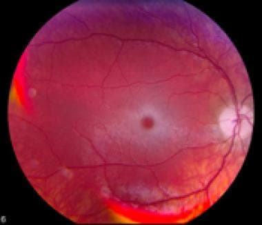
Krabbe disease | Radiology Reference Article | Radiopaedia.org
 SCOPe 2.08: Domain d2chnb1: 2chn B:437-590
HEXA1
- Two of these are due to the deficiency of the heterodimeric (α-β), "A" isoenzyme of lysosomal β-hexosaminidase (HexA). (nih.gov)
Enzyme6
- The GM2 activator protein transports GM2 gangliosides and presents the lipids to hexosaminidase, so a functional hexosaminidase enzyme is able to hydrolyze GM2 gangliosides into GM3 gangliosides by removing the N-acetylgalactosamine (GalNAc) residue from GM2 gangliosides. (wikipedia.org)
- This protein is required for the normal function of an enzyme called beta-hexosaminidase A, which plays a critical role in the brain and spinal cord. (medlineplus.gov)
- The genetic change that causes Tay-Sachs disease results in a deficiency of the enzyme beta-hexosaminidase A. This enzyme is required to break down the fatty substance GM2 ganglioside. (sparrow.org)
- The blood test checks the levels of hexosaminidase A enzyme in the blood. (sparrow.org)
- TDS is particularly caused by inadequate activity of the hexosaminidase 'A' enzyme. (write-right.net)
- The mouse used here has mutation in a gene that makes the housekeeping enzyme hexosaminidase ("hex") deficient and, therefore, has Sandhoff's Disease, a lethal genetic disease related to Tay-Sachs Disease. (sciencedaily.com)
Gangliosidosis1
- Type AB G M2 gangliosidosis is also known as hexosaminidase activator deficiency. (medscape.com)
Mutations2
- There are numerous mutations that lead to hexosaminidase deficiency including gene deletions, nonsense mutations, and missense mutations. (wikipedia.org)
- Mutations in the GM2A gene disrupt the activity of the GM2 ganglioside activator, which prevents beta-hexosaminidase A from breaking down GM2 ganglioside. (medlineplus.gov)
Assay2
- Hexosaminidase Activity Assay, 100 tests - 1 kit is backordered and will ship as soon as it is back in stock. (dnamethsoc.com)
- Using a β-hexosaminidase release assay, several drugs were seen to cause mast cell degranulation in vitro in comparison with unstimulated cells, but only morphine, vancomycin and cisatracurium specifically triggered this receptor, as assessed by the release of β-hexosaminidase in the control versus the MRGPRX2-silenced cells. (cun.es)
Sandhoff1
- Hereditary inability to form functional hexosaminidase enzymes are the cause of lipid storage disorders Tay-Sachs disease and Sandhoff disease. (wikipedia.org)
Metabolism1
- inherited disorders of metabolism, caused by hexosaminidase deficiency that causes severe neurologic symptoms and early death. (msdmanuals.com)
Ganglioside1
- Within lysosomes, the activator protein binds to a fatty substance called GM2 ganglioside and presents it to beta-hexosaminidase A to be broken down. (medlineplus.gov)
Gene2
- The two subunits of hexosaminidase A are shown below: The bifunctional protein NCOAT (nuclear cytoplasmic O-GlcNAcase and acetyltransferase) that is encoded by the MGEA5 gene possesses both hexosaminidase and histone acetyltransferase activities. (wikipedia.org)
- NCOAT is also known as hexosaminidase C and has distinct substrate specificities compared to lysosomal hexosaminidase A. A single-nucleotide polymorphism in the human O-GlcNAcase gene is linked to diabetes mellitus type 2. (wikipedia.org)
Activator protein1
- β-Hexosaminidase and the cofactor GM2 activator protein catalyze the degradation of the GM2 gangliosides and other molecules containing terminal N-acetyl hexosamines. (wikipedia.org)
Subunits1
- Even though the α and β subunits of lysosomal hexosaminidase can both cleave GalNAc residues, only the α subunit is able to hydrolyze GM2 gangliosides because of a key residue, Arg-424, and a loop structure that forms from the amino acid sequence in the alpha subunit. (wikipedia.org)
Disease3
- Tay-Sachs disease occurs when hexosaminidase A loses its ability to function. (wikipedia.org)
- Compared to an apparently healthy control population, 32/35 patients with widely disseminated disease showed elevated values and 18/23 patients with disease confined to the chest had normal hexosaminidase values. (nebraska.edu)
- Detailed studies of 18 patients indicated that declining hexosaminidase values were associated with effective therapy, rising values generally accompanied progressive disease. (nebraska.edu)
Enzymes1
- Functional lysosomal β-hexosaminidase enzymes are dimeric in structure. (wikipedia.org)
Activity3
- Hexosaminidase for Activity is available from Karlan upon request. (dnamethsoc.com)
- Increasing β-hexosaminidase A activity using genetically modified mesenchymal stem cells. (stembook.org)
- The specific activity of urinary hexosaminidase was determined in 58 patients with various histological types of lung carcinoma. (nebraska.edu)
Variant1
- It is also known as the B variant (with increased HEXOSAMINIDASE B but absence of hexosaminidase A) and is strongly associated with Ashkenazic Jewish ancestry. (curehunter.com)
Mutation1
- It is caused by mutation in the alpha subunit of the HEXOSAMINIDASE A resulting in lipid-laden ganglion cells. (curehunter.com)
Dimer1
- β-Hexosaminidase B (Hex B) is a dimer of beta chains. (medscape.com)
Urine1
- Elevated levels of hexosaminidase in blood and/or urine have been proposed as a biomarker of relapse in the treatment of alcoholism. (wikipedia.org)
Expression3
- Violin plots show distribution of expression levels for Beta-hexosaminidase (SMED30033380) in cells (dots) of each of the 12 neoblast clusters. (stowers.org)
- Expression of Beta-hexosaminidase (SMED30033380) in the t-SNE clustered sub-lethally irradiated X1 and X2 cells. (stowers.org)
- Analysis of GFP expression driven by Caenorhabditis hexosaminidase promoters. (boku.ac.at)
Cells1
- TF extract decreased β -hexosaminidase release in RBL-2H3 cells. (hindawi.com)
Blood1
- Routine haematological and biochemical blood tests were normal, as were levels of vitamin B 12 , folate, very long chain fatty acids, hexosaminidase, copper, and HTLV1 antibodies. (bmj.com)
Brain1
- Deficiency of hexosaminidase A results in accumulation of GM2 in the brain. (msdmanuals.com)
Deficiency10
- There are numerous mutations that lead to hexosaminidase deficiency including gene deletions, nonsense mutations, and missense mutations. (wikipedia.org)
- These diseases result from a deficiency of lysosomal enzyme ß- hexosaminidase A (HexA), which is responsible for GM2 ganglioside degradation. (bvsalud.org)
- Hexosaminidase activator deficiency is caused by absence or defects of the hexosaminidase activator. (medscape.com)
- Type AB G M2 gangliosidosis is also known as hexosaminidase activator deficiency. (medscape.com)
- Kaback MM. Hexosaminidase A Deficiency. (epnet.com)
- inherited disorders of metabolism, caused by hexosaminidase deficiency that causes severe neurologic symptoms and early death. (msdmanuals.com)
- Deficiency of hexosaminidase A results in accumulation of GM2 in the brain. (msdmanuals.com)
- There is a combined hexosaminidase A and B deficiency. (msdmanuals.com)
- The documented enzyme deficiency should support a genetically confirmed diagnosis of GM2-gangliosidosis caused by beta-hexosaminidase deficiency resulting from mutations in the HEXA or HEXB genes. (nih.gov)
- The accumulation of GM 2 (due to a deficiency in beta-hexosaminidase) has characterized Tay-Sachs disease (due to a mutation in the gene HEXA) and Sandhoff disease (due to a mutation in the gene HEXB). (matreya.com)
HEXB3
- Description: This is Double-antibody Sandwich Enzyme-linked immunosorbent assay for detection of Mouse Hexosaminidase B Beta (HEXb) in serum, plasma, tissue homogenates and other biological fluids. (srbiosystem.com)
- Description: A sandwich quantitative ELISA assay kit for detection of Rat Hexosaminidase B Beta (HEXb) in samples from serum, plasma, tissue homogenates or other biological fluids. (srbiosystem.com)
- One alpha subunit joins with one beta subunit (produced from the HEXB gene) to form a functioning beta-hexosaminidase A enzyme. (medlineplus.gov)
Lysosomal4
- Functional lysosomal β-hexosaminidase enzymes are dimeric in structure. (wikipedia.org)
- Even though the α and β subunits of lysosomal hexosaminidase can both cleave GalNAc residues, only the α subunit is able to hydrolyze GM2 gangliosides because of a key residue, Arg-424, and a loop structure that forms from the amino acid sequence in the alpha subunit. (wikipedia.org)
- NCOAT is also known as hexosaminidase C and has distinct substrate specificities compared to lysosomal hexosaminidase A. A single-nucleotide polymorphism in the human O-GlcNAcase gene is linked to diabetes mellitus type 2. (wikipedia.org)
- Human lysosomal beta-hexosaminidases remove terminal beta-glycosidically bound N-acetylhexosamine residues from a number of glycoconjugates. (nih.gov)
1.521
- This domain represents the N terminal domain in chitobiases and beta-hexosaminidases EC:3.2.1.52. (embl-heidelberg.de)
Serum2
- Description: A sandwich ELISA kit for detection of Hexosaminidase B Beta from Mouse in samples from blood, serum, plasma, cell culture fluid and other biological fluids. (srbiosystem.com)
- 8. Serum beta-hexosaminidases in pregnancy. (nih.gov)
Lysosomes1
- Within lysosomes, beta-hexosaminidase A forms part of a complex that breaks down a fatty substance called GM2 ganglioside found in cell membranes. (medlineplus.gov)
Genes1
- Assignment of genes encoding dihydrofolate reductase and hexosaminidase B to Mus musculus chromosome 13. (nih.gov)
Genetics1
- 15. The biochemical genetics of the hexosaminidase system in man. (nih.gov)
Hereditary1
- 3. Hereditary heat-labile hexosaminidase B: its implication for recognizing Tay-Sachs genotypes. (nih.gov)
Activator2
- β-Hexosaminidase and the cofactor GM2 activator protein catalyze the degradation of the GM2 gangliosides and other molecules containing terminal N-acetyl hexosamines. (wikipedia.org)
- The GM2 activator protein transports GM2 gangliosides and presents the lipids to hexosaminidase, so a functional hexosaminidase enzyme is able to hydrolyze GM2 gangliosides into GM3 gangliosides by removing the N-acetylgalactosamine (GalNAc) residue from GM2 gangliosides. (wikipedia.org)
Histamine1
- AEDK inhibited the release of histamine and β-hexosaminidase from mast cells by modulating cAMP and intracellular calcium levels. (spandidos-publications.com)
Proteins1
- MS revealed that phycocyanin and the core-membrane linker peptide are the responsible allergens, and MC(-) extracts containing these proteins induced β-hexosaminidase release in rat basophil leukemia cells. (nih.gov)
Catalytic1
- The GH20 hexosaminidases are thought to act via a catalytic mechanism in which the catalytic nucleophile is not provided by solvent or the enzyme, but by the substrate itself. (unl.edu)
Genetically1
- Increasing ß-hexosaminidase A activity using genetically modified mesenchymal stem cells. (bvsalud.org)
Release1
- hexosaminidase release assay to examine the inhibitory effects of fCD23. (ncl.edu.tw)
Beta2
- Beta-hexosaminidase A plays a critical role in the brain and spinal cord (central nervous system). (medlineplus.gov)
- This domain is found in the N terminus of chitobiases and beta-hexosaminidases. (embl-heidelberg.de)
Tissue1
- 14. Characterization and tissue distribution of N-acetyl hexosaminidase C: suggestive evidence for a separate hexosaminidase locus. (nih.gov)