Hemodynamics
Vascular Resistance
Blood Flow Velocity
Cardiac Output
Models, Cardiovascular
Dogs
Hypertension, Pulmonary
Kidney
Pulsatile Flow
Glomerular Filtration Rate
Pulmonary Wedge Pressure
Splanchnic Circulation
Cardiography, Impedance
Pulmonary Artery
Oxygen
Oxygen Consumption
Ventricular Function, Left
Hypertension, Portal
Ultrasonography, Doppler, Transcranial
Spectroscopy, Near-Infrared
Arterial Pressure
Stroke Volume
Venous Pressure
Hypertension
Heart Failure
Diuresis
Hematocrit
Swine
Disease Models, Animal
Pulmonary Gas Exchange
Laser-Doppler Flowmetry
Rats, Sprague-Dawley
Renal Plasma Flow
Antihypertensive Agents
Cardiac Catheterization
Nitric Oxide
Renin
Anesthesia
Intracranial Aneurysm
Kidney Glomerulus
Catheterization, Swan-Ganz
Hemorheology
Ultrasonography, Doppler, Color
Sulfones
Ultrasonography, Doppler
Magnetic Resonance Angiography
Hydrodynamics
Acetazolamide
Middle Cerebral Artery
Heart Ventricles
Isoflurane
Carbon Dioxide
Myocardium
Treatment Outcome
Portal System
Shear Strength
Norepinephrine
Atrial Natriuretic Factor
Oxyhemoglobins
Vasodilation
Stress, Mechanical
Hypotension
Prospective Studies
Fontan Procedure
Hemodilution
Infusions, Intravenous
Epoprostenol
Ventricular Function, Right
Ventricular Pressure
Vascular Stiffness
Receptors, Endothelin
Echocardiography
Angiotensin II
Plasma Volume
Vasoconstriction
Carotid Artery, Internal
Isotonic Solutions
Hypotension, Controlled
Arterioles
Sus scrofa
Meclofenamic Acid
Lung
Purines
Hyperemia
Sheep
Cardiotonic Agents
Ophthalmic Artery
Computer Simulation
Milrinone
Pressure
Dose-Response Relationship, Drug
Endothelin-1
Central Venous Pressure
Echocardiography, Doppler
Medetomidine
Cyclic Nucleotide Phosphodiesterases, Type 5
Hypotension, Orthostatic
Angiotensin-Converting Enzyme Inhibitors
NG-Nitroarginine Methyl Ester
Sympathetic Nervous System
Chronic Disease
Blood Viscosity
Kidney Function Tests
Rats, Wistar
Nitric Oxide Synthase
Ultrasonography, Doppler, Pulsed
Analysis of Variance
Hypertrophy, Right Ventricular
Reference Values
Plasma Substitutes
Vena Cava, Inferior
Rats, Inbred SHR
Compliance
Hypertension, Renal
Random Allocation
Fentanyl
Heart Defects, Congenital
Femoral Vein
Cardiac Pacing, Artificial
Liver Cirrhosis
Sodium
Venous Insufficiency
Hemoglobins
Collateral Circulation
Models, Anatomic
Shock, Septic
Cardiovascular Physiological Phenomena
Cardiovascular System
Magnetic Resonance Imaging
Enalapril
Pulmonary Edema
Anesthesia, Intratracheal
Partial Pressure
Prostaglandins
Tetrazoles
Phenylpropionates
Aortic Valve Stenosis
Receptor, Endothelin A
Rheology
Ventricular Remodeling
Teprotide
Brain
Anesthesia, General
Angiotensin Receptor Antagonists
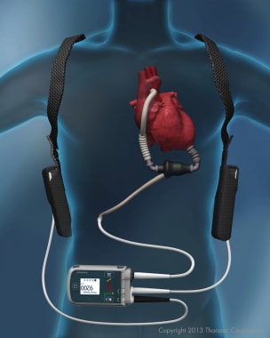
Heart-Assist Devices
Respiratory Physiological Processes
Phosphodiesterase Inhibitors
Aortic Valve
Lypressin
Natriuretic Agents
Double-Blind Method
Cardiopulmonary Bypass
Carotid Stenosis
Retinal Artery
Carotid Arteries
Hemofiltration
Imaging, Three-Dimensional
The evolution of early fibromuscular lesions hemodynamically induced in the dog renal artery. I. Light and transmission electron microscopy. (1/14436)
In view of the important roles of arterial intimal fibromuscular lesions as precursors of atherosclerotic plaque and occlusive lesions in arterial reconstructions, a model has been developed for the rapid hemodynamic induction of these lesions by anastomosis of the dog right renal artery to the inferior vena cava. Light and transmission electron microscopic observations were made on the arterial shunt after periods of rapid flow ranging form 10 minutes to 2 hours to identify initial factor(s) and evolutionary mechanisms in the etiology of the lesions. The sequence of events included aberrations in ruthenium red staining of the endothelial luminal membrane at 10 minutes, multilayered thickening of the subendothelial basement membrane (BM) at 15 minutes, and initial reorientation and migration of smooth muscle cells (SMC) into the intima along with the appearance of areas of degeneration of the internal elastic lamina (IEL) at 30 minutes. The endothelial cells were still intact in some areas overlying the SMC migration and IEL degeneration, but they were separating from the surface in other such areas. As subendothelium became exposed, some platelet adherence was noted. By 2 hours, the entire wall reaction was fully developed. Initial observations indicate that in the evolution of this hemodynamically induced lesion visible alteration in the endothelial cells is not prerequisite to degeneration of the underlying IEL and reorientation and migration of medial SMC. (+info)Signal-, set- and movement-related activity in the human brain: an event-related fMRI study. (2/14436)
Electrophysiological studies on monkeys have been able to distinguish sensory and motor signals close in time by pseudorandomly delaying the cue that instructs the movement from the stimulus that triggers the movement. We have used a similar experimental design in functional magnetic resonance imaging (fMRI), scanning subjects while they performed a visuomotor conditional task with instructed delays. One of four shapes was presented briefly. Two shapes instructed the subjects to flex the index finger; the other two shapes coded the flexion of the middle finger. The subjects were told to perform the movement after a tone. We have exploited a novel use of event-related fMRI. By systematically varying the interval between the visual and acoustic stimuli, it has been possible to estimate the significance of the evoked haemodynamic response (EHR) to each of the stimuli, despite their temporal proximity in relation to the time constant of the EHR. Furthermore, by varying the phase between events and image acquisition, we have been able to achieve high temporal resolution while scanning the whole brain. We dissociated sensory and motor components of the sensorimotor transformations elicited by the task, and assessed sustained activity during the instructed delays. In calcarine and occipitotemporal cortex, the responses were exclusively associated with the visual instruction cues. In temporal auditory cortex and in primary motor cortex, they were exclusively associated with the auditory trigger stimulus. In ventral prefrontal cortex there were movement-related responses preceded by preparatory activity and by signal-related activity. Finally, responses associated with the instruction cue and with sustained activity during the delay period were observed in the dorsal premotor cortex and in the dorsal posterior parietal cortex. Where the association between a visual cue and the appropriate movement is arbitrary, the underlying visuomotor transformations are not achieved exclusively through frontoparietal interactions. Rather, these processes seem to rely on the ventral visual stream, the ventral prefrontal cortex and the anterior part of the dorsal premotor cortex. (+info)Protective effect of bactericidal/permeability-increasing protein (rBPI21) in baboon sepsis is related to its antibacterial, not antiendotoxin, properties. (3/14436)
OBJECTIVE AND SUMMARY BACKGROUND DATA: The recombinant fragment of bactericidal/permeability-increasing protein, rBPI21, has potent bactericidal activity against gram-negative bacteria as well as antiendotoxin (lipopolysaccharide [LPS]) action. On the basis of these activities, the authors sought to discover whether rBPI21 would be protective in baboons with live Escherichia coli-induced sepsis and whether the potential protective effects of rBPI21 (together with antibiotics) would be more closely related to its antibacterial or LPS-neutralizing effects. METHODS: In a prospective, randomized, placebo-controlled subchronic laboratory study, the efficacy of rBPI21 or placebo was studied over 72 hours in chronically instrumented male baboons infused with live E. coli under antibiotic therapy. RESULTS: Intravenous rBPI21 attenuated sepsis-related organ failure and increased survival significantly. Bacteremia was significantly reduced in the rBPI21 group at 2 hours after the start of the E. coli infusion, whereas circulating LPS was less affected. The in vivo formation of tumor necrosis factor was significantly suppressed by the rBPI21 treatment regimen. Microcirculation and organ function were improved. CONCLUSIONS: In baboon live E. coli sepsis, the salutary effect of rBPI21 results from a more prevalent antibacterial than antiendotoxin activity. (+info)NaCl-induced renal vasoconstriction in salt-sensitive African Americans: antipressor and hemodynamic effects of potassium bicarbonate. (4/14436)
In 16 African Americans (blacks, 14 men, 2 women) with average admission mean arterial pressure (MAP, mm Hg) 99.9+/-3.5 (mean+/-SEM), we investigated whether NaCl-induced renal vasoconstriction attends salt sensitivity and, if so, whether supplemental KHCO3 ameliorates both conditions. Throughout a 3-week period under controlled metabolic conditions, all subjects ate diets containing 15 mmol NaCl and 30 mmol potassium (K+) (per 70 kg body wt [BW] per day). Throughout weeks 2 and 3, NaCl was loaded to 250 mmol/d; throughout week 3, dietary K+ was supplemented to 170 mmol/d (KHCO3). On the last day of each study week, we measured renal blood flow (RBF) and glomerular filtration rate (GFR) using renal clearances of PAH and inulin. Ten subjects were salt sensitive (SS) (DeltaMAP >+5%) and 6 salt resistant (SR). In NaCl-loaded SS but not SR subjects, RBF (mL/min/1.73 m2) decreased from 920+/-75 to 828+/-46 (P<0.05); filtration fraction (FF, %) increased from 19. 4+/- to 21.4 (P<0.001); and renal vascular resistance (RVR) (10(3)xmm Hg/[mL/min]) increased from 101+/-8 to 131+/-10 (P<0.001). In all subjects combined, DeltaMAP varied inversely with DeltaRBF (r =-0.57, P=0.02) and directly with DeltaRVR (r = 0.65, P=0.006) and DeltaFF (r = 0.59, P=0.03), but not with MAP before NaCl loading. When supplemental KHCO3 abolished the pressor effect of NaCl in SS subjects, RBF was unaffected but GFR and FF decreased. The results show that in marginally K+-deficient blacks (1) NaCl-induced renal vasoconstrictive dysfunction attends salt sensitivity; (2) the dysfunction varies in extent directly with the NaCl-induced increase in blood pressure (BP); and (3) is complexly affected by supplemented KHCO3, GFR and FF decreasing but RBF not changing. In blacks, NaCl-induced renal vasoconstriction may be a pathogenetic event in salt sensitivity. (+info)Low calorie diet enhances renal, hemodynamic, and humoral effects of exogenous atrial natriuretic peptide in obese hypertensives. (5/14436)
The expression of the natriuretic peptide clearance receptor is abundant in human and rat adipose tissue, where it is specifically inhibited by fasting. In obese hypertensives, plasma atrial natriuretic peptide (ANP) levels were found to be lower than in obese normotensives. Therefore, the increased adipose mass might influence ANP levels and/or its biological activity. The aim of the present study was to evaluate whether the humoral, hemodynamic, and renal effects of exogenous ANP in obese hypertensives might be enhanced by a very low calorie diet. Eight obese hypertensives received a bolus injection of ANP (0.6 mg/kg) after 2 weeks of a normal calorie/normal sodium diet, and blood pressure (BP), heart rate, ANP, cGMP, plasma renin activity, and aldosterone were evaluated for 2 hours before and after the injection. Diuresis and natriuresis were measured every 30 minutes. The patients then started a low calorie/normal sodium diet (510 kcal/150 mmol/d) for 4 days, and then the ANP injection protocol was repeated. The low calorie diet induced a slight weight loss (from 90.6+/-1.1 to 87. 7+/-1.2 kg; P<0.01), which was accompanied by increase of cGMP excretion (from 146.0+/-10.1 to 154.5+/-9.5 nmol/24 h; P<0.05) together with a reduction of BP (P<0.01 versus basal levels). ANP injection after diet was followed by an increase of ANP levels similar to that observed before diet, but plasma cGMP, diuresis, and natriuresis increased significantly only after diet. Similarly, the decrease of BP after ANP administration was significantly higher after diet (change in mean arterial pressure, -6.4+/-0.7 versus -4. 0+/-0.6 mm Hg; P<0.05) as well as that of aldosterone (P<0.01). These data show that a low calorie diet enhances the humoral, renal, and hemodynamic effects of ANP in obese hypertensives and confirm the importance of caloric intake in modulating the biological activity of ANP, suggesting that the natriuretic peptide system can play a role in the acute changes of natriuresis and diuresis associated with caloric restriction. (+info)Sympathetic nerve alterations assessed with 123I-MIBG in the failing human heart. (6/14436)
Norepinephrine (NE) reuptake function is impaired in heart failure and this may participate in myocyte hyperstimulation by the neurotransmitter. This alteration can be assessed by 123I-metaiodobenzylguanidine (MIBG) scintigraphy. METHODS: To determine whether the impairment of neuronal NE reuptake was reversible after metoprolol therapy, we studied 18 patients (43+/-7 y) with idiopathic dilated cardiomyopathy who were stabilized at least for 3 mo with captopril and diuretics. Patients underwent, before and after 6 mo of therapy with metoprolol, measurements of radionuclide left ventricular ejection fraction (LVEF), maximal oxygen consumption and plasma NE concentration. The cardiac adrenergic innervation function was scintigraphically assessed with MIBG uptake and release measurements on the planar images obtained 20 min and 4 h after tracer injection. To evaluate whether metoprolol had a direct interaction with cardiac MIBG uptake and release, six normal subjects were studied before and after a 1-mo metoprolol intake. RESULTS: In controls, neither cardiac MIBG uptake and release nor circulating NE concentration changed after the 1-mo metoprolol intake. Conversely, after a 6-mo therapy with metoprolol, patients showed increased cardiac MIBG uptake (129%+/-10% versus 138%+/-17%; P = 0.009), unchanged cardiac MIBG release and decreased plasma NE concentration (0.930+/-412 versus 0.721+/-0.370 ng/mL; P = 0.02). In parallel, patients showed improved New York Heart Association class (2.44+/-0.51 versus 2.05+/-0.23; P = 0.004) and increased LVEF (20%+/-8% versus 27%+/-8%; P = 0.0005), whereas maximal oxygen uptake remained unchanged. CONCLUSION: Thus, a parallel improvement of myocardial NE reuptake and of hemodynamics was observed after a 6-mo metoprolol therapy, suggesting that such agents may be beneficial in heart failure by directly protecting the myocardium against excessive NE stimulation. (+info)Enteroviral RNA replication in the myocardium of patients with left ventricular dysfunction and clinically suspected myocarditis. (7/14436)
BACKGROUND: Previous studies dealing with the detection of enteroviral RNA in human endomyocardial biopsies have not differentiated between latent persistence of the enteroviral genome and active viral replication. Enteroviruses that are considered important factors for the development of myocarditis have a single-strand RNA genome of positive polarity that is transcribed by a virus-encoded RNA polymerase into a minus-strand mRNA during active viral replication. The synthesis of multiple copies of minus-strand enteroviral RNA therefore occurs only at sites of active viral replication but not in tissues with mere persistence of the viral genome. METHODS AND RESULTS: We investigated enteroviral RNA replication versus enteroviral RNA persistence in endomyocardial biopsies of 45 patients with left ventricular dysfunction and clinically suspected myocarditis. Using reverse-transcriptase polymerase chain reaction in conjunction with Southern blot hybridization, we established a highly sensitive assay to specifically detect plus-strand versus minus-strand enteroviral RNA in the biopsies. Plus-strand enteroviral RNA was detected in endomyocardial biopsies of 18 (40%) of 45 patients, whereas minus-strand RNA as an indication of active enteroviral RNA replication was detected in only 10 (56%) of these 18 plus-strand-positive patients. Enteroviral RNA was not found in biopsies of the control group (n=26). CONCLUSIONS: These data demonstrate that a significant fraction of patients with left ventricular dysfunction and clinically suspected myocarditis had active enteroviral RNA replication in their myocardium (22%). Differentiation between patients with active viral replication and latent viral persistence should be particularly important in future studies evaluating different therapeutic strategies. In addition, molecular genetic detection of enteroviral genome and differentiation between replicating versus persistent viruses is possible in a single endomyocardial biopsy. (+info)Suppression of atherosclerotic development in Watanabe heritable hyperlipidemic rabbits treated with an oral antiallergic drug, tranilast. (8/14436)
BACKGROUND: Inflammatory and immunological responses of vascular cells have been shown to play a significant role in the progression of atheromatous formation. Tranilast [N-(3,4-dimethoxycinnamoyl) anthranillic acid] inhibits release of cytokines and chemical mediators from various cells, including macrophages, leading to suppression of inflammatory and immunological responses. This study tested whether tranilast may suppress atheromatous formation in Watanabe heritable hyperlipidemic (WHHL) rabbits. METHODS AND RESULTS: WHHL rabbits (2 months old) were given either 300 mg x kg-1 x d-1 of tranilast (Tranilast, n=12) or vehicle (Control, n=13) PO for 6 months. Tranilast treatment was found to suppress the aortic area covered with plaque. Immunohistochemical analysis showed that there was no difference in the percentage of the RAM11-positive macrophage area and the frequency of CD5-positive cells (T cells) in intimal plaques between Tranilast and Control. Major histocompatibility complex (MHC) class II expression in macrophages and interleukin-2 (IL-2) receptor expression in T cells, as markers of the immunological activation in these cells, was suppressed in atheromatous plaque by tranilast treatment. Flow cytometry analysis of isolated human and rabbit peripheral blood mononuclear cells showed that an increase in expression both of MHC class II antigen on monocytes by incubation with interferon-gamma and of IL-2 receptor on T cells by IL-2 was suppressed by the combined incubation with tranilast. CONCLUSIONS: The results indicate that tranilast suppresses atherosclerotic development partly through direct inhibition of immunological activation of monocytes/macrophages and T cells in the atheromatous plaque. (+info)Hemodynamics is the study of how blood flows through the cardiovascular system, including the heart and the vascular network. It examines various factors that affect blood flow, such as blood volume, viscosity, vessel length and diameter, and pressure differences between different parts of the circulatory system. Hemodynamics also considers the impact of various physiological and pathological conditions on these variables, and how they in turn influence the function of vital organs and systems in the body. It is a critical area of study in fields such as cardiology, anesthesiology, and critical care medicine.
Vascular resistance is a measure of the opposition to blood flow within a vessel or a group of vessels, typically expressed in units of mmHg/(mL/min) or sometimes as dynes*sec/cm^5. It is determined by the diameter and length of the vessels, as well as the viscosity of the blood flowing through them. In general, a decrease in vessel diameter, an increase in vessel length, or an increase in blood viscosity will result in an increase in vascular resistance, while an increase in vessel diameter, a decrease in vessel length, or a decrease in blood viscosity will result in a decrease in vascular resistance. Vascular resistance is an important concept in the study of circulation and cardiovascular physiology because it plays a key role in determining blood pressure and blood flow within the body.
Renal circulation refers to the blood flow specifically dedicated to the kidneys. The main function of the kidneys is to filter waste and excess fluids from the blood, which then get excreted as urine. To perform this function efficiently, the kidneys receive a substantial amount of the body's total blood supply - about 20-25% in a resting state.
The renal circulation process begins when deoxygenated blood from the rest of the body returns to the right side of the heart and is pumped into the lungs for oxygenation. Oxygen-rich blood then leaves the left side of the heart through the aorta, the largest artery in the body.
A portion of this oxygen-rich blood moves into the renal arteries, which branch directly from the aorta and supply each kidney with blood. Within the kidneys, these arteries divide further into smaller vessels called afferent arterioles, which feed into a network of tiny capillaries called the glomerulus within each nephron (the functional unit of the kidney).
The filtration process occurs in the glomeruli, where waste materials and excess fluids are separated from the blood. The resulting filtrate then moves through another set of capillaries, the peritubular capillaries, which surround the renal tubules (the part of the nephron that reabsorbs necessary substances back into the bloodstream).
The now-deoxygenated blood from the kidneys' capillary network coalesces into venules and then merges into the renal veins, which ultimately drain into the inferior vena cava and return the blood to the right side of the heart. This highly specialized circulation system allows the kidneys to efficiently filter waste while maintaining appropriate blood volume and composition.
Blood pressure is the force exerted by circulating blood on the walls of the blood vessels. It is measured in millimeters of mercury (mmHg) and is given as two figures:
1. Systolic pressure: This is the pressure when the heart pushes blood out into the arteries.
2. Diastolic pressure: This is the pressure when the heart rests between beats, allowing it to fill with blood.
Normal blood pressure for adults is typically around 120/80 mmHg, although this can vary slightly depending on age, sex, and other factors. High blood pressure (hypertension) is generally considered to be a reading of 130/80 mmHg or higher, while low blood pressure (hypotension) is usually defined as a reading below 90/60 mmHg. It's important to note that blood pressure can fluctuate throughout the day and may be affected by factors such as stress, physical activity, and medication use.
Blood flow velocity is the speed at which blood travels through a specific part of the vascular system. It is typically measured in units of distance per time, such as centimeters per second (cm/s) or meters per second (m/s). Blood flow velocity can be affected by various factors, including cardiac output, vessel diameter, and viscosity of the blood. Measuring blood flow velocity is important in diagnosing and monitoring various medical conditions, such as heart disease, stroke, and peripheral vascular disease.
Cardiac output is a measure of the amount of blood that is pumped by the heart in one minute. It is defined as the product of stroke volume (the amount of blood pumped by the left ventricle during each contraction) and heart rate (the number of contractions per minute). Normal cardiac output at rest for an average-sized adult is about 5 to 6 liters per minute. Cardiac output can be increased during exercise or other conditions that require more blood flow, such as during illness or injury. It can be measured noninvasively using techniques such as echocardiography or invasively through a catheter placed in the heart.
Regional blood flow (RBF) refers to the rate at which blood flows through a specific region or organ in the body, typically expressed in milliliters per minute per 100 grams of tissue (ml/min/100g). It is an essential physiological parameter that reflects the delivery of oxygen and nutrients to tissues while removing waste products. RBF can be affected by various factors such as metabolic demands, neural regulation, hormonal influences, and changes in blood pressure or vascular resistance. Measuring RBF is crucial for understanding organ function, diagnosing diseases, and evaluating the effectiveness of treatments.
Cerebrovascular circulation refers to the network of blood vessels that supply oxygenated blood and nutrients to the brain tissue, and remove waste products. It includes the internal carotid arteries, vertebral arteries, circle of Willis, and the intracranial arteries that branch off from them.
The internal carotid arteries and vertebral arteries merge to form the circle of Willis, a polygonal network of vessels located at the base of the brain. The anterior cerebral artery, middle cerebral artery, posterior cerebral artery, and communicating arteries are the major vessels that branch off from the circle of Willis and supply blood to different regions of the brain.
Interruptions or abnormalities in the cerebrovascular circulation can lead to various neurological conditions such as stroke, transient ischemic attack (TIA), and vascular dementia.
Cardiovascular models are simplified representations or simulations of the human cardiovascular system used in medical research, education, and training. These models can be physical, computational, or mathematical and are designed to replicate various aspects of the heart, blood vessels, and blood flow. They can help researchers study the structure and function of the cardiovascular system, test new treatments and interventions, and train healthcare professionals in diagnostic and therapeutic techniques.
Physical cardiovascular models may include artificial hearts, blood vessels, or circulation systems made from materials such as plastic, rubber, or silicone. These models can be used to study the mechanics of heart valves, the effects of different surgical procedures, or the impact of various medical devices on blood flow.
Computational and mathematical cardiovascular models use algorithms and equations to simulate the behavior of the cardiovascular system. These models may range from simple representations of a single heart chamber to complex simulations of the entire circulatory system. They can be used to study the electrical activity of the heart, the biomechanics of blood flow, or the distribution of drugs in the body.
Overall, cardiovascular models play an essential role in advancing our understanding of the human body and improving patient care.
Pulmonary circulation refers to the process of blood flow through the lungs, where blood picks up oxygen and releases carbon dioxide. This is a vital part of the overall circulatory system, which delivers nutrients and oxygen to the body's cells while removing waste products like carbon dioxide.
In pulmonary circulation, deoxygenated blood from the systemic circulation returns to the right atrium of the heart via the superior and inferior vena cava. The blood then moves into the right ventricle through the tricuspid valve and gets pumped into the pulmonary artery when the right ventricle contracts.
The pulmonary artery divides into smaller vessels called arterioles, which further branch into a vast network of tiny capillaries in the lungs. Here, oxygen from the alveoli diffuses into the blood, binding to hemoglobin in red blood cells, while carbon dioxide leaves the blood and is exhaled through the nose or mouth.
The now oxygenated blood collects in venules, which merge to form pulmonary veins. These veins transport the oxygen-rich blood back to the left atrium of the heart, where it enters the systemic circulation once again. This continuous cycle enables the body's cells to receive the necessary oxygen and nutrients for proper functioning while disposing of waste products.
I believe there might be a misunderstanding in your question. "Dogs" is not a medical term or condition. It is the common name for a domesticated carnivore of the family Canidae, specifically the genus Canis, which includes wolves, foxes, and other extant and extinct species of mammals. Dogs are often kept as pets and companions, and they have been bred in a wide variety of forms and sizes for different purposes, such as hunting, herding, guarding, assisting police and military forces, and providing companionship and emotional support.
If you meant to ask about a specific medical condition or term related to dogs, please provide more context so I can give you an accurate answer.
Pulmonary hypertension is a medical condition characterized by increased blood pressure in the pulmonary arteries, which are the blood vessels that carry blood from the right side of the heart to the lungs. This results in higher than normal pressures in the pulmonary circulation and can lead to various symptoms and complications.
Pulmonary hypertension is typically defined as a mean pulmonary artery pressure (mPAP) greater than or equal to 25 mmHg at rest, as measured by right heart catheterization. The World Health Organization (WHO) classifies pulmonary hypertension into five groups based on the underlying cause:
1. Pulmonary arterial hypertension (PAH): This group includes idiopathic PAH, heritable PAH, drug-induced PAH, and associated PAH due to conditions such as connective tissue diseases, HIV infection, portal hypertension, congenital heart disease, and schistosomiasis.
2. Pulmonary hypertension due to left heart disease: This group includes conditions that cause elevated left atrial pressure, such as left ventricular systolic or diastolic dysfunction, valvular heart disease, and congenital cardiovascular shunts.
3. Pulmonary hypertension due to lung diseases and/or hypoxia: This group includes chronic obstructive pulmonary disease (COPD), interstitial lung disease, sleep-disordered breathing, alveolar hypoventilation disorders, and high altitude exposure.
4. Chronic thromboembolic pulmonary hypertension (CTEPH): This group includes persistent obstruction of the pulmonary arteries due to organized thrombi or emboli.
5. Pulmonary hypertension with unclear and/or multifactorial mechanisms: This group includes hematologic disorders, systemic disorders, metabolic disorders, and other conditions that can cause pulmonary hypertension but do not fit into the previous groups.
Symptoms of pulmonary hypertension may include shortness of breath, fatigue, chest pain, lightheadedness, and syncope (fainting). Diagnosis typically involves a combination of medical history, physical examination, imaging studies, and invasive testing such as right heart catheterization. Treatment depends on the underlying cause but may include medications, oxygen therapy, pulmonary rehabilitation, and, in some cases, surgical intervention.
Heart rate is the number of heartbeats per unit of time, often expressed as beats per minute (bpm). It can vary significantly depending on factors such as age, physical fitness, emotions, and overall health status. A resting heart rate between 60-100 bpm is generally considered normal for adults, but athletes and individuals with high levels of physical fitness may have a resting heart rate below 60 bpm due to their enhanced cardiovascular efficiency. Monitoring heart rate can provide valuable insights into an individual's health status, exercise intensity, and response to various treatments or interventions.
A kidney, in medical terms, is one of two bean-shaped organs located in the lower back region of the body. They are essential for maintaining homeostasis within the body by performing several crucial functions such as:
1. Regulation of water and electrolyte balance: Kidneys help regulate the amount of water and various electrolytes like sodium, potassium, and calcium in the bloodstream to maintain a stable internal environment.
2. Excretion of waste products: They filter waste products from the blood, including urea (a byproduct of protein metabolism), creatinine (a breakdown product of muscle tissue), and other harmful substances that result from normal cellular functions or external sources like medications and toxins.
3. Endocrine function: Kidneys produce several hormones with important roles in the body, such as erythropoietin (stimulates red blood cell production), renin (regulates blood pressure), and calcitriol (activated form of vitamin D that helps regulate calcium homeostasis).
4. pH balance regulation: Kidneys maintain the proper acid-base balance in the body by excreting either hydrogen ions or bicarbonate ions, depending on whether the blood is too acidic or too alkaline.
5. Blood pressure control: The kidneys play a significant role in regulating blood pressure through the renin-angiotensin-aldosterone system (RAAS), which constricts blood vessels and promotes sodium and water retention to increase blood volume and, consequently, blood pressure.
Anatomically, each kidney is approximately 10-12 cm long, 5-7 cm wide, and 3 cm thick, with a weight of about 120-170 grams. They are surrounded by a protective layer of fat and connected to the urinary system through the renal pelvis, ureters, bladder, and urethra.
Blood volume refers to the total amount of blood present in an individual's circulatory system at any given time. It is the combined volume of both the plasma (the liquid component of blood) and the formed elements (such as red and white blood cells and platelets) in the blood. In a healthy adult human, the average blood volume is approximately 5 liters (or about 1 gallon). However, blood volume can vary depending on several factors, including age, sex, body weight, and overall health status.
Blood volume plays a critical role in maintaining proper cardiovascular function, as it affects blood pressure, heart rate, and the delivery of oxygen and nutrients to tissues throughout the body. Changes in blood volume can have significant impacts on an individual's health and may be associated with various medical conditions, such as dehydration, hemorrhage, heart failure, and liver disease. Accurate measurement of blood volume is essential for diagnosing and managing these conditions, as well as for guiding treatment decisions in clinical settings.
Pulsatile flow is a type of fluid flow that occurs in a rhythmic, wave-like pattern, typically seen in the cardiovascular system. It refers to the periodic variation in the volume or velocity of a fluid (such as blood) that is caused by the regular beating of the heart. In pulsatile flow, there are periods of high flow followed by periods of low or no flow, which creates a distinct pattern on a graph or tracing. This type of flow is important for maintaining proper function and health in organs and tissues throughout the body.
Glomerular filtration rate (GFR) is a test used to check how well the kidneys are working. Specifically, it estimates how much blood passes through the glomeruli each minute. The glomeruli are the tiny fibers in the kidneys that filter waste from the blood. A lower GFR number means that the kidneys aren't working properly and may indicate kidney disease.
The GFR is typically calculated using a formula that takes into account the patient's serum creatinine level, age, sex, and race. The most commonly used formula is the CKD-EPI (Chronic Kidney Disease Epidemiology Collaboration) equation. A normal GFR is usually above 90 mL/min/1.73m2, but this can vary depending on the individual's age and other factors.
Liver circulation, also known as hepatic circulation, refers to the blood flow through the liver. The liver receives blood from two sources: the hepatic artery and the portal vein.
The hepatic artery delivers oxygenated blood from the heart to the liver, accounting for about 25% of the liver's blood supply. The remaining 75% comes from the portal vein, which carries nutrient-rich, deoxygenated blood from the gastrointestinal tract, spleen, pancreas, and gallbladder to the liver.
In the liver, these two sources of blood mix in the sinusoids, small vessels with large spaces between the endothelial cells that line them. This allows for efficient exchange of substances between the blood and the hepatocytes (liver cells). The blood then leaves the liver through the hepatic veins, which merge into the inferior vena cava and return the blood to the heart.
The unique dual blood supply and extensive sinusoidal network in the liver enable it to perform various critical functions, such as detoxification, metabolism, synthesis, storage, and secretion of numerous substances, maintaining body homeostasis.
Coronary circulation refers to the circulation of blood in the coronary vessels, which supply oxygenated blood to the heart muscle (myocardium) and drain deoxygenated blood from it. The coronary circulation system includes two main coronary arteries - the left main coronary artery and the right coronary artery - that branch off from the aorta just above the aortic valve. These arteries further divide into smaller branches, which supply blood to different regions of the heart muscle.
The left main coronary artery divides into two branches: the left anterior descending (LAD) artery and the left circumflex (LCx) artery. The LAD supplies blood to the front and sides of the heart, while the LCx supplies blood to the back and sides of the heart. The right coronary artery supplies blood to the lower part of the heart, including the right ventricle and the bottom portion of the left ventricle.
The veins that drain the heart muscle include the great cardiac vein, the middle cardiac vein, and the small cardiac vein, which merge to form the coronary sinus. The coronary sinus empties into the right atrium, allowing deoxygenated blood to enter the right side of the heart and be pumped to the lungs for oxygenation.
Coronary circulation is essential for maintaining the health and function of the heart muscle, as it provides the necessary oxygen and nutrients required for proper contraction and relaxation of the myocardium. Any disruption or blockage in the coronary circulation system can lead to serious consequences, such as angina, heart attack, or even death.
Pulmonary wedge pressure, also known as pulmonary capillary wedge pressure (PCWP) or left heart filling pressure, is a measurement obtained during right heart catheterization. It reflects the pressure in the left atrium, which is an estimate of the diastolic pressure in the left ventricle. Normal PCWP ranges from 4 to 12 mmHg. Increased pulmonary wedge pressure can indicate heart failure or other cardiac disorders that affect the left side of the heart.
Splanchnic circulation refers to the blood flow to the visceral organs, including the gastrointestinal tract, pancreas, spleen, and liver. These organs receive a significant portion of the cardiac output, with approximately 25-30% of the total restingly going to the splanchnic circulation. The splanchnic circulation is regulated by a complex interplay of neural and hormonal mechanisms that help maintain adequate blood flow to these vital organs while also allowing for the distribution of blood to other parts of the body as needed.
The splanchnic circulation is unique in its ability to vasodilate and increase blood flow significantly in response to meals or other stimuli, such as stress or hormonal changes. This increased blood flow helps support the digestive process and absorption of nutrients. At the same time, the body must carefully regulate this blood flow to prevent a significant drop in blood pressure or overloading the heart with too much work.
Overall, the splanchnic circulation plays a critical role in maintaining the health and function of the body's vital organs, and dysregulation of this system can contribute to various diseases, including digestive disorders, liver disease, and cardiovascular disease.
Impedance cardiography is a non-invasive method to measure cardiac output and systemic vascular resistance. It uses low-frequency electrical currents passed through the thorax to measure changes in impedance or resistance to flow during each heartbeat. This allows for the calculation of stroke volume and cardiac output. Impedance cardiography can provide continuous, real-time monitoring of cardiovascular function, making it useful in critical care settings and for tracking changes in patients with heart failure or other cardiovascular conditions.
The pulmonary artery is a large blood vessel that carries deoxygenated blood from the right ventricle of the heart to the lungs for oxygenation. It divides into two main branches, the right and left pulmonary arteries, which further divide into smaller vessels called arterioles, and then into a vast network of capillaries in the lungs where gas exchange occurs. The thin walls of these capillaries allow oxygen to diffuse into the blood and carbon dioxide to diffuse out, making the blood oxygen-rich before it is pumped back to the left side of the heart through the pulmonary veins. This process is crucial for maintaining proper oxygenation of the body's tissues and organs.
Oxygen is a colorless, odorless, tasteless gas that constitutes about 21% of the earth's atmosphere. It is a crucial element for human and most living organisms as it is vital for respiration. Inhaled oxygen enters the lungs and binds to hemoglobin in red blood cells, which carries it to tissues throughout the body where it is used to convert nutrients into energy and carbon dioxide, a waste product that is exhaled.
Medically, supplemental oxygen therapy may be provided to patients with conditions such as chronic obstructive pulmonary disease (COPD), pneumonia, heart failure, or other medical conditions that impair the body's ability to extract sufficient oxygen from the air. Oxygen can be administered through various devices, including nasal cannulas, face masks, and ventilators.
Oxygen consumption, also known as oxygen uptake, is the amount of oxygen that is consumed or utilized by the body during a specific period of time, usually measured in liters per minute (L/min). It is a common measurement used in exercise physiology and critical care medicine to assess an individual's aerobic metabolism and overall health status.
In clinical settings, oxygen consumption is often measured during cardiopulmonary exercise testing (CPET) to evaluate cardiovascular function, pulmonary function, and exercise capacity in patients with various medical conditions such as heart failure, chronic obstructive pulmonary disease (COPD), and other respiratory or cardiac disorders.
During exercise, oxygen is consumed by the muscles to generate energy through a process called oxidative phosphorylation. The amount of oxygen consumed during exercise can provide important information about an individual's fitness level, exercise capacity, and overall health status. Additionally, measuring oxygen consumption can help healthcare providers assess the effectiveness of treatments and rehabilitation programs in patients with various medical conditions.
Left ventricular function refers to the ability of the left ventricle (the heart's lower-left chamber) to contract and relax, thereby filling with and ejecting blood. The left ventricle is responsible for pumping oxygenated blood to the rest of the body. Its function is evaluated by measuring several parameters, including:
1. Ejection fraction (EF): This is the percentage of blood that is pumped out of the left ventricle with each heartbeat. A normal ejection fraction ranges from 55% to 70%.
2. Stroke volume (SV): The amount of blood pumped by the left ventricle in one contraction. A typical SV is about 70 mL/beat.
3. Cardiac output (CO): The total volume of blood that the left ventricle pumps per minute, calculated as the product of stroke volume and heart rate. Normal CO ranges from 4 to 8 L/minute.
Assessment of left ventricular function is crucial in diagnosing and monitoring various cardiovascular conditions such as heart failure, coronary artery disease, valvular heart diseases, and cardiomyopathies.
Portal hypertension is a medical condition characterized by an increased pressure in the portal vein, which is the large blood vessel that carries blood from the intestines, spleen, and pancreas to the liver. Normal portal venous pressure is approximately 5-10 mmHg. Portal hypertension is defined as a portal venous pressure greater than 10 mmHg.
The most common cause of portal hypertension is cirrhosis of the liver, which leads to scarring and narrowing of the small blood vessels in the liver, resulting in increased resistance to blood flow. Other causes include blood clots in the portal vein, inflammation of the liver or bile ducts, and invasive tumors that block the flow of blood through the liver.
Portal hypertension can lead to a number of complications, including the development of abnormal blood vessels (varices) in the esophagus, stomach, and intestines, which are prone to bleeding. Ascites, or the accumulation of fluid in the abdominal cavity, is another common complication of portal hypertension. Other potential complications include encephalopathy, which is a condition characterized by confusion, disorientation, and other neurological symptoms, and an increased risk of bacterial infections.
Treatment of portal hypertension depends on the underlying cause and the severity of the condition. Medications to reduce pressure in the portal vein, such as beta blockers or nitrates, may be used. Endoscopic procedures to band or inject varices can help prevent bleeding. In severe cases, surgery or liver transplantation may be necessary.
Blood gas analysis is a medical test that measures the levels of oxygen and carbon dioxide in the blood, as well as the pH level, which indicates the acidity or alkalinity of the blood. This test is often used to evaluate lung function, respiratory disorders, and acid-base balance in the body. It can also be used to monitor the effectiveness of treatments for conditions such as chronic obstructive pulmonary disease (COPD), asthma, and other respiratory illnesses. The analysis is typically performed on a sample of arterial blood, although venous blood may also be used in some cases.
Microcirculation is the circulation of blood in the smallest blood vessels, including arterioles, venules, and capillaries. It's responsible for the delivery of oxygen and nutrients to the tissues and the removal of waste products. The microcirculation plays a crucial role in maintaining tissue homeostasis and is regulated by various physiological mechanisms such as autonomic nervous system activity, local metabolic factors, and hormones.
Impairment of microcirculation can lead to tissue hypoxia, inflammation, and organ dysfunction, which are common features in several diseases, including diabetes, hypertension, sepsis, and ischemia-reperfusion injury. Therefore, understanding the structure and function of the microcirculation is essential for developing new therapeutic strategies to treat these conditions.
In the field of medicine, "time factors" refer to the duration of symptoms or time elapsed since the onset of a medical condition, which can have significant implications for diagnosis and treatment. Understanding time factors is crucial in determining the progression of a disease, evaluating the effectiveness of treatments, and making critical decisions regarding patient care.
For example, in stroke management, "time is brain," meaning that rapid intervention within a specific time frame (usually within 4.5 hours) is essential to administering tissue plasminogen activator (tPA), a clot-busting drug that can minimize brain damage and improve patient outcomes. Similarly, in trauma care, the "golden hour" concept emphasizes the importance of providing definitive care within the first 60 minutes after injury to increase survival rates and reduce morbidity.
Time factors also play a role in monitoring the progression of chronic conditions like diabetes or heart disease, where regular follow-ups and assessments help determine appropriate treatment adjustments and prevent complications. In infectious diseases, time factors are crucial for initiating antibiotic therapy and identifying potential outbreaks to control their spread.
Overall, "time factors" encompass the significance of recognizing and acting promptly in various medical scenarios to optimize patient outcomes and provide effective care.
Transcranial Doppler ultrasonography is a non-invasive diagnostic technique that uses high-frequency sound waves to visualize and measure the velocity of blood flow in the cerebral arteries located in the skull. This imaging modality employs the Doppler effect, which describes the change in frequency of sound waves as they reflect off moving red blood cells. By measuring the frequency shift of the reflected ultrasound waves, the velocity and direction of blood flow can be determined.
Transcranial Doppler ultrasonography is primarily used to assess cerebrovascular circulation and detect abnormalities such as stenosis (narrowing), occlusion (blockage), or embolism (obstruction) in the intracranial arteries. It can also help monitor patients with conditions like sickle cell disease, vasospasm following subarachnoid hemorrhage, and evaluate the effectiveness of treatments such as thrombolysis or angioplasty. The procedure is typically performed by placing a transducer on the patient's skull after applying a coupling gel, and it does not involve radiation exposure or contrast agents.
Near-infrared spectroscopy (NIRS) is a non-invasive optical technique that uses the near-infrared region of the electromagnetic spectrum (approximately 700-2500 nanometers) to analyze various chemical and physical properties of materials, primarily in the fields of biomedical research and industry. In medicine, NIRS is often used to measure tissue oxygenation, hemodynamics, and metabolism, providing valuable information about organ function and physiology. This technique is based on the principle that different molecules absorb and scatter near-infrared light differently, allowing for the identification and quantification of specific chromophores, such as oxyhemoglobin, deoxyhemoglobin, and cytochrome c oxidase. NIRS can be employed in a variety of clinical settings, including monitoring cerebral or muscle oxygenation during surgery, assessing tissue viability in wound healing, and studying brain function in neuroscience research.
Arterial pressure is the pressure exerted by the blood on the walls of the arteries during its flow through them. It is usually measured in millimeters of mercury (mmHg) and is expressed as two numbers: systolic and diastolic pressures. Systolic pressure is the higher value, representing the pressure when the heart contracts and pushes blood into the arteries. Diastolic pressure is the lower value, representing the pressure when the heart relaxes and fills with blood. A normal resting blood pressure for adults is typically around 120/80 mmHg.
Stroke volume is a term used in cardiovascular physiology and medicine. It refers to the amount of blood that is pumped out of the left ventricle of the heart during each contraction (systole). Specifically, it is the difference between the volume of blood in the left ventricle at the end of diastole (when the ventricle is filled with blood) and the volume at the end of systole (when the ventricle has contracted and ejected its contents into the aorta).
Stroke volume is an important measure of heart function, as it reflects the ability of the heart to pump blood effectively to the rest of the body. A low stroke volume may indicate that the heart is not pumping efficiently, while a high stroke volume may suggest that the heart is working too hard. Stroke volume can be affected by various factors, including heart disease, high blood pressure, and physical fitness level.
The formula for calculating stroke volume is:
Stroke Volume = End-Diastolic Volume - End-Systolic Volume
Where end-diastolic volume (EDV) is the volume of blood in the left ventricle at the end of diastole, and end-systolic volume (ESV) is the volume of blood in the left ventricle at the end of systole.
Arteries are blood vessels that carry oxygenated blood away from the heart to the rest of the body. They have thick, muscular walls that can withstand the high pressure of blood being pumped out of the heart. Arteries branch off into smaller vessels called arterioles, which further divide into a vast network of tiny capillaries where the exchange of oxygen, nutrients, and waste occurs between the blood and the body's cells. After passing through the capillary network, deoxygenated blood collects in venules, then merges into veins, which return the blood back to the heart.
Vasodilator agents are pharmacological substances that cause the relaxation or widening of blood vessels by relaxing the smooth muscle in the vessel walls. This results in an increase in the diameter of the blood vessels, which decreases vascular resistance and ultimately reduces blood pressure. Vasodilators can be further classified based on their site of action:
1. Systemic vasodilators: These agents cause a generalized relaxation of the smooth muscle in the walls of both arteries and veins, resulting in a decrease in peripheral vascular resistance and preload (the volume of blood returning to the heart). Examples include nitroglycerin, hydralazine, and calcium channel blockers.
2. Arterial vasodilators: These agents primarily affect the smooth muscle in arterial vessel walls, leading to a reduction in afterload (the pressure against which the heart pumps blood). Examples include angiotensin-converting enzyme (ACE) inhibitors, angiotensin receptor blockers (ARBs), and direct vasodilators like sodium nitroprusside.
3. Venous vasodilators: These agents primarily affect the smooth muscle in venous vessel walls, increasing venous capacitance and reducing preload. Examples include nitroglycerin and other organic nitrates.
Vasodilator agents are used to treat various cardiovascular conditions such as hypertension, heart failure, angina, and pulmonary arterial hypertension. It is essential to monitor their use carefully, as excessive vasodilation can lead to orthostatic hypotension, reflex tachycardia, or fluid retention.
Venous pressure is the pressure exerted on the walls of a vein, which varies depending on several factors such as the volume and flow of blood within the vein, the contractile state of the surrounding muscles, and the position of the body. In clinical settings, venous pressure is often measured in the extremities (e.g., arms or legs) to assess the functioning of the cardiovascular system.
Central venous pressure (CVP) is a specific type of venous pressure that refers to the pressure within the large veins that enter the right atrium of the heart. CVP is an important indicator of right heart function and fluid status, as it reflects the amount of blood returning to the heart and the ability of the heart to pump it forward. Normal CVP ranges from 0 to 8 mmHg (millimeters of mercury) in adults.
Elevated venous pressure can be caused by various conditions such as heart failure, obstruction of blood flow, or fluid overload, while low venous pressure may indicate dehydration or blood loss. Accurate measurement and interpretation of venous pressure require specialized equipment and knowledge, and are typically performed by healthcare professionals in a clinical setting.
Hypertension is a medical term used to describe abnormally high blood pressure in the arteries, often defined as consistently having systolic blood pressure (the top number in a blood pressure reading) over 130 mmHg and/or diastolic blood pressure (the bottom number) over 80 mmHg. It is also commonly referred to as high blood pressure.
Hypertension can be classified into two types: primary or essential hypertension, which has no identifiable cause and accounts for about 95% of cases, and secondary hypertension, which is caused by underlying medical conditions such as kidney disease, hormonal disorders, or use of certain medications.
If left untreated, hypertension can lead to serious health complications such as heart attack, stroke, heart failure, and chronic kidney disease. Therefore, it is important for individuals with hypertension to manage their condition through lifestyle modifications (such as healthy diet, regular exercise, stress management) and medication if necessary, under the guidance of a healthcare professional.
Heart failure is a pathophysiological state in which the heart is unable to pump sufficient blood to meet the metabolic demands of the body or do so only at the expense of elevated filling pressures. It can be caused by various cardiac disorders, including coronary artery disease, hypertension, valvular heart disease, cardiomyopathy, and arrhythmias. Symptoms may include shortness of breath, fatigue, and fluid retention. Heart failure is often classified based on the ejection fraction (EF), which is the percentage of blood that is pumped out of the left ventricle during each contraction. A reduced EF (less than 40%) is indicative of heart failure with reduced ejection fraction (HFrEF), while a preserved EF (greater than or equal to 50%) is indicative of heart failure with preserved ejection fraction (HFpEF). There is also a category of heart failure with mid-range ejection fraction (HFmrEF) for those with an EF between 40-49%.
Diuresis is a medical term that refers to an increased production of urine by the kidneys. It can occur as a result of various factors, including certain medications, medical conditions, or as a response to a physiological need, such as in the case of dehydration. Diuretics are a class of drugs that promote diuresis and are often used to treat conditions such as high blood pressure, heart failure, and edema.
Diuresis can be classified into several types based on its underlying cause or mechanism, including:
1. Osmotic diuresis: This occurs when the kidneys excrete large amounts of urine in response to a high concentration of solutes (such as glucose) in the tubular fluid. The high osmolarity of the tubular fluid causes water to be drawn out of the bloodstream and into the urine, leading to an increase in urine output.
2. Forced diuresis: This is a medical procedure in which large amounts of intravenous fluids are administered to promote diuresis. It is used in certain clinical situations, such as to enhance the excretion of toxic substances or to prevent kidney damage.
3. Natriuretic diuresis: This occurs when the kidneys excrete large amounts of sodium and water in response to the release of natriuretic peptides, which are hormones that regulate sodium balance and blood pressure.
4. Aquaresis: This is a type of diuresis that occurs in response to the ingestion of large amounts of water, leading to dilute urine production.
5. Pathological diuresis: This refers to increased urine production due to underlying medical conditions such as diabetes insipidus or pyelonephritis.
It is important to note that excessive diuresis can lead to dehydration and electrolyte imbalances, so it should be monitored carefully in clinical settings.
Blood circulation, also known as cardiovascular circulation, refers to the process by which blood is pumped by the heart and circulated throughout the body through a network of blood vessels, including arteries, veins, and capillaries. This process ensures that oxygen and nutrients are delivered to cells and tissues, while waste products and carbon dioxide are removed.
The circulation of blood can be divided into two main parts: the pulmonary circulation and the systemic circulation. The pulmonary circulation involves the movement of blood between the heart and the lungs, where it picks up oxygen and releases carbon dioxide. The systemic circulation refers to the movement of blood between the heart and the rest of the body, delivering oxygen and nutrients to cells and tissues while picking up waste products for removal.
The heart plays a central role in blood circulation, acting as a pump that contracts and relaxes to move blood through the body. The contraction of the heart's left ventricle pushes oxygenated blood into the aorta, which then branches off into smaller arteries that carry blood throughout the body. The blood then flows through capillaries, where it exchanges oxygen and nutrients for waste products and carbon dioxide with surrounding cells and tissues. The deoxygenated blood is then collected in veins, which merge together to form larger vessels that eventually return the blood back to the heart's right atrium. From there, the blood is pumped into the lungs to pick up oxygen and release carbon dioxide, completing the cycle of blood circulation.
Natriuresis is the process or condition of excreting an excessive amount of sodium (salt) through urine. It is a physiological response to high sodium levels in the body, which can be caused by various factors such as certain medical conditions (e.g., kidney disease, heart failure), medications, or dietary habits. The increased excretion of sodium helps regulate the body's water balance and maintain normal blood pressure. However, persistent natriuresis may indicate underlying health issues that require medical attention.
Hematocrit is a medical term that refers to the percentage of total blood volume that is made up of red blood cells. It is typically measured as part of a complete blood count (CBC) test. A high hematocrit may indicate conditions such as dehydration, polycythemia, or living at high altitudes, while a low hematocrit may be a sign of anemia, bleeding, or overhydration. It is important to note that hematocrit values can vary depending on factors such as age, gender, and pregnancy status.
"Swine" is a common term used to refer to even-toed ungulates of the family Suidae, including domestic pigs and wild boars. However, in a medical context, "swine" often appears in the phrase "swine flu," which is a strain of influenza virus that typically infects pigs but can also cause illness in humans. The 2009 H1N1 pandemic was caused by a new strain of swine-origin influenza A virus, which was commonly referred to as "swine flu." It's important to note that this virus is not transmitted through eating cooked pork products; it spreads from person to person, mainly through respiratory droplets produced when an infected person coughs or sneezes.
In medical terms, the heart is a muscular organ located in the thoracic cavity that functions as a pump to circulate blood throughout the body. It's responsible for delivering oxygen and nutrients to the tissues and removing carbon dioxide and other wastes. The human heart is divided into four chambers: two atria on the top and two ventricles on the bottom. The right side of the heart receives deoxygenated blood from the body and pumps it to the lungs, while the left side receives oxygenated blood from the lungs and pumps it out to the rest of the body. The heart's rhythmic contractions and relaxations are regulated by a complex electrical conduction system.
Animal disease models are specialized animals, typically rodents such as mice or rats, that have been genetically engineered or exposed to certain conditions to develop symptoms and physiological changes similar to those seen in human diseases. These models are used in medical research to study the pathophysiology of diseases, identify potential therapeutic targets, test drug efficacy and safety, and understand disease mechanisms.
The genetic modifications can include knockout or knock-in mutations, transgenic expression of specific genes, or RNA interference techniques. The animals may also be exposed to environmental factors such as chemicals, radiation, or infectious agents to induce the disease state.
Examples of animal disease models include:
1. Mouse models of cancer: Genetically engineered mice that develop various types of tumors, allowing researchers to study cancer initiation, progression, and metastasis.
2. Alzheimer's disease models: Transgenic mice expressing mutant human genes associated with Alzheimer's disease, which exhibit amyloid plaque formation and cognitive decline.
3. Diabetes models: Obese and diabetic mouse strains like the NOD (non-obese diabetic) or db/db mice, used to study the development of type 1 and type 2 diabetes, respectively.
4. Cardiovascular disease models: Atherosclerosis-prone mice, such as ApoE-deficient or LDLR-deficient mice, that develop plaque buildup in their arteries when fed a high-fat diet.
5. Inflammatory bowel disease models: Mice with genetic mutations affecting intestinal barrier function and immune response, such as IL-10 knockout or SAMP1/YitFc mice, which develop colitis.
Animal disease models are essential tools in preclinical research, but it is important to recognize their limitations. Differences between species can affect the translatability of results from animal studies to human patients. Therefore, researchers must carefully consider the choice of model and interpret findings cautiously when applying them to human diseases.
Pulmonary gas exchange is the process by which oxygen (O2) from inhaled air is transferred to the blood, and carbon dioxide (CO2), a waste product of metabolism, is removed from the blood and exhaled. This process occurs in the lungs, primarily in the alveoli, where the thin walls of the alveoli and capillaries allow for the rapid diffusion of gases between them. The partial pressure gradient between the alveolar air and the blood in the pulmonary capillaries drives this diffusion process. Oxygen-rich blood is then transported to the body's tissues, while CO2-rich blood returns to the lungs to be exhaled.
Laser-Doppler flowmetry (LDF) is a non-invasive, investigative technique used to measure microcirculatory blood flow in real time. It is based on the principle of the Doppler effect, which describes the change in frequency or wavelength of light or sound waves as they encounter a moving object or reflect off a moving surface.
In LDF, a low-power laser beam is directed at the skin or other transparent tissue. The light penetrates the tissue and scatters off the moving red blood cells within the microvasculature. As the light scatters, it undergoes a slight frequency shift due to the movement of the red blood cells. This frequency shift is then detected by a photodetector, which converts it into an electrical signal. The magnitude of this signal is directly proportional to the speed and concentration of the moving red blood cells, providing a measure of microcirculatory blood flow.
LDF has various clinical applications, including the assessment of skin perfusion in patients with peripheral arterial disease, burn injuries, and flaps used in reconstructive surgery. It can also be used to study the effects of drugs or other interventions on microcirculation in research settings.
Sprague-Dawley rats are a strain of albino laboratory rats that are widely used in scientific research. They were first developed by researchers H.H. Sprague and R.C. Dawley in the early 20th century, and have since become one of the most commonly used rat strains in biomedical research due to their relatively large size, ease of handling, and consistent genetic background.
Sprague-Dawley rats are outbred, which means that they are genetically diverse and do not suffer from the same limitations as inbred strains, which can have reduced fertility and increased susceptibility to certain diseases. They are also characterized by their docile nature and low levels of aggression, making them easier to handle and study than some other rat strains.
These rats are used in a wide variety of research areas, including toxicology, pharmacology, nutrition, cancer, and behavioral studies. Because they are genetically diverse, Sprague-Dawley rats can be used to model a range of human diseases and conditions, making them an important tool in the development of new drugs and therapies.
Renal plasma flow (RPF) is a medical term that refers to the volume of plasma delivered to and filtered through the kidneys per unit time. It is typically expressed in milliliters per minute (ml/min). The RPF is an important measure of renal function, as it reflects the ability of the kidneys to filter blood and remove waste products from the body.
RPF can be measured directly using various techniques, such as injecting a substance into the renal artery and measuring its concentration in the venous effluent from the kidney. However, RPF is often estimated indirectly based on the clearance of a substance that is freely filtered by the glomeruli but not reabsorbed or secreted by the tubules, such as para-aminohippuric acid (PAH). The clearance of PAH is proportional to the RPF, and can be used to calculate an estimate of RPF.
Renal plasma flow is affected by various factors, including blood pressure, renal vasodilation or vasoconstriction, and the presence of kidney disease or injury. Decreased RPF may indicate impaired renal function and may contribute to the development of kidney disease.
Antihypertensive agents are a class of medications used to treat high blood pressure (hypertension). They work by reducing the force and rate of heart contractions, dilating blood vessels, or altering neurohormonal activation to lower blood pressure. Examples include diuretics, beta blockers, ACE inhibitors, ARBs, calcium channel blockers, and direct vasodilators. These medications may be used alone or in combination to achieve optimal blood pressure control.
Cardiac catheterization is a medical procedure used to diagnose and treat cardiovascular conditions. In this procedure, a thin, flexible tube called a catheter is inserted into a blood vessel in the arm or leg and threaded up to the heart. The catheter can be used to perform various diagnostic tests, such as measuring the pressure inside the heart chambers and assessing the function of the heart valves.
Cardiac catheterization can also be used to treat certain cardiovascular conditions, such as narrowed or blocked arteries. In these cases, a balloon or stent may be inserted through the catheter to open up the blood vessel and improve blood flow. This procedure is known as angioplasty or percutaneous coronary intervention (PCI).
Cardiac catheterization is typically performed in a hospital cardiac catheterization laboratory by a team of healthcare professionals, including cardiologists, radiologists, and nurses. The procedure may be done under local anesthesia with sedation or general anesthesia, depending on the individual patient's needs and preferences.
Overall, cardiac catheterization is a valuable tool in the diagnosis and treatment of various heart conditions, and it can help improve symptoms, reduce complications, and prolong life for many patients.
Nitric oxide (NO) is a molecule made up of one nitrogen atom and one oxygen atom. In the body, it is a crucial signaling molecule involved in various physiological processes such as vasodilation, immune response, neurotransmission, and inhibition of platelet aggregation. It is produced naturally by the enzyme nitric oxide synthase (NOS) from the amino acid L-arginine. Inhaled nitric oxide is used medically to treat pulmonary hypertension in newborns and adults, as it helps to relax and widen blood vessels, improving oxygenation and blood flow.
Renin is a medically recognized term and it is defined as:
"A protein (enzyme) that is produced and released by specialized cells (juxtaglomerular cells) in the kidney. Renin is a key component of the renin-angiotensin-aldosterone system (RAAS), which helps regulate blood pressure and fluid balance in the body.
When the kidney detects a decrease in blood pressure or a reduction in sodium levels, it releases renin into the bloodstream. Renin then acts on a protein called angiotensinogen, converting it to angiotensin I. Angiotensin-converting enzyme (ACE) subsequently converts angiotensin I to angiotensin II, which is a potent vasoconstrictor that narrows blood vessels and increases blood pressure.
Additionally, angiotensin II stimulates the adrenal glands to release aldosterone, a hormone that promotes sodium reabsorption in the kidneys and increases water retention, further raising blood pressure.
Therefore, renin plays a critical role in maintaining proper blood pressure and electrolyte balance in the body."
Anesthesia is a medical term that refers to the loss of sensation or awareness, usually induced by the administration of various drugs. It is commonly used during surgical procedures to prevent pain and discomfort. There are several types of anesthesia, including:
1. General anesthesia: This type of anesthesia causes a complete loss of consciousness and is typically used for major surgeries.
2. Regional anesthesia: This type of anesthesia numbs a specific area of the body, such as an arm or leg, while the patient remains conscious.
3. Local anesthesia: This type of anesthesia numbs a small area of the body, such as a cut or wound, and is typically used for minor procedures.
Anesthesia can be administered through various routes, including injection, inhalation, or topical application. The choice of anesthesia depends on several factors, including the type and duration of the procedure, the patient's medical history, and their overall health. Anesthesiologists are medical professionals who specialize in administering anesthesia and monitoring patients during surgical procedures to ensure their safety and comfort.
An intracranial aneurysm is a localized, blood-filled dilation or bulging in the wall of a cerebral artery within the skull (intracranial). These aneurysms typically occur at weak points in the arterial walls, often at branching points where the vessel divides into smaller branches. Over time, the repeated pressure from blood flow can cause the vessel wall to weaken and balloon out, forming a sac-like structure. Intracranial aneurysms can vary in size, ranging from a few millimeters to several centimeters in diameter.
There are three main types of intracranial aneurysms:
1. Saccular (berry) aneurysm: This is the most common type, characterized by a round or oval shape with a narrow neck and a bulging sac. They usually develop at branching points in the arteries due to congenital weaknesses in the vessel wall.
2. Fusiform aneurysm: These aneurysms have a dilated segment along the length of the artery, forming a cigar-shaped or spindle-like structure. They are often caused by atherosclerosis and can affect any part of the cerebral arteries.
3. Dissecting aneurysm: This type occurs when there is a tear in the inner lining (intima) of the artery, allowing blood to flow between the layers of the vessel wall. It can lead to narrowing or complete blockage of the affected artery and may cause subarachnoid hemorrhage if it ruptures.
Intracranial aneurysms can be asymptomatic and discovered incidentally during imaging studies for other conditions. However, when they grow larger or rupture, they can lead to severe complications such as subarachnoid hemorrhage, stroke, or even death. Treatment options include surgical clipping, endovascular coiling, or flow diversion techniques to prevent further growth and potential rupture of the aneurysm.
The portal vein is the large venous trunk that carries blood from the gastrointestinal tract, spleen, pancreas, and gallbladder to the liver. It is formed by the union of the superior mesenteric vein (draining the small intestine and a portion of the large intestine) and the splenic vein (draining the spleen and pancreas). The portal vein then divides into right and left branches within the liver, where the blood flows through the sinusoids and gets enriched with oxygen and nutrients before being drained by the hepatic veins into the inferior vena cava. This unique arrangement allows the liver to process and detoxify the absorbed nutrients, remove waste products, and regulate metabolic homeostasis.
A kidney glomerulus is a functional unit in the nephron of the kidney. It is a tuft of capillaries enclosed within a structure called Bowman's capsule, which filters waste and excess fluids from the blood. The glomerulus receives blood from an afferent arteriole and drains into an efferent arteriole.
The process of filtration in the glomerulus is called ultrafiltration, where the pressure within the glomerular capillaries drives plasma fluid and small molecules (such as ions, glucose, amino acids, and waste products) through the filtration membrane into the Bowman's space. Larger molecules, like proteins and blood cells, are retained in the blood due to their larger size. The filtrate then continues down the nephron for further processing, eventually forming urine.
Swan-Ganz catheterization is a medical procedure in which a Swan-Ganz catheter, also known as a pulmonary artery catheter, is inserted into a patient's vein and guided through the heart to the pulmonary artery. The procedure is named after its inventors, Dr. Jeremy Swan and Dr. William Ganz.
The Swan-Ganz catheter is a thin, flexible tube that is equipped with sensors that measure various cardiac functions, such as blood pressure in the heart chambers and lungs, oxygen saturation of the blood, and cardiac output. This information helps doctors evaluate heart function, diagnose heart conditions, and monitor treatment effectiveness.
Swan-Ganz catheterization is typically performed in a hospital setting by trained medical professionals, such as cardiologists or critical care specialists. The procedure may be used to diagnose and manage various heart conditions, including heart failure, pulmonary hypertension, and shock. It may also be used during major surgeries or other medical procedures to monitor the patient's hemodynamic status.
Like any medical procedure, Swan-Ganz catheterization carries some risks, such as infection, bleeding, and damage to blood vessels or heart structures. However, these complications are relatively rare when the procedure is performed by experienced medical professionals.
Cerebral arteries refer to the blood vessels that supply oxygenated blood to the brain. These arteries branch off from the internal carotid arteries and the vertebral arteries, which combine to form the basilar artery. The major cerebral arteries include:
1. Anterior cerebral artery (ACA): This artery supplies blood to the frontal lobes of the brain, including the motor and sensory cortices responsible for movement and sensation in the lower limbs.
2. Middle cerebral artery (MCA): The MCA is the largest of the cerebral arteries and supplies blood to the lateral surface of the brain, including the temporal, parietal, and frontal lobes. It is responsible for providing blood to areas involved in motor function, sensory perception, speech, memory, and vision.
3. Posterior cerebral artery (PCA): The PCA supplies blood to the occipital lobe, which is responsible for visual processing, as well as parts of the temporal and parietal lobes.
4. Anterior communicating artery (ACoA) and posterior communicating arteries (PComAs): These are small arteries that connect the major cerebral arteries, forming an important circulatory network called the Circle of Willis. The ACoA connects the two ACAs, while the PComAs connect the ICA with the PCA and the basilar artery.
These cerebral arteries play a crucial role in maintaining proper brain function by delivering oxygenated blood to various regions of the brain. Any damage or obstruction to these arteries can lead to serious neurological conditions, such as strokes or transient ischemic attacks (TIAs).
Hemorheology is the study of the flow properties of blood and its components, including red blood cells, white blood cells, platelets, and plasma. Specifically, it examines how these components interact with each other and with the walls of blood vessels to affect the flow characteristics of blood under different conditions. Hemorheological factors can influence blood viscosity, which is a major determinant of peripheral vascular resistance and cardiac workload. Abnormalities in hemorheology have been implicated in various diseases such as atherosclerosis, hypertension, diabetes, and sickle cell disease.
Ultrasonography, Doppler, color is a type of diagnostic ultrasound technique that uses the Doppler effect to produce visual images of blood flow in vessels and the heart. The Doppler effect is the change in frequency or wavelength of a wave in relation to an observer who is moving relative to the source of the wave. In this context, it refers to the change in frequency of the ultrasound waves as they reflect off moving red blood cells.
In color Doppler ultrasonography, different colors are used to represent the direction and speed of blood flow. Red typically represents blood flowing toward the transducer (the device that sends and receives sound waves), while blue represents blood flowing away from the transducer. The intensity or brightness of the color is proportional to the velocity of blood flow.
Color Doppler ultrasonography is often used in conjunction with grayscale ultrasound imaging, which provides information about the structure and composition of tissues. Together, these techniques can help diagnose a wide range of conditions, including heart disease, blood clots, and abnormalities in blood flow.
Resuscitation is a medical term that refers to the process of reversing cardiopulmonary arrest or preventing further deterioration of someone in cardiac or respiratory arrest. It involves a series of interventions aimed at restoring spontaneous blood circulation and breathing, thereby preventing or minimizing tissue damage due to lack of oxygen.
The most common form of resuscitation is cardiopulmonary resuscitation (CPR), which combines chest compressions to manually pump blood through the body with rescue breaths to provide oxygen to the lungs. In a hospital setting, more advanced techniques such as defibrillation, medication administration, and intubation may also be used as part of the resuscitation process.
The goal of resuscitation is to stabilize the patient's condition and prevent further harm while treating the underlying cause of the arrest. Successful resuscitation can lead to a full recovery or, in some cases, result in varying degrees of neurological impairment depending on the severity and duration of the cardiac or respiratory arrest.
Sulfones are a group of medications that contain a sulfur atom bonded to two oxygen atoms and one other group, typically a hydrogen or carbon atom. They have various medical uses, including as antibacterial, antifungal, and anti-inflammatory agents. One example of a sulfone is dapsone, which is used to treat bacterial infections such as leprosy and Pneumocystis jirovecii pneumonia (PJP), as well as some inflammatory skin conditions. It's important to note that sulfones can have significant side effects and should only be used under the supervision of a healthcare professional.
Hemorrhagic shock is a type of shock that occurs when there is significant blood loss leading to inadequate perfusion of tissues and organs. It is characterized by hypovolemia (low blood volume), hypotension (low blood pressure), tachycardia (rapid heart rate), and decreased urine output. Hemorrhagic shock can be classified into four stages based on the amount of blood loss and hemodynamic changes. In severe cases, it can lead to multi-organ dysfunction and death if not treated promptly and effectively.
Ultrasonography, Doppler refers to a non-invasive diagnostic medical procedure that uses high-frequency sound waves to create real-time images of the movement of blood flow through vessels, tissues, or heart valves. The Doppler effect is used to measure the frequency shift of the ultrasound waves as they bounce off moving red blood cells, which allows for the calculation of the speed and direction of blood flow. This technique is commonly used to diagnose and monitor various conditions such as deep vein thrombosis, carotid artery stenosis, heart valve abnormalities, and fetal heart development during pregnancy. It does not use radiation or contrast agents and is considered safe with minimal risks.
Magnetic Resonance Angiography (MRA) is a non-invasive medical imaging technique that uses magnetic fields and radio waves to create detailed images of the blood vessels or arteries within the body. It is a type of Magnetic Resonance Imaging (MRI) that focuses specifically on the circulatory system.
MRA can be used to diagnose and evaluate various conditions related to the blood vessels, such as aneurysms, stenosis (narrowing of the vessel), or the presence of plaques or tumors. It can also be used to plan for surgeries or other treatments related to the vascular system. The procedure does not use radiation and is generally considered safe, although people with certain implants like pacemakers may not be able to have an MRA due to safety concerns.
Veins are blood vessels that carry deoxygenated blood from the tissues back to the heart. They have a lower pressure than arteries and contain valves to prevent the backflow of blood. Veins have a thin, flexible wall with a larger lumen compared to arteries, allowing them to accommodate more blood volume. The color of veins is often blue or green due to the absorption characteristics of light and the reduced oxygen content in the blood they carry.
I'm sorry for any confusion, but "hydrodynamics" is not a term typically used in medical definitions. Hydrodynamics is a branch of physics that deals with the motion of fluids and the forces acting on them. It is commonly used in fields such as engineering, particularly in the design of fluid-handling systems, and in the study of phenomena like water waves and blood flow in certain scientific contexts.
If you have any questions related to medicine or health, I would be happy to try to help answer those for you!
Acetazolamide is a medication that belongs to a class of drugs called carbonic anhydrase inhibitors. It works by decreasing the production of bicarbonate in the body, which helps to reduce the amount of fluid in the eye and brain, making it useful for treating conditions such as glaucoma and epilepsy.
In medical terms, acetazolamide can be defined as: "A carbonic anhydrase inhibitor that is used to treat glaucoma, epilepsy, altitude sickness, and other conditions. It works by decreasing the production of bicarbonate in the body, which helps to reduce the amount of fluid in the eye and brain."
Acetazolamide may also be used for other purposes not listed here, so it is important to consult with a healthcare provider for specific medical advice.
The Middle Cerebral Artery (MCA) is one of the main blood vessels that supplies oxygenated blood to the brain. It arises from the internal carotid artery and divides into several branches, which supply the lateral surface of the cerebral hemisphere, including the frontal, parietal, and temporal lobes.
The MCA is responsible for providing blood flow to critical areas of the brain, such as the primary motor and sensory cortices, Broca's area (associated with speech production), Wernicke's area (associated with language comprehension), and the visual association cortex.
Damage to the MCA or its branches can result in a variety of neurological deficits, depending on the specific location and extent of the injury. These may include weakness or paralysis on one side of the body, sensory loss, language impairment, and visual field cuts.
The heart ventricles are the two lower chambers of the heart that receive blood from the atria and pump it to the lungs or the rest of the body. The right ventricle pumps deoxygenated blood to the lungs, while the left ventricle pumps oxygenated blood to the rest of the body. Both ventricles have thick, muscular walls to generate the pressure necessary to pump blood through the circulatory system.
Isoflurane is a volatile halogenated ether used for induction and maintenance of general anesthesia. It is a colorless liquid with a pungent, sweet odor. Isoflurane is an agonist at the gamma-aminobutyric acid type A (GABAA) receptor and inhibits excitatory neurotransmission in the brain, leading to unconsciousness and immobility. It has a rapid onset and offset of action due to its low blood solubility, allowing for quick adjustments in anesthetic depth during surgery. Isoflurane is also known for its bronchodilator effects, making it useful in patients with reactive airway disease. However, it can cause dose-dependent decreases in heart rate and blood pressure, so careful hemodynamic monitoring is required during its use.
Carbon dioxide (CO2) is a colorless, odorless gas that is naturally present in the Earth's atmosphere. It is a normal byproduct of cellular respiration in humans, animals, and plants, and is also produced through the combustion of fossil fuels such as coal, oil, and natural gas.
In medical terms, carbon dioxide is often used as a respiratory stimulant and to maintain the pH balance of blood. It is also used during certain medical procedures, such as laparoscopic surgery, to insufflate (inflate) the abdominal cavity and create a working space for the surgeon.
Elevated levels of carbon dioxide in the body can lead to respiratory acidosis, a condition characterized by an increased concentration of carbon dioxide in the blood and a decrease in pH. This can occur in conditions such as chronic obstructive pulmonary disease (COPD), asthma, or other lung diseases that impair breathing and gas exchange. Symptoms of respiratory acidosis may include shortness of breath, confusion, headache, and in severe cases, coma or death.
The myocardium is the middle layer of the heart wall, composed of specialized cardiac muscle cells that are responsible for pumping blood throughout the body. It forms the thickest part of the heart wall and is divided into two sections: the left ventricle, which pumps oxygenated blood to the rest of the body, and the right ventricle, which pumps deoxygenated blood to the lungs.
The myocardium contains several types of cells, including cardiac muscle fibers, connective tissue, nerves, and blood vessels. The muscle fibers are arranged in a highly organized pattern that allows them to contract in a coordinated manner, generating the force necessary to pump blood through the heart and circulatory system.
Damage to the myocardium can occur due to various factors such as ischemia (reduced blood flow), infection, inflammation, or genetic disorders. This damage can lead to several cardiac conditions, including heart failure, arrhythmias, and cardiomyopathy.
Treatment outcome is a term used to describe the result or effect of medical treatment on a patient's health status. It can be measured in various ways, such as through symptoms improvement, disease remission, reduced disability, improved quality of life, or survival rates. The treatment outcome helps healthcare providers evaluate the effectiveness of a particular treatment plan and make informed decisions about future care. It is also used in clinical research to compare the efficacy of different treatments and improve patient care.
A portal system in medicine refers to a venous system in which veins from various tissues or organs (known as tributaries) drain into a common large vessel (known as the portal vein), which then carries the blood to a specific organ for filtration and processing before it is returned to the systemic circulation. The most well-known example of a portal system is the hepatic portal system, where veins from the gastrointestinal tract, spleen, pancreas, and stomach merge into the portal vein and then transport blood to the liver for detoxification and nutrient processing. Other examples include the hypophyseal portal system, which connects the hypothalamus to the anterior pituitary gland, and the renal portal system found in some animals.
Shear strength is a property of a material that describes its ability to withstand forces that cause internal friction and sliding of one portion of the material relative to another. In the context of human tissues, shear strength is an important factor in understanding how tissues respond to various stresses and strains, such as those experienced during physical activities or injuries.
For example, in the case of bones, shear strength is a critical factor in determining their ability to resist fractures under different types of loading conditions. Similarly, in soft tissues like ligaments and tendons, shear strength plays a crucial role in maintaining the integrity of these structures during movement and preventing excessive deformation or injury.
It's worth noting that measuring the shear strength of human tissues can be challenging due to their complex structure and anisotropic properties. As such, researchers often use specialized techniques and equipment to quantify these properties under controlled conditions in the lab.
Norepinephrine, also known as noradrenaline, is a neurotransmitter and a hormone that is primarily produced in the adrenal glands and is released into the bloodstream in response to stress or physical activity. It plays a crucial role in the "fight-or-flight" response by preparing the body for action through increasing heart rate, blood pressure, respiratory rate, and glucose availability.
As a neurotransmitter, norepinephrine is involved in regulating various functions of the nervous system, including attention, perception, motivation, and arousal. It also plays a role in modulating pain perception and responding to stressful or emotional situations.
In medical settings, norepinephrine is used as a vasopressor medication to treat hypotension (low blood pressure) that can occur during septic shock, anesthesia, or other critical illnesses. It works by constricting blood vessels and increasing heart rate, which helps to improve blood pressure and perfusion of vital organs.
Atrial natriuretic factor (ANF), also known as atrial natriuretic peptide (ANP), is a hormone that is primarily produced and secreted by the atria of the heart in response to stretching of the cardiac muscle cells due to increased blood volume. ANF plays a crucial role in regulating body fluid homeostasis, blood pressure, and cardiovascular function.
The main physiological action of ANF is to promote sodium and water excretion by the kidneys, which helps lower blood volume and reduce blood pressure. ANF also relaxes vascular smooth muscle, dilates blood vessels, and inhibits the renin-angiotensin-aldosterone system (RAAS), further contributing to its blood pressure-lowering effects.
Defects in ANF production or action have been implicated in several cardiovascular disorders, including heart failure, hypertension, and kidney disease. Therefore, ANF and its analogs are being investigated as potential therapeutic agents for the treatment of these conditions.
Oxyhemoglobin is the form of hemoglobin that is combined with oxygen in red blood cells. It's created when oxygen molecules bind to the iron-containing heme groups of the hemoglobin protein inside the lungs, allowing for the transportation of oxygen from the lungs to body tissues. The affinity of hemoglobin for oxygen is influenced by factors such as pH, carbon dioxide concentration, and temperature, which can affect the release of oxygen from oxyhemoglobin in different parts of the body based on their specific needs.
Vasodilation is the widening or increase in diameter of blood vessels, particularly the involuntary relaxation of the smooth muscle in the tunica media (middle layer) of the arteriole walls. This results in an increase in blood flow and a decrease in vascular resistance. Vasodilation can occur due to various physiological and pathophysiological stimuli, such as local metabolic demands, neural signals, or pharmacological agents. It plays a crucial role in regulating blood pressure, tissue perfusion, and thermoregulation.
The renal artery is a pair of blood vessels that originate from the abdominal aorta and supply oxygenated blood to each kidney. These arteries branch into several smaller vessels that provide blood to the various parts of the kidneys, including the renal cortex and medulla. The renal arteries also carry nutrients and other essential components needed for the normal functioning of the kidneys. Any damage or blockage to the renal artery can lead to serious consequences, such as reduced kidney function or even kidney failure.
Mechanical stress, in the context of physiology and medicine, refers to any type of force that is applied to body tissues or organs, which can cause deformation or displacement of those structures. Mechanical stress can be either external, such as forces exerted on the body during physical activity or trauma, or internal, such as the pressure changes that occur within blood vessels or other hollow organs.
Mechanical stress can have a variety of effects on the body, depending on the type, duration, and magnitude of the force applied. For example, prolonged exposure to mechanical stress can lead to tissue damage, inflammation, and chronic pain. Additionally, abnormal or excessive mechanical stress can contribute to the development of various musculoskeletal disorders, such as tendinitis, osteoarthritis, and herniated discs.
In order to mitigate the negative effects of mechanical stress, the body has a number of adaptive responses that help to distribute forces more evenly across tissues and maintain structural integrity. These responses include changes in muscle tone, joint positioning, and connective tissue stiffness, as well as the remodeling of bone and other tissues over time. However, when these adaptive mechanisms are overwhelmed or impaired, mechanical stress can become a significant factor in the development of various pathological conditions.
Myocardial contraction refers to the rhythmic and forceful shortening of heart muscle cells (myocytes) in the myocardium, which is the muscular wall of the heart. This process is initiated by electrical signals generated by the sinoatrial node, causing a wave of depolarization that spreads throughout the heart.
During myocardial contraction, calcium ions flow into the myocytes, triggering the interaction between actin and myosin filaments, which are the contractile proteins in the muscle cells. This interaction causes the myofilaments to slide past each other, resulting in the shortening of the sarcomeres (the functional units of muscle contraction) and ultimately leading to the contraction of the heart muscle.
Myocardial contraction is essential for pumping blood throughout the body and maintaining adequate circulation to vital organs. Any impairment in myocardial contractility can lead to various cardiac disorders, such as heart failure, cardiomyopathy, and arrhythmias.
Hypotension is a medical term that refers to abnormally low blood pressure, usually defined as a systolic blood pressure less than 90 millimeters of mercury (mm Hg) or a diastolic blood pressure less than 60 mm Hg. Blood pressure is the force exerted by the blood against the walls of the blood vessels as the heart pumps blood.
Hypotension can cause symptoms such as dizziness, lightheadedness, weakness, and fainting, especially when standing up suddenly. In severe cases, hypotension can lead to shock, which is a life-threatening condition characterized by multiple organ failure due to inadequate blood flow.
Hypotension can be caused by various factors, including certain medications, medical conditions such as heart disease, endocrine disorders, and dehydration. It is important to seek medical attention if you experience symptoms of hypotension, as it can indicate an underlying health issue that requires treatment.
Prospective studies, also known as longitudinal studies, are a type of cohort study in which data is collected forward in time, following a group of individuals who share a common characteristic or exposure over a period of time. The researchers clearly define the study population and exposure of interest at the beginning of the study and follow up with the participants to determine the outcomes that develop over time. This type of study design allows for the investigation of causal relationships between exposures and outcomes, as well as the identification of risk factors and the estimation of disease incidence rates. Prospective studies are particularly useful in epidemiology and medical research when studying diseases with long latency periods or rare outcomes.
The Fontan procedure is a type of open-heart surgery used to treat specific types of complex congenital (present at birth) heart defects. It's typically performed on children with single ventricle hearts, where one of the heart's lower chambers (the right or left ventricle) is underdeveloped or missing.
In a normal heart, oxygen-poor (blue) blood returns from the body to the right atrium, then flows through the tricuspid valve into the right ventricle. The right ventricle pumps the blue blood to the lungs, where it picks up oxygen and turns red. Oxygen-rich (red) blood then returns from the lungs to the left atrium, flows through the mitral valve into the left ventricle, and the left ventricle pumps it out to the body through the aorta.
However, in a single ventricle heart, the underdeveloped or missing ventricle cannot effectively pump blood to the lungs and the body simultaneously. The Fontan procedure aims to separate the blue and red blood circulation to improve oxygenation of the body's tissues.
The Fontan procedure involves two stages:
1. In the first stage, usually performed in infancy, a shunt or a band is placed around the pulmonary artery (the blood vessel that carries blood from the heart to the lungs) to control the amount of blood flowing into the lungs. This helps prevent lung congestion due to excessive blood flow.
2. The second stage, the Fontan procedure itself, takes place when the child is between 18 months and 4 years old. During this surgery, the surgeon creates a connection between the inferior vena cava (the large vein that returns blue blood from the lower body to the heart) and the pulmonary artery. This allows oxygen-poor blood to flow directly into the lungs without passing through the underdeveloped ventricle.
The Fontan procedure significantly improves the quality of life for many children with single ventricle hearts, although they may still face long-term complications such as heart failure, arrhythmias, and protein-losing enteropathy (a condition where the body loses too much protein in the stool). Regular follow-up care with a pediatric cardiologist is essential to monitor their health and manage any potential issues.
Hemodilution is a medical term that refers to the reduction in the concentration of certain components in the blood, usually referring to red blood cells (RBCs) or hemoglobin. This occurs when an individual's plasma volume expands due to the infusion of intravenous fluids or the body's own production of fluid, such as during severe infection or inflammation. As a result, the number of RBCs per unit of blood decreases, leading to a lower hematocrit and hemoglobin level. It is important to note that while hemodilution reduces the concentration of RBCs in the blood, it does not necessarily indicate anemia or blood loss.
Intravenous (IV) infusion is a medical procedure in which liquids, such as medications, nutrients, or fluids, are delivered directly into a patient's vein through a needle or a catheter. This route of administration allows for rapid absorption and distribution of the infused substance throughout the body. IV infusions can be used for various purposes, including resuscitation, hydration, nutrition support, medication delivery, and blood product transfusion. The rate and volume of the infusion are carefully controlled to ensure patient safety and efficacy of treatment.
Epoprostenol is a medication that belongs to a class of drugs called prostaglandins. It is a synthetic analog of a natural substance in the body called prostacyclin, which widens blood vessels and has anti-platelet effects. Epoprostenol is used to treat pulmonary arterial hypertension (PAH), a condition characterized by high blood pressure in the arteries that supply blood to the lungs.
Epoprostenol works by relaxing the smooth muscle in the walls of the pulmonary arteries, which reduces the resistance to blood flow and lowers the pressure within these vessels. This helps improve symptoms such as shortness of breath, fatigue, and chest pain, and can also prolong survival in people with PAH.
Epoprostenol is administered continuously through a small pump that delivers the medication directly into the bloodstream. It is a potent vasodilator, which means it can cause a sudden drop in blood pressure if not given carefully. Therefore, it is usually started in a hospital setting under close medical supervision.
Common side effects of epoprostenol include headache, flushing, jaw pain, nausea, vomiting, diarrhea, and muscle or joint pain. More serious side effects can include bleeding, infection at the site of the catheter, and an allergic reaction to the medication.
Right Ventricular Function refers to the ability of the right ventricle (RV) of the heart to receive and eject blood during the cardiac cycle. The right ventricle is one of the four chambers of the heart and is responsible for pumping deoxygenated blood from the body to the lungs for re-oxygenation.
Right ventricular function can be assessed by measuring various parameters such as:
1. Right Ventricular Ejection Fraction (RVEF): It is the percentage of blood that is ejected from the right ventricle during each heartbeat. A normal RVEF ranges from 45-75%.
2. Right Ventricular Systolic Function: It refers to the ability of the right ventricle to contract and eject blood during systole (contraction phase). This can be assessed by measuring the tricuspid annular plane systolic excursion (TAPSE) or tissue Doppler imaging.
3. Right Ventricular Diastolic Function: It refers to the ability of the right ventricle to relax and fill with blood during diastole (relaxation phase). This can be assessed by measuring the right ventricular inflow pattern, tricuspid valve E/A ratio, or deceleration time.
4. Right Ventricular Afterload: It refers to the pressure that the right ventricle must overcome to eject blood into the pulmonary artery. Increased afterload can impair right ventricular function.
Abnormalities in right ventricular function can lead to various cardiovascular conditions such as pulmonary hypertension, heart failure, and arrhythmias.
In medical terms, shock is a life-threatening condition that occurs when the body is not getting enough blood flow or when the circulatory system is not functioning properly to distribute oxygen and nutrients to the tissues and organs. This results in a state of hypoxia (lack of oxygen) and cellular dysfunction, which can lead to multiple organ failure and death if left untreated.
Shock can be caused by various factors such as severe blood loss, infection, trauma, heart failure, allergic reactions, and severe burns. The symptoms of shock include low blood pressure, rapid pulse, cool and clammy skin, rapid and shallow breathing, confusion, weakness, and a bluish color to the lips and nails. Immediate medical attention is required for proper diagnosis and treatment of shock.
Ventricular pressure refers to the pressure within the ventricles, which are the lower chambers of the heart. In the left ventricle, the pressure measures the force that the blood exerts on the walls as it is pumped out to the rest of the body. In the right ventricle, the pressure measures the force of the blood being pumped into the pulmonary artery and ultimately to the lungs for oxygenation.
Normally, the left ventricular pressure ranges from 8-12 mmHg at rest when the heart is relaxed (diastolic pressure) and can increase up to 120-140 mmHg during contraction (systolic pressure). The right ventricular pressure is lower than the left, with a normal diastolic pressure of 0-6 mmHg and a systolic pressure ranging from 15-30 mmHg.
Abnormal ventricular pressures can indicate various heart conditions, such as heart failure, hypertension, or valvular heart disease. Regular monitoring of ventricular pressure is essential in managing these conditions and ensuring proper heart function.
Vascular stiffness, also known as arterial stiffness, refers to the reduced elasticity of the blood vessels, particularly the arteries. This results in less efficient pumping of the heart and increased force on the artery walls during each heartbeat. Vascular stiffness can contribute to various cardiovascular diseases, including hypertension, atherosclerosis, and heart failure. It is often assessed through measurements such as pulse wave velocity (PWV) or augmentation index (AI).
Endothelin receptors are a type of G protein-coupled receptor that bind to endothelin, a potent vasoconstrictor peptide. There are two main types of endothelin receptors: ETA and ETB. ETA receptors are found in vascular smooth muscle cells and activate phospholipase C, leading to an increase in intracellular calcium and subsequent contraction of the smooth muscle. ETB receptors are found in both endothelial cells and vascular smooth muscle cells. In endothelial cells, ETB receptor activation leads to the release of nitric oxide and prostacyclin, which cause vasodilation. In vascular smooth muscle cells, ETB receptor activation causes vasoconstriction through a mechanism that is not fully understood.
Endothelin receptors play important roles in regulating blood flow, vascular remodeling, and the development of cardiovascular diseases such as hypertension and heart failure. They are also involved in the regulation of cell growth, differentiation, and apoptosis in various tissues.
Echocardiography is a medical procedure that uses sound waves to produce detailed images of the heart's structure, function, and motion. It is a non-invasive test that can help diagnose various heart conditions, such as valve problems, heart muscle damage, blood clots, and congenital heart defects.
During an echocardiogram, a transducer (a device that sends and receives sound waves) is placed on the chest or passed through the esophagus to obtain images of the heart. The sound waves produced by the transducer bounce off the heart structures and return to the transducer, which then converts them into electrical signals that are processed to create images of the heart.
There are several types of echocardiograms, including:
* Transthoracic echocardiography (TTE): This is the most common type of echocardiogram and involves placing the transducer on the chest.
* Transesophageal echocardiography (TEE): This type of echocardiogram involves passing a specialized transducer through the esophagus to obtain images of the heart from a closer proximity.
* Stress echocardiography: This type of echocardiogram is performed during exercise or medication-induced stress to assess how the heart functions under stress.
* Doppler echocardiography: This type of echocardiogram uses sound waves to measure blood flow and velocity in the heart and blood vessels.
Echocardiography is a valuable tool for diagnosing and managing various heart conditions, as it provides detailed information about the structure and function of the heart. It is generally safe, non-invasive, and painless, making it a popular choice for doctors and patients alike.
Angiotensin II is a potent vasoactive peptide hormone that plays a critical role in the renin-angiotensin-aldosterone system (RAAS), which is a crucial regulator of blood pressure and fluid balance in the body. It is formed from angiotensin I through the action of an enzyme called angiotensin-converting enzyme (ACE).
Angiotensin II has several physiological effects on various organs, including:
1. Vasoconstriction: Angiotensin II causes contraction of vascular smooth muscle, leading to an increase in peripheral vascular resistance and blood pressure.
2. Aldosterone release: Angiotensin II stimulates the adrenal glands to release aldosterone, a hormone that promotes sodium reabsorption and potassium excretion in the kidneys, thereby increasing water retention and blood volume.
3. Sympathetic nervous system activation: Angiotensin II activates the sympathetic nervous system, leading to increased heart rate and contractility, further contributing to an increase in blood pressure.
4. Thirst regulation: Angiotensin II stimulates the hypothalamus to increase thirst, promoting water intake and helping to maintain intravascular volume.
5. Cell growth and fibrosis: Angiotensin II has been implicated in various pathological processes, such as cell growth, proliferation, and fibrosis, which can contribute to the development of cardiovascular and renal diseases.
Angiotensin-converting enzyme inhibitors (ACEIs) and angiotensin receptor blockers (ARBs) are two classes of medications commonly used in clinical practice to target the RAAS by blocking the formation or action of angiotensin II, respectively. These drugs have been shown to be effective in managing hypertension, heart failure, and chronic kidney disease.
Capillaries are the smallest blood vessels in the body, with diameters that range from 5 to 10 micrometers. They form a network of tiny tubes that connect the arterioles (small branches of arteries) and venules (small branches of veins), allowing for the exchange of oxygen, carbon dioxide, nutrients, and waste products between the blood and the surrounding tissues.
Capillaries are composed of a single layer of endothelial cells that surround a hollow lumen through which blood flows. The walls of capillaries are extremely thin, allowing for easy diffusion of molecules between the blood and the surrounding tissue. This is essential for maintaining the health and function of all body tissues.
Capillaries can be classified into three types based on their structure and function: continuous, fenestrated, and sinusoidal. Continuous capillaries have a continuous layer of endothelial cells with tight junctions that restrict the passage of large molecules. Fenestrated capillaries have small pores or "fenestrae" in the endothelial cell walls that allow for the passage of larger molecules, such as proteins and lipids. Sinusoidal capillaries are found in organs with high metabolic activity, such as the liver and spleen, and have large, irregular spaces between the endothelial cells that allow for the exchange of even larger molecules.
Overall, capillaries play a critical role in maintaining the health and function of all body tissues by allowing for the exchange of nutrients, oxygen, and waste products between the blood and surrounding tissues.
Portal pressure, also known as portal hypertension, refers to an increase in the pressure within the portal vein, which is the large blood vessel that carries blood from the gastrointestinal tract and spleen to the liver. Normal portal pressure is usually between 5-10 mmHg.
Portal hypertension can occur as a result of various conditions that cause obstruction or narrowing of the portal vein, or increased resistance to blood flow within the liver. This can lead to the development of collateral vessels, which are abnormal blood vessels that form to bypass the blocked or narrowed vessel, and can result in complications such as variceal bleeding, ascites, and encephalopathy.
The measurement of portal pressure is often used in the diagnosis and management of patients with liver disease and portal hypertension.
Plasma volume refers to the total amount of plasma present in an individual's circulatory system. Plasma is the fluid component of blood, in which cells and chemical components are suspended. It is composed mainly of water, along with various dissolved substances such as nutrients, waste products, hormones, gases, and proteins.
Plasma volume is a crucial factor in maintaining proper blood flow, regulating body temperature, and facilitating the transportation of oxygen, carbon dioxide, and other essential components throughout the body. The average plasma volume for an adult human is approximately 3 liters, but it can vary depending on factors like age, sex, body weight, and overall health status.
Changes in plasma volume can have significant effects on an individual's cardiovascular function and fluid balance. For example, dehydration or blood loss can lead to a decrease in plasma volume, while conditions such as heart failure or liver cirrhosis may result in increased plasma volume due to fluid retention. Accurate measurement of plasma volume is essential for diagnosing various medical conditions and monitoring the effectiveness of treatments.
Cerebral veins are the blood vessels that carry deoxygenated blood from the brain to the dural venous sinuses, which are located between the layers of tissue covering the brain. The largest cerebral vein is the superior sagittal sinus, which runs along the top of the brain. Other major cerebral veins include the straight sinus, transverse sinus, sigmoid sinus, and cavernous sinus. These veins receive blood from smaller veins called venules that drain the surface and deep structures of the brain. The cerebral veins play an important role in maintaining normal circulation and pressure within the brain.
Vasoconstriction is a medical term that refers to the narrowing of blood vessels due to the contraction of the smooth muscle in their walls. This process decreases the diameter of the lumen (the inner space of the blood vessel) and reduces blood flow through the affected vessels. Vasoconstriction can occur throughout the body, but it is most noticeable in the arterioles and precapillary sphincters, which control the amount of blood that flows into the capillary network.
The autonomic nervous system, specifically the sympathetic division, plays a significant role in regulating vasoconstriction through the release of neurotransmitters like norepinephrine (noradrenaline). Various hormones and chemical mediators, such as angiotensin II, endothelin-1, and serotonin, can also induce vasoconstriction.
Vasoconstriction is a vital physiological response that helps maintain blood pressure and regulate blood flow distribution in the body. However, excessive or prolonged vasoconstriction may contribute to several pathological conditions, including hypertension, stroke, and peripheral vascular diseases.
The internal carotid artery is a major blood vessel that supplies oxygenated blood to the brain. It originates from the common carotid artery and passes through the neck, entering the skull via the carotid canal in the temporal bone. Once inside the skull, it branches into several smaller vessels that supply different parts of the brain with blood.
The internal carotid artery is divided into several segments: cervical, petrous, cavernous, clinoid, and supraclinoid. Each segment has distinct clinical significance in terms of potential injury or disease. The most common conditions affecting the internal carotid artery include atherosclerosis, which can lead to stroke or transient ischemic attack (TIA), and dissection, which can cause severe headache, neck pain, and neurological symptoms.
It's important to note that any blockage or damage to the internal carotid artery can have serious consequences, as it can significantly reduce blood flow to the brain and lead to permanent neurological damage or even death. Therefore, regular check-ups and screening tests are recommended for individuals at high risk of developing vascular diseases.
Isotonic solutions are defined in the context of medical and physiological sciences as solutions that contain the same concentration of solutes (dissolved particles) as another solution, usually the bodily fluids like blood. This means that if you compare the concentration of solute particles in two isotonic solutions, they will be equal.
A common example is a 0.9% sodium chloride (NaCl) solution, also known as normal saline. The concentration of NaCl in this solution is approximately equal to the concentration found in the fluid portion of human blood, making it isotonic with blood.
Isotonic solutions are crucial in medical settings for various purposes, such as intravenous (IV) fluids replacement, wound care, and irrigation solutions. They help maintain fluid balance, prevent excessive water movement across cell membranes, and reduce the risk of damaging cells due to osmotic pressure differences between the solution and bodily fluids.
Controlled hypotension is a medical procedure in which the healthcare provider intentionally lowers the patient's blood pressure during surgery. This is done to reduce bleeding and improve surgical conditions. The goal is to maintain the patient's blood pressure at a level that is lower than their normal resting blood pressure, but high enough to ensure adequate blood flow to vital organs such as the heart and brain. Controlled hypotension is closely monitored and managed throughout the surgery to minimize risks and ensure the best possible outcomes for the patient.
Arterioles are small branches of arteries that play a crucial role in regulating blood flow and blood pressure within the body's circulatory system. They are the smallest type of blood vessels that have muscular walls, which allow them to contract or dilate in response to various physiological signals.
Arterioles receive blood from upstream arteries and deliver it to downstream capillaries, where the exchange of oxygen, nutrients, and waste products occurs between the blood and surrounding tissues. The contraction of arteriolar muscles can reduce the diameter of these vessels, causing increased resistance to blood flow and leading to a rise in blood pressure upstream. Conversely, dilation of arterioles reduces resistance and allows for greater blood flow at a lower pressure.
The regulation of arteriolar tone is primarily controlled by the autonomic nervous system, local metabolic factors, and various hormones. This fine-tuning of arteriolar diameter enables the body to maintain adequate blood perfusion to vital organs while also controlling overall blood pressure and distribution.
'Sus scrofa' is the scientific name for the wild boar, a species of suid that is native to much of Eurasia and North Africa. It is not a medical term or concept. If you have any questions related to medical terminology or health-related topics, I would be happy to help with those instead!
Heart function tests are a group of diagnostic exams that are used to evaluate the structure and functioning of the heart. These tests help doctors assess the pumping efficiency of the heart, the flow of blood through the heart, the presence of any heart damage, and the overall effectiveness of the heart in delivering oxygenated blood to the rest of the body.
Some common heart function tests include:
1. Echocardiogram (Echo): This test uses sound waves to create detailed images of the heart's structure and functioning. It can help detect any damage to the heart muscle, valves, or sac surrounding the heart.
2. Nuclear Stress Test: This test involves injecting a small amount of radioactive substance into the patient's bloodstream and taking images of the heart while it is at rest and during exercise. The test helps evaluate blood flow to the heart and detect any areas of reduced blood flow, which could indicate coronary artery disease.
3. Cardiac Magnetic Resonance Imaging (MRI): This test uses magnetic fields and radio waves to create detailed images of the heart's structure and function. It can help detect any damage to the heart muscle, valves, or other structures of the heart.
4. Electrocardiogram (ECG): This test measures the electrical activity of the heart and helps detect any abnormalities in the heart's rhythm or conduction system.
5. Exercise Stress Test: This test involves walking on a treadmill or riding a stationary bike while being monitored for changes in heart rate, blood pressure, and ECG readings. It helps evaluate exercise capacity and detect any signs of coronary artery disease.
6. Cardiac Catheterization: This is an invasive procedure that involves inserting a catheter into the heart to measure pressures and take samples of blood from different parts of the heart. It can help diagnose various heart conditions, including heart valve problems, congenital heart defects, and coronary artery disease.
Overall, heart function tests play an essential role in diagnosing and managing various heart conditions, helping doctors provide appropriate treatment and improve patient outcomes.
Meclofenamic acid is a type of non-steroidal anti-inflammatory drug (NSAID) that is commonly used to relieve pain, reduce inflammation, and lower fever. It works by inhibiting the activity of certain enzymes in the body, such as cyclooxygenase (COX), which are involved in the production of prostaglandins, chemicals that contribute to inflammation and pain.
Meclofenamic acid is often used to treat a variety of conditions, including menstrual cramps, arthritis, and other types of musculoskeletal pain. It may also be used to reduce fever and relieve symptoms associated with colds and flu.
Like other NSAIDs, meclofenamic acid can have side effects, such as stomach ulcers, bleeding, and kidney or liver problems. It should be taken under the guidance of a healthcare provider, who can monitor for potential adverse effects and adjust the dosage accordingly.
Plethysmography is a non-invasive medical technique used to measure changes in volume or blood flow within an organ or body part, typically in the lungs or extremities. There are several types of plethysmography, including:
1. **Whole Body Plethysmography (WBP):** This type of plethysmography is used to assess lung function and volumes by measuring changes in pressure within a sealed chamber that contains the patient's entire body except for their head. The patient breathes normally while wearing a nose clip, allowing technicians to analyze respiratory patterns, airflow, and lung volume changes.
2. **Segmental or Local Plethysmography:** This technique measures volume or blood flow changes in specific body parts, such as the limbs or digits. It can help diagnose and monitor conditions affecting peripheral circulation, like deep vein thrombosis, arterial occlusive disease, or Raynaud's phenomenon.
3. **Impedance Plethysmography (IPG):** This non-invasive method uses electrical impedance to estimate changes in blood volume within an organ or body part. By applying a small electrical current and measuring the opposition to flow (impedance), technicians can determine variations in blood volume, which can help diagnose conditions like deep vein thrombosis or heart failure.
4. **Optical Plethysmography:** This technique uses light to measure changes in blood volume, typically in the skin or mucous membranes. By shining a light on the area and analyzing the reflected or transmitted light, technicians can detect variations in blood volume related to cardiac output, respiration, or other physiological factors.
Overall, plethysmography is an essential tool for diagnosing and monitoring various medical conditions affecting circulation, respiratory function, and organ volumes.
A lung is a pair of spongy, elastic organs in the chest that work together to enable breathing. They are responsible for taking in oxygen and expelling carbon dioxide through the process of respiration. The left lung has two lobes, while the right lung has three lobes. The lungs are protected by the ribcage and are covered by a double-layered membrane called the pleura. The trachea divides into two bronchi, which further divide into smaller bronchioles, leading to millions of tiny air sacs called alveoli, where the exchange of gases occurs.
Systole is the phase of the cardiac cycle during which the heart muscle contracts to pump blood out of the heart. Specifically, it refers to the contraction of the ventricles, the lower chambers of the heart. This is driven by the action of the electrical conduction system of the heart, starting with the sinoatrial node and passing through the atrioventricular node and bundle branches to the Purkinje fibers.
During systole, the pressure within the ventricles increases as they contract, causing the aortic and pulmonary valves to open and allowing blood to be ejected into the systemic and pulmonary circulations, respectively. The duration of systole is typically shorter than that of diastole, the phase during which the heart muscle relaxes and the chambers fill with blood.
In clinical settings, the terms "systolic" and "diastolic" are often used to describe blood pressure measurements, with the former referring to the pressure exerted on the artery walls when the ventricles contract and eject blood, and the latter referring to the pressure when the ventricles are relaxed and filling with blood.
Diastole is the phase of the cardiac cycle during which the heart muscle relaxes and the chambers of the heart fill with blood. It follows systole, the phase in which the heart muscle contracts and pumps blood out to the body. In a normal resting adult, diastole lasts for approximately 0.4-0.5 seconds during each heartbeat. The period of diastole is divided into two phases: early diastole and late diastole. During early diastole, the ventricles fill with blood due to the pressure difference between the atria and ventricles. During late diastole, the atrioventricular valves close, and the ventricles continue to fill with blood due to the relaxation of the ventricular muscle and the compliance of the ventricular walls. The duration and pressure changes during diastole are important for maintaining adequate cardiac output and blood flow to the body.
Purines are heterocyclic aromatic organic compounds that consist of a pyrimidine ring fused to an imidazole ring. They are fundamental components of nucleotides, which are the building blocks of DNA and RNA. In the body, purines can be synthesized endogenously or obtained through dietary sources such as meat, seafood, and certain vegetables.
Once purines are metabolized, they are broken down into uric acid, which is excreted by the kidneys. Elevated levels of uric acid in the body can lead to the formation of uric acid crystals, resulting in conditions such as gout or kidney stones. Therefore, maintaining a balanced intake of purine-rich foods and ensuring proper kidney function are essential for overall health.
Hyperemia is a medical term that refers to an increased flow or accumulation of blood in certain capillaries or vessels within an organ or tissue, resulting in its redness and warmth. This can occur due to various reasons such as physical exertion, emotional excitement, local injury, or specific medical conditions.
There are two types of hyperemia: active and passive. Active hyperemia is a physiological response where the blood flow increases as a result of the metabolic demands of the organ or tissue. For example, during exercise, muscles require more oxygen and nutrients, leading to an increase in blood flow. Passive hyperemia, on the other hand, occurs when there is a blockage in the venous outflow, causing the blood to accumulate in the affected area. This can result from conditions like thrombosis or vasoconstriction.
It's important to note that while hyperemia itself is not a disease, it can be a symptom of various underlying medical conditions and should be evaluated by a healthcare professional if it persists or is accompanied by other symptoms.
I apologize, but "sheep" is not a term that has a medical definition. It is a common animal with the scientific name Ovis aries. If you have any medical or health-related questions, I would be happy to try and help answer those for you.
Cardiotonic agents are a type of medication that have a positive inotropic effect on the heart, meaning they help to improve the contractility and strength of heart muscle contractions. These medications are often used to treat heart failure, as they can help to improve the efficiency of the heart's pumping ability and increase cardiac output.
Cardiotonic agents work by increasing the levels of calcium ions inside heart muscle cells during each heartbeat, which in turn enhances the force of contraction. Some common examples of cardiotonic agents include digitalis glycosides (such as digoxin), which are derived from the foxglove plant, and synthetic medications such as dobutamine and milrinone.
While cardiotonic agents can be effective in improving heart function, they can also have potentially serious side effects, including arrhythmias, electrolyte imbalances, and digestive symptoms. As a result, they are typically used under close medical supervision and their dosages may need to be carefully monitored to minimize the risk of adverse effects.
The ophthalmic artery is the first branch of the internal carotid artery, which supplies blood to the eye and its adnexa. It divides into several branches that provide oxygenated blood to various structures within the eye, including the retina, optic nerve, choroid, iris, ciliary body, and cornea. Any blockage or damage to the ophthalmic artery can lead to serious vision problems or even blindness.
A computer simulation is a process that involves creating a model of a real-world system or phenomenon on a computer and then using that model to run experiments and make predictions about how the system will behave under different conditions. In the medical field, computer simulations are used for a variety of purposes, including:
1. Training and education: Computer simulations can be used to create realistic virtual environments where medical students and professionals can practice their skills and learn new procedures without risk to actual patients. For example, surgeons may use simulation software to practice complex surgical techniques before performing them on real patients.
2. Research and development: Computer simulations can help medical researchers study the behavior of biological systems at a level of detail that would be difficult or impossible to achieve through experimental methods alone. By creating detailed models of cells, tissues, organs, or even entire organisms, researchers can use simulation software to explore how these systems function and how they respond to different stimuli.
3. Drug discovery and development: Computer simulations are an essential tool in modern drug discovery and development. By modeling the behavior of drugs at a molecular level, researchers can predict how they will interact with their targets in the body and identify potential side effects or toxicities. This information can help guide the design of new drugs and reduce the need for expensive and time-consuming clinical trials.
4. Personalized medicine: Computer simulations can be used to create personalized models of individual patients based on their unique genetic, physiological, and environmental characteristics. These models can then be used to predict how a patient will respond to different treatments and identify the most effective therapy for their specific condition.
Overall, computer simulations are a powerful tool in modern medicine, enabling researchers and clinicians to study complex systems and make predictions about how they will behave under a wide range of conditions. By providing insights into the behavior of biological systems at a level of detail that would be difficult or impossible to achieve through experimental methods alone, computer simulations are helping to advance our understanding of human health and disease.
Milrinone is a type of medication known as an inotrope and vasodilator. It works by increasing the force of heart muscle contractions and relaxing the blood vessels, which leads to improved pumping ability of the heart and increased blood flow. Milrinone is primarily used in the treatment of heart failure, either in the hospital setting or after discharge, to improve symptoms and help the heart work more efficiently. It is given intravenously (through an IV) and its effects are closely monitored by healthcare professionals due to the potential for serious side effects such as irregular heart rhythms.
In medical terms, pressure is defined as the force applied per unit area on an object or body surface. It is often measured in millimeters of mercury (mmHg) in clinical settings. For example, blood pressure is the force exerted by circulating blood on the walls of the arteries and is recorded as two numbers: systolic pressure (when the heart beats and pushes blood out) and diastolic pressure (when the heart rests between beats).
Pressure can also refer to the pressure exerted on a wound or incision to help control bleeding, or the pressure inside the skull or spinal canal. High or low pressure in different body systems can indicate various medical conditions and require appropriate treatment.
A dose-response relationship in the context of drugs refers to the changes in the effects or symptoms that occur as the dose of a drug is increased or decreased. Generally, as the dose of a drug is increased, the severity or intensity of its effects also increases. Conversely, as the dose is decreased, the effects of the drug become less severe or may disappear altogether.
The dose-response relationship is an important concept in pharmacology and toxicology because it helps to establish the safe and effective dosage range for a drug. By understanding how changes in the dose of a drug affect its therapeutic and adverse effects, healthcare providers can optimize treatment plans for their patients while minimizing the risk of harm.
The dose-response relationship is typically depicted as a curve that shows the relationship between the dose of a drug and its effect. The shape of the curve may vary depending on the drug and the specific effect being measured. Some drugs may have a steep dose-response curve, meaning that small changes in the dose can result in large differences in the effect. Other drugs may have a more gradual dose-response curve, where larger changes in the dose are needed to produce significant effects.
In addition to helping establish safe and effective dosages, the dose-response relationship is also used to evaluate the potential therapeutic benefits and risks of new drugs during clinical trials. By systematically testing different doses of a drug in controlled studies, researchers can identify the optimal dosage range for the drug and assess its safety and efficacy.
Vasoconstrictor agents are substances that cause the narrowing of blood vessels by constricting the smooth muscle in their walls. This leads to an increase in blood pressure and a decrease in blood flow. They work by activating the sympathetic nervous system, which triggers the release of neurotransmitters such as norepinephrine and epinephrine that bind to alpha-adrenergic receptors on the smooth muscle cells of the blood vessel walls, causing them to contract.
Vasoconstrictor agents are used medically for a variety of purposes, including:
* Treating hypotension (low blood pressure)
* Controlling bleeding during surgery or childbirth
* Relieving symptoms of nasal congestion in conditions such as the common cold or allergies
Examples of vasoconstrictor agents include phenylephrine, oxymetazoline, and epinephrine. It's important to note that prolonged use or excessive doses of vasoconstrictor agents can lead to rebound congestion and other adverse effects, so they should be used with caution and under the guidance of a healthcare professional.
Endothelin-1 is a small peptide (21 amino acids) and a potent vasoconstrictor, which means it narrows blood vessels. It is primarily produced by the endothelial cells that line the interior surface of blood vessels. Endothelin-1 plays a crucial role in regulating vascular tone, cell growth, and inflammation. Its dysregulation has been implicated in various cardiovascular diseases, such as hypertension and heart failure. It exerts its effects by binding to specific G protein-coupled receptors (ETA and ETB) on the surface of target cells.
Central venous pressure (CVP) is the blood pressure measured in the large veins that enter the right atrium of the heart. It reflects the amount of blood returning to the heart and the ability of the heart to pump it effectively. CVP is used as an indicator of a person's intravascular volume status, cardiac function, and overall hemodynamic performance. The measurement is taken using a central venous catheter placed in a large vein such as the internal jugular or subclavian vein. Normal CVP values range from 0 to 8 mmHg (millimeters of mercury) in adults when measured at the level of the right atrium.
Doppler echocardiography is a type of ultrasound test that uses high-frequency sound waves to produce detailed images of the heart and its blood vessels. It measures the direction and speed of blood flow in the heart and major blood vessels leading to and from the heart. This helps to evaluate various conditions such as valve problems, congenital heart defects, and heart muscle diseases.
In Doppler echocardiography, a small handheld device called a transducer is placed on the chest, which emits sound waves that bounce off the heart and blood vessels. The transducer then picks up the returning echoes, which are processed by a computer to create moving images of the heart.
The Doppler effect is used to measure the speed and direction of blood flow. This occurs when the frequency of the sound waves changes as they bounce off moving objects, such as red blood cells. By analyzing these changes, the ultrasound machine can calculate the velocity and direction of blood flow in different parts of the heart.
Doppler echocardiography is a non-invasive test that does not require any needles or dyes. It is generally safe and painless, although patients may experience some discomfort from the pressure applied by the transducer on the chest. The test usually takes about 30 to 60 minutes to complete.
Medetomidine is a potent alpha-2 adrenergic agonist used primarily in veterinary medicine as an sedative, analgesic (pain reliever), and sympatholytic (reduces the sympathetic nervous system's activity). It is used for chemical restraint, procedural sedation, and analgesia during surgery or other medical procedures in various animals.
In humans, medetomidine is not approved by the FDA for use but may be used off-label in certain situations, such as sedation during diagnostic procedures. It can cause a decrease in heart rate and blood pressure, so it must be administered carefully and with close monitoring of the patient's vital signs.
Medetomidine is available under various brand names, including Domitor (for veterinary use) and Sedator (for human use in some countries). It can also be found as a combination product with ketamine, such as Dexdomitor/Domitor + Ketamine or Ketamine + Medetomidine.
Cyclic nucleotide phosphodiesterases (PDEs) are a family of enzymes that regulate intracellular levels of cyclic adenosine monophosphate (cAMP) and cyclic guanosine monophosphate (cGMP) by catalyzing the hydrolysis of these second messenger molecules to their inactive forms. These signaling molecules play crucial roles in various cellular processes, including smooth muscle relaxation, cardiac contractility, and neurotransmission.
Type 5 PDEs (PDE5) are a subtype of this enzyme family that specifically hydrolyze cGMP. They are widely distributed in various tissues, including vascular smooth muscle, lung, platelets, and the corpus cavernosum of the penis. PDE5 is particularly important in the regulation of smooth muscle relaxation in the corpus cavernosum, where it plays a key role in the physiological response to sexual stimulation leading to penile erection.
PDE5 inhibitors, such as sildenafil (Viagra), tadalafil (Cialis), and vardenafil (Levitra), are commonly used to treat erectile dysfunction by increasing cGMP levels in the corpus cavernosum, thereby promoting smooth muscle relaxation and enhancing blood flow to the penis. These medications have also been investigated for their potential therapeutic benefits in other conditions, such as pulmonary arterial hypertension and benign prostatic hyperplasia.
Orthostatic hypotension is a type of low blood pressure that occurs when you stand up from a sitting or lying position. The drop in blood pressure causes a brief period of lightheadedness or dizziness, and can even cause fainting in some cases. This condition is also known as postural hypotension.
Orthostatic hypotension is caused by a rapid decrease in blood pressure when you stand up, which reduces the amount of blood that reaches your brain. Normally, when you stand up, your body compensates for this by increasing your heart rate and constricting blood vessels to maintain blood pressure. However, if these mechanisms fail or are impaired, orthostatic hypotension can occur.
Orthostatic hypotension is more common in older adults, but it can also affect younger people who have certain medical conditions or take certain medications. Some of the risk factors for orthostatic hypotension include dehydration, prolonged bed rest, pregnancy, diabetes, heart disease, Parkinson's disease, and certain neurological disorders.
If you experience symptoms of orthostatic hypotension, it is important to seek medical attention. Your healthcare provider can perform tests to determine the underlying cause of your symptoms and recommend appropriate treatment options. Treatment may include lifestyle changes, such as increasing fluid intake, avoiding alcohol and caffeine, and gradually changing positions from lying down or sitting to standing up. In some cases, medication may be necessary to manage orthostatic hypotension.
Angiotensin-Converting Enzyme (ACE) inhibitors are a class of medications that are commonly used to treat various cardiovascular conditions, such as hypertension (high blood pressure), heart failure, and diabetic nephropathy (kidney damage in people with diabetes).
ACE inhibitors work by blocking the action of angiotensin-converting enzyme, an enzyme that converts the hormone angiotensin I to angiotensin II. Angiotensin II is a potent vasoconstrictor, meaning it narrows blood vessels and increases blood pressure. By inhibiting the conversion of angiotensin I to angiotensin II, ACE inhibitors cause blood vessels to relax and widen, which lowers blood pressure and reduces the workload on the heart.
Some examples of ACE inhibitors include captopril, enalapril, lisinopril, ramipril, and fosinopril. These medications are generally well-tolerated, but they can cause side effects such as cough, dizziness, headache, and elevated potassium levels in the blood. It is important for patients to follow their healthcare provider's instructions carefully when taking ACE inhibitors and to report any unusual symptoms or side effects promptly.
NG-Nitroarginine Methyl Ester (L-NAME) is not a medication, but rather a research chemical used in scientific studies. It is an inhibitor of nitric oxide synthase, an enzyme that synthesizes nitric oxide, a molecule involved in the relaxation of blood vessels.
Therefore, L-NAME is often used in experiments to investigate the role of nitric oxide in various physiological and pathophysiological processes. It is important to note that the use of L-NAME in humans is not approved for therapeutic purposes due to its potential side effects, which can include hypertension, decreased renal function, and decreased cerebral blood flow.
The sympathetic nervous system (SNS) is a part of the autonomic nervous system that operates largely below the level of consciousness, and it functions to produce appropriate physiological responses to perceived danger. It's often associated with the "fight or flight" response. The SNS uses nerve impulses to stimulate target organs, causing them to speed up (e.g., increased heart rate), prepare for action, or otherwise respond to stressful situations.
The sympathetic nervous system is activated due to stressful emotional or physical situations and it prepares the body for immediate actions. It dilates the pupils, increases heart rate and blood pressure, accelerates breathing, and slows down digestion. The primary neurotransmitter involved in this system is norepinephrine (also known as noradrenaline).
A chronic disease is a long-term medical condition that often progresses slowly over a period of years and requires ongoing management and care. These diseases are typically not fully curable, but symptoms can be managed to improve quality of life. Common chronic diseases include heart disease, stroke, cancer, diabetes, arthritis, and COPD (chronic obstructive pulmonary disease). They are often associated with advanced age, although they can also affect children and younger adults. Chronic diseases can have significant impacts on individuals' physical, emotional, and social well-being, as well as on healthcare systems and society at large.
Blood viscosity is a measure of the thickness or flow resistance of blood. It is defined as the ratio of shear stress to shear rate within the flowing blood, which reflects the internal friction or resistance to flow. Blood viscosity is primarily determined by the concentration and size of red blood cells (hematocrit), plasma proteins, and other blood constituents. An increase in any of these components can raise blood viscosity, leading to impaired blood flow, reduced oxygen delivery to tissues, and potential cardiovascular complications if not managed appropriately.
Kidney function tests (KFTs) are a group of diagnostic tests that evaluate how well your kidneys are functioning by measuring the levels of various substances in the blood and urine. The tests typically assess the glomerular filtration rate (GFR), which is an indicator of how efficiently the kidneys filter waste from the blood, as well as the levels of electrolytes, waste products, and proteins in the body.
Some common KFTs include:
1. Serum creatinine: A waste product that's produced by normal muscle breakdown and is excreted by the kidneys. Elevated levels may indicate reduced kidney function.
2. Blood urea nitrogen (BUN): Another waste product that's produced when protein is broken down and excreted by the kidneys. Increased BUN levels can suggest impaired kidney function.
3. Estimated glomerular filtration rate (eGFR): A calculation based on serum creatinine, age, sex, and race that estimates the GFR and provides a more precise assessment of kidney function than creatinine alone.
4. Urinalysis: An examination of a urine sample to detect abnormalities such as protein, blood, or bacteria that may indicate kidney disease.
5. Electrolyte levels: Measurement of sodium, potassium, chloride, and bicarbonate in the blood to ensure they're properly balanced, which is essential for normal kidney function.
KFTs are often ordered as part of a routine check-up or when kidney disease is suspected based on symptoms or other diagnostic tests. Regular monitoring of kidney function can help detect and manage kidney disease early, potentially preventing or slowing down its progression.
The aorta is the largest artery in the human body, which originates from the left ventricle of the heart and carries oxygenated blood to the rest of the body. It can be divided into several parts, including the ascending aorta, aortic arch, and descending aorta. The ascending aorta gives rise to the coronary arteries that supply blood to the heart muscle. The aortic arch gives rise to the brachiocephalic, left common carotid, and left subclavian arteries, which supply blood to the head, neck, and upper extremities. The descending aorta travels through the thorax and abdomen, giving rise to various intercostal, visceral, and renal arteries that supply blood to the chest wall, organs, and kidneys.
"Wistar rats" are a strain of albino rats that are widely used in laboratory research. They were developed at the Wistar Institute in Philadelphia, USA, and were first introduced in 1906. Wistar rats are outbred, which means that they are genetically diverse and do not have a fixed set of genetic characteristics like inbred strains.
Wistar rats are commonly used as animal models in biomedical research because of their size, ease of handling, and relatively low cost. They are used in a wide range of research areas, including toxicology, pharmacology, nutrition, cancer, cardiovascular disease, and behavioral studies. Wistar rats are also used in safety testing of drugs, medical devices, and other products.
Wistar rats are typically larger than many other rat strains, with males weighing between 500-700 grams and females weighing between 250-350 grams. They have a lifespan of approximately 2-3 years. Wistar rats are also known for their docile and friendly nature, making them easy to handle and work with in the laboratory setting.
Nitric Oxide Synthase (NOS) is a group of enzymes that catalyze the production of nitric oxide (NO) from L-arginine. There are three distinct isoforms of NOS, each with different expression patterns and functions:
1. Neuronal Nitric Oxide Synthase (nNOS or NOS1): This isoform is primarily expressed in the nervous system and plays a role in neurotransmission, synaptic plasticity, and learning and memory processes.
2. Inducible Nitric Oxide Synthase (iNOS or NOS2): This isoform is induced by various stimuli such as cytokines, lipopolysaccharides, and hypoxia in a variety of cells including immune cells, endothelial cells, and smooth muscle cells. iNOS produces large amounts of NO, which functions as a potent effector molecule in the immune response, particularly in the defense against microbial pathogens.
3. Endothelial Nitric Oxide Synthase (eNOS or NOS3): This isoform is constitutively expressed in endothelial cells and produces low levels of NO that play a crucial role in maintaining vascular homeostasis by regulating vasodilation, inhibiting platelet aggregation, and preventing smooth muscle cell proliferation.
Overall, NOS plays an essential role in various physiological processes, including neurotransmission, immune response, cardiovascular function, and respiratory regulation. Dysregulation of NOS activity has been implicated in several pathological conditions such as hypertension, atherosclerosis, neurodegenerative diseases, and inflammatory disorders.
"Venae Cavae" is a term that refers to the two large veins in the human body that return deoxygenated blood from the systemic circulation to the right atrium of the heart.
The "Superior Vena Cava" receives blood from the upper half of the body, including the head, neck, upper limbs, and chest, while the "Inferior Vena Cava" collects blood from the lower half of the body, including the abdomen and lower limbs.
Together, these veins play a crucial role in the circulatory system by ensuring that oxygen-depleted blood is efficiently returned to the heart for reoxygenation in the lungs.
Ultrasonography, Doppler, Pulsed is a type of diagnostic ultrasound technique that uses the Doppler effect to measure blood flow in the body. In this technique, short bursts of ultrasound are emitted and then listened for as they bounce back off moving red blood cells. By analyzing the frequency shift of the returning sound waves, the velocity and direction of blood flow can be determined. This information is particularly useful in evaluating conditions such as deep vein thrombosis, carotid artery stenosis, and fetal heart abnormalities. Pulsed Doppler ultrasonography provides more detailed information about blood flow than traditional color Doppler imaging, making it a valuable tool for diagnosing and monitoring various medical conditions.
Analysis of Variance (ANOVA) is a statistical technique used to compare the means of two or more groups and determine whether there are any significant differences between them. It is a way to analyze the variance in a dataset to determine whether the variability between groups is greater than the variability within groups, which can indicate that the groups are significantly different from one another.
ANOVA is based on the concept of partitioning the total variance in a dataset into two components: variance due to differences between group means (also known as "between-group variance") and variance due to differences within each group (also known as "within-group variance"). By comparing these two sources of variance, ANOVA can help researchers determine whether any observed differences between groups are statistically significant, or whether they could have occurred by chance.
ANOVA is a widely used technique in many areas of research, including biology, psychology, engineering, and business. It is often used to compare the means of two or more experimental groups, such as a treatment group and a control group, to determine whether the treatment had a significant effect. ANOVA can also be used to compare the means of different populations or subgroups within a population, to identify any differences that may exist between them.
Right ventricular hypertrophy (RVH) is a medical condition characterized by an enlargement and thickening (hypertrophy) of the right ventricle of the heart. The right ventricle is one of the four chambers of the heart that is responsible for pumping deoxygenated blood to the lungs through the pulmonary artery.
In response to increased workload or pressure overload, such as in chronic lung diseases, pulmonary hypertension, or congenital heart defects, the right ventricle may undergo hypertrophy. This results in an increase in the size and thickness of the right ventricular muscle, which can impair its ability to fill with blood and pump it efficiently to the lungs.
RVH can be diagnosed through various tests, including electrocardiogram (ECG), echocardiography, cardiac magnetic resonance imaging (MRI), or cardiac catheterization. Treatment of RVH depends on the underlying cause and may include medications, oxygen therapy, surgery, or other interventions to reduce the workload on the right ventricle and improve its function.
Reference values, also known as reference ranges or reference intervals, are the set of values that are considered normal or typical for a particular population or group of people. These values are often used in laboratory tests to help interpret test results and determine whether a patient's value falls within the expected range.
The process of establishing reference values typically involves measuring a particular biomarker or parameter in a large, healthy population and then calculating the mean and standard deviation of the measurements. Based on these statistics, a range is established that includes a certain percentage of the population (often 95%) and excludes extreme outliers.
It's important to note that reference values can vary depending on factors such as age, sex, race, and other demographic characteristics. Therefore, it's essential to use reference values that are specific to the relevant population when interpreting laboratory test results. Additionally, reference values may change over time due to advances in measurement technology or changes in the population being studied.
Plasma substitutes are fluids that are used to replace the plasma volume in conditions such as hypovolemia (low blood volume) or plasma loss, for example due to severe burns, trauma, or major surgery. They do not contain cells or clotting factors, but they help to maintain intravascular volume and tissue perfusion. Plasma substitutes can be divided into two main categories: crystalloids and colloids.
Crystalloid solutions contain small molecules that can easily move between intracellular and extracellular spaces. Examples include normal saline (0.9% sodium chloride) and lactated Ringer's solution. They are less expensive and have a lower risk of allergic reactions compared to colloids, but they may require larger volumes to achieve the same effect due to their rapid distribution in the body.
Colloid solutions contain larger molecules that tend to stay within the intravascular space for longer periods, thus increasing the oncotic pressure and helping to maintain fluid balance. Examples include albumin, fresh frozen plasma, and synthetic colloids such as hydroxyethyl starch (HES) and gelatin. Colloids may be more effective in restoring intravascular volume, but they carry a higher risk of allergic reactions and anaphylaxis, and some types have been associated with adverse effects such as kidney injury and coagulopathy.
The choice of plasma substitute depends on various factors, including the patient's clinical condition, the underlying cause of plasma loss, and any contraindications or potential side effects of the available products. It is important to monitor the patient's hemodynamic status, electrolyte balance, and coagulation profile during and after the administration of plasma substitutes to ensure appropriate resuscitation and avoid complications.
The inferior vena cava (IVC) is the largest vein in the human body that carries deoxygenated blood from the lower extremities, pelvis, and abdomen to the right atrium of the heart. It is formed by the union of the left and right common iliac veins at the level of the fifth lumbar vertebra. The inferior vena cava is a retroperitoneal structure, meaning it lies behind the peritoneum, the lining that covers the abdominal cavity. It ascends through the posterior abdominal wall and passes through the central tendon of the diaphragm to enter the thoracic cavity.
The inferior vena cava is composed of three parts:
1. The infrarenal portion, which lies below the renal veins
2. The renal portion, which receives blood from the renal veins
3. The suprahepatic portion, which lies above the liver and receives blood from the hepatic veins before draining into the right atrium of the heart.
The inferior vena cava plays a crucial role in maintaining venous return to the heart and contributing to cardiovascular function.
SHR (Spontaneously Hypertensive Rats) are an inbred strain of rats that were originally developed through selective breeding for high blood pressure. They are widely used as a model to study hypertension and related cardiovascular diseases, as well as neurological disorders such as stroke and dementia.
Inbred strains of animals are created by mating genetically identical individuals (siblings or offspring) for many generations, resulting in a population that is highly homozygous at all genetic loci. This means that the animals within an inbred strain are essentially genetically identical to one another, which makes them useful for studying the effects of specific genes or environmental factors on disease processes.
SHR rats develop high blood pressure spontaneously, without any experimental manipulation, and show many features of human hypertension, such as increased vascular resistance, left ventricular hypertrophy, and renal dysfunction. They also exhibit a number of behavioral abnormalities, including hyperactivity, impulsivity, and cognitive deficits, which make them useful for studying the neurological consequences of hypertension and other cardiovascular diseases.
Overall, inbred SHR rats are an important tool in biomedical research, providing a valuable model for understanding the genetic and environmental factors that contribute to hypertension and related disorders.
In medical terms, compliance refers to the degree to which a patient follows the recommendations or instructions of their healthcare provider. This may include taking prescribed medications as directed, following a treatment plan, making lifestyle changes, or attending follow-up appointments. Good compliance is essential for achieving the best possible health outcomes and can help prevent complications or worsening of medical conditions. Factors that can affect patient compliance include forgetfulness, lack of understanding of the instructions, cost of medications or treatments, and side effects of medications. Healthcare providers can take steps to improve patient compliance by providing clear and concise instructions, discussing potential barriers to compliance, and involving patients in their care plan.
Renal hypertension, also known as renovascular hypertension, is a type of secondary hypertension (high blood pressure) that is caused by narrowing or obstruction of the renal arteries or veins, which supply blood to the kidneys. This can lead to decreased blood flow and oxygen delivery to the kidney tissue, activating the renin-angiotensin-aldosterone system (RAAS) and resulting in increased peripheral vascular resistance, sodium retention, and extracellular fluid volume, ultimately causing hypertension.
Renal hypertension can be classified into two types:
1. Renin-dependent renal hypertension: This is caused by a decrease in blood flow to the kidneys, leading to increased renin release from the juxtaglomerular cells of the kidney. Renin converts angiotensinogen to angiotensin I, which is then converted to angiotensin II by angiotensin-converting enzyme (ACE). Angiotensin II is a potent vasoconstrictor that causes an increase in peripheral vascular resistance and blood pressure.
2. Renin-independent renal hypertension: This is caused by increased sodium retention and extracellular fluid volume, leading to an increase in blood pressure. This can be due to various factors such as obstructive sleep apnea, primary aldosteronism, or pheochromocytoma.
Renal hypertension is often asymptomatic but can lead to serious complications such as kidney damage, heart failure, and stroke if left untreated. Diagnosis of renal hypertension involves imaging studies such as renal artery duplex ultrasound, CT angiography, or magnetic resonance angiography (MRA) to identify any narrowing or obstruction in the renal arteries or veins. Treatment options include medications such as ACE inhibitors, angiotensin receptor blockers (ARBs), calcium channel blockers, and diuretics, as well as interventions such as angioplasty and stenting to improve blood flow to the kidneys.
"Random allocation," also known as "random assignment" or "randomization," is a process used in clinical trials and other research studies to distribute participants into different intervention groups (such as experimental group vs. control group) in a way that minimizes selection bias and ensures the groups are comparable at the start of the study.
In random allocation, each participant has an equal chance of being assigned to any group, and the assignment is typically made using a computer-generated randomization schedule or other objective methods. This process helps to ensure that any differences between the groups are due to the intervention being tested rather than pre-existing differences in the participants' characteristics.
Fentanyl is a potent synthetic opioid analgesic, which is similar to morphine but is 50 to 100 times more potent. It is a schedule II prescription drug, typically used to treat patients with severe pain or to manage pain after surgery. It works by binding to the body's opioid receptors, which are found in the brain, spinal cord, and other areas of the body.
Fentanyl can be administered in several forms, including transdermal patches, lozenges, injectable solutions, and tablets that dissolve in the mouth. Illegally manufactured and distributed fentanyl has also become a major public health concern, as it is often mixed with other drugs such as heroin, cocaine, and counterfeit pills, leading to an increase in overdose deaths.
Like all opioids, fentanyl carries a risk of dependence, addiction, and overdose, especially when used outside of medical supervision or in combination with other central nervous system depressants such as alcohol or benzodiazepines. It is important to use fentanyl only as directed by a healthcare provider and to be aware of the potential risks associated with its use.
Congenital heart defects (CHDs) are structural abnormalities in the heart that are present at birth. They can affect any part of the heart's structure, including the walls of the heart, the valves inside the heart, and the major blood vessels that lead to and from the heart.
Congenital heart defects can range from mild to severe and can cause various symptoms depending on the type and severity of the defect. Some common symptoms of CHDs include cyanosis (a bluish tint to the skin, lips, and fingernails), shortness of breath, fatigue, poor feeding, and slow growth in infants and children.
There are many different types of congenital heart defects, including:
1. Septal defects: These are holes in the walls that separate the four chambers of the heart. The two most common septal defects are atrial septal defect (ASD) and ventricular septal defect (VSD).
2. Valve abnormalities: These include narrowed or leaky valves, which can affect blood flow through the heart.
3. Obstruction defects: These occur when blood flow is blocked or restricted due to narrowing or absence of a part of the heart's structure. Examples include pulmonary stenosis and coarctation of the aorta.
4. Cyanotic heart defects: These cause a lack of oxygen in the blood, leading to cyanosis. Examples include tetralogy of Fallot and transposition of the great arteries.
The causes of congenital heart defects are not fully understood, but genetic factors and environmental influences during pregnancy may play a role. Some CHDs can be detected before birth through prenatal testing, while others may not be diagnosed until after birth or later in childhood. Treatment for CHDs may include medication, surgery, or other interventions to improve blood flow and oxygenation of the body's tissues.
The femoral vein is the large vein that runs through the thigh and carries oxygen-depleted blood from the lower limbs back to the heart. It is located in the femoral triangle, along with the femoral artery and nerve. The femoral vein begins at the knee as the popliteal vein, which then joins with the deep vein of the thigh to form the femoral vein. As it moves up the leg, it is joined by several other veins, including the great saphenous vein, before it becomes the external iliac vein at the inguinal ligament in the groin.
Artificial cardiac pacing is a medical procedure that involves the use of an artificial device to regulate and stimulate the contraction of the heart muscle. This is often necessary when the heart's natural pacemaker, the sinoatrial node, is not functioning properly and the heart is beating too slowly or irregularly.
The artificial pacemaker consists of a small generator that produces electrical impulses and leads that are positioned in the heart to transmit the impulses. The generator is typically implanted just under the skin in the chest, while the leads are inserted into the heart through a vein.
There are different types of artificial cardiac pacing systems, including single-chamber pacemakers, which stimulate either the right atrium or right ventricle, and dual-chamber pacemakers, which stimulate both chambers of the heart. Some pacemakers also have additional features that allow them to respond to changes in the body's needs, such as during exercise or sleep.
Artificial cardiac pacing is a safe and effective treatment for many people with abnormal heart rhythms, and it can significantly improve their quality of life and longevity.
Liver cirrhosis is a chronic, progressive disease characterized by the replacement of normal liver tissue with scarred (fibrotic) tissue, leading to loss of function. The scarring is caused by long-term damage from various sources such as hepatitis, alcohol abuse, nonalcoholic fatty liver disease, and other causes. As the disease advances, it can lead to complications like portal hypertension, fluid accumulation in the abdomen (ascites), impaired brain function (hepatic encephalopathy), and increased risk of liver cancer. It is generally irreversible, but early detection and treatment of underlying causes may help slow down its progression.
Sodium is an essential mineral and electrolyte that is necessary for human health. In a medical context, sodium is often discussed in terms of its concentration in the blood, as measured by serum sodium levels. The normal range for serum sodium is typically between 135 and 145 milliequivalents per liter (mEq/L).
Sodium plays a number of important roles in the body, including:
* Regulating fluid balance: Sodium helps to regulate the amount of water in and around your cells, which is important for maintaining normal blood pressure and preventing dehydration.
* Facilitating nerve impulse transmission: Sodium is involved in the generation and transmission of electrical signals in the nervous system, which is necessary for proper muscle function and coordination.
* Assisting with muscle contraction: Sodium helps to regulate muscle contractions by interacting with other minerals such as calcium and potassium.
Low sodium levels (hyponatremia) can cause symptoms such as confusion, seizures, and coma, while high sodium levels (hypernatremia) can lead to symptoms such as weakness, muscle cramps, and seizures. Both conditions require medical treatment to correct.
Venous insufficiency is a medical condition that occurs when the veins, particularly in the legs, have difficulty returning blood back to the heart due to impaired valve function or obstruction in the vein. This results in blood pooling in the veins, leading to symptoms such as varicose veins, swelling, skin changes, and ulcers. Prolonged venous insufficiency can cause chronic pain and affect the quality of life if left untreated.
Hemoglobin (Hb or Hgb) is the main oxygen-carrying protein in the red blood cells, which are responsible for delivering oxygen throughout the body. It is a complex molecule made up of four globin proteins and four heme groups. Each heme group contains an iron atom that binds to one molecule of oxygen. Hemoglobin plays a crucial role in the transport of oxygen from the lungs to the body's tissues, and also helps to carry carbon dioxide back to the lungs for exhalation.
There are several types of hemoglobin present in the human body, including:
* Hemoglobin A (HbA): This is the most common type of hemoglobin, making up about 95-98% of total hemoglobin in adults. It consists of two alpha and two beta globin chains.
* Hemoglobin A2 (HbA2): This makes up about 1.5-3.5% of total hemoglobin in adults. It consists of two alpha and two delta globin chains.
* Hemoglobin F (HbF): This is the main type of hemoglobin present in fetal life, but it persists at low levels in adults. It consists of two alpha and two gamma globin chains.
* Hemoglobin S (HbS): This is an abnormal form of hemoglobin that can cause sickle cell disease when it occurs in the homozygous state (i.e., both copies of the gene are affected). It results from a single amino acid substitution in the beta globin chain.
* Hemoglobin C (HbC): This is another abnormal form of hemoglobin that can cause mild to moderate hemolytic anemia when it occurs in the homozygous state. It results from a different single amino acid substitution in the beta globin chain than HbS.
Abnormal forms of hemoglobin, such as HbS and HbC, can lead to various clinical disorders, including sickle cell disease, thalassemia, and other hemoglobinopathies.
Collateral circulation refers to the alternate blood supply routes that bypass an obstructed or narrowed vessel and reconnect with the main vascular system. These collateral vessels can develop over time as a result of the body's natural adaptation to chronic ischemia (reduced blood flow) caused by various conditions such as atherosclerosis, thromboembolism, or vasculitis.
The development of collateral circulation helps maintain adequate blood flow and oxygenation to affected tissues, minimizing the risk of tissue damage and necrosis. In some cases, well-developed collateral circulations can help compensate for significant blockages in major vessels, reducing symptoms and potentially preventing the need for invasive interventions like revascularization procedures. However, the extent and effectiveness of collateral circulation vary from person to person and depend on factors such as age, overall health status, and the presence of comorbidities.
Anatomic models are three-dimensional representations of body structures used for educational, training, or demonstration purposes. They can be made from various materials such as plastic, wax, or rubber and may depict the entire body or specific regions, organs, or systems. These models can be used to provide a visual aid for understanding anatomy, physiology, and pathology, and can be particularly useful in situations where actual human specimens are not available or practical to use. They may also be used for surgical planning and rehearsal, as well as in medical research and product development.
Septic shock is a serious condition that occurs as a complication of an infection that has spread throughout the body. It's characterized by a severe drop in blood pressure and abnormalities in cellular metabolism, which can lead to organ failure and death if not promptly treated.
In septic shock, the immune system overreacts to an infection, releasing an overwhelming amount of inflammatory chemicals into the bloodstream. This leads to widespread inflammation, blood vessel dilation, and leaky blood vessels, which can cause fluid to leak out of the blood vessels and into surrounding tissues. As a result, the heart may not be able to pump enough blood to vital organs, leading to organ failure.
Septic shock is often caused by bacterial infections, but it can also be caused by fungal or viral infections. It's most commonly seen in people with weakened immune systems, such as those who have recently undergone surgery, have chronic medical conditions, or are taking medications that suppress the immune system.
Prompt diagnosis and treatment of septic shock is critical to prevent long-term complications and improve outcomes. Treatment typically involves aggressive antibiotic therapy, intravenous fluids, vasopressors to maintain blood pressure, and supportive care in an intensive care unit (ICU).
Cardiovascular physiological phenomena refer to the various functions and processes that occur within the cardiovascular system, which includes the heart and blood vessels. These phenomena are responsible for the transport of oxygen, nutrients, and other essential molecules to tissues throughout the body, as well as the removal of waste products and carbon dioxide.
Some examples of cardiovascular physiological phenomena include:
1. Heart rate and rhythm: The heart's ability to contract regularly and coordinate its contractions with the body's needs for oxygen and nutrients.
2. Blood pressure: The force exerted by blood on the walls of blood vessels, which is determined by the amount of blood pumped by the heart and the resistance of the blood vessels.
3. Cardiac output: The volume of blood that the heart pumps in one minute, calculated as the product of stroke volume (the amount of blood pumped per beat) and heart rate.
4. Blood flow: The movement of blood through the circulatory system, which is influenced by factors such as blood pressure, vessel diameter, and blood viscosity.
5. Vasoconstriction and vasodilation: The narrowing or widening of blood vessels in response to various stimuli, such as hormones, neurotransmitters, and changes in temperature or oxygen levels.
6. Autoregulation: The ability of blood vessels to maintain a constant blood flow to tissues despite changes in perfusion pressure.
7. Blood clotting: The process by which the body forms a clot to stop bleeding after an injury, which involves the activation of platelets and the coagulation cascade.
8. Endothelial function: The ability of the endothelium (the lining of blood vessels) to regulate vascular tone, inflammation, and thrombosis.
9. Myocardial contractility: The strength of heart muscle contractions, which is influenced by factors such as calcium levels, neurotransmitters, and hormones.
10. Electrophysiology: The study of the electrical properties of the heart, including the conduction system that allows for the coordinated contraction of heart muscle.
The cardiovascular system, also known as the circulatory system, is a biological system responsible for pumping and transporting blood throughout the body in animals and humans. It consists of the heart, blood vessels (comprising arteries, veins, and capillaries), and blood. The main function of this system is to transport oxygen, nutrients, hormones, and cellular waste products throughout the body to maintain homeostasis and support organ function.
The heart acts as a muscular pump that contracts and relaxes to circulate blood. It has four chambers: two atria on the top and two ventricles on the bottom. The right side of the heart receives deoxygenated blood from the body, pumps it through the lungs for oxygenation, and then sends it back to the left side of the heart. The left side of the heart then pumps the oxygenated blood through the aorta and into the systemic circulation, reaching all parts of the body via a network of arteries and capillaries. Deoxygenated blood is collected by veins and returned to the right atrium, completing the cycle.
The cardiovascular system plays a crucial role in regulating temperature, pH balance, and fluid balance throughout the body. It also contributes to the immune response and wound healing processes. Dysfunctions or diseases of the cardiovascular system can lead to severe health complications, such as hypertension, coronary artery disease, heart failure, stroke, and peripheral artery disease.
Medical Definition:
Magnetic Resonance Imaging (MRI) is a non-invasive diagnostic imaging technique that uses a strong magnetic field and radio waves to create detailed cross-sectional or three-dimensional images of the internal structures of the body. The patient lies within a large, cylindrical magnet, and the scanner detects changes in the direction of the magnetic field caused by protons in the body. These changes are then converted into detailed images that help medical professionals to diagnose and monitor various medical conditions, such as tumors, injuries, or diseases affecting the brain, spinal cord, heart, blood vessels, joints, and other internal organs. MRI does not use radiation like computed tomography (CT) scans.
Enalapril is a medication that belongs to a class of drugs called angiotensin-converting enzyme (ACE) inhibitors. It works by blocking the action of a hormone in the body called angiotensin II, which causes blood vessels to narrow and tighten. By blocking this hormone, Enalapril helps relax and widen blood vessels, making it easier for the heart to pump blood and reducing the workload on the heart.
Enalapril is commonly used to treat high blood pressure (hypertension), congestive heart failure, and to improve survival after a heart attack. It may also be used to treat other conditions as determined by your doctor.
The medication comes in the form of tablets or capsules that are taken orally, usually once or twice a day with or without food. The dosage will depend on various factors such as the patient's age, weight, and medical condition. It is important to follow the instructions of your healthcare provider when taking Enalapril.
Like all medications, Enalapril can cause side effects, including dry cough, dizziness, headache, fatigue, and nausea. More serious side effects may include allergic reactions, kidney problems, and low blood pressure. If you experience any concerning symptoms while taking Enalapril, it is important to contact your healthcare provider right away.
Pulmonary edema is a medical condition characterized by the accumulation of fluid in the alveoli (air sacs) and interstitial spaces (the area surrounding the alveoli) within the lungs. This buildup of fluid can lead to impaired gas exchange, resulting in shortness of breath, coughing, and difficulty breathing, especially when lying down. Pulmonary edema is often a complication of heart failure, but it can also be caused by other conditions such as pneumonia, trauma, or exposure to certain toxins.
In the early stages of pulmonary edema, patients may experience mild symptoms such as shortness of breath during physical activity. However, as the condition progresses, symptoms can become more severe and include:
* Severe shortness of breath, even at rest
* Wheezing or coughing up pink, frothy sputum
* Rapid breathing and heart rate
* Anxiety or restlessness
* Bluish discoloration of the skin (cyanosis) due to lack of oxygen
Pulmonary edema can be diagnosed through a combination of physical examination, medical history, chest X-ray, and other diagnostic tests such as echocardiography or CT scan. Treatment typically involves addressing the underlying cause of the condition, as well as providing supportive care such as supplemental oxygen, diuretics to help remove excess fluid from the body, and medications to help reduce anxiety and improve breathing. In severe cases, mechanical ventilation may be necessary to support respiratory function.
Intratracheal anesthesia refers to the administration of anesthetic agents directly into the trachea. This type of anesthesia is typically used in specific medical procedures, such as bronchoscopy or airway surgery, where it is necessary to achieve adequate anesthesia and analgesia of the airways while avoiding systemic effects.
Intratracheal anesthesia is usually delivered through a specialized device called a laryngoscope, which is used to visualize the vocal cords and introduce a narrow tube (endotracheal tube) into the trachea. Once the endotracheal tube is in place, anesthetic gases or liquids can be administered directly into the airways, providing rapid onset of action and minimal systemic absorption.
It's important to note that intratracheal anesthesia should only be performed by trained medical professionals, as there are potential risks associated with this procedure, including damage to the airway, respiratory compromise, and other complications.
In the context of medicine, and specifically in physiology and respiratory therapy, partial pressure (P or p) is a measure of the pressure exerted by an individual gas in a mixture of gases. It's commonly used to describe the concentrations of gases in the body, such as oxygen (PO2), carbon dioxide (PCO2), and nitrogen (PN2).
The partial pressure of a specific gas is calculated as the fraction of that gas in the total mixture multiplied by the total pressure of the mixture. This concept is based on Dalton's law, which states that the total pressure exerted by a mixture of gases is equal to the sum of the pressures exerted by each individual gas.
For example, in room air at sea level, the partial pressure of oxygen (PO2) is approximately 160 mmHg (mm of mercury), which represents about 21% of the total barometric pressure (760 mmHg). This concept is crucial for understanding gas exchange in the lungs and how gases move across membranes, such as from alveoli to blood and vice versa.
Prostaglandins are naturally occurring, lipid-derived hormones that play various important roles in the human body. They are produced in nearly every tissue in response to injury or infection, and they have diverse effects depending on the site of release and the type of prostaglandin. Some of their functions include:
1. Regulation of inflammation: Prostaglandins contribute to the inflammatory response by increasing vasodilation, promoting fluid accumulation, and sensitizing pain receptors, which can lead to symptoms such as redness, heat, swelling, and pain.
2. Modulation of gastrointestinal functions: Prostaglandins protect the stomach lining from acid secretion and promote mucus production, maintaining the integrity of the gastric mucosa. They also regulate intestinal motility and secretion.
3. Control of renal function: Prostaglandins help regulate blood flow to the kidneys, maintain sodium balance, and control renin release, which affects blood pressure and fluid balance.
4. Regulation of smooth muscle contraction: Prostaglandins can cause both relaxation and contraction of smooth muscles in various tissues, such as the uterus, bronchioles, and vascular system.
5. Modulation of platelet aggregation: Some prostaglandins inhibit platelet aggregation, preventing blood clots from forming too quickly or becoming too large.
6. Reproductive system regulation: Prostaglandins are involved in the menstrual cycle, ovulation, and labor induction by promoting uterine contractions.
7. Neurotransmission: Prostaglandins can modulate neurotransmitter release and neuronal excitability, affecting pain perception, mood, and cognition.
Prostaglandins exert their effects through specific G protein-coupled receptors (GPCRs) found on the surface of target cells. There are several distinct types of prostaglandins (PGs), including PGD2, PGE2, PGF2α, PGI2 (prostacyclin), and thromboxane A2 (TXA2). Each type has unique functions and acts through specific receptors. Prostaglandins are synthesized from arachidonic acid, a polyunsaturated fatty acid derived from membrane phospholipids, by the action of cyclooxygenase (COX) enzymes. Nonsteroidal anti-inflammatory drugs (NSAIDs), such as aspirin and ibuprofen, inhibit COX activity, reducing prostaglandin synthesis and providing analgesic, anti-inflammatory, and antipyretic effects.
Tetrazoles are a class of heterocyclic aromatic organic compounds that contain a five-membered ring with four nitrogen atoms and one carbon atom. They have the chemical formula of C2H2N4. Tetrazoles are stable under normal conditions, but can decompose explosively when heated or subjected to strong shock.
In the context of medicinal chemistry, tetrazoles are sometimes used as bioisosteres for carboxylic acids, as they can mimic some of their chemical and biological properties. This has led to the development of several drugs that contain tetrazole rings, such as the antiviral drug tenofovir and the anti-inflammatory drug celecoxib.
However, it's important to note that 'tetrazoles' is not a medical term per se, but rather a chemical term that can be used in the context of medicinal chemistry or pharmacology.
Phenylpropionates are a group of organic compounds that contain a phenyl group and a propionate group. In the context of pharmaceuticals, phenylpropionates often refer to a specific type of esterified hormone, such as testosterone phenylpropionate or nandrolone phenylpropionate. These esters are used in some forms of anabolic-androgenic steroids and are created by attaching a phenylpropionate group to the parent hormone molecule. This modification allows for a slower release and longer duration of action when administered intramuscularly.
It is important to note that these substances have medical uses, but they also carry risks and potential side effects, especially when used inappropriately or without medical supervision. They are controlled substances in many countries due to their potential for misuse and abuse.
Aortic valve stenosis is a cardiac condition characterized by the narrowing or stiffening of the aortic valve, which separates the left ventricle (the heart's main pumping chamber) from the aorta (the large artery that carries oxygen-rich blood to the rest of the body). This narrowing or stiffening prevents the aortic valve from opening fully, resulting in reduced blood flow from the left ventricle to the aorta and the rest of the body.
The narrowing can be caused by several factors, including congenital heart defects, calcification (hardening) of the aortic valve due to aging, or scarring of the valve due to rheumatic fever or other inflammatory conditions. As a result, the left ventricle must work harder to pump blood through the narrowed valve, which can lead to thickening and enlargement of the left ventricular muscle (left ventricular hypertrophy).
Symptoms of aortic valve stenosis may include chest pain or tightness, shortness of breath, fatigue, dizziness or fainting, and heart palpitations. Severe aortic valve stenosis can lead to serious complications such as heart failure, arrhythmias, or even sudden cardiac death. Treatment options may include medications to manage symptoms, lifestyle changes, or surgical intervention such as aortic valve replacement.
Ritodrine is a medication that was previously used to prevent or delay premature labor in women at high risk. It is a beta-2 adrenergic agonist, which works by relaxing uterine muscles and slowing down contractions. However, its use in clinical practice has been largely discontinued due to the availability of more effective and safer alternatives for tocolysis (the suppression of premature labor). It's important to note that Ritodrine is not currently a commonly used medication in obstetrics.
Endothelin A (ETA) receptor is a type of G protein-coupled receptor that is activated by the peptide hormone endothelin-1, endothelin-2, and endothelin-3. It is widely expressed in various tissues and organs, including vascular smooth muscle cells, cardiac myocytes, fibroblasts, and kidney cells. Activation of ETA receptor leads to vasoconstriction, increased cell proliferation, and fibrosis, which contribute to the development of hypertension, heart failure, and chronic kidney disease. Therefore, ETA receptor antagonists have been developed as potential therapeutic agents for these conditions.
Rheology is not a term that is specific to medicine, but rather it is a term used in the field of physics to describe the flow and deformation of matter. It specifically refers to the study of how materials flow or deform under various stresses or strains. This concept can be applied to various medical fields such as studying the flow properties of blood (hematology), understanding the movement of tissues and organs during surgical procedures, or analyzing the mechanical behavior of biological materials like bones and cartilages.
Medical Definition of Rest:
1. A state of motionless, inactivity, or repose of the body.
2. A period during which such a state is experienced, usually as a result of sleep or relaxation.
3. The cessation of mental or physical activity; a pause or interval of rest is a period of time in which one does not engage in work or exertion.
4. In medical contexts, rest may also refer to the treatment or management strategy that involves limiting physical activity or exertion in order to allow an injury or illness to heal, reduce pain or prevent further harm. This can include bed rest, where a person is advised to stay in bed for a certain period of time.
5. In physiology, rest refers to the state of the body when it is not engaged in physical activity and the muscles are at their resting length and tension. During rest, the body's systems have an opportunity to recover from the demands placed on them during activity, allowing for optimal functioning and overall health.
Ventricular remodeling is a structural adaptation process of the heart in response to stress or injury, such as myocardial infarction (heart attack) or pressure overload. This process involves changes in size, shape, and function of the ventricles (the lower chambers of the heart).
In ventricular remodeling, the heart muscle may thicken, enlarge, or become more stiff, leading to alterations in the pumping ability of the heart. These changes can ultimately result in cardiac dysfunction, heart failure, and an increased risk of arrhythmias (irregular heart rhythms).
Ventricular remodeling is often classified into two types:
1. Concentric remodeling: This occurs when the ventricular wall thickens (hypertrophy) without a significant increase in chamber size, leading to a decrease in the cavity volume and an increase in the thickness of the ventricular wall.
2. Eccentric remodeling: This involves an increase in both the ventricular chamber size and wall thickness due to the addition of new muscle cells (hyperplasia) or enlargement of existing muscle cells (hypertrophy). As a result, the overall shape of the ventricle becomes more spherical and less elliptical.
Both types of remodeling can negatively impact heart function and contribute to the development of heart failure. Close monitoring and appropriate treatment are essential for managing ventricular remodeling and preventing further complications.
The hepatic veins are blood vessels that carry oxygen-depleted blood from the liver back to the heart. There are typically three major hepatic veins - right, middle, and left - that originate from the posterior aspect of the liver and drain into the inferior vena cava just below the diaphragm. These veins are responsible for returning the majority of the blood flow from the gastrointestinal tract and spleen to the heart. It's important to note that the hepatic veins do not have valves, which can make them susceptible to a condition called Budd-Chiari syndrome, where blood clots form in the veins and obstruct the flow of blood from the liver.
Teprotide is not a medical condition but rather a medication. It's a synthetic peptide that acts as an inhibitor of the enzyme angiotensin-converting enzyme (ACE). ACE plays a crucial role in regulating blood pressure and fluid balance by converting angiotensin I to angiotensin II, which is a potent vasoconstrictor. By blocking this conversion, teprotide can help lower blood pressure and reduce the workload on the heart.
Teprotide was initially used in clinical trials for the treatment of hypertension and heart failure but has since been largely replaced by other ACE inhibitors with more favorable pharmacokinetic properties. It is still occasionally used in research settings to study the renin-angiotensin system's role in various physiological processes.
Retinal vessels refer to the blood vessels that are located in the retina, which is the light-sensitive tissue that lines the inner surface of the eye. The retina contains two types of blood vessels: arteries and veins.
The central retinal artery supplies oxygenated blood to the inner layers of the retina, while the central retinal vein drains deoxygenated blood from the retina. These vessels can be visualized during a routine eye examination using an ophthalmoscope, which allows healthcare professionals to assess their health and any potential abnormalities.
Retinal vessels are essential for maintaining the health and function of the retina, and any damage or changes to these vessels can affect vision and lead to various eye conditions such as diabetic retinopathy, retinal vein occlusion, and hypertensive retinopathy.
Anoxia is a medical condition that refers to the absence or complete lack of oxygen supply in the body or a specific organ, tissue, or cell. This can lead to serious health consequences, including damage or death of cells and tissues, due to the vital role that oxygen plays in supporting cellular metabolism and energy production.
Anoxia can occur due to various reasons, such as respiratory failure, cardiac arrest, severe blood loss, carbon monoxide poisoning, or high altitude exposure. Prolonged anoxia can result in hypoxic-ischemic encephalopathy, a serious condition that can cause brain damage and long-term neurological impairments.
Medical professionals use various diagnostic tests, such as blood gas analysis, pulse oximetry, and electroencephalography (EEG), to assess oxygen levels in the body and diagnose anoxia. Treatment for anoxia typically involves addressing the underlying cause, providing supplemental oxygen, and supporting vital functions, such as breathing and circulation, to prevent further damage.
The brain is the central organ of the nervous system, responsible for receiving and processing sensory information, regulating vital functions, and controlling behavior, movement, and cognition. It is divided into several distinct regions, each with specific functions:
1. Cerebrum: The largest part of the brain, responsible for higher cognitive functions such as thinking, learning, memory, language, and perception. It is divided into two hemispheres, each controlling the opposite side of the body.
2. Cerebellum: Located at the back of the brain, it is responsible for coordinating muscle movements, maintaining balance, and fine-tuning motor skills.
3. Brainstem: Connects the cerebrum and cerebellum to the spinal cord, controlling vital functions such as breathing, heart rate, and blood pressure. It also serves as a relay center for sensory information and motor commands between the brain and the rest of the body.
4. Diencephalon: A region that includes the thalamus (a major sensory relay station) and hypothalamus (regulates hormones, temperature, hunger, thirst, and sleep).
5. Limbic system: A group of structures involved in emotional processing, memory formation, and motivation, including the hippocampus, amygdala, and cingulate gyrus.
The brain is composed of billions of interconnected neurons that communicate through electrical and chemical signals. It is protected by the skull and surrounded by three layers of membranes called meninges, as well as cerebrospinal fluid that provides cushioning and nutrients.
General anesthesia is a state of controlled unconsciousness, induced by administering various medications, that eliminates awareness, movement, and pain sensation during medical procedures. It involves the use of a combination of intravenous and inhaled drugs to produce a reversible loss of consciousness, allowing patients to undergo surgical or diagnostic interventions safely and comfortably. The depth and duration of anesthesia are carefully monitored and adjusted throughout the procedure by an anesthesiologist or certified registered nurse anesthetist (CRNA) to ensure patient safety and optimize recovery. General anesthesia is typically used for more extensive surgical procedures, such as open-heart surgery, major orthopedic surgeries, and neurosurgery.
Angiotensin receptor antagonists (ARAs), also known as angiotensin II receptor blockers (ARBs), are a class of medications used to treat hypertension, heart failure, and protect against kidney damage in patients with diabetes. They work by blocking the action of angiotensin II, a potent vasoconstrictor and hormone that increases blood pressure and promotes tissue fibrosis. By blocking the binding of angiotensin II to its receptors, ARAs cause relaxation of blood vessels, decreased sodium and water retention, and reduced cardiac remodeling, ultimately leading to improved cardiovascular function and reduced risk of organ damage. Examples of ARAs include losartan, valsartan, irbesartan, and candesartan.
Heart-assist devices, also known as mechanical circulatory support devices, are medical equipment designed to help the heart function more efficiently. These devices can be used in patients with advanced heart failure who are not responding to medication or other treatments. They work by taking over some or all of the heart's pumping functions, reducing the workload on the heart and improving blood flow to the rest of the body.
There are several types of heart-assist devices, including:
1. Intra-aortic balloon pumps (IABPs): These devices are inserted into the aorta, the large artery that carries blood from the heart to the rest of the body. The IABP inflates and deflates in time with the heartbeat, helping to improve blood flow to the coronary arteries and reduce the workload on the heart.
2. Ventricular assist devices (VADs): These devices are more invasive than IABPs and are used to support the function of one or both ventricles, the lower chambers of the heart. VADs can be used to support the heart temporarily while a patient recovers from surgery or heart failure, or they can be used as a long-term solution for patients who are not candidates for a heart transplant.
3. Total artificial hearts (TAHs): These devices replace both ventricles and all four valves of the heart. TAHs are used in patients who are not candidates for a heart transplant and have severe biventricular failure, meaning that both ventricles are no longer functioning properly.
Heart-assist devices can be life-saving for some patients with advanced heart failure, but they also carry risks, such as infection, bleeding, and device malfunction. As with any medical treatment, the benefits and risks of using a heart-assist device must be carefully weighed for each individual patient.
Respiratory physiological processes refer to the functions and mechanisms involved in respiration, which is the exchange of oxygen and carbon dioxide between an organism and its environment. This process includes several steps:
1. Ventilation: The movement of air into and out of the lungs, driven by the contraction and relaxation of the diaphragm and intercostal muscles.
2. External Respiration: The exchange of gases between the alveoli (air sacs) in the lungs and the blood in the pulmonary capillaries. Oxygen diffuses from the alveoli into the blood, while carbon dioxide diffuses from the blood into the alveoli.
3. Transport of Gases: The circulation of oxygen and carbon dioxide in the blood. Oxygen is carried by hemoglobin in red blood cells to the body's tissues, while carbon dioxide is carried as bicarbonate ions in plasma or dissolved in the blood.
4. Internal Respiration: The exchange of gases between the blood and the body's tissues. Oxygen diffuses from the blood into the cells, while carbon dioxide diffuses from the cells into the blood.
5. Cellular Respiration: The process by which cells convert glucose and oxygen into water, carbon dioxide, and energy in the form of ATP (adenosine triphosphate). This process occurs in the mitochondria of the cell.
These processes are essential for maintaining life and are regulated to meet the body's changing metabolic needs.
Phosphodiesterase inhibitors (PDE inhibitors) are a class of drugs that work by blocking the action of phosphodiesterase enzymes, which are responsible for breaking down cyclic adenosine monophosphate (cAMP) and cyclic guanosine monophosphate (cGMP), two crucial intracellular signaling molecules.
By inhibiting these enzymes, PDE inhibitors increase the concentration of cAMP and cGMP in the cells, leading to a variety of effects depending on the specific type of PDE enzyme that is inhibited. These drugs have been used in the treatment of various medical conditions such as erectile dysfunction, pulmonary arterial hypertension, and heart failure.
Examples of PDE inhibitors include sildenafil (Viagra), tadalafil (Cialis), vardenafil (Levitra) for erectile dysfunction, and iloprost, treprostinil, and sildenafil for pulmonary arterial hypertension. It's important to note that different PDE inhibitors have varying levels of selectivity for specific PDE isoforms, which can result in different therapeutic effects and side effect profiles.
The aortic valve is the valve located between the left ventricle (the lower left chamber of the heart) and the aorta (the largest artery in the body, which carries oxygenated blood from the heart to the rest of the body). It is made up of three thin flaps or leaflets that open and close to regulate blood flow. During a heartbeat, the aortic valve opens to allow blood to be pumped out of the left ventricle into the aorta, and then closes to prevent blood from flowing back into the ventricle when it relaxes. Any abnormality or damage to this valve can lead to various cardiovascular conditions such as aortic stenosis, aortic regurgitation, or infective endocarditis.
Lypressin is a synthetic analogue of a natural hormone called vasopressin, which is produced by the pituitary gland in the brain. The primary function of vasopressin, also known as antidiuretic hormone (ADH), is to regulate water balance in the body by controlling the amount of urine produced by the kidneys.
Lypressin has similar physiological effects to vasopressin and is used in medical treatments for conditions related to the regulation of water balance, such as diabetes insipidus. Diabetes insipidus is a condition characterized by excessive thirst and the production of large amounts of dilute urine due to a deficiency in vasopressin or an impaired response to it.
In summary, Lypressin is a synthetic form of vasopressin, a hormone that helps regulate water balance in the body by controlling urine production in the kidneys. It is used as a therapeutic agent for treating diabetes insipidus and related conditions.
Natriuretic agents are substances that increase the secretion of sodium in the urine (natriuresis), which subsequently leads to a decrease in extracellular fluid volume and blood pressure. These agents can be endogenous, such as atrial natriuretic peptide (ANP) and brain natriuretic peptide (BNP), which are produced by the heart in response to increased stretching of cardiac muscle cells due to volume or pressure overload. Excessive production of these endogenous natriuretic peptides can contribute to the pathophysiology of heart failure.
Exogenous natriuretic agents, such as loop diuretics (e.g., furosemide), thiazide diuretics, and potassium-sparing diuretics, are used in clinical practice to treat conditions associated with fluid overload, like heart failure, liver cirrhosis, and kidney diseases. These medications act on different parts of the nephron to inhibit sodium reabsorption, thereby promoting natriuresis and diuresis.
The double-blind method is a study design commonly used in research, including clinical trials, to minimize bias and ensure the objectivity of results. In this approach, both the participants and the researchers are unaware of which group the participants are assigned to, whether it be the experimental group or the control group. This means that neither the participants nor the researchers know who is receiving a particular treatment or placebo, thus reducing the potential for bias in the evaluation of outcomes. The assignment of participants to groups is typically done by a third party not involved in the study, and the codes are only revealed after all data have been collected and analyzed.
Cardiopulmonary bypass (CPB) is a medical procedure that temporarily takes over the functions of the heart and lungs during major heart surgery. It allows the surgeon to operate on a still, bloodless heart.
During CPB, the patient's blood is circulated outside the body with the help of a heart-lung machine. The machine pumps the blood through a oxygenator, where it is oxygenated and then returned to the body. This bypasses the heart and lungs, hence the name "cardiopulmonary bypass."
CPB involves several components, including a pump, oxygenator, heat exchanger, and tubing. The patient's blood is drained from the heart through cannulas (tubes) and passed through the oxygenator, where it is oxygenated and carbon dioxide is removed. The oxygenated blood is then warmed to body temperature in a heat exchanger before being pumped back into the body.
While on CPB, the patient's heart is stopped with the help of cardioplegia solution, which is infused directly into the coronary arteries. This helps to protect the heart muscle during surgery. The surgeon can then operate on a still and bloodless heart, allowing for more precise surgical repair.
After the surgery is complete, the patient is gradually weaned off CPB, and the heart is restarted with the help of electrical stimulation or medication. The patient's condition is closely monitored during this time to ensure that their heart and lungs are functioning properly.
While CPB has revolutionized heart surgery and allowed for more complex procedures to be performed, it is not without risks. These include bleeding, infection, stroke, kidney damage, and inflammation. However, with advances in technology and technique, the risks associated with CPB have been significantly reduced over time.
Sodium Chloride is defined as the inorganic compound with the chemical formula NaCl, representing a 1:1 ratio of sodium and chloride ions. It is commonly known as table salt or halite, and it is used extensively in food seasoning and preservation due to its ability to enhance flavor and inhibit bacterial growth. In medicine, sodium chloride is used as a balanced electrolyte solution for rehydration and as a topical wound irrigant and antiseptic. It is also an essential component of the human body's fluid balance and nerve impulse transmission.
Sulfonamides are a group of synthetic antibacterial drugs that contain the sulfonamide group (SO2NH2) in their chemical structure. They are bacteriostatic agents, meaning they inhibit bacterial growth rather than killing them outright. Sulfonamides work by preventing the bacteria from synthesizing folic acid, which is essential for their survival.
The first sulfonamide drug was introduced in the 1930s and since then, many different sulfonamides have been developed with varying chemical structures and pharmacological properties. They are used to treat a wide range of bacterial infections, including urinary tract infections, respiratory tract infections, skin and soft tissue infections, and ear infections.
Some common sulfonamide drugs include sulfisoxazole, sulfamethoxazole, and trimethoprim-sulfamethoxazole (a combination of a sulfonamide and another antibiotic called trimethoprim). While sulfonamides are generally safe and effective when used as directed, they can cause side effects such as rash, nausea, and allergic reactions. It is important to follow the prescribing physician's instructions carefully and to report any unusual symptoms or side effects promptly.
Carotid stenosis is a medical condition that refers to the narrowing or constriction of the lumen (inner space) of the carotid artery. The carotid arteries are major blood vessels that supply oxygenated blood to the head and neck. Carotid stenosis usually results from the buildup of plaque, made up of fat, cholesterol, calcium, and other substances, on the inner walls of the artery. This process is called atherosclerosis.
As the plaque accumulates, it causes the artery to narrow, reducing blood flow to the brain. Severe carotid stenosis can increase the risk of stroke, as a clot or debris from the plaque can break off and travel to the brain, blocking a smaller blood vessel and causing tissue damage or death.
Carotid stenosis is typically diagnosed through imaging tests such as ultrasound, CT angiography, or MRI angiography. Treatment options may include lifestyle modifications (such as quitting smoking, controlling blood pressure, and managing cholesterol levels), medications to reduce the risk of clots, or surgical procedures like endarterectomy or stenting to remove or bypass the blockage.
A retinal artery is a small branch of the ophthalmic artery that supplies oxygenated blood to the inner layers of the retina, which is the light-sensitive tissue located at the back of the eye. There are two main retinal arteries - the central retinal artery and the cilioretinal artery. The central retinal artery enters the eye through the optic nerve and divides into smaller branches to supply blood to the entire retina, while the cilioretinal artery is a smaller artery that supplies blood to a small portion of the retina near the optic nerve. Any damage or blockage to these arteries can lead to serious vision problems, such as retinal artery occlusion or retinal artery embolism.
Prostaglandin antagonists are a class of medications that work by blocking the action of prostaglandins, which are hormone-like substances that play many roles in the body, including causing inflammation, promoting uterine contractions during labor and menstruation, and regulating blood flow in various tissues.
Prostaglandin antagonists are often used to treat conditions that involve excessive prostaglandin activity, such as:
* Pain and inflammation associated with arthritis or musculoskeletal injuries
* Migraines and other headaches
* Dysmenorrhea (painful menstruation)
* Preterm labor
Examples of prostaglandin antagonists include nonsteroidal anti-inflammatory drugs (NSAIDs) such as ibuprofen, naproxen, and celecoxib, as well as specific prostaglandin receptor antagonists such as misoprostol and telmisartan.
It's important to note that while prostaglandin antagonists can be effective in treating certain conditions, they can also have side effects and potential risks, so it's important to use them under the guidance of a healthcare provider.
Renovascular hypertension is a type of secondary hypertension (high blood pressure) that is caused by renal artery stenosis or narrowing. This condition reduces blood flow to the kidneys, leading to the activation of the renin-angiotensin-aldosterone system (RAAS), which causes an increase in peripheral vascular resistance and blood volume, resulting in hypertension.
Renovascular hypertension is often seen in people with atherosclerosis or fibromuscular dysplasia, which are the most common causes of renal artery stenosis. Other conditions that can lead to renovascular hypertension include vasculitis, blood clots, and compression of the renal artery by nearby structures.
Diagnosis of renovascular hypertension typically involves imaging studies such as duplex ultrasound, CT angiography, or magnetic resonance angiography to visualize the renal arteries and assess for stenosis. Treatment may involve medications to control blood pressure, lifestyle modifications, and procedures such as angioplasty and stenting to open up the narrowed renal artery. In some cases, surgery may be necessary to restore blood flow to the kidney.
The carotid arteries are a pair of vital blood vessels in the human body that supply oxygenated blood to the head and neck. Each person has two common carotid arteries, one on each side of the neck, which branch off from the aorta, the largest artery in the body.
The right common carotid artery originates from the brachiocephalic trunk, while the left common carotid artery arises directly from the aortic arch. As they ascend through the neck, they split into two main branches: the internal and external carotid arteries.
The internal carotid artery supplies oxygenated blood to the brain, eyes, and other structures within the skull, while the external carotid artery provides blood to the face, scalp, and various regions of the neck.
Maintaining healthy carotid arteries is crucial for overall cardiovascular health and preventing serious conditions like stroke, which can occur when the arteries become narrowed or blocked due to the buildup of plaque or fatty deposits (atherosclerosis). Regular check-ups with healthcare professionals may include monitoring carotid artery health through ultrasound or other imaging techniques.
Hemofiltration is a type of renal replacement therapy used for treating acute or chronic renal failure. It is a convective process that utilizes a semipermeable membrane to remove waste solutes and water from the blood. In this process, blood is passed through a filter, called a hemofilter, which contains hollow fibers with tiny pores. The pressure gradient across the membrane causes fluid and solutes to move from the blood into the filtrate compartment, based on their size and charge.
The filtrate, which contains waste products and water, is then discarded, while a replacement solution is infused back into the patient's bloodstream to maintain adequate fluid volume and electrolyte balance. Hemofiltration can be performed continuously (continuous hemofiltration) or intermittently (intermittent hemofiltration), depending on the clinical situation and the patient's needs.
Hemofiltration is particularly useful in critically ill patients with fluid overload, electrolyte imbalances, or acute kidney injury, as it can effectively remove large volumes of water and solutes, including inflammatory mediators and toxins, from the blood. It is also used in the management of drug overdoses and poisonings, where rapid removal of toxic substances is required.
Omega-N-Methylarginine (also known as NG, NG-dimethyl-L-arginine) is not a commonly used medical term and it's not a well-known compound in medicine. However, it is a form of methylated arginine that can be found in the body.
Methylated arginines are a group of compounds that are generated through the post-translational modification of proteins by enzymes called protein arginine methyltransferases (PRMTs). These modifications play important roles in various cellular processes, including gene expression and signal transduction.
Omega-N-Methylarginine is a specific type of methylated arginine that has two methyl groups attached to the nitrogen atom at the end of the side chain (omega position) of the amino acid arginine. It can be formed by the action of PRMTs on proteins, and it may have various biological functions in the body. However, its specific medical significance is not well-established, and more research is needed to fully understand its role in health and disease.
Three-dimensional (3D) imaging in medicine refers to the use of technologies and techniques that generate a 3D representation of internal body structures, organs, or tissues. This is achieved by acquiring and processing data from various imaging modalities such as X-ray computed tomography (CT), magnetic resonance imaging (MRI), ultrasound, or confocal microscopy. The resulting 3D images offer a more detailed visualization of the anatomy and pathology compared to traditional 2D imaging techniques, allowing for improved diagnostic accuracy, surgical planning, and minimally invasive interventions.
In 3D imaging, specialized software is used to reconstruct the acquired data into a volumetric model, which can be manipulated and viewed from different angles and perspectives. This enables healthcare professionals to better understand complex anatomical relationships, detect abnormalities, assess disease progression, and monitor treatment response. Common applications of 3D imaging include neuroimaging, orthopedic surgery planning, cancer staging, dental and maxillofacial reconstruction, and interventional radiology procedures.
Ciliary arteries are a type of ocular (eye) artery that originate from the posterior ciliary and muscular arteries. They supply blood to the ciliary body, choroid, and iris of the eye. The ciliary body is a part of the eye that contains muscles responsible for accommodation (the ability to focus on objects at different distances). The choroid is a layer of blood vessels that provides oxygen and nutrients to the outer layers of the retina. The iris is the colored part of the eye that controls the amount of light reaching the retina by adjusting the size of the pupil.
Angiography is a medical procedure in which an x-ray image is taken to visualize the internal structure of blood vessels, arteries, or veins. This is done by injecting a radiopaque contrast agent (dye) into the blood vessel using a thin, flexible catheter. The dye makes the blood vessels visible on an x-ray image, allowing doctors to diagnose and treat various medical conditions such as blockages, narrowing, or malformations of the blood vessels.
There are several types of angiography, including:
* Cardiac angiography (also called coronary angiography) - used to examine the blood vessels of the heart
* Cerebral angiography - used to examine the blood vessels of the brain
* Peripheral angiography - used to examine the blood vessels in the limbs or other parts of the body.
Angiography is typically performed by a radiologist, cardiologist, or vascular surgeon in a hospital setting. It can help diagnose conditions such as coronary artery disease, aneurysms, and peripheral arterial disease, among others.
Exercise is defined in the medical context as a physical activity that is planned, structured, and repetitive, with the primary aim of improving or maintaining one or more components of physical fitness. Components of physical fitness include cardiorespiratory endurance, muscular strength, muscular endurance, flexibility, and body composition. Exercise can be classified based on its intensity (light, moderate, or vigorous), duration (length of time), and frequency (number of times per week). Common types of exercise include aerobic exercises, such as walking, jogging, cycling, and swimming; resistance exercises, such as weightlifting; flexibility exercises, such as stretching; and balance exercises. Exercise has numerous health benefits, including reducing the risk of chronic diseases, improving mental health, and enhancing overall quality of life.
Myocardial infarction (MI), also known as a heart attack, is a medical condition characterized by the death of a segment of heart muscle (myocardium) due to the interruption of its blood supply. This interruption is most commonly caused by the blockage of a coronary artery by a blood clot formed on the top of an atherosclerotic plaque, which is a buildup of cholesterol and other substances in the inner lining of the artery.
The lack of oxygen and nutrients supply to the heart muscle tissue results in damage or death of the cardiac cells, causing the affected area to become necrotic. The extent and severity of the MI depend on the size of the affected area, the duration of the occlusion, and the presence of collateral circulation.
Symptoms of a myocardial infarction may include chest pain or discomfort, shortness of breath, nausea, lightheadedness, and sweating. Immediate medical attention is necessary to restore blood flow to the affected area and prevent further damage to the heart muscle. Treatment options for MI include medications, such as thrombolytics, antiplatelet agents, and pain relievers, as well as procedures such as percutaneous coronary intervention (PCI) or coronary artery bypass grafting (CABG).
Phosphodiesterase 5 (PDE5) inhibitors are a class of medications that work by blocking the phosphodiesterase enzyme, specifically PDE5, which is found in the smooth muscle cells lining the blood vessels of the penis. By inhibiting this enzyme, PDE5 inhibitors increase the levels of cyclic guanosine monophosphate (cGMP), a molecule that relaxes these smooth muscles and allows for increased blood flow into the corpus cavernosum of the penis, leading to an erection.
PDE5 inhibitors are commonly used in the treatment of erectile dysfunction (ED) and include medications such as sildenafil (Viagra), tadalafil (Cialis), vardenafil (Levitra), and avanafil (Stendra). These medications are usually taken orally, and their effects can last for several hours. It is important to note that PDE5 inhibitors only work in the presence of sexual stimulation, and they do not increase sexual desire or arousal on their own.
In addition to their use in ED, PDE5 inhibitors have also been shown to be effective in the treatment of pulmonary arterial hypertension (PAH) by relaxing the smooth muscle cells in the blood vessels of the lungs and reducing the workload on the heart.
Renal blood flow (RBF) is the total volume of blood that flows through the kidneys per unit time. Effective renal plasma flow (ERPF) is the portion of the renal plasma flow that reaches and perfuses the functional units of the kidney, the nephrons. It is called "effective" because it is the fraction of the renal plasma flow that effectively takes part in the filtration process, ultimately forming urine. ERPF can be measured by determining the clearance of a substance that is freely filtered by the glomeruli and neither secreted nor absorbed by the tubules, such as para-aminohippuric acid (PAH). The normal ERPF in humans is approximately 625-675 mL/min per 1.73 m².
Photoplethysmography (PPG) is a non-invasive method used to measure changes in blood volume in the microvascular bed of tissue, typically the skin. It is based on the principle that light absorption and reflection by the skin change as the amount of blood in the capillaries changes due to the cardiac cycle.
A PPG sensor consists of a light-emitting diode (LED) that emits light at a specific wavelength, typically red or infrared, and a photodiode detector that measures the intensity of the transmitted or reflected light. The LED is placed in contact with the skin, and as the blood volume in the capillaries changes during the cardiac cycle, the amount of light absorbed or reflected by the skin also changes.
The PPG signal provides information about the cardiovascular system, including heart rate, blood pressure, and peripheral vascular tone. It is widely used in medical devices such as pulse oximeters, which measure oxygen saturation in the blood, and wearable devices for monitoring vital signs.
Blood volume determination is a medical procedure that involves measuring the total amount of blood present in an individual's circulatory system. This measurement is typically expressed in milliliters (mL) or liters (L) and provides important information about the person's overall cardiovascular health and fluid status.
There are several methods for determining blood volume, including:
1. Direct measurement: This involves withdrawing a known volume of blood from the body, labeling the red blood cells with a radioactive or dye marker, reinfusing the cells back into the body, and then measuring the amount of marked cells that appear in subsequent blood samples over time.
2. Indirect measurement: This method uses formulas based on the person's height, weight, sex, and other factors to estimate their blood volume. One common indirect method is the "hemodynamic" calculation, which takes into account the individual's heart rate, stroke volume (the amount of blood pumped by the heart with each beat), and the concentration of hemoglobin in their red blood cells.
3. Bioimpedance analysis: This non-invasive technique uses electrical signals to measure the body's fluid volumes, including blood volume. By analyzing changes in the body's electrical conductivity in response to a small current, bioimpedance analysis can provide an estimate of blood volume.
Accurate determination of blood volume is important for assessing various medical conditions, such as heart failure, shock, anemia, and dehydration. It can also help guide treatment decisions, including the need for fluid replacement or blood transfusions.
Medical Definition of Respiration:
Respiration, in physiology, is the process by which an organism takes in oxygen and gives out carbon dioxide. It's also known as breathing. This process is essential for most forms of life because it provides the necessary oxygen for cellular respiration, where the cells convert biochemical energy from nutrients into adenosine triphosphate (ATP), and releases waste products, primarily carbon dioxide.
In humans and other mammals, respiration is a two-stage process:
1. Breathing (or external respiration): This involves the exchange of gases with the environment. Air enters the lungs through the mouth or nose, then passes through the pharynx, larynx, trachea, and bronchi, finally reaching the alveoli where the actual gas exchange occurs. Oxygen from the inhaled air diffuses into the blood, while carbon dioxide, a waste product of metabolism, diffuses from the blood into the alveoli to be exhaled.
2. Cellular respiration (or internal respiration): This is the process by which cells convert glucose and other nutrients into ATP, water, and carbon dioxide in the presence of oxygen. The carbon dioxide produced during this process then diffuses out of the cells and into the bloodstream to be exhaled during breathing.
In summary, respiration is a vital physiological function that enables organisms to obtain the necessary oxygen for cellular metabolism while eliminating waste products like carbon dioxide.
Organ size refers to the volume or physical measurement of an organ in the body of an individual. It can be described in terms of length, width, and height or by using specialized techniques such as imaging studies (like CT scans or MRIs) to determine the volume. The size of an organ can vary depending on factors such as age, sex, body size, and overall health status. Changes in organ size may indicate various medical conditions, including growths, inflammation, or atrophy.
Consciousness is a complex and multifaceted concept that is difficult to define succinctly, but in a medical or neurological context, it generally refers to an individual's state of awareness and responsiveness to their surroundings. Consciousness involves a range of cognitive processes, including perception, thinking, memory, and attention, and it requires the integration of sensory information, language, and higher-order cognitive functions.
In medical terms, consciousness is often assessed using measures such as the Glasgow Coma Scale, which evaluates an individual's ability to open their eyes, speak, and move in response to stimuli. A coma is a state of deep unconsciousness where an individual is unable to respond to stimuli or communicate, while a vegetative state is a condition where an individual may have sleep-wake cycles and some automatic responses but lacks any meaningful awareness or cognitive function.
Disorders of consciousness can result from brain injury, trauma, infection, or other medical conditions that affect the functioning of the brainstem or cerebral cortex. The study of consciousness is a rapidly evolving field that involves researchers from various disciplines, including neuroscience, psychology, philosophy, and artificial intelligence.
Halothane is a general anesthetic agent, which is a volatile liquid that evaporates easily and can be inhaled. It is used to produce and maintain general anesthesia (a state of unconsciousness) during surgical procedures. Halothane is known for its rapid onset and offset of action, making it useful for both induction and maintenance of anesthesia.
The medical definition of Halothane is:
Halothane (2-bromo-2-chloro-1,1,1-trifluoroethane) is a volatile liquid general anesthetic agent with a mild, sweet odor. It is primarily used for the induction and maintenance of general anesthesia in surgical procedures due to its rapid onset and offset of action. Halothane is administered via inhalation and acts by depressing the central nervous system, leading to a reversible loss of consciousness and analgesia.
It's important to note that Halothane has been associated with rare cases of severe liver injury (hepatotoxicity) and anaphylaxis (a severe, life-threatening allergic reaction). These risks have led to the development and use of alternative general anesthetic agents with better safety profiles.
A cross-over study is a type of experimental design in which participants receive two or more interventions in a specific order. After a washout period, each participant receives the opposite intervention(s). The primary advantage of this design is that it controls for individual variability by allowing each participant to act as their own control.
In medical research, cross-over studies are often used to compare the efficacy or safety of two treatments. For example, a researcher might conduct a cross-over study to compare the effectiveness of two different medications for treating high blood pressure. Half of the participants would be randomly assigned to receive one medication first and then switch to the other medication after a washout period. The other half of the participants would receive the opposite order of treatments.
Cross-over studies can provide valuable insights into the relative merits of different interventions, but they also have some limitations. For example, they may not be suitable for studying conditions that are chronic or irreversible, as it may not be possible to completely reverse the effects of the first intervention before administering the second one. Additionally, carryover effects from the first intervention can confound the results if they persist into the second treatment period.
Overall, cross-over studies are a useful tool in medical research when used appropriately and with careful consideration of their limitations.
An exercise test, also known as a stress test or an exercise stress test, is a medical procedure used to evaluate the heart's function and response to physical exertion. It typically involves walking on a treadmill or pedaling a stationary bike while being monitored for changes in heart rate, blood pressure, electrocardiogram (ECG), and sometimes other variables such as oxygen consumption or gas exchange.
During the test, the patient's symptoms, such as chest pain or shortness of breath, are also closely monitored. The exercise test can help diagnose coronary artery disease, assess the severity of heart-related symptoms, and evaluate the effectiveness of treatments for heart conditions. It may also be used to determine a person's safe level of physical activity and fitness.
There are different types of exercise tests, including treadmill stress testing, stationary bike stress testing, nuclear stress testing, and stress echocardiography. The specific type of test used depends on the patient's medical history, symptoms, and overall health status.
Inhalational anesthesia is a type of general anesthesia that is induced by the inhalation of gases or vapors. It is administered through a breathing system, which delivers the anesthetic agents to the patient via a face mask, laryngeal mask airway, or endotracheal tube.
The most commonly used inhalational anesthetics include nitrous oxide, sevoflurane, isoflurane, and desflurane. These agents work by depressing the central nervous system, causing a reversible loss of consciousness, amnesia, analgesia, and muscle relaxation.
The depth of anesthesia can be easily adjusted during the procedure by changing the concentration of the anesthetic agent. Once the procedure is complete, the anesthetic agents are eliminated from the body through exhalation, allowing for a rapid recovery.
Inhalational anesthesia is commonly used in a wide range of surgical procedures due to its ease of administration, quick onset and offset of action, and ability to rapidly adjust the depth of anesthesia. However, it requires careful monitoring and management by trained anesthesia providers to ensure patient safety and optimize outcomes.
Perfusion imaging is a medical imaging technique used to evaluate the blood flow or perfusion in various organs and tissues of the body. It is often utilized in conjunction with computed tomography (CT), magnetic resonance imaging (MRI), or single-photon emission computed tomography (SPECT) scans.
During a perfusion imaging procedure, a contrast agent is introduced into the patient's bloodstream, and a series of images are captured to track the flow and distribution of the contrast agent over time. This information helps medical professionals assess tissue viability, identify areas of reduced or blocked blood flow, and detect various pathological conditions such as stroke, heart attack, pulmonary embolism, and tumors.
In summary, perfusion imaging is a valuable diagnostic tool for evaluating the circulatory function of different organs and tissues in the body.
Exercise tolerance is a term used to describe the ability of an individual to perform physical activity or exercise without experiencing symptoms such as shortness of breath, chest pain, or undue fatigue. It is often used as a measure of cardiovascular fitness and can be assessed through various tests, such as a stress test or a six-minute walk test. Exercise intolerance may indicate the presence of underlying medical conditions, such as heart disease, lung disease, or deconditioning.
In medical terms, the leg refers to the lower portion of the human body that extends from the knee down to the foot. It includes the thigh (femur), lower leg (tibia and fibula), foot, and ankle. The leg is primarily responsible for supporting the body's weight and enabling movements such as standing, walking, running, and jumping.
The leg contains several important structures, including bones, muscles, tendons, ligaments, blood vessels, nerves, and joints. These structures work together to provide stability, support, and mobility to the lower extremity. Common medical conditions that can affect the leg include fractures, sprains, strains, infections, peripheral artery disease, and neurological disorders.
Esophageal varices and gastric varices are abnormal, enlarged veins in the lower part of the esophagus (the tube that connects the throat to the stomach) and in the stomach lining, respectively. They occur as a result of increased pressure in the portal vein, which is the large blood vessel that carries blood from the digestive organs to the liver. This condition is known as portal hypertension.
Esophageal varices are more common than gastric varices and tend to be more symptomatic. They can cause bleeding, which can be life-threatening if not treated promptly. Gastric varices may also bleed, but they are often asymptomatic until they rupture.
The most common causes of esophageal and gastric varices are cirrhosis (scarring of the liver) and portal hypertension due to other liver diseases such as schistosomiasis or Budd-Chiari syndrome. Treatment options for esophageal and gastric varices include medications to reduce bleeding, endoscopic therapies to treat active bleeding or prevent recurrent bleeding, and surgical procedures to relieve portal hypertension.
Electrocardiography (ECG or EKG) is a medical procedure that records the electrical activity of the heart. It provides a graphic representation of the electrical changes that occur during each heartbeat. The resulting tracing, called an electrocardiogram, can reveal information about the heart's rate and rhythm, as well as any damage to its cells or abnormalities in its conduction system.
During an ECG, small electrodes are placed on the skin of the chest, arms, and legs. These electrodes detect the electrical signals produced by the heart and transmit them to a machine that amplifies and records them. The procedure is non-invasive, painless, and quick, usually taking only a few minutes.
ECGs are commonly used to diagnose and monitor various heart conditions, including arrhythmias, coronary artery disease, heart attacks, and electrolyte imbalances. They can also be used to evaluate the effectiveness of certain medications or treatments.
Cardiac output is a measure of the amount of blood that the heart pumps in one minute. It is calculated by multiplying the stroke volume (the amount of blood pumped by the left ventricle during each contraction) by the heart rate (the number of times the heart beats per minute). Low cardiac output refers to a condition in which the heart is not able to pump enough blood to meet the body's needs. This can occur due to various reasons such as heart failure, heart attack, or any other conditions that weaken the heart muscle. Symptoms of low cardiac output may include fatigue, shortness of breath, and decreased mental status. Treatment for low cardiac output depends on the underlying cause and may include medications, surgery, or medical devices to help support heart function.
Carbonic anhydrase inhibitors are a class of medications that work by blocking the action of carbonic anhydrase, an enzyme that is responsible for converting carbon dioxide and water into carbonic acid. This enzyme is found in various tissues throughout the body, including the eyes, kidneys, and nervous system.
By inhibiting the activity of carbonic anhydrase, these medications can reduce the production of bicarbonate ions in the body, which helps to lower the rate of fluid buildup in certain tissues. As a result, carbonic anhydrase inhibitors are often used to treat conditions such as glaucoma, epilepsy, and altitude sickness.
In glaucoma, for example, these medications can help to reduce pressure within the eye by promoting the drainage of fluid from the eye. In epilepsy, carbonic anhydrase inhibitors can help to reduce the frequency and severity of seizures by reducing the acidity of the blood and brain. And in altitude sickness, these medications can help to alleviate symptoms such as headache, nausea, and shortness of breath by reducing the buildup of fluid in the lungs.
Some common examples of carbonic anhydrase inhibitors include acetazolamide, methazolamide, and dorzolamide. These medications are available in various forms, including tablets, capsules, and eye drops, and are typically prescribed by a healthcare professional.
Malignant hypertension is a severe form of hypertension (high blood pressure) that is characterized by extremely high blood pressure readings, typically greater than 180/120 mmHg, along with evidence of damage to one or more organ systems. This condition is considered a medical emergency and requires immediate treatment.
Malignant hypertension can cause rapid and severe damage to various organs in the body, including the brain, heart, kidneys, and eyes. Symptoms may include severe headache, visual disturbances, confusion, shortness of breath, chest pain, nausea, vomiting, seizures, and even coma.
The exact cause of malignant hypertension is not always known, but it can be associated with certain underlying medical conditions such as kidney disease, autoimmune disorders, pregnancy-related complications, or the use of certain medications. Treatment typically involves aggressive blood pressure control using intravenous medications in a hospital setting, along with management of any underlying conditions and prevention of further organ damage.
A bath generally refers to the act of immersing or cleaning the body in a mixture of water and sometimes other substances, such as soap or essential oils. In a medical context, there are several types of therapeutic baths that may be prescribed for various purposes:
1. Sitz bath: A shallow bath that only covers the hips and buttocks, used to treat conditions like hemorrhoids, anal fissures, or other localized infections.
2. Hydrotherapy bath: A therapeutic bath using water at different temperatures, pressures, or with added substances (e.g., Epsom salts, essential oils) for relaxation, pain relief, or to improve circulation and promote healing.
3. Balneotherapy: The use of mineral-rich waters from natural springs or artificial mineral baths for therapeutic purposes, often used in the treatment of skin conditions, arthritis, or musculoskeletal disorders.
4. Medicated bath: A bath with added medical substances (e.g., medicated oils, salts) to treat various skin conditions, promote relaxation, or relieve pain.
5. Whirlpool bath: A therapeutic bath using water jets to create a swirling motion and provide hydrotherapy benefits for relaxation, pain relief, or improved circulation.
It is essential to follow medical advice when taking therapeutic baths, as incorrect usage can lead to adverse effects.
Ligation, in the context of medical terminology, refers to the process of tying off a part of the body, usually blood vessels or tissue, with a surgical suture or another device. The goal is to stop the flow of fluids such as blood or other substances within the body. It is commonly used during surgeries to control bleeding or to block the passage of fluids, gases, or solids in various parts of the body.
Anesthetics are medications that are used to block or reduce feelings of pain and sensation, either locally in a specific area of the body or generally throughout the body. They work by depressing the nervous system, interrupting the communication between nerves and the brain. Anesthetics can be administered through various routes such as injection, inhalation, or topical application, depending on the type and the desired effect. There are several classes of anesthetics, including:
1. Local anesthetics: These numb a specific area of the body and are commonly used during minor surgical procedures, dental work, or to relieve pain from injuries. Examples include lidocaine, prilocaine, and bupivacaine.
2. Regional anesthetics: These block nerve impulses in a larger area of the body, such as an arm or leg, and can be used for more extensive surgical procedures. They are often administered through a catheter to provide continuous pain relief over a longer period. Examples include spinal anesthesia, epidural anesthesia, and peripheral nerve blocks.
3. General anesthetics: These cause a state of unconsciousness and are used for major surgical procedures or when the patient needs to be completely immobile during a procedure. They can be administered through inhalation or injection and affect the entire body. Examples include propofol, sevoflurane, and isoflurane.
Anesthetics are typically safe when used appropriately and under medical supervision. However, they can have side effects such as drowsiness, nausea, and respiratory depression. Proper dosing and monitoring by a healthcare professional are essential to minimize the risks associated with anesthesia.
The postoperative period is the time following a surgical procedure during which the patient's response to the surgery and anesthesia is monitored, and any complications or adverse effects are managed. This period can vary in length depending on the type of surgery and the individual patient's needs, but it typically includes the immediate recovery phase in the post-anesthesia care unit (PACU) or recovery room, as well as any additional time spent in the hospital for monitoring and management of pain, wound healing, and other aspects of postoperative care.
The goals of postoperative care are to ensure the patient's safety and comfort, promote optimal healing and rehabilitation, and minimize the risk of complications such as infection, bleeding, or other postoperative issues. The specific interventions and treatments provided during this period will depend on a variety of factors, including the type and extent of surgery performed, the patient's overall health and medical history, and any individualized care plans developed in consultation with the patient and their healthcare team.
Coronary vessels refer to the network of blood vessels that supply oxygenated blood and nutrients to the heart muscle, also known as the myocardium. The two main coronary arteries are the left main coronary artery and the right coronary artery.
The left main coronary artery branches off into the left anterior descending artery (LAD) and the left circumflex artery (LCx). The LAD supplies blood to the front of the heart, while the LCx supplies blood to the side and back of the heart.
The right coronary artery supplies blood to the right lower part of the heart, including the right atrium and ventricle, as well as the back of the heart.
Coronary vessel disease (CVD) occurs when these vessels become narrowed or blocked due to the buildup of plaque, leading to reduced blood flow to the heart muscle. This can result in chest pain, shortness of breath, or a heart attack.
Cerebral angiography is a medical procedure that involves taking X-ray images of the blood vessels in the brain after injecting a contrast dye into them. This procedure helps doctors to diagnose and treat various conditions affecting the blood vessels in the brain, such as aneurysms, arteriovenous malformations, and stenosis (narrowing of the blood vessels).
During the procedure, a catheter is inserted into an artery in the leg and threaded through the body to the blood vessels in the neck or brain. The contrast dye is then injected through the catheter, and X-ray images are taken to visualize the blood flow through the brain's blood vessels.
Cerebral angiography provides detailed images of the blood vessels in the brain, allowing doctors to identify any abnormalities or blockages that may be causing symptoms or increasing the risk of stroke. Based on the results of the cerebral angiography, doctors can develop a treatment plan to address these issues and prevent further complications.
Captopril is a medication that belongs to a class of drugs called ACE (angiotensin-converting enzyme) inhibitors. It works by blocking the action of a chemical in the body called angiotensin II, which causes blood vessels to narrow and release hormones that can increase blood pressure. By blocking the action of angiotensin II, captopril helps relax and widen blood vessels, which lowers blood pressure and improves blood flow.
Captopril is used to treat high blood pressure (hypertension), congestive heart failure, and to improve survival after a heart attack. It may also be used to protect the kidneys from damage due to diabetes or high blood pressure. The medication comes in the form of tablets that are taken by mouth, usually two to three times per day.
Common side effects of captopril include cough, dizziness, headache, and skin rash. More serious side effects may include allergic reactions, kidney problems, and changes in blood cell counts. It is important for patients taking captopril to follow their doctor's instructions carefully and report any unusual symptoms or side effects promptly.
Moyamoya Disease is a rare, progressive cerebrovascular disorder characterized by the narrowing or occlusion (blockage) of the internal carotid artery and its main branches. The name "moyamoya" means "puff of smoke" in Japanese and describes the look of the tangle of tiny vessels formed to compensate for the blockage. Over time, these fragile vessels can become less effective or rupture, leading to transient ischemic attacks (mini-strokes), strokes, bleeding in the brain, or cognitive decline. The exact cause of moyamoya disease is unknown, but it may be associated with genetic factors and certain medical conditions such as Down syndrome, neurofibromatosis type 1, and sickle cell anemia. Treatment options include surgical procedures to improve blood flow to the brain.
Respiratory mechanics refers to the biomechanical properties and processes that involve the movement of air through the respiratory system during breathing. It encompasses the mechanical behavior of the lungs, chest wall, and the muscles of respiration, including the diaphragm and intercostal muscles.
Respiratory mechanics includes several key components:
1. **Compliance**: The ability of the lungs and chest wall to expand and recoil during breathing. High compliance means that the structures can easily expand and recoil, while low compliance indicates greater resistance to expansion and recoil.
2. **Resistance**: The opposition to airflow within the respiratory system, primarily due to the friction between the air and the airway walls. Airway resistance is influenced by factors such as airway diameter, length, and the viscosity of the air.
3. **Lung volumes and capacities**: These are the amounts of air present in the lungs during different phases of the breathing cycle. They include tidal volume (the amount of air inspired or expired during normal breathing), inspiratory reserve volume (additional air that can be inspired beyond the tidal volume), expiratory reserve volume (additional air that can be exhaled beyond the tidal volume), and residual volume (the air remaining in the lungs after a forced maximum exhalation).
4. **Work of breathing**: The energy required to overcome the resistance and elastic forces during breathing. This work is primarily performed by the respiratory muscles, which contract to generate negative intrathoracic pressure and expand the chest wall, allowing air to flow into the lungs.
5. **Pressure-volume relationships**: These describe how changes in lung volume are associated with changes in pressure within the respiratory system. Important pressure components include alveolar pressure (the pressure inside the alveoli), pleural pressure (the pressure between the lungs and the chest wall), and transpulmonary pressure (the difference between alveolar and pleural pressures).
Understanding respiratory mechanics is crucial for diagnosing and managing various respiratory disorders, such as chronic obstructive pulmonary disease (COPD), asthma, and restrictive lung diseases.
Dilated cardiomyopathy (DCM) is a type of cardiomyopathy characterized by the enlargement and weakened contraction of the heart's main pumping chamber (the left ventricle). This enlargement and weakness can lead to symptoms such as shortness of breath, fatigue, and fluid retention. DCM can be caused by various factors including genetics, viral infections, alcohol and drug abuse, and other medical conditions like high blood pressure and diabetes. It is important to note that this condition can lead to heart failure if left untreated.
Artificial organs are medical devices that are implanted in the human body to replace the function of a damaged, diseased, or failing organ. These devices can be made from a variety of materials, including metals, plastics, and synthetic biomaterials. They are designed to mimic the structure and function of natural organs as closely as possible, with the goal of improving the patient's quality of life and extending their lifespan.
Some examples of artificial organs include:
1. Artificial heart: A device that is implanted in the chest to replace the function of a failing heart. It can be used as a temporary or permanent solution for patients with end-stage heart failure.
2. Artificial pancreas: A device that is used to treat type 1 diabetes by regulating blood sugar levels. It consists of an insulin pump and a continuous glucose monitor, which work together to deliver insulin automatically based on the patient's needs.
3. Artificial kidney: A device that filters waste products from the blood, similar to a natural kidney. It can be used as a temporary or permanent solution for patients with end-stage renal disease.
4. Artificial lung: A device that helps patients with respiratory failure breathe by exchanging oxygen and carbon dioxide in the blood.
5. Artificial bladder: A device that is implanted in the body to help patients with bladder dysfunction urinate.
6. Artificial eyes: Prosthetic devices that are used to replace a missing or damaged eye, providing cosmetic and sometimes functional benefits.
It's important to note that while artificial organs can significantly improve the quality of life for many patients, they are not without risks. Complications such as infection, rejection, and device failure can occur, and ongoing medical care is necessary to monitor and manage these risks.
Perfusion, in medical terms, refers to the process of circulating blood through the body's organs and tissues to deliver oxygen and nutrients and remove waste products. It is a measure of the delivery of adequate blood flow to specific areas or tissues in the body. Perfusion can be assessed using various methods, including imaging techniques like computed tomography (CT) scans, magnetic resonance imaging (MRI), and perfusion scintigraphy.
Perfusion is critical for maintaining proper organ function and overall health. When perfusion is impaired or inadequate, it can lead to tissue hypoxia, acidosis, and cell death, which can result in organ dysfunction or failure. Conditions that can affect perfusion include cardiovascular disease, shock, trauma, and certain surgical procedures.
Contrast media are substances that are administered to a patient in order to improve the visibility of internal body structures or processes in medical imaging techniques such as X-rays, CT scans, MRI scans, and ultrasounds. These media can be introduced into the body through various routes, including oral, rectal, or intravenous administration.
Contrast media work by altering the appearance of bodily structures in imaging studies. For example, when a patient undergoes an X-ray examination, contrast media can be used to highlight specific organs, tissues, or blood vessels, making them more visible on the resulting images. In CT and MRI scans, contrast media can help to enhance the differences between normal and abnormal tissues, allowing for more accurate diagnosis and treatment planning.
There are several types of contrast media available, each with its own specific properties and uses. Some common examples include barium sulfate, which is used as a contrast medium in X-ray studies of the gastrointestinal tract, and iodinated contrast media, which are commonly used in CT scans to highlight blood vessels and other structures.
While contrast media are generally considered safe, they can sometimes cause adverse reactions, ranging from mild symptoms such as nausea or hives to more serious complications such as anaphylaxis or kidney damage. As a result, it is important for healthcare providers to carefully evaluate each patient's medical history and individual risk factors before administering contrast media.
An acute disease is a medical condition that has a rapid onset, develops quickly, and tends to be short in duration. Acute diseases can range from minor illnesses such as a common cold or flu, to more severe conditions such as pneumonia, meningitis, or a heart attack. These types of diseases often have clear symptoms that are easy to identify, and they may require immediate medical attention or treatment.
Acute diseases are typically caused by an external agent or factor, such as a bacterial or viral infection, a toxin, or an injury. They can also be the result of a sudden worsening of an existing chronic condition. In general, acute diseases are distinct from chronic diseases, which are long-term medical conditions that develop slowly over time and may require ongoing management and treatment.
Examples of acute diseases include:
* Acute bronchitis: a sudden inflammation of the airways in the lungs, often caused by a viral infection.
* Appendicitis: an inflammation of the appendix that can cause severe pain and requires surgical removal.
* Gastroenteritis: an inflammation of the stomach and intestines, often caused by a viral or bacterial infection.
* Migraine headaches: intense headaches that can last for hours or days, and are often accompanied by nausea, vomiting, and sensitivity to light and sound.
* Myocardial infarction (heart attack): a sudden blockage of blood flow to the heart muscle, often caused by a buildup of plaque in the coronary arteries.
* Pneumonia: an infection of the lungs that can cause coughing, chest pain, and difficulty breathing.
* Sinusitis: an inflammation of the sinuses, often caused by a viral or bacterial infection.
It's important to note that while some acute diseases may resolve on their own with rest and supportive care, others may require medical intervention or treatment to prevent complications and promote recovery. If you are experiencing symptoms of an acute disease, it is always best to seek medical attention to ensure proper diagnosis and treatment.
An autonomic nerve block is a medical procedure that involves injecting a local anesthetic or other medication into or near the nerves that make up the autonomic nervous system. This type of nerve block is used to diagnose and treat certain medical conditions that affect the autonomic nervous system, such as neuropathy or complex regional pain syndrome (CRPS).
The autonomic nervous system is responsible for controlling many involuntary bodily functions, such as heart rate, blood pressure, digestion, and body temperature. It is made up of two parts: the sympathetic nervous system and the parasympathetic nervous system. The sympathetic nervous system is responsible for preparing the body for "fight or flight" responses, while the parasympathetic nervous system helps the body relax and rest.
An autonomic nerve block can be used to diagnose a problem with the autonomic nervous system by temporarily blocking the nerves' signals and observing how this affects the body's functions. It can also be used to treat pain or other symptoms caused by damage to the autonomic nerves. The injection is usually given in the area near the spine, and the specific location will depend on the nerves being targeted.
It is important to note that an autonomic nerve block is a medical procedure that should only be performed by a qualified healthcare professional. As with any medical procedure, there are risks and benefits associated with an autonomic nerve block, and it is important for patients to discuss these with their doctor before deciding whether this treatment is right for them.
I believe there may be some confusion in your question. "Rabbits" is a common name used to refer to the Lagomorpha species, particularly members of the family Leporidae. They are small mammals known for their long ears, strong legs, and quick reproduction.
However, if you're referring to "rabbits" in a medical context, there is a term called "rabbit syndrome," which is a rare movement disorder characterized by repetitive, involuntary movements of the fingers, resembling those of a rabbit chewing. It is also known as "finger-chewing chorea." This condition is usually associated with certain medications, particularly antipsychotics, and typically resolves when the medication is stopped or adjusted.
Arginine is an α-amino acid that is classified as a semi-essential or conditionally essential amino acid, depending on the developmental stage and health status of the individual. The adult human body can normally synthesize sufficient amounts of arginine to meet its needs, but there are certain circumstances, such as periods of rapid growth or injury, where the dietary intake of arginine may become necessary.
The chemical formula for arginine is C6H14N4O2. It has a molecular weight of 174.20 g/mol and a pKa value of 12.48. Arginine is a basic amino acid, which means that it contains a side chain with a positive charge at physiological pH levels. The side chain of arginine is composed of a guanidino group, which is a functional group consisting of a nitrogen atom bonded to three methyl groups.
In the body, arginine plays several important roles. It is a precursor for the synthesis of nitric oxide, a molecule that helps regulate blood flow and immune function. Arginine is also involved in the detoxification of ammonia, a waste product produced by the breakdown of proteins. Additionally, arginine can be converted into other amino acids, such as ornithine and citrulline, which are involved in various metabolic processes.
Foods that are good sources of arginine include meat, poultry, fish, dairy products, nuts, seeds, and legumes. Arginine supplements are available and may be used for a variety of purposes, such as improving exercise performance, enhancing wound healing, and boosting immune function. However, it is important to consult with a healthcare provider before taking arginine supplements, as they can interact with certain medications and have potential side effects.
Ventricular function, in the context of cardiac medicine, refers to the ability of the heart's ventricles (the lower chambers) to fill with blood during the diastole phase and eject blood during the systole phase. The ventricles are primarily responsible for pumping oxygenated blood out to the body (left ventricle) and deoxygenated blood to the lungs (right ventricle).
There are several ways to assess ventricular function, including:
1. Ejection Fraction (EF): This is the most commonly used measure of ventricular function. It represents the percentage of blood that is ejected from the ventricle during each heartbeat. A normal left ventricular ejection fraction is typically between 55% and 70%.
2. Fractional Shortening (FS): This is another measure of ventricular function, which calculates the change in size of the ventricle during contraction as a percentage of the original size. A normal FS for the left ventricle is typically between 25% and 45%.
3. Stroke Volume (SV): This refers to the amount of blood that is pumped out of the ventricle with each heartbeat. SV is calculated by multiplying the ejection fraction by the end-diastolic volume (the amount of blood in the ventricle at the end of diastole).
4. Cardiac Output (CO): This is the total amount of blood that the heart pumps in one minute. It is calculated by multiplying the stroke volume by the heart rate.
Impaired ventricular function can lead to various cardiovascular conditions, such as heart failure, cardiomyopathy, and valvular heart disease. Assessing ventricular function is crucial for diagnosing these conditions, monitoring treatment response, and guiding clinical decision-making.
Thromboxane B2 (TXB2) is a stable metabolite of thromboxane A2 (TXA2), which is a potent vasoconstrictor and platelet aggregator synthesized by activated platelets. TXA2 has a very short half-life, quickly undergoing spontaneous conversion to the more stable TXB2.
TXB2 itself does not have significant biological activity but serves as a marker for TXA2 production in various physiological and pathophysiological conditions, such as thrombosis, inflammation, and atherosclerosis. It can be measured in blood or other bodily fluids to assess platelet activation and the status of hemostatic and inflammatory processes.
The Renin-Angiotensin System (RAS) is a complex hormonal system that regulates blood pressure, fluid and electrolyte balance, and vascular resistance. It plays a crucial role in the pathophysiology of hypertension, heart failure, and kidney diseases.
Here's a brief overview of how it works:
1. Renin is an enzyme that is released by the juxtaglomerular cells in the kidneys in response to decreased blood pressure or reduced salt delivery to the distal tubules.
2. Renin acts on a protein called angiotensinogen, which is produced by the liver, converting it into angiotensin I.
3. Angiotensin-converting enzyme (ACE), found in the lungs and other tissues, then converts angiotensin I into angiotensin II, a potent vasoconstrictor that narrows blood vessels and increases blood pressure.
4. Angiotensin II also stimulates the release of aldosterone from the adrenal glands, which promotes sodium and water reabsorption in the kidneys, further increasing blood volume and blood pressure.
5. Additionally, angiotensin II has direct effects on the heart, promoting hypertrophy and remodeling, which can contribute to heart failure.
6. The RAS can be modulated by various medications, such as ACE inhibitors, angiotensin receptor blockers (ARBs), and aldosterone antagonists, which are commonly used to treat hypertension, heart failure, and kidney diseases.
Endothelin is a type of peptide (small protein) that is produced by the endothelial cells, which line the interior surface of blood vessels. Endothelins are known to be potent vasoconstrictors, meaning they cause the narrowing of blood vessels, and thus increase blood pressure. There are three major types of endothelin molecules, known as Endothelin-1, Endothelin-2, and Endothelin-3. These endothelins bind to specific receptors (ETA, ETB) on the surface of smooth muscle cells in the blood vessel walls, leading to contraction and subsequent vasoconstriction. Additionally, endothelins have been implicated in various physiological and pathophysiological processes such as regulation of cell growth, inflammation, and fibrosis.
The cerebrum is the largest part of the brain, located in the frontal part of the skull. It is divided into two hemispheres, right and left, which are connected by a band of nerve fibers called the corpus callosum. The cerebrum is responsible for higher cognitive functions such as thinking, learning, memory, language, perception, and consciousness.
The outer layer of the cerebrum is called the cerebral cortex, which is made up of gray matter containing billions of neurons. This region is responsible for processing sensory information, generating motor commands, and performing higher-level cognitive functions. The cerebrum also contains several subcortical structures such as the thalamus, hypothalamus, hippocampus, and amygdala, which play important roles in various brain functions.
Damage to different parts of the cerebrum can result in a range of neurological symptoms, depending on the location and severity of the injury. For example, damage to the left hemisphere may affect language function, while damage to the right hemisphere may affect spatial perception and visual-spatial skills.
Inhalational anesthetics are a type of general anesthetic that is administered through the person's respiratory system. They are typically delivered in the form of vapor or gas, which is inhaled through a mask or breathing tube. Commonly used inhalational anesthetics include sevoflurane, desflurane, isoflurane, and nitrous oxide. These agents work by depressing the central nervous system, leading to a loss of consciousness and an inability to feel pain. They are often used for their rapid onset and offset of action, making them useful for both induction and maintenance of anesthesia during surgical procedures.
Pathological constriction refers to an abnormal narrowing or tightening of a body passage or organ, which can interfere with the normal flow of blood, air, or other substances through the area. This constriction can occur due to various reasons such as inflammation, scarring, or abnormal growths, and can affect different parts of the body, including blood vessels, airways, intestines, and ureters. Pathological constriction can lead to a range of symptoms and complications depending on its location and severity, and may require medical intervention to correct.
Fluid therapy, in a medical context, refers to the administration of fluids into a patient's circulatory system for various therapeutic purposes. This can be done intravenously (through a vein), intraosseously (through a bone), or subcutaneously (under the skin). The goal of fluid therapy is to correct or prevent imbalances in the body's fluids and electrolytes, maintain or restore blood volume, and support organ function.
The types of fluids used in fluid therapy can include crystalloids (which contain electrolytes and water) and colloids (which contain larger molecules like proteins). The choice of fluid depends on the patient's specific needs and condition. Fluid therapy is commonly used in the treatment of dehydration, shock, sepsis, trauma, surgery, and other medical conditions that can affect the body's fluid balance.
Proper administration of fluid therapy requires careful monitoring of the patient's vital signs, urine output, electrolyte levels, and overall clinical status to ensure that the therapy is effective and safe.
Indomethacin is a non-steroidal anti-inflammatory drug (NSAID) that is commonly used to reduce pain, inflammation, and fever. It works by inhibiting the activity of certain enzymes in the body, including cyclooxygenase (COX), which plays a role in producing prostaglandins, chemicals involved in the inflammatory response.
Indomethacin is available in various forms, such as capsules, suppositories, and injectable solutions, and is used to treat a wide range of conditions, including rheumatoid arthritis, osteoarthritis, ankylosing spondylitis, gout, and bursitis. It may also be used to relieve pain and reduce fever in other conditions, such as dental procedures or after surgery.
Like all NSAIDs, indomethacin can have side effects, including stomach ulcers, bleeding, and kidney damage, especially when taken at high doses or for long periods of time. It may also increase the risk of heart attack and stroke. Therefore, it is important to use indomethacin only as directed by a healthcare provider and to report any unusual symptoms or side effects promptly.
An exchange transfusion of whole blood is a medical procedure in which a patient's blood is gradually replaced with donor whole blood. This procedure is typically performed in newborns or infants who have severe jaundice caused by excessive levels of bilirubin, a yellowish pigment that forms when hemoglobin from red blood cells breaks down.
During an exchange transfusion, the baby's blood is removed through a vein or artery and replaced with donor whole blood through another vein or artery. The process is repeated several times until a significant portion of the baby's blood has been exchanged with donor blood. This helps to reduce the levels of bilirubin in the baby's blood, which can help prevent or treat brain damage caused by excessive bilirubin.
Exchange transfusions are typically performed in a neonatal intensive care unit (NICU) and require close monitoring by a team of healthcare professionals. The procedure carries some risks, including infection, bleeding, and changes in blood pressure or heart rate. However, it can be a lifesaving treatment for newborns with severe jaundice who are at risk of developing serious complications.
Left ventricular dysfunction (LVD) is a condition characterized by the impaired ability of the left ventricle of the heart to pump blood efficiently during contraction. The left ventricle is one of the four chambers of the heart and is responsible for pumping oxygenated blood to the rest of the body.
LVD can be caused by various underlying conditions, such as coronary artery disease, cardiomyopathy, valvular heart disease, or hypertension. These conditions can lead to structural changes in the left ventricle, including remodeling, hypertrophy, and dilation, which ultimately impair its contractile function.
The severity of LVD is often assessed by measuring the ejection fraction (EF), which is the percentage of blood that is pumped out of the left ventricle during each contraction. A normal EF ranges from 55% to 70%, while an EF below 40% is indicative of LVD.
LVD can lead to various symptoms, such as shortness of breath, fatigue, fluid retention, and decreased exercise tolerance. It can also increase the risk of complications, such as heart failure, arrhythmias, and cardiac arrest. Treatment for LVD typically involves managing the underlying cause, along with medications to improve contractility, reduce fluid buildup, and control heart rate. In severe cases, devices such as implantable cardioverter-defibrillators (ICDs) or left ventricular assist devices (LVADs) may be required.
The term "mouth floor" is not a standard medical terminology. However, it might refer to the floor of the mouth, which is the part of the oral cavity located beneath the tongue and above the hyoid bone, which is a U-shaped bone in the front of the neck that helps support the tongue. The mouth floor contains several salivary glands, muscles, and nerves that are important for functions such as swallowing and speaking.
Monocrotaline is not a medical condition but a toxic compound that is found in certain plants, including the Crotalaria species (also known as "rattlebox" or "crowtoe"). It has been used in research to create laboratory models of pulmonary hypertension. Ingestion or inhalation of monocrotaline can lead to serious health effects, including lung damage and death.
Therefore, there is no medical definition for 'Monocrotaline' as it is not a disease or condition.
Furosemide is a loop diuretic medication that is primarily used to treat edema (fluid retention) associated with various medical conditions such as heart failure, liver cirrhosis, and kidney disease. It works by inhibiting the sodium-potassium-chloride cotransporter in the ascending loop of Henle in the kidneys, thereby promoting the excretion of water, sodium, and chloride ions. This increased urine output helps reduce fluid accumulation in the body and lower blood pressure.
Furosemide is also known by its brand names Lasix and Frusid. It can be administered orally or intravenously, depending on the patient's condition and the desired rate of diuresis. Common side effects include dehydration, electrolyte imbalances, hearing loss (in high doses), and increased blood sugar levels.
It is essential to monitor kidney function, electrolyte levels, and fluid balance while using furosemide to minimize potential adverse effects and ensure appropriate treatment.
Renal artery obstruction is a medical condition that refers to the blockage or restriction of blood flow in the renal artery, which is the main vessel that supplies oxygenated and nutrient-rich blood to the kidneys. This obstruction can be caused by various factors, such as blood clots, atherosclerosis (the buildup of fats, cholesterol, and other substances in and on the artery walls), emboli (tiny particles or air bubbles that travel through the bloodstream and lodge in smaller vessels), or compressive masses like tumors.
The obstruction can lead to reduced kidney function, hypertension, and even kidney failure in severe cases. Symptoms may include high blood pressure, proteinuria (the presence of protein in the urine), hematuria (blood in the urine), and a decrease in kidney function as measured by serum creatinine levels. Diagnosis typically involves imaging studies like Doppler ultrasound, CT angiography, or magnetic resonance angiography to visualize the renal artery and assess the extent of the obstruction. Treatment options may include medications to control blood pressure and reduce kidney damage, as well as invasive procedures like angioplasty and stenting or surgical intervention to remove the obstruction and restore normal blood flow to the kidneys.
Preanesthetic medication, also known as premedication, refers to the administration of medications before anesthesia to help prepare the patient for the upcoming procedure. These medications can serve various purposes, such as:
1. Anxiolysis: Reducing anxiety and promoting relaxation in patients before surgery.
2. Amnesia: Causing temporary memory loss to help patients forget the events leading up to the surgery.
3. Analgesia: Providing pain relief to minimize discomfort during and after the procedure.
4. Antisialagogue: Decreasing saliva production to reduce the risk of aspiration during intubation.
5. Bronchodilation: Relaxing bronchial smooth muscles, which can help improve respiratory function in patients with obstructive lung diseases.
6. Antiemetic: Preventing or reducing the likelihood of postoperative nausea and vomiting.
7. Sedation: Inducing a state of calmness and drowsiness to facilitate a smooth induction of anesthesia.
Common preanesthetic medications include benzodiazepines (e.g., midazolam), opioids (e.g., fentanyl), anticholinergics (e.g., glycopyrrolate), and H1-antihistamines (e.g., diphenhydramine). The choice of preanesthetic medication depends on the patient's medical history, comorbidities, and the type of anesthesia to be administered.
Kidney tubules are the structural and functional units of the kidney responsible for reabsorption, secretion, and excretion of various substances. They are part of the nephron, which is the basic unit of the kidney's filtration and reabsorption process.
There are three main types of kidney tubules:
1. Proximal tubule: This is the initial segment of the kidney tubule that receives the filtrate from the glomerulus. It is responsible for reabsorbing approximately 65% of the filtrate, including water, glucose, amino acids, and electrolytes.
2. Loop of Henle: This U-shaped segment of the tubule consists of a thin descending limb, a thin ascending limb, and a thick ascending limb. The loop of Henle helps to concentrate urine by creating an osmotic gradient that allows water to be reabsorbed in the collecting ducts.
3. Distal tubule: This is the final segment of the kidney tubule before it empties into the collecting duct. It is responsible for fine-tuning the concentration of electrolytes and pH balance in the urine by selectively reabsorbing or secreting substances such as sodium, potassium, chloride, and hydrogen ions.
Overall, kidney tubules play a critical role in maintaining fluid and electrolyte balance, regulating acid-base balance, and removing waste products from the body.
Liquid ventilation is a medical procedure that involves the use of an oxygen-rich liquid, such as perfluorocarbons (PFCs), to replace air in the lungs. This technique is used to improve gas exchange and lung function in patients with severe respiratory distress syndrome (RDS) or other forms of acute lung injury.
During liquid ventilation, the liquid is instilled into the lungs through a special endotracheal tube, causing the alveoli (tiny air sacs in the lungs) to fill up and float in the liquid. The PFCs used in liquid ventilation are capable of dissolving large amounts of oxygen and carbon dioxide, allowing for efficient gas exchange between the lungs and the bloodstream.
The use of liquid ventilation has been shown to improve lung compliance, reduce lung injury, and decrease the need for mechanical ventilation in some patients with severe respiratory distress. However, further research is needed to fully understand its potential benefits and risks.
Vasopressin, also known as antidiuretic hormone (ADH), is a hormone that helps regulate water balance in the body. It is produced by the hypothalamus and stored in the posterior pituitary gland. When the body is dehydrated or experiencing low blood pressure, vasopressin is released into the bloodstream, where it causes the kidneys to decrease the amount of urine they produce and helps to constrict blood vessels, thereby increasing blood pressure. This helps to maintain adequate fluid volume in the body and ensure that vital organs receive an adequate supply of oxygen-rich blood. In addition to its role in water balance and blood pressure regulation, vasopressin also plays a role in social behaviors such as pair bonding and trust.
Dobutamine is a synthetic catecholamine used in medical treatment, specifically as a positive inotrope and vasodilator. It works by stimulating the beta-1 adrenergic receptors of the heart, thereby increasing its contractility and stroke volume. This results in an improved cardiac output, making dobutamine beneficial in treating heart failure, cardiogenic shock, and other conditions where heart function is compromised.
It's important to note that dobutamine should be administered under strict medical supervision due to its potential to cause adverse effects such as arrhythmias, hypotension, or hypertension. The dosage, frequency, and duration of administration are determined by the patient's specific condition and response to treatment.
Intravenous injections are a type of medical procedure where medication or fluids are administered directly into a vein using a needle and syringe. This route of administration is also known as an IV injection. The solution injected enters the patient's bloodstream immediately, allowing for rapid absorption and onset of action. Intravenous injections are commonly used to provide quick relief from symptoms, deliver medications that are not easily absorbed by other routes, or administer fluids and electrolytes in cases of dehydration or severe illness. It is important that intravenous injections are performed using aseptic technique to minimize the risk of infection.
Pulmonary ventilation, also known as pulmonary respiration or simply ventilation, is the process of moving air into and out of the lungs to facilitate gas exchange. It involves two main phases: inhalation (or inspiration) and exhalation (or expiration). During inhalation, the diaphragm and external intercostal muscles contract, causing the chest volume to increase and the pressure inside the chest to decrease, which then draws air into the lungs. Conversely, during exhalation, these muscles relax, causing the chest volume to decrease and the pressure inside the chest to increase, which pushes air out of the lungs. This process ensures that oxygen-rich air from the atmosphere enters the alveoli (air sacs in the lungs), where it can diffuse into the bloodstream, while carbon dioxide-rich air from the bloodstream in the capillaries surrounding the alveoli is expelled out of the body.
Penile erection is a physiological response that involves the engagement of the corpus cavernosum and spongiosum (erectile tissue) of the penis with blood, leading to its stiffness and rigidity. This process is primarily regulated by the autonomic nervous system and is influenced by factors such as sexual arousal, emotional state, and certain medications or medical conditions. A penile erection may also occur in non-sexual situations, such as during sleep (nocturnal penile tumescence) or due to other physical stimuli.
A Severity of Illness Index is a measurement tool used in healthcare to assess the severity of a patient's condition and the risk of mortality or other adverse outcomes. These indices typically take into account various physiological and clinical variables, such as vital signs, laboratory values, and co-morbidities, to generate a score that reflects the patient's overall illness severity.
Examples of Severity of Illness Indices include the Acute Physiology and Chronic Health Evaluation (APACHE) system, the Simplified Acute Physiology Score (SAPS), and the Mortality Probability Model (MPM). These indices are often used in critical care settings to guide clinical decision-making, inform prognosis, and compare outcomes across different patient populations.
It is important to note that while these indices can provide valuable information about a patient's condition, they should not be used as the sole basis for clinical decision-making. Rather, they should be considered in conjunction with other factors, such as the patient's overall clinical presentation, treatment preferences, and goals of care.
Positive-pressure respiration is a type of mechanical ventilation where positive pressure is applied to the airway and lungs, causing them to expand and inflate. This can be used to support or replace spontaneous breathing in patients who are unable to breathe effectively on their own due to conditions such as respiratory failure, neuromuscular disorders, or sedation for surgery.
During positive-pressure ventilation, a mechanical ventilator delivers breaths to the patient through an endotracheal tube or a tracheostomy tube. The ventilator is set to deliver a specific volume or pressure of air with each breath, and the patient's breathing is synchronized with the ventilator to ensure proper delivery of the breaths.
Positive-pressure ventilation can help improve oxygenation and remove carbon dioxide from the lungs, but it can also have potential complications such as barotrauma (injury to lung tissue due to excessive pressure), volutrauma (injury due to overdistention of the lungs), hemodynamic compromise (decreased blood pressure and cardiac output), and ventilator-associated pneumonia. Therefore, careful monitoring and adjustment of ventilator settings are essential to minimize these risks and provide safe and effective respiratory support.
"Inbred strains of rats" are genetically identical rodents that have been produced through many generations of brother-sister mating. This results in a high degree of homozygosity, where the genes at any particular locus in the genome are identical in all members of the strain.
Inbred strains of rats are widely used in biomedical research because they provide a consistent and reproducible genetic background for studying various biological phenomena, including the effects of drugs, environmental factors, and genetic mutations on health and disease. Additionally, inbred strains can be used to create genetically modified models of human diseases by introducing specific mutations into their genomes.
Some commonly used inbred strains of rats include the Wistar Kyoto (WKY), Sprague-Dawley (SD), and Fischer 344 (F344) rat strains. Each strain has its own unique genetic characteristics, making them suitable for different types of research.
Posture is the position or alignment of body parts supported by the muscles, especially the spine and head in relation to the vertebral column. It can be described as static (related to a stationary position) or dynamic (related to movement). Good posture involves training your body to stand, walk, sit, and lie in positions where the least strain is placed on supporting muscles and ligaments during movement or weight-bearing activities. Poor posture can lead to various health issues such as back pain, neck pain, headaches, and respiratory problems.
Diagnostic techniques in cardiovascular medicine refer to the various tests and methods used to diagnose and evaluate conditions related to the heart and blood vessels. These techniques can be non-invasive or invasive and are designed to provide critical information about a patient's cardiovascular health, such as heart function, blood flow, and the presence of any abnormalities or diseases. Here are some common diagnostic techniques used in cardiovascular medicine:
1. Electrocardiogram (ECG): An ECG is a non-invasive test that records the electrical activity of the heart. It can help detect heart conditions such as arrhythmias, heart attacks, and structural abnormalities.
2. Echocardiogram: This is a non-invasive ultrasound test that produces images of the heart's structures, including the chambers, valves, and major blood vessels. It can help assess heart function, identify damage from heart attacks, and detect various cardiovascular conditions.
3. Stress testing: A stress test involves exercising on a treadmill or stationary bike while being monitored by an ECG to evaluate the heart's response to physical exertion. It can help diagnose coronary artery disease, assess exercise capacity, and determine the need for further testing or treatment.
4. Cardiac catheterization: This is an invasive procedure where a thin, flexible tube (catheter) is inserted into a blood vessel in the arm or leg and guided to the heart. It can help diagnose and treat various cardiovascular conditions, such as blocked arteries, heart valve problems, and congenital heart defects.
5. Coronary angiography: During a cardiac catheterization, a special dye is injected into the coronary arteries to visualize blood flow using X-ray imaging. This can help identify blockages or narrowing in the coronary arteries and guide treatment decisions.
6. Nuclear stress testing: This test combines the use of a radioactive tracer with exercise or pharmacological stress to evaluate heart function and blood flow. It can help diagnose coronary artery disease, assess the effectiveness of treatments, and determine the need for further interventions.
7. Cardiac magnetic resonance imaging (MRI): This non-invasive imaging technique uses a strong magnetic field and radio waves to create detailed images of the heart's structure and function. It can help diagnose various cardiovascular conditions, such as heart muscle disorders, valve problems, and congenital heart defects.
8. Transesophageal echocardiography (TEE): This is a specialized ultrasound technique where a probe is inserted through the esophagus to obtain detailed images of the heart's structure and function. It can help diagnose conditions such as blood clots, valve problems, and infective endocarditis.
9. Positron emission tomography (PET) scanning: This imaging technique uses a small amount of radioactive tracer to evaluate the metabolic activity of the heart. It can help diagnose coronary artery disease, assess the effectiveness of treatments, and determine the need for further interventions.
10. Electrophysiology studies (EPS): These are invasive procedures where catheters are inserted into the heart to study its electrical system. They can help diagnose and treat various arrhythmias, such as atrial fibrillation, ventricular tachycardia, and Wolff-Parkinson-White syndrome.
The vasomotor system is a part of the autonomic nervous system that controls the diameter of blood vessels, particularly the smooth muscle in the walls of arterioles and precapillary sphincters. It regulates blood flow to different parts of the body by constricting or dilating these vessels. The vasomotor center located in the medulla oblongata of the brainstem controls the system, receiving input from various sensory receptors and modulating the sympathetic and parasympathetic nervous systems' activity. Vasoconstriction decreases blood flow, while vasodilation increases it.
Cerebral revascularization is a surgical procedure aimed at restoring blood flow to the brain. This is often performed in cases where there is narrowing or blockage of the cerebral arteries, a condition known as cerebrovascular disease. The most common type of cerebral revascularization is called carotid endarterectomy, which involves removing plaque buildup from the carotid artery in the neck to improve blood flow to the brain. Another type is extracranial-intracranial bypass, where a new connection is created between an external carotid artery and an intracranial artery to bypass a blockage.
Nicardipine is a medication that belongs to a class of drugs called calcium channel blockers. It works by relaxing the muscles of your heart and blood vessels, which helps to lower your blood pressure and increase the supply of oxygen and blood to your heart.
Medically, Nicardipine is defined as a dihydropyridine calcium antagonist that is used in the management of hypertension and angina pectoris. It selectively inhibits the transmembrane influx of calcium ions into cardiac and vascular smooth muscle cells, which leads to vasodilation and decreased peripheral resistance. Nicardipine also reduces afterload and myocardial oxygen demand, making it useful in the treatment of hypertension and angina pectoris. It is available in immediate-release and extended-release formulations for oral administration, as well as in an intravenous formulation for use in hospital settings.
"Inhalation administration" is a medical term that refers to the method of delivering medications or therapeutic agents directly into the lungs by inhaling them through the airways. This route of administration is commonly used for treating respiratory conditions such as asthma, COPD (chronic obstructive pulmonary disease), and cystic fibrosis.
Inhalation administration can be achieved using various devices, including metered-dose inhalers (MDIs), dry powder inhalers (DPIs), nebulizers, and soft-mist inhalers. Each device has its unique mechanism of delivering the medication into the lungs, but they all aim to provide a high concentration of the drug directly to the site of action while minimizing systemic exposure and side effects.
The advantages of inhalation administration include rapid onset of action, increased local drug concentration, reduced systemic side effects, and improved patient compliance due to the ease of use and non-invasive nature of the delivery method. However, proper technique and device usage are crucial for effective therapy, as incorrect usage may result in suboptimal drug deposition and therapeutic outcomes.
The supine position is a term used in medicine to describe a body posture where an individual is lying down on their back, with their face and torso facing upwards. This position is often adopted during various medical procedures, examinations, or when resting, as it allows for easy access to the front of the body. It is also the position automatically assumed by most people who are falling asleep.
It's important to note that in the supine position, the head can be flat on the surface or raised with the use of pillows or specialized medical equipment like a hospital bed. This can help to alleviate potential issues such as breathing difficulties or swelling in the face and head.
Cardiopulmonary resuscitation (CPR) is a lifesaving procedure that is performed when someone's breathing or heartbeat has stopped. It involves a series of steps that are designed to manually pump blood through the body and maintain the flow of oxygen to the brain until advanced medical treatment can be provided.
CPR typically involves a combination of chest compressions and rescue breaths, which are delivered in a specific rhythm and frequency. The goal is to maintain circulation and oxygenation of vital organs, particularly the brain, until advanced life support measures such as defibrillation or medication can be administered.
Chest compressions are used to manually pump blood through the heart and into the rest of the body. This is typically done by placing both hands on the lower half of the chest and pressing down with enough force to compress the chest by about 2 inches. The compressions should be delivered at a rate of at least 100-120 compressions per minute.
Rescue breaths are used to provide oxygen to the lungs and maintain oxygenation of the body's tissues. This is typically done by pinching the nose shut, creating a seal around the person's mouth with your own, and blowing in enough air to make the chest rise. The breath should be delivered over about one second, and this process should be repeated until the person begins to breathe on their own or advanced medical help arrives.
CPR can be performed by trained laypeople as well as healthcare professionals. It is an important skill that can help save lives in emergency situations where a person's breathing or heartbeat has stopped.
The Valsalva maneuver is a medical procedure that involves forced exhalation against a closed airway, typically by closing one's mouth, pinching the nose shut, and then blowing. This maneuver increases the pressure in the chest and affects the heart's filling and pumping capabilities, as well as the pressures within the ears and eyes.
It is often used during medical examinations to test for conditions such as heart murmurs or to help clear the ears during changes in air pressure (like when scuba diving or flying). It can also be used to help diagnose or monitor conditions related to the autonomic nervous system, such as orthostatic hypotension or dysautonomia.
However, it's important to perform the Valsalva maneuver correctly and under medical supervision, as improper technique or overdoing it can lead to adverse effects like increased heart rate, changes in blood pressure, or even damage to the eardrum.
Enzyme inhibitors are substances that bind to an enzyme and decrease its activity, preventing it from catalyzing a chemical reaction in the body. They can work by several mechanisms, including blocking the active site where the substrate binds, or binding to another site on the enzyme to change its shape and prevent substrate binding. Enzyme inhibitors are often used as drugs to treat various medical conditions, such as high blood pressure, abnormal heart rhythms, and bacterial infections. They can also be found naturally in some foods and plants, and can be used in research to understand enzyme function and regulation.
The renal veins are a pair of large veins that carry oxygen-depleted blood and waste products from the kidneys to the inferior vena cava, which is the largest vein in the body that returns blood to the heart. The renal veins are formed by the union of several smaller veins that drain blood from different parts of the kidney.
In humans, the right renal vein is shorter and passes directly into the inferior vena cava, while the left renal vein is longer and passes in front of the aorta before entering the inferior vena cava. The left renal vein also receives blood from the gonadal (testicular or ovarian) veins, suprarenal (adrenal) veins, and the lumbar veins.
It is important to note that the renal veins are vulnerable to compression by surrounding structures, such as the overlying artery or a tumor, which can lead to renal vein thrombosis, a serious condition that requires prompt medical attention.
Dihydralazine is a medication that belongs to a class of drugs called vasodilators. It works by relaxing the muscles in the walls of blood vessels, which causes the vessels to widen and allows for increased blood flow. Dihydralazine is primarily used to treat high blood pressure (hypertension), although it may also be used to manage heart failure.
The medical definition of Dihydralazine can be described as:
A synthetic pyridine derivative and a direct-acting vasodilator, which selectively relaxes arteriolar smooth muscle. It is used in the treatment of severe hypertension and chronic heart failure. The mechanism of its action is not fully understood, but it appears to block calcium channels and to result in the stimulation of nitric oxide release.
Respiratory Distress Syndrome, Adult (RDSa or ARDS), also known as Acute Respiratory Distress Syndrome, is a severe form of acute lung injury characterized by rapid onset of widespread inflammation in the lungs. This results in increased permeability of the alveolar-capillary membrane, pulmonary edema, and hypoxemia (low oxygen levels in the blood). The inflammation can be triggered by various direct or indirect insults to the lung, such as sepsis, pneumonia, trauma, or aspiration.
The hallmark of ARDS is the development of bilateral pulmonary infiltrates on chest X-ray, which can resemble pulmonary edema, but without evidence of increased left atrial pressure. The condition can progress rapidly and may require mechanical ventilation with positive end-expiratory pressure (PEEP) to maintain adequate oxygenation and prevent further lung injury.
The management of ARDS is primarily supportive, focusing on protecting the lungs from further injury, optimizing oxygenation, and providing adequate nutrition and treatment for any underlying conditions. The use of low tidal volumes and limiting plateau pressures during mechanical ventilation have been shown to improve outcomes in patients with ARDS.
Homeostasis is a fundamental concept in the field of medicine and physiology, referring to the body's ability to maintain a stable internal environment, despite changes in external conditions. It is the process by which biological systems regulate their internal environment to remain in a state of dynamic equilibrium. This is achieved through various feedback mechanisms that involve sensors, control centers, and effectors, working together to detect, interpret, and respond to disturbances in the system.
For example, the body maintains homeostasis through mechanisms such as temperature regulation (through sweating or shivering), fluid balance (through kidney function and thirst), and blood glucose levels (through insulin and glucagon secretion). When homeostasis is disrupted, it can lead to disease or dysfunction in the body.
In summary, homeostasis is the maintenance of a stable internal environment within biological systems, through various regulatory mechanisms that respond to changes in external conditions.
The Circle of Willis is a circulatory arrangement in the brain where the major arteries that supply blood to the brain converge to form an almost circular structure. It is named after Thomas Willis, an English physician who first described it in 1664.
This circle is formed by the joining of the two internal carotid arteries, which divide into the anterior cerebral and middle cerebral arteries, with the basilar artery, which arises from the vertebral arteries. These vessels anastomose, or connect, to form a polygon-like structure at the base of the brain.
The Circle of Willis plays a crucial role in maintaining adequate blood flow to the brain, as it allows for collateral circulation. If one of the arteries that make up the circle becomes blocked or narrowed, blood can still reach the affected area through the other vessels in the circle. This helps to minimize the risk of stroke and other neurological disorders.
Capillary permeability refers to the ability of substances to pass through the walls of capillaries, which are the smallest blood vessels in the body. These tiny vessels connect the arterioles and venules, allowing for the exchange of nutrients, waste products, and gases between the blood and the surrounding tissues.
The capillary wall is composed of a single layer of endothelial cells that are held together by tight junctions. The permeability of these walls varies depending on the size and charge of the molecules attempting to pass through. Small, uncharged molecules such as water, oxygen, and carbon dioxide can easily diffuse through the capillary wall, while larger or charged molecules such as proteins and large ions have more difficulty passing through.
Increased capillary permeability can occur in response to inflammation, infection, or injury, allowing larger molecules and immune cells to enter the surrounding tissues. This can lead to swelling (edema) and tissue damage if not controlled. Decreased capillary permeability, on the other hand, can lead to impaired nutrient exchange and tissue hypoxia.
Overall, the permeability of capillaries is a critical factor in maintaining the health and function of tissues throughout the body.
Ventricular dysfunction is a term that refers to the impaired ability of the ventricles, which are the lower chambers of the heart, to fill with blood or pump it efficiently to the rest of the body. This condition can lead to reduced cardiac output and may cause symptoms such as shortness of breath, fatigue, and fluid retention.
There are two types of ventricular dysfunction:
1. Systolic dysfunction: This occurs when the ventricles cannot contract forcefully enough to eject an adequate amount of blood out of the heart during each beat. This is often due to damage to the heart muscle, such as that caused by a heart attack or cardiomyopathy.
2. Diastolic dysfunction: This happens when the ventricles are unable to relax and fill properly with blood between beats. This can be caused by stiffening of the heart muscle, often due to aging, high blood pressure, or diabetes.
Both types of ventricular dysfunction can lead to heart failure, a serious condition in which the heart is unable to pump blood effectively to meet the body's needs. Treatment for ventricular dysfunction may include medications, lifestyle changes, and in some cases, medical procedures or surgery.
An animal model in medicine refers to the use of non-human animals in experiments to understand, predict, and test responses and effects of various biological and chemical interactions that may also occur in humans. These models are used when studying complex systems or processes that cannot be easily replicated or studied in human subjects, such as genetic manipulation or exposure to harmful substances. The choice of animal model depends on the specific research question being asked and the similarities between the animal's and human's biological and physiological responses. Examples of commonly used animal models include mice, rats, rabbits, guinea pigs, and non-human primates.
Intracranial pressure (ICP) is the pressure inside the skull and is typically measured in millimeters of mercury (mmHg). It's the measurement of the pressure exerted by the cerebrospinal fluid (CSF), blood, and brain tissue within the confined space of the skull.
Normal ICP ranges from 5 to 15 mmHg in adults when lying down. Intracranial pressure may increase due to various reasons such as bleeding in the brain, swelling of the brain, increased production or decreased absorption of CSF, and brain tumors. Elevated ICP is a serious medical emergency that can lead to brain damage or even death if not promptly treated. Symptoms of high ICP may include severe headache, vomiting, altered consciousness, and visual changes.
Physiological monitoring is the continuous or intermittent observation and measurement of various body functions or parameters in a patient, with the aim of evaluating their health status, identifying any abnormalities or changes, and guiding clinical decision-making and treatment. This may involve the use of specialized medical equipment, such as cardiac monitors, pulse oximeters, blood pressure monitors, and capnographs, among others. The data collected through physiological monitoring can help healthcare professionals assess the effectiveness of treatments, detect complications early, and make timely adjustments to patient care plans.
Video microscopy is a medical technique that involves the use of a microscope equipped with a video camera to capture and display real-time images of specimens on a monitor. This allows for the observation and documentation of dynamic processes, such as cell movement or chemical reactions, at a level of detail that would be difficult or impossible to achieve with the naked eye. Video microscopy can also be used in conjunction with image analysis software to measure various parameters, such as size, shape, and motion, of individual cells or structures within the specimen.
There are several types of video microscopy, including brightfield, darkfield, phase contrast, fluorescence, and differential interference contrast (DIC) microscopy. Each type uses different optical techniques to enhance contrast and reveal specific features of the specimen. For example, fluorescence microscopy uses fluorescent dyes or proteins to label specific structures within the specimen, allowing them to be visualized against a dark background.
Video microscopy is used in various fields of medicine, including pathology, microbiology, and neuroscience. It can help researchers and clinicians diagnose diseases, study disease mechanisms, develop new therapies, and understand fundamental biological processes at the cellular and molecular level.
Indocyanine green (ICG) is a sterile, water-soluble, tricarbocyanine dye that is used as a diagnostic agent in medical imaging. It is primarily used in ophthalmology for fluorescein angiography to examine blood flow in the retina and choroid, and in cardiac surgery to assess cardiac output and perfusion. When injected into the body, ICG binds to plasma proteins and fluoresces when exposed to near-infrared light, allowing for visualization of various tissues and structures. It is excreted primarily by the liver and has a half-life of approximately 3-4 minutes in the bloodstream.
Ultrasonography, Doppler, and Duplex are diagnostic medical techniques that use sound waves to create images of internal body structures and assess their function. Here are the definitions for each:
1. Ultrasonography: Also known as ultrasound, this is a non-invasive imaging technique that uses high-frequency sound waves to produce images of internal organs and tissues. A small handheld device called a transducer is placed on the skin surface, which emits and receives sound waves. The returning echoes are then processed to create real-time visual images of the internal structures.
2. Doppler: This is a type of ultrasound that measures the velocity and direction of blood flow in the body by analyzing the frequency shift of the reflected sound waves. It can be used to assess blood flow in various parts of the body, such as the heart, arteries, and veins.
3. Duplex: Duplex ultrasonography is a combination of both gray-scale ultrasound and Doppler ultrasound. It provides detailed images of internal structures, as well as information about blood flow velocity and direction. This technique is often used to evaluate conditions such as deep vein thrombosis, carotid artery stenosis, and peripheral arterial disease.
In summary, ultrasonography is a diagnostic imaging technique that uses sound waves to create images of internal structures, Doppler is a type of ultrasound that measures blood flow velocity and direction, and duplex is a combination of both techniques that provides detailed images and information about blood flow.
Thromboxanes are a type of lipid compound that is derived from arachidonic acid, a type of fatty acid found in the cell membranes of many organisms. They are synthesized in the body through the action of an enzyme called cyclooxygenase (COX).
Thromboxanes are primarily produced by platelets, a type of blood cell that plays a key role in clotting. Once formed, thromboxanes act as powerful vasoconstrictors, causing blood vessels to narrow and blood flow to decrease. They also promote the aggregation of platelets, which can lead to the formation of blood clots.
Thromboxanes are involved in many physiological processes, including hemostasis (the process by which bleeding is stopped) and inflammation. However, excessive production of thromboxanes has been implicated in a number of pathological conditions, such as heart attacks, strokes, and pulmonary hypertension.
There are several different types of thromboxanes, including thromboxane A2 (TXA2) and thromboxane B2 (TXB2). TXA2 is the most biologically active form and has a very short half-life, while TXB2 is a more stable metabolite that can be measured in the blood to assess thromboxane production.
I apologize for any confusion, but "Pyridazines" is not a medical term. It is a chemical term that refers to a class of heterocyclic organic compounds which contain a six-membered ring with two nitrogen atoms. These types of compounds are often used in the synthesis of various pharmaceuticals and agrochemicals, but "Pyridazines" itself is not a medical concept or diagnosis. If you have any questions related to medicine or health, I would be happy to try to help answer those for you.
Follow-up studies are a type of longitudinal research that involve repeated observations or measurements of the same variables over a period of time, in order to understand their long-term effects or outcomes. In medical context, follow-up studies are often used to evaluate the safety and efficacy of medical treatments, interventions, or procedures.
In a typical follow-up study, a group of individuals (called a cohort) who have received a particular treatment or intervention are identified and then followed over time through periodic assessments or data collection. The data collected may include information on clinical outcomes, adverse events, changes in symptoms or functional status, and other relevant measures.
The results of follow-up studies can provide important insights into the long-term benefits and risks of medical interventions, as well as help to identify factors that may influence treatment effectiveness or patient outcomes. However, it is important to note that follow-up studies can be subject to various biases and limitations, such as loss to follow-up, recall bias, and changes in clinical practice over time, which must be carefully considered when interpreting the results.
Piperazines are a class of heterocyclic organic compounds that contain a seven-membered ring with two nitrogen atoms at positions 1 and 4. They have the molecular formula N-NRR' where R and R' can be alkyl or aryl groups. Piperazines have a wide range of uses in pharmaceuticals, agrochemicals, and as building blocks in organic synthesis.
In a medical context, piperazines are used in the manufacture of various drugs, including some antipsychotics, antidepressants, antihistamines, and anti-worm medications. For example, the antipsychotic drug trifluoperazine and the antidepressant drug nefazodone both contain a piperazine ring in their chemical structure.
However, it's important to note that some piperazines are also used as recreational drugs due to their stimulant and euphoric effects. These include compounds such as BZP (benzylpiperazine) and TFMPP (trifluoromethylphenylpiperazine), which have been linked to serious health risks, including addiction, seizures, and death. Therefore, the use of these substances should be avoided.
The Predictive Value of Tests, specifically the Positive Predictive Value (PPV) and Negative Predictive Value (NPV), are measures used in diagnostic tests to determine the probability that a positive or negative test result is correct.
Positive Predictive Value (PPV) is the proportion of patients with a positive test result who actually have the disease. It is calculated as the number of true positives divided by the total number of positive results (true positives + false positives). A higher PPV indicates that a positive test result is more likely to be a true positive, and therefore the disease is more likely to be present.
Negative Predictive Value (NPV) is the proportion of patients with a negative test result who do not have the disease. It is calculated as the number of true negatives divided by the total number of negative results (true negatives + false negatives). A higher NPV indicates that a negative test result is more likely to be a true negative, and therefore the disease is less likely to be present.
The predictive value of tests depends on the prevalence of the disease in the population being tested, as well as the sensitivity and specificity of the test. A test with high sensitivity and specificity will generally have higher predictive values than a test with low sensitivity and specificity. However, even a highly sensitive and specific test can have low predictive values if the prevalence of the disease is low in the population being tested.
Microvessels are the smallest blood vessels in the body, including capillaries, venules, and arterioles. They form a crucial part of the circulatory system, responsible for delivering oxygen and nutrients to tissues and organs while removing waste products. Capillaries, the tiniest microvessels, facilitate the exchange of substances between blood and tissue cells through their thin walls. Overall, microvessels play a vital role in maintaining proper organ function and overall health.
Blood vessels are the part of the circulatory system that transport blood throughout the body. They form a network of tubes that carry blood to and from the heart, lungs, and other organs. The main types of blood vessels are arteries, veins, and capillaries. Arteries carry oxygenated blood away from the heart to the rest of the body, while veins return deoxygenated blood back to the heart. Capillaries connect arteries and veins and facilitate the exchange of oxygen, nutrients, and waste materials between the blood and the body's tissues.
Lung compliance is a measure of the ease with which the lungs expand and is defined as the change in lung volume for a given change in transpulmonary pressure. It is often expressed in units of liters per centimeter of water (L/cm H2O). A higher compliance indicates that the lungs are more easily distensible, while a lower compliance suggests that the lungs are stiffer and require more force to expand. Lung compliance can be affected by various conditions such as pulmonary fibrosis, pneumonia, acute respiratory distress syndrome (ARDS), and chronic obstructive pulmonary disease (COPD).
A sodium-restricted diet is a meal plan designed to limit the amount of sodium (salt) intake. The recommended daily sodium intake for adults is less than 2,300 milligrams (mg), but for those with certain medical conditions such as high blood pressure, heart failure, or chronic kidney disease, a lower daily sodium limit of 1,500 to 2,000 mg may be recommended.
A sodium-restricted diet typically involves avoiding processed and packaged foods, which are often high in sodium, and limiting the use of salt when cooking or at the table. Fresh fruits, vegetables, lean proteins, and whole grains are encouraged as they are naturally low in sodium. It is important to read food labels carefully, as some foods may contain hidden sources of sodium.
Adhering to a sodium-restricted diet can help manage blood pressure, reduce fluid retention, and decrease the risk of heart disease and stroke. However, it is important to consult with a healthcare provider or a registered dietitian before starting any new diet plan to ensure that it meets individual nutritional needs and medical conditions.
Parenteral infusions refer to the administration of fluids or medications directly into a patient's vein or subcutaneous tissue using a needle or catheter. This route bypasses the gastrointestinal tract and allows for rapid absorption and onset of action. Parenteral infusions can be used to correct fluid and electrolyte imbalances, administer medications that cannot be given orally, provide nutritional support, and deliver blood products. Common types of parenteral infusions include intravenous (IV) drips, IV push, and subcutaneous infusions. It is important that parenteral infusions are administered using aseptic technique to reduce the risk of infection.
Cardiac arrest, also known as heart arrest, is a medical condition where the heart suddenly stops beating or functioning properly. This results in the cessation of blood flow to the rest of the body, including the brain, leading to loss of consciousness and pulse. Cardiac arrest is often caused by electrical disturbances in the heart that disrupt its normal rhythm, known as arrhythmias. If not treated immediately with cardiopulmonary resuscitation (CPR) and defibrillation, it can lead to death or permanent brain damage due to lack of oxygen supply. It's important to note that a heart attack is different from cardiac arrest; a heart attack occurs when blood flow to a part of the heart is blocked, often by a clot, causing damage to the heart muscle, but the heart continues to beat. However, a heart attack can sometimes trigger a cardiac arrest.
Cardiovascular surgical procedures refer to a range of surgeries performed on the heart and blood vessels to treat or manage various cardiovascular conditions. These surgeries can be open or minimally invasive, and they aim to correct structural abnormalities, improve blood flow, or replace damaged or diseased parts of the cardiovascular system.
Some common types of cardiovascular surgical procedures include:
1. Coronary artery bypass grafting (CABG): This surgery involves taking a healthy blood vessel from another part of the body and using it to create a detour around a blocked or narrowed coronary artery, improving blood flow to the heart muscle.
2. Heart valve repair or replacement: When one or more heart valves become damaged or diseased, they may not open or close properly, leading to reduced blood flow or leakage of blood backward through the valve. In these cases, surgeons may repair or replace the affected valve with a mechanical or biological prosthetic valve.
3. Aneurysm repair: An aneurysm is a weakened area in the wall of an artery that can bulge and potentially rupture, causing severe bleeding. Surgeons can repair an aneurysm by reinforcing the weakened area with a graft or by replacing the affected section of the blood vessel.
4. Heart transplant: In cases where heart failure is irreversible and all other treatment options have been exhausted, a heart transplant may be necessary. This procedure involves removing the damaged heart and replacing it with a healthy donor heart.
5. Ventricular assist devices (VADs): These are mechanical pumps that can be implanted to help support heart function in patients with advanced heart failure who are not candidates for heart transplants. VADs can help improve blood flow, reduce symptoms, and increase the patient's quality of life.
6. Minimally invasive procedures: Advances in technology have led to the development of several minimally invasive cardiovascular surgical procedures, such as robotic-assisted heart surgery, video-assisted thoracoscopic surgery (VATS), and transcatheter aortic valve replacement (TAVR). These techniques typically involve smaller incisions, reduced blood loss, shorter hospital stays, and faster recovery times compared to traditional open-heart surgeries.
Xenon is a noble gas with symbol Xe and atomic number 54. It's a colorless, heavy, odorless, and chemically inert gas. In the field of medicine, xenon has been used as a general anesthetic due to its ability to produce unconsciousness while preserving physiological reflexes and cardiovascular stability. Its use is limited due to high cost compared to other anesthetics.
The umbilical arteries are a pair of vessels that develop within the umbilical cord during fetal development. They carry oxygenated and nutrient-rich blood from the mother to the developing fetus through the placenta. These arteries arise from the internal iliac arteries in the fetus and pass through the umbilical cord to connect with the two umbilical veins within the placenta. After birth, the umbilical arteries become ligaments (the medial umbilical ligaments) that run along the inner abdominal wall.
Cardiac surgical procedures are operations that are performed on the heart or great vessels (the aorta and vena cava) by cardiothoracic surgeons. These surgeries are often complex and require a high level of skill and expertise. Some common reasons for cardiac surgical procedures include:
1. Coronary artery bypass grafting (CABG): This is a surgery to improve blood flow to the heart in patients with coronary artery disease. During the procedure, a healthy blood vessel from another part of the body is used to create a detour around the blocked or narrowed portion of the coronary artery.
2. Valve repair or replacement: The heart has four valves that control blood flow through and out of the heart. If one or more of these valves become damaged or diseased, they may need to be repaired or replaced. This can be done using artificial valves or valves from animal or human donors.
3. Aneurysm repair: An aneurysm is a weakened area in the wall of an artery that can bulge out and potentially rupture. If an aneurysm occurs in the aorta, it may require surgical repair to prevent rupture.
4. Heart transplantation: In some cases, heart failure may be so severe that a heart transplant is necessary. This involves removing the diseased heart and replacing it with a healthy donor heart.
5. Arrhythmia surgery: Certain types of abnormal heart rhythms (arrhythmias) may require surgical treatment. One such procedure is called the Maze procedure, which involves creating a pattern of scar tissue in the heart to disrupt the abnormal electrical signals that cause the arrhythmia.
6. Congenital heart defect repair: Some people are born with structural problems in their hearts that require surgical correction. These may include holes between the chambers of the heart or abnormal blood vessels.
Cardiac surgical procedures carry risks, including bleeding, infection, stroke, and death. However, for many patients, these surgeries can significantly improve their quality of life and longevity.
X-ray computed tomography (CT or CAT scan) is a medical imaging method that uses computer-processed combinations of many X-ray images taken from different angles to produce cross-sectional (tomographic) images (virtual "slices") of the body. These cross-sectional images can then be used to display detailed internal views of organs, bones, and soft tissues in the body.
The term "computed tomography" is used instead of "CT scan" or "CAT scan" because the machines take a series of X-ray measurements from different angles around the body and then use a computer to process these data to create detailed images of internal structures within the body.
CT scanning is a noninvasive, painless medical test that helps physicians diagnose and treat medical conditions. CT imaging provides detailed information about many types of tissue including lung, bone, soft tissue and blood vessels. CT examinations can be performed on every part of the body for a variety of reasons including diagnosis, surgical planning, and monitoring of therapeutic responses.
In computed tomography (CT), an X-ray source and detector rotate around the patient, measuring the X-ray attenuation at many different angles. A computer uses this data to construct a cross-sectional image by the process of reconstruction. This technique is called "tomography". The term "computed" refers to the use of a computer to reconstruct the images.
CT has become an important tool in medical imaging and diagnosis, allowing radiologists and other physicians to view detailed internal images of the body. It can help identify many different medical conditions including cancer, heart disease, lung nodules, liver tumors, and internal injuries from trauma. CT is also commonly used for guiding biopsies and other minimally invasive procedures.
In summary, X-ray computed tomography (CT or CAT scan) is a medical imaging technique that uses computer-processed combinations of many X-ray images taken from different angles to produce cross-sectional images of the body. It provides detailed internal views of organs, bones, and soft tissues in the body, allowing physicians to diagnose and treat medical conditions.
nitroprusside (ni-troe-rus-ide)
A rapid-acting vasodilator used in the management of severe hypertension, acute heart failure, and to reduce afterload in patients undergoing cardiac surgery. It is a potent arterial and venous dilator that decreases preload and afterload, thereby reducing myocardial oxygen demand. Nitroprusside is metabolized to cyanide, which must be monitored closely during therapy to prevent toxicity.
Pharmacologic class: Peripheral vasodilators
Therapeutic class: Antihypertensives, Vasodilators
Medical Categories: Cardiovascular Drugs, Hypertension Agents
A bioprosthesis is a type of medical implant that is made from biological materials, such as heart valves or tendons taken from animals (xenografts) or humans (allografts). These materials are processed and sterilized to be used in surgical procedures to replace damaged or diseased tissues in the body.
Bioprosthetic implants are often used in cardiac surgery, such as heart valve replacement, because they are less likely to cause an immune response than synthetic materials. However, they may have a limited lifespan due to calcification and degeneration of the biological tissue over time. Therefore, bioprosthetic implants may need to be replaced after several years.
Bioprostheses can also be used in other types of surgical procedures, such as ligament or tendon repair, where natural tissue is needed to restore function and mobility. These prostheses are designed to mimic the properties of native tissues and provide a more physiological solution than synthetic materials.
Intracranial arteriovenous malformations (AVMs) are abnormal, tangled connections between the arteries and veins in the brain. These connections bypass the capillary system, which can lead to high-flow shunting and potential complications such as hemorrhage, stroke, or neurological deficits. AVMs are congenital conditions, meaning they are present at birth, although symptoms may not appear until later in life. They are relatively rare, affecting approximately 0.1% of the population. Treatment options for AVMs include surgery, radiation therapy, and endovascular embolization, depending on the size, location, and specific characteristics of the malformation.
Extracorporeal circulation (ECC) is a term used in medicine to describe the process of temporarily taking over the functions of the heart and lungs by using a machine. This allows the surgeon to perform certain types of surgery, such as open-heart surgery, on a still and bloodless operating field.
During ECC, the patient's blood is circulated outside the body through a pump and oxygenator. The pump helps to maintain blood flow and pressure, while the oxygenator adds oxygen to the blood and removes carbon dioxide. This allows the surgeon to stop the heart and arrest its motion, making it easier to perform delicate procedures on the heart and surrounding structures.
Extracorporeal circulation is a complex and high-risk procedure that requires careful monitoring and management by a team of healthcare professionals. It carries risks such as bleeding, infection, and injury to blood vessels or organs. However, when performed correctly, it can be a life-saving measure for patients undergoing certain types of surgery.
A portosystemic shunt is a surgical procedure that creates a connection between the portal vein (the blood vessel that carries blood from the digestive organs to the liver) and another systemic vein (a vein that carries blood away from the liver). This procedure is typically performed in animals, particularly dogs, to treat conditions such as portal hypertension or liver disease.
In a surgical portosystemic shunt, the surgeon creates a connection between the portal vein and a systemic vein, allowing blood from the digestive organs to bypass the liver. This can help to reduce the pressure in the portal vein and improve blood flow to the liver. The specific type of shunt created and the surgical approach used may vary depending on the individual patient's needs and the surgeon's preference.
It is important to note that while a surgical portosystemic shunt can be an effective treatment for certain conditions, it is not without risks and potential complications. As with any surgical procedure, there is always a risk of infection, bleeding, or other complications. Additionally, the creation of a portosystemic shunt can have long-term effects on the liver and overall health of the patient. It is important for pet owners to carefully consider the risks and benefits of this procedure and to discuss any questions or concerns they may have with their veterinarian.
Intravenous anesthesia, also known as IV anesthesia, is a type of anesthesia that involves the administration of one or more drugs into a patient's vein to achieve a state of unconsciousness and analgesia (pain relief) during medical procedures. The drugs used in intravenous anesthesia can include sedatives, hypnotics, analgesics, and muscle relaxants, which are carefully selected and dosed based on the patient's medical history, physical status, and the type and duration of the procedure.
The administration of IV anesthesia is typically performed by a trained anesthesiologist or nurse anesthetist, who monitors the patient's vital signs and adjusts the dosage of the drugs as needed to ensure the patient's safety and comfort throughout the procedure. The onset of action for IV anesthesia is relatively rapid, usually within minutes, and the depth and duration of anesthesia can be easily titrated to meet the needs of the individual patient.
Compared to general anesthesia, which involves the administration of inhaled gases or vapors to achieve a state of unconsciousness, intravenous anesthesia is associated with fewer adverse effects on respiratory and cardiovascular function, and may be preferred for certain types of procedures or patients. However, like all forms of anesthesia, IV anesthesia carries risks and potential complications, including allergic reactions, infection, bleeding, and respiratory depression, and requires careful monitoring and management by trained medical professionals.
Reproducibility of results in a medical context refers to the ability to obtain consistent and comparable findings when a particular experiment or study is repeated, either by the same researcher or by different researchers, following the same experimental protocol. It is an essential principle in scientific research that helps to ensure the validity and reliability of research findings.
In medical research, reproducibility of results is crucial for establishing the effectiveness and safety of new treatments, interventions, or diagnostic tools. It involves conducting well-designed studies with adequate sample sizes, appropriate statistical analyses, and transparent reporting of methods and findings to allow other researchers to replicate the study and confirm or refute the results.
The lack of reproducibility in medical research has become a significant concern in recent years, as several high-profile studies have failed to produce consistent findings when replicated by other researchers. This has led to increased scrutiny of research practices and a call for greater transparency, rigor, and standardization in the conduct and reporting of medical research.
Losartan is an angiotensin II receptor blocker (ARB) medication that is primarily used to treat hypertension (high blood pressure), but can also be used to manage chronic heart failure and protect against kidney damage in patients with type 2 diabetes. It works by blocking the action of angiotensin II, a hormone that causes blood vessels to narrow and blood pressure to rise. By blocking this hormone's effects, losartan helps relax and widen blood vessels, making it easier for the heart to pump blood and reducing the workload on the cardiovascular system.
The medical definition of losartan is: "A synthetic angiotensin II receptor antagonist used in the treatment of hypertension, chronic heart failure, and diabetic nephropathy. It selectively blocks the binding of angiotensin II to the AT1 receptor, leading to vasodilation, decreased aldosterone secretion, and increased renin activity."
Proteinuria is a medical term that refers to the presence of excess proteins, particularly albumin, in the urine. Under normal circumstances, only small amounts of proteins should be found in the urine because the majority of proteins are too large to pass through the glomeruli, which are the filtering units of the kidneys.
However, when the glomeruli become damaged or diseased, they may allow larger molecules such as proteins to leak into the urine. Persistent proteinuria is often a sign of kidney disease and can indicate damage to the glomeruli. It is usually detected through a routine urinalysis and may be confirmed with further testing.
The severity of proteinuria can vary, and it can be a symptom of various underlying conditions such as diabetes, hypertension, glomerulonephritis, and other kidney diseases. Treatment for proteinuria depends on the underlying cause and may include medications to control blood pressure, manage diabetes, or reduce protein loss in the urine.
Computer-assisted image processing is a medical term that refers to the use of computer systems and specialized software to improve, analyze, and interpret medical images obtained through various imaging techniques such as X-ray, CT (computed tomography), MRI (magnetic resonance imaging), ultrasound, and others.
The process typically involves several steps, including image acquisition, enhancement, segmentation, restoration, and analysis. Image processing algorithms can be used to enhance the quality of medical images by adjusting contrast, brightness, and sharpness, as well as removing noise and artifacts that may interfere with accurate diagnosis. Segmentation techniques can be used to isolate specific regions or structures of interest within an image, allowing for more detailed analysis.
Computer-assisted image processing has numerous applications in medical imaging, including detection and characterization of lesions, tumors, and other abnormalities; assessment of organ function and morphology; and guidance of interventional procedures such as biopsies and surgeries. By automating and standardizing image analysis tasks, computer-assisted image processing can help to improve diagnostic accuracy, efficiency, and consistency, while reducing the potential for human error.
Aldosterone is a hormone produced by the adrenal gland. It plays a key role in regulating sodium and potassium balance and maintaining blood pressure through its effects on the kidneys. Aldosterone promotes the reabsorption of sodium ions and the excretion of potassium ions in the distal tubules and collecting ducts of the nephrons in the kidneys. This increases the osmotic pressure in the blood, which in turn leads to water retention and an increase in blood volume and blood pressure.
Aldosterone is released from the adrenal gland in response to a variety of stimuli, including angiotensin II (a peptide hormone produced as part of the renin-angiotensin-aldosterone system), potassium ions, and adrenocorticotropic hormone (ACTH) from the pituitary gland. The production of aldosterone is regulated by a negative feedback mechanism involving sodium levels in the blood. High sodium levels inhibit the release of aldosterone, while low sodium levels stimulate its release.
In addition to its role in maintaining fluid and electrolyte balance and blood pressure, aldosterone has been implicated in various pathological conditions, including hypertension, heart failure, and primary hyperaldosteronism (a condition characterized by excessive production of aldosterone).
Biomedical engineering is a field that combines engineering principles and design concepts with medical and biological sciences to develop solutions to healthcare challenges. It involves the application of engineering methods to analyze, understand, and solve problems in biology and medicine, with the goal of improving human health and well-being. Biomedical engineers may work on a wide range of projects, including developing new medical devices, designing artificial organs, creating diagnostic tools, simulating biological systems, and optimizing healthcare delivery systems. They often collaborate with other professionals such as doctors, nurses, and scientists to develop innovative solutions that meet the needs of patients and healthcare providers.
In medicine, elasticity refers to the ability of a tissue or organ to return to its original shape after being stretched or deformed. This property is due to the presence of elastic fibers in the extracellular matrix of the tissue, which can stretch and recoil like rubber bands.
Elasticity is an important characteristic of many tissues, particularly those that are subjected to repeated stretching or compression, such as blood vessels, lungs, and skin. For example, the elasticity of the lungs allows them to expand and contract during breathing, while the elasticity of blood vessels helps maintain normal blood pressure by allowing them to expand and constrict in response to changes in blood flow.
In addition to its role in normal physiology, elasticity is also an important factor in the diagnosis and treatment of various medical conditions. For example, decreased elasticity in the lungs can be a sign of lung disease, while increased elasticity in the skin can be a sign of aging or certain genetic disorders. Medical professionals may use techniques such as pulmonary function tests or skin biopsies to assess elasticity and help diagnose these conditions.
The popliteal vein is the continuation of the tibial and fibular (or anterior and posterior tibial) veins, forming in the lower leg's back portion or popliteal fossa. It carries blood from the leg towards the heart. The popliteal vein is located deep within the body and is accompanied by the popliteal artery, which supplies oxygenated blood to the lower leg. This venous structure is a crucial part of the venous system in the lower extremities and is often assessed during physical examinations for signs of venous insufficiency or deep vein thrombosis (DVT).
Adrenergic alpha-agonists are a type of medication that binds to and activates adrenergic alpha receptors, which are found in the nervous system and other tissues throughout the body. These receptors are activated naturally by chemicals called catecholamines, such as norepinephrine and epinephrine (also known as adrenaline), that are released in response to stress or excitement.
When adrenergic alpha-agonists bind to these receptors, they mimic the effects of catecholamines and cause various physiological responses, such as vasoconstriction (constriction of blood vessels), increased heart rate and force of heart contractions, and relaxation of smooth muscle in the airways.
Adrenergic alpha-agonists are used to treat a variety of medical conditions, including hypertension (high blood pressure), glaucoma, nasal congestion, and attention deficit hyperactivity disorder (ADHD). Examples of adrenergic alpha-agonists include phenylephrine, clonidine, and guanfacine.
It's important to note that adrenergic alpha-agonists can have both beneficial and harmful effects, depending on the specific medication, dosage, and individual patient factors. Therefore, they should only be used under the guidance of a healthcare professional.
Pregnancy is a physiological state or condition where a fertilized egg (zygote) successfully implants and grows in the uterus of a woman, leading to the development of an embryo and finally a fetus. This process typically spans approximately 40 weeks, divided into three trimesters, and culminates in childbirth. Throughout this period, numerous hormonal and physical changes occur to support the growing offspring, including uterine enlargement, breast development, and various maternal adaptations to ensure the fetus's optimal growth and well-being.
Eclampsia is a serious pregnancy complication characterized by the onset of seizures or convulsions in a woman who has already developed preeclampsia, which is a condition marked by high blood pressure and damage to organs such as the liver and kidneys. Eclampsia can occur before, during, or after delivery and is considered a medical emergency that requires immediate treatment. It can pose significant risks to both the mother and the baby, including premature birth, fetal growth restriction, and even maternal and fetal death.
The exact causes of eclampsia are not fully understood, but it is thought to be related to problems with the placenta and abnormal blood vessel development in the uterus. Risk factors for developing eclampsia include preexisting medical conditions such as chronic hypertension or diabetes, a history of preeclampsia or eclampsia in previous pregnancies, multiple gestation (carrying more than one baby), and certain genetic factors.
Treatment for eclampsia typically involves delivering the baby as soon as possible to prevent further complications. In some cases, medication may be given to manage seizures and prevent their recurrence. Close monitoring of both the mother and the baby is essential to ensure the best possible outcomes.
A single-blind method in medical research is a study design where the participants are unaware of the group or intervention they have been assigned to, but the researchers conducting the study know which participant belongs to which group. This is done to prevent bias from the participants' expectations or knowledge of their assignment, while still allowing the researchers to control the study conditions and collect data.
In a single-blind trial, the participants do not know whether they are receiving the active treatment or a placebo (a sham treatment that looks like the real thing but has no therapeutic effect), whereas the researcher knows which participant is receiving which intervention. This design helps to ensure that the participants' responses and outcomes are not influenced by their knowledge of the treatment assignment, while still allowing the researchers to assess the effectiveness or safety of the intervention being studied.
Single-blind methods are commonly used in clinical trials and other medical research studies where it is important to minimize bias and control for confounding variables that could affect the study results.
The hepatic artery is a branch of the celiac trunk or abdominal aorta that supplies oxygenated blood to the liver. It typically divides into two main branches, the right and left hepatic arteries, which further divide into smaller vessels to supply different regions of the liver. The hepatic artery also gives off branches to supply other organs such as the gallbladder, pancreas, and duodenum.
It's worth noting that there is significant variability in the anatomy of the hepatic artery, with some individuals having additional branches or variations in the origin of the vessel. This variability can have implications for surgical procedures involving the liver and surrounding organs.
The external carotid artery is a major blood vessel in the neck that supplies oxygenated blood to the structures of the head and neck, excluding the brain. It originates from the common carotid artery at the level of the upper border of the thyroid cartilage, then divides into several branches that supply various regions of the head and neck, including the face, scalp, ears, and neck muscles.
The external carotid artery has eight branches:
1. Superior thyroid artery: Supplies blood to the thyroid gland, larynx, and surrounding muscles.
2. Ascending pharyngeal artery: Supplies blood to the pharynx, palate, and meninges of the brain.
3. Lingual artery: Supplies blood to the tongue and floor of the mouth.
4. Facial artery: Supplies blood to the face, nose, lips, and palate.
5. Occipital artery: Supplies blood to the scalp and muscles of the neck.
6. Posterior auricular artery: Supplies blood to the ear and surrounding muscles.
7. Maxillary artery: Supplies blood to the lower face, nasal cavity, palate, and meninges of the brain.
8. Superficial temporal artery: Supplies blood to the scalp, face, and temporomandibular joint.
The external carotid artery is an essential structure for maintaining adequate blood flow to the head and neck, and any damage or blockage can lead to serious medical conditions such as stroke or tissue necrosis.
Dietary sodium is a mineral that is primarily found in table salt (sodium chloride) and many processed foods. It is an essential nutrient for human health, playing a crucial role in maintaining fluid balance, transmitting nerve impulses, and regulating muscle contractions. However, consuming too much dietary sodium can increase blood pressure and contribute to the development of hypertension, heart disease, stroke, and kidney problems.
The recommended daily intake of dietary sodium is less than 2,300 milligrams (mg) per day for most adults, but the American Heart Association recommends no more than 1,500 mg per day for optimal heart health. It's important to note that many processed and restaurant foods contain high levels of sodium, so it's essential to read food labels and choose fresh, whole foods whenever possible to help limit dietary sodium intake.
The endothelium is a thin layer of simple squamous epithelial cells that lines the interior surface of blood vessels, lymphatic vessels, and heart chambers. The vascular endothelium, specifically, refers to the endothelial cells that line the blood vessels. These cells play a crucial role in maintaining vascular homeostasis by regulating vasomotor tone, coagulation, platelet activation, inflammation, and permeability of the vessel wall. They also contribute to the growth and repair of the vascular system and are involved in various pathological processes such as atherosclerosis, hypertension, and diabetes.
The femoral artery is the major blood vessel that supplies oxygenated blood to the lower extremity of the human body. It is a continuation of the external iliac artery and becomes the popliteal artery as it passes through the adductor hiatus in the adductor magnus muscle of the thigh.
The femoral artery is located in the femoral triangle, which is bound by the sartorius muscle anteriorly, the adductor longus muscle medially, and the biceps femoris muscle posteriorly. It can be easily palpated in the groin region, making it a common site for taking blood samples, measuring blood pressure, and performing surgical procedures such as femoral artery catheterization and bypass grafting.
The femoral artery gives off several branches that supply blood to the lower limb, including the deep femoral artery, the superficial femoral artery, and the profunda femoris artery. These branches provide blood to the muscles, bones, skin, and other tissues of the leg, ankle, and foot.
Extravascular lung water (EVLW) refers to the amount of fluid that has accumulated in the lungs outside of the pulmonary vasculature. It is not a part of the normal physiology and can be a sign of various pathological conditions, such as heart failure, sepsis, or acute respiratory distress syndrome (ARDS).
EVLW can be measured using various techniques, including transpulmonary thermodilution and pulmonary artery catheterization. Increased EVLW is associated with worse outcomes in critically ill patients, as it can lead to impaired gas exchange, decreased lung compliance, and increased work of breathing.
It's important to note that while EVLW can provide valuable information about a patient's condition, it should be interpreted in the context of other clinical findings and used as part of a comprehensive assessment.
The abdominal aorta is the portion of the aorta, which is the largest artery in the body, that runs through the abdomen. It originates from the thoracic aorta at the level of the diaphragm and descends through the abdomen, where it branches off into several smaller arteries that supply blood to the pelvis, legs, and various abdominal organs. The abdominal aorta is typically divided into four segments: the suprarenal, infrarenal, visceral, and parietal portions. Disorders of the abdominal aorta can include aneurysms, atherosclerosis, and dissections, which can have serious consequences if left untreated.
Brain Natriuretic Peptide (BNP) is a type of natriuretic peptide that is primarily produced in the heart, particularly in the ventricles. Although it was initially identified in the brain, hence its name, it is now known that the cardiac ventricles are the main source of BNP in the body.
BNP is released into the bloodstream in response to increased stretching or distension of the heart muscle cells due to conditions such as heart failure, hypertension, and myocardial infarction (heart attack). Once released, BNP binds to specific receptors in the kidneys, causing an increase in urine production and excretion of sodium, which helps reduce fluid volume and decrease the workload on the heart.
BNP also acts as a hormone that regulates various physiological functions, including blood pressure, cardiac remodeling, and inflammation. Measuring BNP levels in the blood is a useful diagnostic tool for detecting and monitoring heart failure, as higher levels of BNP are associated with more severe heart dysfunction.
A Peritoneovenous Shunt is a medical device used to treat severe ascites, a condition characterized by the accumulation of excess fluid in the abdominal cavity. The shunt consists of a small tube or catheter that is surgically implanted into the abdominal cavity and connected to another tube that is inserted into a vein, usually in the chest or neck.
The shunt works by allowing the excess fluid in the abdomen to flow through the tube and into the bloodstream, where it can be eliminated from the body through the kidneys. This helps to alleviate the symptoms of ascites, such as abdominal pain and swelling, and can improve the patient's quality of life.
Peritoneovenous shunts are typically used in patients who have not responded to other treatments for ascites, such as diuretics or paracentesis (a procedure in which excess fluid is drained from the abdomen using a needle and syringe). While peritoneovenous shunts can be effective in managing ascites, they do carry some risks, including infection, bleeding, and blockage of the shunt. As with any surgical procedure, it's important for patients to discuss the potential benefits and risks with their healthcare provider before deciding whether a peritoneovenous shunt is right for them.
The kidney medulla is the inner portion of the renal pyramids in the kidney, consisting of multiple conical structures found within the kidney. It is composed of loops of Henle and collecting ducts responsible for concentrating urine by reabsorbing water and producing a hyperosmotic environment. The kidney medulla has a unique blood supply and is divided into an inner and outer zone, with the inner zone having a higher osmolarity than the outer zone. This region of the kidney helps regulate electrolyte and fluid balance in the body.
The kidney cortex is the outer region of the kidney where most of the functional units called nephrons are located. It plays a crucial role in filtering blood and regulating water, electrolyte, and acid-base balance in the body. The kidney cortex contains the glomeruli, proximal tubules, loop of Henle, and distal tubules, which work together to reabsorb necessary substances and excrete waste products into the urine.
Water-electrolyte balance refers to the regulation of water and electrolytes (sodium, potassium, chloride, bicarbonate) in the body to maintain homeostasis. This is crucial for various bodily functions such as nerve impulse transmission, muscle contraction, fluid balance, and pH regulation. The body maintains this balance through mechanisms that control water intake, excretion, and electrolyte concentration in various body fluids like blood and extracellular fluid. Disruptions in water-electrolyte balance can lead to dehydration or overhydration, and imbalances in electrolytes can cause conditions such as hyponatremia (low sodium levels) or hyperkalemia (high potassium levels).
Hyperoxia is a medical term that refers to an abnormally high concentration of oxygen in the body or in a specific organ or tissue. It is often defined as the partial pressure of oxygen (PaO2) in arterial blood being greater than 100 mmHg.
This condition can occur due to various reasons such as exposure to high concentrations of oxygen during medical treatments, like mechanical ventilation, or due to certain diseases and conditions that cause the body to produce too much oxygen.
While oxygen is essential for human life, excessive levels can be harmful and lead to oxidative stress, which can damage cells and tissues. Hyperoxia has been linked to various complications, including lung injury, retinopathy of prematurity, and impaired wound healing.
A pulse is a medical term that refers to the tactile sensation of the heartbeat that can be felt in various parts of the body, most commonly at the wrist, neck, or groin. It is caused by the surge of blood through an artery as the heart pushes blood out into the body during systole (contraction). The pulse can provide important information about a person's heart rate, rhythm, and strength, which are all crucial vital signs that help healthcare professionals assess a patient's overall health and identify any potential medical issues.
In summary, a pulse is a palpable manifestation of the heartbeat felt in an artery due to the ejection of blood by the heart during systole.
Right ventricular dysfunction is a condition characterized by the impaired ability of the right ventricle (one of the two pumping chambers in the heart) to fill with blood during the diastolic phase or eject blood during the systolic phase. This results in reduced cardiac output from the right ventricle, which can lead to various complications such as fluid accumulation in the body, particularly in the abdomen and lower extremities, and ultimately congestive heart failure if left untreated.
Right ventricular dysfunction can be caused by various factors, including damage to the heart muscle due to a heart attack, high blood pressure in the lungs (pulmonary hypertension), chronic lung diseases, congenital heart defects, viral infections, and certain medications. Symptoms of right ventricular dysfunction may include shortness of breath, fatigue, swelling in the legs, ankles, or abdomen, and a decreased tolerance for physical activity.
Diagnosis of right ventricular dysfunction typically involves a combination of medical history, physical examination, imaging tests such as echocardiography, cardiac MRI, or CT scan, and other diagnostic procedures such as electrocardiogram (ECG) or cardiac catheterization. Treatment options depend on the underlying cause but may include medications to reduce fluid buildup, improve heart function, and manage symptoms, as well as lifestyle modifications such as reducing salt intake and increasing physical activity levels. In severe cases, more invasive treatments such as surgery or implantable devices like pacemakers or ventricular assist devices may be necessary.
Cardiac volume refers to the amount of blood contained within the heart chambers at any given point in time. It is a measure of the volume of blood that is being moved by the heart during each cardiac cycle, which includes both systole (contraction) and diastole (relaxation) phases.
There are several types of cardiac volumes that are commonly measured or estimated using medical imaging techniques such as echocardiography or cardiac magnetic resonance imaging (MRI). These include:
1. End-diastolic volume (EDV): This is the volume of blood in the heart chambers at the end of diastole, when the heart chambers are fully filled with blood.
2. End-systolic volume (ESV): This is the volume of blood in the heart chambers at the end of systole, when the heart chambers have contracted and ejected most of the blood.
3. Stroke volume (SV): This is the difference between the EDV and ESV, and represents the amount of blood that is pumped out of the heart with each beat.
4. Cardiac output (CO): This is the product of the stroke volume and heart rate, and represents the total amount of blood that is pumped by the heart in one minute.
Abnormalities in cardiac volumes can indicate various heart conditions such as heart failure, valvular heart disease, or cardiomyopathy.
Adenosine is a purine nucleoside that is composed of a sugar (ribose) and the base adenine. It plays several important roles in the body, including serving as a precursor for the synthesis of other molecules such as ATP, NAD+, and RNA.
In the medical context, adenosine is perhaps best known for its use as a pharmaceutical agent to treat certain cardiac arrhythmias. When administered intravenously, it can help restore normal sinus rhythm in patients with paroxysmal supraventricular tachycardia (PSVT) by slowing conduction through the atrioventricular node and interrupting the reentry circuit responsible for the arrhythmia.
Adenosine can also be used as a diagnostic tool to help differentiate between narrow-complex tachycardias of supraventricular origin and those that originate from below the ventricles (such as ventricular tachycardia). This is because adenosine will typically terminate PSVT but not affect the rhythm of VT.
It's worth noting that adenosine has a very short half-life, lasting only a few seconds in the bloodstream. This means that its effects are rapidly reversible and generally well-tolerated, although some patients may experience transient symptoms such as flushing, chest pain, or shortness of breath.
Imidazoles are a class of heterocyclic organic compounds that contain a double-bonded nitrogen atom and two additional nitrogen atoms in the ring. They have the chemical formula C3H4N2. In a medical context, imidazoles are commonly used as antifungal agents. Some examples of imidazole-derived antifungals include clotrimazole, miconazole, and ketoconazole. These medications work by inhibiting the synthesis of ergosterol, a key component of fungal cell membranes, leading to increased permeability and death of the fungal cells. Imidazoles may also have anti-inflammatory, antibacterial, and anticancer properties.
Isothiuronium is not a medical term, but it is a chemical compound that can be referred to in a medical context. It is a type of organic compound called an isothiouronium salt, which contains a nitrogen atom bonded to a sulfur atom and two organic groups.
Isothiouronium compounds are known to have various biological activities, including inhibition of certain enzymes and potential use as therapeutic agents. However, they can also be toxic in high concentrations. Therefore, exposure to isothiuronium compounds may require medical attention, particularly if it occurs through inhalation, ingestion, or skin contact.
In a medical context, isothiuronium may be mentioned in the context of drug metabolism, toxicology, or pharmacology, depending on the specific compound and its biological activity.
Microspheres are tiny, spherical particles that range in size from 1 to 1000 micrometers in diameter. They are made of biocompatible and biodegradable materials such as polymers, glass, or ceramics. In medical terms, microspheres have various applications, including drug delivery systems, medical imaging, and tissue engineering.
In drug delivery, microspheres can be used to encapsulate drugs and release them slowly over time, improving the efficacy of the treatment while reducing side effects. They can also be used for targeted drug delivery, where the microspheres are designed to accumulate in specific tissues or organs.
In medical imaging, microspheres can be labeled with radioactive isotopes or magnetic materials and used as contrast agents to enhance the visibility of tissues or organs during imaging procedures such as X-ray, CT, MRI, or PET scans.
In tissue engineering, microspheres can serve as a scaffold for cell growth and differentiation, promoting the regeneration of damaged tissues or organs. Overall, microspheres have great potential in various medical applications due to their unique properties and versatility.
Retrospective studies, also known as retrospective research or looking back studies, are a type of observational study that examines data from the past to draw conclusions about possible causal relationships between risk factors and outcomes. In these studies, researchers analyze existing records, medical charts, or previously collected data to test a hypothesis or answer a specific research question.
Retrospective studies can be useful for generating hypotheses and identifying trends, but they have limitations compared to prospective studies, which follow participants forward in time from exposure to outcome. Retrospective studies are subject to biases such as recall bias, selection bias, and information bias, which can affect the validity of the results. Therefore, retrospective studies should be interpreted with caution and used primarily to generate hypotheses for further testing in prospective studies.
Photoacoustic techniques, also known as optoacoustic techniques, refer to a group of diagnostic methods used in medicine and biology that combine the principles of laser-induced ultrasound and optical absorption contrast. These techniques involve illuminating a sample or tissue with a short laser pulse, which is absorbed by chromophores (light-absorbing molecules) within the tissue. The absorption of light energy leads to thermoelastic expansion and generates broadband acoustic waves, which are then detected using ultrasound transducers.
The resulting photoacoustic signals can provide information about the optical absorption properties, concentration, and distribution of chromophores in the tissue, such as hemoglobin, melanin, or exogenous contrast agents. Photoacoustic techniques have several advantages over traditional optical imaging methods, including deeper penetration depths (up to a few centimeters) and higher spatial resolution (down to a few micrometers).
There are various photoacoustic techniques, including photoacoustic microscopy (PAM), photoacoustic tomography (PAT), and functional photoacoustic imaging. These methods have shown great potential in various biomedical applications, such as cancer detection, blood oxygenation mapping, skin melanoma characterization, and brain function monitoring.
"Gravity suits" is not a recognized medical term. However, in the context of space medicine and space travel, gravity suits, also known as g-suits or anti-G suits, are specialized garments worn by pilots and astronauts to prevent or reduce the negative effects of high gravitational forces (G-forces) on their bodies during high-speed maneuvers or while re-entering the Earth's atmosphere.
These suits work by applying pressure to specific areas of the body, typically around the lower abdomen and legs, to prevent the pooling of blood in those areas due to the increased G-forces. This helps maintain adequate blood flow to the brain and other vital organs, reducing the risk of loss of consciousness (G-induced Loss of Consciousness or G-LOC) and other symptoms associated with high G-forces such as blackouts, vision impairment, and disorientation.
It's important to note that gravity suits are not used as a medical treatment for any specific condition but rather as a protective measure during space travel and high-performance aviation.
Epinephrine, also known as adrenaline, is a hormone and a neurotransmitter that is produced in the body. It is released by the adrenal glands in response to stress or excitement, and it prepares the body for the "fight or flight" response. Epinephrine works by binding to specific receptors in the body, which causes a variety of physiological effects, including increased heart rate and blood pressure, improved muscle strength and alertness, and narrowing of the blood vessels in the skin and intestines. It is also used as a medication to treat various medical conditions, such as anaphylaxis (a severe allergic reaction), cardiac arrest, and low blood pressure.
Intravenous anesthetics are a type of medication that is administered directly into a vein to cause a loss of consciousness and provide analgesia (pain relief) during medical procedures. They work by depressing the central nervous system, inhibiting nerve impulse transmission and ultimately preventing the patient from feeling pain or discomfort during surgery or other invasive procedures.
There are several different types of intravenous anesthetics, each with its own specific properties and uses. Some common examples include propofol, etomidate, ketamine, and barbiturates. These drugs may be used alone or in combination with other medications to provide a safe and effective level of anesthesia for the patient.
The choice of intravenous anesthetic depends on several factors, including the patient's medical history, the type and duration of the procedure, and the desired depth and duration of anesthesia. Anesthesiologists must carefully consider these factors when selecting an appropriate medication regimen for each individual patient.
While intravenous anesthetics are generally safe and effective, they can have side effects and risks, such as respiratory depression, hypotension, and allergic reactions. Anesthesia providers must closely monitor patients during and after the administration of these medications to ensure their safety and well-being.
Arterial occlusive diseases are medical conditions characterized by the blockage or narrowing of the arteries, which can lead to a reduction in blood flow to various parts of the body. This reduction in blood flow can cause tissue damage and may result in serious complications such as tissue death (gangrene), organ dysfunction, or even death.
The most common cause of arterial occlusive diseases is atherosclerosis, which is the buildup of plaque made up of fat, cholesterol, calcium, and other substances in the inner lining of the artery walls. Over time, this plaque can harden and narrow the arteries, restricting blood flow. Other causes of arterial occlusive diseases include blood clots, emboli (tiny particles that travel through the bloodstream and lodge in smaller vessels), inflammation, trauma, and certain inherited conditions.
Symptoms of arterial occlusive diseases depend on the location and severity of the blockage. Common symptoms include:
* Pain, cramping, or fatigue in the affected limb, often triggered by exercise and relieved by rest (claudication)
* Numbness, tingling, or weakness in the affected limb
* Coldness or discoloration of the skin in the affected area
* Slow-healing sores or wounds on the toes, feet, or legs
* Erectile dysfunction in men
Treatment for arterial occlusive diseases may include lifestyle changes such as quitting smoking, exercising regularly, and eating a healthy diet. Medications to lower cholesterol, control blood pressure, prevent blood clots, or manage pain may also be prescribed. In severe cases, surgical procedures such as angioplasty, stenting, or bypass surgery may be necessary to restore blood flow.
Liver transplantation is a surgical procedure in which a diseased or failing liver is replaced with a healthy one from a deceased donor or, less commonly, a portion of a liver from a living donor. The goal of the procedure is to restore normal liver function and improve the patient's overall health and quality of life.
Liver transplantation may be recommended for individuals with end-stage liver disease, acute liver failure, certain genetic liver disorders, or liver cancers that cannot be treated effectively with other therapies. The procedure involves complex surgery to remove the diseased liver and implant the new one, followed by a period of recovery and close medical monitoring to ensure proper function and minimize the risk of complications.
The success of liver transplantation has improved significantly in recent years due to advances in surgical techniques, immunosuppressive medications, and post-transplant care. However, it remains a major operation with significant risks and challenges, including the need for lifelong immunosuppression to prevent rejection of the new liver, as well as potential complications such as infection, bleeding, and organ failure.
Placental circulation refers to the specialized circulatory system that develops during pregnancy to allow for the exchange of nutrients, oxygen, and waste products between the mother's blood and the fetal blood in the placenta. The placenta is a highly vascular organ that grows within the uterus and is connected to the developing fetus via the umbilical cord.
In the maternal side of the placenta, the spiral arteries branch into smaller vessels called the intervillous spaces, where they come in close contact with the fetal blood vessels within the villi (finger-like projections) of the placenta. The intervillous spaces are filled with maternal blood that flows around the villi, allowing for the exchange of gases and nutrients between the two circulations.
On the fetal side, the umbilical cord contains two umbilical arteries that carry oxygen-depleted blood from the fetus to the placenta, and one umbilical vein that returns oxygenated blood back to the fetus. The umbilical arteries branch into smaller vessels within the villi, where they exchange gases and nutrients with the maternal blood in the intervillous spaces.
Overall, the placental circulation is a crucial component of fetal development, allowing for the growing fetus to receive the necessary oxygen and nutrients to support its growth and development.
Right atrial function refers to the role and performance of the right atrium in the heart. The right atrium is one of the four chambers of the heart and is responsible for receiving deoxygenated blood from the body via the superior and inferior vena cava. It then contracts to help pump the blood into the right ventricle, which subsequently sends it to the lungs for oxygenation.
Right atrial function can be assessed through various methods, including echocardiography, cardiac magnetic resonance imaging (MRI), and electrocardiogram (ECG). Abnormalities in right atrial function may indicate underlying heart conditions such as right-sided heart failure, atrial fibrillation, or other cardiovascular diseases. Proper evaluation and monitoring of right atrial function are essential for effective diagnosis, treatment, and management of these conditions.
Iloprost is a synthetic analogue of prostacyclin, a naturally occurring substance in the body. It is a medication that belongs to a class of drugs called vasodilators, which work by relaxing and widening blood vessels. Iloprost is used to treat pulmonary arterial hypertension (PAH), a condition characterized by high blood pressure in the arteries that supply blood to the lungs. By dilating these blood vessels, iloprost helps reduce the workload on the heart and improve symptoms associated with PAH such as shortness of breath, fatigue, and dizziness.
Iloprost is administered through inhalation using a nebulizer, typically several times a day. It may also be used to prevent or treat episodes of digital ischemia, a condition that causes reduced blood flow to the fingers and toes, leading to pain and tissue damage.
It's important to note that while iloprost can help manage symptoms of PAH and digital ischemia, it does not cure these conditions. Close monitoring by a healthcare provider is necessary to ensure safe and effective use of this medication.
"Newborn animals" refers to the very young offspring of animals that have recently been born. In medical terminology, newborns are often referred to as "neonates," and they are classified as such from birth until about 28 days of age. During this time period, newborn animals are particularly vulnerable and require close monitoring and care to ensure their survival and healthy development.
The specific needs of newborn animals can vary widely depending on the species, but generally, they require warmth, nutrition, hydration, and protection from harm. In many cases, newborns are unable to regulate their own body temperature or feed themselves, so they rely heavily on their mothers for care and support.
In medical settings, newborn animals may be examined and treated by veterinarians to ensure that they are healthy and receiving the care they need. This can include providing medical interventions such as feeding tubes, antibiotics, or other treatments as needed to address any health issues that arise. Overall, the care and support of newborn animals is an important aspect of animal medicine and conservation efforts.
Artificial respiration is an emergency procedure that can be used to provide oxygen to a person who is not breathing or is breathing inadequately. It involves manually forcing air into the lungs, either by compressing the chest or using a device to deliver breaths. The goal of artificial respiration is to maintain adequate oxygenation of the body's tissues and organs until the person can breathe on their own or until advanced medical care arrives. Artificial respiration may be used in conjunction with cardiopulmonary resuscitation (CPR) in cases of cardiac arrest.
The basilar artery is a major blood vessel that supplies oxygenated blood to the brainstem and cerebellum. It is formed by the union of two vertebral arteries at the lower part of the brainstem, near the junction of the medulla oblongata and pons.
The basilar artery runs upward through the center of the brainstem and divides into two posterior cerebral arteries at the upper part of the brainstem, near the midbrain. The basilar artery gives off several branches that supply blood to various parts of the brainstem, including the pons, medulla oblongata, and midbrain, as well as to the cerebellum.
The basilar artery is an important part of the circle of Willis, a network of arteries at the base of the brain that ensures continuous blood flow to the brain even if one of the arteries becomes blocked or narrowed.
Ischemia is the medical term used to describe a lack of blood flow to a part of the body, often due to blocked or narrowed blood vessels. This can lead to a shortage of oxygen and nutrients in the tissues, which can cause them to become damaged or die. Ischemia can affect many different parts of the body, including the heart, brain, legs, and intestines. Symptoms of ischemia depend on the location and severity of the blockage, but they may include pain, cramping, numbness, weakness, or coldness in the affected area. In severe cases, ischemia can lead to tissue death (gangrene) or organ failure. Treatment for ischemia typically involves addressing the underlying cause of the blocked blood flow, such as through medication, surgery, or lifestyle changes.
A ruptured aneurysm is a serious medical condition that occurs when the wall of an artery or a blood vessel weakens and bulges out, forming an aneurysm, which then bursts, causing bleeding into the surrounding tissue. This can lead to internal hemorrhage, organ damage, and even death, depending on the location and severity of the rupture.
Ruptured aneurysms are often caused by factors such as high blood pressure, smoking, aging, and genetic predisposition. They can occur in any part of the body but are most common in the aorta (the largest artery in the body) and the cerebral arteries (in the brain).
Symptoms of a ruptured aneurysm may include sudden and severe pain, weakness or paralysis, difficulty breathing, confusion, loss of consciousness, and shock. Immediate medical attention is required to prevent further complications and increase the chances of survival. Treatment options for a ruptured aneurysm may include surgery, endovascular repair, or medication to manage symptoms and prevent further bleeding.
A domestic sheep (Ovis aries) is not a medical term, but it is an animal species that humans keep and breed for a variety of purposes, including meat, wool, and milk production. While the term "sheep" may appear in medical contexts, such as in discussions of zoonotic diseases (diseases transmissible between animals and humans), the specific definition you are looking for is not medical in nature. Domestic sheep are social herbivores that prefer to eat short grasses and can be found in various parts of the world. They have been domesticated for thousands of years, making them one of the earliest animals to be domesticated by humans.
Nitroglycerin, also known as glyceryl trinitrate, is a medication used primarily for the treatment of angina pectoris (chest pain due to coronary artery disease) and hypertensive emergencies (severe high blood pressure). It belongs to a class of drugs called nitrates or organic nitrites.
Nitroglycerin works by relaxing and dilating the smooth muscle in blood vessels, which leads to decreased workload on the heart and increased oxygen delivery to the myocardium (heart muscle). This results in reduced symptoms of angina and improved cardiac function during hypertensive emergencies.
The drug is available in various forms, including sublingual tablets, sprays, transdermal patches, ointments, and intravenous solutions. The choice of formulation depends on the specific clinical situation and patient needs. Common side effects of nitroglycerin include headache, dizziness, and hypotension (low blood pressure).
A nephron is the basic structural and functional unit of the kidney. It is responsible for filtering blood, reabsorbing necessary substances, and excreting waste products into the urine. Each human kidney contains approximately one million nephrons.
The structure of a nephron includes a glomerulus, which is a tuft of capillaries surrounded by Bowman's capsule. The glomerulus filters blood, allowing small molecules like water and solutes to pass through while keeping larger molecules like proteins and blood cells within the capillaries.
The filtrate then passes through the tubular portion of the nephron, which includes the proximal convoluted tubule, loop of Henle, distal convoluted tubule, and collecting duct. The tubular portion reabsorbs necessary substances like water, glucose, amino acids, and electrolytes back into the bloodstream while excreting waste products like urea and creatinine into the urine.
Overall, nephrons play a critical role in maintaining fluid and electrolyte balance, regulating blood pressure, and removing waste products from the body.
Altitude is the height above a given level, especially mean sea level. In medical terms, altitude often refers to high altitude, which is generally considered to be 1500 meters (about 5000 feet) or more above sea level. At high altitudes, the air pressure is lower and there is less oxygen available, which can lead to altitude sickness in some people. Symptoms of altitude sickness can include headache, dizziness, shortness of breath, and fatigue. It's important for people who are traveling to high altitudes to allow themselves time to adjust to the lower oxygen levels and to watch for signs of altitude sickness.
3',5'-Cyclic guanosine monophosphate (cGMP) phosphodiesterases are a group of enzymes that play a role in regulating the levels of cGMP, an important intracellular signaling molecule involved in various biological processes. These enzymes catalyze the hydrolysis of cGMP to 5'-GMP, thereby terminating cGMP-mediated signals within cells.
There are several isoforms of cGMP phosphodiesterases, which differ in their regulatory properties, substrate specificity, and cellular distribution. These enzymes can be activated or inhibited by various factors, including drugs, hormones, and neurotransmitters, and play a crucial role in modulating the activity of cGMP-dependent signaling pathways in different tissues and organs.
Dysregulation of cGMP phosphodiesterase activity has been implicated in various diseases, including cardiovascular disorders, pulmonary hypertension, neurodegenerative diseases, and cancer. Therefore, these enzymes are considered important targets for the development of novel therapeutic strategies for the treatment of these conditions.
Thiophenes are organic compounds that contain a heterocyclic ring made up of four carbon atoms and one sulfur atom. The structure of thiophene is similar to benzene, with the benzene ring being replaced by a thiophene ring. Thiophenes are aromatic compounds, which means they have a stable, planar ring structure and delocalized electrons.
Thiophenes can be found in various natural sources such as coal tar, crude oil, and some foods like onions and garlic. They also occur in certain medications, dyes, and pesticides. Some thiophene derivatives have been synthesized and studied for their potential therapeutic uses, including anti-inflammatory, antiviral, and antitumor activities.
In the medical field, thiophenes are used in some pharmaceuticals as building blocks to create drugs with various therapeutic effects. For example, tipepidine, a cough suppressant, contains a thiophene ring. Additionally, some anesthetics and antipsychotic medications also contain thiophene moieties.
It is important to note that while thiophenes themselves are not typically considered medical terms, they play a role in the chemistry of various pharmaceuticals and other medical-related compounds.
Optical phenomena refer to the various observable patterns and effects that occur due to the interaction of light with the environment or with structures in our eye. These can include natural phenomena such as rainbows, mirages, and halos around the sun or moon, as well as visual artifacts created by the eye itself, such as afterimages, floaters, and flashes of light. Some optical phenomena are caused by the refraction, reflection, or interference of light waves, while others may result from abnormalities in the eye's structure or function. Understanding these phenomena can provide insight into the properties of light and the functioning of the visual system.
Kidney disease, also known as nephropathy or renal disease, refers to any functional or structural damage to the kidneys that impairs their ability to filter blood, regulate electrolytes, produce hormones, and maintain fluid balance. This damage can result from a wide range of causes, including diabetes, hypertension, glomerulonephritis, polycystic kidney disease, lupus, infections, drugs, toxins, and congenital or inherited disorders.
Depending on the severity and progression of the kidney damage, kidney diseases can be classified into two main categories: acute kidney injury (AKI) and chronic kidney disease (CKD). AKI is a sudden and often reversible loss of kidney function that occurs over hours to days, while CKD is a progressive and irreversible decline in kidney function that develops over months or years.
Symptoms of kidney diseases may include edema, proteinuria, hematuria, hypertension, electrolyte imbalances, metabolic acidosis, anemia, and decreased urine output. Treatment options depend on the underlying cause and severity of the disease and may include medications, dietary modifications, dialysis, or kidney transplantation.
Prosthesis design is a specialized field in medical device technology that involves creating and developing artificial substitutes to replace a missing body part, such as a limb, tooth, eye, or internal organ. The design process typically includes several stages: assessment of the patient's needs, selection of appropriate materials, creation of a prototype, testing and refinement, and final fabrication and fitting of the prosthesis.
The goal of prosthesis design is to create a device that functions as closely as possible to the natural body part it replaces, while also being comfortable, durable, and aesthetically pleasing for the patient. The design process may involve collaboration between medical professionals, engineers, and designers, and may take into account factors such as the patient's age, lifestyle, occupation, and overall health.
Prosthesis design can be highly complex, particularly for advanced devices such as robotic limbs or implantable organs. These devices often require sophisticated sensors, actuators, and control systems to mimic the natural functions of the body part they replace. As a result, prosthesis design is an active area of research and development in the medical field, with ongoing efforts to improve the functionality, comfort, and affordability of these devices for patients.
A heart valve prosthesis is a medical device that is implanted in the heart to replace a damaged or malfunctioning heart valve. The prosthetic valve can be made of biological tissue (such as from a pig or cow) or artificial materials (such as carbon or polyester). Its function is to allow for the proper directional flow of blood through the heart, opening and closing with each heartbeat to prevent backflow of blood.
There are several types of heart valve prostheses, including:
1. Mechanical valves: These are made entirely of artificial materials and have a longer lifespan than biological valves. However, they require the patient to take blood-thinning medication for the rest of their life to prevent blood clots from forming on the valve.
2. Bioprosthetic valves: These are made of biological tissue and typically last 10-15 years before needing replacement. They do not require the patient to take blood-thinning medication, but there is a higher risk of reoperation due to degeneration of the tissue over time.
3. Homografts or allografts: These are human heart valves that have been donated and preserved for transplantation. They have similar longevity to bioprosthetic valves and do not require blood-thinning medication.
4. Autografts: In this case, the patient's own pulmonary valve is removed and used to replace the damaged aortic valve. This procedure is called the Ross procedure and has excellent long-term results, but it requires advanced surgical skills and is not widely available.
The choice of heart valve prosthesis depends on various factors, including the patient's age, overall health, lifestyle, and personal preferences.
Chloralose is not a medical term commonly used in modern medicine. However, historically, it is a chemical compound that has been used in research and veterinary medicine as an sedative and hypnotic agent. It is a combination of chloral hydrate and sodium pentobarbital.
Chloralose has been used in research to study the effects of sedation on various physiological processes, such as respiration and circulation. In veterinary medicine, it has been used as an anesthetic for small animals during surgical procedures. However, due to its potential for serious side effects, including respiratory depression and cardiac arrest, chloralose is not commonly used in clinical practice today.
Cardiomegaly is a medical term that refers to an enlarged heart. It can be caused by various conditions such as high blood pressure, heart valve problems, cardiomyopathy, or fluid accumulation around the heart (pericardial effusion). Cardiomegaly can be detected through imaging tests like chest X-rays or echocardiograms. Depending on the underlying cause, treatment options may include medications, lifestyle changes, or in some cases, surgery. It is important to consult with a healthcare professional for proper diagnosis and treatment.
Blood substitutes, also known as artificial blood or blood surrogates, are fluids that are designed to mimic some of the properties and functions of human blood. They are used as a replacement for blood transfusions in situations where blood is not available or when it is not safe to use. Blood substitutes can be divided into two main categories: oxygen-carrying and non-oxygen-carrying.
Oxygen-carrying blood substitutes contain artificial molecules called hemoglobin-based oxygen carriers (HBOCs) that are designed to carry oxygen from the lungs to the body's tissues. These HBOCs can be derived from human or animal hemoglobin, or they can be synthetically produced.
Non-oxygen-carrying blood substitutes, on the other hand, do not contain hemoglobin and are used primarily to restore intravascular volume and maintain blood pressure in cases of hypovolemia (low blood volume) caused by bleeding or dehydration. These products include crystalloids, such as saline solution and lactated Ringer's solution, and colloids, such as albumin and hydroxyethyl starch solutions.
It is important to note that while blood substitutes can be useful in certain situations, they are not a perfect substitute for human blood. They do not provide all of the functions of blood, such as immune defense and clotting, and their use is associated with some risks, including allergic reactions, kidney damage, and increased oxygen free radical production. Therefore, they should only be used when there is no suitable alternative available.
Diuretics are a type of medication that increase the production of urine and help the body eliminate excess fluid and salt. They work by interfering with the reabsorption of sodium in the kidney tubules, which in turn causes more water to be excreted from the body. Diuretics are commonly used to treat conditions such as high blood pressure, heart failure, liver cirrhosis, and kidney disease. There are several types of diuretics, including loop diuretics, thiazide diuretics, potassium-sparing diuretics, and osmotic diuretics, each with its own mechanism of action and potential side effects. It is important to use diuretics under the guidance of a healthcare professional, as they can interact with other medications and have an impact on electrolyte balance in the body.
Restrictive cardiomyopathy (RCM) is a type of heart muscle disorder characterized by impaired relaxation and filling of the lower chambers of the heart (the ventricles), leading to reduced pump function. This is caused by stiffening or rigidity of the heart muscle, often due to fibrosis or scarring. The stiffness prevents the ventricles from filling properly with blood during the diastolic phase, which can result in symptoms such as shortness of breath, fatigue, and fluid retention.
RCM is a less common form of cardiomyopathy compared to dilated or hypertrophic cardiomyopathies. It can be idiopathic (no known cause) or secondary to other conditions like amyloidosis, sarcoidosis, or storage diseases. Diagnosis typically involves a combination of medical history, physical examination, echocardiography, and sometimes cardiac MRI or biopsy. Treatment is focused on managing symptoms and addressing underlying causes when possible.
Inulin is a soluble fiber that is not digestible by human enzymes. It is a fructan, a type of carbohydrate made up of chains of fructose molecules, and is found in various plants such as chicory root, Jerusalem artichokes, and onions.
Inulin has a number of potential health benefits, including promoting the growth of beneficial bacteria in the gut (prebiotic effect), slowing down the absorption of sugar to help regulate blood glucose levels, and increasing feelings of fullness to aid in weight management. It is often used as a functional food ingredient or dietary supplement for these purposes.
Inulin can also be used as a diagnostic tool in medical testing to measure kidney function, as it is excreted unchanged in the urine.
Cardiogenic shock is a serious condition characterized by the inability of the heart to pump enough blood to meet the body's needs. It is a type of shock that originates from a primary cardiac dysfunction, such as severe heart muscle damage (myocardial infarction or heart attack), abnormal heart rhythms (arrhythmias), or acute valvular insufficiency.
In cardiogenic shock, the low cardiac output leads to inadequate tissue perfusion and oxygenation, resulting in multiple organ dysfunction and failure. Symptoms of cardiogenic shock include severe hypotension (low blood pressure), cool extremities, decreased urine output, altered mental status, and signs of congestive heart failure such as shortness of breath, cough, and peripheral edema.
Cardiogenic shock is a medical emergency that requires prompt diagnosis and immediate treatment, which may include medications to support blood pressure and heart function, mechanical assist devices, or even emergency heart transplantation in some cases.
A bandage is a medical dressing or covering applied to a wound, injury, or sore with the intention of promoting healing or preventing infection. Bandages can be made of a variety of materials such as gauze, cotton, elastic, or adhesive tape and come in different sizes and shapes to accommodate various body parts. They can also have additional features like fasteners, non-slip surfaces, or transparent windows for monitoring the condition of the wound.
Bandages serve several purposes, including:
1. Absorbing drainage or exudate from the wound
2. Protecting the wound from external contaminants and bacteria
3. Securing other medical devices such as catheters or splints in place
4. Reducing swelling or promoting immobilization of the affected area
5. Providing compression to control bleeding or prevent fluid accumulation
6. Relieving pain by reducing pressure on sensitive nerves or structures.
Proper application and care of bandages are essential for effective wound healing and prevention of complications such as infection or delayed recovery.
The forearm is the region of the upper limb between the elbow and the wrist. It consists of two bones, the radius and ulna, which are located side by side and run parallel to each other. The forearm is responsible for movements such as flexion, extension, supination, and pronation of the hand and wrist.
Myocardial ischemia is a condition in which the blood supply to the heart muscle (myocardium) is reduced or blocked, leading to insufficient oxygen delivery and potential damage to the heart tissue. This reduction in blood flow typically results from the buildup of fatty deposits, called plaques, in the coronary arteries that supply the heart with oxygen-rich blood. The plaques can rupture or become unstable, causing the formation of blood clots that obstruct the artery and limit blood flow.
Myocardial ischemia may manifest as chest pain (angina pectoris), shortness of breath, fatigue, or irregular heartbeats (arrhythmias). In severe cases, it can lead to myocardial infarction (heart attack) if the oxygen supply is significantly reduced or cut off completely, causing permanent damage or death of the heart muscle. Early diagnosis and treatment of myocardial ischemia are crucial for preventing further complications and improving patient outcomes.
Papaverine is defined as a smooth muscle relaxant and a non-narcotic alkaloid derived from the opium poppy. It works by blocking the phosphodiesterase enzyme, leading to an increase in cyclic adenosine monophosphate (cAMP) levels within the cells, which in turn results in muscle relaxation.
It is used medically for its vasodilatory effects to treat conditions such as cerebral or peripheral vascular spasms and occlusive diseases, Raynaud's phenomenon, and priapism. Papaverine can also be used as an anti-arrhythmic agent in the management of certain types of cardiac arrhythmias.
It is important to note that papaverine has a narrow therapeutic index, and its use should be closely monitored due to the potential for adverse effects such as hypotension, reflex tachycardia, and gastrointestinal disturbances.
Nitric Oxide Synthase Type III (NOS-III), also known as endothelial Nitric Oxide Synthase (eNOS), is an enzyme responsible for the production of nitric oxide (NO) in the endothelium, the lining of blood vessels. This enzyme catalyzes the conversion of L-arginine to L-citrulline, producing NO as a byproduct. The release of NO from eNOS plays an important role in regulating vascular tone and homeostasis, including the relaxation of smooth muscle cells in the blood vessel walls, inhibition of platelet aggregation, and modulation of immune function. Mutations or dysfunction in NOS-III can contribute to various cardiovascular diseases such as hypertension, atherosclerosis, and erectile dysfunction.
Fibrosis is a pathological process characterized by the excessive accumulation and/or altered deposition of extracellular matrix components, particularly collagen, in various tissues and organs. This results in the formation of fibrous scar tissue that can impair organ function and structure. Fibrosis can occur as a result of chronic inflammation, tissue injury, or abnormal repair mechanisms, and it is a common feature of many diseases, including liver cirrhosis, lung fibrosis, heart failure, and kidney disease.
In medical terms, fibrosis is defined as:
"The process of producing scar tissue (consisting of collagen) in response to injury or chronic inflammation in normal connective tissue. This can lead to the thickening and stiffening of affected tissues and organs, impairing their function."
Biphenyl compounds, also known as diphenyls, are a class of organic compounds consisting of two benzene rings linked by a single carbon-carbon bond. The chemical structure of biphenyl compounds can be represented as C6H5-C6H5. These compounds are widely used in the industrial sector, including as intermediates in the synthesis of other chemicals, as solvents, and in the production of plastics and dyes. Some biphenyl compounds also have biological activity and can be found in natural products. For example, some plant-derived compounds that belong to this class have been shown to have anti-inflammatory, antioxidant, and anticancer properties.
Russell's Viper is not a medical condition or term. It is a type of venomous snake, scientifically known as Daboia russelii, found in parts of Asia. The bite of this viper can cause severe symptoms such as pain, swelling, bleeding, tissue damage, and potentially life-threatening systemic effects like kidney failure, blood clotting problems, and cardiac arrest. Medical personnel should be notified immediately in case of a snakebite, and appropriate antivenom therapy should be initiated as soon as possible to reduce the risk of complications or mortality.
Lactates, also known as lactic acid, are compounds that are produced by muscles during intense exercise or other conditions of low oxygen supply. They are formed from the breakdown of glucose in the absence of adequate oxygen to complete the full process of cellular respiration. This results in the production of lactate and a hydrogen ion, which can lead to a decrease in pH and muscle fatigue.
In a medical context, lactates may be measured in the blood as an indicator of tissue oxygenation and metabolic status. Elevated levels of lactate in the blood, known as lactic acidosis, can indicate poor tissue perfusion or hypoxia, and may be seen in conditions such as sepsis, cardiac arrest, and severe shock. It is important to note that lactates are not the primary cause of acidemia (low pH) in lactic acidosis, but rather a marker of the underlying process.
Bloodletting is a medical procedure that was commonly used in the past to balance the four humors of the body, which were believed to be blood, phlegm, black bile, and yellow bile. The procedure involved withdrawing blood from a patient through various methods such as venesection (making an incision in a vein), leeches, or cupping.
The theory behind bloodletting was that if one humor became overabundant, it could cause disease or illness. By removing some of the excess humor, practitioners believed they could restore balance and promote healing. Bloodletting was used to treat a wide variety of conditions, including fever, inflammation, and pain.
While bloodletting is no longer practiced in modern medicine, it was once a common treatment for many different ailments. The practice dates back to ancient times and was used by various cultures throughout history, including the Greeks, Romans, Egyptians, and Chinese. However, its effectiveness as a medical treatment has been called into question, and it is now considered an outdated and potentially harmful procedure.
The dye dilution technique is a method used in medicine, specifically in the field of pharmacology and physiology, to measure cardiac output and blood volume. This technique involves injecting a known quantity of a dye that mixes thoroughly with the blood, and then measuring the concentration of the dye as it circulates through the body.
The basic principle behind this technique is that the amount of dye in a given volume of blood (concentration) decreases as it gets diluted by the total blood volume. By measuring the concentration of the dye at two or more points in time, and knowing the rate at which the dye is being distributed throughout the body, it is possible to calculate the cardiac output and blood volume.
The most commonly used dye for this technique is indocyanine green (ICG), which is a safe and non-toxic dye that is readily taken up by plasma proteins and has a high extinction coefficient in the near-infrared region of the spectrum. This makes it easy to measure its concentration using specialized equipment.
The dye dilution technique is a valuable tool for assessing cardiovascular function in various clinical settings, including during surgery, critical care, and research. However, it requires careful calibration and standardization to ensure accurate results.
Intra-arterial infusion is a medical procedure in which a liquid medication or fluid is delivered directly into an artery. This technique is used to deliver drugs directly to a specific organ or region of the body, bypassing the usual systemic circulation and allowing for higher concentrations of the drug to reach the target area. It is often used in cancer treatment to deliver chemotherapeutic agents directly to tumors, as well as in other conditions such as severe infections or inflammation.
Intra-arterial infusions are typically administered through a catheter that is inserted into an artery, usually under the guidance of imaging techniques such as fluoroscopy, CT, or MRI. The procedure requires careful monitoring and precise control to ensure proper placement of the catheter and accurate delivery of the medication.
It's important to note that intra-arterial infusions are different from intra venous (IV) infusions, where medications are delivered into a vein instead of an artery. The choice between intra-arterial and intra-venous infusion depends on various factors such as the type of medication being used, the location of the target area, and the patient's overall medical condition.
Respiratory alkalosis is a medical condition that occurs when there is an excess base (bicarbonate) and/or a decrease in carbon dioxide in the body. This leads to an increase in pH level of the blood, making it more alkaline than normal. Respiratory alkalosis is usually caused by conditions that result in hyperventilation, such as anxiety, lung disease, or high altitude. It can also be caused by certain medications and medical procedures. Symptoms of respiratory alkalosis may include lightheadedness, confusion, and tingling in the fingers and toes. Treatment typically involves addressing the underlying cause of the condition.
Methyl ethers are a type of organic compound where a methyl group (CH3-) is attached to an oxygen atom, which in turn is connected to another carbon atom. They are formed by the process of methylation, where a methyl group replaces a hydrogen atom in another molecule.
Methyl ethers can be found in various natural and synthetic substances. For example, dimethyl ether (CH3-O-CH3) is a common fuel used in refrigeration systems and as a propellant in aerosol sprays. Anisole (CH3-O-C6H5), another methyl ether, is found in anise oil and is used as a flavoring agent and solvent.
It's worth noting that some methyl ethers have been associated with potential health risks, particularly when they are volatile and can be inhaled or ingested. For example, exposure to high levels of dimethyl ether can cause respiratory irritation, headaches, and dizziness. Therefore, it's important to handle these substances with care and follow appropriate safety guidelines.
Echoencephalography (EEG) is a type of neurosonology technique that uses ultrasound to assess the structures of the brain and detect any abnormalities. It is also known as brain ultrasound or transcranial Doppler ultrasound. This non-invasive procedure involves placing a small ultrasound probe on the skull, which emits sound waves that travel through the skull and bounce back (echo) when they reach the brain tissue. The resulting echoes are then analyzed to create images of the brain's structures, including the ventricles, cerebral arteries, and other blood vessels.
EEG is often used in infants and young children, as their skulls are still thin enough to allow for clear ultrasound imaging. It can help diagnose conditions such as hydrocephalus (fluid buildup in the brain), intracranial hemorrhage (bleeding in the brain), stroke, and other neurological disorders. EEG is a safe and painless procedure that does not require any radiation or contrast agents, making it an attractive alternative to other imaging techniques such as CT or MRI scans. However, its use is limited in older children and adults due to the thickening of the skull bones, which can make it difficult to obtain clear images.
Myocardial reperfusion injury is a pathological process that occurs when blood flow is restored to the heart muscle (myocardium) after a period of ischemia or reduced oxygen supply, such as during a myocardial infarction (heart attack). The restoration of blood flow, although necessary to salvage the dying tissue, can itself cause further damage to the heart muscle. This paradoxical phenomenon is known as myocardial reperfusion injury.
The mechanisms behind myocardial reperfusion injury are complex and involve several processes, including:
1. Oxidative stress: The sudden influx of oxygen into the previously ischemic tissue leads to an overproduction of reactive oxygen species (ROS), which can damage cellular structures, such as proteins, lipids, and DNA.
2. Calcium overload: During reperfusion, there is an increase in calcium influx into the cardiomyocytes (heart muscle cells). This elevated intracellular calcium level can disrupt normal cellular functions, leading to further damage.
3. Inflammation: Reperfusion triggers an immune response, with the recruitment of inflammatory cells, such as neutrophils and monocytes, to the site of injury. These cells release cytokines and other mediators that can exacerbate tissue damage.
4. Mitochondrial dysfunction: The restoration of blood flow can cause mitochondria, the powerhouses of the cell, to malfunction, leading to the release of pro-apoptotic factors and contributing to cell death.
5. Vasoconstriction and microvascular obstruction: During reperfusion, there may be vasoconstriction of the small blood vessels (microvasculature) in the heart, which can further limit blood flow and contribute to tissue damage.
Myocardial reperfusion injury is a significant concern because it can negate some of the benefits of early reperfusion therapy, such as thrombolysis or primary percutaneous coronary intervention (PCI), used to treat acute myocardial infarction. Strategies to minimize myocardial reperfusion injury are an area of active research and include pharmacological interventions, ischemic preconditioning, and remote ischemic conditioning.
Saralasin is a synthetic analog of the natural hormone angiotensin II, which is used in research and medicine. It acts as an antagonist of the angiotensin II receptor, blocking its effects. Saralasin is primarily used in research to study the role of the renin-angiotensin system in various physiological processes. In clinical medicine, it has been used in the diagnosis and treatment of conditions such as hypertension and pheochromocytoma, although its use is not widespread due to the availability of more effective and selective drugs.
Creatinine is a waste product that's produced by your muscles and removed from your body by your kidneys. Creatinine is a breakdown product of creatine, a compound found in meat and fish, as well as in the muscles of vertebrates, including humans.
In healthy individuals, the kidneys filter out most of the creatinine and eliminate it through urine. However, when the kidneys are not functioning properly, creatinine levels in the blood can rise. Therefore, measuring the amount of creatinine in the blood or urine is a common way to test how well the kidneys are working. High creatinine levels in the blood may indicate kidney damage or kidney disease.
Left ventricular hypertrophy (LVH) is a medical condition in which the left ventricle of the heart undergoes an enlargement or thickening of its muscle wall. The left ventricle is the main pumping chamber of the heart that supplies oxygenated blood to the rest of the body.
In response to increased workload, such as hypertension (high blood pressure), aortic valve stenosis, or athletic training, the left ventricular muscle may thicken and enlarge. This process is called "hypertrophy." While some degree of hypertrophy can be adaptive in athletes, significant or excessive hypertrophy can lead to impaired relaxation and filling of the left ventricle during diastole, reduced pumping capacity, and decreased compliance of the chamber.
Left ventricular hypertrophy is often asymptomatic initially but can increase the risk of various cardiovascular complications such as heart failure, arrhythmias, myocardial infarction (heart attack), and sudden cardiac death over time. It is typically diagnosed through imaging techniques like echocardiography or cardiac MRI and confirmed by measuring the thickness of the left ventricular wall.
Pentazocine is a synthetic opioid analgesic, chemically unrelated to other opiates or opioids. It acts as an agonist at the kappa-opioid receptor and as an antagonist at the mu-opioid receptor, which means it can produce pain relief but block the effects of full agonists such as heroin or morphine. Pentazocine is used for the management of moderate to severe pain and is available in oral, intramuscular, and intravenous formulations. Common side effects include dizziness, lightheadedness, sedation, nausea, and vomiting.
Aortic valve insufficiency, also known as aortic regurgitation or aortic incompetence, is a cardiac condition in which the aortic valve does not close properly during the contraction phase of the heart cycle. This allows blood to flow back into the left ventricle from the aorta, instead of being pumped out to the rest of the body. As a result, the left ventricle must work harder to maintain adequate cardiac output, which can lead to left ventricular enlargement and heart failure over time if left untreated.
The aortic valve is a trileaflet valve that lies between the left ventricle and the aorta. During systole (the contraction phase of the heart cycle), the aortic valve opens to allow blood to be pumped out of the left ventricle into the aorta and then distributed to the rest of the body. During diastole (the relaxation phase of the heart cycle), the aortic valve closes to prevent blood from flowing back into the left ventricle.
Aortic valve insufficiency can be caused by various conditions, including congenital heart defects, infective endocarditis, rheumatic heart disease, Marfan syndrome, and trauma. Symptoms of aortic valve insufficiency may include shortness of breath, fatigue, chest pain, palpitations, and edema (swelling). Diagnosis is typically made through physical examination, echocardiography, and other imaging studies. Treatment options depend on the severity of the condition and may include medication, surgery to repair or replace the aortic valve, or a combination of both.
Chlorothiazide is a medication that belongs to a class of diuretics known as thiazide diuretics. It works by increasing the excretion of salt and water from the body through urine, which helps to reduce blood pressure and decrease edema (swelling). Chlorothiazide is used to treat hypertension (high blood pressure), heart failure, and edema caused by various medical conditions.
The medical definition of Chlorothiazide is:
A thiazide diuretic drug used in the treatment of hypertension, heart failure, and edema. It acts by inhibiting the reabsorption of sodium and chloride ions in the distal convoluted tubule of the nephron, leading to increased excretion of salt and water in the urine. Chlorothiazide has a rapid onset of action and a short duration of effect, making it useful for acute situations requiring prompt diuresis. It is available in oral and injectable forms.
The common carotid artery is a major blood vessel in the neck that supplies oxygenated blood to the head and neck. It originates from the brachiocephalic trunk or the aortic arch and divides into the internal and external carotid arteries at the level of the upper border of the thyroid cartilage. The common carotid artery is an important structure in the circulatory system, and any damage or blockage to it can have serious consequences, including stroke.
Intermittent Pneumatic Compression (IPC) devices are medical devices that use inflatable garments to apply controlled pressure in a rhythmic, intermittent manner to the extremities of the body, most commonly used on the legs. These devices are designed to help improve venous and lymphatic flow, reduce edema (swelling), and prevent the formation of blood clots (deep vein thrombosis) in patients who are at risk.
The IPC device typically consists of a pump, hoses, and an air-filled garment that covers the affected limb. The pump regulates the pressure and inflation pattern, while the garment applies pressure to the limb. The compression cycle usually starts with low pressure and gradually increases to a peak pressure before decreasing again. This process is repeated at regular intervals, providing intermittent compression that mimics natural muscle contractions and helps promote blood flow.
IPC devices are often used in clinical settings such as hospitals and rehabilitation centers, but they can also be prescribed for home use. They are commonly recommended for patients who have undergone surgery, experienced trauma, or have conditions that increase their risk of developing blood clots, such as prolonged immobilization, varicose veins, or certain medical disorders.
It is essential to follow the healthcare provider's instructions when using IPC devices and report any discomfort, pain, or unusual symptoms during treatment.
Adrenergic beta-agonists are a class of medications that bind to and activate beta-adrenergic receptors, which are found in various tissues throughout the body. These receptors are part of the sympathetic nervous system and mediate the effects of the neurotransmitter norepinephrine (also called noradrenaline) and the hormone epinephrine (also called adrenaline).
When beta-agonists bind to these receptors, they stimulate a range of physiological responses, including relaxation of smooth muscle in the airways, increased heart rate and contractility, and increased metabolic rate. As a result, adrenergic beta-agonists are often used to treat conditions such as asthma, chronic obstructive pulmonary disease (COPD), and bronchitis, as they can help to dilate the airways and improve breathing.
There are several different types of beta-agonists, including short-acting and long-acting formulations. Short-acting beta-agonists (SABAs) are typically used for quick relief of symptoms, while long-acting beta-agonists (LABAs) are used for more sustained symptom control. Examples of adrenergic beta-agonists include albuterol (also known as salbutamol), terbutaline, formoterol, and salmeterol.
It's worth noting that while adrenergic beta-agonists can be very effective in treating respiratory conditions, they can also have side effects, particularly if used in high doses or for prolonged periods of time. These may include tremors, anxiety, palpitations, and increased blood pressure. As with any medication, it's important to use adrenergic beta-agonists only as directed by a healthcare professional.
Catecholamines are a group of hormones and neurotransmitters that are derived from the amino acid tyrosine. The most well-known catecholamines are dopamine, norepinephrine (also known as noradrenaline), and epinephrine (also known as adrenaline). These hormones are produced by the adrenal glands and are released into the bloodstream in response to stress. They play important roles in the "fight or flight" response, increasing heart rate, blood pressure, and alertness. In addition to their role as hormones, catecholamines also function as neurotransmitters, transmitting signals in the nervous system. Disorders of catecholamine regulation can lead to a variety of medical conditions, including hypertension, mood disorders, and neurological disorders.
Adrenergic beta-antagonists, also known as beta blockers, are a class of medications that block the effects of adrenaline and noradrenaline (also known as epinephrine and norepinephrine) on beta-adrenergic receptors. These receptors are found in various tissues throughout the body, including the heart, lungs, and blood vessels.
Beta blockers work by binding to these receptors and preventing the activation of certain signaling pathways that lead to increased heart rate, force of heart contractions, and relaxation of blood vessels. As a result, beta blockers can lower blood pressure, reduce heart rate, and decrease the workload on the heart.
Beta blockers are used to treat a variety of medical conditions, including hypertension (high blood pressure), angina (chest pain), heart failure, irregular heart rhythms, migraines, and certain anxiety disorders. Some common examples of beta blockers include metoprolol, atenolol, propranolol, and bisoprolol.
It is important to note that while beta blockers can have many benefits, they can also cause side effects such as fatigue, dizziness, and shortness of breath. Additionally, sudden discontinuation of beta blocker therapy can lead to rebound hypertension or worsening chest pain. Therefore, it is important to follow the dosing instructions provided by a healthcare provider carefully when taking these medications.
Heart valve prosthesis implantation is a surgical procedure where an artificial heart valve is inserted to replace a damaged or malfunctioning native heart valve. This can be necessary for patients with valvular heart disease, including stenosis (narrowing) or regurgitation (leaking), who do not respond to medical management and are at risk of heart failure or other complications.
There are two main types of artificial heart valves used in prosthesis implantation: mechanical valves and biological valves. Mechanical valves are made of synthetic materials, such as carbon and metal, and can last a long time but require lifelong anticoagulation therapy to prevent blood clots from forming. Biological valves, on the other hand, are made from animal or human tissue and typically do not require anticoagulation therapy but may have a limited lifespan and may need to be replaced in the future.
The decision to undergo heart valve prosthesis implantation is based on several factors, including the patient's age, overall health, type and severity of valvular disease, and personal preferences. The procedure can be performed through traditional open-heart surgery or minimally invasive techniques, such as robotic-assisted surgery or transcatheter aortic valve replacement (TAVR). Recovery time varies depending on the approach used and individual patient factors.
I'm sorry for any confusion, but "pilot projects" is not a medical term per se. Instead, it is a general term used in various fields, including healthcare and medicine, to describe a small-scale initiative that is implemented on a temporary basis to evaluate its feasibility, effectiveness, or impact before deciding whether to expand or continue it.
In the context of healthcare, pilot projects might involve testing new treatment protocols, implementing innovative care models, or introducing technology solutions in a limited setting to assess their potential benefits and drawbacks. The results of these projects can help inform decisions about broader implementation and provide valuable insights for improving the quality and efficiency of healthcare services.
Vasopressin receptors are a type of G protein-coupled receptor that bind to and are activated by the hormone vasopressin (also known as antidiuretic hormone or ADH). There are two main types of vasopressin receptors, V1 and V2.
V1 receptors are found in various tissues throughout the body, including vascular smooth muscle, heart, liver, and kidney. Activation of V1 receptors leads to vasoconstriction (constriction of blood vessels), increased heart rate and force of heart contractions, and release of glycogen from the liver.
V2 receptors are primarily found in the kidney's collecting ducts. When activated, they increase water permeability in the collecting ducts, allowing for the reabsorption of water into the bloodstream and reducing urine production. This helps to regulate fluid balance and maintain normal blood pressure.
Abnormalities in vasopressin receptor function can contribute to various medical conditions, including hypertension, heart failure, and kidney disease.
 Hemodynamics - Wikipedia
 Synonyms of hemodynamics | FactMonster
 Euler, Father of Hemodynamics
 Gravitational haemodynamics and oedema prevention in the giraffe | Nature
 Hemodynamics of artificial devices used in extracorporeal life support | KTH
 PDF) Hemodynamics in Idealized Stented Coronary Arteries: Important Stent Design Considerations
 Haemodynamics
 PRIME PubMed | Hemodynamics of experimental portal venous occlusion in dogs
 Cardiovascular Hemodynamics | Create Standard Operating Procedures | Research | BIOPAC
 Arterial stiffness and hemodynamics - Institute of Biomedical Engineering and Technology (IBiTech) - Ghent University
 Hemodynamics (Echo) | QxMD
 Frontiers | Prefrontal cortex hemodynamics and age: a pilot study using functional near infrared spectroscopy in children
 Diabetes mellitus and hemodynamics in advanced heart failure - Resultat
How Do Coil Configuration and Packing Density Influence Intra-Aneurysmal Hemodynamics? | American Journal of Neuroradiology
 Advanced Heart Failure Grand Rounds - Hemodynamics in the selection and management of mechanical circulatory support in...
 Understanding of Fingernail-Bone Interaction and Fingertip Hemodynamics for Fingernail Sensor Design | Sciweavers
![Article Metrics] Restoration of renal hemodynamics and functions during black cumin (Ni | JEP](data:image/png;base64,iVBORw0KGgoAAAANSUhEUgAAABAAAAAQCAMAAAAoLQ9TAAAATlBMVEX////+/v4zha4zha00ha4wg63p8fa/2eWkyNpWmbs/i7LT4+zG3einydy00eDb6O9no8FIkLXw9fh4rciRu9JcnL2cwtZvqMW20uH1+PpZ1C38AAAAj0lEQVQYlVXPyQ6DMAwEUC/jQgIJCUsp//+jjYED+GQ9jeQxjf05KeWJmYmoKyJSSgAsZAfmKvYhmvtoWFqGOZgD86fCVs+EM9FoNIFvATfwBnwd9AKiVbC/IIttL/hB35BElgcwLybpeXaOiLMXw1WMd2grz0eEdQ2Gouhb9RxUFbVG033w4lN3z3Cc7/8BycoFTf53rTgAAAAASUVORK5CYII=) Article Metrics] Restoration of renal hemodynamics and functions during black cumin (Ni | JEP
Article Metrics] Restoration of renal hemodynamics and functions during black cumin (Ni | JEP
 White matter haemodynamics: basic physiology and disruption in neuroinflammatory disease | bioRxiv
Hemodynamics and Anatomy of Elastase-Induced Rabbit Aneurysm Models: Similarity to Human Cerebral Aneurysms? | American Journal...
 Bergen Open Research Archive: New Evidence About Aortic Valve Stenosis and Cardiovascular Hemodynamics
Effects of l-arginine on systemic and renal haemodynamics in conscious dogs | Clinical Science | Portland Press
 Sleep-related O2 desaturation and daytime pulmonary haemodynamics in COPD patients with mild hypoxaemia | European Respiratory...
Effects of sotatercept on haemodynamics and right heart function: analysis of the STELLAR trial | European Respiratory Society
 Computational Hemodynamics Coupled With
Mechanical Behaviour Of The Surrounded
Materials, In The Specific Case Of The...
Computational Hemodynamics Coupled With
Mechanical Behaviour Of The Surrounded
Materials, In The Specific Case Of The...
 A Possible Role of Amyloidogenic Blood Clotting in the Evolving Haemodynamics of Female Migraine-With-Aura: Results From a...
 Dryad | Data -- Obesity and the role of short duration submaximal work on cardiovascular and cerebral hemodynamics
Numerical Haemodynamics in the Human Heart von Anna Christina Daub | ISBN 978-3-7315-0757-4 | Buch online kaufen
Arterial haemodynamics in patients after repair of tetralogy of Fallot: influence on left ventricular after load and aortic...
 Pulmonary haemodynamics after surfactant replacement in severe neonatal respiratory distress syndrome. | ADC Fetal & Neonatal...
 Adrenomedullin peptides and precursor levels in relation to haemodynamics and prognosis after heart transplantation | Lund...
Cerebral6
- Cerebral hemodynamics reflect cognitive processes and underlying physiological processes, both of which are captured by functional near infrared spectroscopy (fNIRS). (frontiersin.org)
- Temporal variations in the concentration of these species can therefore reveal the cerebral hemodynamics of functional brain activity. (frontiersin.org)
- Hemodynamics and Anatomy of Elastase-Induced Rabbit Aneurysm Models: Similarity to Human Cerebral Aneurysms? (ajnr.org)
- However, both groups exhibited similar cerebral hemodynamics during recovery. (datadryad.org)
- Internet Medical Journal: Cerebral hemodynamics of the aging brain: risk of Alzheimer disease and benefit of aerobic exercise. (medjournal.com)
- In this feasibility study, we evaluated the utility of functional near infrared spectroscopy (fNIRS) in measuring cerebral hemodynamics in the prefrontal cortex (PFC) in toddlers between 18 and 36 months of age. (nih.gov)
Relation to haemodynamics2
- Our aim was to identify plasma ECM proteins associated to end-stage HF and secondary PH in relation to haemodynamics, before and after heart transplantation (HT). (lu.se)
- Our aim was to identify plasma ECM proteins associated to end-stage HF and secondary PH in relation to haemodynamics, before and after heart transplantation (HT).METHODS AND RESULTS: Twenty ECM plasma proteins were analysed with proximity extension assay in 20 controls and 26 HF patients pre-HT and 1 year post-HT. (lu.se)
Renal hemodynamics1
- Do Sodium-Glucose Cotransporter-2 Inhibitors Affect Renal Hemodynamics by Different Mechanisms in Type 1 and Type 2 Diabetes? (cdc.gov)
Systemic hemodynamics2
- We conducted several studies examining the role of relaxin in modulating systemic hemodynamics and vascular wall mechanical properties. (pitt.edu)
- Treatment is improvement of systemic hemodynamics. (msdmanuals.com)
Aortic valve area2
- In patients with severe AS, small changes in aortic valve area can lead to large changes in hemodynamics. (uib.no)
- In terms of hemodynamics, aortic valve area at discharge increased significantly with BVF vs without (1.6 vs 1.4 cm 2 ), starting from 0.8 cm 2 at baseline in both groups. (medscape.com)
Circulatory1
- Parag C. Patel, M.D., a cardiothoracic surgeon specializing in heart failure and heart transplantation at Mayo Clinic in Florida, presents in the Advanced Heart Failure Grand Rounds series on hemodynamics in the selection and management of mechanical circulatory support in cardiogenic shock. (mayoclinic.org)
Cardiovascular3
- These results indicating abnormal arterial haemodynamics after TOF repair highlight the importance of regular monitoring of the systemic arterial bed and potentially relevant cardiovascular events in long-term follow-up of TOF. (bmj.com)
- We aimed to investigate such cardiovascular proteins and their relationship to haemodynamics before and 1 year after heart transplantation (HT), as well as their prognostic value in advanced HF with PH. (lu.se)
- The last decade has witnessed exponential growth in the field of Neonatal Hemodynamics and functional echocardiography in neonatal intensive care units, driven in response to the need for improvement in understanding and management of the rapidly changing cardiovascular physiology in critically ill neonates. (neonatalhemodynamics.com)
1914-19181
- Formats: Text / Collections: World War 1, 1914-1918 / Subjects: Physical Exertion and Hemodynamics / Authors: Mann, Hubert. (nih.gov)
Aneurysm7
- Moreover, the success of coiling is intimately related to the biologic responses to hemodynamics inside the aneurysm. (ajnr.org)
- To simulate the hemodynamics in coiled aneurysm geometries, researchers have modeled explicitly the coils, including straight 12 or helical cylinders, 13 big hollow spheres, 14 or small overlapping spheres along the axis of curved filaments. (ajnr.org)
- 1 - 6 As a result, a large body of research has been directed at categorizing the hemodynamics inside human IAs and correlating aneurysm growth as well as rupture with specific hemodynamic markers such as abnormally high or low magnitudes of WSS and OSI. (ajnr.org)
- The coupling between hemodynamics and aneurysm pathophysiology is complex and remains poorly understood. (ajnr.org)
- Aneurysm growth occurs at region of low wall shear stress: patient-specific correlation of hemodynamics and growth in a longitudinal study. (insa-lyon.fr)
- High-resolution numerical simulations are carried out to systematically investigate the effect of the incoming flow waveform on the hemodynamics and wall shear stress patterns of an anatomic sidewall intracranial aneurysm model. (asme.org)
- We finally argue that the ratio between the characteristic time scale of transport by the mean flow across the neck and the time scale of vortex ring formation can be used to predict for a given sidewall aneurysm model the critical value of the waveform PI for which the hemodynamics will transition from the cavity mode to the vortex ring mode. (asme.org)
Arterial4
- It is commonly accepted that hemodynamics play a role in the initiation and further development of arterial pathologies such as IAs. (ajnr.org)
- Such changes in arterial wall structure may significantly alter arterial wall mechanical properties, and thus cause abnormal arterial haemodynamics. (bmj.com)
- To test the hypothesis that after repair of TOF, there are abnormal arterial haemodynamics which are associated with aortic dilatation and which increased after load on the left ventricle. (bmj.com)
- Systemic arterial haemodynamics were investigated by measuring aortic input impedance during cardiac catheterisation. (bmj.com)
Abnormal2
- Type 2 diabetes (T2D) is associated with dementia, and abnormal blood pressure (BP), aortic stiffness, and haemodynamics. (atlantis-press.com)
- In patients with a bicuspid aortic valve (BAV), these degenerative changes occur at an earlier age as a consequence of the abnormal hemodynamics across the valve leaflets. (medscape.com)
Pulmonary1
- Pulmonary haemodynamics after surfactant replacement in severe neonatal respiratory distress syndrome. (bmj.com)
Invasive2
- To assess the central hemodynamics using calculation forms and invasive-noninvasive systems on the background of using inhaled minimal-streaming anesthesia at thyroidectomies in patients with thyrotoxicosis. (uran.ua)
- Kapur NK, Sorajja P. Invasive hemodynamics. (medlineplus.gov)
Preoperative2
- Right heart catherization haemodynamics were assessed in the patients during the preoperative evaluation and at the 1 year follow-up post-HT. (lu.se)
- METHODS: To examine the role of MRI-derived indexes of hepatic hemodynamics in the preoperative assessment of liver function, 39 consecutive liver transplantation candidates were studied in a prospective manner. (duke.edu)
Neonatal2
- Therefore, the Neonatal Hemodynamic Research Center (NHRC) was envisioned to establish an independent platform to increase the scientific and educational engagements between hemodynamics researchers, expert hemodynamic clinicians and upcoming faculty and trainees in academic programs globally. (neonatalhemodynamics.com)
- The vision of NHRC is to create an international network of thought leaders in neonatal hemodynamics and foster high-quality collaborative education, research, and quality improvement work. (neonatalhemodynamics.com)
Correlation1
- Scholars@Duke publication: Magnetic resonance imaging and hepatic hemodynamics: correlation with metabolic function in liver transplantation candidates. (duke.edu)
Cardiac3
- Local hemodynamics worsened with luminal protrusion of the stent and with stent malapposition, adverse high WSS and WSSG were identified around peak flow and throughout the cardiac cycle respectively. (researchgate.net)
- The correct understanding of cardiac hemodynamics and its interaction with vascular function is of paramount importance for correct identification of severe AS and to plan effective strategies for its treatment. (uib.no)
- The Cardiosurgery and Hemodynamics Service at the Salus Hospital enables the diagnosis and treatment of vascular, cardiac and peripheral diseases. (salus.al)
Functional1
- These data show that MRI-derived indexes of portal hemodynamics and hepatic mass (1) correlate well with biochemical indexes of hepatic dysfunction and (2) serve as anatomic and hemodynamic correlates to Child's functional classification. (duke.edu)
Cognitive1
- BP, aortic stiffness and altered haemodynamics contribute to early cognitive change and loss of white matter integrity in T2D. (atlantis-press.com)
Objective1
- Calculative indices by M.M. Savitsky formula don't reliably differ comparing with the objective instrumental method of hemodynamics control by DopplerEchoCG and can be used as initial indices for the comparison with instrumental and calculative ones. (uran.ua)
Patients8
- It is unclear if hemodynamics in HF patients with DM differ from those of non-diabetic patients and how this might influence outcome. (ku.dk)
- This study aims to discover the impact of DM on hemodynamics in HF patients. (ku.dk)
- Right heart catheterization was used to measure the haemodynamics of the HF patients pre-operatively and at 1 year follow-up after HT. (lu.se)
- CONCLUSIONS: The normalization pattern in HF patients of plasma prolargin and MMP-2 post-HT towards controls' levels and their associations with improved haemodynamics indicate that prolargin and MMP-2 may reflect, in part, the aberrant ECM remodelling involved in the pathophysiology of HF and associated PH. (lu.se)
- Purpose: To investigate the feasibility and reproducibility of a technique that uses a catheter-like mathematical model (virtual catheter) to assess volumetric intra-aortic hemodynamics from 4D flow MRI in patients with BAV. (northwestern.edu)
- Conclusion: The proposed four-dimensional (4D) virtual catheter technique enabled reproducible automated evaluation of volumetric intra-aortic hemodynamics alterations from 4D flow MRI in patients with bicuspid aortic valve. (northwestern.edu)
- Changes in ocular hemodynamics in patients recovered from COVID-19]. (bvsalud.org)
- Compte tenu du nombre croissant d'infections et de décès par le virus de la grippe pandémique (H1N1) 2009 confirmés en laboratoire dans la Région OMS de la Méditerranée orientale, le Bureau régional a convoqué une réunion consultative d'experts impliqués dans la prise en charge clinique de patients infectés par ce virus. (who.int)
Assess1
- The purpose of this work is to assess the influence of coil configuration and packing density on intra-aneurysmal hemodynamics. (ajnr.org)
Investigate1
- 9 Certainly, CFD is an effective and well-accepted technique to investigate intravascular hemodynamics in silico. (ajnr.org)
Pilot study1
- A Possible Role of Amyloidogenic Blood Clotting in the Evolving Haemodynamics of Female Migraine-With-Aura: Results From a Pilot Study. (iasp-pain.org)
Blood5
- Hemodynamics or haemodynamics are the dynamics of blood flow. (wikipedia.org)
- Hemodynamics explains the physical laws that govern the flow of blood in the blood vessels. (wikipedia.org)
- Because blood vessels are not rigid tubes, classic hydrodynamics and fluids mechanics based on the use of classical viscometers are not capable of explaining haemodynamics. (wikipedia.org)
- The study of the blood flow is called hemodynamics, and the study of the properties of the blood flow is called hemorheology. (wikipedia.org)
- Therefore, Euler's pioneering and seminal work in the area of blood flow justifies he be called the father of hemodynamics. (scirp.org)
Effects1
- 1. The effects of l -arginine on systemic and renal haemodynamics were investigated in conscious dogs. (portlandpress.com)
Studies2
Local1
- Statistical tests were performed to evaluate the relative effect of coil configuration on local hemodynamics. (ajnr.org)
Portal1
- TY - JOUR T1 - Hemodynamics of experimental portal venous occlusion in dogs. (unboundmedicine.com)
Brain1
- Measurements included neuropsychological battery, brain MRI, and Mobil-o-Graph-derived (IEM, Germany) aortic BP and haemodynamics. (atlantis-press.com)