Heel Spur
Fasciitis, Plantar
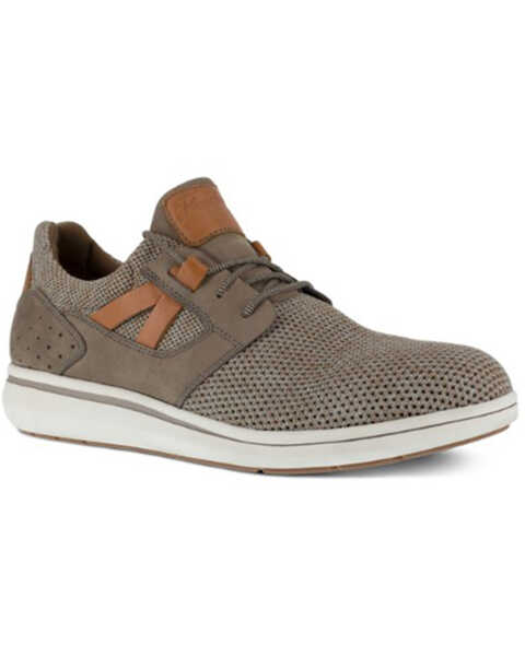
Shoes
Blood Specimen Collection
Fasciitis
Foot
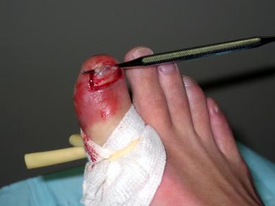
Foot Ulcer
Crying
Mechanical properties of heel pads reconstructed with flaps. (1/147)
We compared the mechanical properties of normal and reconstructed heel pads in seven patients. Four had latissimus dorsi flaps and one each an anterior thigh flap, a local dorsalis pedis flap and a sural arterial flap. The thickness of the heel pad was measured under serial incremental loads of 0.5 kg to a maximum of 3 kg and then relaxed sequentially. The load-displacement curve of the heel pad during a loading-unloading cycle was plotted and from this the unloaded heel-pad thickness (UHPT), compressibility index (CI), elastic modulus (Ep), and energy dissipation ratio (EDR) were calculated. The EDR was significantly increased in the reconstructed heels (53.7 +/- 18% v 23.4 +/- 6.5%, p = 0.003) indicating that in them more energy is dissipated as heat. Insufficient functional capacity in the reconstructed heel pad can lead to the development of shock-induced discomfort and ulceration. (+info)Ultrasound study of heel to calcaneum depth in neonates. (2/147)
AIM: To investigate whether it would be safe to extend the currently recommended area of sampling to the whole heel in neonates. METHODS: Eighty newborn infants were studied, weight range 0.56 to 4.34 kg, gestation 24 to 42 weeks. Ultrasound scanning was used to measure the shortest distance between the skin and the perichondrium of the calcaneum. RESULTS: The shortest depth of perichondrium was in the centre of the heel and ranged from 3 to 8 mm. In 78 of the 80 infants the distance was 4 mm or more. There was a small but significant positive correlation with weight. CONCLUSIONS: Standard automated lancets for preterm use that puncture to a depth of 2.4 mm may be safely used anywhere over the plantar surface of the heel. The posterior aspect of the heel should be avoided. Reducing the density of heel pricks should reduce the associated pain. (+info)Plantar fasciitis and other causes of heel pain. (3/147)
The most common cause of heel pain is plantar fasciitis. It is usually caused by a biomechanical imbalance resulting in tension along the plantar fascia. The diagnosis is typically based on the history and the finding of localized tenderness. Treatment consists of medial arch support, anti-inflammatory medications, ice massage and stretching. Corticosteroid injections and casting may also be tried. Surgical fasciotomy should be reserved for use in patients in whom conservative measures have failed despite correction of biomechanical abnormalities. Heel pain may also have a neurologic, traumatic or systemic origin. (+info)Clinical correlates of plantar pressure among diabetic veterans. (4/147)
OBJECTIVE: To assess the relationship between diabetes characteristics, medical history, foot deformity, sensory neuropathy, and plantar foot pressure. RESEARCH DESIGN AND METHODS: There were 517 subjects from a cohort of diabetic veterans enrolled in a prospective study of risk factors for foot complications who contributed 1,017 limbs for study. We interviewed subjects to collect data on demographics, diabetes characteristics, and medical history. A research nurse practitioner performed a directed physical exam of the lower extremities, assessing foot deformities and including quantitative sensory testing with a 5.07 monofilament. In-shoe foot-pressure measurements were obtained with F-scan insoles on subjects wearing their own footwear while walking 10 m at their usual pace. RESULTS: In univariate analyses, significant associations were seen between at least one measure of plantar pressure and body mass, sex, race, age, insulin use, certain foot deformities, plantar callus, and sensory neuropathy. Diabetes duration, HbA1c, and history of foot ulcer or amputation were unrelated to plantar pressure. In multiple regression analyses, body mass measured as log (weight), insulin use, white race, male sex, plantar callus, and diabetes duration were significantly related to certain pressures. Foot deformities were related primarily to forefoot pressures. With high pressure at two or more sites defined as the outcome, only body mass remained statistically significant as a predictor of this outcome in a backwards elimination logistic regression model. CONCLUSIONS: High in-shoe plantar pressure in diabetic subjects can be predicted in part from readily available clinical characteristics. The mechanisms by which these characteristics may be related to plantar pressure require further study. (+info)Usefulness of quantitative heel ultrasound compared with dual-energy X-ray absorptiometry in determining bone mineral density in chronic haemodialysis patients. (5/147)
BACKGROUND: Reduced bone mineral density (BMD) is associated with renal osteodystrophy and osteoporosis in end-stage renal failure patients. Dual-energy X-ray absorptiometry (DXA) is the standard non-invasive method to assess BMD, but is not always widely available. Quantitative heel ultrasound (QUS) is a mobile, relatively inexpensive, easy to perform and radiation-free method which can predict fractures to the same extent as DXA. This study assessed the usefulness of QUS vs DXA in determining BMD in chronic haemodialysis patients. METHODS: Patients had their BMD at the hip and spine measured by DXA (Lunar Expert). QUS of the left heel (McCue CubaClinical II machine) measured broadband ultrasound attenuation (BUA) and velocity of sound (VOS). Correlations between DXA and QUS parameters were calculated. Receiver operator characteristic (ROC) curves were plotted for BUA and VOS and used to define cut-off points for calculating sensitivities and specificities for BUA and VOS. Femoral neck BMD was applied as the standard for diagnosing osteoporosis (T< or =-2.5) and osteopaenia (T>-2.5 and < or =-1) by WHO criteria. RESULTS: Eighty eight patients (45.5% women), mean age 58+/-17 years, were studied. A total of 19% and 49% had femoral neck BMDs in the 'osteoporosis' and 'osteopaenia' ranges, respectively. There were good correlations between hip BMD and QUS parameters (r=0.68-0.79, P<0.001). Areas under the ROC curves for BUA and VOS in diagnosing 'osteoporosis' were 0.86 and 0.80, respectively. BUA and VOS had sensitivities of 76 and 71% and specificities of 80 and 69%, respectively, for diagnosing 'osteoporosis'. The positive predictive values for BUA and VOS were 48 and 35%, respectively, and the negative predictive values were 93 and 91% respectively. CONCLUSIONS: DXA and QUS parameters were significantly correlated. However, sensitivities and specificities of QUS parameters were not sufficiently high for QUS to be used simply as an alternative to DXA. The relatively high negative predictive values suggest that QUS may reliably screen out patients unlikely to have a BMD in the osteoporotic range. The relatively low positive predictive values, however, mean that subjects classified as osteoporotic using QUS require further investigations such as DXA to confirm the diagnosis. (+info)Disarticulation at the ankle using an anterior flap. A preliminary report. (6/147)
Disarticulation has been carried out in ten ankles in nine patients in whom it was not possible to use a heel flap. Four patients were able to walk with a prosthesis which gave satisfactory function. In five who were bedridden, healing was achieved and was of sufficient quality to allow transfers. There was no operative morbidity or mortality. This technique can be used instead of a transtibial amputation if necrosis or ischaemia of the heel is a contraindication to conventional Syme's amputation. (+info)Efficacy of dorsal pedal artery bypass in limb salvage for ischemic heel ulcers. (7/147)
PURPOSE: Although pedal artery bypass has been established as an effective and durable limb salvage procedure, the utility of these bypass grafts in limb salvage, specifically for the difficult problem of heel ulceration, remains undefined. METHODS: We retrospectively reviewed 432 pedal bypass grafts placed for indications of ischemic gangrene or ulceration isolated to either the forefoot (n = 336) or heel (n = 96). Lesion-healing rates and life-table analysis of survival, patency, and limb salvage were compared for forefoot versus heel lesions. Preoperative angiograms were reviewed to evaluate the influence of an intact pedal arch on heel lesion healing. RESULTS: Complete healing rates for forefoot and heel lesions were similar (90.5% vs 86.5%, P =.26), with comparable rates of major lower extremity amputation (9.8% vs 9.3%, P =.87). Time to complete healing in the heel lesion group ranged from 13 to 716 days, with a mean of 139 days. Preoperative angiography demonstrated an intact pedal arch in 48.8% of the patients with heel lesions. Healing and graft patency rates in these patients with heel lesions were independent of the presence of an intact arch, with healing rates of 90.2% and 83.7% (P =.38) and 2-year patency rates of 73.4% and 67.0% in complete and incomplete pedal arches, respectively. Comparison of 5-year primary and secondary patency rates between the forefoot and heel lesion groups were essentially identical, with primary rates of 56.9% versus 62.1% (P =.57) and secondary rates of 67.2% versus 60.3% (P =.50), respectively. CONCLUSION: Bypass grafts to the dorsalis pedis artery provide substantial perfusion to the posterior foot such that the resulting limb salvage and healing rates for revascularized heel lesions is excellent and comparable with those observed for ischemic forefoot pathology. (+info)A systematic review of treatments for the painful heel. (8/147)
OBJECTIVE: To establish the efficacy for treatments of pain on the plantar aspect of the heel. METHODS: Systematic review of the published and unpublished literature. Electronic search of Medline, BIDS and the Cochrane database of clinical trials. An assessment of the quality of the reporting was made of studies included in the review. MAIN OUTCOME MEASURE: patients' pain scores. STUDY SELECTION: randomized controlled trials, published or unpublished, that evaluated treatments used for plantar heel pain. Foreign language papers were excluded. RESULTS: Eleven randomized controlled trials were included in the review. These evaluated some of the most frequently described treatments (steroid injections and orthoses) and some experimental therapies (extracorporeal shock wave therapy and directed electrons). The methodological assessment scores of the published trials were low; small sample sizes and failure to conceal the treatment allocation from study participants prevents more definitive statements about the efficacy of treatments. In 10 of the included trials, patients in both the intervention and control arms reported improved pain scores at the final outcome measure. CONCLUSIONS: Although much has been written about the treatment of plantar heel pain, the few randomized controlled trials involve small populations of patients and do not provide robust scientific evidence of treatment efficacy. (+info)In medical terms, "heel" generally refers to the posterior and largest part of the foot, specifically the calcaneus bone. The heel is the first part of the foot to make contact with the ground during walking or running, and it plays a crucial role in supporting the body's weight and absorbing shock during movement.
The term "heel" can also be used to describe a structure or device that is attached to the back of a shoe or boot to provide additional height, support, or protection to the wearer's heel. These types of heels are often worn for fashion purposes or to compensate for differences in leg length.
A heel spur, also known as a calcaneal spur, is a bony growth or projection that develops on the underside of the heel bone (calcaneus). It typically occurs where the plantar fascia, a band of tissue that supports the arch of the foot, attaches to the heel bone.
Heel spurs are often caused by repetitive stress and strain on the foot, particularly in people who have plantar fasciitis, an inflammation of the plantar fascia. Over time, this tension can cause the body to lay down new bone tissue, leading to the formation of a spur.
Heel spurs themselves are not necessarily painful, but they can cause pain and discomfort if they rub against shoes or if they irritate surrounding tissues. Treatment for heel spurs typically involves addressing the underlying causes of the condition, such as plantar fasciitis, through measures such as rest, ice, stretching exercises, physical therapy, and orthotics. In some cases, surgery may be necessary to remove the spur.
Plantar fasciitis is a medical condition that involves inflammation of the plantar fascia, which is a thick band of tissue that runs along the bottom of your foot, connecting your heel bone to your toes. This tissue supports the arch of your foot and absorbs shock when you walk or run.
Plantar fasciitis is often caused by repetitive stress or overuse, leading to small tears and inflammation in the fascia. People who have high arches or flat feet, those who spend a lot of time on their feet, and athletes who engage in activities that put repeated stress on the heel and attached tissue, such as runners, are at a higher risk of developing plantar fasciitis.
Symptoms of plantar fasciitis include pain and stiffness in the heel or bottom of the foot, especially when taking the first few steps after getting out of bed or after prolonged periods of sitting or standing. The pain may worsen over time if left untreated, making it difficult to walk, climb stairs, or participate in physical activities.
Treatment for plantar fasciitis typically includes rest, ice, compression, and elevation (RICE) therapy, as well as physical therapy exercises to stretch and strengthen the foot and lower leg muscles. In some cases, medication, orthotics, or even surgery may be necessary to alleviate severe pain and inflammation.
Foot diseases refer to various medical conditions that affect the foot, including its structures such as the bones, joints, muscles, tendons, ligaments, blood vessels, and nerves. These conditions can cause symptoms like pain, swelling, numbness, difficulty walking, and skin changes. Examples of foot diseases include:
1. Plantar fasciitis: inflammation of the band of tissue that connects the heel bone to the toes.
2. Bunions: a bony bump that forms on the joint at the base of the big toe.
3. Hammertoe: a deformity in which the toe is bent at the middle joint, resembling a hammer.
4. Diabetic foot: a group of conditions that can occur in people with diabetes, including nerve damage, poor circulation, and increased risk of infection.
5. Athlete's foot: a fungal infection that affects the skin between the toes and on the soles of the feet.
6. Ingrown toenails: a condition where the corner or side of a toenail grows into the flesh of the toe.
7. Gout: a type of arthritis that causes sudden, severe attacks of pain, swelling, redness, and tenderness in the joints, often starting with the big toe.
8. Foot ulcers: open sores or wounds that can occur on the feet, especially in people with diabetes or poor circulation.
9. Morton's neuroma: a thickening of the tissue around a nerve between the toes, causing pain and numbness.
10. Osteoarthritis: wear and tear of the joints, leading to pain, stiffness, and reduced mobility.
Foot diseases can affect people of all ages and backgrounds, and some may be prevented or managed with proper foot care, hygiene, and appropriate medical treatment.
I'm sorry for any confusion, but "shoes" are not a medical term. Shoes are items of footwear intended to protect and comfort the feet. They typically have a durable sole that provides protection from sharp objects, hot surfaces, and the effects of moisture. The upper part of a shoe can be made from various materials such as leather, plastic, or textiles, and is designed to provide coverage and support for the foot.
If you have any questions related to medical terminology or health-related topics, I'd be happy to help!
Blood specimen collection is the process of obtaining a sample of blood from a patient for laboratory testing and analysis. This procedure is performed by trained healthcare professionals, such as nurses or phlebotomists, using sterile equipment to minimize the risk of infection and ensure accurate test results. The collected blood sample may be used to diagnose and monitor various medical conditions, assess overall health and organ function, and check for the presence of drugs, alcohol, or other substances. Proper handling, storage, and transportation of the specimen are crucial to maintain its integrity and prevent contamination.
Fasciitis is a medical condition characterized by inflammation or irritation of the fascia, which are the bands of connective tissue that surround muscles, tendons, and bones in the body. The most common type of fasciitis is plantar fasciitis, which affects the fascia on the bottom of the foot and can cause heel pain. Other types of fasciitis include:
* Achilles tendonitis or Achilles tendinopathy, which affects the fascia that connects the calf muscle to the heel bone
* Shin splints, which affect the fascia that covers the front of the lower leg
* Necrotizing fasciitis, a rare and serious bacterial infection that can cause extensive tissue damage and is potentially life-threatening.
The symptoms of fasciitis may include pain, stiffness, or tenderness in the affected area, especially after prolonged periods of rest or physical activity. Treatment for fasciitis typically involves rest, ice, compression, and elevation (RICE) of the affected area, as well as physical therapy exercises to stretch and strengthen the fascia and surrounding muscles. In some cases, medication or surgery may be necessary to relieve symptoms and promote healing.
In medical terms, the foot is the part of the lower limb that is distal to the leg and below the ankle, extending from the tarsus to the toes. It is primarily responsible for supporting body weight and facilitating movement through push-off during walking or running. The foot is a complex structure made up of 26 bones, 33 joints, and numerous muscles, tendons, ligaments, and nerves that work together to provide stability, balance, and flexibility. It can be divided into three main parts: the hindfoot, which contains the talus and calcaneus (heel) bones; the midfoot, which includes the navicular, cuboid, and cuneiform bones; and the forefoot, which consists of the metatarsals and phalanges that form the toes.
The forefoot is the front part of the human foot that contains the toes and the associated bones, muscles, ligaments, and tendons. It is made up of five long bones called metatarsals and fourteen phalanges, which are the bones in the toes. The forefoot plays a crucial role in weight-bearing, balance, and propulsion during walking and running. The joints in the forefoot allow for flexion, extension, abduction, and adduction of the toes, enabling us to maintain our footing on various surfaces and adapt to different terrain.
A foot ulcer is a wound or sore on the foot that occurs most commonly in people with diabetes, but can also affect other individuals with poor circulation or nerve damage. These ulcers can be challenging to heal and are prone to infection, making it essential for individuals with foot ulcers to seek medical attention promptly.
Foot ulcers typically develop due to prolonged pressure on bony prominences of the foot, leading to breakdown of the skin and underlying tissues. The development of foot ulcers can be attributed to several factors, including:
1. Neuropathy (nerve damage): This condition causes a loss of sensation in the feet, making it difficult for individuals to feel pain or discomfort associated with pressure points, leading to the formation of ulcers.
2. Peripheral artery disease (PAD): Reduced blood flow to the lower extremities can impair wound healing and make the body more susceptible to infection.
3. Deformities: Structural foot abnormalities, such as bunions or hammertoes, can cause increased pressure on specific areas of the foot, increasing the risk of ulcer formation.
4. Poorly fitting shoes: Shoes that are too tight, narrow, or ill-fitting can create friction and pressure points, contributing to the development of foot ulcers.
5. Trauma: Injuries or trauma to the feet can lead to the formation of ulcers, particularly in individuals with neuropathy who may not feel the initial pain associated with the injury.
6. Foot care neglect: Failure to inspect and care for the feet regularly can result in undetected wounds or sores that progress into ulcers.
Foot ulcers are classified based on their depth, severity, and extent of tissue involvement. Proper assessment, treatment, and prevention strategies are crucial in managing foot ulcers and minimizing the risk of complications such as infection, gangrene, and amputation.
Crying is not a medical term itself, but it can be a symptom or a response to various medical and emotional conditions. In a broader sense, crying refers to the production of tears and the audible sounds that accompany this action due to strong emotions such as sadness, happiness, frustration, or pain.
From a physiological standpoint, crying involves the activation of the autonomic nervous system, which leads to the production of tears by the lacrimal glands and the contraction of various facial muscles responsible for the expression of emotion. The parasympathetic branch of the autonomic nervous system is primarily responsible for the initiation of crying, leading to increased tear production and a decrease in heart rate.
There are several types of crying:
1. Emotional crying: This type of crying is a response to strong emotional states such as sadness, joy, frustration, or anger. It can be accompanied by sobbing, which involves deep, convulsive breaths and audible sounds.
2. Reflex crying: This occurs when the eyes are irritated due to foreign particles, bright lights, or other environmental factors. The reflex is designed to protect the eyes by producing tears to wash away the irritant.
3. Basal tearing: This type of tear production is continuous and helps keep the eyes lubricated and protected from drying out. It occurs at a low rate throughout the day and is not typically associated with crying as an emotional response.
In summary, while crying is not a medical term per se, it can be indicative of various emotional or physical states that may warrant medical attention. For instance, excessive or inappropriate crying might be a sign of underlying neurological or psychological conditions and should be evaluated by a healthcare professional if it becomes a concern.
Neonatal nursing is a specialized field of nursing that cares for newborn infants who are born prematurely or sick. These newborns often require advanced, intensive medical care and monitoring, which neonatal nurses are trained to provide. The neonatal period refers to the first 28 days of life. Neonatal nursing can be further categorized into three levels based on the degree of care provided:
1. Level I or Well Newborn Nursery: This level of care is provided to healthy newborns who do not require any special medical attention. The nurses in this unit provide routine care, such as feeding, bathing, and monitoring vital signs.
2. Level II or Special Care Nursery: This level of care is for infants born between 32 weeks and full-term (37-40 weeks) who require additional medical support, such as oxygen therapy, intravenous fluids, or phototherapy. Nurses in this unit provide more advanced care and monitoring than those in Level I.
3. Level III or Neonatal Intensive Care Unit (NICU): This level of care is for critically ill or premature newborns who require the highest level of medical intervention and technology, such as mechanical ventilation, continuous positive airway pressure (CPAP), or therapeutic hypothermia. Nurses in this unit are highly skilled and trained to provide complex care and support to these fragile infants and their families.
Neonatal nurses work closely with neonatologists, pediatricians, and other healthcare professionals to ensure the best possible outcomes for their patients. They also play a crucial role in providing emotional support and education to parents during this challenging time.
 Heel
Heel Block Heel - Women's - QVC.com
 Heel the pain - Healthy.net
 high heels
 Heels - IGN
 Kitten Heel - Macy's
 Heel pain: MedlinePlus Medical Encyclopedia
 The Daily Tar Heel
 Schutz block heel | Shopbop
 Bruised heel remedies and when to see a doctor
 Unilateral Concentric and Eccentric Heel Raises | APTA
 Designer Mid-Heel Boots | Mytheresa
 Wayward Wanderings Platform Heel | ModCloth
 Public Desire Kian heel sandals in beige | ASOS
 Coach rylie heel sandal + FREE SHIPPING | Zappos.com
 Uber's Achilles heel exposed by US court ruling
 We dare you, Posh! Romanian designer creates 12in heel | Daily Mail Online
 Best Holiday Heels | POPSUGAR Fashion
 Garita Block Heel Sandals - Nine West
 Heel Grounders Online Store | Future Electronics
 Bruised Heel Artist Profile | Broadjam.com
 Red Teela Mule Heel | Shoes | BCBGMAXAZRIA
 Targeting cancer's Achilles' heel | Science News
 Microjig - GRR-Ripper Gravity Heel Accessory
 Heel | Page 3 | Dog Star Daily
 Crampon Heel Lever - Regular | Black Diamond®