HeLa Cells
Molecular Sequence Data
Base Sequence
Cell Nucleus
Transfection
Poliovirus
Transcription, Genetic
Amino Acid Sequence
DNA-Binding Proteins
RNA, Messenger
Promoter Regions, Genetic
Nuclear Proteins
Adenoviruses, Human
Mitosis
DNA
Protein Binding
Plasmids
Transcription Factors
Cloning, Molecular
Recombinant Fusion Proteins
Apoptosis
Protein Biosynthesis
Cell Cycle
Cytoplasm
Gene Expression Regulation
Mutation
Phosphorylation
Electrophoresis, Polyacrylamide Gel
RNA, Small Interfering
Uridine
Microscopy, Fluorescence
RNA
Cell Survival
Cell Membrane
Cell Nucleolus
RNA Interference
Bacterial Adhesion
Fluorescent Antibody Technique
Rhinovirus
Carrier Proteins
RNA Splicing
Virus Replication
RNA, Heterogeneous Nuclear
Interphase
Green Fluorescent Proteins
Adenovirus Early Proteins
Centrifugation, Density Gradient
RNA Precursors
Cell Division
Vaccinia virus
Protein Transport
Blotting, Western
Dactinomycin
Luminescent Proteins
Ribonucleoproteins, Small Nuclear
Enterovirus B, Human
Heterogeneous-Nuclear Ribonucleoproteins
RNA-Binding Proteins
DNA, Complementary
Cell-Free System
Cytotoxins
Tritium
Genes
Cycloheximide
RNA Cap-Binding Proteins
Chloramphenicol O-Acetyltransferase
Shigella flexneri
Precipitin Tests
Membrane Proteins
Mitochondria
Biological Transport
RNA, Small Nuclear
Gene Expression
Tumor Cells, Cultured
Tissue Culture Techniques
The incorporation of 5-iodo-2'-deoxyuridine into the DNA of HeLa cells and the induction of alkaline phosphatase activity. (1/37241)
Inhibition of DNA synthesis during the period of exposure of HeLa cells to 5-iodo-2'-deoxyuridine (IUdR) inhibited the induction of alkaline phosphatase activity. This finding, taken together with previous findings that IUdR did not induce alkaline phosphatase activity in the presence of 2-fold molar excess thymidinemonstrated that IUdR incorporation into DNA is correlated with the increase in alkaline phosphatase activity. With the exception of an interim period described in the text, induction of alkaline phosphatase activity was linearly related to medium concentrations of IUdR of up to at least 3 muM. However, the extent of IUdR substitution in DNA did not appear to be related to the degree of enzyme induction. Alkaline phosphatase activity continued to increase at medium concentrations of IUdR from 1 to 3 muM, while little further substitution of DNA occurred. (+info)Stimulation of thymidine uptake and cell proliferation in mouse embryo fibroblasts by conditioned medium from mammary cells in culture. (2/37241)
Undialyzed conditioned medium from several cell culture sources did not stimulate thymidine incorporation or cell overgrowth in quiescent, density-inhibited mouse embryo fibroblast cells. However, dialyzed conditioned medium (DCM) from clonal mouse mammary cell lines MCG-V14, MCG-T14, MCG-T10; HeLa cells; primary mouse adenocarcinoma cells; and BALB/c normal mouse mammary epithelial cells promoted growth in quiescent fibroblasts. The amount of growth-promoting activity produced per cell varied from 24% (HeLa) to 213% (MCG-V14) of the activity produced by primary tumor cells. The production of growth-promoting activity was not unique to tumor-derived cells or cells of high tumorigenicity. The amount of growth-promoting activity produced per cell in the active cultures was not correlated with any of the following: tumorigenicity, growth rat, cell density achieved at saturation, cell type, or species of cell origin. It is concluded that transformed and non-transformed cells of diverse origin, cell type, and tumorigenicity can produce growth factors in culture. The growth-promoting potential of the active media from primary tumor cultures accumulated with time of contact with cells and was too great to be accounted for entirely by the removal of low-molecular-weight inhibitors by dialysis. The results are consistent with the hypothesis that conditioned medium from the active cultures contained a dialyzable, growth-promoting activity. Different cell lines exhibited differential sensitivity to tumor cell DCM and fetal bovine serum. Furthermore, quiescent fibroblasts were stimulated by primary tumor cell DCM in the presence of saturating concentrations of fetal bovine serum. These observations support the notion that the active growth-promoting principle in primary tumor cell DCM may not be a serum factor(s). (+info)Diphtheria toxin effects on human cells in tissue culture. (3/37241)
HeLa cells exposed to a single sublethal concentration of diphtheria toxin were found to have diminished sensitivity when subsequently reexposed to the toxin. Three cells strains exhibiting toxin resistance were developed. In the cells that had previously been exposed to toxin at 0.015 mug/ml, 50% inhibition of protein synthesis required a toxin concentration of 0.3 mug/ml, which is more than 10 times that required in normal HeLa cells. There appears to be a threshold level of diphtheria toxin action. Concentrations of toxin greater than that required for 50% inhibition of protein synthesis (0.01 mug/ml) are associated with cytotoxicity, whereas those below this concentration may not be lethal. Several established human cell lines of both normal and neoplastic origin were tested for their sensitivity to the effects of the toxin. No special sensitivity was observed with the cells of tumor origin. Fifty % inhibition of protein synthesis of HeLa cells was achieved with diphtheria toxin (0.01 mug/ml) as compared to the normal human cell lines tested (0.03 and 0.5 mug/ml) and a cell line derived from a human pancreatic adenocarcinoma (0.2 mug/ml). A human breast carcinoma cell line showed a maximum of 45% inhibition of protein synthesis. This required a diphtheria toxin concentration of 5 mug/ml. These results suggest that different human cell lines show wide variation in their sensitivity to the toxin. (+info)Evidence on the conformation of HeLa-cell 5.8S ribosomal ribonucleic acid from the reaction of specific cytidine residues with sodium bisulphite. (4/37241)
The reaction of HeLa-cell 5.8S rRNA with NaHSO3 under conditions in which exposed cytidine residues are deaminated to uridine was studied. It was possible to estimate the reactivities of most of the 46 cytidine residues in the nucleotide sequence by comparing 'fingerprints' of the bisulphite-treated RNA with those of untreated RNA. The findings were consistent with the main features of the secondary-structure model for mammalian 5.85S rRNA proposed by Nazar, Sitz, & Busch [J. Biol. Chem (1975) 250, 8591--8597]. Five out of six regions that are depicted in the model as single-stranded loops contain cytidine residues that are reactive towards bisulphite at 25 degrees C (the other loop contains no cytidine). The cytidine residue nearest to the 3'-terminus is also reactive. Several cytidines residues that are internally located within proposed double-helical regions show little or no reactivity towards bisulphite, but the cytidine residues of several C.G pairs at the ends of helical regions show some reactivity, and one of the proposed loops appears to contain six nucleotides, rather than the minimum of four suggested by the primary structure. Two cytidine residues that are thought to be 'looped out' by small helix imperfections also show some reactivity. (+info)Tyrosine phosphorylation is required for actin-based motility of vaccinia but not Listeria or Shigella. (5/37241)
Studies of the actin-based motility of pathogens have provided important insights into the events occurring at the leading edge of motile cells [1] [2] [3]. To date, several actin-cytoskeleton-associated proteins have been implicated in the motility of Listeria or Shigella: vasodilator-stimulated phosphoprotein (VASP), vinculin and the actin-related protein complex of Arp2 and Arp3 [4] [5] [6] [7]. To further investigate the underlying mechanism of actin-tail assembly, we examined the localization of components of the actin cytoskeleton including Arp3, VASP, vinculin and zyxin during vaccinia, Listeria and Shigella infections. The most striking difference between the systems was that a phosphotyrosine signal was observed only at the site of vaccinia actin-tail assembly. Micro-injection experiments demonstrated that a phosphotyrosine protein plays an important role in vaccinia actin-tail formation. In addition, we observed a phosphotyrosine signal on clathrin-coated vesicles that have associated actin-tail-like structures and on endogenous vesicles in Xenopus egg extracts which are able to nucleate actin tails [8] [9]. Our observations indicate that a host phosphotyrosine protein is required for the nucleation of actin filaments by vaccinia and suggest that this phosphoprotein might be associated with cellular membranes that can nucleate actin. (+info)Bcl-2 regulates amplification of caspase activation by cytochrome c. (6/37241)
Caspases, a family of specific proteases, have central roles in apoptosis [1]. Caspase activation in response to diverse apoptotic stimuli involves the relocalisation of cytochrome c from mitochondria to the cytoplasm where it stimulates the proteolytic processing of caspase precursors. Cytochrome c release is controlled by members of the Bcl-2 family of apoptosis regulators [2] [3]. The anti-apoptotic members Bcl-2 and Bcl-xL may also control caspase activation independently of cytochrome c relocalisation or may inhibit a positive feedback mechanism [4] [5] [6] [7]. Here, we investigate the role of Bcl-2 family proteins in the regulation of caspase activation using a model cell-free system. We found that Bcl-2 and Bcl-xL set a threshold in the amount of cytochrome c required to activate caspases, even in soluble extracts lacking mitochondria. Addition of dATP (which stimulates the procaspase-processing factor Apaf-1 [8] [9]) overcame inhibition of caspase activation by Bcl-2, but did not prevent the control of cytochrome c release from mitochondria by Bcl-2. Cytochrome c release was accelerated by active caspase-3 and this positive feedback was negatively regulated by Bcl-2. These results provide evidence for a mechanism to amplify caspase activation that is suppressed at several distinct steps by Bcl-2, even after cytochrome c is released from mitochondria. (+info)The splicing factor-associated protein, p32, regulates RNA splicing by inhibiting ASF/SF2 RNA binding and phosphorylation. (7/37241)
The cellular protein p32 was isolated originally as a protein tightly associated with the essential splicing factor ASF/SF2 during its purification from HeLa cells. ASF/SF2 is a member of the SR family of splicing factors, which stimulate constitutive splicing and regulate alternative RNA splicing in a positive or negative fashion, depending on where on the pre-mRNA they bind. Here we present evidence that p32 interacts with ASF/SF2 and SRp30c, another member of the SR protein family. We further show that p32 inhibits ASF/SF2 function as both a splicing enhancer and splicing repressor protein by preventing stable ASF/SF2 interaction with RNA, but p32 does not block SRp30c function. ASF/SF2 is highly phosphorylated in vivo, a modification required for stable RNA binding and protein-protein interaction during spliceosome formation, and this phosphorylation, either through HeLa nuclear extracts or through specific SR protein kinases, is inhibited by p32. Our results suggest that p32 functions as an ASF/SF2 inhibitory factor, regulating ASF/SF2 RNA binding and phosphorylation. These findings place p32 into a new group of proteins that control RNA splicing by sequestering an essential RNA splicing factor into an inhibitory complex. (+info)The amino-terminal C/H1 domain of CREB binding protein mediates zta transcriptional activation of latent Epstein-Barr virus. (8/37241)
Latent Epstein-Barr virus (EBV) is maintained as a nucleosome-covered episome that can be transcriptionally activated by overexpression of the viral immediate-early protein, Zta. We show here that reactivation of latent EBV by Zta can be significantly enhanced by coexpression of the cellular coactivators CREB binding protein (CBP) and p300. A stable complex containing both Zta and CBP could be isolated from lytically stimulated, but not latently infected RAJI nuclear extracts. Zta-mediated viral reactivation and transcriptional activation were both significantly inhibited by coexpression of the E1A 12S protein but not by an N-terminal deletion mutation of E1A (E1ADelta2-36), which fails to bind CBP. Zta bound directly to two related cysteine- and histidine-rich domains of CBP, referred to as C/H1 and C/H3. These domains both interacted specifically with the transcriptional activation domain of Zta in an electrophoretic mobility shift assay. Interestingly, we found that the C/H3 domain was a potent dominant negative inhibitor of Zta transcriptional activation function. In contrast, an amino-terminal fragment containing the C/H1 domain was sufficient for coactivation of Zta transcription and viral reactivation function. Thus, CBP can stimulate the transcription of latent EBV in a histone acetyltransferase-independent manner mediated by the CBP amino-terminal C/H1-containing domain. We propose that CBP may regulate aspects of EBV latency and reactivation by integrating cellular signals mediated by competitive interactions between C/H1, C/H3, and the Zta activation domain. (+info)HeLa cells are a type of immortalized cell line used in scientific research. They are derived from a cancer that developed in the cervical tissue of Henrietta Lacks, an African-American woman, in 1951. After her death, cells taken from her tumor were found to be capable of continuous division and growth in a laboratory setting, making them an invaluable resource for medical research.
HeLa cells have been used in a wide range of scientific studies, including research on cancer, viruses, genetics, and drug development. They were the first human cell line to be successfully cloned and are able to grow rapidly in culture, doubling their population every 20-24 hours. This has made them an essential tool for many areas of biomedical research.
It is important to note that while HeLa cells have been instrumental in numerous scientific breakthroughs, the story of their origin raises ethical questions about informed consent and the use of human tissue in research.
Molecular sequence data refers to the specific arrangement of molecules, most commonly nucleotides in DNA or RNA, or amino acids in proteins, that make up a biological macromolecule. This data is generated through laboratory techniques such as sequencing, and provides information about the exact order of the constituent molecules. This data is crucial in various fields of biology, including genetics, evolution, and molecular biology, allowing for comparisons between different organisms, identification of genetic variations, and studies of gene function and regulation.
A base sequence in the context of molecular biology refers to the specific order of nucleotides in a DNA or RNA molecule. In DNA, these nucleotides are adenine (A), guanine (G), cytosine (C), and thymine (T). In RNA, uracil (U) takes the place of thymine. The base sequence contains genetic information that is transcribed into RNA and ultimately translated into proteins. It is the exact order of these bases that determines the genetic code and thus the function of the DNA or RNA molecule.
The cell nucleus is a membrane-bound organelle found in the eukaryotic cells (cells with a true nucleus). It contains most of the cell's genetic material, organized as DNA molecules in complex with proteins, RNA molecules, and histones to form chromosomes.
The primary function of the cell nucleus is to regulate and control the activities of the cell, including growth, metabolism, protein synthesis, and reproduction. It also plays a crucial role in the process of mitosis (cell division) by separating and protecting the genetic material during this process. The nuclear membrane, or nuclear envelope, surrounding the nucleus is composed of two lipid bilayers with numerous pores that allow for the selective transport of molecules between the nucleoplasm (nucleus interior) and the cytoplasm (cell exterior).
The cell nucleus is a vital structure in eukaryotic cells, and its dysfunction can lead to various diseases, including cancer and genetic disorders.
Transfection is a term used in molecular biology that refers to the process of deliberately introducing foreign genetic material (DNA, RNA or artificial gene constructs) into cells. This is typically done using chemical or physical methods, such as lipofection or electroporation. Transfection is widely used in research and medical settings for various purposes, including studying gene function, producing proteins, developing gene therapies, and creating genetically modified organisms. It's important to note that transfection is different from transduction, which is the process of introducing genetic material into cells using viruses as vectors.
Poliovirus is a human enterovirus, specifically a type of picornavirus, that is the causative agent of poliomyelitis (polio). It is a small, non-enveloped, single-stranded, positive-sense RNA virus. There are three serotypes of Poliovirus (types 1, 2 and 3) which can cause different degrees of severity in the disease. The virus primarily spreads through the fecal-oral route and infects the gastrointestinal tract, from where it can invade the nervous system and cause paralysis.
The Poliovirus has an icosahedral symmetry, with a diameter of about 30 nanometers. It contains a single stranded RNA genome which is encapsidated in a protein shell called capsid. The capsid is made up of 60 units of four different proteins (VP1, VP2, VP3 and VP4).
Poliovirus has been eradicated from most countries of the world through widespread vaccination with inactivated poliovirus vaccine (IPV) or oral poliovirus vaccine (OPV). However, it still remains endemic in a few countries and is considered a major public health concern.
Genetic transcription is the process by which the information in a strand of DNA is used to create a complementary RNA molecule. This process is the first step in gene expression, where the genetic code in DNA is converted into a form that can be used to produce proteins or functional RNAs.
During transcription, an enzyme called RNA polymerase binds to the DNA template strand and reads the sequence of nucleotide bases. As it moves along the template, it adds complementary RNA nucleotides to the growing RNA chain, creating a single-stranded RNA molecule that is complementary to the DNA template strand. Once transcription is complete, the RNA molecule may undergo further processing before it can be translated into protein or perform its functional role in the cell.
Transcription can be either "constitutive" or "regulated." Constitutive transcription occurs at a relatively constant rate and produces essential proteins that are required for basic cellular functions. Regulated transcription, on the other hand, is subject to control by various intracellular and extracellular signals, allowing cells to respond to changing environmental conditions or developmental cues.
A cell line is a culture of cells that are grown in a laboratory for use in research. These cells are usually taken from a single cell or group of cells, and they are able to divide and grow continuously in the lab. Cell lines can come from many different sources, including animals, plants, and humans. They are often used in scientific research to study cellular processes, disease mechanisms, and to test new drugs or treatments. Some common types of human cell lines include HeLa cells (which come from a cancer patient named Henrietta Lacks), HEK293 cells (which come from embryonic kidney cells), and HUVEC cells (which come from umbilical vein endothelial cells). It is important to note that cell lines are not the same as primary cells, which are cells that are taken directly from a living organism and have not been grown in the lab.
An amino acid sequence is the specific order of amino acids in a protein or peptide molecule, formed by the linking of the amino group (-NH2) of one amino acid to the carboxyl group (-COOH) of another amino acid through a peptide bond. The sequence is determined by the genetic code and is unique to each type of protein or peptide. It plays a crucial role in determining the three-dimensional structure and function of proteins.
DNA-binding proteins are a type of protein that have the ability to bind to DNA (deoxyribonucleic acid), the genetic material of organisms. These proteins play crucial roles in various biological processes, such as regulation of gene expression, DNA replication, repair and recombination.
The binding of DNA-binding proteins to specific DNA sequences is mediated by non-covalent interactions, including electrostatic, hydrogen bonding, and van der Waals forces. The specificity of binding is determined by the recognition of particular nucleotide sequences or structural features of the DNA molecule.
DNA-binding proteins can be classified into several categories based on their structure and function, such as transcription factors, histones, and restriction enzymes. Transcription factors are a major class of DNA-binding proteins that regulate gene expression by binding to specific DNA sequences in the promoter region of genes and recruiting other proteins to modulate transcription. Histones are DNA-binding proteins that package DNA into nucleosomes, the basic unit of chromatin structure. Restriction enzymes are DNA-binding proteins that recognize and cleave specific DNA sequences, and are widely used in molecular biology research and biotechnology applications.
Messenger RNA (mRNA) is a type of RNA (ribonucleic acid) that carries genetic information copied from DNA in the form of a series of three-base code "words," each of which specifies a particular amino acid. This information is used by the cell's machinery to construct proteins, a process known as translation. After being transcribed from DNA, mRNA travels out of the nucleus to the ribosomes in the cytoplasm where protein synthesis occurs. Once the protein has been synthesized, the mRNA may be degraded and recycled. Post-transcriptional modifications can also occur to mRNA, such as alternative splicing and addition of a 5' cap and a poly(A) tail, which can affect its stability, localization, and translation efficiency.
Promoter regions in genetics refer to specific DNA sequences located near the transcription start site of a gene. They serve as binding sites for RNA polymerase and various transcription factors that regulate the initiation of gene transcription. These regulatory elements help control the rate of transcription and, therefore, the level of gene expression. Promoter regions can be composed of different types of sequences, such as the TATA box and CAAT box, and their organization and composition can vary between different genes and species.
Nuclear proteins are a category of proteins that are primarily found in the nucleus of a eukaryotic cell. They play crucial roles in various nuclear functions, such as DNA replication, transcription, repair, and RNA processing. This group includes structural proteins like lamins, which form the nuclear lamina, and regulatory proteins, such as histones and transcription factors, that are involved in gene expression. Nuclear localization signals (NLS) often help target these proteins to the nucleus by interacting with importin proteins during active transport across the nuclear membrane.
Adenoviruses, Human: A group of viruses that commonly cause respiratory illnesses, such as bronchitis, pneumonia, and croup, in humans. They can also cause conjunctivitis (pink eye), cystitis (bladder infection), and gastroenteritis (stomach and intestinal infection).
Human adenoviruses are non-enveloped, double-stranded DNA viruses that belong to the family Adenoviridae. There are more than 50 different types of human adenoviruses, which can be classified into seven species (A-G). Different types of adenoviruses tend to cause specific illnesses, such as respiratory or gastrointestinal infections.
Human adenoviruses are highly contagious and can spread through close personal contact, respiratory droplets, or contaminated surfaces. They can also be transmitted through contaminated water sources. Some people may become carriers of the virus and experience no symptoms but still spread the virus to others.
Most human adenovirus infections are mild and resolve on their own within a few days to a week. However, some types of adenoviruses can cause severe illness, particularly in people with weakened immune systems, such as infants, young children, older adults, and individuals with HIV/AIDS or organ transplants.
There are no specific antiviral treatments for human adenovirus infections, but supportive care, such as hydration, rest, and fever reduction, can help manage symptoms. Preventive measures include practicing good hygiene, such as washing hands frequently, avoiding close contact with sick individuals, and not sharing personal items like towels or utensils.
Mitosis is a type of cell division in which the genetic material of a single cell, called the mother cell, is equally distributed into two identical daughter cells. It's a fundamental process that occurs in multicellular organisms for growth, maintenance, and repair, as well as in unicellular organisms for reproduction.
The process of mitosis can be broken down into several stages: prophase, prometaphase, metaphase, anaphase, and telophase. During prophase, the chromosomes condense and become visible, and the nuclear envelope breaks down. In prometaphase, the nuclear membrane is completely disassembled, and the mitotic spindle fibers attach to the chromosomes at their centromeres.
During metaphase, the chromosomes align at the metaphase plate, an imaginary line equidistant from the two spindle poles. In anaphase, sister chromatids are pulled apart by the spindle fibers and move toward opposite poles of the cell. Finally, in telophase, new nuclear envelopes form around each set of chromosomes, and the chromosomes decondense and become less visible.
Mitosis is followed by cytokinesis, a process that divides the cytoplasm of the mother cell into two separate daughter cells. The result of mitosis and cytokinesis is two genetically identical cells, each with the same number and kind of chromosomes as the original parent cell.
Deoxyribonucleic acid (DNA) is the genetic material present in the cells of organisms where it is responsible for the storage and transmission of hereditary information. DNA is a long molecule that consists of two strands coiled together to form a double helix. Each strand is made up of a series of four nucleotide bases - adenine (A), guanine (G), cytosine (C), and thymine (T) - that are linked together by phosphate and sugar groups. The sequence of these bases along the length of the molecule encodes genetic information, with A always pairing with T and C always pairing with G. This base-pairing allows for the replication and transcription of DNA, which are essential processes in the functioning and reproduction of all living organisms.
In the context of medicine and pharmacology, "kinetics" refers to the study of how a drug moves throughout the body, including its absorption, distribution, metabolism, and excretion (often abbreviated as ADME). This field is called "pharmacokinetics."
1. Absorption: This is the process of a drug moving from its site of administration into the bloodstream. Factors such as the route of administration (e.g., oral, intravenous, etc.), formulation, and individual physiological differences can affect absorption.
2. Distribution: Once a drug is in the bloodstream, it gets distributed throughout the body to various tissues and organs. This process is influenced by factors like blood flow, protein binding, and lipid solubility of the drug.
3. Metabolism: Drugs are often chemically modified in the body, typically in the liver, through processes known as metabolism. These changes can lead to the formation of active or inactive metabolites, which may then be further distributed, excreted, or undergo additional metabolic transformations.
4. Excretion: This is the process by which drugs and their metabolites are eliminated from the body, primarily through the kidneys (urine) and the liver (bile).
Understanding the kinetics of a drug is crucial for determining its optimal dosing regimen, potential interactions with other medications or foods, and any necessary adjustments for special populations like pediatric or geriatric patients, or those with impaired renal or hepatic function.
Protein binding, in the context of medical and biological sciences, refers to the interaction between a protein and another molecule (known as the ligand) that results in a stable complex. This process is often reversible and can be influenced by various factors such as pH, temperature, and concentration of the involved molecules.
In clinical chemistry, protein binding is particularly important when it comes to drugs, as many of them bind to proteins (especially albumin) in the bloodstream. The degree of protein binding can affect a drug's distribution, metabolism, and excretion, which in turn influence its therapeutic effectiveness and potential side effects.
Protein-bound drugs may be less available for interaction with their target tissues, as only the unbound or "free" fraction of the drug is active. Therefore, understanding protein binding can help optimize dosing regimens and minimize adverse reactions.
A plasmid is a small, circular, double-stranded DNA molecule that is separate from the chromosomal DNA of a bacterium or other organism. Plasmids are typically not essential for the survival of the organism, but they can confer beneficial traits such as antibiotic resistance or the ability to degrade certain types of pollutants.
Plasmids are capable of replicating independently of the chromosomal DNA and can be transferred between bacteria through a process called conjugation. They often contain genes that provide resistance to antibiotics, heavy metals, and other environmental stressors. Plasmids have also been engineered for use in molecular biology as cloning vectors, allowing scientists to replicate and manipulate specific DNA sequences.
Plasmids are important tools in genetic engineering and biotechnology because they can be easily manipulated and transferred between organisms. They have been used to produce vaccines, diagnostic tests, and genetically modified organisms (GMOs) for various applications, including agriculture, medicine, and industry.
Transcription factors are proteins that play a crucial role in regulating gene expression by controlling the transcription of DNA to messenger RNA (mRNA). They function by binding to specific DNA sequences, known as response elements, located in the promoter region or enhancer regions of target genes. This binding can either activate or repress the initiation of transcription, depending on the properties and interactions of the particular transcription factor. Transcription factors often act as part of a complex network of regulatory proteins that determine the precise spatiotemporal patterns of gene expression during development, differentiation, and homeostasis in an organism.
Molecular cloning is a laboratory technique used to create multiple copies of a specific DNA sequence. This process involves several steps:
1. Isolation: The first step in molecular cloning is to isolate the DNA sequence of interest from the rest of the genomic DNA. This can be done using various methods such as PCR (polymerase chain reaction), restriction enzymes, or hybridization.
2. Vector construction: Once the DNA sequence of interest has been isolated, it must be inserted into a vector, which is a small circular DNA molecule that can replicate independently in a host cell. Common vectors used in molecular cloning include plasmids and phages.
3. Transformation: The constructed vector is then introduced into a host cell, usually a bacterial or yeast cell, through a process called transformation. This can be done using various methods such as electroporation or chemical transformation.
4. Selection: After transformation, the host cells are grown in selective media that allow only those cells containing the vector to grow. This ensures that the DNA sequence of interest has been successfully cloned into the vector.
5. Amplification: Once the host cells have been selected, they can be grown in large quantities to amplify the number of copies of the cloned DNA sequence.
Molecular cloning is a powerful tool in molecular biology and has numerous applications, including the production of recombinant proteins, gene therapy, functional analysis of genes, and genetic engineering.
Molecular weight, also known as molecular mass, is the mass of a molecule. It is expressed in units of atomic mass units (amu) or daltons (Da). Molecular weight is calculated by adding up the atomic weights of each atom in a molecule. It is a useful property in chemistry and biology, as it can be used to determine the concentration of a substance in a solution, or to calculate the amount of a substance that will react with another in a chemical reaction.
Ribonucleoproteins (RNPs) are complexes composed of ribonucleic acid (RNA) and proteins. They play crucial roles in various cellular processes, including gene expression, RNA processing, transport, stability, and degradation. Different types of RNPs exist, such as ribosomes, spliceosomes, and signal recognition particles, each having specific functions in the cell.
Ribosomes are large RNP complexes responsible for protein synthesis, where messenger RNA (mRNA) is translated into proteins. They consist of two subunits: a smaller subunit containing ribosomal RNA (rRNA) and proteins that recognize the start codon on mRNA, and a larger subunit with rRNA and proteins that facilitate peptide bond formation during translation.
Spliceosomes are dynamic RNP complexes involved in pre-messenger RNA (pre-mRNA) splicing, where introns (non-coding sequences) are removed, and exons (coding sequences) are joined together to form mature mRNA. Spliceosomes consist of five small nuclear ribonucleoproteins (snRNPs), each containing a specific small nuclear RNA (snRNA) and several proteins, as well as numerous additional proteins.
Other RNP complexes include signal recognition particles (SRPs), which are responsible for targeting secretory and membrane proteins to the endoplasmic reticulum during translation, and telomerase, an enzyme that maintains the length of telomeres (the protective ends of chromosomes) by adding repetitive DNA sequences using its built-in RNA component.
In summary, ribonucleoproteins are essential complexes in the cell that participate in various aspects of RNA metabolism and protein synthesis.
Recombinant fusion proteins are artificially created biomolecules that combine the functional domains or properties of two or more different proteins into a single protein entity. They are generated through recombinant DNA technology, where the genes encoding the desired protein domains are linked together and expressed as a single, chimeric gene in a host organism, such as bacteria, yeast, or mammalian cells.
The resulting fusion protein retains the functional properties of its individual constituent proteins, allowing for novel applications in research, diagnostics, and therapeutics. For instance, recombinant fusion proteins can be designed to enhance protein stability, solubility, or immunogenicity, making them valuable tools for studying protein-protein interactions, developing targeted therapies, or generating vaccines against infectious diseases or cancer.
Examples of recombinant fusion proteins include:
1. Etaglunatide (ABT-523): A soluble Fc fusion protein that combines the heavy chain fragment crystallizable region (Fc) of an immunoglobulin with the extracellular domain of the human interleukin-6 receptor (IL-6R). This fusion protein functions as a decoy receptor, neutralizing IL-6 and its downstream signaling pathways in rheumatoid arthritis.
2. Etanercept (Enbrel): A soluble TNF receptor p75 Fc fusion protein that binds to tumor necrosis factor-alpha (TNF-α) and inhibits its proinflammatory activity, making it a valuable therapeutic option for treating autoimmune diseases like rheumatoid arthritis, ankylosing spondylitis, and psoriasis.
3. Abatacept (Orencia): A fusion protein consisting of the extracellular domain of cytotoxic T-lymphocyte antigen 4 (CTLA-4) linked to the Fc region of an immunoglobulin, which downregulates T-cell activation and proliferation in autoimmune diseases like rheumatoid arthritis.
4. Belimumab (Benlysta): A monoclonal antibody that targets B-lymphocyte stimulator (BLyS) protein, preventing its interaction with the B-cell surface receptor and inhibiting B-cell activation in systemic lupus erythematosus (SLE).
5. Romiplostim (Nplate): A fusion protein consisting of a thrombopoietin receptor agonist peptide linked to an immunoglobulin Fc region, which stimulates platelet production in patients with chronic immune thrombocytopenia (ITP).
6. Darbepoetin alfa (Aranesp): A hyperglycosylated erythropoiesis-stimulating protein that functions as a longer-acting form of recombinant human erythropoietin, used to treat anemia in patients with chronic kidney disease or cancer.
7. Palivizumab (Synagis): A monoclonal antibody directed against the F protein of respiratory syncytial virus (RSV), which prevents RSV infection and is administered prophylactically to high-risk infants during the RSV season.
8. Ranibizumab (Lucentis): A recombinant humanized monoclonal antibody fragment that binds and inhibits vascular endothelial growth factor A (VEGF-A), used in the treatment of age-related macular degeneration, diabetic retinopathy, and other ocular disorders.
9. Cetuximab (Erbitux): A chimeric monoclonal antibody that binds to epidermal growth factor receptor (EGFR), used in the treatment of colorectal cancer and head and neck squamous cell carcinoma.
10. Adalimumab (Humira): A fully humanized monoclonal antibody that targets tumor necrosis factor-alpha (TNF-α), used in the treatment of various inflammatory diseases, including rheumatoid arthritis, psoriasis, and Crohn's disease.
11. Bevacizumab (Avastin): A recombinant humanized monoclonal antibody that binds to VEGF-A, used in the treatment of various cancers, including colorectal, lung, breast, and kidney cancer.
12. Trastuzumab (Herceptin): A humanized monoclonal antibody that targets HER2/neu receptor, used in the treatment of breast cancer.
13. Rituximab (Rituxan): A chimeric monoclonal antibody that binds to CD20 antigen on B cells, used in the treatment of non-Hodgkin's lymphoma and rheumatoid arthritis.
14. Palivizumab (Synagis): A humanized monoclonal antibody that binds to the F protein of respiratory syncytial virus, used in the prevention of respiratory syncytial virus infection in high-risk infants.
15. Infliximab (Remicade): A chimeric monoclonal antibody that targets TNF-α, used in the treatment of various inflammatory diseases, including Crohn's disease, ulcerative colitis, rheumatoid arthritis, and ankylosing spondylitis.
16. Natalizumab (Tysabri): A humanized monoclonal antibody that binds to α4β1 integrin, used in the treatment of multiple sclerosis and Crohn's disease.
17. Adalimumab (Humira): A fully human monoclonal antibody that targets TNF-α, used in the treatment of various inflammatory diseases, including rheumatoid arthritis, psoriatic arthritis, ankylosing spondylitis, Crohn's disease, and ulcerative colitis.
18. Golimumab (Simponi): A fully human monoclonal antibody that targets TNF-α, used in the treatment of rheumatoid arthritis, psoriatic arthritis, ankylosing spondylitis, and ulcerative colitis.
19. Certolizumab pegol (Cimzia): A PEGylated Fab' fragment of a humanized monoclonal antibody that targets TNF-α, used in the treatment of rheumatoid arthritis, psoriatic arthritis, ankylosing spondylitis, and Crohn's disease.
20. Ustekinumab (Stelara): A fully human monoclonal antibody that targets IL-12 and IL-23, used in the treatment of psoriasis, psoriatic arthritis, and Crohn's disease.
21. Secukinumab (Cosentyx): A fully human monoclonal antibody that targets IL-17A, used in the treatment of psoriasis, psoriatic arthritis, and ankylosing spondylitis.
22. Ixekizumab (Taltz): A fully human monoclonal antibody that targets IL-17A, used in the treatment of psoriasis and psoriatic arthritis.
23. Brodalumab (Siliq): A fully human monoclonal antibody that targets IL-17 receptor A, used in the treatment of psoriasis.
24. Sarilumab (Kevzara): A fully human monoclonal antibody that targets the IL-6 receptor, used in the treatment of rheumatoid arthritis.
25. Tocilizumab (Actemra): A humanized monoclonal antibody that targets the IL-6 receptor, used in the treatment of rheumatoid arthritis, systemic juvenile idiopathic arthritis, polyarticular juvenile idiopathic arthritis, giant cell arteritis, and chimeric antigen receptor T-cell-induced cytokine release syndrome.
26. Siltuximab (Sylvant): A chimeric monoclonal antibody that targets IL-6, used in the treatment of multicentric Castleman disease.
27. Satralizumab (Enspryng): A humanized monoclonal antibody that targets IL-6 receptor alpha, used in the treatment of neuromyelitis optica spectrum disorder.
28. Sirukumab (Plivensia): A human monoclonal antibody that targets IL-6, used in the treatment
Apoptosis is a programmed and controlled cell death process that occurs in multicellular organisms. It is a natural process that helps maintain tissue homeostasis by eliminating damaged, infected, or unwanted cells. During apoptosis, the cell undergoes a series of morphological changes, including cell shrinkage, chromatin condensation, and fragmentation into membrane-bound vesicles called apoptotic bodies. These bodies are then recognized and engulfed by neighboring cells or phagocytic cells, preventing an inflammatory response. Apoptosis is regulated by a complex network of intracellular signaling pathways that involve proteins such as caspases, Bcl-2 family members, and inhibitors of apoptosis (IAPs).
Protein biosynthesis is the process by which cells generate new proteins. It involves two major steps: transcription and translation. Transcription is the process of creating a complementary RNA copy of a sequence of DNA. This RNA copy, or messenger RNA (mRNA), carries the genetic information to the site of protein synthesis, the ribosome. During translation, the mRNA is read by transfer RNA (tRNA) molecules, which bring specific amino acids to the ribosome based on the sequence of nucleotides in the mRNA. The ribosome then links these amino acids together in the correct order to form a polypeptide chain, which may then fold into a functional protein. Protein biosynthesis is essential for the growth and maintenance of all living organisms.
The cell cycle is a series of events that take place in a cell leading to its division and duplication. It consists of four main phases: G1 phase, S phase, G2 phase, and M phase.
During the G1 phase, the cell grows in size and synthesizes mRNA and proteins in preparation for DNA replication. In the S phase, the cell's DNA is copied, resulting in two complete sets of chromosomes. During the G2 phase, the cell continues to grow and produces more proteins and organelles necessary for cell division.
The M phase is the final stage of the cell cycle and consists of mitosis (nuclear division) and cytokinesis (cytoplasmic division). Mitosis results in two genetically identical daughter nuclei, while cytokinesis divides the cytoplasm and creates two separate daughter cells.
The cell cycle is regulated by various checkpoints that ensure the proper completion of each phase before progressing to the next. These checkpoints help prevent errors in DNA replication and division, which can lead to mutations and cancer.
Cell extracts refer to the mixture of cellular components that result from disrupting or breaking open cells. The process of obtaining cell extracts is called cell lysis. Cell extracts can contain various types of molecules, such as proteins, nucleic acids (DNA and RNA), carbohydrates, lipids, and metabolites, depending on the methods used for cell disruption and extraction.
Cell extracts are widely used in biochemical and molecular biology research to study various cellular processes and pathways. For example, cell extracts can be used to measure enzyme activities, analyze protein-protein interactions, characterize gene expression patterns, and investigate metabolic pathways. In some cases, specific cellular components can be purified from the cell extracts for further analysis or application, such as isolating pure proteins or nucleic acids.
It is important to note that the composition of cell extracts may vary depending on the type of cells, the growth conditions, and the methods used for cell disruption and extraction. Therefore, it is essential to optimize the experimental conditions to obtain representative and meaningful results from cell extract studies.
Cytoplasm is the material within a eukaryotic cell (a cell with a true nucleus) that lies between the nuclear membrane and the cell membrane. It is composed of an aqueous solution called cytosol, in which various organelles such as mitochondria, ribosomes, endoplasmic reticulum, Golgi apparatus, lysosomes, and vacuoles are suspended. Cytoplasm also contains a variety of dissolved nutrients, metabolites, ions, and enzymes that are involved in various cellular processes such as metabolism, signaling, and transport. It is where most of the cell's metabolic activities take place, and it plays a crucial role in maintaining the structure and function of the cell.
'Gene expression regulation' refers to the processes that control whether, when, and where a particular gene is expressed, meaning the production of a specific protein or functional RNA encoded by that gene. This complex mechanism can be influenced by various factors such as transcription factors, chromatin remodeling, DNA methylation, non-coding RNAs, and post-transcriptional modifications, among others. Proper regulation of gene expression is crucial for normal cellular function, development, and maintaining homeostasis in living organisms. Dysregulation of gene expression can lead to various diseases, including cancer and genetic disorders.
Viral proteins are the proteins that are encoded by the viral genome and are essential for the viral life cycle. These proteins can be structural or non-structural and play various roles in the virus's replication, infection, and assembly process. Structural proteins make up the physical structure of the virus, including the capsid (the protein shell that surrounds the viral genome) and any envelope proteins (that may be present on enveloped viruses). Non-structural proteins are involved in the replication of the viral genome and modulation of the host cell environment to favor viral replication. Overall, a thorough understanding of viral proteins is crucial for developing antiviral therapies and vaccines.
A mutation is a permanent change in the DNA sequence of an organism's genome. Mutations can occur spontaneously or be caused by environmental factors such as exposure to radiation, chemicals, or viruses. They may have various effects on the organism, ranging from benign to harmful, depending on where they occur and whether they alter the function of essential proteins. In some cases, mutations can increase an individual's susceptibility to certain diseases or disorders, while in others, they may confer a survival advantage. Mutations are the driving force behind evolution, as they introduce new genetic variability into populations, which can then be acted upon by natural selection.
DNA replication is the biological process by which DNA makes an identical copy of itself during cell division. It is a fundamental mechanism that allows genetic information to be passed down from one generation of cells to the next. During DNA replication, each strand of the double helix serves as a template for the synthesis of a new complementary strand. This results in the creation of two identical DNA molecules. The enzymes responsible for DNA replication include helicase, which unwinds the double helix, and polymerase, which adds nucleotides to the growing strands.
Phosphorylation is the process of adding a phosphate group (a molecule consisting of one phosphorus atom and four oxygen atoms) to a protein or other organic molecule, which is usually done by enzymes called kinases. This post-translational modification can change the function, localization, or activity of the target molecule, playing a crucial role in various cellular processes such as signal transduction, metabolism, and regulation of gene expression. Phosphorylation is reversible, and the removal of the phosphate group is facilitated by enzymes called phosphatases.
Electrophoresis, polyacrylamide gel (EPG) is a laboratory technique used to separate and analyze complex mixtures of proteins or nucleic acids (DNA or RNA) based on their size and electrical charge. This technique utilizes a matrix made of cross-linked polyacrylamide, a type of gel, which provides a stable and uniform environment for the separation of molecules.
In this process:
1. The polyacrylamide gel is prepared by mixing acrylamide monomers with a cross-linking agent (bis-acrylamide) and a catalyst (ammonium persulfate) in the presence of a buffer solution.
2. The gel is then poured into a mold and allowed to polymerize, forming a solid matrix with uniform pore sizes that depend on the concentration of acrylamide used. Higher concentrations result in smaller pores, providing better resolution for separating smaller molecules.
3. Once the gel has set, it is placed in an electrophoresis apparatus containing a buffer solution. Samples containing the mixture of proteins or nucleic acids are loaded into wells on the top of the gel.
4. An electric field is applied across the gel, causing the negatively charged molecules to migrate towards the positive electrode (anode) while positively charged molecules move toward the negative electrode (cathode). The rate of migration depends on the size, charge, and shape of the molecules.
5. Smaller molecules move faster through the gel matrix and will migrate farther from the origin compared to larger molecules, resulting in separation based on size. Proteins and nucleic acids can be selectively stained after electrophoresis to visualize the separated bands.
EPG is widely used in various research fields, including molecular biology, genetics, proteomics, and forensic science, for applications such as protein characterization, DNA fragment analysis, cloning, mutation detection, and quality control of nucleic acid or protein samples.
Small interfering RNA (siRNA) is a type of short, double-stranded RNA molecule that plays a role in the RNA interference (RNAi) pathway. The RNAi pathway is a natural cellular process that regulates gene expression by targeting and destroying specific messenger RNA (mRNA) molecules, thereby preventing the translation of those mRNAs into proteins.
SiRNAs are typically 20-25 base pairs in length and are generated from longer double-stranded RNA precursors called hairpin RNAs or dsRNAs by an enzyme called Dicer. Once generated, siRNAs associate with a protein complex called the RNA-induced silencing complex (RISC), which uses one strand of the siRNA (the guide strand) to recognize and bind to complementary sequences in the target mRNA. The RISC then cleaves the target mRNA, leading to its degradation and the inhibition of protein synthesis.
SiRNAs have emerged as a powerful tool for studying gene function and have shown promise as therapeutic agents for a variety of diseases, including viral infections, cancer, and genetic disorders. However, their use as therapeutics is still in the early stages of development, and there are challenges associated with delivering siRNAs to specific cells and tissues in the body.
Recombinant proteins are artificially created proteins produced through the use of recombinant DNA technology. This process involves combining DNA molecules from different sources to create a new set of genes that encode for a specific protein. The resulting recombinant protein can then be expressed, purified, and used for various applications in research, medicine, and industry.
Recombinant proteins are widely used in biomedical research to study protein function, structure, and interactions. They are also used in the development of diagnostic tests, vaccines, and therapeutic drugs. For example, recombinant insulin is a common treatment for diabetes, while recombinant human growth hormone is used to treat growth disorders.
The production of recombinant proteins typically involves the use of host cells, such as bacteria, yeast, or mammalian cells, which are engineered to express the desired protein. The host cells are transformed with a plasmid vector containing the gene of interest, along with regulatory elements that control its expression. Once the host cells are cultured and the protein is expressed, it can be purified using various chromatography techniques.
Overall, recombinant proteins have revolutionized many areas of biology and medicine, enabling researchers to study and manipulate proteins in ways that were previously impossible.
Uridine is a nucleoside that consists of a pyrimidine base (uracil) linked to a pentose sugar (ribose). It is a component of RNA, where it pairs with adenine. Uridine can also be found in various foods such as beer, broccoli, yeast, and meat. In the body, uridine can be synthesized from orotate or from the breakdown of RNA. It has several functions, including acting as a building block for RNA, contributing to energy metabolism, and regulating cell growth and differentiation. Uridine is also available as a dietary supplement and has been studied for its potential benefits in various health conditions.
Fluorescence microscopy is a type of microscopy that uses fluorescent dyes or proteins to highlight and visualize specific components within a sample. In this technique, the sample is illuminated with high-energy light, typically ultraviolet (UV) or blue light, which excites the fluorescent molecules causing them to emit lower-energy, longer-wavelength light, usually visible light in the form of various colors. This emitted light is then collected by the microscope and detected to produce an image.
Fluorescence microscopy has several advantages over traditional brightfield microscopy, including the ability to visualize specific structures or molecules within a complex sample, increased sensitivity, and the potential for quantitative analysis. It is widely used in various fields of biology and medicine, such as cell biology, neuroscience, and pathology, to study the structure, function, and interactions of cells and proteins.
There are several types of fluorescence microscopy techniques, including widefield fluorescence microscopy, confocal microscopy, two-photon microscopy, and total internal reflection fluorescence (TIRF) microscopy, each with its own strengths and limitations. These techniques can provide valuable insights into the behavior of cells and proteins in health and disease.
RNA (Ribonucleic Acid) is a single-stranded, linear polymer of ribonucleotides. It is a nucleic acid present in the cells of all living organisms and some viruses. RNAs play crucial roles in various biological processes such as protein synthesis, gene regulation, and cellular signaling. There are several types of RNA including messenger RNA (mRNA), ribosomal RNA (rRNA), transfer RNA (tRNA), small nuclear RNA (snRNA), microRNA (miRNA), and long non-coding RNA (lncRNA). These RNAs differ in their structure, function, and location within the cell.
Cell survival refers to the ability of a cell to continue living and functioning normally, despite being exposed to potentially harmful conditions or treatments. This can include exposure to toxins, radiation, chemotherapeutic drugs, or other stressors that can damage cells or interfere with their normal processes.
In scientific research, measures of cell survival are often used to evaluate the effectiveness of various therapies or treatments. For example, researchers may expose cells to a particular drug or treatment and then measure the percentage of cells that survive to assess its potential therapeutic value. Similarly, in toxicology studies, measures of cell survival can help to determine the safety of various chemicals or substances.
It's important to note that cell survival is not the same as cell proliferation, which refers to the ability of cells to divide and multiply. While some treatments may promote cell survival, they may also inhibit cell proliferation, making them useful for treating diseases such as cancer. Conversely, other treatments may be designed to specifically target and kill cancer cells, even if it means sacrificing some healthy cells in the process.
A cell membrane, also known as the plasma membrane, is a thin semi-permeable phospholipid bilayer that surrounds all cells in animals, plants, and microorganisms. It functions as a barrier to control the movement of substances in and out of the cell, allowing necessary molecules such as nutrients, oxygen, and signaling molecules to enter while keeping out harmful substances and waste products. The cell membrane is composed mainly of phospholipids, which have hydrophilic (water-loving) heads and hydrophobic (water-fearing) tails. This unique structure allows the membrane to be flexible and fluid, yet selectively permeable. Additionally, various proteins are embedded in the membrane that serve as channels, pumps, receptors, and enzymes, contributing to the cell's overall functionality and communication with its environment.
The nucleolus is a structure found within the nucleus of eukaryotic cells (cells that contain a true nucleus). It plays a central role in the production and assembly of ribosomes, which are complex molecular machines responsible for protein synthesis. The nucleolus is not a distinct organelle with a membrane surrounding it, but rather a condensed region within the nucleus where ribosomal biogenesis takes place.
The process of ribosome formation begins in the nucleolus with the transcription of ribosomal DNA (rDNA) genes into long precursor RNA molecules called rRNAs (ribosomal RNAs). Within the nucleolus, these rRNA molecules are cleaved, modified, and assembled together with ribosomal proteins to form small and large ribosomal subunits. Once formed, these subunits are transported through the nuclear pores to the cytoplasm, where they come together to form functional ribosomes that can engage in protein synthesis.
In addition to its role in ribosome biogenesis, the nucleolus has been implicated in other cellular processes such as stress response, cell cycle regulation, and aging. Changes in nucleolar structure and function have been associated with various diseases, including cancer and neurodegenerative disorders.
RNA interference (RNAi) is a biological process in which RNA molecules inhibit the expression of specific genes. This process is mediated by small RNA molecules, including microRNAs (miRNAs) and small interfering RNAs (siRNAs), that bind to complementary sequences on messenger RNA (mRNA) molecules, leading to their degradation or translation inhibition.
RNAi plays a crucial role in regulating gene expression and defending against foreign genetic elements, such as viruses and transposons. It has also emerged as an important tool for studying gene function and developing therapeutic strategies for various diseases, including cancer and viral infections.
Bacterial adhesion is the initial and crucial step in the process of bacterial colonization, where bacteria attach themselves to a surface or tissue. This process involves specific interactions between bacterial adhesins (proteins, fimbriae, or pili) and host receptors (glycoproteins, glycolipids, or extracellular matrix components). The attachment can be either reversible or irreversible, depending on the strength of interaction. Bacterial adhesion is a significant factor in initiating biofilm formation, which can lead to various infectious diseases and medical device-associated infections.
The Fluorescent Antibody Technique (FAT) is a type of immunofluorescence assay used in laboratory medicine and pathology for the detection and localization of specific antigens or antibodies in tissues, cells, or microorganisms. In this technique, a fluorescein-labeled antibody is used to selectively bind to the target antigen or antibody, forming an immune complex. When excited by light of a specific wavelength, the fluorescein label emits light at a longer wavelength, typically visualized as green fluorescence under a fluorescence microscope.
The FAT is widely used in diagnostic microbiology for the identification and characterization of various bacteria, viruses, fungi, and parasites. It has also been applied in the diagnosis of autoimmune diseases and certain cancers by detecting specific antibodies or antigens in patient samples. The main advantage of FAT is its high sensitivity and specificity, allowing for accurate detection and differentiation of various pathogens and disease markers. However, it requires specialized equipment and trained personnel to perform and interpret the results.
Rhinovirus is a type of virus that belongs to the Picornaviridae family. It's one of the most common causes of the common cold in humans, responsible for around 10-40% of all adult cases and up to 80% of cases in children. The virus replicates in the upper respiratory tract, leading to symptoms such as nasal congestion, sneezing, sore throat, and cough.
Rhinovirus infections are typically mild and self-limiting, but they can be more severe or even life-threatening in people with weakened immune systems, such as those with HIV/AIDS or who are undergoing cancer treatment. There is no vaccine available to prevent rhinovirus infections, and treatment is generally supportive, focusing on relieving symptoms rather than targeting the virus itself.
The virus can be transmitted through respiratory droplets or direct contact with contaminated surfaces, and it's highly contagious. It can survive on surfaces for several hours, making hand hygiene and environmental disinfection important measures to prevent its spread.
Carrier proteins, also known as transport proteins, are a type of protein that facilitates the movement of molecules across cell membranes. They are responsible for the selective and active transport of ions, sugars, amino acids, and other molecules from one side of the membrane to the other, against their concentration gradient. This process requires energy, usually in the form of ATP (adenosine triphosphate).
Carrier proteins have a specific binding site for the molecule they transport, and undergo conformational changes upon binding, which allows them to move the molecule across the membrane. Once the molecule has been transported, the carrier protein returns to its original conformation, ready to bind and transport another molecule.
Carrier proteins play a crucial role in maintaining the balance of ions and other molecules inside and outside of cells, and are essential for many physiological processes, including nerve impulse transmission, muscle contraction, and nutrient uptake.
RNA splicing is a post-transcriptional modification process in which the non-coding sequences (introns) are removed and the coding sequences (exons) are joined together in a messenger RNA (mRNA) molecule. This results in a continuous mRNA sequence that can be translated into a single protein. Alternative splicing, where different combinations of exons are included or excluded, allows for the creation of multiple proteins from a single gene.
Virus replication is the process by which a virus produces copies or reproduces itself inside a host cell. This involves several steps:
1. Attachment: The virus attaches to a specific receptor on the surface of the host cell.
2. Penetration: The viral genetic material enters the host cell, either by invagination of the cell membrane or endocytosis.
3. Uncoating: The viral genetic material is released from its protective coat (capsid) inside the host cell.
4. Replication: The viral genetic material uses the host cell's machinery to produce new viral components, such as proteins and nucleic acids.
5. Assembly: The newly synthesized viral components are assembled into new virus particles.
6. Release: The newly formed viruses are released from the host cell, often through lysis (breaking) of the cell membrane or by budding off the cell membrane.
The specific mechanisms and details of virus replication can vary depending on the type of virus. Some viruses, such as DNA viruses, use the host cell's DNA polymerase to replicate their genetic material, while others, such as RNA viruses, use their own RNA-dependent RNA polymerase or reverse transcriptase enzymes. Understanding the process of virus replication is important for developing antiviral therapies and vaccines.
Heterogeneous Nuclear RNA (hnRNA) is a type of RNA molecule found in the nucleus of eukaryotic cells during the early stages of gene expression. The term "heterogeneous" refers to the diverse range of sizes and structures that these RNAs exhibit, which can vary from several hundred to tens of thousands of nucleotides in length.
HnRNA is transcribed from DNA templates by the enzyme RNA polymerase II and includes both introns (non-coding sequences) and exons (coding sequences) that will eventually be spliced together to form mature mRNA molecules. HnRNA also contains additional sequences, such as 5' cap structures and 3' poly(A) tails, which are added during post-transcriptional processing.
Because hnRNA is a precursor to mature mRNA, it is often used as a marker for transcriptionally active genes. However, not all hnRNA molecules are ultimately processed into mRNA; some may be degraded or converted into other types of RNA, such as microRNAs or long non-coding RNAs.
Overall, hnRNA plays a critical role in the regulation and expression of genes in eukaryotic cells.
Interphase is a phase in the cell cycle during which the cell primarily performs its functions of growth and DNA replication. It is the longest phase of the cell cycle, consisting of G1 phase (during which the cell grows and prepares for DNA replication), S phase (during which DNA replication occurs), and G2 phase (during which the cell grows further and prepares for mitosis). During interphase, the chromosomes are in their relaxed, extended form and are not visible under the microscope. Interphase is followed by mitosis, during which the chromosomes condense and separate to form two genetically identical daughter cells.
A viral RNA (ribonucleic acid) is the genetic material found in certain types of viruses, as opposed to viruses that contain DNA (deoxyribonucleic acid). These viruses are known as RNA viruses. The RNA can be single-stranded or double-stranded and can exist as several different forms, such as positive-sense, negative-sense, or ambisense RNA. Upon infecting a host cell, the viral RNA uses the host's cellular machinery to translate the genetic information into proteins, leading to the production of new virus particles and the continuation of the viral life cycle. Examples of human diseases caused by RNA viruses include influenza, COVID-19 (SARS-CoV-2), hepatitis C, and polio.
Green Fluorescent Protein (GFP) is not a medical term per se, but a scientific term used in the field of molecular biology. GFP is a protein that exhibits bright green fluorescence when exposed to light, particularly blue or ultraviolet light. It was originally discovered in the jellyfish Aequorea victoria.
In medical and biological research, scientists often use recombinant DNA technology to introduce the gene for GFP into other organisms, including bacteria, plants, and animals, including humans. This allows them to track the expression and localization of specific genes or proteins of interest in living cells, tissues, or even whole organisms.
The ability to visualize specific cellular structures or processes in real-time has proven invaluable for a wide range of research areas, from studying the development and function of organs and organ systems to understanding the mechanisms of diseases and the effects of therapeutic interventions.
Adenovirus early proteins refer to the viral proteins that are expressed by adenoviruses during the early phase of their replication cycle. Adenoviruses are a group of viruses that can cause various symptoms, such as respiratory illness, conjunctivitis, and gastroenteritis.
The adenovirus replication cycle is divided into two phases: the early phase and the late phase. During the early phase, which occurs shortly after the virus infects a host cell, the viral genome is transcribed and translated into early proteins that help to prepare the host cell for viral replication. These early proteins play various roles in regulating the host cell's transcription, translation, and DNA replication machinery, as well as inhibiting the host cell's antiviral response.
There are several different adenovirus early proteins that have been identified, each with its own specific function. For example, E1A is an early protein that acts as a transcriptional activator and helps to activate the expression of other viral genes. E1B is another early protein that functions as a DNA-binding protein and inhibits the host cell's apoptosis (programmed cell death) response.
Overall, adenovirus early proteins are critical for the efficient replication of the virus within host cells, and understanding their functions can provide valuable insights into the mechanisms of viral infection and pathogenesis.
Centrifugation, Density Gradient is a medical laboratory technique used to separate and purify different components of a mixture based on their size, density, and shape. This method involves the use of a centrifuge and a density gradient medium, such as sucrose or cesium chloride, to create a stable density gradient within a column or tube.
The sample is carefully layered onto the top of the gradient and then subjected to high-speed centrifugation. During centrifugation, the particles in the sample move through the gradient based on their size, density, and shape, with heavier particles migrating faster and further than lighter ones. This results in the separation of different components of the mixture into distinct bands or zones within the gradient.
This technique is commonly used to purify and concentrate various types of biological materials, such as viruses, organelles, ribosomes, and subcellular fractions, from complex mixtures. It allows for the isolation of pure and intact particles, which can then be collected and analyzed for further study or use in downstream applications.
In summary, Centrifugation, Density Gradient is a medical laboratory technique used to separate and purify different components of a mixture based on their size, density, and shape using a centrifuge and a density gradient medium.
RNA precursors, also known as primary transcripts or pre-messenger RNAs (pre-mRNAs), refer to the initial RNA molecules that are synthesized during the transcription process in which DNA is copied into RNA. These precursor molecules still contain non-coding sequences and introns, which need to be removed through a process called splicing, before they can become mature and functional RNAs such as messenger RNAs (mRNAs), ribosomal RNAs (rRNAs), or transfer RNAs (tRNAs).
Pre-mRNAs undergo several processing steps, including 5' capping, 3' polyadenylation, and splicing, to generate mature mRNA molecules that can be translated into proteins. The accurate and efficient production of RNA precursors and their subsequent processing are crucial for gene expression and regulation in cells.
Cell division is the process by which a single eukaryotic cell (a cell with a true nucleus) divides into two identical daughter cells. This complex process involves several stages, including replication of DNA, separation of chromosomes, and division of the cytoplasm. There are two main types of cell division: mitosis and meiosis.
Mitosis is the type of cell division that results in two genetically identical daughter cells. It is a fundamental process for growth, development, and tissue repair in multicellular organisms. The stages of mitosis include prophase, prometaphase, metaphase, anaphase, and telophase, followed by cytokinesis, which divides the cytoplasm.
Meiosis, on the other hand, is a type of cell division that occurs in the gonads (ovaries and testes) during the production of gametes (sex cells). Meiosis results in four genetically unique daughter cells, each with half the number of chromosomes as the parent cell. This process is essential for sexual reproduction and genetic diversity. The stages of meiosis include meiosis I and meiosis II, which are further divided into prophase, prometaphase, metaphase, anaphase, and telophase.
In summary, cell division is the process by which a single cell divides into two daughter cells, either through mitosis or meiosis. This process is critical for growth, development, tissue repair, and sexual reproduction in multicellular organisms.
Vaccinia virus is a large, complex DNA virus that belongs to the Poxviridae family. It is the virus used in the production of the smallpox vaccine. The vaccinia virus is not identical to the variola virus, which causes smallpox, but it is closely related and provides cross-protection against smallpox infection.
The vaccinia virus has a unique replication cycle that occurs entirely in the cytoplasm of infected cells, rather than in the nucleus like many other DNA viruses. This allows the virus to evade host cell defenses and efficiently produce new virions. The virus causes the formation of pocks or lesions on the skin, which contain large numbers of virus particles that can be transmitted to others through close contact.
Vaccinia virus has also been used as a vector for the delivery of genes encoding therapeutic proteins, vaccines against other infectious diseases, and cancer therapies. However, the use of vaccinia virus as a vector is limited by its potential to cause adverse reactions in some individuals, particularly those with weakened immune systems or certain skin conditions.
Protein transport, in the context of cellular biology, refers to the process by which proteins are actively moved from one location to another within or between cells. This is a crucial mechanism for maintaining proper cell function and regulation.
Intracellular protein transport involves the movement of proteins within a single cell. Proteins can be transported across membranes (such as the nuclear envelope, endoplasmic reticulum, Golgi apparatus, or plasma membrane) via specialized transport systems like vesicles and transport channels.
Intercellular protein transport refers to the movement of proteins from one cell to another, often facilitated by exocytosis (release of proteins in vesicles) and endocytosis (uptake of extracellular substances via membrane-bound vesicles). This is essential for communication between cells, immune response, and other physiological processes.
It's important to note that any disruption in protein transport can lead to various diseases, including neurological disorders, cancer, and metabolic conditions.
Western blotting is a laboratory technique used in molecular biology to detect and quantify specific proteins in a mixture of many different proteins. This technique is commonly used to confirm the expression of a protein of interest, determine its size, and investigate its post-translational modifications. The name "Western" blotting distinguishes this technique from Southern blotting (for DNA) and Northern blotting (for RNA).
The Western blotting procedure involves several steps:
1. Protein extraction: The sample containing the proteins of interest is first extracted, often by breaking open cells or tissues and using a buffer to extract the proteins.
2. Separation of proteins by electrophoresis: The extracted proteins are then separated based on their size by loading them onto a polyacrylamide gel and running an electric current through the gel (a process called sodium dodecyl sulfate-polyacrylamide gel electrophoresis or SDS-PAGE). This separates the proteins according to their molecular weight, with smaller proteins migrating faster than larger ones.
3. Transfer of proteins to a membrane: After separation, the proteins are transferred from the gel onto a nitrocellulose or polyvinylidene fluoride (PVDF) membrane using an electric current in a process called blotting. This creates a replica of the protein pattern on the gel but now immobilized on the membrane for further analysis.
4. Blocking: The membrane is then blocked with a blocking agent, such as non-fat dry milk or bovine serum albumin (BSA), to prevent non-specific binding of antibodies in subsequent steps.
5. Primary antibody incubation: A primary antibody that specifically recognizes the protein of interest is added and allowed to bind to its target protein on the membrane. This step may be performed at room temperature or 4°C overnight, depending on the antibody's properties.
6. Washing: The membrane is washed with a buffer to remove unbound primary antibodies.
7. Secondary antibody incubation: A secondary antibody that recognizes the primary antibody (often coupled to an enzyme or fluorophore) is added and allowed to bind to the primary antibody. This step may involve using a horseradish peroxidase (HRP)-conjugated or alkaline phosphatase (AP)-conjugated secondary antibody, depending on the detection method used later.
8. Washing: The membrane is washed again to remove unbound secondary antibodies.
9. Detection: A detection reagent is added to visualize the protein of interest by detecting the signal generated from the enzyme-conjugated or fluorophore-conjugated secondary antibody. This can be done using chemiluminescent, colorimetric, or fluorescent methods.
10. Analysis: The resulting image is analyzed to determine the presence and quantity of the protein of interest in the sample.
Western blotting is a powerful technique for identifying and quantifying specific proteins within complex mixtures. It can be used to study protein expression, post-translational modifications, protein-protein interactions, and more. However, it requires careful optimization and validation to ensure accurate and reproducible results.
Dactinomycin is an antineoplastic antibiotic, which means it is used to treat cancer. It is specifically used to treat certain types of testicular cancer, Wilms' tumor (a type of kidney cancer that occurs in children), and some gestational trophoblastic tumors (a type of tumor that can develop in the uterus after pregnancy). Dactinomycin works by interfering with the DNA in cancer cells, which prevents them from dividing and growing. It is often used in combination with other chemotherapy drugs as part of a treatment regimen.
Dactinomycin is administered intravenously (through an IV) and its use is usually limited to hospitals or specialized cancer treatment centers due to the need for careful monitoring during administration. Common side effects include nausea, vomiting, and hair loss. More serious side effects can include bone marrow suppression, which can lead to an increased risk of infection, and tissue damage at the site where the drug is injected. Dactinomycin can also cause severe allergic reactions in some people.
It's important to note that dactinomycin should only be used under the supervision of a qualified healthcare professional, as its use requires careful monitoring and management of potential side effects.
Luminescent proteins are a type of protein that emit light through a chemical reaction, rather than by absorbing and re-emitting light like fluorescent proteins. This process is called bioluminescence. The light emitted by luminescent proteins is often used in scientific research as a way to visualize and track biological processes within cells and organisms.
One of the most well-known luminescent proteins is Green Fluorescent Protein (GFP), which was originally isolated from jellyfish. However, GFP is actually a fluorescent protein, not a luminescent one. A true example of a luminescent protein is the enzyme luciferase, which is found in fireflies and other bioluminescent organisms. When luciferase reacts with its substrate, luciferin, it produces light through a process called oxidation.
Luminescent proteins have many applications in research, including as reporters for gene expression, as markers for protein-protein interactions, and as tools for studying the dynamics of cellular processes. They are also used in medical imaging and diagnostics, as well as in the development of new therapies.
Small nuclear ribonucleoproteins (snRNPs) are a type of ribonucleoprotein (RNP) found within the nucleus of eukaryotic cells. They are composed of small nuclear RNA (snRNA) molecules and associated proteins, which are involved in various aspects of RNA processing, particularly in the modification and splicing of messenger RNA (mRNA).
The snRNPs play a crucial role in the formation of spliceosomes, large ribonucleoprotein complexes that remove introns (non-coding sequences) from pre-mRNA and join exons (coding sequences) together to form mature mRNA. Each snRNP contains a specific snRNA molecule, such as U1, U2, U4, U5, or U6, which recognizes and binds to specific sequences within the pre-mRNA during splicing. The associated proteins help stabilize the snRNP structure and facilitate its interactions with other components of the spliceosome.
In addition to their role in splicing, some snRNPs are also involved in other cellular processes, such as transcription regulation, RNA export, and DNA damage response. Dysregulation or mutations in snRNP components have been implicated in various human diseases, including cancer, neurological disorders, and autoimmune diseases.
Enterovirus B, Human (HEVB) is a type of enterovirus that infects humans. Enteroviruses are small viruses that belong to the Picornaviridae family and are named after the Greek word "pico" meaning small. They are further classified into several species, including Human Enterovirus B (HEV-B).
HEVB includes several serotypes, such as Coxsackievirus A9, A16, and B types, and Echoviruses. These viruses are typically transmitted through the fecal-oral route or respiratory droplets and can cause a range of illnesses, from mild symptoms like fever, rash, and sore throat to more severe diseases such as meningitis, myocarditis, and paralysis.
HEVB infections are common worldwide, and people of all ages can be affected. However, young children and individuals with weakened immune systems are at higher risk for severe illness. Prevention measures include good hygiene practices, such as washing hands frequently and avoiding close contact with sick individuals. There is no specific treatment for HEVB infections, and most cases resolve on their own within a few days to a week. However, hospitalization may be necessary for severe cases.
Heterogeneous Nuclear Ribonucleoproteins (hnRNPs) are a type of nuclear protein complex associated with nascent RNA transcripts in the nucleus of eukaryotic cells. They play crucial roles in various aspects of RNA metabolism, including processing, transport, stability, and translation.
The term "heterogeneous" refers to the diverse range of proteins that make up these complexes, while "nuclear" indicates their location within the nucleus. The hnRNPs are composed of a core protein component and associated RNA molecules, primarily heterogeneous nuclear RNAs (hnRNAs) or pre-messenger RNAs (pre-mRNAs).
There are over 20 different hnRNP proteins identified so far, each with distinct functions and structures. Some of the well-known hnRNPs include hnRNP A1, hnRNP C, and hnRNP U. These proteins contain several domains that facilitate RNA binding, protein-protein interactions, and post-translational modifications.
The primary function of hnRNPs is to regulate gene expression at the post-transcriptional level by interacting with RNA molecules. They participate in splicing, 3' end processing, export, localization, stability, and translation of mRNAs. Dysregulation of hnRNP function has been implicated in various human diseases, including neurological disorders and cancer.
RNA-binding proteins (RBPs) are a class of proteins that selectively interact with RNA molecules to form ribonucleoprotein complexes. These proteins play crucial roles in the post-transcriptional regulation of gene expression, including pre-mRNA processing, mRNA stability, transport, localization, and translation. RBPs recognize specific RNA sequences or structures through their modular RNA-binding domains, which can be highly degenerate and allow for the recognition of a wide range of RNA targets. The interaction between RBPs and RNA is often dynamic and can be regulated by various post-translational modifications of the proteins or by environmental stimuli, allowing for fine-tuning of gene expression in response to changing cellular needs. Dysregulation of RBP function has been implicated in various human diseases, including neurological disorders and cancer.
Complementary DNA (cDNA) is a type of DNA that is synthesized from a single-stranded RNA molecule through the process of reverse transcription. In this process, the enzyme reverse transcriptase uses an RNA molecule as a template to synthesize a complementary DNA strand. The resulting cDNA is therefore complementary to the original RNA molecule and is a copy of its coding sequence, but it does not contain non-coding regions such as introns that are present in genomic DNA.
Complementary DNA is often used in molecular biology research to study gene expression, protein function, and other genetic phenomena. For example, cDNA can be used to create cDNA libraries, which are collections of cloned cDNA fragments that represent the expressed genes in a particular cell type or tissue. These libraries can then be screened for specific genes or gene products of interest. Additionally, cDNA can be used to produce recombinant proteins in heterologous expression systems, allowing researchers to study the structure and function of proteins that may be difficult to express or purify from their native sources.
A cell-free system is a biochemical environment in which biological reactions can occur outside of an intact living cell. These systems are often used to study specific cellular processes or pathways, as they allow researchers to control and manipulate the conditions in which the reactions take place. In a cell-free system, the necessary enzymes, substrates, and cofactors for a particular reaction are provided in a test tube or other container, rather than within a whole cell.
Cell-free systems can be derived from various sources, including bacteria, yeast, and mammalian cells. They can be used to study a wide range of cellular processes, such as transcription, translation, protein folding, and metabolism. For example, a cell-free system might be used to express and purify a specific protein, or to investigate the regulation of a particular metabolic pathway.
One advantage of using cell-free systems is that they can provide valuable insights into the mechanisms of cellular processes without the need for time-consuming and resource-intensive cell culture or genetic manipulation. Additionally, because cell-free systems are not constrained by the limitations of a whole cell, they offer greater flexibility in terms of reaction conditions and the ability to study complex or transient interactions between biological molecules.
Overall, cell-free systems are an important tool in molecular biology and biochemistry, providing researchers with a versatile and powerful means of investigating the fundamental processes that underlie life at the cellular level.
Cytotoxins are substances that are toxic to cells. They can cause damage and death to cells by disrupting their membranes, interfering with their metabolism, or triggering programmed cell death (apoptosis). Cytotoxins can be produced by various organisms such as bacteria, fungi, plants, and animals, and they can also be synthesized artificially.
In medicine, cytotoxic drugs are used to treat cancer because they selectively target and kill rapidly dividing cells, including cancer cells. Examples of cytotoxic drugs include chemotherapy agents such as doxorubicin, cyclophosphamide, and methotrexate. However, these drugs can also damage normal cells, leading to side effects such as nausea, hair loss, and immune suppression.
It's important to note that cytotoxins are not the same as toxins, which are poisonous substances produced by living organisms that can cause harm to other organisms. While all cytotoxins are toxic to cells, not all toxins are cytotoxic. Some toxins may have systemic effects on organs or tissues rather than directly killing cells.
In a medical context, "hot temperature" is not a standard medical term with a specific definition. However, it is often used in relation to fever, which is a common symptom of illness. A fever is typically defined as a body temperature that is higher than normal, usually above 38°C (100.4°F) for adults and above 37.5-38°C (99.5-101.3°F) for children, depending on the source.
Therefore, when a medical professional talks about "hot temperature," they may be referring to a body temperature that is higher than normal due to fever or other causes. It's important to note that a high environmental temperature can also contribute to an elevated body temperature, so it's essential to consider both the body temperature and the environmental temperature when assessing a patient's condition.
Tritium is not a medical term, but it is a term used in the field of nuclear physics and chemistry. Tritium (symbol: T or 3H) is a radioactive isotope of hydrogen with two neutrons and one proton in its nucleus. It is also known as heavy hydrogen or superheavy hydrogen.
Tritium has a half-life of about 12.3 years, which means that it decays by emitting a low-energy beta particle (an electron) to become helium-3. Due to its radioactive nature and relatively short half-life, tritium is used in various applications, including nuclear weapons, fusion reactors, luminous paints, and medical research.
In the context of medicine, tritium may be used as a radioactive tracer in some scientific studies or medical research, but it is not a term commonly used to describe a medical condition or treatment.
A gene is a specific sequence of nucleotides in DNA that carries genetic information. Genes are the fundamental units of heredity and are responsible for the development and function of all living organisms. They code for proteins or RNA molecules, which carry out various functions within cells and are essential for the structure, function, and regulation of the body's tissues and organs.
Each gene has a specific location on a chromosome, and each person inherits two copies of every gene, one from each parent. Variations in the sequence of nucleotides in a gene can lead to differences in traits between individuals, including physical characteristics, susceptibility to disease, and responses to environmental factors.
Medical genetics is the study of genes and their role in health and disease. It involves understanding how genes contribute to the development and progression of various medical conditions, as well as identifying genetic risk factors and developing strategies for prevention, diagnosis, and treatment.
Cycloheximide is an antibiotic that is primarily used in laboratory settings to inhibit protein synthesis in eukaryotic cells. It is derived from the actinobacteria species Streptomyces griseus. In medical terms, it is not used as a therapeutic drug in humans due to its significant side effects, including liver toxicity and potential neurotoxicity. However, it remains a valuable tool in research for studying protein function and cellular processes.
The antibiotic works by binding to the 60S subunit of the ribosome, thereby preventing the transfer RNA (tRNA) from delivering amino acids to the growing polypeptide chain during translation. This inhibition of protein synthesis can be lethal to cells, making cycloheximide a useful tool in studying cellular responses to protein depletion or misregulation.
In summary, while cycloheximide has significant research applications due to its ability to inhibit protein synthesis in eukaryotic cells, it is not used as a therapeutic drug in humans because of its toxic side effects.
RNA cap-binding proteins are a type of protein that bind to the 5' cap structure of RNA molecules, which is a modified guanine nucleotide (m7G) attached to the first nucleotide of the RNA chain. This cap structure plays a crucial role in various aspects of RNA metabolism, including RNA processing, stability, and translation.
RNA cap-binding proteins recognize and interact with the RNA cap structure through specific domains, such as the eukaryotic initiation factor 4E (eIF4E) or the cap-binding complex (CBC). These proteins are involved in different cellular processes, such as:
1. Initiation of translation: eIF4E is a key player in the assembly of the translation initiation complex by recognizing and binding to the m7G cap structure, which helps recruit other components necessary for protein synthesis.
2. RNA splicing: Some RNA cap-binding proteins are involved in pre-mRNA splicing, where they recognize and bind to the cap structure of intron-containing RNAs and facilitate spliceosome assembly.
3. RNA stability and localization: Cap-binding proteins can also contribute to RNA stability by protecting the 5' end from exonucleolytic degradation, and they may play a role in RNA localization within the cell.
Overall, RNA cap-binding proteins are essential for regulating various aspects of RNA metabolism and function in eukaryotic cells.
Chloramphenicol O-acetyltransferase is an enzyme that is encoded by the cat gene in certain bacteria. This enzyme is responsible for adding acetyl groups to chloramphenicol, which is an antibiotic that inhibits bacterial protein synthesis. When chloramphenicol is acetylated by this enzyme, it becomes inactivated and can no longer bind to the ribosome and prevent bacterial protein synthesis.
Bacteria that are resistant to chloramphenicol often have a plasmid-borne cat gene, which encodes for the production of Chloramphenicol O-acetyltransferase. This enzyme allows the bacteria to survive in the presence of chloramphenicol by rendering it ineffective. The transfer of this plasmid between bacteria can also confer resistance to other susceptible strains.
In summary, Chloramphenicol O-acetyltransferase is an enzyme that inactivates chloramphenicol by adding acetyl groups to it, making it an essential factor in bacterial resistance to this antibiotic.
Shigella flexneri is a species of Gram-negative, facultatively anaerobic, rod-shaped bacteria that belongs to the family Enterobacteriaceae. It is one of the four species of the genus Shigella, which are the causative agents of shigellosis, also known as bacillary dysentery.
Shigella flexneri is responsible for causing a significant proportion of shigellosis cases worldwide, particularly in developing countries with poor sanitation and hygiene practices. The bacteria can be transmitted through the fecal-oral route, often via contaminated food or water, and can cause severe gastrointestinal symptoms such as diarrhea, abdominal cramps, fever, and tenesmus (the urgent need to defecate).
The infection can lead to inflammation of the mucous membrane lining the intestines, resulting in the destruction of the epithelial cells and the formation of ulcers. In severe cases, Shigella flexneri can invade the bloodstream and cause systemic infections, which can be life-threatening for young children, the elderly, and immunocompromised individuals.
The diagnosis of Shigella flexneri infection typically involves the detection of the bacteria in stool samples using culture methods or molecular techniques such as PCR. Treatment usually involves antibiotics, although resistance to multiple drugs has been reported in some strains. Preventive measures include good hygiene practices, safe food handling, and access to clean water.
A precipitin test is a type of immunodiagnostic test used to detect and measure the presence of specific antibodies or antigens in a patient's serum. The test is based on the principle of antigen-antibody interaction, where the addition of an antigen to a solution containing its corresponding antibody results in the formation of an insoluble immune complex known as a precipitin.
In this test, a small amount of the patient's serum is added to a solution containing a known antigen or antibody. If the patient has antibodies or antigens that correspond to the added reagent, they will bind and form a visible precipitate. The size and density of the precipitate can be used to quantify the amount of antibody or antigen present in the sample.
Precipitin tests are commonly used in the diagnosis of various infectious diseases, autoimmune disorders, and allergies. They can also be used in forensic science to identify biological samples. However, they have largely been replaced by more modern immunological techniques such as enzyme-linked immunosorbent assays (ELISAs) and radioimmunoassays (RIAs).
In the field of medicine, "time factors" refer to the duration of symptoms or time elapsed since the onset of a medical condition, which can have significant implications for diagnosis and treatment. Understanding time factors is crucial in determining the progression of a disease, evaluating the effectiveness of treatments, and making critical decisions regarding patient care.
For example, in stroke management, "time is brain," meaning that rapid intervention within a specific time frame (usually within 4.5 hours) is essential to administering tissue plasminogen activator (tPA), a clot-busting drug that can minimize brain damage and improve patient outcomes. Similarly, in trauma care, the "golden hour" concept emphasizes the importance of providing definitive care within the first 60 minutes after injury to increase survival rates and reduce morbidity.
Time factors also play a role in monitoring the progression of chronic conditions like diabetes or heart disease, where regular follow-ups and assessments help determine appropriate treatment adjustments and prevent complications. In infectious diseases, time factors are crucial for initiating antibiotic therapy and identifying potential outbreaks to control their spread.
Overall, "time factors" encompass the significance of recognizing and acting promptly in various medical scenarios to optimize patient outcomes and provide effective care.
Membrane proteins are a type of protein that are embedded in the lipid bilayer of biological membranes, such as the plasma membrane of cells or the inner membrane of mitochondria. These proteins play crucial roles in various cellular processes, including:
1. Cell-cell recognition and signaling
2. Transport of molecules across the membrane (selective permeability)
3. Enzymatic reactions at the membrane surface
4. Energy transduction and conversion
5. Mechanosensation and signal transduction
Membrane proteins can be classified into two main categories: integral membrane proteins, which are permanently associated with the lipid bilayer, and peripheral membrane proteins, which are temporarily or loosely attached to the membrane surface. Integral membrane proteins can further be divided into three subcategories based on their topology:
1. Transmembrane proteins, which span the entire width of the lipid bilayer with one or more alpha-helices or beta-barrels.
2. Lipid-anchored proteins, which are covalently attached to lipids in the membrane via a glycosylphosphatidylinositol (GPI) anchor or other lipid modifications.
3. Monotopic proteins, which are partially embedded in the membrane and have one or more domains exposed to either side of the bilayer.
Membrane proteins are essential for maintaining cellular homeostasis and are targets for various therapeutic interventions, including drug development and gene therapy. However, their structural complexity and hydrophobicity make them challenging to study using traditional biochemical methods, requiring specialized techniques such as X-ray crystallography, nuclear magnetic resonance (NMR) spectroscopy, and single-particle cryo-electron microscopy (cryo-EM).
Mitochondria are specialized structures located inside cells that convert the energy from food into ATP (adenosine triphosphate), which is the primary form of energy used by cells. They are often referred to as the "powerhouses" of the cell because they generate most of the cell's supply of chemical energy. Mitochondria are also involved in various other cellular processes, such as signaling, differentiation, and apoptosis (programmed cell death).
Mitochondria have their own DNA, known as mitochondrial DNA (mtDNA), which is inherited maternally. This means that mtDNA is passed down from the mother to her offspring through the egg cells. Mitochondrial dysfunction has been linked to a variety of diseases and conditions, including neurodegenerative disorders, diabetes, and aging.
Biological transport refers to the movement of molecules, ions, or solutes across biological membranes or through cells in living organisms. This process is essential for maintaining homeostasis, regulating cellular functions, and enabling communication between cells. There are two main types of biological transport: passive transport and active transport.
Passive transport does not require the input of energy and includes:
1. Diffusion: The random movement of molecules from an area of high concentration to an area of low concentration until equilibrium is reached.
2. Osmosis: The diffusion of solvent molecules (usually water) across a semi-permeable membrane from an area of lower solute concentration to an area of higher solute concentration.
3. Facilitated diffusion: The assisted passage of polar or charged substances through protein channels or carriers in the cell membrane, which increases the rate of diffusion without consuming energy.
Active transport requires the input of energy (in the form of ATP) and includes:
1. Primary active transport: The direct use of ATP to move molecules against their concentration gradient, often driven by specific transport proteins called pumps.
2. Secondary active transport: The coupling of the movement of one substance down its electrochemical gradient with the uphill transport of another substance, mediated by a shared transport protein. This process is also known as co-transport or counter-transport.
Small nuclear RNA (snRNA) are a type of RNA molecules that are typically around 100-300 nucleotides in length. They are found within the nucleus of eukaryotic cells and are components of small nuclear ribonucleoproteins (snRNPs), which play important roles in various aspects of RNA processing, including splicing of pre-messenger RNA (pre-mRNA) and regulation of transcription.
There are several classes of snRNAs, each with a distinct function. The most well-studied class is the spliceosomal snRNAs, which include U1, U2, U4, U5, and U6 snRNAs. These snRNAs form complexes with proteins to form small nuclear ribonucleoprotein particles (snRNPs) that recognize specific sequences in pre-mRNA and catalyze the removal of introns during splicing.
Other classes of snRNAs include signal recognition particle (SRP) RNA, which is involved in targeting proteins to the endoplasmic reticulum, and Ro60 RNA, which is associated with autoimmune diseases such as systemic lupus erythematosus.
Overall, small nuclear RNAs are essential components of the cellular machinery that regulates gene expression and protein synthesis in eukaryotic cells.
Gene expression is the process by which the information encoded in a gene is used to synthesize a functional gene product, such as a protein or RNA molecule. This process involves several steps: transcription, RNA processing, and translation. During transcription, the genetic information in DNA is copied into a complementary RNA molecule, known as messenger RNA (mRNA). The mRNA then undergoes RNA processing, which includes adding a cap and tail to the mRNA and splicing out non-coding regions called introns. The resulting mature mRNA is then translated into a protein on ribosomes in the cytoplasm through the process of translation.
The regulation of gene expression is a complex and highly controlled process that allows cells to respond to changes in their environment, such as growth factors, hormones, and stress signals. This regulation can occur at various stages of gene expression, including transcriptional activation or repression, RNA processing, mRNA stability, and translation. Dysregulation of gene expression has been implicated in many diseases, including cancer, genetic disorders, and neurological conditions.
Viral DNA refers to the genetic material present in viruses that consist of DNA as their core component. Deoxyribonucleic acid (DNA) is one of the two types of nucleic acids that are responsible for storing and transmitting genetic information in living organisms. Viruses are infectious agents much smaller than bacteria that can only replicate inside the cells of other organisms, called hosts.
Viral DNA can be double-stranded (dsDNA) or single-stranded (ssDNA), depending on the type of virus. Double-stranded DNA viruses have a genome made up of two complementary strands of DNA, while single-stranded DNA viruses contain only one strand of DNA.
Examples of dsDNA viruses include Adenoviruses, Herpesviruses, and Poxviruses, while ssDNA viruses include Parvoviruses and Circoviruses. Viral DNA plays a crucial role in the replication cycle of the virus, encoding for various proteins necessary for its multiplication and survival within the host cell.
'Tumor cells, cultured' refers to the process of removing cancerous cells from a tumor and growing them in controlled laboratory conditions. This is typically done by isolating the tumor cells from a patient's tissue sample, then placing them in a nutrient-rich environment that promotes their growth and multiplication.
The resulting cultured tumor cells can be used for various research purposes, including the study of cancer biology, drug development, and toxicity testing. They provide a valuable tool for researchers to better understand the behavior and characteristics of cancer cells outside of the human body, which can lead to the development of more effective cancer treatments.
It is important to note that cultured tumor cells may not always behave exactly the same way as they do in the human body, so findings from cell culture studies must be validated through further research, such as animal models or clinical trials.
Tissue culture techniques refer to the methods used to maintain and grow cells, tissues or organs from multicellular organisms in an artificial environment outside of the living body, called an in vitro culture. These techniques are widely used in various fields such as biology, medicine, and agriculture for research, diagnostics, and therapeutic purposes.
The basic components of tissue culture include a sterile growth medium that contains nutrients, growth factors, and other essential components to support the growth of cells or tissues. The growth medium is often supplemented with antibiotics to prevent contamination by microorganisms. The cells or tissues are cultured in specialized containers called culture vessels, which can be plates, flasks, or dishes, depending on the type and scale of the culture.
There are several types of tissue culture techniques, including:
1. Monolayer Culture: In this technique, cells are grown as a single layer on a flat surface, allowing for easy observation and manipulation of individual cells.
2. Organoid Culture: This method involves growing three-dimensional structures that resemble the organization and function of an organ in vivo.
3. Co-culture: In co-culture, two or more cell types are grown together to study their interactions and communication.
4. Explant Culture: In this technique, small pieces of tissue are cultured to maintain the original structure and organization of the cells within the tissue.
5. Primary Culture: This refers to the initial culture of cells directly isolated from a living organism. These cells can be further subcultured to generate immortalized cell lines.
Tissue culture techniques have numerous applications, such as studying cell behavior, drug development and testing, gene therapy, tissue engineering, and regenerative medicine.
Regulatory sequences in nucleic acid refer to specific DNA or RNA segments that control the spatial and temporal expression of genes without encoding proteins. They are crucial for the proper functioning of cells as they regulate various cellular processes such as transcription, translation, mRNA stability, and localization. Regulatory sequences can be found in both coding and non-coding regions of DNA or RNA.
Some common types of regulatory sequences in nucleic acid include:
1. Promoters: DNA sequences typically located upstream of the gene that provide a binding site for RNA polymerase and transcription factors to initiate transcription.
2. Enhancers: DNA sequences, often located at a distance from the gene, that enhance transcription by binding to specific transcription factors and increasing the recruitment of RNA polymerase.
3. Silencers: DNA sequences that repress transcription by binding to specific proteins that inhibit the recruitment of RNA polymerase or promote chromatin compaction.
4. Intron splice sites: Specific nucleotide sequences within introns (non-coding regions) that mark the boundaries between exons (coding regions) and are essential for correct splicing of pre-mRNA.
5. 5' untranslated regions (UTRs): Regions located at the 5' end of an mRNA molecule that contain regulatory elements affecting translation efficiency, stability, and localization.
6. 3' untranslated regions (UTRs): Regions located at the 3' end of an mRNA molecule that contain regulatory elements influencing translation termination, stability, and localization.
7. miRNA target sites: Specific sequences in mRNAs that bind to microRNAs (miRNAs) leading to translational repression or degradation of the target mRNA.
 HeLa
HeLa HeLa (cancer) cells in culture | Nikon's Small World
 Kinetic modeling of H2O2 dynamics in the mitochondria of HeLa cells | PLOS Computational Biology
 Hela cells Archives - The Crown Publishing Group
 Pre-made Human FHL2 knockout HeLa cell lysate (ab257441) | Abcam
 Descendants of Henrietta Lacks reach settlement with Thermo Fisher over its use of HeLa cells
 Purification, cloning, and RXR identity of the HeLa cell factor with which RAR or TR heterodimerizes to bind target sequences...
 Protein binding studies with human serum albumin, molecular docking and in vitro cytotoxicity studies using HeLa cervical...
 Volume of nucleus of Hela cell - Human Homo sapiens - BNID 101402
 AID 1254003 - Induction of apoptosis in human HeLa cells assessed as accumulation at sub-G1 phase after 24 hrs in presence of...
 The Control of RNA Synthesis in Isolated HeLa Cell Mitochondria - CaltechTHESIS
 HeLa cells | Cell-culture Database - TOKU-E
 HeLa Cells
 Tubulin, F-Actin, and DNA Distribution in HeLa Cells | Olympus LS
 Transfection Protocol - HELA CELL LINE
 HeLa GFP Reporter Cell Line|AcceGen
 SLC17A5 Knockout cell line(Hela) | Ubigene
 Cellosaurus cell line HeLa P110 (CVCL WH95)
 The Working Scientist | ScienceBlogs
 Intercellular Calcium Waves in HeLa Cells Expressing GFP-labeled Connexin 43; 32; or 26 • MatTek Life Sciences
 Bioauxilium-Stimulation-Phospho-p38aby-(T180-Y182)-Hela-cells | Bioauxilium
 Functional characterization of an organic cation transporter (hOCT1) in a transiently transfected human cell line (HeLa). |...
 Frontiers | Anti-herpetic Activity of Macrocystis pyrifera and Durvillaea antarctica Algae Extracts Against HSV-1 and HSV-2
 The fractured genome of HeLa cells | Genome Biology | Full Text
 US NIH authorizes first uses for genomic data from HeLa cells
 Blue Excitation: B-2E/C (Bandpass Emission) | Nikon's MicroscopyU
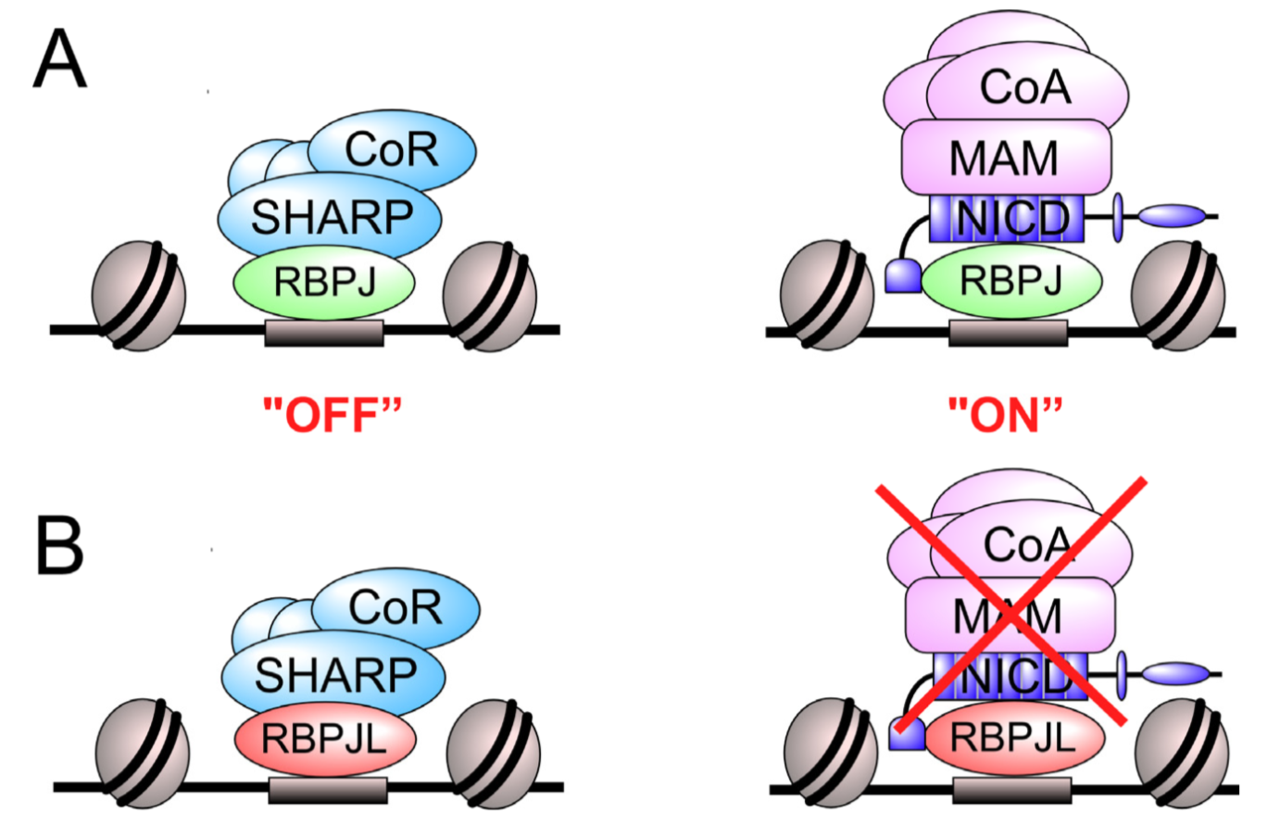


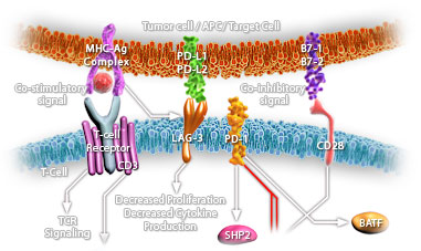
.jpg)