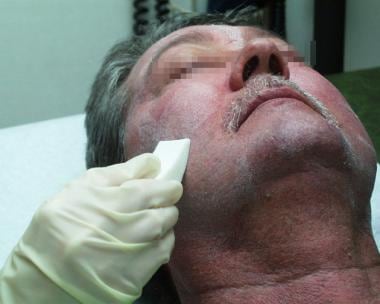
Granulation Tissue
Arachnoid
Tracheal Stenosis
Skin
Granuloma, Foreign-Body
Powders
Fibroblasts
Negative-Pressure Wound Therapy
Silicones
Granuloma
Excipients
Histamine Agonists
Drug Compounding
Technology, Pharmaceutical
Tablets
Myofibroblasts
Collagen
Neovascularization, Physiologic
Skin Ulcer
Dimaprit
Cicatrix, Hypertrophic
Carrageenan
Surgical Sponges
Wounds and Injuries
Subcutaneous Tissue
Debridement
Chemistry, Pharmaceutical
Cholesteatoma, Middle Ear
Death, Sudden
Bereavement
Coroners and Medical Examiners
Cause of Death
Spatial and temporal expression of parathyroid hormone-related protein during wound healing. (1/330)
Parathyroid hormone-related protein is produced by many normal tissues including the skin, where it regulates growth and differentiation of keratinocytes. To define better the role of parathyroid hormone-related protein in the skin, we investigated the spatial and temporal expression of parathyroid hormone-related protein and mRNA by immunohistochemistry and in situ hybridization during the healing of skin wounds, and the effects of topical administration of a parathyroid hormone-related protein agonist [parathyroid hormone-related protein (1-36)] and a parathyroid hormone-related protein antagonist [parathyroid hormone (7-34)] on the healing rate and morphology of the wounds. Wounds were produced on the back of guinea pigs with a 4 mm punch, and wound sites were collected at different time points during the healing process. Parathyroid hormone-related protein was expressed in normal skin by all viable keratinocyte layers, hair follicles, and adnexae. Following injury, migratory keratinocytes at wound margins and the newly restored epidermis expressed increased levels of parathyroid hormone-related protein. The remodeling phase was associated with progressive restoration of the pattern of parathyroid hormone-related protein expression in normal epidermis. Granulation tissue myofibroblasts and infiltrating macrophages also expressed parathyroid hormone-related protein. In vitro studies using THP-1 cells (a promonocytic cell line) confirmed that macrophages expressed parathyroid hormone-related protein, especially after activation. Topical application of parathyroid hormone related protein (1-36) or parathyroid hormone (7-34) did not result in significant changes in the healing rate and morphology of the wounds. These findings demonstrated that, in addition to keratinocytes, myofibroblasts and macrophages also represent sources of parathyroid hormone-related protein during the healing of skin wounds. Although the data suggest a role for parathyroid hormone-related protein in the healing of skin and in the restoration of epidermal homeostasis, parathyroid hormone-related protein does not appear to be required for proper re-epithelialization in response to injury, potentially because of redundancy in epidermal growth and wound healing, as has been shown for other paracrine and autocrine growth factors of the epidermis. (+info)Chimera analysis reveals that fibroblasts and endothelial cells require platelet-derived growth factor receptorbeta expression for participation in reactive connective tissue formation in adults but not during development. (2/330)
The hypothesis that platelet-derived growth factor (PDGF) plays an important role in repair of connective tissue has been difficult to test experimentally, in part because the disruption of any of the PDGF ligand and receptor genes is embryonic lethal. We have developed a method that circumvents the embryonic lethality of the PDGF receptor (R)beta-/- genotype and minimizes the tendency of compensatory processes to mask the phenotype of gene disruption by comparing the behavior of wild-type and PDGFRbeta-/- cells within individual chimeric mice. This quantitative chimera analysis method has revealed that during development PDGFRbeta expression is important for all muscle lineages but not for fibroblast or endothelial lineages. Here we report that fibroblasts and endothelial cells, but not leukocytes, are dependent on PDGFRbeta expression during the formation of new connective tissue in and around sponges implanted under the skin. Even the 50% reduction in PDGFRbeta gene dosage in PDGFRbeta+/- cells reduces fibroblast and endothelial cell participation by 85%. These results demonstrate that the PDGFRbeta/PDGF B-chain system plays an important direct role in driving both fibroblast and endothelial cell participation in connective tissue repair, that cell behavior can be regulated by relatively small changes in PDGFRbeta expression, and that the functions served by PDGF in wound healing are different from the roles served during development. (+info)Fibrin microbeads (FMB) as biodegradable carriers for culturing cells and for accelerating wound healing. (3/330)
We have developed biodegradable fibrin-derived microbeads as potent cell carriers. The fibrin-derived microbeads, 50-200 microm in diameter, were tested for their attachment to a wide range of cell types. Fibrin-derived microbeads were shown to be greatly haptotactic to cells (such as endothelial cells, smooth muscle cells and fibroblasts), which respond to fibrinogen in contrast to keratinocytes and different cell lines derived from leukocytic lineage. The cells on fibrin-derived microbeads could be maintained for more than 10 d and achieved a high density. 31P-nuclear magnetic resonance was employed to monitor phosphate metabolism in cells, with densities on the order of 100 million cells per g of fibrin-derived microbeads. The 31P-nuclear magnetic resonance adenosine triphosphate and phosphocreatine signals, equivalent to the signal obtained with perfused normal skin, indicated that metabolism of cells on fibrin-derived microbeads was responsive to oxygenation and nutrients. Light, fluorescent, and confocal laser microscopy revealed that the porous fibrin-derived microbeads accommodate up to 200-300 cells due to their high surface area which minimized contact inhibition. Cells could degrade the fibrin-derived microbeads and be transferred to seed culture flasks without trypsinization. In a pig skin wound healing model, fibrin-derived microbeads + fibroblasts were transplanted into full thickness punch wounds. This procedure was compared with other treatment modalities, such as the addition of human platelet-derived growth factor BB or fibrin-derived microbeads alone. By the third day after wounding, only the wounds in which fibroblasts on fibrin-derived microbeads were added showed significant formation of granulation tissue. Based on the above, we project many uses of our novel fibrin-derived microbead technology for cell culturing, wound healing and tissue engineering. (+info)Survival, integration, and differentiation of cardiomyocyte grafts: a study in normal and injured rat hearts. (4/330)
BACKGROUND: Cardiomyocyte grafting augments myocyte numbers in the heart. We investigated (1) how developmental stage influences graft survival; (2) whether acutely necrotic or healing cardiac lesions support grafts; and (3) the differentiation and integration of cardiomyocyte grafts in injured hearts. METHODS AND RESULTS: Cardiomyocytes from fetal, neonatal, or adult inbred rats were grafted into normal myocardium, acutely cryoinjured myocardium, or granulation tissue (6 days after injury). Adult cardiomyocytes did not survive under any conditions. In contrast, fetal and neonatal cardiomyocytes formed viable grafts under all conditions. Time-course studies with neonatal cardiomyocytes showed that the grafts recapitulated many aspects of normal development. The adherens junction protein N-cadherin was distributed circumferentially at day 1 but began to organize into intercalated disk-like structures by day 6. The gap junction protein connexin43 followed a similar but delayed pattern relative to N-cadherin. From 2 to 8 weeks, there was progressive hypertrophy and the formation of mature intercalated disks. In some hearts, graft cells formed adherens and gap junctions with host cardiomyocytes, suggesting electromechanical coupling. More commonly, however, grafts were separated from the host myocardium by scar tissue. Gap and adherens junctions formed between neonatal and adult cardiomyocytes in coculture, as evidenced by dye transfer and localization of cadherin and connexin43 at intercellular junctions. CONCLUSIONS: Grafted fetal and neonatal cardiomyocytes form new, mature myocardium with the capacity to couple with injured host myocardium. Optimal repair, however, may require reducing the isolation of the graft by the intervening scar tissue. (+info)Expression of cardiac angiotensin-converting enzyme after myocardial infarction. (5/330)
AIM: To localize cardiac angiotensin-converting enzyme (ACE) during left ventricular repair after myocardial infarction (MI). METHODS: Cardiac ACE was examined by immunohistochemical staining using monoclonal and polyclonal antibodies against ACE 24 h, 1 wk, 2 wk, 3 wk, and 6 wk after coronary artery ligation in rats. Immunofluorescent double staining technique was applied to distinguish the cells which express ACE. RESULTS: ACE staining was confined to the endothelial cells and distributed in normal cardiac tissue in a gradient pattern along the vascular tree: present around the whole circle of arterial endothelium, present in about 20% of the capillaries, and absent in the veins. One week after MI, ACE expression was noted in the granulation tissue. Three weeks after MI, necrosis within the infarction was replaced by granulation tissue and fibrous tissue which showed strong over-expression of ACE. Six weeks after MI, the region with positive ACE staining regressed and the area with high collagen content on the endocardial side showed only weak ACE stain. Most of the ACE-positive cells in the ACE-over-expression-area were endothelial cells. A few macrophages seen in these regions were also ACE-positive. CONCLUSION: Cardiac ACE was over expressed during the process of tissue repair following MI, reaching a peak in 3 wk. Endothelial cells took the most part of ACE expression. (+info)Expression of cathepsin K messenger RNA in giant cells and their precursors in human osteoarthritic synovial tissues. (6/330)
OBJECTIVE: To investigate the expression of cathepsin K messenger RNA (mRNA) in the giant cells found in human osteoarthritic (OA) synovium and associated reparative connective tissues, and to compare this with mRNA expression of cathepsins B, L, and S, which are cysteine proteases known to be highly expressed by cells of the monocyte/macrophage lineage. METHODS: Sections of human OA synovium were processed for in situ hybridization and probed for cathepsins K, B, L, and S. Serial sections were reacted for tartrate-resistant acid phosphatase (TRAP) and nonspecific esterase (NSE) activity, which are selective markers for the osteoclast and cells of the macrophage/monocyte lineage, respectively. RESULTS: At 3 sites of monocyte infiltration/giant cell formation (granulation tissue, the intimal and subintimal synovial layers, and deep stroma extending to the periphery of osteophytic tissue), both TRAP-positive mono- and multinucleated cells and TRAP-negative, NSE-positive mononuclear precursors were identified. Cells containing both enzyme activities were also found, potentially indicating an intermediate stage of differentiation. The TRAP-positive mononuclear/giant cells, and the occasional NSE-positive precursor, expressed an intense signal for cathepsin K mRNA, but did not express cathepsins B, L, and S. In contrast, the deep zone of phagocytic-like cells adjacent to sites of ossification expressed high levels of mRNA for cathepsins L, B, and S as well as cathepsin K mRNA. CONCLUSION: Giant cells that form within OA synovial tissue express high levels of cathepsin K mRNA. It appears that cathepsin K acts principally to digest the bone (and cartilage) fragments sheered from the joint surface during OA. The high TRAP activity and the undetectable expression of the macrophage-associated degradative proteases (cathepsins B, L, and S) by synovial giant cells strengthens the hypothesis that cathepsin K is the primary protease involved in bone degradation. At sites of synovial osteogenesis, a population of phagocytic-like cells expressed TRAP and cathepsins B, L, S, and K, and may represent blood-derived macrophages pushed toward an osteoclast phenotype. (+info)Connective tissue growth factor mediates transforming growth factor beta-induced collagen synthesis: down-regulation by cAMP. (7/330)
Connective tissue growth factor (CTGF) is a cysteine-rich peptide synthesized and secreted by fibroblastic cells after activation with transforming growth factor beta (TGF-beta) that acts as a downstream mediator of TGF-beta-induced fibroblast proliferation. We performed in vitro and in vivo studies to determine whether CTGF is also essential for TGF-beta-induced fibroblast collagen synthesis. In vitro studies with normal rat kidney (NRK) fibroblasts demonstrated CTGF potently induces collagen synthesis and transfection with an antisense CTGF gene blocked TGF-beta stimulated collagen synthesis. Moreover, TGF-beta-induced collagen synthesis in both NRK and human foreskin fibroblasts was effectively blocked with specific anti-CTGF antibodies and by suppressing TGF-beta-induced CTGF gene expression by elevating intracellular cAMP levels with either membrane-permeable 8-Br-cAMP or an adenylyl cyclase activator, cholera toxin (CTX). cAMP also inhibited collagen synthesis induced by CTGF itself, in contrast to its previously reported lack of effect on CTGF-induced DNA synthesis. In animal assays, CTX injected intradermally in transgenic mice suppressed TGF-beta activation of a human CTGF promoter/lacZ reporter transgene. Both 8-Br-cAMP and CTX blocked TGF-beta-induced collagen deposition in a wound chamber model of fibrosis in rats. CTX also reduced dermal granulation tissue fibroblast population increases induced by TGF-beta in neonatal mice, but not increases induced by CTGF or TGF-beta combined with CTGF. Our data indicate that CTGF mediates TGF-beta-induced fibroblast collagen synthesis and that in vivo blockade of CTGF synthesis or action reduces TGF-beta-induced granulation tissue formation by inhibiting both collagen synthesis and fibroblast accumulation. (+info)Arnebin-1 accelerates normal and hydrocortisone-induced impaired wound healing. (8/330)
Wound healing involves inflammation, cell proliferation, matrix deposition, and tissue remodeling. Interaction of different cells, extracellular matrix proteins, and their receptors are mediated by cytokines and growth factors during wound healing. In this study, we have evaluated the effect of arnebin-1, a natural product isolated from Arnebia nobilis, on normal and impaired wound healing in cutaneous punch wound model. Arnebin-1 was applied topically daily on wounds of hydrocortisone-treated or untreated animals. Arnebin-1 significantly accelerated healing of wounds with or without hydrocortisone treatment as revealed by a reduction in the wound width and gap length compared with controls. Arnebin-1 treatment promoted the cell proliferation, migration, and vessel formation to form a thick granulation tissue and re-epithelialization of the wounds. An increase in the synthesis of collagen, fibronectin and transforming growth factor-beta1 was seen in arnebin-1-treated wounds compared with the untreated control. As transforming growth factor-beta1 is known to enhance wound healing, and associated with the wound healing defect in hydrocortisone-treated wounds, the enhanced expression of transforming growth factor-beta1 at both translational and transcriptional level by arnebin-1 may be responsible for the enhancement of wound healing during normal and impaired wound repair. These studies suggest that arnebin-1 could be developed as a potent therapeutic agent for wound healing in steroid-impaired wounds. (+info)Granulation tissue is the pinkish, bumpy material that forms on the surface of a healing wound. It's composed of tiny blood vessels (capillaries), white blood cells, and fibroblasts - cells that produce collagen, which is a protein that helps to strengthen and support the tissue.
Granulation tissue plays a crucial role in the wound healing process by filling in the wound space, contracting the wound, and providing a foundation for the growth of new skin cells (epithelialization). It's typically formed within 3-5 days after an injury and continues to develop until the wound is fully healed.
It's important to note that while granulation tissue is a normal part of the healing process, excessive or overgrowth of granulation tissue can lead to complications such as delayed healing, infection, or the formation of hypertrophic scars or keloids. In these cases, medical intervention may be necessary to manage the excess tissue and promote proper healing.
Wound healing is a complex and dynamic process that occurs after tissue injury, aiming to restore the integrity and functionality of the damaged tissue. It involves a series of overlapping phases: hemostasis, inflammation, proliferation, and remodeling.
1. Hemostasis: This initial phase begins immediately after injury and involves the activation of the coagulation cascade to form a clot, which stabilizes the wound and prevents excessive blood loss.
2. Inflammation: Activated inflammatory cells, such as neutrophils and monocytes/macrophages, infiltrate the wound site to eliminate pathogens, remove debris, and release growth factors that promote healing. This phase typically lasts for 2-5 days post-injury.
3. Proliferation: In this phase, various cell types, including fibroblasts, endothelial cells, and keratinocytes, proliferate and migrate to the wound site to synthesize extracellular matrix (ECM) components, form new blood vessels (angiogenesis), and re-epithelialize the wounded area. This phase can last up to several weeks depending on the size and severity of the wound.
4. Remodeling: The final phase of wound healing involves the maturation and realignment of collagen fibers, leading to the restoration of tensile strength in the healed tissue. This process can continue for months to years after injury, although the tissue may never fully regain its original structure and function.
It is important to note that wound healing can be compromised by several factors, including age, nutrition, comorbidities (e.g., diabetes, vascular disease), and infection, which can result in delayed healing or non-healing chronic wounds.
The arachnoid is one of the three membranes that cover the brain and the spinal cord, known as the meninges. It is located between the dura mater (the outermost layer) and the pia mater (the innermost layer). The arachnoid is a thin, delicate membrane that is filled with cerebrospinal fluid, which provides protection and nutrition to the central nervous system.
The arachnoid has a spider-web like appearance, hence its name, and it is composed of several layers of collagen fibers and elastic tissue. It is highly vascularized, meaning that it contains many blood vessels, and it plays an important role in regulating the flow of cerebrospinal fluid around the brain and spinal cord.
In some cases, the arachnoid can become inflamed or irritated, leading to a condition called arachnoiditis. This can cause a range of symptoms, including pain, muscle weakness, and sensory changes, and it may require medical treatment to manage.
Tracheal stenosis is a medical condition characterized by the abnormal narrowing of the trachea (windpipe), which can lead to difficulty breathing. This narrowing can be caused by various factors such as inflammation, scarring, or the growth of abnormal tissue in the airway. Symptoms may include wheezing, coughing, shortness of breath, and chest discomfort, particularly during physical activity. Treatment options for tracheal stenosis depend on the severity and underlying cause of the condition and may include medications, bronchodilators, corticosteroids, or surgical interventions such as laser surgery, stent placement, or tracheal reconstruction.
In medical terms, the skin is the largest organ of the human body. It consists of two main layers: the epidermis (outer layer) and dermis (inner layer), as well as accessory structures like hair follicles, sweat glands, and oil glands. The skin plays a crucial role in protecting us from external factors such as bacteria, viruses, and environmental hazards, while also regulating body temperature and enabling the sense of touch.
A granuloma is a type of organized immune response that occurs when the body encounters a foreign substance that it cannot eliminate. A "foreign-body" granuloma specifically refers to this reaction in response to an exogenous material, such as a splinter, suture, or other types of medical implants.
Foreign-body granulomas are characterized by the formation of a collection of immune cells, including macrophages and lymphocytes, which surround and attempt to isolate the foreign material. Over time, this collection of immune cells can become walled off and form a well-circumscribed mass or nodule.
Foreign-body granulomas may cause localized symptoms such as pain, swelling, or inflammation, depending on their location and size. In some cases, they may also lead to complications such as infection or tissue damage. Treatment typically involves removing the foreign body, if possible, followed by anti-inflammatory therapy to manage any residual symptoms or complications.
A cicatrix is a medical term that refers to a scar or the process of scar formation. It is the result of the healing process following damage to body tissues, such as from an injury, wound, or surgery. During the healing process, specialized cells called fibroblasts produce collagen, which helps to reconnect and strengthen the damaged tissue. The resulting scar tissue may have a different texture, color, or appearance compared to the surrounding healthy tissue.
Cicatrix formation is a natural part of the body's healing response, but excessive scarring can sometimes cause functional impairment, pain, or cosmetic concerns. In such cases, various treatments may be used to minimize or improve the appearance of scars, including topical creams, steroid injections, laser therapy, and surgical revision.
In the context of medical terminology, "powders" do not have a specific technical definition. However, in a general sense, powders refer to dry, finely ground or pulverized solid substances that can be dispersed in air or liquid mediums. In medicine, powders may include various forms of medications, such as crushed tablets or capsules, which are intended to be taken orally, mixed with liquids, or applied topically. Additionally, certain medical treatments and therapies may involve the use of medicated powders for various purposes, such as drying agents, abrasives, or delivery systems for active ingredients.
Fibroblasts are specialized cells that play a critical role in the body's immune response and wound healing process. They are responsible for producing and maintaining the extracellular matrix (ECM), which is the non-cellular component present within all tissues and organs, providing structural support and biochemical signals for surrounding cells.
Fibroblasts produce various ECM proteins such as collagens, elastin, fibronectin, and laminins, forming a complex network of fibers that give tissues their strength and flexibility. They also help in the regulation of tissue homeostasis by controlling the turnover of ECM components through the process of remodeling.
In response to injury or infection, fibroblasts become activated and start to proliferate rapidly, migrating towards the site of damage. Here, they participate in the inflammatory response, releasing cytokines and chemokines that attract immune cells to the area. Additionally, they deposit new ECM components to help repair the damaged tissue and restore its functionality.
Dysregulation of fibroblast activity has been implicated in several pathological conditions, including fibrosis (excessive scarring), cancer (where they can contribute to tumor growth and progression), and autoimmune diseases (such as rheumatoid arthritis).
Negative-Pressure Wound Therapy (NPWT) is a medical treatment used to promote wound healing and prevent infection in acute or chronic wounds. It involves the application of controlled sub-atmospheric pressure to the surface of the wound, usually through the use of a vacuum-assisted device.
The negative pressure helps to remove excess fluid and infectious materials from the wound, while also promoting the growth of new tissue by increasing blood flow and stimulating cell proliferation. NPWT can be used in various types of wounds, including diabetic foot ulcers, pressure ulcers, surgical wounds, and traumatic injuries.
The therapy is typically administered through a sealed dressing that covers the wound and is connected to a vacuum pump. The negative pressure is applied continuously or intermittently, depending on the specific needs of the patient and the type of wound being treated. NPWT has been shown to be effective in reducing wound size, promoting healing, and improving overall clinical outcomes in many patients with complex wounds.
Silicones are not a medical term, but they are commonly used in the medical field, particularly in medical devices and healthcare products. Silicones are synthetic polymers made up of repeating units of siloxane, which is a chain of alternating silicon and oxygen atoms. They can exist in various forms such as oils, gels, rubbers, and resins.
In the medical context, silicones are often used for their unique properties, including:
1. Biocompatibility - Silicones have a low risk of causing an adverse reaction when they come into contact with living tissue.
2. Inertness - They do not react chemically with other substances, making them suitable for use in medical devices that need to remain stable over time.
3. Temperature resistance - Silicones can maintain their flexibility and elasticity even under extreme temperature conditions.
4. Gas permeability - Some silicone materials allow gases like oxygen and water vapor to pass through, which is useful in applications where maintaining a moist environment is essential.
5. Durability - Silicones have excellent resistance to aging, weathering, and environmental factors, ensuring long-lasting performance.
Examples of medical applications for silicones include:
1. Breast implants
2. Contact lenses
3. Catheters
4. Artificial joints and tendons
5. Bandages and wound dressings
6. Drug delivery systems
7. Medical adhesives
8. Infant care products (nipples, pacifiers)
A granuloma is a small, nodular inflammatory lesion that occurs in various tissues in response to chronic infection, foreign body reaction, or autoimmune conditions. Histologically, it is characterized by the presence of epithelioid macrophages, which are specialized immune cells with enlarged nuclei and abundant cytoplasm, often arranged in a palisading pattern around a central area containing necrotic debris, microorganisms, or foreign material.
Granulomas can be found in various medical conditions such as tuberculosis, sarcoidosis, fungal infections, and certain autoimmune disorders like Crohn's disease. The formation of granulomas is a complex process involving both innate and adaptive immune responses, which aim to contain and eliminate the offending agent while minimizing tissue damage.
Excipients are inactive substances that serve as vehicles or mediums for the active ingredients in medications. They make up the bulk of a pharmaceutical formulation and help to stabilize, preserve, and enhance the delivery of the active drug compound. Common examples of excipients include binders, fillers, coatings, disintegrants, flavors, sweeteners, and colors. While excipients are generally considered safe and inert, they can sometimes cause allergic reactions or other adverse effects in certain individuals.
The mastoid is a term used in anatomy and refers to the bony prominence located at the base of the skull, posterior to the ear. More specifically, it's part of the temporal bone, one of the bones that forms the side and base of the skull. The mastoid process provides attachment for various muscles involved in chewing and moving the head.
In a medical context, "mastoid" can also refer to conditions or procedures related to this area. For example, mastoiditis is an infection of the mastoid process, while a mastoidectomy is a surgical procedure that involves removing part or all of the mastoid process.
Histamine agonists are substances that bind to and activate histamine receptors, leading to the initiation or enhancement of various physiological responses. Histamine is a naturally occurring molecule that plays a key role in the body's immune and allergic responses, as well as in the regulation of sleep, wakefulness, and appetite.
There are four main types of histamine receptors (H1, H2, H3, and H4), each with distinct functions and signaling pathways. Histamine agonists can be selective for one or more of these receptor subtypes, depending on their pharmacological properties.
For example, H1 agonists are commonly used as decongestants and antihistamines to treat allergies, while H2 agonists are used to treat gastroesophageal reflux disease (GERD) and peptic ulcers. H3 agonists have been investigated for their potential therapeutic use in the treatment of neurological disorders such as Parkinson's disease and schizophrenia, while H4 agonists are being studied for their role in inflammation and immune regulation.
It is important to note that histamine agonists can also have adverse effects, particularly if they are not selective for a specific receptor subtype or if they are used at high doses. These effects may include increased heart rate, blood pressure, and bronchodilation (opening of the airways), as well as gastrointestinal symptoms such as nausea, vomiting, and diarrhea.
Drug compounding is the process of combining, mixing, or altering ingredients to create a customized medication to meet the specific needs of an individual patient. This can be done for a variety of reasons, such as when a patient has an allergy to a certain ingredient in a mass-produced medication, or when a patient requires a different dosage or formulation than what is available commercially.
Compounding requires specialized training and equipment, and compounding pharmacists must follow strict guidelines to ensure the safety and efficacy of the medications they produce. Compounded medications are not approved by the U.S. Food and Drug Administration (FDA), but the FDA does regulate the ingredients used in compounding and has oversight over the practices of compounding pharmacies.
It's important to note that while compounding can provide benefits for some patients, it also carries risks, such as the potential for contamination or incorrect dosing. Patients should only receive compounded medications from reputable pharmacies that follow proper compounding standards and procedures.
Medical technology, also known as health technology, refers to the use of medical devices, medicines, vaccines, procedures, and systems for the purpose of preventing, diagnosing, or treating disease and disability. This can include a wide range of products and services, from simple devices like tongue depressors and bandages, to complex technologies like MRI machines and artificial organs.
Pharmaceutical technology, on the other hand, specifically refers to the application of engineering and scientific principles to the development, production, and control of pharmaceutical drugs and medical devices. This can include the design and construction of manufacturing facilities, the development of new drug delivery systems, and the implementation of quality control measures to ensure the safety and efficacy of pharmaceutical products.
Both medical technology and pharmaceutical technology play crucial roles in modern healthcare, helping to improve patient outcomes, reduce healthcare costs, and enhance the overall quality of life for individuals around the world.
In the context of medical terminology, tablets refer to pharmaceutical dosage forms that contain various active ingredients. They are often manufactured in a solid, compressed form and can be administered orally. Tablets may come in different shapes, sizes, colors, and flavors, depending on their intended use and the manufacturer's specifications.
Some tablets are designed to disintegrate or dissolve quickly in the mouth, making them easier to swallow, while others are formulated to release their active ingredients slowly over time, allowing for extended drug delivery. These types of tablets are known as sustained-release or controlled-release tablets.
Tablets may contain a single active ingredient or a combination of several ingredients, depending on the intended therapeutic effect. They are typically manufactured using a variety of excipients, such as binders, fillers, and disintegrants, which help to hold the tablet together and ensure that it breaks down properly when ingested.
Overall, tablets are a convenient and widely used dosage form for administering medications, offering patients an easy-to-use and often palatable option for receiving their prescribed treatments.
Myofibroblasts are specialized cells that are present in various tissues throughout the body. They play a crucial role in wound healing and tissue repair, but they can also contribute to the development of fibrosis or scarring when their activation and proliferation persist beyond the normal healing process. Here is a medical definition of myofibroblasts:
Medical Definition of Myofibroblasts:
Myofibroblasts are modified fibroblasts that exhibit features of both smooth muscle cells and fibroblasts, including the expression of alpha-smooth muscle actin stress fibers. They are involved in the contraction of wounds, tissue remodeling, and the production of extracellular matrix components such as collagen, elastin, and fibronectin. Myofibroblasts can differentiate from various cell types, including resident fibroblasts, epithelial cells (epithelial-mesenchymal transition), endothelial cells (endothelial-mesenchymal transition), and circulating fibrocytes. Persistent activation of myofibroblasts can lead to excessive scarring and fibrosis in various organs, such as the lungs, liver, kidneys, and heart.
Collagen is the most abundant protein in the human body, and it is a major component of connective tissues such as tendons, ligaments, skin, and bones. Collagen provides structure and strength to these tissues and helps them to withstand stretching and tension. It is made up of long chains of amino acids, primarily glycine, proline, and hydroxyproline, which are arranged in a triple helix structure. There are at least 16 different types of collagen found in the body, each with slightly different structures and functions. Collagen is important for maintaining the integrity and health of tissues throughout the body, and it has been studied for its potential therapeutic uses in various medical conditions.
Physiologic neovascularization is the natural and controlled formation of new blood vessels in the body, which occurs as a part of normal growth and development, as well as in response to tissue repair and wound healing. This process involves the activation of endothelial cells, which line the interior surface of blood vessels, and their migration, proliferation, and tube formation to create new capillaries. Physiologic neovascularization is tightly regulated by a balance of pro-angiogenic and anti-angiogenic factors, ensuring that it occurs only when and where it is needed. It plays crucial roles in various physiological processes, such as embryonic development, tissue regeneration, and wound healing.
Bronchoscopy is a medical procedure that involves the examination of the inside of the airways and lungs with a flexible or rigid tube called a bronchoscope. This procedure allows healthcare professionals to directly visualize the airways, take tissue samples for biopsy, and remove foreign objects or secretions. Bronchoscopy can be used to diagnose and manage various respiratory conditions such as lung infections, inflammation, cancer, and bleeding. It is usually performed under local or general anesthesia to minimize discomfort and risks associated with the procedure.
A foreign-body reaction is an immune response that occurs when a non-native substance, or "foreign body," is introduced into the human body. This can include things like splinters, surgical implants, or even injected medications. The immune system recognizes these substances as foreign and mounts a response to try to eliminate them.
The initial response to a foreign body is often an acute inflammatory reaction, characterized by the release of chemical mediators that cause vasodilation, increased blood flow, and the migration of white blood cells to the site. This can result in symptoms such as redness, swelling, warmth, and pain.
If the foreign body is not eliminated, a chronic inflammatory response may develop, which can lead to the formation of granulation tissue, fibrosis, and encapsulation of the foreign body. In some cases, this reaction can cause significant tissue damage or impede proper healing.
It's worth noting that not all foreign bodies necessarily elicit a strong immune response. The nature and size of the foreign body, as well as its location in the body, can all influence the severity of the reaction.
A skin ulcer is a defined as a loss of continuity or disruption of the skin surface, often accompanied by inflammation and/or infection. These lesions can result from various causes including pressure, venous or arterial insufficiency, diabetes, and chronic dermatological conditions. Skin ulcers are typically characterized by their appearance, depth, location, and underlying cause. Common types of skin ulcers include pressure ulcers (also known as bedsores), venous leg ulcers, arterial ulcers, and diabetic foot ulcers. Proper evaluation, wound care, management of underlying conditions, and prevention strategies are crucial in the treatment of skin ulcers to promote healing and prevent complications.
Dimaprit is not a medical condition or disease. It is actually a synthetic peptide that acts as an agonist for certain types of receptors found in the body, specifically the H2 histamine receptors. These receptors are involved in various physiological processes, such as regulating gastric acid secretion and modulating immune responses.
As a research tool, Dimaprit is used to study the functions of H2 histamine receptors and their roles in different biological systems. It is not typically used as a therapeutic agent in clinical medicine.
A hypertrophic cicatrix is a type of scar that forms when the body overproduces collagen during the healing process. Collagen is a protein that helps to repair and strengthen tissues in the body. However, when too much collagen is produced, it can cause the scar to become thickened, raised, and firm.
Hypertrophic scars are usually red or pink in color and may be itchy or painful. They typically develop within a few weeks of an injury or surgery and can continue to grow for several months before eventually stabilizing. Unlike keloids, which are a more severe type of scar that can grow beyond the boundaries of the original wound, hypertrophic scars do not extend beyond the site of the injury.
While hypertrophic scars can be unsightly and cause discomfort, they are generally not harmful to one's health. Treatment options may include corticosteroid injections, silicone gel sheeting, pressure therapy, or laser surgery to help reduce the size and appearance of the scar. It is important to seek medical advice if you are concerned about a hypertrophic scar or if it is causing significant discomfort or distress.
Penetrating wounds are a type of traumatic injury that occurs when an object pierces through the skin and underlying tissues, creating a hole or cavity in the body. These wounds can vary in severity, depending on the size and shape of the object, as well as the location and depth of the wound.
Penetrating wounds are typically caused by sharp objects such as knives, bullets, or glass. They can damage internal organs, blood vessels, nerves, and bones, leading to serious complications such as bleeding, infection, organ failure, and even death if not treated promptly and properly.
The management of penetrating wounds involves a thorough assessment of the wound and surrounding tissues, as well as the identification and treatment of any associated injuries or complications. This may include wound cleaning and closure, antibiotics to prevent infection, pain management, and surgery to repair damaged structures. In some cases, hospitalization and close monitoring may be necessary to ensure proper healing and recovery.
Carriageenans are a family of linear sulfated polysaccharides that are extracted from red edible seaweeds. They have been widely used in the food industry as thickening, gelling, and stabilizing agents. In the medical field, they have been studied for their potential therapeutic applications, such as in the treatment of gastrointestinal disorders and inflammation. However, some studies have suggested that certain types of carriageenans may have negative health effects, including promoting inflammation and damaging the gut lining. Therefore, more research is needed to fully understand their safety and efficacy.
Surgical sponges are absorbent, sterile materials used in medical procedures to soak up bodily fluids and help maintain a clean surgical field. They are typically made from gauze material and come in various sizes and shapes to accommodate different surgical needs. Surgical sponges are carefully counted before and after a procedure to ensure that none are accidentally left inside the patient's body.
A wound is a type of injury that occurs when the skin or other tissues are cut, pierced, torn, or otherwise broken. Wounds can be caused by a variety of factors, including accidents, violence, surgery, or certain medical conditions. There are several different types of wounds, including:
* Incisions: These are cuts that are made deliberately, often during surgery. They are usually straight and clean.
* Lacerations: These are tears in the skin or other tissues. They can be irregular and jagged.
* Abrasions: These occur when the top layer of skin is scraped off. They may look like a bruise or a scab.
* Punctures: These are wounds that are caused by sharp objects, such as needles or knives. They are usually small and deep.
* Avulsions: These occur when tissue is forcibly torn away from the body. They can be very serious and require immediate medical attention.
Injuries refer to any harm or damage to the body, including wounds. Injuries can range from minor scrapes and bruises to more severe injuries such as fractures, dislocations, and head trauma. It is important to seek medical attention for any injury that is causing significant pain, swelling, or bleeding, or if there is a suspected bone fracture or head injury.
In general, wounds and injuries should be cleaned and covered with a sterile bandage to prevent infection. Depending on the severity of the wound or injury, additional medical treatment may be necessary. This may include stitches for deep cuts, immobilization for broken bones, or surgery for more serious injuries. It is important to follow your healthcare provider's instructions carefully to ensure proper healing and to prevent complications.
Subcutaneous tissue, also known as the subcutis or hypodermis, is the layer of fatty connective tissue found beneath the dermis (the inner layer of the skin) and above the muscle fascia. It is composed mainly of adipose tissue, which serves as a energy storage reservoir and provides insulation and cushioning to the body. The subcutaneous tissue also contains blood vessels, nerves, and immune cells that support the skin's functions. This layer varies in thickness depending on the location in the body and can differ significantly between individuals based on factors such as age, genetics, and weight.
A bandage is a medical dressing or covering applied to a wound, injury, or sore with the intention of promoting healing or preventing infection. Bandages can be made of a variety of materials such as gauze, cotton, elastic, or adhesive tape and come in different sizes and shapes to accommodate various body parts. They can also have additional features like fasteners, non-slip surfaces, or transparent windows for monitoring the condition of the wound.
Bandages serve several purposes, including:
1. Absorbing drainage or exudate from the wound
2. Protecting the wound from external contaminants and bacteria
3. Securing other medical devices such as catheters or splints in place
4. Reducing swelling or promoting immobilization of the affected area
5. Providing compression to control bleeding or prevent fluid accumulation
6. Relieving pain by reducing pressure on sensitive nerves or structures.
Proper application and care of bandages are essential for effective wound healing and prevention of complications such as infection or delayed recovery.
Debridement is a medical procedure that involves the removal of dead, damaged, or infected tissue to improve the healing process or prevent further infection. This can be done through various methods such as surgical debridement (removal of tissue using scalpel or scissors), mechanical debridement (use of wound irrigation or high-pressure water jet), autolytic debridement (using the body's own enzymes to break down and reabsorb dead tissue), and enzymatic debridement (application of topical enzymes to dissolve necrotic tissue). The goal of debridement is to promote healthy tissue growth, reduce the risk of infection, and improve overall wound healing.
Pharmaceutical chemistry is a branch of chemistry that deals with the design, synthesis, and development of chemical entities used as medications. It involves the study of drugs' physical, chemical, and biological properties, as well as their interactions with living organisms. This field also encompasses understanding the absorption, distribution, metabolism, and excretion (ADME) of drugs in the body, which are critical factors in drug design and development. Pharmaceutical chemists often work closely with biologists, medical professionals, and engineers to develop new medications and improve existing ones.
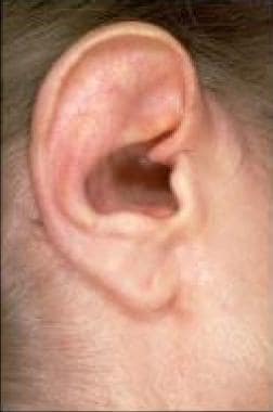
Cholesteatoma, middle ear is a medical condition characterized by the abnormal growth of skin cells (keratinizing squamous epithelium) within the middle ear space. This skin cells accumulation forms a pearly, white, or gray mass that can erode and destroy surrounding structures such as the ossicles (the tiny bones in the middle ear), the mastoid process (a bony prominence behind the ear), and even the inner ear or brain.
Cholesteatomas can be congenital (present at birth) or acquired (develop later in life). Acquired cholesteatomas are more common and usually result from repeated middle ear infections that cause a retraction pocket of the eardrum, which then traps skin cells leading to their abnormal growth. Symptoms of cholesteatoma may include hearing loss, ear drainage, ear pain, vertigo, or facial weakness. Treatment typically involves surgical removal of the cholesteatoma and restoration of any damaged structures.
Actinomycosis is a type of infection caused by bacteria that are normally found in the mouth, intestines, and female genital tract. These bacteria can cause abscesses or chronic inflammation if they infect body tissues, often after trauma or surgery. The infection typically affects the face, neck, or chest, and can spread to other parts of the body over time. Symptoms may include swelling, redness, pain, and the formation of pus-filled abscesses that may discharge a characteristic yellowish granular material called "sulfur granules." Treatment typically involves long-term antibiotic therapy, often requiring high doses and intravenous administration. Surgical drainage or removal of infected tissue may also be necessary in some cases.
Sudden death is a term used to describe a situation where a person dies abruptly and unexpectedly, often within minutes to hours of the onset of symptoms. It is typically caused by cardiac or respiratory arrest, which can be brought on by various medical conditions such as heart disease, stroke, severe infections, drug overdose, or trauma. In some cases, the exact cause of sudden death may remain unknown even after a thorough post-mortem examination.
It is important to note that sudden death should not be confused with "sudden cardiac death," which specifically refers to deaths caused by the abrupt loss of heart function (cardiac arrest). Sudden cardiac death is often related to underlying heart conditions such as coronary artery disease, cardiomyopathy, or electrical abnormalities in the heart.
An autopsy, also known as a post-mortem examination or obduction, is a medical procedure in which a qualified professional (usually a pathologist) examines a deceased person's body to determine the cause and manner of death. This process may involve various investigative techniques, such as incisions to study internal organs, tissue sampling, microscopic examination, toxicology testing, and other laboratory analyses. The primary purpose of an autopsy is to gather objective evidence about the medical conditions and factors contributing to the individual's demise, which can be essential for legal, insurance, or public health purposes. Additionally, autopsies can provide valuable insights into disease processes and aid in advancing medical knowledge.
Bereavement is the state of loss or grief experienced when a person experiences the death of a loved one, friend, or family member. It is a normal response to the death of someone close and can involve a range of emotions such as sadness, anger, guilt, and anxiety. The grieving process can be different for everyone and can take time to work through. Professional support may be sought to help cope with the loss.
A coroner and medical examiner are officials in the legal system who are responsible for investigating and determining the cause of death in certain cases. While their roles can overlap, there are some differences between them.
A coroner is a public official who is typically appointed or elected to serve in a particular jurisdiction, such as a county or district. The coroner's primary responsibility is to investigate any sudden, unexpected, or suspicious deaths that occur within their jurisdiction. This may include deaths that occur due to violence, accidents, suicide, or unknown causes.
In order to determine the cause of death, the coroner may conduct an autopsy, order toxicology tests, and review medical records and other evidence. The coroner may also hold an inquest, which is a formal hearing in which witnesses are called to testify about the circumstances surrounding the death. Based on the evidence gathered during the investigation, the coroner will make a determination as to the cause and manner of death.
A medical examiner, on the other hand, is a physician who has completed specialized training in forensic pathology. Medical examiners are typically appointed or hired by a government agency, such as a state or county, to perform autopsies and investigate deaths.
Medical examiners are responsible for determining the cause of death in cases where there is a suspicion of foul play, as well as in other circumstances where the cause of death may not be immediately apparent. They may also testify in court as expert witnesses based on their findings.
In some jurisdictions, the roles of coroner and medical examiner are combined, with the official serving as both a public administrator and a trained physician. In other cases, the two roles are separate, with the coroner responsible for administrative functions and the medical examiner responsible for determining the cause of death.
Forensic pathology is a subspecialty of pathology that focuses on determining the cause and manner of death by examining a corpse. It involves applying scientific knowledge and techniques to investigate criminal or suspicious deaths, often in conjunction with law enforcement agencies. A forensic pathologist performs autopsies (postmortem examinations) to evaluate internal and external injuries, diseases, and other conditions that may have contributed to the individual's death. They also collect evidence such as tissue samples, which can be used for toxicological, microbiological, or histological analysis. The information gathered by forensic pathologists is crucial in helping to establish the facts surrounding a person's death and assisting legal proceedings.
The "cause of death" is a medical determination of the disease, injury, or event that directly results in a person's death. This information is typically documented on a death certificate and may be used for public health surveillance, research, and legal purposes. The cause of death is usually determined by a physician based on their clinical judgment and any available medical evidence, such as laboratory test results, autopsy findings, or eyewitness accounts. In some cases, the cause of death may be uncertain or unknown, and the death may be classified as "natural," "accidental," "homicide," or "suicide" based on the available information.
"Terminology as a topic" in the context of medical education and practice refers to the study and use of specialized language and terms within the field of medicine. This includes understanding the meaning, origins, and appropriate usage of medical terminology in order to effectively communicate among healthcare professionals and with patients. It may also involve studying the evolution and cultural significance of medical terminology. The importance of "terminology as a topic" lies in promoting clear and accurate communication, which is essential for providing safe and effective patient care.
 Granulation tissue
Connective tissue
Telocyte
Callous ulcer
Tracheobronchial injury
Pemphigus vegetans
Foreign body reaction
Oral mucocele
Myofibroblast
Intraoral dental sinus
Coagulative necrosis
Myeloid sarcoma
Granulation
Barrier membrane
Pannus
Kimura's disease
Umbilical granuloma
Ear pain
Type V collagen
Pyogenic granuloma
Alginate dressing
Epulis
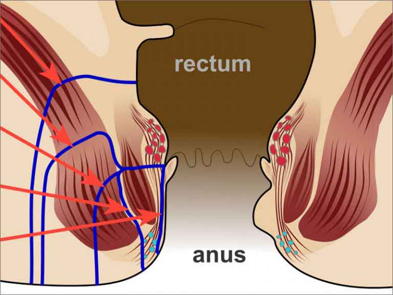
Anal fistula
Ranula
Healing
Tendinopathy
Bone healing
Jerome Gross
Phineas Gage
Wound healing
Granulation tissue - Wikipedia
 removal granulation tissue vaginal cuff - Forum - Codapedia™
 Immunoglobulins in Periodontal Tissues. II. Concentrations of Immunoglobulins in Granulation Tissue from Pockets of...
 Subjects: Granulation Tissue - Digital Collections - National Library of Medicine Search Results
The Role of mPGES-1 in Promoting Granulation Tissue Angiogenesis Through Regulatory T-cell Accumulation | In Vivo
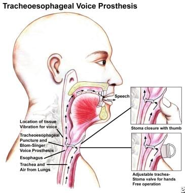
 Laryngectomy Rehabilitation: Overview, Evaluating Tracheoesophageal Speech, Tracheoesophageal Speech Prostheses
 Granulation Tissue and matrix metalloproteinase (MMP) Archives - ACR Meeting Abstracts
 On-Time Pressure Ulcer Healing: Facilitator Training | Agency for Healthcare Research and Quality
 December 1974 - Volume 54 - Issue 6 : Plastic and Reconstructive Surgery
Cholesteatoma Treatment & Management: Approach Considerations, Mastoidectomy, Endoscopic Ear Surgery
 Laser Hair Removal - DnaTube.com - Scientific Video and Animation Site
 Gastrostomy Tube (G-Tube) (for Parents) - CareSource
 Hydrogen Sulfide in Skin Diseases: A Novel Mediator and Therapeutic Target
 Biomechanical regulation of blood vessel growth during tissue vascularization | Nature Medicine
Neurosyphilis: Overview of Syphilis of the CNS, Pathophysiology of Syphilis, Epidemiology of Syphilis
Cutaneous Legionella longbeachae Infection in Immunosuppressed Woman, United Kingdom - Volume 21, Number 8-August 2015 -...
Pathology of Sudden Natural Death: Overview, Terminology, Medical Examiner Role and Autopsy Indications
 Post-Surgical Clinical Monitoring of Soft Tissue Wound Healing in Periodontal and Implant Surgery
Complex Airway Program | BIDMC of Boston
 How wounds heal: MedlinePlus Medical Encyclopedia
 Frontiers | Anti-herpetic Activity of Macrocystis pyrifera and Durvillaea antarctica Algae Extracts Against HSV-1 and HSV-2
 FAQ - Education | AUGS
 LCD - Pressure Reducing Support Surfaces - Group 3 (L33692)
 Enduring: June 2007
 Case Definitions for Public Health Surveillance
 Sanjay R Parikh, MD
 Routine laboratory parameters in patients with necrotizing pancreatitis by the time of operative pancreatic debridement: Food...
 Wound Care: A Guide to Practice for Healthcare Professionals
 Ji-Young Han - Search Results - PubMed
 JCM | Free Full-Text | The Role of Antioxidants on Wound Healing: A Review of the Current Evidence
Connective10
- Granulation tissue is new connective tissue and microscopic blood vessels that form on the surfaces of a wound during the healing process. (wikipedia.org)
- Lastly, a fourth type of wound healing can also be considered when the overlying tissue is partially lost (abrasion) or intentionally removed (epithelialized free gingival graft donor site), so a de-epithelialized connective tissue layer is exposed and heals by re-epithelialization from the normal contiguous epithelium 2 . (medsci.org)
- Protein found in blood and connective tissue. (memory.com)
- Elastic protein found in connective tissue. (memory.com)
- Fibroblasts are present in connective tissue and are capable of forming collagen fibers. (nutrimedical.com)
- Stimulate tissue granulation and connective tissue projections, which are part of the healing process of wounds, ulcers or inflamed tissue. (nutrimedical.com)
- Granulation tissue gives rise to interfragmentary connective tissue, which eventually is remodeled into fibrocartilage. (veterinarypracticenews.com)
- it is characterized by damage to the epithelial tissue and underlying connective tissue[ 3 ] due to mechanical injury, thermal, electrical or chemical burn. (thieme-connect.de)
- Granulation tissue is the perfused, fibrous connective tissue that replaces a fibrin clot in healing wounds. (t-vox.org)
- Extravasation of mucus into underlying connective tissue is the result of rupture of an excretory gland of a minor salivary gland, caused by trauma or laceration 4 . (bvsalud.org)
Wounds7
- Granulation tissue typically grows from the base of a wound and is able to fill wounds of almost any size. (wikipedia.org)
- Thus, tissue repair from wounds and gastric ulcers depends on angiogenesis ( 3 , 4 ). (iiarjournals.org)
- Secondary healing, on the other hand, occurs in areas which are not covered by normally epithelialized tissue due to intentional (extraction sockets, apically repositioned flaps) or accidental (wounds with full thickness loss of substance) exposure, or due to an insufficient amount of lining tissue to be used for coverage. (medsci.org)
- Although oral surgical wounds heal in a very similar way, soft tissue healing is somewhat conditioned by that of the underlying bone tissue. (medsci.org)
- Studies have shown that when purified growth factors were deposited in granulation tissue, the wounds showed accelerated granulation tissue formation and wound healing (Lynch et al. (dentistryiq.com)
- In chronic wounds - ones that show little or no sign of healing, despite appropriate therapy, within an acceptable timeframe - this healthy healing process is disturbed and tissue regeneration is delayed. (hartmann.info)
- Unfortunately, others may have wounds that heal improperly and develop excessive scar tissue surrounding the healing wound. (grestech.com)
Fibroblasts6
- The extracellular matrix of granulation tissue is created and modified by fibroblasts. (wikipedia.org)
- Fibroblasts, the main cells that deposit granulation tissue, depend on oxygen to proliferate and lay down the new extracellular matrix. (wikipedia.org)
- Using neovascularization models based on the chick chorioallantoic membrane and the healing mouse cornea, we found that tissue tension generated by activated fibroblasts or myofibroblasts during wound contraction mediated and directed translocation of the vasculature. (nature.com)
- Gabbiani, G., Ryan, G.B. & Majne, G. Presence of modified fibroblasts in granulation tissue and their possible role in wound contraction. (nature.com)
- Attracts other fibroblasts and macrophages by chemotaxis to the healing tissue. (memory.com)
- The fibroblasts found in the granulation tissue are actively laying down the extracellular matrix. (dentistryiq.com)
Exudate1
- Granulation tissue (firm, beefy red tissue) requires some exudate management and protection. (ausmed.com.au)
Polyps3
- Examples of granulation tissue can be seen in pyogenic granulomas and pulp polyps. (wikipedia.org)
- Persistent chronic suppurative otitis media may result in destructive changes in the middle ear (such as necrosis of the long process of the incus) or aural polyps (granulation tissue prolapsing into the ear canal through the TM perforation). (msdmanuals.com)
- Any obstructive lung disease characterized by consolidated formation of GRANULATION TISSUE polyps within ALVEOLAR DUCTS AND ALVEOLI. (bvsalud.org)
Angiogenesis7
- citation needed] In vascularisation, also called angiogenesis, endothelial cells quickly grow into the tissue from older, intact blood vessels. (wikipedia.org)
- Regulatory T cells (Tregs) regulate not only immune tolerance but also tissue repair and angiogenesis. (iiarjournals.org)
- We examined whether the mPGES-1/PGE 2 axis contributes to wound-induced angiogenesis and granulation tissue formation through Treg accumulation. (iiarjournals.org)
- Angiogenesis was estimated by determining the wet weight of sponge tissues and the expression of proangiogenic factors including CD31, vascular endothelial growth factor (VEGF), and transforming growth factor β (TGF-β) in granulation tissues. (iiarjournals.org)
- We implanted a polyurethane sponge disk subcutaneously to elicit angiogenesis in the surrounding granulation tissues in rodents to analyse the mechanisms of angiogenesis in vivo ( 7 , 8 ). (iiarjournals.org)
- Formation of new vessels in granulation tissue during wound healing has been assumed to occur solely through sprouting angiogenesis. (nature.com)
- During the first one to two days there is recruitment of neutrophils to the wound site, followed by epithelialization, granulation tissue formation, and angiogenesis. (dentistryiq.com)
Vaginal cuff3
- Does anyone know of a CPT® code for removal of granulation tissue of the vaginal cuff? (codapedia.com)
- Ops notes states: There was approximately 2x1x1 granulation tissue formation on the vaginal cuff incision site. (codapedia.com)
- How do I code for a revision of a vaginal cuff with removal of granulation tissue and excision of suture due to extrusion of the permanent uterosacral sutures (gortex) one year post op? (augs.org)
Formation5
- Granulation tissue formation may begin by day four. (dentistryiq.com)
- Irrespective of the type of wound and the extent of tissue loss, the wound-healing process takes place in three dynamic stages: the cleansing stage, the granulation phase (tissue formation) and the epithelisation phase (epidermisation). (hartmann.info)
- Bone, similar to soft tissue, undergoes stages of healing including inflammation, phagocytosis of cellular and organic debris, cell proliferation and granulation tissue formation. (veterinarypracticenews.com)
- In the first few hours after ICH onset, primary brain injury by ICH is mainly caused by the oppression and destruction to the near tissue by hematoma formation. (karger.com)
- [ 1 , 5-7 ] This hyperinflammatory, proteolytic environment prevents the wound from progressing into the proliferative phase, resulting in stalled reepithelialization and the formation of defective granulation tissue. (medscape.com)
Vessels6
- citation needed] It is necessary for a network of blood vessels to be established as soon as possible to provide the growing tissue with nutrients, to take away cellular wastes, and transport new leukocytes to the area. (wikipedia.org)
- The dermis is the second one, a subjacent fibrous-collagenous-elastic tissue that hosts vessels, nerves, and sensory receptors. (hindawi.com)
- These mechanical forces pulled vessels from the preexisting vascular bed as vascular loops with functional circulation that expanded as an integral part of the growing granulation tissue through vessel enlargement and elongation. (nature.com)
- This model explains the rapid appearance of large functional vessels in granulation tissue during wound healing. (nature.com)
- Over the next 3 weeks or so, the body repairs broken blood vessels and new tissue grows. (medlineplus.gov)
- Lymphatic vasculature forms a network of vessels that drain interstitial fluid from tissues and return this fluid to the blood. (nii.ac.jp)
Regeneration2
- equipment offers 7 specific settings to promote different levels of tissue healing and regeneration. (nutrimedical.com)
- The Granulation Phase describes the regeneration of tissue, when the wound is filled from the inside. (hartmann.info)
Scar5
- This is later replaced by the stronger, long-stranded type-I collagen, as evidenced in scar tissue. (wikipedia.org)
- Surgeons choose this method to guide the G-tube into place when other methods are not a good choice - for example, if there is scar tissue from a past surgery or if the child needs another surgery done at the same time. (kidshealth.org)
- By increasing collagen production less scar tissue is formed at the damaged site. (nutrimedical.com)
- However, bone repair differs from soft tissue in that its repair process does not produce scar tissue. (veterinarypracticenews.com)
- This scar tissue, which is called keloids, can develop on any part of the body. (grestech.com)
Necrotic5
- Necrotic tissue type and amount. (ahrq.gov)
- Ideally, the quickest (and often safest) way to remove necrotic tissue is to involve a surgeon who will then surgically debride the offending tissue. (ausmed.com.au)
- Without a doubt, removal of necrotic tissue and management of infective tissue are two priorities in wound care. (ausmed.com.au)
- Infective tissue is best removed when possible by employing the same methods as with necrotic tissue. (ausmed.com.au)
- At this stage the priority is to remove avital and necrotic tissues and, to promote wound cleansing, remove bacteria and toxins that could contribute to delayed healing. (hartmann.info)
Macrophages2
- citation needed] The main immune cells active in the tissue are macrophages and neutrophils, although other leukocytes are also present. (wikipedia.org)
- The main immune cells active in the tissue are macrophages and neutrophils, although other leukocytes are also present. (t-vox.org)
Collagen5
- Red blood cells help create collagen, which are tough, white fibers that form the foundation for new tissue. (medlineplus.gov)
- Bind to fibronectin and to collagen and help stabilize tissue undergoing repair. (memory.com)
- Assembled into thin supporting filaments and the predominant collagen type found in cartilaginous tissue. (memory.com)
- Collagen is the essential protein used to repair damaged tissue and to replace old tissue. (nutrimedical.com)
- Lavender increases collagen production and helps form granulation tissue. (nowloss.com)
Occur2
- Tissue growth and rebuilding occur next. (medlineplus.gov)
- Separation of tissue margins may occur. (memory.com)
Scars1
- Scars form because the new tissue grows back differently than the original tissue. (medlineplus.gov)
Infection3
- These work to phagocytize old or damaged tissue, and protect the healing tissue from pathogenic infection. (wikipedia.org)
- Furthermore, the term tertiary intention is used to define delayed healing which occurs in both types of healing after an infected wound is left open for days until the infection disappears and is completely covered by surgical closure of the overlying tissue 1 , 2 . (medsci.org)
- After activation in the wound area, platelets will release growth factors to regulate cell migration, proliferation, and matrix deposition and reduce the risk of infection from the wound, increasing the quality of healing and tissue repair. (thieme-connect.de)
Subcutaneous2
- Materials and Methods: The dorsal subcutaneous tissues of male mPGES-1-deficient (mPGES-1 −/− ) and C57BL/6 wild-type (WT) mice were implanted with polyurethane sponge disks. (iiarjournals.org)
- The subcutaneous tissue hypodermis is the deepest layer [ 15 ]. (hindawi.com)
Inflammatory1
- An inflammatory reaction is induced in adjacent tissues, in which neutrophil and macrophage defense cells predominate, creating granulation tissue surrounding the pooled mucus, isolating the lesion and giving it the appearance of a pseudocyst 5,6 . (bvsalud.org)
Edema1
- Brain edema is a pathological phenomenon that water and brain tissue volume increase. (karger.com)
Adjacent2
- Spine infections are rare infections that can involve the intervertebral disc space (discitis), the vertebral bones, the spinal canal or adjacent soft tissues. (wheelessonline.com)
- Lytic enzymes, such as collagenases, produced by the cholesteatoma can destroy adjacent bone and soft tissue. (msdmanuals.com)
Surgically1
- In primary intention healing there is no loss of tissue and all tissues are replaced in the same anatomic position and with the same structure they had before injury, although this definition is usually referred to as healing which occurs when the lining tissues are closely approximated surgically to perfectly cover all underlying injured tissues. (medsci.org)
Wound Healing2
- Clinical features of surgical soft tissue wound healing in dentistry have been rarely discussed in the international literature. (medsci.org)
- Advances in tissue engineering technology have led to the production of novel human skin equivalents and organoids that reproduce cell-cell interactions with tissue-scale tensional homeostasis, and enable us to evaluate skin tissue morphology, functionality, drug response and wound healing. (mdpi.com)
Clinical1
- Her clinical interests lie broadly within equine and ruminant soft tissue and orthopaedic surgical diseases. (ku.dk)
Cellular2
- Cells involved in the tissue repair response produce these proteins that regulate cellular reactions involved in healing. (memory.com)
- Visible and infrared light have been shown to affect positive therapeutic benefits to living tissues and organisms on a cellular level. (nutrimedical.com)
Protects the tissue2
- The blood clots dry and form a scab, which protects the tissue underneath from germs. (medlineplus.gov)
- A dressing that maintains a minimally moist environment and protects the tissue, is generally required. (ausmed.com.au)
Form3
- New skin begins to form over this tissue. (medlineplus.gov)
- She said the granulation tissue is beginning to form around the tendon, and there is some thickening as a result - all of which is to be expected. (endurance.net)
- She says he will look quite different in three weeks, and that the slowest part of the recovery will be waiting for the granulation tissue to form along the gap between the two flaps of skin. (endurance.net)
Removal1
- Treatment requires complete cleaning of the ear canal several times daily, careful removal of granulation tissue, and application of topical corticosteroids and antibiotics. (msdmanuals.com)
Processes1
- One of the processes of tissue healing begins soon after tissue injury or death and occurs. (memory.com)
Bone1
- The process of bone repair differs from soft tissue repair. (veterinarypracticenews.com)
Topical1
- If the wound is locally infected, the clinician may choose to manage the infective tissue with debridement and topical antimicrobials (not topical antibiotics) (Lipsky & Hoey 2009). (ausmed.com.au)
Dressings3
- A structured approach is essential, as the most common error in wound care management is rushing in to select the latest and greatest new wound dressings without actually giving thought to wound aetiology, tissue type and immediate aim. (ausmed.com.au)
- Emerging dressing types include bioactive dressings and tissue-engineered skin substitutes. (worldwidewounds.com)
- The following discussion focuses on novel types of 'bioactive' dressings, the tissue-engineered 'skin substitutes', and the trials used to test their effectiveness. (worldwidewounds.com)
Soft1
- This soft, gelatinous, highly exuding tissue requires specific treatment. (ausmed.com.au)
Blood1
- Results from serial bacterial cultures of blood, feces, and tissue failed to yield further positive results. (cdc.gov)
Bones1
- Predominant in strong tissues such as tendons and bones. (memory.com)
Destruction2
- Hence, increasing numbers of contaminating bacteria in the wound increase the potential for neutrophil-mediated tissue destruction. (dentistryiq.com)
- The oppression and destruction by hematoma to brain tissue cause the primary brain injury. (karger.com)
Forms1
- This tissue forms over raw, broken skin as your body recovers from a wound. (nowloss.com)
Area1
- It was felt that research should be continued in this area, and a study was initiated to determine the IgA, IgG, and IgM concentrations in the granulation tissue removed from deep infrabony pockets of patients with periodontosis and advanced periodontitis. (dtic.mil)
Skin1
- For example, despite improvements in cell culture techniques and developments in dermal matrices, tissue-engineered skin substitutes have yet to achieve widespread use by clinicians. (worldwidewounds.com)
Protein1
- Most important protein providing structural support and tensile strength for almost all tissues and organs of the body. (memory.com)
Depends1
- The main generation of H 2 S in cutaneous tissue mostly depends on enzymatic routes using L-cysteine and homocysteine by two pyridoxal-5 - phosphate-dependent enzymes, cystathionine β -synthase (CBS) and cystathionine γ -lyase (CSE). (hindawi.com)
Healthy1
- granulation tissue looks healthy. (endurance.net)