Goblet Cells
Mucins
Conjunctiva
Mucin-2
Mucin 5AC
Mucus
Metaplasia
Intestinal Mucosa
Mucin-3
Exocrine Glands
Alcian Blue
Colon
Hyperplasia
Periodic Acid-Schiff Reaction
Paneth Cells
Dry Eye Syndromes
Respiratory Mucosa
Intestine, Small
Epithelium
Interleukin-13
Intestine, Large
Nippostrongylus
Mucin-5B
Hepatocyte Nuclear Factor 3-gamma
Epithelial Cells
Glycoconjugates
Conjunctival Diseases
Cell Count
Mucoproteins
Intestines
Trachea
Bronchi
Duodenum
Oxyuroidea
Amorphophallus
Jejunum
Bronchial Hyperreactivity
Gastric Mucins
Lung
Histocytochemistry
Cell Differentiation
Bronchoalveolar Lavage Fluid
Asthma
Disease Models, Animal
Allergens
Ileum
Keratin-7
Lacrimal Apparatus
Mice, Inbred C57BL
Keratoconjunctivitis Sicca
Airway Remodeling
Respiratory Hypersensitivity
Lactobacillus helveticus
Mucous Membrane
Mice, Knockout
Mice, Inbred BALB C
Enterocytes
Carcinoid Tumor
Fluorescent Antibody Technique, Indirect
Lectins
Immunohistochemistry
Barrett Esophagus
Goblet cell-specific expression mediated by the MUC2 mucin gene promoter in the intestine of transgenic mice. (1/560)
The regulation of MUC2, a major goblet cell mucin gene, was examined by constructing transgenic mice containing bases -2864 to +17 of the human MUC2 5'-flanking region fused into the 5'-untranslated region of a human growth hormone (hGH) reporter gene. Four of eight transgenic lines expressed reporter. hGH message expression was highest in the distal small intestine, with only one line expressing comparable levels in the colon. This contrasts with endogenous MUC2 expression, which is expressed at its highest levels in the colon. Immunohistochemical analysis indicated that goblet cell-specific expression of reporter begins deep in the crypts, as does endogenous MUC2 gene expression. These results indicate that the MUC2 5'-flanking sequence contains elements sufficient for the appropriate expression of MUC2 in small intestinal goblet cells. Conversely, elements located outside this region appear necessary for efficient colonic expression, implying that the two tissues utilize different regulatory elements. Thus many, but not all, of the elements necessary for MUC2 gene regulation reside between bases -2864 and +17 of the 5'-flanking region. (+info)Epidermal growth factor system regulates mucin production in airways. (2/560)
Goblet-cell hyperplasia is a critical pathological feature in hypersecretory diseases of airways. However, the underlying mechanisms are unknown, and no effective therapy exists. Here we show that stimulation of epidermal growth factor receptors (EGF-R) by its ligands, EGF and transforming growth factor alpha (TGFalpha), causes MUC5AC expression in airway epithelial cells both in in vitro and in vivo. We found that a MUC5AC-inducing epithelial cell line, NCI-H292, expresses EGF-R constitutively; EGF-R gene expression was stimulated further by tumor necrosis factor alpha (TNFalpha). EGF-R ligands increased the expression of MUC5AC at both gene and protein levels, and this effect was potentiated by TNFalpha. Selective EGF-R tyrosine kinase inhibitors blocked MUC5AC expression induced by EGF-R ligands. Pathogen-free rats expressed little EGF-R protein in airway epithelial cells; intratracheal instillation of TNFalpha induced EGF-R in airway epithelial cells, and subsequent instillation of EGF-R ligands increased the number of goblet cells, Alcian blue-periodic acid-Schiff staining (reflecting mucous glycoconjugates), and MUC5AC gene expression, whereas TNFalpha, EGF, or TGFalpha alone was without effect. In sensitized rats, three intratracheal instillations of ovalbumin resulted in EGF-R expression and goblet-cell production in airway epithelium. Pretreatment with EGF-R tyrosine kinase inhibitor, BIBX1522, prevented goblet-cell production both in rats stimulated by TNFalpha-EGF-R ligands and in an asthma model. These findings suggest potential roles for inhibitors of the EGF-R cascade in hypersecretory diseases of airways. (+info)Weaning anorexia may contribute to local inflammation in the piglet small intestine. (3/560)
Compromising alterations in villus-crypt structure are common in pigs postweaning. Possible contributions of local inflammatory reactions to villus-crypt alterations during the weaning transition have not been described. This study evaluated local inflammatory responses and their relationship with morphological changes in the intestine in 21-d-old pigs (n = 112) killed either at weaning (Day 0) or 0.5, 1, 2, 4 or 7 d after weaning to either milk- or soy-based pelleted diets. Cumulative intake averaged <100 g during the first 2 d postweaning, regardless of diet. During this period of weaning anorexia, inflammatory T-cell numbers and local expression of the matrix metalloproteinase stromelysin increased while jejunal villus height, crypt depth and major histocompatibility complex (MHC) class I RNA expression decreased. Upon resumption of feed intake by the fourth d postweaning, villus height and crypt depth, CD8(+) T cell numbers, MHC class I RNA expression and local expression of stromelysin returned to Day 0 values. Together the results indicate that inadequate feed intake during the immediate postweaning period may contribute to intestinal inflammation and thereby compromise villus-crypt structure and function. (+info)Cloning of the gene gob-4, which is expressed in intestinal goblet cells in mice. (4/560)
We isolated the novel cDNA gob-4, which was shown to be expressed in intestinal goblet cells. The deduced amino acid sequence is similar to the gene coding for the Xenopus laevis cement gland-specific XAG-2. These sequence and expression data suggest this gene may be involved in the secretory function. (+info)Nitric oxide synthase inhibitor attenuates intestinal damage induced by zinc deficiency in rats. (5/560)
A nitric oxide synthase (NOS) inhibitor, NG-nitro-L -arginine methyl ester (L-NAME), was given to zinc-deficient (ZD) rats to determine whether it prevents the intestinal damage usually observed under these conditions. Weanling male rats were given free access to a ZD diet (2 mg zinc/kg), whereas control rats including pair-fed (PF) and ad libitum consumption (AL) groups were given a zinc-supplemented (50.8 mg zinc/kg) diet for 4 wk. Half of the ZD rats received L-NAME (0.3 g/L in drinking water) for 3 wk starting at the wk 2 of the deficient period. Plasma zinc concentration in ZD rats was significantly lower (P < 0.05) than that of AL and PF rats. Administration of L-NAME did not alter this concentration. Intestinal zinc concentration did not differ among groups. However, metallothionein-1 (MT-1) mRNA level was significantly lower in the intestine of ZD rats than in AL or PF rats. Treatment of ZD rats with L-NAME did not affect this level. Intestinal microvascular permeability evaluated by Evans blue showed significantly higher extravasation in ZD rats than in AL rats, whereas L-NAME administration inhibited the extravasation. Expression of inducible NOS mRNA was observed in intestine of ZD but not of AL or PF rats, and there was no significant difference between ZD rats, regardless of L-NAME treatment. The activity ratio of inducible NOS to total NOS in ZD rats not receiving L-NAME was significantly higher than that in AL rats or ZD rats treated with L-NAME (P < 0.05). The number of apoptotic-positive and goblet cells in intestinal villi was significantly higher in ZD rats compared with AL or PF rats. L-NAME administration in ZD rats reversed this effect. These results indicate that inhibition of NOS ameliorates zinc deficiency-induced intestinal damage in rats. (+info)Overexpression of glycine-extended gastrin in transgenic mice results in increased colonic proliferation. (6/560)
Gastrin is a peptide hormone involved in the growth of both normal and malignant gastrointestinal tissue. Recent studies suggest that the glycine-extended biosynthetic intermediates mediate many of these trophic effects, but the in vivo relevance of glycine-extended gastrin (G-Gly) has not been tested. We have generated mice (MTI/G-GLY) that overexpress progastrin truncated at glycine-72 to evaluate the trophic effects of G-Gly in an in vivo model. MTI/G-GLY mice have elevated serum and colonic mucosal levels of G-Gly compared with wild-type mice. MTI/G-GLY mice had a 43% increase in colonic mucosal thickness and a 41% increase in the percentage of goblet cells per crypt. MTI/G-GLY mice exhibited increased colonic proliferation compared with wild-type controls, with an expansion of the proliferative zone into the upper third of the colonic crypts. Continuous infusion of G-Gly into gastrin-deficient mice for two weeks also resulted in elevated G-Gly levels, a 10% increase in colonic mucosal thickness, and an 81% increase in colonic proliferation when compared with gastrin-deficient mice that received saline alone. To our knowledge, these studies demonstrate for the first time that G-Gly's contribute to colonic mucosal proliferation in vivo. (+info)IL-4 induces mucin gene expression and goblet cell metaplasia in vitro and in vivo. (7/560)
Goblet cell metaplasia and mucus hypersecretion are important features in the pathogenesis of asthma. The cytokine IL-4 has been shown to play a role in animal models of asthma, where it induces Th2 lymphocyte differentiation and B lymphocyte IgE class switch. IL-4 has also been implicated in the differentiation of goblet cells via effects on lymphocytes and eosinophils. In this study we hypothesized that IL-4 induces airway epithelial cell mucin gene expression and mucous glycoconjugate production by direct action on these cells. In vitro, cultured airway epithelial cells (NCI-H292) expressed IL-4R constitutively, and IL-4 (10 ng/ml) induced MUC2 gene expression and mucous glycoconjugate production. In vivo, mouse airway epithelial cells expressed IL-4R constitutively, and IL-4 (250 ng) increased MUC5 gene expression and Alcian blue/periodic acid-Schiff-positive staining at 24 h; IL-4 did not increase inflammatory cell numbers in airway tissue or in bronchoalveolar lavage. TNF-alpha and IL-1beta levels in bronchoalveolar lavage were not increased in response to IL-4 instillation. These results indicate that airway epithelial cells express IL-4R constitutively and that IL-4 directly induces the differentiation of epithelium into mucous glycoconjugate-containing goblet cells. (+info)Immunolocalization of muscarinic and VIP receptor subtypes and their role in stimulating goblet cell secretion. (8/560)
PURPOSE: To determine the subtypes of cholinergic muscarinic receptors and receptors for vasoactive intestinal peptide (VIP) present in rat conjunctival goblet cells and whether cholinergic agonists and VIP stimulate goblet cell secretion. METHODS: Immunofluorescence studies were performed using antibodies against the m1, m2, and m3 muscarinic receptor subtypes and VIP receptors 1 and 2 (VIPR1 and VIPR2). The lectin Ulex europeus agglutinin I was used to measure glycoconjugate secretion, the index of secretion, from goblet cells in an enzyme-linked lectin assay. In this assay, pieces of conjunctiva were placed on filter paper and incubated for 15 to 120 minutes, with or without increasing concentrations of the cholinergic agonist carbachol or VIP. The muscarinic antagonist atropine and the muscarinic receptor-subtype-selective antagonists pirenzepine (M1), gallamine (M2), and 4-4-diphenylacetoxy-N-(2-chloroethyl)-piperidine hydrochloride (4-DAMP mustard; M3) were incubated with carbachol to determine specificity of receptor activation. RESULTS: Immunoreactivity to M2 and M3 receptors was found on goblet cell membranes subjacent to the secretory granules. Immunoreactivity to M1 receptor was not on goblet cells but was on the stratitfied squamous cells. Immunoreactivity to VIPR2 was found on goblet cells with a localization similar to that of the M2 and M3 receptors. VIPR1 was not found on goblet cells or on the stratified squamous cells. Carbachol and VIP induced a time- and concentration-dependent stimulation of glycoconjugate secretion. Carbachol, at 10(-4) M, induced a threefold increase in glycoconjugate secretion, which was completely inhibited by atropine (10(-5) M). Carbachol-induced secretion was inhibited 54% +/- 8% by pirenzepine (10(-5) M), 69% +/- 14% by gallamine (10(-5) M), and 72% +/- 11% by 4-DAMP mustard (10(-5) M). A twofold increase in glycoconjugate secretion was obtained with VIP at 10(-8) M. CONCLUSIONS: Cholinergic agonists, through M2 and/or M3 muscarinic receptors, and VIP, through VIPR2, regulate conjunctival goblet cell secretion, suggesting that goblet cell secretion in vivo is under the control of parasympathetic nerves. (+info)Goblet cells are specialized epithelial cells that are located in various mucosal surfaces, including the respiratory and gastrointestinal tracts. They are named for their goblet-like shape, which is characterized by a narrow base and a wide, rounded top that contains secretory granules. These cells play an essential role in producing and secreting mucins, which are high molecular weight glycoproteins that form the gel-like component of mucus.
Mucus serves as a protective barrier for the underlying epithelial cells by trapping foreign particles, microorganisms, and toxins, preventing them from coming into contact with the epithelium. Goblet cells also help maintain the hydration of the mucosal surface, which is important for normal ciliary function in the respiratory tract and for the movement of food through the gastrointestinal tract.
In summary, goblet cells are secretory cells that produce and release mucins to form the mucus layer, providing a protective barrier and maintaining the homeostasis of mucosal surfaces.
Mucins are high molecular weight, heavily glycosylated proteins that are the major components of mucus. They are produced and secreted by specialized epithelial cells in various organs, including the respiratory, gastrointestinal, and urogenital tracts, as well as the eyes and ears.
Mucins have a characteristic structure consisting of a protein backbone with numerous attached oligosaccharide side chains, which give them their gel-forming properties and provide a protective barrier against pathogens, environmental insults, and digestive enzymes. They also play important roles in lubrication, hydration, and cell signaling.
Mucins can be classified into two main groups based on their structure and function: secreted mucins and membrane-bound mucins. Secreted mucins are released from cells and form a physical barrier on the surface of mucosal tissues, while membrane-bound mucins are integrated into the cell membrane and participate in cell adhesion and signaling processes.
Abnormalities in mucin production or function have been implicated in various diseases, including chronic inflammation, cancer, and cystic fibrosis.
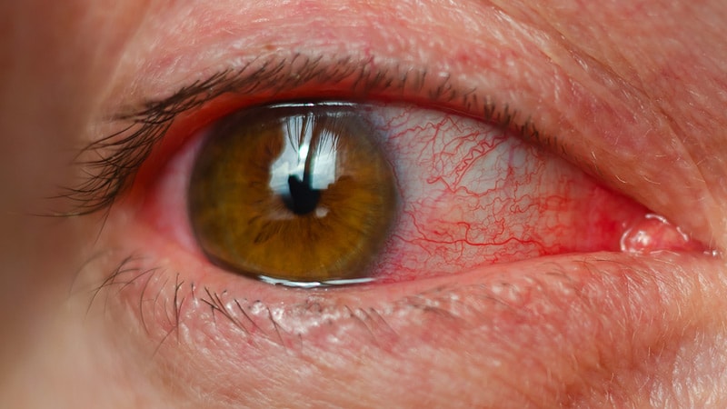
The conjunctiva is the mucous membrane that lines the inner surface of the eyelids and covers the front part of the eye, also known as the sclera. It helps to keep the eye moist and protected from irritants. The conjunctiva can become inflamed or infected, leading to conditions such as conjunctivitis (pink eye).
Mucin-2, also known as MUC2, is a type of mucin that is primarily produced by the goblet cells in the mucous membranes lining the gastrointestinal tract. It is a large, heavily glycosylated protein that forms the gel-like structure of mucus, which provides lubrication and protection to the epithelial surfaces. Mucin-2 is the major component of intestinal mucus and plays an important role in maintaining the integrity of the gut barrier by preventing the adhesion and colonization of harmful microorganisms. Additionally, it has been shown to have anti-inflammatory properties and may play a role in regulating immune responses in the gut.
Mucin 5AC, also known as MUC5AC, is a type of mucin protein that is heavily glycosylated and secreted by the goblet cells in the mucous membranes of the respiratory and gastrointestinal tracts. It plays an essential role in the protection and lubrication of these surfaces, as well as in the clearance of inhaled particles and microorganisms from the lungs.
MUC5AC is a high molecular weight mucin that forms a gel-like substance when secreted, which traps foreign particles and pathogens, facilitating their removal from the body. Abnormalities in MUC5AC production or function have been implicated in various respiratory and gastrointestinal diseases, including chronic obstructive pulmonary disease (COPD), asthma, cystic fibrosis, and inflammatory bowel disease (IBD).
In summary, Mucin 5AC is a crucial component of the mucosal defense system in the respiratory and gastrointestinal tracts, contributing to the maintenance of tissue homeostasis and protection against infection and injury.
Mucus is a viscous, slippery secretion produced by the mucous membranes that line various body cavities such as the respiratory and gastrointestinal tracts. It serves to lubricate and protect these surfaces from damage, infection, and foreign particles. Mucus contains water, proteins, salts, and other substances, including antibodies, enzymes, and glycoproteins called mucins that give it its characteristic gel-like consistency.
In the respiratory system, mucus traps inhaled particles such as dust, allergens, and pathogens, preventing them from reaching the lungs. The cilia, tiny hair-like structures lining the airways, move the mucus upward toward the throat, where it can be swallowed or expelled through coughing or sneezing. In the gastrointestinal tract, mucus helps protect the lining of the stomach and intestines from digestive enzymes and other harmful substances.
Excessive production of mucus can occur in various medical conditions such as allergies, respiratory infections, chronic lung diseases, and gastrointestinal disorders, leading to symptoms such as coughing, wheezing, nasal congestion, and diarrhea.
Metaplasia is a term used in pathology to describe the replacement of one differentiated cell type with another differentiated cell type within a tissue or organ. It is an adaptive response of epithelial cells to chronic irritation, inflammation, or injury and can be reversible if the damaging stimulus is removed. Metaplastic changes are often associated with an increased risk of cancer development in the affected area.
For example, in the case of gastroesophageal reflux disease (GERD), chronic exposure to stomach acid can lead to metaplasia of the esophageal squamous epithelium into columnar epithelium, a condition known as Barrett's esophagus. This metaplastic change is associated with an increased risk of developing esophageal adenocarcinoma.
The intestinal mucosa is the innermost layer of the intestines, which comes into direct contact with digested food and microbes. It is a specialized epithelial tissue that plays crucial roles in nutrient absorption, barrier function, and immune defense. The intestinal mucosa is composed of several cell types, including absorptive enterocytes, mucus-secreting goblet cells, hormone-producing enteroendocrine cells, and immune cells such as lymphocytes and macrophages.
The surface of the intestinal mucosa is covered by a single layer of epithelial cells, which are joined together by tight junctions to form a protective barrier against harmful substances and microorganisms. This barrier also allows for the selective absorption of nutrients into the bloodstream. The intestinal mucosa also contains numerous lymphoid follicles, known as Peyer's patches, which are involved in immune surveillance and defense against pathogens.
In addition to its role in absorption and immunity, the intestinal mucosa is also capable of producing hormones that regulate digestion and metabolism. Dysfunction of the intestinal mucosa can lead to various gastrointestinal disorders, such as inflammatory bowel disease, celiac disease, and food allergies.
Mucin-3, also known as MUC3A or CA15-3, is a type of mucin protein that is heavily glycosylated and found on the apical surface of epithelial cells in the gastrointestinal tract. It is a transmembrane protein that plays a role in protecting the epithelial surface from damage, infection, and inflammation. Mucin-3 has been identified as a tumor antigen and its expression is often upregulated in various types of cancer, including colon, pancreatic, and ovarian cancers. The soluble form of Mucin-3 can be measured in the blood and used as a tumor marker to monitor the progression of certain cancers.
Sialomucins are a type of glycoprotein mucins that contain high amounts of sialic acid, which is a family of negatively charged sugars found on the surface of many cell types. These mucins are produced by the major salivary glands and are a major component of saliva. They play an important role in lubricating and protecting the oral cavity, as well as contributing to the mouth's ability to resist infection and damage.
Sialomucins have also been shown to have various biological functions, such as regulating cell adhesion, modulating immune responses, and serving as receptors for certain viruses and bacteria. Abnormalities in sialomucin expression or structure have been implicated in several diseases, including cancer, autoimmune disorders, and infectious diseases.
Exocrine glands are a type of gland in the human body that produce and release substances through ducts onto an external or internal surface. These glands are responsible for secreting various substances such as enzymes, hormones, and lubricants that help in digestion, protection, and other bodily functions.
Exocrine glands can be further classified into three types based on their mode of secretion:
1. Merocrine glands: These glands release their secretions by exocytosis, where the secretory product is enclosed in a vesicle that fuses with the cell membrane and releases its contents outside the cell. Examples include sweat glands and mucous glands.
2. Apocrine glands: These glands release their secretions by pinching off a portion of the cytoplasm along with the secretory product. An example is the apocrine sweat gland found in the armpits and genital area.
3. Holocrine glands: These glands release their secretions by disintegrating and releasing the entire cell, including its organelles and secretory products. An example is the sebaceous gland found in the skin, which releases an oily substance called sebum.
Alcian Blue is a type of dye that is commonly used in histology, which is the study of the microscopic structure of tissues. It is particularly useful for staining acidic mucopolysaccharides and proteoglycans, which are important components of the extracellular matrix in many tissues.
Alcian Blue binds to these negatively charged molecules through ionic interactions, forming a complex that can be visualized under a microscope. The dye is often used in combination with other stains to provide contrast and highlight specific structures within tissues.
The intensity of the Alcian Blue stain can also provide information about the degree of sulfation or carboxylation of the mucopolysaccharides, which can be useful in diagnosing certain diseases or abnormalities. For example, changes in the staining pattern of proteoglycans have been associated with various types of arthritis and other joint disorders.
Overall, Alcian Blue is an important tool in the field of histology and has contributed significantly to our understanding of tissue structure and function.
The colon, also known as the large intestine, is a part of the digestive system in humans and other vertebrates. It is an organ that eliminates waste from the body and is located between the small intestine and the rectum. The main function of the colon is to absorb water and electrolytes from digested food, forming and storing feces until they are eliminated through the anus.
The colon is divided into several regions, including the cecum, ascending colon, transverse colon, descending colon, sigmoid colon, rectum, and anus. The walls of the colon contain a layer of muscle that helps to move waste material through the organ by a process called peristalsis.
The inner surface of the colon is lined with mucous membrane, which secretes mucus to lubricate the passage of feces. The colon also contains a large population of bacteria, known as the gut microbiota, which play an important role in digestion and immunity.
Hyperplasia is a medical term that refers to an abnormal increase in the number of cells in an organ or tissue, leading to an enlargement of the affected area. It's a response to various stimuli such as hormones, chronic irritation, or inflammation. Hyperplasia can be physiological, like the growth of breast tissue during pregnancy, or pathological, like in the case of benign or malignant tumors. The process is generally reversible if the stimulus is removed. It's important to note that hyperplasia itself is not cancerous, but some forms of hyperplasia can increase the risk of developing cancer over time.
The Periodic Acid-Schiff (PAS) reaction is a histological staining method used to detect the presence of certain carbohydrates, such as glycogen and glycoproteins, in tissues or cells. This technique involves treating the tissue with periodic acid, which oxidizes the vicinal hydroxyl groups in the carbohydrates, creating aldehydes. The aldehydes then react with Schiff's reagent, forming a magenta-colored complex that is visible under a microscope.
The PAS reaction is commonly used to identify and analyze various tissue components, such as basement membranes, fungal cell walls, and mucins in the respiratory and gastrointestinal tracts. It can also be used to diagnose certain medical conditions, like kidney diseases, where abnormal accumulations of carbohydrates occur in the renal tubules or glomeruli.
In summary, the Periodic Acid-Schiff reaction is a staining method that detects specific carbohydrates in tissues or cells, which can aid in diagnostic and research applications.
Paneth cells are specialized epithelial cells located in the small intestine, specifically in the crypts of Lieberkühn. They play an essential role in the immune function and maintenance of the intestinal environment. Paneth cells are characterized by their large, granulated secretory vesicles that contain antimicrobial peptides and proteins, such as defensins and lysozyme. These substances help to control the growth of bacteria in the small intestine and maintain a balanced microbiota. Additionally, Paneth cells secrete other factors that support the function and survival of stem cells located in the crypts. They are also involved in the inflammatory response by producing cytokines and chemokines, which help to recruit immune cells to the site of infection or injury.
Dry eye syndrome, also known as keratoconjunctivitis sicca, is a condition characterized by insufficient lubrication and moisture of the eyes. This occurs when the tears produced by the eyes are not sufficient in quantity or quality to keep the eyes moist and comfortable. The medical definition of dry eye syndromes includes the following symptoms:
1. A gritty or sandy sensation in the eyes
2. Burning or stinging sensations
3. Redness and irritation
4. Blurred vision that improves with blinking
5. Light sensitivity
6. A feeling of something foreign in the eye
7. Stringy mucus in or around the eyes
8. Difficulty wearing contact lenses
9. Watery eyes, which may seem contradictory but can be a response to dryness
10. Eye fatigue and discomfort after prolonged screen time or reading
The causes of dry eye syndromes can include aging, hormonal changes, certain medical conditions (such as diabetes, rheumatoid arthritis, lupus, Sjogren's syndrome), medications (antihistamines, decongestants, antidepressants, birth control pills), environmental factors (dry air, wind, smoke, dust), and prolonged screen time or reading.
Treatment for dry eye syndromes depends on the severity of the condition and its underlying causes. It may include artificial tears, lifestyle changes, prescription medications, and in some cases, surgical procedures to improve tear production or drainage.
Respiratory mucosa refers to the mucous membrane that lines the respiratory tract, including the nose, throat, bronchi, and lungs. It is a specialized type of tissue that is composed of epithelial cells, goblet cells, and glands that produce mucus, which helps to trap inhaled particles such as dust, allergens, and pathogens.
The respiratory mucosa also contains cilia, tiny hair-like structures that move rhythmically to help propel the mucus and trapped particles out of the airways and into the upper part of the throat, where they can be swallowed or coughed up. This defense mechanism is known as the mucociliary clearance system.
In addition to its role in protecting the respiratory tract from harmful substances, the respiratory mucosa also plays a crucial role in immune function by containing various types of immune cells that help to detect and respond to pathogens and other threats.
In medical terms, "tears" are a clear, salty liquid that is produced by the tear glands (lacrimal glands) in our eyes. They serve to keep the eyes moist, protect against dust and other foreign particles, and help to provide clear vision by maintaining a smooth surface on the front of the eye. Tears consist of water, oil, and mucus, which help to prevent evaporation and ensure that the tears spread evenly across the surface of the eye. Emotional or reflexive responses, such as crying or yawning, can also stimulate the production of tears.
The small intestine is the portion of the gastrointestinal tract that extends from the pylorus of the stomach to the beginning of the large intestine (cecum). It plays a crucial role in the digestion and absorption of nutrients from food. The small intestine is divided into three parts: the duodenum, jejunum, and ileum.
1. Duodenum: This is the shortest and widest part of the small intestine, approximately 10 inches long. It receives chyme (partially digested food) from the stomach and begins the process of further digestion with the help of various enzymes and bile from the liver and pancreas.
2. Jejunum: The jejunum is the middle section, which measures about 8 feet in length. It has a large surface area due to the presence of circular folds (plicae circulares), finger-like projections called villi, and microvilli on the surface of the absorptive cells (enterocytes). These structures increase the intestinal surface area for efficient absorption of nutrients, electrolytes, and water.
3. Ileum: The ileum is the longest and final section of the small intestine, spanning about 12 feet. It continues the absorption process, mainly of vitamin B12, bile salts, and any remaining nutrients. At the end of the ileum, there is a valve called the ileocecal valve that prevents backflow of contents from the large intestine into the small intestine.
The primary function of the small intestine is to absorb the majority of nutrients, electrolytes, and water from ingested food. The mucosal lining of the small intestine contains numerous goblet cells that secrete mucus, which protects the epithelial surface and facilitates the movement of chyme through peristalsis. Additionally, the small intestine hosts a diverse community of microbiota, which contributes to various physiological functions, including digestion, immunity, and protection against pathogens.
Epithelium is the tissue that covers the outer surface of the body, lines the internal cavities and organs, and forms various glands. It is composed of one or more layers of tightly packed cells that have a uniform shape and size, and rest on a basement membrane. Epithelial tissues are avascular, meaning they do not contain blood vessels, and are supplied with nutrients by diffusion from the underlying connective tissue.
Epithelial cells perform a variety of functions, including protection, secretion, absorption, excretion, and sensation. They can be classified based on their shape and the number of cell layers they contain. The main types of epithelium are:
1. Squamous epithelium: composed of flat, scalelike cells that fit together like tiles on a roof. It forms the lining of blood vessels, air sacs in the lungs, and the outermost layer of the skin.
2. Cuboidal epithelium: composed of cube-shaped cells with equal height and width. It is found in glands, tubules, and ducts.
3. Columnar epithelium: composed of tall, rectangular cells that are taller than they are wide. It lines the respiratory, digestive, and reproductive tracts.
4. Pseudostratified epithelium: appears stratified or layered but is actually made up of a single layer of cells that vary in height. The nuclei of these cells appear at different levels, giving the tissue a stratified appearance. It lines the respiratory and reproductive tracts.
5. Transitional epithelium: composed of several layers of cells that can stretch and change shape to accommodate changes in volume. It is found in the urinary bladder and ureters.
Epithelial tissue provides a barrier between the internal and external environments, protecting the body from physical, chemical, and biological damage. It also plays a crucial role in maintaining homeostasis by regulating the exchange of substances between the body and its environment.
Dibenzazepines are a class of chemical compounds that contain a dibenzazepine structure, which is a fusion of a benzene ring with a diazepine ring. Dibenzazepines have a wide range of pharmacological activities and are used in the treatment of various medical conditions.
Some of the medically relevant dibenzazepines include:
1. Antipsychotics: Some antipsychotic drugs, such as clozapine and olanzapine, have a dibenzazepine structure. These drugs are used to treat schizophrenia and other psychotic disorders.
2. Antidepressants: Mianserin and mirtazapine are dibenzazepine antidepressants that work by blocking the uptake of serotonin and noradrenaline in the brain. They are used to treat depression, anxiety, and insomnia.
3. Anticonvulsants: Some anticonvulsant drugs, such as levetiracetam and brivaracetam, have a dibenzazepine structure. These drugs are used to treat epilepsy and other seizure disorders.
4. Anxiolytics: Prazepam is a benzodiazepine derivative with a dibenzazepine structure that is used to treat anxiety disorders.
5. Analgesics: Tramadol is a centrally acting analgesic with a dibenzazepine structure that is used to treat moderate to severe pain.
It's important to note that while these drugs have a dibenzazepine structure, they may also contain other functional groups and have different mechanisms of action. Therefore, it's essential to consider the specific pharmacological properties of each drug when prescribing or administering them.
Interleukin-13 (IL-13) is a cytokine that plays a crucial role in the immune response, particularly in the development of allergic inflammation and hypersensitivity reactions. It is primarily produced by activated Th2 cells, mast cells, basophils, and eosinophils. IL-13 mediates its effects through binding to the IL-13 receptor complex, which consists of the IL-13Rα1 and IL-4Rα chains.
IL-13 has several functions in the body, including:
* Regulation of IgE production by B cells
* Induction of eosinophil differentiation and activation
* Inhibition of proinflammatory cytokine production by macrophages
* Promotion of mucus production and airway hyperresponsiveness in the lungs, contributing to the pathogenesis of asthma.
Dysregulation of IL-13 has been implicated in various diseases, such as allergic asthma, atopic dermatitis, and chronic rhinosinusitis. Therefore, targeting IL-13 with biologic therapies has emerged as a promising approach for the treatment of these conditions.
The large intestine, also known as the colon, is the lower part of the gastrointestinal tract that extends from the cecum, where it joins the small intestine, to the anus. It is called "large" because it has a larger diameter compared to the small intestine and is responsible for several important functions in the digestive process.
The large intestine measures about 1.5 meters (5 feet) long in adults and consists of four main regions: the ascending colon, transverse colon, descending colon, and sigmoid colon. The primary function of the large intestine is to absorb water and electrolytes from undigested food materials, compact the remaining waste into feces, and store it until it is eliminated through defecation.
The large intestine also contains a diverse population of bacteria that aid in digestion by breaking down complex carbohydrates, producing vitamins like vitamin K and some B vitamins, and competing with harmful microorganisms to maintain a healthy balance within the gut. Additionally, the large intestine plays a role in immune function and helps protect the body from pathogens through the production of mucus, antimicrobial substances, and the activation of immune cells.
Nippostrongylus is a genus of parasitic nematode (roundworm) that primarily infects the gastrointestinal tract of various mammalian hosts, including rodents and primates. The most common species that infects humans is Nippostrongylus brasiliensis, although it's not a common human parasite in normal circumstances. It is more frequently used in laboratory settings as a model organism to study immunology and host-parasite interactions.
The adult worms live in the alveoli of the lungs, where they mature and reproduce, releasing eggs that are coughed up, swallowed, and then hatch in the small intestine. The larvae then mature into adults and complete the life cycle. Infections can cause symptoms such as coughing, wheezing, abdominal pain, and diarrhea, but these are typically mild in immunocompetent individuals.
It's worth noting that human infections with Nippostrongylus are rare and usually occur in people who have close contact with infected animals or who consume contaminated food or water. Proper sanitation and hygiene practices can help prevent infection.
Mucin-5B, also known as MUC5B, is a type of mucin protein that is heavily glycosylated and found in the respiratory tract. It is one of the major components of airway mucus, which helps to trap and remove inhaled particles and microorganisms from the lungs.
Mucin-5B is a large molecular weight gel-forming mucin that is produced by goblet cells and submucosal glands in the respiratory epithelium. It has a complex structure, consisting of a protein backbone with numerous oligosaccharide side chains that give it its gel-like properties.
Mutations in the MUC5B gene have been associated with several lung diseases, including chronic obstructive pulmonary disease (COPD), bronchiectasis, and idiopathic pulmonary fibrosis (IPF). In particular, a common genetic variant in the MUC5B promoter region has been identified as a significant risk factor for developing IPF.
Hepatocyte Nuclear Factor 3-gamma (HNF-3γ, also known as FOXA3) is a member of the forkhead box (FOX) family of transcription factors. It plays crucial roles in the development and function of the liver, pancreas, and other organs. In the liver, HNF-3γ helps regulate the expression of genes involved in glucose and lipid metabolism, bile acid synthesis, and detoxification processes. Mutations in the HNF-3γ gene have been associated with various liver diseases, including monogenic forms of diabetes.
Epithelial cells are types of cells that cover the outer surfaces of the body, line the inner surfaces of organs and glands, and form the lining of blood vessels and body cavities. They provide a protective barrier against the external environment, regulate the movement of materials between the internal and external environments, and are involved in the sense of touch, temperature, and pain. Epithelial cells can be squamous (flat and thin), cuboidal (square-shaped and of equal height), or columnar (tall and narrow) in shape and are classified based on their location and function.
Strongylida infections are a group of parasitic diseases caused by roundworms that belong to the order Strongylida. These nematodes infect various hosts, including humans, causing different clinical manifestations depending on the specific species involved. Here are some examples:
1. Strongyloidiasis: This is an infection caused by the nematode Strongyloides stercoralis. The parasite can penetrate the skin and migrate to the lungs and small intestine, causing respiratory and gastrointestinal symptoms such as cough, wheezing, abdominal pain, and diarrhea. In immunocompromised individuals, the infection can become severe and disseminated, leading to systemic illness and even death.
2. Hookworm infections: The hookworms Ancylostoma duodenale and Necator americanus infect humans through skin contact with contaminated soil. The larvae migrate to the lungs and then to the small intestine, where they attach to the intestinal wall and feed on blood. Heavy infections can cause anemia, protein loss, and developmental delays in children.
3. Trichostrongyliasis: This is a group of infections caused by various species of nematodes that infect the gastrointestinal tract of humans and animals. The parasites can cause symptoms such as abdominal pain, diarrhea, and anemia.
4. Toxocariasis: This is an infection caused by the roundworms Toxocara canis or Toxocara cati, which infect dogs and cats, respectively. Humans can become infected through accidental ingestion of contaminated soil or food. The larvae migrate to various organs such as the liver, lungs, and eyes, causing symptoms such as fever, cough, abdominal pain, and vision loss.
Preventive measures for Strongylida infections include personal hygiene, proper sanitation, and avoidance of contact with contaminated soil or water. Treatment usually involves antiparasitic drugs such as albendazole or ivermectin, depending on the specific infection and severity of symptoms.
Glycoconjugates are a type of complex molecule that form when a carbohydrate (sugar) becomes chemically linked to a protein or lipid (fat) molecule. This linkage, known as a glycosidic bond, results in the formation of a new molecule that combines the properties and functions of both the carbohydrate and the protein or lipid component.
Glycoconjugates can be classified into several categories based on the type of linkage and the nature of the components involved. For example, glycoproteins are glycoconjugates that consist of a protein backbone with one or more carbohydrate chains attached to it. Similarly, glycolipids are molecules that contain a lipid anchor linked to one or more carbohydrate residues.
Glycoconjugates play important roles in various biological processes, including cell recognition, signaling, and communication. They are also involved in the immune response, inflammation, and the development of certain diseases such as cancer and infectious disorders. As a result, understanding the structure and function of glycoconjugates is an active area of research in biochemistry, cell biology, and medical science.
Cholinergic agonists are substances that bind to and activate cholinergic receptors, which are neuroreceptors that respond to the neurotransmitter acetylcholine. These agents can mimic the effects of acetylcholine in the body and are used in medical treatment to produce effects such as pupil constriction, increased gastrointestinal motility, bronchodilation, and improved cognition. Examples of cholinergic agonists include pilocarpine, bethanechol, and donepezil.
Conjunctival diseases refer to a group of medical conditions that affect the conjunctiva, which is the thin, clear mucous membrane that covers the inner surface of the eyelids and the white part of the eye (known as the sclera). The conjunctiva helps to keep the eye moist and protected from irritants.
Conjunctival diseases can cause a range of symptoms, including redness, itching, burning, discharge, grittiness, and pain. Some common conjunctival diseases include:
1. Conjunctivitis (pink eye): This is an inflammation or infection of the conjunctiva that can be caused by viruses, bacteria, or allergies. Symptoms may include redness, itching, discharge, and watery eyes.
2. Pinguecula: This is a yellowish, raised bump that forms on the conjunctiva, usually near the corner of the eye. It is caused by an overgrowth of connective tissue and may be related to sun exposure or dry eye.
3. Pterygium: This is a fleshy growth that extends from the conjunctiva onto the cornea (the clear front part of the eye). It can cause redness, irritation, and vision problems if it grows large enough to cover the pupil.
4. Allergic conjunctivitis: This is an inflammation of the conjunctiva caused by an allergic reaction to substances such as pollen, dust mites, or pet dander. Symptoms may include redness, itching, watery eyes, and swelling.
5. Chemical conjunctivitis: This is an irritation or inflammation of the conjunctiva caused by exposure to chemicals such as chlorine, smoke, or fumes. Symptoms may include redness, burning, and tearing.
6. Giant papillary conjunctivitis (GPC): This is a type of allergic reaction that occurs in response to the presence of a foreign body in the eye, such as a contact lens. Symptoms may include itching, mucus discharge, and a gritty feeling in the eye.
Treatment for conjunctival diseases depends on the underlying cause. In some cases, over-the-counter medications or home remedies may be sufficient to relieve symptoms. However, more severe cases may require prescription medication or medical intervention. It is important to consult with a healthcare provider if you experience persistent or worsening symptoms of conjunctival disease.
"Cell count" is a medical term that refers to the process of determining the number of cells present in a given volume or sample of fluid or tissue. This can be done through various laboratory methods, such as counting individual cells under a microscope using a specialized grid called a hemocytometer, or using automated cell counters that use light scattering and electrical impedance techniques to count and classify different types of cells.
Cell counts are used in a variety of medical contexts, including hematology (the study of blood and blood-forming tissues), microbiology (the study of microscopic organisms), and pathology (the study of diseases and their causes). For example, a complete blood count (CBC) is a routine laboratory test that includes a white blood cell (WBC) count, red blood cell (RBC) count, hemoglobin level, hematocrit value, and platelet count. Abnormal cell counts can indicate the presence of various medical conditions, such as infections, anemia, or leukemia.
Ovalbumin is the major protein found in egg white, making up about 54-60% of its total protein content. It is a glycoprotein with a molecular weight of around 45 kDa and has both hydrophilic and hydrophobic regions. Ovalbumin is a single polypeptide chain consisting of 385 amino acids, including four disulfide bridges that contribute to its structure.
Ovalbumin is often used in research as a model antigen for studying immune responses and allergies. In its native form, ovalbumin is not allergenic; however, when it is denatured or degraded into smaller peptides through cooking or digestion, it can become an allergen for some individuals.
In addition to being a food allergen, ovalbumin has been used in various medical and research applications, such as vaccine development, immunological studies, and protein structure-function analysis.
Mucoproteins are a type of complex protein that contain covalently bound carbohydrate chains, also known as glycoproteins. They are found in various biological tissues and fluids, including mucous secretions, blood, and connective tissue. In mucous secretions, mucoproteins help to form a protective layer over epithelial surfaces, such as the lining of the respiratory and gastrointestinal tracts, by providing lubrication, hydration, and protection against pathogens and environmental insults.
The carbohydrate chains in mucoproteins are composed of various sugars, including hexoses, hexosamines, and sialic acids, which can vary in length and composition depending on the specific protein. These carbohydrate chains play important roles in the structure and function of mucoproteins, such as modulating their solubility, stability, and interactions with other molecules.
Mucoproteins have been implicated in various physiological and pathological processes, including inflammation, immune response, and tissue repair. Abnormalities in the structure or function of mucoproteins have been associated with several diseases, such as mucopolysaccharidoses, a group of inherited metabolic disorders caused by deficiencies in enzymes that break down glycosaminoglycans (GAGs), which are long, unbranched carbohydrate chains found in mucoproteins.
The intestines, also known as the bowel, are a part of the digestive system that extends from the stomach to the anus. They are responsible for the further breakdown and absorption of nutrients from food, as well as the elimination of waste products. The intestines can be divided into two main sections: the small intestine and the large intestine.
The small intestine is a long, coiled tube that measures about 20 feet in length and is lined with tiny finger-like projections called villi, which increase its surface area and enhance nutrient absorption. The small intestine is where most of the digestion and absorption of nutrients takes place.
The large intestine, also known as the colon, is a wider tube that measures about 5 feet in length and is responsible for absorbing water and electrolytes from digested food, forming stool, and eliminating waste products from the body. The large intestine includes several regions, including the cecum, colon, rectum, and anus.
Together, the intestines play a critical role in maintaining overall health and well-being by ensuring that the body receives the nutrients it needs to function properly.
The trachea, also known as the windpipe, is a tube-like structure in the respiratory system that connects the larynx (voice box) to the bronchi (the two branches leading to each lung). It is composed of several incomplete rings of cartilage and smooth muscle, which provide support and flexibility. The trachea plays a crucial role in directing incoming air to the lungs during inspiration and outgoing air to the larynx during expiration.
"Bronchi" are a pair of airways in the respiratory system that branch off from the trachea (windpipe) and lead to the lungs. They are responsible for delivering oxygen-rich air to the lungs and removing carbon dioxide during exhalation. The right bronchus is slightly larger and more vertical than the left, and they further divide into smaller branches called bronchioles within the lungs. Any abnormalities or diseases affecting the bronchi can impact lung function and overall respiratory health.
The duodenum is the first part of the small intestine, immediately following the stomach. It is a C-shaped structure that is about 10-12 inches long and is responsible for continuing the digestion process that begins in the stomach. The duodenum receives partially digested food from the stomach through the pyloric valve and mixes it with digestive enzymes and bile produced by the pancreas and liver, respectively. These enzymes help break down proteins, fats, and carbohydrates into smaller molecules, allowing for efficient absorption in the remaining sections of the small intestine.
Oxyuroidea is a superfamily of small parasitic worms, also known as nematodes, that includes pinworms and other related species. These parasites are primarily found in the intestinal tracts of various animals, including humans, and can cause a number of health problems, such as itching, irritation, and infection.
Pinworms, which are the most common type of Oxyuroidea, are tiny white worms that live in the human colon and rectum. They are particularly common in children and can be spread easily through close contact or contaminated surfaces. Symptoms of pinworm infection may include itching around the anus, restless sleep, and irritability.
Other species of Oxyuroidea can infect a wide range of animals, including dogs, cats, and livestock. These parasites can cause similar symptoms in their hosts, such as diarrhea, weight loss, and decreased appetite. In severe cases, they can lead to more serious health problems if left untreated.
Treatment for Oxyuroidea infections typically involves the use of anti-parasitic drugs, which can help to kill the worms and alleviate symptoms. Good hygiene practices, such as washing hands frequently and cleaning contaminated surfaces, can also help to prevent the spread of these parasites.
"Amorphophallus" is a genus of flowering plants in the family Araceae, also known as the aroid family. These plants are native to tropical regions of Africa, Asia, and the Pacific Islands. They are characterized by their large, distinctive inflorescences, which are often accompanied by a strong, unpleasant odor that attracts pollinators such as flies and beetles.
The name "Amorphophallus" comes from the Greek words "amorphos," meaning formless, and "phallos," meaning penis, and refers to the shape of the inflorescence in some species. The most well-known species is Amorphophallus titanum, also known as the corpse flower, which produces one of the largest and smelliest inflorescences in the plant kingdom.
In addition to their unusual inflorescences, many species of Amorphophallus are also grown for their large, starchy tubers, which are used as a food source in some cultures.
"Trichuris" is a genus of parasitic roundworms that are known to infect the intestines of various mammals, including humans. The species that commonly infects humans is called "Trichuris trichiura," which is also known as the human whipworm. These worms are named for their long, thin shape that resembles a whip.
The life cycle of Trichuris involves ingestion of eggs containing infective larvae through contaminated food or water. Once inside the human body, the larvae hatch and migrate to the large intestine, where they mature into adult worms that live in the caecum and colon. Adult female worms lay thousands of eggs every day, which are passed in the feces and can survive in the environment for years, waiting to infect a new host.
Infections with Trichuris trichiura can cause symptoms such as diarrhea, abdominal pain, bloating, and weight loss. In severe cases, it can lead to anemia, malnutrition, and impaired growth in children. Treatment for trichuriasis typically involves medication that kills the adult worms, such as albendazole or mebendazole.
The jejunum is the middle section of the small intestine, located between the duodenum and the ileum. It is responsible for the majority of nutrient absorption that occurs in the small intestine, particularly carbohydrates, proteins, and some fats. The jejunum is characterized by its smooth muscle structure, which allows it to contract and mix food with digestive enzymes and absorb nutrients through its extensive network of finger-like projections called villi.
The jejunum is also lined with microvilli, which further increase the surface area available for absorption. Additionally, the jejunum contains numerous lymphatic vessels called lacteals, which help to absorb fats and fat-soluble vitamins into the bloodstream. Overall, the jejunum plays a critical role in the digestion and absorption of nutrients from food.
Bronchial hyperresponsiveness (BHR) or bronchial hyperreactivity (BH) is a medical term that refers to the increased sensitivity and exaggerated response of the airways to various stimuli. In people with BHR, the airways narrow (constrict) more than usual in response to certain triggers such as allergens, cold air, exercise, or irritants like smoke or fumes. This narrowing can cause symptoms such as wheezing, coughing, chest tightness, and shortness of breath.
BHR is often associated with asthma and other respiratory conditions, including chronic obstructive pulmonary disease (COPD) and bronchiectasis. It is typically diagnosed through a series of tests that measure the degree of airway narrowing in response to various stimuli. These tests may include spirometry, methacholine challenge test, or histamine challenge test.
BHR can be managed with medications such as bronchodilators and anti-inflammatory drugs, which help to relax the muscles around the airways and reduce inflammation. It is also important to avoid triggers that can exacerbate symptoms and make BHR worse.
Gastric mucins refer to the mucin proteins that are produced and secreted by the mucus-secreting cells in the stomach lining, also known as gastric mucosa. These mucins are part of the gastric mucus layer that coats and protects the stomach from damage caused by digestive acids and enzymes, as well as from physical and chemical injuries.
Gastric mucins have a complex structure and are composed of large glycoprotein molecules that contain both protein and carbohydrate components. They form a gel-like substance that provides a physical barrier between the stomach lining and the gastric juices, preventing acid and enzymes from damaging the underlying tissues.
There are several types of gastric mucins, including MUC5AC and MUC6, which have different structures and functions. MUC5AC is the predominant mucin in the stomach and is produced by surface mucous cells, while MUC6 is produced by deeper glandular cells.
Abnormalities in gastric mucin production or composition can contribute to various gastrointestinal disorders, including gastritis, gastric ulcers, and gastric cancer.
Trimecaine is not a recognized or commonly used term in medicine. It may be a variation of "tetracaine," which is a type of local anesthetic used to numb the skin or mucous membranes before certain medical procedures. Tetracaine works by blocking nerve signals in your body, and it's often found in creams, ointments, or solutions that are applied directly to the skin or mucous membranes.
If you meant to ask about tetracaine, I would be happy to provide more information about it. If "trimecaine" is a term used in a specific medical context or by a particular manufacturer, I apologize for any confusion and would appreciate any additional context or details you can provide so that I can give a more accurate response.
A lung is a pair of spongy, elastic organs in the chest that work together to enable breathing. They are responsible for taking in oxygen and expelling carbon dioxide through the process of respiration. The left lung has two lobes, while the right lung has three lobes. The lungs are protected by the ribcage and are covered by a double-layered membrane called the pleura. The trachea divides into two bronchi, which further divide into smaller bronchioles, leading to millions of tiny air sacs called alveoli, where the exchange of gases occurs.
Appendiceal neoplasms refer to various types of tumors that can develop in the appendix, a small tube-like structure attached to the large intestine. These neoplasms can be benign or malignant and can include:
1. Adenomas: These are benign tumors that arise from the glandular cells lining the appendix. They are usually slow-growing and may not cause any symptoms.
2. Carcinoids: These are neuroendocrine tumors that arise from the hormone-producing cells in the appendix. They are typically small and slow-growing, but some can be aggressive and spread to other parts of the body.
3. Mucinous neoplasms: These are tumors that produce mucin, a slippery substance that can cause the appendix to become distended and filled with mucus. They can be low-grade (less aggressive) or high-grade (more aggressive) and may spread to other parts of the abdomen.
4. Adenocarcinomas: These are malignant tumors that arise from the glandular cells lining the appendix. They are relatively rare but can be aggressive and spread to other parts of the body.
5. Pseudomyxoma peritonei: This is a condition in which mucin produced by an appendiceal neoplasm leaks into the abdominal cavity, causing a jelly-like accumulation of fluid and tissue. It can be caused by both benign and malignant tumors.
Treatment for appendiceal neoplasms depends on the type and stage of the tumor, as well as the patient's overall health. Treatment options may include surgery, chemotherapy, or radiation therapy.
Trichuriasis is a parasitic infection caused by the nematode (roundworm) Trichuris trichiura, also known as the whipworm. This infection primarily affects the large intestine (cecum and colon). The main symptoms of trichuriasis include diarrhea, abdominal pain, and weight loss. In heavy infections, there can be severe complications such as anemia, growth retardation, and rectal prolapse. Trichuriasis is typically transmitted through the ingestion of contaminated soil containing Trichuris trichiura eggs, often through poor hygiene practices or exposure to contaminated food and water.
Histochemistry is the branch of pathology that deals with the microscopic localization of cellular or tissue components using specific chemical reactions. It involves the application of chemical techniques to identify and locate specific biomolecules within tissues, cells, and subcellular structures. This is achieved through the use of various staining methods that react with specific antigens or enzymes in the sample, allowing for their visualization under a microscope. Histochemistry is widely used in diagnostic pathology to identify different types of tissues, cells, and structures, as well as in research to study cellular and molecular processes in health and disease.
Cell differentiation is the process by which a less specialized cell, or stem cell, becomes a more specialized cell type with specific functions and structures. This process involves changes in gene expression, which are regulated by various intracellular signaling pathways and transcription factors. Differentiation results in the development of distinct cell types that make up tissues and organs in multicellular organisms. It is a crucial aspect of embryonic development, tissue repair, and maintenance of homeostasis in the body.
Bronchoalveolar lavage (BAL) fluid is a type of clinical specimen obtained through a procedure called bronchoalveolar lavage. This procedure involves inserting a bronchoscope into the lungs and instilling a small amount of saline solution into a specific area of the lung, then gently aspirating the fluid back out. The fluid that is recovered is called bronchoalveolar lavage fluid.
BAL fluid contains cells and other substances that are present in the lower respiratory tract, including the alveoli (the tiny air sacs where gas exchange occurs). By analyzing BAL fluid, doctors can diagnose various lung conditions, such as pneumonia, interstitial lung disease, and lung cancer. They can also monitor the effectiveness of treatments for these conditions by comparing the composition of BAL fluid before and after treatment.
BAL fluid is typically analyzed for its cellular content, including the number and type of white blood cells present, as well as for the presence of bacteria, viruses, or other microorganisms. The fluid may also be tested for various proteins, enzymes, and other biomarkers that can provide additional information about lung health and disease.
Asthma is a chronic respiratory disease characterized by inflammation and narrowing of the airways, leading to symptoms such as wheezing, coughing, shortness of breath, and chest tightness. The airway obstruction in asthma is usually reversible, either spontaneously or with treatment.
The underlying cause of asthma involves a combination of genetic and environmental factors that result in hypersensitivity of the airways to certain triggers, such as allergens, irritants, viruses, exercise, and emotional stress. When these triggers are encountered, the airways constrict due to smooth muscle spasm, swell due to inflammation, and produce excess mucus, leading to the characteristic symptoms of asthma.
Asthma is typically managed with a combination of medications that include bronchodilators to relax the airway muscles, corticosteroids to reduce inflammation, and leukotriene modifiers or mast cell stabilizers to prevent allergic reactions. Avoiding triggers and monitoring symptoms are also important components of asthma management.
There are several types of asthma, including allergic asthma, non-allergic asthma, exercise-induced asthma, occupational asthma, and nocturnal asthma, each with its own set of triggers and treatment approaches. Proper diagnosis and management of asthma can help prevent exacerbations, improve quality of life, and reduce the risk of long-term complications.
Animal disease models are specialized animals, typically rodents such as mice or rats, that have been genetically engineered or exposed to certain conditions to develop symptoms and physiological changes similar to those seen in human diseases. These models are used in medical research to study the pathophysiology of diseases, identify potential therapeutic targets, test drug efficacy and safety, and understand disease mechanisms.
The genetic modifications can include knockout or knock-in mutations, transgenic expression of specific genes, or RNA interference techniques. The animals may also be exposed to environmental factors such as chemicals, radiation, or infectious agents to induce the disease state.
Examples of animal disease models include:
1. Mouse models of cancer: Genetically engineered mice that develop various types of tumors, allowing researchers to study cancer initiation, progression, and metastasis.
2. Alzheimer's disease models: Transgenic mice expressing mutant human genes associated with Alzheimer's disease, which exhibit amyloid plaque formation and cognitive decline.
3. Diabetes models: Obese and diabetic mouse strains like the NOD (non-obese diabetic) or db/db mice, used to study the development of type 1 and type 2 diabetes, respectively.
4. Cardiovascular disease models: Atherosclerosis-prone mice, such as ApoE-deficient or LDLR-deficient mice, that develop plaque buildup in their arteries when fed a high-fat diet.
5. Inflammatory bowel disease models: Mice with genetic mutations affecting intestinal barrier function and immune response, such as IL-10 knockout or SAMP1/YitFc mice, which develop colitis.
Animal disease models are essential tools in preclinical research, but it is important to recognize their limitations. Differences between species can affect the translatability of results from animal studies to human patients. Therefore, researchers must carefully consider the choice of model and interpret findings cautiously when applying them to human diseases.
An allergen is a substance that can cause an allergic reaction in some people. These substances are typically harmless to most people, but for those with allergies, the immune system mistakenly identifies them as threats and overreacts, leading to the release of histamines and other chemicals that cause symptoms such as itching, sneezing, runny nose, rashes, hives, and difficulty breathing. Common allergens include pollen, dust mites, mold spores, pet dander, insect venom, and certain foods or medications. When a person comes into contact with an allergen, they may experience symptoms that range from mild to severe, depending on the individual's sensitivity to the substance and the amount of exposure.
The ileum is the third and final segment of the small intestine, located between the jejunum and the cecum (the beginning of the large intestine). It plays a crucial role in nutrient absorption, particularly for vitamin B12 and bile salts. The ileum is characterized by its thin, lined walls and the presence of Peyer's patches, which are part of the immune system and help surveil for pathogens.
Keratin-7 is not a medical term itself, but it is a specific type of keratin protein that is often used in pathology as a marker for certain types of carcinomas. Keratins are a family of fibrous proteins that make up the structural framework of epithelial cells, which line the surfaces and glands of the body.
Keratin-7 is typically expressed in simple epithelia, such as those found in the gastrointestinal tract, pancreas, bile ducts, and respiratory and genitourinary tracts. It can be used as a marker to help identify carcinomas that arise from these tissues, such as adenocarcinomas of the pancreas or biliary system.
In medical terminology, keratin-7 positivity is often reported in the pathology report of a biopsy or surgical specimen to indicate the presence of this protein in cancer cells. This information can be helpful in determining the origin and behavior of the tumor, as well as guiding treatment decisions.
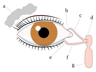
The lacrimal apparatus is a complex system in the eye that produces, stores, and drains tears. It consists of several components including:
1. Lacrimal glands: These are located in the upper outer part of the eyelid and produce tears to keep the eye surface moist and protected from external agents.
2. Tear ducts (lacrimal canaliculi): These are small tubes that drain tears from the surface of the eye into the lacrimal sac.
3. Lacrimal sac: This is a small pouch-like structure located in the inner part of the eyelid, which collects tears from the tear ducts and drains them into the nasolacrimal duct.
4. Nasolacrimal duct: This is a tube that runs from the lacrimal sac to the nose and drains tears into the nasal cavity.
The lacrimal apparatus helps maintain the health and comfort of the eye by keeping it lubricated, protecting it from infection, and removing any foreign particles or debris.
C57BL/6 (C57 Black 6) is an inbred strain of laboratory mouse that is widely used in biomedical research. The term "inbred" refers to a strain of animals where matings have been carried out between siblings or other closely related individuals for many generations, resulting in a population that is highly homozygous at most genetic loci.
The C57BL/6 strain was established in 1920 by crossing a female mouse from the dilute brown (DBA) strain with a male mouse from the black strain. The resulting offspring were then interbred for many generations to create the inbred C57BL/6 strain.
C57BL/6 mice are known for their robust health, longevity, and ease of handling, making them a popular choice for researchers. They have been used in a wide range of biomedical research areas, including studies of cancer, immunology, neuroscience, cardiovascular disease, and metabolism.
One of the most notable features of the C57BL/6 strain is its sensitivity to certain genetic modifications, such as the introduction of mutations that lead to obesity or impaired glucose tolerance. This has made it a valuable tool for studying the genetic basis of complex diseases and traits.
Overall, the C57BL/6 inbred mouse strain is an important model organism in biomedical research, providing a valuable resource for understanding the genetic and molecular mechanisms underlying human health and disease.
Keratoconjunctivitis Sicca, also known as dry eye syndrome, is a condition characterized by decreased quality and/or quantity of tears to lubricate and nourish the eye. This can result in discomfort, visual disturbance, and potentially damage to the ocular surface. It is often associated with inflammation of the conjunctiva and the cornea. The symptoms may include dryness, scratchiness, burning, foreign body sensation, pain, redness, blurred vision, and light sensitivity.
Airway remodeling is a term used to describe the structural changes that occur in the airways as a result of chronic inflammation in respiratory diseases such as asthma. These changes include thickening of the airway wall, increased smooth muscle mass, and abnormal deposition of extracellular matrix components. These alterations can lead to narrowing of the airways, decreased lung function, and increased severity of symptoms. Airway remodeling is thought to be a major contributor to the persistent airflow obstruction that is characteristic of severe asthma.
Respiratory hypersensitivity, also known as respiratory allergies or hypersensitive pneumonitis, refers to an exaggerated immune response in the lungs to inhaled substances or allergens. This condition occurs when the body's immune system overreacts to harmless particles, leading to inflammation and damage in the airways and alveoli (air sacs) of the lungs.
There are two types of respiratory hypersensitivity: immediate and delayed. Immediate hypersensitivity, also known as type I hypersensitivity, is mediated by immunoglobulin E (IgE) antibodies and results in symptoms such as sneezing, runny nose, and asthma-like symptoms within minutes to hours of exposure to the allergen. Delayed hypersensitivity, also known as type III or type IV hypersensitivity, is mediated by other immune mechanisms and can take several hours to days to develop after exposure to the allergen.
Common causes of respiratory hypersensitivity include mold spores, animal dander, dust mites, pollen, and chemicals found in certain occupations. Symptoms may include coughing, wheezing, shortness of breath, chest tightness, and fatigue. Treatment typically involves avoiding the allergen, if possible, and using medications such as corticosteroids, bronchodilators, or antihistamines to manage symptoms. In severe cases, immunotherapy (allergy shots) may be recommended to help desensitize the immune system to the allergen.
Lactobacillus helveticus is a species of gram-positive, facultatively anaerobic, rod-shaped bacteria that belongs to the lactic acid bacteria group. It is commonly found in various environments such as dairy products, plants, and the gastrointestinal tracts of animals, including humans.
L. helveticus has been widely used in the food industry for the production of fermented dairy products like cheese and yogurt due to its ability to produce lactic acid, break down proteins, and contribute to flavor development. It is also known for its potential health benefits when consumed as a probiotic, including improving gut health, boosting the immune system, and reducing symptoms of lactose intolerance.
In addition, L. helveticus has been studied for its potential role in mental health, with some research suggesting that it may help reduce anxiety and improve cognitive function. However, more research is needed to fully understand the mechanisms behind these effects and their clinical relevance.
Allergic conjunctivitis is a type of conjunctivitis (inflammation of the conjunctiva, the membrane that covers the white part of the eye and the inner surface of the eyelids) caused by an allergic reaction to substances such as pollen, dust mites, or pet dander. It is often characterized by redness, itching, watering, and swelling of the eyes. In some cases, the eyes may also become sensitive to light. Allergic conjunctivitis is not contagious and can be treated with medications such as antihistamines, decongestants, or mast cell stabilizers.
A mucous membrane is a type of moist, protective lining that covers various body surfaces inside the body, including the respiratory, gastrointestinal, and urogenital tracts, as well as the inner surface of the eyelids and the nasal cavity. These membranes are composed of epithelial cells that produce mucus, a slippery secretion that helps trap particles, microorganisms, and other foreign substances, preventing them from entering the body or causing damage to tissues. The mucous membrane functions as a barrier against infection and irritation while also facilitating the exchange of gases, nutrients, and waste products between the body and its environment.
A "knockout" mouse is a genetically engineered mouse in which one or more genes have been deleted or "knocked out" using molecular biology techniques. This allows researchers to study the function of specific genes and their role in various biological processes, as well as potential associations with human diseases. The mice are generated by introducing targeted DNA modifications into embryonic stem cells, which are then used to create a live animal. Knockout mice have been widely used in biomedical research to investigate gene function, disease mechanisms, and potential therapeutic targets.
BALB/c is an inbred strain of laboratory mouse that is widely used in biomedical research. The strain was developed at the Institute of Cancer Research in London by Henry Baldwin and his colleagues in the 1920s, and it has since become one of the most commonly used inbred strains in the world.
BALB/c mice are characterized by their black coat color, which is determined by a recessive allele at the tyrosinase locus. They are also known for their docile and friendly temperament, making them easy to handle and work with in the laboratory.
One of the key features of BALB/c mice that makes them useful for research is their susceptibility to certain types of tumors and immune responses. For example, they are highly susceptible to developing mammary tumors, which can be induced by chemical carcinogens or viral infection. They also have a strong Th2-biased immune response, which makes them useful models for studying allergic diseases and asthma.
BALB/c mice are also commonly used in studies of genetics, neuroscience, behavior, and infectious diseases. Because they are an inbred strain, they have a uniform genetic background, which makes it easier to control for genetic factors in experiments. Additionally, because they have been bred in the laboratory for many generations, they are highly standardized and reproducible, making them ideal subjects for scientific research.
Enterocytes are the absorptive cells that line the villi of the small intestine. They are a type of epithelial cell and play a crucial role in the absorption of nutrients from food into the bloodstream. Enterocytes have finger-like projections called microvilli on their apical surface, which increases their surface area and enhances their ability to absorb nutrients. They also contain enzymes that help digest and break down carbohydrates, proteins, and fats into smaller molecules that can be absorbed. Additionally, enterocytes play a role in the absorption of ions, water, and vitamins.
A carcinoid tumor is a type of slow-growing neuroendocrine tumor that usually originates in the digestive tract, particularly in the small intestine. These tumors can also arise in other areas such as the lungs, appendix, and rarely in other organs. Carcinoid tumors develop from cells of the diffuse endocrine system (also known as the neuroendocrine system) that are capable of producing hormones or biologically active amines.
Carcinoid tumors can produce and release various hormones and bioactive substances, such as serotonin, histamine, bradykinins, prostaglandins, and tachykinins, which can lead to a variety of symptoms. The most common syndrome associated with carcinoid tumors is the carcinoid syndrome, characterized by flushing, diarrhea, abdominal cramping, and wheezing or difficulty breathing.
Carcinoid tumors are typically classified as functional or nonfunctional based on whether they produce and secrete hormones that cause symptoms. Functional carcinoid tumors account for approximately 30% of cases and can lead to the development of carcinoid syndrome, while nonfunctional tumors do not produce significant amounts of hormones and are often asymptomatic until they grow large enough to cause local or distant complications.
Treatment options for carcinoid tumors depend on the location, size, and extent of the tumor, as well as whether it is functional or nonfunctional. Treatment may include surgery, medications (such as somatostatin analogs, chemotherapy, or targeted therapies), and radiation therapy. Regular follow-up with imaging studies and biochemical tests is essential to monitor for recurrence and assess treatment response.
The Fluorescent Antibody Technique (FAT), Indirect is a type of immunofluorescence assay used to detect the presence of specific antigens in a sample. In this method, the sample is first incubated with a primary antibody that binds to the target antigen. After washing to remove unbound primary antibodies, a secondary fluorescently labeled antibody is added, which recognizes and binds to the primary antibody. This indirect labeling approach allows for amplification of the signal, making it more sensitive than direct methods. The sample is then examined under a fluorescence microscope to visualize the location and amount of antigen based on the emitted light from the fluorescent secondary antibody. It's commonly used in diagnostic laboratories for detection of various bacteria, viruses, and other antigens in clinical specimens.
Lectins are a type of proteins that bind specifically to carbohydrates and have been found in various plant and animal sources. They play important roles in biological recognition events, such as cell-cell adhesion, and can also be involved in the immune response. Some lectins can agglutinate certain types of cells or precipitate glycoproteins, while others may have a more direct effect on cellular processes. In some cases, lectins from plants can cause adverse effects in humans if ingested, such as digestive discomfort or allergic reactions.
Immunohistochemistry (IHC) is a technique used in pathology and laboratory medicine to identify specific proteins or antigens in tissue sections. It combines the principles of immunology and histology to detect the presence and location of these target molecules within cells and tissues. This technique utilizes antibodies that are specific to the protein or antigen of interest, which are then tagged with a detection system such as a chromogen or fluorophore. The stained tissue sections can be examined under a microscope, allowing for the visualization and analysis of the distribution and expression patterns of the target molecule in the context of the tissue architecture. Immunohistochemistry is widely used in diagnostic pathology to help identify various diseases, including cancer, infectious diseases, and immune-mediated disorders.
Barrett esophagus is a condition in which the tissue lining of the lower esophagus changes, becoming more like the tissue that lines the intestines (intestinal metaplasia). This change can increase the risk of developing esophageal adenocarcinoma, a type of cancer. The exact cause of Barrett esophagus is not known, but it is often associated with long-term gastroesophageal reflux disease (GERD), also known as chronic acid reflux.
In Barrett esophagus, the normal squamous cells that line the lower esophagus are replaced by columnar epithelial cells. This change is usually detected during an upper endoscopy and biopsy. The diagnosis of Barrett esophagus is confirmed when the biopsy shows intestinal metaplasia in the lower esophagus.
It's important to note that not everyone with GERD will develop Barrett esophagus, and not everyone with Barrett esophagus will develop esophageal cancer. However, if you have been diagnosed with Barrett esophagus, your healthcare provider may recommend regular endoscopies and biopsies to monitor the condition and reduce the risk of cancer. Treatment options for Barrett esophagus include medications to control acid reflux, lifestyle changes, and in some cases, surgery.
 Goblet cell
Goblet cell carcinoid
Simple columnar epithelium
Intestinal villus
Epiglottis
Interleukin 13
Gastrointestinal wall
SPDEF
Ileum
Mucous gland
Gene silencing
Trefoil factor 3
Neuroendocrine tumor
Guttural pouch
Nasal concha
Alkaliphilus
Rhinitis
List of intestinal stem cell marker genes
Trefoil factor 1
Bart Deplancke
Bronchitis
Trefoil factor 2
Club cell
Immune dysregulation
Chloride channel blocker
SCIN
Peyer's patch
List of intestinal epithelial differentiation genes
CLCA1
FOXJ1
Hyperplastic polyp
Goblet cell - Wikipedia
goblet cell | Encyclopedia.com
 Lung - Metaplasia, Goblet Cell - Nonneoplastic Lesion Atlas
 Goblet Cells - Medical Dictionary online-medical-dictionary.org
 Goblet Cell Carcinoid
 Airway epithelial SPDEF integrates goblet cell differentiation and pulmonary Th2 inflammation
 Goblet Cell Density Key in Smokers' Prognosis
 JCI Insight -
A nerve-goblet cell association promotes allergic conjunctivitis through rapid antigen passage
JCI Insight -
A nerve-goblet cell association promotes allergic conjunctivitis through rapid antigen passage
 Generation of functional conjunctival epithelium, including goblet cells, from human iPSCs - List Labs
 Goblet Cell from the collection of Guild of Natural Science Illustrators | Artwork Archive
 Goblet Cells | Profiles RNS
 Respiratory Goblet Cell Cell Types - CZ CELLxGENE CellGuide
 Harry Potter and the Goblet of Fire Film Cell Mini Montage
Optimization of goblet cell density quantification methods - Centre for Ocular Research & Education
 Understanding neuroendocrine medical terms
Age-related changes of human conjunctiva on in vivo confocal microscopy | British Journal of Ophthalmology
 The human Glandular epithelial cells - The Human Protein Atlas
 DNA methyltransferase 3A controls intestinal epithelial barrier function and regeneration in the colon | Nature Communications
Goblet cell carcinoid of the appendix: Two case reports and a review of the literature<...
 Pathology of Serrated Colon Adenomas: Definition, Epidemiology, Etiology
 Appendiceal goblet cell carcinoma has marginal advantages from perioperative chemotherapy: a population-based study with an...
 Neuroendocrine Markers Insulinoma-Associated Protein 1, Chromogranin, Synaptophysin, and CD56 Show Rare Positivity in...
Baseline goblet cell mucin secretion in the airways exceeds stimulated secretion over extended time periods, and Is sensitive...
 Expression profiling of ileal mucosa in asthma reveals upregulation of innate immunity and genes characteristic of Paneth and...
 Ethanol Extract of Annona Muricata L. Leaves Reduced Goblet Cell in Bronchus of Animal Model of Smoking, IJSR, Call for Papers,...
 The physicochemical properties of preserved and preservative-free latanoprost eye drops and their impact on human conjunctival...
 Plus it
 How the BRAF V600E Mutation Defines a Distinct Subgroup of Colorectal Cancer: Molecular and Clinical Implications
Transcriptomic response of primary human airway epithelial cells to flavoring chemicals in electronic cigarettes | Scientific...
Carcinoid4
- Goblet cell carcinoid or carcinoma (GCC) is a rare tumor found incidentally during routine management of acute appendicitis. (biu.ac.il)
- Adenocarcinoma ex-goblet cell carcinoid (AdexGCC) was considered a neuroendocrine adenocarcinoma, despite majority of tumor cells being negative for conventional neuroendocrine markers such as chromogranin and synaptophysin. (gastrores.org)
- Appendiceal goblet cell carcinoid (GCC) and adenocarcinoma ex-goblet cell carcinoid (AdexGCC) have previously been described as two entities in a spectrum of neoplasms showing both glandular/mucinous and neuroendocrine differentiation [ 1 ]. (gastrores.org)
- About two thirds of appendix tumours are carcinoid tumours which develop in hormone-producing cells that are present in many parts of the body, including the appendix. (cancerindex.org)
Hyperplasia8
- There has been much debate regarding the proper term for this lesion (metaplasia vs. hyperplasia), but since goblet cells are not normally present in the epithelium of the smaller airways and are not normally visible in the upper airways, it should be diagnosed as goblet cell metaplasia in NTP studies. (nih.gov)
- The researchers wanted to examine the relationship between goblet cell hyperplasia and smoking to find out if people with chronic bronchitis and current smokers would have more GCD, whether or not they had any airflow obstruction. (hcplive.com)
- Another possible conclusion of this study is that "goblet cell hyperplasia does not contribute to airflow obstruction but rather that active smoking is its primary determinant. (hcplive.com)
- Although the group of participants in this study was small, the researchers contend that excluding people who may have conditions linked to lower airway goblet cell hyperplasia such as upper airway disease or recent exacerbations, adds strength to the findings. (hcplive.com)
- Despite the possible weaknesses, the researchers "were able to demonstrate in this small sample that current smoking and chronic bronchitis, and not airflow obstruction, were independent factors associated with goblet cell hyperplasia. (hcplive.com)
- Pathomorphological studies of gastrointestinal mucosa of asthma patients indicated increased activity of mucus-producing cells (as in the bronchi), barrier degeneration, and hyperplasia [ 10 ]. (biomedcentral.com)
- Chronic inflammation in COPD leads to airways remodeling which is mark by Goblet cell hyperplasia and mucous overproduction. (ijsr.net)
- Airway mucus obstruction: mucin glycoproteins, MUC gene regulation and goblet cell hyperplasia. (nih.gov)
Epithelium7
- Biased differentiation of airway basal cells in the respiratory epithelium, into goblet cells plays a key role in the excessive mucus production, known as mucus hypersecretion seen in many respiratory diseases, including chronic bronchitis, and asthma. (wikipedia.org)
- goblet cell A goblet-shaped cell, found in the epithelium of the intestine and respiratory system in mammals and in the epidermis of fish, that secretes mucus . (encyclopedia.com)
- Several functional components of the epithelium contribute to this unique role, among them the secretion of a mucus layer and life-long regeneration of the different epithelial cell types from a stem cell compartment, allowing for quick repair of barrier defects 2 . (nature.com)
- Pulmonary exposure to NQ dose-dependently aggravated antigen-related airway inflammation, as characterised by infiltration of eosinophils and lymphocytes around the airways and an increase in goblet cells in the bronchial epithelium. (ersjournals.com)
- Numerous goblet cells are intermixed with the epithelium. (medscape.com)
- The follicle-associated epithelium of Salmonella serovar Choleraesuis inoculated loops responded with more filopodia and lamellipodia while exhibiting less cell swelling than Salmonella serovar Typhimurium. (usda.gov)
- Together with mucus-secreting goblet cells within the stratified epithelium, melanocytic cells are found in the basal layer of the conjunctiva. (medscape.com)
Mucin8
- Goblet cells are simple columnar epithelial cells that secrete gel-forming mucins, like mucin 5AC. (wikipedia.org)
- The goblet cell is highly polarized with the nucleus and other organelles concentrated at the base of the cell and secretory granules containing mucin, at the apical surface. (wikipedia.org)
- In the conjunctiva goblet cells are a source of mucin in tears and they also secrete different types of mucins onto the ocular surface. (wikipedia.org)
- The cytoplasm of goblet cells tends to be displaced toward the basal end of the cell body by the large mucin granules, which accumulate near the apical surface of the cell along the Golgi apparatus, which lies between the granules and the nucleus. (wikipedia.org)
- In mucicarmine stains, deep red mucin is found within goblet cell bodies. (wikipedia.org)
- To investigate the impact on human conjunctival goblet cell (GC) survival and mucin release for benzalkonium chloride (BAK) preserved and preservative-free (PF) 0.005% (w/v) latanoprost (LT) eye drops, and to compare the eye drops' physicochemical properties. (ku.dk)
- in this study, CIC with a separation and distortion of epithelial cells specificity of 94% and sensitivity of 93%, and losses of mucin-secreting goblet cells, was used [14]. (who.int)
- CIC the presence and density of goblet cells or is not, however, usually used in children mucin spots and density, size and shape of below 24 months due to the inability of the epithelial cells in relation to specimen area child to cooperate. (who.int)
Conjunctiva1
- In the present study, we found that the instillation of pollen shells rapidly induces a large number of goblet cell-associated antigen passages (GAPs) in the conjunctiva. (jci.org)
Mucous3
- An intestinal gland from the human intestine with goblet cells visible Goblet cell in ileum Section of mouse intestine, mucus of goblet cells in blue Goblet cells in large intestine The main role of goblet cells is to secrete mucus in order to protect the mucous membranes where they are found. (wikipedia.org)
- Rats and mice have very low numbers of goblet (mucous) cells in their lungs. (nih.gov)
- Goblet cell carcinoma (synonyms: adenocarcinoid, mucous adenocarcinoid) belongs to the last category. (wjgnet.com)
Epithelial cell2
- A glandular epithelial cell or a unicellular gland. (online-medical-dictionary.org)
- The repair phase after Cl2 exposure was characterized by increased airway epithelial cell proliferation, measured by immunoreactive proliferating cell nuclear antigen (PCNA), with maximal proliferation occurring 5 days after Cl2 exposure. (cdc.gov)
Airways5
- Anomalies in the number of goblet cells are associated with changes in the secretion of mucins, which can result in many of the abnormalities seen in asthma patients, such as clogged airways due to mucus hypersecretion, and eventual loss of lung function. (wikipedia.org)
- Epithelial cells that line the conducting airways provide the initial barrier and innate immune responses to the abundant particles, microbes, and allergens that are inhaled throughout life. (nih.gov)
- The transcription factors SPDEF and FOXA3 are both selectively expressed in epithelial cells lining the conducting airways, where they regulate goblet cell differentiation and mucus production. (nih.gov)
- Our intestines and airways are studded with goblet cells. (eurasiareview.com)
- Mucus cells lining abnormal airways from patients with IPF expressed MUC5B and SOX2 but lacked SAM pointed domain containing ETS transcription factor (SPDEF) and forkhead box A3(FOXA3). (bmj.com)
Dendritic Cells2
- Antigen acquisition by stromal cells, including macrophages and CD11b+ dendritic cells, correlated with surface GAP formation. (jci.org)
- The morphology and densities of conjunctival epithelial cells, goblet cells (GCs), dendritic cells (DCs) and the positive rate of conjunctival microcysts were analysed. (bmj.com)
Appendix4
- After 7 days, highly dense 3D cultures were either split 1:2 and embedded in Matrigel, frozen in liquid nitrogen for further use, or dissociated into single cell suspension and plated as undifferentiated monolayers, as described previously ( 2 ) (Technical Appendix Figure 1). (cdc.gov)
- Pathological examination of the appendix revealed the presence of adenocarcinoma with goblet cells and a Ki-67 index of 25% (grade 3) and 15% (grade 2), respectively. (biu.ac.il)
- Cancer of the Appendix (also known as appendiceal cancer) occurs when cells in the appendix become abnormal and multiply without control. (cancerindex.org)
- Less common tumours of the appendix include signet-ring cell adenocarcinoma and paraganglioma . (cancerindex.org)
Intestinal10
- Goblet cells are found scattered among the epithelial lining of organs, such as the intestinal and respiratory tracts. (wikipedia.org)
- DNMT3A expression is downregulated in intestinal epithelial cells from IBD patients and upon tumor necrosis factor treatment in murine intestinal organoids. (nature.com)
- Genetic deletion of Dnmt3a in intestinal epithelial cells ( Dnmt3a ΔIEC ) in mice confirms the phenotype of an altered epithelial ultrastructure with shortened apical-junctional complexes, reduced Goblet cell numbers and increased intestinal permeability in the colon in vivo. (nature.com)
- Intestinal epithelial cells (IECs) have evolved to build a complex physiological barrier that separates the intestinal lumen from the underlying mucosal tissue. (nature.com)
- In children diagnosed with IBD, genome-wide DNA methylation profiling of purified intestinal epithelial cells clearly distinguishes between healthy individuals and the IBD group 8 . (nature.com)
- The intricate molecular mechanisms involved in the regenerative process of the normal intestine and the identity of putative somatic intestinal stem cells have become clear. (mdpi.com)
- The research, published in Cell , shows that pain neurons in the mouse gut regulate the presence of protective mucus under normal conditions and stimulate intestinal cells to release more mucus during states of inflammation. (eurasiareview.com)
- The new research found that intestinal goblet cells release protective mucus when triggered by direct interaction with pain-sensing neurons in the gut. (eurasiareview.com)
- Serovars exhibited differences in early invasion of cells lining the intestinal wall. (usda.gov)
- We investigated if infectivity of GII.12 HuNoV in gnotobiotic (Gn) pigs is altered by intestinal colonization with Escherichia fergusonii known to express HBGA A and H on their cell surface. (bvsalud.org)
Neuroendocrine2
- CgA is a chemical/hormone found inside neuroendocrine cells. (christie.nhs.uk)
- New insights emerged last decade that enriched our knowledge regarding the biological behavior of appendiceal neuroendocrine tumors (NETs), which range from totally benign tumors less than 1cm to goblet cell carcinomas which behave similarly to colorectal adenocarcinoma. (wjgnet.com)
Increase in goblet cells1
- No increase in goblet cells occurred at any time point. (cdc.gov)
Airway epithelial cells2
- Here, we show that expression of SPDEF or FOXA3 in airway epithelial cells in neonatal mice caused goblet cell differentiation, spontaneous eosinophilic inflammation, and airway hyperresponsiveness to methacholine. (nih.gov)
- Conclusion Expression of SOX2 and MUC5B and lack of SPDEF in atypically differentiated cells of bronchiolised distal airspaces are consistent with abnormal programming of airway epithelial cells in IPF. (bmj.com)
Metaplasia5
- Goblet cell metaplasia may occur in association with infectious agents or exposure to inhaled irritants in both rats and mice. (nih.gov)
- The preferred terminology for this lesion is Lung - Metaplasia, Goblet cell. (nih.gov)
- Lung, Bronchiole - Metaplasia, Goblet cell from a male Sprague-Dawley rat in an acute study. (nih.gov)
- Together, our results indicate that SPDEF causes goblet cell differentiation and Th2 inflammation during postnatal development and is required for goblet cell metaplasia and normal Th2 inflammatory responses to HDM aeroallergen. (nih.gov)
- Singularly, SPDEF, a transcription factor associated with mucus metaplasia, was rarely detected in mucus cells in IPF. (bmj.com)
Carcinoma1
- PurposeThe aim is to clarify the use of perioperative chemotherapy in resectable goblet cell carcinoma (GCC).MethodsA retrospective study was carried out based on the Surveillance, Epidemiology, and End Results study. (unibo.it)
Glycoproteins2
- Goblet cells accomplish this by secreting mucins, large glycoproteins formed mostly by carbohydrates. (wikipedia.org)
- Mucins, the main constituents of the mucus layer, are glycoproteins synthesized by goblet cells. (scielo.br)
Respiratory2
- Goblet cells are typically found in the respiratory, reproductive and gastrointestinal tracts and are surrounded by other columnar cells. (wikipedia.org)
- Goblet cells are specialized cells found in the respiratory and digestive tracts that produce and secrete mucus, which plays a vital role in protecting and lubricating these surfaces. (proprofs.com)
Intestine2
- Harvard Medical School researchers have analyzed the molecular crosstalk between pain fibers in the gut and goblet cells that line the walls of the intestine. (eurasiareview.com)
- The virus has long been established as a pathogen of the gastrointestinal tract, targeting small intestine epithelial cells and leading to diarrhea, nausea, and vomiting. (bvsalud.org)
Secretion1
- The goblet cells mainly use the merocrine method of secretion, secreting vesicles into a duct, but may use apocrine methods, budding off their secretions, when under stress. (wikipedia.org)
Tumor1
- INSM1 exhibits low expression in AdexGCCs and is expressed by a lower proportion of tumor cells compared to chromogranin and synaptophysin. (gastrores.org)
Lung3
- Increased Il4, Il13, Ccl17, and Il25 expression was accompanied by recruitment of Th2 lymphocytes, group 2 innate lymphoid cells, and eosinophils to the lung. (nih.gov)
- The current authors have recently shown that PQ induces recruitment of inflammatory cells, such as eosinophils and neutrophils, into the lung with the lung expression of pro-inflammatory molecules such as interleukin (IL)-5 and eotaxin in vivo 24 . (ersjournals.com)
- In this work, pathways governing mucus cell differentiation were investigated in lung tissues from patients with IPF and other chronic pulmonary disorders. (bmj.com)
Ileum2
- Significant differential expression was found in the ileum alone, and not in any other cell/tissue types. (biomedcentral.com)
- Additionally, inoculated pigs had HRV-specific IgM antibody-secreting cells present in the ileum, tonsils, and facial lymphoid tissues. (bvsalud.org)
Secrete mucus3
- There are other cells that secrete mucus (such as the foveolar cells of the stomach) but these are distinguished histologically from goblet cells. (wikipedia.org)
- Goblet cells secrete MUCUS . (online-medical-dictionary.org)
- The best example of physiology among the given choices is "Goblet cells secrete mucus. (proprofs.com)
Induce3
- Previous studies have indicated that organic chemicals extracted from DEP result in induction of apoptosis 11 , increase oxidative stress 12 , and induce the production of inflammatory cells 13 - 15 through the release of pro-inflammatory molecules in vitro 15 , 16 . (ersjournals.com)
- The work shows that chemical signals from pain neurons induce goblet cells to release protective mucus that coats the gut and shields it from damage. (eurasiareview.com)
- Certain bacterial strains from the microbiome induce a potent, antigen-specific T cell response1-5. (bvsalud.org)
Mice4
- SPDEF was required for goblet cell differentiation and pulmonary Th2 inflammation in response to house dust mite (HDM) extract, as both were decreased in neonatal and adult Spdef(-/-) mice compared with control animals. (nih.gov)
- When researchers gave mice capsaicin, the main ingredient in chili peppers known for its ability to trigger intense, acute pain, the mice's pain neurons got swiftly activated, causing goblet cells to release abundant amounts of protective mucus. (eurasiareview.com)
- By contrast, mice lacking either pain neurons or goblet cell receptors for CGRP were more susceptible to colitis, a form of gut inflammation. (eurasiareview.com)
- Here, we colonize germ-free mice with complex defined communities (roughly 100 bacterial strains) and profile T cell responses to each strain. (bvsalud.org)
Mucus layer1
- Mucins are continuously made and secreted by goblet cells in order to repair and replace the existing mucus layer. (wikipedia.org)
Density1
- The researchers also found there was "a significant correlation between pack-year history of smoking with goblet cell density. (hcplive.com)
SPDEF1
- Methods Pathways governing airway mucus cell differentiation include SRY (sex determining region Y)-box 2 (SOX2), Notch, forkhead box A3(FOXA3)/SAM pointed domain containing ETS transcription factor (SPDEF), epidermal growth factor (EGF) and the EGF-related neuregulins NRG1α and NRG1β. (bmj.com)
Cytoplasm1
- The cytoplasm is vacuolated in some cells. (medscape.com)
Innate lymphoid cells1
- A hallmark of the predominant asthma subtype is the stimulation of the adaptive immune system [ 2 ], specifically a Th2-type response, with involvement of interleukin (IL)-4 and IL-13, as well as antigen-specific immunoglobulin (Ig) type E. Type 2 innate immunity typically involves type 2 innate lymphoid cells, especially in older patients. (biomedcentral.com)
Differentiation3
- The authors sought to identify mechanisms driving the differentiation of mucus cells during the bronchiolisation process. (bmj.com)
- Neuregulin1α, which drives mucus cell differentiation in vitro, was expressed in normal airway submucosal glands and in lungs from patients with IPF. (bmj.com)
- Human Fibrinogen for Maintenance and Differentiation of Induced Pluripotent Stem Cells in Two Dimensions and Three Dimensions. (mayo.edu)
Genes5
- marker genes Computational marker genes for this cell type are unavailable at this time. (cziscience.com)
- Ileal mucosa in asthma exhibits a specific transcriptomic profile, which includes the overexpression of innate immune genes, mostly characteristic of Paneth and goblet cells, in addition to other changes that may resemble Crohn's disease. (biomedcentral.com)
- Using RNA-Seq, we identified a total of 163 and 568 differentially expressed genes in primary normal human bronchial epithelial (NHBE) cells that were exposed to diacetyl and 2,3-pentanedione, respectively. (nature.com)
- Consistent with this, qRT-PCR confirmed that the expression of multiple genes involved in cilia biogenesis was significantly downregulated by diacetyl and 2,3-pentanedione in NHBE cells. (nature.com)
- Our scientists pursue every aspect of cancer research-from exploring the biology of genes and cells, to developing immune-based treatments, uncovering the causes of metastasis, and more. (mskcc.org)
Chronic1
- Acute and chronic inflammatory cells are found within these fronds. (medscape.com)
Antigen1
- 14 As the strongest antigen presenting cell (APCs), DCs play an important role in the immunity of the ocular surface. (bmj.com)
Bronchial1
- In this study, we utilized primary normal human bronchial epithelial (NHBE) cells that are cultured at an air-liquid interface (ALI) to mimic the in vivo airway characteristics 30 . (nature.com)
Typically1
- but this stain is not typically used to identify goblet cells, and Alcian blue is preferred. (nih.gov)
Specificity2
- Although DNMT3A and DNMT3B are highly homologous, their redundancy seems to be limited, and their unique biological function, e.g., specificity for methylation patterns in distinct cell types, is still elusive 9 . (nature.com)
- However, the specificity of microbiome-induced T cells has not been explored at the strain level across the gut community. (bvsalud.org)
Inflammatory cells1
- Numerous goblet cells are seen along with acute inflammatory cells. (medscape.com)
Found2
- They found that the surfaces of goblet cells contain a type of receptor, called RAMP1, that ensures the cells can respond to adjacent pain neurons, which are activated by dietary and microbial signals, as well as mechanical pressure, chemical irritation or drastic changes in temperature. (eurasiareview.com)
- These RAMP1 receptors, the researchers found, are also present in both human and mouse goblet cells, thus rendering them responsive to pain signals. (eurasiareview.com)
Bodies1
- These hyaline bodies might represent occluded goblet cells with the unreleased contents as reported by Kessing, 10 which were considered to be more common with increasing age. (bmj.com)
Involves1
- BRAF is a protein kinase and part of the MAP kinase signalling cascade which involves transduction of a growth signal from the cell membrane to the nucleus via a chain of protein kinases and is responsible for cellular proliferation and survival. (hindawi.com)
Intestines1
- Our work shows how pain-mediating nerves in the gut talk to nearby epithelial cells that line the intestines," said study senior investigator Isaac Chiu , associate professor of immunobiology in the Blavatnik Institute at HMS. (eurasiareview.com)
Tumour2
- How the tumour cells look under the microscope. (christie.nhs.uk)
- Ki-67 is a measure of the proliferation index (number of dividing/proliferating cells) of the tumour and can be determined from looking at the tumour under the microscope. (christie.nhs.uk)
Receptor1
- This signal transduction pathway is initiated by epidermal growth factor ligands binding to and activating receptor tyrosine kinases (RTK) at the cell membrane. (hindawi.com)
Acinar1
- In the lacrimal glands, mucus is synthesized by acinar cells instead. (wikipedia.org)
Abnormal1
- Cells look very abnormal and are likely to grow more quickly. (christie.nhs.uk)
Acute1
- Some authors have reported on the use of laboratory tests including sedimentation rate, white blood cell counts, and C-reactive protein levels to help diagnose acute sinusitis. (medscape.com)
Researchers1
- To clarify just how this protective crosstalk occurs, the researchers analyzed the behavior of goblet cells in the presence and in the absence of pain neurons. (eurasiareview.com)