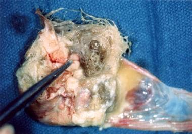
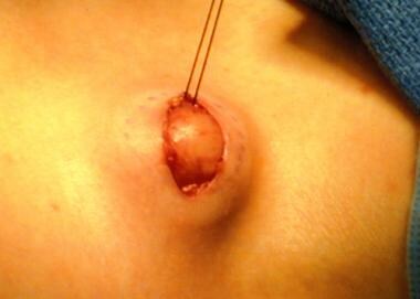
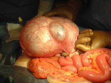
Giant Lymph Node Hyperplasia
Lymph Nodes
Human herpesvirus 8 in hematologic diseases. (1/254)
Human herpesvirus type 8 (HHV-8), also known as Kaposi's sarcoma-associated herpesvirus (KSHV) is a new member of the g-herpesvirus family. It is an unusual herpesvirus in that it carries a large number of genes that encode oncoproteins or cell signaling proteins. In addition to being the causative agent of both HIV-associated and non-HIV-associated Kaposi's sarcoma this DNA tumor virus has been implicated in the pathogenesis of several diseases. These include multiple myeloma (MM), Waldenstom's macroglobulinemia (WM), multicentric Castleman's disease (MCD), body cavity-based lymphoma (BCBL), and various other conditions such as sarcoidosis and pemphigus. While the causative role of the viral infection is fairly certain in the development of BCBL and multicentric Castleman's disease, HHV-8 may act through a different mechanism to induce plasma cell malignancies. It has been suggested though the finding is still controversial - that infection of bone marrow stromal dendritic cells by HHV-8 might be a key factor in the etiology and pathogenesis of monoclonal gammopathies. The aim of this review is to provide a short introduction into the tumorigenic potential of HHV-8 as well as to detail the available data and possible mechanisms on the involvement of this virus in different hematologic diseases. (+info)Cellular tropism and viral interleukin-6 expression distinguish human herpesvirus 8 involvement in Kaposi's sarcoma, primary effusion lymphoma, and multicentric Castleman's disease. (2/254)
Human herpesvirus 8 (HHV-8) infection has been implicated in the etiology of Kaposi's sarcoma (KS), primary effusion lymphoma (PEL), and multicentric Castleman's disease (MCD), three diseases that frequently develop in immunocompromised, human immunodeficiency virus-positive individuals. One hypothesis that would account for different pathological manifestations of infection by the same virus is that viral genes are differentially expressed in heterogeneous cell types. To test this hypothesis, we analyzed the localization and levels of expression of two viral genes expressed in latent and lytic infections and the viral homologue of interleukin-6 (vIL-6). We show that PEL parallels KS in the pattern of latent and lytic cycle viral gene expression but that the predominant infected cell type is a B cell. We also show that MCD differs from KS not only in the infected cell type (B-cell and T-cell lineage) but also in the pattern of viral gene expression. Only a few cells in the lesion are infected and all of these cells express lytic-cycle genes. Of possibly greater significance is the fact that in a comparison of KS, PEL, and MCD, we found dramatic differences in the levels of expression of vIL-6. Interleukin-6 is a B-cell growth and differentiation factor whose altered expression has been linked to plasma cell abnormalities, as well as myeloid and lymphoid malignancies. Our findings support the hypothesis that HHV-8 plays an important role in the pathogenesis of PEL and MCD, in which vIL-6 acts as an autocrine or paracrine factor in the lymphoproliferative processes common to both. (+info)Distribution of human herpesvirus-8 latently infected cells in Kaposi's sarcoma, multicentric Castleman's disease, and primary effusion lymphoma. (3/254)
Human herpesvirus 8 (HHV-8, also called KSHV) is linked to the etiopathogenesis of Kaposi's sarcoma (KS), multicentric Castleman's disease (MCD), and primary effusion lymphoma (PEL). The universal presence of HHV-8 in early KS has not yet been shown. We used a mAb (LN53) against latent nuclear antigen-1 (LNA-1) of HHV-8 encoded by ORF73 to study the distribution of the cell types latently infected by HHV-8 in patch, plaque, and nodular KS, MCD, and PEL. In early KS, HHV-8 is present in <10% of cells forming the walls of ectatic vessels. In nodular KS, HHV-8 is present in cells surrounding slit-like vessels and in >90% of spindle cells, but not in normal vascular endothelium. In addition, HHV-8 colocalizes with vascular endothelial growth factor receptor-3 (VEGFR-3), a marker of lymphatic and precursor endothelium. In early KS lesions, VEGFR-3 is more extensively expressed than LNA-1, indicating that HHV-8 is not inducing the proliferation of VEGFR-3-positive endothelium directly. In MCD, HHV-8 is present in mantle zone large immunoblastic B cells. No staining for LNA-1 is seen in samples from multiple myeloma, prostate cancer, and angiosarcoma, supporting the absence of any etiological link between these diseases and HHV-8. (+info)Kaposi's sarcoma-associated herpesvirus infection in the lung in multicentric Castleman's disease. (4/254)
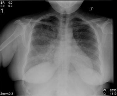
A 32-year-old female was admitted for evaluation of multiple infiltrates on a chest radiograph. A diagnosis of multicentric Castleman's disease was made on the basis of typical clinical manifestations. Transbronchial lung biopsy (TBLB) revealed histological findings reported in lymphocytic interstitial pneumonia. Both the polymerase chain reaction and in situ hybridization with a probe specific for Kaposi's sarcoma-associated herpesvirus (KSHV) sequences demonstrated the presence of KSHV in the TBLB sample. (+info)Human herpesvirus 8 infection in patients with POEMS syndrome-associated multicentric Castleman's disease. (5/254)
The polyneuropathy, organomegaly, endocrinopathy, M protein, skin changes (POEMS) syndrome is a rare multisystemic disorder associated with osteosclerotic myeloma and multicentric Castleman's disease (MCD). Human herpesvirus type 8 (HHV-8) DNA sequences have been detected in lymph nodes of about 40% of human immunodeficiency virus (HIV)-negative patients with MCD, and in bone marrow stromal cells of patients with multiple myeloma. Considering these data, we investigated the presence of HHV-8 in 18 patients with POEMS syndrome (9 with MCD), by nested polymerase chain reaction (N-PCR) to detect DNA sequenses in various cells and tissues obtained by biopsy or at autopsy (13 patients, of whom 7 had MCD), and by an immunofluorescence assay to detect anti-HHV-8 IgG antibodies in blood (18 patients, of whom 9 had MCD). Detection of HHV-8 DNA was performed using three different N-PCR, targeting nonoverlapping regions in open reading frame (ORF) 25 and ORF26. Seven of 13 (54%) POEMS patients had HHV-8 DNA sequences in their tissues, as assessed by all three N-PCR, and 9 of 18 (50%) had circulating anti-HHV-8 antibodies. HHV-8 was mainly detected in the subset of POEMS patients with MCD (6 of 7 [85%] for DNA sequences; 7 of 9 [78%] for antibodies). The percentage of positive N-PCR was higher in lymph nodes than in bone marrow samples (P <.02). Sequencing of amplicons showed a homogeneous restricted variability in the ORF26 region, characteristic of the minority subgroup B defined by Zong, and responsible for isoleucine and glycine substitutions at amino acid positions 134 and 167. These findings strongly suggest an association of HHV-8 infection with POEMS syndrome-associated MCD. (+info)Treatment of multicentric Castleman's disease complicated by the development of non-Hodgkin's lymphoma with high-dose chemotherapy and autologous peripheral stem-cell support. (6/254)
BACKGROUND: Castleman's disease or angiofollicular lymph node hyperplasia is a rare entity with a localized/unicentric or a generalized/multicentric presentation. While surgery is curable for most localized presentations, there is limited information regarding the optimal management of the multicentric type. The latter type is associated with a poor prognoses and can be associated with the development of lymphoma and infections. PATIENTS AND METHODS: In this report we describe a case of multicentric Castleman's disease who failed steroids and chemotherapy and developed a follicular mixed lymphoma. He was treated with high-dose chemotherapy with autologous stem-cell support and remains disease at four years of follow-up. CONCLUSIONS: A long-term durable remission may be possible with high dose chemotherapy with stem-cell support. This treatment modality should be considered an option in the management of multicentric Castleman's disease. (+info)Development of a calcifying fibrous pseudotumour within a lesion of Castleman disease, hyaline-vascular subtype. (7/254)
A nine year old boy with localised Castleman disease of the hyaline-vascular subtype developed a calcifying fibrous pseudotumour. This pathological association does not appear to have been described before. In this case, the development of this very unusual soft tissue tumour-like process was thought to be related to a previous fine needle aspiration biopsy, which was performed because of lymphadenopathy localised to the right inguinal area. This case provides further evidence of the reactive nature of calcifying fibrous pseudotumour and also broadens the pathological spectrum of the stromal cell proliferation that occasionally supervenes within lesions of Castleman disease, hyaline-vascular type. (+info)Improvement in Castleman's disease by humanized anti-interleukin-6 receptor antibody therapy. (8/254)
Castleman's disease, an atypical lymphoproliferative disorder, can be classified into 2 types: hyaline-vascular and plasma cell types according to the histologic features of the affected lymph nodes. The plasma cell type is frequently associated with systemic manifestations and is often refractory to systemic therapy including corticosteroids and chemotherapy, particularly in multicentric form. Dysregulated overproduction of interleukin-6 (IL-6) from affected lymph nodes is thought to be responsible for the systemic manifestations of this disease. Therefore, interference with IL-6 signal transduction may constitute a new therapeutic strategy for this disease. We used humanized anti-IL-6 receptor antibody (rhPM-1) to treat 7 patients with multicentric plasma cell or mixed type Castleman's disease. All patients had systemic manifestations including secondary amyloidosis in 3. With the approval of our institution's ethics committee and the consent of the patients, they were treated with 50 to 100 mg rhPM-1 either once or twice weekly. Immediately after administration of rhPM-1, fever and fatigue disappeared, and anemia as well as serum levels of C-reactive protein (CRP), fibrinogen, and albumin started to improve. After 3 months of treatment, hypergammaglobulinemia and lymphadenopathy were remarkably alleviated, as were renal function abnormalities in patients with amyloidosis. Treatment was well tolerated with only transient leukopenia. Histopathologic examination revealed reduced follicular hyperplasia and vascularity after rhPM-1 treatment. The pathophysiologic significance of IL-6 in Castleman's disease was thus confirmed, and blockade of the IL-6 signal by rhPM-1 is thought to have potential as a new therapy based on the pathophysiologic mechanism of multicentric Castleman's disease. (Blood. 2000;95:56-61) (+info)Giant lymph node hyperplasia, also known as Castlemans disease, is a rare benign condition characterized by the abnormal enlargement of lymph nodes due to an overgrowth of cells. It can affect people of any age but is more commonly seen in young adults and children.
The enlarged lymph nodes caused by this condition are typically round, firm, and mobile, and they may be found in various locations throughout the body, including the neck, chest, abdomen, and pelvis. In some cases, the enlarged lymph nodes may cause symptoms such as pain, pressure, or difficulty swallowing, depending on their location.
Giant lymph node hyperplasia can be classified into two main types: unicentric and multicentric. Unicentric Castleman's disease affects a single group of lymph nodes, while multicentric Castleman's disease affects multiple groups of lymph nodes throughout the body.
The exact cause of giant lymph node hyperplasia is not fully understood, but it is thought to be related to an overactive immune response. In some cases, it may be associated with viral infections such as HIV or HHV-8. Treatment for this condition typically involves surgical removal of the affected lymph nodes, along with medications to manage any associated symptoms and prevent recurrence.
Lymph nodes are small, bean-shaped organs that are part of the immune system. They are found throughout the body, especially in the neck, armpits, groin, and abdomen. Lymph nodes filter lymph fluid, which carries waste and unwanted substances such as bacteria, viruses, and cancer cells. They contain white blood cells called lymphocytes that help fight infections and diseases by attacking and destroying the harmful substances found in the lymph fluid. When an infection or disease is present, lymph nodes may swell due to the increased number of immune cells and fluid accumulation as they work to fight off the invaders.
Hyperplasia is a medical term that refers to an abnormal increase in the number of cells in an organ or tissue, leading to an enlargement of the affected area. It's a response to various stimuli such as hormones, chronic irritation, or inflammation. Hyperplasia can be physiological, like the growth of breast tissue during pregnancy, or pathological, like in the case of benign or malignant tumors. The process is generally reversible if the stimulus is removed. It's important to note that hyperplasia itself is not cancerous, but some forms of hyperplasia can increase the risk of developing cancer over time.
 Rangelia
Castleman disease
Unicentric Castleman disease
Kinase
Idiopathic multicentric Castleman disease
HHV-8-associated MCD
List of MeSH codes (C20)
List of MeSH codes (C15)
Cat-scratch disease
Epstein-Barr virus-associated lymphoproliferative diseases
Santosh G. Honavar
Ovarian cancer
Transitional epithelium
Index of oncology articles
Invasive carcinoma of no special type
Dog health
Melanoma
List of skin conditions
List of dog diseases
 The clinicopathological analysis of pulmonary parenchymal involvement of multicentric giant lymph node hyperplasia (Castleman's...
Rangelia - Wikipedia
Orphanet: Search a disease
 Cat Scratch Disease (Cat Scratch Fever): Background, Pathophysiology, Etiology
 POEMS syndrome | DermNet
 Biologics
NIOSHTIC-2 Search Results - Full View
HuGE Navigator|Genopedia|PHGKB
DeCS 2018 - Changed terms
DeCS 2018 - Changed terms
 DeCS 2018 - Changed terms
DeCS 2018 - Changed terms
 Hyperplasia. Medical search
Angioimmunoblastic Lymphoma: Overview, Pathophysiology, Etiology
 Aims and Scope
 Alternative Treatment for SacroIliac Joint Dysfunction with Ayurveda
Female Health Archives | Page 3 of 55 | Dr. Vikram Chauhan's Blog
 Persistent Night Sweats: Diagnostic Evaluation | AAFP
 International Journal of Head and Neck Surgery
asia - Search Results | Abbreviation Finder
 Understanding the different types of Castleman disease - Healthhandbook.online
 Case of the Week
Kochi City Chapter - IRIA
 EurekaMag PDF full texts Chapter 5873
 Castleman Disease in the Buccal Space Mimicking Lymphoma: A Case Report | Hashemi | Advances in Bioscience and Clinical Medicine
 Castleman Disease | medtigo
AICDAbase: Non-X-linked hyper-IgM syndrome | Main page
 Registration Dossier - ECHA
 International Classification of Diseases - Diseases of the Circulatory System
Spleen4
- The liver, the lymph nodes, and the spleen are the organs most frequently involved. (medscape.com)
- Enlargement of the lymph nodes and spleen is secondary to changes consistent with Castleman disease (giant angiofollicular hyperplasia, multicentric plasma cell variant) in most patients. (medscape.com)
- We detected no acid-fast bacilli in the mesenteric lymph nodes, the spleen, or the liver. (cdc.gov)
- 1990. Spleen, lymph nodes, and thymus. (nih.gov)
Castleman5
- One of disorders related to lymph nodes is Castleman tumor. (planetayurveda.net)
- Castleman tumor is a rare condition that involves the excess and rapid growth of cells in lymph nodes and related tissues. (planetayurveda.net)
- Castleman tumor was firstly described by Dr.Benjamin Castleman in 1950, when he detected uncommon lymph-node disorder in one of his patients. (planetayurveda.net)
- Multicentric castleman disease is the type of disease affecting numerous areas of lymph nodes or lymph nodes throughout the body. (planetayurveda.net)
- Lymph nodes of the neck, underarm, groin areas and collarbone become enlarged in a multicentric castleman tumor. (planetayurveda.net)
Axillary2
- The medical anamnesis revealed that the patient was operated (modified radical mastectomy with axillary lymph node dissection) 16 years ago for breast carcinoma of the right breast. (hasekidergisi.com)
- We report a case of total excision of a rare giant axillary mass classified as ectopic breast tissue in a male patient and suggest the need to reconsider the possibility of various diseases rarely reported in men and the need for further research. (jbd.or.kr)
Benign prostatic h2
- Prostate embolization in the treatment of benign prostatic hyperplasia: what s the point? (rb.org.br)
- By far and away the most common cause is a hormonally fed enlargement of the prostate called benign prostatic hyperplasia or BPH (more of this later). (pawdiet.com)
Lymphoid1
- Tentative characterization of new described ( 1-6 ), but most reports have showed hemorrhages, lymphoid environmental giant viruses by MALDI- discussed experimental infections or hyperplasia, and small foci of TOF mass-spectrometry. (cdc.gov)
Surrounding tissues1
- The peripheral lymphatic system originates from the primary lymph sacs, then spreads by endothelial sprouting into the surrounding tissues and organs, where local capillaries are formed. (slideshare.net)
Histopathology1
- 2006. Enhanced histopathology of the lymph nodes. (nih.gov)
Castleman's2
Mesenteric3
- and enlarged mesenteric lymph nodes. (cdc.gov)
- Histologically, the nodular lesions in the ileum showed severe granulomatous enteritis with large areas of necrosis and numerous multinucleated giant cells (Figure, panel A). Ziehl-Neelsen stain demonstrated large numbers of acid-fast bacilli in macrophages and multinucleated giant cells in the intestine ( Figure , panel B). The mesenteric lymph node also exhibited a granulomatous inflammation with multinucleated giant cells. (cdc.gov)
- Pigment is a common finding in lymph nodes of control and treated rodents, particularly in the mesenteric and mandibular lymph nodes. (nih.gov)
Hypertrophy3
- In lung, the principal changes were 1) inflammation centered around the bronchioloalveolar junction, 2) vasculitis, and 3) bronchiolar epithelial hypertrophy and hyperplasia. (cdc.gov)
- Bronchiolar hypertrophy and hyperplasia were present after 4 days and persisted. (cdc.gov)
- Excessive growth of the gingiva either by an increase in the size of the constituent cells (GINGIVAL HYPERTROPHY) or by an increase in their number (GINGIVAL HYPERPLASIA). (lookformedical.com)
Malignant2
- The majority were nonspecific reactive hyperplasia with a polymorphic, high cell density pattern and without malignant features, followed by pyogenic tuberculous lymphadenitis. (who.int)
- Giant cell carcinomas are composed mostly of malignant giant cells, without differentiated carcinomatous elements 4 . (radiopaedia.org)
Lymphatic2
- Though MCD is classified as a non-cancerous disorder, it causes an overproduction of benign lymphatic cells in the lymph nodes and lymphatic tissues, which puts patients at high risk of developing certain forms of cancer. (keefelaw.com)
- Lymphatic drainage of the ovaries is through the pelvic and para-aortic lymph nodes. (esehospitaldebaranoa.gov.co)
Histiocytic1
- Peribronchiolar inflammation was principally histiocytic and neutrophilic with occasional giant cells. (cdc.gov)
Tumor1
- Introduction: The autosomal dominant multiple endocrine neoplasia type 1 (MEN1), characterized by parathyroid hyperplasia (PH), neuroendocrine digestive tumours (NET) and pituitary adenomas (PA), is due to mutations in the tumor suppressor gene MEN1 encoding a 610-amino acid protein, menin. (endocrine-abstracts.org)
Superficial1
- Fine needle aspirates (FNA) of superficial lymph nodes were obtained from 150 patients complaining of lymphadenopathy. (who.int)
Inflammation1
- Pulmonary inflammation, epithelial hyperplasia, and lymph node translocation after multi-walled carbon nanotube inhalation. (cdc.gov)
Reactive3
- Benign lesions were found in more than half of the patients (55.3%), the majority of which were nonspecific reactive hyperplasia followed by tuberculous lymphadenitis. (who.int)
- Aspirates of the remaining 13 revealed nothing more than pyogenic infection [4], nonspecific reactive hyperplasia [5], or else they were inconclusive. (who.int)
- indicate that a factor secreted by fibroblast/osteoblast (receptor activator of nuclear factor ligand (RANKL)) binds to stromal-monocyte derived cells to induce giant cell formation that might be reactive [ 8 ]. (hindawi.com)
Excision1
- Surgical excision of the same lymph node was carried out immediately after aspiration, and finally the results of cytological and histological diagnosis were tabulated and compared. (who.int)
Focal1
- Additionally, the rabbit had mild suppurative splenitis, mild lymphohistiocytic to granulomatous hepatitis, mild focal lymphocytic interstitial orchitis, and a hyperplasia of the myeloic cell line in the femoral and sternal bone marrow. (cdc.gov)
Carcinomas2
- Giant cell carcinomas of the lung are a rare type of non-small cell lung carcinomas ( NSCLC ) classified under sarcomatoid carcinomas of the lungs . (radiopaedia.org)
- Giant cell carcinomas often present with peripheral neutrophilia and fever related to the production of granulocyte colony-stimulating factor (G-CSF) 3 . (radiopaedia.org)
Necrosis1
- Cytologically, changes suggestive of tuberculous lymphadenitis were in the form of granulomatous reactions, and included eosinophilic caseous necrosis, epithelioid and multinucleated giant cells. (who.int)
Cells7
- Langhans type multinucleate giant cells may be present in the lymph nodes and the choroid plexus. (wikipedia.org)
- MWCNT translocation to the tracheobronchial lymph node progressed with time and localized to the deep paracortex, the normal location of T lymphocytes and dendritic cells. (cdc.gov)
- Additionally, the disease seems to target individuals who are already ill, as it is often diagnosed in patients who also have the human herpes virus 8 (HHV8) infection, HIV, or who develop Kaposi sarcoma, which is a type of cancer that develops from cells lining the lymph nodes and blood vessels. (keefelaw.com)
- Incisional biopsy was done and specimen was sent for histopathological examination which showed multinucleated giant cells containing 15-30 nuclei. (hindawi.com)
- According to WHO, it is an intraosseous lesion consisting of cellular fibrous tissue that contains multiple foci of haemorrhage, aggregations of multinucleated giant cells, and occasional trabeculae of woven bone [ 2 ]. (hindawi.com)
- Sabin's Model : The isolated primitive lymph sacs originate from endothelial cells that bud from the veins during early development. (slideshare.net)
- Histiocytosis, histiocytosis, or histiocytosis syndrome is a group of diseases caused by abnormal proliferation of histocytes, such as giant cells, in various parts of the body. (drleepediatrics.com)
Diagnosis2
- Cytological findings were analysed in correlation with the corresponding histopathological diagnosis of the same excised nodes. (who.int)
- Based on clinical, radiological, and histopathological findings provisional diagnosis of central giant cell granuloma was made. (hindawi.com)
Bone2
- Erythroid hyperplasia may be present in the bone marrow. (wikipedia.org)
- Central giant cell granuloma (CGCG) is a benign bone lesion which can be locally aggressive or may be asymptomatic in nature. (hindawi.com)
Patients3
- Patients who present with a central giant cell lesion in the maxilla or mandible should be screened for hyperparathyroidism (HPT) to differentiate the lesion as a brown tumour. (hindawi.com)
- Patterns B and C patients also mostly presented at stage I with negative lymph nodes but had greater frequency of recurrence. (stanford.edu)
- Epidemiological features of lung giant cell carcinoma and therapy for patients with EGFR mutations based on case reports and the surveillance, epidemiology, and end results (SEER) database. (radiopaedia.org)
Junction2
- The two jugular lymph sacs were proposed to develop in the junction of the sub-clavian and anterior cardinal veins by endothelial budding from the anterior cardinal veins. (slideshare.net)
- The remaining lymph sacs originate from the mesonephric vein and those in the dorsomedial edge of the Wolffian bodies in the junction of the subclavian and anterior cardinal veins. (slideshare.net)
Lung1
- Histopathologic sections of lung and tracheobronchial lymph nodes were examined at all time points and sections of nose (4 levels) were examined after the 12 day exposure. (cdc.gov)
Present3
- Electron microscopic studies reveal giant mitochondria, a finding not present in any other type of pituitary gland adenomas. (nih.gov)
- Lymph glands are present in small groups in various parts of the body. (planetayurveda.net)
- A giant mass measuring more than 10 cm in diameter was present in the patient's right axilla. (jbd.or.kr)
Chest1
- In this type, only one group of lymph nodes are getting infected and most affected areas are of chest and belly. (planetayurveda.net)
Infections2
- Most of the reports were of all examined animals, as well as large experimental or isolated spontaneous lymph nodes of the cervical region, infections in pets ( 1 , 2 ), zoo ( 3 ), mainly retropharyngeal. (cdc.gov)
- Lymph nodes are tiny, bean-shaped glands which defend the body against infections and by filtering germs from the body. (planetayurveda.net)
Common1
- Moreover, lymph nodes are a common site of metastasis for different cancers. (who.int)
Viruses1
- Lymph nodes act as filters to trap bacteria, viruses and other harmful pathogens in it. (planetayurveda.net)
Regional3
- When specifically related to treatment, exogenous pigment is generally most prominent in lymph nodes regional to the route of test article administration. (nih.gov)
- On palpation, there were no visible subcutaneous nodules in areas adjacent to ulcers and regional lymph nodes seemed normal. (scialert.net)
- Regional lymph nodes were not palpable. (hasekidergisi.com)
Primary2
- Primary HPT results from autonomous hyperplasia or tumour, usually an adenoma. (hindawi.com)
- They suggested that the primary lymph sacs arise in the mesenchyme, independent of the veins, and secondarily establish venous connections. (slideshare.net)
Normal1
- But sometimes, lymph nodes have to suffer from unwell conditions which interfere with their normal functioning. (planetayurveda.net)
Clinical1
- In this case report, we describe a rare clinical case of a giant unilateral mass of ectopic breast tissue occurring in the axilla of a male patient. (jbd.or.kr)
Found1
- Melanin is another endogenous pigment that can be found within lymph nodes. (nih.gov)
Body2
- Our body consists of lymph nodes which plays a vital role in supporting the body to fight with infection causing microbes. (planetayurveda.net)
- So, we can understand the importance of lymph nodes in our body. (planetayurveda.net)
Made1
- Pictures of lymph nodes are made by strong magnet and radio waves. (planetayurveda.net)
Study1
- Lymph node, Medullary sinuses - Pigment in a male Harlan Sprague-Dawley rat from a chronic study. (nih.gov)
Rare1
- MCD-also known as giant lymph node hyperplasia and angiofollicular lymph node hyperplasia-is a rare condition that severely weakens the immune system. (keefelaw.com)