Gene Rearrangement
Lymphocytes
Gene Rearrangement, B-Lymphocyte, Light Chain
Immunoglobulin Light Chains
Gene Rearrangement, B-Lymphocyte, Heavy Chain
Gene Rearrangement, T-Lymphocyte
B-Lymphocytes
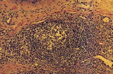
Myosin Light Chains
Gene Rearrangement, gamma-Chain T-Cell Antigen Receptor
Gene Rearrangement, B-Lymphocyte
Lymphocyte Activation
Immunoglobulin Heavy Chains
Immunoglobulin kappa-Chains
Genes, Immunoglobulin
Gene Rearrangement, beta-Chain T-Cell Antigen Receptor
Immunoglobulin lambda-Chains
T-Lymphocytes
Gene Rearrangement, delta-Chain T-Cell Antigen Receptor
Molecular Sequence Data
Polymerase Chain Reaction
Base Sequence
Immunoglobulin Variable Region
Receptors, Antigen, B-Cell
Gene Rearrangement, alpha-Chain T-Cell Antigen Receptor
Myosin-Light-Chain Kinase
Clone Cells
Genes, T-Cell Receptor gamma
Translocation, Genetic
Amino Acid Sequence
Immunoglobulin Joining Region
Immunoglobulin mu-Chains
Immunoglobulins
Myosins
Blotting, Southern
Lymphocyte Subsets
Receptors, Antigen, T-Cell
B-Lymphocyte Subsets
Cells, Cultured
Immunophenotyping
Immunoglobulin J-Chains
Leukemia, Lymphoid
Lymphoma, B-Cell
Immunoglobulin M
Flow Cytometry
Receptors, Antigen, T-Cell, gamma-delta
In Situ Hybridization, Fluorescence
Cell Differentiation
Recombination, Genetic
DNA
Thymus Gland
Herpesvirus 4, Human
Antigens, CD
Mice, Inbred C57BL
Immunoglobulin Light Chains, Surrogate
Antibody Diversity
Palatine Tonsil
Lymphoma, T-Cell
Mutation
Genes, RAG-1
VDJ Recombinases
Antigens, CD19
Bone Marrow
Mice, Inbred BALB C
RNA, Messenger
Phenotype
Receptors, Antigen, T-Cell, alpha-beta
Immunoglobulin Constant Regions
Genes, Immunoglobulin Light Chain
Myeloid-Lymphoid Leukemia Protein
Immunoglobulin D
Chromosome Aberrations
Lymph Nodes
Precursor Cell Lymphoblastic Leukemia-Lymphoma
Cloning, Molecular
Immunoglobulin G
Proto-Oncogenes
Antigens, Surface
Antigens, CD5
DNA-Binding Proteins
DNA Primers
Oncogene Proteins, Fusion
Genes
Mice, Transgenic
Antigens, CD3
Phosphorylation
Antigens, Differentiation, B-Lymphocyte
Burkitt Lymphoma
Genes, Immunoglobulin Heavy Chain
Plasma Cells
Signal Transduction
Chickens
T-Lymphocyte Subsets
Reverse Transcriptase Polymerase Chain Reaction
Leukocyte Count
Nucleic Acid Hybridization
Transcription, Genetic
Lymphocyte Culture Test, Mixed
Plasmacytoma
V(D)J Recombination
Reed-Sternberg Cells
Leukemia
Lymphoproliferative Disorders
Immunohistochemistry
Cell Separation
Gene Expression Regulation
Lymphoid Tissue
Electrophoresis, Polyacrylamide Gel
Protein Binding
Multiple Myeloma
Antigens, Differentiation, T-Lymphocyte
CD4-Positive T-Lymphocytes
Leukemia, Lymphocytic, Chronic, B-Cell
Cell Division
Lymphoma, Follicular
Immunoglobulin gamma-Chains
Actins
Myosin-Light-Chain Phosphatase
Restriction Mapping
Chromosome Inversion
Hybridomas
Rabbits
Complementarity Determining Regions
Concanavalin A
Chromosomes, Human, Pair 11
Chromosomes, Human, Pair 14
Mitogens
DNA Nucleotidylexotransferase
Bone Marrow Cells
Sequence Analysis, DNA
Rosette Formation
Lymphocytes, Tumor-Infiltrating
Antibody Formation
Phytohemagglutinins
Mice, Knockout
Hodgkin Disease
Precursor Cells, B-Lymphoid
Binding Sites
Genes, T-Cell Receptor beta
Gene Fusion
Fluorescent Antibody Technique
Tumor Cells, Cultured
Interleukin-2
Transcription Factors
Genes, bcl-2
Binding Sites, Antibody
Precursor B-Cell Lymphoblastic Leukemia-Lymphoma
Oncogene Fusion
Antibody Specificity
Gene Expression
Peptide Fragments
Cell Line, Transformed
Antigens, CD20
Gizzard
Immune Adherence Reaction
Mice, Inbred Strains
Antibodies, Anti-Idiotypic
Chromosome Mapping
Immunoglobulin delta-Chains
Bence Jones Protein
Genes, T-Cell Receptor alpha
Myosin Subfragments
Immunologic Deficiency Syndromes
Alleles
T-Lymphocytes, Cytotoxic
Lymphoma, Large B-Cell, Diffuse
Pre-B Cell Receptors
Protein Conformation
Lymphoma, Non-Hodgkin
Cytotoxicity, Immunologic
Muscle, Smooth
Somatic Hypermutation, Immunoglobulin
Proto-Oncogene Proteins
Gene Deletion
DNA Probes
CD8-Positive T-Lymphocytes
Neoplasm, Residual
Lymphocyte Transfusion
Models, Molecular
Transfection
Calcium
Induction of Ig light chain gene rearrangement in heavy chain-deficient B cells by activated Ras. (1/243)
During B cell development, rearrangement and expression of Ig heavy chain (HC) genes promote development and expansion of pre-B cells accompanied by the onset of Ig light chain (LC) variable region gene assembly. To elucidate the signaling pathways that control these events, we have tested the ability of activated Ras expression to promote B cell differentiation to the stage of LC gene rearrangement in the absence of Ig HC gene expression. For this purpose, we introduced an activated Ras expression construct into JH-deleted embryonic stem cells that lack the ability to assemble HC variable region genes and assayed differentiation potential by recombination activating gene (RAG) 2-deficient blastocyst complementation. We found that activated Ras expression induces the progression of B lineage cells beyond the developmental checkpoint ordinarily controlled by mu HC. Such Ras/JH-deleted B cells accumulate in the periphery but continue to express markers associated with precursor B cells including RAG gene products. These peripheral Ras/JH-deleted B cell populations show extensive Ig LC gene rearrangement but maintain an extent of kappa LC gene rearrangement and a preference for kappa over lambda LC gene rearrangement similar to that of wild-type B cells. We discuss these findings in the context of potential mechanisms that may regulate Ig LC gene rearrangement. (+info)The degrees of plasma cell clonality and marrow infiltration adversely influence the prognosis of AL amyloidosis patients. (2/243)
BACKGROUND AND OBJECTIVE: Primary amyloidosis is a lethal form of plasma cell (PC) dyscrasia characterized by deposits of monoclonal immunoglobulin light chains that cause organ dysfunction. In contrast to multiple myeloma, the amyloid clone is typically indolent and of small size, and marrow PC clonality is not always apparent. This is generally investigated by analyzing the light chain isotype ratio in bone marrow PC. We investigated whether the degree of PC infiltration (PC%) and clonality (PC isotype ratio) affected survival in 56 consecutive patients with primary amyloidosis. DESIGN AND METHODS: PC% was determined by morphologic examination. Immunofluorescence microscopy was used to determine the PC light chain isotype ratio. Statistical analysis was carried out using Cox regression models. RESULTS: The degrees of PC clonality and infiltration were inversely correlated with survival (PC isotype ratio, p = 0.001; PC%, p = 0.008). The two variables were weakly correlated (p = 0.02; r = 0.3). Bone marrow PC isotype ratio demonstrated a powerful independent prognostic value at multivariate analysis when analyzed together with congestive heart failure (the major known negative prognostic factor) and PC%. k/l ratio cut-off values of 0.2 (l patients, p = 0.022) and 16 (k patients, p = 0.03) discriminated two groups with a similar number of patients and significantly different survivals. INTERPRETATION AND CONCLUSIONS: PC clonality and marrow infiltration are important parameters that influence prognosis, presumably because they reflect the amount of pathogenic light chain synthesis. (+info)Ig lambda and heavy chain gene usage in early untreated systemic lupus erythematosus suggests intensive B cell stimulation. (3/243)
To determine the distribution of Vlambda and Jlambda as well as VH and JH gene usage in a patient with systemic lupus erythematosus (SLE), productive and nonproductive VJ and V(D)J rearrangements were amplified from individual peripheral CD19+ B cells and were analyzed. No differences in the Vlambda and Jlambda or the VH and JH gene usage in the nonproductive gene repertoire of this SLE patient were found compared with the distribution of genes found in normal adults, whereas marked skewing of both Vlambda and VH was noted among the productive rearrangements. The distribution of productive Vlambda rearrangements was skewed, with significantly greater representation of the Jlambda distal cluster C Vlambda genes and the Vlambda distal Jlambda7 element, consistent with the possibility that there was receptor editing of the Vlambda locus in this patient. Significant bias in VH gene usage was also noted with VH3 family members dominating the peripheral B cell repertoire of the SLE patient (83%) compared with that found in normal subjects (55%; p < 0.001). Notably, a clone of B cells employing the VH3-11 gene for the heavy chain and the Vlambda1G segment for the light chain was detected. These data are most consistent with the conclusion that extreme B cell overactivity drives the initial stages of SLE leading to remarkable changes in the peripheral V gene usage that may underlie on fail to prevent the emergence of autoimmunity. (+info)Lambda5 is required for rearrangement of the Ig kappa light chain gene in pro-B cell lines. (4/243)
Lambda5 associates with V(pre-B) to form the surrogate light (L) chain. The phenotype of lambda5 knockout mice showed severe impairment of B cell development from pro-B to immature B cell stages. To investigate the function of the surrogate L chain at this stage, we restored expression of lambda5 to lambda5-deficient pro-B cell lines which were established from bone marrow cells of lambda5 knockout mice in the presence of IL-7 and a stromal cell line. Some of these lines are severely impaired in B cell development from pro-B to immature B cell stages as is seen in vivo in lambda5 knockout mice. Restoration of lambda5 protein by retroviral-mediated gene transfer into established lambda5-deficient pro-B cell lines induced rearrangement of the Ig kappa L chain genes after removal of IL-7 from the culture. Immunoprecipitation revealed that the restored lambda5 in the cell line is coupled with V(pre-B) to form the surrogate L chain. The results demonstrate that formation of a complete surrogate L chain, consisting of both lambda5 and V(pre-B), stimulates efficient rearrangement of the kappa L chain genes. (+info)Detection of immunoglobulin kappa light chain rearrangements by polymerase chain reaction. An improved method for detecting clonal B-cell lymphoproliferative disorders. (5/243)
The clonal determination of B-cell lymphoproliferative disorders by immunoglobulin heavy chain (IgH) rearrangement by polymerase chain reaction (PCR) is widely used. However, few attempts have been made to detect immunoglobulin kappa light chain (Igkappa) gene rearrangement using PCR. We studied 145 cases of B-cell neoplasms, along with 58 atypical and 18 reactive lymphoproliferative disorders, using newly designed degenerate oligoprimers recognizing the framework 3 (FR3kappa) and the joint (Jkappa) regions of the Igkappa gene. PCR products were analyzed on nondenaturing polyacrylamide gel (ndPAGE). Clonal B-cell determination was further investigated using IgH rearrangement and t(11:14) or t(14:18). By combining these methods, we detected either clonality or translocation in 117 of 137 cases (85%) in mature B-cell neoplasms. The additional analysis of Igkappa rearrangement improved sensitivity from 66% to 85%. To investigate whether the Ig gene configuration could be characterized using Igkappa PCR in B-cell neoplasms showing severe breakdown of genomic DNA, 18 selected cases were analyzed. Successful amplification was detected in 72% of the cases using either FR3/2-JH and/or FR3Jkappa oligoprimers. Finally, clonality was detected in 21 of 58 atypical B-cell proliferations, and among them, the atypical marginal cell (54%) and atypical large cell (50%) proliferations showed the highest frequency of clonal immunoglobulin gene products. We concluded that PCR/ndPAGE analysis of Igkappa is a sensitive, rapid, and efficient method for assessing clonality in conjunction with IgH and specific translocation analysis. This approach is particularly useful in the characterization of B-cell lymphoproliferative disorders in archival material with poor preservation of the genomic DNA. (+info)Models for antigen receptor gene rearrangement. I. Biased receptor editing in B cells: implications for allelic exclusion. (6/243)
Recent evidence suggests that lymphocyte Ag receptor gene rearrangement does not always stop after the expression of the first productively rearranged receptor. Light chain gene rearrangement in B cells, and alpha-chain rearrangement in T cells can continue, which raises the question: how is allelic exclusion maintained, if at all, in the face of continued rearrangement? In this and the accompanying paper, we present comprehensive models of Ag receptor gene rearrangement and the interaction of this process with clonal selection. Our B cell model enables us to reconcile observations on the kappa:lambda ratio and on kappa allele usage, showing that B cell receptor gene rearrangement must be a highly ordered, rather than a random, process. We show that order is exhibited on three levels: a preference for rearranging kappa rather than lambda light chain genes; a preference to make secondary rearrangements on the allele that has already been rearranged, rather than choosing the location of the next rearrangement at random; and a sequentiality of J segment choice within each kappa allele. This order, combined with the stringency of negative selection, is shown to lead to effective allelic exclusion. (+info)Hodgkin's disease: immunoglobulin heavy and light chain gene rearrangements revealed in single Hodgkin/Reed-Sternberg cells. (7/243)
AIM: To corroborate and investigate the nature of Hodgkin/Reed-Sternberg cells (H/R-S) of various subtypes of Hodgkin's disease. METHOD: Single H/R-S cells were micro-picked from frozen sections of tissues affected by Hodgkin's disease. The DNA from these cells was amplified by the polymerase chain reaction (PCR) with immunoglobulin heavy chain (IgH) gene FRIIIa/JH primers and light chain gene family specific primers. RESULTS: Fifty two of 135 isolated cells gave specific reaction products (36%). IgH and V kappa 4 gene rearrangements were found repeatedly in many H/R-S cells from one case of lymphocyte predominant Hodgkin's disease. Repeated V kappa 4 and individual IgH/V kappa 4,2 rearrangements were seen in one case, and individual IgH and V lambda 3/V kappa 4 rearrangements were seen in another case of nodular sclerosis-type Hodgkin's disease. Repeated IgH/V lambda 3 and individual V lambda 2,4 rearrangements, repeated V kappa 4 and individual IgH/V kappa 3 rearrangements, and repeated IgH and individual V kappa 3/V kappa 4 rearrangement were detected, respectively, in three cases of mixed cellularity-type Hodgkin's disease. Repeated and individual IgH rearrangements were found in another two cases of mixed cellularity-type Hodgkin's disease. CONCLUSION: The H/R-S cells isolated from lymphocyte predominant Hodgkin's disease had IgH and V kappa 4 gene rearrangements, which supports the conclusion that this disease results from a proliferation of neoplastic B cells. The IgH and kappa and/or lambda gene rearrangements seen in H/R-S cells isolated from classic Hodgkin's disease (mixed cellularity-type and nodular sclerosis-type) support the theory that these cells derive from B lineage cells at various stages of differentiation. To our knowledge, this is first time that lambda gene rearrangements have been detected in H/R-S cells. (+info)Concanavalin A stimulation enhanced secondary VlambdaJlambda rearrangement in some human plasma B cells without up-regulation of recombination-activating gene expression and Vlambda germline transcription. (8/243)
Light chain shifting is a phenomenon that occurs in certain human antibody-producing plasma B cells which, when stimulated with concanavalin A (Con A), shift production of the original light chain to new light chains. Here we investigated the effect of Con A stimulation on these light chain shift-inducible cells. Analysis of transcripts and VJ-coding joints for new light chains revealed that a leaky amount of secondary VlambdaJlambda rearrangement occurs spontaneously, without Con A stimulation, and that Con A stimulation markedly increases VJ-coding joints and transcripts for new light chains. It was also shown that new light chain producers, which have carried out secondary rearrangement, do not further rearrange their light chain genes, even when stimulated with Con A. Recombination-activating gene (RAG) products and Vlambda germline transcription were constitutively expressed in these cell lines and their expression levels were not affected by Con A stimulation. These results suggest that Con A stimulation enhanced secondary VlambdaJlambda rearrangement, but this was not a result of the up-regulation of RAG expression and Vlambda germline transcription, which are believed to be sufficient for the process of VlambdaJlambda rearrangement. (+info)"Gene rearrangement" is a process that involves the alteration of the order, orientation, or copy number of genes or gene segments within an organism's genome. This natural mechanism plays a crucial role in generating diversity and specificity in the immune system, particularly in vertebrates.
In the context of the immune system, gene rearrangement occurs during the development of B-cells and T-cells, which are responsible for adaptive immunity. The process involves breaking and rejoining DNA segments that encode antigen recognition sites, resulting in a unique combination of gene segments and creating a vast array of possible antigen receptors.
There are two main types of gene rearrangement:
1. V(D)J recombination: This process occurs in both B-cells and T-cells. It involves the recombination of variable (V), diversity (D), and joining (J) gene segments to form a functional antigen receptor gene. In humans, there are multiple copies of V, D, and J segments for each antigen receptor gene, allowing for a vast number of possible combinations.
2. Class switch recombination: This process occurs only in mature B-cells after antigen exposure. It involves the replacement of the constant (C) region of the immunoglobulin heavy chain gene with another C region, resulting in the production of different isotypes of antibodies (IgG, IgA, or IgE) that have distinct effector functions while maintaining the same antigen specificity.
These processes contribute to the generation of a diverse repertoire of antigen receptors, allowing the immune system to recognize and respond effectively to a wide range of pathogens.
Lymphocytes are a type of white blood cell that is an essential part of the immune system. They are responsible for recognizing and responding to potentially harmful substances such as viruses, bacteria, and other foreign invaders. There are two main types of lymphocytes: B-lymphocytes (B-cells) and T-lymphocytes (T-cells).
B-lymphocytes produce antibodies, which are proteins that help to neutralize or destroy foreign substances. When a B-cell encounters a foreign substance, it becomes activated and begins to divide and differentiate into plasma cells, which produce and secrete large amounts of antibodies. These antibodies bind to the foreign substance, marking it for destruction by other immune cells.
T-lymphocytes, on the other hand, are involved in cell-mediated immunity. They directly attack and destroy infected cells or cancerous cells. T-cells can also help to regulate the immune response by producing chemical signals that activate or inhibit other immune cells.
Lymphocytes are produced in the bone marrow and mature in either the bone marrow (B-cells) or the thymus gland (T-cells). They circulate throughout the body in the blood and lymphatic system, where they can be found in high concentrations in lymph nodes, the spleen, and other lymphoid organs.
Abnormalities in the number or function of lymphocytes can lead to a variety of immune-related disorders, including immunodeficiency diseases, autoimmune disorders, and cancer.
'Gene rearrangement in B-lymphocytes, light chain' refers to the biological process that occurs during the development of B-lymphocytes (a type of white blood cell) in the bone marrow. Specifically, it relates to the rearrangement of genes that code for the light chains of immunoglobulins, which are antibodies that help the immune system recognize and fight off foreign substances.
During gene rearrangement, the variable region genes of the light chain locus (which consist of multiple gene segments, including V, D, and J segments) undergo a series of DNA recombination events to form a functional variable region exon. This process allows for the generation of a vast diversity of antibody molecules with different specificities, enabling the immune system to recognize and respond to a wide range of potential threats.
Abnormalities in this gene rearrangement process can lead to various immunodeficiency disorders or malignancies such as B-cell lymphomas.
Immunoglobulin light chains are the smaller protein subunits of an immunoglobulin, also known as an antibody. They are composed of two polypeptide chains, called kappa (κ) and lambda (λ), which are produced by B cells during the immune response. Each immunoglobulin molecule contains either two kappa or two lambda light chains, in association with two heavy chains.
Light chains play a crucial role in the antigen-binding site of an antibody, where they contribute to the specificity and affinity of the interaction between the antibody and its target antigen. In addition to their role in immune function, abnormal production or accumulation of light chains can lead to various diseases, such as multiple myeloma and amyloidosis.
'Gene rearrangement in B-lymphocytes, heavy chain' refers to the biological process that occurs during the development of B-lymphocytes (a type of white blood cell) in the bone marrow. This process involves the rearrangement of genetic material on chromosome 14, specifically within the immunoglobulin heavy chain gene locus.
During B-cell maturation, the variable region of the heavy chain gene is assembled from several gene segments, including the variable (V), diversity (D), and joining (J) segments. Through a series of genetic recombination events, these segments are randomly selected and joined together to form a unique V(D)J exon that encodes the variable region of the immunoglobulin heavy chain protein.
This gene rearrangement process allows for the generation of a diverse repertoire of antibodies with different specificities, enabling B-lymphocytes to recognize and respond to a wide range of foreign antigens. However, if errors occur during this process, it can lead to the production of autoantibodies that target the body's own cells and tissues, contributing to the development of certain immune disorders such as autoimmune diseases.
"T-lymphocyte gene rearrangement" refers to the process that occurs during the development of T-cells (a type of white blood cell) in which the genes that code for their antigen receptors are rearranged to create a unique receptor that can recognize and bind to specific foreign molecules, such as viruses or tumor cells.
The T-cell receptor (TCR) is made up of two chains, alpha and beta, which are composed of variable and constant regions. During gene rearrangement, the variable region genes are rearranged through a process called V(D)J recombination, in which specific segments of DNA are cut and joined together to form a unique combination that encodes for a diverse range of antigen receptors.
This allows T-cells to recognize and respond to a wide variety of foreign molecules, contributing to the adaptive immune response. However, this process can also lead to errors and the generation of T-cells with self-reactive receptors, which can contribute to autoimmune diseases if not properly regulated.
B-lymphocytes, also known as B-cells, are a type of white blood cell that plays a key role in the immune system's response to infection. They are responsible for producing antibodies, which are proteins that help to neutralize or destroy pathogens such as bacteria and viruses.
When a B-lymphocyte encounters a pathogen, it becomes activated and begins to divide and differentiate into plasma cells, which produce and secrete large amounts of antibodies specific to the antigens on the surface of the pathogen. These antibodies bind to the pathogen, marking it for destruction by other immune cells such as neutrophils and macrophages.
B-lymphocytes also have a role in presenting antigens to T-lymphocytes, another type of white blood cell involved in the immune response. This helps to stimulate the activation and proliferation of T-lymphocytes, which can then go on to destroy infected cells or help to coordinate the overall immune response.
Overall, B-lymphocytes are an essential part of the adaptive immune system, providing long-lasting immunity to previously encountered pathogens and helping to protect against future infections.
Myosin light chains are regulatory proteins that bind to the myosin head region of myosin molecules, which are involved in muscle contraction. There are two types of myosin light chains, essential and regulatory, that have different functions. The essential light chains are necessary for the assembly and stability of the myosin filaments, while the regulatory light chains control the calcium-sensitive activation of the myosin ATPase activity during muscle contraction. Phosphorylation of the regulatory light chains plays a critical role in regulating muscle contraction and relaxation.
Gamma-chain T-cell antigen receptor gene rearrangement refers to the genetic process that occurs during the development of T-cells in the thymus. The T-cell antigen receptor (TCR) is a protein complex found on the surface of T-cells, which plays a critical role in adaptive immunity by recognizing and binding to specific peptide antigens presented in the context of major histocompatibility complex (MHC) molecules.
The TCR is composed of two types of polypeptide chains: alpha and beta chains or gamma and delta chains, which are encoded by separate genes. The gene rearrangement process involves the somatic recombination of variable (V), diversity (D), joining (J), and constant (C) gene segments to generate a diverse repertoire of TCRs capable of recognizing a wide range of antigens.
Gamma-chain TCR gene rearrangement specifically refers to the genetic rearrangement that occurs in the genes encoding the gamma chain of the TCR. This process involves the recombination of V, J, and C gene segments to form a functional gamma chain gene. The resulting gamma chain protein pairs with the delta chain to form the gamma-delta TCR, which is expressed on a subset of T-cells that have distinct functions in immune surveillance and defense against infections and cancer.
Abnormalities in gamma-chain TCR gene rearrangement can lead to the development of various immunodeficiency disorders or malignancies, such as T-cell acute lymphoblastic leukemia (T-ALL) or gamma-delta T-cell lymphomas.
B-lymphocyte gene rearrangement is a fundamental biological process that occurs during the development of B-lymphocytes (also known as B cells), which are a type of white blood cell responsible for producing antibodies to help fight infections. This process involves the rearrangement of genetic material within the B-lymphocyte's immunoglobulin genes, specifically the heavy chain (IgH) and light chain (IgL) genes, to create a diverse repertoire of antibodies with unique specificities.
During B-lymphocyte gene rearrangement, large segments of DNA are cut, deleted, or inverted, and then rejoined to form a functional IgH or IgL gene that encodes an antigen-binding site on the antibody molecule. The process occurs in two main steps:
1. Variable (V), diversity (D), and joining (J) gene segments are rearranged to form the heavy chain gene, which is located on chromosome 14. This results in a vast array of possible combinations, allowing for the generation of a diverse set of antibody molecules.
2. A separate variable (V) and joining (J) gene segment rearrangement occurs to form the light chain gene, which can be either kappa or lambda type, located on chromosomes 2 and 22, respectively.
Once the heavy and light chain genes are successfully rearranged, they are transcribed into mRNA and translated into immunoglobulin proteins, forming a functional antibody molecule. If the initial gene rearrangement fails to produce a functional antibody, additional attempts at rearrangement can occur, involving different combinations of V, D, and J segments or the use of alternative reading frames.
Errors in B-lymphocyte gene rearrangement can lead to various genetic disorders, such as lymphomas and leukemias, due to the production of aberrant antibodies or uncontrolled cell growth.
In the context of medical terminology, "light" doesn't have a specific or standardized definition on its own. However, it can be used in various medical terms and phrases. For example, it could refer to:
1. Visible light: The range of electromagnetic radiation that can be detected by the human eye, typically between wavelengths of 400-700 nanometers. This is relevant in fields such as ophthalmology and optometry.
2. Therapeutic use of light: In some therapies, light is used to treat certain conditions. An example is phototherapy, which uses various wavelengths of ultraviolet (UV) or visible light for conditions like newborn jaundice, skin disorders, or seasonal affective disorder.
3. Light anesthesia: A state of reduced consciousness in which the patient remains responsive to verbal commands and physical stimulation. This is different from general anesthesia where the patient is completely unconscious.
4. Pain relief using light: Certain devices like transcutaneous electrical nerve stimulation (TENS) units have a 'light' setting, indicating lower intensity or frequency of electrical impulses used for pain management.
Without more context, it's hard to provide a precise medical definition of 'light'.
Lymphocyte activation is the process by which B-cells and T-cells (types of lymphocytes) become activated to perform effector functions in an immune response. This process involves the recognition of specific antigens presented on the surface of antigen-presenting cells, such as dendritic cells or macrophages.
The activation of B-cells leads to their differentiation into plasma cells that produce antibodies, while the activation of T-cells results in the production of cytotoxic T-cells (CD8+ T-cells) that can directly kill infected cells or helper T-cells (CD4+ T-cells) that assist other immune cells.
Lymphocyte activation involves a series of intracellular signaling events, including the binding of co-stimulatory molecules and the release of cytokines, which ultimately result in the expression of genes involved in cell proliferation, differentiation, and effector functions. The activation process is tightly regulated to prevent excessive or inappropriate immune responses that can lead to autoimmunity or chronic inflammation.
Immunoglobulin heavy chains are proteins that make up the framework of antibodies, which are Y-shaped immune proteins. These heavy chains, along with light chains, form the antigen-binding sites of an antibody, which recognize and bind to specific foreign substances (antigens) in order to neutralize or remove them from the body.
The heavy chain is composed of a variable region, which contains the antigen-binding site, and constant regions that determine the class and function of the antibody. There are five classes of immunoglobulins (IgA, IgD, IgE, IgG, and IgM) that differ in their heavy chain constant regions and therefore have different functions in the immune response.
Immunoglobulin heavy chains are synthesized by B cells, a type of white blood cell involved in the adaptive immune response. The genetic rearrangement of immunoglobulin heavy chain genes during B cell development results in the production of a vast array of different antibodies with unique antigen-binding sites, allowing for the recognition and elimination of a wide variety of pathogens.
Immunoglobulin kappa-chains are one of the two types of light chains (the other being lambda-chains) that make up an immunoglobulin molecule, also known as an antibody. These light chains combine with heavy chains to form the antigen-binding site of an antibody, which is responsible for recognizing and binding to specific antigens or foreign substances in the body.
Kappa-chains contain a variable region that differs between different antibodies and contributes to the diversity of the immune system's response to various antigens. They also have a constant region, which is consistent across all kappa-chains. Approximately 60% of all human antibodies contain kappa-chains, while the remaining 40% contain lambda-chains. The relative proportions of kappa and lambda chains can be used in diagnostic tests to identify clonal expansions of B cells, which may indicate a malignancy such as multiple myeloma or lymphoma.
Immunoglobulins (Igs), also known as antibodies, are proteins produced by the immune system to recognize and neutralize foreign substances such as pathogens or toxins. They are composed of four polypeptide chains: two heavy chains and two light chains, which are held together by disulfide bonds. The variable regions of the heavy and light chains contain loops that form the antigen-binding site, allowing each Ig molecule to recognize a specific epitope (antigenic determinant) on an antigen.
Genes encoding immunoglobulins are located on chromosome 14 (light chain genes) and chromosomes 22 and 2 (heavy chain genes). The diversity of the immune system is generated through a process called V(D)J recombination, where variable (V), diversity (D), and joining (J) gene segments are randomly selected and assembled to form the variable regions of the heavy and light chains. This results in an enormous number of possible combinations, allowing the immune system to recognize and respond to a vast array of potential threats.
There are five classes of immunoglobulins: IgA, IgD, IgE, IgG, and IgM, each with distinct functions and structures. For example, IgG is the most abundant class in serum and provides long-term protection against pathogens, while IgA is found on mucosal surfaces and helps prevent the entry of pathogens into the body.
Beta-chain gene rearrangement in the T-cell antigen receptor (TCR) refers to the genetic process that occurs during the development of T cells, a type of white blood cell crucial for adaptive immunity. The TCR is a heterodimeric protein complex expressed on the surface of T cells, responsible for recognizing and binding to specific peptide antigens presented in the context of major histocompatibility complex (MHC) molecules.
The beta-chain of the TCR is encoded by a set of gene segments called V (variable), D (diversity), J (joining), and C (constant) segments, located on chromosome 7 in humans. During T-cell development in the thymus, the following rearrangement events occur:
1. A random selection and recombination of a V, D, and J segment take place, forming a variable region exon that encodes the antigen-binding site of the beta-chain. This process introduces nucleotide insertions or deletions at the junctions between these segments, further increasing diversity.
2. The rearranged VDJ segment then combines with a C segment through RNA splicing to form a continuous mRNA sequence that encodes the complete beta-chain protein.
3. The resulting beta-chain pairs with an alpha-chain (encoded by similar gene segments on chromosome 14) to create a functional TCR heterodimer, which is then expressed on the T-cell surface.
This gene rearrangement process allows for the generation of a vast array of unique TCRs capable of recognizing various peptide antigens, ensuring broad coverage against potential pathogens and tumor cells.
Immunoglobulin lambda-chains (Igλ) are one type of light chain found in the immunoglobulins, also known as antibodies. Antibodies are proteins that play a crucial role in the immune system's response to foreign substances, such as bacteria and viruses.
Immunoglobulins are composed of two heavy chains and two light chains, which are interconnected by disulfide bonds. There are two types of light chains: kappa (κ) and lambda (λ). Igλ chains are one type of light chain that can be found in association with heavy chains to form functional antibodies.
Igλ chains contain a variable region, which is responsible for recognizing and binding to specific antigens, and a constant region, which determines the class of the immunoglobulin (e.g., IgA, IgD, IgE, IgG, or IgM).
In humans, approximately 60% of all antibodies contain Igλ chains, while the remaining 40% contain Igκ chains. The ratio of Igλ to Igκ chains can vary depending on the type of immunoglobulin and its function in the immune response.
T-lymphocytes, also known as T-cells, are a type of white blood cell that plays a key role in the adaptive immune system's response to infection. They are produced in the bone marrow and mature in the thymus gland. There are several different types of T-cells, including CD4+ helper T-cells, CD8+ cytotoxic T-cells, and regulatory T-cells (Tregs).
CD4+ helper T-cells assist in activating other immune cells, such as B-lymphocytes and macrophages. They also produce cytokines, which are signaling molecules that help coordinate the immune response. CD8+ cytotoxic T-cells directly kill infected cells by releasing toxic substances. Regulatory T-cells help maintain immune tolerance and prevent autoimmune diseases by suppressing the activity of other immune cells.
T-lymphocytes are important in the immune response to viral infections, cancer, and other diseases. Dysfunction or depletion of T-cells can lead to immunodeficiency and increased susceptibility to infections. On the other hand, an overactive T-cell response can contribute to autoimmune diseases and chronic inflammation.
"Gene rearrangement, delta-chain T-cell antigen receptor" refers to the genetic process that occurs during the development of T cells, a type of immune cell in the body.
T cells recognize and respond to specific targets on infected or abnormal cells through their antigen receptors, which are composed of alpha and beta chains (in most T cells) or gamma and delta chains (in a small subset of T cells called gamma-delta T cells).
The delta-chain of the T-cell antigen receptor is produced through a series of genetic rearrangements that occur during T-cell development in the thymus. The gene segments that encode the delta-chain are located on chromosome 14 and include variable (V), diversity (D), and joining (J) segments, similar to those found in the immunoglobulin genes of B cells.
Through a process involving DNA recombination and deletion, the V, D, and J segments are randomly selected and joined together to form a unique delta-chain gene sequence. This rearrangement process allows for the generation of a diverse repertoire of T-cell antigen receptors that can recognize and respond to a wide variety of targets.
Defects in the gene rearrangement process for the delta-chain T-cell antigen receptor can lead to immunodeficiency or autoimmune disorders.
Molecular sequence data refers to the specific arrangement of molecules, most commonly nucleotides in DNA or RNA, or amino acids in proteins, that make up a biological macromolecule. This data is generated through laboratory techniques such as sequencing, and provides information about the exact order of the constituent molecules. This data is crucial in various fields of biology, including genetics, evolution, and molecular biology, allowing for comparisons between different organisms, identification of genetic variations, and studies of gene function and regulation.
Polymerase Chain Reaction (PCR) is a laboratory technique used to amplify specific regions of DNA. It enables the production of thousands to millions of copies of a particular DNA sequence in a rapid and efficient manner, making it an essential tool in various fields such as molecular biology, medical diagnostics, forensic science, and research.
The PCR process involves repeated cycles of heating and cooling to separate the DNA strands, allow primers (short sequences of single-stranded DNA) to attach to the target regions, and extend these primers using an enzyme called Taq polymerase, resulting in the exponential amplification of the desired DNA segment.
In a medical context, PCR is often used for detecting and quantifying specific pathogens (viruses, bacteria, fungi, or parasites) in clinical samples, identifying genetic mutations or polymorphisms associated with diseases, monitoring disease progression, and evaluating treatment effectiveness.
A base sequence in the context of molecular biology refers to the specific order of nucleotides in a DNA or RNA molecule. In DNA, these nucleotides are adenine (A), guanine (G), cytosine (C), and thymine (T). In RNA, uracil (U) takes the place of thymine. The base sequence contains genetic information that is transcribed into RNA and ultimately translated into proteins. It is the exact order of these bases that determines the genetic code and thus the function of the DNA or RNA molecule.
The Immunoglobulin (Ig) variable region is the antigen-binding part of an antibody, which is highly variable in its amino acid sequence and therefore specific to a particular epitope (the site on an antigen that is recognized by the antigen-binding site of an antibody). This variability is generated during the process of V(D)J recombination in the maturation of B cells, allowing for a diverse repertoire of antibodies to be produced and recognizing a wide range of potential pathogens.
The variable region is composed of several sub-regions including:
1. The heavy chain variable region (VH)
2. The light chain variable region (VL)
3. The heavy chain joining region (JH)
4. The light chain joining region (JL)
These regions are further divided into framework regions and complementarity-determining regions (CDRs). The CDRs, particularly CDR3, contain the most variability and are primarily responsible for antigen recognition.
1. Receptors: In the context of physiology and medicine, receptors are specialized proteins found on the surface of cells or inside cells that detect and respond to specific molecules, known as ligands. These interactions can trigger a variety of responses within the cell, such as starting a signaling cascade or changing the cell's metabolism. Receptors play crucial roles in various biological processes, including communication between cells, regulation of immune responses, and perception of senses.
2. Antigen: An antigen is any substance (usually a protein) that can be recognized by the adaptive immune system, specifically by B-cells and T-cells. Antigens can be derived from various sources, such as microorganisms (like bacteria, viruses, or fungi), pollen, dust mites, or even components of our own cells (for instance, in autoimmune diseases). An antigen's ability to stimulate an immune response is determined by its molecular structure and whether it can be recognized by the receptors on immune cells.
3. B-Cell: B-cells are a type of white blood cell that plays a critical role in the adaptive immune system, particularly in humoral immunity. They originate from hematopoietic stem cells in the bone marrow and are responsible for producing antibodies, which are proteins that recognize and bind to specific antigens. Each B-cell has receptors on its surface called B-cell receptors (BCRs) that can recognize a unique antigen. When a B-cell encounters its specific antigen, it becomes activated, undergoes proliferation, and differentiates into plasma cells that secrete large amounts of antibodies to neutralize or eliminate the antigen.
Alpha-chain T-cell antigen receptor gene rearrangement refers to the genetic process that occurs during the development of T-cells in the thymus. This process involves the rearrangement of gene segments that encode for the variable region of the alpha chain of the T-cell receptor (TCR).
The TCR is a protein complex found on the surface of T-cells, which plays a critical role in adaptive immunity by recognizing and binding to specific antigens presented by major histocompatibility complex (MHC) molecules. The variable region of the TCR alpha chain is responsible for recognizing and binding to a specific portion of the antigen called the epitope.
During gene rearrangement, the DNA segments that encode for the variable region of the TCR alpha chain are cut and joined together in a random manner, resulting in a unique combination of gene segments that code for a diverse range of TCR alpha chains. This allows for the recognition of a vast array of different antigens, thereby enhancing the ability of the immune system to respond to various pathogens.
Abnormalities in TCR alpha chain gene rearrangement can lead to the production of T-cells with incorrect or non-functional TCRs, which may contribute to the development of certain immunodeficiencies or autoimmune disorders.
Myosin-Light-Chain Kinase (MLCK) is an enzyme that plays a crucial role in muscle contraction. It phosphorylates the regulatory light chains of myosin, a protein involved in muscle contraction, leading to the activation of myosin and the initiation of the contractile process. MLCK is activated by calcium ions and calmodulin, and its activity is essential for various cellular processes, including cytokinesis, cell motility, and maintenance of cell shape. In addition to its role in muscle contraction, MLCK has been implicated in several pathological conditions, such as hypertension, atherosclerosis, and cancer.
A clone is a group of cells that are genetically identical to each other because they are derived from a common ancestor cell through processes such as mitosis or asexual reproduction. Therefore, the term "clone cells" refers to a population of cells that are genetic copies of a single parent cell.
In the context of laboratory research, cells can be cloned by isolating a single cell and allowing it to divide in culture, creating a population of genetically identical cells. This is useful for studying the behavior and characteristics of individual cell types, as well as for generating large quantities of cells for use in experiments.
It's important to note that while clone cells are genetically identical, they may still exhibit differences in their phenotype (physical traits) due to epigenetic factors or environmental influences.
T-cell receptor (TCR) genes are a set of genes that encode the proteins found on the surface of T-cells, a type of white blood cell involved in the adaptive immune response. The TCR gamma chain is one component of the TCR complex, which also includes the TCR alpha, beta, and delta chains.
The TCR gamma gene is located on human chromosome 7 and is composed of variable (V), diversity (D), joining (J), and constant (C) regions. During the development of T-cells in the thymus, the V, D, and J segments recombine to form a unique TCR gamma chain that can recognize specific antigens presented by major histocompatibility complex (MHC) molecules on the surface of infected or damaged cells.
The TCR gamma chain is typically expressed in a subset of T-cells called gamma delta T-cells, which are involved in the early stages of immune responses and can be found in various tissues throughout the body. Mutations or abnormalities in the TCR gamma gene can lead to immunodeficiency disorders or cancer, such as T-cell leukemia or lymphoma.
Translocation, genetic, refers to a type of chromosomal abnormality in which a segment of a chromosome is transferred from one chromosome to another, resulting in an altered genome. This can occur between two non-homologous chromosomes (non-reciprocal translocation) or between two homologous chromosomes (reciprocal translocation). Genetic translocations can lead to various clinical consequences, depending on the genes involved and the location of the translocation. Some translocations may result in no apparent effects, while others can cause developmental abnormalities, cancer, or other genetic disorders. In some cases, translocations can also increase the risk of having offspring with genetic conditions.
An amino acid sequence is the specific order of amino acids in a protein or peptide molecule, formed by the linking of the amino group (-NH2) of one amino acid to the carboxyl group (-COOH) of another amino acid through a peptide bond. The sequence is determined by the genetic code and is unique to each type of protein or peptide. It plays a crucial role in determining the three-dimensional structure and function of proteins.
The Immunoglobulin Joining Region (IgJ or J chain) is a polypeptide chain that is a component of certain immunoglobulins, specifically IgM and IgA. The J chain plays a crucial role in the polymerization of these immunoglobulins, allowing them to form higher-order structures such as pentamers (in the case of IgM) or dimers (in the case of IgA). This polymerization is important for the functioning of these immunoglobulins in the immune response. The J chain contains multiple cysteine residues that form disulfide bonds with each other and with the heavy chains of the immunoglobulin molecules, helping to stabilize the polymeric structure.
Immunoglobulin mu-chains (IgM) are a type of heavy chain found in immunoglobulins, also known as antibodies. IgM is the first antibody to be produced in response to an initial exposure to an antigen and plays a crucial role in the early stages of the immune response.
IgM antibodies are composed of four monomeric units, each consisting of two heavy chains and two light chains. The heavy chains in IgM are called mu-chains, which have a molecular weight of approximately 72 kDa. Each mu-chain contains five domains: one variable (V) domain at the N-terminus, four constant (C) domains (Cμ1-4), and a membrane-spanning region followed by a short cytoplasmic tail.
IgM antibodies are primarily found on the surface of B cells as part of the B cell receptor (BCR). When a B cell encounters an antigen, the BCR binds to it, triggering a series of intracellular signaling events that lead to B cell activation and differentiation into plasma cells. In response to activation, the B cell begins to secrete IgM antibodies into the bloodstream.
IgM antibodies have several unique features that make them effective in the early stages of an immune response. They are highly efficient at agglutination, or clumping together, of pathogens and antigens, which helps to neutralize them. IgM antibodies also activate the complement system, a group of proteins that work together to destroy pathogens.
Overall, Immunoglobulin mu-chains are an essential component of the immune system, providing early protection against pathogens and initiating the adaptive immune response.
Immunoglobulins (Igs), also known as antibodies, are glycoprotein molecules produced by the immune system's B cells in response to the presence of foreign substances, such as bacteria, viruses, and toxins. These Y-shaped proteins play a crucial role in identifying and neutralizing pathogens and other antigens, thereby protecting the body against infection and disease.
Immunoglobulins are composed of four polypeptide chains: two identical heavy chains and two identical light chains, held together by disulfide bonds. The variable regions of these chains form the antigen-binding sites, which recognize and bind to specific epitopes on antigens. Based on their heavy chain type, immunoglobulins are classified into five main isotypes or classes: IgA, IgD, IgE, IgG, and IgM. Each class has distinct functions in the immune response, such as providing protection in different body fluids and tissues, mediating hypersensitivity reactions, and aiding in the development of immunological memory.
In medical settings, immunoglobulins can be administered therapeutically to provide passive immunity against certain diseases or to treat immune deficiencies, autoimmune disorders, and other conditions that may benefit from immunomodulation.
Myosins are a large family of motor proteins that play a crucial role in various cellular processes, including muscle contraction and intracellular transport. They consist of heavy chains, which contain the motor domain responsible for generating force and motion, and light chains, which regulate the activity of the myosin. Based on their structural and functional differences, myosins are classified into over 35 classes, with classes II, V, and VI being the most well-studied.
Class II myosins, also known as conventional myosins, are responsible for muscle contraction in skeletal, cardiac, and smooth muscles. They form filaments called thick filaments, which interact with actin filaments to generate force and movement during muscle contraction.
Class V myosins, also known as unconventional myosins, are involved in intracellular transport and organelle positioning. They have a long tail that can bind to various cargoes, such as vesicles, mitochondria, and nuclei, and a motor domain that moves along actin filaments to transport the cargoes to their destinations.
Class VI myosins are also unconventional myosins involved in intracellular transport and organelle positioning. They have two heads connected by a coiled-coil tail, which can bind to various cargoes. Class VI myosins move along actin filaments in a unique hand-over-hand motion, allowing them to transport their cargoes efficiently.
Overall, myosins are essential for many cellular functions and have been implicated in various diseases, including cardiovascular diseases, neurological disorders, and cancer.
Southern blotting is a type of membrane-based blotting technique that is used in molecular biology to detect and locate specific DNA sequences within a DNA sample. This technique is named after its inventor, Edward M. Southern.
In Southern blotting, the DNA sample is first digested with one or more restriction enzymes, which cut the DNA at specific recognition sites. The resulting DNA fragments are then separated based on their size by gel electrophoresis. After separation, the DNA fragments are denatured to convert them into single-stranded DNA and transferred onto a nitrocellulose or nylon membrane.
Once the DNA has been transferred to the membrane, it is hybridized with a labeled probe that is complementary to the sequence of interest. The probe can be labeled with radioactive isotopes, fluorescent dyes, or chemiluminescent compounds. After hybridization, the membrane is washed to remove any unbound probe and then exposed to X-ray film (in the case of radioactive probes) or scanned (in the case of non-radioactive probes) to detect the location of the labeled probe on the membrane.
The position of the labeled probe on the membrane corresponds to the location of the specific DNA sequence within the original DNA sample. Southern blotting is a powerful tool for identifying and characterizing specific DNA sequences, such as those associated with genetic diseases or gene regulation.
Lymphocyte subsets refer to distinct populations of white blood cells called lymphocytes, which are crucial components of the adaptive immune system. There are two main types of lymphocytes: T cells and B cells, and each type has several subsets based on their surface receptors, functions, and activation status.
1. T cell subsets: These include CD4+ T helper cells (Th cells), CD8+ cytotoxic T cells (Tc cells), regulatory T cells (Tregs), and memory T cells. Th cells are further divided into Th1, Th2, Th17, and Tfh cells based on their cytokine production profiles and functions.
* CD4+ T helper cells (Th cells) play a central role in orchestrating the immune response by producing various cytokines that activate other immune cells.
* CD8+ cytotoxic T cells (Tc cells) directly kill virus-infected or malignant cells upon recognition of specific antigens presented on their surface.
* Regulatory T cells (Tregs) suppress the activation and proliferation of other immune cells to maintain self-tolerance and prevent autoimmunity.
* Memory T cells are long-lived cells that remain in the body after an initial infection or immunization, providing rapid protection upon subsequent encounters with the same pathogen.
2. B cell subsets: These include naïve B cells, memory B cells, and plasma cells. Upon activation by antigens, B cells differentiate into antibody-secreting plasma cells that produce specific antibodies to neutralize or eliminate pathogens.
* Naïve B cells are resting cells that have not yet encountered their specific antigen.
* Memory B cells are long-lived cells generated after initial antigen exposure, which can quickly differentiate into antibody-secreting plasma cells upon re-exposure to the same antigen.
* Plasma cells are terminally differentiated B cells that secrete large amounts of specific antibodies.
Analyzing lymphocyte subsets is essential for understanding immune system function and dysfunction, as well as monitoring the effectiveness of immunotherapies and vaccinations.
1. Receptors: In the context of physiology and medicine, receptors are specialized proteins found on the surface of cells or inside cells that detect and respond to specific molecules, known as ligands. These interactions can trigger a range of responses within the cell, such as starting a signaling pathway or changing the cell's behavior. There are various types of receptors, including ion channels, G protein-coupled receptors, and enzyme-linked receptors.
2. Antigen: An antigen is any substance (usually a protein) that can be recognized by the immune system, specifically by antibodies or T-cells, as foreign and potentially harmful. Antigens can be derived from various sources, such as bacteria, viruses, fungi, parasites, or even non-living substances like pollen, chemicals, or toxins. An antigen typically contains epitopes, which are the specific regions that antibodies or T-cell receptors recognize and bind to.
3. T-Cell: Also known as T lymphocytes, T-cells are a type of white blood cell that plays a crucial role in cell-mediated immunity, a part of the adaptive immune system. They are produced in the bone marrow and mature in the thymus gland. There are several types of T-cells, including CD4+ helper T-cells, CD8+ cytotoxic T-cells, and regulatory T-cells (Tregs). T-cells recognize antigens presented to them by antigen-presenting cells (APCs) via their surface receptors called the T-cell receptor (TCR). Once activated, T-cells can proliferate and differentiate into various effector cells that help eliminate infected or damaged cells.
The spleen is an organ in the upper left side of the abdomen, next to the stomach and behind the ribs. It plays multiple supporting roles in the body:
1. It fights infection by acting as a filter for the blood. Old red blood cells are recycled in the spleen, and platelets and white blood cells are stored there.
2. The spleen also helps to control the amount of blood in the body by removing excess red blood cells and storing platelets.
3. It has an important role in immune function, producing antibodies and removing microorganisms and damaged red blood cells from the bloodstream.
The spleen can be removed without causing any significant problems, as other organs take over its functions. This is known as a splenectomy and may be necessary if the spleen is damaged or diseased.
B-lymphocytes, also known as B-cells, are a type of white blood cell that plays a central role in the humoral immune response. They are responsible for producing antibodies, which are proteins that help to neutralize or destroy pathogens such as viruses and bacteria.
B-lymphocyte subsets refer to distinct populations of B-cells that can be identified based on their surface receptors and functional characteristics. Some common B-lymphocyte subsets include:
1. Naive B-cells: These are mature B-cells that have not yet been exposed to an antigen. They express surface receptors called immunoglobulin M (IgM) and immunoglobulin D (IgD).
2. Memory B-cells: These are B-cells that have previously encountered an antigen and mounted an immune response. They express high levels of surface immunoglobulins and can quickly differentiate into antibody-secreting plasma cells upon re-exposure to the same antigen.
3. Plasma cells: These are fully differentiated B-cells that secrete large amounts of antibodies in response to an antigen. They lack surface immunoglobulins and do not undergo further division.
4. Regulatory B-cells: These are a subset of B-cells that modulate the immune response by producing anti-inflammatory cytokines and suppressing the activation of other immune cells.
5. B-1 cells: These are a population of B-cells that are primarily found in the peripheral blood and mucosal tissues. They produce natural antibodies that provide early protection against pathogens and help to maintain tissue homeostasis.
Understanding the different B-lymphocyte subsets and their functions is important for diagnosing and treating immune-related disorders, including autoimmune diseases, infections, and cancer.
"Cells, cultured" is a medical term that refers to cells that have been removed from an organism and grown in controlled laboratory conditions outside of the body. This process is called cell culture and it allows scientists to study cells in a more controlled and accessible environment than they would have inside the body. Cultured cells can be derived from a variety of sources, including tissues, organs, or fluids from humans, animals, or cell lines that have been previously established in the laboratory.
Cell culture involves several steps, including isolation of the cells from the tissue, purification and characterization of the cells, and maintenance of the cells in appropriate growth conditions. The cells are typically grown in specialized media that contain nutrients, growth factors, and other components necessary for their survival and proliferation. Cultured cells can be used for a variety of purposes, including basic research, drug development and testing, and production of biological products such as vaccines and gene therapies.
It is important to note that cultured cells may behave differently than they do in the body, and results obtained from cell culture studies may not always translate directly to human physiology or disease. Therefore, it is essential to validate findings from cell culture experiments using additional models and ultimately in clinical trials involving human subjects.
A lymphocyte count is a laboratory test that measures the number of white blood cells called lymphocytes in a sample of blood. Lymphocytes are a vital part of the immune system and help fight off infections and diseases. A normal lymphocyte count ranges from 1,000 to 4,800 cells per microliter (µL) of blood for adults.
An abnormal lymphocyte count can indicate an infection, immune disorder, or blood cancer. A low lymphocyte count is called lymphopenia, while a high lymphocyte count is called lymphocytosis. The cause of an abnormal lymphocyte count should be investigated through further testing and clinical evaluation.
Immunophenotyping is a medical laboratory technique used to identify and classify cells, usually in the context of hematologic (blood) disorders and malignancies (cancers), based on their surface or intracellular expression of various proteins and antigens. This technique utilizes specific antibodies tagged with fluorochromes, which bind to the target antigens on the cell surface or within the cells. The labeled cells are then analyzed using flow cytometry, allowing for the detection and quantification of multiple antigenic markers simultaneously.
Immunophenotyping helps in understanding the distribution of different cell types, their subsets, and activation status, which can be crucial in diagnosing various hematological disorders, immunodeficiencies, and distinguishing between different types of leukemias, lymphomas, and other malignancies. Additionally, it can also be used to monitor the progression of diseases, evaluate the effectiveness of treatments, and detect minimal residual disease (MRD) during follow-up care.
Lymphoma is a type of cancer that originates from the white blood cells called lymphocytes, which are part of the immune system. These cells are found in various parts of the body such as the lymph nodes, spleen, bone marrow, and other organs. Lymphoma can be classified into two main types: Hodgkin lymphoma (HL) and non-Hodgkin lymphoma (NHL).
HL is characterized by the presence of a specific type of abnormal lymphocyte called Reed-Sternberg cells, while NHL includes a diverse group of lymphomas that lack these cells. The symptoms of lymphoma may include swollen lymph nodes, fever, night sweats, weight loss, and fatigue.
The exact cause of lymphoma is not known, but it is believed to result from genetic mutations in the lymphocytes that lead to uncontrolled cell growth and division. Exposure to certain viruses, chemicals, and radiation may increase the risk of developing lymphoma. Treatment options for lymphoma depend on various factors such as the type and stage of the disease, age, and overall health of the patient. Common treatments include chemotherapy, radiation therapy, immunotherapy, and stem cell transplantation.
Immunoglobulin J-chains are small protein structures that play a role in the assembly and structure of certain types of antibodies, specifically IgM and IgA. The J-chain is a polypeptide chain that contains multiple cysteine residues, which allow it to form disulfide bonds with the heavy chains of IgM and IgA molecules.
In IgM antibodies, the J-chain helps to link the five identical heavy chain units together to form a pentameric structure. In IgA antibodies, the J-chain links two dimeric structures together to form a tetrameric structure. This polymerization of IgM and IgA molecules is important for their function in the immune system, as it allows them to form large complexes that can effectively agglutinate and neutralize pathogens.
The J-chain is synthesized by a specialized group of B cells called plasma cells, which are responsible for producing and secreting antibodies. Once synthesized, the J-chain is covalently linked to the heavy chains of IgM or IgA molecules during their assembly in the endoplasmic reticulum of the plasma cell.
Overall, the Immunoglobulin J-chain plays a crucial role in the structure and function of certain classes of antibodies, contributing to their ability to effectively combat pathogens and protect the body from infection.
A cell line is a culture of cells that are grown in a laboratory for use in research. These cells are usually taken from a single cell or group of cells, and they are able to divide and grow continuously in the lab. Cell lines can come from many different sources, including animals, plants, and humans. They are often used in scientific research to study cellular processes, disease mechanisms, and to test new drugs or treatments. Some common types of human cell lines include HeLa cells (which come from a cancer patient named Henrietta Lacks), HEK293 cells (which come from embryonic kidney cells), and HUVEC cells (which come from umbilical vein endothelial cells). It is important to note that cell lines are not the same as primary cells, which are cells that are taken directly from a living organism and have not been grown in the lab.
Leukemia, lymphoid is a type of cancer that affects the lymphoid cells, which are a vital part of the body's immune system. It is characterized by the uncontrolled production of abnormal white blood cells (leukocytes or WBCs) in the bone marrow, specifically the lymphocytes. These abnormal lymphocytes accumulate and interfere with the production of normal blood cells, leading to a decrease in red blood cells (anemia), platelets (thrombocytopenia), and healthy white blood cells (leukopenia).
There are two main types of lymphoid leukemia: acute lymphoblastic leukemia (ALL) and chronic lymphocytic leukemia (CLL). Acute lymphoblastic leukemia progresses rapidly, while chronic lymphocytic leukemia has a slower onset and progression.
Symptoms of lymphoid leukemia may include fatigue, frequent infections, easy bruising or bleeding, weight loss, swollen lymph nodes, and bone pain. Treatment options depend on the type, stage, and individual patient factors but often involve chemotherapy, radiation therapy, targeted therapy, immunotherapy, or stem cell transplantation.
B-cell lymphoma is a type of cancer that originates from the B-lymphocytes, which are a part of the immune system and play a crucial role in fighting infections. These cells can develop mutations in their DNA, leading to uncontrolled growth and division, resulting in the formation of a tumor.
B-cell lymphomas can be classified into two main categories: Hodgkin's lymphoma and non-Hodgkin's lymphoma. B-cell lymphomas are further divided into subtypes based on their specific characteristics, such as the appearance of the cells under a microscope, the genetic changes present in the cancer cells, and the aggressiveness of the disease.
Some common types of B-cell lymphomas include diffuse large B-cell lymphoma, follicular lymphoma, mantle cell lymphoma, and Burkitt lymphoma. Treatment options for B-cell lymphomas depend on the specific subtype, stage of the disease, and other individual factors. Treatment may include chemotherapy, radiation therapy, immunotherapy, targeted therapy, or stem cell transplantation.
Immunoglobulin M (IgM) is a type of antibody that is primarily found in the blood and lymph fluid. It is the first antibody to be produced in response to an initial exposure to an antigen, making it an important part of the body's primary immune response. IgM antibodies are large molecules that are composed of five basic units, giving them a pentameric structure. They are primarily found on the surface of B cells as membrane-bound immunoglobulins (mlgM), where they function as receptors for antigens. Once an mlgM receptor binds to an antigen, it triggers the activation and differentiation of the B cell into a plasma cell that produces and secretes large amounts of soluble IgM antibodies.
IgM antibodies are particularly effective at agglutination (clumping) and complement activation, which makes them important in the early stages of an immune response to help clear pathogens from the bloodstream. However, they are not as stable or long-lived as other types of antibodies, such as IgG, and their levels tend to decline after the initial immune response has occurred.
In summary, Immunoglobulin M (IgM) is a type of antibody that plays a crucial role in the primary immune response to antigens by agglutination and complement activation. It is primarily found in the blood and lymph fluid, and it is produced by B cells after they are activated by an antigen.
Flow cytometry is a medical and research technique used to measure physical and chemical characteristics of cells or particles, one cell at a time, as they flow in a fluid stream through a beam of light. The properties measured include:
* Cell size (light scatter)
* Cell internal complexity (granularity, also light scatter)
* Presence or absence of specific proteins or other molecules on the cell surface or inside the cell (using fluorescent antibodies or other fluorescent probes)
The technique is widely used in cell counting, cell sorting, protein engineering, biomarker discovery and monitoring disease progression, particularly in hematology, immunology, and cancer research.
1. Receptors: In the context of physiology and medicine, receptors are specialized proteins found on the surface of cells or inside cells that detect and respond to specific molecules, known as ligands. They play a crucial role in various biological processes, including signal transduction, cell communication, and regulation of physiological functions.
2. Antigen: An antigen is a foreign substance (usually a protein) that triggers an immune response when introduced into the body. Antigens can be derived from various sources, such as bacteria, viruses, fungi, or parasites. They are recognized by the immune system as non-self and stimulate the production of antibodies and activation of immune cells, like T-cells, to eliminate the threat.
3. T-Cell: T-cells, also known as T-lymphocytes, are a type of white blood cell that plays a central role in cell-mediated immunity. They are produced in the bone marrow and mature in the thymus gland. T-cells have receptors on their surface called T-cell receptors (TCRs) that enable them to recognize and respond to specific antigens presented by antigen-presenting cells (APCs). There are several types of T-cells, including CD4+ helper T-cells, CD8+ cytotoxic T-cells, and regulatory T-cells.
4. gamma-delta (γδ) T-Cell: Gamma-delta (γδ) T-cells are a subset of T-cells that possess a distinct T-cell receptor (TCR) composed of gamma and delta chains. Unlike conventional T-cells, which typically recognize peptide antigens presented by major histocompatibility complex (MHC) molecules, γδ T-cells can directly recognize various non-peptide antigens, such as lipids, glycolipids, and small metabolites. They are involved in the early stages of immune responses, tissue homeostasis, and cancer surveillance.
In situ hybridization, fluorescence (FISH) is a type of molecular cytogenetic technique used to detect and localize the presence or absence of specific DNA sequences on chromosomes through the use of fluorescent probes. This technique allows for the direct visualization of genetic material at a cellular level, making it possible to identify chromosomal abnormalities such as deletions, duplications, translocations, and other rearrangements.
The process involves denaturing the DNA in the sample to separate the double-stranded molecules into single strands, then adding fluorescently labeled probes that are complementary to the target DNA sequence. The probe hybridizes to the complementary sequence in the sample, and the location of the probe is detected by fluorescence microscopy.
FISH has a wide range of applications in both clinical and research settings, including prenatal diagnosis, cancer diagnosis and monitoring, and the study of gene expression and regulation. It is a powerful tool for identifying genetic abnormalities and understanding their role in human disease.
Monoclonal antibodies are a type of antibody that are identical because they are produced by a single clone of cells. They are laboratory-produced molecules that act like human antibodies in the immune system. They can be designed to attach to specific proteins found on the surface of cancer cells, making them useful for targeting and treating cancer. Monoclonal antibodies can also be used as a therapy for other diseases, such as autoimmune disorders and inflammatory conditions.
Monoclonal antibodies are produced by fusing a single type of immune cell, called a B cell, with a tumor cell to create a hybrid cell, or hybridoma. This hybrid cell is then able to replicate indefinitely, producing a large number of identical copies of the original antibody. These antibodies can be further modified and engineered to enhance their ability to bind to specific targets, increase their stability, and improve their effectiveness as therapeutic agents.
Monoclonal antibodies have several mechanisms of action in cancer therapy. They can directly kill cancer cells by binding to them and triggering an immune response. They can also block the signals that promote cancer growth and survival. Additionally, monoclonal antibodies can be used to deliver drugs or radiation directly to cancer cells, increasing the effectiveness of these treatments while minimizing their side effects on healthy tissues.
Monoclonal antibodies have become an important tool in modern medicine, with several approved for use in cancer therapy and other diseases. They are continuing to be studied and developed as a promising approach to treating a wide range of medical conditions.
Cell differentiation is the process by which a less specialized cell, or stem cell, becomes a more specialized cell type with specific functions and structures. This process involves changes in gene expression, which are regulated by various intracellular signaling pathways and transcription factors. Differentiation results in the development of distinct cell types that make up tissues and organs in multicellular organisms. It is a crucial aspect of embryonic development, tissue repair, and maintenance of homeostasis in the body.
Genetic recombination is the process by which genetic material is exchanged between two similar or identical molecules of DNA during meiosis, resulting in new combinations of genes on each chromosome. This exchange occurs during crossover, where segments of DNA are swapped between non-sister homologous chromatids, creating genetic diversity among the offspring. It is a crucial mechanism for generating genetic variability and facilitating evolutionary change within populations. Additionally, recombination also plays an essential role in DNA repair processes through mechanisms such as homologous recombinational repair (HRR) and non-homologous end joining (NHEJ).
Deoxyribonucleic acid (DNA) is the genetic material present in the cells of organisms where it is responsible for the storage and transmission of hereditary information. DNA is a long molecule that consists of two strands coiled together to form a double helix. Each strand is made up of a series of four nucleotide bases - adenine (A), guanine (G), cytosine (C), and thymine (T) - that are linked together by phosphate and sugar groups. The sequence of these bases along the length of the molecule encodes genetic information, with A always pairing with T and C always pairing with G. This base-pairing allows for the replication and transcription of DNA, which are essential processes in the functioning and reproduction of all living organisms.
The term "DNA, neoplasm" is not a standard medical term or concept. DNA refers to deoxyribonucleic acid, which is the genetic material present in the cells of living organisms. A neoplasm, on the other hand, is a tumor or growth of abnormal tissue that can be benign (non-cancerous) or malignant (cancerous).
In some contexts, "DNA, neoplasm" may refer to genetic alterations found in cancer cells. These genetic changes can include mutations, amplifications, deletions, or rearrangements of DNA sequences that contribute to the development and progression of cancer. Identifying these genetic abnormalities can help doctors diagnose and treat certain types of cancer more effectively.
However, it's important to note that "DNA, neoplasm" is not a term that would typically be used in medical reports or research papers without further clarification. If you have any specific questions about DNA changes in cancer cells or neoplasms, I would recommend consulting with a healthcare professional or conducting further research on the topic.
The thymus gland is an essential organ of the immune system, located in the upper chest, behind the sternum and surrounding the heart. It's primarily active until puberty and begins to shrink in size and activity thereafter. The main function of the thymus gland is the production and maturation of T-lymphocytes (T-cells), which are crucial for cell-mediated immunity, helping to protect the body from infection and cancer.
The thymus gland provides a protected environment where immune cells called pre-T cells develop into mature T cells. During this process, they learn to recognize and respond appropriately to foreign substances while remaining tolerant to self-tissues, which is crucial for preventing autoimmune diseases.
Additionally, the thymus gland produces hormones like thymosin that regulate immune cell activities and contribute to the overall immune response.
Medical Definition of "Herpesvirus 4, Human" (Epstein-Barr Virus)
"Herpesvirus 4, Human," also known as Epstein-Barr virus (EBV), is a member of the Herpesviridae family and is one of the most common human viruses. It is primarily transmitted through saliva and is often referred to as the "kissing disease."
EBV is the causative agent of infectious mononucleosis (IM), also known as glandular fever, which is characterized by symptoms such as fatigue, sore throat, fever, and swollen lymph nodes. The virus can also cause other diseases, including certain types of cancer, such as Burkitt's lymphoma, Hodgkin's lymphoma, and nasopharyngeal carcinoma.
Once a person becomes infected with EBV, the virus remains in the body for the rest of their life, residing in certain white blood cells called B lymphocytes. In most people, the virus remains dormant and does not cause any further symptoms. However, in some individuals, the virus may reactivate, leading to recurrent or persistent symptoms.
EBV infection is diagnosed through various tests, including blood tests that detect antibodies against the virus or direct detection of the virus itself through polymerase chain reaction (PCR) assays. There is no cure for EBV infection, and treatment is generally supportive, focusing on relieving symptoms and managing complications. Prevention measures include practicing good hygiene, avoiding close contact with infected individuals, and not sharing personal items such as toothbrushes or drinking glasses.
CD (cluster of differentiation) antigens are cell-surface proteins that are expressed on leukocytes (white blood cells) and can be used to identify and distinguish different subsets of these cells. They are important markers in the field of immunology and hematology, and are commonly used to diagnose and monitor various diseases, including cancer, autoimmune disorders, and infectious diseases.
CD antigens are designated by numbers, such as CD4, CD8, CD19, etc., which refer to specific proteins found on the surface of different types of leukocytes. For example, CD4 is a protein found on the surface of helper T cells, while CD8 is found on cytotoxic T cells.
CD antigens can be used as targets for immunotherapy, such as monoclonal antibody therapy, in which antibodies are designed to bind to specific CD antigens and trigger an immune response against cancer cells or infected cells. They can also be used as markers to monitor the effectiveness of treatments and to detect minimal residual disease (MRD) after treatment.
It's important to note that not all CD antigens are exclusive to leukocytes, some can be found on other cell types as well, and their expression can vary depending on the activation state or differentiation stage of the cells.
C57BL/6 (C57 Black 6) is an inbred strain of laboratory mouse that is widely used in biomedical research. The term "inbred" refers to a strain of animals where matings have been carried out between siblings or other closely related individuals for many generations, resulting in a population that is highly homozygous at most genetic loci.
The C57BL/6 strain was established in 1920 by crossing a female mouse from the dilute brown (DBA) strain with a male mouse from the black strain. The resulting offspring were then interbred for many generations to create the inbred C57BL/6 strain.
C57BL/6 mice are known for their robust health, longevity, and ease of handling, making them a popular choice for researchers. They have been used in a wide range of biomedical research areas, including studies of cancer, immunology, neuroscience, cardiovascular disease, and metabolism.
One of the most notable features of the C57BL/6 strain is its sensitivity to certain genetic modifications, such as the introduction of mutations that lead to obesity or impaired glucose tolerance. This has made it a valuable tool for studying the genetic basis of complex diseases and traits.
Overall, the C57BL/6 inbred mouse strain is an important model organism in biomedical research, providing a valuable resource for understanding the genetic and molecular mechanisms underlying human health and disease.
I'm sorry for any confusion, but "Immunoglobulin Light Chains, Surrogate" is not a recognized medical term or concept in the immunology field. Immunoglobulins, also known as antibodies, are proteins produced by the immune system to identify and neutralize foreign substances like bacteria and viruses. They consist of two heavy chains and two light chains, which can be either kappa or lambda. However, there is no such thing as "surrogate" light chains in this context.
If you have any other questions about medical terminology or concepts, I'd be happy to help!
Antibody diversity refers to the variety of different antibodies that an organism can produce in response to exposure to various antigens. This diversity is generated through a process called V(D)J recombination, which occurs during the development of B cells in the bone marrow.
The variable regions of heavy and light chains of antibody molecules are generated by the random selection and rearrangement of gene segments (V, D, and J) from different combinations. This results in a unique antigen-binding site for each antibody molecule, allowing the immune system to recognize and respond to a vast array of potential pathogens.
Further diversity is generated through the processes of somatic hypermutation and class switch recombination, which introduce additional changes in the variable regions of antibodies during an immune response. These processes allow for the affinity maturation of antibodies, where the binding strength between the antibody and antigen is increased over time, leading to a more effective immune response.
Overall, antibody diversity is critical for the adaptive immune system's ability to recognize and respond to a wide range of pathogens and protect against infection and disease.
The palatine tonsils, also known as the "tonsils," are two masses of lymphoid tissue located on either side of the oropharynx, at the back of the throat. They are part of the immune system and play a role in protecting the body from inhaled or ingested pathogens. Each tonsil has a surface covered with crypts and follicles that contain lymphocytes, which help to filter out bacteria and viruses that enter the mouth and nose.
The palatine tonsils are visible through the mouth and can be seen during a routine physical examination. They vary in size, but typically are about the size of a large olive or almond. Swelling or inflammation of the tonsils is called tonsillitis, which can cause symptoms such as sore throat, difficulty swallowing, fever, and swollen lymph nodes in the neck. In some cases, enlarged tonsils may need to be removed through a surgical procedure called a tonsillectomy.
T-cell lymphoma is a type of cancer that affects the T-cells, which are a specific type of white blood cell responsible for immune function. These lymphomas develop from mature T-cells and can be classified into various subtypes based on their clinical and pathological features.
T-cell lymphomas can arise in many different organs, including the lymph nodes, skin, and other soft tissues. They often present with symptoms such as enlarged lymph nodes, fever, night sweats, and weight loss. The diagnosis of T-cell lymphoma typically involves a biopsy of the affected tissue, followed by immunophenotyping and genetic analysis to determine the specific subtype.
Treatment for T-cell lymphomas may include chemotherapy, radiation therapy, immunotherapy, or stem cell transplantation, depending on the stage and aggressiveness of the disease. The prognosis for T-cell lymphoma varies widely depending on the subtype and individual patient factors.
A mutation is a permanent change in the DNA sequence of an organism's genome. Mutations can occur spontaneously or be caused by environmental factors such as exposure to radiation, chemicals, or viruses. They may have various effects on the organism, ranging from benign to harmful, depending on where they occur and whether they alter the function of essential proteins. In some cases, mutations can increase an individual's susceptibility to certain diseases or disorders, while in others, they may confer a survival advantage. Mutations are the driving force behind evolution, as they introduce new genetic variability into populations, which can then be acted upon by natural selection.
RAG-1 (Recombination Activating Gene 1) is a protein involved in the process of V(D)J recombination, which is a crucial step in the development of the immune system. Specifically, RAG-1 plays a role in generating diversity in the antigen receptors of T and B cells by rearranging gene segments that encode for the variable regions of these receptors.
RAG-1 forms a complex with another protein called RAG-2, and together they initiate the V(D)J recombination process by introducing DNA double-strand breaks at specific sites within the antigen receptor genes. This allows for the precise joining of different gene segments to create a functional antigen receptor that can recognize a wide variety of foreign molecules (antigens).
Mutations in the RAG-1 gene can lead to severe combined immunodeficiency (SCID), a condition characterized by an impaired immune system and increased susceptibility to infections.
VDJ Recombinases are a set of enzymes that play a crucial role in the adaptive immune system, specifically in the diversification of antigen receptors in vertebrates. The name "VDJ" refers to the variable (V), diversity (D), and joining (J) gene segments that undergo recombination to generate a vast array of unique antigen receptor genes.
The VDJ Recombinases are composed of two main enzymatic components: RAG1 and RAG2, which are responsible for initiating the recombination process, and Artemis, which is involved in the cleavage and joining of the gene segments. The recombination process mediated by these enzymes occurs during the development of B and T lymphocytes, allowing for the generation of a diverse repertoire of antigen receptors that can recognize and respond to a wide range of pathogens.
The RAG1 and RAG2 proteins recognize specific DNA sequences called recombination signal sequences (RSSs) that flank the V, D, and J gene segments. They introduce double-stranded breaks at the junctions between these gene segments, creating a hairpin structure at one end of each break. The hairpins are then cleaved by Artemis, and the resulting overhangs are joined together by another set of enzymes to form a functional antigen receptor gene.
Overall, VDJ Recombinases play a critical role in the adaptive immune system's ability to generate diverse and specific responses to pathogens, making them an essential component of vertebrate immunity.
CD19 is a type of protein found on the surface of B cells, which are a type of white blood cell that plays a key role in the body's immune response. CD19 is a marker that helps identify and distinguish B cells from other types of cells in the body. It is also a target for immunotherapy in certain diseases, such as B-cell malignancies.
An antigen is any substance that can stimulate an immune response, particularly the production of antibodies. In the context of CD19, antigens refer to substances that can bind to CD19 and trigger a response from the immune system. This can include proteins, carbohydrates, or other molecules found on the surface of bacteria, viruses, or cancer cells.
Therefore, 'antigens, CD19' refers to any substances that can bind to the CD19 protein on B cells and trigger an immune response. These antigens may be used in the development of immunotherapies for the treatment of B-cell malignancies or other diseases.
Bone marrow is the spongy tissue found inside certain bones in the body, such as the hips, thighs, and vertebrae. It is responsible for producing blood-forming cells, including red blood cells, white blood cells, and platelets. There are two types of bone marrow: red marrow, which is involved in blood cell production, and yellow marrow, which contains fatty tissue.
Red bone marrow contains hematopoietic stem cells, which can differentiate into various types of blood cells. These stem cells continuously divide and mature to produce new blood cells that are released into the circulation. Red blood cells carry oxygen throughout the body, white blood cells help fight infections, and platelets play a crucial role in blood clotting.
Bone marrow also serves as a site for immune cell development and maturation. It contains various types of immune cells, such as lymphocytes, macrophages, and dendritic cells, which help protect the body against infections and diseases.
Abnormalities in bone marrow function can lead to several medical conditions, including anemia, leukopenia, thrombocytopenia, and various types of cancer, such as leukemia and multiple myeloma. Bone marrow aspiration and biopsy are common diagnostic procedures used to evaluate bone marrow health and function.
BALB/c is an inbred strain of laboratory mouse that is widely used in biomedical research. The strain was developed at the Institute of Cancer Research in London by Henry Baldwin and his colleagues in the 1920s, and it has since become one of the most commonly used inbred strains in the world.
BALB/c mice are characterized by their black coat color, which is determined by a recessive allele at the tyrosinase locus. They are also known for their docile and friendly temperament, making them easy to handle and work with in the laboratory.
One of the key features of BALB/c mice that makes them useful for research is their susceptibility to certain types of tumors and immune responses. For example, they are highly susceptible to developing mammary tumors, which can be induced by chemical carcinogens or viral infection. They also have a strong Th2-biased immune response, which makes them useful models for studying allergic diseases and asthma.
BALB/c mice are also commonly used in studies of genetics, neuroscience, behavior, and infectious diseases. Because they are an inbred strain, they have a uniform genetic background, which makes it easier to control for genetic factors in experiments. Additionally, because they have been bred in the laboratory for many generations, they are highly standardized and reproducible, making them ideal subjects for scientific research.
Messenger RNA (mRNA) is a type of RNA (ribonucleic acid) that carries genetic information copied from DNA in the form of a series of three-base code "words," each of which specifies a particular amino acid. This information is used by the cell's machinery to construct proteins, a process known as translation. After being transcribed from DNA, mRNA travels out of the nucleus to the ribosomes in the cytoplasm where protein synthesis occurs. Once the protein has been synthesized, the mRNA may be degraded and recycled. Post-transcriptional modifications can also occur to mRNA, such as alternative splicing and addition of a 5' cap and a poly(A) tail, which can affect its stability, localization, and translation efficiency.
A phenotype is the physical or biochemical expression of an organism's genes, or the observable traits and characteristics resulting from the interaction of its genetic constitution (genotype) with environmental factors. These characteristics can include appearance, development, behavior, and resistance to disease, among others. Phenotypes can vary widely, even among individuals with identical genotypes, due to differences in environmental influences, gene expression, and genetic interactions.
1. Receptors: In the context of physiology and medicine, receptors are specialized proteins found on the surface of cells or inside cells that detect and respond to specific molecules, known as ligands. Receptors play a crucial role in signal transduction, enabling cells to communicate with each other and respond to changes in their environment.
2. Antigen: An antigen is any substance (usually a protein) that can be recognized by the immune system and stimulate an immune response. Antigens can be foreign substances such as bacteria, viruses, or pollen, or they can be components of our own cells, such as tumor antigens in cancer cells. Antigens are typically bound and presented to the immune system by specialized cells called antigen-presenting cells (APCs).
3. T-Cell: T-cells, also known as T lymphocytes, are a type of white blood cell that plays a central role in cell-mediated immunity. T-cells are produced in the bone marrow and mature in the thymus gland. There are two main types of T-cells: CD4+ helper T-cells and CD8+ cytotoxic T-cells. Helper T-cells assist other immune cells, such as B-cells and macrophages, in mounting an immune response, while cytotoxic T-cells directly kill infected or cancerous cells.
4. Alpha-Beta: Alpha-beta is a type of T-cell receptor (TCR) that is found on the surface of most mature T-cells. The alpha-beta TCR is composed of two polypeptide chains, an alpha chain and a beta chain, that are held together by disulfide bonds. The alpha-beta TCR recognizes and binds to specific antigens presented in the context of major histocompatibility complex (MHC) molecules on the surface of APCs. This interaction is critical for initiating an immune response against infected or cancerous cells.
Immunoglobulin constant regions are the invariant portions of antibody molecules (immunoglobulins) that are identical in all antibodies of the same isotype. These regions are responsible for effector functions such as complement activation, binding to Fc receptors, and initiating immune responses. They are composed of amino acid sequences that remain unchanged during antigen-driven somatic hypermutation, allowing them to interact with various components of the immune system. The constant regions are found in the heavy chains (CH) and light chains (CL) of an immunoglobulin molecule. In contrast, the variable regions are responsible for recognizing and binding to specific antigens.
Immunoglobulin light chains are proteins that play a crucial role in the immune system's response to foreign substances. They are called "light chains" because they are smaller than the heavy chains that make up the other part of an antibody molecule.
There are two types of light chains, known as kappa (κ) and lambda (λ) chains, which are produced by genes located on chromosomes 2 and 22, respectively. Each immunoglobulin molecule contains either two kappa or two lambda light chains, in addition to two heavy chains.
The genes that code for light chains undergo a process called V(D)J recombination during the development of B cells, which allows for the generation of a diverse repertoire of antibodies with different specificities. This process involves the selection and rearrangement of various gene segments to create a unique immunoglobulin light chain protein.
Defects in the genes that code for immunoglobulin light chains can lead to various immune disorders, such as immunodeficiencies or autoimmune diseases. Additionally, abnormal light chain proteins can accumulate in the body and form amyloid fibrils, leading to a condition called light chain amyloidosis.
The Myeloid-Lymphoid Leukemia (MLL) protein, also known as MLL1 or HRX, is a histone methyltransferase that plays a crucial role in the regulation of gene expression. It is involved in various cellular processes, including embryonic development and hematopoiesis (the formation of blood cells).
The MLL protein is encoded by the MLL gene, which is located on chromosome 11q23. This gene is frequently rearranged or mutated in certain types of leukemia, leading to the production of abnormal fusion proteins that contribute to tumor development and progression. These MLL-rearranged leukemias are aggressive and have a poor prognosis, making them an important area of research in the field of oncology.
Immunoglobulin D (IgD) is a type of antibody that is present in the blood and other bodily fluids. It is one of the five classes of immunoglobulins (IgA, IgD, IgE, IgG, and IgM) found in humans and plays a role in the immune response.
IgD is produced by B cells, a type of white blood cell that is responsible for producing antibodies. It is primarily found on the surface of mature B cells, where it functions as a receptor for antigens (foreign substances that trigger an immune response). When an antigen binds to IgD on the surface of a B cell, it activates the B cell and stimulates it to produce and secrete antibodies specific to that antigen.
IgD is found in relatively low concentrations in the blood compared to other immunoglobulins, and its precise functions are not fully understood. However, it is thought to play a role in the regulation of B cell activation and the immune response. Additionally, some research suggests that IgD may have a direct role in protecting against certain types of infections.
It's worth noting that genetic deficiencies in IgD are not typically associated with any significant immunological abnormalities or increased susceptibility to infection.
Chromosome aberrations refer to structural and numerical changes in the chromosomes that can occur spontaneously or as a result of exposure to mutagenic agents. These changes can affect the genetic material encoded in the chromosomes, leading to various consequences such as developmental abnormalities, cancer, or infertility.
Structural aberrations include deletions, duplications, inversions, translocations, and rings, which result from breaks and rearrangements of chromosome segments. Numerical aberrations involve changes in the number of chromosomes, such as aneuploidy (extra or missing chromosomes) or polyploidy (multiples of a complete set of chromosomes).
Chromosome aberrations can be detected and analyzed using various cytogenetic techniques, including karyotyping, fluorescence in situ hybridization (FISH), and comparative genomic hybridization (CGH). These methods allow for the identification and characterization of chromosomal changes at the molecular level, providing valuable information for genetic counseling, diagnosis, and research.
Lymph nodes are small, bean-shaped organs that are part of the immune system. They are found throughout the body, especially in the neck, armpits, groin, and abdomen. Lymph nodes filter lymph fluid, which carries waste and unwanted substances such as bacteria, viruses, and cancer cells. They contain white blood cells called lymphocytes that help fight infections and diseases by attacking and destroying the harmful substances found in the lymph fluid. When an infection or disease is present, lymph nodes may swell due to the increased number of immune cells and fluid accumulation as they work to fight off the invaders.
Precursor Cell Lymphoblastic Leukemia-Lymphoma (previously known as Precursor T-lymphoblastic Leukemia/Lymphoma) is a type of cancer that affects the early stages of T-cell development. It is a subtype of acute lymphoblastic leukemia (ALL), which is characterized by the overproduction of immature white blood cells called lymphoblasts in the bone marrow, blood, and other organs.
In Precursor Cell Lymphoblastic Leukemia-Lymphoma, these abnormal lymphoblasts accumulate primarily in the lymphoid tissues such as the thymus and lymph nodes, leading to the enlargement of these organs. This subtype is more aggressive than other forms of ALL and has a higher risk of spreading to the central nervous system (CNS).
The medical definition of Precursor Cell Lymphoblastic Leukemia-Lymphoma includes:
1. A malignant neoplasm of immature T-cell precursors, also known as lymphoblasts.
2. Characterized by the proliferation and accumulation of these abnormal cells in the bone marrow, blood, and lymphoid tissues such as the thymus and lymph nodes.
3. Often associated with chromosomal abnormalities, genetic mutations, or aberrant gene expression that contribute to its aggressive behavior and poor prognosis.
4. Typically presents with symptoms related to bone marrow failure (anemia, neutropenia, thrombocytopenia), lymphadenopathy (swollen lymph nodes), hepatosplenomegaly (enlarged liver and spleen), and potential CNS involvement.
5. Diagnosed through a combination of clinical evaluation, imaging studies, and laboratory tests, including bone marrow aspiration and biopsy, immunophenotyping, cytogenetic analysis, and molecular genetic testing.
6. Treated with intensive multi-agent chemotherapy regimens, often combined with radiation therapy and/or stem cell transplantation to achieve remission and improve survival outcomes.
Molecular cloning is a laboratory technique used to create multiple copies of a specific DNA sequence. This process involves several steps:
1. Isolation: The first step in molecular cloning is to isolate the DNA sequence of interest from the rest of the genomic DNA. This can be done using various methods such as PCR (polymerase chain reaction), restriction enzymes, or hybridization.
2. Vector construction: Once the DNA sequence of interest has been isolated, it must be inserted into a vector, which is a small circular DNA molecule that can replicate independently in a host cell. Common vectors used in molecular cloning include plasmids and phages.
3. Transformation: The constructed vector is then introduced into a host cell, usually a bacterial or yeast cell, through a process called transformation. This can be done using various methods such as electroporation or chemical transformation.
4. Selection: After transformation, the host cells are grown in selective media that allow only those cells containing the vector to grow. This ensures that the DNA sequence of interest has been successfully cloned into the vector.
5. Amplification: Once the host cells have been selected, they can be grown in large quantities to amplify the number of copies of the cloned DNA sequence.
Molecular cloning is a powerful tool in molecular biology and has numerous applications, including the production of recombinant proteins, gene therapy, functional analysis of genes, and genetic engineering.
Immunoglobulin G (IgG) is a type of antibody, which is a protective protein produced by the immune system in response to foreign substances like bacteria or viruses. IgG is the most abundant type of antibody in human blood, making up about 75-80% of all antibodies. It is found in all body fluids and plays a crucial role in fighting infections caused by bacteria, viruses, and toxins.
IgG has several important functions:
1. Neutralization: IgG can bind to the surface of bacteria or viruses, preventing them from attaching to and infecting human cells.
2. Opsonization: IgG coats the surface of pathogens, making them more recognizable and easier for immune cells like neutrophils and macrophages to phagocytose (engulf and destroy) them.
3. Complement activation: IgG can activate the complement system, a group of proteins that work together to help eliminate pathogens from the body. Activation of the complement system leads to the formation of the membrane attack complex, which creates holes in the cell membranes of bacteria, leading to their lysis (destruction).
4. Antibody-dependent cellular cytotoxicity (ADCC): IgG can bind to immune cells like natural killer (NK) cells and trigger them to release substances that cause target cells (such as virus-infected or cancerous cells) to undergo apoptosis (programmed cell death).
5. Immune complex formation: IgG can form immune complexes with antigens, which can then be removed from the body through various mechanisms, such as phagocytosis by immune cells or excretion in urine.
IgG is a critical component of adaptive immunity and provides long-lasting protection against reinfection with many pathogens. It has four subclasses (IgG1, IgG2, IgG3, and IgG4) that differ in their structure, function, and distribution in the body.
Proto-oncogenes are normal genes that are present in all cells and play crucial roles in regulating cell growth, division, and death. They code for proteins that are involved in signal transduction pathways that control various cellular processes such as proliferation, differentiation, and survival. When these genes undergo mutations or are activated abnormally, they can become oncogenes, which have the potential to cause uncontrolled cell growth and lead to cancer. Oncogenes can contribute to tumor formation through various mechanisms, including promoting cell division, inhibiting programmed cell death (apoptosis), and stimulating blood vessel growth (angiogenesis).
Surface antigens are molecules found on the surface of cells that can be recognized by the immune system as being foreign or different from the host's own cells. Antigens are typically proteins or polysaccharides that are capable of stimulating an immune response, leading to the production of antibodies and activation of immune cells such as T-cells.
Surface antigens are important in the context of infectious diseases because they allow the immune system to identify and target infected cells for destruction. For example, viruses and bacteria often display surface antigens that are distinct from those found on host cells, allowing the immune system to recognize and attack them. In some cases, these surface antigens can also be used as targets for vaccines or other immunotherapies.
In addition to their role in infectious diseases, surface antigens are also important in the context of cancer. Tumor cells often display abnormal surface antigens that differ from those found on normal cells, allowing the immune system to potentially recognize and attack them. However, tumors can also develop mechanisms to evade the immune system, making it difficult to mount an effective response.
Overall, understanding the properties and behavior of surface antigens is crucial for developing effective immunotherapies and vaccines against infectious diseases and cancer.
CD5 is a type of protein found on the surface of certain cells in the human body, including some immune cells like T cells and B cells. It is also known as a cell marker or identifier. Antigens are substances (usually proteins) on the surface of cells that can be recognized by the immune system, triggering an immune response.
In the context of CD5, antigens refer to foreign substances that can bind to the CD5 protein and stimulate an immune response. However, it's important to note that CD5 itself is not typically considered an antigen in the medical community. Instead, it is a marker used to identify certain types of cells and monitor their behavior in health and disease states.
In some cases, abnormal expression or regulation of CD5 has been associated with various diseases, including certain types of cancer. For example, some B-cell lymphomas may overexpress CD5, which can help doctors diagnose and monitor the progression of the disease. However, in these contexts, CD5 is not considered an antigen in the traditional sense.
Karyotyping is a medical laboratory test used to study the chromosomes in a cell. It involves obtaining a sample of cells from a patient, usually from blood or bone marrow, and then staining the chromosomes so they can be easily seen under a microscope. The chromosomes are then arranged in pairs based on their size, shape, and other features to create a karyotype. This visual representation allows for the identification and analysis of any chromosomal abnormalities, such as extra or missing chromosomes, or structural changes like translocations or inversions. These abnormalities can provide important information about genetic disorders, diseases, and developmental problems.
DNA-binding proteins are a type of protein that have the ability to bind to DNA (deoxyribonucleic acid), the genetic material of organisms. These proteins play crucial roles in various biological processes, such as regulation of gene expression, DNA replication, repair and recombination.
The binding of DNA-binding proteins to specific DNA sequences is mediated by non-covalent interactions, including electrostatic, hydrogen bonding, and van der Waals forces. The specificity of binding is determined by the recognition of particular nucleotide sequences or structural features of the DNA molecule.
DNA-binding proteins can be classified into several categories based on their structure and function, such as transcription factors, histones, and restriction enzymes. Transcription factors are a major class of DNA-binding proteins that regulate gene expression by binding to specific DNA sequences in the promoter region of genes and recruiting other proteins to modulate transcription. Histones are DNA-binding proteins that package DNA into nucleosomes, the basic unit of chromatin structure. Restriction enzymes are DNA-binding proteins that recognize and cleave specific DNA sequences, and are widely used in molecular biology research and biotechnology applications.
In the context of medicine and pharmacology, "kinetics" refers to the study of how a drug moves throughout the body, including its absorption, distribution, metabolism, and excretion (often abbreviated as ADME). This field is called "pharmacokinetics."
1. Absorption: This is the process of a drug moving from its site of administration into the bloodstream. Factors such as the route of administration (e.g., oral, intravenous, etc.), formulation, and individual physiological differences can affect absorption.
2. Distribution: Once a drug is in the bloodstream, it gets distributed throughout the body to various tissues and organs. This process is influenced by factors like blood flow, protein binding, and lipid solubility of the drug.
3. Metabolism: Drugs are often chemically modified in the body, typically in the liver, through processes known as metabolism. These changes can lead to the formation of active or inactive metabolites, which may then be further distributed, excreted, or undergo additional metabolic transformations.
4. Excretion: This is the process by which drugs and their metabolites are eliminated from the body, primarily through the kidneys (urine) and the liver (bile).
Understanding the kinetics of a drug is crucial for determining its optimal dosing regimen, potential interactions with other medications or foods, and any necessary adjustments for special populations like pediatric or geriatric patients, or those with impaired renal or hepatic function.
DNA primers are short single-stranded DNA molecules that serve as a starting point for DNA synthesis. They are typically used in laboratory techniques such as the polymerase chain reaction (PCR) and DNA sequencing. The primer binds to a complementary sequence on the DNA template through base pairing, providing a free 3'-hydroxyl group for the DNA polymerase enzyme to add nucleotides and synthesize a new strand of DNA. This allows for specific and targeted amplification or analysis of a particular region of interest within a larger DNA molecule.
An oncogene protein fusion is a result of a genetic alteration in which parts of two different genes combine to create a hybrid gene that can contribute to the development of cancer. This fusion can lead to the production of an abnormal protein that promotes uncontrolled cell growth and division, ultimately resulting in a malignant tumor. Oncogene protein fusions are often caused by chromosomal rearrangements such as translocations, inversions, or deletions and are commonly found in various types of cancer, including leukemia and sarcoma. These genetic alterations can serve as potential targets for cancer diagnosis and therapy.
A gene is a specific sequence of nucleotides in DNA that carries genetic information. Genes are the fundamental units of heredity and are responsible for the development and function of all living organisms. They code for proteins or RNA molecules, which carry out various functions within cells and are essential for the structure, function, and regulation of the body's tissues and organs.
Each gene has a specific location on a chromosome, and each person inherits two copies of every gene, one from each parent. Variations in the sequence of nucleotides in a gene can lead to differences in traits between individuals, including physical characteristics, susceptibility to disease, and responses to environmental factors.
Medical genetics is the study of genes and their role in health and disease. It involves understanding how genes contribute to the development and progression of various medical conditions, as well as identifying genetic risk factors and developing strategies for prevention, diagnosis, and treatment.
Gene order, in the context of genetics and genomics, refers to the specific sequence or arrangement of genes along a chromosome. The order of genes on a chromosome is not random, but rather, it is highly conserved across species and is often used as a tool for studying evolutionary relationships between organisms.
The study of gene order has also provided valuable insights into genome organization, function, and regulation. For example, the clustering of genes that are involved in specific pathways or functions can provide information about how those pathways or functions have evolved over time. Similarly, the spatial arrangement of genes relative to each other can influence their expression levels and patterns, which can have important consequences for phenotypic traits.
Overall, gene order is an important aspect of genome biology that continues to be a focus of research in fields such as genomics, genetics, evolutionary biology, and bioinformatics.
Transgenic mice are genetically modified rodents that have incorporated foreign DNA (exogenous DNA) into their own genome. This is typically done through the use of recombinant DNA technology, where a specific gene or genetic sequence of interest is isolated and then introduced into the mouse embryo. The resulting transgenic mice can then express the protein encoded by the foreign gene, allowing researchers to study its function in a living organism.
The process of creating transgenic mice usually involves microinjecting the exogenous DNA into the pronucleus of a fertilized egg, which is then implanted into a surrogate mother. The offspring that result from this procedure are screened for the presence of the foreign DNA, and those that carry the desired genetic modification are used to establish a transgenic mouse line.
Transgenic mice have been widely used in biomedical research to model human diseases, study gene function, and test new therapies. They provide a valuable tool for understanding complex biological processes and developing new treatments for a variety of medical conditions.
CD3 antigens are a group of proteins found on the surface of T-cells, which are a type of white blood cell that plays a central role in the immune response. The CD3 antigens are composed of several different subunits (ε, δ, γ, and α) that associate to form the CD3 complex, which is involved in T-cell activation and signal transduction.
The CD3 complex is associated with the T-cell receptor (TCR), which recognizes and binds to specific antigens presented by antigen-presenting cells. When the TCR binds to an antigen, it triggers a series of intracellular signaling events that lead to T-cell activation and the initiation of an immune response.
CD3 antigens are important targets for immunotherapy in some diseases, such as certain types of cancer. For example, monoclonal antibodies that target CD3 have been developed to activate T-cells and enhance their ability to recognize and destroy tumor cells. However, CD3-targeted therapies can also cause side effects, such as cytokine release syndrome, which can be serious or life-threatening in some cases.
Phosphorylation is the process of adding a phosphate group (a molecule consisting of one phosphorus atom and four oxygen atoms) to a protein or other organic molecule, which is usually done by enzymes called kinases. This post-translational modification can change the function, localization, or activity of the target molecule, playing a crucial role in various cellular processes such as signal transduction, metabolism, and regulation of gene expression. Phosphorylation is reversible, and the removal of the phosphate group is facilitated by enzymes called phosphatases.
Antigens are substances that can stimulate an immune response, particularly the production of antibodies by B-lymphocytes. Differentiation refers to the process by which cells mature and become more specialized in their functions. In the context of B-lymphocytes, differentiation involves the maturation of naive B-cells into plasma cells that are capable of producing large amounts of antibodies in response to an antigenic stimulus.
B-lymphocytes, also known as B-cells, are a type of white blood cell that plays a critical role in the adaptive immune system. They are responsible for producing antibodies, which are proteins that recognize and bind to specific antigens, marking them for destruction by other immune cells.
When a B-lymphocyte encounters an antigen, it becomes activated and begins to differentiate into a plasma cell. During this process, the B-cell undergoes several changes, including an increase in size, the expression of new surface receptors, and the production of large amounts of antibodies specific to the antigen. These antibodies are then released into the bloodstream, where they can bind to the antigen and help to neutralize or eliminate it.
Overall, the differentiation of B-lymphocytes in response to antigens is a critical component of the adaptive immune system, allowing the body to mount targeted responses to specific pathogens and other foreign substances.
Burkitt lymphoma is a type of aggressive non-Hodgkin lymphoma (NHL), which is a cancer that originates in the lymphatic system. It is named after Denis Parsons Burkitt, an Irish surgeon who first described this form of cancer in African children in the 1950s.
Burkitt lymphoma is characterized by the rapid growth and spread of abnormal B-lymphocytes (a type of white blood cell), which can affect various organs and tissues, including the lymph nodes, spleen, liver, gastrointestinal tract, and central nervous system.
There are three main types of Burkitt lymphoma: endemic, sporadic, and immunodeficiency-associated. The endemic form is most common in equatorial Africa and is strongly associated with Epstein-Barr virus (EBV) infection. The sporadic form occurs worldwide but is rare, accounting for less than 1% of all NHL cases in the United States. Immunodeficiency-associated Burkitt lymphoma is seen in individuals with weakened immune systems due to HIV/AIDS or immunosuppressive therapy after organ transplantation.
Burkitt lymphoma typically presents as a rapidly growing mass, often involving the jaw, facial bones, or abdominal organs. Symptoms may include swollen lymph nodes, fever, night sweats, weight loss, and fatigue. Diagnosis is made through a biopsy of the affected tissue, followed by immunohistochemical staining and genetic analysis to confirm the presence of characteristic chromosomal translocations involving the MYC oncogene.
Treatment for Burkitt lymphoma typically involves intensive chemotherapy regimens, often combined with targeted therapy or immunotherapy. The prognosis is generally good when treated aggressively and promptly, with a high cure rate in children and young adults. However, the prognosis may be poorer in older patients or those with advanced-stage disease at diagnosis.
Immunoglobulin heavy chains (IgH) are proteins that make up the framework of antibodies, which are crucial components of the adaptive immune system. These heavy chains are produced by B cells and plasma cells, and they contain variable regions that can bind to specific antigens, as well as constant regions that determine the effector functions of the antibody.
The genes that encode for immunoglobulin heavy chains are located on chromosome 14 in humans, within a region known as the IgH locus. These genes undergo a complex process of rearrangement during B cell development, whereby different gene segments (V, D, and J) are joined together to create a unique variable region that can recognize a specific antigen. This process of gene rearrangement is critical for the diversity and specificity of the antibody response.
Therefore, the medical definition of 'Genes, Immunoglobulin Heavy Chain' refers to the set of genetic elements that encode for the immunoglobulin heavy chain proteins, and their complex process of rearrangement during B cell development.
Plasma cells are a type of white blood cell that are derived from B cells (another type of white blood cell) and are responsible for producing antibodies. Antibodies are proteins that help the body to fight against infections by recognizing and binding to specific antigens, such as bacteria or viruses. Plasma cells are found in the bone marrow, spleen, and lymph nodes, and they play a crucial role in the immune system's response to infection.
Plasma cells are characterized by their large size, eccentric nucleus, and abundant cytoplasm filled with rough endoplasmic reticulum, which is where antibody proteins are synthesized and stored. When activated, plasma cells can produce and secrete large amounts of antibodies into the bloodstream and lymphatic system, where they can help to neutralize or eliminate pathogens.
It's worth noting that while plasma cells play an important role in the immune response, abnormal accumulations of these cells can also be a sign of certain diseases, such as multiple myeloma, a type of cancer that affects plasma cells.
Signal transduction is the process by which a cell converts an extracellular signal, such as a hormone or neurotransmitter, into an intracellular response. This involves a series of molecular events that transmit the signal from the cell surface to the interior of the cell, ultimately resulting in changes in gene expression, protein activity, or metabolism.
The process typically begins with the binding of the extracellular signal to a receptor located on the cell membrane. This binding event activates the receptor, which then triggers a cascade of intracellular signaling molecules, such as second messengers, protein kinases, and ion channels. These molecules amplify and propagate the signal, ultimately leading to the activation or inhibition of specific cellular responses.
Signal transduction pathways are highly regulated and can be modulated by various factors, including other signaling molecules, post-translational modifications, and feedback mechanisms. Dysregulation of these pathways has been implicated in a variety of diseases, including cancer, diabetes, and neurological disorders.
"Chickens" is a common term used to refer to the domesticated bird, Gallus gallus domesticus, which is widely raised for its eggs and meat. However, in medical terms, "chickens" is not a standard term with a specific definition. If you have any specific medical concern or question related to chickens, such as food safety or allergies, please provide more details so I can give a more accurate answer.
T-lymphocyte subsets refer to distinct populations of T-cells, which are a type of white blood cell that plays a central role in cell-mediated immunity. The two main types of T-lymphocytes are CD4+ and CD8+ cells, which are defined by the presence or absence of specific proteins called cluster differentiation (CD) molecules on their surface.
CD4+ T-cells, also known as helper T-cells, play a crucial role in activating other immune cells, such as B-lymphocytes and macrophages, to mount an immune response against pathogens. They also produce cytokines that help regulate the immune response.
CD8+ T-cells, also known as cytotoxic T-cells, directly kill infected cells or tumor cells by releasing toxic substances such as perforins and granzymes.
The balance between these two subsets of T-cells is critical for maintaining immune homeostasis and mounting effective immune responses against pathogens while avoiding excessive inflammation and autoimmunity. Therefore, the measurement of T-lymphocyte subsets is essential in diagnosing and monitoring various immunological disorders, including HIV infection, cancer, and autoimmune diseases.
Reverse Transcriptase Polymerase Chain Reaction (RT-PCR) is a laboratory technique used in molecular biology to amplify and detect specific DNA sequences. This technique is particularly useful for the detection and quantification of RNA viruses, as well as for the analysis of gene expression.
The process involves two main steps: reverse transcription and polymerase chain reaction (PCR). In the first step, reverse transcriptase enzyme is used to convert RNA into complementary DNA (cDNA) by reading the template provided by the RNA molecule. This cDNA then serves as a template for the PCR amplification step.
In the second step, the PCR reaction uses two primers that flank the target DNA sequence and a thermostable polymerase enzyme to repeatedly copy the targeted cDNA sequence. The reaction mixture is heated and cooled in cycles, allowing the primers to anneal to the template, and the polymerase to extend the new strand. This results in exponential amplification of the target DNA sequence, making it possible to detect even small amounts of RNA or cDNA.
RT-PCR is a sensitive and specific technique that has many applications in medical research and diagnostics, including the detection of viruses such as HIV, hepatitis C virus, and SARS-CoV-2 (the virus that causes COVID-19). It can also be used to study gene expression, identify genetic mutations, and diagnose genetic disorders.
Leukemia, B-cell is a type of cancer that affects the blood and bone marrow, characterized by an overproduction of abnormal B-lymphocytes, a type of white blood cell. These abnormal cells accumulate in the bone marrow and interfere with the production of normal blood cells, leading to anemia, infection, and bleeding.
B-cells are a type of lymphocyte that plays a crucial role in the immune system by producing antibodies to help fight off infections. In B-cell leukemia, the cancerous B-cells do not mature properly and accumulate in the bone marrow, leading to a decrease in the number of healthy white blood cells, red blood cells, and platelets.
There are several types of B-cell leukemia, including acute lymphoblastic leukemia (ALL) and chronic lymphocytic leukemia (CLL). ALL is more common in children and young adults, while CLL is more common in older adults. Treatment options for B-cell leukemia depend on the type and stage of the disease and may include chemotherapy, radiation therapy, stem cell transplantation, or targeted therapies.
A leukocyte count, also known as a white blood cell (WBC) count, is a laboratory test that measures the number of leukocytes in a sample of blood. Leukocytes are a vital part of the body's immune system and help fight infection and inflammation. A high or low leukocyte count may indicate an underlying medical condition, such as an infection, inflammation, or a bone marrow disorder. The normal range for a leukocyte count in adults is typically between 4,500 and 11,000 cells per microliter (mcL) of blood. However, the normal range can vary slightly depending on the laboratory and the individual's age and sex.
Nucleic acid hybridization is a process in molecular biology where two single-stranded nucleic acids (DNA, RNA) with complementary sequences pair together to form a double-stranded molecule through hydrogen bonding. The strands can be from the same type of nucleic acid or different types (i.e., DNA-RNA or DNA-cDNA). This process is commonly used in various laboratory techniques, such as Southern blotting, Northern blotting, polymerase chain reaction (PCR), and microarray analysis, to detect, isolate, and analyze specific nucleic acid sequences. The hybridization temperature and conditions are critical to ensure the specificity of the interaction between the two strands.
Genetic transcription is the process by which the information in a strand of DNA is used to create a complementary RNA molecule. This process is the first step in gene expression, where the genetic code in DNA is converted into a form that can be used to produce proteins or functional RNAs.
During transcription, an enzyme called RNA polymerase binds to the DNA template strand and reads the sequence of nucleotide bases. As it moves along the template, it adds complementary RNA nucleotides to the growing RNA chain, creating a single-stranded RNA molecule that is complementary to the DNA template strand. Once transcription is complete, the RNA molecule may undergo further processing before it can be translated into protein or perform its functional role in the cell.
Transcription can be either "constitutive" or "regulated." Constitutive transcription occurs at a relatively constant rate and produces essential proteins that are required for basic cellular functions. Regulated transcription, on the other hand, is subject to control by various intracellular and extracellular signals, allowing cells to respond to changing environmental conditions or developmental cues.
A Lymphocyte Culture Test, Mixed (LCTM) is not a standardized medical test with a universally accepted definition. However, in some contexts, it may refer to a laboratory procedure where both T-lymphocytes and B-lymphocytes are cultured together from a sample of peripheral blood or other tissues. This test is sometimes used in research or specialized diagnostic settings to evaluate the immune function or to study the interactions between T-cells and B-cells in response to various stimuli, such as antigens or mitogens.
The test typically involves isolating lymphocytes from a sample, adding them to a culture medium along with appropriate stimulants, and then incubating the mixture for a period of time. The resulting responses, such as proliferation, differentiation, or production of cytokines, can be measured and analyzed to gain insights into the immune function or dysfunction.
It's important to note that LCTM is not a routine diagnostic test and its use and interpretation may vary depending on the specific laboratory or research setting.
A plasmacytoma is a discrete tumor mass that is composed of neoplastic plasma cells, which are a type of white blood cell found in the bone marrow. Plasmacytomas can be solitary (a single tumor) or multiple (many tumors), and they can develop in various locations throughout the body.
Solitary plasmacytoma is a rare cancer that typically affects older adults, and it usually involves a single bone lesion, most commonly found in the vertebrae, ribs, or pelvis. In some cases, solitary plasmacytomas can also occur outside of the bone (extramedullary plasmacytoma), which can affect soft tissues such as the upper respiratory tract, gastrointestinal tract, or skin.
Multiple myeloma is a more common and aggressive cancer that involves multiple plasmacytomas in the bone marrow, leading to the replacement of normal bone marrow cells with malignant plasma cells. This can result in various symptoms such as bone pain, anemia, infections, and kidney damage.
The diagnosis of plasmacytoma typically involves a combination of imaging studies, biopsy, and laboratory tests to assess the extent of the disease and determine the appropriate treatment plan. Treatment options for solitary plasmacytoma may include surgery or radiation therapy, while multiple myeloma is usually treated with chemotherapy, targeted therapy, immunotherapy, and/or stem cell transplantation.
In the field of medicine, "time factors" refer to the duration of symptoms or time elapsed since the onset of a medical condition, which can have significant implications for diagnosis and treatment. Understanding time factors is crucial in determining the progression of a disease, evaluating the effectiveness of treatments, and making critical decisions regarding patient care.
For example, in stroke management, "time is brain," meaning that rapid intervention within a specific time frame (usually within 4.5 hours) is essential to administering tissue plasminogen activator (tPA), a clot-busting drug that can minimize brain damage and improve patient outcomes. Similarly, in trauma care, the "golden hour" concept emphasizes the importance of providing definitive care within the first 60 minutes after injury to increase survival rates and reduce morbidity.
Time factors also play a role in monitoring the progression of chronic conditions like diabetes or heart disease, where regular follow-ups and assessments help determine appropriate treatment adjustments and prevent complications. In infectious diseases, time factors are crucial for initiating antibiotic therapy and identifying potential outbreaks to control their spread.
Overall, "time factors" encompass the significance of recognizing and acting promptly in various medical scenarios to optimize patient outcomes and provide effective care.
Reed-Sternberg cells are a type of large, abnormal cell that are present in Hodgkin lymphoma, a cancer of the lymphatic system. These cells are typically characterized by the presence of two or more nuclei, one of which is often larger and irregularly shaped, giving them a "owl's eye" appearance. Reed-Sternberg cells are important in the diagnosis of Hodgkin lymphoma as they are present in all cases of this type of cancer. However, it is worth noting that Reed-Sternberg-like cells can also be found in other conditions, such as some types of non-Hodgkin lymphoma and certain inflammatory disorders, so their presence alone is not enough to make a definitive diagnosis of Hodgkin lymphoma.
Leukemia is a type of cancer that originates from the bone marrow - the soft, inner part of certain bones where new blood cells are made. It is characterized by an abnormal production of white blood cells, known as leukocytes or blasts. These abnormal cells accumulate in the bone marrow and interfere with the production of normal blood cells, leading to a decrease in red blood cells (anemia), platelets (thrombocytopenia), and healthy white blood cells (leukopenia).
There are several types of leukemia, classified based on the specific type of white blood cell affected and the speed at which the disease progresses:
1. Acute Leukemias - These types of leukemia progress rapidly, with symptoms developing over a few weeks or months. They involve the rapid growth and accumulation of immature, nonfunctional white blood cells (blasts) in the bone marrow and peripheral blood. The two main categories are:
- Acute Lymphoblastic Leukemia (ALL) - Originates from lymphoid progenitor cells, primarily affecting children but can also occur in adults.
- Acute Myeloid Leukemia (AML) - Develops from myeloid progenitor cells and is more common in older adults.
2. Chronic Leukemias - These types of leukemia progress slowly, with symptoms developing over a period of months to years. They involve the production of relatively mature, but still abnormal, white blood cells that can accumulate in large numbers in the bone marrow and peripheral blood. The two main categories are:
- Chronic Lymphocytic Leukemia (CLL) - Affects B-lymphocytes and is more common in older adults.
- Chronic Myeloid Leukemia (CML) - Originates from myeloid progenitor cells, characterized by the presence of a specific genetic abnormality called the Philadelphia chromosome. It can occur at any age but is more common in middle-aged and older adults.
Treatment options for leukemia depend on the type, stage, and individual patient factors. Treatments may include chemotherapy, targeted therapy, immunotherapy, stem cell transplantation, or a combination of these approaches.
Lymphoproliferative disorders (LPDs) are a group of diseases characterized by the excessive proliferation of lymphoid cells, which are crucial components of the immune system. These disorders can arise from both B-cells and T-cells, leading to various clinical manifestations ranging from benign to malignant conditions.
LPDs can be broadly classified into reactive and neoplastic categories:
1. Reactive Lymphoproliferative Disorders: These are typically triggered by infections, autoimmune diseases, or immunodeficiency states. They involve an exaggerated response of the immune system leading to the excessive proliferation of lymphoid cells. Examples include:
* Infectious mononucleosis (IM) caused by Epstein-Barr virus (EBV)
* Lymph node enlargement due to various infections or autoimmune disorders
* Post-transplant lymphoproliferative disorder (PTLD), which occurs in the context of immunosuppression following organ transplantation
2. Neoplastic Lymphoproliferative Disorders: These are malignant conditions characterized by uncontrolled growth and accumulation of abnormal lymphoid cells, leading to the formation of tumors. They can be further classified into Hodgkin lymphoma (HL) and non-Hodgkin lymphoma (NHL). Examples include:
* Hodgkin lymphoma (HL): Classical HL and nodular lymphocyte-predominant HL
* Non-Hodgkin lymphoma (NHL): Various subtypes, such as diffuse large B-cell lymphoma, follicular lymphoma, mantle cell lymphoma, and Burkitt lymphoma
It is important to note that the distinction between reactive and neoplastic LPDs can sometimes be challenging, requiring careful clinical, histopathological, immunophenotypic, and molecular evaluations. Proper diagnosis and classification of LPDs are crucial for determining appropriate treatment strategies and predicting patient outcomes.
Immunohistochemistry (IHC) is a technique used in pathology and laboratory medicine to identify specific proteins or antigens in tissue sections. It combines the principles of immunology and histology to detect the presence and location of these target molecules within cells and tissues. This technique utilizes antibodies that are specific to the protein or antigen of interest, which are then tagged with a detection system such as a chromogen or fluorophore. The stained tissue sections can be examined under a microscope, allowing for the visualization and analysis of the distribution and expression patterns of the target molecule in the context of the tissue architecture. Immunohistochemistry is widely used in diagnostic pathology to help identify various diseases, including cancer, infectious diseases, and immune-mediated disorders.
Cell separation is a process used to separate and isolate specific cell types from a heterogeneous mixture of cells. This can be accomplished through various physical or biological methods, depending on the characteristics of the cells of interest. Some common techniques for cell separation include:
1. Density gradient centrifugation: In this method, a sample containing a mixture of cells is layered onto a density gradient medium and then centrifuged. The cells are separated based on their size, density, and sedimentation rate, with denser cells settling closer to the bottom of the tube and less dense cells remaining near the top.
2. Magnetic-activated cell sorting (MACS): This technique uses magnetic beads coated with antibodies that bind to specific cell surface markers. The labeled cells are then passed through a column placed in a magnetic field, which retains the magnetically labeled cells while allowing unlabeled cells to flow through.
3. Fluorescence-activated cell sorting (FACS): In this method, cells are stained with fluorochrome-conjugated antibodies that recognize specific cell surface or intracellular markers. The stained cells are then passed through a laser beam, which excites the fluorophores and allows for the detection and sorting of individual cells based on their fluorescence profile.
4. Filtration: This simple method relies on the physical size differences between cells to separate them. Cells can be passed through filters with pore sizes that allow smaller cells to pass through while retaining larger cells.
5. Enzymatic digestion: In some cases, cells can be separated by enzymatically dissociating tissues into single-cell suspensions and then using various separation techniques to isolate specific cell types.
These methods are widely used in research and clinical settings for applications such as isolating immune cells, stem cells, or tumor cells from biological samples.
'Gene expression regulation' refers to the processes that control whether, when, and where a particular gene is expressed, meaning the production of a specific protein or functional RNA encoded by that gene. This complex mechanism can be influenced by various factors such as transcription factors, chromatin remodeling, DNA methylation, non-coding RNAs, and post-transcriptional modifications, among others. Proper regulation of gene expression is crucial for normal cellular function, development, and maintaining homeostasis in living organisms. Dysregulation of gene expression can lead to various diseases, including cancer and genetic disorders.
Lymphocyte cooperation is a term used in immunology to describe the interaction and communication between different types of lymphocytes, specifically T cells and B cells, to mount an effective immune response against pathogens.
T cells, also known as T lymphocytes, are a type of white blood cell that plays a central role in cell-mediated immunity. They can directly kill infected cells or produce cytokines that regulate the immune response. B cells, on the other hand, are responsible for humoral immunity, producing antibodies that neutralize pathogens or mark them for destruction by other immune cells.
Lymphocyte cooperation occurs when a T cell recognizes an antigen presented to it by an antigen-presenting cell (APC) in the context of major histocompatibility complex (MHC) molecules. Once activated, the T cell can then interact with B cells that have also been activated by recognizing the same antigen. The T cell provides help to the B cell by producing cytokines that stimulate its proliferation and differentiation into antibody-secreting plasma cells.
This cooperation between T and B cells is crucial for an effective immune response, as it allows for the generation of a targeted and specific response against pathogens. Defects in lymphocyte cooperation can lead to immunodeficiency or autoimmune disorders.
Lymphoid tissue is a specialized type of connective tissue that is involved in the immune function of the body. It is composed of lymphocytes (a type of white blood cell), which are responsible for producing antibodies and destroying infected or cancerous cells. Lymphoid tissue can be found throughout the body, but it is particularly concentrated in certain areas such as the lymph nodes, spleen, tonsils, and Peyer's patches in the small intestine.
Lymphoid tissue provides a site for the activation, proliferation, and differentiation of lymphocytes, which are critical components of the adaptive immune response. It also serves as a filter for foreign particles, such as bacteria and viruses, that may enter the body through various routes. The lymphatic system, which includes lymphoid tissue, helps to maintain the health and integrity of the body by protecting it from infection and disease.
Electrophoresis, polyacrylamide gel (EPG) is a laboratory technique used to separate and analyze complex mixtures of proteins or nucleic acids (DNA or RNA) based on their size and electrical charge. This technique utilizes a matrix made of cross-linked polyacrylamide, a type of gel, which provides a stable and uniform environment for the separation of molecules.
In this process:
1. The polyacrylamide gel is prepared by mixing acrylamide monomers with a cross-linking agent (bis-acrylamide) and a catalyst (ammonium persulfate) in the presence of a buffer solution.
2. The gel is then poured into a mold and allowed to polymerize, forming a solid matrix with uniform pore sizes that depend on the concentration of acrylamide used. Higher concentrations result in smaller pores, providing better resolution for separating smaller molecules.
3. Once the gel has set, it is placed in an electrophoresis apparatus containing a buffer solution. Samples containing the mixture of proteins or nucleic acids are loaded into wells on the top of the gel.
4. An electric field is applied across the gel, causing the negatively charged molecules to migrate towards the positive electrode (anode) while positively charged molecules move toward the negative electrode (cathode). The rate of migration depends on the size, charge, and shape of the molecules.
5. Smaller molecules move faster through the gel matrix and will migrate farther from the origin compared to larger molecules, resulting in separation based on size. Proteins and nucleic acids can be selectively stained after electrophoresis to visualize the separated bands.
EPG is widely used in various research fields, including molecular biology, genetics, proteomics, and forensic science, for applications such as protein characterization, DNA fragment analysis, cloning, mutation detection, and quality control of nucleic acid or protein samples.
Protein binding, in the context of medical and biological sciences, refers to the interaction between a protein and another molecule (known as the ligand) that results in a stable complex. This process is often reversible and can be influenced by various factors such as pH, temperature, and concentration of the involved molecules.
In clinical chemistry, protein binding is particularly important when it comes to drugs, as many of them bind to proteins (especially albumin) in the bloodstream. The degree of protein binding can affect a drug's distribution, metabolism, and excretion, which in turn influence its therapeutic effectiveness and potential side effects.
Protein-bound drugs may be less available for interaction with their target tissues, as only the unbound or "free" fraction of the drug is active. Therefore, understanding protein binding can help optimize dosing regimens and minimize adverse reactions.
Multiple myeloma is a type of cancer that forms in a type of white blood cell called a plasma cell. Plasma cells help your body fight infection by producing antibodies. In multiple myeloma, cancerous plasma cells accumulate in the bone marrow and crowd out healthy blood cells. Rather than producing useful antibodies, the cancer cells produce abnormal proteins that can cause complications such as kidney damage, bone pain and fractures.
Multiple myeloma is a type of cancer called a plasma cell neoplasm. Plasma cell neoplasms are diseases in which there is an overproduction of a single clone of plasma cells. In multiple myeloma, this results in the crowding out of normal plasma cells, red and white blood cells and platelets, leading to many of the complications associated with the disease.
The abnormal proteins produced by the cancer cells can also cause damage to organs and tissues in the body. These abnormal proteins can be detected in the blood or urine and are often used to monitor the progression of multiple myeloma.
Multiple myeloma is a relatively uncommon cancer, but it is the second most common blood cancer after non-Hodgkin lymphoma. It typically occurs in people over the age of 65, and men are more likely to develop multiple myeloma than women. While there is no cure for multiple myeloma, treatments such as chemotherapy, radiation therapy, and stem cell transplantation can help manage the disease and its symptoms, and improve quality of life.
Antigens are substances (usually proteins) on the surface of cells, viruses, fungi, or bacteria that the immune system recognizes as foreign and mounts a response against.
Differentiation in the context of T-lymphocytes refers to the process by which immature T-cells mature and develop into different types of T-cells with specific functions, such as CD4+ helper T-cells or CD8+ cytotoxic T-cells.
T-lymphocytes, also known as T-cells, are a type of white blood cell that plays a central role in cell-mediated immunity. They are produced in the bone marrow and mature in the thymus gland. Once mature, they circulate throughout the body in search of foreign antigens to attack and destroy.
Therefore, 'Antigens, Differentiation, T-Lymphocyte' refers to the process by which T-lymphocytes mature and develop the ability to recognize and respond to specific foreign antigens.
CD4-positive T-lymphocytes, also known as CD4+ T cells or helper T cells, are a type of white blood cell that plays a crucial role in the immune response. They express the CD4 receptor on their surface and help coordinate the immune system's response to infectious agents such as viruses and bacteria.
CD4+ T cells recognize and bind to specific antigens presented by antigen-presenting cells, such as dendritic cells or macrophages. Once activated, they can differentiate into various subsets of effector cells, including Th1, Th2, Th17, and Treg cells, each with distinct functions in the immune response.
CD4+ T cells are particularly important in the immune response to HIV (human immunodeficiency virus), which targets and destroys these cells, leading to a weakened immune system and increased susceptibility to opportunistic infections. The number of CD4+ T cells is often used as a marker of disease progression in HIV infection, with lower counts indicating more advanced disease.
Chronic lymphocytic leukemia (CLL) is a type of cancer that starts from cells that become certain white blood cells (called lymphocytes) in the bone marrow. The cancer (leukemia) cells start in the bone marrow but then go into the blood.
In CLL, the leukemia cells often build up slowly. Many people don't have any symptoms for at least a few years. But over time, the cells can spread to other parts of the body, including the lymph nodes, liver, and spleen.
The "B-cell" part of the name refers to the fact that the cancer starts in a type of white blood cell called a B lymphocyte or B cell. The "chronic" part means that this leukemia usually progresses more slowly than other types of leukemia.
It's important to note that chronic lymphocytic leukemia is different from chronic myelogenous leukemia (CML). Although both are cancers of the white blood cells, they start in different types of white blood cells and progress differently.
Cell division is the process by which a single eukaryotic cell (a cell with a true nucleus) divides into two identical daughter cells. This complex process involves several stages, including replication of DNA, separation of chromosomes, and division of the cytoplasm. There are two main types of cell division: mitosis and meiosis.
Mitosis is the type of cell division that results in two genetically identical daughter cells. It is a fundamental process for growth, development, and tissue repair in multicellular organisms. The stages of mitosis include prophase, prometaphase, metaphase, anaphase, and telophase, followed by cytokinesis, which divides the cytoplasm.
Meiosis, on the other hand, is a type of cell division that occurs in the gonads (ovaries and testes) during the production of gametes (sex cells). Meiosis results in four genetically unique daughter cells, each with half the number of chromosomes as the parent cell. This process is essential for sexual reproduction and genetic diversity. The stages of meiosis include meiosis I and meiosis II, which are further divided into prophase, prometaphase, metaphase, anaphase, and telophase.
In summary, cell division is the process by which a single cell divides into two daughter cells, either through mitosis or meiosis. This process is critical for growth, development, tissue repair, and sexual reproduction in multicellular organisms.
Follicular lymphoma is a specific type of low-grade or indolent non-Hodgkin lymphoma (NHL). It develops from the B-lymphocytes, a type of white blood cell found in the lymphatic system. This lymphoma is characterized by the presence of abnormal follicles or nodules in the lymph nodes and other organs. The neoplastic cells in this subtype exhibit a distinct growth pattern that resembles normal follicular centers, hence the name "follicular lymphoma."
The majority of cases involve a translocation between chromosomes 14 and 18 [t(14;18)], leading to an overexpression of the BCL-2 gene. This genetic alteration contributes to the cancer cells' resistance to programmed cell death, allowing them to accumulate in the body.
Follicular lymphoma is typically slow-growing and may not cause symptoms for a long time. Common manifestations include painless swelling of lymph nodes, fatigue, weight loss, and night sweats. Treatment options depend on various factors such as the stage of the disease, patient's age, and overall health. Watchful waiting, chemotherapy, immunotherapy, targeted therapy, radiation therapy, or a combination of these approaches may be used to manage follicular lymphoma.
Immunoglobulin G (IgG) gamma chains are the heavy, constant region proteins found in IgG immunoglobulins, which are a type of antibody. These gamma chains are composed of four subunits - two heavy chains and two light chains - and play a crucial role in the immune response by recognizing and binding to specific antigens, such as pathogens or foreign substances.
IgG is the most abundant type of antibody in human serum and provides long-term immunity against bacterial and viral infections. The gamma chains contain a region that binds to Fc receptors found on various immune cells, which facilitates the destruction of pathogens or foreign substances. Additionally, IgG can cross the placenta, providing passive immunity to the fetus.
Abnormalities in the production or function of IgG gamma chains can lead to various immunodeficiency disorders, such as X-linked agammaglobulinemia, which is characterized by a lack of functional B cells and low levels of IgG antibodies.
Actin is a type of protein that forms part of the contractile apparatus in muscle cells, and is also found in various other cell types. It is a globular protein that polymerizes to form long filaments, which are important for many cellular processes such as cell division, cell motility, and the maintenance of cell shape. In muscle cells, actin filaments interact with another type of protein called myosin to enable muscle contraction. Actins can be further divided into different subtypes, including alpha-actin, beta-actin, and gamma-actin, which have distinct functions and expression patterns in the body.
Myosin-Light-Chain Phosphatase (MLCP) is an enzyme complex that plays a crucial role in the regulation of muscle contraction and relaxation. It is responsible for dephosphorylating the myosin light chains, which are key regulatory components of the contractile apparatus in muscles.
The phosphorylation state of the myosin light chains regulates the interaction between actin and myosin filaments, which is necessary for muscle contraction. When the myosin light chains are phosphorylated, they bind more strongly to actin, leading to increased contractile force. Conversely, when the myosin light chains are dephosphorylated by MLCP, the interaction between actin and myosin is weakened, allowing for muscle relaxation.
MLCP is composed of three subunits: a catalytic subunit (PP1cδ), a regulatory subunit (MYPT1), and a small subunit (M20). The regulatory subunit contains binding sites for various signaling molecules that can modulate the activity of MLCP, such as calcium/calmodulin, protein kinase C, and Rho-associated protein kinase (ROCK). Dysregulation of MLCP has been implicated in various muscle disorders, including hypertrophic cardiomyopathy, dilated cardiomyopathy, and muscle atrophy.
Restriction mapping is a technique used in molecular biology to identify the location and arrangement of specific restriction endonuclease recognition sites within a DNA molecule. Restriction endonucleases are enzymes that cut double-stranded DNA at specific sequences, producing fragments of various lengths. By digesting the DNA with different combinations of these enzymes and analyzing the resulting fragment sizes through techniques such as agarose gel electrophoresis, researchers can generate a restriction map - a visual representation of the locations and distances between recognition sites on the DNA molecule. This information is crucial for various applications, including cloning, genome analysis, and genetic engineering.
A chromosome inversion is a genetic rearrangement where a segment of a chromosome has been reversed end to end, so that its order of genes is opposite to the original. This means that the gene sequence on the segment of the chromosome has been inverted.
In an inversion, the chromosome breaks in two places, and the segment between the breaks rotates 180 degrees before reattaching. This results in a portion of the chromosome being inverted, or turned upside down, relative to the rest of the chromosome.
Chromosome inversions can be either paracentric or pericentric. Paracentric inversions involve a segment that does not include the centromere (the central constriction point of the chromosome), while pericentric inversions involve a segment that includes the centromere.
Inversions can have various effects on an individual's phenotype, depending on whether the inversion involves genes and if so, how those genes are affected by the inversion. In some cases, inversions may have no noticeable effect, while in others they may cause genetic disorders or predispose an individual to certain health conditions.
A hybridoma is a type of hybrid cell that is created in a laboratory by fusing a cancer cell (usually a B cell) with a normal immune cell. The resulting hybrid cell combines the ability of the cancer cell to grow and divide indefinitely with the ability of the immune cell to produce antibodies, which are proteins that help the body fight infection.
Hybridomas are commonly used to produce monoclonal antibodies, which are identical copies of a single antibody produced by a single clone of cells. These antibodies can be used for a variety of purposes, including diagnostic tests and treatments for diseases such as cancer and autoimmune disorders.
To create hybridomas, B cells are first isolated from the spleen or blood of an animal that has been immunized with a specific antigen (a substance that triggers an immune response). The B cells are then fused with cancer cells using a chemical agent such as polyethylene glycol. The resulting hybrid cells are called hybridomas and are grown in culture medium, where they can be selected for their ability to produce antibodies specific to the antigen of interest. These antibody-producing hybridomas can then be cloned to produce large quantities of monoclonal antibodies.
I believe there may be some confusion in your question. "Rabbits" is a common name used to refer to the Lagomorpha species, particularly members of the family Leporidae. They are small mammals known for their long ears, strong legs, and quick reproduction.
However, if you're referring to "rabbits" in a medical context, there is a term called "rabbit syndrome," which is a rare movement disorder characterized by repetitive, involuntary movements of the fingers, resembling those of a rabbit chewing. It is also known as "finger-chewing chorea." This condition is usually associated with certain medications, particularly antipsychotics, and typically resolves when the medication is stopped or adjusted.
Complementarity Determining Regions (CDRs) are the portions of an antibody that recognize and bind to a specific antigen. These regions are located in the variable domains of both the heavy and light chains of the antibody molecule. The CDRs are formed by the hypervariable loops within these domains, which have unique sequences that allow them to bind specifically to a particular epitope on an antigen. There are three CDRs in each variable domain, for a total of six CDRs per antibody. The CDRs are primarily responsible for the antigen-binding specificity and affinity of an antibody.
Lymphocytosis is a medical term that refers to an abnormal increase in the number of lymphocytes (a type of white blood cell) in the peripheral blood. A normal lymphocyte count ranges from 1,000 to 4,800 cells per microliter (μL) of blood in adults. Lymphocytosis is typically defined as a lymphocyte count greater than 4,800 cells/μL in adults or higher than age-specific normal values in children.
There are various causes of lymphocytosis, including viral infections (such as mononucleosis), bacterial infections, tuberculosis, fungal infections, parasitic infections, autoimmune disorders, allergies, and certain cancers like chronic lymphocytic leukemia or lymphoma. It is essential to investigate the underlying cause of lymphocytosis through a thorough clinical evaluation, medical history, physical examination, and appropriate diagnostic tests, such as blood tests, imaging studies, or biopsies.
It's important to note that an isolated episode of mild lymphocytosis is often not clinically significant and may resolve on its own without any specific treatment. However, persistent or severe lymphocytosis requires further evaluation and management based on the underlying cause.
Concanavalin A (Con A) is a type of protein known as a lectin, which is found in the seeds of the plant Canavalia ensiformis, also known as jack bean. It is often used in laboratory settings as a tool to study various biological processes, such as cell division and the immune response, due to its ability to bind specifically to certain sugars on the surface of cells. Con A has been extensively studied for its potential applications in medicine, including as a possible treatment for cancer and viral infections. However, more research is needed before these potential uses can be realized.
Human chromosome pair 11 consists of two rod-shaped structures present in the nucleus of each cell in the human body. Each member of the pair is a single chromosome, and together they contain the genetic material that is inherited from both parents. They are located on the eleventh position in the standard karyotype, which is a visual representation of the 23 pairs of human chromosomes.
Chromosome 11 is one of the largest human chromosomes and contains an estimated 135 million base pairs. It contains approximately 1,400 genes that provide instructions for making proteins, as well as many non-coding RNA molecules that play a role in regulating gene expression.
Chromosome 11 is known to contain several important genes and genetic regions associated with various human diseases and conditions. For example, it contains the Wilms' tumor 1 (WT1) gene, which is associated with kidney cancer in children, and the neurofibromatosis type 1 (NF1) gene, which is associated with a genetic disorder that causes benign tumors to grow on nerves throughout the body. Additionally, chromosome 11 contains the region where the ABO blood group genes are located, which determine a person's blood type.
It's worth noting that human chromosomes come in pairs because they contain two copies of each gene, one inherited from the mother and one from the father. This redundancy allows for genetic diversity and provides a backup copy of essential genes, ensuring their proper function and maintaining the stability of the genome.
Human chromosome pair 14 consists of two rod-shaped structures present in the nucleus of human cells, which contain genetic material in the form of DNA and proteins. Each member of the pair contains a single very long DNA molecule that carries an identical set of genes and other genetic elements, totaling approximately 105 million base pairs. These chromosomes play a crucial role in the development, functioning, and reproduction of human beings.
Chromosome 14 is one of the autosomal chromosomes, meaning it is not involved in determining the sex of an individual. It contains around 800-1,000 genes that provide instructions for producing various proteins responsible for numerous cellular functions and processes. Some notable genes located on chromosome 14 include those associated with neurodevelopmental disorders, cancer susceptibility, and immune system regulation.
Human cells typically have 23 pairs of chromosomes, including 22 autosomal pairs (numbered 1-22) and one pair of sex chromosomes (XX for females or XY for males). Chromosome pair 14 is the eighth largest autosomal pair in terms of its total length.
It's important to note that genetic information on chromosome 14, like all human chromosomes, can vary between individuals due to genetic variations and mutations. These differences contribute to the unique characteristics and traits found among humans.
Mitogens are substances that stimulate mitosis, or cell division, in particular, the proliferation of cells derived from the immune system. They are often proteins or glycoproteins found on the surface of certain bacteria, viruses, and other cells, which can bind to receptors on the surface of immune cells and trigger a signal transduction pathway that leads to cell division.
Mitogens are commonly used in laboratory research to study the growth and behavior of immune cells, as well as to assess the function of the immune system. For example, mitogens can be added to cultures of lymphocytes (a type of white blood cell) to stimulate their proliferation and measure their response to various stimuli.
Examples of mitogens include phytohemagglutinin (PHA), concanavalin A (ConA), and pokeweed mitogen (PWM). It's important to note that while mitogens can be useful tools in research, they can also have harmful effects if they are introduced into the body in large quantities or inappropriately, as they can stimulate an overactive immune response.
DNA nucleotidylexotransferase is not a widely recognized or established medical term. It appears to be a combination of the terms "DNA," "nucleotide," and "lexotransferase," but the specific meaning or function of this enzyme is unclear.
"DNA" refers to deoxyribonucleic acid, which is the genetic material found in the cells of most living organisms.
"Nucleotide" refers to a molecule that consists of a nitrogenous base, a sugar, and one or more phosphate groups. Nucleotides are the building blocks of DNA and RNA.
"Lexotransferase" is not a recognized enzyme class or function. It may be a typographical error or a term that has been misused or misunderstood.
Therefore, it is not possible to provide a medical definition for 'DNA nucleotidylexotransferase'. If you have more information about the context in which this term was used, I may be able to provide further clarification.
Bone marrow cells are the types of cells found within the bone marrow, which is the spongy tissue inside certain bones in the body. The main function of bone marrow is to produce blood cells. There are two types of bone marrow: red and yellow. Red bone marrow is where most blood cell production takes place, while yellow bone marrow serves as a fat storage site.
The three main types of bone marrow cells are:
1. Hematopoietic stem cells (HSCs): These are immature cells that can differentiate into any type of blood cell, including red blood cells, white blood cells, and platelets. They have the ability to self-renew, meaning they can divide and create more hematopoietic stem cells.
2. Red blood cell progenitors: These are immature cells that will develop into mature red blood cells, also known as erythrocytes. Red blood cells carry oxygen from the lungs to the body's tissues and carbon dioxide back to the lungs.
3. Myeloid and lymphoid white blood cell progenitors: These are immature cells that will develop into various types of white blood cells, which play a crucial role in the body's immune system by fighting infections and diseases. Myeloid progenitors give rise to granulocytes (neutrophils, eosinophils, and basophils), monocytes, and megakaryocytes (which eventually become platelets). Lymphoid progenitors differentiate into B cells, T cells, and natural killer (NK) cells.
Bone marrow cells are essential for maintaining a healthy blood cell count and immune system function. Abnormalities in bone marrow cells can lead to various medical conditions, such as anemia, leukopenia, leukocytosis, thrombocytopenia, or thrombocytosis, depending on the specific type of blood cell affected. Additionally, bone marrow cells are often used in transplantation procedures to treat patients with certain types of cancer, such as leukemia and lymphoma, or other hematologic disorders.
DNA Sequence Analysis is the systematic determination of the order of nucleotides in a DNA molecule. It is a critical component of modern molecular biology, genetics, and genetic engineering. The process involves determining the exact order of the four nucleotide bases - adenine (A), guanine (G), cytosine (C), and thymine (T) - in a DNA molecule or fragment. This information is used in various applications such as identifying gene mutations, studying evolutionary relationships, developing molecular markers for breeding, and diagnosing genetic diseases.
The process of DNA Sequence Analysis typically involves several steps, including DNA extraction, PCR amplification (if necessary), purification, sequencing reaction, and electrophoresis. The resulting data is then analyzed using specialized software to determine the exact sequence of nucleotides.
In recent years, high-throughput DNA sequencing technologies have revolutionized the field of genomics, enabling the rapid and cost-effective sequencing of entire genomes. This has led to an explosion of genomic data and new insights into the genetic basis of many diseases and traits.
Rosette formation is a term used in pathology and histology, which refers to the circular arrangement of cells or structures around a central point, creating a pattern that resembles a rose flower. This phenomenon can be observed in various tissues and diseases. For example, in the context of cancer, rosette formation may be seen in certain types of tumors, such as medulloblastomas or retinoblastomas, where cancer cells cluster around blood vessels or form distinctive arrangements that are characteristic of these malignancies. In some cases, rosette formation can provide valuable clues for the diagnosis and classification of neoplasms. However, it is essential to consider other histological features and clinical context when interpreting rosette formation in diagnostic pathology.
Tumor-infiltrating lymphocytes (TILs) are a type of immune cell that have migrated from the bloodstream into a tumor. They are primarily composed of T cells, B cells, and natural killer (NK) cells. TILs can be found in various types of solid tumors, and their presence and composition have been shown to correlate with patient prognosis and response to certain therapies.
TILs play a crucial role in the immune response against cancer, as they are able to recognize and kill cancer cells. They can also release cytokines and chemokines that attract other immune cells to the tumor site, enhancing the anti-tumor immune response. However, tumors can develop mechanisms to evade or suppress the immune response, including the suppression of TILs.
TILs have emerged as a promising target for cancer immunotherapy, with adoptive cell transfer (ACT) being one of the most widely studied approaches. In ACT, TILs are isolated from a patient's tumor, expanded in the laboratory, and then reinfused back into the patient to enhance their anti-tumor immune response. This approach has shown promising results in clinical trials for several types of cancer, including melanoma and cervical cancer.
Antibody formation, also known as humoral immune response, is the process by which the immune system produces proteins called antibodies in response to the presence of a foreign substance (antigen) in the body. This process involves several steps:
1. Recognition: The antigen is recognized and bound by a type of white blood cell called a B lymphocyte or B cell, which then becomes activated.
2. Differentiation: The activated B cell undergoes differentiation to become a plasma cell, which is a type of cell that produces and secretes large amounts of antibodies.
3. Antibody production: The plasma cells produce and release antibodies, which are proteins made up of four polypeptide chains (two heavy chains and two light chains) arranged in a Y-shape. Each antibody has two binding sites that can recognize and bind to specific regions on the antigen called epitopes.
4. Neutralization or elimination: The antibodies bind to the antigens, neutralizing them or marking them for destruction by other immune cells. This helps to prevent the spread of infection and protect the body from harmful substances.
Antibody formation is an important part of the adaptive immune response, which allows the body to specifically recognize and respond to a wide variety of pathogens and foreign substances.
Phytohemagglutinins (PHA) are a type of lectin, specifically a mitogen, found in certain plants such as red kidney beans, white kidney beans, and butter beans. They have the ability to agglutinate erythrocytes (red blood cells) and stimulate the proliferation of lymphocytes (a type of white blood cell). PHA is often used in medical research and diagnostics as a means to study immune system function, particularly the activation and proliferation of T-cells. It's also used in some immunological assays. However, it should be noted that ingesting large amounts of raw or undercooked beans containing high levels of PHA can cause adverse gastrointestinal symptoms due to their ability to interact with the cells lining the digestive tract.
A "knockout" mouse is a genetically engineered mouse in which one or more genes have been deleted or "knocked out" using molecular biology techniques. This allows researchers to study the function of specific genes and their role in various biological processes, as well as potential associations with human diseases. The mice are generated by introducing targeted DNA modifications into embryonic stem cells, which are then used to create a live animal. Knockout mice have been widely used in biomedical research to investigate gene function, disease mechanisms, and potential therapeutic targets.
Hodgkin disease, also known as Hodgkin lymphoma, is a type of cancer that originates in the white blood cells called lymphocytes. It typically affects the lymphatic system, which is a network of vessels and glands spread throughout the body. The disease is characterized by the presence of a specific type of abnormal cell, known as a Reed-Sternberg cell, within the affected lymph nodes.
The symptoms of Hodgkin disease may include painless swelling of the lymph nodes in the neck, armpits, or groin; fever; night sweats; weight loss; and fatigue. The exact cause of Hodgkin disease is unknown, but it is thought to involve a combination of genetic, environmental, and infectious factors.
Hodgkin disease is typically treated with a combination of chemotherapy, radiation therapy, and/or immunotherapy, depending on the stage and extent of the disease. With appropriate treatment, the prognosis for Hodgkin disease is generally very good, with a high cure rate. However, long-term side effects of treatment may include an increased risk of secondary cancers and other health problems.
B-lymphoid precursor cells, also known as progenitor B cells, are hematopoietic stem cells that have committed to the B-cell lineage and are in the process of differentiating into mature B cells. These cells originate in the bone marrow and undergo a series of developmental stages, including commitment to the B-cell lineage, rearrangement of immunoglobulin genes, expression of surface immunoglobulins, and selection for a functional B cell receptor.
B-lymphoid precursor cells can be further divided into several subsets based on their stage of differentiation and the expression of specific cell surface markers. These subsets include early pro-B cells, late pro-B cells, pre-B cells, and immature B cells. Each subset represents a distinct stage in B-cell development and is characterized by unique genetic and epigenetic features that regulate its differentiation and function.
Abnormalities in the development and differentiation of B-lymphoid precursor cells can lead to various hematological disorders, including leukemias and lymphomas. Therefore, understanding the biology of these cells is crucial for developing new therapeutic strategies for the treatment of these diseases.
In the context of medical and biological sciences, a "binding site" refers to a specific location on a protein, molecule, or cell where another molecule can attach or bind. This binding interaction can lead to various functional changes in the original protein or molecule. The other molecule that binds to the binding site is often referred to as a ligand, which can be a small molecule, ion, or even another protein.
The binding between a ligand and its target binding site can be specific and selective, meaning that only certain ligands can bind to particular binding sites with high affinity. This specificity plays a crucial role in various biological processes, such as signal transduction, enzyme catalysis, or drug action.
In the case of drug development, understanding the location and properties of binding sites on target proteins is essential for designing drugs that can selectively bind to these sites and modulate protein function. This knowledge can help create more effective and safer therapeutic options for various diseases.
T-cell receptor beta (TCRβ) is a type of protein found on the surface of certain immune cells called T cells. These receptors play a critical role in the adaptive immune response, enabling T cells to recognize and respond to specific antigens presented by other cells in the body. The TCRβ chain is one of the two polypeptide chains that make up the T-cell receptor complex, with the other being the TCR alpha (TCRα) chain.
Genes related to TCRβ are located within a region of the human genome known as the T-cell receptor beta locus, which spans approximately 600 kilobases on chromosome 7 (7q34). This locus contains around 58 variable (V), 2 diversity (D), and 13 joining (J) gene segments, along with a constant (C) region. During the development of T cells in the thymus, a process called V(D)J recombination occurs, where one V, one D, and one J segment are randomly selected and joined together to form a unique variable region exon that encodes the antigen-binding site of the TCRβ protein. This diversification mechanism allows for the recognition of a vast array of different antigens, contributing to the specificity and adaptability of the immune response.
A gene fusion, also known as a chromosomal translocation or fusion gene, is an abnormal genetic event where parts of two different genes combine to create a single, hybrid gene. This can occur due to various mechanisms such as chromosomal rearrangements, deletions, or inversions, leading to the formation of a chimeric gene with new and often altered functions.
Gene fusions can result in the production of abnormal fusion proteins that may contribute to cancer development and progression by promoting cell growth, inhibiting apoptosis (programmed cell death), or activating oncogenic signaling pathways. In some cases, gene fusions are specific to certain types of cancer and serve as valuable diagnostic markers and therapeutic targets for personalized medicine.
The Fluorescent Antibody Technique (FAT) is a type of immunofluorescence assay used in laboratory medicine and pathology for the detection and localization of specific antigens or antibodies in tissues, cells, or microorganisms. In this technique, a fluorescein-labeled antibody is used to selectively bind to the target antigen or antibody, forming an immune complex. When excited by light of a specific wavelength, the fluorescein label emits light at a longer wavelength, typically visualized as green fluorescence under a fluorescence microscope.
The FAT is widely used in diagnostic microbiology for the identification and characterization of various bacteria, viruses, fungi, and parasites. It has also been applied in the diagnosis of autoimmune diseases and certain cancers by detecting specific antibodies or antigens in patient samples. The main advantage of FAT is its high sensitivity and specificity, allowing for accurate detection and differentiation of various pathogens and disease markers. However, it requires specialized equipment and trained personnel to perform and interpret the results.
'Tumor cells, cultured' refers to the process of removing cancerous cells from a tumor and growing them in controlled laboratory conditions. This is typically done by isolating the tumor cells from a patient's tissue sample, then placing them in a nutrient-rich environment that promotes their growth and multiplication.
The resulting cultured tumor cells can be used for various research purposes, including the study of cancer biology, drug development, and toxicity testing. They provide a valuable tool for researchers to better understand the behavior and characteristics of cancer cells outside of the human body, which can lead to the development of more effective cancer treatments.
It is important to note that cultured tumor cells may not always behave exactly the same way as they do in the human body, so findings from cell culture studies must be validated through further research, such as animal models or clinical trials.
Interleukin-2 (IL-2) is a type of cytokine, which are signaling molecules that mediate and regulate immunity, inflammation, and hematopoiesis. Specifically, IL-2 is a growth factor for T cells, a type of white blood cell that plays a central role in the immune response. It is primarily produced by CD4+ T cells (also known as T helper cells) and stimulates the proliferation and differentiation of activated T cells, including effector T cells and regulatory T cells. IL-2 also has roles in the activation and function of other immune cells, such as B cells, natural killer cells, and dendritic cells. Dysregulation of IL-2 production or signaling can contribute to various pathological conditions, including autoimmune diseases, chronic infections, and cancer.
Transcription factors are proteins that play a crucial role in regulating gene expression by controlling the transcription of DNA to messenger RNA (mRNA). They function by binding to specific DNA sequences, known as response elements, located in the promoter region or enhancer regions of target genes. This binding can either activate or repress the initiation of transcription, depending on the properties and interactions of the particular transcription factor. Transcription factors often act as part of a complex network of regulatory proteins that determine the precise spatiotemporal patterns of gene expression during development, differentiation, and homeostasis in an organism.
Bcl-2 is a family of proteins that play a crucial role in regulating cell death (apoptosis), which is a normal process that eliminates damaged or unnecessary cells from the body. Specifically, Bcl-2 proteins are involved in controlling the mitochondrial pathway of apoptosis.
The bcl-2 gene provides instructions for making one member of this protein family, called B-cell lymphoma 2 protein. This protein is located primarily on the outer membrane of mitochondria and helps to prevent apoptosis by inhibiting the release of cytochrome c from the mitochondria into the cytoplasm.
In healthy cells, the balance between pro-apoptotic (promoting cell death) and anti-apoptotic (inhibiting cell death) proteins is critical for maintaining normal tissue homeostasis. However, in some cancers, including certain types of leukemia and lymphoma, the bcl-2 gene is abnormally overexpressed, leading to an excess of Bcl-2 protein that disrupts this balance and allows cancer cells to survive and proliferate.
Therefore, understanding the role of bcl-2 in apoptosis has important implications for developing new therapies for cancer and other diseases associated with abnormal cell death regulation.
A binding site on an antibody refers to the specific region on the surface of the antibody molecule that can recognize and bind to a specific antigen. Antibodies are proteins produced by the immune system in response to the presence of foreign substances called antigens. They have two main functions: to neutralize the harmful effects of antigens and to help eliminate them from the body.
The binding site of an antibody is located at the tips of its Y-shaped structure, formed by the variable regions of the heavy and light chains of the antibody molecule. These regions contain unique amino acid sequences that determine the specificity of the antibody for a particular antigen. The binding site can recognize and bind to a specific epitope or region on the antigen, forming an antigen-antibody complex.
The binding between the antibody and antigen is highly specific and depends on non-covalent interactions such as hydrogen bonds, van der Waals forces, and electrostatic attractions. This interaction plays a crucial role in the immune response, as it allows the immune system to recognize and eliminate pathogens and other foreign substances from the body.
Precursor B-cell Acute Lymphoblastic Leukemia/Lymphoma (also known as Precursor B-cell ALL or Precursor B-cell Non-Hodgkin Lymphoma) is a type of cancer that affects the early stages of B-cell development. It is characterized by the uncontrolled proliferation of immature B-cells, also known as lymphoblasts, in the bone marrow, blood, and sometimes in other organs such as the lymph nodes. These malignant cells accumulate and interfere with the normal production of blood cells, leading to symptoms such as anemia, infection, and bleeding.
The distinction between Precursor B-cell ALL and Precursor B-cell Lymphoma is based on the site of involvement. If the majority of the cancerous cells are found in the bone marrow and/or blood, it is classified as a leukemia (ALL). However, if the malignant cells primarily involve the lymph nodes or other extramedullary sites, it is considered a lymphoma. Despite this distinction, both entities share similar biological features, treatment approaches, and prognoses.
It's important to note that medical definitions can vary slightly based on the source and context. For the most accurate information, consult authoritative resources such as medical textbooks or peer-reviewed articles.
An oncogene fusion, also known as oncogenic fusion or chimeric oncogene, is a result of a genetic rearrangement where parts of two different genes combine to form a hybrid gene. This fusion can lead to the production of an abnormal protein that contributes to cancer development and progression. In many cases, one of the fused genes is a proto-oncogene, a normal gene that regulates cell growth and division. When this gene is altered through fusion, it can acquire increased activity or new functions, promoting uncontrolled cell growth and eventually leading to tumor formation. Oncogene fusions are often associated with specific types of cancer and can be used as diagnostic markers or therapeutic targets for cancer treatment.
Antibody specificity refers to the ability of an antibody to bind to a specific epitope or antigenic determinant on an antigen. Each antibody has a unique structure that allows it to recognize and bind to a specific region of an antigen, typically a small portion of the antigen's surface made up of amino acids or sugar residues. This highly specific binding is mediated by the variable regions of the antibody's heavy and light chains, which form a pocket that recognizes and binds to the epitope.
The specificity of an antibody is determined by its unique complementarity-determining regions (CDRs), which are loops of amino acids located in the variable domains of both the heavy and light chains. The CDRs form a binding site that recognizes and interacts with the epitope on the antigen. The precise fit between the antibody's binding site and the epitope is critical for specificity, as even small changes in the structure of either can prevent binding.
Antibody specificity is important in immune responses because it allows the immune system to distinguish between self and non-self antigens. This helps to prevent autoimmune reactions where the immune system attacks the body's own cells and tissues. Antibody specificity also plays a crucial role in diagnostic tests, such as ELISA assays, where antibodies are used to detect the presence of specific antigens in biological samples.
Gene expression is the process by which the information encoded in a gene is used to synthesize a functional gene product, such as a protein or RNA molecule. This process involves several steps: transcription, RNA processing, and translation. During transcription, the genetic information in DNA is copied into a complementary RNA molecule, known as messenger RNA (mRNA). The mRNA then undergoes RNA processing, which includes adding a cap and tail to the mRNA and splicing out non-coding regions called introns. The resulting mature mRNA is then translated into a protein on ribosomes in the cytoplasm through the process of translation.
The regulation of gene expression is a complex and highly controlled process that allows cells to respond to changes in their environment, such as growth factors, hormones, and stress signals. This regulation can occur at various stages of gene expression, including transcriptional activation or repression, RNA processing, mRNA stability, and translation. Dysregulation of gene expression has been implicated in many diseases, including cancer, genetic disorders, and neurological conditions.
A peptide fragment is a short chain of amino acids that is derived from a larger peptide or protein through various biological or chemical processes. These fragments can result from the natural breakdown of proteins in the body during regular physiological processes, such as digestion, or they can be produced experimentally in a laboratory setting for research or therapeutic purposes.
Peptide fragments are often used in research to map the structure and function of larger peptides and proteins, as well as to study their interactions with other molecules. In some cases, peptide fragments may also have biological activity of their own and can be developed into drugs or diagnostic tools. For example, certain peptide fragments derived from hormones or neurotransmitters may bind to receptors in the body and mimic or block the effects of the full-length molecule.
A "cell line, transformed" is a type of cell culture that has undergone a stable genetic alteration, which confers the ability to grow indefinitely in vitro, outside of the organism from which it was derived. These cells have typically been immortalized through exposure to chemical or viral carcinogens, or by introducing specific oncogenes that disrupt normal cell growth regulation pathways.
Transformed cell lines are widely used in scientific research because they offer a consistent and renewable source of biological material for experimentation. They can be used to study various aspects of cell biology, including signal transduction, gene expression, drug discovery, and toxicity testing. However, it is important to note that transformed cells may not always behave identically to their normal counterparts, and results obtained using these cells should be validated in more physiologically relevant systems when possible.
CD20 is not a medical definition of an antigen, but rather it is a cell surface marker that helps identify a specific type of white blood cell called B-lymphocytes or B-cells. These cells are part of the adaptive immune system and play a crucial role in producing antibodies to fight off infections.
CD20 is a protein found on the surface of mature B-cells, and it is used as a target for monoclonal antibody therapies in the treatment of certain types of cancer and autoimmune diseases. Rituximab is an example of a monoclonal antibody that targets CD20 and is used to treat conditions such as non-Hodgkin lymphoma, chronic lymphocytic leukemia, and rheumatoid arthritis.
While CD20 itself is not an antigen, it can be recognized by the immune system as a foreign substance when a monoclonal antibody such as rituximab binds to it. This binding can trigger an immune response, leading to the destruction of the B-cells that express CD20 on their surface.
In human anatomy, a "gizzard" does not exist as it is not part of the human digestive system. However, in veterinary medicine, the gizzard refers to a part of the stomach in birds and some other animals, such as crocodiles and alligators. It is a muscular, thick-walled portion where food is stored and mechanically broken down by grinding and mixing it with grit that the animal has swallowed. This action helps in the digestion process, especially for birds that do not have teeth to chew their food.
Hematopoietic stem cells (HSCs) are immature, self-renewing cells that give rise to all the mature blood and immune cells in the body. They are capable of both producing more hematopoietic stem cells (self-renewal) and differentiating into early progenitor cells that eventually develop into red blood cells, white blood cells, and platelets. HSCs are found in the bone marrow, umbilical cord blood, and peripheral blood. They have the ability to repair damaged tissues and offer significant therapeutic potential for treating various diseases, including hematological disorders, genetic diseases, and cancer.
An epitope is a specific region on the surface of an antigen (a molecule that can trigger an immune response) that is recognized by an antibody, B-cell receptor, or T-cell receptor. It is also commonly referred to as an antigenic determinant. Epitopes are typically composed of linear amino acid sequences or conformational structures made up of discontinuous amino acids in the antigen. They play a crucial role in the immune system's ability to differentiate between self and non-self molecules, leading to the targeted destruction of foreign substances like viruses and bacteria. Understanding epitopes is essential for developing vaccines, diagnostic tests, and immunotherapies.
The term "Immune Adherence Reaction" is not widely used in modern immunology or medicine. It appears to be an outdated concept that refers to the attachment of immune complexes (consisting of antigens, antibodies, and complement components) to Fc receptors on phagocytic cells, such as neutrophils and monocytes. This interaction facilitates the clearance of immune complexes from circulation and helps to prevent tissue damage caused by their deposition.
However, it is important to note that this term is not commonly used in current scientific literature or clinical settings. Instead, the processes it describes are typically discussed within the broader context of immune complex-mediated inflammation, complement activation, and phagocytosis.
Inbred strains of mice are defined as lines of mice that have been brother-sister mated for at least 20 consecutive generations. This results in a high degree of homozygosity, where the mice of an inbred strain are genetically identical to one another, with the exception of spontaneous mutations.
Inbred strains of mice are widely used in biomedical research due to their genetic uniformity and stability, which makes them useful for studying the genetic basis of various traits, diseases, and biological processes. They also provide a consistent and reproducible experimental system, as compared to outbred or genetically heterogeneous populations.
Some commonly used inbred strains of mice include C57BL/6J, BALB/cByJ, DBA/2J, and 129SvEv. Each strain has its own unique genetic background and phenotypic characteristics, which can influence the results of experiments. Therefore, it is important to choose the appropriate inbred strain for a given research question.
Anti-idiotypic antibodies are a type of immune protein that recognizes and binds to the unique identifying region (idiotype) of another antibody. These antibodies are produced by the immune system as part of a regulatory feedback mechanism, where they can modulate or inhibit the activity of the original antibody. They have been studied for their potential use in immunotherapy and vaccine development.
Chromosome mapping, also known as physical mapping, is the process of determining the location and order of specific genes or genetic markers on a chromosome. This is typically done by using various laboratory techniques to identify landmarks along the chromosome, such as restriction enzyme cutting sites or patterns of DNA sequence repeats. The resulting map provides important information about the organization and structure of the genome, and can be used for a variety of purposes, including identifying the location of genes associated with genetic diseases, studying evolutionary relationships between organisms, and developing genetic markers for use in breeding or forensic applications.
Immunoglobulin delta-chains (IgD) are a type of heavy chain found in immunoglobulins, which are also known as antibodies. Antibodies are proteins that play a crucial role in the immune system's response to foreign substances, such as bacteria and viruses.
The heavy chains of an antibody consist of four polypeptide regions: the variable region, which varies between different antibodies and is responsible for recognizing and binding to specific antigens; and three constant regions, known as Cμ, Cγ, Cα, or Cδ, which determine the class of the antibody and its effector functions.
IgD heavy chains contain a single Cδ region and are found only in a small subset of antibodies, primarily located on the surface of mature B cells. IgD is co-expressed with IgM on the surface of naive B cells and plays a role in activating the immune response by binding to antigens and initiating signal transduction pathways that lead to B cell activation and differentiation into antibody-secreting plasma cells.
While the function of IgD is not fully understood, it is thought to play a role in regulating the immune response, including modulating allergic reactions and protecting against autoimmunity. Additionally, IgD has been found to have a role in the development and survival of B cells, as well as in the regulation of calcium signaling in B cells.
Membrane glycoproteins are proteins that contain oligosaccharide chains (glycans) covalently attached to their polypeptide backbone. They are integral components of biological membranes, spanning the lipid bilayer and playing crucial roles in various cellular processes.
The glycosylation of these proteins occurs in the endoplasmic reticulum (ER) and Golgi apparatus during protein folding and trafficking. The attached glycans can vary in structure, length, and composition, which contributes to the diversity of membrane glycoproteins.
Membrane glycoproteins can be classified into two main types based on their orientation within the lipid bilayer:
1. Type I (N-linked): These glycoproteins have a single transmembrane domain and an extracellular N-terminus, where the oligosaccharides are predominantly attached via asparagine residues (Asn-X-Ser/Thr sequon).
2. Type II (C-linked): These glycoproteins possess two transmembrane domains and an intracellular C-terminus, with the oligosaccharides linked to tryptophan residues via a mannose moiety.
Membrane glycoproteins are involved in various cellular functions, such as:
* Cell adhesion and recognition
* Receptor-mediated signal transduction
* Enzymatic catalysis
* Transport of molecules across membranes
* Cell-cell communication
* Immunological responses
Some examples of membrane glycoproteins include cell surface receptors (e.g., growth factor receptors, cytokine receptors), adhesion molecules (e.g., integrins, cadherins), and transporters (e.g., ion channels, ABC transporters).
Recombinant proteins are artificially created proteins produced through the use of recombinant DNA technology. This process involves combining DNA molecules from different sources to create a new set of genes that encode for a specific protein. The resulting recombinant protein can then be expressed, purified, and used for various applications in research, medicine, and industry.
Recombinant proteins are widely used in biomedical research to study protein function, structure, and interactions. They are also used in the development of diagnostic tests, vaccines, and therapeutic drugs. For example, recombinant insulin is a common treatment for diabetes, while recombinant human growth hormone is used to treat growth disorders.
The production of recombinant proteins typically involves the use of host cells, such as bacteria, yeast, or mammalian cells, which are engineered to express the desired protein. The host cells are transformed with a plasmid vector containing the gene of interest, along with regulatory elements that control its expression. Once the host cells are cultured and the protein is expressed, it can be purified using various chromatography techniques.
Overall, recombinant proteins have revolutionized many areas of biology and medicine, enabling researchers to study and manipulate proteins in ways that were previously impossible.
Bence Jones protein is a type of immunoglobulin light chain that can be detected in the urine or blood of some patients with certain diseases, most notably multiple myeloma. It's named after Henry Bence Jones, a 19th-century English physician who first described it.
These proteins are produced by malignant plasma cells, which are a type of white blood cell found in the bone marrow. In multiple myeloma, these cancerous cells multiply and produce abnormal amounts of immunoglobulins, leading to the overproduction of Bence Jones proteins.
When these proteins are excreted in the urine, they can cause damage to the kidneys, leading to kidney dysfunction or failure. Therefore, the detection of Bence Jones protein in the urine can be a sign of multiple myeloma or other related diseases. However, it's important to note that not all patients with multiple myeloma will have Bence Jones proteins in their urine.
T-cell receptor (TCR) alpha genes are part of the human genome that contain the genetic information necessary for the development and function of alpha chains of the T-cell receptor. These receptors are found on the surface of T-cells, a type of white blood cell that plays a central role in the adaptive immune response. The TCR recognizes and binds to specific antigens presented in the context of major histocompatibility complex (MHC) molecules on the surface of infected or damaged cells, triggering an immune response.
The TCR alpha genes are located on chromosome 14 and consist of several variable (V), diversity (D), joining (J), and constant (C) gene segments. During the development of T-cells in the thymus, a process called V(D)J recombination randomly assembles these gene segments to generate a diverse repertoire of TCR alpha chains with unique antigen specificities. This allows the immune system to recognize and respond to a wide variety of potential threats.
Myosin subfragments refer to the smaller components that result from the dissociation or proteolytic digestion of myosin, a motor protein involved in muscle contraction. The two main subfragments are called S1 and S2.
S1 is the "head" of the myosin molecule, which contains the actin-binding site, ATPase activity, and the ability to generate force and motion during muscle contraction. It has a molecular weight of approximately 120 kDa.
S2 is the "tail" of the myosin molecule, which has a molecular weight of about 350 kDa and is responsible for forming the backbone of the thick filament in muscle sarcomeres. S2 can be further divided into light meromyosin (LMM) and heavy meromyosin (HMM). HMM consists of S1 and part of S2, while LMM comprises the remaining portion of S2.
These subfragments are essential for understanding myosin's structure, function, and interactions with other muscle components at a molecular level.
Molecular weight, also known as molecular mass, is the mass of a molecule. It is expressed in units of atomic mass units (amu) or daltons (Da). Molecular weight is calculated by adding up the atomic weights of each atom in a molecule. It is a useful property in chemistry and biology, as it can be used to determine the concentration of a substance in a solution, or to calculate the amount of a substance that will react with another in a chemical reaction.
A mitochondrial genome refers to the genetic material present in the mitochondria, which are small organelles found in the cytoplasm of eukaryotic cells (cells with a true nucleus). The mitochondrial genome is typically circular and contains a relatively small number of genes compared to the nuclear genome.
Mitochondrial DNA (mtDNA) encodes essential components of the electron transport chain, which is vital for cellular respiration and energy production. MtDNA also contains genes that code for some mitochondrial tRNAs and rRNAs needed for protein synthesis within the mitochondria.
In humans, the mitochondrial genome is about 16.6 kilobases in length and consists of 37 genes: 2 ribosomal RNA (rRNA) genes, 22 transfer RNA (tRNA) genes, and 13 protein-coding genes. The mitochondrial genome is inherited maternally, as sperm contribute very few or no mitochondria during fertilization. Mutations in the mitochondrial genome can lead to various genetic disorders, often affecting tissues with high energy demands, such as muscle and nerve cells.
Immunologic deficiency syndromes refer to a group of disorders characterized by defective functioning of the immune system, leading to increased susceptibility to infections and malignancies. These deficiencies can be primary (genetic or congenital) or secondary (acquired due to environmental factors, medications, or diseases).
Primary immunodeficiency syndromes (PIDS) are caused by inherited genetic mutations that affect the development and function of immune cells, such as T cells, B cells, and phagocytes. Examples include severe combined immunodeficiency (SCID), common variable immunodeficiency (CVID), Wiskott-Aldrich syndrome, and X-linked agammaglobulinemia.
Secondary immunodeficiency syndromes can result from various factors, including:
1. HIV/AIDS: Human Immunodeficiency Virus infection leads to the depletion of CD4+ T cells, causing profound immune dysfunction and increased vulnerability to opportunistic infections and malignancies.
2. Medications: Certain medications, such as chemotherapy, immunosuppressive drugs, and long-term corticosteroid use, can impair immune function and increase infection risk.
3. Malnutrition: Deficiencies in essential nutrients like protein, vitamins, and minerals can weaken the immune system and make individuals more susceptible to infections.
4. Aging: The immune system naturally declines with age, leading to an increased incidence of infections and poorer vaccine responses in older adults.
5. Other medical conditions: Chronic diseases such as diabetes, cancer, and chronic kidney or liver disease can also compromise the immune system and contribute to immunodeficiency syndromes.
Immunologic deficiency syndromes require appropriate diagnosis and management strategies, which may include antimicrobial therapy, immunoglobulin replacement, hematopoietic stem cell transplantation, or targeted treatments for the underlying cause.
An allele is a variant form of a gene that is located at a specific position on a specific chromosome. Alleles are alternative forms of the same gene that arise by mutation and are found at the same locus or position on homologous chromosomes.
Each person typically inherits two copies of each gene, one from each parent. If the two alleles are identical, a person is said to be homozygous for that trait. If the alleles are different, the person is heterozygous.
For example, the ABO blood group system has three alleles, A, B, and O, which determine a person's blood type. If a person inherits two A alleles, they will have type A blood; if they inherit one A and one B allele, they will have type AB blood; if they inherit two B alleles, they will have type B blood; and if they inherit two O alleles, they will have type O blood.
Alleles can also influence traits such as eye color, hair color, height, and other physical characteristics. Some alleles are dominant, meaning that only one copy of the allele is needed to express the trait, while others are recessive, meaning that two copies of the allele are needed to express the trait.
Cytotoxic T-lymphocytes, also known as CD8+ T cells, are a type of white blood cell that plays a central role in the cell-mediated immune system. They are responsible for identifying and destroying virus-infected cells and cancer cells. When a cytotoxic T-lymphocyte recognizes a specific antigen presented on the surface of an infected or malignant cell, it becomes activated and releases toxic substances such as perforins and granzymes, which can create pores in the target cell's membrane and induce apoptosis (programmed cell death). This process helps to eliminate the infected or malignant cells and prevent the spread of infection or cancer.
Large B-cell lymphoma, diffuse is a type of cancer that starts in cells called B-lymphocytes, which are part of the body's immune system. "Large B-cell" refers to the size and appearance of the abnormal cells when viewed under a microscope. "Diffuse" means that the abnormal cells are spread throughout the lymph node or tissue where the cancer has started, rather than being clustered in one area.
This type of lymphoma is typically aggressive, which means it grows and spreads quickly. It can occur almost anywhere in the body, but most commonly affects the lymph nodes, spleen, and bone marrow. Symptoms may include swollen lymph nodes, fever, night sweats, weight loss, and fatigue.
Treatment for large B-cell lymphoma, diffuse typically involves chemotherapy, radiation therapy, or a combination of both. In some cases, stem cell transplantation or targeted therapy may also be recommended. The prognosis varies depending on several factors, including the stage and location of the cancer, as well as the patient's age and overall health.
Pre-B cell receptors (pre-BCRs) are multi-protein complexes found on the surface of developing B cells, or lymphocytes, in the bone marrow. They play a critical role in the early stages of B cell development and maturation.
Pre-BCRs consist of a membrane-bound immunoglobulin M (IgM) molecule, called the surrogate light chain, which is non-covalently associated with a heterodimer of two signaling chains, λ5 and Igα/Igβ. The pre-BCR is assembled after the successful rearrangement of the heavy chain gene segments during B cell development.
The primary function of pre-BCRs is to initiate a signaling cascade that triggers further genetic rearrangements, known as light chain gene rearrangements, and ensures the proper assembly of complete IgM molecules on the surface of mature B cells. Pre-BCR signaling also contributes to the selection and survival of developing B cells, helping to maintain a diverse and functional repertoire of B cell receptors (BCRs) in the immune system.
Dysregulation or abnormalities in pre-BCR function can lead to various B cell developmental disorders and malignancies, such as leukemias and lymphomas.
Protein conformation refers to the specific three-dimensional shape that a protein molecule assumes due to the spatial arrangement of its constituent amino acid residues and their associated chemical groups. This complex structure is determined by several factors, including covalent bonds (disulfide bridges), hydrogen bonds, van der Waals forces, and ionic bonds, which help stabilize the protein's unique conformation.
Protein conformations can be broadly classified into two categories: primary, secondary, tertiary, and quaternary structures. The primary structure represents the linear sequence of amino acids in a polypeptide chain. The secondary structure arises from local interactions between adjacent amino acid residues, leading to the formation of recurring motifs such as α-helices and β-sheets. Tertiary structure refers to the overall three-dimensional folding pattern of a single polypeptide chain, while quaternary structure describes the spatial arrangement of multiple folded polypeptide chains (subunits) that interact to form a functional protein complex.
Understanding protein conformation is crucial for elucidating protein function, as the specific three-dimensional shape of a protein directly influences its ability to interact with other molecules, such as ligands, nucleic acids, or other proteins. Any alterations in protein conformation due to genetic mutations, environmental factors, or chemical modifications can lead to loss of function, misfolding, aggregation, and disease states like neurodegenerative disorders and cancer.
Non-Hodgkin lymphoma (NHL) is a type of cancer that originates in the lymphatic system, which is part of the immune system. It involves the abnormal growth and proliferation of malignant lymphocytes (a type of white blood cell), leading to the formation of tumors in lymph nodes, spleen, bone marrow, or other organs. NHL can be further classified into various subtypes based on the specific type of lymphocyte involved and its characteristics.
The symptoms of Non-Hodgkin lymphoma may include:
* Painless swelling of lymph nodes in the neck, armpits, or groin
* Persistent fatigue
* Unexplained weight loss
* Fever
* Night sweats
* Itchy skin
The exact cause of Non-Hodgkin lymphoma is not well understood, but it has been associated with certain risk factors such as age (most common in people over 60), exposure to certain chemicals, immune system deficiencies, and infection with viruses like Epstein-Barr virus or HIV.
Treatment for Non-Hodgkin lymphoma depends on the stage and subtype of the disease, as well as the patient's overall health. Treatment options may include chemotherapy, radiation therapy, immunotherapy, targeted therapy, stem cell transplantation, or a combination of these approaches. Regular follow-up care is essential to monitor the progression of the disease and manage any potential long-term side effects of treatment.
A chromosome deletion is a type of genetic abnormality that occurs when a portion of a chromosome is missing or deleted. Chromosomes are thread-like structures located in the nucleus of cells that contain our genetic material, which is organized into genes.
Chromosome deletions can occur spontaneously during the formation of reproductive cells (eggs or sperm) or can be inherited from a parent. They can affect any chromosome and can vary in size, from a small segment to a large portion of the chromosome.
The severity of the symptoms associated with a chromosome deletion depends on the size and location of the deleted segment. In some cases, the deletion may be so small that it does not cause any noticeable symptoms. However, larger deletions can lead to developmental delays, intellectual disabilities, physical abnormalities, and various medical conditions.
Chromosome deletions are typically detected through a genetic test called karyotyping, which involves analyzing the number and structure of an individual's chromosomes. Other more precise tests, such as fluorescence in situ hybridization (FISH) or chromosomal microarray analysis (CMA), may also be used to confirm the diagnosis and identify the specific location and size of the deletion.
Myeloma proteins, also known as monoclonal immunoglobulins or M-proteins, are entire or abnormal immunoglobulin (antibody) molecules produced by a single clone of plasma cells, which are malignant in the case of multiple myeloma and some related disorders. These proteins accumulate in the blood and/or urine and can cause damage to various organs and tissues.
In multiple myeloma, the excessive proliferation of these plasma cells leads to the overproduction of a single type of immunoglobulin or its fragments, which can be detected and quantified in serum and/or urine electrophoresis. The most common types of myeloma proteins are IgG and IgA, followed by light chains (Bence Jones proteins) and, less frequently, IgD and IgM.
The presence and levels of myeloma proteins are important diagnostic markers for multiple myeloma and related disorders, such as monoclonal gammopathy of undetermined significance (MGUS) and Waldenström macroglobulinemia. Regular monitoring of these proteins helps assess the disease's activity, response to treatment, and potential complications like kidney dysfunction or amyloidosis.
Immunologic cytotoxicity refers to the damage or destruction of cells that occurs as a result of an immune response. This process involves the activation of immune cells, such as cytotoxic T cells and natural killer (NK) cells, which release toxic substances, such as perforins and granzymes, that can kill target cells.
In addition, antibodies produced by B cells can also contribute to immunologic cytotoxicity by binding to antigens on the surface of target cells and triggering complement-mediated lysis or antibody-dependent cellular cytotoxicity (ADCC) by activating immune effector cells.
Immunologic cytotoxicity plays an important role in the body's defense against viral infections, cancer cells, and other foreign substances. However, it can also contribute to tissue damage and autoimmune diseases if the immune system mistakenly targets healthy cells or tissues.
Smooth muscle, also known as involuntary muscle, is a type of muscle that is controlled by the autonomic nervous system and functions without conscious effort. These muscles are found in the walls of hollow organs such as the stomach, intestines, bladder, and blood vessels, as well as in the eyes, skin, and other areas of the body.
Smooth muscle fibers are shorter and narrower than skeletal muscle fibers and do not have striations or sarcomeres, which give skeletal muscle its striped appearance. Smooth muscle is controlled by the autonomic nervous system through the release of neurotransmitters such as acetylcholine and norepinephrine, which bind to receptors on the smooth muscle cells and cause them to contract or relax.
Smooth muscle plays an important role in many physiological processes, including digestion, circulation, respiration, and elimination. It can also contribute to various medical conditions, such as hypertension, gastrointestinal disorders, and genitourinary dysfunction, when it becomes overactive or underactive.
Somatic hypermutation is a process that occurs in the immune system, specifically within B cells, which are a type of white blood cell responsible for producing antibodies. This process involves the introduction of point mutations into the immunoglobulin (Ig) genes, which encode for the variable regions of antibodies.
Somatic hypermutation occurs in the germinal centers of lymphoid follicles in response to antigen stimulation. The activation-induced cytidine deaminase (AID) enzyme is responsible for initiating this process by deaminating cytosines to uracils in the Ig genes. This leads to the introduction of point mutations during DNA replication and repair, which can result in changes to the antibody's binding affinity for the antigen.
The somatic hypermutation process allows for the selection of B cells with higher affinity antibodies that can better recognize and neutralize pathogens. This is an important mechanism for the development of humoral immunity and the generation of long-lived memory B cells. However, excessive or aberrant somatic hypermutation can also contribute to the development of certain types of B cell malignancies, such as lymphomas and leukemias.
Proto-oncogene proteins are normal cellular proteins that play crucial roles in various cellular processes, such as signal transduction, cell cycle regulation, and apoptosis (programmed cell death). They are involved in the regulation of cell growth, differentiation, and survival under physiological conditions.
When proto-oncogene proteins undergo mutations or aberrations in their expression levels, they can transform into oncogenic forms, leading to uncontrolled cell growth and division. These altered proteins are then referred to as oncogene products or oncoproteins. Oncogenic mutations can occur due to various factors, including genetic predisposition, environmental exposures, and aging.
Examples of proto-oncogene proteins include:
1. Ras proteins: Involved in signal transduction pathways that regulate cell growth and differentiation. Activating mutations in Ras genes are found in various human cancers.
2. Myc proteins: Regulate gene expression related to cell cycle progression, apoptosis, and metabolism. Overexpression of Myc proteins is associated with several types of cancer.
3. EGFR (Epidermal Growth Factor Receptor): A transmembrane receptor tyrosine kinase that regulates cell proliferation, survival, and differentiation. Mutations or overexpression of EGFR are linked to various malignancies, such as lung cancer and glioblastoma.
4. Src family kinases: Intracellular tyrosine kinases that regulate signal transduction pathways involved in cell proliferation, survival, and migration. Dysregulation of Src family kinases is implicated in several types of cancer.
5. Abl kinases: Cytoplasmic tyrosine kinases that regulate various cellular processes, including cell growth, differentiation, and stress responses. Aberrant activation of Abl kinases, as seen in chronic myelogenous leukemia (CML), leads to uncontrolled cell proliferation.
Understanding the roles of proto-oncogene proteins and their dysregulation in cancer development is essential for developing targeted cancer therapies that aim to inhibit or modulate these aberrant signaling pathways.
Gene deletion is a type of mutation where a segment of DNA, containing one or more genes, is permanently lost or removed from a chromosome. This can occur due to various genetic mechanisms such as homologous recombination, non-homologous end joining, or other types of genomic rearrangements.
The deletion of a gene can have varying effects on the organism, depending on the function of the deleted gene and its importance for normal physiological processes. If the deleted gene is essential for survival, the deletion may result in embryonic lethality or developmental abnormalities. However, if the gene is non-essential or has redundant functions, the deletion may not have any noticeable effects on the organism's phenotype.
Gene deletions can also be used as a tool in genetic research to study the function of specific genes and their role in various biological processes. For example, researchers may use gene deletion techniques to create genetically modified animal models to investigate the impact of gene deletion on disease progression or development.
A DNA probe is a single-stranded DNA molecule that contains a specific sequence of nucleotides, and is labeled with a detectable marker such as a radioisotope or a fluorescent dye. It is used in molecular biology to identify and locate a complementary sequence within a sample of DNA. The probe hybridizes (forms a stable double-stranded structure) with its complementary sequence through base pairing, allowing for the detection and analysis of the target DNA. This technique is widely used in various applications such as genetic testing, diagnosis of infectious diseases, and forensic science.
CD8-positive T-lymphocytes, also known as CD8+ T cells or cytotoxic T cells, are a type of white blood cell that plays a crucial role in the adaptive immune system. They are named after the CD8 molecule found on their surface, which is a protein involved in cell signaling and recognition.
CD8+ T cells are primarily responsible for identifying and destroying virus-infected cells or cancerous cells. When activated, they release cytotoxic granules that contain enzymes capable of inducing apoptosis (programmed cell death) in the target cells. They also produce cytokines such as interferon-gamma, which can help coordinate the immune response and activate other immune cells.
CD8+ T cells are generated in the thymus gland and are a type of T cell, which is a lymphocyte that matures in the thymus and plays a central role in cell-mediated immunity. They recognize and respond to specific antigens presented on the surface of infected or cancerous cells in conjunction with major histocompatibility complex (MHC) class I molecules.
Overall, CD8+ T cells are an essential component of the immune system's defense against viral infections and cancer.
A residual neoplasm is a term used in pathology and oncology to describe the remaining abnormal tissue or cancer cells after a surgical procedure or treatment aimed at completely removing a tumor. This means that some cancer cells have been left behind and continue to persist in the body. The presence of residual neoplasm can increase the risk of recurrence or progression of the disease, as these remaining cells may continue to grow and divide.
Residual neoplasm is often assessed during follow-up appointments and monitoring, using imaging techniques like CT scans, MRIs, or PET scans, and sometimes through biopsies. The extent of residual neoplasm can influence the choice of further treatment options, such as additional surgery, radiation therapy, chemotherapy, or targeted therapies, to eliminate the remaining cancer cells and reduce the risk of recurrence.
A lymphocyte transfusion is not a standard medical practice. However, the term "lymphocyte transfusion" generally refers to the infusion of lymphocytes, a type of white blood cell, from a donor to a recipient. This procedure is rarely performed and primarily used in research or experimental settings, such as in the context of adoptive immunotherapy for cancer treatment.
In adoptive immunotherapy, T lymphocytes (a subtype of lymphocytes) are collected from the patient or a donor, activated, expanded in the laboratory, and then reinfused into the patient to enhance their immune response against cancer cells. This is not a common procedure and should only be performed under the guidance of experienced medical professionals in specialized centers.
It's important to note that lymphocyte transfusions are different from stem cell or bone marrow transplants, which involve the infusion of hematopoietic stem cells to reconstitute the recipient's entire blood and immune system.
Molecular models are three-dimensional representations of molecular structures that are used in the field of molecular biology and chemistry to visualize and understand the spatial arrangement of atoms and bonds within a molecule. These models can be physical or computer-generated and allow researchers to study the shape, size, and behavior of molecules, which is crucial for understanding their function and interactions with other molecules.
Physical molecular models are often made up of balls (representing atoms) connected by rods or sticks (representing bonds). These models can be constructed manually using materials such as plastic or wooden balls and rods, or they can be created using 3D printing technology.
Computer-generated molecular models, on the other hand, are created using specialized software that allows researchers to visualize and manipulate molecular structures in three dimensions. These models can be used to simulate molecular interactions, predict molecular behavior, and design new drugs or chemicals with specific properties. Overall, molecular models play a critical role in advancing our understanding of molecular structures and their functions.
Transfection is a term used in molecular biology that refers to the process of deliberately introducing foreign genetic material (DNA, RNA or artificial gene constructs) into cells. This is typically done using chemical or physical methods, such as lipofection or electroporation. Transfection is widely used in research and medical settings for various purposes, including studying gene function, producing proteins, developing gene therapies, and creating genetically modified organisms. It's important to note that transfection is different from transduction, which is the process of introducing genetic material into cells using viruses as vectors.
Calcium is an essential mineral that is vital for various physiological processes in the human body. The medical definition of calcium is as follows:
Calcium (Ca2+) is a crucial cation and the most abundant mineral in the human body, with approximately 99% of it found in bones and teeth. It plays a vital role in maintaining structural integrity, nerve impulse transmission, muscle contraction, hormonal secretion, blood coagulation, and enzyme activation.
Calcium homeostasis is tightly regulated through the interplay of several hormones, including parathyroid hormone (PTH), calcitonin, and vitamin D. Dietary calcium intake, absorption, and excretion are also critical factors in maintaining optimal calcium levels in the body.
Hypocalcemia refers to low serum calcium levels, while hypercalcemia indicates high serum calcium levels. Both conditions can have detrimental effects on various organ systems and require medical intervention to correct.
Pseudolymphoma is a term used to describe a benign reactive lymphoid hyperplasia that mimics the clinical and histopathological features of malignant lymphomas. It is also known as pseudolymphomatous cutis or reactive lymphoid hyperplasia.
Pseudolymphoma can occur in various organs, but it is most commonly found in the skin. It is usually caused by a localized immune response to an antigenic stimulus such as insect bites, tattoos, radiation therapy, or certain medications. The condition presents as a solitary or multiple nodular lesions that may resemble lymphoma both clinically and histologically.
Histologically, pseudolymphoma is characterized by a dense infiltrate of lymphocytes, plasma cells, and other immune cells, which can mimic the appearance of malignant lymphoma. However, unlike malignant lymphomas, pseudolymphomas lack cytological atypia, mitotic activity, and clonal proliferation of lymphoid cells.
Pseudolymphoma is usually a self-limiting condition that resolves spontaneously or with the removal of the antigenic stimulus. However, in some cases, it may persist or recur, requiring further evaluation and treatment to exclude malignant lymphoma.
Natural Killer (NK) cells are a type of lymphocyte, which are large granular innate immune cells that play a crucial role in the host's defense against viral infections and malignant transformations. They do not require prior sensitization to target and destroy abnormal cells, such as virus-infected cells or tumor cells. NK cells recognize their targets through an array of germline-encoded activating and inhibitory receptors that detect the alterations in the cell surface molecules of potential targets. Upon activation, NK cells release cytotoxic granules containing perforins and granzymes to induce target cell apoptosis, and they also produce a variety of cytokines and chemokines to modulate immune responses. Overall, natural killer cells serve as a critical component of the innate immune system, providing rapid and effective responses against infected or malignant cells.
"Cattle" is a term used in the agricultural and veterinary fields to refer to domesticated animals of the genus *Bos*, primarily *Bos taurus* (European cattle) and *Bos indicus* (Zebu). These animals are often raised for meat, milk, leather, and labor. They are also known as bovines or cows (for females), bulls (intact males), and steers/bullocks (castrated males). However, in a strict medical definition, "cattle" does not apply to humans or other animals.
An antigen is a substance (usually a protein) that is recognized as foreign by the immune system and stimulates an immune response, leading to the production of antibodies or activation of T-cells. Antigens can be derived from various sources, including bacteria, viruses, fungi, parasites, and tumor cells. They can also come from non-living substances such as pollen, dust mites, or chemicals.
Antigens contain epitopes, which are specific regions on the antigen molecule that are recognized by the immune system. The immune system's response to an antigen depends on several factors, including the type of antigen, its size, and its location in the body.
In general, antigens can be classified into two main categories:
1. T-dependent antigens: These require the help of T-cells to stimulate an immune response. They are typically larger, more complex molecules that contain multiple epitopes capable of binding to both MHC class II molecules on antigen-presenting cells and T-cell receptors on CD4+ T-cells.
2. T-independent antigens: These do not require the help of T-cells to stimulate an immune response. They are usually smaller, simpler molecules that contain repetitive epitopes capable of cross-linking B-cell receptors and activating them directly.
Understanding antigens and their properties is crucial for developing vaccines, diagnostic tests, and immunotherapies.
Cell transformation, viral refers to the process by which a virus causes normal cells to become cancerous or tumorigenic. This occurs when the genetic material of the virus integrates into the DNA of the host cell and alters its regulation, leading to uncontrolled cell growth and division. Some viruses known to cause cell transformation include human papillomavirus (HPV), hepatitis B virus (HBV), and certain types of herpesviruses.
Chromosome breakage is a medical term that refers to the breaking or fragmentation of chromosomes, which are thread-like structures located in the nucleus of cells that carry genetic information. Normally, chromosomes are tightly coiled and consist of two strands called chromatids, joined together at a central point called the centromere.
Chromosome breakage can occur spontaneously or be caused by environmental factors such as radiation or chemicals, or inherited genetic disorders. When a chromosome breaks, it can result in various genetic abnormalities, depending on the location and severity of the break.
For instance, if the break occurs in a region containing important genes, it can lead to the loss or alteration of those genes, causing genetic diseases or birth defects. In some cases, the broken ends of the chromosome may rejoin incorrectly, leading to chromosomal rearrangements such as translocations, deletions, or inversions. These rearrangements can also result in genetic disorders or cancer.
Chromosome breakage is commonly observed in individuals with certain inherited genetic conditions, such as Bloom syndrome, Fanconi anemia, and ataxia-telangiectasia, which are characterized by an increased susceptibility to chromosome breakage due to defects in DNA repair mechanisms.
Mycosis fungoides is the most common type of cutaneous T-cell lymphoma (CTCL), a rare cancer that affects the skin's immune system. It is characterized by the infiltration of malignant CD4+ T-lymphocytes into the skin, leading to the formation of patches, plaques, and tumors. The disease typically progresses slowly over many years, often starting with scaly, itchy rashes that can be mistaken for eczema or psoriasis. As the disease advances, tumors may form, and the lymphoma may spread to other organs, such as the lymph nodes, lungs, or spleen. Mycosis fungoides is not contagious and cannot be spread from person to person. The exact cause of mycosis fungoides is unknown, but it is thought to result from a combination of genetic, environmental, and immune system factors.
A germinal center is a microanatomical structure found within the secondary lymphoid organs, such as the spleen, lymph nodes, and Peyer's patches. It is a transient structure that forms during the humoral immune response, specifically during the activation of B cells by antigens.
Germinal centers are the sites where activated B cells undergo rapid proliferation, somatic hypermutation, and class switch recombination to generate high-affinity antibody-secreting plasma cells and memory B cells. These processes help to refine the immune response and provide long-lasting immunity against pathogens.
The germinal center is composed of two main regions: the dark zone (or proliferation center) and the light zone (or selection area). The dark zone contains rapidly dividing B cells, while the light zone contains follicular dendritic cells that present antigens to the B cells. Through a process called affinity maturation, B cells with higher-affinity antibodies are selected for survival and further differentiation into plasma cells or memory B cells.
Overall, germinal centers play a critical role in the adaptive immune response by generating high-affinity antibodies and providing long-term immunity against pathogens.
B-cell-specific activator protein, also known as BASP1, is a protein that belongs to the family of intracellular signaling molecules called "activator proteins." It is specifically expressed in B cells, which are a type of white blood cell that plays a central role in the immune system.
BASP1 has been shown to interact with several other proteins involved in signal transduction pathways and regulation of gene expression. It has been implicated in various cellular processes, including cell proliferation, differentiation, and survival. Dysregulation of BASP1 has been associated with certain diseases, such as cancer and autoimmune disorders.
In B cells, BASP1 is involved in regulating the activation and differentiation of these cells in response to antigen stimulation. It has been shown to interact with the B-cell receptor (BCR) complex and modulate its signaling pathways. Additionally, BASP1 may play a role in the development and progression of certain B-cell malignancies, such as lymphomas and leukemias.
Overall, while further research is needed to fully understand the functions and mechanisms of BASP1 in B cells, it is clear that this protein plays an important role in regulating immune responses and maintaining homeostasis in the body.
Sequence homology in nucleic acids refers to the similarity or identity between the nucleotide sequences of two or more DNA or RNA molecules. It is often used as a measure of biological relationship between genes, organisms, or populations. High sequence homology suggests a recent common ancestry or functional constraint, while low sequence homology may indicate a more distant relationship or different functions.
Nucleic acid sequence homology can be determined by various methods such as pairwise alignment, multiple sequence alignment, and statistical analysis. The degree of homology is typically expressed as a percentage of identical or similar nucleotides in a given window of comparison.
It's important to note that the interpretation of sequence homology depends on the biological context and the evolutionary distance between the sequences compared. Therefore, functional and experimental validation is often necessary to confirm the significance of sequence homology.
In genetics, sequence alignment is the process of arranging two or more DNA, RNA, or protein sequences to identify regions of similarity or homology between them. This is often done using computational methods to compare the nucleotide or amino acid sequences and identify matching patterns, which can provide insight into evolutionary relationships, functional domains, or potential genetic disorders. The alignment process typically involves adjusting gaps and mismatches in the sequences to maximize the similarity between them, resulting in an aligned sequence that can be visually represented and analyzed.
Macromolecular substances, also known as macromolecules, are large, complex molecules made up of repeating subunits called monomers. These substances are formed through polymerization, a process in which many small molecules combine to form a larger one. Macromolecular substances can be naturally occurring, such as proteins, DNA, and carbohydrates, or synthetic, such as plastics and synthetic fibers.
In the context of medicine, macromolecular substances are often used in the development of drugs and medical devices. For example, some drugs are designed to bind to specific macromolecules in the body, such as proteins or DNA, in order to alter their function and produce a therapeutic effect. Additionally, macromolecular substances may be used in the creation of medical implants, such as artificial joints and heart valves, due to their strength and durability.
It is important for healthcare professionals to have an understanding of macromolecular substances and how they function in the body, as this knowledge can inform the development and use of medical treatments.
Immunoglobulin alpha-chains (IgA) are a type of immunoglobulin or antibody that plays a crucial role in the immune system. They are composed of two heavy chains, known as alpha-chains, and two light chains. IgA is primarily found in secretions such as tears, saliva, breast milk, and respiratory and intestinal mucus, where they provide protection against pathogens that enter the body through these surfaces.
IgA can exist in two forms: a monomeric form, which consists of a single IgA molecule, and a polymeric form, which consists of several IgA molecules joined together by a J chain. The polymeric form is more common in secretions, where it provides an effective barrier against pathogens.
IgA functions by binding to antigens on the surface of pathogens, preventing them from attaching to and infecting host cells. It can also neutralize toxins produced by some bacteria and viruses. Additionally, IgA can activate the complement system, a group of proteins that work together to destroy pathogens, and initiate an immune response by recruiting other immune cells to the site of infection.
Deficiencies in IgA are relatively common and usually do not cause any significant health problems. However, in some cases, people with IgA deficiency may develop recurrent infections or allergies.
Species specificity is a term used in the field of biology, including medicine, to refer to the characteristic of a biological entity (such as a virus, bacterium, or other microorganism) that allows it to interact exclusively or preferentially with a particular species. This means that the biological entity has a strong affinity for, or is only able to infect, a specific host species.
For example, HIV is specifically adapted to infect human cells and does not typically infect other animal species. Similarly, some bacterial toxins are species-specific and can only affect certain types of animals or humans. This concept is important in understanding the transmission dynamics and host range of various pathogens, as well as in developing targeted therapies and vaccines.
DNA nucleotidyltransferases are a class of enzymes that catalyze the addition of one or more nucleotides to the 3'-hydroxyl end of a DNA molecule. These enzymes play important roles in various biological processes, including DNA repair, recombination, and replication.
The reaction catalyzed by DNA nucleotidyltransferases involves the transfer of a nucleotide triphosphate (NTP) to the 3'-hydroxyl end of a DNA molecule, resulting in the formation of a phosphodiester bond and the release of pyrophosphate. The enzymes can add a single nucleotide or multiple nucleotides, depending on the specific enzyme and its function.
DNA nucleotidyltransferases are classified into several subfamilies based on their sequence similarity and function, including polymerases, terminal transferases, and primases. These enzymes have been extensively studied for their potential applications in biotechnology and medicine, such as in DNA sequencing, diagnostics, and gene therapy.
Myosin Heavy Chains are the large, essential components of myosin molecules, which are responsible for the molecular motility in muscle cells. These heavy chains have a molecular weight of approximately 200 kDa and form the motor domain of myosin, which binds to actin filaments and hydrolyzes ATP to generate force and movement during muscle contraction. There are several different types of myosin heavy chains, each with specific roles in various tissues and cellular functions. In skeletal and cardiac muscles, for example, myosin heavy chains have distinct isoforms that contribute to the contractile properties of these tissues.
B-cell activating factor (BAFF) is a type of protein belonging to the tumor necrosis factor (TNF) family. Its primary function is to stimulate and activate B cells, which are a type of white blood cell that plays a crucial role in the immune system by producing antibodies. BAFF helps to promote the survival, differentiation, and activation of B cells, thereby contributing to the adaptive immune response.
BAFF binds to its receptor, known as BAFF receptor (BAFF-R), which is expressed on the surface of B cells. This interaction leads to the activation of various signaling pathways that promote B cell survival and proliferation. Overexpression or excessive production of BAFF has been implicated in several autoimmune diseases, such as rheumatoid arthritis, systemic lupus erythematosus (SLE), and Sjogren's syndrome, due to the abnormal activation and expansion of B cells.
In summary, B-cell activating factor is a protein that plays an essential role in the activation and survival of B cells, which are crucial for the immune response. However, its overexpression or dysregulation can contribute to the development of autoimmune diseases.
Recombinant fusion proteins are artificially created biomolecules that combine the functional domains or properties of two or more different proteins into a single protein entity. They are generated through recombinant DNA technology, where the genes encoding the desired protein domains are linked together and expressed as a single, chimeric gene in a host organism, such as bacteria, yeast, or mammalian cells.
The resulting fusion protein retains the functional properties of its individual constituent proteins, allowing for novel applications in research, diagnostics, and therapeutics. For instance, recombinant fusion proteins can be designed to enhance protein stability, solubility, or immunogenicity, making them valuable tools for studying protein-protein interactions, developing targeted therapies, or generating vaccines against infectious diseases or cancer.
Examples of recombinant fusion proteins include:
1. Etaglunatide (ABT-523): A soluble Fc fusion protein that combines the heavy chain fragment crystallizable region (Fc) of an immunoglobulin with the extracellular domain of the human interleukin-6 receptor (IL-6R). This fusion protein functions as a decoy receptor, neutralizing IL-6 and its downstream signaling pathways in rheumatoid arthritis.
2. Etanercept (Enbrel): A soluble TNF receptor p75 Fc fusion protein that binds to tumor necrosis factor-alpha (TNF-α) and inhibits its proinflammatory activity, making it a valuable therapeutic option for treating autoimmune diseases like rheumatoid arthritis, ankylosing spondylitis, and psoriasis.
3. Abatacept (Orencia): A fusion protein consisting of the extracellular domain of cytotoxic T-lymphocyte antigen 4 (CTLA-4) linked to the Fc region of an immunoglobulin, which downregulates T-cell activation and proliferation in autoimmune diseases like rheumatoid arthritis.
4. Belimumab (Benlysta): A monoclonal antibody that targets B-lymphocyte stimulator (BLyS) protein, preventing its interaction with the B-cell surface receptor and inhibiting B-cell activation in systemic lupus erythematosus (SLE).
5. Romiplostim (Nplate): A fusion protein consisting of a thrombopoietin receptor agonist peptide linked to an immunoglobulin Fc region, which stimulates platelet production in patients with chronic immune thrombocytopenia (ITP).
6. Darbepoetin alfa (Aranesp): A hyperglycosylated erythropoiesis-stimulating protein that functions as a longer-acting form of recombinant human erythropoietin, used to treat anemia in patients with chronic kidney disease or cancer.
7. Palivizumab (Synagis): A monoclonal antibody directed against the F protein of respiratory syncytial virus (RSV), which prevents RSV infection and is administered prophylactically to high-risk infants during the RSV season.
8. Ranibizumab (Lucentis): A recombinant humanized monoclonal antibody fragment that binds and inhibits vascular endothelial growth factor A (VEGF-A), used in the treatment of age-related macular degeneration, diabetic retinopathy, and other ocular disorders.
9. Cetuximab (Erbitux): A chimeric monoclonal antibody that binds to epidermal growth factor receptor (EGFR), used in the treatment of colorectal cancer and head and neck squamous cell carcinoma.
10. Adalimumab (Humira): A fully humanized monoclonal antibody that targets tumor necrosis factor-alpha (TNF-α), used in the treatment of various inflammatory diseases, including rheumatoid arthritis, psoriasis, and Crohn's disease.
11. Bevacizumab (Avastin): A recombinant humanized monoclonal antibody that binds to VEGF-A, used in the treatment of various cancers, including colorectal, lung, breast, and kidney cancer.
12. Trastuzumab (Herceptin): A humanized monoclonal antibody that targets HER2/neu receptor, used in the treatment of breast cancer.
13. Rituximab (Rituxan): A chimeric monoclonal antibody that binds to CD20 antigen on B cells, used in the treatment of non-Hodgkin's lymphoma and rheumatoid arthritis.
14. Palivizumab (Synagis): A humanized monoclonal antibody that binds to the F protein of respiratory syncytial virus, used in the prevention of respiratory syncytial virus infection in high-risk infants.
15. Infliximab (Remicade): A chimeric monoclonal antibody that targets TNF-α, used in the treatment of various inflammatory diseases, including Crohn's disease, ulcerative colitis, rheumatoid arthritis, and ankylosing spondylitis.
16. Natalizumab (Tysabri): A humanized monoclonal antibody that binds to α4β1 integrin, used in the treatment of multiple sclerosis and Crohn's disease.
17. Adalimumab (Humira): A fully human monoclonal antibody that targets TNF-α, used in the treatment of various inflammatory diseases, including rheumatoid arthritis, psoriatic arthritis, ankylosing spondylitis, Crohn's disease, and ulcerative colitis.
18. Golimumab (Simponi): A fully human monoclonal antibody that targets TNF-α, used in the treatment of rheumatoid arthritis, psoriatic arthritis, ankylosing spondylitis, and ulcerative colitis.
19. Certolizumab pegol (Cimzia): A PEGylated Fab' fragment of a humanized monoclonal antibody that targets TNF-α, used in the treatment of rheumatoid arthritis, psoriatic arthritis, ankylosing spondylitis, and Crohn's disease.
20. Ustekinumab (Stelara): A fully human monoclonal antibody that targets IL-12 and IL-23, used in the treatment of psoriasis, psoriatic arthritis, and Crohn's disease.
21. Secukinumab (Cosentyx): A fully human monoclonal antibody that targets IL-17A, used in the treatment of psoriasis, psoriatic arthritis, and ankylosing spondylitis.
22. Ixekizumab (Taltz): A fully human monoclonal antibody that targets IL-17A, used in the treatment of psoriasis and psoriatic arthritis.
23. Brodalumab (Siliq): A fully human monoclonal antibody that targets IL-17 receptor A, used in the treatment of psoriasis.
24. Sarilumab (Kevzara): A fully human monoclonal antibody that targets the IL-6 receptor, used in the treatment of rheumatoid arthritis.
25. Tocilizumab (Actemra): A humanized monoclonal antibody that targets the IL-6 receptor, used in the treatment of rheumatoid arthritis, systemic juvenile idiopathic arthritis, polyarticular juvenile idiopathic arthritis, giant cell arteritis, and chimeric antigen receptor T-cell-induced cytokine release syndrome.
26. Siltuximab (Sylvant): A chimeric monoclonal antibody that targets IL-6, used in the treatment of multicentric Castleman disease.
27. Satralizumab (Enspryng): A humanized monoclonal antibody that targets IL-6 receptor alpha, used in the treatment of neuromyelitis optica spectrum disorder.
28. Sirukumab (Plivensia): A human monoclonal antibody that targets IL-6, used in the treatment
Immunoglobulins, also known as antibodies, are proteins produced by the immune system to recognize and neutralize foreign substances like pathogens or antigens. The term "immunoglobulin isotypes" refers to the different classes of immunoglobulins that share a similar structure but have distinct functions and properties.
There are five main isotypes of immunoglobulins in humans, namely IgA, IgD, IgE, IgG, and IgM. Each isotype has a unique heavy chain constant region (CH) that determines its effector functions, such as binding to Fc receptors, complement activation, or protection against pathogens.
IgA is primarily found in external secretions like tears, saliva, and breast milk, providing localized immunity at mucosal surfaces. IgD is expressed on the surface of B cells and plays a role in their activation and differentiation. IgE is associated with allergic responses and binds to mast cells and basophils, triggering the release of histamine and other mediators of inflammation.
IgG is the most abundant isotype in serum and has several subclasses (IgG1, IgG2, IgG3, and IgG4) that differ in their effector functions. IgG can cross the placenta, providing passive immunity to the fetus. IgM is the first antibody produced during an immune response and is primarily found in the bloodstream, where it forms large pentameric complexes that are effective at agglutination and complement activation.
Overall, immunoglobulin isotypes play a crucial role in the adaptive immune response, providing specific and diverse mechanisms for recognizing and neutralizing foreign substances.
Pokeweed mitogens are substances derived from the pokeweed plant (Phytolacca americana) that have the ability to stimulate the production and proliferation of various types of cells, particularly white blood cells (lymphocytes). They are often used in laboratory settings as tools for studying the immune system and cell biology.
Pokeweed mitogens are typically extracted from the roots or leaves of the pokeweed plant and purified for use in research and diagnostic applications. When introduced to cells, they bind to specific receptors on the surface of lymphocytes and trigger a series of intracellular signaling events that lead to cell division and growth.
These mitogens are commonly used in immunological assays to measure immune function, such as assessing the proliferative response of lymphocytes to mitogenic stimulation. They can also be used to study the mechanisms of signal transduction and gene regulation in lymphocytes and other cell types.
It is important to note that pokeweed mitogens should only be handled by trained professionals in a controlled laboratory setting, as they can cause adverse reactions if improperly administered or ingested.
Antibody-producing cells, also known as plasma cells, are a type of white blood cell that is responsible for producing and secreting antibodies in response to a foreign substance or antigen. These cells are derived from B lymphocytes, which become activated upon encountering an antigen and differentiate into plasma cells.
Once activated, plasma cells can produce large amounts of specific antibodies that bind to the antigen, marking it for destruction by other immune cells. Antibody-producing cells play a crucial role in the body's humoral immune response, which helps protect against infection and disease.
Lectins are a type of proteins that bind specifically to carbohydrates and have been found in various plant and animal sources. They play important roles in biological recognition events, such as cell-cell adhesion, and can also be involved in the immune response. Some lectins can agglutinate certain types of cells or precipitate glycoproteins, while others may have a more direct effect on cellular processes. In some cases, lectins from plants can cause adverse effects in humans if ingested, such as digestive discomfort or allergic reactions.
Chromosome banding is a technique used in cytogenetics to identify and describe the physical structure and organization of chromosomes. This method involves staining the chromosomes with specific dyes that bind differently to the DNA and proteins in various regions of the chromosome, resulting in a distinct pattern of light and dark bands when viewed under a microscope.
The most commonly used banding techniques are G-banding (Giemsa banding) and R-banding (reverse banding). In G-banding, the chromosomes are stained with Giemsa dye, which preferentially binds to the AT-rich regions, creating a characteristic banding pattern. The bands are numbered from the centromere (the constriction point where the chromatids join) outwards, with the darker bands (rich in A-T base pairs and histone proteins) labeled as "q" arms and the lighter bands (rich in G-C base pairs and arginine-rich proteins) labeled as "p" arms.
R-banding, on the other hand, uses a different staining procedure that results in a reversed banding pattern compared to G-banding. The darker R-bands correspond to the lighter G-bands, and vice versa. This technique is particularly useful for identifying and analyzing specific regions of chromosomes that may be difficult to visualize with G-banding alone.
Chromosome banding plays a crucial role in diagnosing genetic disorders, identifying chromosomal abnormalities, and studying the structure and function of chromosomes in both clinical and research settings.
Neoplasm antigens, also known as tumor antigens, are substances that are produced by cancer cells (neoplasms) and can stimulate an immune response. These antigens can be proteins, carbohydrates, or other molecules that are either unique to the cancer cells or are overexpressed or mutated versions of normal cellular proteins.
Neoplasm antigens can be classified into two main categories: tumor-specific antigens (TSAs) and tumor-associated antigens (TAAs). TSAs are unique to cancer cells and are not expressed by normal cells, while TAAs are present at low levels in normal cells but are overexpressed or altered in cancer cells.
TSAs can be further divided into viral antigens and mutated antigens. Viral antigens are produced when cancer is caused by a virus, such as human papillomavirus (HPV) in cervical cancer. Mutated antigens are the result of genetic mutations that occur during cancer development and are unique to each patient's tumor.
Neoplasm antigens play an important role in the immune response against cancer. They can be recognized by the immune system, leading to the activation of immune cells such as T cells and natural killer (NK) cells, which can then attack and destroy cancer cells. However, cancer cells often develop mechanisms to evade the immune response, allowing them to continue growing and spreading.
Understanding neoplasm antigens is important for the development of cancer immunotherapies, which aim to enhance the body's natural immune response against cancer. These therapies include checkpoint inhibitors, which block proteins that inhibit T cell activation, and therapeutic vaccines, which stimulate an immune response against specific tumor antigens.
Cellular immunity, also known as cell-mediated immunity, is a type of immune response that involves the activation of immune cells, such as T lymphocytes (T cells), to protect the body against infected or damaged cells. This form of immunity is important for fighting off infections caused by viruses and intracellular bacteria, as well as for recognizing and destroying cancer cells.
Cellular immunity involves a complex series of interactions between various immune cells and molecules. When a pathogen infects a cell, the infected cell displays pieces of the pathogen on its surface in a process called antigen presentation. This attracts T cells, which recognize the antigens and become activated. Activated T cells then release cytokines, chemicals that help coordinate the immune response, and can directly attack and kill infected cells or help activate other immune cells to do so.
Cellular immunity is an important component of the adaptive immune system, which is able to learn and remember specific pathogens in order to mount a faster and more effective response upon subsequent exposure. This form of immunity is also critical for the rejection of transplanted organs, as the immune system recognizes the transplanted tissue as foreign and attacks it.
T-cell receptors (TCRs) are proteins found on the surface of T cells, which are a type of white blood cell in the immune system. They play a critical role in adaptive immunity, allowing T cells to recognize and respond to specific targets such as infected or cancerous cells.
A gene is a segment of DNA that contains the instructions for making a particular protein. In the case of TCRs, there are two types of genes involved: TCR alpha (TRAV) and TCR beta (TRB) genes. These genes are located in a region of the human genome called the T-cell receptor locus.
During T-cell development, a process called V(D)J recombination occurs, which randomly assembles different segments of the TRAV and TRB genes to create a unique TCR alpha and TCR beta chain, respectively. This results in a vast diversity of TCRs, allowing the immune system to recognize a wide variety of targets.
The assembled TCR alpha and beta chains then form a heterodimer that is expressed on the surface of the T cell. When a TCR recognizes its specific target, it triggers a series of events that ultimately leads to the destruction of the targeted cell.
Cutaneous T-cell lymphoma (CTCL) is a type of cancer that affects T-cells, a specific group of white blood cells called lymphocytes. These cells play a crucial role in the body's immune system and help protect against infection and disease. In CTCL, the T-cells become malignant and accumulate in the skin, leading to various skin symptoms and lesions.
CTCL is a subtype of non-Hodgkin lymphoma (NHL), which refers to a group of cancers that originate from lymphocytes. Within NHL, CTCL is categorized as a type of extranodal lymphoma since it primarily involves organs or tissues outside the lymphatic system, in this case, the skin.
The two most common subtypes of CTCL are mycosis fungoides and Sézary syndrome:
1. Mycosis fungoides (MF): This is the more prevalent form of CTCL, characterized by patches, plaques, or tumors on the skin. The lesions may be scaly, itchy, or change in size, shape, and color over time. MF usually progresses slowly, with early-stage disease often confined to the skin for several years before spreading to lymph nodes or other organs.
2. Sézary syndrome (SS): This is a more aggressive form of CTCL that involves not only the skin but also the blood and lymph nodes. SS is characterized by the presence of malignant T-cells, known as Sézary cells, in the peripheral blood. Patients with SS typically have generalized erythroderma (reddening and scaling of the entire body), pruritus (severe itching), lymphadenopathy (swollen lymph nodes), and alopecia (hair loss).
The diagnosis of CTCL usually involves a combination of clinical examination, skin biopsy, and immunophenotyping to identify the malignant T-cells. Treatment options depend on the stage and subtype of the disease and may include topical therapies, phototherapy, systemic medications, or targeted therapies.
Lymphocyte depletion is a medical term that refers to the reduction in the number of lymphocytes (a type of white blood cell) in the body. Lymphocytes play a crucial role in the immune system, as they help to fight off infections and diseases.
Lymphocyte depletion can occur due to various reasons, including certain medical treatments such as chemotherapy or radiation therapy, immune disorders, viral infections, or bone marrow transplantation. This reduction in lymphocytes can make a person more susceptible to infections and diseases, as their immune system is weakened.
There are different types of lymphocytes, including T cells, B cells, and natural killer (NK) cells, and lymphocyte depletion can affect one or all of these types. In some cases, lymphocyte depletion may be temporary and resolve on its own or with treatment. However, in other cases, it may be more prolonged and require medical intervention to manage the associated risks and complications.
Human chromosome pair 18 consists of two rod-shaped structures present in the nucleus of each cell of the human body. Chromosomes are made up of DNA, protein, and RNA, and they carry genetic information that determines an individual's physical characteristics, biochemical processes, and susceptibility to disease.
Chromosome pair 18 is one of the 23 pairs of chromosomes that make up the human genome. Each member of chromosome pair 18 has a length of about 75 million base pairs and contains around 600 genes. Chromosome pair 18 is also known as the "smart chromosome" because it contains many genes involved in brain development, function, and cognition.
Abnormalities in chromosome pair 18 can lead to genetic disorders such as Edwards syndrome (trisomy 18), in which there is an extra copy of chromosome 18, or deletion of a portion of the chromosome, leading to various developmental and cognitive impairments.
VDJ exons refer to specific regions within the genes that encode the variable region of immunoglobulins, also known as antibodies, in the human immune system. The term "VDJ" stands for the three types of gene segments that are involved in the generation of a diverse repertoire of antibodies: Variable (V), Diversity (D), and Joining (J) segments.
Exons are regions of DNA that code for protein sequences and are spliced together during the process of gene transcription to form mature mRNA molecules. In the case of VDJ exons, these regions correspond to the V, D, and J gene segments that undergo a process called somatic recombination during the development of B lymphocytes in the bone marrow.
Through this process, one V segment, one D segment (in heavy chain genes only), one J segment, and a short leader sequence are randomly selected and joined together to form a single exon that encodes the variable region of an antibody molecule. This allows for the generation of a vast array of different antibodies with unique specificities, enabling the immune system to recognize and respond to a wide variety of pathogens.
Apoptosis is a programmed and controlled cell death process that occurs in multicellular organisms. It is a natural process that helps maintain tissue homeostasis by eliminating damaged, infected, or unwanted cells. During apoptosis, the cell undergoes a series of morphological changes, including cell shrinkage, chromatin condensation, and fragmentation into membrane-bound vesicles called apoptotic bodies. These bodies are then recognized and engulfed by neighboring cells or phagocytic cells, preventing an inflammatory response. Apoptosis is regulated by a complex network of intracellular signaling pathways that involve proteins such as caspases, Bcl-2 family members, and inhibitors of apoptosis (IAPs).
Lymphopoiesis is the process of formation and development of lymphocytes, which are a type of white blood cell that plays a crucial role in the immune system. Lymphocytes include B cells, T cells, and natural killer (NK) cells, which are responsible for defending the body against infectious diseases and cancer.
Lymphopoiesis occurs in the bone marrow and lymphoid organs such as the spleen, lymph nodes, and tonsils. In the bone marrow, hematopoietic stem cells differentiate into common lymphoid progenitors (CLPs), which then give rise to B cells, T cells, and NK cells through a series of intermediate stages.
B cells mature in the bone marrow, while T cells mature in the thymus gland. Once matured, these lymphocytes migrate to the peripheral lymphoid organs where they can encounter foreign antigens and mount an immune response. The process of lymphopoiesis is tightly regulated by various growth factors, cytokines, and transcription factors that control the differentiation, proliferation, and survival of lymphocytes.
Phylogeny is the evolutionary history and relationship among biological entities, such as species or genes, based on their shared characteristics. In other words, it refers to the branching pattern of evolution that shows how various organisms have descended from a common ancestor over time. Phylogenetic analysis involves constructing a tree-like diagram called a phylogenetic tree, which depicts the inferred evolutionary relationships among organisms or genes based on molecular sequence data or other types of characters. This information is crucial for understanding the diversity and distribution of life on Earth, as well as for studying the emergence and spread of diseases.
Lymphopenia is a term used in medicine to describe an abnormally low count of lymphocytes, which are a type of white blood cell that plays a crucial role in the body's immune system. Lymphocytes help fight off infections and diseases by producing antibodies and attacking infected cells.
A normal lymphocyte count ranges from 1,000 to 4,800 cells per microliter (cells/μL) of blood in adults. A lymphocyte count lower than 1,000 cells/μL is generally considered lymphopenia.
Several factors can cause lymphopenia, including viral infections, certain medications, autoimmune disorders, and cancer. It's important to note that a low lymphocyte count alone may not indicate a specific medical condition, and further testing may be necessary to determine the underlying cause. If left untreated, lymphopenia can increase the risk of infections and other complications.
Antigen receptors are specialized proteins found on the surface of immune cells, particularly B cells and T cells. These receptors are responsible for recognizing and binding to specific antigens, which are foreign substances such as proteins, carbohydrates, or lipids that stimulate an immune response.
B cell receptors (BCRs) are membrane-bound antibodies that recognize and bind to native antigens. When a BCR binds to its specific antigen, it triggers a series of intracellular signals that lead to the activation and differentiation of the B cell into an antibody-secreting plasma cell.
T cell receptors (TCRs) are membrane-bound proteins found on T cells that recognize and bind to antigens presented in the context of major histocompatibility complex (MHC) molecules on the surface of antigen-presenting cells. TCRs can distinguish between self and non-self antigens, allowing T cells to mount an immune response against infected or cancerous cells while sparing healthy cells.
Overall, antigen receptors play a critical role in the adaptive immune system's ability to recognize and respond to a wide variety of foreign substances.
Dyneins are a type of motor protein that play an essential role in the movement of cellular components and structures within eukaryotic cells. They are responsible for generating force and motion along microtubules, which are critical components of the cell's cytoskeleton. Dyneins are involved in various cellular processes, including intracellular transport, organelle positioning, and cell division.
There are several types of dyneins, but the two main categories are cytoplasmic dyneins and axonemal dyneins. Cytoplasmic dyneins are responsible for moving various cargoes, such as vesicles, organelles, and mRNA complexes, toward the minus-end of microtubules, which is usually located near the cell center. Axonemal dyneins, on the other hand, are found in cilia and flagella and are responsible for their movement by sliding adjacent microtubules past each other.
Dyneins consist of multiple subunits, including heavy chains, intermediate chains, light-intermediate chains, and light chains. The heavy chains contain the motor domain that binds to microtubules and hydrolyzes ATP to generate force. Dysfunction in dynein proteins has been linked to various human diseases, such as neurodevelopmental disorders, ciliopathies, and cancer.
Molecular evolution is the process of change in the DNA sequence or protein structure over time, driven by mechanisms such as mutation, genetic drift, gene flow, and natural selection. It refers to the evolutionary study of changes in DNA, RNA, and proteins, and how these changes accumulate and lead to new species and diversity of life. Molecular evolution can be used to understand the history and relationships among different organisms, as well as the functional consequences of genetic changes.
The Abelson murine leukemia virus (Abelson murine leukemia virus, or A-MuLV) is a type of retrovirus that can cause cancer in mice. It was first discovered in 1970 and has since been widely studied as a model system for understanding the mechanisms of retroviral infection and cancer development.
A-MuLV is named after Peter Nowell and David A. Harrison, who first described the virus and its ability to cause leukemia in mice. The virus contains an oncogene called "v-abl," which encodes a tyrosine kinase enzyme that can activate various signaling pathways involved in cell growth and division. When the v-abl oncogene is integrated into the genome of an infected mouse cell, it can cause uncontrolled cell growth and division, leading to the development of leukemia.
A-MuLV has been used extensively in laboratory research to study the molecular mechanisms of cancer development and to develop new therapies for treating cancer. It has also been used as a tool for introducing specific genetic modifications into mouse cells, allowing researchers to study the effects of those modifications on cell behavior and function.
Membrane proteins are a type of protein that are embedded in the lipid bilayer of biological membranes, such as the plasma membrane of cells or the inner membrane of mitochondria. These proteins play crucial roles in various cellular processes, including:
1. Cell-cell recognition and signaling
2. Transport of molecules across the membrane (selective permeability)
3. Enzymatic reactions at the membrane surface
4. Energy transduction and conversion
5. Mechanosensation and signal transduction
Membrane proteins can be classified into two main categories: integral membrane proteins, which are permanently associated with the lipid bilayer, and peripheral membrane proteins, which are temporarily or loosely attached to the membrane surface. Integral membrane proteins can further be divided into three subcategories based on their topology:
1. Transmembrane proteins, which span the entire width of the lipid bilayer with one or more alpha-helices or beta-barrels.
2. Lipid-anchored proteins, which are covalently attached to lipids in the membrane via a glycosylphosphatidylinositol (GPI) anchor or other lipid modifications.
3. Monotopic proteins, which are partially embedded in the membrane and have one or more domains exposed to either side of the bilayer.
Membrane proteins are essential for maintaining cellular homeostasis and are targets for various therapeutic interventions, including drug development and gene therapy. However, their structural complexity and hydrophobicity make them challenging to study using traditional biochemical methods, requiring specialized techniques such as X-ray crystallography, nuclear magnetic resonance (NMR) spectroscopy, and single-particle cryo-electron microscopy (cryo-EM).
A cell membrane, also known as the plasma membrane, is a thin semi-permeable phospholipid bilayer that surrounds all cells in animals, plants, and microorganisms. It functions as a barrier to control the movement of substances in and out of the cell, allowing necessary molecules such as nutrients, oxygen, and signaling molecules to enter while keeping out harmful substances and waste products. The cell membrane is composed mainly of phospholipids, which have hydrophilic (water-loving) heads and hydrophobic (water-fearing) tails. This unique structure allows the membrane to be flexible and fluid, yet selectively permeable. Additionally, various proteins are embedded in the membrane that serve as channels, pumps, receptors, and enzymes, contributing to the cell's overall functionality and communication with its environment.
Nuclear proteins are a category of proteins that are primarily found in the nucleus of a eukaryotic cell. They play crucial roles in various nuclear functions, such as DNA replication, transcription, repair, and RNA processing. This group includes structural proteins like lamins, which form the nuclear lamina, and regulatory proteins, such as histones and transcription factors, that are involved in gene expression. Nuclear localization signals (NLS) often help target these proteins to the nucleus by interacting with importin proteins during active transport across the nuclear membrane.
Adult T-cell Leukemia/Lymphoma (ATLL) is a rare and aggressive type of cancer that affects the circulating white blood cells called T-lymphocytes or T-cells. It is caused by the human T-cell leukemia virus type 1 (HTLV-1), which infects CD4+ T-cells and leads to their malignant transformation. The disease can present as either acute or chronic leukemia, or as lymphoma, depending on the clinical features and laboratory findings.
The acute form of ATLL is characterized by the rapid proliferation of abnormal T-cells in the blood, bone marrow, and other organs. Patients with acute ATLL typically have a poor prognosis, with a median survival of only a few months. Symptoms may include skin rashes, lymphadenopathy (swollen lymph nodes), hepatosplenomegaly (enlarged liver and spleen), and hypercalcemia (high levels of calcium in the blood).
The chronic form of ATLL is less aggressive than the acute form, but it can still lead to serious complications. Chronic ATLL is characterized by the accumulation of abnormal T-cells in the blood and lymph nodes, as well as skin lesions and hypercalcemia. The median survival for patients with chronic ATLL is around two years.
ATLL can also present as a lymphoma, which is characterized by the proliferation of abnormal T-cells in the lymph nodes, spleen, and other organs. Lymphoma may occur in isolation or in combination with leukemic features.
The diagnosis of ATLL is based on clinical findings, laboratory tests, and the detection of HTLV-1 antibodies or proviral DNA in the blood or tissue samples. Treatment options for ATLL include chemotherapy, antiretroviral therapy, immunotherapy, and stem cell transplantation. The choice of treatment depends on several factors, including the patient's age, overall health, and the stage and type of ATLL.
Cytoplasmic dyneins are a type of motor protein found in the cytoplasm of cells. They are responsible for transporting various cellular cargoes, such as vesicles, organelles, and mRNA, along microtubules toward the minus-end of the microtubule, which is typically located near the cell center or nucleus.
Cytoplasmic dyneins are large protein complexes composed of multiple subunits, including heavy chains, intermediate chains, light intermediate chains, and light chains. The heavy chains contain the motor domain that binds to microtubules and hydrolyzes ATP to generate force for movement. Different isoforms of cytoplasmic dyneins exist, which can transport different cargoes and have distinct functions in cells.
Dysfunction of cytoplasmic dyneins has been implicated in various human diseases, including neurodegenerative disorders such as motor neuron disease and Alzheimer's disease, as well as cancer and developmental abnormalities.
Tertiary protein structure refers to the three-dimensional arrangement of all the elements (polypeptide chains) of a single protein molecule. It is the highest level of structural organization and results from interactions between various side chains (R groups) of the amino acids that make up the protein. These interactions, which include hydrogen bonds, ionic bonds, van der Waals forces, and disulfide bridges, give the protein its unique shape and stability, which in turn determines its function. The tertiary structure of a protein can be stabilized by various factors such as temperature, pH, and the presence of certain ions. Any changes in these factors can lead to denaturation, where the protein loses its tertiary structure and thus its function.
Immunological models are simplified representations or simulations of the immune system's structure, function, and interactions with pathogens or other entities. These models can be theoretical (conceptual), mathematical, or computational and are used to understand, explain, and predict immunological phenomena. They help researchers study complex immune processes and responses that cannot be easily observed or manipulated in vivo.
Theoretical immunological models provide conceptual frameworks for understanding immune system behavior, often using diagrams or flowcharts to illustrate interactions between immune components. Mathematical models use mathematical equations to describe immune system dynamics, allowing researchers to simulate and analyze the outcomes of various scenarios. Computational models, also known as in silico models, are created using computer software and can incorporate both theoretical and mathematical concepts to create detailed simulations of immunological processes.
Immunological models are essential tools for advancing our understanding of the immune system and developing new therapies and vaccines. They enable researchers to test hypotheses, explore the implications of different assumptions, and identify areas requiring further investigation.
Antibodies are proteins produced by the immune system in response to the presence of a foreign substance, such as a bacterium or virus. They are capable of identifying and binding to specific antigens (foreign substances) on the surface of these invaders, marking them for destruction by other immune cells. Antibodies are also known as immunoglobulins and come in several different types, including IgA, IgD, IgE, IgG, and IgM, each with a unique function in the immune response. They are composed of four polypeptide chains, two heavy chains and two light chains, that are held together by disulfide bonds. The variable regions of the heavy and light chains form the antigen-binding site, which is specific to a particular antigen.
Calmodulin is a small, ubiquitous calcium-binding protein that plays a critical role in various intracellular signaling pathways. It functions as a calcium sensor, binding to and regulating the activity of numerous target proteins upon calcium ion (Ca^2+^) binding. Calmodulin is expressed in all eukaryotic cells and participates in many cellular processes, including muscle contraction, neurotransmitter release, gene expression, metabolism, and cell cycle progression.
The protein contains four EF-hand motifs that can bind Ca^2+^ ions. Upon calcium binding, conformational changes occur in the calmodulin structure, exposing hydrophobic surfaces that facilitate its interaction with target proteins. Calmodulin's targets include enzymes (such as protein kinases and phosphatases), ion channels, transporters, and cytoskeletal components. By modulating the activity of these proteins, calmodulin helps regulate essential cellular functions in response to changes in intracellular Ca^2+^ concentrations.
Calmodulin's molecular weight is approximately 17 kDa, and it consists of a single polypeptide chain with 148-150 amino acid residues. The protein can be found in both the cytoplasm and the nucleus of cells. In addition to its role as a calcium sensor, calmodulin has been implicated in various pathological conditions, including cancer, neurodegenerative diseases, and cardiovascular disorders.
Electron microscopy (EM) is a type of microscopy that uses a beam of electrons to create an image of the sample being examined, resulting in much higher magnification and resolution than light microscopy. There are several types of electron microscopy, including transmission electron microscopy (TEM), scanning electron microscopy (SEM), and reflection electron microscopy (REM).
In TEM, a beam of electrons is transmitted through a thin slice of the sample, and the electrons that pass through the sample are focused to form an image. This technique can provide detailed information about the internal structure of cells, viruses, and other biological specimens, as well as the composition and structure of materials at the atomic level.
In SEM, a beam of electrons is scanned across the surface of the sample, and the electrons that are scattered back from the surface are detected to create an image. This technique can provide information about the topography and composition of surfaces, as well as the structure of materials at the microscopic level.
REM is a variation of SEM in which the beam of electrons is reflected off the surface of the sample, rather than scattered back from it. This technique can provide information about the surface chemistry and composition of materials.
Electron microscopy has a wide range of applications in biology, medicine, and materials science, including the study of cellular structure and function, disease diagnosis, and the development of new materials and technologies.
'Cell lineage' is a term used in biology and medicine to describe the developmental history or relationship of a cell or group of cells to other cells, tracing back to the original progenitor or stem cell. It refers to the series of cell divisions and differentiation events that give rise to specific types of cells in an organism over time.
In simpler terms, cell lineage is like a family tree for cells, showing how they are related to each other through a chain of cell division and specialization events. This concept is important in understanding the development, growth, and maintenance of tissues and organs in living beings.
A sequence deletion in a genetic context refers to the removal or absence of one or more nucleotides (the building blocks of DNA or RNA) from a specific region in a DNA or RNA molecule. This type of mutation can lead to the loss of genetic information, potentially resulting in changes in the function or expression of a gene. If the deletion involves a critical portion of the gene, it can cause diseases, depending on the role of that gene in the body. The size of the deleted sequence can vary, ranging from a single nucleotide to a large segment of DNA.
Sequence homology, amino acid, refers to the similarity in the order of amino acids in a protein or a portion of a protein between two or more species. This similarity can be used to infer evolutionary relationships and functional similarities between proteins. The higher the degree of sequence homology, the more likely it is that the proteins are related and have similar functions. Sequence homology can be determined through various methods such as pairwise alignment or multiple sequence alignment, which compare the sequences and calculate a score based on the number and type of matching amino acids.
Cell movement, also known as cell motility, refers to the ability of cells to move independently and change their location within tissue or inside the body. This process is essential for various biological functions, including embryonic development, wound healing, immune responses, and cancer metastasis.
There are several types of cell movement, including:
1. **Crawling or mesenchymal migration:** Cells move by extending and retracting protrusions called pseudopodia or filopodia, which contain actin filaments. This type of movement is common in fibroblasts, immune cells, and cancer cells during tissue invasion and metastasis.
2. **Amoeboid migration:** Cells move by changing their shape and squeezing through tight spaces without forming protrusions. This type of movement is often observed in white blood cells (leukocytes) as they migrate through the body to fight infections.
3. **Pseudopodial extension:** Cells extend pseudopodia, which are temporary cytoplasmic projections containing actin filaments. These protrusions help the cell explore its environment and move forward.
4. **Bacterial flagellar motion:** Bacteria use a whip-like structure called a flagellum to propel themselves through their environment. The rotation of the flagellum is driven by a molecular motor in the bacterial cell membrane.
5. **Ciliary and ependymal movement:** Ciliated cells, such as those lining the respiratory tract and fallopian tubes, have hair-like structures called cilia that beat in coordinated waves to move fluids or mucus across the cell surface.
Cell movement is regulated by a complex interplay of signaling pathways, cytoskeletal rearrangements, and adhesion molecules, which enable cells to respond to environmental cues and navigate through tissues.
Paraproteinemias refer to the presence of abnormal levels of paraproteins in the blood. Paraproteins are immunoglobulins (antibodies) produced by plasma cells, which are a type of white blood cell found in the bone marrow. In healthy individuals, paraproteins play a role in the immune system's response to infection and disease. However, in certain conditions, such as multiple myeloma, monoclonal gammopathy of undetermined significance (MGUS), and Waldenstrom macroglobulinemia, plasma cells produce excessive amounts of a single type of paraprotein, leading to its accumulation in the blood.
Paraproteinemias can cause various symptoms depending on the level of paraproteins present and their impact on organs and tissues. These symptoms may include fatigue, weakness, numbness or tingling in the extremities, bone pain, recurrent infections, and kidney problems. In some cases, paraproteinemias may not cause any symptoms and may only be detected during routine blood tests.
It is important to note that while paraproteinemias are often associated with plasma cell disorders, they can also occur in other conditions such as chronic inflammation or autoimmune diseases. Therefore, further testing and evaluation are necessary to determine the underlying cause of paraproteinemia and develop an appropriate treatment plan.
The Bursa of Fabricius is a lymphoid organ located in the cloaca of birds. It plays a crucial role in the development of the bird's immune system, specifically in the maturation and differentiation of B cells, which are a type of white blood cell responsible for producing antibodies to fight off infections.
The Bursa of Fabricius is named after the Italian anatomist Hieronymus Fabricius (1537-1619), who first described it in 1621. It is a sac-like structure that is lined with epithelial cells and contains lymphoid follicles, which are clusters of B cells at various stages of development.
In chickens, the Bursa of Fabricius begins to develop around the 5th day of incubation and reaches its maximum size by the time the bird is about 3 weeks old. After this point, it gradually involutes and disappears by the time the bird reaches adulthood.
It's worth noting that the Bursa of Fabricius has no direct equivalent in mammals, including humans. While mammals also have lymphoid organs such as the spleen, lymph nodes, and tonsils, these organs serve different functions and are not directly involved in the maturation of B cells.
Cardiac myosins are a type of myosin protein that are specifically expressed in the cardiac muscle cells (or cardiomyocytes) of the heart. These proteins play a crucial role in the contraction and relaxation of heart muscles, which is essential for proper heart function and blood circulation.
Myosins are molecular motors that use chemical energy from ATP to generate force and movement. In the context of cardiac muscle cells, cardiac myosins interact with another protein called actin to form sarcomeres, which are the basic contractile units of muscle fibers. During contraction, the heads of cardiac myosin molecules bind to actin filaments and pull them together, causing the muscle fiber to shorten and generate force.
There are different isoforms of cardiac myosins that can vary in their structure and function. Mutations in the genes encoding these proteins have been linked to various forms of cardiomyopathy, which are diseases of the heart muscle that can lead to heart failure and other complications. Therefore, understanding the structure and function of cardiac myosins is an important area of research for developing therapies and treatments for heart disease.
Rho-associated kinases (ROCKs) are serine/threonine kinases that are involved in the regulation of various cellular processes, including actin cytoskeleton organization, cell migration, and gene expression. They are named after their association with the small GTPase RhoA, which activates them upon binding.
ROCKs exist as two isoforms, ROCK1 and ROCK2, which share a high degree of sequence homology and have similar functions. They contain several functional domains, including a kinase domain, a coiled-coil region that mediates protein-protein interactions, and a Rho-binding domain (RBD) that binds to active RhoA.
Once activated by RhoA, ROCKs phosphorylate a variety of downstream targets, including myosin light chain (MLC), LIM kinase (LIMK), and moesin, leading to the regulation of actomyosin contractility, stress fiber formation, and focal adhesion turnover. Dysregulation of ROCK signaling has been implicated in various pathological conditions, such as cancer, cardiovascular diseases, neurological disorders, and fibrosis. Therefore, ROCKs have emerged as promising therapeutic targets for the treatment of these diseases.
Interleukin-7 (IL-7) is a small signaling protein that is involved in the development and function of immune cells, particularly T cells and B cells. It is produced by stromal cells found in the bone marrow, thymus, and lymphoid organs. IL-7 binds to its receptor, IL-7R, which is expressed on the surface of immature T cells and B cells, as well as some mature immune cells.
IL-7 plays a critical role in the survival, proliferation, and differentiation of T cells and B cells during their development in the thymus and bone marrow, respectively. It also helps to maintain the homeostasis of these cell populations in peripheral tissues by promoting their survival and preventing apoptosis.
In addition to its role in immune cell development and homeostasis, IL-7 has been shown to have potential therapeutic applications in the treatment of various diseases, including cancer, infectious diseases, and autoimmune disorders. However, further research is needed to fully understand its mechanisms of action and potential side effects before it can be widely used in clinical settings.
DNA restriction enzymes, also known as restriction endonucleases, are a type of enzyme that cut double-stranded DNA at specific recognition sites. These enzymes are produced by bacteria and archaea as a defense mechanism against foreign DNA, such as that found in bacteriophages (viruses that infect bacteria).
Restriction enzymes recognize specific sequences of nucleotides (the building blocks of DNA) and cleave the phosphodiester bonds between them. The recognition sites for these enzymes are usually palindromic, meaning that the sequence reads the same in both directions when facing the opposite strands of DNA.
Restriction enzymes are widely used in molecular biology research for various applications such as genetic engineering, genome mapping, and DNA fingerprinting. They allow scientists to cut DNA at specific sites, creating precise fragments that can be manipulated and analyzed. The use of restriction enzymes has been instrumental in the development of recombinant DNA technology and the Human Genome Project.
Immunoglobulin idiotypes refer to the unique antigenic determinants found on the variable regions of an immunoglobulin (antibody) molecule. These determinants are specific to each individual antibody and can be used to distinguish between different antibodies produced by a single individual or between antibodies produced by different individuals.
The variable region of an antibody is responsible for recognizing and binding to a specific antigen. The amino acid sequence in this region varies between different antibodies, and it is these variations that give rise to the unique idiotypes. Idiotypes can be used as markers to study the immune response, including the clonal selection and affinity maturation of B cells during an immune response.
Immunoglobulin idiotypes are also important in the development of monoclonal antibodies for therapeutic use. By identifying and isolating a specific antibody with the desired idiotype, it is possible to produce large quantities of identical antibodies that can be used to treat various diseases, including cancer and autoimmune disorders.
A neoplasm is a tumor or growth that is formed by an abnormal and excessive proliferation of cells, which can be benign or malignant. Neoplasm proteins are therefore any proteins that are expressed or produced in these neoplastic cells. These proteins can play various roles in the development, progression, and maintenance of neoplasms.
Some neoplasm proteins may contribute to the uncontrolled cell growth and division seen in cancer, such as oncogenic proteins that promote cell cycle progression or inhibit apoptosis (programmed cell death). Others may help the neoplastic cells evade the immune system, allowing them to proliferate undetected. Still others may be involved in angiogenesis, the formation of new blood vessels that supply the tumor with nutrients and oxygen.
Neoplasm proteins can also serve as biomarkers for cancer diagnosis, prognosis, or treatment response. For example, the presence or level of certain neoplasm proteins in biological samples such as blood or tissue may indicate the presence of a specific type of cancer, help predict the likelihood of cancer recurrence, or suggest whether a particular therapy will be effective.
Overall, understanding the roles and behaviors of neoplasm proteins can provide valuable insights into the biology of cancer and inform the development of new diagnostic and therapeutic strategies.
The cytoskeleton is a complex network of various protein filaments that provides structural support, shape, and stability to the cell. It plays a crucial role in maintaining cellular integrity, intracellular organization, and enabling cell movement. The cytoskeleton is composed of three major types of protein fibers: microfilaments (actin filaments), intermediate filaments, and microtubules. These filaments work together to provide mechanical support, participate in cell division, intracellular transport, and help maintain the cell's architecture. The dynamic nature of the cytoskeleton allows cells to adapt to changing environmental conditions and respond to various stimuli.
Immunologic capping is a biological process that occurs in immune cells, particularly B lymphocytes and neutrophils. It refers to the redistribution and clustering of immunoglobulin receptors or antibodies on the cell surface upon engagement with their specific antigens. This phenomenon leads to the formation of a cap-like structure at one pole of the cell, which is then internalized by endocytosis, followed by the degradation of the antigen-antibody complex in lysosomes. Immunologic capping helps regulate immune responses and contributes to the elimination of antigens from the cell surface.
Interleukin-2 (IL-2) receptors are a type of cell surface receptor that bind to and interact with the cytokine interleukin-2. IL-2 is a protein that plays an important role in the immune system, particularly in the activation and proliferation of T cells, a type of white blood cell that helps protect the body from infection and disease.
IL-2 receptors are composed of three subunits: alpha (CD25), beta (CD122), and gamma (CD132). These subunits can combine to form different types of IL-2 receptors, each with different functions. The high-affinity IL-2 receptor is made up of all three subunits and is found on the surface of activated T cells. This type of receptor has a strong binding affinity for IL-2 and plays a crucial role in T cell activation and proliferation.
The intermediate-affinity IL-2 receptor, which consists of the beta and gamma subunits, is found on the surface of resting T cells and natural killer (NK) cells. This type of receptor has a lower binding affinity for IL-2 and plays a role in activating and proliferating these cells.
IL-2 receptors are important targets for immunotherapy, as they play a key role in the regulation of the immune response. Drugs that target IL-2 receptors, such as aldesleukin (Proleukin), have been used to treat certain types of cancer and autoimmune diseases.
Proto-oncogene proteins c-BCL-6, also known as B-cell lymphoma 6 protein, are normal cellular proteins that play a role in regulating gene expression and controlling cell growth and differentiation. They function as transcriptional repressors, which means they bind to DNA and inhibit the transcription of specific genes.
The c-BCL-6 proto-oncogene is located on chromosome 3 (3q27) and encodes a nuclear phosphoprotein that contains several functional domains, including a zinc finger domain, a BTB/POZ domain, and a C-terminal activation domain. These domains allow c-BCL-6 to interact with other proteins and regulate gene expression.
In normal cells, c-BCL-6 is involved in the development and differentiation of B cells, a type of white blood cell that produces antibodies. However, when the c-BCL-6 gene is mutated or its expression is deregulated, it can contribute to the development of cancer. In particular, c-BCL-6 has been implicated in the pathogenesis of several types of B-cell lymphomas, including diffuse large B-cell lymphoma (DLBCL), follicular lymphoma (FL), and Burkitt lymphoma (BL).
In these cancers, c-BCL-6 can act as an oncogene by inhibiting the transcription of tumor suppressor genes and promoting cell survival and proliferation. Overexpression of c-BCL-6 has been associated with poor clinical outcomes in patients with DLBCL and FL, making it a potential target for cancer therapy.
Protein-Tyrosine Kinases (PTKs) are a type of enzyme that plays a crucial role in various cellular functions, including signal transduction, cell growth, differentiation, and metabolism. They catalyze the transfer of a phosphate group from ATP to the tyrosine residues of proteins, thereby modifying their activity, localization, or interaction with other molecules.
PTKs can be divided into two main categories: receptor tyrosine kinases (RTKs) and non-receptor tyrosine kinases (NRTKs). RTKs are transmembrane proteins that become activated upon binding to specific ligands, such as growth factors or hormones. NRTKs, on the other hand, are intracellular enzymes that can be activated by various signals, including receptor-mediated signaling and intracellular messengers.
Dysregulation of PTK activity has been implicated in several diseases, such as cancer, diabetes, and inflammatory disorders. Therefore, PTKs are important targets for drug development and therapy.
A muscle is a soft tissue in our body that contracts to produce force and motion. It is composed mainly of specialized cells called muscle fibers, which are bound together by connective tissue. There are three types of muscles: skeletal (voluntary), smooth (involuntary), and cardiac. Skeletal muscles attach to bones and help in movement, while smooth muscles are found within the walls of organs and blood vessels, helping with functions like digestion and circulation. Cardiac muscle is the specific type that makes up the heart, allowing it to pump blood throughout the body.
Western blotting is a laboratory technique used in molecular biology to detect and quantify specific proteins in a mixture of many different proteins. This technique is commonly used to confirm the expression of a protein of interest, determine its size, and investigate its post-translational modifications. The name "Western" blotting distinguishes this technique from Southern blotting (for DNA) and Northern blotting (for RNA).
The Western blotting procedure involves several steps:
1. Protein extraction: The sample containing the proteins of interest is first extracted, often by breaking open cells or tissues and using a buffer to extract the proteins.
2. Separation of proteins by electrophoresis: The extracted proteins are then separated based on their size by loading them onto a polyacrylamide gel and running an electric current through the gel (a process called sodium dodecyl sulfate-polyacrylamide gel electrophoresis or SDS-PAGE). This separates the proteins according to their molecular weight, with smaller proteins migrating faster than larger ones.
3. Transfer of proteins to a membrane: After separation, the proteins are transferred from the gel onto a nitrocellulose or polyvinylidene fluoride (PVDF) membrane using an electric current in a process called blotting. This creates a replica of the protein pattern on the gel but now immobilized on the membrane for further analysis.
4. Blocking: The membrane is then blocked with a blocking agent, such as non-fat dry milk or bovine serum albumin (BSA), to prevent non-specific binding of antibodies in subsequent steps.
5. Primary antibody incubation: A primary antibody that specifically recognizes the protein of interest is added and allowed to bind to its target protein on the membrane. This step may be performed at room temperature or 4°C overnight, depending on the antibody's properties.
6. Washing: The membrane is washed with a buffer to remove unbound primary antibodies.
7. Secondary antibody incubation: A secondary antibody that recognizes the primary antibody (often coupled to an enzyme or fluorophore) is added and allowed to bind to the primary antibody. This step may involve using a horseradish peroxidase (HRP)-conjugated or alkaline phosphatase (AP)-conjugated secondary antibody, depending on the detection method used later.
8. Washing: The membrane is washed again to remove unbound secondary antibodies.
9. Detection: A detection reagent is added to visualize the protein of interest by detecting the signal generated from the enzyme-conjugated or fluorophore-conjugated secondary antibody. This can be done using chemiluminescent, colorimetric, or fluorescent methods.
10. Analysis: The resulting image is analyzed to determine the presence and quantity of the protein of interest in the sample.
Western blotting is a powerful technique for identifying and quantifying specific proteins within complex mixtures. It can be used to study protein expression, post-translational modifications, protein-protein interactions, and more. However, it requires careful optimization and validation to ensure accurate and reproducible results.
Gene duplication, in the context of genetics and genomics, refers to an event where a segment of DNA that contains a gene is copied, resulting in two identical copies of that gene. This can occur through various mechanisms such as unequal crossing over during meiosis, retrotransposition, or whole genome duplication. The duplicate genes are then passed on to the next generation.
Gene duplications can have several consequences. Often, one copy may continue to function normally while the other is free to mutate without affecting the organism's survival, potentially leading to new functions (neofunctionalization) or subfunctionalization where each copy takes on some of the original gene's roles.
Gene duplication plays a significant role in evolution by providing raw material for the creation of novel genes and genetic diversity. However, it can also lead to various genetic disorders if multiple copies of a gene become dysfunctional or if there are too many copies, leading to an overdose effect.
"CBA" is an abbreviation for a specific strain of inbred mice that were developed at the Cancer Research Institute in London. The "Inbred CBA" mice are genetically identical individuals within the same strain, due to many generations of brother-sister matings. This results in a homozygous population, making them valuable tools for research because they reduce variability and increase reproducibility in experimental outcomes.
The CBA strain is known for its susceptibility to certain diseases, such as autoimmune disorders and cancer, which makes it a popular choice for researchers studying those conditions. Additionally, the CBA strain has been widely used in studies related to transplantation immunology, infectious diseases, and genetic research.
It's important to note that while "Inbred CBA" mice are a well-established and useful tool in biomedical research, they represent only one of many inbred strains available for scientific investigation. Each strain has its own unique characteristics and advantages, depending on the specific research question being asked.
SCID mice is an acronym for Severe Combined Immunodeficiency mice. These are genetically modified mice that lack a functional immune system due to the mutation or knockout of several key genes required for immunity. This makes them ideal for studying the human immune system, infectious diseases, and cancer, as well as testing new therapies and treatments in a controlled environment without the risk of interference from the mouse's own immune system. SCID mice are often used in xenotransplantation studies, where human cells or tissues are transplanted into the mouse to study their behavior and interactions with the human immune system.
Interleukin-4 (IL-4) is a type of cytokine, which is a cell signaling molecule that mediates communication between cells in the immune system. Specifically, IL-4 is produced by activated T cells and mast cells, among other cells, and plays an important role in the differentiation and activation of immune cells called Th2 cells.
Th2 cells are involved in the immune response to parasites, as well as in allergic reactions. IL-4 also promotes the growth and survival of B cells, which produce antibodies, and helps to regulate the production of certain types of antibodies. In addition, IL-4 has anti-inflammatory effects and can help to downregulate the immune response in some contexts.
Defects in IL-4 signaling have been implicated in a number of diseases, including asthma, allergies, and certain types of cancer.
Complementary DNA (cDNA) is a type of DNA that is synthesized from a single-stranded RNA molecule through the process of reverse transcription. In this process, the enzyme reverse transcriptase uses an RNA molecule as a template to synthesize a complementary DNA strand. The resulting cDNA is therefore complementary to the original RNA molecule and is a copy of its coding sequence, but it does not contain non-coding regions such as introns that are present in genomic DNA.
Complementary DNA is often used in molecular biology research to study gene expression, protein function, and other genetic phenomena. For example, cDNA can be used to create cDNA libraries, which are collections of cloned cDNA fragments that represent the expressed genes in a particular cell type or tissue. These libraries can then be screened for specific genes or gene products of interest. Additionally, cDNA can be used to produce recombinant proteins in heterologous expression systems, allowing researchers to study the structure and function of proteins that may be difficult to express or purify from their native sources.
T-cell receptor (TCR) genes are a set of genetic loci that encode the proteins found on the surface of T-cells, which are crucial for adaptive immunity in vertebrates. The TCR delta chain is one of the two types of TCR chains expressed on a subset of T-cells called gamma-delta (γδ) T-cells.
The TCR delta gene locus is located on human chromosome 14 and contains several variable (V), diversity (D), joining (J), and constant (C) segments that can recombine during T-cell development to generate a diverse repertoire of TCR delta chains. The recombination of these segments creates a unique antigen recognition site on the TCR delta chain, enabling γδ T-cells to recognize and respond to a variety of antigens, including those presented by major histocompatibility complex (MHC) molecules and those that are not.
It is worth noting that the function of γδ T-cells and their TCRs is still an area of active research, and their precise roles in the immune response are not yet fully understood.
Peptides are short chains of amino acid residues linked by covalent bonds, known as peptide bonds. They are formed when two or more amino acids are joined together through a condensation reaction, which results in the elimination of a water molecule and the formation of an amide bond between the carboxyl group of one amino acid and the amino group of another.
Peptides can vary in length from two to about fifty amino acids, and they are often classified based on their size. For example, dipeptides contain two amino acids, tripeptides contain three, and so on. Oligopeptides typically contain up to ten amino acids, while polypeptides can contain dozens or even hundreds of amino acids.
Peptides play many important roles in the body, including serving as hormones, neurotransmitters, enzymes, and antibiotics. They are also used in medical research and therapeutic applications, such as drug delivery and tissue engineering.
Cell adhesion refers to the binding of cells to extracellular matrices or to other cells, a process that is fundamental to the development, function, and maintenance of multicellular organisms. Cell adhesion is mediated by various cell surface receptors, such as integrins, cadherins, and immunoglobulin-like cell adhesion molecules (Ig-CAMs), which interact with specific ligands in the extracellular environment. These interactions lead to the formation of specialized junctions, such as tight junctions, adherens junctions, and desmosomes, that help to maintain tissue architecture and regulate various cellular processes, including proliferation, differentiation, migration, and survival. Disruptions in cell adhesion can contribute to a variety of diseases, including cancer, inflammation, and degenerative disorders.
Interferon-gamma (IFN-γ) is a soluble cytokine that is primarily produced by the activation of natural killer (NK) cells and T lymphocytes, especially CD4+ Th1 cells and CD8+ cytotoxic T cells. It plays a crucial role in the regulation of the immune response against viral and intracellular bacterial infections, as well as tumor cells. IFN-γ has several functions, including activating macrophages to enhance their microbicidal activity, increasing the presentation of major histocompatibility complex (MHC) class I and II molecules on antigen-presenting cells, stimulating the proliferation and differentiation of T cells and NK cells, and inducing the production of other cytokines and chemokines. Additionally, IFN-γ has direct antiproliferative effects on certain types of tumor cells and can enhance the cytotoxic activity of immune cells against infected or malignant cells.
Neoplastic cell transformation is a process in which a normal cell undergoes genetic alterations that cause it to become cancerous or malignant. This process involves changes in the cell's DNA that result in uncontrolled cell growth and division, loss of contact inhibition, and the ability to invade surrounding tissues and metastasize (spread) to other parts of the body.
Neoplastic transformation can occur as a result of various factors, including genetic mutations, exposure to carcinogens, viral infections, chronic inflammation, and aging. These changes can lead to the activation of oncogenes or the inactivation of tumor suppressor genes, which regulate cell growth and division.
The transformation of normal cells into cancerous cells is a complex and multi-step process that involves multiple genetic and epigenetic alterations. It is characterized by several hallmarks, including sustained proliferative signaling, evasion of growth suppressors, resistance to cell death, enabling replicative immortality, induction of angiogenesis, activation of invasion and metastasis, reprogramming of energy metabolism, and evading immune destruction.
Neoplastic cell transformation is a fundamental concept in cancer biology and is critical for understanding the molecular mechanisms underlying cancer development and progression. It also has important implications for cancer diagnosis, prognosis, and treatment, as identifying the specific genetic alterations that underlie neoplastic transformation can help guide targeted therapies and personalized medicine approaches.
Lymphocyte Function-Associated Antigen-1 (LFA-1) is a type of integrin, which is a family of cell surface proteins that are important for cell-cell adhesion and signal transduction. LFA-1 is composed of two subunits, called alpha-L (CD11a) and beta-2 (CD18), and it is widely expressed on various leukocytes, including T cells, B cells, and natural killer cells.
LFA-1 plays a crucial role in the immune system by mediating the adhesion of leukocytes to other cells, such as endothelial cells that line blood vessels, and extracellular matrix components. This adhesion is necessary for leukocyte migration from the bloodstream into tissues during inflammation or immune responses. LFA-1 also contributes to the activation of T cells and their interaction with antigen-presenting cells, such as dendritic cells and macrophages.
The binding of LFA-1 to its ligands, including intercellular adhesion molecule 1 (ICAM-1) and ICAM-2, triggers intracellular signaling pathways that regulate various cellular functions, such as cytoskeletal reorganization, gene expression, and cell survival. Dysregulation of LFA-1 function has been implicated in several immune-related diseases, including autoimmune disorders, inflammatory diseases, and cancer.
A multigene family is a group of genetically related genes that share a common ancestry and have similar sequences or structures. These genes are arranged in clusters on a chromosome and often encode proteins with similar functions. They can arise through various mechanisms, including gene duplication, recombination, and transposition. Multigene families play crucial roles in many biological processes, such as development, immunity, and metabolism. Examples of multigene families include the globin genes involved in oxygen transport, the immune system's major histocompatibility complex (MHC) genes, and the cytochrome P450 genes associated with drug metabolism.
I'm not aware of any recognized medical term or condition specifically referred to as "turkeys." The term "turkey" is most commonly used in a non-medical context to refer to the large, bird-like domesticated fowl native to North America, scientifically known as Meleagris gallopavo.
However, if you are referring to a medical condition called "turkey neck," it is a colloquial term used to describe sagging or loose skin around the neck area, which can resemble a turkey's wattle. This condition is not a formal medical diagnosis but rather a descriptive term for an aesthetic concern some people may have about their appearance.
If you meant something else by "turkeys," please provide more context so I can give you a more accurate answer.
Hairy cell leukemia (HCL) is a rare, slow-growing type of cancer in which the bone marrow makes too many B cells (a type of white blood cell). These excess B cells are often referred to as "hairy cells" because they look abnormal under the microscope, with fine projections or "hair-like" cytoplasmic protrusions.
In HCL, these abnormal B cells can build up in the bone marrow and spleen, causing both of them to enlarge. The accumulation of hairy cells in the bone marrow can crowd out healthy blood cells, leading to a shortage of red blood cells (anemia), platelets (thrombocytopenia), and normal white blood cells (leukopenia). This can result in fatigue, increased risk of infection, and easy bruising or bleeding.
HCL is typically an indolent disease, meaning that it progresses slowly over time. However, some cases may require treatment to manage symptoms and prevent complications. Treatment options for HCL include chemotherapy, immunotherapy, targeted therapy, and stem cell transplantation. Regular follow-up with a healthcare provider is essential to monitor the disease's progression and adjust treatment plans as needed.
Antigens are substances (usually proteins) on the surface of cells, viruses, fungi, or bacteria that can be recognized by the immune system and provoke an immune response. In the context of differentiation, antigens refer to specific markers that identify the developmental stage or lineage of a cell.
Differentiation antigens are proteins or carbohydrates expressed on the surface of cells during various stages of differentiation, which can be used to distinguish between cells at different maturation stages or of different cell types. These antigens play an essential role in the immune system's ability to recognize and respond to abnormal or infected cells while sparing healthy cells.
Examples of differentiation antigens include:
1. CD (cluster of differentiation) molecules: A group of membrane proteins used to identify and define various cell types, such as T cells, B cells, natural killer cells, monocytes, and granulocytes.
2. Lineage-specific antigens: Antigens that are specific to certain cell lineages, such as CD3 for T cells or CD19 for B cells.
3. Maturation markers: Antigens that indicate the maturation stage of a cell, like CD34 and CD38 on hematopoietic stem cells.
Understanding differentiation antigens is crucial in immunology, cancer research, transplantation medicine, and vaccine development.
Leukemia, T-cell is a type of cancer that affects the T-cells or T-lymphocytes, which are a type of white blood cells responsible for cell-mediated immunity. It is characterized by an excessive and uncontrolled production of abnormal T-cells in the bone marrow, leading to the displacement of healthy cells and impairing the body's ability to fight infections and regulate immune responses.
T-cell leukemia can be acute or chronic, depending on the rate at which the disease progresses. Acute T-cell leukemia progresses rapidly, while chronic T-cell leukemia has a slower course of progression. Symptoms may include fatigue, fever, frequent infections, weight loss, easy bruising or bleeding, and swollen lymph nodes. Treatment typically involves chemotherapy, radiation therapy, stem cell transplantation, or targeted therapy, depending on the type and stage of the disease.
Immunoglobulin A (IgA) is a type of antibody that plays a crucial role in the immune function of the human body. It is primarily found in external secretions, such as saliva, tears, breast milk, and sweat, as well as in mucous membranes lining the respiratory and gastrointestinal tracts. IgA exists in two forms: a monomeric form found in serum and a polymeric form found in secretions.
The primary function of IgA is to provide immune protection at mucosal surfaces, which are exposed to various environmental antigens, such as bacteria, viruses, parasites, and allergens. By doing so, it helps prevent the entry and colonization of pathogens into the body, reducing the risk of infections and inflammation.
IgA functions by binding to antigens present on the surface of pathogens or allergens, forming immune complexes that can neutralize their activity. These complexes are then transported across the epithelial cells lining mucosal surfaces and released into the lumen, where they prevent the adherence and invasion of pathogens.
In summary, Immunoglobulin A (IgA) is a vital antibody that provides immune defense at mucosal surfaces by neutralizing and preventing the entry of harmful antigens into the body.
Mitochondrial genes are a type of gene that is located in the DNA (deoxyribonucleic acid) found in the mitochondria, which are small organelles present in the cytoplasm of eukaryotic cells (cells with a true nucleus). Mitochondria are responsible for generating energy for the cell through a process called oxidative phosphorylation.
The human mitochondrial genome is a circular DNA molecule that contains 37 genes, including 13 genes that encode for proteins involved in oxidative phosphorylation, 22 genes that encode for transfer RNAs (tRNAs), and 2 genes that encode for ribosomal RNAs (rRNAs). Mutations in mitochondrial genes can lead to a variety of inherited mitochondrial disorders, which can affect any organ system in the body and can present at any age.
Mitochondrial DNA is maternally inherited, meaning that it is passed down from the mother to her offspring through the egg cell. This is because during fertilization, only the sperm's nucleus enters the egg, while the mitochondria remain outside. As a result, all of an individual's mitochondrial DNA comes from their mother.
Cytotoxicity tests, immunologic are a group of laboratory assays used to measure the immune-mediated damage or destruction (cytotoxicity) of cells. These tests are often used in medical research and clinical settings to evaluate the potential toxicity of drugs, biological agents, or environmental factors on specific types of cells.
Immunologic cytotoxicity tests typically involve the use of immune effector cells, such as cytotoxic T lymphocytes (CTLs) or natural killer (NK) cells, which can recognize and kill target cells that express specific antigens on their surface. The tests may also involve the use of antibodies or other immune molecules that can bind to target cells and trigger complement-mediated cytotoxicity.
There are several types of immunologic cytotoxicity tests, including:
1. Cytotoxic T lymphocyte (CTL) assays: These tests measure the ability of CTLs to recognize and kill target cells that express specific antigens. The test involves incubating target cells with CTLs and then measuring the amount of cell death or damage.
2. Natural killer (NK) cell assays: These tests measure the ability of NK cells to recognize and kill target cells that lack self-antigens or express stress-induced antigens. The test involves incubating target cells with NK cells and then measuring the amount of cell death or damage.
3. Antibody-dependent cellular cytotoxicity (ADCC) assays: These tests measure the ability of antibodies to bind to target cells and recruit immune effector cells, such as NK cells or macrophages, to mediate cell lysis. The test involves incubating target cells with antibodies and then measuring the amount of cell death or damage.
4. Complement-dependent cytotoxicity (CDC) assays: These tests measure the ability of complement proteins to bind to target cells and form a membrane attack complex that leads to cell lysis. The test involves incubating target cells with complement proteins and then measuring the amount of cell death or damage.
Immunologic cytotoxicity tests are important tools in immunology, cancer research, and drug development. They can help researchers understand how immune cells recognize and kill infected or damaged cells, as well as how to develop new therapies that enhance or inhibit these processes.
An oligonucleotide probe is a short, single-stranded DNA or RNA molecule that contains a specific sequence of nucleotides designed to hybridize with a complementary sequence in a target nucleic acid (DNA or RNA). These probes are typically 15-50 nucleotides long and are used in various molecular biology techniques, such as polymerase chain reaction (PCR), DNA sequencing, microarray analysis, and blotting methods.
Oligonucleotide probes can be labeled with various reporter molecules, like fluorescent dyes or radioactive isotopes, to enable the detection of hybridized targets. The high specificity of oligonucleotide probes allows for the precise identification and quantification of target nucleic acids in complex biological samples, making them valuable tools in diagnostic, research, and forensic applications.
A "mutant strain of mice" in a medical context refers to genetically engineered mice that have specific genetic mutations introduced into their DNA. These mutations can be designed to mimic certain human diseases or conditions, allowing researchers to study the underlying biological mechanisms and test potential therapies in a controlled laboratory setting.
Mutant strains of mice are created through various techniques, including embryonic stem cell manipulation, gene editing technologies such as CRISPR-Cas9, and radiation-induced mutagenesis. These methods allow scientists to introduce specific genetic changes into the mouse genome, resulting in mice that exhibit altered physiological or behavioral traits.
These strains of mice are widely used in biomedical research because their short lifespan, small size, and high reproductive rate make them an ideal model organism for studying human diseases. Additionally, the mouse genome has been well-characterized, and many genetic tools and resources are available to researchers working with these animals.
Examples of mutant strains of mice include those that carry mutations in genes associated with cancer, neurodegenerative disorders, metabolic diseases, and immunological conditions. These mice provide valuable insights into the pathophysiology of human diseases and help advance our understanding of potential therapeutic interventions.
Cell survival refers to the ability of a cell to continue living and functioning normally, despite being exposed to potentially harmful conditions or treatments. This can include exposure to toxins, radiation, chemotherapeutic drugs, or other stressors that can damage cells or interfere with their normal processes.
In scientific research, measures of cell survival are often used to evaluate the effectiveness of various therapies or treatments. For example, researchers may expose cells to a particular drug or treatment and then measure the percentage of cells that survive to assess its potential therapeutic value. Similarly, in toxicology studies, measures of cell survival can help to determine the safety of various chemicals or substances.
It's important to note that cell survival is not the same as cell proliferation, which refers to the ability of cells to divide and multiply. While some treatments may promote cell survival, they may also inhibit cell proliferation, making them useful for treating diseases such as cancer. Conversely, other treatments may be designed to specifically target and kill cancer cells, even if it means sacrificing some healthy cells in the process.
Monocytes are a type of white blood cell that are part of the immune system. They are large cells with a round or oval shape and a nucleus that is typically indented or horseshoe-shaped. Monocytes are produced in the bone marrow and then circulate in the bloodstream, where they can differentiate into other types of immune cells such as macrophages and dendritic cells.
Monocytes play an important role in the body's defense against infection and tissue damage. They are able to engulf and digest foreign particles, microorganisms, and dead or damaged cells, which helps to clear them from the body. Monocytes also produce cytokines, which are signaling molecules that help to coordinate the immune response.
Elevated levels of monocytes in the bloodstream can be a sign of an ongoing infection, inflammation, or other medical conditions such as cancer or autoimmune disorders.
Cytokines are a broad and diverse category of small signaling proteins that are secreted by various cells, including immune cells, in response to different stimuli. They play crucial roles in regulating the immune response, inflammation, hematopoiesis, and cellular communication.
Cytokines mediate their effects by binding to specific receptors on the surface of target cells, which triggers intracellular signaling pathways that ultimately result in changes in gene expression, cell behavior, and function. Some key functions of cytokines include:
1. Regulating the activation, differentiation, and proliferation of immune cells such as T cells, B cells, natural killer (NK) cells, and macrophages.
2. Coordinating the inflammatory response by recruiting immune cells to sites of infection or tissue damage and modulating their effector functions.
3. Regulating hematopoiesis, the process of blood cell formation in the bone marrow, by controlling the proliferation, differentiation, and survival of hematopoietic stem and progenitor cells.
4. Modulating the development and function of the nervous system, including neuroinflammation, neuroprotection, and neuroregeneration.
Cytokines can be classified into several categories based on their structure, function, or cellular origin. Some common types of cytokines include interleukins (ILs), interferons (IFNs), tumor necrosis factors (TNFs), chemokines, colony-stimulating factors (CSFs), and transforming growth factors (TGFs). Dysregulation of cytokine production and signaling has been implicated in various pathological conditions, such as autoimmune diseases, chronic inflammation, cancer, and neurodegenerative disorders.
CD40 is a type of protein known as a tumor necrosis factor receptor that is found on the surface of various cells in the body, including B cells, dendritic cells, and activated T cells. It plays an important role in the immune system by interacting with another protein called CD154 (also known as CD40 ligand) to activate immune responses.
CD40 antigens are molecules that can stimulate an immune response when introduced into the body because they are recognized as foreign substances by the immune system. They may be used in vaccines or other immunotherapies to induce an immune response against specific targets, such as cancer cells or infectious agents.
CD40 antigens can also be found on some types of tumor cells, and activating CD40 with CD154 has been shown to enhance the anti-tumor immune response in preclinical models. Therefore, CD40 agonists are being investigated as potential cancer therapies.
In summary, CD40 antigens are proteins that can stimulate an immune response and are involved in activating immune cells. They have potential applications in vaccines, immunotherapies, and cancer treatments.
Immunoglobulin fragments refer to the smaller protein units that are formed by the digestion or break-down of an intact immunoglobulin, also known as an antibody. Immunoglobulins are large Y-shaped proteins produced by the immune system to identify and neutralize foreign substances such as pathogens or toxins. They consist of two heavy chains and two light chains, held together by disulfide bonds.
The digestion or break-down of an immunoglobulin can occur through enzymatic cleavage, which results in the formation of distinct fragments. The most common immunoglobulin fragments are:
1. Fab (Fragment, antigen binding) fragments: These are formed by the digestion of an intact immunoglobulin using the enzyme papain. Each Fab fragment contains a single antigen-binding site, consisting of a portion of one heavy chain and one light chain. The Fab fragments retain their ability to bind to specific antigens.
2. Fc (Fragment, crystallizable) fragments: These are formed by the digestion of an intact immunoglobulin using the enzyme pepsin or through the natural breakdown process in the body. The Fc fragment contains the constant region of both heavy chains and is responsible for effector functions such as complement activation, binding to Fc receptors on immune cells, and antibody-dependent cellular cytotoxicity (ADCC).
These immunoglobulin fragments play crucial roles in various immune responses and diagnostic applications. For example, Fab fragments can be used in immunoassays for the detection of specific antigens, while Fc fragments can mediate effector functions that help eliminate pathogens or damaged cells from the body.
Lymphocyte homing receptors are specialized molecules found on the surface of lymphocytes (white blood cells that include T-cells and B-cells), which play a crucial role in the immune system's response to infection and disease. These receptors facilitate the targeted migration and trafficking of lymphocytes from the bloodstream to specific secondary lymphoid organs, such as lymph nodes, spleen, and Peyer's patches in the intestines, where they can encounter antigens and mount an immune response.
The homing receptors consist of two main components: adhesion molecules and chemokine receptors. Adhesion molecules, such as selectins and integrins, mediate the initial attachment and rolling of lymphocytes along the endothelial cells that line the blood vessels in lymphoid organs. Chemokine receptors, on the other hand, interact with chemokines (a type of cytokine) that are secreted by the endothelial cells and stromal cells within the lymphoid organs. This interaction triggers a signaling cascade that activates integrins, leading to their firm adhesion to the endothelium and subsequent transmigration into the lymphoid tissue.
The specificity of this homing process is determined by the unique combination of adhesion molecules and chemokine receptors expressed on different subsets of lymphocytes, which allows them to home to distinct anatomical locations in response to various chemokine gradients. This targeted migration ensures that the immune system can effectively mount a rapid and localized response against pathogens while minimizing unnecessary inflammation in other parts of the body.
Immunoblastic lymphadenopathy is a histopathological term used to describe the enlargement (or "adenopathy") of lymph nodes due to an abnormal proliferation or accumulation of immunoblasts, which are large, activated B cells. This condition can be seen in various reactive and neoplastic disorders, including certain infections, autoimmune diseases, and lymphomas.
In the context of lymphoma, immunoblastic lymphadenopathy is often associated with diffuse large B-cell lymphoma (DLBCL), a type of aggressive lymphoma characterized by the proliferation of malignant immunoblasts. The presence of immunoblastic lymphadenopathy in this setting suggests an advanced or more aggressive disease course, and it may warrant specific diagnostic workup and treatment approaches.
It is important to note that a definitive diagnosis of immunoblastic lymphadenopathy requires histological examination of tissue samples by a pathologist, who will assess the cellular morphology, immunophenotype, and genetic features of the cells involved.
Mollusca is not a medical term per se, but a major group of invertebrate animals that includes snails, clams, octopuses, and squids. However, medically, some mollusks can be relevant as they can act as vectors for various diseases, such as schistosomiasis (transmitted by freshwater snails) and fascioliasis (transmitted by aquatic snails). Therefore, a medical definition might describe Mollusca as a phylum of mostly marine invertebrates that can sometimes play a role in the transmission of certain infectious diseases.
Splenic neoplasms refer to abnormal growths or tumors in the spleen, which can be benign (non-cancerous) or malignant (cancerous). These growths can arise from various cell types present within the spleen, including hematopoietic cells (red and white blood cells, platelets), stromal cells (supporting tissue), or lymphoid cells (part of the immune system).
There are several types of splenic neoplasms:
1. Hematologic malignancies: These are cancers that affect the blood and bone marrow, such as leukemias, lymphomas, and multiple myeloma. They often involve the spleen, causing enlargement (splenomegaly) and neoplastic infiltration of splenic tissue.
2. Primary splenic tumors: These are rare and include benign lesions like hemangiomas, lymphangiomas, and hamartomas, as well as malignant tumors such as angiosarcoma, littoral cell angiosarcoma, and primary splenic lymphoma.
3. Metastatic splenic tumors: These occur when cancer cells from other primary sites spread (metastasize) to the spleen. Common sources of metastasis include lung, breast, colon, and ovarian cancers, as well as melanomas and sarcomas.
Symptoms of splenic neoplasms may vary depending on the type and extent of the disease but often include abdominal pain or discomfort, fatigue, weight loss, and anemia. Diagnosis typically involves imaging studies (such as ultrasound, CT, or MRI scans) and sometimes requires a biopsy for confirmation. Treatment options depend on the type of neoplasm and may include surgery, chemotherapy, radiation therapy, targeted therapy, or immunotherapy.
Carrier proteins, also known as transport proteins, are a type of protein that facilitates the movement of molecules across cell membranes. They are responsible for the selective and active transport of ions, sugars, amino acids, and other molecules from one side of the membrane to the other, against their concentration gradient. This process requires energy, usually in the form of ATP (adenosine triphosphate).
Carrier proteins have a specific binding site for the molecule they transport, and undergo conformational changes upon binding, which allows them to move the molecule across the membrane. Once the molecule has been transported, the carrier protein returns to its original conformation, ready to bind and transport another molecule.
Carrier proteins play a crucial role in maintaining the balance of ions and other molecules inside and outside of cells, and are essential for many physiological processes, including nerve impulse transmission, muscle contraction, and nutrient uptake.
A hapten is a small molecule that can elicit an immune response only when it is attached to a larger carrier protein. On its own, a hapten is too small to be recognized by the immune system as a foreign substance. However, when it binds to a carrier protein, it creates a new antigenic site that can be detected by the immune system. This process is known as haptenization.
Haptens are important in the study of immunology and allergies because they can cause an allergic response when they bind to proteins in the body. For example, certain chemicals found in cosmetics, drugs, or industrial products can act as haptens and trigger an allergic reaction when they come into contact with the skin or mucous membranes. The resulting immune response can cause symptoms such as rash, itching, or inflammation.
Haptens can also be used in the development of vaccines and diagnostic tests, where they are attached to carrier proteins to stimulate an immune response and produce specific antibodies that can be measured or used for therapy.
Histiocytes are a type of immune cell that are part of the mononuclear phagocyte system. They originate from monocytes, which are derived from hematopoietic stem cells in the bone marrow. Histiocytes play an important role in the immune system by engulfing and destroying foreign substances, such as bacteria and viruses, as well as removing dead cells and other debris from the body. They can be found in various tissues throughout the body, including the skin, lymph nodes, spleen, and liver.
Histiocytes include several different types of cells, such as macrophages, dendritic cells, and Langerhans cells. These cells have different functions but all play a role in the immune response. For example, macrophages are involved in inflammation and tissue repair, while dendritic cells are important for presenting antigens to T cells and initiating an immune response.
Abnormal accumulations or dysfunction of histiocytes can lead to various diseases, such as histiocytosis, which is a group of disorders characterized by the abnormal proliferation and accumulation of histiocytes in various tissues.
CD45 is a protein that is found on the surface of many types of white blood cells, including T-cells, B-cells, and natural killer (NK) cells. It is also known as leukocyte common antigen because it is present on almost all leukocytes. CD45 is a tyrosine phosphatase that plays a role in regulating the activity of various proteins involved in cell signaling pathways.
As an antigen, CD45 is used as a marker to identify and distinguish different types of white blood cells. It has several isoforms that are generated by alternative splicing of its mRNA, resulting in different molecular weights. The size of the CD45 isoform can be used to distinguish between different subsets of T-cells and B-cells.
CD45 is an important molecule in the immune system, and abnormalities in its expression or function have been implicated in various diseases, including autoimmune disorders and cancer.
Immunoenzyme techniques are a group of laboratory methods used in immunology and clinical chemistry that combine the specificity of antibody-antigen reactions with the sensitivity and amplification capabilities of enzyme reactions. These techniques are primarily used for the detection, quantitation, or identification of various analytes (such as proteins, hormones, drugs, viruses, or bacteria) in biological samples.
In immunoenzyme techniques, an enzyme is linked to an antibody or antigen, creating a conjugate. This conjugate then interacts with the target analyte in the sample, forming an immune complex. The presence and amount of this immune complex can be visualized or measured by detecting the enzymatic activity associated with it.
There are several types of immunoenzyme techniques, including:
1. Enzyme-linked Immunosorbent Assay (ELISA): A widely used method for detecting and quantifying various analytes in a sample. In ELISA, an enzyme is attached to either the capture antibody or the detection antibody. After the immune complex formation, a substrate is added that reacts with the enzyme, producing a colored product that can be measured spectrophotometrically.
2. Immunoblotting (Western blot): A method used for detecting specific proteins in a complex mixture, such as a protein extract from cells or tissues. In this technique, proteins are separated by gel electrophoresis and transferred to a membrane, where they are probed with an enzyme-conjugated antibody directed against the target protein.
3. Immunohistochemistry (IHC): A method used for detecting specific antigens in tissue sections or cells. In IHC, an enzyme-conjugated primary or secondary antibody is applied to the sample, and the presence of the antigen is visualized using a chromogenic substrate that produces a colored product at the site of the antigen-antibody interaction.
4. Immunofluorescence (IF): A method used for detecting specific antigens in cells or tissues by employing fluorophore-conjugated antibodies. The presence of the antigen is visualized using a fluorescence microscope.
5. Enzyme-linked immunosorbent assay (ELISA): A method used for detecting and quantifying specific antigens or antibodies in liquid samples, such as serum or culture supernatants. In ELISA, an enzyme-conjugated detection antibody is added after the immune complex formation, and a substrate is added that reacts with the enzyme to produce a colored product that can be measured spectrophotometrically.
These techniques are widely used in research and diagnostic laboratories for various applications, including protein characterization, disease diagnosis, and monitoring treatment responses.
Agammaglobulinemia is a medical condition characterized by a severe deficiency or complete absence of gamma globulins (a type of antibodies) in the blood. This deficiency results from a lack of functional B cells, which are a type of white blood cell that produces antibodies to help fight off infections.
There are two main types of agammaglobulinemia: X-linked agammaglobulinemia (XLA) and autosomal recessive agammaglobulinemia (ARA). XLA is caused by mutations in the BTK gene and primarily affects males, while ARA is caused by mutations in other genes and can affect both males and females.
People with agammaglobulinemia are at increased risk for recurrent bacterial infections, particularly respiratory tract infections such as pneumonia and sinusitis. They may also be more susceptible to certain viral and parasitic infections. Treatment typically involves replacement therapy with intravenous immunoglobulin (IVIG) to provide the patient with functional antibodies.
Proteins are complex, large molecules that play critical roles in the body's functions. They are made up of amino acids, which are organic compounds that are the building blocks of proteins. Proteins are required for the structure, function, and regulation of the body's tissues and organs. They are essential for the growth, repair, and maintenance of body tissues, and they play a crucial role in many biological processes, including metabolism, immune response, and cellular signaling. Proteins can be classified into different types based on their structure and function, such as enzymes, hormones, antibodies, and structural proteins. They are found in various foods, especially animal-derived products like meat, dairy, and eggs, as well as plant-based sources like beans, nuts, and grains.
The thoracic duct is the largest lymphatic vessel in the human body. It is a part of the lymphatic system, which helps to regulate fluid balance and immune function. The thoracic duct originates from the cisterna chyli, a dilated sac located in the abdomen near the aorta.
The thoracic duct collects lymph from the lower extremities, abdomen, pelvis, and left side of the thorax (chest). It ascends through the diaphragm and enters the chest, where it passes through the mediastinum (the central part of the chest between the lungs) and eventually drains into the left subclavian vein.
The thoracic duct plays a crucial role in transporting lymphatic fluid, which contains white blood cells, fats, proteins, and other substances, back into the circulatory system. Any obstruction or damage to the thoracic duct can lead to lymph accumulation in the surrounding tissues, causing swelling and other symptoms.
Myosin Type II, also known as myosin II or heavy meromyosin, is a type of motor protein involved in muscle contraction and other cellular movements. It is a hexameric protein composed of two heavy chains and four light chains. The heavy chains have a head domain that binds to actin filaments and an tail domain that forms a coiled-coil structure, allowing the formation of filaments. Myosin II uses the energy from ATP hydrolysis to move along actin filaments, generating force and causing muscle contraction or other cell movements. It plays a crucial role in various cellular processes such as cytokinesis, cell motility, and maintenance of cell shape.
Exons are the coding regions of DNA that remain in the mature, processed mRNA after the removal of non-coding intronic sequences during RNA splicing. These exons contain the information necessary to encode proteins, as they specify the sequence of amino acids within a polypeptide chain. The arrangement and order of exons can vary between different genes and even between different versions of the same gene (alternative splicing), allowing for the generation of multiple protein isoforms from a single gene. This complexity in exon structure and usage significantly contributes to the diversity and functionality of the proteome.
Cytogenetic analysis is a laboratory technique used to identify and study the structure and function of chromosomes, which are the structures in the cell that contain genetic material. This type of analysis involves examining the number, size, shape, and banding pattern of chromosomes in cells, typically during metaphase when they are at their most condensed state.
There are several methods used for cytogenetic analysis, including karyotyping, fluorescence in situ hybridization (FISH), and comparative genomic hybridization (CGH). Karyotyping involves staining the chromosomes with a dye to visualize their banding patterns and then arranging them in pairs based on their size and shape. FISH uses fluorescent probes to label specific DNA sequences, allowing for the detection of genetic abnormalities such as deletions, duplications, or translocations. CGH compares the DNA content of two samples to identify differences in copy number, which can be used to detect chromosomal imbalances.
Cytogenetic analysis is an important tool in medical genetics and is used for a variety of purposes, including prenatal diagnosis, cancer diagnosis and monitoring, and the identification of genetic disorders.
Gamma-globulins are a type of protein found in the blood serum, specifically a class of immunoglobulins (antibodies) known as IgG. They are the most abundant type of antibody and provide long-term defense against bacterial and viral infections. Gamma-globulins can also be referred to as "gamma globulin" or "gamma immune globulins."
These proteins are produced by B cells, a type of white blood cell, in response to an antigen (a foreign substance that triggers an immune response). IgG gamma-globulins have the ability to cross the placenta and provide passive immunity to the fetus. They can be measured through various medical tests such as serum protein electrophoresis (SPEP) or immunoelectrophoresis, which are used to diagnose and monitor conditions related to immune system disorders, such as multiple myeloma or primary immunodeficiency diseases.
In addition, gamma-globulins can be administered therapeutically in the form of intravenous immunoglobulin (IVIG) to provide passive immunity for patients with immunodeficiencies, autoimmune disorders, or infectious diseases.
Enzyme activation refers to the process by which an enzyme becomes biologically active and capable of carrying out its specific chemical or biological reaction. This is often achieved through various post-translational modifications, such as proteolytic cleavage, phosphorylation, or addition of cofactors or prosthetic groups to the enzyme molecule. These modifications can change the conformation or structure of the enzyme, exposing or creating a binding site for the substrate and allowing the enzymatic reaction to occur.
For example, in the case of proteolytic cleavage, an inactive precursor enzyme, known as a zymogen, is cleaved into its active form by a specific protease. This is seen in enzymes such as trypsin and chymotrypsin, which are initially produced in the pancreas as inactive precursors called trypsinogen and chymotrypsinogen, respectively. Once they reach the small intestine, they are activated by enteropeptidase, a protease that cleaves a specific peptide bond, releasing the active enzyme.
Phosphorylation is another common mechanism of enzyme activation, where a phosphate group is added to a specific serine, threonine, or tyrosine residue on the enzyme by a protein kinase. This modification can alter the conformation of the enzyme and create a binding site for the substrate, allowing the enzymatic reaction to occur.
Enzyme activation is a crucial process in many biological pathways, as it allows for precise control over when and where specific reactions take place. It also provides a mechanism for regulating enzyme activity in response to various signals and stimuli, such as hormones, neurotransmitters, or changes in the intracellular environment.
Sensitivity and specificity are statistical measures used to describe the performance of a diagnostic test or screening tool in identifying true positive and true negative results.
* Sensitivity refers to the proportion of people who have a particular condition (true positives) who are correctly identified by the test. It is also known as the "true positive rate" or "recall." A highly sensitive test will identify most or all of the people with the condition, but may also produce more false positives.
* Specificity refers to the proportion of people who do not have a particular condition (true negatives) who are correctly identified by the test. It is also known as the "true negative rate." A highly specific test will identify most or all of the people without the condition, but may also produce more false negatives.
In medical testing, both sensitivity and specificity are important considerations when evaluating a diagnostic test. High sensitivity is desirable for screening tests that aim to identify as many cases of a condition as possible, while high specificity is desirable for confirmatory tests that aim to rule out the condition in people who do not have it.
It's worth noting that sensitivity and specificity are often influenced by factors such as the prevalence of the condition in the population being tested, the threshold used to define a positive result, and the reliability and validity of the test itself. Therefore, it's important to consider these factors when interpreting the results of a diagnostic test.
Chromosome disorders are a group of genetic conditions caused by abnormalities in the number or structure of chromosomes. Chromosomes are thread-like structures located in the nucleus of cells that contain most of the body's genetic material, which is composed of DNA and proteins. Normally, humans have 23 pairs of chromosomes, for a total of 46 chromosomes.
Chromosome disorders can result from changes in the number of chromosomes (aneuploidy) or structural abnormalities in one or more chromosomes. Some common examples of chromosome disorders include:
1. Down syndrome: a condition caused by an extra copy of chromosome 21, resulting in intellectual disability, developmental delays, and distinctive physical features.
2. Turner syndrome: a condition that affects only females and is caused by the absence of all or part of one X chromosome, resulting in short stature, lack of sexual development, and other symptoms.
3. Klinefelter syndrome: a condition that affects only males and is caused by an extra copy of the X chromosome, resulting in tall stature, infertility, and other symptoms.
4. Cri-du-chat syndrome: a condition caused by a deletion of part of the short arm of chromosome 5, resulting in intellectual disability, developmental delays, and a distinctive cat-like cry.
5. Fragile X syndrome: a condition caused by a mutation in the FMR1 gene on the X chromosome, resulting in intellectual disability, behavioral problems, and physical symptoms.
Chromosome disorders can be diagnosed through various genetic tests, such as karyotyping, chromosomal microarray analysis (CMA), or fluorescence in situ hybridization (FISH). Treatment for these conditions depends on the specific disorder and its associated symptoms and may include medical interventions, therapies, and educational support.
CD79 is a type of protein that is found on the surface of B cells, which are a type of white blood cell that plays a key role in the immune system. CD79 combines with another protein called CD19 to form a complex that helps to activate B cells and initiate an immune response when the body encounters an antigen.
An antigen is any substance that can stimulate an immune response, particularly the production of antibodies. Antigens can be proteins, polysaccharides, or other molecules found on the surface of viruses, bacteria, or other foreign substances. When a B cell encounters an antigen, it engulfs and processes the antigen, then displays a portion of it on its surface along with CD79 and CD19. This helps to activate the B cell and stimulate it to divide and differentiate into plasma cells, which produce and secrete large amounts of antibodies that recognize and bind to the antigen.
CD79 is an important marker for identifying and studying B cells, and it has been implicated in various B-cell malignancies such as chronic lymphocytic leukemia (CLL) and non-Hodgkin lymphoma (NHL).
A biopsy is a medical procedure in which a small sample of tissue is taken from the body to be examined under a microscope for the presence of disease. This can help doctors diagnose and monitor various medical conditions, such as cancer, infections, or autoimmune disorders. The type of biopsy performed will depend on the location and nature of the suspected condition. Some common types of biopsies include:
1. Incisional biopsy: In this procedure, a surgeon removes a piece of tissue from an abnormal area using a scalpel or other surgical instrument. This type of biopsy is often used when the lesion is too large to be removed entirely during the initial biopsy.
2. Excisional biopsy: An excisional biopsy involves removing the entire abnormal area, along with a margin of healthy tissue surrounding it. This technique is typically employed for smaller lesions or when cancer is suspected.
3. Needle biopsy: A needle biopsy uses a thin, hollow needle to extract cells or fluid from the body. There are two main types of needle biopsies: fine-needle aspiration (FNA) and core needle biopsy. FNA extracts loose cells, while a core needle biopsy removes a small piece of tissue.
4. Punch biopsy: In a punch biopsy, a round, sharp tool is used to remove a small cylindrical sample of skin tissue. This type of biopsy is often used for evaluating rashes or other skin abnormalities.
5. Shave biopsy: During a shave biopsy, a thin slice of tissue is removed from the surface of the skin using a sharp razor-like instrument. This technique is typically used for superficial lesions or growths on the skin.
After the biopsy sample has been collected, it is sent to a laboratory where a pathologist will examine the tissue under a microscope and provide a diagnosis based on their findings. The results of the biopsy can help guide further treatment decisions and determine the best course of action for managing the patient's condition.
DNA Mutational Analysis is a laboratory test used to identify genetic variations or changes (mutations) in the DNA sequence of a gene. This type of analysis can be used to diagnose genetic disorders, predict the risk of developing certain diseases, determine the most effective treatment for cancer, or assess the likelihood of passing on an inherited condition to offspring.
The test involves extracting DNA from a patient's sample (such as blood, saliva, or tissue), amplifying specific regions of interest using polymerase chain reaction (PCR), and then sequencing those regions to determine the precise order of nucleotide bases in the DNA molecule. The resulting sequence is then compared to reference sequences to identify any variations or mutations that may be present.
DNA Mutational Analysis can detect a wide range of genetic changes, including single-nucleotide polymorphisms (SNPs), insertions, deletions, duplications, and rearrangements. The test is often used in conjunction with other diagnostic tests and clinical evaluations to provide a comprehensive assessment of a patient's genetic profile.
It is important to note that not all mutations are pathogenic or associated with disease, and the interpretation of DNA Mutational Analysis results requires careful consideration of the patient's medical history, family history, and other relevant factors.
Immunoglobulin class switching, also known as isotype switching or class switch recombination (CSR), is a biological process that occurs in B lymphocytes as part of the adaptive immune response. This mechanism allows a mature B cell to change the type of antibody it produces from one class to another (e.g., from IgM to IgG, IgA, or IgE) while keeping the same antigen-binding specificity.
During immunoglobulin class switching, the constant region genes of the heavy chain undergo a DNA recombination event, which results in the deletion of the original constant region exons and the addition of new constant region exons downstream. This switch allows the B cell to express different effector functions through the production of antibodies with distinct constant regions, tailoring the immune response to eliminate pathogens more effectively. The process is regulated by various cytokines and signals from T cells and is critical for mounting an effective humoral immune response.
Complement receptor 3d (CR3d or CD11b/CD18) is not a medical definition in itself, but rather a specific type of integrin receptor that plays a crucial role in the immune system. Here's a breakdown of the components:
1. Complement Receptors: These are proteins found on the surface of various cells, including white blood cells (leukocytes), that recognize and bind to complement components, which are proteins involved in the immune response. The binding of complement components to their receptors helps facilitate communication between cells, enhances phagocytosis (the process by which certain cells engulf and destroy foreign particles or microorganisms), and contributes to the inflammatory response.
2. CR3 (Complement Receptor 3): Complement Receptor 3 is a heterodimeric receptor composed of two subunits, CD11b (also known as integrin alpha M) and CD18 (also known as integrin beta 2). Together, they form the integrin Mac-1 or αMβ2.
3. CR3d (CD11b/CD18): CR3d specifically refers to the CD11b subunit of the Complement Receptor 3 heterodimer. The CD11b subunit is responsible for recognizing and binding to complement component C3b, iC3b, and C4b fragments, as well as other ligands such as fibrinogen, ICAM-1 (Intercellular Adhesion Molecule 1), and factor X.
In summary, Complement Receptor 3d (CR3d or CD11b/CD18) is a type of integrin receptor found on the surface of various immune cells that recognizes and binds to complement components C3b, iC3b, and C4b fragments, as well as other ligands. This binding facilitates communication between cells, enhances phagocytosis, and contributes to the inflammatory response.
Mitochondrial DNA (mtDNA) is the genetic material present in the mitochondria, which are specialized structures within cells that generate energy. Unlike nuclear DNA, which is present in the cell nucleus and inherited from both parents, mtDNA is inherited solely from the mother.
MtDNA is a circular molecule that contains 37 genes, including 13 genes that encode for proteins involved in oxidative phosphorylation, a process that generates energy in the form of ATP. The remaining genes encode for rRNAs and tRNAs, which are necessary for protein synthesis within the mitochondria.
Mutations in mtDNA can lead to a variety of genetic disorders, including mitochondrial diseases, which can affect any organ system in the body. These mutations can also be used in forensic science to identify individuals and establish biological relationships.
CD4 antigens, also known as CD4 proteins or CD4 molecules, are a type of cell surface receptor found on certain immune cells, including T-helper cells and monocytes. They play a critical role in the immune response by binding to class II major histocompatibility complex (MHC) molecules on the surface of antigen-presenting cells and helping to activate T-cells. CD4 antigens are also the primary target of the human immunodeficiency virus (HIV), which causes AIDS, leading to the destruction of CD4-positive T-cells and a weakened immune system.
Parapsoriasis is a term used to describe two uncommon, chronic, and relatively benign inflammatory skin conditions. These are small plaque parapsoriasis (SPP) and large plaque parapsoriasis (LPP), also known as retiform or digitate dermatosis of Köbner.
Small plaque parapsoriasis is characterized by scaly, thin, pink to red patches or plaques, usually less than 3-5 cm in diameter. The lesions are often asymptomatic or mildly pruritic and can be found on the trunk and proximal extremities.
Large plaque parapsoriasis presents as larger, irregularly shaped, scaly patches or thin plaques, typically greater than 5 cm in diameter. The lesions are often asymptomatic but may occasionally be pruritic. LPP is considered a precursor to a rare cutaneous T-cell lymphoma called mycosis fungoides, especially when the lesions become thicker or more numerous over time.
It's important to note that these conditions can sometimes be challenging to diagnose and may require a skin biopsy for accurate diagnosis. Dermatologists and pathologists should carefully evaluate the clinical presentation, histopathological features, and any potential progression to ensure appropriate management.
Hypergammaglobulinemia is a medical condition characterized by an elevated level of gamma globulins (a type of immunoglobulins or antibodies) in the blood. These proteins are part of the body's immune system and help to fight off infections. However, when their levels become too high, it can indicate an underlying medical disorder.
There are several types of hypergammaglobulinemia, including:
1. Primary hypergammaglobulinemia: This is a rare condition that is present at birth or develops during early childhood. It is caused by genetic mutations that lead to overproduction of immunoglobulins.
2. Secondary hypergammaglobulinemia: This type is more common and is caused by an underlying medical condition, such as chronic infections, autoimmune disorders, or certain types of cancer.
Symptoms of hypergammaglobulinemia can vary depending on the cause and severity of the condition. They may include recurrent infections, fatigue, swelling of the lymph nodes, and joint pain. Treatment typically involves addressing the underlying cause of the condition, if possible, as well as managing symptoms and preventing complications.
Null lymphocytes are a type of immune cells that do not express typical surface markers found on mature T lymphocytes or B lymphocytes. They lack both CD4 and CD8 proteins, which are commonly used to identify T cells, as well as CD19 and CD20 proteins, which are used to identify B cells.
Null lymphocytes can be further divided into two subsets: double negative (DN) and double positive (DP) null cells. DN null cells lack both CD4 and CD8 proteins, while DP null cells express both of these proteins simultaneously. The function of null lymphocytes is not well understood, but they are thought to play a role in the immune response, particularly in the early stages of an infection or inflammation.
It's worth noting that null lymphocytes can also be found in some pathological conditions, such as certain types of leukemia and lymphoma, where they can accumulate in large numbers and contribute to the disease process.
Macrophages are a type of white blood cell that are an essential part of the immune system. They are large, specialized cells that engulf and destroy foreign substances, such as bacteria, viruses, parasites, and fungi, as well as damaged or dead cells. Macrophages are found throughout the body, including in the bloodstream, lymph nodes, spleen, liver, lungs, and connective tissues. They play a critical role in inflammation, immune response, and tissue repair and remodeling.
Macrophages originate from monocytes, which are a type of white blood cell produced in the bone marrow. When monocytes enter the tissues, they differentiate into macrophages, which have a larger size and more specialized functions than monocytes. Macrophages can change their shape and move through tissues to reach sites of infection or injury. They also produce cytokines, chemokines, and other signaling molecules that help coordinate the immune response and recruit other immune cells to the site of infection or injury.
Macrophages have a variety of surface receptors that allow them to recognize and respond to different types of foreign substances and signals from other cells. They can engulf and digest foreign particles, bacteria, and viruses through a process called phagocytosis. Macrophages also play a role in presenting antigens to T cells, which are another type of immune cell that helps coordinate the immune response.
Overall, macrophages are crucial for maintaining tissue homeostasis, defending against infection, and promoting wound healing and tissue repair. Dysregulation of macrophage function has been implicated in a variety of diseases, including cancer, autoimmune disorders, and chronic inflammatory conditions.
Repetitive sequences in nucleic acid refer to repeated stretches of DNA or RNA nucleotide bases that are present in a genome. These sequences can vary in length and can be arranged in different patterns such as direct repeats, inverted repeats, or tandem repeats. In some cases, these repetitive sequences do not code for proteins and are often found in non-coding regions of the genome. They can play a role in genetic instability, regulation of gene expression, and evolutionary processes. However, certain types of repeat expansions have been associated with various neurodegenerative disorders and other human diseases.
Histocompatibility antigens Class II are a group of cell surface proteins that play a crucial role in the immune system's response to foreign substances. They are expressed on the surface of various cells, including immune cells such as B lymphocytes, macrophages, dendritic cells, and activated T lymphocytes.
Class II histocompatibility antigens are encoded by the major histocompatibility complex (MHC) class II genes, which are located on chromosome 6 in humans. These antigens are composed of two non-covalently associated polypeptide chains, an alpha (α) and a beta (β) chain, which form a heterodimer. There are three main types of Class II histocompatibility antigens, known as HLA-DP, HLA-DQ, and HLA-DR.
Class II histocompatibility antigens present peptide antigens to CD4+ T helper cells, which then activate other immune cells, such as B cells and macrophages, to mount an immune response against the presented antigen. Because of their role in initiating an immune response, Class II histocompatibility antigens are important in transplantation medicine, where mismatches between donor and recipient can lead to rejection of the transplanted organ or tissue.
Biological models, also known as physiological models or organismal models, are simplified representations of biological systems, processes, or mechanisms that are used to understand and explain the underlying principles and relationships. These models can be theoretical (conceptual or mathematical) or physical (such as anatomical models, cell cultures, or animal models). They are widely used in biomedical research to study various phenomena, including disease pathophysiology, drug action, and therapeutic interventions.
Examples of biological models include:
1. Mathematical models: These use mathematical equations and formulas to describe complex biological systems or processes, such as population dynamics, metabolic pathways, or gene regulation networks. They can help predict the behavior of these systems under different conditions and test hypotheses about their underlying mechanisms.
2. Cell cultures: These are collections of cells grown in a controlled environment, typically in a laboratory dish or flask. They can be used to study cellular processes, such as signal transduction, gene expression, or metabolism, and to test the effects of drugs or other treatments on these processes.
3. Animal models: These are living organisms, usually vertebrates like mice, rats, or non-human primates, that are used to study various aspects of human biology and disease. They can provide valuable insights into the pathophysiology of diseases, the mechanisms of drug action, and the safety and efficacy of new therapies.
4. Anatomical models: These are physical representations of biological structures or systems, such as plastic models of organs or tissues, that can be used for educational purposes or to plan surgical procedures. They can also serve as a basis for developing more sophisticated models, such as computer simulations or 3D-printed replicas.
Overall, biological models play a crucial role in advancing our understanding of biology and medicine, helping to identify new targets for therapeutic intervention, develop novel drugs and treatments, and improve human health.
Northern blotting is a laboratory technique used in molecular biology to detect and analyze specific RNA molecules (such as mRNA) in a mixture of total RNA extracted from cells or tissues. This technique is called "Northern" blotting because it is analogous to the Southern blotting method, which is used for DNA detection.
The Northern blotting procedure involves several steps:
1. Electrophoresis: The total RNA mixture is first separated based on size by running it through an agarose gel using electrical current. This separates the RNA molecules according to their length, with smaller RNA fragments migrating faster than larger ones.
2. Transfer: After electrophoresis, the RNA bands are denatured (made single-stranded) and transferred from the gel onto a nitrocellulose or nylon membrane using a technique called capillary transfer or vacuum blotting. This step ensures that the order and relative positions of the RNA fragments are preserved on the membrane, similar to how they appear in the gel.
3. Cross-linking: The RNA is then chemically cross-linked to the membrane using UV light or heat treatment, which helps to immobilize the RNA onto the membrane and prevent it from washing off during subsequent steps.
4. Prehybridization: Before adding the labeled probe, the membrane is prehybridized in a solution containing blocking agents (such as salmon sperm DNA or yeast tRNA) to minimize non-specific binding of the probe to the membrane.
5. Hybridization: A labeled nucleic acid probe, specific to the RNA of interest, is added to the prehybridization solution and allowed to hybridize (form base pairs) with its complementary RNA sequence on the membrane. The probe can be either a DNA or an RNA molecule, and it is typically labeled with a radioactive isotope (such as ³²P) or a non-radioactive label (such as digoxigenin).
6. Washing: After hybridization, the membrane is washed to remove unbound probe and reduce background noise. The washing conditions (temperature, salt concentration, and detergent concentration) are optimized based on the stringency required for specific hybridization.
7. Detection: The presence of the labeled probe is then detected using an appropriate method, depending on the type of label used. For radioactive probes, this typically involves exposing the membrane to X-ray film or a phosphorimager screen and analyzing the resulting image. For non-radioactive probes, detection can be performed using colorimetric, chemiluminescent, or fluorescent methods.
8. Data analysis: The intensity of the signal is quantified and compared to controls (such as housekeeping genes) to determine the relative expression level of the RNA of interest. This information can be used for various purposes, such as identifying differentially expressed genes in response to a specific treatment or comparing gene expression levels across different samples or conditions.
Actomyosin is a contractile protein complex that consists of actin and myosin filaments. It plays an essential role in muscle contraction, cell motility, and cytokinesis (the process of cell division where the cytoplasm is divided into two daughter cells). The interaction between actin and myosin generates force and movement through a mechanism called sliding filament theory. In this process, myosin heads bind to actin filaments and then undergo a power stroke, which results in the sliding of one filament relative to the other and ultimately leads to muscle contraction or cellular movements. Actomyosin complexes are also involved in various non-muscle cellular processes such as cytoplasmic streaming, intracellular transport, and maintenance of cell shape.
Immunoglobulin (Ig) Fab fragments are the antigen-binding portions of an antibody that result from the digestion of the whole antibody molecule by enzymes such as papain. An antibody, also known as an immunoglobulin, is a Y-shaped protein produced by the immune system to identify and neutralize foreign substances like bacteria, viruses, or toxins. The antibody has two identical antigen-binding sites, located at the tips of the two shorter arms, which can bind specifically to a target antigen.
Fab fragments are formed when an antibody is cleaved by papain, resulting in two Fab fragments and one Fc fragment. Each Fab fragment contains one antigen-binding site, composed of a variable region (Fv) and a constant region (C). The Fv region is responsible for the specificity and affinity of the antigen binding, while the C region contributes to the effector functions of the antibody.
Fab fragments are often used in various medical applications, such as immunodiagnostics and targeted therapies, due to their ability to bind specifically to target antigens without triggering an immune response or other effector functions associated with the Fc region.
Immunoglobulin allotypes refer to the genetic variations in the constant region of immunoglobulins (antibodies) that are caused by differences in the amino acid sequences. These variations are determined by specific alleles at polymorphic loci on chromosome 14 and 22, which are inherited in a Mendelian fashion.
Immunoglobulin allotypes can be used as markers for ancestry, immune response, and the identification of tissue types in transplantation. They also play a role in the regulation of the immune response and can affect the affinity and specificity of antibodies.
It's important to note that while immunoglobulin allotypes are inherited and do not change over an individual's lifetime, they should not be confused with immunoglobulin isotypes (IgA, IgD, IgE, IgG, and IgM) which refer to the different classes of antibodies that have distinct structures and functions.
Promoter regions in genetics refer to specific DNA sequences located near the transcription start site of a gene. They serve as binding sites for RNA polymerase and various transcription factors that regulate the initiation of gene transcription. These regulatory elements help control the rate of transcription and, therefore, the level of gene expression. Promoter regions can be composed of different types of sequences, such as the TATA box and CAAT box, and their organization and composition can vary between different genes and species.
Proto-oncogene proteins c-bcl-2 are a group of proteins that play a role in regulating cell death (apoptosis). The c-bcl-2 gene produces one of these proteins, which helps to prevent cells from undergoing apoptosis. This protein is located on the membrane of mitochondria and endoplasmic reticulum and it can inhibit the release of cytochrome c, a key player in the activation of caspases, which are enzymes that trigger apoptosis.
In normal cells, the regulation of c-bcl-2 protein helps to maintain a balance between cell proliferation and cell death, ensuring proper tissue homeostasis. However, when the c-bcl-2 gene is mutated or its expression is dysregulated, it can contribute to cancer development by allowing cancer cells to survive and proliferate. High levels of c-bcl-2 protein have been found in many types of cancer, including leukemia, lymphoma, and carcinomas, and are often associated with a poor prognosis.
A cell line that is derived from tumor cells and has been adapted to grow in culture. These cell lines are often used in research to study the characteristics of cancer cells, including their growth patterns, genetic changes, and responses to various treatments. They can be established from many different types of tumors, such as carcinomas, sarcomas, and leukemias. Once established, these cell lines can be grown and maintained indefinitely in the laboratory, allowing researchers to conduct experiments and studies that would not be feasible using primary tumor cells. It is important to note that tumor cell lines may not always accurately represent the behavior of the original tumor, as they can undergo genetic changes during their time in culture.
Immune tolerance, also known as immunological tolerance or specific immune tolerance, is a state of unresponsiveness or non-reactivity of the immune system towards a particular substance (antigen) that has the potential to elicit an immune response. This occurs when the immune system learns to distinguish "self" from "non-self" and does not attack the body's own cells, tissues, and organs.
In the context of transplantation, immune tolerance refers to the absence of a destructive immune response towards the transplanted organ or tissue, allowing for long-term graft survival without the need for immunosuppressive therapy. Immune tolerance can be achieved through various strategies, including hematopoietic stem cell transplantation, costimulation blockade, and regulatory T cell induction.
In summary, immune tolerance is a critical mechanism that prevents the immune system from attacking the body's own structures while maintaining the ability to respond appropriately to foreign pathogens and antigens.
Gene amplification is a process in molecular biology where a specific gene or set of genes are copied multiple times, leading to an increased number of copies of that gene within the genome. This can occur naturally in cells as a response to various stimuli, such as stress or exposure to certain chemicals, but it can also be induced artificially through laboratory techniques for research purposes.
In cancer biology, gene amplification is often associated with tumor development and progression, where the amplified genes can contribute to increased cell growth, survival, and drug resistance. For example, the overamplification of the HER2/neu gene in breast cancer has been linked to more aggressive tumors and poorer patient outcomes.
In diagnostic and research settings, gene amplification techniques like polymerase chain reaction (PCR) are commonly used to detect and analyze specific genes or genetic sequences of interest. These methods allow researchers to quickly and efficiently generate many copies of a particular DNA sequence, facilitating downstream analysis and detection of low-abundance targets.
Paraffin embedding is a process in histology (the study of the microscopic structure of tissues) where tissue samples are impregnated with paraffin wax to create a solid, stable block. This allows for thin, uniform sections of the tissue to be cut and mounted on slides for further examination under a microscope.
The process involves fixing the tissue sample with a chemical fixative to preserve its structure, dehydrating it through a series of increasing concentrations of alcohol, clearing it in a solvent such as xylene to remove the alcohol, and then impregnating it with melted paraffin wax. The tissue is then cooled and hardened into a block, which can be stored, transported, and sectioned as needed.
Paraffin embedding is a commonly used technique in histology due to its relative simplicity, low cost, and ability to produce high-quality sections for microscopic examination.
Sezary Syndrome is a rare and aggressive form of cutaneous T-cell lymphoma (CTCL), a type of cancer that involves the skin's immune system. It is characterized by the presence of malignant T-lymphocytes, known as Sezary cells, in the blood, skin, and lymph nodes.
Sezary cells are typically found in large numbers in the peripheral blood, and they have a distinctive appearance with convoluted or "cerebriform" nuclei. These cells can infiltrate the skin, leading to erythroderma (a widespread redness and scaling of the skin), pruritus (severe itching), alopecia (hair loss), and lymphadenopathy (swelling of the lymph nodes).
Sezary Syndrome is often treatment-resistant, and its prognosis is generally poor. Treatment options may include chemotherapy, radiation therapy, photopheresis, immunotherapy, and stem cell transplantation.
Mononuclear leukocytes are a type of white blood cells (leukocytes) that have a single, large nucleus. They include lymphocytes (B-cells, T-cells, and natural killer cells), monocytes, and dendritic cells. These cells play important roles in the body's immune system, including defending against infection and disease, and participating in immune responses and surveillance. Mononuclear leukocytes can be found in the bloodstream as well as in tissues throughout the body. They are involved in both innate and adaptive immunity, providing specific and nonspecific defense mechanisms to protect the body from harmful pathogens and other threats.
A fatal outcome is a term used in medical context to describe a situation where a disease, injury, or illness results in the death of an individual. It is the most severe and unfortunate possible outcome of any medical condition, and is often used as a measure of the severity and prognosis of various diseases and injuries. In clinical trials and research, fatal outcome may be used as an endpoint to evaluate the effectiveness and safety of different treatments or interventions.
Immunologic memory, also known as adaptive immunity, refers to the ability of the immune system to recognize and mount a more rapid and effective response upon subsequent exposure to a pathogen or antigen that it has encountered before. This is a key feature of the vertebrate immune system and allows for long-term protection against infectious diseases.
Immunologic memory is mediated by specialized cells called memory T cells and B cells, which are produced during the initial response to an infection or immunization. These cells persist in the body after the pathogen has been cleared and can quickly respond to future encounters with the same or similar antigens. This rapid response leads to a more effective and efficient elimination of the pathogen, resulting in fewer symptoms and reduced severity of disease.
Immunologic memory is the basis for vaccines, which work by exposing the immune system to a harmless form of a pathogen or its components, inducing an initial response and generating memory cells that provide long-term protection against future infections.
"Preleukemia" is a term that was used historically to describe conditions characterized by the presence of preleukemic cells or certain genetic changes that could potentially progress into acute leukemia. However, this terminology has largely been replaced in modern medicine.
Currently, the preferred terms are "clonal hematopoiesis" or "clonal cytopenias of undetermined significance (CCUS)" for conditions where there is an expansion of blood cells with certain genetic mutations but without evidence of progression to acute leukemia.
One example of this is a condition called "clonal hematopoiesis of indeterminate potential" (CHIP), which is defined by the presence of certain somatic mutations in hematopoietic stem cells, but without evidence of cytopenias or progression to malignancy.
It's important to note that not all individuals with CHIP will develop leukemia, and many may never experience any symptoms related to this condition. However, the presence of CHIP has been associated with an increased risk of hematologic cancers, as well as cardiovascular disease.
Cell proliferation is the process by which cells increase in number, typically through the process of cell division. In the context of biology and medicine, it refers to the reproduction of cells that makes up living tissue, allowing growth, maintenance, and repair. It involves several stages including the transition from a phase of quiescence (G0 phase) to an active phase (G1 phase), DNA replication in the S phase, and mitosis or M phase, where the cell divides into two daughter cells.
Abnormal or uncontrolled cell proliferation is a characteristic feature of many diseases, including cancer, where deregulated cell cycle control leads to excessive and unregulated growth of cells, forming tumors that can invade surrounding tissues and metastasize to distant sites in the body.
Genetic enhancer elements are DNA sequences that increase the transcription of specific genes. They work by binding to regulatory proteins called transcription factors, which in turn recruit RNA polymerase II, the enzyme responsible for transcribing DNA into messenger RNA (mRNA). This results in the activation of gene transcription and increased production of the protein encoded by that gene.
Enhancer elements can be located upstream, downstream, or even within introns of the genes they regulate, and they can act over long distances along the DNA molecule. They are an important mechanism for controlling gene expression in a tissue-specific and developmental stage-specific manner, allowing for the precise regulation of gene activity during embryonic development and throughout adult life.
It's worth noting that genetic enhancer elements are often referred to simply as "enhancers," and they are distinct from other types of regulatory DNA sequences such as promoters, silencers, and insulators.
RNA (Ribonucleic Acid) is a single-stranded, linear polymer of ribonucleotides. It is a nucleic acid present in the cells of all living organisms and some viruses. RNAs play crucial roles in various biological processes such as protein synthesis, gene regulation, and cellular signaling. There are several types of RNA including messenger RNA (mRNA), ribosomal RNA (rRNA), transfer RNA (tRNA), small nuclear RNA (snRNA), microRNA (miRNA), and long non-coding RNA (lncRNA). These RNAs differ in their structure, function, and location within the cell.
Histiocytic sarcoma is a rare type of cancer that originates from histiocytes, which are cells that are part of the immune system and found in various tissues throughout the body. These cells normally function to help fight infection and remove foreign substances. In histiocytic sarcoma, there is an abnormal accumulation and proliferation of these cells, leading to the formation of tumors.
Histiocytic sarcoma can affect people of any age but is more commonly found in adults, with a slight male predominance. It can occur in various parts of the body, such as the lymph nodes, skin, soft tissues, and internal organs like the spleen, liver, and lungs. The exact cause of histiocytic sarcoma remains unknown, but it is not considered to be hereditary.
The symptoms of histiocytic sarcoma depend on the location and extent of the tumor(s). Common signs include swollen lymph nodes, fatigue, fever, weight loss, night sweats, and pain or discomfort in the affected area. Diagnosis typically involves a combination of imaging studies (like CT scans, PET scans, or MRI), biopsies, and laboratory tests to confirm the presence of histiocytic sarcoma and assess its extent.
Treatment for histiocytic sarcoma usually involves a multidisciplinary approach, including surgery, radiation therapy, and chemotherapy. The choice of treatment depends on several factors, such as the location and stage of the disease, the patient's overall health, and their personal preferences. Clinical trials may also be an option for some patients, allowing them to access new and experimental therapies.
Prognosis for histiocytic sarcoma is generally poor, with a five-year survival rate of approximately 15-30%. However, outcomes can vary significantly depending on individual factors, such as the patient's age, the extent of the disease at diagnosis, and the effectiveness of treatment. Continued research is necessary to improve our understanding of this rare cancer and develop more effective therapies for those affected.
T-lymphocytes, also known as T-cells, are a type of white blood cell that plays a key role in the immune response. They help to protect the body from infection and disease by identifying and attacking foreign substances such as viruses and bacteria.
Helper-inducer T-lymphocytes, also known as CD4+ T-cells or Th0 cells, are a specific subset of T-lymphocytes that help to coordinate the immune response. They do this by activating other immune cells, such as B-lymphocytes (which produce antibodies) and cytotoxic T-lymphocytes (which directly attack infected cells). Helper-inducer T-lymphocytes also release cytokines, which are signaling molecules that help to regulate the immune response.
Helper-inducer T-lymphocytes can differentiate into different subsets of T-cells, depending on the type of cytokines they are exposed to. For example, they can differentiate into Th1 cells, which produce cytokines that help to activate cytotoxic T-lymphocytes and macrophages; or Th2 cells, which produce cytokines that help to activate B-lymphocytes and eosinophils.
It is important to note that helper-inducer T-lymphocytes play a crucial role in the immune response, and dysfunction of these cells can lead to immunodeficiency or autoimmune disorders.
Genotype, in genetics, refers to the complete heritable genetic makeup of an individual organism, including all of its genes. It is the set of instructions contained in an organism's DNA for the development and function of that organism. The genotype is the basis for an individual's inherited traits, and it can be contrasted with an individual's phenotype, which refers to the observable physical or biochemical characteristics of an organism that result from the expression of its genes in combination with environmental influences.
It is important to note that an individual's genotype is not necessarily identical to their genetic sequence. Some genes have multiple forms called alleles, and an individual may inherit different alleles for a given gene from each parent. The combination of alleles that an individual inherits for a particular gene is known as their genotype for that gene.
Understanding an individual's genotype can provide important information about their susceptibility to certain diseases, their response to drugs and other treatments, and their risk of passing on inherited genetic disorders to their offspring.
Oncogenes are genes that have the potential to cause cancer. They can do this by promoting cell growth and division (cellular proliferation), preventing cell death (apoptosis), or enabling cells to invade surrounding tissue and spread to other parts of the body (metastasis). Oncogenes can be formed when normal genes, called proto-oncogenes, are mutated or altered in some way. This can happen as a result of exposure to certain chemicals or radiation, or through inherited genetic mutations. When activated, oncogenes can contribute to the development of cancer by causing cells to divide and grow in an uncontrolled manner.
A fetus is the developing offspring in a mammal, from the end of the embryonic period (approximately 8 weeks after fertilization in humans) until birth. In humans, the fetal stage of development starts from the eleventh week of pregnancy and continues until childbirth, which is termed as full-term pregnancy at around 37 to 40 weeks of gestation. During this time, the organ systems become fully developed and the body grows in size. The fetus is surrounded by the amniotic fluid within the amniotic sac and is connected to the placenta via the umbilical cord, through which it receives nutrients and oxygen from the mother. Regular prenatal care is essential during this period to monitor the growth and development of the fetus and ensure a healthy pregnancy and delivery.
I'm sorry for any confusion, but "Hemolytic Plaque Technique" is not a widely recognized or established medical term. It seems like it might be a combination of two different concepts in medical and scientific research: the Hemolytic Assay and the Plaque Assay technique.
A Hemolytic Assay is a method used to measure the amount of hemolysis, or the rupturing of red blood cells, caused by a substance such as a toxin or an antibody. This assay can help determine the concentration of the hemolysin in a sample.
On the other hand, the Plaque Assay Technique is a method used to measure the number of infectious virus particles in a sample. It involves adding a layer of cells (like bacteria) that the virus can infect and then covering it with a nutrient agar overlay. After a period of incubation, clear areas or "plaques" appear in the agar where the viruses have infected and lysed the cells. By counting these plaques, researchers can estimate the number of infectious virus particles present in the original sample.
Therefore, if you're looking for a definition of a Hemolytic Plaque Technique, it might refer to a research method that combines both concepts, possibly measuring the amount of a substance (like an antibody) that causes hemolysis in red blood cells and correlating it with the number of infectious virus particles present. However, I would recommend consulting the original source or author for clarification on their intended meaning.
Severe Combined Immunodeficiency (SCID) is a group of rare genetic disorders characterized by deficient or absent immune responses. It results from mutations in different genes involved in the development and function of T lymphocytes, B lymphocytes, or both, leading to a severe impairment in cell-mediated and humoral immunity.
Infants with SCID are extremely vulnerable to infections, which can be life-threatening. Common symptoms include chronic diarrhea, failure to thrive, recurrent pneumonia, and persistent candidiasis (thrush). If left untreated, it can lead to severe disability or death within the first two years of life. Treatment typically involves bone marrow transplantation or gene therapy to restore immune function.
Large B-cell lymphoma, immunoblastic variant, is a type of cancer that starts in the white blood cells called lymphocytes, which are part of the body's immune system. It is a subtype of diffuse large B-cell lymphoma (DLBCL), which is an aggressive (fast-growing) lymphoma.
Immunoblastic large B-cell lymphoma is characterized by the presence of large, immunoblastic cells that have a specific appearance under the microscope. These cells are typically found in the lymph nodes or other lymphoid tissues, such as the spleen or bone marrow.
This type of lymphoma can be aggressive and may require prompt treatment with chemotherapy, radiation therapy, or stem cell transplantation. The prognosis for immunoblastic large B-cell lymphoma varies depending on several factors, including the stage of the disease at diagnosis, the patient's age and overall health, and the specific features of the tumor.
It is important to note that a medical definition may vary based on different medical sources or guidelines, and it is always best to consult with a healthcare professional for accurate information.
Homeodomain proteins are a group of transcription factors that play crucial roles in the development and differentiation of cells in animals and plants. They are characterized by the presence of a highly conserved DNA-binding domain called the homeodomain, which is typically about 60 amino acids long. The homeodomain consists of three helices, with the third helix responsible for recognizing and binding to specific DNA sequences.
Homeodomain proteins are involved in regulating gene expression during embryonic development, tissue maintenance, and organismal growth. They can act as activators or repressors of transcription, depending on the context and the presence of cofactors. Mutations in homeodomain proteins have been associated with various human diseases, including cancer, congenital abnormalities, and neurological disorders.
Some examples of homeodomain proteins include PAX6, which is essential for eye development, HOX genes, which are involved in body patterning, and NANOG, which plays a role in maintaining pluripotency in stem cells.
Autoantibodies are defined as antibodies that are produced by the immune system and target the body's own cells, tissues, or organs. These antibodies mistakenly identify certain proteins or molecules in the body as foreign invaders and attack them, leading to an autoimmune response. Autoantibodies can be found in various autoimmune diseases such as rheumatoid arthritis, lupus, and thyroiditis. The presence of autoantibodies can also be used as a diagnostic marker for certain conditions.
'C3H' is the name of an inbred strain of laboratory mice that was developed at the Jackson Laboratory in Bar Harbor, Maine. The mice are characterized by their uniform genetic background and have been widely used in biomedical research for many decades.
The C3H strain is particularly notable for its susceptibility to certain types of cancer, including mammary tumors and lymphomas. It also has a high incidence of age-related macular degeneration and other eye diseases. The strain is often used in studies of immunology, genetics, and carcinogenesis.
Like all inbred strains, the C3H mice are the result of many generations of brother-sister matings, which leads to a high degree of genetic uniformity within the strain. This makes them useful for studying the effects of specific genes or environmental factors on disease susceptibility and other traits. However, it also means that they may not always be representative of the genetic diversity found in outbred populations, including humans.
An Enzyme-Linked Immunosorbent Assay (ELISA) is a type of analytical biochemistry assay used to detect and quantify the presence of a substance, typically a protein or peptide, in a liquid sample. It takes its name from the enzyme-linked antibodies used in the assay.
In an ELISA, the sample is added to a well containing a surface that has been treated to capture the target substance. If the target substance is present in the sample, it will bind to the surface. Next, an enzyme-linked antibody specific to the target substance is added. This antibody will bind to the captured target substance if it is present. After washing away any unbound material, a substrate for the enzyme is added. If the enzyme is present due to its linkage to the antibody, it will catalyze a reaction that produces a detectable signal, such as a color change or fluorescence. The intensity of this signal is proportional to the amount of target substance present in the sample, allowing for quantification.
ELISAs are widely used in research and clinical settings to detect and measure various substances, including hormones, viruses, and bacteria. They offer high sensitivity, specificity, and reproducibility, making them a reliable choice for many applications.
I'm sorry for any confusion, but "T-complex genome region" is not a widely recognized or established medical or genetic term. The term "T complex" is sometimes used in the context of genetics to refer to a region on chromosome 6 in mice that contains several genes involved in development and growth. This region is also known as the "developmental regulatory gene complex" or the "T region." However, there isn't a specific medical definition associated with the term "T-complex genome region."
If you are looking for information about a specific genetic region or condition, please provide more context or details so I can give you a more accurate and helpful response.
Fc receptors (FcRs) are specialized proteins found on the surface of various immune cells, including neutrophils, monocytes, macrophages, eosinophils, basophils, mast cells, and B lymphocytes. They play a crucial role in the immune response by recognizing and binding to the Fc region of antibodies (IgG, IgA, and IgE) after they have interacted with their specific antigens.
FcRs can be classified into several types based on the class of antibody they bind:
1. FcγRs - bind to the Fc region of IgG antibodies
2. FcαRs - bind to the Fc region of IgA antibodies
3. FcεRs - bind to the Fc region of IgE antibodies
The binding of antibodies to Fc receptors triggers various cellular responses, such as phagocytosis, degranulation, and antibody-dependent cellular cytotoxicity (ADCC), which contribute to the elimination of pathogens, immune complexes, and other foreign substances. Dysregulation of Fc receptor function has been implicated in several diseases, including autoimmune disorders and allergies.
Deoxyribonuclease BamHI is a type of enzyme that belongs to the class of restriction endonucleases. These enzymes are capable of cutting double-stranded DNA molecules at specific recognition sites, and BamHI recognizes the sequence 5'-G|GATCC-3'. The vertical line indicates the point of cleavage, where the phosphodiester bond is broken, resulting in sticky ends that can reattach to other complementary sticky ends.
BamHI restriction endonuclease is derived from the bacterium Bacillus amyloliquefaciens H and is widely used in molecular biology research for various applications such as DNA fragmentation, cloning, and genetic engineering. It is essential to note that the activity of this enzyme can be affected by several factors, including temperature, pH, and the presence of inhibitors or activators.
Genetic models are theoretical frameworks used in genetics to describe and explain the inheritance patterns and genetic architecture of traits, diseases, or phenomena. These models are based on mathematical equations and statistical methods that incorporate information about gene frequencies, modes of inheritance, and the effects of environmental factors. They can be used to predict the probability of certain genetic outcomes, to understand the genetic basis of complex traits, and to inform medical management and treatment decisions.
There are several types of genetic models, including:
1. Mendelian models: These models describe the inheritance patterns of simple genetic traits that follow Mendel's laws of segregation and independent assortment. Examples include autosomal dominant, autosomal recessive, and X-linked inheritance.
2. Complex trait models: These models describe the inheritance patterns of complex traits that are influenced by multiple genes and environmental factors. Examples include heart disease, diabetes, and cancer.
3. Population genetics models: These models describe the distribution and frequency of genetic variants within populations over time. They can be used to study evolutionary processes, such as natural selection and genetic drift.
4. Quantitative genetics models: These models describe the relationship between genetic variation and phenotypic variation in continuous traits, such as height or IQ. They can be used to estimate heritability and to identify quantitative trait loci (QTLs) that contribute to trait variation.
5. Statistical genetics models: These models use statistical methods to analyze genetic data and infer the presence of genetic associations or linkage. They can be used to identify genetic risk factors for diseases or traits.
Overall, genetic models are essential tools in genetics research and medical genetics, as they allow researchers to make predictions about genetic outcomes, test hypotheses about the genetic basis of traits and diseases, and develop strategies for prevention, diagnosis, and treatment.
B-cell marginal zone lymphoma (MZL) is a type of indolent (slow-growing) non-Hodgkin lymphoma (NHL). It arises from B-lymphocytes, a type of white blood cell found in the lymphatic system. MZLs typically involve the marginal zone of lymphoid follicles, which are structures found in lymph nodes and other lymphatic tissues.
There are three subtypes of MZL: extranodal MZL (also known as mucosa-associated lymphoid tissue or MALT lymphoma), nodal MZL, and splenic MZL. Extranodal MZL is the most common form and can occur at various extranodal sites, such as the stomach, lungs, skin, eyes, and salivary glands. Nodal MZL involves the lymph nodes without evidence of extranodal disease, while splenic MZL primarily affects the spleen.
MZLs are typically low-grade malignancies, but they can transform into more aggressive forms over time. Treatment options depend on the stage and location of the disease, as well as the patient's overall health. Common treatments include watchful waiting, radiation therapy, chemotherapy, immunotherapy, targeted therapy, or a combination of these approaches.
Lipopolysaccharides (LPS) are large molecules found in the outer membrane of Gram-negative bacteria. They consist of a hydrophilic polysaccharide called the O-antigen, a core oligosaccharide, and a lipid portion known as Lipid A. The Lipid A component is responsible for the endotoxic activity of LPS, which can trigger a powerful immune response in animals, including humans. This response can lead to symptoms such as fever, inflammation, and septic shock, especially when large amounts of LPS are introduced into the bloodstream.
'Immune sera' refers to the serum fraction of blood that contains antibodies produced in response to an antigenic stimulus, such as a vaccine or an infection. These antibodies are proteins known as immunoglobulins, which are secreted by B cells (a type of white blood cell) and can recognize and bind to specific antigens. Immune sera can be collected from an immunized individual and used as a source of passive immunity to protect against infection or disease. It is often used in research and diagnostic settings to identify or measure the presence of specific antigens or antibodies.
Epstein-Barr virus nuclear antigens (EBV NA) are proteins found inside the nucleus of cells that have been infected with the Epstein-Barr virus (EBV). EBV is a type of herpesvirus that is best known as the cause of infectious mononucleosis (also known as "mono" or "the kissing disease").
There are two main types of EBV NA: EBNA-1 and EBNA-2. These proteins play a role in the replication and survival of the virus within infected cells. They can be detected using laboratory tests, such as immunofluorescence assays or Western blotting, to help diagnose EBV infection or detect the presence of EBV-associated diseases, such as certain types of lymphoma and nasopharyngeal carcinoma.
EBNA-1 is essential for the maintenance and replication of the EBV genome within infected cells, while EBNA-2 activates viral gene expression and modulates the host cell's immune response to promote virus survival. Both proteins are considered potential targets for the development of antiviral therapies and vaccines against EBV infection.
Genetic variation refers to the differences in DNA sequences among individuals and populations. These variations can result from mutations, genetic recombination, or gene flow between populations. Genetic variation is essential for evolution by providing the raw material upon which natural selection acts. It can occur within a single gene, between different genes, or at larger scales, such as differences in the number of chromosomes or entire sets of chromosomes. The study of genetic variation is crucial in understanding the genetic basis of diseases and traits, as well as the evolutionary history and relationships among species.
Trans-activators are proteins that increase the transcriptional activity of a gene or a set of genes. They do this by binding to specific DNA sequences and interacting with the transcription machinery, thereby enhancing the recruitment and assembly of the complexes needed for transcription. In some cases, trans-activators can also modulate the chromatin structure to make the template more accessible to the transcription machinery.
In the context of HIV (Human Immunodeficiency Virus) infection, the term "trans-activator" is often used specifically to refer to the Tat protein. The Tat protein is a viral regulatory protein that plays a critical role in the replication of HIV by activating the transcription of the viral genome. It does this by binding to a specific RNA structure called the Trans-Activation Response Element (TAR) located at the 5' end of all nascent HIV transcripts, and recruiting cellular cofactors that enhance the processivity and efficiency of RNA polymerase II, leading to increased viral gene expression.
Genetic markers are specific segments of DNA that are used in genetic mapping and genotyping to identify specific genetic locations, diseases, or traits. They can be composed of short tandem repeats (STRs), single nucleotide polymorphisms (SNPs), restriction fragment length polymorphisms (RFLPs), or variable number tandem repeats (VNTRs). These markers are useful in various fields such as genetic research, medical diagnostics, forensic science, and breeding programs. They can help to track inheritance patterns, identify genetic predispositions to diseases, and solve crimes by linking biological evidence to suspects or victims.
A chromosome breakpoint is a specific location on a chromosome where a chromosomal rearrangement, such as a translocation or inversion, has occurred. A breakpoint is the point at which the chromosome has broken and then rejoined, often with another chromosome, resulting in a changed genetic sequence. These changes can have various consequences, including altered gene expression, loss of genetic material, or gain of new genetic material, which can lead to genetic disorders or predisposition to certain diseases. The identification and characterization of breakpoints are important for understanding the molecular basis of genomic rearrangements and their associated phenotypes.
Acute Myelomonocytic Leukemia (AML-M4) is a subtype of acute myeloid leukemia, which is a type of cancer that affects the blood and bone marrow. In AML-M4, there is an overproduction of immature white blood cells called myeloblasts and monoblasts, which accumulate in the bone marrow and interfere with normal blood cell production.
These abnormal cells can also spread to other parts of the body, such as the skin, lymph nodes, and organs. Symptoms of AML-M4 may include fatigue, fever, frequent infections, easy bruising or bleeding, and shortness of breath. Treatment typically involves chemotherapy, radiation therapy, and/or stem cell transplantation.
It is important to note that a diagnosis of acute myelomonocytic leukemia should be made by a qualified healthcare professional based on a thorough medical evaluation, including a review of the patient's medical history, physical examination, and diagnostic test results.
Fluorescence microscopy is a type of microscopy that uses fluorescent dyes or proteins to highlight and visualize specific components within a sample. In this technique, the sample is illuminated with high-energy light, typically ultraviolet (UV) or blue light, which excites the fluorescent molecules causing them to emit lower-energy, longer-wavelength light, usually visible light in the form of various colors. This emitted light is then collected by the microscope and detected to produce an image.
Fluorescence microscopy has several advantages over traditional brightfield microscopy, including the ability to visualize specific structures or molecules within a complex sample, increased sensitivity, and the potential for quantitative analysis. It is widely used in various fields of biology and medicine, such as cell biology, neuroscience, and pathology, to study the structure, function, and interactions of cells and proteins.
There are several types of fluorescence microscopy techniques, including widefield fluorescence microscopy, confocal microscopy, two-photon microscopy, and total internal reflection fluorescence (TIRF) microscopy, each with its own strengths and limitations. These techniques can provide valuable insights into the behavior of cells and proteins in health and disease.
Azepines are heterocyclic chemical compounds that contain a seven-membered ring with one nitrogen atom and six carbon atoms. The term "azepine" refers to the basic structure, and various substituted azepines exist with different functional groups attached to the carbon and nitrogen atoms.
Azepines are not typically used in medical contexts as a therapeutic agent or a target for drug design. However, some azepine derivatives have been investigated for their potential biological activities, such as anti-inflammatory, antiviral, and anticancer properties. These compounds may be the subject of ongoing research, but they are not yet established as medical treatments.
It's worth noting that while azepines themselves are not a medical term, some of their derivatives or analogs may have medical relevance. Therefore, it is essential to consult medical literature and databases for accurate and up-to-date information on the medical use of specific azepine compounds.
Histocompatibility antigens, also known as human leukocyte antigens (HLAs), are proteins found on the surface of most cells in the body. They play a critical role in the immune system's ability to differentiate between "self" and "non-self" cells. Histocompatibility antigens are encoded by a group of genes called the major histocompatibility complex (MHC).
There are two main types of histocompatibility antigens: class I and class II. Class I antigens are found on almost all nucleated cells, while class II antigens are primarily expressed on immune cells such as B cells, macrophages, and dendritic cells. These antigens present pieces of proteins (peptides) from both inside and outside the cell to T-cells, a type of white blood cell that plays a central role in the immune response.
When foreign peptides are presented to T-cells by histocompatibility antigens, it triggers an immune response aimed at eliminating the threat. This is why histocompatibility antigens are so important in organ transplantation - if the donor's and recipient's antigens do not match closely enough, the recipient's immune system may recognize the transplanted organ as foreign and attack it.
Understanding the role of histocompatibility antigens has been crucial in developing techniques for matching donors and recipients in organ transplantation, as well as in diagnosing and treating various autoimmune diseases and cancers.
Oligodeoxyribonucleotides (ODNs) are relatively short, synthetic single-stranded DNA molecules. They typically contain 15 to 30 nucleotides, but can range from 2 to several hundred nucleotides in length. ODNs are often used as tools in molecular biology research for various applications such as:
1. Nucleic acid detection and quantification (e.g., real-time PCR)
2. Gene regulation (antisense, RNA interference)
3. Gene editing (CRISPR-Cas systems)
4. Vaccine development
5. Diagnostic purposes
Due to their specificity and affinity towards complementary DNA or RNA sequences, ODNs can be designed to target a particular gene or sequence of interest. This makes them valuable tools in understanding gene function, regulation, and interaction with other molecules within the cell.
Single-Stranded Conformational Polymorphism (SSCP) is not a medical condition but rather a laboratory technique used in molecular biology and genetics. It refers to the phenomenon where a single-stranded DNA or RNA molecule can adopt different conformations or shapes based on its nucleotide sequence, even if the difference in the sequence is as small as a single base pair change. This property is used in SSCP analysis to detect mutations or variations in DNA or RNA sequences.
In SSCP analysis, the denatured single-stranded DNA or RNA sample is subjected to electrophoresis on a non-denaturing polyacrylamide gel. The different conformations of the single-stranded molecules migrate at different rates in the gel, creating multiple bands that can be visualized by staining or other detection methods. The presence of additional bands or shifts in band patterns can indicate the presence of a sequence variant or mutation.
SSCP analysis is often used as a screening tool for genetic diseases, cancer, and infectious diseases to identify genetic variations associated with these conditions. However, it has largely been replaced by more sensitive and accurate methods such as next-generation sequencing.
RNA (Ribonucleic acid) is a single-stranded molecule similar in structure to DNA, involved in the process of protein synthesis in the cell. It acts as a messenger carrying genetic information from DNA to the ribosomes, where proteins are produced.
A neoplasm, on the other hand, is an abnormal growth of cells, which can be benign or malignant. Benign neoplasms are not cancerous and do not invade nearby tissues or spread to other parts of the body. Malignant neoplasms, however, are cancerous and have the potential to invade surrounding tissues and spread to distant sites in the body through a process called metastasis.
Therefore, an 'RNA neoplasm' is not a recognized medical term as RNA is not a type of growth or tumor. However, there are certain types of cancer-causing viruses known as oncoviruses that contain RNA as their genetic material and can cause neoplasms. For example, human T-cell leukemia virus (HTLV-1) and hepatitis C virus (HCV) are RNA viruses that can cause certain types of cancer in humans.
A ligand, in the context of biochemistry and medicine, is a molecule that binds to a specific site on a protein or a larger biomolecule, such as an enzyme or a receptor. This binding interaction can modify the function or activity of the target protein, either activating it or inhibiting it. Ligands can be small molecules, like hormones or neurotransmitters, or larger structures, like antibodies. The study of ligand-protein interactions is crucial for understanding cellular processes and developing drugs, as many therapeutic compounds function by binding to specific targets within the body.
A "reading frame" in genetics refers to the way nucleotides in DNA or RNA are grouped and read in multiples of three to form amino acids during protein synthesis. In other words, it is a continuous sequence of codons that starts with an initiation codon (usually AUG) and ends with a termination codon (UAA, UAG, or UGA).
There are three possible reading frames for every DNA or RNA sequence: one forward frame and two backward frames. In the forward frame, the sequence is read from the 5' end to the 3' end, while in the two backward frames, the sequence is read from the 3' end to the 5' end, but in a different register.
It is important to note that the genetic code is degenerate, meaning that most amino acids can be encoded by more than one codon. This means that a single change in the nucleotide sequence can shift the reading frame and result in a completely different protein sequence or even a premature stop codon, leading to truncated or nonfunctional proteins.
Cytoplasm is the material within a eukaryotic cell (a cell with a true nucleus) that lies between the nuclear membrane and the cell membrane. It is composed of an aqueous solution called cytosol, in which various organelles such as mitochondria, ribosomes, endoplasmic reticulum, Golgi apparatus, lysosomes, and vacuoles are suspended. Cytoplasm also contains a variety of dissolved nutrients, metabolites, ions, and enzymes that are involved in various cellular processes such as metabolism, signaling, and transport. It is where most of the cell's metabolic activities take place, and it plays a crucial role in maintaining the structure and function of the cell.
Jurkat cells are a type of human immortalized T lymphocyte (a type of white blood cell) cell line that is commonly used in scientific research. They were originally isolated from the peripheral blood of a patient with acute T-cell leukemia. Jurkat cells are widely used as a model system to study T-cell activation, signal transduction, and apoptosis (programmed cell death). They are also used in the study of HIV infection and replication, as they can be infected with the virus and used to investigate viral replication and host cell responses.
A plasmid is a small, circular, double-stranded DNA molecule that is separate from the chromosomal DNA of a bacterium or other organism. Plasmids are typically not essential for the survival of the organism, but they can confer beneficial traits such as antibiotic resistance or the ability to degrade certain types of pollutants.
Plasmids are capable of replicating independently of the chromosomal DNA and can be transferred between bacteria through a process called conjugation. They often contain genes that provide resistance to antibiotics, heavy metals, and other environmental stressors. Plasmids have also been engineered for use in molecular biology as cloning vectors, allowing scientists to replicate and manipulate specific DNA sequences.
Plasmids are important tools in genetic engineering and biotechnology because they can be easily manipulated and transferred between organisms. They have been used to produce vaccines, diagnostic tests, and genetically modified organisms (GMOs) for various applications, including agriculture, medicine, and industry.
Skin neoplasms refer to abnormal growths or tumors in the skin that can be benign (non-cancerous) or malignant (cancerous). They result from uncontrolled multiplication of skin cells, which can form various types of lesions. These growths may appear as lumps, bumps, sores, patches, or discolored areas on the skin.
Benign skin neoplasms include conditions such as moles, warts, and seborrheic keratoses, while malignant skin neoplasms are primarily classified into melanoma, squamous cell carcinoma, and basal cell carcinoma. These three types of cancerous skin growths are collectively known as non-melanoma skin cancers (NMSCs). Melanoma is the most aggressive and dangerous form of skin cancer, while NMSCs tend to be less invasive but more common.
It's essential to monitor any changes in existing skin lesions or the appearance of new growths and consult a healthcare professional for proper evaluation and treatment if needed.
Leukocytes, also known as white blood cells (WBCs), are a crucial component of the human immune system. They are responsible for protecting the body against infections and foreign substances. Leukocytes are produced in the bone marrow and circulate throughout the body in the bloodstream and lymphatic system.
There are several types of leukocytes, including:
1. Neutrophils - These are the most abundant type of leukocyte and are primarily responsible for fighting bacterial infections. They contain enzymes that can destroy bacteria.
2. Lymphocytes - These are responsible for producing antibodies and destroying virus-infected cells, as well as cancer cells. There are two main types of lymphocytes: B-lymphocytes and T-lymphocytes.
3. Monocytes - These are the largest type of leukocyte and help to break down and remove dead or damaged tissues, as well as microorganisms.
4. Eosinophils - These play a role in fighting parasitic infections and are also involved in allergic reactions and inflammation.
5. Basophils - These release histamine and other chemicals that cause inflammation in response to allergens or irritants.
An abnormal increase or decrease in the number of leukocytes can indicate an underlying medical condition, such as an infection, inflammation, or a blood disorder.
Viral DNA refers to the genetic material present in viruses that consist of DNA as their core component. Deoxyribonucleic acid (DNA) is one of the two types of nucleic acids that are responsible for storing and transmitting genetic information in living organisms. Viruses are infectious agents much smaller than bacteria that can only replicate inside the cells of other organisms, called hosts.
Viral DNA can be double-stranded (dsDNA) or single-stranded (ssDNA), depending on the type of virus. Double-stranded DNA viruses have a genome made up of two complementary strands of DNA, while single-stranded DNA viruses contain only one strand of DNA.
Examples of dsDNA viruses include Adenoviruses, Herpesviruses, and Poxviruses, while ssDNA viruses include Parvoviruses and Circoviruses. Viral DNA plays a crucial role in the replication cycle of the virus, encoding for various proteins necessary for its multiplication and survival within the host cell.
An antigen-antibody reaction is a specific immune response that occurs when an antigen (a foreign substance, such as a protein or polysaccharide on the surface of a bacterium or virus) comes into contact with a corresponding antibody (a protective protein produced by the immune system in response to the antigen). The antigen and antibody bind together, forming an antigen-antibody complex. This interaction can neutralize the harmful effects of the antigen, mark it for destruction by other immune cells, or activate complement proteins to help eliminate the antigen from the body. Antigen-antibody reactions are a crucial part of the adaptive immune response and play a key role in the body's defense against infection and disease.
Clathrin Heavy Chains are the major structural components of clathrin coated vesicles, which are involved in intracellular trafficking and transport of proteins and lipids between different cellular compartments. These chains combine with light chains to form triskelions, a three-legged structure that polymerizes to form a cage-like lattice surrounding the vesicle membrane during the process of vesicle formation. The heavy chains are large polypeptides with a molecular weight of approximately 190 kDa and are subject to post-translational modifications such as phosphorylation, which can regulate their function in clathrin-mediated endocytosis.
Interphase is a phase in the cell cycle during which the cell primarily performs its functions of growth and DNA replication. It is the longest phase of the cell cycle, consisting of G1 phase (during which the cell grows and prepares for DNA replication), S phase (during which DNA replication occurs), and G2 phase (during which the cell grows further and prepares for mitosis). During interphase, the chromosomes are in their relaxed, extended form and are not visible under the microscope. Interphase is followed by mitosis, during which the chromosomes condense and separate to form two genetically identical daughter cells.
A point mutation is a type of genetic mutation where a single nucleotide base (A, T, C, or G) in DNA is altered, deleted, or substituted with another nucleotide. Point mutations can have various effects on the organism, depending on the location of the mutation and whether it affects the function of any genes. Some point mutations may not have any noticeable effect, while others might lead to changes in the amino acids that make up proteins, potentially causing diseases or altering traits. Point mutations can occur spontaneously due to errors during DNA replication or be inherited from parents.
Immunoglobulin Fc fragments are the crystallizable fragment of an antibody that is responsible for effector functions such as engagement with Fc receptors on immune cells, activation of the complement system, and neutralization of toxins. The Fc region is located at the tail end of the Y-shaped immunoglobulin molecule, and it is made up of constant regions of the heavy chains of the antibody.
When an antibody binds to its target antigen, the Fc region can interact with other proteins in the immune system, leading to a variety of responses such as phagocytosis, antibody-dependent cellular cytotoxicity (ADCC), and complement activation. These effector functions help to eliminate pathogens and infected cells from the body.
Immunoglobulin Fc fragments can be produced artificially through enzymatic digestion of intact antibodies, resulting in a fragment that retains the ability to interact with Fc receptors and other proteins involved in immune responses. These fragments have potential therapeutic applications in a variety of diseases, including autoimmune disorders, inflammatory conditions, and cancer.
A transgene is a segment of DNA that has been artificially transferred from one organism to another, typically between different species, to introduce a new trait or characteristic. The term "transgene" specifically refers to the genetic material that has been transferred and has become integrated into the host organism's genome. This technology is often used in genetic engineering and biomedical research, including the development of genetically modified organisms (GMOs) for agricultural purposes or the creation of animal models for studying human diseases.
Transgenes can be created using various techniques, such as molecular cloning, where a desired gene is isolated, manipulated, and then inserted into a vector (a small DNA molecule, such as a plasmid) that can efficiently enter the host organism's cells. Once inside the cell, the transgene can integrate into the host genome, allowing for the expression of the new trait in the resulting transgenic organism.
It is important to note that while transgenes can provide valuable insights and benefits in research and agriculture, their use and release into the environment are subjects of ongoing debate due to concerns about potential ecological impacts and human health risks.
Human chromosome pair 4 consists of two rod-shaped structures present in the nucleus of each cell in the human body. Each member of the pair is a single chromosome, and they are identical or very similar in length and gene content. Chromosomes are made up of DNA, which contains genetic information, and proteins that package and organize the DNA.
Human chromosomes are numbered from 1 to 22, with chromosome pair 4 being one of the autosomal pairs, meaning it is not a sex chromosome (X or Y). Chromosome pair 4 is a medium-sized pair and contains an estimated 1,800-2,000 genes. These genes provide instructions for making proteins that are essential for various functions in the body, such as development, growth, and metabolism.
Abnormalities in chromosome pair 4 can lead to genetic disorders, including Wolf-Hirschhorn syndrome, which is caused by a deletion of part of the short arm of chromosome 4, and 4p16.3 microdeletion syndrome, which is caused by a deletion of a specific region on the short arm of chromosome 4. These conditions can result in developmental delays, intellectual disability, physical abnormalities, and other health problems.
An antigen-antibody complex is a type of immune complex that forms when an antibody binds to a specific antigen. An antigen is any substance that triggers an immune response, while an antibody is a protein produced by the immune system to neutralize or destroy foreign substances like antigens.
When an antibody binds to an antigen, it forms a complex that can be either soluble or insoluble. Soluble complexes are formed when the antigen is small and can move freely through the bloodstream. Insoluble complexes, on the other hand, are formed when the antigen is too large to move freely, such as when it is part of a bacterium or virus.
The formation of antigen-antibody complexes plays an important role in the immune response. Once formed, these complexes can be recognized and cleared by other components of the immune system, such as phagocytes, which help to prevent further damage to the body. However, in some cases, the formation of large numbers of antigen-antibody complexes can lead to inflammation and tissue damage, contributing to the development of certain autoimmune diseases.
The cell nucleus is a membrane-bound organelle found in the eukaryotic cells (cells with a true nucleus). It contains most of the cell's genetic material, organized as DNA molecules in complex with proteins, RNA molecules, and histones to form chromosomes.
The primary function of the cell nucleus is to regulate and control the activities of the cell, including growth, metabolism, protein synthesis, and reproduction. It also plays a crucial role in the process of mitosis (cell division) by separating and protecting the genetic material during this process. The nuclear membrane, or nuclear envelope, surrounding the nucleus is composed of two lipid bilayers with numerous pores that allow for the selective transport of molecules between the nucleoplasm (nucleus interior) and the cytoplasm (cell exterior).
The cell nucleus is a vital structure in eukaryotic cells, and its dysfunction can lead to various diseases, including cancer and genetic disorders.
Isoantigens are antigens that are present on the cells or tissues of one individual of a species, but are absent or different in another individual of the same species. They are also known as "alloantigens." Isoantigens are most commonly found on the surface of red blood cells and other tissues, and they can stimulate an immune response when transplanted into a different individual. This is because the recipient's immune system recognizes the isoantigens as foreign and mounts a defense against them. Isoantigens are important in the field of transplantation medicine, as they must be carefully matched between donor and recipient to reduce the risk of rejection.
Muscle contraction is the physiological process in which muscle fibers shorten and generate force, leading to movement or stability of a body part. This process involves the sliding filament theory where thick and thin filaments within the sarcomeres (the functional units of muscles) slide past each other, facilitated by the interaction between myosin heads and actin filaments. The energy required for this action is provided by the hydrolysis of adenosine triphosphate (ATP). Muscle contractions can be voluntary or involuntary, and they play a crucial role in various bodily functions such as locomotion, circulation, respiration, and posture maintenance.
Thymidine is a pyrimidine nucleoside that consists of a thymine base linked to a deoxyribose sugar by a β-N1-glycosidic bond. It plays a crucial role in DNA replication and repair processes as one of the four nucleosides in DNA, along with adenosine, guanosine, and cytidine. Thymidine is also used in research and clinical settings for various purposes, such as studying DNA synthesis or as a component of antiviral and anticancer therapies.
Tetanus toxin, also known as tetanospasmin, is a potent neurotoxin produced by the bacterium Clostridium tetani. This toxin binds to nerve endings and is transported to the nervous system's inhibitory neurons, where it blocks the release of inhibitory neurotransmitters, particularly glycine and GABA (gamma-aminobutyric acid). As a result, it causes uncontrolled muscle contractions or spasms, which are the hallmark symptoms of tetanus disease.
The toxin has two main components: an N-terminal portion called the light chain, which is the enzymatically active part that inhibits neurotransmitter release, and a C-terminal portion called the heavy chain, which facilitates the toxin's entry into neurons. The heavy chain also contains a binding domain that allows the toxin to recognize specific receptors on nerve cells.
Tetanus toxin is one of the most potent toxins known, with an estimated human lethal dose of just 2.5-3 nanograms per kilogram of body weight when introduced into the bloodstream. Fortunately, tetanus can be prevented through vaccination with the tetanus toxoid, which is part of the standard diphtheria-tetanus-pertussis (DTaP or Tdap) immunization series for children and adolescents and the tetanus-diphtheria (Td) booster for adults.
Epstein-Barr virus (EBV) infections, also known as infectious mononucleosis or "mono," is a viral infection that most commonly affects adolescents and young adults. The virus is transmitted through saliva and other bodily fluids, and can cause a variety of symptoms including fever, sore throat, swollen lymph nodes, fatigue, and skin rash.
EBV is a member of the herpesvirus family and establishes lifelong latency in infected individuals. After the initial infection, the virus remains dormant in the body and can reactivate later in life, causing symptoms such as fatigue and swollen lymph nodes. In some cases, EBV infection has been associated with the development of certain types of cancer, such as Burkitt's lymphoma and nasopharyngeal carcinoma.
The diagnosis of EBV infections is typically made based on a combination of clinical symptoms and laboratory tests, such as blood tests that detect the presence of EBV antibodies or viral DNA. Treatment is generally supportive and aimed at alleviating symptoms, as there is no specific antiviral therapy for EBV infections.
Leukemia, myeloid is a type of cancer that originates in the bone marrow, where blood cells are produced. Myeloid leukemia affects the myeloid cells, which include red blood cells, platelets, and most types of white blood cells. In this condition, the bone marrow produces abnormal myeloid cells that do not mature properly and accumulate in the bone marrow and blood. These abnormal cells hinder the production of normal blood cells, leading to various symptoms such as anemia, fatigue, increased risk of infections, and easy bruising or bleeding.
There are several types of myeloid leukemias, including acute myeloid leukemia (AML) and chronic myeloid leukemia (CML). AML progresses rapidly and requires immediate treatment, while CML tends to progress more slowly. The exact causes of myeloid leukemia are not fully understood, but risk factors include exposure to radiation or certain chemicals, smoking, genetic disorders, and a history of chemotherapy or other cancer treatments.
Adenosine triphosphatases (ATPases) are a group of enzymes that catalyze the conversion of adenosine triphosphate (ATP) into adenosine diphosphate (ADP) and inorganic phosphate. This reaction releases energy, which is used to drive various cellular processes such as muscle contraction, transport of ions across membranes, and synthesis of proteins and nucleic acids.
ATPases are classified into several types based on their structure, function, and mechanism of action. Some examples include:
1. P-type ATPases: These ATPases form a phosphorylated intermediate during the reaction cycle and are involved in the transport of ions across membranes, such as the sodium-potassium pump and calcium pumps.
2. F-type ATPases: These ATPases are found in mitochondria, chloroplasts, and bacteria, and are responsible for generating a proton gradient across the membrane, which is used to synthesize ATP.
3. V-type ATPases: These ATPases are found in vacuolar membranes and endomembranes, and are involved in acidification of intracellular compartments.
4. A-type ATPases: These ATPases are found in the plasma membrane and are involved in various functions such as cell signaling and ion transport.
Overall, ATPases play a crucial role in maintaining the energy balance of cells and regulating various physiological processes.
Prognosis is a medical term that refers to the prediction of the likely outcome or course of a disease, including the chances of recovery or recurrence, based on the patient's symptoms, medical history, physical examination, and diagnostic tests. It is an important aspect of clinical decision-making and patient communication, as it helps doctors and patients make informed decisions about treatment options, set realistic expectations, and plan for future care.
Prognosis can be expressed in various ways, such as percentages, categories (e.g., good, fair, poor), or survival rates, depending on the nature of the disease and the available evidence. However, it is important to note that prognosis is not an exact science and may vary depending on individual factors, such as age, overall health status, and response to treatment. Therefore, it should be used as a guide rather than a definitive forecast.
Antilymphocyte serum (ALS) is a type of immune serum that contains antibodies against human lymphocytes. It is produced by immunizing animals, such as horses or rabbits, with human lymphocytes to stimulate an immune response and the production of anti-lymphocyte antibodies. The resulting serum is then collected and can be used as a therapeutic agent to suppress the activity of the immune system in certain medical conditions.
ALS is primarily used in the treatment of transplant rejection, particularly in organ transplantation, where it helps to prevent the recipient's immune system from attacking and rejecting the transplanted organ. It can also be used in the management of autoimmune diseases, such as rheumatoid arthritis and lupus, to suppress the overactive immune response that contributes to these conditions.
It is important to note that the use of ALS carries a risk of side effects, including allergic reactions, fever, and decreased white blood cell counts. Close monitoring and appropriate management of these potential adverse events are essential during treatment with ALS.
"Swine" is a common term used to refer to even-toed ungulates of the family Suidae, including domestic pigs and wild boars. However, in a medical context, "swine" often appears in the phrase "swine flu," which is a strain of influenza virus that typically infects pigs but can also cause illness in humans. The 2009 H1N1 pandemic was caused by a new strain of swine-origin influenza A virus, which was commonly referred to as "swine flu." It's important to note that this virus is not transmitted through eating cooked pork products; it spreads from person to person, mainly through respiratory droplets produced when an infected person coughs or sneezes.
Trypsin is a proteolytic enzyme, specifically a serine protease, that is secreted by the pancreas as an inactive precursor, trypsinogen. Trypsinogen is converted into its active form, trypsin, in the small intestine by enterokinase, which is produced by the intestinal mucosa.
Trypsin plays a crucial role in digestion by cleaving proteins into smaller peptides at specific arginine and lysine residues. This enzyme helps to break down dietary proteins into amino acids, allowing for their absorption and utilization by the body. Additionally, trypsin can activate other zymogenic pancreatic enzymes, such as chymotrypsinogen and procarboxypeptidases, thereby contributing to overall protein digestion.
Immunologic receptors are specialized proteins found on the surface of immune cells that recognize and bind to specific molecules, known as antigens, on the surface of pathogens or infected cells. This binding triggers a series of intracellular signaling events that activate the immune cell and initiate an immune response.
There are several types of immunologic receptors, including:
1. T-cell receptors (TCRs): These receptors are found on the surface of T cells and recognize antigens presented in the context of major histocompatibility complex (MHC) molecules.
2. B-cell receptors (BCRs): These receptors are found on the surface of B cells and recognize free antigens in solution.
3. Pattern recognition receptors (PRRs): These receptors are found inside immune cells and recognize conserved molecular patterns associated with pathogens, such as lipopolysaccharides and flagellin.
4. Fc receptors: These receptors are found on the surface of various immune cells and bind to the constant region of antibodies, mediating effector functions such as phagocytosis and antibody-dependent cellular cytotoxicity (ADCC).
Immunologic receptors play a critical role in the recognition and elimination of pathogens and infected cells, and dysregulation of these receptors can lead to immune disorders and diseases.
Lymphatic diseases refer to a group of conditions that affect the lymphatic system, which is an important part of the immune and circulatory systems. The lymphatic system consists of a network of vessels, organs, and tissues that help to transport lymph fluid throughout the body, fight infection, and remove waste products.
Lymphatic diseases can be caused by various factors, including genetics, infections, cancer, and autoimmune disorders. Some common types of lymphatic diseases include:
1. Lymphedema: A condition that causes swelling in the arms or legs due to a blockage or damage in the lymphatic vessels.
2. Lymphoma: A type of cancer that affects the lymphatic system, including Hodgkin's and non-Hodgkin's lymphoma.
3. Infections: Certain bacterial and viral infections can affect the lymphatic system, such as tuberculosis, cat-scratch disease, and HIV/AIDS.
4. Autoimmune disorders: Conditions such as rheumatoid arthritis, lupus, and scleroderma can cause inflammation and damage to the lymphatic system.
5. Congenital abnormalities: Some people are born with abnormalities in their lymphatic system, such as malformations or missing lymph nodes.
Symptoms of lymphatic diseases may vary depending on the specific condition and its severity. Treatment options may include medication, physical therapy, surgery, or radiation therapy. It is important to seek medical attention if you experience symptoms of a lymphatic disease, as early diagnosis and treatment can improve outcomes.
Infectious Mononucleosis, also known as "mono" or the "kissing disease," is a common infectious illness caused by the Epstein-Barr virus (EBV). It primarily affects adolescents and young adults. The medical definition of Infectious Mononucleosis includes the following signs and symptoms:
1. Infection: Infectious Mononucleosis is an infection that spreads through saliva, hence the nickname "kissing disease." It can also be transmitted through sharing food, drinks, or personal items such as toothbrushes or utensils with an infected person.
2. Incubation period: The incubation period for Infectious Mononucleosis is typically 4-6 weeks after exposure to the virus.
3. Symptoms: Common symptoms of Infectious Mononucleosis include fever, sore throat (often severe and may resemble strep throat), fatigue, swollen lymph nodes (particularly in the neck and armpits), and skin rash (in some cases).
4. Diagnosis: The diagnosis of Infectious Mononucleosis is typically made based on a combination of clinical symptoms, physical examination findings, and laboratory test results. A complete blood count (CBC) may reveal an increased number of white blood cells, particularly atypical lymphocytes. Additionally, the Paul-Bunnell or Monospot test can detect heterophile antibodies, which are present in about 85% of cases after the first week of illness.
5. Treatment: There is no specific antiviral treatment for Infectious Mononucleosis. Management typically involves supportive care, such as rest, hydration, and pain relief for symptoms like sore throat and fever.
6. Complications: Although most cases of Infectious Mononucleosis resolve without significant complications, some individuals may experience complications such as splenomegaly (enlarged spleen), hepatitis, or neurological issues. Rarely, the virus can cause more severe complications like myocarditis (inflammation of the heart muscle) or hemolytic anemia (destruction of red blood cells).
7. Prevention: Preventing Infectious Mononucleosis is difficult since it is primarily spread through respiratory droplets and saliva. However, practicing good hygiene, such as covering the mouth and nose when coughing or sneezing and avoiding sharing personal items like utensils or drinking glasses, can help reduce the risk of transmission.
In situ hybridization (ISH) is a molecular biology technique used to detect and localize specific nucleic acid sequences, such as DNA or RNA, within cells or tissues. This technique involves the use of a labeled probe that is complementary to the target nucleic acid sequence. The probe can be labeled with various types of markers, including radioisotopes, fluorescent dyes, or enzymes.
During the ISH procedure, the labeled probe is hybridized to the target nucleic acid sequence in situ, meaning that the hybridization occurs within the intact cells or tissues. After washing away unbound probe, the location of the labeled probe can be visualized using various methods depending on the type of label used.
In situ hybridization has a wide range of applications in both research and diagnostic settings, including the detection of gene expression patterns, identification of viral infections, and diagnosis of genetic disorders.
Introns are non-coding sequences of DNA that are present within the genes of eukaryotic organisms, including plants, animals, and humans. Introns are removed during the process of RNA splicing, in which the initial RNA transcript is cut and reconnected to form a mature, functional RNA molecule.
After the intron sequences are removed, the remaining coding sequences, known as exons, are joined together to create a continuous stretch of genetic information that can be translated into a protein or used to produce non-coding RNAs with specific functions. The removal of introns allows for greater flexibility in gene expression and regulation, enabling the generation of multiple proteins from a single gene through alternative splicing.
In summary, introns are non-coding DNA sequences within genes that are removed during RNA processing to create functional RNA molecules or proteins.
The CD4-CD8 ratio is a measurement of the relative numbers of two types of immune cells, CD4+ T cells (also known as helper T cells) and CD8+ T cells (also known as cytotoxic T cells), in the blood. The CD4-CD8 ratio is commonly used as a marker of immune function and health.
CD4+ T cells play an important role in the immune response by helping to coordinate the activity of other immune cells, producing chemical signals that activate them, and producing antibodies. CD8+ T cells are responsible for directly killing infected cells and tumor cells.
A normal CD4-CD8 ratio is typically between 1.0 and 3.0. A lower ratio may indicate an impaired immune system, such as in cases of HIV infection or other immunodeficiency disorders. A higher ratio may be seen in some viral infections, autoimmune diseases, or cancer. It's important to note that the CD4-CD8 ratio should be interpreted in conjunction with other laboratory and clinical findings for a more accurate assessment of immune function.
X-ray crystallography is a technique used in structural biology to determine the three-dimensional arrangement of atoms in a crystal lattice. In this method, a beam of X-rays is directed at a crystal and diffracts, or spreads out, into a pattern of spots called reflections. The intensity and angle of each reflection are measured and used to create an electron density map, which reveals the position and type of atoms in the crystal. This information can be used to determine the molecular structure of a compound, including its shape, size, and chemical bonds. X-ray crystallography is a powerful tool for understanding the structure and function of biological macromolecules such as proteins and nucleic acids.
'Escherichia coli' (E. coli) is a type of gram-negative, facultatively anaerobic, rod-shaped bacterium that commonly inhabits the intestinal tract of humans and warm-blooded animals. It is a member of the family Enterobacteriaceae and one of the most well-studied prokaryotic model organisms in molecular biology.
While most E. coli strains are harmless and even beneficial to their hosts, some serotypes can cause various forms of gastrointestinal and extraintestinal illnesses in humans and animals. These pathogenic strains possess virulence factors that enable them to colonize and damage host tissues, leading to diseases such as diarrhea, urinary tract infections, pneumonia, and sepsis.
E. coli is a versatile organism with remarkable genetic diversity, which allows it to adapt to various environmental niches. It can be found in water, soil, food, and various man-made environments, making it an essential indicator of fecal contamination and a common cause of foodborne illnesses. The study of E. coli has contributed significantly to our understanding of fundamental biological processes, including DNA replication, gene regulation, and protein synthesis.
The liver is a large, solid organ located in the upper right portion of the abdomen, beneath the diaphragm and above the stomach. It plays a vital role in several bodily functions, including:
1. Metabolism: The liver helps to metabolize carbohydrates, fats, and proteins from the food we eat into energy and nutrients that our bodies can use.
2. Detoxification: The liver detoxifies harmful substances in the body by breaking them down into less toxic forms or excreting them through bile.
3. Synthesis: The liver synthesizes important proteins, such as albumin and clotting factors, that are necessary for proper bodily function.
4. Storage: The liver stores glucose, vitamins, and minerals that can be released when the body needs them.
5. Bile production: The liver produces bile, a digestive juice that helps to break down fats in the small intestine.
6. Immune function: The liver plays a role in the immune system by filtering out bacteria and other harmful substances from the blood.
Overall, the liver is an essential organ that plays a critical role in maintaining overall health and well-being.
Medical Definition:
Microtubule-associated proteins (MAPs) are a diverse group of proteins that bind to microtubules, which are key components of the cytoskeleton in eukaryotic cells. MAPs play crucial roles in regulating microtubule dynamics and stability, as well as in mediating interactions between microtubules and other cellular structures. They can be classified into several categories based on their functions, including:
1. Microtubule stabilizers: These MAPs promote the assembly of microtubules and protect them from disassembly by enhancing their stability. Examples include tau proteins and MAP2.
2. Microtubule dynamics regulators: These MAPs modulate the rate of microtubule polymerization and depolymerization, allowing for dynamic reorganization of the cytoskeleton during cell division and other processes. Examples include stathmin and XMAP215.
3. Microtubule motor proteins: These MAPs use energy from ATP hydrolysis to move along microtubules, transporting various cargoes within the cell. Examples include kinesin and dynein.
4. Adapter proteins: These MAPs facilitate interactions between microtubules and other cellular structures, such as membranes, organelles, or signaling molecules. Examples include MAP4 and CLASPs.
Dysregulation of MAPs has been implicated in several diseases, including neurodegenerative disorders like Alzheimer's disease (where tau proteins form abnormal aggregates called neurofibrillary tangles) and cancer (where altered microtubule dynamics can contribute to uncontrolled cell division).
Intracellular signaling peptides and proteins are molecules that play a crucial role in transmitting signals within cells, which ultimately lead to changes in cell behavior or function. These signals can originate from outside the cell (extracellular) or within the cell itself. Intracellular signaling molecules include various types of peptides and proteins, such as:
1. G-protein coupled receptors (GPCRs): These are seven-transmembrane domain receptors that bind to extracellular signaling molecules like hormones, neurotransmitters, or chemokines. Upon activation, they initiate a cascade of intracellular signals through G proteins and secondary messengers.
2. Receptor tyrosine kinases (RTKs): These are transmembrane receptors that bind to growth factors, cytokines, or hormones. Activation of RTKs leads to autophosphorylation of specific tyrosine residues, creating binding sites for intracellular signaling proteins such as adapter proteins, phosphatases, and enzymes like Ras, PI3K, and Src family kinases.
3. Second messenger systems: Intracellular second messengers are small molecules that amplify and propagate signals within the cell. Examples include cyclic adenosine monophosphate (cAMP), cyclic guanosine monophosphate (cGMP), diacylglycerol (DAG), inositol triphosphate (IP3), calcium ions (Ca2+), and nitric oxide (NO). These second messengers activate or inhibit various downstream effectors, leading to changes in cellular responses.
4. Signal transduction cascades: Intracellular signaling proteins often form complex networks of interacting molecules that relay signals from the plasma membrane to the nucleus. These cascades involve kinases (protein kinases A, B, C, etc.), phosphatases, and adapter proteins, which ultimately regulate gene expression, cell cycle progression, metabolism, and other cellular processes.
5. Ubiquitination and proteasome degradation: Intracellular signaling pathways can also control protein stability by modulating ubiquitin-proteasome degradation. E3 ubiquitin ligases recognize specific substrates and conjugate them with ubiquitin molecules, targeting them for proteasomal degradation. This process regulates the abundance of key signaling proteins and contributes to signal termination or amplification.
In summary, intracellular signaling pathways involve a complex network of interacting proteins that relay signals from the plasma membrane to various cellular compartments, ultimately regulating gene expression, metabolism, and other cellular processes. Dysregulation of these pathways can contribute to disease development and progression, making them attractive targets for therapeutic intervention.
A dose-response relationship in the context of drugs refers to the changes in the effects or symptoms that occur as the dose of a drug is increased or decreased. Generally, as the dose of a drug is increased, the severity or intensity of its effects also increases. Conversely, as the dose is decreased, the effects of the drug become less severe or may disappear altogether.
The dose-response relationship is an important concept in pharmacology and toxicology because it helps to establish the safe and effective dosage range for a drug. By understanding how changes in the dose of a drug affect its therapeutic and adverse effects, healthcare providers can optimize treatment plans for their patients while minimizing the risk of harm.
The dose-response relationship is typically depicted as a curve that shows the relationship between the dose of a drug and its effect. The shape of the curve may vary depending on the drug and the specific effect being measured. Some drugs may have a steep dose-response curve, meaning that small changes in the dose can result in large differences in the effect. Other drugs may have a more gradual dose-response curve, where larger changes in the dose are needed to produce significant effects.
In addition to helping establish safe and effective dosages, the dose-response relationship is also used to evaluate the potential therapeutic benefits and risks of new drugs during clinical trials. By systematically testing different doses of a drug in controlled studies, researchers can identify the optimal dosage range for the drug and assess its safety and efficacy.
Autoimmune diseases are a group of disorders in which the immune system, which normally protects the body from foreign invaders like bacteria and viruses, mistakenly attacks the body's own cells and tissues. This results in inflammation and damage to various organs and tissues in the body.
In autoimmune diseases, the body produces autoantibodies that target its own proteins or cell receptors, leading to their destruction or malfunction. The exact cause of autoimmune diseases is not fully understood, but it is believed that a combination of genetic and environmental factors contribute to their development.
There are over 80 different types of autoimmune diseases, including rheumatoid arthritis, lupus, multiple sclerosis, type 1 diabetes, Hashimoto's thyroiditis, Graves' disease, psoriasis, and inflammatory bowel disease. Symptoms can vary widely depending on the specific autoimmune disease and the organs or tissues affected. Treatment typically involves managing symptoms and suppressing the immune system to prevent further damage.
Phosphoproteins are proteins that have been post-translationally modified by the addition of a phosphate group (-PO3H2) onto specific amino acid residues, most commonly serine, threonine, or tyrosine. This process is known as phosphorylation and is mediated by enzymes called kinases. Phosphoproteins play crucial roles in various cellular processes such as signal transduction, cell cycle regulation, metabolism, and gene expression. The addition or removal of a phosphate group can activate or inhibit the function of a protein, thereby serving as a switch to control its activity. Phosphoproteins can be detected and quantified using techniques such as Western blotting, mass spectrometry, and immunofluorescence.
Capillary electrophoresis (CE) is a laboratory technique used to separate and analyze charged particles such as proteins, nucleic acids, and other molecules based on their size and charge. In CE, the sample is introduced into a narrow capillary tube filled with a buffer solution, and an electric field is applied. The charged particles in the sample migrate through the capillary towards the electrode with the opposite charge, and the different particles become separated as they migrate based on their size and charge.
The separation process in CE is monitored by detecting the changes in the optical properties of the particles as they pass through a detector, typically located at the end of the capillary. The resulting data can be used to identify and quantify the individual components in the sample. Capillary electrophoresis has many applications in research and clinical settings, including the analysis of DNA fragments, protein identification and characterization, and the detection of genetic variations.
A CD4 lymphocyte count is a laboratory test that measures the number of CD4 T-cells (also known as CD4+ T-cells or helper T-cells) in a sample of blood. CD4 cells are a type of white blood cell that plays a crucial role in the body's immune response, particularly in fighting off infections caused by viruses and other pathogens.
CD4 cells express a protein on their surface called the CD4 receptor, which is used by human immunodeficiency virus (HIV) to infect and destroy these cells. As a result, people with HIV infection or AIDS often have low CD4 lymphocyte counts, which can make them more susceptible to opportunistic infections and other complications.
A normal CD4 lymphocyte count ranges from 500 to 1,200 cells per cubic millimeter of blood (cells/mm3) in healthy adults. A lower than normal CD4 count is often used as a marker for the progression of HIV infection and the development of AIDS. CD4 counts are typically monitored over time to assess the effectiveness of antiretroviral therapy (ART) and to guide clinical decision-making regarding the need for additional interventions, such as prophylaxis against opportunistic infections.
In medical terms, the skin is the largest organ of the human body. It consists of two main layers: the epidermis (outer layer) and dermis (inner layer), as well as accessory structures like hair follicles, sweat glands, and oil glands. The skin plays a crucial role in protecting us from external factors such as bacteria, viruses, and environmental hazards, while also regulating body temperature and enabling the sense of touch.
I'm sorry for any confusion, but "Genes, myc" is not a recognized medical term or abbreviation. It seems like there might be a misunderstanding or a missing word in the request. "Myc" could refer to the Myc family of transcription factors that are involved in cell growth and division, and are often deregulated in cancer. However, without more context, it's difficult to provide an accurate definition. If you could provide more information or clarify your question, I would be happy to help further!
Neoplastic stem cells, also known as cancer stem cells (CSCs), are a subpopulation of cells within a tumor that are capable of self-renewal and generating the heterogeneous lineages of cells that comprise the tumor. These cells are believed to be responsible for the initiation, maintenance, and progression of cancer, as well as its recurrence and resistance to therapy.
CSCs share some similarities with normal stem cells, such as their ability to divide asymmetrically and give rise to differentiated progeny. However, they also have distinct characteristics that distinguish them from their normal counterparts, including aberrant gene expression, altered signaling pathways, and increased resistance to apoptosis (programmed cell death).
The existence of CSCs has important implications for cancer diagnosis, treatment, and prevention. Targeting these cells specifically may be necessary to achieve durable remissions and prevent relapse, as they are thought to survive conventional therapies that target the bulk of the tumor. Further research is needed to better understand the biology of CSCs and develop effective strategies for their elimination.
Organ specificity, in the context of immunology and toxicology, refers to the phenomenon where a substance (such as a drug or toxin) or an immune response primarily affects certain organs or tissues in the body. This can occur due to various reasons such as:
1. The presence of specific targets (like antigens in the case of an immune response or receptors in the case of drugs) that are more abundant in these organs.
2. The unique properties of certain cells or tissues that make them more susceptible to damage.
3. The way a substance is metabolized or cleared from the body, which can concentrate it in specific organs.
For example, in autoimmune diseases, organ specificity describes immune responses that are directed against antigens found only in certain organs, such as the thyroid gland in Hashimoto's disease. Similarly, some toxins or drugs may have a particular affinity for liver cells, leading to liver damage or specific drug interactions.
Immunologic techniques are a group of laboratory methods that utilize the immune system's ability to recognize and respond to specific molecules, known as antigens. These techniques are widely used in medicine, biology, and research to detect, measure, or identify various substances, including proteins, hormones, viruses, bacteria, and other antigens.
Some common immunologic techniques include:
1. Enzyme-linked Immunosorbent Assay (ELISA): A sensitive assay used to detect and quantify antigens or antibodies in a sample. This technique uses an enzyme linked to an antibody or antigen, which reacts with a substrate to produce a colored product that can be measured and quantified.
2. Immunofluorescence: A microscopic technique used to visualize the location of antigens or antibodies in tissues or cells. This technique uses fluorescent dyes conjugated to antibodies, which bind to specific antigens and emit light when excited by a specific wavelength of light.
3. Western Blotting: A laboratory technique used to detect and identify specific proteins in a sample. This technique involves separating proteins based on their size using electrophoresis, transferring them to a membrane, and then probing the membrane with antibodies that recognize the protein of interest.
4. Immunoprecipitation: A laboratory technique used to isolate and purify specific antigens or antibodies from a complex mixture. This technique involves incubating the mixture with an antibody that recognizes the antigen or antibody of interest, followed by precipitation of the antigen-antibody complex using a variety of methods.
5. Radioimmunoassay (RIA): A sensitive assay used to detect and quantify antigens or antibodies in a sample. This technique uses radioactively labeled antigens or antibodies, which bind to specific antigens or antibodies in the sample, allowing for detection and quantification using a scintillation counter.
These techniques are important tools in medical diagnosis, research, and forensic science.
Systemic Lupus Erythematosus (SLE) is a complex autoimmune disease that can affect almost any organ or system in the body. In SLE, the immune system produces an exaggerated response, leading to the production of autoantibodies that attack the body's own cells and tissues, causing inflammation and damage. The symptoms and severity of SLE can vary widely from person to person, but common features include fatigue, joint pain, skin rashes (particularly a "butterfly" rash across the nose and cheeks), fever, hair loss, and sensitivity to sunlight.
Systemic lupus erythematosus can also affect the kidneys, heart, lungs, brain, blood vessels, and other organs, leading to a wide range of symptoms such as kidney dysfunction, chest pain, shortness of breath, seizures, and anemia. The exact cause of SLE is not fully understood, but it is believed to involve a combination of genetic, environmental, and hormonal factors. Treatment typically involves medications to suppress the immune system and manage symptoms, and may require long-term management by a team of healthcare professionals.
An antigen is any substance that can stimulate an immune response, particularly the production of antibodies. Viral antigens are antigens that are found on or produced by viruses. They can be proteins, glycoproteins, or carbohydrates present on the surface or inside the viral particle.
Viral antigens play a crucial role in the immune system's recognition and response to viral infections. When a virus infects a host cell, it may display its antigens on the surface of the infected cell. This allows the immune system to recognize and target the infected cells for destruction, thereby limiting the spread of the virus.
Viral antigens are also important targets for vaccines. Vaccines typically work by introducing a harmless form of a viral antigen to the body, which then stimulates the production of antibodies and memory T-cells that can recognize and respond quickly and effectively to future infections with the actual virus.
It's worth noting that different types of viruses have different antigens, and these antigens can vary between strains of the same virus. This is why there are often different vaccines available for different viral diseases, and why flu vaccines need to be updated every year to account for changes in the circulating influenza virus strains.
T-cell peripheral lymphoma is a type of cancer that affects the T-cells, which are a type of white blood cell that plays a crucial role in the body's immune system. This type of lymphoma is called "peripheral" because it typically develops in T-cells that have matured and are found in various tissues and organs outside of the bone marrow, such as the lymph nodes, spleen, skin, and digestive tract.
Peripheral T-cell lymphomas (PTCL) are relatively rare and can be aggressive, with a tendency to spread quickly throughout the body. They can arise from different types of T-cells, leading to various subtypes of PTCL that may have different clinical features, treatment options, and prognoses.
Some common subtypes of peripheral T-cell lymphoma include:
1. PTCL, not otherwise specified (NOS): This is the most common subtype, accounting for about 25-30% of all PTCL cases. It includes cases that do not fit into any specific category or have features of more than one subtype.
2. Anaplastic large cell lymphoma (ALCL): ALCL can be further divided into two groups: systemic ALCL and cutaneous ALCL. Systemic ALCL is a more aggressive form, while cutaneous ALCL tends to be less aggressive and primarily affects the skin.
3. Angioimmunoblastic T-cell lymphoma (AITL): AITL is an aggressive subtype that often involves the lymph nodes and can affect other organs such as the spleen, liver, and bone marrow. It frequently presents with B symptoms (fever, night sweats, and weight loss) and abnormal blood tests.
4. Enteropathy-associated T-cell lymphoma (EATL): EATL is a rare but aggressive subtype that primarily affects the intestines, particularly in individuals with a history of celiac disease or gluten sensitivity.
5. Adult T-cell leukemia/lymphoma (ATLL): ATLL is caused by the human T-cell leukemia virus type 1 (HTLV-1) and primarily affects adults from regions where HTLV-1 is endemic, such as Japan, the Caribbean, and parts of Africa.
Treatment for PTCL depends on the specific subtype, stage, and individual patient factors. Common treatment options include chemotherapy, targeted therapy, immunotherapy, radiation therapy, stem cell transplantation, or a combination of these approaches. Clinical trials are also available for eligible patients to test new therapies and combinations.
A genome is the complete set of genetic material (DNA, or in some viruses, RNA) present in a single cell of an organism. It includes all of the genes, both coding and noncoding, as well as other regulatory elements that together determine the unique characteristics of that organism. The human genome, for example, contains approximately 3 billion base pairs and about 20,000-25,000 protein-coding genes.
The term "genome" was first coined by Hans Winkler in 1920, derived from the word "gene" and the suffix "-ome," which refers to a complete set of something. The study of genomes is known as genomics.
Understanding the genome can provide valuable insights into the genetic basis of diseases, evolution, and other biological processes. With advancements in sequencing technologies, it has become possible to determine the entire genomic sequence of many organisms, including humans, and use this information for various applications such as personalized medicine, gene therapy, and biotechnology.
An acute disease is a medical condition that has a rapid onset, develops quickly, and tends to be short in duration. Acute diseases can range from minor illnesses such as a common cold or flu, to more severe conditions such as pneumonia, meningitis, or a heart attack. These types of diseases often have clear symptoms that are easy to identify, and they may require immediate medical attention or treatment.
Acute diseases are typically caused by an external agent or factor, such as a bacterial or viral infection, a toxin, or an injury. They can also be the result of a sudden worsening of an existing chronic condition. In general, acute diseases are distinct from chronic diseases, which are long-term medical conditions that develop slowly over time and may require ongoing management and treatment.
Examples of acute diseases include:
* Acute bronchitis: a sudden inflammation of the airways in the lungs, often caused by a viral infection.
* Appendicitis: an inflammation of the appendix that can cause severe pain and requires surgical removal.
* Gastroenteritis: an inflammation of the stomach and intestines, often caused by a viral or bacterial infection.
* Migraine headaches: intense headaches that can last for hours or days, and are often accompanied by nausea, vomiting, and sensitivity to light and sound.
* Myocardial infarction (heart attack): a sudden blockage of blood flow to the heart muscle, often caused by a buildup of plaque in the coronary arteries.
* Pneumonia: an infection of the lungs that can cause coughing, chest pain, and difficulty breathing.
* Sinusitis: an inflammation of the sinuses, often caused by a viral or bacterial infection.
It's important to note that while some acute diseases may resolve on their own with rest and supportive care, others may require medical intervention or treatment to prevent complications and promote recovery. If you are experiencing symptoms of an acute disease, it is always best to seek medical attention to ensure proper diagnosis and treatment.
I apologize, but "sheep" is not a term that has a medical definition. It is a common animal with the scientific name Ovis aries. If you have any medical or health-related questions, I would be happy to try and help answer those for you.
Dendritic cells (DCs) are a type of immune cell that play a critical role in the body's defense against infection and cancer. They are named for their dendrite-like projections, which they use to interact with and sample their environment. DCs are responsible for processing antigens (foreign substances that trigger an immune response) and presenting them to T cells, a type of white blood cell that plays a central role in the immune system's response to infection and cancer.
DCs can be found throughout the body, including in the skin, mucous membranes, and lymphoid organs. They are able to recognize and respond to a wide variety of antigens, including those from bacteria, viruses, fungi, and parasites. Once they have processed an antigen, DCs migrate to the lymph nodes, where they present the antigen to T cells. This interaction activates the T cells, which then go on to mount a targeted immune response against the invading pathogen or cancerous cells.
DCs are a diverse group of cells that can be divided into several subsets based on their surface markers and function. Some DCs, such as Langerhans cells and dermal DCs, are found in the skin and mucous membranes, where they serve as sentinels for invading pathogens. Other DCs, such as plasmacytoid DCs and conventional DCs, are found in the lymphoid organs, where they play a role in activating T cells and initiating an immune response.
Overall, dendritic cells are essential for the proper functioning of the immune system, and dysregulation of these cells has been implicated in a variety of diseases, including autoimmune disorders and cancer.
Botulinum toxins are neurotoxic proteins produced by the bacterium Clostridium botulinum and related species. They are the most potent naturally occurring toxins, and are responsible for the paralytic illness known as botulism. There are seven distinct botulinum toxin serotypes (A-G), each of which targets specific proteins in the nervous system, leading to inhibition of neurotransmitter release and subsequent muscle paralysis.
In clinical settings, botulinum toxins have been used for therapeutic purposes due to their ability to cause temporary muscle relaxation. Botulinum toxin type A (Botox) is the most commonly used serotype in medical treatments, including management of dystonias, spasticity, migraines, and certain neurological disorders. Additionally, botulinum toxins are widely employed in aesthetic medicine for reducing wrinkles and fine lines by temporarily paralyzing facial muscles.
It is important to note that while botulinum toxins have therapeutic benefits when used appropriately, they can also pose significant health risks if misused or improperly handled. Proper medical training and supervision are essential for safe and effective utilization of these powerful toxins.
Transcription Factor 7-Like 1 Protein (TF7L1P) is not a widely recognized or established term in medical literature or clinical medicine. However, based on the individual terms:
Transcription factor: These are proteins that regulate gene expression by binding to specific DNA sequences, thus controlling the rate of transcription of genetic information from DNA to RNA.
7-Like: This suggests similarity to a particular class or family of proteins. In this case, it likely refers to the nuclear receptor subfamily 7 (NR7).
TF7L1P would then refer to a protein that is a member of the nuclear receptor subfamily 7 and functions as a transcription factor. However, I couldn't find specific information on a protein named 'Transcription Factor 7-Like 1 Protein'. It is possible that you may be referring to a specific protein within the NR7 family, such as NR7A1 (also known as EAR2 or ESRRG), but further clarification would be needed.
Chemokine (C-X-C motif) ligand 13 (CXCL13), also known as B cell-attracting chemokine 1 (BCA-1) or B lymphocyte chemoattractant (BLC), is a small signaling protein belonging to the CXC chemokine family. Chemokines are a group of chemotactic cytokines that play crucial roles in immunological and inflammatory processes, mainly by recruiting and activating various leukocytes.
CXCL13 is primarily produced by stromal cells, including follicular dendritic cells (FDCs) within secondary lymphoid organs such as lymph nodes, spleen, and Peyer's patches. This chemokine specifically binds to the C-X-C chemokine receptor type 5 (CXCR5), which is expressed on various immune cells, most notably B cells, follicular helper T cells (Tfh), and some dendritic cell subsets.
The primary function of CXCL13 is to orchestrate the migration and positioning of immune cells, particularly B cells, within secondary lymphoid organs during an immune response. By attracting CXCR5-expressing B cells and Tfh cells, CXCL13 plays a critical role in the formation and maintenance of germinal centers (GCs), which are specialized microanatomical structures where affinity maturation and class switch recombination of B cells occur.
Abnormal levels or functions of CXCL13 have been implicated in several pathological conditions, including autoimmune diseases such as rheumatoid arthritis and systemic lupus erythematosus (SLE), certain types of cancer, and neurological disorders like multiple sclerosis (MS) and Alzheimer's disease.
Neprilysin (NEP), also known as membrane metallo-endopeptidase or CD10, is a type II transmembrane glycoprotein that functions as a zinc-dependent metalloprotease. It is widely expressed in various tissues, including the kidney, brain, heart, and vasculature. Neprilysin plays a crucial role in the breakdown and regulation of several endogenous bioactive peptides, such as natriuretic peptides, bradykinin, substance P, and angiotensin II. By degrading these peptides, neprilysin helps maintain cardiovascular homeostasis, modulate inflammation, and regulate neurotransmission. In the context of heart failure, neprilysin inhibitors have been developed to increase natriuretic peptide levels, promoting diuresis and vasodilation, ultimately improving cardiac function.
Up-regulation is a term used in molecular biology and medicine to describe an increase in the expression or activity of a gene, protein, or receptor in response to a stimulus. This can occur through various mechanisms such as increased transcription, translation, or reduced degradation of the molecule. Up-regulation can have important functional consequences, for example, enhancing the sensitivity or response of a cell to a hormone, neurotransmitter, or drug. It is a normal physiological process that can also be induced by disease or pharmacological interventions.
Amino acids are organic compounds that serve as the building blocks of proteins. They consist of a central carbon atom, also known as the alpha carbon, which is bonded to an amino group (-NH2), a carboxyl group (-COOH), a hydrogen atom (H), and a variable side chain (R group). The R group can be composed of various combinations of atoms such as hydrogen, oxygen, sulfur, nitrogen, and carbon, which determine the unique properties of each amino acid.
There are 20 standard amino acids that are encoded by the genetic code and incorporated into proteins during translation. These include:
1. Alanine (Ala)
2. Arginine (Arg)
3. Asparagine (Asn)
4. Aspartic acid (Asp)
5. Cysteine (Cys)
6. Glutamine (Gln)
7. Glutamic acid (Glu)
8. Glycine (Gly)
9. Histidine (His)
10. Isoleucine (Ile)
11. Leucine (Leu)
12. Lysine (Lys)
13. Methionine (Met)
14. Phenylalanine (Phe)
15. Proline (Pro)
16. Serine (Ser)
17. Threonine (Thr)
18. Tryptophan (Trp)
19. Tyrosine (Tyr)
20. Valine (Val)
Additionally, there are several non-standard or modified amino acids that can be incorporated into proteins through post-translational modifications, such as hydroxylation, methylation, and phosphorylation. These modifications expand the functional diversity of proteins and play crucial roles in various cellular processes.
Amino acids are essential for numerous biological functions, including protein synthesis, enzyme catalysis, neurotransmitter production, energy metabolism, and immune response regulation. Some amino acids can be synthesized by the human body (non-essential), while others must be obtained through dietary sources (essential).
Human chromosome pair 7 consists of two rod-shaped structures present in the nucleus of each cell in the human body. Each member of the pair is a single chromosome, and together they contain the genetic material that is inherited from both parents. They are identical in size, shape, and banding pattern and are therefore referred to as homologous chromosomes.
Chromosome 7 is one of the autosomal chromosomes, meaning it is not a sex chromosome (X or Y). It is composed of double-stranded DNA that contains approximately 159 million base pairs and around 1,200 genes. Chromosome 7 contains several important genes associated with human health and disease, including those involved in the development of certain types of cancer, such as colon cancer and lung cancer, as well as genetic disorders such as Williams-Beuren syndrome and Charcot-Marie-Tooth disease.
Abnormalities in chromosome 7 have been linked to various genetic conditions, including deletions, duplications, translocations, and other structural changes. These abnormalities can lead to developmental delays, intellectual disabilities, physical abnormalities, and increased risk of certain types of cancer.
Siglec-2, also known as CD22, is a type of cell surface protein that belongs to the sialic acid-binding immunoglobulin-like lectins (Siglecs) family. It is primarily expressed on mature B cells and plays a crucial role in regulating B cell activation and function. Siglec-2 recognizes and binds to sialic acid residues on glycoproteins and gangliosides, which are sugars that are attached to proteins and lipids on the surface of cells. This binding can lead to inhibitory signals that dampen B cell activation and help prevent autoimmunity. Siglec-2 has also been implicated in the regulation of B cell migration and adhesion.
Immunization is defined medically as the process where an individual is made immune or resistant to an infectious disease, typically through the administration of a vaccine. The vaccine stimulates the body's own immune system to recognize and fight off the specific disease-causing organism, thereby preventing or reducing the severity of future infections with that organism.
Immunization can be achieved actively, where the person is given a vaccine to trigger an immune response, or passively, where antibodies are transferred to the person through immunoglobulin therapy. Immunizations are an important part of preventive healthcare and have been successful in controlling and eliminating many infectious diseases worldwide.
Erythrocytes, also known as red blood cells (RBCs), are the most common type of blood cell in circulating blood in mammals. They are responsible for transporting oxygen from the lungs to the body's tissues and carbon dioxide from the tissues to the lungs.
Erythrocytes are formed in the bone marrow and have a biconcave shape, which allows them to fold and bend easily as they pass through narrow blood vessels. They do not have a nucleus or mitochondria, which makes them more flexible but also limits their ability to reproduce or repair themselves.
In humans, erythrocytes are typically disc-shaped and measure about 7 micrometers in diameter. They contain the protein hemoglobin, which binds to oxygen and gives blood its red color. The lifespan of an erythrocyte is approximately 120 days, after which it is broken down in the liver and spleen.
Abnormalities in erythrocyte count or function can lead to various medical conditions, such as anemia, polycythemia, and sickle cell disease.
Immunoelectrophoresis (IEP) is a laboratory technique used in the field of clinical pathology and immunology. It is a method for separating and identifying proteins, particularly immunoglobulins or antibodies, in a sample. This technique combines the principles of electrophoresis, which separates proteins based on their electric charge and size, with immunological reactions, which detect specific proteins using antigen-antibody interactions.
In IEP, a protein sample is first separated by electrophoresis in an agarose or agar gel matrix on a glass slide or in a test tube. After separation, an antibody specific to the protein of interest is layered on top of the gel and allowed to diffuse towards the separated proteins. This creates a reaction between the antigen (protein) and the antibody, forming a visible precipitate at the point where they meet. The precipitate line's position and intensity can then be analyzed to identify and quantify the protein of interest.
Immunoelectrophoresis is particularly useful in diagnosing various medical conditions, such as immunodeficiency disorders, monoclonal gammopathies (like multiple myeloma), and other plasma cell dyscrasias. It can help detect abnormal protein patterns, quantify specific immunoglobulins, and identify the presence of M-proteins or Bence Jones proteins, which are indicative of monoclonal gammopathies.
Core Binding Factor Alpha 2 Subunit, also known as CBF-A2 or CEBP-α, is a protein that forms a complex with other proteins to act as a transcription factor. Transcription factors are proteins that help regulate the expression of genes by binding to specific DNA sequences and controlling the rate of transcription of genetic information from DNA to RNA.
CBF-A2 is a member of the CCAAT/enhancer-binding protein (C/EBP) family of transcription factors, which are important in regulating various biological processes such as cell growth, development, and inflammation. CBF-A2 forms a heterodimer with Core Binding Factor Beta (CBF-β) to form the active transcription factor complex known as the core binding factor (CBF).
The CBF complex binds to the CCAAT box, a specific DNA sequence found in the promoter regions of many genes. By binding to this sequence, the CBF complex can either activate or repress the transcription of target genes, depending on the context and the presence of other regulatory factors.
Mutations in the gene encoding CBF-A2 have been associated with several human diseases, including acute myeloid leukemia (AML) and multiple myeloma. In AML, mutations in the CBF-A2 gene can lead to the formation of abnormal CBF complexes that disrupt normal gene expression patterns and contribute to the development of leukemia.
Antigen-presenting cells (APCs) are a group of specialized cells in the immune system that play a critical role in initiating and regulating immune responses. They have the ability to engulf, process, and present antigens (molecules derived from pathogens or other foreign substances) on their surface in conjunction with major histocompatibility complex (MHC) molecules. This presentation of antigens allows APCs to activate T cells, which are crucial for adaptive immunity.
There are several types of APCs, including:
1. Dendritic cells (DCs): These are the most potent and professional APCs, found in various tissues throughout the body. DCs can capture antigens from their environment, process them, and migrate to lymphoid organs where they present antigens to T cells.
2. Macrophages: These large phagocytic cells are found in many tissues and play a role in both innate and adaptive immunity. They can engulf and digest pathogens, then present processed antigens on their MHC class II molecules to activate CD4+ T helper cells.
3. B cells: These are primarily responsible for humoral immune responses by producing antibodies against antigens. When activated, B cells can also function as APCs and present antigens on their MHC class II molecules to CD4+ T cells.
The interaction between APCs and T cells is critical for the development of an effective immune response against pathogens or other foreign substances. This process helps ensure that the immune system can recognize and eliminate threats while minimizing damage to healthy tissues.
Temperature, in a medical context, is a measure of the degree of hotness or coldness of a body or environment. It is usually measured using a thermometer and reported in degrees Celsius (°C), degrees Fahrenheit (°F), or kelvin (K). In the human body, normal core temperature ranges from about 36.5-37.5°C (97.7-99.5°F) when measured rectally, and can vary slightly depending on factors such as time of day, physical activity, and menstrual cycle. Elevated body temperature is a common sign of infection or inflammation, while abnormally low body temperature can indicate hypothermia or other medical conditions.
Hemocyanin is a copper-containing protein found in the blood of some mollusks and arthropods, responsible for oxygen transport. Unlike hemoglobin in vertebrates, which uses iron to bind oxygen, hemocyanins have copper ions that reversibly bind to oxygen, turning the blood blue when oxygenated. When deoxygenated, the color of the blood is pale blue-gray. Hemocyanins are typically found in a multi-subunit form and are released into the hemolymph (the equivalent of blood in vertebrates) upon exposure to air or oxygen. They play a crucial role in supplying oxygen to various tissues and organs within these invertebrate organisms.
Regulatory T-lymphocytes (Tregs), also known as suppressor T cells, are a subpopulation of T-cells that play a critical role in maintaining immune tolerance and preventing autoimmune diseases. They function to suppress the activation and proliferation of other immune cells, thereby regulating the immune response and preventing it from attacking the body's own tissues.
Tregs constitutively express the surface markers CD4 and CD25, as well as the transcription factor Foxp3, which is essential for their development and function. They can be further divided into subsets based on their expression of other markers, such as CD127 and CD45RA.
Tregs are critical for maintaining self-tolerance by suppressing the activation of self-reactive T cells that have escaped negative selection in the thymus. They also play a role in regulating immune responses to foreign antigens, such as those encountered during infection or cancer, and can contribute to the immunosuppressive microenvironment found in tumors.
Dysregulation of Tregs has been implicated in various autoimmune diseases, including type 1 diabetes, rheumatoid arthritis, and multiple sclerosis, as well as in cancer and infectious diseases. Therefore, understanding the mechanisms that regulate Treg function is an important area of research with potential therapeutic implications.
Protein isoforms are different forms or variants of a protein that are produced from a single gene through the process of alternative splicing, where different exons (or parts of exons) are included in the mature mRNA molecule. This results in the production of multiple, slightly different proteins that share a common core structure but have distinct sequences and functions. Protein isoforms can also arise from genetic variations such as single nucleotide polymorphisms or mutations that alter the protein-coding sequence of a gene. These differences in protein sequence can affect the stability, localization, activity, or interaction partners of the protein isoform, leading to functional diversity and specialization within cells and organisms.
A genetic vector is a vehicle, often a plasmid or a virus, that is used to introduce foreign DNA into a host cell as part of genetic engineering or gene therapy techniques. The vector contains the desired gene or genes, along with regulatory elements such as promoters and enhancers, which are needed for the expression of the gene in the target cells.
The choice of vector depends on several factors, including the size of the DNA to be inserted, the type of cell to be targeted, and the efficiency of uptake and expression required. Commonly used vectors include plasmids, adenoviruses, retroviruses, and lentiviruses.
Plasmids are small circular DNA molecules that can replicate independently in bacteria. They are often used as cloning vectors to amplify and manipulate DNA fragments. Adenoviruses are double-stranded DNA viruses that infect a wide range of host cells, including human cells. They are commonly used as gene therapy vectors because they can efficiently transfer genes into both dividing and non-dividing cells.
Retroviruses and lentiviruses are RNA viruses that integrate their genetic material into the host cell's genome. This allows for stable expression of the transgene over time. Lentiviruses, a subclass of retroviruses, have the advantage of being able to infect non-dividing cells, making them useful for gene therapy applications in post-mitotic tissues such as neurons and muscle cells.
Overall, genetic vectors play a crucial role in modern molecular biology and medicine, enabling researchers to study gene function, develop new therapies, and modify organisms for various purposes.
The actin cytoskeleton is a complex, dynamic network of filamentous (threadlike) proteins that provides structural support and shape to cells, allows for cell movement and division, and plays a role in intracellular transport. Actin filaments are composed of actin monomers that polymerize to form long, thin fibers. These filaments can be organized into different structures, such as stress fibers, which provide tension and support, or lamellipodia and filopodia, which are involved in cell motility. The actin cytoskeleton is constantly remodeling in response to various intracellular and extracellular signals, allowing for changes in cell shape and behavior.
A tumor virus infection is a condition in which a person's cells become cancerous or transformed due to the integration and disruption of normal cellular functions by a viral pathogen. These viruses are also known as oncoviruses, and they can cause tumors or cancer by altering the host cell's genetic material, promoting uncontrolled cell growth and division, evading immune surveillance, and inhibiting apoptosis (programmed cell death).
Examples of tumor viruses include:
1. DNA tumor viruses: These are double-stranded DNA viruses that can cause cancer in humans. Examples include human papillomavirus (HPV), hepatitis B virus (HBV), and Merkel cell polyomavirus (MCV).
2. RNA tumor viruses: Also known as retroviruses, these single-stranded RNA viruses can cause cancer in humans. Examples include human T-cell leukemia virus type 1 (HTLV-1) and human immunodeficiency virus (HIV).
Tumor virus infections are responsible for approximately 15-20% of all cancer cases worldwide, making them a significant public health concern. Prevention strategies, such as vaccination against HPV and HBV, have been shown to reduce the incidence of associated cancers.
Fibroblasts are specialized cells that play a critical role in the body's immune response and wound healing process. They are responsible for producing and maintaining the extracellular matrix (ECM), which is the non-cellular component present within all tissues and organs, providing structural support and biochemical signals for surrounding cells.
Fibroblasts produce various ECM proteins such as collagens, elastin, fibronectin, and laminins, forming a complex network of fibers that give tissues their strength and flexibility. They also help in the regulation of tissue homeostasis by controlling the turnover of ECM components through the process of remodeling.
In response to injury or infection, fibroblasts become activated and start to proliferate rapidly, migrating towards the site of damage. Here, they participate in the inflammatory response, releasing cytokines and chemokines that attract immune cells to the area. Additionally, they deposit new ECM components to help repair the damaged tissue and restore its functionality.
Dysregulation of fibroblast activity has been implicated in several pathological conditions, including fibrosis (excessive scarring), cancer (where they can contribute to tumor growth and progression), and autoimmune diseases (such as rheumatoid arthritis).
Collodion is a clear, colorless, viscous solution that is used in medicine and photography. Medically, collodion is often used as a temporary protective dressing for wounds, burns, or skin abrasions. When applied to the skin, it dries to form a flexible, waterproof film that helps to prevent infection and promote healing. Collodion is typically made from a mixture of nitrocellulose, alcohol, and ether.
In photography, collodion was historically used as a medium for wet plate photography, which was popular in the mid-19th century. The photographer would coat a glass plate with a thin layer of collodion, then sensitize it with silver salts before exposing and developing the image while the collodion was still wet. This process required the photographer to carry a portable darkroom and develop the plates immediately after exposure. Despite its challenges, the wet plate collodion process was able to produce highly detailed images, making it a popular technique for portrait photography during its time.
Human chromosome pair 12 consists of two rod-shaped structures present in the nucleus of each cell in the human body. Each chromosome is made up of DNA tightly coiled around histone proteins, forming a complex structure called a chromatin.
Chromosomes come in pairs, with one chromosome inherited from each parent. In humans, there are 23 pairs of chromosomes, for a total of 46 chromosomes in each cell. Chromosome pair 12 is the 12th pair of autosomal chromosomes, meaning they are not sex chromosomes (X or Y).
Chromosome 12 is a medium-sized chromosome and contains an estimated 130 million base pairs of DNA. It contains around 1,200 genes that provide instructions for making proteins and regulating various cellular processes. Some of the genes located on chromosome 12 include those involved in metabolism, development, and response to environmental stimuli.
Abnormalities in chromosome 12 can lead to genetic disorders, such as partial trisomy 12q, which is characterized by an extra copy of the long arm of chromosome 12, and Jacobsen syndrome, which is caused by a deletion of the distal end of the long arm of chromosome 12.
Enzyme inhibitors are substances that bind to an enzyme and decrease its activity, preventing it from catalyzing a chemical reaction in the body. They can work by several mechanisms, including blocking the active site where the substrate binds, or binding to another site on the enzyme to change its shape and prevent substrate binding. Enzyme inhibitors are often used as drugs to treat various medical conditions, such as high blood pressure, abnormal heart rhythms, and bacterial infections. They can also be found naturally in some foods and plants, and can be used in research to understand enzyme function and regulation.
Chromosomes are thread-like structures that contain genetic material, made up of DNA and proteins, in the nucleus of cells. In humans, there are typically 46 chromosomes arranged in 23 pairs, with one member of each pair coming from each parent. The six pairs of chromosomes numbered 6 through 12, along with the X chromosome, are part of these 23 pairs and are referred to as autosomal chromosomes and a sex chromosome.
Human chromosome 6 is one of the autosomal chromosomes and contains an estimated 170 million base pairs and around 1,500 genes. It plays a role in several important functions, including immune response, cell signaling, and nervous system function.
Human chromosome 7 is another autosomal chromosome that contains approximately 159 million base pairs and around 1,200 genes. Chromosome 7 is best known for containing the gene for the cystic fibrosis transmembrane conductance regulator (CFTR) protein, whose mutations can lead to cystic fibrosis.
Human chromosome 8 is an autosomal chromosome that contains around 146 million base pairs and approximately 900 genes. Chromosome 8 has been associated with several genetic disorders, including Smith-Magenis syndrome and 8p deletion syndrome.
Human chromosome 9 is an autosomal chromosome that contains around 139 million base pairs and approximately 950 genes. Chromosome 9 has been linked to several genetic disorders, including Hereditary Spherocytosis and CHARGE syndrome.
Human chromosome 10 is an autosomal chromosome that contains around 135 million base pairs and approximately 800 genes. Chromosome 10 has been associated with several genetic disorders, including Dyschondrosteosis and Melanoma.
Human chromosome 11 is an autosomal chromosome that contains around 135 million base pairs and approximately 800 genes. Chromosome 11 has been linked to several genetic disorders, including Wilms tumor and Beckwith-Wiedemann syndrome.
Human chromosome 12 is an autosomal chromosome that contains around 133 million base pairs and approximately 750 genes. Chromosome 12 has been associated with several genetic disorders, including Charcot-Marie-Tooth disease type 1A and Hereditary Neuropathy with Liability to Pressure Palsies (HNPP).
The X chromosome is one of the two sex chromosomes in humans. Females have two X chromosomes, while males have one X and one Y chromosome. The X chromosome contains around 155 million base pairs and approximately 1,000 genes. It has been linked to several genetic disorders, including Duchenne muscular dystrophy and Fragile X syndrome.
The Y chromosome is the other sex chromosome in humans. Males have one X and one Y chromosome, while females have two X chromosomes. The Y chromosome contains around 59 million base pairs and approximately 70 genes. It is primarily responsible for male sexual development and fertility.
In summary, the human genome consists of 23 pairs of chromosomes, including 22 autosomal pairs and one sex chromosome pair (XX in females and XY in males). The total length of the human genome is approximately 3 billion base pairs, and it contains around 20,000-25,000 protein-coding genes. Chromosomes are made up of DNA and proteins called histones, which help to package the DNA into a compact structure. The chromosomes contain genetic information that is passed down from parents to their offspring through reproduction.
Leukemic infiltration is the abnormal spread and accumulation of malignant white blood cells (leukemia cells) in various tissues and organs outside the bone marrow. The bone marrow is the spongy tissue inside bones where blood cells are normally produced. In leukemia, the bone marrow produces large numbers of abnormal white blood cells that do not function properly. These abnormal cells can sometimes spill into the bloodstream and infiltrate other organs, such as the lymph nodes, spleen, liver, and central nervous system (brain and spinal cord). Leukemic infiltration can cause damage to these organs and lead to various symptoms. The pattern of organ involvement and the severity of infiltration depend on the type and stage of leukemia.
The B-cell activation factor receptor, also known as BAFF-R or CD268, is a protein found on the surface of B cells, which are a type of white blood cell that plays a key role in the immune system. The BAFF-R receptor binds to a protein called BAFF (B-cell activating factor), which is a member of the tumor necrosis factor (TNF) family.
When BAFF binds to BAFF-R, it triggers a series of intracellular signaling events that promote the survival, activation, and differentiation of B cells. This interaction is critical for the normal development and function of the immune system, as it helps to maintain the balance between the proliferation and deletion of B cells.
However, abnormal activation of the BAFF-R pathway has been implicated in several autoimmune diseases, including rheumatoid arthritis, lupus, and Sjogren's syndrome. In these conditions, excessive levels of BAFF can lead to the overactivation of B cells, resulting in the production of autoantibodies that attack the body's own tissues.
Therefore, therapies that target the BAFF-R pathway are being investigated as potential treatments for autoimmune diseases. These include monoclonal antibodies that bind to BAFF or BAFF-R and block their interaction, as well as small molecule inhibitors that interfere with downstream signaling events.
Peyer's patches are specialized lymphoid nodules found in the mucosa of the ileum, a part of the small intestine. They are a component of the immune system and play a crucial role in monitoring and defending against harmful pathogens that are ingested with food and drink. Peyer's patches contain large numbers of B-lymphocytes, T-lymphocytes, and macrophages, which work together to identify and eliminate potential threats. They also have a unique structure that allows them to sample and analyze the contents of the intestinal lumen, providing an early warning system for the immune system.
HLA-DR antigens are a type of human leukocyte antigen (HLA) class II molecule that plays a crucial role in the immune system. They are found on the surface of antigen-presenting cells, such as dendritic cells, macrophages, and B lymphocytes. HLA-DR molecules present peptide antigens to CD4+ T cells, also known as helper T cells, thereby initiating an immune response.
HLA-DR antigens are highly polymorphic, meaning that there are many different variants of these molecules in the human population. This diversity allows for a wide range of potential peptide antigens to be presented and recognized by the immune system. HLA-DR antigens are encoded by genes located on chromosome 6 in the major histocompatibility complex (MHC) region.
In transplantation, HLA-DR compatibility between donor and recipient is an important factor in determining the success of the transplant. Incompatibility can lead to a heightened immune response against the transplanted organ or tissue, resulting in rejection. Additionally, certain HLA-DR types have been associated with increased susceptibility to autoimmune diseases, such as rheumatoid arthritis and multiple sclerosis.
Antibody affinity refers to the strength and specificity of the interaction between an antibody and its corresponding antigen at a molecular level. It is a measure of how strongly and selectively an antibody binds to its target antigen. A higher affinity indicates a more stable and specific binding, while a lower affinity suggests weaker and less specific interactions. Affinity is typically measured in terms of the dissociation constant (Kd), which describes the concentration of antigen needed to achieve half-maximal binding to an antibody. Generally, a smaller Kd value corresponds to a higher affinity, indicating a tighter and more selective bond. This parameter is crucial in the development of diagnostic and therapeutic applications, such as immunoassays and targeted therapies, where high-affinity antibodies are preferred for improved sensitivity and specificity.
Precursor T-cell lymphoblastic leukemia-lymphoma (previously known as T-cell acute lymphoblastic leukemia/lymphoma or T-ALL) is a type of cancer that affects the early stages of T-cell development. It is characterized by the uncontrolled proliferation and accumulation of malignant precursor T-cell lymphoblasts in the bone marrow, blood, and sometimes in other organs such as the lymph nodes, spleen, and liver. These malignant cells can interfere with the normal functioning of the bone marrow and immune system, leading to symptoms like fatigue, frequent infections, and anemia. The distinction between precursor T-cell lymphoblastic leukemia and lymphoma is based on the extent of involvement of extramedullary sites (like lymph nodes) and the proportion of bone marrow involvement. Treatment typically involves intensive chemotherapy regimens, with possible additional treatments such as stem cell transplantation or targeted therapy depending on the individual case.
A dose-response relationship in immunology refers to the quantitative relationship between the dose or amount of an antigen (a substance that triggers an immune response) and the magnitude or strength of the resulting immune response. Generally, as the dose of an antigen increases, the intensity and/or duration of the immune response also increase, up to a certain point. This relationship helps in determining the optimal dosage for vaccines and immunotherapies, ensuring sufficient immune activation while minimizing potential adverse effects.
Thymus neoplasms are abnormal growths in the thymus gland that result from uncontrolled cell division. The term "neoplasm" refers to any new and abnormal growth of tissue, also known as a tumor. Thymus neoplasms can be benign or malignant (cancerous).
Malignant thymus neoplasms are called thymomas or thymic carcinomas. Thymomas are the most common type and tend to grow slowly, invading nearby tissues and organs. They can also spread (metastasize) to other parts of the body. Thymic carcinomas are rarer and more aggressive, growing and spreading more quickly than thymomas.
Symptoms of thymus neoplasms may include coughing, chest pain, difficulty breathing, or swelling in the neck or upper chest. Treatment options for thymus neoplasms depend on the type, size, location, and stage of the tumor, as well as the patient's overall health. Treatment may include surgery, radiation therapy, chemotherapy, or a combination of these approaches.
Acute myeloid leukemia (AML) is a type of cancer that originates in the bone marrow, the soft inner part of certain bones where new blood cells are made. In AML, the immature cells, called blasts, in the bone marrow fail to mature into normal blood cells. Instead, these blasts accumulate and interfere with the production of normal blood cells, leading to a shortage of red blood cells (anemia), platelets (thrombocytopenia), and normal white blood cells (leukopenia).
AML is called "acute" because it can progress quickly and become severe within days or weeks without treatment. It is a type of myeloid leukemia, which means that it affects the myeloid cells in the bone marrow. Myeloid cells are a type of white blood cell that includes monocytes and granulocytes, which help fight infection and defend the body against foreign invaders.
In AML, the blasts can build up in the bone marrow and spread to other parts of the body, including the blood, lymph nodes, liver, spleen, and brain. This can cause a variety of symptoms, such as fatigue, fever, frequent infections, easy bruising or bleeding, and weight loss.
AML is typically treated with a combination of chemotherapy, radiation therapy, and/or stem cell transplantation. The specific treatment plan will depend on several factors, including the patient's age, overall health, and the type and stage of the leukemia.
Cricetinae is a subfamily of rodents that includes hamsters, gerbils, and relatives. These small mammals are characterized by having short limbs, compact bodies, and cheek pouches for storing food. They are native to various parts of the world, particularly in Europe, Asia, and Africa. Some species are popular pets due to their small size, easy care, and friendly nature. In a medical context, understanding the biology and behavior of Cricetinae species can be important for individuals who keep them as pets or for researchers studying their physiology.
Acute Monocytic Leukemia (AML-M5) is a subtype of acute myeloid leukemia (AML), which is a type of cancer affecting the blood and bone marrow. In AML-M5, there is an overproduction of abnormal monocytes, a type of white blood cell that normally helps fight infection and is involved in the body's immune response. These abnormal monocytes accumulate in the bone marrow and interfere with the production of normal blood cells, leading to symptoms such as fatigue, frequent infections, and easy bruising or bleeding. The disease progresses rapidly without treatment, making it crucial to begin therapy as soon as possible after diagnosis.
Viral matrix proteins are structural proteins that play a crucial role in the morphogenesis and life cycle of many viruses. They are often located between the viral envelope and the viral genome, serving as a scaffold for virus assembly and budding. These proteins also interact with other viral components, such as the viral genome, capsid proteins, and envelope proteins, to form an infectious virion. Additionally, matrix proteins can have regulatory functions, influencing viral transcription, replication, and host cell responses. The specific functions of viral matrix proteins vary among different virus families.
Tumor markers are substances that can be found in the body and their presence can indicate the presence of certain types of cancer or other conditions. Biological tumor markers refer to those substances that are produced by cancer cells or by other cells in response to cancer or certain benign (non-cancerous) conditions. These markers can be found in various bodily fluids such as blood, urine, or tissue samples.
Examples of biological tumor markers include:
1. Proteins: Some tumor markers are proteins that are produced by cancer cells or by other cells in response to the presence of cancer. For example, prostate-specific antigen (PSA) is a protein produced by normal prostate cells and in higher amounts by prostate cancer cells.
2. Genetic material: Tumor markers can also include genetic material such as DNA, RNA, or microRNA that are shed by cancer cells into bodily fluids. For example, circulating tumor DNA (ctDNA) is genetic material from cancer cells that can be found in the bloodstream.
3. Metabolites: Tumor markers can also include metabolic products produced by cancer cells or by other cells in response to cancer. For example, lactate dehydrogenase (LDH) is an enzyme that is released into the bloodstream when cancer cells break down glucose for energy.
It's important to note that tumor markers are not specific to cancer and can be elevated in non-cancerous conditions as well. Therefore, they should not be used alone to diagnose cancer but rather as a tool in conjunction with other diagnostic tests and clinical evaluations.
I'm sorry for any confusion, but "Hymenoptera" is not a medical term. It is a scientific classification in biology, specifically referring to an order of insects that includes sawflies, bees, ants, and wasps. These insects are characterized by having two pairs of membranous wings (except for some species that have only one pair or are wingless) and a slender waist. Some people may have allergic reactions to the venom of stinging Hymenoptera, such as bees and wasps, which can cause medical issues. However, "Hymenoptera" itself is not a medical term.
Protein kinases are a group of enzymes that play a crucial role in many cellular processes by adding phosphate groups to other proteins, a process known as phosphorylation. This modification can activate or deactivate the target protein's function, thereby regulating various signaling pathways within the cell. Protein kinases are essential for numerous biological functions, including metabolism, signal transduction, cell cycle progression, and apoptosis (programmed cell death). Abnormal regulation of protein kinases has been implicated in several diseases, such as cancer, diabetes, and neurological disorders.
Animal disease models are specialized animals, typically rodents such as mice or rats, that have been genetically engineered or exposed to certain conditions to develop symptoms and physiological changes similar to those seen in human diseases. These models are used in medical research to study the pathophysiology of diseases, identify potential therapeutic targets, test drug efficacy and safety, and understand disease mechanisms.
The genetic modifications can include knockout or knock-in mutations, transgenic expression of specific genes, or RNA interference techniques. The animals may also be exposed to environmental factors such as chemicals, radiation, or infectious agents to induce the disease state.
Examples of animal disease models include:
1. Mouse models of cancer: Genetically engineered mice that develop various types of tumors, allowing researchers to study cancer initiation, progression, and metastasis.
2. Alzheimer's disease models: Transgenic mice expressing mutant human genes associated with Alzheimer's disease, which exhibit amyloid plaque formation and cognitive decline.
3. Diabetes models: Obese and diabetic mouse strains like the NOD (non-obese diabetic) or db/db mice, used to study the development of type 1 and type 2 diabetes, respectively.
4. Cardiovascular disease models: Atherosclerosis-prone mice, such as ApoE-deficient or LDLR-deficient mice, that develop plaque buildup in their arteries when fed a high-fat diet.
5. Inflammatory bowel disease models: Mice with genetic mutations affecting intestinal barrier function and immune response, such as IL-10 knockout or SAMP1/YitFc mice, which develop colitis.
Animal disease models are essential tools in preclinical research, but it is important to recognize their limitations. Differences between species can affect the translatability of results from animal studies to human patients. Therefore, researchers must carefully consider the choice of model and interpret findings cautiously when applying them to human diseases.
The intestinal mucosa is the innermost layer of the intestines, which comes into direct contact with digested food and microbes. It is a specialized epithelial tissue that plays crucial roles in nutrient absorption, barrier function, and immune defense. The intestinal mucosa is composed of several cell types, including absorptive enterocytes, mucus-secreting goblet cells, hormone-producing enteroendocrine cells, and immune cells such as lymphocytes and macrophages.
The surface of the intestinal mucosa is covered by a single layer of epithelial cells, which are joined together by tight junctions to form a protective barrier against harmful substances and microorganisms. This barrier also allows for the selective absorption of nutrients into the bloodstream. The intestinal mucosa also contains numerous lymphoid follicles, known as Peyer's patches, which are involved in immune surveillance and defense against pathogens.
In addition to its role in absorption and immunity, the intestinal mucosa is also capable of producing hormones that regulate digestion and metabolism. Dysfunction of the intestinal mucosa can lead to various gastrointestinal disorders, such as inflammatory bowel disease, celiac disease, and food allergies.
Secondary protein structure refers to the local spatial arrangement of amino acid chains in a protein, typically described as regular repeating patterns held together by hydrogen bonds. The two most common types of secondary structures are the alpha-helix (α-helix) and the beta-pleated sheet (β-sheet). In an α-helix, the polypeptide chain twists around itself in a helical shape, with each backbone atom forming a hydrogen bond with the fourth amino acid residue along the chain. This forms a rigid rod-like structure that is resistant to bending or twisting forces. In β-sheets, adjacent segments of the polypeptide chain run parallel or antiparallel to each other and are connected by hydrogen bonds, forming a pleated sheet-like arrangement. These secondary structures provide the foundation for the formation of tertiary and quaternary protein structures, which determine the overall three-dimensional shape and function of the protein.
Cell adhesion molecules (CAMs) are a type of protein found on the surface of cells that mediate the attachment or adhesion of cells to either other cells or to the extracellular matrix (ECM), which is the network of proteins and carbohydrates that provides structural and biochemical support to surrounding cells.
CAMs play crucial roles in various biological processes, including tissue development, differentiation, repair, and maintenance of tissue architecture and function. They are also involved in cell signaling, migration, and regulation of the immune response.
There are several types of CAMs, classified based on their structure and function, such as immunoglobulin-like CAMs (IgCAMs), cadherins, integrins, and selectins. Dysregulation of CAMs has been implicated in various diseases, including cancer, inflammation, and neurological disorders.
Chromosomes are thread-like structures that exist in the nucleus of cells, carrying genetic information in the form of genes. They are composed of DNA and proteins, and are typically present in pairs in the nucleus, with one set inherited from each parent. In humans, there are 23 pairs of chromosomes for a total of 46 chromosomes. Chromosomes come in different shapes and forms, including sex chromosomes (X and Y) that determine the biological sex of an individual. Changes or abnormalities in the number or structure of chromosomes can lead to genetic disorders and diseases.
Autoimmunity is a medical condition in which the body's immune system mistakenly attacks and destroys healthy tissues within the body. In normal function, the immune system recognizes and fights off foreign substances such as bacteria, viruses, and toxins. However, when autoimmunity occurs, the immune system identifies self-molecules or tissues as foreign and produces an immune response against them.
This misguided response can lead to chronic inflammation, tissue damage, and impaired organ function. Autoimmune diseases can affect various parts of the body, including the joints, skin, glands, muscles, and blood vessels. Some common examples of autoimmune diseases are rheumatoid arthritis, lupus, multiple sclerosis, type 1 diabetes, Hashimoto's thyroiditis, and Graves' disease.
The exact cause of autoimmunity is not fully understood, but it is believed to involve a combination of genetic, environmental, and lifestyle factors that trigger an abnormal immune response in susceptible individuals. Treatment for autoimmune diseases typically involves managing symptoms, reducing inflammation, and suppressing the immune system's overactive response using medications such as corticosteroids, immunosuppressants, and biologics.
Down-regulation is a process that occurs in response to various stimuli, where the number or sensitivity of cell surface receptors or the expression of specific genes is decreased. This process helps maintain homeostasis within cells and tissues by reducing the ability of cells to respond to certain signals or molecules.
In the context of cell surface receptors, down-regulation can occur through several mechanisms:
1. Receptor internalization: After binding to their ligands, receptors can be internalized into the cell through endocytosis. Once inside the cell, these receptors may be degraded or recycled back to the cell surface in smaller numbers.
2. Reduced receptor synthesis: Down-regulation can also occur at the transcriptional level, where the expression of genes encoding for specific receptors is decreased, leading to fewer receptors being produced.
3. Receptor desensitization: Prolonged exposure to a ligand can lead to a decrease in receptor sensitivity or affinity, making it more difficult for the cell to respond to the signal.
In the context of gene expression, down-regulation refers to the decreased transcription and/or stability of specific mRNAs, leading to reduced protein levels. This process can be induced by various factors, including microRNA (miRNA)-mediated regulation, histone modification, or DNA methylation.
Down-regulation is an essential mechanism in many physiological processes and can also contribute to the development of several diseases, such as cancer and neurodegenerative disorders.
Chemotaxis, Leukocyte is the movement of leukocytes (white blood cells) towards a higher concentration of a particular chemical substance, known as a chemotactic factor. This process plays a crucial role in the immune system's response to infection and injury.
When there is an infection or tissue damage, certain cells release chemotactic factors, which are small molecules or proteins that can attract leukocytes to the site of inflammation. Leukocytes have receptors on their surface that can detect these chemotactic factors and move towards them through a process called chemotaxis.
Once they reach the site of inflammation, leukocytes can help eliminate pathogens or damaged cells by phagocytosis (engulfing and destroying) or releasing toxic substances that kill the invading microorganisms. Chemotaxis is an essential part of the immune system's defense mechanisms and helps to maintain tissue homeostasis and prevent the spread of infection.
Substrate specificity in the context of medical biochemistry and enzymology refers to the ability of an enzyme to selectively bind and catalyze a chemical reaction with a particular substrate (or a group of similar substrates) while discriminating against other molecules that are not substrates. This specificity arises from the three-dimensional structure of the enzyme, which has evolved to match the shape, charge distribution, and functional groups of its physiological substrate(s).
Substrate specificity is a fundamental property of enzymes that enables them to carry out highly selective chemical transformations in the complex cellular environment. The active site of an enzyme, where the catalysis takes place, has a unique conformation that complements the shape and charge distribution of its substrate(s). This ensures efficient recognition, binding, and conversion of the substrate into the desired product while minimizing unwanted side reactions with other molecules.
Substrate specificity can be categorized as:
1. Absolute specificity: An enzyme that can only act on a single substrate or a very narrow group of structurally related substrates, showing no activity towards any other molecule.
2. Group specificity: An enzyme that prefers to act on a particular functional group or class of compounds but can still accommodate minor structural variations within the substrate.
3. Broad or promiscuous specificity: An enzyme that can act on a wide range of structurally diverse substrates, albeit with varying catalytic efficiencies.
Understanding substrate specificity is crucial for elucidating enzymatic mechanisms, designing drugs that target specific enzymes or pathways, and developing biotechnological applications that rely on the controlled manipulation of enzyme activities.
Gel chromatography is a type of liquid chromatography that separates molecules based on their size or molecular weight. It uses a stationary phase that consists of a gel matrix made up of cross-linked polymers, such as dextran, agarose, or polyacrylamide. The gel matrix contains pores of various sizes, which allow smaller molecules to penetrate deeper into the matrix while larger molecules are excluded.
In gel chromatography, a mixture of molecules is loaded onto the top of the gel column and eluted with a solvent that moves down the column by gravity or pressure. As the sample components move down the column, they interact with the gel matrix and get separated based on their size. Smaller molecules can enter the pores of the gel and take longer to elute, while larger molecules are excluded from the pores and elute more quickly.
Gel chromatography is commonly used to separate and purify proteins, nucleic acids, and other biomolecules based on their size and molecular weight. It is also used in the analysis of polymers, colloids, and other materials with a wide range of applications in chemistry, biology, and medicine.
A precipitin test is a type of immunodiagnostic test used to detect and measure the presence of specific antibodies or antigens in a patient's serum. The test is based on the principle of antigen-antibody interaction, where the addition of an antigen to a solution containing its corresponding antibody results in the formation of an insoluble immune complex known as a precipitin.
In this test, a small amount of the patient's serum is added to a solution containing a known antigen or antibody. If the patient has antibodies or antigens that correspond to the added reagent, they will bind and form a visible precipitate. The size and density of the precipitate can be used to quantify the amount of antibody or antigen present in the sample.
Precipitin tests are commonly used in the diagnosis of various infectious diseases, autoimmune disorders, and allergies. They can also be used in forensic science to identify biological samples. However, they have largely been replaced by more modern immunological techniques such as enzyme-linked immunosorbent assays (ELISAs) and radioimmunoassays (RIAs).
Gene dosage, in genetic terms, refers to the number of copies of a particular gene present in an organism's genome. Each gene usually has two copies (alleles) in diploid organisms, one inherited from each parent. An increase or decrease in the number of copies of a specific gene can lead to changes in the amount of protein it encodes, which can subsequently affect various biological processes and phenotypic traits.
For example, gene dosage imbalances have been associated with several genetic disorders, such as Down syndrome (trisomy 21), where an individual has three copies of chromosome 21 instead of the typical two copies, leading to developmental delays and intellectual disabilities. Similarly, in certain cases of cancer, gene amplification (an increase in the number of copies of a particular gene) can result in overexpression of oncogenes, contributing to tumor growth and progression.
Receptor Protein-Tyrosine Kinases (RTKs) are a type of transmembrane receptors found on the cell surface that play a crucial role in signal transduction and regulation of various cellular processes, including cell growth, differentiation, metabolism, and survival. They are called "tyrosine kinases" because they possess an intrinsic enzymatic activity that catalyzes the transfer of a phosphate group from ATP to tyrosine residues on target proteins, thereby modulating their function.
RTKs are composed of three main domains: an extracellular domain that binds to specific ligands (growth factors, hormones, or cytokines), a transmembrane domain that spans the cell membrane, and an intracellular domain with tyrosine kinase activity. Upon ligand binding, RTKs undergo conformational changes that lead to their dimerization or oligomerization, which in turn activates their tyrosine kinase activity. Activated RTKs then phosphorylate specific tyrosine residues on downstream signaling proteins, initiating a cascade of intracellular signaling events that ultimately result in the appropriate cellular response.
Dysregulation of RTK signaling has been implicated in various human diseases, including cancer, diabetes, and developmental disorders. As such, RTKs are important targets for therapeutic intervention in these conditions.
Interleukin-7 (IL-7) receptors are a type of cell surface receptor that play a crucial role in the development and functioning of the immune system. The IL-7 receptor is a heterodimer, consisting of two subunits: the alpha chain (CD127) and the common gamma chain (CD132).
IL-7 is a cytokine that is involved in the survival, proliferation, and differentiation of T cells, B cells, and other immune cells. The binding of IL-7 to its receptor leads to the activation of several signaling pathways, including the JAK-STAT (Janus kinase-signal transducer and activator of transcription) pathway, which regulates gene expression and cellular responses.
Mutations in the genes encoding the IL-7 receptor subunits have been associated with various immune disorders, such as severe combined immunodeficiency (SCID), autoimmune diseases, and certain types of cancer. For example, loss-of-function mutations in the CD127 gene can lead to T cell deficiencies, while gain-of-function mutations in the common gamma chain gene have been linked to leukemia and lymphoma.
Therefore, a proper understanding of IL-7 receptors and their signaling pathways is essential for developing targeted therapies for various immune-related diseases.
Tuberculin is not a medical condition but a diagnostic tool used in the form of a purified protein derivative (PPD) to detect tuberculosis infection. It is prepared from the culture filtrate of Mycobacterium tuberculosis, the bacterium that causes TB. The PPD tuberculin is injected intradermally, and the resulting skin reaction is measured after 48-72 hours to determine if a person has developed an immune response to the bacteria, indicating a past or present infection with TB. It's important to note that a positive tuberculin test does not necessarily mean that active disease is present, but it does indicate that further evaluation is needed.
Recombinases are enzymes that catalyze the process of recombination between two or more DNA molecules by breaking and rejoining their strands. They play a crucial role in various biological processes such as DNA repair, genetic recombination during meiosis, and site-specific genetic modifications.
Recombinases recognize and bind to specific DNA sequences, called recognition sites or crossover sites, where they cleave the phosphodiester bonds of the DNA backbone, forming a Holliday junction intermediate. The recombinase then catalyzes the exchange of strands between the two DNA molecules at the junction and subsequently ligates the broken ends to form new phosphodiester bonds, resulting in the recombination of the DNA molecules.
There are several types of recombinases, including serine recombinases, tyrosine recombinases, and lambda integrase. These enzymes differ in their recognition sites, catalytic mechanisms, and biological functions. Recombinases have important applications in molecular biology and genetic engineering, such as generating targeted DNA deletions or insertions, constructing genetic circuits, and developing gene therapy strategies.
Deoxyribonuclease EcoRI is a type of enzyme that belongs to the class of endonucleases. It is isolated from the bacterium called Escherichia coli (E. coli) and recognizes and cleaves specific sequences of double-stranded DNA. The recognition site for EcoRI is the six-base pair sequence 5'-GAATTC-3'. When this enzyme cuts the DNA, it leaves sticky ends that are complementary to each other, which allows for the precise joining or ligation of different DNA molecules. This property makes EcoRI and other similar restriction enzymes essential tools in various molecular biology techniques such as genetic engineering and cloning.
Immunoblotting, also known as western blotting, is a laboratory technique used in molecular biology and immunogenetics to detect and quantify specific proteins in a complex mixture. This technique combines the electrophoretic separation of proteins by gel electrophoresis with their detection using antibodies that recognize specific epitopes (protein fragments) on the target protein.
The process involves several steps: first, the protein sample is separated based on size through sodium dodecyl sulfate-polyacrylamide gel electrophoresis (SDS-PAGE). Next, the separated proteins are transferred onto a nitrocellulose or polyvinylidene fluoride (PVDF) membrane using an electric field. The membrane is then blocked with a blocking agent to prevent non-specific binding of antibodies.
After blocking, the membrane is incubated with a primary antibody that specifically recognizes the target protein. Following this, the membrane is washed to remove unbound primary antibodies and then incubated with a secondary antibody conjugated to an enzyme such as horseradish peroxidase (HRP) or alkaline phosphatase (AP). The enzyme catalyzes a colorimetric or chemiluminescent reaction that allows for the detection of the target protein.
Immunoblotting is widely used in research and clinical settings to study protein expression, post-translational modifications, protein-protein interactions, and disease biomarkers. It provides high specificity and sensitivity, making it a valuable tool for identifying and quantifying proteins in various biological samples.
A heterozygote is an individual who has inherited two different alleles (versions) of a particular gene, one from each parent. This means that the individual's genotype for that gene contains both a dominant and a recessive allele. The dominant allele will be expressed phenotypically (outwardly visible), while the recessive allele may or may not have any effect on the individual's observable traits, depending on the specific gene and its function. Heterozygotes are often represented as 'Aa', where 'A' is the dominant allele and 'a' is the recessive allele.
Antigen presentation is the process by which certain cells in the immune system, known as antigen presenting cells (APCs), display foreign or abnormal proteins (antigens) on their surface to other immune cells, such as T-cells. This process allows the immune system to recognize and mount a response against harmful pathogens, infected or damaged cells.
There are two main types of antigen presentation: major histocompatibility complex (MHC) class I and MHC class II presentation.
1. MHC class I presentation: APCs, such as dendritic cells, macrophages, and B-cells, process and load antigens onto MHC class I molecules, which are expressed on the surface of almost all nucleated cells in the body. The MHC class I-antigen complex is then recognized by CD8+ T-cells (cytotoxic T-cells), leading to the destruction of infected or damaged cells.
2. MHC class II presentation: APCs, particularly dendritic cells and B-cells, process and load antigens onto MHC class II molecules, which are mainly expressed on the surface of professional APCs. The MHC class II-antigen complex is then recognized by CD4+ T-cells (helper T-cells), leading to the activation of other immune cells, such as B-cells and macrophages, to eliminate the pathogen or damaged cells.
In summary, antigen presentation is a crucial step in the adaptive immune response, allowing for the recognition and elimination of foreign or abnormal substances that could potentially harm the body.
Glycoproteins are complex proteins that contain oligosaccharide chains (glycans) covalently attached to their polypeptide backbone. These glycans are linked to the protein through asparagine residues (N-linked) or serine/threonine residues (O-linked). Glycoproteins play crucial roles in various biological processes, including cell recognition, cell-cell interactions, cell adhesion, and signal transduction. They are widely distributed in nature and can be found on the outer surface of cell membranes, in extracellular fluids, and as components of the extracellular matrix. The structure and composition of glycoproteins can vary significantly depending on their function and location within an organism.
'DBA' is an abbreviation for 'Database of Genotypes and Phenotypes,' but in the context of "Inbred DBA mice," it refers to a specific strain of laboratory mice that have been inbred for many generations. The DBA strain is one of the oldest inbred strains, and it was established in 1909 by C.C. Little at the Bussey Institute of Harvard University.
The "Inbred DBA" mice are genetically identical mice that have been produced by brother-sister matings for more than 20 generations. This extensive inbreeding results in a homozygous population, where all members of the strain have the same genetic makeup. The DBA strain is further divided into several sub-strains, including DBA/1, DBA/2, and DBA/J, among others.
DBA mice are known for their black coat color, which can fade to gray with age, and they exhibit a range of phenotypic traits that make them useful for research purposes. For example, DBA mice have a high incidence of retinal degeneration, making them a valuable model for studying eye diseases. They also show differences in behavior, immune response, and susceptibility to various diseases compared to other inbred strains.
In summary, "Inbred DBA" mice are a specific strain of laboratory mice that have been inbred for many generations, resulting in a genetically identical population with distinct phenotypic traits. They are widely used in biomedical research to study various diseases and biological processes.
Trinitrobenzenes are not typically referred to in the context of medical definitions, as they are chemical compounds that do not have direct relevance to medical fields. However, I can provide you with some general information about trinitrobenzenes.
Trinitrobenzenes are a group of chemical compounds consisting of a benzene ring (a cyclic aromatic hydrocarbon) with three nitro groups (-NO2) attached to it. The most common and well-known compound in this group is 1,3,5-trinitrobenzene (TNB). These compounds are highly explosive and have been used in military and industrial applications for their energetic properties.
In a medical or clinical context, trinitrobenzenes may be encountered only if someone is exposed to these chemicals in an occupational or accidental setting. Exposure can lead to local skin irritation, eye damage, or respiratory issues. Chronic exposure or high-dose acute exposure might cause more severe health problems, including damage to the liver and kidneys. However, trinitrobenzenes are not used as therapeutic agents or diagnostic tools in medicine.
Tumor Necrosis Factor-alpha (TNF-α) is a cytokine, a type of small signaling protein involved in immune response and inflammation. It is primarily produced by activated macrophages, although other cell types such as T-cells, natural killer cells, and mast cells can also produce it.
TNF-α plays a crucial role in the body's defense against infection and tissue injury by mediating inflammatory responses, activating immune cells, and inducing apoptosis (programmed cell death) in certain types of cells. It does this by binding to its receptors, TNFR1 and TNFR2, which are found on the surface of many cell types.
In addition to its role in the immune response, TNF-α has been implicated in the pathogenesis of several diseases, including autoimmune disorders such as rheumatoid arthritis, inflammatory bowel disease, and psoriasis, as well as cancer, where it can promote tumor growth and metastasis.
Therapeutic agents that target TNF-α, such as infliximab, adalimumab, and etanercept, have been developed to treat these conditions. However, these drugs can also increase the risk of infections and other side effects, so their use must be carefully monitored.
"Cell count" is a medical term that refers to the process of determining the number of cells present in a given volume or sample of fluid or tissue. This can be done through various laboratory methods, such as counting individual cells under a microscope using a specialized grid called a hemocytometer, or using automated cell counters that use light scattering and electrical impedance techniques to count and classify different types of cells.
Cell counts are used in a variety of medical contexts, including hematology (the study of blood and blood-forming tissues), microbiology (the study of microscopic organisms), and pathology (the study of diseases and their causes). For example, a complete blood count (CBC) is a routine laboratory test that includes a white blood cell (WBC) count, red blood cell (RBC) count, hemoglobin level, hematocrit value, and platelet count. Abnormal cell counts can indicate the presence of various medical conditions, such as infections, anemia, or leukemia.
Cell communication, also known as cell signaling, is the process by which cells exchange and transmit signals between each other and their environment. This complex system allows cells to coordinate their functions and maintain tissue homeostasis. Cell communication can occur through various mechanisms including:
1. Autocrine signaling: When a cell releases a signal that binds to receptors on the same cell, leading to changes in its behavior or function.
2. Paracrine signaling: When a cell releases a signal that binds to receptors on nearby cells, influencing their behavior or function.
3. Endocrine signaling: When a cell releases a hormone into the bloodstream, which then travels to distant target cells and binds to specific receptors, triggering a response.
4. Synaptic signaling: In neurons, communication occurs through the release of neurotransmitters that cross the synapse and bind to receptors on the postsynaptic cell, transmitting electrical or chemical signals.
5. Contact-dependent signaling: When cells physically interact with each other, allowing for the direct exchange of signals and information.
Cell communication is essential for various physiological processes such as growth, development, differentiation, metabolism, immune response, and tissue repair. Dysregulation in cell communication can contribute to diseases, including cancer, diabetes, and neurological disorders.
Human chromosome pair 19 refers to a group of 19 identical chromosomes that are present in every cell of the human body, except for the sperm and egg cells which contain only 23 chromosomes. Chromosomes are thread-like structures that carry genetic information in the form of DNA (deoxyribonucleic acid) molecules.
Each chromosome is made up of two arms, a shorter p arm and a longer q arm, separated by a centromere. Human chromosome pair 19 is an acrocentric chromosome, which means that the centromere is located very close to the end of the short arm (p arm).
Chromosome pair 19 contains approximately 58 million base pairs of DNA and encodes for around 1,400 genes. It is one of the most gene-dense chromosomes in the human genome, with many genes involved in important biological processes such as metabolism, immunity, and neurological function.
Abnormalities in chromosome pair 19 have been associated with various genetic disorders, including Sotos syndrome, which is characterized by overgrowth, developmental delay, and distinctive facial features, and Smith-Magenis syndrome, which is marked by intellectual disability, behavioral problems, and distinct physical features.
Gene expression profiling is a laboratory technique used to measure the activity (expression) of thousands of genes at once. This technique allows researchers and clinicians to identify which genes are turned on or off in a particular cell, tissue, or organism under specific conditions, such as during health, disease, development, or in response to various treatments.
The process typically involves isolating RNA from the cells or tissues of interest, converting it into complementary DNA (cDNA), and then using microarray or high-throughput sequencing technologies to determine which genes are expressed and at what levels. The resulting data can be used to identify patterns of gene expression that are associated with specific biological states or processes, providing valuable insights into the underlying molecular mechanisms of diseases and potential targets for therapeutic intervention.
In recent years, gene expression profiling has become an essential tool in various fields, including cancer research, drug discovery, and personalized medicine, where it is used to identify biomarkers of disease, predict patient outcomes, and guide treatment decisions.
Molecular structure, in the context of biochemistry and molecular biology, refers to the arrangement and organization of atoms and chemical bonds within a molecule. It describes the three-dimensional layout of the constituent elements, including their spatial relationships, bond lengths, and angles. Understanding molecular structure is crucial for elucidating the functions and reactivities of biological macromolecules such as proteins, nucleic acids, lipids, and carbohydrates. Various experimental techniques, like X-ray crystallography, nuclear magnetic resonance (NMR) spectroscopy, and cryo-electron microscopy (cryo-EM), are employed to determine molecular structures at atomic resolution, providing valuable insights into their biological roles and potential therapeutic targets.
Dimerization is a process in which two molecules, usually proteins or similar structures, bind together to form a larger complex. This can occur through various mechanisms, such as the formation of disulfide bonds, hydrogen bonding, or other non-covalent interactions. Dimerization can play important roles in cell signaling, enzyme function, and the regulation of gene expression.
In the context of medical research and therapy, dimerization is often studied in relation to specific proteins that are involved in diseases such as cancer. For example, some drugs have been developed to target and inhibit the dimerization of certain proteins, with the goal of disrupting their function and slowing or stopping the progression of the disease.
An amide is a functional group or a compound that contains a carbonyl group (a double-bonded carbon atom) and a nitrogen atom. The nitrogen atom is connected to the carbonyl carbon atom by a single bond, and it also has a lone pair of electrons. Amides are commonly found in proteins and peptides, where they form amide bonds (also known as peptide bonds) between individual amino acids.
The general structure of an amide is R-CO-NHR', where R and R' can be alkyl or aryl groups. Amides can be classified into several types based on the nature of R and R' substituents:
* Primary amides: R-CO-NH2
* Secondary amides: R-CO-NHR'
* Tertiary amides: R-CO-NR''R'''
Amides have several important chemical properties. They are generally stable and resistant to hydrolysis under neutral or basic conditions, but they can be hydrolyzed under acidic conditions or with strong bases. Amides also exhibit a characteristic infrared absorption band around 1650 cm-1 due to the carbonyl stretching vibration.
In addition to their prevalence in proteins and peptides, amides are also found in many natural and synthetic compounds, including pharmaceuticals, dyes, and polymers. They have a wide range of applications in chemistry, biology, and materials science.
Protein-Serine-Threonine Kinases (PSTKs) are a type of protein kinase that catalyzes the transfer of a phosphate group from ATP to the hydroxyl side chains of serine or threonine residues on target proteins. This phosphorylation process plays a crucial role in various cellular signaling pathways, including regulation of metabolism, gene expression, cell cycle progression, and apoptosis. PSTKs are involved in many physiological and pathological processes, and their dysregulation has been implicated in several diseases, such as cancer, diabetes, and neurodegenerative disorders.
A "gene library" is not a recognized term in medical genetics or molecular biology. However, the closest concept that might be referred to by this term is a "genomic library," which is a collection of DNA clones that represent the entire genetic material of an organism. These libraries are used for various research purposes, such as identifying and studying specific genes or gene functions.
"Competitive binding" is a term used in pharmacology and biochemistry to describe the behavior of two or more molecules (ligands) competing for the same binding site on a target protein or receptor. In this context, "binding" refers to the physical interaction between a ligand and its target.
When a ligand binds to a receptor, it can alter the receptor's function, either activating or inhibiting it. If multiple ligands compete for the same binding site, they will compete to bind to the receptor. The ability of each ligand to bind to the receptor is influenced by its affinity for the receptor, which is a measure of how strongly and specifically the ligand binds to the receptor.
In competitive binding, if one ligand is present in high concentrations, it can prevent other ligands with lower affinity from binding to the receptor. This is because the higher-affinity ligand will have a greater probability of occupying the binding site and blocking access to the other ligands. The competition between ligands can be described mathematically using equations such as the Langmuir isotherm, which describes the relationship between the concentration of ligand and the fraction of receptors that are occupied by the ligand.
Competitive binding is an important concept in drug development, as it can be used to predict how different drugs will interact with their targets and how they may affect each other's activity. By understanding the competitive binding properties of a drug, researchers can optimize its dosage and delivery to maximize its therapeutic effect while minimizing unwanted side effects.
A chimera, in the context of medicine and biology, is a single organism that is composed of cells with different genetics. This can occur naturally in some situations, such as when fraternal twins do not fully separate in utero and end up sharing some organs or tissues. The term "chimera" can also refer to an organism that contains cells from two different species, which can happen in certain types of genetic research or medical treatments. For example, a patient's cells might be genetically modified in a lab and then introduced into their body to treat a disease; if some of these modified cells mix with the patient's original cells, the result could be a chimera.
It's worth noting that the term "chimera" comes from Greek mythology, where it referred to a fire-breathing monster that was part lion, part goat, and part snake. In modern scientific usage, the term has a specific technical meaning related to genetics and organisms, but it may still evoke images of fantastical creatures for some people.
Bone marrow transplantation (BMT) is a medical procedure in which damaged or destroyed bone marrow is replaced with healthy bone marrow from a donor. Bone marrow is the spongy tissue inside bones that produces blood cells. The main types of BMT are autologous, allogeneic, and umbilical cord blood transplantation.
In autologous BMT, the patient's own bone marrow is used for the transplant. This type of BMT is often used in patients with lymphoma or multiple myeloma who have undergone high-dose chemotherapy or radiation therapy to destroy their cancerous bone marrow.
In allogeneic BMT, bone marrow from a genetically matched donor is used for the transplant. This type of BMT is often used in patients with leukemia, lymphoma, or other blood disorders who have failed other treatments.
Umbilical cord blood transplantation involves using stem cells from umbilical cord blood as a source of healthy bone marrow. This type of BMT is often used in children and adults who do not have a matched donor for allogeneic BMT.
The process of BMT typically involves several steps, including harvesting the bone marrow or stem cells from the donor, conditioning the patient's body to receive the new bone marrow or stem cells, transplanting the new bone marrow or stem cells into the patient's body, and monitoring the patient for signs of engraftment and complications.
BMT is a complex and potentially risky procedure that requires careful planning, preparation, and follow-up care. However, it can be a life-saving treatment for many patients with blood disorders or cancer.
Synteny, in the context of genetics and genomics, refers to the presence of two or more genetic loci (regions) on the same chromosome, in the same relative order and orientation. This term is often used to describe conserved gene organization between different species, indicating a common ancestry.
It's important to note that synteny should not be confused with "colinearity," which refers to the conservation of gene content and order within a genome or between genomes of closely related species. Synteny is a broader concept that can also include conserved gene order across more distantly related species, even if some genes have been lost or gained in the process.
In medical research, synteny analysis can be useful for identifying conserved genetic elements and regulatory regions that may play important roles in disease susceptibility or other biological processes.
A biological marker, often referred to as a biomarker, is a measurable indicator that reflects the presence or severity of a disease state, or a response to a therapeutic intervention. Biomarkers can be found in various materials such as blood, tissues, or bodily fluids, and they can take many forms, including molecular, histologic, radiographic, or physiological measurements.
In the context of medical research and clinical practice, biomarkers are used for a variety of purposes, such as:
1. Diagnosis: Biomarkers can help diagnose a disease by indicating the presence or absence of a particular condition. For example, prostate-specific antigen (PSA) is a biomarker used to detect prostate cancer.
2. Monitoring: Biomarkers can be used to monitor the progression or regression of a disease over time. For instance, hemoglobin A1c (HbA1c) levels are monitored in diabetes patients to assess long-term blood glucose control.
3. Predicting: Biomarkers can help predict the likelihood of developing a particular disease or the risk of a negative outcome. For example, the presence of certain genetic mutations can indicate an increased risk for breast cancer.
4. Response to treatment: Biomarkers can be used to evaluate the effectiveness of a specific treatment by measuring changes in the biomarker levels before and after the intervention. This is particularly useful in personalized medicine, where treatments are tailored to individual patients based on their unique biomarker profiles.
It's important to note that for a biomarker to be considered clinically valid and useful, it must undergo rigorous validation through well-designed studies, including demonstrating sensitivity, specificity, reproducibility, and clinical relevance.
Ficoll is not a medical term itself, but it is a type of synthetic polymer that is often used in laboratory settings for various medical and scientific purposes. Ficoll is a high-molecular-weight coopolymer of sucrose and epichlorohydrin, which forms a highly flexible and soluble structure with unique physical properties.
In medicine and research, Ficoll is commonly used as a component in density gradient media for the separation and purification of biological cells, viruses, and other particles based on their size, density, or sedimentation rate. The most common application of Ficoll is in the preparation of peripheral blood mononuclear cells (PBMCs) from whole blood samples.
Ficoll-Paque is a commercially available density gradient medium that contains Ficoll and a high-density solution of sodium diatrizoate. When a blood sample is layered onto the Ficoll-Paque solution and centrifuged, the various cell types in the blood separate into distinct bands based on their densities. The PBMCs, which include lymphocytes, monocytes, and other immune cells, collect at the interface between the Ficoll layer and the plasma layer, allowing for easy isolation and further analysis.
Therefore, while not a medical term itself, Ficoll plays an essential role in many laboratory procedures used in medical research and diagnostics.
Adaptor proteins are a type of protein that play a crucial role in intracellular signaling pathways by serving as a link between different components of the signaling complex. Specifically, "signal transducing adaptor proteins" refer to those adaptor proteins that are involved in signal transduction processes, where they help to transmit signals from the cell surface receptors to various intracellular effectors. These proteins typically contain modular domains that allow them to interact with multiple partners, thereby facilitating the formation of large signaling complexes and enabling the integration of signals from different pathways.
Signal transducing adaptor proteins can be classified into several families based on their structural features, including the Src homology 2 (SH2) domain, the Src homology 3 (SH3) domain, and the phosphotyrosine-binding (PTB) domain. These domains enable the adaptor proteins to recognize and bind to specific motifs on other signaling molecules, such as receptor tyrosine kinases, G protein-coupled receptors, and cytokine receptors.
One well-known example of a signal transducing adaptor protein is the growth factor receptor-bound protein 2 (Grb2), which contains an SH2 domain that binds to phosphotyrosine residues on activated receptor tyrosine kinases. Grb2 also contains an SH3 domain that interacts with proline-rich motifs on other signaling proteins, such as the guanine nucleotide exchange factor SOS. This interaction facilitates the activation of the Ras small GTPase and downstream signaling pathways involved in cell growth, differentiation, and survival.
Overall, signal transducing adaptor proteins play a critical role in regulating various cellular processes by modulating intracellular signaling pathways in response to extracellular stimuli. Dysregulation of these proteins has been implicated in various diseases, including cancer and inflammatory disorders.
Immunosuppression is a state in which the immune system's ability to mount an immune response is reduced, compromised or inhibited. This can be caused by certain medications (such as those used to prevent rejection of transplanted organs), diseases (like HIV/AIDS), or genetic disorders. As a result, the body becomes more susceptible to infections and cancer development. It's important to note that immunosuppression should not be confused with immunity, which refers to the body's ability to resist and fight off infections and diseases.
Experimental leukemia refers to the stage of research or clinical trials where new therapies, treatments, or diagnostic methods are being studied for leukemia. Leukemia is a type of cancer that affects the blood and bone marrow, leading to an overproduction of abnormal white blood cells.
In the experimental stage, researchers investigate various aspects of leukemia, such as its causes, progression, and potential treatments. They may conduct laboratory studies using cell cultures or animal models to understand the disease better and test new therapeutic approaches. Additionally, clinical trials may be conducted to evaluate the safety and efficacy of novel treatments in human patients with leukemia.
Experimental research in leukemia is crucial for advancing our understanding of the disease and developing more effective treatment strategies. It involves a rigorous and systematic process that adheres to ethical guidelines and scientific standards to ensure the validity and reliability of the findings.
CD95 (also known as Fas or APO-1) is a type of cell surface receptor that can bind to specific proteins and trigger programmed cell death, also known as apoptosis. It is an important regulator of the immune system and helps to control the activation and deletion of immune cells. CD95 ligand (CD95L), the protein that binds to CD95, is expressed on activated T-cells and can induce apoptosis in other cells that express CD95, including other T-cells and tumor cells.
An antigen is any substance that can stimulate an immune response, leading to the production of antibodies or activation of immune cells. In the context of CD95, antigens may refer to substances that can induce the expression of CD95 on the surface of cells, making them susceptible to CD95L-mediated apoptosis. These antigens could include viral proteins, tumor antigens, or other substances that trigger an immune response.
Therefore, the medical definition of 'antigens, CD95' may refer to substances that can induce the expression of CD95 on the surface of cells and make them targets for CD95L-mediated apoptosis.
The complement system is a group of proteins found in the blood and on the surface of cells that when activated, work together to help eliminate pathogens such as bacteria, viruses, and fungi from the body. The proteins are normally inactive in the bloodstream. When they encounter an invading microorganism or foreign substance, a series of reactions take place leading to the activation of the complement system. Activation results in the production of effector molecules that can punch holes in the cell membranes of pathogens, recruit and activate immune cells, and help remove debris and dead cells from the body.
There are three main pathways that can lead to complement activation: the classical pathway, the lectin pathway, and the alternative pathway. Each pathway involves a series of proteins that work together in a cascade-like manner to amplify the response and generate effector molecules. The three main effector molecules produced by the complement system are C3b, C4b, and C5b. These molecules can bind to the surface of pathogens, marking them for destruction by other immune cells.
Complement proteins also play a role in the regulation of the immune response. They help to prevent excessive activation of the complement system, which could damage host tissues. Dysregulation of the complement system has been implicated in a number of diseases, including autoimmune disorders and inflammatory conditions.
In summary, Complement System Proteins are a group of proteins that play a crucial role in the immune response by helping to eliminate pathogens and regulate the immune response. They can be activated through three different pathways, leading to the production of effector molecules that mark pathogens for destruction. Dysregulation of the complement system has been linked to various diseases.
Transfer RNA (tRNA) is a type of RNA molecule that plays a crucial role in protein synthesis, the process by which cells create proteins. In protein synthesis, tRNAs serve as adaptors, translating the genetic code present in messenger RNA (mRNA) into the corresponding amino acids required to build a protein.
Each tRNA molecule has a distinct structure, consisting of approximately 70-90 nucleotides arranged in a cloverleaf shape with several loops and stems. The most important feature of a tRNA is its anticodon, a sequence of three nucleotides located in one of the loops. This anticodon base-pairs with a complementary codon on the mRNA during translation, ensuring that the correct amino acid is added to the growing polypeptide chain.
Before tRNAs can participate in protein synthesis, they must be charged with their specific amino acids through an enzymatic process involving aminoacyl-tRNA synthetases. These enzymes recognize and bind to both the tRNA and its corresponding amino acid, forming a covalent bond between them. Once charged, the aminoacyl-tRNA complex is ready to engage in translation and contribute to protein formation.
In summary, transfer RNA (tRNA) is a small RNA molecule that facilitates protein synthesis by translating genetic information from messenger RNA into specific amino acids, ultimately leading to the creation of functional proteins within cells.
Chromosome painting is a molecular cytogenetic technique used to identify and visualize the specific chromosomes or chromosomal regions that are present in an abnormal location or number in a cell. This technique uses fluorescent probes that bind specifically to different chromosomes or chromosomal regions, allowing for their identification under a fluorescence microscope.
The process of chromosome painting involves labeling different chromosomes or chromosomal regions with fluorescent dyes of distinct colors. The labeled probes are then hybridized to the metaphase chromosomes of a cell, and any excess probe is washed away. The resulting fluorescent pattern allows for the identification of specific chromosomes or chromosomal regions that have been gained, lost, or rearranged in the genome.
Chromosome painting has numerous applications in medical genetics, including prenatal diagnosis, cancer cytogenetics, and constitutional genetic disorders. It can help to identify chromosomal abnormalities such as translocations, deletions, and duplications that may contribute to disease or cancer development.
Protein Kinase C (PKC) is a family of serine-threonine kinases that play crucial roles in various cellular signaling pathways. These enzymes are activated by second messengers such as diacylglycerol (DAG) and calcium ions (Ca2+), which result from the activation of cell surface receptors like G protein-coupled receptors (GPCRs) and receptor tyrosine kinases (RTKs).
Once activated, PKC proteins phosphorylate downstream target proteins, thereby modulating their activities. This regulation is involved in numerous cellular processes, including cell growth, differentiation, apoptosis, and membrane trafficking. There are at least 10 isoforms of PKC, classified into three subfamilies based on their second messenger requirements and structural features: conventional (cPKC; α, βI, βII, and γ), novel (nPKC; δ, ε, η, and θ), and atypical (aPKC; ζ and ι/λ). Dysregulation of PKC signaling has been implicated in several diseases, such as cancer, diabetes, and neurological disorders.
Thymectomy is a surgical procedure that involves the removal of the thymus gland. The thymus gland is a part of the immune system located in the upper chest, behind the sternum (breastbone), and above the heart. It is responsible for producing white blood cells called T-lymphocytes, which help fight infections.
Thymectomy is often performed as a treatment option for patients with certain medical conditions, such as:
* Myasthenia gravis: an autoimmune disorder that causes muscle weakness and fatigue. In some cases, the thymus gland may contain abnormal cells that contribute to the development of myasthenia gravis. Removing the thymus gland can help improve symptoms in some patients with this condition.
* Thymomas: tumors that develop in the thymus gland. While most thymomas are benign (non-cancerous), some can be malignant (cancerous) and may require surgical removal.
* Myasthenic syndrome: a group of disorders characterized by muscle weakness and fatigue, similar to myasthenia gravis. In some cases, the thymus gland may be abnormal and contribute to the development of these conditions. Removing the thymus gland can help improve symptoms in some patients.
Thymectomy can be performed using various surgical approaches, including open surgery (through a large incision in the chest), video-assisted thoracoscopic surgery (VATS, using small incisions and a camera to guide the procedure), or robotic-assisted surgery (using a robot to perform the procedure through small incisions). The choice of surgical approach depends on several factors, including the size and location of the thymus gland, the patient's overall health, and the surgeon's expertise.
Catalysis is the process of increasing the rate of a chemical reaction by adding a substance known as a catalyst, which remains unchanged at the end of the reaction. A catalyst lowers the activation energy required for the reaction to occur, thereby allowing the reaction to proceed more quickly and efficiently. This can be particularly important in biological systems, where enzymes act as catalysts to speed up metabolic reactions that are essential for life.
Cross reactions, in the context of medical diagnostics and immunology, refer to a situation where an antibody or a immune response directed against one antigen also reacts with a different antigen due to similarities in their molecular structure. This can occur in allergy testing, where a person who is allergic to a particular substance may have a positive test result for a different but related substance because of cross-reactivity between them. For example, some individuals who are allergic to birch pollen may also have symptoms when eating certain fruits, such as apples, due to cross-reactive proteins present in both.
Bacterial proteins are a type of protein that are produced by bacteria as part of their structural or functional components. These proteins can be involved in various cellular processes, such as metabolism, DNA replication, transcription, and translation. They can also play a role in bacterial pathogenesis, helping the bacteria to evade the host's immune system, acquire nutrients, and multiply within the host.
Bacterial proteins can be classified into different categories based on their function, such as:
1. Enzymes: Proteins that catalyze chemical reactions in the bacterial cell.
2. Structural proteins: Proteins that provide structural support and maintain the shape of the bacterial cell.
3. Signaling proteins: Proteins that help bacteria to communicate with each other and coordinate their behavior.
4. Transport proteins: Proteins that facilitate the movement of molecules across the bacterial cell membrane.
5. Toxins: Proteins that are produced by pathogenic bacteria to damage host cells and promote infection.
6. Surface proteins: Proteins that are located on the surface of the bacterial cell and interact with the environment or host cells.
Understanding the structure and function of bacterial proteins is important for developing new antibiotics, vaccines, and other therapeutic strategies to combat bacterial infections.
Neoplastic gene expression regulation refers to the processes that control the production of proteins and other molecules from genes in neoplastic cells, or cells that are part of a tumor or cancer. In a normal cell, gene expression is tightly regulated to ensure that the right genes are turned on or off at the right time. However, in cancer cells, this regulation can be disrupted, leading to the overexpression or underexpression of certain genes.
Neoplastic gene expression regulation can be affected by a variety of factors, including genetic mutations, epigenetic changes, and signals from the tumor microenvironment. These changes can lead to the activation of oncogenes (genes that promote cancer growth and development) or the inactivation of tumor suppressor genes (genes that prevent cancer).
Understanding neoplastic gene expression regulation is important for developing new therapies for cancer, as targeting specific genes or pathways involved in this process can help to inhibit cancer growth and progression.
Large granular lymphocytic (LGL) leukemia is a rare type of blood cancer that affects a specific group of white blood cells called large granular lymphocytes (LGLs), which include both T-cell and natural killer (NK) cell populations. This disorder is characterized by an abnormal increase in the number of these LGL cells in the peripheral blood, bone marrow, and spleen.
In LGL leukemia, the overproduction of these abnormal lymphocytes can lead to cytopenias (low counts) of one or more types of blood cells, such as anemia, neutropenia, or thrombocytopenia. These cytopenias are caused by the abnormal LGL cells infiltrating and disrupting the normal function of the bone marrow, where blood cells are produced.
There are two main types of large granular lymphocytic leukemia: T-cell LGL leukemia and natural killer (NK)-cell LGL leukemia. The T-cell type is more common and tends to have a better prognosis compared to the NK-cell type.
Symptoms of LGL leukemia can vary but may include fatigue, recurrent infections, easy bruising or bleeding, and enlarged lymph nodes. The diagnosis typically involves a combination of blood tests, bone marrow aspiration and biopsy, and sometimes immunophenotyping to identify the specific type of LGL cells involved. Treatment options may include chemotherapy, immunosuppressive therapy, or targeted therapies, depending on the individual case and the patient's overall health.
I'm sorry for any confusion, but "Pyridines" is not a medical term. It is a chemical term that refers to a class of organic compounds with the chemical structure of a six-membered ring containing one nitrogen atom and five carbon atoms (heterocyclic aromatic compound).
In a biological or medical context, pyridine derivatives can be found in various natural and synthetic substances. For example, some medications contain pyridine rings as part of their chemical structure. However, "Pyridines" itself is not a medical term or condition.
Large cell anaplastic lymphoma is a type of cancer that starts in white blood cells called lymphocytes, which are part of the body's immune system. It is classified as a type of non-Hodgkin lymphoma (NHL).
Anaplastic large cell lymphoma (ALCL) is a subtype of NHL characterized by the presence of large cancer cells that look abnormal under a microscope. These cells are called "anaplastic" because they lack many of the usual features of mature lymphocytes.
ALCL can occur in many different parts of the body, including the lymph nodes, skin, lungs, and soft tissues. It is typically an aggressive form of NHL that grows and spreads quickly.
ALCL is further divided into two main subtypes based on the presence or absence of a genetic abnormality involving a protein called ALK (anaplastic lymphoma kinase). ALK-positive ALCL tends to occur in younger patients and has a better prognosis than ALK-negative ALCL.
Treatment for large cell anaplastic lymphoma typically involves chemotherapy, radiation therapy, and/or immunotherapy, depending on the stage and location of the cancer. In some cases, stem cell transplantation may also be recommended.
Waldenstrom macroglobulinemia is a type of rare cancer called a lymphoplasmacytic lymphoma. It is characterized by the uncontrolled growth of malignant white blood cells, specifically B lymphocytes or plasma cells, in the bone marrow and sometimes in other organs. These abnormal cells produce an excessive amount of a protein called macroglobulin, which can lead to the thickening of the blood and various symptoms associated with this condition.
The signs and symptoms of Waldenstrom macroglobulinemia may include fatigue, weakness, bruising or bleeding, frequent infections, numbness or tingling in the hands and feet, visual disturbances, and confusion or difficulty thinking. The diagnosis typically involves a combination of blood tests, bone marrow biopsy, imaging studies, and sometimes genetic testing to confirm the presence of the disease and determine its extent.
Treatment options for Waldenstrom macroglobulinemia depend on the severity of the symptoms and the stage of the disease. They may include chemotherapy, targeted therapy, immunotherapy, stem cell transplantation, or a combination of these approaches. Regular follow-up care is essential to monitor the progression of the disease and adjust treatment plans as needed.
Myoepithelioma is a very rare, benign (non-cancerous) tumor that arises from the myoepithelial cells, which are found in various glands throughout the body, including salivary glands, sweat glands, and mammary glands. These tumors typically appear as slow-growing, painless masses. While they are usually benign, some myoepitheliomas can become malignant (cancerous) and invasive, leading to more serious health concerns. Treatment for myoepithelioma typically involves surgical removal of the tumor.
In the context of medicine, "chemistry" often refers to the field of study concerned with the properties, composition, and structure of elements and compounds, as well as their reactions with one another. It is a fundamental science that underlies much of modern medicine, including pharmacology (the study of drugs), toxicology (the study of poisons), and biochemistry (the study of the chemical processes that occur within living organisms).
In addition to its role as a basic science, chemistry is also used in medical testing and diagnosis. For example, clinical chemistry involves the analysis of bodily fluids such as blood and urine to detect and measure various substances, such as glucose, cholesterol, and electrolytes, that can provide important information about a person's health status.
Overall, chemistry plays a critical role in understanding the mechanisms of diseases, developing new treatments, and improving diagnostic tests and techniques.
Confocal microscopy is a powerful imaging technique used in medical and biological research to obtain high-resolution, contrast-rich images of thick samples. This super-resolution technology provides detailed visualization of cellular structures and processes at various depths within a specimen.
In confocal microscopy, a laser beam focused through a pinhole illuminates a small spot within the sample. The emitted fluorescence or reflected light from this spot is then collected by a detector, passing through a second pinhole that ensures only light from the focal plane reaches the detector. This process eliminates out-of-focus light, resulting in sharp images with improved contrast compared to conventional widefield microscopy.
By scanning the laser beam across the sample in a raster pattern and collecting fluorescence at each point, confocal microscopy generates optical sections of the specimen. These sections can be combined to create three-dimensional reconstructions, allowing researchers to study cellular architecture and interactions within complex tissues.
Confocal microscopy has numerous applications in medical research, including studying protein localization, tracking intracellular dynamics, analyzing cell morphology, and investigating disease mechanisms at the cellular level. Additionally, it is widely used in clinical settings for diagnostic purposes, such as analyzing skin lesions or detecting pathogens in patient samples.
Adenosine Triphosphate (ATP) is a high-energy molecule that stores and transports energy within cells. It is the main source of energy for most cellular processes, including muscle contraction, nerve impulse transmission, and protein synthesis. ATP is composed of a base (adenine), a sugar (ribose), and three phosphate groups. The bonds between these phosphate groups contain a significant amount of energy, which can be released when the bond between the second and third phosphate group is broken, resulting in the formation of adenosine diphosphate (ADP) and inorganic phosphate. This process is known as hydrolysis and can be catalyzed by various enzymes to drive a wide range of cellular functions. ATP can also be regenerated from ADP through various metabolic pathways, such as oxidative phosphorylation or substrate-level phosphorylation, allowing for the continuous supply of energy to cells.
CD40 ligand (CD40L or CD154) is a type II transmembrane protein and a member of the tumor necrosis factor (TNF) superfamily. It is primarily expressed on activated CD4+ T cells, but can also be found on other immune cells such as activated B cells, macrophages, and dendritic cells.
CD40 ligand binds to its receptor, CD40, which is mainly expressed on the surface of antigen-presenting cells (APCs) such as B cells, dendritic cells, and macrophages. The interaction between CD40L and CD40 plays a crucial role in the activation and regulation of the immune response.
CD40L-CD40 signaling is essential for T cell-dependent B cell activation, antibody production, and class switching. It also contributes to the activation and maturation of dendritic cells, promoting their ability to stimulate T cell responses. Dysregulation of CD40L-CD40 signaling has been implicated in various autoimmune diseases, transplant rejection, and cancer.
HIV-1 (Human Immunodeficiency Virus type 1) is a species of the retrovirus genus that causes acquired immunodeficiency syndrome (AIDS). It is primarily transmitted through sexual contact, exposure to infected blood or blood products, and from mother to child during pregnancy, childbirth, or breastfeeding. HIV-1 infects vital cells in the human immune system, such as CD4+ T cells, macrophages, and dendritic cells, leading to a decline in their numbers and weakening of the immune response over time. This results in the individual becoming susceptible to various opportunistic infections and cancers that ultimately cause death if left untreated. HIV-1 is the most prevalent form of HIV worldwide and has been identified as the causative agent of the global AIDS pandemic.
Autoantigens are substances that are typically found in an individual's own body, but can stimulate an immune response because they are recognized as foreign by the body's own immune system. In autoimmune diseases, the immune system mistakenly attacks and damages healthy tissues and organs because it recognizes some of their components as autoantigens. These autoantigens can be proteins, DNA, or other molecules that are normally present in the body but have become altered or exposed due to various factors such as infection, genetics, or environmental triggers. The immune system then produces antibodies and activates immune cells to attack these autoantigens, leading to tissue damage and inflammation.
CD8 antigens are a type of protein found on the surface of certain immune cells called cytotoxic T lymphocytes or cytotoxic T cells. These cells play a critical role in the adaptive immune response, which is the specific and targeted response of the immune system to foreign substances (antigens) that invade the body.
CD8 antigens help cytotoxic T cells recognize and respond to infected or abnormal cells, such as those that have been infected by a virus or have become cancerous. When a cytotoxic T cell encounters a cell displaying a specific antigen bound to a CD8 molecule, it becomes activated and releases toxic substances that can kill the target cell.
CD8 antigens are also known as cluster of differentiation 8 antigens or CD8 receptors. They belong to a larger family of proteins called major histocompatibility complex class I (MHC class I) molecules, which present antigens to T cells and play a crucial role in the immune system's ability to distinguish between self and non-self.
Coculture techniques refer to a type of experimental setup in which two or more different types of cells or organisms are grown and studied together in a shared culture medium. This method allows researchers to examine the interactions between different cell types or species under controlled conditions, and to study how these interactions may influence various biological processes such as growth, gene expression, metabolism, and signal transduction.
Coculture techniques can be used to investigate a wide range of biological phenomena, including the effects of host-microbe interactions on human health and disease, the impact of different cell types on tissue development and homeostasis, and the role of microbial communities in shaping ecosystems. These techniques can also be used to test the efficacy and safety of new drugs or therapies by examining their effects on cells grown in coculture with other relevant cell types.
There are several different ways to establish cocultures, depending on the specific research question and experimental goals. Some common methods include:
1. Mixed cultures: In this approach, two or more cell types are simply mixed together in a culture dish or flask and allowed to grow and interact freely.
2. Cell-layer cultures: Here, one cell type is grown on a porous membrane or other support structure, while the second cell type is grown on top of it, forming a layered coculture.
3. Conditioned media cultures: In this case, one cell type is grown to confluence and its culture medium is collected and then used to grow a second cell type. This allows the second cell type to be exposed to any factors secreted by the first cell type into the medium.
4. Microfluidic cocultures: These involve growing cells in microfabricated channels or chambers, which allow for precise control over the spatial arrangement and flow of nutrients, waste products, and signaling molecules between different cell types.
Overall, coculture techniques provide a powerful tool for studying complex biological systems and gaining insights into the mechanisms that underlie various physiological and pathological processes.
According to the National Institutes of Health (NIH), stem cells are "initial cells" or "precursor cells" that have the ability to differentiate into many different cell types in the body. They can also divide without limit to replenish other cells for as long as the person or animal is still alive.
There are two main types of stem cells: embryonic stem cells, which come from human embryos, and adult stem cells, which are found in various tissues throughout the body. Embryonic stem cells have the ability to differentiate into all cell types in the body, while adult stem cells have more limited differentiation potential.
Stem cells play an essential role in the development and repair of various tissues and organs in the body. They are currently being studied for their potential use in the treatment of a wide range of diseases and conditions, including cancer, diabetes, heart disease, and neurological disorders. However, more research is needed to fully understand the properties and capabilities of these cells before they can be used safely and effectively in clinical settings.
Beta-2 microglobulin (β2M) is a small protein that is a component of the major histocompatibility complex class I molecule, which plays a crucial role in the immune system. It is found on the surface of almost all nucleated cells in the body and is involved in presenting intracellular peptides to T-cells for immune surveillance.
β2M is produced at a relatively constant rate by cells throughout the body and is freely filtered by the glomeruli in the kidneys. Under normal circumstances, most of the filtrated β2M is reabsorbed and catabolized in the proximal tubules of the nephrons. However, when the glomerular filtration rate (GFR) is decreased, as in chronic kidney disease (CKD), the reabsorption capacity of the proximal tubules becomes overwhelmed, leading to increased levels of β2M in the blood and its subsequent appearance in the urine.
Elevated serum and urinary β2M levels have been associated with various clinical conditions, such as CKD, multiple myeloma, autoimmune disorders, and certain infectious diseases. Measuring β2M concentrations can provide valuable information for diagnostic, prognostic, and monitoring purposes in these contexts.
HeLa cells are a type of immortalized cell line used in scientific research. They are derived from a cancer that developed in the cervical tissue of Henrietta Lacks, an African-American woman, in 1951. After her death, cells taken from her tumor were found to be capable of continuous division and growth in a laboratory setting, making them an invaluable resource for medical research.
HeLa cells have been used in a wide range of scientific studies, including research on cancer, viruses, genetics, and drug development. They were the first human cell line to be successfully cloned and are able to grow rapidly in culture, doubling their population every 20-24 hours. This has made them an essential tool for many areas of biomedical research.
It is important to note that while HeLa cells have been instrumental in numerous scientific breakthroughs, the story of their origin raises ethical questions about informed consent and the use of human tissue in research.
Chymotrypsin is a proteolytic enzyme, specifically a serine protease, that is produced in the pancreas and secreted into the small intestine as an inactive precursor called chymotrypsinogen. Once activated, chymotrypsin helps to digest proteins in food by breaking down specific peptide bonds in protein molecules. Its activity is based on the recognition of large hydrophobic side chains in amino acids like phenylalanine, tryptophan, and tyrosine. Chymotrypsin plays a crucial role in maintaining normal digestion and absorption processes in the human body.
T-independent antigens are types of antigens that can stimulate an immune response without the help of T cells. They are typically small molecules with repetitive structures, such as polysaccharides found on bacterial cell walls, that can directly activate B cells through their surface receptors. This results in the production of antibodies specific to the antigen, but it does not lead to the development of immunological memory. Therefore, immunity to T-independent antigens is usually short-lived and provides limited protection against future infections.
"Genetic crosses" refer to the breeding of individuals with different genetic characteristics to produce offspring with specific combinations of traits. This process is commonly used in genetics research to study the inheritance patterns and function of specific genes.
There are several types of genetic crosses, including:
1. Monohybrid cross: A cross between two individuals that differ in the expression of a single gene or trait.
2. Dihybrid cross: A cross between two individuals that differ in the expression of two genes or traits.
3. Backcross: A cross between an individual from a hybrid population and one of its parental lines.
4. Testcross: A cross between an individual with unknown genotype and a homozygous recessive individual.
5. Reciprocal cross: A cross in which the male and female parents are reversed to determine if there is any effect of sex on the expression of the trait.
These genetic crosses help researchers to understand the mode of inheritance, linkage, recombination, and other genetic phenomena.
Smooth muscle myosin is a type of motor protein that is responsible for the contraction and relaxation of smooth muscles, which are found in various organs such as the bladder, blood vessels, and digestive tract. Smooth muscle myosin is composed of two heavy chains and four light chains, forming a hexameric structure. The heavy chains have an N-terminal head domain that contains the ATPase activity and a C-terminal tail domain that mediates filament assembly.
The smooth muscle myosin molecule has several unique features compared to other types of myosins, such as skeletal or cardiac myosin. For example, smooth muscle myosin has a longer lever arm, which allows for greater force generation during contraction. Additionally, the regulatory mechanism of smooth muscle myosin is different from that of skeletal or cardiac myosin. In smooth muscles, the contractile activity is regulated by phosphorylation of the light chains, which is mediated by a specific kinase called myosin light chain kinase (MLCK).
Overall, the proper regulation and function of smooth muscle myosin are critical for maintaining normal physiological functions in various organs. Dysregulation or mutations in smooth muscle myosin can lead to several diseases, such as hypertension, atherosclerosis, and gastrointestinal motility disorders.
Dinitrobenzenes are a group of organic compounds that contain two nitro groups (-NO2) attached to a benzene ring. There are three isomers of dinitrobenzenes, depending on the position of the nitro groups on the benzene ring:
1. 1,2-Dinitrobenzene: This isomer has both nitro groups attached to adjacent carbon atoms on the benzene ring. It is a yellow crystalline solid with a melting point of 89-90°C and is soluble in organic solvents such as ethanol, ether, and benzene.
2. 1,3-Dinitrobenzene: This isomer has the nitro groups attached to carbon atoms separated by one carbon atom on the benzene ring. It is a white crystalline solid with a melting point of 90°C and is soluble in organic solvents such as ethanol, ether, and benzene.
3. 1,4-Dinitrobenzene: This isomer has the nitro groups attached to opposite carbon atoms on the benzene ring. It is a white crystalline solid with a melting point of 169°C and is soluble in organic solvents such as ethanol, ether, and benzene.
Dinitrobenzenes are used in chemical synthesis, particularly in the production of dyes, pharmaceuticals, and explosives. However, they are also known to be toxic and can cause skin irritation, respiratory problems, and damage to the liver and kidneys if ingested or inhaled in large quantities. Therefore, handling and use of these compounds should be done with caution and appropriate safety measures.
Hematopoiesis is the process of forming and developing blood cells. It occurs in the bone marrow and includes the production of red blood cells (erythropoiesis), white blood cells (leukopoiesis), and platelets (thrombopoiesis). This process is regulated by various growth factors, hormones, and cytokines. Hematopoiesis begins early in fetal development and continues throughout a person's life. Disorders of hematopoiesis can result in conditions such as anemia, leukopenia, leukocytosis, thrombocytopenia, or thrombocytosis.
Chemical phenomena refer to the changes and interactions that occur at the molecular or atomic level when chemicals are involved. These phenomena can include chemical reactions, in which one or more substances (reactants) are converted into different substances (products), as well as physical properties that change as a result of chemical interactions, such as color, state of matter, and solubility. Chemical phenomena can be studied through various scientific disciplines, including chemistry, biochemistry, and physics.
A Structure-Activity Relationship (SAR) in the context of medicinal chemistry and pharmacology refers to the relationship between the chemical structure of a drug or molecule and its biological activity or effect on a target protein, cell, or organism. SAR studies aim to identify patterns and correlations between structural features of a compound and its ability to interact with a specific biological target, leading to a desired therapeutic response or undesired side effects.
By analyzing the SAR, researchers can optimize the chemical structure of lead compounds to enhance their potency, selectivity, safety, and pharmacokinetic properties, ultimately guiding the design and development of novel drugs with improved efficacy and reduced toxicity.
'Echogenic bowel' is a term used in medical imaging, particularly ultrasonography, to describe the appearance of increased echogenicity or brightness in the bowel or intestines of a fetus. Echogenicity refers to the ability of a structure to reflect sound waves, which are emitted by the ultrasound transducer and bounce back to create an image.
An echogenic bowel appears brighter than usual on an ultrasound scan due to the presence of abnormal amounts of gas or protein-containing material in the intestines. This finding can be associated with a range of conditions, from benign and transient to more serious and potentially harmful.
In some cases, an echogenic bowel may be observed as an isolated finding, without any other abnormalities detected on the ultrasound exam. In these instances, the condition is often considered benign and may resolve on its own without causing any harm to the fetus. However, further monitoring and follow-up may be recommended by the healthcare provider to ensure that the echogenic bowel does not indicate a more significant underlying issue.
In other cases, an echogenic bowel may be associated with chromosomal abnormalities, genetic disorders, infections, or other conditions that can affect fetal development and health. For example, an echogenic bowel is more commonly observed in fetuses with Down syndrome, cystic fibrosis, and certain viral infections.
Therefore, the medical definition of 'echogenic bowel' refers to the appearance of increased brightness or echogenicity in the intestines on an ultrasound exam, which can be associated with a range of conditions and may require further evaluation and monitoring by a healthcare provider.
I am not aware of a medical definition for the term "darkness." In general, darkness refers to the absence of light. It is not a term that is commonly used in the medical field, and it does not have a specific clinical meaning. If you have a question about a specific medical term or concept, I would be happy to try to help you understand it.
Kininogens are a group of proteins found in the blood plasma that play a crucial role in the inflammatory response and blood coagulation. They are precursors to bradykinin, a potent vasodilator and inflammatory mediator. There are two types of kininogens: high molecular weight kininogen (HMWK) and low molecular weight kininogen (LMWK). HMWK is involved in the intrinsic pathway of blood coagulation, while LMWK is responsible for the release of bradykinin. Both kininogens are important targets in the regulation of inflammation and hemostasis.
Tetradecanoylphorbol acetate (TPA) is defined as a pharmacological agent that is a derivative of the phorbol ester family. It is a potent tumor promoter and activator of protein kinase C (PKC), a group of enzymes that play a role in various cellular processes such as signal transduction, proliferation, and differentiation. TPA has been widely used in research to study PKC-mediated signaling pathways and its role in cancer development and progression. It is also used in topical treatments for skin conditions such as psoriasis.
Cross-linking reagents are chemical agents that are used to create covalent bonds between two or more molecules, creating a network of interconnected molecules known as a cross-linked structure. In the context of medical and biological research, cross-linking reagents are often used to stabilize protein structures, study protein-protein interactions, and develop therapeutic agents.
Cross-linking reagents work by reacting with functional groups on adjacent molecules, such as amino groups (-NH2) or sulfhydryl groups (-SH), to form a covalent bond between them. This can help to stabilize protein structures and prevent them from unfolding or aggregating.
There are many different types of cross-linking reagents, each with its own specificity and reactivity. Some common examples include glutaraldehyde, formaldehyde, disuccinimidyl suberate (DSS), and bis(sulfosuccinimidyl) suberate (BS3). The choice of cross-linking reagent depends on the specific application and the properties of the molecules being cross-linked.
It is important to note that cross-linking reagents can also have unintended effects, such as modifying or disrupting the function of the proteins they are intended to stabilize. Therefore, it is essential to use them carefully and with appropriate controls to ensure accurate and reliable results.
Oligonucleotide Array Sequence Analysis is a type of microarray analysis that allows for the simultaneous measurement of the expression levels of thousands of genes in a single sample. In this technique, oligonucleotides (short DNA sequences) are attached to a solid support, such as a glass slide, in a specific pattern. These oligonucleotides are designed to be complementary to specific target mRNA sequences from the sample being analyzed.
During the analysis, labeled RNA or cDNA from the sample is hybridized to the oligonucleotide array. The level of hybridization is then measured and used to determine the relative abundance of each target sequence in the sample. This information can be used to identify differences in gene expression between samples, which can help researchers understand the underlying biological processes involved in various diseases or developmental stages.
It's important to note that this technique requires specialized equipment and bioinformatics tools for data analysis, as well as careful experimental design and validation to ensure accurate and reproducible results.
Pregnancy is a physiological state or condition where a fertilized egg (zygote) successfully implants and grows in the uterus of a woman, leading to the development of an embryo and finally a fetus. This process typically spans approximately 40 weeks, divided into three trimesters, and culminates in childbirth. Throughout this period, numerous hormonal and physical changes occur to support the growing offspring, including uterine enlargement, breast development, and various maternal adaptations to ensure the fetus's optimal growth and well-being.
HLA (Human Leukocyte Antigen) antigens are a group of proteins found on the surface of cells in our body. They play a crucial role in the immune system's ability to differentiate between "self" and "non-self." HLA antigens are encoded by a group of genes located on chromosome 6, known as the major histocompatibility complex (MHC).
There are three types of HLA antigens: HLA class I, HLA class II, and HLA class III. HLA class I antigens are found on the surface of almost all cells in the body and help the immune system recognize and destroy virus-infected or cancerous cells. They consist of three components: HLA-A, HLA-B, and HLA-C.
HLA class II antigens are primarily found on the surface of immune cells, such as macrophages, B cells, and dendritic cells. They assist in the presentation of foreign particles (like bacteria and viruses) to CD4+ T cells, which then activate other parts of the immune system. HLA class II antigens include HLA-DP, HLA-DQ, and HLA-DR.
HLA class III antigens consist of various molecules involved in immune responses, such as cytokines and complement components. They are not directly related to antigen presentation.
The genetic diversity of HLA antigens is extensive, with thousands of variations or alleles. This diversity allows for a better ability to recognize and respond to a wide range of pathogens. However, this variation can also lead to compatibility issues in organ transplantation, as the recipient's immune system may recognize the donor's HLA antigens as foreign and attack the transplanted organ.
A homozygote is an individual who has inherited the same allele (version of a gene) from both parents and therefore possesses two identical copies of that allele at a specific genetic locus. This can result in either having two dominant alleles (homozygous dominant) or two recessive alleles (homozygous recessive). In contrast, a heterozygote has inherited different alleles from each parent for a particular gene.
The term "homozygote" is used in genetics to describe the genetic makeup of an individual at a specific locus on their chromosomes. Homozygosity can play a significant role in determining an individual's phenotype (observable traits), as having two identical alleles can strengthen the expression of certain characteristics compared to having just one dominant and one recessive allele.
Heavy Chain Disease (HCD) is a rare and serious condition related to B-cell disorders, where the immunoglobulin molecules produced by the immune system are defective. Normally, an immunoglobulin molecule consists of two heavy chains and two light chains. However, in Heavy Chain Disease, the immunoglobulins lack light chains and have only one or two heavy chains. This leads to the production of abnormal antibodies that can cause damage to various organs, particularly the spleen, lymph nodes, and bone marrow.
There are three types of Heavy Chain Disease: Alpha (α), Gamma (γ), and Mu (μ) HCD, each named after the type of heavy chain involved. The most common form is Alpha-HCD, which primarily affects children and young adults in Mediterranean countries and is often associated with an underlying immune deficiency disorder. Gamma-HCD and Mu-HCD are rarer and typically occur in older adults without any known immune deficiency.
Heavy Chain Disease can be challenging to diagnose due to its rarity and nonspecific symptoms, which may include fatigue, weight loss, frequent infections, anemia, and enlarged lymph nodes or spleen. Diagnosis usually involves a combination of clinical evaluation, laboratory tests, imaging studies, and sometimes bone marrow biopsy. Treatment options depend on the type and severity of HCD and may include chemotherapy, immunotherapy, targeted therapy, or stem cell transplantation.
A newborn infant is a baby who is within the first 28 days of life. This period is also referred to as the neonatal period. Newborns require specialized care and attention due to their immature bodily systems and increased vulnerability to various health issues. They are closely monitored for signs of well-being, growth, and development during this critical time.
Tyrosine is an non-essential amino acid, which means that it can be synthesized by the human body from another amino acid called phenylalanine. Its name is derived from the Greek word "tyros," which means cheese, as it was first isolated from casein, a protein found in cheese.
Tyrosine plays a crucial role in the production of several important substances in the body, including neurotransmitters such as dopamine, norepinephrine, and epinephrine, which are involved in various physiological processes, including mood regulation, stress response, and cognitive functions. It also serves as a precursor to melanin, the pigment responsible for skin, hair, and eye color.
In addition, tyrosine is involved in the structure of proteins and is essential for normal growth and development. Some individuals may require tyrosine supplementation if they have a genetic disorder that affects tyrosine metabolism or if they are phenylketonurics (PKU), who cannot metabolize phenylalanine, which can lead to elevated tyrosine levels in the blood. However, it is important to consult with a healthcare professional before starting any supplementation regimen.
Immunosuppressive agents are medications that decrease the activity of the immune system. They are often used to prevent the rejection of transplanted organs and to treat autoimmune diseases, where the immune system mistakenly attacks the body's own tissues. These drugs work by interfering with the immune system's normal responses, which helps to reduce inflammation and damage to tissues. However, because they suppress the immune system, people who take immunosuppressive agents are at increased risk for infections and other complications. Examples of immunosuppressive agents include corticosteroids, azathioprine, cyclophosphamide, mycophenolate mofetil, tacrolimus, and sirolimus.
A syndrome, in medical terms, is a set of symptoms that collectively indicate or characterize a disease, disorder, or underlying pathological process. It's essentially a collection of signs and/or symptoms that frequently occur together and can suggest a particular cause or condition, even though the exact physiological mechanisms might not be fully understood.
For example, Down syndrome is characterized by specific physical features, cognitive delays, and other developmental issues resulting from an extra copy of chromosome 21. Similarly, metabolic syndromes like diabetes mellitus type 2 involve a group of risk factors such as obesity, high blood pressure, high blood sugar, and abnormal cholesterol or triglyceride levels that collectively increase the risk of heart disease, stroke, and diabetes.
It's important to note that a syndrome is not a specific diagnosis; rather, it's a pattern of symptoms that can help guide further diagnostic evaluation and management.
RhoA (Ras Homolog Family Member A) is a small GTPase protein that acts as a molecular switch, cycling between an inactive GDP-bound state and an active GTP-bound state. It plays a crucial role in regulating various cellular processes such as actin cytoskeleton organization, gene expression, cell cycle progression, and cell migration.
RhoA GTP-binding protein becomes activated when it binds to GTP, and this activation leads to the recruitment of downstream effectors that mediate its functions. The activity of RhoA is tightly regulated by several proteins, including guanine nucleotide exchange factors (GEFs) that promote the exchange of GDP for GTP, GTPase-activating proteins (GAPs) that stimulate the intrinsic GTPase activity of RhoA to hydrolyze GTP to GDP and return it to an inactive state, and guanine nucleotide dissociation inhibitors (GDIs) that sequester RhoA in the cytoplasm and prevent its association with the membrane.
Mutations or dysregulation of RhoA GTP-binding protein have been implicated in various human diseases, including cancer, neurological disorders, and cardiovascular diseases.
Clathrin is a type of protein that plays a crucial role in the formation of coated vesicles within cells. These vesicles are responsible for transporting materials between different cellular compartments, such as from the plasma membrane to the endoplasmic reticulum or Golgi apparatus. Clathrin molecules form a lattice-like structure that curves around the vesicle, providing stability and shape to the coated vesicle. This process is known as clathrin-mediated endocytosis.
The formation of clathrin-coated vesicles begins with the recruitment of clathrin proteins to specific sites on the membrane, where they assemble into a polygonal lattice structure. As more clathrin molecules join the assembly, the lattice curves and eventually pinches off from the membrane, forming a closed vesicle. The clathrin coat then disassembles, releasing the vesicle to continue with its intracellular transport mission.
Disruptions in clathrin-mediated endocytosis can lead to various cellular dysfunctions and diseases, including neurodegenerative disorders and certain types of cancer.
Electrophoresis, Agar Gel is a laboratory technique used to separate and analyze DNA, RNA, or proteins based on their size and electrical charge. In this method, the sample is mixed with agarose gel, a gelatinous substance derived from seaweed, and then solidified in a horizontal slab-like format. An electric field is applied to the gel, causing the negatively charged DNA or RNA molecules to migrate towards the positive electrode. The smaller molecules move faster through the gel than the larger ones, resulting in their separation based on size. This technique is widely used in molecular biology and genetics research, as well as in diagnostic testing for various genetic disorders.
Genetic polymorphism refers to the occurrence of multiple forms (called alleles) of a particular gene within a population. These variations in the DNA sequence do not generally affect the function or survival of the organism, but they can contribute to differences in traits among individuals. Genetic polymorphisms can be caused by single nucleotide changes (SNPs), insertions or deletions of DNA segments, or other types of genetic rearrangements. They are important for understanding genetic diversity and evolution, as well as for identifying genetic factors that may contribute to disease susceptibility in humans.
High-performance liquid chromatography (HPLC) is a type of chromatography that separates and analyzes compounds based on their interactions with a stationary phase and a mobile phase under high pressure. The mobile phase, which can be a gas or liquid, carries the sample mixture through a column containing the stationary phase.
In HPLC, the mobile phase is a liquid, and it is pumped through the column at high pressures (up to several hundred atmospheres) to achieve faster separation times and better resolution than other types of liquid chromatography. The stationary phase can be a solid or a liquid supported on a solid, and it interacts differently with each component in the sample mixture, causing them to separate as they travel through the column.
HPLC is widely used in analytical chemistry, pharmaceuticals, biotechnology, and other fields to separate, identify, and quantify compounds present in complex mixtures. It can be used to analyze a wide range of substances, including drugs, hormones, vitamins, pigments, flavors, and pollutants. HPLC is also used in the preparation of pure samples for further study or use.
Magnetic Resonance Spectroscopy (MRS) is a non-invasive diagnostic technique that provides information about the biochemical composition of tissues, including their metabolic state. It is often used in conjunction with Magnetic Resonance Imaging (MRI) to analyze various metabolites within body tissues, such as the brain, heart, liver, and muscles.
During MRS, a strong magnetic field, radio waves, and a computer are used to produce detailed images and data about the concentration of specific metabolites in the targeted tissue or organ. This technique can help detect abnormalities related to energy metabolism, neurotransmitter levels, pH balance, and other biochemical processes, which can be useful for diagnosing and monitoring various medical conditions, including cancer, neurological disorders, and metabolic diseases.
There are different types of MRS, such as Proton (^1^H) MRS, Phosphorus-31 (^31^P) MRS, and Carbon-13 (^13^C) MRS, each focusing on specific elements or metabolites within the body. The choice of MRS technique depends on the clinical question being addressed and the type of information needed for diagnosis or monitoring purposes.
Phosphoprotein phosphatases (PPPs) are a family of enzymes that play a crucial role in the regulation of various cellular processes by removing phosphate groups from serine, threonine, and tyrosine residues on proteins. Phosphorylation is a post-translational modification that regulates protein function, localization, and stability, and dephosphorylation by PPPs is essential for maintaining the balance of this regulation.
The PPP family includes several subfamilies, such as PP1, PP2A, PP2B (also known as calcineurin), PP4, PP5, and PP6. Each subfamily has distinct substrate specificities and regulatory mechanisms. For example, PP1 and PP2A are involved in the regulation of metabolism, signal transduction, and cell cycle progression, while PP2B is involved in immune response and calcium signaling.
Dysregulation of PPPs has been implicated in various diseases, including cancer, neurodegenerative disorders, and cardiovascular disease. Therefore, understanding the function and regulation of PPPs is important for developing therapeutic strategies to target these diseases.
Major Histocompatibility Complex (MHC) Class II genes are a group of genes that encode cell surface proteins responsible for presenting peptide antigens to CD4+ T cells, which are crucial in the adaptive immune response. These proteins are expressed mainly on professional antigen-presenting cells such as dendritic cells, macrophages, and B cells. MHC Class II molecules present extracellular antigens derived from bacteria, viruses, and other pathogens, facilitating the activation of appropriate immune responses to eliminate the threat. The genes responsible for these proteins are found within the MHC locus on chromosome 6 in humans (chromosome 17 in mice).
Factor VIII is a protein in the blood that is essential for normal blood clotting. It is also known as antihemophilic factor (AHF). Deficiency or dysfunction of this protein results in hemophilia A, a genetic disorder characterized by prolonged bleeding and easy bruising. Factor VIII works together with other proteins to help form a clot and stop bleeding at the site of an injury. It acts as a cofactor for another clotting factor, IX, in the so-called intrinsic pathway of blood coagulation. Intravenous infusions of Factor VIII concentrate are used to treat and prevent bleeding episodes in people with hemophilia A.
Immunoglobulin E (IgE) is a type of antibody that plays a key role in the immune response to parasitic infections and allergies. It is produced by B cells in response to stimulation by antigens, such as pollen, pet dander, or certain foods. Once produced, IgE binds to receptors on the surface of mast cells and basophils, which are immune cells found in tissues and blood respectively. When an individual with IgE antibodies encounters the allergen again, the cross-linking of IgE molecules bound to the FcεRI receptor triggers the release of mediators such as histamine, leukotrienes, prostaglandins, and various cytokines from these cells. These mediators cause the symptoms of an allergic reaction, such as itching, swelling, and redness. IgE also plays a role in protecting against certain parasitic infections by activating eosinophils, which can kill the parasites.
In summary, Immunoglobulin E (IgE) is a type of antibody that plays a crucial role in the immune response to allergens and parasitic infections, it binds to receptors on the surface of mast cells and basophils, when an individual with IgE antibodies encounters the allergen again, it triggers the release of mediators from these cells causing the symptoms of an allergic reaction.
Neutrophils are a type of white blood cell that are part of the immune system's response to infection. They are produced in the bone marrow and released into the bloodstream where they circulate and are able to move quickly to sites of infection or inflammation in the body. Neutrophils are capable of engulfing and destroying bacteria, viruses, and other foreign substances through a process called phagocytosis. They are also involved in the release of inflammatory mediators, which can contribute to tissue damage in some cases. Neutrophils are characterized by the presence of granules in their cytoplasm, which contain enzymes and other proteins that help them carry out their immune functions.
Green Fluorescent Protein (GFP) is not a medical term per se, but a scientific term used in the field of molecular biology. GFP is a protein that exhibits bright green fluorescence when exposed to light, particularly blue or ultraviolet light. It was originally discovered in the jellyfish Aequorea victoria.
In medical and biological research, scientists often use recombinant DNA technology to introduce the gene for GFP into other organisms, including bacteria, plants, and animals, including humans. This allows them to track the expression and localization of specific genes or proteins of interest in living cells, tissues, or even whole organisms.
The ability to visualize specific cellular structures or processes in real-time has proven invaluable for a wide range of research areas, from studying the development and function of organs and organ systems to understanding the mechanisms of diseases and the effects of therapeutic interventions.
Developmental gene expression regulation refers to the processes that control the activation or repression of specific genes during embryonic and fetal development. These regulatory mechanisms ensure that genes are expressed at the right time, in the right cells, and at appropriate levels to guide proper growth, differentiation, and morphogenesis of an organism.
Developmental gene expression regulation is a complex and dynamic process involving various molecular players, such as transcription factors, chromatin modifiers, non-coding RNAs, and signaling molecules. These regulators can interact with cis-regulatory elements, like enhancers and promoters, to fine-tune the spatiotemporal patterns of gene expression during development.
Dysregulation of developmental gene expression can lead to various congenital disorders and developmental abnormalities. Therefore, understanding the principles and mechanisms governing developmental gene expression regulation is crucial for uncovering the etiology of developmental diseases and devising potential therapeutic strategies.
CD30 is a type of protein found on the surface of some cells in the human body, including certain immune cells like T-cells and B-cells. It is also known as Ki-1 antigen. CD30 plays a role in the regulation of the immune response and can be activated during an immune reaction.
CD30 is often used as a marker to identify certain types of cancer, such as Hodgkin lymphoma and anaplastic large cell lymphoma. These cancers are characterized by the presence of cells that express CD30 on their surface.
CD30 antigens can be targeted with immunotherapy, such as monoclonal antibodies, to treat these types of cancer. For example, brentuximab vedotin is a monoclonal antibody that targets CD30 and has been approved for the treatment of Hodgkin lymphoma and anaplastic large cell lymphoma.
The Major Histocompatibility Complex (MHC) is a group of cell surface proteins in vertebrates that play a central role in the adaptive immune system. They are responsible for presenting peptide antigens to T-cells, which helps the immune system distinguish between self and non-self. The MHC is divided into two classes:
1. MHC Class I: These proteins present endogenous (intracellular) peptides to CD8+ T-cells (cytotoxic T-cells). The MHC class I molecule consists of a heavy chain and a light chain, together with an antigenic peptide.
2. MHC Class II: These proteins present exogenous (extracellular) peptides to CD4+ T-cells (helper T-cells). The MHC class II molecule is composed of two heavy chains and two light chains, together with an antigenic peptide.
MHC genes are highly polymorphic, meaning there are many different alleles within a population. This diversity allows for better recognition and presentation of various pathogens, leading to a more robust immune response. The term "histocompatibility" refers to the compatibility between donor and recipient MHC molecules in tissue transplantation. Incompatible MHC molecules can lead to rejection of the transplanted tissue due to an activated immune response against the foreign MHC antigens.
CD2 is a type of cell surface protein known as a glycoprotein that is found on the surface of T cells, natural killer (NK) cells, and thymocytes in humans. It plays a role in the activation and regulation of the immune response. CD2 can also function as an adhesion molecule, helping to bind T cells to other cells during an immune response.
An antigen is any substance that can stimulate an immune response, leading to the production of antibodies or the activation of immune cells such as T cells. In the context of CD2, an "antigen" may refer to a specific molecule or structure that interacts with CD2 and triggers a response from T cells or other immune cells.
It's worth noting that while CD2 can interact with certain antigens, it is not itself an antigen in the traditional sense. However, the term "antigen" is sometimes used more broadly to refer to any molecule that interacts with the immune system and triggers a response, so it is possible for CD2 to be referred to as an "antigen" in this context.
Human chromosome pair 17 consists of two rod-shaped structures present in the nucleus of each human cell. Each chromosome is made up of DNA tightly coiled around histone proteins, forming a complex called chromatin. Chromosomes carry genetic information in the form of genes, which are segments of DNA that contain instructions for the development and function of an organism.
Human cells typically have 23 pairs of chromosomes, for a total of 46 chromosomes. Pair 17 is one of the autosomal pairs, meaning it is not a sex chromosome (X or Y). Chromosome 17 is a medium-sized chromosome and contains an estimated 800 million base pairs of DNA. It contains approximately 1,500 genes that provide instructions for making proteins and regulating various cellular processes.
Chromosome 17 is associated with several genetic disorders, including inherited cancer syndromes such as Li-Fraumeni syndrome and hereditary nonpolyposis colorectal cancer (HNPCC). Mutations in genes located on chromosome 17 can increase the risk of developing various types of cancer, including breast, ovarian, colon, and pancreatic cancer.
Blood protein electrophoresis (BPE) is a laboratory test that separates and measures the different proteins in the blood, such as albumin, alpha-1 globulins, alpha-2 globulins, beta globulins, and gamma globulins. This test is often used to help diagnose or monitor conditions related to abnormal protein levels, such as multiple myeloma, macroglobulinemia, and other plasma cell disorders.
In this test, a sample of the patient's blood is placed on a special gel and an electric current is applied. The proteins in the blood migrate through the gel based on their electrical charge and size, creating bands that can be visualized and measured. By comparing the band patterns to reference ranges, doctors can identify any abnormal protein levels or ratios, which may indicate underlying medical conditions.
It's important to note that while BPE is a useful diagnostic tool, it should be interpreted in conjunction with other clinical findings and laboratory tests for accurate diagnosis and management of the patient's condition.
The Philadelphia chromosome is a specific genetic alteration in certain types of leukemia and lymphoma, including chronic myelogenous leukemia (CML) and acute lymphoblastic leukemia (ALL). It is the result of a translocation between chromosomes 9 and 22, which forms an abnormal fusion gene called BCR-ABL. This gene produces an abnormal protein that leads to unregulated cell growth and division, causing cancer. The Philadelphia chromosome was first discovered in Philadelphia, USA, hence the name.
Disulfides are a type of organic compound that contains a sulfur-sulfur bond. In the context of biochemistry and medicine, disulfide bonds are often found in proteins, where they play a crucial role in maintaining their three-dimensional structure and function. These bonds form when two sulfhydryl groups (-SH) on cysteine residues within a protein molecule react with each other, releasing a molecule of water and creating a disulfide bond (-S-S-) between the two cysteines. Disulfide bonds can be reduced back to sulfhydryl groups by various reducing agents, which is an important process in many biological reactions. The formation and reduction of disulfide bonds are critical for the proper folding, stability, and activity of many proteins, including those involved in various physiological processes and diseases.
Protein biosynthesis is the process by which cells generate new proteins. It involves two major steps: transcription and translation. Transcription is the process of creating a complementary RNA copy of a sequence of DNA. This RNA copy, or messenger RNA (mRNA), carries the genetic information to the site of protein synthesis, the ribosome. During translation, the mRNA is read by transfer RNA (tRNA) molecules, which bring specific amino acids to the ribosome based on the sequence of nucleotides in the mRNA. The ribosome then links these amino acids together in the correct order to form a polypeptide chain, which may then fold into a functional protein. Protein biosynthesis is essential for the growth and maintenance of all living organisms.
H-2 antigens are a group of cell surface proteins found in mice that play a critical role in the immune system. They are similar to the human leukocyte antigen (HLA) complex in humans and are involved in the presentation of peptide antigens to T cells, which is a crucial step in the adaptive immune response.
The H-2 antigens are encoded by a cluster of genes located on chromosome 17 in mice. They are highly polymorphic, meaning that there are many different variations of these proteins circulating in the population. This genetic diversity allows for a wide range of potential peptide antigens to be presented to T cells, thereby enhancing the ability of the immune system to recognize and respond to a variety of pathogens.
The H-2 antigens are divided into two classes based on their function and structure. Class I H-2 antigens are found on almost all nucleated cells and consist of a heavy chain, a light chain, and a peptide fragment. They present endogenous peptides, such as those derived from viruses that infect the cell, to CD8+ T cells.
Class II H-2 antigens, on the other hand, are found primarily on professional antigen-presenting cells, such as dendritic cells and macrophages. They consist of an alpha chain and a beta chain and present exogenous peptides, such as those derived from bacteria that have been engulfed by the cell, to CD4+ T cells.
Overall, H-2 antigens are essential components of the mouse immune system, allowing for the recognition and elimination of pathogens and infected cells.
Adoptive transfer is a medical procedure in which immune cells are transferred from a donor to a recipient with the aim of providing immunity or treating a disease, such as cancer. This technique is often used in the field of immunotherapy and involves isolating specific immune cells (like T-cells) from the donor, expanding their numbers in the laboratory, and then infusing them into the patient. The transferred cells are expected to recognize and attack the target cells, such as malignant or infected cells, leading to a therapeutic effect. This process requires careful matching of donor and recipient to minimize the risk of rejection and graft-versus-host disease.
Aging is a complex, progressive and inevitable process of bodily changes over time, characterized by the accumulation of cellular damage and degenerative changes that eventually lead to increased vulnerability to disease and death. It involves various biological, genetic, environmental, and lifestyle factors that contribute to the decline in physical and mental functions. The medical field studies aging through the discipline of gerontology, which aims to understand the underlying mechanisms of aging and develop interventions to promote healthy aging and extend the human healthspan.
Complement C3d is a protein fragment that is formed during the activation of the complement system, which is a part of the immune system. The complement system helps to eliminate pathogens such as bacteria and viruses from the body by tagging them for destruction and attracting immune cells to the site of infection.
C3d is a cleavage product of complement component C3, which is one of the central proteins in the complement system. When C3 is activated, it is cleaved into two fragments: C3a and C3b. C3b can then be further cleaved into C3d and C3c.
C3d plays a role in the activation of the immune system by helping to link the complement system with the adaptive immune response. It does this by binding to receptors on B cells, which are a type of white blood cell that produces antibodies. This interaction can help to stimulate the production of antibodies and enhance the immune response to pathogens.
C3d has also been implicated in the development of certain autoimmune diseases, as it can contribute to the formation of immune complexes that can cause tissue damage.
The cell cycle is a series of events that take place in a cell leading to its division and duplication. It consists of four main phases: G1 phase, S phase, G2 phase, and M phase.
During the G1 phase, the cell grows in size and synthesizes mRNA and proteins in preparation for DNA replication. In the S phase, the cell's DNA is copied, resulting in two complete sets of chromosomes. During the G2 phase, the cell continues to grow and produces more proteins and organelles necessary for cell division.
The M phase is the final stage of the cell cycle and consists of mitosis (nuclear division) and cytokinesis (cytoplasmic division). Mitosis results in two genetically identical daughter nuclei, while cytokinesis divides the cytoplasm and creates two separate daughter cells.
The cell cycle is regulated by various checkpoints that ensure the proper completion of each phase before progressing to the next. These checkpoints help prevent errors in DNA replication and division, which can lead to mutations and cancer.
Paraproteins, also known as M-proteins or monoclonal proteins, are immunoglobulins (antibodies) that are produced in abnormal amounts by a single clone of plasma cells. These proteins are typically produced in response to a stimulus such as an infection, but when they are produced in excessive and/or unusual forms, it can indicate the presence of a clonal disorder, such as multiple myeloma, Waldenstrom macroglobulinemia, or other related conditions.
Paraproteins can be detected in the blood or urine and are often used as a marker for disease progression and response to treatment. They can also cause various symptoms and complications, depending on their size, concentration, and location. These may include damage to organs such as the kidneys, nerves, and bones.
Human chromosome pair 21 consists of two rod-shaped structures present in the nucleus of each cell in the human body. Each member of the pair is a single chromosome, and they are identical to each other. Chromosomes are made up of DNA, which contains genetic information that determines many of an individual's traits and characteristics.
Chromosome pair 21 is one of the 23 pairs of human autosomal chromosomes, meaning they are not sex chromosomes (X or Y). Chromosome pair 21 is the smallest of the human chromosomes, and it contains approximately 48 million base pairs of DNA. It contains around 200-300 genes that provide instructions for making proteins and regulating various cellular processes.
Down syndrome, a genetic disorder characterized by intellectual disability, developmental delays, distinct facial features, and sometimes heart defects, is caused by an extra copy of chromosome pair 21 or a part of it. This additional genetic material can lead to abnormalities in brain development and function, resulting in the characteristic symptoms of Down syndrome.
Complement receptors are proteins found on the surface of various cells in the human body, including immune cells and some non-immune cells. They play a crucial role in the complement system, which is a part of the innate immune response that helps to eliminate pathogens and damaged cells from the body. Complement receptors bind to complement proteins or fragments that are generated during the activation of the complement system. This binding triggers various intracellular signaling events that can lead to diverse cellular responses, such as phagocytosis, inflammation, and immune regulation.
There are several types of complement receptors, including:
1. CR1 (CD35): A receptor found on erythrocytes, B cells, neutrophils, monocytes, macrophages, and glomerular podocytes. It functions in the clearance of immune complexes and regulates complement activation.
2. CR2 (CD21): Expressed mainly on B cells and follicular dendritic cells. It facilitates antigen presentation, B-cell activation, and immune regulation.
3. CR3 (CD11b/CD18, Mac-1): Present on neutrophils, monocytes, macrophages, and some T cells. It mediates cell adhesion, phagocytosis, and intracellular signaling.
4. CR4 (CD11c/CD18, p150,95): Expressed on neutrophils, monocytes, macrophages, and dendritic cells. It is involved in cell adhesion, phagocytosis, and intracellular signaling.
5. C5aR (CD88): Found on various immune cells, including neutrophils, monocytes, macrophages, mast cells, eosinophils, and dendritic cells. It binds to the complement protein C5a and mediates chemotaxis, degranulation, and inflammation.
6. C5L2 (GPR77): Present on various cell types, including immune cells. Its function is not well understood but may involve regulating C5a-mediated responses or acting as a receptor for other ligands.
These receptors play crucial roles in the immune response and inflammation by mediating various functions such as chemotaxis, phagocytosis, cell adhesion, and intracellular signaling. Dysregulation of these receptors has been implicated in several diseases, including autoimmune disorders, infections, and cancer.
Fluorescence spectrometry is a type of analytical technique used to investigate the fluorescent properties of a sample. It involves the measurement of the intensity of light emitted by a substance when it absorbs light at a specific wavelength and then re-emits it at a longer wavelength. This process, known as fluorescence, occurs because the absorbed energy excites electrons in the molecules of the substance to higher energy states, and when these electrons return to their ground state, they release the excess energy as light.
Fluorescence spectrometry typically measures the emission spectrum of a sample, which is a plot of the intensity of emitted light versus the wavelength of emission. This technique can be used to identify and quantify the presence of specific fluorescent molecules in a sample, as well as to study their photophysical properties.
Fluorescence spectrometry has many applications in fields such as biochemistry, environmental science, and materials science. For example, it can be used to detect and measure the concentration of pollutants in water samples, to analyze the composition of complex biological mixtures, or to study the properties of fluorescent nanomaterials.
Post-translational protein processing refers to the modifications and changes that proteins undergo after their synthesis on ribosomes, which are complex molecular machines responsible for protein synthesis. These modifications occur through various biochemical processes and play a crucial role in determining the final structure, function, and stability of the protein.
The process begins with the translation of messenger RNA (mRNA) into a linear polypeptide chain, which is then subjected to several post-translational modifications. These modifications can include:
1. Proteolytic cleavage: The removal of specific segments or domains from the polypeptide chain by proteases, resulting in the formation of mature, functional protein subunits.
2. Chemical modifications: Addition or modification of chemical groups to the side chains of amino acids, such as phosphorylation (addition of a phosphate group), glycosylation (addition of sugar moieties), methylation (addition of a methyl group), acetylation (addition of an acetyl group), and ubiquitination (addition of a ubiquitin protein).
3. Disulfide bond formation: The oxidation of specific cysteine residues within the polypeptide chain, leading to the formation of disulfide bonds between them. This process helps stabilize the three-dimensional structure of proteins, particularly in extracellular environments.
4. Folding and assembly: The acquisition of a specific three-dimensional conformation by the polypeptide chain, which is essential for its function. Chaperone proteins assist in this process to ensure proper folding and prevent aggregation.
5. Protein targeting: The directed transport of proteins to their appropriate cellular locations, such as the nucleus, mitochondria, endoplasmic reticulum, or plasma membrane. This is often facilitated by specific signal sequences within the protein that are recognized and bound by transport machinery.
Collectively, these post-translational modifications contribute to the functional diversity of proteins in living organisms, allowing them to perform a wide range of cellular processes, including signaling, catalysis, regulation, and structural support.
CD27 is a protein that is found on the surface of certain immune cells, including T cells and B cells. It is a type of molecule known as a cell-surface antigen, which can be recognized by other immune cells and used to target those cells for activation or destruction. CD27 plays a role in the regulation of the immune response, particularly in the activation and differentiation of T cells.
CD27 is also a member of the tumor necrosis factor receptor (TNFR) superfamily, which means that it has a specific structure and function that allows it to interact with other molecules called ligands. The interaction between CD27 and its ligand, CD70, helps to activate T cells and promote their survival and proliferation.
In addition to its role in the immune response, CD27 has also been studied as a potential target for cancer immunotherapy. Because CD27 is expressed on certain types of tumor cells, it may be possible to use therapies that target CD27 to stimulate an immune response against the tumor and help to destroy it. However, more research is needed to determine the safety and effectiveness of these approaches.
The myocardium is the middle layer of the heart wall, composed of specialized cardiac muscle cells that are responsible for pumping blood throughout the body. It forms the thickest part of the heart wall and is divided into two sections: the left ventricle, which pumps oxygenated blood to the rest of the body, and the right ventricle, which pumps deoxygenated blood to the lungs.
The myocardium contains several types of cells, including cardiac muscle fibers, connective tissue, nerves, and blood vessels. The muscle fibers are arranged in a highly organized pattern that allows them to contract in a coordinated manner, generating the force necessary to pump blood through the heart and circulatory system.
Damage to the myocardium can occur due to various factors such as ischemia (reduced blood flow), infection, inflammation, or genetic disorders. This damage can lead to several cardiac conditions, including heart failure, arrhythmias, and cardiomyopathy.
Rheumatoid arthritis (RA) is a systemic autoimmune disease that primarily affects the joints. It is characterized by persistent inflammation, synovial hyperplasia, and subsequent damage to the articular cartilage and bone. The immune system mistakenly attacks the body's own tissues, specifically targeting the synovial membrane lining the joint capsule. This results in swelling, pain, warmth, and stiffness in affected joints, often most severely in the hands and feet.
RA can also have extra-articular manifestations, affecting other organs such as the lungs, heart, skin, eyes, and blood vessels. The exact cause of RA remains unknown, but it is believed to involve a complex interplay between genetic susceptibility and environmental triggers. Early diagnosis and treatment are crucial in managing rheumatoid arthritis to prevent joint damage, disability, and systemic complications.
Hydrogen-ion concentration, also known as pH, is a measure of the acidity or basicity of a solution. It is defined as the negative logarithm (to the base 10) of the hydrogen ion activity in a solution. The standard unit of measurement is the pH unit. A pH of 7 is neutral, less than 7 is acidic, and greater than 7 is basic.
In medical terms, hydrogen-ion concentration is important for maintaining homeostasis within the body. For example, in the stomach, a high hydrogen-ion concentration (low pH) is necessary for the digestion of food. However, in other parts of the body such as blood, a high hydrogen-ion concentration can be harmful and lead to acidosis. Conversely, a low hydrogen-ion concentration (high pH) in the blood can lead to alkalosis. Both acidosis and alkalosis can have serious consequences on various organ systems if not corrected.
A fusion protein known as "BCR-ABL" is formed due to a genetic abnormality called the Philadelphia chromosome (derived from a reciprocal translocation between chromosomes 9 and 22). This results in the formation of the oncogenic BCR-ABL tyrosine kinase, which contributes to unregulated cell growth and division, leading to chronic myeloid leukemia (CML) and some types of acute lymphoblastic leukemia (ALL). The BCR-ABL fusion protein has constitutively active tyrosine kinase activity, which results in the activation of various signaling pathways promoting cell proliferation, survival, and inhibition of apoptosis. This genetic alteration is crucial in the development and progression of CML and some types of ALL, making BCR-ABL an important therapeutic target for these malignancies.
Phenytoin is an anticonvulsant drug, which is chemically classified as a hydantoin. The term "phenylthiohydantoin" refers to the functional group that makes up the core structure of phenytoin and other related compounds. This group consists of a phenyl ring (a benzene ring with a hydrogen atom replaced by a hydrocarbon group) attached to a thiocarbonyl group (-C=S), which is in turn attached to a hydantoin ring.
The hydantoin ring is a six-membered ring containing two nitrogen atoms and two carbonyl groups, which makes it a cyclic urea derivative. Phenytoin's chemical formula is C15H14N2O2S, and its molecular structure can be represented as follows:

Phenytoin is primarily used as an antiepileptic drug to control tonic-clonic (grand mal) and complex partial seizures. It works by stabilizing the inactive state of voltage-gated sodium channels in the brain, which reduces their excitability and helps prevent abnormal electrical activity leading to seizures.
In a medical context, "phenylthiohydantoin" is not typically used as a standalone definition but rather refers to the core structure of phenytoin and related compounds.
A radiation chimera is not a widely used or recognized medical term. However, in the field of genetics and radiation biology, a "chimera" refers to an individual that contains cells with different genetic backgrounds. A radiation chimera, therefore, could refer to an organism that has become a chimera as a result of exposure to radiation, which can cause mutations and changes in the genetic makeup of cells.
Ionizing radiation, such as that used in cancer treatments or nuclear accidents, can cause DNA damage and mutations in cells. If an organism is exposed to radiation and some of its cells undergo mutations while others do not, this could result in a chimera with genetically distinct populations of cells.
However, it's important to note that the term "radiation chimera" is not commonly used in medical literature or clinical settings. If you encounter this term in a different context, I would recommend seeking clarification from the source to ensure a proper understanding.
Passive immunization is a type of temporary immunity that is transferred to an individual through the injection of antibodies produced outside of the body, rather than through the active production of antibodies in the body in response to vaccination or infection. This can be done through the administration of preformed antibodies, such as immune globulins, which contain a mixture of antibodies that provide immediate protection against specific diseases.
Passive immunization is often used in situations where individuals have been exposed to a disease and do not have time to develop their own active immune response, or in cases where individuals are unable to produce an adequate immune response due to certain medical conditions. It can also be used as a short-term measure to provide protection until an individual can receive a vaccination that will confer long-term immunity.
Passive immunization provides immediate protection against disease, but the protection is typically short-lived, lasting only a few weeks or months. This is because the transferred antibodies are gradually broken down and eliminated by the body over time. In contrast, active immunization confers long-term immunity through the production of memory cells that can mount a rapid and effective immune response upon re-exposure to the same pathogen in the future.
A bone marrow examination is a medical procedure in which a sample of bone marrow, the spongy tissue inside bones where blood cells are produced, is removed and examined. This test is used to diagnose or monitor various conditions affecting blood cell production, such as infections, leukemia, anemia, and other disorders of the bone marrow.
The sample is typically taken from the hipbone (iliac crest) or breastbone (sternum) using a special needle. The procedure may be done under local anesthesia or with sedation to minimize discomfort. Once the sample is obtained, it is examined under a microscope for the presence of abnormal cells, changes in cell size and shape, and other characteristics that can help diagnose specific conditions. Various stains, cultures, and other tests may also be performed on the sample to provide additional information.
Bone marrow examination is an important diagnostic tool in hematology and oncology, as it allows for a detailed assessment of blood cell production and can help guide treatment decisions for patients with various blood disorders.
Site-directed mutagenesis is a molecular biology technique used to introduce specific and targeted changes to a specific DNA sequence. This process involves creating a new variant of a gene or a specific region of interest within a DNA molecule by introducing a planned, deliberate change, or mutation, at a predetermined site within the DNA sequence.
The methodology typically involves the use of molecular tools such as PCR (polymerase chain reaction), restriction enzymes, and/or ligases to introduce the desired mutation(s) into a plasmid or other vector containing the target DNA sequence. The resulting modified DNA molecule can then be used to transform host cells, allowing for the production of large quantities of the mutated gene or protein for further study.
Site-directed mutagenesis is a valuable tool in basic research, drug discovery, and biotechnology applications where specific changes to a DNA sequence are required to understand gene function, investigate protein structure/function relationships, or engineer novel biological properties into existing genes or proteins.
NF-κB (Nuclear Factor kappa-light-chain-enhancer of activated B cells) is a protein complex that plays a crucial role in regulating the immune response to infection and inflammation, as well as in cell survival, differentiation, and proliferation. It is composed of several subunits, including p50, p52, p65 (RelA), c-Rel, and RelB, which can form homodimers or heterodimers that bind to specific DNA sequences called κB sites in the promoter regions of target genes.
Under normal conditions, NF-κB is sequestered in the cytoplasm by inhibitory proteins known as IκBs (inhibitors of κB). However, upon stimulation by various signals such as cytokines, bacterial or viral products, and stress, IκBs are phosphorylated, ubiquitinated, and degraded, leading to the release and activation of NF-κB. Activated NF-κB then translocates to the nucleus, where it binds to κB sites and regulates the expression of target genes involved in inflammation, immunity, cell survival, and proliferation.
Dysregulation of NF-κB signaling has been implicated in various pathological conditions such as cancer, chronic inflammation, autoimmune diseases, and neurodegenerative disorders. Therefore, targeting NF-κB signaling has emerged as a potential therapeutic strategy for the treatment of these diseases.
Ionomycin is not a medical term per se, but it is a chemical compound used in medical and biological research. Ionomycin is a type of ionophore, which is a molecule that can transport ions across cell membranes. Specifically, ionomycin is known to transport calcium ions (Ca²+).
In medical research, ionomycin is often used to study the role of calcium in various cellular processes, such as signal transduction, gene expression, and muscle contraction. It can be used to selectively increase intracellular calcium concentrations in experiments, allowing researchers to observe the effects on cell function. Ionomycin is also used in the study of calcium-dependent enzymes and channels.
It's important to note that ionomycin is not used as a therapeutic agent in clinical medicine due to its potential toxicity and narrow range of applications.
Stereoisomerism is a type of isomerism (structural arrangement of atoms) in which molecules have the same molecular formula and sequence of bonded atoms, but differ in the three-dimensional orientation of their atoms in space. This occurs when the molecule contains asymmetric carbon atoms or other rigid structures that prevent free rotation, leading to distinct spatial arrangements of groups of atoms around a central point. Stereoisomers can have different chemical and physical properties, such as optical activity, boiling points, and reactivities, due to differences in their shape and the way they interact with other molecules.
There are two main types of stereoisomerism: enantiomers (mirror-image isomers) and diastereomers (non-mirror-image isomers). Enantiomers are pairs of stereoisomers that are mirror images of each other, but cannot be superimposed on one another. Diastereomers, on the other hand, are non-mirror-image stereoisomers that have different physical and chemical properties.
Stereoisomerism is an important concept in chemistry and biology, as it can affect the biological activity of molecules, such as drugs and natural products. For example, some enantiomers of a drug may be active, while others are inactive or even toxic. Therefore, understanding stereoisomerism is crucial for designing and synthesizing effective and safe drugs.
Chronic myelogenous leukemia (CML), BCR-ABL positive is a specific subtype of leukemia that originates in the bone marrow and involves the excessive production of mature granulocytes, a type of white blood cell. It is characterized by the presence of the Philadelphia chromosome, which is formed by a genetic translocation between chromosomes 9 and 22, resulting in the formation of the BCR-ABL fusion gene. This gene encodes for an abnormal protein with increased tyrosine kinase activity, leading to uncontrolled cell growth and division. The presence of this genetic abnormality is used to confirm the diagnosis and guide treatment decisions.
The Immunoglobulin (Ig) switch region, also known as the switch (S) region or switch area, is a segment of DNA located within the heavy chain constant region (Cμ, Cδ, Cγ, Cε, and Cα) genes of the immunoglobulin locus. These regions are found in chromosome 14 in humans.
The Ig switch regions are crucial for antibody class switching, a process that allows B cells to change the type of heavy chain constant region (Cμ, Cδ, Cγ, Cε, or Cα) expressed in their immunoglobulin, thus modifying the effector functions of the antibodies they produce without altering their antigen specificity. This mechanism enables the immune system to generate a more diverse response against various pathogens and adapt to new challenges.
The switch regions are composed of repetitive DNA sequences that vary in length and sequence between different immunoglobulin isotypes (IgM, IgD, IgG, IgA, and IgE). During class switching, an activated B cell utilizes the enzyme activation-induced cytidine deaminase (AID) to introduce DNA double-strand breaks within a specific switch region. The broken ends of the DNA are then joined together through a process called class switch recombination (CSR), resulting in the deletion of the intervening DNA and the fusion of the upstream V(D)J region with a new downstream constant region gene, thereby altering the isotype of the expressed antibody.
Cell surface receptors, also known as membrane receptors, are proteins located on the cell membrane that bind to specific molecules outside the cell, known as ligands. These receptors play a crucial role in signal transduction, which is the process of converting an extracellular signal into an intracellular response.
Cell surface receptors can be classified into several categories based on their structure and mechanism of action, including:
1. Ion channel receptors: These receptors contain a pore that opens to allow ions to flow across the cell membrane when they bind to their ligands. This ion flux can directly activate or inhibit various cellular processes.
2. G protein-coupled receptors (GPCRs): These receptors consist of seven transmembrane domains and are associated with heterotrimeric G proteins that modulate intracellular signaling pathways upon ligand binding.
3. Enzyme-linked receptors: These receptors possess an intrinsic enzymatic activity or are linked to an enzyme, which becomes activated when the receptor binds to its ligand. This activation can lead to the initiation of various signaling cascades within the cell.
4. Receptor tyrosine kinases (RTKs): These receptors contain intracellular tyrosine kinase domains that become activated upon ligand binding, leading to the phosphorylation and activation of downstream signaling molecules.
5. Integrins: These receptors are transmembrane proteins that mediate cell-cell or cell-matrix interactions by binding to extracellular matrix proteins or counter-receptors on adjacent cells. They play essential roles in cell adhesion, migration, and survival.
Cell surface receptors are involved in various physiological processes, including neurotransmission, hormone signaling, immune response, and cell growth and differentiation. Dysregulation of these receptors can contribute to the development of numerous diseases, such as cancer, diabetes, and neurological disorders.
Electrophoresis is a laboratory technique used in the field of molecular biology and chemistry to separate charged particles, such as DNA, RNA, or proteins, based on their size and charge. This technique uses an electric field to drive the movement of these charged particles through a medium, such as gel or liquid.
In electrophoresis, the sample containing the particles to be separated is placed in a matrix, such as a gel or a capillary tube, and an electric current is applied. The particles in the sample have a net charge, either positive or negative, which causes them to move through the matrix towards the oppositely charged electrode.
The rate at which the particles move through the matrix depends on their size and charge. Larger particles move more slowly than smaller ones, and particles with a higher charge-to-mass ratio move faster than those with a lower charge-to-mass ratio. By comparing the distance that each particle travels in the matrix, researchers can identify and quantify the different components of a mixture.
Electrophoresis has many applications in molecular biology and medicine, including DNA sequencing, genetic fingerprinting, protein analysis, and diagnosis of genetic disorders.
A lung is a pair of spongy, elastic organs in the chest that work together to enable breathing. They are responsible for taking in oxygen and expelling carbon dioxide through the process of respiration. The left lung has two lobes, while the right lung has three lobes. The lungs are protected by the ribcage and are covered by a double-layered membrane called the pleura. The trachea divides into two bronchi, which further divide into smaller bronchioles, leading to millions of tiny air sacs called alveoli, where the exchange of gases occurs.
Isoenzymes, also known as isoforms, are multiple forms of an enzyme that catalyze the same chemical reaction but differ in their amino acid sequence, structure, and/or kinetic properties. They are encoded by different genes or alternative splicing of the same gene. Isoenzymes can be found in various tissues and organs, and they play a crucial role in biological processes such as metabolism, detoxification, and cell signaling. Measurement of isoenzyme levels in body fluids (such as blood) can provide valuable diagnostic information for certain medical conditions, including tissue damage, inflammation, and various diseases.
I'm sorry for any confusion, but "thermodynamics" is not a term that has a specific medical definition. It is a branch of physics that deals with the relationships between heat and other forms of energy. However, the principles of thermodynamics can be applied to biological systems, including those in the human body, such as in the study of metabolism or muscle function. But in a medical context, "thermodynamics" would not be a term used independently as a diagnosis, treatment, or any medical condition.
Amyloid is a term used in medicine to describe abnormally folded protein deposits that can accumulate in various tissues and organs of the body. These misfolded proteins can form aggregates known as amyloid fibrils, which have a characteristic beta-pleated sheet structure. Amyloid deposits can be composed of different types of proteins, depending on the specific disease associated with the deposit.
In some cases, amyloid deposits can cause damage to organs and tissues, leading to various clinical symptoms. Some examples of diseases associated with amyloidosis include Alzheimer's disease (where amyloid-beta protein accumulates in the brain), systemic amyloidosis (where amyloid fibrils deposit in various organs such as the heart, kidneys, and liver), and type 2 diabetes (where amyloid deposits form in the pancreas).
It's important to note that not all amyloid deposits are harmful or associated with disease. However, when they do cause problems, treatment typically involves managing the underlying condition that is leading to the abnormal protein accumulation.
Calmodulin-binding proteins are a diverse group of proteins that have the ability to bind to calmodulin, a ubiquitous calcium-binding protein found in eukaryotic cells. Calmodulin plays a critical role in various cellular processes by regulating the activity of its target proteins in a calcium-dependent manner.
Calmodulin-binding proteins contain specific domains or motifs that enable them to interact with calmodulin. These domains can be classified into two main categories: IQ motifs and CaM motifs. The IQ motif is a short amino acid sequence that contains the consensus sequence IQXXXRGXXR, where X represents any amino acid. This motif binds to the C-lobe of calmodulin in a calcium-dependent manner. On the other hand, CaM motifs are longer sequences that can bind to both lobes of calmodulin with high affinity and in a calcium-dependent manner.
Calmodulin-binding proteins play crucial roles in various cellular functions, including signal transduction, gene regulation, cytoskeleton organization, and ion channel regulation. For example, calmodulin-binding proteins such as calcineurin and CaM kinases are involved in the regulation of immune responses, learning, and memory. Similarly, myosin regulatory light chains, which contain IQ motifs, play a critical role in muscle contraction by regulating the interaction between actin and myosin filaments.
In summary, calmodulin-binding proteins are a diverse group of proteins that interact with calmodulin to regulate various cellular processes. They contain specific domains or motifs that enable them to bind to calmodulin in a calcium-dependent manner, thereby modulating the activity of their target proteins.
Human chromosome pair 3 consists of two rod-shaped structures present in the nucleus of each cell in the human body. Each member of the pair is a single chromosome, and together they contain the genetic material that is inherited from both parents. Chromosomes are made up of DNA, which contains the instructions for the development and function of all living organisms.
Human chromosomes are numbered from 1 to 22, with an additional two sex chromosomes (X and Y) that determine biological sex. Chromosome pair 3 is one of the autosomal pairs, meaning it contains genes that are not related to sex determination. Each member of chromosome pair 3 is identical in size and shape and contains a single long DNA molecule that is coiled tightly around histone proteins to form a compact structure.
Chromosome pair 3 is associated with several genetic disorders, including Waardenburg syndrome, which affects pigmentation and hearing; Marfan syndrome, which affects the connective tissue; and some forms of retinoblastoma, a rare eye cancer that typically affects young children.
Lymphokines are a type of cytokines that are produced and released by activated lymphocytes, a type of white blood cell, in response to an antigenic stimulation. They play a crucial role in the regulation of immune responses and inflammation. Lymphokines can mediate various biological activities such as chemotaxis, activation, proliferation, and differentiation of different immune cells including lymphocytes, monocytes, macrophages, and eosinophils. Examples of lymphokines include interleukins (ILs), interferons (IFNs), tumor necrosis factor (TNF), and colony-stimulating factors (CSFs).
Histocompatibility antigens, class I are proteins found on the surface of most cells in the body. They play a critical role in the immune system's ability to differentiate between "self" and "non-self." These antigens are composed of three polypeptides - two heavy chains and one light chain - and are encoded by genes in the major histocompatibility complex (MHC) on chromosome 6 in humans.
Class I MHC molecules present peptide fragments from inside the cell to CD8+ T cells, also known as cytotoxic T cells. This presentation allows the immune system to detect and destroy cells that have been infected by viruses or other intracellular pathogens, or that have become cancerous.
There are three main types of class I MHC molecules in humans: HLA-A, HLA-B, and HLA-C. The term "HLA" stands for human leukocyte antigen, which reflects the original identification of these proteins on white blood cells (leukocytes). The genes encoding these molecules are highly polymorphic, meaning there are many different variants in the population, and matching HLA types is essential for successful organ transplantation to minimize the risk of rejection.
Virus replication is the process by which a virus produces copies or reproduces itself inside a host cell. This involves several steps:
1. Attachment: The virus attaches to a specific receptor on the surface of the host cell.
2. Penetration: The viral genetic material enters the host cell, either by invagination of the cell membrane or endocytosis.
3. Uncoating: The viral genetic material is released from its protective coat (capsid) inside the host cell.
4. Replication: The viral genetic material uses the host cell's machinery to produce new viral components, such as proteins and nucleic acids.
5. Assembly: The newly synthesized viral components are assembled into new virus particles.
6. Release: The newly formed viruses are released from the host cell, often through lysis (breaking) of the cell membrane or by budding off the cell membrane.
The specific mechanisms and details of virus replication can vary depending on the type of virus. Some viruses, such as DNA viruses, use the host cell's DNA polymerase to replicate their genetic material, while others, such as RNA viruses, use their own RNA-dependent RNA polymerase or reverse transcriptase enzymes. Understanding the process of virus replication is important for developing antiviral therapies and vaccines.
Mutagenesis is the process by which the genetic material (DNA or RNA) of an organism is changed in a way that can alter its phenotype, or observable traits. These changes, known as mutations, can be caused by various factors such as chemicals, radiation, or viruses. Some mutations may have no effect on the organism, while others can cause harm, including diseases and cancer. Mutagenesis is a crucial area of study in genetics and molecular biology, with implications for understanding evolution, genetic disorders, and the development of new medical treatments.
Delayed hypersensitivity, also known as type IV hypersensitivity, is a type of immune response that takes place several hours to days after exposure to an antigen. It is characterized by the activation of T cells (a type of white blood cell) and the release of various chemical mediators, leading to inflammation and tissue damage. This reaction is typically associated with chronic inflammatory diseases, such as contact dermatitis, granulomatous disorders (e.g. tuberculosis), and certain autoimmune diseases.
The reaction process involves the following steps:
1. Sensitization: The first time an individual is exposed to an antigen, T cells are activated and become sensitized to it. This process can take several days.
2. Memory: Some of the activated T cells differentiate into memory T cells, which remain in the body and are ready to respond quickly if the same antigen is encountered again.
3. Effector phase: Upon subsequent exposure to the antigen, the memory T cells become activated and release cytokines, which recruit other immune cells (e.g. macrophages) to the site of inflammation. These cells cause tissue damage through various mechanisms, such as phagocytosis, degranulation, and the release of reactive oxygen species.
4. Chronic inflammation: The ongoing immune response can lead to chronic inflammation, which may result in tissue destruction and fibrosis (scarring).
Examples of conditions associated with delayed hypersensitivity include:
* Contact dermatitis (e.g. poison ivy, nickel allergy)
* Tuberculosis
* Leprosy
* Sarcoidosis
* Rheumatoid arthritis
* Type 1 diabetes mellitus
* Multiple sclerosis
* Inflammatory bowel disease (e.g. Crohn's disease, ulcerative colitis)
A chemical model is a simplified representation or description of a chemical system, based on the laws of chemistry and physics. It is used to explain and predict the behavior of chemicals and chemical reactions. Chemical models can take many forms, including mathematical equations, diagrams, and computer simulations. They are often used in research, education, and industry to understand complex chemical processes and develop new products and technologies.
For example, a chemical model might be used to describe the way that atoms and molecules interact in a particular reaction, or to predict the properties of a new material. Chemical models can also be used to study the behavior of chemicals at the molecular level, such as how they bind to each other or how they are affected by changes in temperature or pressure.
It is important to note that chemical models are simplifications of reality and may not always accurately represent every aspect of a chemical system. They should be used with caution and validated against experimental data whenever possible.
Mantle cell lymphoma (MCL) is a type of non-Hodgkin lymphoma (NHL), which is a cancer of the lymphatic system. Specifically, MCL arises from abnormal B-lymphocytes (a type of white blood cell) that typically reside in the "mantle zone" of the lymph node. The malignant cells in MCL tend to have a characteristic genetic abnormality where the cyclin D1 gene is translocated to the immunoglobulin heavy chain gene locus, resulting in overexpression of cyclin D1 protein. This leads to uncontrolled cell division and proliferation.
Mantle cell lymphoma often presents with advanced-stage disease, involving multiple lymph nodes, bone marrow, and sometimes extranodal sites such as the gastrointestinal tract. Symptoms may include swollen lymph nodes, fatigue, weight loss, night sweats, and abdominal pain or discomfort.
Treatment for MCL typically involves a combination of chemotherapy, immunotherapy, and sometimes targeted therapy or stem cell transplantation. However, the prognosis for MCL is generally less favorable compared to other types of NHL, with a median overall survival of around 5-7 years.
Fas Ligand Protein (FasL or CD95L) is a type II transmembrane protein belonging to the tumor necrosis factor (TNF) superfamily. It plays a crucial role in programmed cell death, also known as apoptosis. The FasL protein binds to its receptor, Fas (CD95 or APO-1), which is found on the surface of various cells including immune cells. This binding triggers a signaling cascade that leads to apoptosis, helping to regulate the immune response and maintain homeostasis in tissues.
FasL can also be produced as a soluble protein (sFasL) through alternative splicing or proteolytic cleavage of the membrane-bound form. Soluble FasL may have different functions compared to its membrane-bound counterpart, and its role in physiology and disease is still under investigation.
Dysregulation of the Fas/FasL system has been implicated in various pathological conditions, including autoimmune diseases, neurodegenerative disorders, and cancer.
I'm sorry for any confusion, but "hybrid cells" is not a standard medical term with a widely accepted or specific definition in the field of medicine. The term "hybrid" is used in various scientific and medical contexts to describe combinations or mixtures of different elements, such as hybridoma cells (a type of fusion cell used in research, created by combining a B cell and a tumor cell) or hybridization (in genetics, the process of combining DNA from two different sources).
Without more specific context, it's difficult to provide an accurate medical definition for "hybrid cells." If you could provide more information about the context in which this term was used, I would be happy to help you further!
Isoantibodies are antibodies produced by the immune system that recognize and react to antigens (markers) found on the cells or tissues of another individual of the same species. These antigens are typically proteins or carbohydrates present on the surface of red blood cells, but they can also be found on other cell types.
Isoantibodies are formed when an individual is exposed to foreign antigens, usually through blood transfusions, pregnancy, or tissue transplantation. The exposure triggers the immune system to produce specific antibodies against these antigens, which can cause a harmful immune response if the individual receives another transfusion or transplant from the same donor in the future.
There are two main types of isoantibodies:
1. Agglutinins: These are IgM antibodies that cause red blood cells to clump together (agglutinate) when mixed with the corresponding antigen. They develop rapidly after exposure and can cause immediate transfusion reactions or hemolytic disease of the newborn in pregnant women.
2. Hemolysins: These are IgG antibodies that destroy red blood cells by causing their membranes to become more permeable, leading to lysis (bursting) of the cells and release of hemoglobin into the plasma. They take longer to develop but can cause delayed transfusion reactions or hemolytic disease of the newborn in pregnant women.
Isoantibodies are detected through blood tests, such as the crossmatch test, which determines compatibility between a donor's and recipient's blood before transfusions or transplants.
Protein kinase inhibitors (PKIs) are a class of drugs that work by interfering with the function of protein kinases. Protein kinases are enzymes that play a crucial role in many cellular processes by adding a phosphate group to specific proteins, thereby modifying their activity, localization, or interaction with other molecules. This process of adding a phosphate group is known as phosphorylation and is a key mechanism for regulating various cellular functions, including signal transduction, metabolism, and cell division.
In some diseases, such as cancer, protein kinases can become overactive or mutated, leading to uncontrolled cell growth and division. Protein kinase inhibitors are designed to block the activity of these dysregulated kinases, thereby preventing or slowing down the progression of the disease. These drugs can be highly specific, targeting individual protein kinases or families of kinases, making them valuable tools for targeted therapy in cancer and other diseases.
Protein kinase inhibitors can work in various ways to block the activity of protein kinases. Some bind directly to the active site of the enzyme, preventing it from interacting with its substrates. Others bind to allosteric sites, changing the conformation of the enzyme and making it inactive. Still, others target upstream regulators of protein kinases or interfere with their ability to form functional complexes.
Examples of protein kinase inhibitors include imatinib (Gleevec), which targets the BCR-ABL kinase in chronic myeloid leukemia, and gefitinib (Iressa), which inhibits the EGFR kinase in non-small cell lung cancer. These drugs have shown significant clinical benefits in treating these diseases and have become important components of modern cancer therapy.
Epithelial cells are types of cells that cover the outer surfaces of the body, line the inner surfaces of organs and glands, and form the lining of blood vessels and body cavities. They provide a protective barrier against the external environment, regulate the movement of materials between the internal and external environments, and are involved in the sense of touch, temperature, and pain. Epithelial cells can be squamous (flat and thin), cuboidal (square-shaped and of equal height), or columnar (tall and narrow) in shape and are classified based on their location and function.
Hydrolysis is a chemical process, not a medical one. However, it is relevant to medicine and biology.
Hydrolysis is the breakdown of a chemical compound due to its reaction with water, often resulting in the formation of two or more simpler compounds. In the context of physiology and medicine, hydrolysis is a crucial process in various biological reactions, such as the digestion of food molecules like proteins, carbohydrates, and fats. Enzymes called hydrolases catalyze these hydrolysis reactions to speed up the breakdown process in the body.
Radiation scattering is a physical process in which radiation particles or waves deviate from their original direction due to interaction with matter. This phenomenon can occur through various mechanisms such as:
1. Elastic Scattering: Also known as Thomson scattering or Rayleigh scattering, it occurs when the energy of the scattered particle or wave remains unchanged after the collision. In the case of electromagnetic radiation (e.g., light), this results in a change of direction without any loss of energy.
2. Inelastic Scattering: This type of scattering involves an exchange of energy between the scattered particle and the target medium, leading to a change in both direction and energy of the scattered particle or wave. An example is Compton scattering, where high-energy photons (e.g., X-rays or gamma rays) interact with charged particles (usually electrons), resulting in a decrease in photon energy and an increase in electron kinetic energy.
3. Coherent Scattering: In this process, the scattered radiation maintains its phase relationship with the incident radiation, leading to constructive and destructive interference patterns. An example is Bragg scattering, which occurs when X-rays interact with a crystal lattice, resulting in diffraction patterns that reveal information about the crystal structure.
In medical contexts, radiation scattering can have both beneficial and harmful effects. For instance, in diagnostic imaging techniques like computed tomography (CT) scans, radiation scattering contributes to image noise and reduces contrast resolution. However, in radiation therapy for cancer treatment, controlled scattering of therapeutic radiation beams can help ensure that the tumor receives a uniform dose while minimizing exposure to healthy tissues.
Spectral karyotyping (SKY) is a molecular cytogenetic technique used to analyze the chromosomal composition and structure of cells. It involves the use of fluorescent probes that bind specifically to each chromosome pair, with each probe labeled with a different color. This allows for the visualization of individual chromosomes in multiple colors throughout the genome, creating a "spectrum" of colors for each chromosome pair.
The technique is particularly useful in identifying complex chromosomal rearrangements, such as translocations, deletions, and duplications, that may be associated with various genetic disorders or cancer. By comparing the spectral karyotype of a patient's cells to a normal reference karyotype, researchers and clinicians can identify abnormalities and gain insights into the underlying genetic causes of diseases.
Overall, spectral karyotyping is an important tool in the field of genetics and genomics, providing a powerful means of visualizing and analyzing chromosomal structure and composition at the molecular level.
Genetic linkage is the phenomenon where two or more genetic loci (locations on a chromosome) tend to be inherited together because they are close to each other on the same chromosome. This occurs during the process of sexual reproduction, where homologous chromosomes pair up and exchange genetic material through a process called crossing over.
The closer two loci are to each other on a chromosome, the lower the probability that they will be separated by a crossover event. As a result, they are more likely to be inherited together and are said to be linked. The degree of linkage between two loci can be measured by their recombination frequency, which is the percentage of meiotic events in which a crossover occurs between them.
Linkage analysis is an important tool in genetic research, as it allows researchers to identify and map genes that are associated with specific traits or diseases. By analyzing patterns of linkage between markers (identifiable DNA sequences) and phenotypes (observable traits), researchers can infer the location of genes that contribute to those traits or diseases on chromosomes.
A viral RNA (ribonucleic acid) is the genetic material found in certain types of viruses, as opposed to viruses that contain DNA (deoxyribonucleic acid). These viruses are known as RNA viruses. The RNA can be single-stranded or double-stranded and can exist as several different forms, such as positive-sense, negative-sense, or ambisense RNA. Upon infecting a host cell, the viral RNA uses the host's cellular machinery to translate the genetic information into proteins, leading to the production of new virus particles and the continuation of the viral life cycle. Examples of human diseases caused by RNA viruses include influenza, COVID-19 (SARS-CoV-2), hepatitis C, and polio.
Papain is defined as a proteolytic enzyme that is derived from the latex of the papaya tree (Carica papaya). It has the ability to break down other proteins into smaller peptides or individual amino acids. Papain is widely used in various industries, including the food industry for tenderizing meat and brewing beer, as well as in the medical field for its digestive and anti-inflammatory properties.
In medicine, papain is sometimes used topically to help heal burns, wounds, and skin ulcers. It can also be taken orally to treat indigestion, parasitic infections, and other gastrointestinal disorders. However, its use as a medical treatment is not widely accepted and more research is needed to establish its safety and efficacy.
COS cells are a type of cell line that are commonly used in molecular biology and genetic research. The name "COS" is an acronym for "CV-1 in Origin," as these cells were originally derived from the African green monkey kidney cell line CV-1. COS cells have been modified through genetic engineering to express high levels of a protein called SV40 large T antigen, which allows them to efficiently take up and replicate exogenous DNA.
There are several different types of COS cells that are commonly used in research, including COS-1, COS-3, and COS-7 cells. These cells are widely used for the production of recombinant proteins, as well as for studies of gene expression, protein localization, and signal transduction.
It is important to note that while COS cells have been a valuable tool in scientific research, they are not without their limitations. For example, because they are derived from monkey kidney cells, there may be differences in the way that human genes are expressed or regulated in these cells compared to human cells. Additionally, because COS cells express SV40 large T antigen, they may have altered cell cycle regulation and other phenotypic changes that could affect experimental results. Therefore, it is important to carefully consider the choice of cell line when designing experiments and interpreting results.
The immune system is a complex network of cells, tissues, and organs that work together to defend the body against harmful invaders. It recognizes and responds to threats such as bacteria, viruses, parasites, fungi, and damaged or abnormal cells, including cancer cells. The immune system has two main components: the innate immune system, which provides a general defense against all types of threats, and the adaptive immune system, which mounts specific responses to particular threats.
The innate immune system includes physical barriers like the skin and mucous membranes, chemical barriers such as stomach acid and enzymes in tears and saliva, and cellular defenses like phagocytes (white blood cells that engulf and destroy invaders) and natural killer cells (which recognize and destroy virus-infected or cancerous cells).
The adaptive immune system is more specific and takes longer to develop a response but has the advantage of "remembering" previous encounters with specific threats. This allows it to mount a faster and stronger response upon subsequent exposures, providing immunity to certain diseases. The adaptive immune system includes T cells (which help coordinate the immune response) and B cells (which produce antibodies that neutralize or destroy invaders).
Overall, the immune system is essential for maintaining health and preventing disease. Dysfunction of the immune system can lead to a variety of disorders, including autoimmune diseases, immunodeficiencies, and allergies.
An oral ulcer is a defect or break in the continuity of the epithelium, the tissue that lines the inner surface of the mouth, leading to an inflamed, painful, and sometimes bleeding lesion. They can be classified as primary (e.g., aphthous ulcers, traumatic ulcers) or secondary (e.g., those caused by infections, underlying systemic conditions, or reactions to medications). Oral ulcers may cause discomfort, impacting speech and food consumption, and their presence might indicate an underlying medical issue that requires further evaluation.
Two-dimensional (2D) gel electrophoresis is a type of electrophoretic technique used in the separation and analysis of complex protein mixtures. This method combines two types of electrophoresis – isoelectric focusing (IEF) and sodium dodecyl sulfate polyacrylamide gel electrophoresis (SDS-PAGE) – to separate proteins based on their unique physical and chemical properties in two dimensions.
In the first dimension, IEF separates proteins according to their isoelectric points (pI), which is the pH at which a protein carries no net electrical charge. The proteins are focused into narrow zones along a pH gradient established within a gel strip. In the second dimension, SDS-PAGE separates the proteins based on their molecular weights by applying an electric field perpendicular to the first dimension.
The separated proteins form distinct spots on the 2D gel, which can be visualized using various staining techniques. The resulting protein pattern provides valuable information about the composition and modifications of the protein mixture, enabling researchers to identify and compare different proteins in various samples. Two-dimensional gel electrophoresis is widely used in proteomics research, biomarker discovery, and quality control in protein production.
Cyclophosphamide is an alkylating agent, which is a type of chemotherapy medication. It works by interfering with the DNA of cancer cells, preventing them from dividing and growing. This helps to stop the spread of cancer in the body. Cyclophosphamide is used to treat various types of cancer, including lymphoma, leukemia, multiple myeloma, and breast cancer. It can be given orally as a tablet or intravenously as an injection.
Cyclophosphamide can also have immunosuppressive effects, which means it can suppress the activity of the immune system. This makes it useful in treating certain autoimmune diseases, such as rheumatoid arthritis and lupus. However, this immunosuppression can also increase the risk of infections and other side effects.
Like all chemotherapy medications, cyclophosphamide can cause a range of side effects, including nausea, vomiting, hair loss, fatigue, and increased susceptibility to infections. It is important for patients receiving cyclophosphamide to be closely monitored by their healthcare team to manage these side effects and ensure the medication is working effectively.
Autoradiography is a medical imaging technique used to visualize and localize the distribution of radioactively labeled compounds within tissues or organisms. In this process, the subject is first exposed to a radioactive tracer that binds to specific molecules or structures of interest. The tissue is then placed in close contact with a radiation-sensitive film or detector, such as X-ray film or an imaging plate.
As the radioactive atoms decay, they emit particles (such as beta particles) that interact with the film or detector, causing chemical changes and leaving behind a visible image of the distribution of the labeled compound. The resulting autoradiogram provides information about the location, quantity, and sometimes even the identity of the molecules or structures that have taken up the radioactive tracer.
Autoradiography has been widely used in various fields of biology and medical research, including pharmacology, neuroscience, genetics, and cell biology, to study processes such as protein-DNA interactions, gene expression, drug metabolism, and neuronal connectivity. However, due to the use of radioactive materials and potential hazards associated with them, this technique has been gradually replaced by non-radioactive alternatives like fluorescence in situ hybridization (FISH) or immunofluorescence techniques.
Integrins are a type of cell-adhesion molecule that play a crucial role in cell-cell and cell-extracellular matrix (ECM) interactions. They are heterodimeric transmembrane receptors composed of non-covalently associated α and β subunits, which form more than 24 distinct integrin heterodimers in humans.
Integrins bind to specific ligands, such as ECM proteins (e.g., collagen, fibronectin, laminin), cell surface molecules, and soluble factors, through their extracellular domains. The intracellular domains of integrins interact with the cytoskeleton and various signaling proteins, allowing them to transduce signals from the ECM into the cell (outside-in signaling) and vice versa (inside-out signaling).
These molecular interactions are essential for numerous biological processes, including cell adhesion, migration, proliferation, differentiation, survival, and angiogenesis. Dysregulation of integrin function has been implicated in various pathological conditions, such as cancer, fibrosis, inflammation, and autoimmune diseases.
'Antibodies, Neoplasm' is a medical term that refers to abnormal antibodies produced by neoplastic cells, which are cells that have undergone uncontrolled division and form a tumor or malignancy. These antibodies can be produced in large quantities and may have altered structures or functions compared to normal antibodies.
Neoplastic antibodies can arise from various types of malignancies, including leukemias, lymphomas, and multiple myeloma. In some cases, these abnormal antibodies can interfere with the normal functioning of the immune system and contribute to the progression of the disease.
In addition, neoplastic antibodies can also be used as tumor markers for diagnostic purposes. For example, certain types of monoclonal gammopathy, such as multiple myeloma, are characterized by the overproduction of a single type of immunoglobulin, which can be detected in the blood or urine and used to monitor the disease.
Overall, 'Antibodies, Neoplasm' is a term that encompasses a wide range of abnormal antibodies produced by neoplastic cells, which can have significant implications for both the diagnosis and treatment of malignancies.
CXCR5 is a type of chemokine receptor that is primarily expressed on the surface of certain immune cells, including B cells and some T cells. It belongs to the family of G protein-coupled receptors (GPCRs) and plays a crucial role in the trafficking and homing of these immune cells to specific tissues in the body.
CXCR5 specifically binds to a chemokine ligand called CXCL13, which is produced by various cell types, including stromal cells in lymphoid organs. The binding of CXCL13 to CXCR5 triggers a signaling cascade that leads to the activation of several downstream signaling pathways, ultimately resulting in the migration and accumulation of immune cells in the vicinity of the CXCL13 source.
In the context of the immune system, CXCR5 is essential for the formation of germinal centers, which are specialized structures within lymphoid organs where B cells undergo activation, proliferation, and differentiation into antibody-secreting plasma cells. The interaction between CXCL13 and CXCR5 helps to recruit B cells and follicular T helper (Tfh) cells to the germinal center, where they can engage in productive interactions that drive humoral immune responses.
Abnormalities in CXCR5 signaling have been implicated in various pathological conditions, including autoimmune diseases, cancer, and infectious diseases. Therefore, understanding the molecular mechanisms underlying CXCR5 function is of great interest for the development of novel therapeutic strategies to target these disorders.
'Inbred AKR mice' is a strain of laboratory mice used in biomedical research. The 'AKR' designation stands for "Akita Radioactive," referring to the location where this strain was first developed in Akita, Japan. These mice are inbred, meaning that they have been produced by many generations of brother-sister matings, resulting in a genetically homogeneous population with minimal genetic variation.
Inbred AKR mice are known for their susceptibility to certain types of leukemia and lymphoma, making them valuable models for studying these diseases and testing potential therapies. They also develop age-related cataracts and have a higher incidence of diabetes than some other strains.
It is important to note that while inbred AKR mice are widely used in research, their genetic uniformity may limit the applicability of findings to more genetically diverse human populations.
According to the medical definition, ultraviolet (UV) rays are invisible radiations that fall in the range of the electromagnetic spectrum between 100-400 nanometers. UV rays are further divided into three categories: UVA (320-400 nm), UVB (280-320 nm), and UVC (100-280 nm).
UV rays have various sources, including the sun and artificial sources like tanning beds. Prolonged exposure to UV rays can cause damage to the skin, leading to premature aging, eye damage, and an increased risk of skin cancer. UVA rays penetrate deeper into the skin and are associated with skin aging, while UVB rays primarily affect the outer layer of the skin and are linked to sunburns and skin cancer. UVC rays are the most harmful but fortunately, they are absorbed by the Earth's atmosphere and do not reach the surface.
Healthcare professionals recommend limiting exposure to UV rays, wearing protective clothing, using broad-spectrum sunscreen with an SPF of at least 30, and avoiding tanning beds to reduce the risk of UV-related health problems.
Recurrence, in a medical context, refers to the return of symptoms or signs of a disease after a period of improvement or remission. It indicates that the condition has not been fully eradicated and may require further treatment. Recurrence is often used to describe situations where a disease such as cancer comes back after initial treatment, but it can also apply to other medical conditions. The likelihood of recurrence varies depending on the type of disease and individual patient factors.
DNA transposable elements, also known as transposons or jumping genes, are mobile genetic elements that can change their position within a genome. They are composed of DNA sequences that include genes encoding the enzymes required for their own movement (transposase) and regulatory elements. When activated, the transposase recognizes specific sequences at the ends of the element and catalyzes the excision and reintegration of the transposable element into a new location in the genome. This process can lead to genetic variation, as the insertion of a transposable element can disrupt the function of nearby genes or create new combinations of gene regulatory elements. Transposable elements are widespread in both prokaryotic and eukaryotic genomes and are thought to play a significant role in genome evolution.
Gene expression regulation in leukemia refers to the processes that control the production or activation of specific proteins encoded by genes in leukemic cells. These regulatory mechanisms include various molecular interactions that can either promote or inhibit gene transcription and translation. In leukemia, abnormal gene expression regulation can lead to uncontrolled proliferation, differentiation arrest, and accumulation of malignant white blood cells (leukemia cells) in the bone marrow and peripheral blood.
Dysregulated gene expression in leukemia may involve genetic alterations such as mutations, chromosomal translocations, or epigenetic changes that affect DNA methylation patterns and histone modifications. These changes can result in the overexpression of oncogenes (genes with cancer-promoting functions) or underexpression of tumor suppressor genes (genes that prevent uncontrolled cell growth).
Understanding gene expression regulation in leukemia is crucial for developing targeted therapies and improving diagnostic, prognostic, and treatment strategies.
Molecular conformation, also known as spatial arrangement or configuration, refers to the specific three-dimensional shape and orientation of atoms that make up a molecule. It describes the precise manner in which bonds between atoms are arranged around a molecular framework, taking into account factors such as bond lengths, bond angles, and torsional angles.
Conformational isomers, or conformers, are different spatial arrangements of the same molecule that can interconvert without breaking chemical bonds. These isomers may have varying energies, stability, and reactivity, which can significantly impact a molecule's biological activity and function. Understanding molecular conformation is crucial in fields such as drug design, where small changes in conformation can lead to substantial differences in how a drug interacts with its target.
Treatment outcome is a term used to describe the result or effect of medical treatment on a patient's health status. It can be measured in various ways, such as through symptoms improvement, disease remission, reduced disability, improved quality of life, or survival rates. The treatment outcome helps healthcare providers evaluate the effectiveness of a particular treatment plan and make informed decisions about future care. It is also used in clinical research to compare the efficacy of different treatments and improve patient care.
I'm sorry for any confusion, but "sharks" are not a medical term. Sharks are a group of elasmobranch fish characterized by a cartilaginous skeleton, five to seven gill slits on the sides of the head, and pectoral and dorsal fins without spines.
If you have any medical questions or terms that you would like defined, I'd be happy to help!
I believe there might be a misunderstanding in your question. "Dogs" is not a medical term or condition. It is the common name for a domesticated carnivore of the family Canidae, specifically the genus Canis, which includes wolves, foxes, and other extant and extinct species of mammals. Dogs are often kept as pets and companions, and they have been bred in a wide variety of forms and sizes for different purposes, such as hunting, herding, guarding, assisting police and military forces, and providing companionship and emotional support.
If you meant to ask about a specific medical condition or term related to dogs, please provide more context so I can give you an accurate answer.
Peptide mapping is a technique used in proteomics and analytical chemistry to analyze and identify the sequence and structure of peptides or proteins. This method involves breaking down a protein into smaller peptide fragments using enzymatic or chemical digestion, followed by separation and identification of these fragments through various analytical techniques such as liquid chromatography (LC) and mass spectrometry (MS).
The resulting peptide map serves as a "fingerprint" of the protein, providing information about its sequence, modifications, and structure. Peptide mapping can be used for a variety of applications, including protein identification, characterization of post-translational modifications, and monitoring of protein degradation or cleavage.
In summary, peptide mapping is a powerful tool in proteomics that enables the analysis and identification of proteins and their modifications at the peptide level.
Human chromosome pair 22 consists of two rod-shaped structures present in the nucleus of each cell in the human body. Each chromosome is made up of DNA tightly coiled around histone proteins, forming a complex structure called a chromatin.
Chromosome pair 22 is one of the 22 autosomal pairs of human chromosomes, meaning they are not sex chromosomes (X or Y). Chromosome 22 is the second smallest human chromosome, with each arm of the chromosome designated as p and q. The short arm is labeled "p," and the long arm is labeled "q."
Chromosome 22 contains several genes that are associated with various genetic disorders, including DiGeorge syndrome, velocardiofacial syndrome, and cat-eye syndrome, which result from deletions or duplications of specific regions on the chromosome. Additionally, chromosome 22 is the location of the NRXN1 gene, which has been associated with an increased risk for autism spectrum disorder (ASD) and schizophrenia when deleted or disrupted.
Understanding the genetic makeup of human chromosome pair 22 can provide valuable insights into human genetics, evolution, and disease susceptibility, as well as inform medical diagnoses, treatments, and research.
Viral proteins are the proteins that are encoded by the viral genome and are essential for the viral life cycle. These proteins can be structural or non-structural and play various roles in the virus's replication, infection, and assembly process. Structural proteins make up the physical structure of the virus, including the capsid (the protein shell that surrounds the viral genome) and any envelope proteins (that may be present on enveloped viruses). Non-structural proteins are involved in the replication of the viral genome and modulation of the host cell environment to favor viral replication. Overall, a thorough understanding of viral proteins is crucial for developing antiviral therapies and vaccines.
Fetal blood refers to the blood circulating in a fetus during pregnancy. It is essential for the growth and development of the fetus, as it carries oxygen and nutrients from the placenta to the developing tissues and organs. Fetal blood also removes waste products, such as carbon dioxide, from the fetal tissues and transports them to the placenta for elimination.
Fetal blood has several unique characteristics that distinguish it from adult blood. For example, fetal hemoglobin (HbF) is the primary type of hemoglobin found in fetal blood, whereas adults primarily have adult hemoglobin (HbA). Fetal hemoglobin has a higher affinity for oxygen than adult hemoglobin, which allows it to more efficiently extract oxygen from the maternal blood in the placenta.
Additionally, fetal blood contains a higher proportion of reticulocytes (immature red blood cells) and nucleated red blood cells compared to adult blood. These differences reflect the high turnover rate of red blood cells in the developing fetus and the need for rapid growth and development.
Examination of fetal blood can provide important information about the health and well-being of the fetus during pregnancy. For example, fetal blood sampling (also known as cordocentesis or percutaneous umbilical blood sampling) can be used to diagnose genetic disorders, infections, and other conditions that may affect fetal development. However, this procedure carries risks, including preterm labor, infection, and fetal loss, and is typically only performed when there is a significant risk of fetal compromise or when other diagnostic tests have been inconclusive.
Dinitrophenols (DNP) are a class of chemical compounds that contain two nitro groups (-NO2) attached to a phenol group. Dinitrophenols have been used in the past as industrial dyes, wood preservatives, and pesticides. However, they have also been misused as weight loss supplements due to their ability to increase metabolic rate and cause weight loss.
The use of DNP for weight loss is dangerous and has been linked to several fatalities. DNP works by disrupting the normal functioning of the mitochondria in cells, which are responsible for producing energy. This disruption causes an increase in metabolic rate, leading to a rapid breakdown of fat and carbohydrates, and ultimately weight loss. However, this increased metabolism can also produce excessive heat, leading to hyperthermia, dehydration, and damage to organs such as the heart, liver, and kidneys.
Due to their potential for serious harm, DNP-containing products are banned in many countries, including the United States. Medical professionals should be aware of the dangers associated with DNP use and advise patients accordingly.
Interleukin-10 (IL-10) is an anti-inflammatory cytokine that plays a crucial role in the modulation of immune responses. It is produced by various cell types, including T cells, macrophages, and dendritic cells. IL-10 inhibits the production of pro-inflammatory cytokines, such as TNF-α, IL-1, IL-6, IL-8, and IL-12, and downregulates the expression of costimulatory molecules on antigen-presenting cells. This results in the suppression of T cell activation and effector functions, which ultimately helps to limit tissue damage during inflammation and promote tissue repair. Dysregulation of IL-10 has been implicated in various pathological conditions, including chronic infections, autoimmune diseases, and cancer.
Antinuclear antibodies (ANA) are a type of autoantibody that target structures found in the nucleus of a cell. These antibodies are produced by the immune system and attack the body's own cells and tissues, leading to inflammation and damage. The presence of ANA is often used as a marker for certain autoimmune diseases, such as systemic lupus erythematosus (SLE), Sjogren's syndrome, rheumatoid arthritis, scleroderma, and polymyositis.
ANA can be detected through a blood test called the antinuclear antibody test. A positive result indicates the presence of ANA in the blood, but it does not necessarily mean that a person has an autoimmune disease. Further testing is usually needed to confirm a diagnosis and determine the specific type of autoantibodies present.
It's important to note that ANA can also be found in healthy individuals, particularly as they age. Therefore, the test results should be interpreted in conjunction with other clinical findings and symptoms.
Fluorescent dyes are substances that emit light upon excitation by absorbing light of a shorter wavelength. In a medical context, these dyes are often used in various diagnostic tests and procedures to highlight or mark certain structures or substances within the body. For example, fluorescent dyes may be used in imaging techniques such as fluorescence microscopy or fluorescence angiography to help visualize cells, tissues, or blood vessels. These dyes can also be used in flow cytometry to identify and sort specific types of cells. The choice of fluorescent dye depends on the specific application and the desired properties, such as excitation and emission spectra, quantum yield, and photostability.
'Staining and labeling' are techniques commonly used in pathology, histology, cytology, and molecular biology to highlight or identify specific components or structures within tissues, cells, or molecules. These methods enable researchers and medical professionals to visualize and analyze the distribution, localization, and interaction of biological entities, contributing to a better understanding of diseases, cellular processes, and potential therapeutic targets.
Medical definitions for 'staining' and 'labeling' are as follows:
1. Staining: A process that involves applying dyes or stains to tissues, cells, or molecules to enhance their contrast and reveal specific structures or components. Stains can be categorized into basic stains (which highlight acidic structures) and acidic stains (which highlight basic structures). Common staining techniques include Hematoxylin and Eosin (H&E), which differentiates cell nuclei from the surrounding cytoplasm and extracellular matrix; special stains, such as PAS (Periodic Acid-Schiff) for carbohydrates or Masson's trichrome for collagen fibers; and immunostains, which use antibodies to target specific proteins.
2. Labeling: A process that involves attaching a detectable marker or tag to a molecule of interest, allowing its identification, quantification, or tracking within a biological system. Labels can be direct, where the marker is directly conjugated to the targeting molecule, or indirect, where an intermediate linker molecule is used to attach the label to the target. Common labeling techniques include fluorescent labels (such as FITC, TRITC, or Alexa Fluor), enzymatic labels (such as horseradish peroxidase or alkaline phosphatase), and radioactive labels (such as ³²P or ¹⁴C). Labeling is often used in conjunction with staining techniques to enhance the specificity and sensitivity of detection.
Together, staining and labeling provide valuable tools for medical research, diagnostics, and therapeutic development, offering insights into cellular and molecular processes that underlie health and disease.
Nucleic acid conformation refers to the three-dimensional structure that nucleic acids (DNA and RNA) adopt as a result of the bonding patterns between the atoms within the molecule. The primary structure of nucleic acids is determined by the sequence of nucleotides, while the conformation is influenced by factors such as the sugar-phosphate backbone, base stacking, and hydrogen bonding.
Two common conformations of DNA are the B-form and the A-form. The B-form is a right-handed helix with a diameter of about 20 Å and a pitch of 34 Å, while the A-form has a smaller diameter (about 18 Å) and a shorter pitch (about 25 Å). RNA typically adopts an A-form conformation.
The conformation of nucleic acids can have significant implications for their function, as it can affect their ability to interact with other molecules such as proteins or drugs. Understanding the conformational properties of nucleic acids is therefore an important area of research in molecular biology and medicine.
Repressor proteins are a type of regulatory protein in molecular biology that suppress the transcription of specific genes into messenger RNA (mRNA) by binding to DNA. They function as part of gene regulation processes, often working in conjunction with an operator region and a promoter region within the DNA molecule. Repressor proteins can be activated or deactivated by various signals, allowing for precise control over gene expression in response to changing cellular conditions.
There are two main types of repressor proteins:
1. DNA-binding repressors: These directly bind to specific DNA sequences (operator regions) near the target gene and prevent RNA polymerase from transcribing the gene into mRNA.
2. Allosteric repressors: These bind to effector molecules, which then cause a conformational change in the repressor protein, enabling it to bind to DNA and inhibit transcription.
Repressor proteins play crucial roles in various biological processes, such as development, metabolism, and stress response, by controlling gene expression patterns in cells.
Immunologic adjuvants are substances that are added to a vaccine to enhance the body's immune response to the antigens contained in the vaccine. They work by stimulating the immune system and promoting the production of antibodies and activating immune cells, such as T-cells and macrophages, which help to provide a stronger and more sustained immune response to the vaccine.
Immunologic adjuvants can be derived from various sources, including bacteria, viruses, and chemicals. Some common examples include aluminum salts (alum), oil-in-water emulsions (such as MF59), and bacterial components (such as lipopolysaccharide or LPS).
The use of immunologic adjuvants in vaccines can help to improve the efficacy of the vaccine, particularly for vaccines that contain weak or poorly immunogenic antigens. They can also help to reduce the amount of antigen needed in a vaccine, which can be beneficial for vaccines that are difficult or expensive to produce.
It's important to note that while adjuvants can enhance the immune response to a vaccine, they can also increase the risk of adverse reactions, such as inflammation and pain at the injection site. Therefore, the use of immunologic adjuvants must be carefully balanced against their potential benefits and risks.
Light signal transduction is a biological process that refers to the way in which cells convert light signals into chemical or electrical responses. This process typically involves several components, including a light-sensitive receptor (such as a photopigment), a signaling molecule (like a G-protein or calcium ion), and an effector protein that triggers a downstream response.
In the visual system, for example, light enters the eye and activates photoreceptor cells in the retina. These cells contain a light-sensitive pigment called rhodopsin, which undergoes a chemical change when struck by a photon of light. This change triggers a cascade of signaling events that ultimately lead to the transmission of visual information to the brain.
Light signal transduction is also involved in other biological processes, such as the regulation of circadian rhythms and the synthesis of vitamin D. In these cases, specialized cells contain light-sensitive receptors that allow them to detect changes in ambient light levels and adjust their physiology accordingly.
Overall, light signal transduction is a critical mechanism by which organisms are able to sense and respond to their environment.
Oxidation-Reduction (redox) reactions are a type of chemical reaction involving a transfer of electrons between two species. The substance that loses electrons in the reaction is oxidized, and the substance that gains electrons is reduced. Oxidation and reduction always occur together in a redox reaction, hence the term "oxidation-reduction."
In biological systems, redox reactions play a crucial role in many cellular processes, including energy production, metabolism, and signaling. The transfer of electrons in these reactions is often facilitated by specialized molecules called electron carriers, such as nicotinamide adenine dinucleotide (NAD+/NADH) and flavin adenine dinucleotide (FAD/FADH2).
The oxidation state of an element in a compound is a measure of the number of electrons that have been gained or lost relative to its neutral state. In redox reactions, the oxidation state of one or more elements changes as they gain or lose electrons. The substance that is oxidized has a higher oxidation state, while the substance that is reduced has a lower oxidation state.
Overall, oxidation-reduction reactions are fundamental to the functioning of living organisms and are involved in many important biological processes.
A conserved sequence in the context of molecular biology refers to a pattern of nucleotides (in DNA or RNA) or amino acids (in proteins) that has remained relatively unchanged over evolutionary time. These sequences are often functionally important and are highly conserved across different species, indicating strong selection pressure against changes in these regions.
In the case of protein-coding genes, the corresponding amino acid sequence is deduced from the DNA sequence through the genetic code. Conserved sequences in proteins may indicate structurally or functionally important regions, such as active sites or binding sites, that are critical for the protein's activity. Similarly, conserved non-coding sequences in DNA may represent regulatory elements that control gene expression.
Identifying conserved sequences can be useful for inferring evolutionary relationships between species and for predicting the function of unknown genes or proteins.
Dithionitrobenzoic acid is not a medical term, as it is related to chemistry rather than medicine. It is an organic compound with the formula C6H4N2O4S2. This compound is a type of benzenediol that contains two sulfur atoms and two nitro groups. It is a white crystalline powder that is soluble in water and alcohol.
Dithionitrobenzoic acid is not used directly in medical applications, but it can be used as a reagent in chemical reactions that are relevant to medical research or analysis. For example, it can be used to determine the concentration of iron in biological samples through a reaction that produces a colored complex. However, if you have any specific questions related to its use or application in a medical context, I would recommend consulting with a medical professional or a researcher in the relevant field.
CD38 is a type of antigen that is found on the surface of many different types of cells in the human body, including immune cells such as T-cells and B-cells. Antigens are substances (usually proteins) on the surface of cells that can be recognized by the immune system, triggering an immune response.
CD38 plays a role in several different cellular processes, including the regulation of calcium levels within cells, the production of energy in the form of ATP, and the modulation of immune responses. It is also involved in the activation and proliferation of T-cells and B-cells, which are critical components of the adaptive immune system.
CD38 can be targeted by certain types of immunotherapy, such as monoclonal antibodies, to help stimulate an immune response against cancer cells that express this antigen on their surface.
Blood cells are the formed elements in the blood, including red blood cells (erythrocytes), white blood cells (leukocytes), and platelets (thrombocytes). These cells are produced in the bone marrow and play crucial roles in the body's functions. Red blood cells are responsible for carrying oxygen to tissues and carbon dioxide away from them, while white blood cells are part of the immune system and help defend against infection and disease. Platelets are cell fragments that are essential for normal blood clotting.
Lymphotoxin-alpha (LT-alpha), also known as Tumor Necrosis Factor-beta (TNF-beta), is a cytokine that belongs to the TNF superfamily. It is primarily produced by activated CD4+ and CD8+ T cells, and to some extent by B cells, natural killer (NK) cells, and neutrophils. LT-alpha can form homotrimers or heterotrimers with Lymphotoxin-beta (LT-beta), which bind to the LT-beta receptor (LTβR) and herceptin-resistant tumor cells (HRT) on the surface of various cell types, including immune cells, fibroblasts, and endothelial cells.
The activation of the LTβR signaling pathway plays a crucial role in the development and organization of secondary lymphoid organs, such as lymph nodes, Peyer's patches, and spleen. Additionally, LT-alpha has proinflammatory effects, inducing apoptosis in susceptible cells, activating immune cells, and contributing to the pathogenesis of several inflammatory diseases, including rheumatoid arthritis, psoriasis, and Crohn's disease.
Protein folding is the process by which a protein molecule naturally folds into its three-dimensional structure, following the synthesis of its amino acid chain. This complex process is determined by the sequence and properties of the amino acids, as well as various environmental factors such as temperature, pH, and the presence of molecular chaperones. The final folded conformation of a protein is crucial for its proper function, as it enables the formation of specific interactions between different parts of the molecule, which in turn define its biological activity. Protein misfolding can lead to various diseases, including neurodegenerative disorders such as Alzheimer's and Parkinson's disease.
The Transmembrane Activator and CAML Interactor protein (also known as TACI or TNFRSF13B) is a type I transmembrane protein that belongs to the tumor necrosis factor receptor superfamily. It is involved in the regulation of immune responses, specifically in the activation and differentiation of B cells, which are a type of white blood cell that plays a central role in the humoral immune response.
TACI has two main ligands, or binding partners: A Proliferation-Inducing Ligand (APRIL) and B cell Activating Factor (BAFF). These ligands bind to TACI and activate downstream signaling pathways that lead to the activation and differentiation of B cells.
Mutations in the TACI gene have been associated with various immune disorders, including common variable immunodeficiency (CVID), a primary immunodeficiency disorder characterized by low levels of antibodies and recurrent infections. Additionally, variations in the TACI gene have been linked to an increased risk of developing autoimmune diseases such as systemic lupus erythematosus (SLE) and rheumatoid arthritis (RA).
An amino acid substitution is a type of mutation in which one amino acid in a protein is replaced by another. This occurs when there is a change in the DNA sequence that codes for a particular amino acid in a protein. The genetic code is redundant, meaning that most amino acids are encoded by more than one codon (a sequence of three nucleotides). As a result, a single base pair change in the DNA sequence may not necessarily lead to an amino acid substitution. However, if a change does occur, it can have a variety of effects on the protein's structure and function, depending on the nature of the substituted amino acids. Some substitutions may be harmless, while others may alter the protein's activity or stability, leading to disease.
Cell migration inhibition refers to the process or agents that restrict the movement of cells, particularly in the context of cancer metastasis. Cell migration is a critical biological process involved in various physiological and pathological conditions, including embryonic development, wound healing, and tumor cell dissemination. Inhibiting cell migration can help prevent the spread of cancer to distant organs, thereby improving treatment outcomes and patient survival rates.
Various factors and mechanisms contribute to cell migration inhibition, such as:
1. Modulation of signaling pathways: Cell migration is regulated by complex intracellular signaling networks that control cytoskeletal rearrangements, adhesion molecules, and other components required for cell motility. Inhibiting specific signaling proteins or pathways can suppress cell migration.
2. Extracellular matrix (ECM) modifications: The ECM provides structural support and biochemical cues that guide cell migration. Altering the composition or organization of the ECM can hinder cell movement.
3. Inhibition of adhesion molecules: Cell-cell and cell-matrix interactions are mediated by adhesion molecules, such as integrins and cadherins. Blocking these molecules can prevent cells from attaching to their surroundings and migrating.
4. Targeting cytoskeletal components: The cytoskeleton is responsible for the mechanical forces required for cell migration. Inhibiting cytoskeletal proteins, such as actin or tubulin, can impair cell motility.
5. Use of pharmacological agents: Several drugs and compounds have been identified to inhibit cell migration, either by targeting specific molecules or indirectly affecting the overall cellular environment. These agents include chemotherapeutic drugs, natural compounds, and small molecule inhibitors.
Understanding the mechanisms underlying cell migration inhibition can provide valuable insights into developing novel therapeutic strategies for cancer treatment and other diseases involving aberrant cell migration.
Muscle proteins are a type of protein that are found in muscle tissue and are responsible for providing structure, strength, and functionality to muscles. The two major types of muscle proteins are:
1. Contractile proteins: These include actin and myosin, which are responsible for the contraction and relaxation of muscles. They work together to cause muscle movement by sliding along each other and shortening the muscle fibers.
2. Structural proteins: These include titin, nebulin, and desmin, which provide structural support and stability to muscle fibers. Titin is the largest protein in the human body and acts as a molecular spring that helps maintain the integrity of the sarcomere (the basic unit of muscle contraction). Nebulin helps regulate the length of the sarcomere, while desmin forms a network of filaments that connects adjacent muscle fibers together.
Overall, muscle proteins play a critical role in maintaining muscle health and function, and their dysregulation can lead to various muscle-related disorders such as muscular dystrophy, myopathies, and sarcopenia.
Comparative genomic hybridization (CGH) is a molecular cytogenetic technique used to detect and measure changes in the DNA content of an individual's genome. It is a type of microarray-based analysis that compares the DNA of two samples, typically a test sample and a reference sample, to identify copy number variations (CNVs), including gains or losses of genetic material.
In CGH, the DNA from both samples is labeled with different fluorescent dyes, typically one sample with a green fluorophore and the other with a red fluorophore. The labeled DNAs are then co-hybridized to a microarray, which contains thousands of DNA probes representing specific genomic regions. The intensity of each spot on the array reflects the amount of DNA from each sample that has hybridized to the probe.
By comparing the ratio of green to red fluorescence intensities for each probe, CGH can detect gains or losses of genetic material in the test sample relative to the reference sample. A ratio of 1 indicates no difference in copy number between the two samples, while a ratio greater than 1 suggests a gain of genetic material, and a ratio less than 1 suggests a loss.
CGH is a powerful tool for detecting genomic imbalances associated with various genetic disorders, including cancer, developmental delay, intellectual disability, and congenital abnormalities. It can also be used to study the genomics of organisms in evolutionary biology and ecological studies.
I'm happy to help! "Rats, Inbred Lew" is a specific strain of laboratory rats that have been inbred for research purposes. The "Lew" part of the name refers to the location where they were first developed, the Lewis Institute in Lake Bluff, Illinois, USA.
Inbreeding is a process of mating closely related individuals over many generations to create a genetically homogeneous population. This results in a high degree of genetic similarity among members of the strain, making them ideal for use as experimental models because any differences observed between individuals are more likely to be due to the experimental manipulation rather than genetic variation.
Inbred Lew rats have been widely used in biomedical research, particularly in studies related to hypertension and cardiovascular disease. They exhibit a number of unique characteristics that make them useful for these types of studies, including their susceptibility to developing high blood pressure when fed a high-salt diet or given certain drugs.
It's important to note that while inbred strains like Lew rats can be very useful tools for researchers, they are not perfect models for human disease. Because they have been bred in a controlled environment and selected for specific traits, they may not respond to experimental manipulations in the same way that humans or other animals would. Therefore, it's important to interpret findings from these studies with caution and consider multiple lines of evidence before drawing any firm conclusions.
Tritium is not a medical term, but it is a term used in the field of nuclear physics and chemistry. Tritium (symbol: T or 3H) is a radioactive isotope of hydrogen with two neutrons and one proton in its nucleus. It is also known as heavy hydrogen or superheavy hydrogen.
Tritium has a half-life of about 12.3 years, which means that it decays by emitting a low-energy beta particle (an electron) to become helium-3. Due to its radioactive nature and relatively short half-life, tritium is used in various applications, including nuclear weapons, fusion reactors, luminous paints, and medical research.
In the context of medicine, tritium may be used as a radioactive tracer in some scientific studies or medical research, but it is not a term commonly used to describe a medical condition or treatment.
"Newborn animals" refers to the very young offspring of animals that have recently been born. In medical terminology, newborns are often referred to as "neonates," and they are classified as such from birth until about 28 days of age. During this time period, newborn animals are particularly vulnerable and require close monitoring and care to ensure their survival and healthy development.
The specific needs of newborn animals can vary widely depending on the species, but generally, they require warmth, nutrition, hydration, and protection from harm. In many cases, newborns are unable to regulate their own body temperature or feed themselves, so they rely heavily on their mothers for care and support.
In medical settings, newborn animals may be examined and treated by veterinarians to ensure that they are healthy and receiving the care they need. This can include providing medical interventions such as feeding tubes, antibiotics, or other treatments as needed to address any health issues that arise. Overall, the care and support of newborn animals is an important aspect of animal medicine and conservation efforts.
Hematopoietic Stem Cell Transplantation (HSCT) is a medical procedure where hematopoietic stem cells (immature cells that give rise to all blood cell types) are transplanted into a patient. This procedure is often used to treat various malignant and non-malignant disorders affecting the hematopoietic system, such as leukemias, lymphomas, multiple myeloma, aplastic anemia, inherited immune deficiency diseases, and certain genetic metabolic disorders.
The transplantation can be autologous (using the patient's own stem cells), allogeneic (using stem cells from a genetically matched donor, usually a sibling or unrelated volunteer), or syngeneic (using stem cells from an identical twin).
The process involves collecting hematopoietic stem cells, most commonly from the peripheral blood or bone marrow. The collected cells are then infused into the patient after the recipient's own hematopoietic system has been ablated (or destroyed) using high-dose chemotherapy and/or radiation therapy. This allows the donor's stem cells to engraft, reconstitute, and restore the patient's hematopoietic system.
HSCT is a complex and potentially risky procedure with various complications, including graft-versus-host disease, infections, and organ damage. However, it offers the potential for cure or long-term remission in many patients with otherwise fatal diseases.
Homologous transplantation is a type of transplant surgery where organs or tissues are transferred between two genetically non-identical individuals of the same species. The term "homologous" refers to the similarity in structure and function of the donated organ or tissue to the recipient's own organ or tissue.
For example, a heart transplant from one human to another is an example of homologous transplantation because both organs are hearts and perform the same function. Similarly, a liver transplant, kidney transplant, lung transplant, and other types of organ transplants between individuals of the same species are also considered homologous transplantations.
Homologous transplantation is in contrast to heterologous or xenogeneic transplantation, where organs or tissues are transferred from one species to another, such as a pig heart transplanted into a human. Homologous transplantation is more commonly performed than heterologous transplantation due to the increased risk of rejection and other complications associated with xenogeneic transplants.
DNA damage refers to any alteration in the structure or composition of deoxyribonucleic acid (DNA), which is the genetic material present in cells. DNA damage can result from various internal and external factors, including environmental exposures such as ultraviolet radiation, tobacco smoke, and certain chemicals, as well as normal cellular processes such as replication and oxidative metabolism.
Examples of DNA damage include base modifications, base deletions or insertions, single-strand breaks, double-strand breaks, and crosslinks between the two strands of the DNA helix. These types of damage can lead to mutations, genomic instability, and chromosomal aberrations, which can contribute to the development of diseases such as cancer, neurodegenerative disorders, and aging-related conditions.
The body has several mechanisms for repairing DNA damage, including base excision repair, nucleotide excision repair, mismatch repair, and double-strand break repair. However, if the damage is too extensive or the repair mechanisms are impaired, the cell may undergo apoptosis (programmed cell death) to prevent the propagation of potentially harmful mutations.
The endothelium is a thin layer of simple squamous epithelial cells that lines the interior surface of blood vessels, lymphatic vessels, and heart chambers. The vascular endothelium, specifically, refers to the endothelial cells that line the blood vessels. These cells play a crucial role in maintaining vascular homeostasis by regulating vasomotor tone, coagulation, platelet activation, inflammation, and permeability of the vessel wall. They also contribute to the growth and repair of the vascular system and are involved in various pathological processes such as atherosclerosis, hypertension, and diabetes.
Artificial bacterial chromosomes (ABCs) are synthetic replicons that are designed to function like natural bacterial chromosomes. They are created through the use of molecular biology techniques, such as recombination and cloning, to construct large DNA molecules that can stably replicate and segregate within a host bacterium.
ABCs are typically much larger than traditional plasmids, which are smaller circular DNA molecules that can also replicate in bacteria but have a limited capacity for carrying genetic information. ABCs can accommodate large DNA inserts, making them useful tools for cloning and studying large genes, gene clusters, or even entire genomes of other organisms.
There are several types of ABCs, including bacterial artificial chromosomes (BACs), P1-derived artificial chromosomes (PACs), and yeast artificial chromosomes (YACs). BACs are the most commonly used type of ABC and can accommodate inserts up to 300 kilobases (kb) in size. They have been widely used in genome sequencing projects, functional genomics studies, and protein production.
Overall, artificial bacterial chromosomes provide a powerful tool for manipulating and studying large DNA molecules in a controlled and stable manner within bacterial hosts.
Chemokines are a family of small signaling proteins that are involved in immune regulation and inflammation. They mediate their effects by interacting with specific cell surface receptors, leading to the activation and migration of various types of immune cells. Chemokines can be divided into four subfamilies based on the arrangement of conserved cysteine residues near the N-terminus: CXC, CC, C, and CX3C.
CXC chemokines are characterized by the presence of a single amino acid (X) between the first two conserved cysteine residues. They play important roles in the recruitment and activation of neutrophils, which are critical effector cells in the early stages of inflammation. CXC chemokines can be further divided into two subgroups based on the presence or absence of a specific amino acid sequence (ELR motif) near the N-terminus: ELR+ and ELR-.
ELR+ CXC chemokines, such as IL-8, are potent chemoattractants for neutrophils and play important roles in the recruitment of these cells to sites of infection or injury. They bind to and activate the CXCR1 and CXCR2 receptors on the surface of neutrophils, leading to their migration towards the source of the chemokine.
ELR- CXC chemokines, such as IP-10 and MIG, are involved in the recruitment of T cells and other immune cells to sites of inflammation. They bind to and activate different receptors, such as CXCR3, on the surface of these cells, leading to their migration towards the source of the chemokine.
Overall, CXC chemokines play important roles in the regulation of immune responses and inflammation, and dysregulation of their expression or activity has been implicated in a variety of diseases, including cancer, autoimmune disorders, and infectious diseases.
Prednisone is a synthetic glucocorticoid, which is a type of corticosteroid hormone. It is primarily used to reduce inflammation in various conditions such as asthma, allergies, arthritis, and autoimmune disorders. Prednisone works by mimicking the effects of natural hormones produced by the adrenal glands, suppressing the immune system's response and reducing the release of substances that cause inflammation.
It is available in oral tablet form and is typically prescribed to be taken at specific times during the day, depending on the condition being treated. Common side effects of prednisone include increased appetite, weight gain, mood changes, insomnia, and easy bruising. Long-term use or high doses can lead to more serious side effects such as osteoporosis, diabetes, cataracts, and increased susceptibility to infections.
Healthcare providers closely monitor patients taking prednisone for extended periods to minimize the risk of adverse effects. It is essential to follow the prescribed dosage regimen and not discontinue the medication abruptly without medical supervision, as this can lead to withdrawal symptoms or a rebound of the underlying condition.
L-Selectin, also known as LECAM-1 (Leukocyte Cell Adhesion Molecule 1), is a type of cell adhesion molecule that is found on the surface of leukocytes (white blood cells). It plays an important role in the immune system by mediating the initial attachment and rolling of leukocytes along the endothelial lining of blood vessels, which is a critical step in the process of inflammation and immune response.
L-Selectin recognizes specific sugar structures called sialyl Lewis x (sLeX) and related structures on the surface of endothelial cells, allowing leukocytes to bind to them. This interaction helps to slow down the leukocytes and facilitate their extravasation from the blood vessels into the surrounding tissues, where they can carry out their immune functions.
L-Selectin is involved in a variety of immunological processes, including the recruitment of leukocytes to sites of infection or injury, the homing of lymphocytes to lymphoid organs, and the regulation of immune cell trafficking under homeostatic conditions.
Solubility is a fundamental concept in pharmaceutical sciences and medicine, which refers to the maximum amount of a substance (solute) that can be dissolved in a given quantity of solvent (usually water) at a specific temperature and pressure. Solubility is typically expressed as mass of solute per volume or mass of solvent (e.g., grams per liter, milligrams per milliliter). The process of dissolving a solute in a solvent results in a homogeneous solution where the solute particles are dispersed uniformly throughout the solvent.
Understanding the solubility of drugs is crucial for their formulation, administration, and therapeutic effectiveness. Drugs with low solubility may not dissolve sufficiently to produce the desired pharmacological effect, while those with high solubility might lead to rapid absorption and short duration of action. Therefore, optimizing drug solubility through various techniques like particle size reduction, salt formation, or solubilization is an essential aspect of drug development and delivery.
Fluorescein is not a medical condition, but rather a diagnostic dye that is used in various medical tests and procedures. It is a fluorescent compound that absorbs light at one wavelength and emits light at another wavelength, which makes it useful for imaging and detecting various conditions.
In ophthalmology, fluorescein is commonly used in eye examinations to evaluate the health of the cornea, conjunctiva, and anterior chamber of the eye. A fluorescein dye is applied to the surface of the eye, and then the eye is examined under a blue light. The dye highlights any damage or abnormalities on the surface of the eye, such as scratches, ulcers, or inflammation.
Fluorescein is also used in angiography, a medical imaging technique used to examine blood vessels in the body. A fluorescein dye is injected into a vein, and then a special camera takes pictures of the dye as it flows through the blood vessels. This can help doctors diagnose and monitor conditions such as cancer, diabetes, and macular degeneration.
Overall, fluorescein is a valuable diagnostic tool that helps medical professionals detect and monitor various conditions in the body.
Regulatory sequences in nucleic acid refer to specific DNA or RNA segments that control the spatial and temporal expression of genes without encoding proteins. They are crucial for the proper functioning of cells as they regulate various cellular processes such as transcription, translation, mRNA stability, and localization. Regulatory sequences can be found in both coding and non-coding regions of DNA or RNA.
Some common types of regulatory sequences in nucleic acid include:
1. Promoters: DNA sequences typically located upstream of the gene that provide a binding site for RNA polymerase and transcription factors to initiate transcription.
2. Enhancers: DNA sequences, often located at a distance from the gene, that enhance transcription by binding to specific transcription factors and increasing the recruitment of RNA polymerase.
3. Silencers: DNA sequences that repress transcription by binding to specific proteins that inhibit the recruitment of RNA polymerase or promote chromatin compaction.
4. Intron splice sites: Specific nucleotide sequences within introns (non-coding regions) that mark the boundaries between exons (coding regions) and are essential for correct splicing of pre-mRNA.
5. 5' untranslated regions (UTRs): Regions located at the 5' end of an mRNA molecule that contain regulatory elements affecting translation efficiency, stability, and localization.
6. 3' untranslated regions (UTRs): Regions located at the 3' end of an mRNA molecule that contain regulatory elements influencing translation termination, stability, and localization.
7. miRNA target sites: Specific sequences in mRNAs that bind to microRNAs (miRNAs) leading to translational repression or degradation of the target mRNA.
Mitosis is a type of cell division in which the genetic material of a single cell, called the mother cell, is equally distributed into two identical daughter cells. It's a fundamental process that occurs in multicellular organisms for growth, maintenance, and repair, as well as in unicellular organisms for reproduction.
The process of mitosis can be broken down into several stages: prophase, prometaphase, metaphase, anaphase, and telophase. During prophase, the chromosomes condense and become visible, and the nuclear envelope breaks down. In prometaphase, the nuclear membrane is completely disassembled, and the mitotic spindle fibers attach to the chromosomes at their centromeres.
During metaphase, the chromosomes align at the metaphase plate, an imaginary line equidistant from the two spindle poles. In anaphase, sister chromatids are pulled apart by the spindle fibers and move toward opposite poles of the cell. Finally, in telophase, new nuclear envelopes form around each set of chromosomes, and the chromosomes decondense and become less visible.
Mitosis is followed by cytokinesis, a process that divides the cytoplasm of the mother cell into two separate daughter cells. The result of mitosis and cytokinesis is two genetically identical cells, each with the same number and kind of chromosomes as the original parent cell.
Rho GTP-binding proteins are a subfamily of the Ras superfamily of small GTPases, which function as molecular switches in various cellular signaling pathways. These proteins play crucial roles in regulating diverse cellular processes such as actin cytoskeleton dynamics, gene expression, cell cycle progression, and cell migration.
Rho GTP-binding proteins cycle between an active GTP-bound state and an inactive GDP-bound state. In the active state, they interact with various downstream effectors to regulate their respective cellular functions. Guanine nucleotide exchange factors (GEFs) activate Rho GTP-binding proteins by promoting the exchange of GDP for GTP, while GTPase-activating proteins (GAPs) inactivate them by enhancing their intrinsic GTP hydrolysis activity.
There are several members of the Rho GTP-binding protein family, including RhoA, RhoB, RhoC, Rac1, Rac2, Rac3, Cdc42, and Rnd proteins, each with distinct functions and downstream effectors. Dysregulation of Rho GTP-binding proteins has been implicated in various human diseases, including cancer, cardiovascular disease, neurological disorders, and inflammatory diseases.
Chromosomes are thread-like structures that contain genetic material, i.e., DNA and proteins, present in the nucleus of human cells. In humans, there are 23 pairs of chromosomes, for a total of 46 chromosomes, in each diploid cell. Twenty-two of these pairs are called autosomal chromosomes, which come in identical pairs and contain genes that determine various traits unrelated to sex.
The last pair is referred to as the sex chromosomes (X and Y), which determines a person's biological sex. Females have two X chromosomes (46, XX), while males possess one X and one Y chromosome (46, XY). Chromosomes vary in size, with the largest being chromosome 1 and the smallest being the Y chromosome.
Human chromosomes are typically visualized during mitosis or meiosis using staining techniques that highlight their banding patterns, allowing for identification of specific regions and genes. Chromosomal abnormalities can lead to various genetic disorders, including Down syndrome (trisomy 21), Turner syndrome (monosomy X), and Klinefelter syndrome (XXY).
IgE receptors, also known as Fc epsilon RI receptors, are membrane-bound proteins found on the surface of mast cells and basophils. They play a crucial role in the immune response to parasitic infections and allergies. IgE receptors bind to the Fc region of immunoglobulin E (IgE) antibodies, which are produced by B cells in response to certain antigens. When an allergen cross-links two adjacent IgE molecules bound to the same IgE receptor, it triggers a signaling cascade that leads to the release of mediators such as histamine, leukotrienes, and prostaglandins. These mediators cause the symptoms associated with allergic reactions, including inflammation, itching, and vasodilation. IgE receptors are also involved in the activation of the adaptive immune response by promoting the presentation of antigens to T cells.
Circular dichroism (CD) is a technique used in physics and chemistry to study the structure of molecules, particularly large biological molecules such as proteins and nucleic acids. It measures the difference in absorption of left-handed and right-handed circularly polarized light by a sample. This difference in absorption can provide information about the three-dimensional structure of the molecule, including its chirality or "handedness."
In more technical terms, CD is a form of spectroscopy that measures the differential absorption of left and right circularly polarized light as a function of wavelength. The CD signal is measured in units of millidegrees (mdeg) and can be positive or negative, depending on the type of chromophore and its orientation within the molecule.
CD spectra can provide valuable information about the secondary and tertiary structure of proteins, as well as the conformation of nucleic acids. For example, alpha-helical proteins typically exhibit a strong positive band near 190 nm and two negative bands at around 208 nm and 222 nm, while beta-sheet proteins show a strong positive band near 195 nm and two negative bands at around 217 nm and 175 nm.
CD spectroscopy is a powerful tool for studying the structural changes that occur in biological molecules under different conditions, such as temperature, pH, or the presence of ligands or other molecules. It can also be used to monitor the folding and unfolding of proteins, as well as the binding of drugs or other small molecules to their targets.
Granulocytes are a type of white blood cell that plays a crucial role in the body's immune system. They are called granulocytes because they contain small granules in their cytoplasm, which are filled with various enzymes and proteins that help them fight off infections and destroy foreign substances.
There are three types of granulocytes: neutrophils, eosinophils, and basophils. Neutrophils are the most abundant type and are primarily responsible for fighting bacterial infections. Eosinophils play a role in defending against parasitic infections and regulating immune responses. Basophils are involved in inflammatory reactions and allergic responses.
Granulocytes are produced in the bone marrow and released into the bloodstream, where they circulate and patrol for any signs of infection or foreign substances. When they encounter a threat, they quickly move to the site of infection or injury and release their granules to destroy the invading organisms or substances.
Abnormal levels of granulocytes in the blood can indicate an underlying medical condition, such as an infection, inflammation, or a bone marrow disorder.
Microfilament proteins are a type of structural protein that form part of the cytoskeleton in eukaryotic cells. They are made up of actin monomers, which polymerize to form long, thin filaments. These filaments are involved in various cellular processes such as muscle contraction, cell division, and cell motility. Microfilament proteins also interact with other cytoskeletal components like intermediate filaments and microtubules to maintain the overall shape and integrity of the cell. Additionally, they play a crucial role in the formation of cell-cell junctions and cell-matrix adhesions, which are essential for tissue structure and function.
Catalytic antibodies, also known as abzymes or catalytic immune proteins, are a type of antibody that possesses enzymatic activity. They are capable of accelerating specific chemical reactions in a manner similar to traditional enzymes. This unique property arises from the ability of certain antibodies to bind substrates and promote their conversion into products through a series of chemical transformations.
Catalytic antibodies are generated by immunizing an organism with a transition state analogue, a molecule that mimics the high-energy, transient structure of a substrate during a chemical reaction. The immune system recognizes this analogue as foreign and produces antibodies against it. Some of these antibodies will bind to the transition state analogue in a way that stabilizes its geometry and lowers the energy barrier for the conversion of the substrate into the product. This results in the formation of a catalytic antibody, which can then accelerate this specific chemical reaction when presented with the appropriate substrate.
These specialized antibodies have attracted significant interest in the fields of chemistry, biochemistry, and immunology due to their potential applications in various areas, including drug design, diagnostics, and environmental monitoring. However, it is important to note that catalytic antibodies are still a subject of ongoing research, and their use as practical tools in these applications is not yet widespread.
Microtubules are hollow, cylindrical structures composed of tubulin proteins in the cytoskeleton of eukaryotic cells. They play crucial roles in various cellular processes such as maintaining cell shape, intracellular transport, and cell division (mitosis and meiosis). Microtubules are dynamic, undergoing continuous assembly and disassembly, which allows them to rapidly reorganize in response to cellular needs. They also form part of important cellular structures like centrioles, basal bodies, and cilia/flagella.
Interferon Regulatory Factors (IRFs) are a family of transcription factors that play crucial roles in the regulation of immune responses, particularly in the expression of interferons (IFNs) and other genes involved in innate immunity and inflammation. In humans, there are nine known IRF proteins (IRF1-9), each with distinct functions and patterns of expression.
The primary function of IRFs is to regulate the transcription of type I IFNs (IFN-α and IFN-β) and other immune response genes in response to various stimuli, such as viral infections, bacterial components, and proinflammatory cytokines. IRFs can either activate or repress gene expression by binding to specific DNA sequences called interferon-stimulated response elements (ISREs) and/or IFN consensus sequences (ICSs) in the promoter regions of target genes.
IRF1, IRF3, and IRF7 are primarily involved in type I IFN regulation, with IRF1 acting as a transcriptional activator for IFN-β and various ISRE-containing genes, while IRF3 and IRF7 function as master regulators of the type I IFN response to viral infections. Upon viral recognition by pattern recognition receptors (PRRs), IRF3 and IRF7 are activated through phosphorylation and translocate to the nucleus, where they induce the expression of type I IFNs and other antiviral genes.
IRF2, IRF4, IRF5, and IRF8 have more diverse roles in immune regulation, including the control of T-cell differentiation, B-cell development, and myeloid cell function. For example, IRF4 is essential for the development and function of Th2 cells, while IRF5 and IRF8 are involved in the differentiation of dendritic cells and macrophages.
IRF6 and IRF9 have unique functions compared to other IRFs. IRF6 is primarily involved in epithelial cell development and differentiation, while IRF9 forms a complex with STAT1 and STAT2 to regulate the transcription of IFN-stimulated genes (ISGs) during the type I IFN response.
In summary, IRFs are a family of transcription factors that play crucial roles in various aspects of immune regulation, including antiviral responses, T-cell and B-cell development, and myeloid cell function. Dysregulation of IRF activity can lead to the development of autoimmune diseases, chronic inflammation, and cancer.
Disease progression is the worsening or advancement of a medical condition over time. It refers to the natural course of a disease, including its development, the severity of symptoms and complications, and the impact on the patient's overall health and quality of life. Understanding disease progression is important for developing appropriate treatment plans, monitoring response to therapy, and predicting outcomes.
The rate of disease progression can vary widely depending on the type of medical condition, individual patient factors, and the effectiveness of treatment. Some diseases may progress rapidly over a short period of time, while others may progress more slowly over many years. In some cases, disease progression may be slowed or even halted with appropriate medical interventions, while in other cases, the progression may be inevitable and irreversible.
In clinical practice, healthcare providers closely monitor disease progression through regular assessments, imaging studies, and laboratory tests. This information is used to guide treatment decisions and adjust care plans as needed to optimize patient outcomes and improve quality of life.
Cell biology is the branch of biology that deals with the study of cells, which are the basic units of life. It involves understanding the structure, function, and behavior of cells, as well as their interactions with one another and with their environment. Cell biologists may study various aspects of cellular processes, such as cell growth and division, metabolism, gene expression, signal transduction, and intracellular transport. They use a variety of techniques, including microscopy, biochemistry, genetics, and molecular biology, to investigate the complex and dynamic world inside cells. The ultimate goal of cell biology is to gain a deeper understanding of how cells work, which can have important implications for human health and disease.
Rheumatoid factor (RF) is an autoantibody, specifically an immunoglobulin M (IgM) antibody, that can be detected in the blood serum of some people with rheumatoid arthritis (RA), other inflammatory conditions, and infectious diseases. RF targets the Fc portion of IgG, leading to immune complex formation and subsequent inflammation, which contributes to the pathogenesis of RA. However, not all patients with RA test positive for RF, and its presence does not necessarily confirm a diagnosis of RA. Other conditions can also lead to elevated RF levels, such as infections, liver diseases, and certain malignancies. Therefore, the interpretation of RF results should be considered alongside other clinical, laboratory, and imaging findings for an accurate diagnosis and appropriate management.
Proto-oncogene proteins c-ets are a family of transcription factors that play crucial roles in regulating various cellular processes, including cell growth, differentiation, and apoptosis. These proteins contain a highly conserved DNA-binding domain known as the ETS domain, which recognizes and binds to specific DNA sequences in the promoter regions of target genes.
The c-ets proto-oncogenes encode for these transcription factors, and they can become oncogenic when they are abnormally activated or overexpressed due to genetic alterations such as chromosomal translocations, gene amplifications, or point mutations. Once activated, c-ets proteins can dysregulate the expression of genes involved in cell cycle control, survival, and angiogenesis, leading to tumor development and progression.
Abnormal activation of c-ets proto-oncogene proteins has been implicated in various types of cancer, including leukemia, lymphoma, breast, prostate, and lung cancer. Therefore, understanding the function and regulation of c-ets proto-oncogene proteins is essential for developing novel therapeutic strategies to treat cancer.
Nonmuscle Myosin Type IIA (NMIIA) is a type of non-muscle myosin protein that belongs to the myosin II family. These motor proteins are responsible for generating contractile forces in non-muscle cells, which allows them to change shape and move. NMIIA is widely expressed in various tissues and plays crucial roles in numerous cellular processes, including cytokinesis (cell division), maintenance of cell shape, and intracellular transport.
NMIIA consists of two heavy chains, two regulatory light chains, and two essential light chains. The heavy chains have a motor domain that binds to actin filaments and hydrolyzes ATP to generate force for movement along the actin filament. The regulatory and essential light chains regulate the activity and assembly of NMIIA.
Mutations in the gene encoding NMIIA (MYH9) have been associated with several human genetic disorders, such as May-Hegglin anomaly, Fechtner syndrome, and Delletten-Patterson syndrome, which are characterized by thrombocytopenia, bleeding disorders, and hearing loss.
Protein transport, in the context of cellular biology, refers to the process by which proteins are actively moved from one location to another within or between cells. This is a crucial mechanism for maintaining proper cell function and regulation.
Intracellular protein transport involves the movement of proteins within a single cell. Proteins can be transported across membranes (such as the nuclear envelope, endoplasmic reticulum, Golgi apparatus, or plasma membrane) via specialized transport systems like vesicles and transport channels.
Intercellular protein transport refers to the movement of proteins from one cell to another, often facilitated by exocytosis (release of proteins in vesicles) and endocytosis (uptake of extracellular substances via membrane-bound vesicles). This is essential for communication between cells, immune response, and other physiological processes.
It's important to note that any disruption in protein transport can lead to various diseases, including neurological disorders, cancer, and metabolic conditions.
A kidney, in medical terms, is one of two bean-shaped organs located in the lower back region of the body. They are essential for maintaining homeostasis within the body by performing several crucial functions such as:
1. Regulation of water and electrolyte balance: Kidneys help regulate the amount of water and various electrolytes like sodium, potassium, and calcium in the bloodstream to maintain a stable internal environment.
2. Excretion of waste products: They filter waste products from the blood, including urea (a byproduct of protein metabolism), creatinine (a breakdown product of muscle tissue), and other harmful substances that result from normal cellular functions or external sources like medications and toxins.
3. Endocrine function: Kidneys produce several hormones with important roles in the body, such as erythropoietin (stimulates red blood cell production), renin (regulates blood pressure), and calcitriol (activated form of vitamin D that helps regulate calcium homeostasis).
4. pH balance regulation: Kidneys maintain the proper acid-base balance in the body by excreting either hydrogen ions or bicarbonate ions, depending on whether the blood is too acidic or too alkaline.
5. Blood pressure control: The kidneys play a significant role in regulating blood pressure through the renin-angiotensin-aldosterone system (RAAS), which constricts blood vessels and promotes sodium and water retention to increase blood volume and, consequently, blood pressure.
Anatomically, each kidney is approximately 10-12 cm long, 5-7 cm wide, and 3 cm thick, with a weight of about 120-170 grams. They are surrounded by a protective layer of fat and connected to the urinary system through the renal pelvis, ureters, bladder, and urethra.
Epithelium is the tissue that covers the outer surface of the body, lines the internal cavities and organs, and forms various glands. It is composed of one or more layers of tightly packed cells that have a uniform shape and size, and rest on a basement membrane. Epithelial tissues are avascular, meaning they do not contain blood vessels, and are supplied with nutrients by diffusion from the underlying connective tissue.
Epithelial cells perform a variety of functions, including protection, secretion, absorption, excretion, and sensation. They can be classified based on their shape and the number of cell layers they contain. The main types of epithelium are:
1. Squamous epithelium: composed of flat, scalelike cells that fit together like tiles on a roof. It forms the lining of blood vessels, air sacs in the lungs, and the outermost layer of the skin.
2. Cuboidal epithelium: composed of cube-shaped cells with equal height and width. It is found in glands, tubules, and ducts.
3. Columnar epithelium: composed of tall, rectangular cells that are taller than they are wide. It lines the respiratory, digestive, and reproductive tracts.
4. Pseudostratified epithelium: appears stratified or layered but is actually made up of a single layer of cells that vary in height. The nuclei of these cells appear at different levels, giving the tissue a stratified appearance. It lines the respiratory and reproductive tracts.
5. Transitional epithelium: composed of several layers of cells that can stretch and change shape to accommodate changes in volume. It is found in the urinary bladder and ureters.
Epithelial tissue provides a barrier between the internal and external environments, protecting the body from physical, chemical, and biological damage. It also plays a crucial role in maintaining homeostasis by regulating the exchange of substances between the body and its environment.
The brain is the central organ of the nervous system, responsible for receiving and processing sensory information, regulating vital functions, and controlling behavior, movement, and cognition. It is divided into several distinct regions, each with specific functions:
1. Cerebrum: The largest part of the brain, responsible for higher cognitive functions such as thinking, learning, memory, language, and perception. It is divided into two hemispheres, each controlling the opposite side of the body.
2. Cerebellum: Located at the back of the brain, it is responsible for coordinating muscle movements, maintaining balance, and fine-tuning motor skills.
3. Brainstem: Connects the cerebrum and cerebellum to the spinal cord, controlling vital functions such as breathing, heart rate, and blood pressure. It also serves as a relay center for sensory information and motor commands between the brain and the rest of the body.
4. Diencephalon: A region that includes the thalamus (a major sensory relay station) and hypothalamus (regulates hormones, temperature, hunger, thirst, and sleep).
5. Limbic system: A group of structures involved in emotional processing, memory formation, and motivation, including the hippocampus, amygdala, and cingulate gyrus.
The brain is composed of billions of interconnected neurons that communicate through electrical and chemical signals. It is protected by the skull and surrounded by three layers of membranes called meninges, as well as cerebrospinal fluid that provides cushioning and nutrients.
Inflammation is a complex biological response of tissues to harmful stimuli, such as pathogens, damaged cells, or irritants. It is characterized by the following signs: rubor (redness), tumor (swelling), calor (heat), dolor (pain), and functio laesa (loss of function). The process involves the activation of the immune system, recruitment of white blood cells, and release of inflammatory mediators, which contribute to the elimination of the injurious stimuli and initiation of the healing process. However, uncontrolled or chronic inflammation can also lead to tissue damage and diseases.
Calcium-binding proteins (CaBPs) are a diverse group of proteins that have the ability to bind calcium ions (Ca^2+^) with high affinity and specificity. They play crucial roles in various cellular processes, including signal transduction, muscle contraction, neurotransmitter release, and protection against oxidative stress.
The binding of calcium ions to these proteins induces conformational changes that can either activate or inhibit their functions. Some well-known CaBPs include calmodulin, troponin C, S100 proteins, and parvalbumins. These proteins are essential for maintaining calcium homeostasis within cells and for mediating the effects of calcium as a second messenger in various cellular signaling pathways.
Autophagy is a fundamental cellular process that involves the degradation and recycling of damaged or unnecessary cellular components, such as proteins and organelles. The term "autophagy" comes from the Greek words "auto" meaning self and "phagy" meaning eating. It is a natural process that occurs in all types of cells and helps maintain cellular homeostasis by breaking down and recycling these components.
There are several different types of autophagy, including macroautophagy, microautophagy, and chaperone-mediated autophagy (CMA). Macroautophagy is the most well-known form and involves the formation of a double-membraned vesicle called an autophagosome, which engulfs the cellular component to be degraded. The autophagosome then fuses with a lysosome, an organelle containing enzymes that break down and recycle the contents of the autophagosome.
Autophagy plays important roles in various cellular processes, including adaptation to starvation, removal of damaged organelles, clearance of protein aggregates, and regulation of programmed cell death (apoptosis). Dysregulation of autophagy has been implicated in a number of diseases, including cancer, neurodegenerative disorders, and infectious diseases.
A "Graft versus Host Reaction" (GVHR) is a condition that can occur after an organ or bone marrow transplant, where the immune cells in the graft (transplanted tissue) recognize and attack the recipient's (host's) tissues as foreign. This reaction occurs because the donor's immune cells (graft) are able to recognize the host's cells as different from their own due to differences in proteins called human leukocyte antigens (HLAs).
The GVHR can affect various organs, including the skin, liver, gastrointestinal tract, and lungs. Symptoms may include rash, diarrhea, jaundice, and respiratory distress. The severity of the reaction can vary widely, from mild to life-threatening.
To prevent or reduce the risk of GVHR, immunosuppressive drugs are often given to the recipient before and after transplantation to suppress their immune system and prevent it from attacking the graft. Despite these measures, GVHR can still occur in some cases, particularly when there is a significant mismatch between the donor and recipient HLAs.
Retroviridae is a family of viruses that includes human immunodeficiency virus (HIV) and other viruses that primarily use RNA as their genetic material. The name "retrovirus" comes from the fact that these viruses reverse transcribe their RNA genome into DNA, which then becomes integrated into the host cell's genome. This is a unique characteristic of retroviruses, as most other viruses use DNA as their genetic material.
Retroviruses can cause a variety of diseases in animals and humans, including cancer, neurological disorders, and immunodeficiency syndromes like AIDS. They have a lipid membrane envelope that contains glycoprotein spikes, which allow them to attach to and enter host cells. Once inside the host cell, the viral RNA is reverse transcribed into DNA by the enzyme reverse transcriptase, which is then integrated into the host genome by the enzyme integrase.
Retroviruses can remain dormant in the host genome for extended periods of time, and may be reactivated under certain conditions to produce new viral particles. This ability to integrate into the host genome has also made retroviruses useful tools in molecular biology, where they are used as vectors for gene therapy and other genetic manipulations.
Serine endopeptidases are a type of enzymes that cleave peptide bonds within proteins (endopeptidases) and utilize serine as the nucleophilic amino acid in their active site for catalysis. These enzymes play crucial roles in various biological processes, including digestion, blood coagulation, and programmed cell death (apoptosis). Examples of serine endopeptidases include trypsin, chymotrypsin, thrombin, and elastase.
Antineoplastic agents are a class of drugs used to treat malignant neoplasms or cancer. These agents work by inhibiting the growth and proliferation of cancer cells, either by killing them or preventing their division and replication. Antineoplastic agents can be classified based on their mechanism of action, such as alkylating agents, antimetabolites, topoisomerase inhibitors, mitotic inhibitors, and targeted therapy agents.
Alkylating agents work by adding alkyl groups to DNA, which can cause cross-linking of DNA strands and ultimately lead to cell death. Antimetabolites interfere with the metabolic processes necessary for DNA synthesis and replication, while topoisomerase inhibitors prevent the relaxation of supercoiled DNA during replication. Mitotic inhibitors disrupt the normal functioning of the mitotic spindle, which is essential for cell division. Targeted therapy agents are designed to target specific molecular abnormalities in cancer cells, such as mutated oncogenes or dysregulated signaling pathways.
It's important to note that antineoplastic agents can also affect normal cells and tissues, leading to various side effects such as nausea, vomiting, hair loss, and myelosuppression (suppression of bone marrow function). Therefore, the use of these drugs requires careful monitoring and management of their potential adverse effects.
Naphthalene is not typically referred to as a medical term, but it is a chemical compound with the formula C10H8. It is a white crystalline solid that is aromatic and volatile, and it is known for its distinctive mothball smell. In a medical context, naphthalene is primarily relevant as a potential toxin or irritant.
Naphthalene can be found in some chemical products, such as mothballs and toilet deodorant blocks. Exposure to high levels of naphthalene can cause symptoms such as nausea, vomiting, diarrhea, and headaches. Long-term exposure has been linked to anemia and damage to the liver and nervous system.
In addition, naphthalene is a known environmental pollutant that can be found in air, water, and soil. It is produced by the combustion of fossil fuels and is also released from some industrial processes. Naphthalene has been shown to have toxic effects on aquatic life and may pose a risk to human health if exposure levels are high enough.
CD43, also known as leukosialin or sialophorin, is a protein found on the surface of various types of immune cells, including T cells, B cells, and natural killer (NK) cells. It is a type of transmembrane glycoprotein that is involved in cell-cell interactions, adhesion, and signaling.
CD43 is not typically considered an antigen in the traditional sense, as it does not elicit an immune response on its own. However, it can be used as a marker for identifying certain types of cells, particularly those of hematopoietic origin (i.e., cells that give rise to blood cells).
CD43 is also a target for some immunotherapy approaches, such as monoclonal antibody therapy, in the treatment of certain types of cancer. By binding to CD43 on the surface of cancer cells, these therapies aim to trigger an immune response against the cancer cells and promote their destruction.
Affinity chromatography is a type of chromatography technique used in biochemistry and molecular biology to separate and purify proteins based on their biological characteristics, such as their ability to bind specifically to certain ligands or molecules. This method utilizes a stationary phase that is coated with a specific ligand (e.g., an antibody, antigen, receptor, or enzyme) that selectively interacts with the target protein in a sample.
The process typically involves the following steps:
1. Preparation of the affinity chromatography column: The stationary phase, usually a solid matrix such as agarose beads or magnetic beads, is modified by covalently attaching the ligand to its surface.
2. Application of the sample: The protein mixture is applied to the top of the affinity chromatography column, allowing it to flow through the stationary phase under gravity or pressure.
3. Binding and washing: As the sample flows through the column, the target protein selectively binds to the ligand on the stationary phase, while other proteins and impurities pass through. The column is then washed with a suitable buffer to remove any unbound proteins and contaminants.
4. Elution of the bound protein: The target protein can be eluted from the column using various methods, such as changing the pH, ionic strength, or polarity of the buffer, or by introducing a competitive ligand that displaces the bound protein.
5. Collection and analysis: The eluted protein fraction is collected and analyzed for purity and identity, often through techniques like SDS-PAGE or mass spectrometry.
Affinity chromatography is a powerful tool in biochemistry and molecular biology due to its high selectivity and specificity, enabling the efficient isolation of target proteins from complex mixtures. However, it requires careful consideration of the binding affinity between the ligand and the protein, as well as optimization of the elution conditions to minimize potential damage or denaturation of the purified protein.
Interleukin-6 (IL-6) is a cytokine, a type of protein that plays a crucial role in communication between cells, especially in the immune system. It is produced by various cells including T-cells, B-cells, fibroblasts, and endothelial cells in response to infection, injury, or inflammation.
IL-6 has diverse effects on different cell types. In the immune system, it stimulates the growth and differentiation of B-cells into plasma cells that produce antibodies. It also promotes the activation and survival of T-cells. Moreover, IL-6 plays a role in fever induction by acting on the hypothalamus to raise body temperature during an immune response.
In addition to its functions in the immune system, IL-6 has been implicated in various physiological processes such as hematopoiesis (the formation of blood cells), bone metabolism, and neural development. However, abnormal levels of IL-6 have also been associated with several diseases, including autoimmune disorders, chronic inflammation, and cancer.
AIDS-related lymphoma (ARL) is a type of cancer that affects the lymphatic system and is associated with acquired immunodeficiency syndrome (AIDS). It is caused by the infection of the lymphocytes, a type of white blood cell, with the human immunodeficiency virus (HIV), which weakens the immune system and makes individuals more susceptible to developing lymphoma.
There are two main types of AIDS-related lymphomas: diffuse large B-cell lymphoma (DLBCL) and Burkitt lymphoma (BL). DLBCL is the most common type and tends to grow rapidly, while BL is a more aggressive form that can also spread quickly.
Symptoms of AIDS-related lymphoma may include swollen lymph nodes, fever, night sweats, fatigue, weight loss, and decreased appetite. Diagnosis typically involves a biopsy of the affected lymph node or other tissue, followed by various imaging tests to determine the extent of the disease.
Treatment for AIDS-related lymphoma usually involves a combination of chemotherapy, radiation therapy, and/or immunotherapy, along with antiretroviral therapy (ART) to manage HIV infection. The prognosis for ARL varies depending on several factors, including the type and stage of the disease, the patient's overall health, and their response to treatment.
Bacterial antigens are substances found on the surface or produced by bacteria that can stimulate an immune response in a host organism. These antigens can be proteins, polysaccharides, teichoic acids, lipopolysaccharides, or other molecules that are recognized as foreign by the host's immune system.
When a bacterial antigen is encountered by the host's immune system, it triggers a series of responses aimed at eliminating the bacteria and preventing infection. The host's immune system recognizes the antigen as foreign through the use of specialized receptors called pattern recognition receptors (PRRs), which are found on various immune cells such as macrophages, dendritic cells, and neutrophils.
Once a bacterial antigen is recognized by the host's immune system, it can stimulate both the innate and adaptive immune responses. The innate immune response involves the activation of inflammatory pathways, the recruitment of immune cells to the site of infection, and the production of antimicrobial peptides.
The adaptive immune response, on the other hand, involves the activation of T cells and B cells, which are specific to the bacterial antigen. These cells can recognize and remember the antigen, allowing for a more rapid and effective response upon subsequent exposures.
Bacterial antigens are important in the development of vaccines, as they can be used to stimulate an immune response without causing disease. By identifying specific bacterial antigens that are associated with virulence or pathogenicity, researchers can develop vaccines that target these antigens and provide protection against infection.
Neoplasms are abnormal growths of cells or tissues in the body that serve no physiological function. They can be benign (non-cancerous) or malignant (cancerous). Benign neoplasms are typically slow growing and do not spread to other parts of the body, while malignant neoplasms are aggressive, invasive, and can metastasize to distant sites.
Neoplasms occur when there is a dysregulation in the normal process of cell division and differentiation, leading to uncontrolled growth and accumulation of cells. This can result from genetic mutations or other factors such as viral infections, environmental exposures, or hormonal imbalances.
Neoplasms can develop in any organ or tissue of the body and can cause various symptoms depending on their size, location, and type. Treatment options for neoplasms include surgery, radiation therapy, chemotherapy, immunotherapy, and targeted therapy, among others.
Luminescent proteins are a type of protein that emit light through a chemical reaction, rather than by absorbing and re-emitting light like fluorescent proteins. This process is called bioluminescence. The light emitted by luminescent proteins is often used in scientific research as a way to visualize and track biological processes within cells and organisms.
One of the most well-known luminescent proteins is Green Fluorescent Protein (GFP), which was originally isolated from jellyfish. However, GFP is actually a fluorescent protein, not a luminescent one. A true example of a luminescent protein is the enzyme luciferase, which is found in fireflies and other bioluminescent organisms. When luciferase reacts with its substrate, luciferin, it produces light through a process called oxidation.
Luminescent proteins have many applications in research, including as reporters for gene expression, as markers for protein-protein interactions, and as tools for studying the dynamics of cellular processes. They are also used in medical imaging and diagnostics, as well as in the development of new therapies.
Dysgammaglobulinemia is a medical term that refers to an abnormal gamma globulin or immunoglobulin (antibody) level in the blood. Gamma globulins are proteins that play a crucial role in the immune system and help fight off infections. Immunoglobulins are classified into five types (IgA, IgD, IgE, IgG, and IgM), each with a specific function in the immune response.
In dysgammaglobulinemia, there is an imbalance in the levels of these immunoglobulins, which can be either elevated or decreased. This condition can result from various underlying causes, including genetic disorders, autoimmune diseases, infections, and malignancies that affect the bone marrow or lymphatic system.
Depending on the specific pattern of immunoglobulin levels, dysgammaglobulinemia can be further classified into different types, such as:
1. Hypogammaglobulinemia - a decrease in one or more classes of immunoglobulins
2. Agammaglobulinemia - a severe deficiency or absence of all classes of immunoglobulins
3. Hypergammaglobulinemia - an elevation of one or more classes of immunoglobulins
Dysgammaglobulinemia can lead to increased susceptibility to infections, autoimmune disorders, and other health complications. Therefore, it is essential to identify the underlying cause and provide appropriate treatment to manage the condition and prevent further complications.
Adenine Phosphoribosyltransferase (APRT) is an enzyme that plays a crucial role in the metabolism of purines, specifically adenine, in the body. The enzyme catalyzes the conversion of adenine to AMP (adenosine monophosphate) by transferring a phosphoribosyl group from 5-phosphoribosyl-1-pyrophosphate (PRPP) to adenine.
Deficiency in APRT can lead to a rare genetic disorder known as Adenine Phosphoribosyltransferase Deficiency or APRT Deficiency. This condition results in the accumulation of 2,8-dihydroxyadenine (DHA) crystals in the renal tubules, which can cause kidney stones and chronic kidney disease. Proper diagnosis and management, including dietary modifications and medication, are essential to prevent complications associated with APRT Deficiency.
Base composition in genetics refers to the relative proportion of the four nucleotide bases (adenine, thymine, guanine, and cytosine) in a DNA or RNA molecule. In DNA, adenine pairs with thymine, and guanine pairs with cytosine, so the base composition is often expressed in terms of the ratio of adenine + thymine (A-T) to guanine + cytosine (G-C). This ratio can vary between species and even between different regions of the same genome. The base composition can provide important clues about the function, evolution, and structure of genetic material.
Blood proteins, also known as serum proteins, are a group of complex molecules present in the blood that are essential for various physiological functions. These proteins include albumin, globulins (alpha, beta, and gamma), and fibrinogen. They play crucial roles in maintaining oncotic pressure, transporting hormones, enzymes, vitamins, and minerals, providing immune defense, and contributing to blood clotting.
Albumin is the most abundant protein in the blood, accounting for about 60% of the total protein mass. It functions as a transporter of various substances, such as hormones, fatty acids, and drugs, and helps maintain oncotic pressure, which is essential for fluid balance between the blood vessels and surrounding tissues.
Globulins are divided into three main categories: alpha, beta, and gamma globulins. Alpha and beta globulins consist of transport proteins like lipoproteins, hormone-binding proteins, and enzymes. Gamma globulins, also known as immunoglobulins or antibodies, are essential for the immune system's defense against pathogens.
Fibrinogen is a protein involved in blood clotting. When an injury occurs, fibrinogen is converted into fibrin, which forms a mesh to trap platelets and form a clot, preventing excessive bleeding.
Abnormal levels of these proteins can indicate various medical conditions, such as liver or kidney disease, malnutrition, infections, inflammation, or autoimmune disorders. Blood protein levels are typically measured through laboratory tests like serum protein electrophoresis (SPE) and immunoelectrophoresis (IEP).
Inbred A mice are a strain of laboratory mice that have been produced by many generations of brother-sister matings. This results in a high degree of genetic similarity among individuals within the strain, making them useful for research purposes where a consistent genetic background is desired. The Inbred A strain is maintained through continued brother-sister mating. It's important to note that while these mice are called "Inbred A," the designation does not refer to any specific medical condition or characteristic. Instead, it refers to the breeding practices used to create and maintain this particular strain of laboratory mice.
Retrospective studies, also known as retrospective research or looking back studies, are a type of observational study that examines data from the past to draw conclusions about possible causal relationships between risk factors and outcomes. In these studies, researchers analyze existing records, medical charts, or previously collected data to test a hypothesis or answer a specific research question.
Retrospective studies can be useful for generating hypotheses and identifying trends, but they have limitations compared to prospective studies, which follow participants forward in time from exposure to outcome. Retrospective studies are subject to biases such as recall bias, selection bias, and information bias, which can affect the validity of the results. Therefore, retrospective studies should be interpreted with caution and used primarily to generate hypotheses for further testing in prospective studies.
The X chromosome is one of the two types of sex-determining chromosomes in humans (the other being the Y chromosome). It's one of the 23 pairs of chromosomes that make up a person's genetic material. Females typically have two copies of the X chromosome (XX), while males usually have one X and one Y chromosome (XY).
The X chromosome contains hundreds of genes that are responsible for the production of various proteins, many of which are essential for normal bodily functions. Some of the critical roles of the X chromosome include:
1. Sex Determination: The presence or absence of the Y chromosome determines whether an individual is male or female. If there is no Y chromosome, the individual will typically develop as a female.
2. Genetic Disorders: Since females have two copies of the X chromosome, they are less likely to be affected by X-linked genetic disorders than males. Males, having only one X chromosome, will express any recessive X-linked traits they inherit.
3. Dosage Compensation: To compensate for the difference in gene dosage between males and females, a process called X-inactivation occurs during female embryonic development. One of the two X chromosomes is randomly inactivated in each cell, resulting in a single functional copy per cell.
The X chromosome plays a crucial role in human genetics and development, contributing to various traits and characteristics, including sex determination and dosage compensation.
CHO cells, or Chinese Hamster Ovary cells, are a type of immortalized cell line that are commonly used in scientific research and biotechnology. They were originally derived from the ovaries of a female Chinese hamster (Cricetulus griseus) in the 1950s.
CHO cells have several characteristics that make them useful for laboratory experiments. They can grow and divide indefinitely under appropriate conditions, which allows researchers to culture large quantities of them for study. Additionally, CHO cells are capable of expressing high levels of recombinant proteins, making them a popular choice for the production of therapeutic drugs, vaccines, and other biologics.
In particular, CHO cells have become a workhorse in the field of biotherapeutics, with many approved monoclonal antibody-based therapies being produced using these cells. The ability to genetically modify CHO cells through various methods has further expanded their utility in research and industrial applications.
It is important to note that while CHO cells are widely used in scientific research, they may not always accurately represent human cell behavior or respond to drugs and other compounds in the same way as human cells do. Therefore, results obtained using CHO cells should be validated in more relevant systems when possible.
ADP-ribosyl cyclase is an enzyme that catalyzes the conversion of nicotinamide adenine dinucleotide (NAD+) to cyclic ADP-ribose (cADPR). This enzyme plays a role in intracellular signaling, particularly in calcium mobilization in various cell types including immune cells and neurons. The regulation of this enzyme has been implicated in several physiological processes as well as in the pathophysiology of some diseases such as cancer and neurodegenerative disorders.
Experimental neoplasms refer to abnormal growths or tumors that are induced and studied in a controlled laboratory setting, typically in animals or cell cultures. These studies are conducted to understand the fundamental mechanisms of cancer development, progression, and potential treatment strategies. By manipulating various factors such as genetic mutations, environmental exposures, and pharmacological interventions, researchers can gain valuable insights into the complex processes underlying neoplasm formation and identify novel targets for cancer therapy. It is important to note that experimental neoplasms may not always accurately represent human cancers, and further research is needed to translate these findings into clinically relevant applications.
I must clarify that the term "Guinea Pigs" is not typically used in medical definitions. However, in colloquial or informal language, it may refer to people who are used as the first to try out a new medical treatment or drug. This is known as being a "test subject" or "in a clinical trial."
In the field of scientific research, particularly in studies involving animals, guinea pigs are small rodents that are often used as experimental subjects due to their size, cost-effectiveness, and ease of handling. They are not actually pigs from Guinea, despite their name's origins being unclear. However, they do not exactly fit the description of being used in human medical experiments.
Melanoma is defined as a type of cancer that develops from the pigment-containing cells known as melanocytes. It typically occurs in the skin but can rarely occur in other parts of the body, including the eyes and internal organs. Melanoma is characterized by the uncontrolled growth and multiplication of melanocytes, which can form malignant tumors that invade and destroy surrounding tissue.
Melanoma is often caused by exposure to ultraviolet (UV) radiation from the sun or tanning beds, but it can also occur in areas of the body not exposed to the sun. It is more likely to develop in people with fair skin, light hair, and blue or green eyes, but it can affect anyone, regardless of their skin type.
Melanoma can be treated effectively if detected early, but if left untreated, it can spread to other parts of the body and become life-threatening. Treatment options for melanoma include surgery, radiation therapy, chemotherapy, immunotherapy, and targeted therapy, depending on the stage and location of the cancer. Regular skin examinations and self-checks are recommended to detect any changes or abnormalities in moles or other pigmented lesions that may indicate melanoma.
CD28 is a co-stimulatory molecule that plays an important role in the activation and regulation of T cells, which are key players in the immune response. It is a type of protein found on the surface of T cells and interacts with other proteins called B7-1 (also known as CD80) and B7-2 (also known as CD86) that are expressed on the surface of antigen-presenting cells (APCs).
When a T cell encounters an APC that is presenting an antigen, the T cell receptor (TCR) on the surface of the T cell recognizes and binds to the antigen. However, this interaction alone is not enough to fully activate the T cell. The engagement of CD28 with B7-1 or B7-2 provides a critical co-stimulatory signal that promotes T cell activation, proliferation, and survival.
CD28 is also an important target for immune checkpoint inhibitors, which are drugs used to treat cancer by blocking the inhibitory signals that prevent T cells from attacking tumor cells. By blocking CD28, these drugs can enhance the anti-tumor response of T cells and improve cancer outcomes.
Prostatic neoplasms refer to abnormal growths in the prostate gland, which can be benign or malignant. The term "neoplasm" simply means new or abnormal tissue growth. When it comes to the prostate, neoplasms are often referred to as tumors.
Benign prostatic neoplasms, such as prostate adenomas, are non-cancerous overgrowths of prostate tissue. They usually grow slowly and do not spread to other parts of the body. While they can cause uncomfortable symptoms like difficulty urinating, they are generally not life-threatening.
Malignant prostatic neoplasms, on the other hand, are cancerous growths. The most common type of prostate cancer is adenocarcinoma, which arises from the glandular cells in the prostate. Prostate cancer often grows slowly and may not cause any symptoms for many years. However, some types of prostate cancer can be aggressive and spread quickly to other parts of the body, such as the bones or lymph nodes.
It's important to note that while prostate neoplasms can be concerning, early detection and treatment can significantly improve outcomes for many men. Regular check-ups with a healthcare provider are key to monitoring prostate health and catching any potential issues early on.
Interleukins (ILs) are a group of naturally occurring proteins that are important in the immune system. They are produced by various cells, including immune cells like lymphocytes and macrophages, and they help regulate the immune response by facilitating communication between different types of cells. Interleukins can have both pro-inflammatory and anti-inflammatory effects, depending on the specific interleukin and the context in which it is produced. They play a role in various biological processes, including the development of immune responses, inflammation, and hematopoiesis (the formation of blood cells).
There are many different interleukins that have been identified, and they are numbered according to the order in which they were discovered. For example, IL-1, IL-2, IL-3, etc. Each interleukin has a specific set of functions and targets certain types of cells. Dysregulation of interleukins has been implicated in various diseases, including autoimmune disorders, infections, and cancer.
Genetic transduction is a process in molecular biology that describes the transfer of genetic material from one bacterium to another by a viral vector called a bacteriophage (or phage). In this process, the phage infects one bacterium and incorporates a portion of the bacterial DNA into its own genetic material. When the phage then infects a second bacterium, it can transfer the incorporated bacterial DNA to the new host. This can result in the horizontal gene transfer (HGT) of traits such as antibiotic resistance or virulence factors between bacteria.
There are two main types of transduction: generalized and specialized. In generalized transduction, any portion of the bacterial genome can be packaged into the phage particle, leading to a random assortment of genetic material being transferred. In specialized transduction, only specific genes near the site where the phage integrates into the bacterial chromosome are consistently transferred.
It's important to note that genetic transduction is not to be confused with transformation or conjugation, which are other mechanisms of HGT in bacteria.
Chemokine receptors are a type of G protein-coupled receptor (GPCR) that bind to chemokines, which are small signaling proteins involved in immune cell trafficking and inflammation. These receptors play a crucial role in the regulation of immune responses, hematopoiesis, and development. Chemokine receptors are expressed on the surface of various cells, including leukocytes, endothelial cells, and fibroblasts. Upon binding to their respective chemokines, these receptors activate intracellular signaling pathways that lead to cell migration, activation, or proliferation. There are several subfamilies of chemokine receptors, including CXCR, CCR, CX3CR, and XCR, each with distinct specificities for different chemokines. Dysregulation of chemokine receptor signaling has been implicated in various pathological conditions, such as autoimmune diseases, cancer, and viral infections.
Curing lights, dental, are specialized devices used in dentistry to initiate the polymerization (hardening) of light-cured restorative materials, such as composite resins and sealants. These lights emit high-intensity, visible blue light with a wavelength range typically between 450-490 nanometers. This blue light activates photoinitiators within the dental material, which then undergo a chemical reaction that causes the material to harden and solidify.
There are two primary types of curing lights used in dental practice:
1. Quartz Tungsten Halogen (QTH) Lamps: These are traditional curing lights that use a halogen bulb to produce the necessary light intensity. They provide a broad spectrum of light, which allows them to cure a wide variety of materials. However, they tend to produce more heat and have a shorter lifespan compared to newer alternatives.
2. Light-Emitting Diodes (LED) Curing Lights: These are more modern curing lights that utilize LEDs as the light source. They offer several advantages over QTH lamps, including cooler operation, longer lifespan, and lower energy consumption. Additionally, some LED curing lights can emit higher light intensities, which may lead to shorter curing times and better polymerization of the restorative material.
Proper use of dental curing lights is essential for ensuring optimal physical and mechanical properties of the restored teeth, such as strength, wear resistance, and marginal seal.
HIV (Human Immunodeficiency Virus) infection is a viral illness that progressively attacks and weakens the immune system, making individuals more susceptible to other infections and diseases. The virus primarily infects CD4+ T cells, a type of white blood cell essential for fighting off infections. Over time, as the number of these immune cells declines, the body becomes increasingly vulnerable to opportunistic infections and cancers.
HIV infection has three stages:
1. Acute HIV infection: This is the initial stage that occurs within 2-4 weeks after exposure to the virus. During this period, individuals may experience flu-like symptoms such as fever, fatigue, rash, swollen glands, and muscle aches. The virus replicates rapidly, and the viral load in the body is very high.
2. Chronic HIV infection (Clinical latency): This stage follows the acute infection and can last several years if left untreated. Although individuals may not show any symptoms during this phase, the virus continues to replicate at low levels, and the immune system gradually weakens. The viral load remains relatively stable, but the number of CD4+ T cells declines over time.
3. AIDS (Acquired Immunodeficiency Syndrome): This is the most advanced stage of HIV infection, characterized by a severely damaged immune system and numerous opportunistic infections or cancers. At this stage, the CD4+ T cell count drops below 200 cells/mm3 of blood.
It's important to note that with proper antiretroviral therapy (ART), individuals with HIV infection can effectively manage the virus, maintain a healthy immune system, and significantly reduce the risk of transmission to others. Early diagnosis and treatment are crucial for improving long-term health outcomes and reducing the spread of HIV.
Intercellular Adhesion Molecule-1 (ICAM-1), also known as CD54, is a transmembrane glycoprotein expressed on the surface of various cell types including endothelial cells, fibroblasts, and immune cells. ICAM-1 plays a crucial role in the inflammatory response and the immune system by mediating the adhesion of leukocytes (white blood cells) to the endothelium, allowing them to migrate into surrounding tissues during an immune response or inflammation.
ICAM-1 contains five immunoglobulin-like domains in its extracellular region and binds to several integrins present on leukocytes, such as LFA-1 (lymphocyte function-associated antigen 1) and Mac-1 (macrophage-1 antigen). This interaction facilitates the firm adhesion of leukocytes to the endothelium, which is a critical step in the extravasation process.
In addition to its role in inflammation and immunity, ICAM-1 has been implicated in several pathological conditions, including atherosclerosis, cancer, and autoimmune diseases. Increased expression of ICAM-1 on endothelial cells is associated with the recruitment of immune cells to sites of injury or infection, making it an important target for therapeutic interventions in various inflammatory disorders.
Skeletal muscle, also known as striated or voluntary muscle, is a type of muscle that is attached to bones by tendons or aponeuroses and functions to produce movements and support the posture of the body. It is composed of long, multinucleated fibers that are arranged in parallel bundles and are characterized by alternating light and dark bands, giving them a striped appearance under a microscope. Skeletal muscle is under voluntary control, meaning that it is consciously activated through signals from the nervous system. It is responsible for activities such as walking, running, jumping, and lifting objects.
An epitope is a specific region on an antigen (a substance that triggers an immune response) that is recognized and bound by an antibody or a T-cell receptor. In the case of T-lymphocytes, which are a type of white blood cell that plays a central role in cell-mediated immunity, epitopes are typically presented on the surface of infected cells in association with major histocompatibility complex (MHC) molecules.
T-lymphocytes recognize and respond to epitopes through their T-cell receptors (TCRs), which are membrane-bound proteins that can bind to specific epitopes presented on the surface of infected cells. There are two main types of T-lymphocytes: CD4+ T-cells, also known as helper T-cells, and CD8+ T-cells, also known as cytotoxic T-cells.
CD4+ T-cells recognize epitopes presented in the context of MHC class II molecules, which are typically expressed on the surface of professional antigen-presenting cells such as dendritic cells, macrophages, and B-cells. CD4+ T-cells help to coordinate the immune response by producing cytokines that activate other immune cells.
CD8+ T-cells recognize epitopes presented in the context of MHC class I molecules, which are expressed on the surface of almost all nucleated cells. CD8+ T-cells are able to directly kill infected cells by releasing cytotoxic granules that contain enzymes that can induce apoptosis (programmed cell death) in the target cell.
In summary, epitopes are specific regions on antigens that are recognized and bound by T-lymphocytes through their T-cell receptors. CD4+ T-cells recognize epitopes presented in the context of MHC class II molecules, while CD8+ T-cells recognize epitopes presented in the context of MHC class I molecules.
Cell death is the process by which cells cease to function and eventually die. There are several ways that cells can die, but the two most well-known and well-studied forms of cell death are apoptosis and necrosis.
Apoptosis is a programmed form of cell death that occurs as a normal and necessary process in the development and maintenance of healthy tissues. During apoptosis, the cell's DNA is broken down into small fragments, the cell shrinks, and the membrane around the cell becomes fragmented, allowing the cell to be easily removed by phagocytic cells without causing an inflammatory response.
Necrosis, on the other hand, is a form of cell death that occurs as a result of acute tissue injury or overwhelming stress. During necrosis, the cell's membrane becomes damaged and the contents of the cell are released into the surrounding tissue, causing an inflammatory response.
There are also other forms of cell death, such as autophagy, which is a process by which cells break down their own organelles and proteins to recycle nutrients and maintain energy homeostasis, and pyroptosis, which is a form of programmed cell death that occurs in response to infection and involves the activation of inflammatory caspases.
Cell death is an important process in many physiological and pathological processes, including development, tissue homeostasis, and disease. Dysregulation of cell death can contribute to the development of various diseases, including cancer, neurodegenerative disorders, and autoimmune diseases.
Biological transport refers to the movement of molecules, ions, or solutes across biological membranes or through cells in living organisms. This process is essential for maintaining homeostasis, regulating cellular functions, and enabling communication between cells. There are two main types of biological transport: passive transport and active transport.
Passive transport does not require the input of energy and includes:
1. Diffusion: The random movement of molecules from an area of high concentration to an area of low concentration until equilibrium is reached.
2. Osmosis: The diffusion of solvent molecules (usually water) across a semi-permeable membrane from an area of lower solute concentration to an area of higher solute concentration.
3. Facilitated diffusion: The assisted passage of polar or charged substances through protein channels or carriers in the cell membrane, which increases the rate of diffusion without consuming energy.
Active transport requires the input of energy (in the form of ATP) and includes:
1. Primary active transport: The direct use of ATP to move molecules against their concentration gradient, often driven by specific transport proteins called pumps.
2. Secondary active transport: The coupling of the movement of one substance down its electrochemical gradient with the uphill transport of another substance, mediated by a shared transport protein. This process is also known as co-transport or counter-transport.
Reference values, also known as reference ranges or reference intervals, are the set of values that are considered normal or typical for a particular population or group of people. These values are often used in laboratory tests to help interpret test results and determine whether a patient's value falls within the expected range.
The process of establishing reference values typically involves measuring a particular biomarker or parameter in a large, healthy population and then calculating the mean and standard deviation of the measurements. Based on these statistics, a range is established that includes a certain percentage of the population (often 95%) and excludes extreme outliers.
It's important to note that reference values can vary depending on factors such as age, sex, race, and other demographic characteristics. Therefore, it's essential to use reference values that are specific to the relevant population when interpreting laboratory test results. Additionally, reference values may change over time due to advances in measurement technology or changes in the population being studied.
DNA replication is the biological process by which DNA makes an identical copy of itself during cell division. It is a fundamental mechanism that allows genetic information to be passed down from one generation of cells to the next. During DNA replication, each strand of the double helix serves as a template for the synthesis of a new complementary strand. This results in the creation of two identical DNA molecules. The enzymes responsible for DNA replication include helicase, which unwinds the double helix, and polymerase, which adds nucleotides to the growing strands.
Remission induction is a treatment approach in medicine, particularly in the field of oncology and hematology. It refers to the initial phase of therapy aimed at reducing or eliminating the signs and symptoms of active disease, such as cancer or autoimmune disorders. The primary goal of remission induction is to achieve a complete response (disappearance of all detectable signs of the disease) or a partial response (a decrease in the measurable extent of the disease). This phase of treatment is often intensive and may involve the use of multiple drugs or therapies, including chemotherapy, immunotherapy, or targeted therapy. After remission induction, patients may receive additional treatments to maintain the remission and prevent relapse, known as consolidation or maintenance therapy.
Endocytosis is the process by which cells absorb substances from their external environment by engulfing them in membrane-bound structures, resulting in the formation of intracellular vesicles. This mechanism allows cells to take up large molecules, such as proteins and lipids, as well as small particles, like bacteria and viruses. There are two main types of endocytosis: phagocytosis (cell eating) and pinocytosis (cell drinking). Phagocytosis involves the engulfment of solid particles, while pinocytosis deals with the uptake of fluids and dissolved substances. Other specialized forms of endocytosis include receptor-mediated endocytosis and caveolae-mediated endocytosis, which allow for the specific internalization of molecules through the interaction with cell surface receptors.
A case-control study is an observational research design used to identify risk factors or causes of a disease or health outcome. In this type of study, individuals with the disease or condition (cases) are compared with similar individuals who do not have the disease or condition (controls). The exposure history or other characteristics of interest are then compared between the two groups to determine if there is an association between the exposure and the disease.
Case-control studies are often used when it is not feasible or ethical to conduct a randomized controlled trial, as they can provide valuable insights into potential causes of diseases or health outcomes in a relatively short period of time and at a lower cost than other study designs. However, because case-control studies rely on retrospective data collection, they are subject to biases such as recall bias and selection bias, which can affect the validity of the results. Therefore, it is important to carefully design and conduct case-control studies to minimize these potential sources of bias.
Immunotherapy is a type of medical treatment that uses the body's own immune system to fight against diseases, such as cancer. It involves the use of substances (like vaccines, medications, or immune cells) that stimulate or suppress the immune system to help it recognize and destroy harmful disease-causing cells or agents, like tumor cells.
Immunotherapy can work in several ways:
1. Activating the immune system: Certain immunotherapies boost the body's natural immune responses, helping them recognize and attack cancer cells more effectively.
2. Suppressing immune system inhibitors: Some immunotherapies target and block proteins or molecules that can suppress the immune response, allowing the immune system to work more efficiently against diseases.
3. Replacing or enhancing specific immune cells: Immunotherapy can also involve administering immune cells (like T-cells) that have been genetically engineered or modified to recognize and destroy cancer cells.
Immunotherapies have shown promising results in treating various types of cancer, autoimmune diseases, and allergies. However, they can also cause side effects, as an overactive immune system may attack healthy tissues and organs. Therefore, careful monitoring is necessary during immunotherapy treatment.
Antibodies, viral are proteins produced by the immune system in response to an infection with a virus. These antibodies are capable of recognizing and binding to specific antigens on the surface of the virus, which helps to neutralize or destroy the virus and prevent its replication. Once produced, these antibodies can provide immunity against future infections with the same virus.
Viral antibodies are typically composed of four polypeptide chains - two heavy chains and two light chains - that are held together by disulfide bonds. The binding site for the antigen is located at the tip of the Y-shaped structure, formed by the variable regions of the heavy and light chains.
There are five classes of antibodies in humans: IgA, IgD, IgE, IgG, and IgM. Each class has a different function and is distributed differently throughout the body. For example, IgG is the most common type of antibody found in the bloodstream and provides long-term immunity against viruses, while IgA is found primarily in mucous membranes and helps to protect against respiratory and gastrointestinal infections.
In addition to their role in the immune response, viral antibodies can also be used as diagnostic tools to detect the presence of a specific virus in a patient's blood or other bodily fluids.
Tissue Microarray (TMA) analysis is a surgical pathology technique that allows for the simultaneous analysis of multiple tissue samples (known as "cores") from different patients or even different regions of the same tumor, on a single microscope slide. This technique involves the extraction of small cylindrical samples of tissue, which are then arrayed in a grid-like pattern on a recipient paraffin block. Once the TMA is created, sections can be cut and stained with various histochemical or immunohistochemical stains to evaluate the expression of specific proteins or other molecules of interest.
Tissue Array Analysis has become an important tool in biomedical research, enabling high-throughput analysis of tissue samples for molecular markers, gene expression patterns, and other features that can help inform clinical decision making, drug development, and our understanding of disease processes. It's widely used in cancer research to study the heterogeneity of tumors, identify new therapeutic targets, and evaluate patient prognosis.
A smooth muscle within the vascular system refers to the involuntary, innervated muscle that is found in the walls of blood vessels. These muscles are responsible for controlling the diameter of the blood vessels, which in turn regulates blood flow and blood pressure. They are called "smooth" muscles because their individual muscle cells do not have the striations, or cross-striped patterns, that are observed in skeletal and cardiac muscle cells. Smooth muscle in the vascular system is controlled by the autonomic nervous system and by hormones, and can contract or relax slowly over a period of time.
Tissue distribution, in the context of pharmacology and toxicology, refers to the way that a drug or xenobiotic (a chemical substance found within an organism that is not naturally produced by or expected to be present within that organism) is distributed throughout the body's tissues after administration. It describes how much of the drug or xenobiotic can be found in various tissues and organs, and is influenced by factors such as blood flow, lipid solubility, protein binding, and the permeability of cell membranes. Understanding tissue distribution is important for predicting the potential effects of a drug or toxin on different parts of the body, and for designing drugs with improved safety and efficacy profiles.
Recombinant DNA is a term used in molecular biology to describe DNA that has been created by combining genetic material from more than one source. This is typically done through the use of laboratory techniques such as molecular cloning, in which fragments of DNA are inserted into vectors (such as plasmids or viruses) and then introduced into a host organism where they can replicate and produce many copies of the recombinant DNA molecule.
Recombinant DNA technology has numerous applications in research, medicine, and industry, including the production of recombinant proteins for use as therapeutics, the creation of genetically modified organisms (GMOs) for agricultural or industrial purposes, and the development of new tools for genetic analysis and manipulation.
It's important to note that while recombinant DNA technology has many potential benefits, it also raises ethical and safety concerns, and its use is subject to regulation and oversight in many countries.
Thy-1, also known as Thy-1 antigen or CD90, is a glycosylphosphatidylinositol (GPI)-anchored protein found on the surface of various cells in the body. It was first discovered as a cell surface antigen on thymocytes, hence the name Thy-1.
Thy-1 is a member of the immunoglobulin superfamily and is widely expressed in different tissues, including the brain, where it is found on the surface of neurons and glial cells. In the immune system, Thy-1 is expressed on the surface of T lymphocytes, natural killer (NK) cells, and some subsets of dendritic cells.
The function of Thy-1 is not fully understood, but it has been implicated in various biological processes, including cell adhesion, signal transduction, and regulation of immune responses. Thy-1 has also been shown to play a role in the development and maintenance of the nervous system, as well as in the pathogenesis of certain neurological disorders.
As an antigen, Thy-1 can be recognized by specific antibodies, which can be used in various research and clinical applications, such as immunohistochemistry, flow cytometry, and cell sorting.
I apologize for any confusion, but "lizards" is not a term used in medical definition. Lizards are a group of scaled reptiles constituting the order Squamata, characterized by having skin covered with overlapping scales, external ears, and movable eyelids. They are found worldwide in a variety of habitats, ranging from desert to tropical environments. If you have any questions related to medical terminology or health concerns, I'd be happy to help!
Antibody-Dependent Cell Cytotoxicity (ADCC) is a type of immune response in which the effector cells of the immune system, such as natural killer (NK) cells, cytotoxic T-cells or macrophages, recognize and destroy virus-infected or cancer cells that are coated with antibodies.
In this process, an antibody produced by B-cells binds specifically to an antigen on the surface of a target cell. The other end of the antibody then interacts with Fc receptors found on the surface of effector cells. This interaction triggers the effector cells to release cytotoxic substances, such as perforins and granzymes, which create pores in the target cell membrane and induce apoptosis (programmed cell death).
ADCC plays an important role in the immune defense against viral infections and cancer. It is also a mechanism of action for some monoclonal antibody therapies used in cancer treatment.
IgG receptors, also known as Fcγ receptors (Fc gamma receptors), are specialized protein molecules found on the surface of various immune cells, such as neutrophils, monocytes, macrophages, and some lymphocytes. These receptors recognize and bind to the Fc region of IgG antibodies, one of the five classes of immunoglobulins in the human body.
IgG receptors play a crucial role in immune responses by mediating different effector functions, including:
1. Antibody-dependent cellular cytotoxicity (ADCC): IgG receptors on natural killer (NK) cells and other immune cells bind to IgG antibodies coated on the surface of virus-infected or cancer cells, leading to their destruction.
2. Phagocytosis: When IgG antibodies tag pathogens or foreign particles, phagocytes like neutrophils and macrophages recognize and bind to these immune complexes via IgG receptors, facilitating the engulfment and removal of the targeted particles.
3. Antigen presentation: IgG receptors on antigen-presenting cells (APCs) can internalize immune complexes, process the antigens, and present them to T cells, thereby initiating adaptive immune responses.
4. Inflammatory response regulation: IgG receptors can modulate inflammation by activating or inhibiting downstream signaling pathways in immune cells, depending on the specific type of Fcγ receptor and its activation state.
There are several types of IgG receptors (FcγRI, FcγRII, FcγRIII, and FcγRIV) with varying affinities for different subclasses of IgG antibodies (IgG1, IgG2, IgG3, and IgG4). The distinct functions and expression patterns of these receptors contribute to the complexity and fine-tuning of immune responses in the human body.
Tumor Necrosis Factor (TNF) Receptors are cell surface receptors that bind to tumor necrosis factor cytokines. They play crucial roles in the regulation of a variety of immune cell functions, including inflammation, immunity, and cell survival or death (apoptosis).
There are two major types of TNF receptors: TNFR1 (also known as p55 or CD120a) and TNFR2 (also known as p75 or CD120b). TNFR1 is widely expressed in most tissues, while TNFR2 has a more restricted expression pattern and is mainly found on immune cells.
TNF receptors have an intracellular domain called the death domain, which can trigger signaling pathways leading to apoptosis when activated by TNF ligands. However, they can also activate other signaling pathways that promote cell survival, differentiation, and inflammation. Dysregulation of TNF receptor signaling has been implicated in various diseases, including cancer, autoimmune disorders, and neurodegenerative conditions.
Herpesviridae infections refer to diseases caused by the Herpesviridae family of double-stranded DNA viruses, which include herpes simplex virus type 1 (HSV-1), herpes simplex virus type 2 (HSV-2), varicella-zoster virus (VZV), cytomegalovirus (CMV), human herpesvirus 6 (HHV-6), human herpesvirus 7 (HHV-7), and human herpesvirus 8 (HHV-8). These viruses can cause a variety of clinical manifestations, ranging from mild skin lesions to severe systemic diseases.
After the initial infection, these viruses typically become latent in various tissues and may reactivate later in life, causing recurrent symptoms. The clinical presentation of Herpesviridae infections depends on the specific virus and the immune status of the host. Common manifestations include oral or genital ulcers (HSV-1 and HSV-2), chickenpox and shingles (VZV), mononucleosis (CMV), roseola (HHV-6), and Kaposi's sarcoma (HHV-8).
Preventive measures include avoiding close contact with infected individuals during the active phase of the infection, practicing safe sex, and avoiding sharing personal items that may come into contact with infectious lesions. Antiviral medications are available to treat Herpesviridae infections and reduce the severity and duration of symptoms.
Protein multimerization refers to the process where multiple protein subunits assemble together to form a complex, repetitive structure called a multimer or oligomer. This can involve the association of identical or similar protein subunits through non-covalent interactions such as hydrogen bonding, ionic bonding, and van der Waals forces. The resulting multimeric structures can have various shapes, sizes, and functions, including enzymatic activity, transport, or structural support. Protein multimerization plays a crucial role in many biological processes and is often necessary for the proper functioning of proteins within cells.
I must clarify that the term "pedigree" is not typically used in medical definitions. Instead, it is often employed in genetics and breeding, where it refers to the recorded ancestry of an individual or a family, tracing the inheritance of specific traits or diseases. In human genetics, a pedigree can help illustrate the pattern of genetic inheritance in families over multiple generations. However, it is not a medical term with a specific clinical definition.
Ewing Sarcoma (EWS) RNA-Binding Protein, also known as EWSR1, is a protein that plays a role in gene expression by binding to RNA. It is a member of the FET family of proteins, which also includes FUS and TAF15. These proteins are involved in various cellular processes such as transcription, splicing, and translation.
Mutations in the EWSR1 gene have been associated with several types of cancer, most notably Ewing sarcoma, a rare tumor that typically affects children and adolescents. In Ewing sarcoma, a fusion protein is formed when EWSR1 combines with another protein, most commonly ETS translocation variant 1 (ETV1), FLI1, ERG or FEV. This fusion protein can lead to abnormal gene expression and tumor formation.
EWSR1 has also been found to be involved in other types of cancer such as acute myeloid leukemia, clear cell sarcoma, desmoplastic small round cell tumors and liposarcomas.
It's important to note that while EWSR1 is a RNA-binding protein, it can also bind to DNA in certain contexts, such as when it forms a fusion protein with an ETS transcription factor in Ewing sarcoma.
I apologize for any confusion, but "insects" are not a medical term. Insects are small air-breathing arthropods that have a segmented body with six legs and usually have wings. They make up the largest group of animals on Earth, with over a million described species.
If you're looking for information about a specific medical condition or topic, please provide more details so I can offer a relevant response.
Sjögren's syndrome is a chronic autoimmune disorder in which the body's immune system mistakenly attacks its own moisture-producing glands, particularly the tear and salivary glands. This can lead to symptoms such as dry eyes, dry mouth, and dryness in other areas of the body. In some cases, it may also affect other organs, leading to a variety of complications.
There are two types of Sjögren's syndrome: primary and secondary. Primary Sjögren's syndrome occurs when the condition develops on its own, while secondary Sjögren's syndrome occurs when it develops in conjunction with another autoimmune disease, such as rheumatoid arthritis or lupus.
The exact cause of Sjögren's syndrome is not fully understood, but it is believed to involve a combination of genetic and environmental factors. Treatment typically focuses on relieving symptoms and may include artificial tears, saliva substitutes, medications to stimulate saliva production, and immunosuppressive drugs in more severe cases.
Kinesin is not a medical term per se, but a term from the field of cellular biology. However, understanding how kinesins work is important in the context of medical and cellular research.
Kinesins are a family of motor proteins that play a crucial role in transporting various cargoes within cells, such as vesicles, organelles, and chromosomes. They move along microtubule filaments, using the energy derived from ATP hydrolysis to generate mechanical force and motion. This process is essential for several cellular functions, including intracellular transport, mitosis, and meiosis.
In a medical context, understanding kinesin function can provide insights into various diseases and conditions related to impaired intracellular transport, such as neurodegenerative disorders (e.g., Alzheimer's disease, Parkinson's disease, and Huntington's disease) and certain genetic disorders affecting motor neurons. Research on kinesins can potentially lead to the development of novel therapeutic strategies targeting these conditions.
Histochemistry is the branch of pathology that deals with the microscopic localization of cellular or tissue components using specific chemical reactions. It involves the application of chemical techniques to identify and locate specific biomolecules within tissues, cells, and subcellular structures. This is achieved through the use of various staining methods that react with specific antigens or enzymes in the sample, allowing for their visualization under a microscope. Histochemistry is widely used in diagnostic pathology to identify different types of tissues, cells, and structures, as well as in research to study cellular and molecular processes in health and disease.
Cytoskeletal proteins are a type of structural proteins that form the cytoskeleton, which is the internal framework of cells. The cytoskeleton provides shape, support, and structure to the cell, and plays important roles in cell division, intracellular transport, and maintenance of cell shape and integrity.
There are three main types of cytoskeletal proteins: actin filaments, intermediate filaments, and microtubules. Actin filaments are thin, rod-like structures that are involved in muscle contraction, cell motility, and cell division. Intermediate filaments are thicker than actin filaments and provide structural support to the cell. Microtubules are hollow tubes that are involved in intracellular transport, cell division, and maintenance of cell shape.
Cytoskeletal proteins are composed of different subunits that polymerize to form filamentous structures. These proteins can be dynamically assembled and disassembled, allowing cells to change their shape and move. Mutations in cytoskeletal proteins have been linked to various human diseases, including cancer, neurological disorders, and muscular dystrophies.
Lung neoplasms refer to abnormal growths or tumors in the lung tissue. These tumors can be benign (non-cancerous) or malignant (cancerous). Malignant lung neoplasms are further classified into two main types: small cell lung carcinoma and non-small cell lung carcinoma. Lung neoplasms can cause symptoms such as cough, chest pain, shortness of breath, and weight loss. They are often caused by smoking or exposure to secondhand smoke, but can also occur due to genetic factors, radiation exposure, and other environmental carcinogens. Early detection and treatment of lung neoplasms is crucial for improving outcomes and survival rates.
"Frozen sections" is a medical term that refers to the process of quickly preparing and examining a small piece of tissue during surgery. This procedure is typically performed by a pathologist in order to provide immediate diagnostic information to the surgeon, who can then make informed decisions about the course of the operation.
To create a frozen section, the surgical team first removes a small sample of tissue from the patient's body. This sample is then quickly frozen, typically using a special machine that can freeze the tissue in just a few seconds. Once the tissue is frozen, it can be cut into thin slices and stained with dyes to help highlight its cellular structures.
The stained slides are then examined under a microscope by a pathologist, who looks for any abnormalities or signs of disease. The results of this examination are typically available within 10-30 minutes, allowing the surgeon to make real-time decisions about whether to remove more tissue, change the surgical approach, or take other actions based on the findings.
Frozen sections are often used in cancer surgery to help ensure that all of the cancerous tissue has been removed, and to guide decisions about whether additional treatments such as radiation therapy or chemotherapy are necessary. They can also be used in other types of surgeries to help diagnose conditions and make treatment decisions during the procedure.
DNA repair is the process by which cells identify and correct damage to the DNA molecules that encode their genome. DNA can be damaged by a variety of internal and external factors, such as radiation, chemicals, and metabolic byproducts. If left unrepaired, this damage can lead to mutations, which may in turn lead to cancer and other diseases.
There are several different mechanisms for repairing DNA damage, including:
1. Base excision repair (BER): This process repairs damage to a single base in the DNA molecule. An enzyme called a glycosylase removes the damaged base, leaving a gap that is then filled in by other enzymes.
2. Nucleotide excision repair (NER): This process repairs more severe damage, such as bulky adducts or crosslinks between the two strands of the DNA molecule. An enzyme cuts out a section of the damaged DNA, and the gap is then filled in by other enzymes.
3. Mismatch repair (MMR): This process repairs errors that occur during DNA replication, such as mismatched bases or small insertions or deletions. Specialized enzymes recognize the error and remove a section of the newly synthesized strand, which is then replaced by new nucleotides.
4. Double-strand break repair (DSBR): This process repairs breaks in both strands of the DNA molecule. There are two main pathways for DSBR: non-homologous end joining (NHEJ) and homologous recombination (HR). NHEJ directly rejoins the broken ends, while HR uses a template from a sister chromatid to repair the break.
Overall, DNA repair is a crucial process that helps maintain genome stability and prevent the development of diseases caused by genetic mutations.
Innate immunity, also known as non-specific immunity or natural immunity, is the inherent defense mechanism that provides immediate protection against potentially harmful pathogens (like bacteria, viruses, fungi, and parasites) without the need for prior exposure. This type of immunity is present from birth and does not adapt to specific threats over time.
Innate immune responses involve various mechanisms such as:
1. Physical barriers: Skin and mucous membranes prevent pathogens from entering the body.
2. Chemical barriers: Enzymes, stomach acid, and lysozyme in tears, saliva, and sweat help to destroy or inhibit the growth of microorganisms.
3. Cellular responses: Phagocytic cells (neutrophils, monocytes, macrophages) recognize and engulf foreign particles and pathogens, while natural killer (NK) cells target and eliminate virus-infected or cancerous cells.
4. Inflammatory response: When an infection occurs, the innate immune system triggers inflammation to increase blood flow, recruit immune cells, and remove damaged tissue.
5. Complement system: A group of proteins that work together to recognize and destroy pathogens directly or enhance phagocytosis by coating them with complement components (opsonization).
Innate immunity plays a crucial role in initiating the adaptive immune response, which is specific to particular pathogens and provides long-term protection through memory cells. Both innate and adaptive immunity work together to maintain overall immune homeostasis and protect the body from infections and diseases.
Cell size refers to the volume or spatial dimensions of a cell, which can vary widely depending on the type and function of the cell. In general, eukaryotic cells (cells with a true nucleus) tend to be larger than prokaryotic cells (cells without a true nucleus). The size of a cell is determined by various factors such as genetic makeup, the cell's role in the organism, and its environment.
The study of cell size and its relationship to cell function is an active area of research in biology, with implications for our understanding of cellular processes, evolution, and disease. For example, changes in cell size have been linked to various pathological conditions, including cancer and neurodegenerative disorders. Therefore, measuring and analyzing cell size can provide valuable insights into the health and function of cells and tissues.
Amino acid motifs are recurring patterns or sequences of amino acids in a protein molecule. These motifs can be identified through various sequence analysis techniques and often have functional or structural significance. They can be as short as two amino acids in length, but typically contain at least three to five residues.
Some common examples of amino acid motifs include:
1. Active site motifs: These are specific sequences of amino acids that form the active site of an enzyme and participate in catalyzing chemical reactions. For example, the catalytic triad in serine proteases consists of three residues (serine, histidine, and aspartate) that work together to hydrolyze peptide bonds.
2. Signal peptide motifs: These are sequences of amino acids that target proteins for secretion or localization to specific organelles within the cell. For example, a typical signal peptide consists of a positively charged n-region, a hydrophobic h-region, and a polar c-region that directs the protein to the endoplasmic reticulum membrane for translocation.
3. Zinc finger motifs: These are structural domains that contain conserved sequences of amino acids that bind zinc ions and play important roles in DNA recognition and regulation of gene expression.
4. Transmembrane motifs: These are sequences of hydrophobic amino acids that span the lipid bilayer of cell membranes and anchor transmembrane proteins in place.
5. Phosphorylation sites: These are specific serine, threonine, or tyrosine residues that can be phosphorylated by protein kinases to regulate protein function.
Understanding amino acid motifs is important for predicting protein structure and function, as well as for identifying potential drug targets in disease-associated proteins.
"Age factors" refer to the effects, changes, or differences that age can have on various aspects of health, disease, and medical care. These factors can encompass a wide range of issues, including:
1. Physiological changes: As people age, their bodies undergo numerous physical changes that can affect how they respond to medications, illnesses, and medical procedures. For example, older adults may be more sensitive to certain drugs or have weaker immune systems, making them more susceptible to infections.
2. Chronic conditions: Age is a significant risk factor for many chronic diseases, such as heart disease, diabetes, cancer, and arthritis. As a result, age-related medical issues are common and can impact treatment decisions and outcomes.
3. Cognitive decline: Aging can also lead to cognitive changes, including memory loss and decreased decision-making abilities. These changes can affect a person's ability to understand and comply with medical instructions, leading to potential complications in their care.
4. Functional limitations: Older adults may experience physical limitations that impact their mobility, strength, and balance, increasing the risk of falls and other injuries. These limitations can also make it more challenging for them to perform daily activities, such as bathing, dressing, or cooking.
5. Social determinants: Age-related factors, such as social isolation, poverty, and lack of access to transportation, can impact a person's ability to obtain necessary medical care and affect their overall health outcomes.
Understanding age factors is critical for healthcare providers to deliver high-quality, patient-centered care that addresses the unique needs and challenges of older adults. By taking these factors into account, healthcare providers can develop personalized treatment plans that consider a person's age, physical condition, cognitive abilities, and social circumstances.
Gene targeting is a research technique in molecular biology used to precisely modify specific genes within the genome of an organism. This technique allows scientists to study gene function by creating targeted genetic changes, such as insertions, deletions, or mutations, in a specific gene of interest. The process typically involves the use of engineered nucleases, such as CRISPR-Cas9 or TALENs, to introduce double-stranded breaks at desired locations within the genome. These breaks are then repaired by the cell's own DNA repair machinery, often leading to the incorporation of designed changes in the targeted gene. Gene targeting is a powerful tool for understanding gene function and has wide-ranging applications in basic research, agriculture, and therapeutic development.
Iodine isotopes are different forms of the chemical element iodine, which have different numbers of neutrons in their nuclei. Iodine has a total of 53 protons in its nucleus, and its stable isotope, iodine-127, has 74 neutrons, giving it a mass number of 127. However, there are also radioactive isotopes of iodine, which have different numbers of neutrons and are therefore unstable.
Radioactive isotopes of iodine emit radiation as they decay towards a stable state. For example, iodine-131 is a commonly used isotope in medical imaging and therapy, with a half-life of about 8 days. It decays by emitting beta particles and gamma rays, making it useful for treating thyroid cancer and other conditions that involve overactive thyroid glands.
Other radioactive iodine isotopes include iodine-123, which has a half-life of about 13 hours and is used in medical imaging, and iodine-125, which has a half-life of about 60 days and is used in brachytherapy (a type of radiation therapy that involves placing radioactive sources directly into or near tumors).
It's important to note that exposure to radioactive iodine isotopes can be harmful, especially if it occurs through inhalation or ingestion. This is because the iodine can accumulate in the thyroid gland and cause damage over time. Therefore, appropriate safety measures must be taken when handling or working with radioactive iodine isotopes.
SRC-family kinases (SFKs) are a group of non-receptor tyrosine kinases that play important roles in various cellular processes, including cell proliferation, differentiation, survival, and migration. They are named after the founding member, SRC, which was first identified as an oncogene in Rous sarcoma virus.
SFKs share a common structure, consisting of an N-terminal unique domain, a SH3 domain, a SH2 domain, a catalytic kinase domain, and a C-terminal regulatory tail with a negative regulatory tyrosine residue (Y527 in human SRC). In their inactive state, SFKs are maintained in a closed conformation through intramolecular interactions between the SH3 domain, SH2 domain, and the phosphorylated C-terminal tyrosine.
Upon activation by various signals, such as growth factors, cytokines, or integrin engagement, SFKs are activated through a series of events that involve dephosphorylation of the regulatory tyrosine residue, recruitment to membrane receptors via their SH2 and SH3 domains, and trans-autophosphorylation of the activation loop in the kinase domain.
Once activated, SFKs can phosphorylate a wide range of downstream substrates, including other protein kinases, adaptor proteins, and cytoskeletal components, thereby regulating various signaling pathways that control cell behavior. Dysregulation of SFK activity has been implicated in various diseases, including cancer, inflammation, and neurological disorders.
Cell aggregation is the process by which individual cells come together and adhere to each other to form a group or cluster. This phenomenon can occur naturally during embryonic development, tissue repair, and wound healing, as well as in the formation of multicellular organisms such as slime molds. In some cases, cell aggregation may also be induced in the laboratory setting through the use of various techniques, including the use of cell culture surfaces that promote cell-to-cell adhesion or the addition of factors that stimulate the expression of adhesion molecules on the cell surface.
Cell aggregation can be influenced by a variety of factors, including the type and properties of the cells involved, as well as environmental conditions such as pH, temperature, and nutrient availability. The ability of cells to aggregate is often mediated by the presence of adhesion molecules on the cell surface, such as cadherins, integrins, and immunoglobulin-like cell adhesion molecules (Ig-CAMs). These molecules interact with each other and with extracellular matrix components to promote cell-to-cell adhesion and maintain the stability of the aggregate.
In some contexts, abnormal or excessive cell aggregation can contribute to the development of diseases such as cancer, fibrosis, and inflammatory disorders. For example, the aggregation of cancer cells can facilitate their invasion and metastasis, while the accumulation of fibrotic cells in tissues can lead to organ dysfunction and failure. Understanding the mechanisms that regulate cell aggregation is therefore an important area of research with potential implications for the development of new therapies and treatments for a variety of diseases.
Monoclonal murine-derived antibodies are a type of laboratory-produced antibody that is identical in structure, having been derived from a single clone of cells. These antibodies are created using mouse cells and are therefore composed entirely of mouse immune proteins. They are designed to bind specifically to a particular target protein or antigen, making them useful tools for research, diagnostic testing, and therapeutic applications.
Monoclonal antibodies offer several advantages over polyclonal antibodies (which are derived from multiple clones of cells and can recognize multiple epitopes on an antigen). Monoclonal antibodies have a consistent and uniform structure, making them more reliable for research and diagnostic purposes. They also have higher specificity and affinity for their target antigens, allowing for more sensitive detection and measurement.
However, there are some limitations to using monoclonal murine-derived antibodies in therapeutic applications. Because they are composed entirely of mouse proteins, they can elicit an immune response in humans, leading to the production of human anti-mouse antibodies (HAMA) that can neutralize their effectiveness. To overcome this limitation, researchers have developed chimeric and humanized monoclonal antibodies that incorporate human protein sequences, reducing the risk of an immune response.
Inbred NOD (Nonobese Diabetic) mice are a strain of laboratory mice that are genetically predisposed to develop autoimmune diabetes. This strain was originally developed in Japan and has been widely used as an animal model for studying type 1 diabetes and its complications.
NOD mice typically develop diabetes spontaneously at around 12-14 weeks of age, although the onset and severity of the disease can vary between individual mice. The disease is caused by a breakdown in immune tolerance, leading to an autoimmune attack on the insulin-producing beta cells of the pancreas.
Inbred NOD mice are highly valuable for research purposes because they exhibit many of the same genetic and immunological features as human patients with type 1 diabetes. By studying these mice, researchers can gain insights into the underlying mechanisms of the disease and develop new treatments and therapies.
'Cercopithecus aethiops' is the scientific name for the monkey species more commonly known as the green monkey. It belongs to the family Cercopithecidae and is native to western Africa. The green monkey is omnivorous, with a diet that includes fruits, nuts, seeds, insects, and small vertebrates. They are known for their distinctive greenish-brown fur and long tail. Green monkeys are also important animal models in biomedical research due to their susceptibility to certain diseases, such as SIV (simian immunodeficiency virus), which is closely related to HIV.
A codon is a sequence of three adjacent nucleotides in DNA or RNA that specifies the insertion of a particular amino acid during protein synthesis, or signals the beginning or end of translation. In DNA, these triplets are read during transcription to produce a complementary mRNA molecule, which is then translated into a polypeptide chain during translation. There are 64 possible codons in the standard genetic code, with 61 encoding for specific amino acids and three serving as stop codons that signal the termination of protein synthesis.
Cell culture is a technique used in scientific research to grow and maintain cells from plants, animals, or humans in a controlled environment outside of their original organism. This environment typically consists of a sterile container called a cell culture flask or plate, and a nutrient-rich liquid medium that provides the necessary components for the cells' growth and survival, such as amino acids, vitamins, minerals, and hormones.
There are several different types of cell culture techniques used in research, including:
1. Adherent cell culture: In this technique, cells are grown on a flat surface, such as the bottom of a tissue culture dish or flask. The cells attach to the surface and spread out, forming a monolayer that can be observed and manipulated under a microscope.
2. Suspension cell culture: In suspension culture, cells are grown in liquid medium without any attachment to a solid surface. These cells remain suspended in the medium and can be agitated or mixed to ensure even distribution of nutrients.
3. Organoid culture: Organoids are three-dimensional structures that resemble miniature organs and are grown from stem cells or other progenitor cells. They can be used to study organ development, disease processes, and drug responses.
4. Co-culture: In co-culture, two or more different types of cells are grown together in the same culture dish or flask. This technique is used to study cell-cell interactions and communication.
5. Conditioned medium culture: In this technique, cells are grown in a medium that has been conditioned by previous cultures of other cells. The conditioned medium contains factors secreted by the previous cells that can influence the growth and behavior of the new cells.
Cell culture techniques are widely used in biomedical research to study cellular processes, develop drugs, test toxicity, and investigate disease mechanisms. However, it is important to note that cell cultures may not always accurately represent the behavior of cells in a living organism, and results from cell culture experiments should be validated using other methods.
Cytidine deaminase is an enzyme that catalyzes the removal of an amino group from cytidine, converting it to uridine. This reaction is part of the process of RNA degradation and also plays a role in the immune response to viral infections.
Cytidine deaminase can be found in various organisms, including bacteria, humans, and other mammals. In humans, cytidine deaminase is encoded by the APOBEC3 gene family, which consists of several different enzymes that have distinct functions and expression patterns. Some members of this gene family are involved in the restriction of retroviruses, such as HIV-1, while others play a role in the regulation of endogenous retroelements and the modification of cellular RNA.
Mutations in cytidine deaminase genes have been associated with various diseases, including cancer and autoimmune disorders. For example, mutations in the APOBEC3B gene have been linked to an increased risk of breast cancer, while mutations in other members of the APOBEC3 family have been implicated in the development of lymphoma and other malignancies. Additionally, aberrant expression of cytidine deaminase enzymes has been observed in some autoimmune diseases, such as rheumatoid arthritis and systemic lupus erythematosus, suggesting a potential role for these enzymes in the pathogenesis of these conditions.
Thyroid neoplasms refer to abnormal growths or tumors in the thyroid gland, which can be benign (non-cancerous) or malignant (cancerous). These growths can vary in size and may cause a noticeable lump or nodule in the neck. Thyroid neoplasms can also affect the function of the thyroid gland, leading to hormonal imbalances and related symptoms. The exact causes of thyroid neoplasms are not fully understood, but risk factors include radiation exposure, family history, and certain genetic conditions. It is important to note that most thyroid nodules are benign, but a proper medical evaluation is necessary to determine the nature of the growth and develop an appropriate treatment plan.
Isomerism is a term used in chemistry and biochemistry, including the field of medicine, to describe the existence of molecules that have the same molecular formula but different structural formulas. This means that although these isomers contain the same number and type of atoms, they differ in the arrangement of these atoms in space.
There are several types of isomerism, including constitutional isomerism (also known as structural isomerism) and stereoisomerism. Constitutional isomers have different arrangements of atoms, while stereoisomers have the same arrangement of atoms but differ in the spatial arrangement of their atoms in three-dimensional space.
Stereoisomerism can be further divided into subcategories such as enantiomers (mirror-image stereoisomers), diastereomers (non-mirror-image stereoisomers), and conformational isomers (stereoisomers that can interconvert by rotating around single bonds).
In the context of medicine, isomerism can be important because different isomers of a drug may have different pharmacological properties. For example, some drugs may exist as pairs of enantiomers, and one enantiomer may be responsible for the desired therapeutic effect while the other enantiomer may be inactive or even harmful. In such cases, it may be important to develop methods for producing pure enantiomers of the drug in order to maximize its efficacy and minimize its side effects.
C-type lectins are a family of proteins that contain one or more carbohydrate recognition domains (CRDs) with a characteristic pattern of conserved sequence motifs. These proteins are capable of binding to specific carbohydrate structures in a calcium-dependent manner, making them important in various biological processes such as cell adhesion, immune recognition, and initiation of inflammatory responses.
C-type lectins can be further classified into several subfamilies based on their structure and function, including selectins, collectins, and immunoglobulin-like receptors. They play a crucial role in the immune system by recognizing and binding to carbohydrate structures on the surface of pathogens, facilitating their clearance by phagocytic cells. Additionally, C-type lectins are involved in various physiological processes such as cell development, tissue repair, and cancer progression.
It is important to note that some C-type lectins can also bind to self-antigens and contribute to autoimmune diseases. Therefore, understanding the structure and function of these proteins has important implications for developing new therapeutic strategies for various diseases.
Bacterial polysaccharides are complex carbohydrates that consist of long chains of sugar molecules (monosaccharides) linked together by glycosidic bonds. They are produced and used by bacteria for various purposes such as:
1. Structural components: Bacterial polysaccharides, such as peptidoglycan and lipopolysaccharide (LPS), play a crucial role in maintaining the structural integrity of bacterial cells. Peptidoglycan is a major component of the bacterial cell wall, while LPS forms the outer layer of the outer membrane in gram-negative bacteria.
2. Nutrient storage: Some bacteria synthesize and store polysaccharides as an energy reserve, similar to how plants store starch. These polysaccharides can be broken down and utilized by the bacterium when needed.
3. Virulence factors: Bacterial polysaccharides can also function as virulence factors, contributing to the pathogenesis of bacterial infections. For example, certain bacteria produce capsular polysaccharides (CPS) that surround and protect the bacterial cells from host immune defenses, allowing them to evade phagocytosis and persist within the host.
4. Adhesins: Some polysaccharides act as adhesins, facilitating the attachment of bacteria to surfaces or host cells. This is important for biofilm formation, which helps bacteria resist environmental stresses and antibiotic treatments.
5. Antigenic properties: Bacterial polysaccharides can be highly antigenic, eliciting an immune response in the host. The antigenicity of these molecules can vary between different bacterial species or even strains within a species, making them useful as targets for vaccines and diagnostic tests.
In summary, bacterial polysaccharides are complex carbohydrates that serve various functions in bacteria, including structural support, nutrient storage, virulence factor production, adhesion, and antigenicity.
Oligopeptides are defined in medicine and biochemistry as short chains of amino acids, typically containing fewer than 20 amino acid residues. These small peptides are important components in various biological processes, such as serving as signaling molecules, enzyme inhibitors, or structural elements in some proteins. They can be found naturally in foods and may also be synthesized for use in medical research and therapeutic applications.
Adoptive immunotherapy is a type of cancer treatment that involves the removal of immune cells from a patient, followed by their modification and expansion in the laboratory, and then reinfusion back into the patient to help boost their immune system's ability to fight cancer. This approach can be used to enhance the natural ability of T-cells (a type of white blood cell) to recognize and destroy cancer cells.
There are different types of adoptive immunotherapy, including:
1. T-cell transfer therapy: In this approach, T-cells are removed from the patient's tumor or blood, activated and expanded in the laboratory, and then reinfused back into the patient. Some forms of T-cell transfer therapy involve genetically modifying the T-cells to express chimeric antigen receptors (CARs) that recognize specific proteins on the surface of cancer cells.
2. Tumor-infiltrating lymphocyte (TIL) therapy: This type of adoptive immunotherapy involves removing T-cells directly from a patient's tumor, expanding them in the laboratory, and then reinfusing them back into the patient. The expanded T-cells are specifically targeted to recognize and destroy cancer cells.
3. Dendritic cell (DC) vaccine: DCs are specialized immune cells that help activate T-cells. In this approach, DCs are removed from the patient, exposed to tumor antigens in the laboratory, and then reinfused back into the patient to stimulate a stronger immune response against cancer cells.
Adoptive immunotherapy has shown promise in treating certain types of cancer, such as melanoma and leukemia, but more research is needed to determine its safety and efficacy in other types of cancer.
Chemokines are a family of small proteins that are involved in immune responses and inflammation. They mediate the chemotaxis (directed migration) of various cells, including leukocytes (white blood cells). Chemokines are classified into four major subfamilies based on the arrangement of conserved cysteine residues near the amino terminus: CXC, CC, C, and CX3C.
CC chemokines, also known as β-chemokines, are characterized by the presence of two adjacent cysteine residues near their N-terminal end. There are 27 known human CC chemokines, including MCP-1 (monocyte chemoattractant protein-1), RANTES (regulated on activation, normal T cell expressed and secreted), and eotaxin.
CC chemokines play important roles in the recruitment of immune cells to sites of infection or injury, as well as in the development and maintenance of immune responses. They bind to specific G protein-coupled receptors (GPCRs) on the surface of target cells, leading to the activation of intracellular signaling pathways that regulate cell migration, proliferation, and survival.
Dysregulation of CC chemokines and their receptors has been implicated in various inflammatory and autoimmune diseases, as well as in cancer. Therefore, targeting CC chemokine-mediated signaling pathways has emerged as a promising therapeutic strategy for the treatment of these conditions.
A micronucleus test is a type of genetic toxicology assay used to detect the presence of micronuclei in cells, which are small chromosomal fragments or whole chromosomes that have been missegregated during cell division. The test measures the frequency of micronuclei in cells exposed to a potential genotoxic agent, such as a chemical or radiation, and compares it to the frequency in untreated control cells.
The assay is typically performed on cultured mammalian cells, such as human lymphocytes or Chinese hamster ovary (CHO) cells, and involves exposing the cells to the test agent for a specific period of time, followed by staining and examination of the cells under a microscope. The micronuclei are identified based on their size, shape, and staining characteristics, and the frequency of micronucleated cells is calculated as a measure of genotoxic potential.
Micronucleus tests are widely used in regulatory toxicology to assess the genetic safety of chemicals, drugs, and other substances, and can provide valuable information on potential risks to human health. The test is also used in basic research to study the mechanisms of genotoxicity and chromosomal instability.
Isoelectric focusing (IEF) is a technique used in electrophoresis, which is a method for separating proteins or other molecules based on their electrical charges. In IEF, a mixture of ampholytes (molecules that can carry both positive and negative charges) is used to create a pH gradient within a gel matrix. When an electric field is applied, the proteins or molecules migrate through the gel until they reach the point in the gradient where their net charge is zero, known as their isoelectric point (pI). At this point, they focus into a sharp band and stop moving, resulting in a highly resolved separation of the different components based on their pI. This technique is widely used in protein research for applications such as protein identification, characterization, and purification.
A two-hybrid system technique is a type of genetic screening method used in molecular biology to identify protein-protein interactions within an organism, most commonly baker's yeast (Saccharomyces cerevisiae) or Escherichia coli. The name "two-hybrid" refers to the fact that two separate proteins are being examined for their ability to interact with each other.
The technique is based on the modular nature of transcription factors, which typically consist of two distinct domains: a DNA-binding domain (DBD) and an activation domain (AD). In a two-hybrid system, one protein of interest is fused to the DBD, while the second protein of interest is fused to the AD. If the two proteins interact, the DBD and AD are brought in close proximity, allowing for transcriptional activation of a reporter gene that is linked to a specific promoter sequence recognized by the DBD.
The main components of a two-hybrid system include:
1. Bait protein (fused to the DNA-binding domain)
2. Prey protein (fused to the activation domain)
3. Reporter gene (transcribed upon interaction between bait and prey proteins)
4. Promoter sequence (recognized by the DBD when brought in proximity due to interaction)
The two-hybrid system technique has several advantages, including:
1. Ability to screen large libraries of potential interacting partners
2. High sensitivity for detecting weak or transient interactions
3. Applicability to various organisms and protein types
4. Potential for high-throughput analysis
However, there are also limitations to the technique, such as false positives (interactions that do not occur in vivo) and false negatives (lack of detection of true interactions). Additionally, the fusion proteins may not always fold or localize correctly, leading to potential artifacts. Despite these limitations, two-hybrid system techniques remain a valuable tool for studying protein-protein interactions and have contributed significantly to our understanding of various cellular processes.
Genomic instability is a term used in genetics and molecular biology to describe a state of increased susceptibility to genetic changes or mutations in the genome. It can be defined as a condition where the integrity and stability of the genome are compromised, leading to an increased rate of DNA alterations such as point mutations, insertions, deletions, and chromosomal rearrangements.
Genomic instability is a hallmark of cancer cells and can also be observed in various other diseases, including genetic disorders and aging. It can arise due to defects in the DNA repair mechanisms, telomere maintenance, epigenetic regulation, or chromosome segregation during cell division. These defects can result from inherited genetic mutations, acquired somatic mutations, exposure to environmental mutagens, or age-related degenerative changes.
Genomic instability is a significant factor in the development and progression of cancer as it promotes the accumulation of oncogenic mutations that contribute to tumor initiation, growth, and metastasis. Therefore, understanding the mechanisms underlying genomic instability is crucial for developing effective strategies for cancer prevention, diagnosis, and treatment.
Reproducibility of results in a medical context refers to the ability to obtain consistent and comparable findings when a particular experiment or study is repeated, either by the same researcher or by different researchers, following the same experimental protocol. It is an essential principle in scientific research that helps to ensure the validity and reliability of research findings.
In medical research, reproducibility of results is crucial for establishing the effectiveness and safety of new treatments, interventions, or diagnostic tools. It involves conducting well-designed studies with adequate sample sizes, appropriate statistical analyses, and transparent reporting of methods and findings to allow other researchers to replicate the study and confirm or refute the results.
The lack of reproducibility in medical research has become a significant concern in recent years, as several high-profile studies have failed to produce consistent findings when replicated by other researchers. This has led to increased scrutiny of research practices and a call for greater transparency, rigor, and standardization in the conduct and reporting of medical research.
"Inbred strains of rats" are genetically identical rodents that have been produced through many generations of brother-sister mating. This results in a high degree of homozygosity, where the genes at any particular locus in the genome are identical in all members of the strain.
Inbred strains of rats are widely used in biomedical research because they provide a consistent and reproducible genetic background for studying various biological phenomena, including the effects of drugs, environmental factors, and genetic mutations on health and disease. Additionally, inbred strains can be used to create genetically modified models of human diseases by introducing specific mutations into their genomes.
Some commonly used inbred strains of rats include the Wistar Kyoto (WKY), Sprague-Dawley (SD), and Fischer 344 (F344) rat strains. Each strain has its own unique genetic characteristics, making them suitable for different types of research.
Mass spectrometry (MS) is an analytical technique used to identify and quantify the chemical components of a mixture or compound. It works by ionizing the sample, generating charged molecules or fragments, and then measuring their mass-to-charge ratio in a vacuum. The resulting mass spectrum provides information about the molecular weight and structure of the analytes, allowing for identification and characterization.
In simpler terms, mass spectrometry is a method used to determine what chemicals are present in a sample and in what quantities, by converting the chemicals into ions, measuring their masses, and generating a spectrum that shows the relative abundances of each ion type.
Heterocyclic compounds with 4 or more rings refer to a class of organic compounds that contain at least four aromatic or non-aromatic rings in their structure, where one or more of the rings contains atoms other than carbon (heteroatoms) such as nitrogen, oxygen, sulfur, or selenium. These compounds are widely found in nature and have significant importance in medicinal chemistry due to their diverse biological activities. Many natural and synthetic drugs, pigments, vitamins, and antibiotics contain heterocyclic structures with four or more rings. The properties of these compounds depend on the size, shape, and nature of the rings, as well as the presence and position of functional groups.
Homeostasis is a fundamental concept in the field of medicine and physiology, referring to the body's ability to maintain a stable internal environment, despite changes in external conditions. It is the process by which biological systems regulate their internal environment to remain in a state of dynamic equilibrium. This is achieved through various feedback mechanisms that involve sensors, control centers, and effectors, working together to detect, interpret, and respond to disturbances in the system.
For example, the body maintains homeostasis through mechanisms such as temperature regulation (through sweating or shivering), fluid balance (through kidney function and thirst), and blood glucose levels (through insulin and glucagon secretion). When homeostasis is disrupted, it can lead to disease or dysfunction in the body.
In summary, homeostasis is the maintenance of a stable internal environment within biological systems, through various regulatory mechanisms that respond to changes in external conditions.
"Saccharomyces cerevisiae" is not typically considered a medical term, but it is a scientific name used in the field of microbiology. It refers to a species of yeast that is commonly used in various industrial processes, such as baking and brewing. It's also widely used in scientific research due to its genetic tractability and eukaryotic cellular organization.
However, it does have some relevance to medical fields like medicine and nutrition. For example, certain strains of S. cerevisiae are used as probiotics, which can provide health benefits when consumed. They may help support gut health, enhance the immune system, and even assist in the digestion of certain nutrients.
In summary, "Saccharomyces cerevisiae" is a species of yeast with various industrial and potential medical applications.
'Drosophila proteins' refer to the proteins that are expressed in the fruit fly, Drosophila melanogaster. This organism is a widely used model system in genetics, developmental biology, and molecular biology research. The study of Drosophila proteins has contributed significantly to our understanding of various biological processes, including gene regulation, cell signaling, development, and aging.
Some examples of well-studied Drosophila proteins include:
1. HSP70 (Heat Shock Protein 70): A chaperone protein involved in protein folding and protection from stress conditions.
2. TUBULIN: A structural protein that forms microtubules, important for cell division and intracellular transport.
3. ACTIN: A cytoskeletal protein involved in muscle contraction, cell motility, and maintenance of cell shape.
4. BETA-GALACTOSIDASE (LACZ): A reporter protein often used to monitor gene expression patterns in transgenic flies.
5. ENDOGLIN: A protein involved in the development of blood vessels during embryogenesis.
6. P53: A tumor suppressor protein that plays a crucial role in preventing cancer by regulating cell growth and division.
7. JUN-KINASE (JNK): A signaling protein involved in stress response, apoptosis, and developmental processes.
8. DECAPENTAPLEGIC (DPP): A member of the TGF-β (Transforming Growth Factor Beta) superfamily, playing essential roles in embryonic development and tissue homeostasis.
These proteins are often studied using various techniques such as biochemistry, genetics, molecular biology, and structural biology to understand their functions, interactions, and regulation within the cell.
Stress fibers are specialized cytoskeletal structures composed primarily of actin filaments, along with myosin II and other associated proteins. They are called "stress" fibers because they are thought to provide cells with the ability to resist and respond to mechanical stresses. These structures play a crucial role in maintaining cell shape, facilitating cell migration, and mediating cell-cell and cell-matrix adhesions. Stress fibers form bundles that span the length of the cell and connect to focal adhesion complexes at their ends, allowing for the transmission of forces between the extracellular matrix and the cytoskeleton. They are dynamic structures that can undergo rapid assembly and disassembly in response to various stimuli, including changes in mechanical stress, growth factor signaling, and cellular differentiation.
Nephelometry and turbidimetry are methods used in clinical laboratories to measure the amount of particles, such as proteins or cells, present in a liquid sample. The main difference between these two techniques lies in how they detect and quantify the particles.
1. Nephelometry: This is a laboratory method that measures the amount of light scattered by suspended particles in a liquid medium at a 90-degree angle to the path of the incident light. When light passes through a sample containing particles, some of the light is absorbed, while some is scattered in various directions. In nephelometry, a light beam is shone into the sample, and a detector measures the intensity of the scattered light at a right angle to the light source. The more particles present in the sample, the higher the intensity of scattered light, which correlates with the concentration of particles in the sample. Nephelometry is often used to measure the levels of immunoglobulins, complement components, and other proteins in serum or plasma.
2. Turbidimetry: This is another laboratory method that measures the amount of light blocked or absorbed by suspended particles in a liquid medium. In turbidimetry, a light beam is shone through the sample, and the intensity of the transmitted light is measured. The more particles present in the sample, the more light is absorbed or scattered, resulting in lower transmitted light intensity. Turbidimetric measurements are typically reported as percent transmittance, which is the ratio of the intensity of transmitted light to that of the incident light expressed as a percentage. Turbidimetry can be used to measure various substances, such as proteins, cells, and crystals, in body fluids like urine, serum, or plasma.
In summary, nephelometry measures the amount of scattered light at a 90-degree angle, while turbidimetry quantifies the reduction in transmitted light intensity due to particle presence. Both methods are useful for determining the concentration of particles in liquid samples and are commonly used in clinical laboratories for diagnostic purposes.
B-cell maturation antigen (BCMA) is a protein that is primarily found on the surface of mature B cells and plasma cells. It plays a crucial role in the survival and growth of these cells. BCMA is also a target for immunotherapy in certain types of cancer, such as multiple myeloma, because it is often overexpressed on the surface of malignant plasma cells.
Immunotherapies that target BCMA include monoclonal antibodies, bispecific antibodies, and chimeric antigen receptor (CAR) T-cell therapies. These treatments work by binding to BCMA on the surface of cancer cells, which can then trigger an immune response to destroy the cancer cells.
It's important to note that while these therapies have shown promise in clinical trials, they are still being studied and may have potential side effects or limitations.
Lysine is an essential amino acid, which means that it cannot be synthesized by the human body and must be obtained through the diet. Its chemical formula is (2S)-2,6-diaminohexanoic acid. Lysine is necessary for the growth and maintenance of tissues in the body, and it plays a crucial role in the production of enzymes, hormones, and antibodies. It is also essential for the absorption of calcium and the formation of collagen, which is an important component of bones and connective tissue. Foods that are good sources of lysine include meat, poultry, fish, eggs, and dairy products.
Biological evolution is the change in the genetic composition of populations of organisms over time, from one generation to the next. It is a process that results in descendants differing genetically from their ancestors. Biological evolution can be driven by several mechanisms, including natural selection, genetic drift, gene flow, and mutation. These processes can lead to changes in the frequency of alleles (variants of a gene) within populations, resulting in the development of new species and the extinction of others over long periods of time. Biological evolution provides a unifying explanation for the diversity of life on Earth and is supported by extensive evidence from many different fields of science, including genetics, paleontology, comparative anatomy, and biogeography.
Complement C3 is a protein that plays a central role in the complement system, which is a part of the immune system that helps to clear pathogens and damaged cells from the body. Complement C3 can be activated through three different pathways: the classical pathway, the lectin pathway, and the alternative pathway. Once activated, it breaks down into two fragments, C3a and C3b.
C3a is an anaphylatoxin that helps to recruit immune cells to the site of infection or injury, while C3b plays a role in opsonization, which is the process of coating pathogens or damaged cells with proteins to make them more recognizable to the immune system. Additionally, C3b can also activate the membrane attack complex (MAC), which forms a pore in the membrane of target cells leading to their lysis or destruction.
In summary, Complement C3 is an important protein in the complement system that helps to identify and eliminate pathogens and damaged cells from the body through various mechanisms.
Blood platelets, also known as thrombocytes, are small, colorless cell fragments in our blood that play an essential role in normal blood clotting. They are formed in the bone marrow from large cells called megakaryocytes and circulate in the blood in an inactive state until they are needed to help stop bleeding. When a blood vessel is damaged, platelets become activated and change shape, releasing chemicals that attract more platelets to the site of injury. These activated platelets then stick together to form a plug, or clot, that seals the wound and prevents further blood loss. In addition to their role in clotting, platelets also help to promote healing by releasing growth factors that stimulate the growth of new tissue.
Potassium chloride is an essential electrolyte that is often used in medical settings as a medication. It's a white, crystalline salt that is highly soluble in water and has a salty taste. In the body, potassium chloride plays a crucial role in maintaining fluid and electrolyte balance, nerve function, and muscle contraction.
Medically, potassium chloride is commonly used to treat or prevent low potassium levels (hypokalemia) in the blood. Hypokalemia can occur due to various reasons such as certain medications, kidney diseases, vomiting, diarrhea, or excessive sweating. Potassium chloride is available in various forms, including tablets, capsules, and liquids, and it's usually taken by mouth.
It's important to note that potassium chloride should be used with caution and under the supervision of a healthcare provider, as high levels of potassium (hyperkalemia) can be harmful and even life-threatening. Hyperkalemia can cause symptoms such as muscle weakness, irregular heartbeat, and cardiac arrest.
Glycosylation is the enzymatic process of adding a sugar group, or glycan, to a protein, lipid, or other organic molecule. This post-translational modification plays a crucial role in modulating various biological functions, such as protein stability, trafficking, and ligand binding. The structure and composition of the attached glycans can significantly influence the functional properties of the modified molecule, contributing to cell-cell recognition, signal transduction, and immune response regulation. Abnormal glycosylation patterns have been implicated in several disease states, including cancer, diabetes, and neurodegenerative disorders.
Basic Helix-Loop-Helix (bHLH) transcription factors are a type of proteins that regulate gene expression through binding to specific DNA sequences. They play crucial roles in various biological processes, including cell growth, differentiation, and apoptosis. The bHLH domain is composed of two amphipathic α-helices separated by a loop region. This structure allows the formation of homodimers or heterodimers, which then bind to the E-box DNA motif (5'-CANNTG-3') to regulate transcription.
The bHLH family can be further divided into several subfamilies based on their sequence similarities and functional characteristics. Some members of this family are involved in the development and function of the nervous system, while others play critical roles in the development of muscle and bone. Dysregulation of bHLH transcription factors has been implicated in various human diseases, including cancer and neurodevelopmental disorders.
CD98, also known as 4F2 cell surface antigen or solute carrier family 3 member 2 (SLC3A2), is a heterodimeric amino acid transporter protein. It is composed of two subunits: a heavy chain (CD98hc) and a light chain (4F2hc). CD98 is widely expressed in various tissues, including hematopoietic cells, endothelial cells, and epithelial cells.
As an antigen, CD98 can be recognized by specific antibodies and play a role in immune responses. The protein is involved in several biological processes, such as cell proliferation, differentiation, adhesion, and migration. It also functions as a receptor for certain viruses, including human immunodeficiency virus (HIV) and hepatitis C virus (HCV).
CD98 has been implicated in various diseases, including cancer, autoimmune disorders, and infectious diseases. In cancer, CD98 overexpression has been associated with poor prognosis and resistance to chemotherapy. In autoimmune disorders, CD98 may contribute to the pathogenesis of diseases such as rheumatoid arthritis and multiple sclerosis. In infectious diseases, CD98 can serve as a target for viral entry and replication.
Overall, CD98 is a multifunctional protein that plays important roles in various physiological and pathological processes, making it an attractive target for therapeutic interventions.
Segmental duplications, genomic (also known as copy number variants or CNVs) refer to stretches of DNA that are present in two or more copies in the same individual's genome. These segments are usually larger than 1 kilobase (kb) in size and share >90% sequence identity with each other. They can arise due to errors during DNA replication, repair, or recombination, leading to the duplication of genetic material.
Segmental duplications can have various effects on genomic function and stability. They can lead to changes in gene dosage, disrupt gene structure and regulation, and create new hybrid genes with novel functions. Additionally, they are often associated with genomic disorders, susceptibility to diseases, and evolutionary innovation. Segmental duplications are a significant source of genetic variation and play an essential role in shaping genomes.
Molecular diagnostic techniques are a group of laboratory methods used to analyze biological markers in DNA, RNA, and proteins to identify specific health conditions or diseases at the molecular level. These techniques include various methods such as polymerase chain reaction (PCR), DNA sequencing, gene expression analysis, fluorescence in situ hybridization (FISH), and mass spectrometry.
Molecular diagnostic techniques are used to detect genetic mutations, chromosomal abnormalities, viral and bacterial infections, and other molecular changes associated with various diseases, including cancer, genetic disorders, infectious diseases, and neurological disorders. These techniques provide valuable information for disease diagnosis, prognosis, treatment planning, and monitoring of treatment response.
Compared to traditional diagnostic methods, molecular diagnostic techniques offer several advantages, such as higher sensitivity, specificity, and speed. They can detect small amounts of genetic material or proteins, even in early stages of the disease, and provide accurate results with a lower risk of false positives or negatives. Additionally, molecular diagnostic techniques can be automated, standardized, and performed in high-throughput formats, making them suitable for large-scale screening and research applications.
HLA-D antigens, also known as HLA class II antigens, are a group of proteins found on the surface of cells that play an important role in the immune system. "HLA" stands for Human Leukocyte Antigen, which is a part of the major histocompatibility complex (MHC) in humans.
HLA-D antigens are primarily expressed by immune cells such as B lymphocytes, macrophages, and dendritic cells, but they can also be found on other cell types under certain conditions. These antigens help the immune system distinguish between "self" and "non-self" by presenting pieces of proteins (peptides) from both inside and outside the cell to T lymphocytes, a type of white blood cell that is crucial for mounting an immune response.
HLA-D antigens are divided into three subtypes: HLA-DP, HLA-DQ, and HLA-DR. Each subtype has a specific function in presenting peptides to T lymphocytes. The genes that encode HLA-D antigens are highly polymorphic, meaning there are many different variations of these genes in the population. This genetic diversity allows for a better match between an individual's immune system and the wide variety of pathogens they may encounter.
Abnormalities in HLA-D antigens have been associated with several autoimmune diseases, such as rheumatoid arthritis, type 1 diabetes, and multiple sclerosis. Additionally, certain variations in HLA-D genes can influence the severity of infectious diseases, such as HIV/AIDS and hepatitis C.
Bacterial DNA refers to the genetic material found in bacteria. It is composed of a double-stranded helix containing four nucleotide bases - adenine (A), thymine (T), guanine (G), and cytosine (C) - that are linked together by phosphodiester bonds. The sequence of these bases in the DNA molecule carries the genetic information necessary for the growth, development, and reproduction of bacteria.
Bacterial DNA is circular in most bacterial species, although some have linear chromosomes. In addition to the main chromosome, many bacteria also contain small circular pieces of DNA called plasmids that can carry additional genes and provide resistance to antibiotics or other environmental stressors.
Unlike eukaryotic cells, which have their DNA enclosed within a nucleus, bacterial DNA is present in the cytoplasm of the cell, where it is in direct contact with the cell's metabolic machinery. This allows for rapid gene expression and regulation in response to changing environmental conditions.
DNA breaks refer to any damage or disruption in the DNA molecule that results in a separation of the double helix strands. There are two types of DNA breaks: single-strand breaks (SSBs) and double-strand breaks (DSBs).
Single-strand breaks occur when one of the two strands in the DNA duplex is cleaved, leaving the other strand intact. These breaks are usually repaired quickly and efficiently by enzymes that can recognize and repair the damage.
Double-strand breaks, on the other hand, are more serious forms of DNA damage because they result in a complete separation of both strands of the DNA duplex. DSBs can lead to genomic instability, chromosomal aberrations, and cell death if not repaired promptly and accurately.
DSBs can be caused by various factors, including ionizing radiation, chemotherapeutic agents, oxidative stress, and errors during DNA replication or repair. The body has several mechanisms to repair DSBs, including non-homologous end joining (NHEJ) and homologous recombination (HR). However, if these repair pathways are impaired or overwhelmed, DSBs can lead to mutations, cancer, and other diseases.
Muramidase, also known as lysozyme, is an enzyme that hydrolyzes the glycosidic bond between N-acetylmuramic acid and N-acetylglucosamine in peptidoglycan, a polymer found in bacterial cell walls. This enzymatic activity plays a crucial role in the innate immune system by contributing to the destruction of invading bacteria. Muramidase is widely distributed in various tissues and bodily fluids, such as tears, saliva, and milk, and is also found in several types of white blood cells, including neutrophils and monocytes.
Staphylococcal Protein A (SpA) is a cell wall-associated protein found on many strains of the bacterium Staphylococcus aureus. It plays an important role in the pathogenesis of staphylococcal infections. SpA has several domains that allow it to bind to various host proteins, including immunoglobulins (Igs), complement components, and fibrinogen.
The protein A's ability to bind to the Fc region of Igs, particularly IgG, enables it to inhibit phagocytosis by masking the antibodies' binding sites, thus helping the bacterium evade the host immune system. Additionally, SpA can activate complement component C1 and initiate the classical complement pathway, leading to the release of anaphylatoxins and the formation of the membrane attack complex, which can cause tissue damage.
Furthermore, SpA's binding to fibrinogen promotes bacterial adherence and colonization of host tissues, contributing to the establishment of infection. Overall, Staphylococcal Protein A is a crucial virulence factor in S. aureus infections, making it an important target for the development of novel therapeutic strategies.
Chemokines are a family of small cytokines, or signaling proteins, that are secreted by cells and play an important role in the immune system. They are chemotactic, meaning they can attract and guide the movement of various immune cells to specific locations within the body. Chemokines do this by binding to G protein-coupled receptors on the surface of target cells, initiating a signaling cascade that leads to cell migration.
There are four main subfamilies of chemokines, classified based on the arrangement of conserved cysteine residues near the amino terminus: CXC, CC, C, and CX3C. Different chemokines have specific roles in inflammation, immune surveillance, hematopoiesis, and development. Dysregulation of chemokine function has been implicated in various diseases, including autoimmune disorders, infections, and cancer.
In summary, Chemokines are a group of signaling proteins that play a crucial role in the immune system by directing the movement of immune cells to specific locations within the body, thus helping to coordinate the immune response.
Myofibrils are the basic contractile units of muscle fibers, composed of highly organized arrays of thick and thin filaments. They are responsible for generating the force necessary for muscle contraction. The thick filaments are primarily made up of the protein myosin, while the thin filaments are mainly composed of actin. Myofibrils are surrounded by a membrane called the sarcolemma and are organized into repeating sections called sarcomeres, which are the functional units of muscle contraction.
Intestinal neoplasms refer to abnormal growths in the tissues of the intestines, which can be benign or malignant. These growths are called neoplasms and they result from uncontrolled cell division. In the case of intestinal neoplasms, these growths occur in the small intestine, large intestine (colon), rectum, or appendix.
Benign intestinal neoplasms are not cancerous and often do not invade surrounding tissues or spread to other parts of the body. However, they can still cause problems if they grow large enough to obstruct the intestines or cause bleeding. Common types of benign intestinal neoplasms include polyps, leiomyomas, and lipomas.
Malignant intestinal neoplasms, on the other hand, are cancerous and can invade surrounding tissues and spread to other parts of the body. The most common type of malignant intestinal neoplasm is adenocarcinoma, which arises from the glandular cells lining the inside of the intestines. Other types of malignant intestinal neoplasms include lymphomas, sarcomas, and carcinoid tumors.
Symptoms of intestinal neoplasms can vary depending on their size, location, and type. Common symptoms include abdominal pain, bloating, changes in bowel habits, rectal bleeding, weight loss, and fatigue. If you experience any of these symptoms, it is important to seek medical attention promptly.
Fluorescence Polarization (FP) is not a medical term per se, but a technique used in medical research and diagnostics. Here's a general definition:
Fluorescence Polarization is a biophysical technique used to measure the rotational movement of molecules in solution after they have been excited by polarized light. When a fluorophore (a fluorescent molecule) absorbs light, its electrons become excited and then return to their ground state, releasing energy in the form of light. This emitted light often has different properties than the incident light, one of which can be its polarization. If the fluorophore is large or bound to a large structure, it may not rotate significantly during the time between absorption and emission, resulting in emitted light that maintains the same polarization as the excitation light. Conversely, if the fluorophore is small or unbound, it will rotate rapidly during this period, and the emitted light will be depolarized. By measuring the degree of polarization of the emitted light, researchers can gain information about the size, shape, and mobility of the fluorophore and the molecules to which it is attached. This technique is widely used in various fields including life sciences, biochemistry, and diagnostics.
Apoferritins are the protein shells or apoproteins of ferritin molecules that are devoid of iron. Ferritin is a protein in cells that stores iron and releases it in a form that can be used by the body. Apoferritin can bind with iron ions to form ferritin. It has a hollow, spherical structure and is often used as a model for studying protein folding and assembly.
The appendix is a small, tube-like structure that projects from the large intestine, located in the lower right quadrant of the abdomen. Its function in humans is not well understood and is often considered vestigial, meaning it no longer serves a necessary purpose. However, in some animals, the appendix plays a role in the immune system. Inflammation of the appendix, known as appendicitis, can cause severe abdominal pain and requires medical attention, often leading to surgical removal of the appendix (appendectomy).
Immunologic factors refer to the elements of the immune system that contribute to the body's defense against foreign substances, infectious agents, and cancerous cells. These factors include various types of white blood cells (such as lymphocytes, neutrophils, monocytes, and eosinophils), antibodies, complement proteins, cytokines, and other molecules involved in the immune response.
Immunologic factors can be categorized into two main types: innate immunity and adaptive immunity. Innate immunity is the non-specific defense mechanism that provides immediate protection against pathogens through physical barriers (e.g., skin, mucous membranes), chemical barriers (e.g., stomach acid, enzymes), and inflammatory responses. Adaptive immunity, on the other hand, is a specific defense mechanism that develops over time as the immune system learns to recognize and respond to particular pathogens or antigens.
Abnormalities in immunologic factors can lead to various medical conditions, such as autoimmune disorders, immunodeficiency diseases, and allergies. Therefore, understanding immunologic factors is crucial for diagnosing and treating these conditions.
Monoclonal gammopathy of undetermined significance (MGUS) is a medical condition characterized by the presence of a monoclonal protein, or M-protein, in the blood or urine, but without any signs or symptoms of related disorders. The M-protein is produced by a single clone of plasma cells, which are a type of white blood cell found in the bone marrow.
In MGUS, the level of M-protein is typically low (less than 3 grams per deciliter), and there are no signs of damage to organs such as the bones, kidneys, or nervous system. However, people with MGUS have a higher risk of developing certain related conditions, such as multiple myeloma, amyloidosis, or lymphoplasmacytic lymphoma, compared to those without MGUP.
MGUS is usually detected through routine blood or urine tests and is typically asymptomatic. However, in some cases, people with MGUS may experience symptoms such as fatigue, bone pain, or recurrent infections. If these symptoms occur, further testing may be necessary to determine if MGUS has progressed to a more serious condition.
It's important to note that MGUS is not a cancer itself, but rather a potential precursor to certain types of cancer. Regular monitoring with blood or urine tests and physical examinations is recommended for people diagnosed with MGUS to monitor for any changes that may indicate progression to a more serious condition.
Collagen is the most abundant protein in the human body, and it is a major component of connective tissues such as tendons, ligaments, skin, and bones. Collagen provides structure and strength to these tissues and helps them to withstand stretching and tension. It is made up of long chains of amino acids, primarily glycine, proline, and hydroxyproline, which are arranged in a triple helix structure. There are at least 16 different types of collagen found in the body, each with slightly different structures and functions. Collagen is important for maintaining the integrity and health of tissues throughout the body, and it has been studied for its potential therapeutic uses in various medical conditions.
Cell polarity refers to the asymmetric distribution of membrane components, cytoskeleton, and organelles in a cell. This asymmetry is crucial for various cellular functions such as directed transport, cell division, and signal transduction. The plasma membrane of polarized cells exhibits distinct domains with unique protein and lipid compositions that define apical, basal, and lateral surfaces of the cell.
In epithelial cells, for example, the apical surface faces the lumen or external environment, while the basolateral surface interacts with other cells or the extracellular matrix. The establishment and maintenance of cell polarity are regulated by various factors including protein complexes, lipids, and small GTPases. Loss of cell polarity has been implicated in several diseases, including cancer and neurological disorders.
I'm sorry for any confusion, but "photochemistry" is not a term typically used in medical definitions. Photochemistry is a branch of chemistry that deals with the chemical effects of light. It involves the absorption of light by a substance, which can lead to the promotion of an electron to a higher energy state, and subsequently result in various chemical reactions.
In a medical context, photochemical processes might be discussed in relation to certain therapies or diagnostic techniques, such as photodynamic therapy for cancer treatment, where a photosensitizing agent is used that reacts with light to produce singlet oxygen or other reactive species to destroy nearby cells. However, it's not a term used to define a specific medical condition or concept in the same way that one might define "inflammation" or "metabolism."
Cysteine is a semi-essential amino acid, which means that it can be produced by the human body under normal circumstances, but may need to be obtained from external sources in certain conditions such as illness or stress. Its chemical formula is HO2CCH(NH2)CH2SH, and it contains a sulfhydryl group (-SH), which allows it to act as a powerful antioxidant and participate in various cellular processes.
Cysteine plays important roles in protein structure and function, detoxification, and the synthesis of other molecules such as glutathione, taurine, and coenzyme A. It is also involved in wound healing, immune response, and the maintenance of healthy skin, hair, and nails.
Cysteine can be found in a variety of foods, including meat, poultry, fish, dairy products, eggs, legumes, nuts, seeds, and some grains. It is also available as a dietary supplement and can be used in the treatment of various medical conditions such as liver disease, bronchitis, and heavy metal toxicity. However, excessive intake of cysteine may have adverse effects on health, including gastrointestinal disturbances, nausea, vomiting, and headaches.
Lymph is a colorless, transparent fluid that circulates throughout the lymphatic system, which is a part of the immune and circulatory systems. It consists of white blood cells called lymphocytes, proteins, lipids, glucose, electrolytes, hormones, and waste products. Lymph plays an essential role in maintaining fluid balance, absorbing fats from the digestive tract, and defending the body against infection by transporting immune cells to various tissues and organs. It is collected from tissues through lymph capillaries and flows through increasingly larger lymphatic vessels, ultimately returning to the bloodstream via the subclavian veins in the chest region.
Carbohydrates are a major nutrient class consisting of organic compounds that primarily contain carbon, hydrogen, and oxygen atoms. They are classified as saccharides, which include monosaccharides (simple sugars), disaccharides (double sugars), oligosaccharides (short-chain sugars), and polysaccharides (complex carbohydrates).
Monosaccharides, such as glucose, fructose, and galactose, are the simplest form of carbohydrates. They consist of a single sugar molecule that cannot be broken down further by hydrolysis. Disaccharides, like sucrose (table sugar), lactose (milk sugar), and maltose (malt sugar), are formed from two monosaccharide units joined together.
Oligosaccharides contain a small number of monosaccharide units, typically less than 20, while polysaccharides consist of long chains of hundreds to thousands of monosaccharide units. Polysaccharides can be further classified into starch (found in plants), glycogen (found in animals), and non-starchy polysaccharides like cellulose, chitin, and pectin.
Carbohydrates play a crucial role in providing energy to the body, with glucose being the primary source of energy for most cells. They also serve as structural components in plants (cellulose) and animals (chitin), participate in various metabolic processes, and contribute to the taste, texture, and preservation of foods.
Vincristine is an antineoplastic agent, specifically a vinca alkaloid. It is derived from the Madagascar periwinkle plant (Catharanthus roseus). Vincristine binds to tubulin, a protein found in microtubules, and inhibits their polymerization, which results in disruption of mitotic spindles leading to cell cycle arrest and apoptosis (programmed cell death). It is used in the treatment of various types of cancer including leukemias, lymphomas, and solid tumors. Common side effects include peripheral neuropathy, constipation, and alopecia.
Acute Promyelocytic Leukemia (APL) is a specific subtype of acute myeloid leukemia (AML), a cancer of the blood and bone marrow. It is characterized by the accumulation of abnormal promyelocytes, which are immature white blood cells, in the bone marrow and blood. These abnormal cells are produced due to a genetic mutation that involves the retinoic acid receptor alpha (RARA) gene on chromosome 17, often as a result of a translocation with the promyelocytic leukemia (PML) gene on chromosome 15 [t(15;17)]. This genetic alteration disrupts the normal differentiation and maturation process of the promyelocytes, leading to their uncontrolled proliferation and impaired function.
APL typically presents with symptoms related to decreased blood cell production, such as anemia (fatigue, weakness, shortness of breath), thrombocytopenia (easy bruising, bleeding, or petechiae), and neutropenia (increased susceptibility to infections). Additionally, APL is often associated with a high risk of disseminated intravascular coagulation (DIC), a serious complication characterized by abnormal blood clotting and bleeding.
The treatment for Acute Promyelocytic Leukemia typically involves a combination of chemotherapy and all-trans retinoic acid (ATRA) or arsenic trioxide (ATO) therapy, which target the specific genetic alteration in APL cells. This approach has significantly improved the prognosis for patients with this disease, with many achieving long-term remission and even cures.
Intracellular fluid (ICF) refers to the fluid that is contained within the cells of the body. It makes up about two-thirds of the total body water and is found in the cytosol, which is the liquid inside the cell's membrane. The intracellular fluid contains various ions, nutrients, waste products, and other molecules that are necessary for the proper functioning of the cell.
The main ions present in the ICF include potassium (K+), magnesium (Mg2+), and phosphate (HPO42-). The concentration of these ions inside the cell is different from their concentration outside the cell, which creates an electrochemical gradient that plays a crucial role in various physiological processes such as nerve impulse transmission, muscle contraction, and cell volume regulation.
Maintaining the balance of intracellular fluid is essential for normal cell function, and any disruption in this balance can lead to various health issues. Factors that can affect the ICF balance include changes in hydration status, electrolyte imbalances, and certain medical conditions such as kidney disease or heart failure.
Perforin is a protein that plays a crucial role in the immune system's response to virally infected or cancerous cells. It is primarily produced and released by cytotoxic T-cells and natural killer (NK) cells, two types of white blood cells involved in defending the body against infection and disease.
Perforin functions by creating pores or holes in the membrane of target cells, leading to their lysis or destruction. This process allows for the release of cellular contents and the exposure of intracellular antigens, which can then be processed and presented to other immune cells, thereby enhancing the immune response against the pathogen or abnormal cells.
In summary, perforin is a vital component of the immune system's cytotoxic activity, contributing to the elimination of infected or malignant cells and maintaining overall health and homeostasis in the body.
A protein subunit refers to a distinct and independently folding polypeptide chain that makes up a larger protein complex. Proteins are often composed of multiple subunits, which can be identical or different, that come together to form the functional unit of the protein. These subunits can interact with each other through non-covalent interactions such as hydrogen bonds, ionic bonds, and van der Waals forces, as well as covalent bonds like disulfide bridges. The arrangement and interaction of these subunits contribute to the overall structure and function of the protein.
Genomics is the scientific study of genes and their functions. It involves the sequencing and analysis of an organism's genome, which is its complete set of DNA, including all of its genes. Genomics also includes the study of how genes interact with each other and with the environment. This field of study can provide important insights into the genetic basis of diseases and can lead to the development of new diagnostic tools and treatments.
Quaternary protein structure refers to the arrangement and interaction of multiple folded protein molecules in a multi-subunit complex. These subunits can be identical or different forms of the same protein or distinctly different proteins that associate to form a functional complex. The quaternary structure is held together by non-covalent interactions, such as hydrogen bonds, ionic bonds, and van der Waals forces. Understanding quaternary structure is crucial for comprehending the function, regulation, and assembly of many protein complexes involved in various cellular processes.
Thrombin is a serine protease enzyme that plays a crucial role in the coagulation cascade, which is a complex series of biochemical reactions that leads to the formation of a blood clot (thrombus) to prevent excessive bleeding during an injury. Thrombin is formed from its precursor protein, prothrombin, through a process called activation, which involves cleavage by another enzyme called factor Xa.
Once activated, thrombin converts fibrinogen, a soluble plasma protein, into fibrin, an insoluble protein that forms the structural framework of a blood clot. Thrombin also activates other components of the coagulation cascade, such as factor XIII, which crosslinks and stabilizes the fibrin network, and platelets, which contribute to the formation and growth of the clot.
Thrombin has several regulatory mechanisms that control its activity, including feedback inhibition by antithrombin III, a plasma protein that inactivates thrombin and other serine proteases, and tissue factor pathway inhibitor (TFPI), which inhibits the activation of factor Xa, thereby preventing further thrombin formation.
Overall, thrombin is an essential enzyme in hemostasis, the process that maintains the balance between bleeding and clotting in the body. However, excessive or uncontrolled thrombin activity can lead to pathological conditions such as thrombosis, atherosclerosis, and disseminated intravascular coagulation (DIC).
I'm sorry for any confusion, but "p-Azobenzenearsonate" doesn't seem to have a specific medical definition. The term "azobenzenearsonate" refers to a type of chemical compound known as an azo dye, which is formed by the reaction between diazonium salts and phenols. These compounds are widely used in the textile industry for dyeing and printing.
However, I did find some research where "p-azobenzenearsonate" was used as a photosensitive molecule to study its effects on cells, but it is not a term commonly used in medical terminology. If you are referring to a specific medical context or application for this compound, could you please provide more details? I'll do my best to give you accurate and helpful information based on the context you provide.
Sprague-Dawley rats are a strain of albino laboratory rats that are widely used in scientific research. They were first developed by researchers H.H. Sprague and R.C. Dawley in the early 20th century, and have since become one of the most commonly used rat strains in biomedical research due to their relatively large size, ease of handling, and consistent genetic background.
Sprague-Dawley rats are outbred, which means that they are genetically diverse and do not suffer from the same limitations as inbred strains, which can have reduced fertility and increased susceptibility to certain diseases. They are also characterized by their docile nature and low levels of aggression, making them easier to handle and study than some other rat strains.
These rats are used in a wide variety of research areas, including toxicology, pharmacology, nutrition, cancer, and behavioral studies. Because they are genetically diverse, Sprague-Dawley rats can be used to model a range of human diseases and conditions, making them an important tool in the development of new drugs and therapies.
Transition temperature is a term used in the field of biophysics and physical chemistry, particularly in relation to the structure and properties of lipids and proteins. It does not have a specific application in general medicine or clinical practice. However, in the context of biophysics, transition temperature refers to the critical temperature at which a lipid bilayer or a protein molecule changes its phase or conformation.
For example, in the case of lipid bilayers, the transition temperature (Tm) is the temperature at which the membrane transitions from a gel phase to a liquid crystalline phase. In the gel phase, the lipid acyl chains are tightly packed and relatively immobile, while in the liquid crystalline phase, they are more disordered and can move more freely.
In the case of proteins, the transition temperature can refer to the temperature at which a protein undergoes a conformational change that affects its function or stability. For example, some proteins may denature or unfold at high temperatures, leading to a loss of function.
Overall, the transition temperature is an important concept in understanding how biological membranes and proteins respond to changes in temperature and other environmental factors.
Mitochondria are specialized structures located inside cells that convert the energy from food into ATP (adenosine triphosphate), which is the primary form of energy used by cells. They are often referred to as the "powerhouses" of the cell because they generate most of the cell's supply of chemical energy. Mitochondria are also involved in various other cellular processes, such as signaling, differentiation, and apoptosis (programmed cell death).
Mitochondria have their own DNA, known as mitochondrial DNA (mtDNA), which is inherited maternally. This means that mtDNA is passed down from the mother to her offspring through the egg cells. Mitochondrial dysfunction has been linked to a variety of diseases and conditions, including neurodegenerative disorders, diabetes, and aging.
Th2 cells, or T helper 2 cells, are a type of CD4+ T cell that plays a key role in the immune response to parasites and allergens. They produce cytokines such as IL-4, IL-5, IL-13 which promote the activation and proliferation of eosinophils, mast cells, and B cells, leading to the production of antibodies such as IgE. Th2 cells also play a role in the pathogenesis of allergic diseases such as asthma, atopic dermatitis, and allergic rhinitis.
It's important to note that an imbalance in Th1/Th2 response can lead to immune dysregulation and disease states. For example, an overactive Th2 response can lead to allergic reactions while an underactive Th2 response can lead to decreased ability to fight off parasitic infections.
It's also worth noting that there are other subsets of CD4+ T cells such as Th1, Th17, Treg and others, each with their own specific functions and cytokine production profiles.
An epitope is a specific region on an antigen (a substance that triggers an immune response) that is recognized and bound by an antibody or a B-lymphocyte (a type of white blood cell that produces antibodies). Epitopes are also sometimes referred to as antigenic determinants.
B-lymphocytes, or B cells, are a type of immune cell that plays a key role in the humoral immune response. They produce and secrete antibodies, which are proteins that recognize and bind to specific epitopes on antigens. When a B cell encounters an antigen, it binds to the antigen at its surface receptor, which recognizes a specific epitope on the antigen. This binding activates the B cell, causing it to divide and differentiate into plasma cells, which produce and secrete large amounts of antibody that is specific for the epitope on the antigen.
The ability of an antibody or a B cell to recognize and bind to a specific epitope is determined by the structure of the variable region of the antibody or B cell receptor. The variable region is made up of several loops of amino acids, called complementarity-determining regions (CDRs), that form a binding site for the antigen. The CDRs are highly variable in sequence and length, allowing them to recognize and bind to a wide variety of different epitopes.
In summary, an epitope is a specific region on an antigen that is recognized and bound by an antibody or a B-lymphocyte. The ability of an antibody or a B cell to recognize and bind to a specific epitope is determined by the structure of the variable region of the antibody or B cell receptor.
The Predictive Value of Tests, specifically the Positive Predictive Value (PPV) and Negative Predictive Value (NPV), are measures used in diagnostic tests to determine the probability that a positive or negative test result is correct.
Positive Predictive Value (PPV) is the proportion of patients with a positive test result who actually have the disease. It is calculated as the number of true positives divided by the total number of positive results (true positives + false positives). A higher PPV indicates that a positive test result is more likely to be a true positive, and therefore the disease is more likely to be present.
Negative Predictive Value (NPV) is the proportion of patients with a negative test result who do not have the disease. It is calculated as the number of true negatives divided by the total number of negative results (true negatives + false negatives). A higher NPV indicates that a negative test result is more likely to be a true negative, and therefore the disease is less likely to be present.
The predictive value of tests depends on the prevalence of the disease in the population being tested, as well as the sensitivity and specificity of the test. A test with high sensitivity and specificity will generally have higher predictive values than a test with low sensitivity and specificity. However, even a highly sensitive and specific test can have low predictive values if the prevalence of the disease is low in the population being tested.
Spectrophotometry, Ultraviolet (UV-Vis) is a type of spectrophotometry that measures how much ultraviolet (UV) and visible light is absorbed or transmitted by a sample. It uses a device called a spectrophotometer to measure the intensity of light at different wavelengths as it passes through a sample. The resulting data can be used to determine the concentration of specific components within the sample, identify unknown substances, or evaluate the physical and chemical properties of materials.
UV-Vis spectroscopy is widely used in various fields such as chemistry, biology, pharmaceuticals, and environmental science. It can detect a wide range of substances including organic compounds, metal ions, proteins, nucleic acids, and dyes. The technique is non-destructive, meaning that the sample remains unchanged after the measurement.
In UV-Vis spectroscopy, the sample is placed in a cuvette or other container, and light from a source is directed through it. The light then passes through a monochromator, which separates it into its component wavelengths. The monochromatic light is then directed through the sample, and the intensity of the transmitted or absorbed light is measured by a detector.
The resulting absorption spectrum can provide information about the concentration and identity of the components in the sample. For example, if a compound has a known absorption maximum at a specific wavelength, its concentration can be determined by measuring the absorbance at that wavelength and comparing it to a standard curve.
Overall, UV-Vis spectrophotometry is a versatile and powerful analytical technique for quantitative and qualitative analysis of various samples in different fields.
I'm sorry for any confusion, but "Nitrohydroxyiodophenylacetate" doesn't appear to be a recognized or established medical term or substance. It's possible that there might be a mistake in the spelling or terminology.
If you meant "Hypoiodite," that is a medical term, but it refers to a compound that contains iodine in the +1 oxidation state, typically as a solution of iodine in sodium hydroxide (NaOH).
If you could provide more context or clarify the term, I'd be happy to help further!
A chick embryo refers to the developing organism that arises from a fertilized chicken egg. It is often used as a model system in biological research, particularly during the stages of development when many of its organs and systems are forming and can be easily observed and manipulated. The study of chick embryos has contributed significantly to our understanding of various aspects of developmental biology, including gastrulation, neurulation, organogenesis, and pattern formation. Researchers may use various techniques to observe and manipulate the chick embryo, such as surgical alterations, cell labeling, and exposure to drugs or other agents.
Gene conversion is a process in genetics that involves the non-reciprocal transfer of genetic information from one region of a chromosome to a corresponding region on its homologous chromosome. This process results in a segment of DNA on one chromosome being replaced with a corresponding segment from the other chromosome, leading to a change in the genetic sequence and potentially the phenotype.
Gene conversion can occur during meiosis, as a result of homologous recombination between two similar or identical sequences. It is a natural process that helps maintain genetic diversity within populations and can also play a role in the evolution of genes and genomes. However, gene conversion can also lead to genetic disorders if it occurs in an important gene and results in a deleterious mutation.
A circadian rhythm is a roughly 24-hour biological cycle that regulates various physiological and behavioral processes in living organisms. It is driven by the body's internal clock, which is primarily located in the suprachiasmatic nucleus (SCN) of the hypothalamus in the brain.
The circadian rhythm controls many aspects of human physiology, including sleep-wake cycles, hormone secretion, body temperature, and metabolism. It helps to synchronize these processes with the external environment, particularly the day-night cycle caused by the rotation of the Earth.
Disruptions to the circadian rhythm can have negative effects on health, leading to conditions such as insomnia, sleep disorders, depression, bipolar disorder, and even increased risk of chronic diseases like cancer, diabetes, and cardiovascular disease. Factors that can disrupt the circadian rhythm include shift work, jet lag, irregular sleep schedules, and exposure to artificial light at night.
Insertional mutagenesis is a process of introducing new genetic material into an organism's genome at a specific location, which can result in a change or disruption of the function of the gene at that site. This technique is often used in molecular biology research to study gene function and regulation. The introduction of the foreign DNA is typically accomplished through the use of mobile genetic elements, such as transposons or viruses, which are capable of inserting themselves into the genome.
The insertion of the new genetic material can lead to a loss or gain of function in the affected gene, resulting in a mutation. This type of mutagenesis is called "insertional" because the mutation is caused by the insertion of foreign DNA into the genome. The effects of insertional mutagenesis can range from subtle changes in gene expression to the complete inactivation of a gene.
This technique has been widely used in genetic research, including the study of developmental biology, cancer, and genetic diseases. It is also used in the development of genetically modified organisms (GMOs) for agricultural and industrial applications.
Sequence analysis in the context of molecular biology and genetics refers to the systematic examination and interpretation of DNA or protein sequences to understand their features, structures, functions, and evolutionary relationships. It involves using various computational methods and bioinformatics tools to compare, align, and analyze sequences to identify patterns, conserved regions, motifs, or mutations that can provide insights into molecular mechanisms, disease associations, or taxonomic classifications.
In a medical context, sequence analysis can be applied to diagnose genetic disorders, predict disease susceptibility, inform treatment decisions, and guide research in personalized medicine. For example, analyzing the sequence of a gene associated with a particular inherited condition can help identify the specific mutation responsible for the disorder, providing valuable information for genetic counseling and family planning. Similarly, comparing the sequences of pathogens from different patients can reveal drug resistance patterns or transmission dynamics, informing infection control strategies and therapeutic interventions.
Fluorescein-5-isothiocyanate (FITC) is not a medical term per se, but a chemical compound commonly used in biomedical research and clinical diagnostics. Therefore, I will provide a general definition of this term:
Fluorescein-5-isothiocyanate (FITC) is a fluorescent dye with an absorption maximum at approximately 492-495 nm and an emission maximum at around 518-525 nm. It is widely used as a labeling reagent for various biological molecules, such as antibodies, proteins, and nucleic acids, to study their structure, function, and interactions in techniques like flow cytometry, immunofluorescence microscopy, and western blotting. The isothiocyanate group (-N=C=S) in the FITC molecule reacts with primary amines (-NH2) present in biological molecules to form a stable thiourea bond, enabling specific labeling of target molecules for detection and analysis.
Tropomyosin is a protein that plays a crucial role in muscle contraction. It is a long, thin filamentous protein that runs along the length of actin filaments in muscle cells, forming part of the troponin-tropomyosin complex. This complex regulates the interaction between actin and myosin, which are the other two key proteins involved in muscle contraction.
In a relaxed muscle, tropomyosin blocks the myosin-binding sites on actin, preventing muscle contraction from occurring. When a signal is received to contract, calcium ions are released into the muscle cell, which binds to troponin and causes a conformational change that moves tropomyosin out of the way, exposing the myosin-binding sites on actin. This allows myosin to bind to actin and generate force, leading to muscle contraction.
Tropomyosin is composed of two alpha-helical chains that wind around each other in a coiled-coil structure. There are several isoforms of tropomyosin found in different types of muscle cells, including skeletal, cardiac, and smooth muscle. Mutations in the genes encoding tropomyosin have been associated with various inherited muscle disorders, such as hypertrophic cardiomyopathy and distal arthrogryposis.
CD34 is a type of antigen that is found on the surface of certain cells in the human body. Specifically, CD34 antigens are present on hematopoietic stem cells, which are immature cells that can develop into different types of blood cells. These stem cells are found in the bone marrow and are responsible for producing red blood cells, white blood cells, and platelets.
CD34 antigens are a type of cell surface marker that is used in medical research and clinical settings to identify and isolate hematopoietic stem cells. They are also used in the development of stem cell therapies and transplantation procedures. CD34 antigens can be detected using various laboratory techniques, such as flow cytometry or immunohistochemistry.
It's important to note that while CD34 is a useful marker for identifying hematopoietic stem cells, it is not exclusive to these cells and can also be found on other cell types, such as endothelial cells that line blood vessels. Therefore, additional markers are often used in combination with CD34 to more specifically identify and isolate hematopoietic stem cells.
Th1 cells, or Type 1 T helper cells, are a subset of CD4+ T cells that play a crucial role in the cell-mediated immune response. They are characterized by the production of specific cytokines, such as interferon-gamma (IFN-γ), tumor necrosis factor-alpha (TNF-α), and interleukin-2 (IL-2). Th1 cells are essential for protecting against intracellular pathogens, including viruses, bacteria, and parasites. They activate macrophages to destroy ingested microorganisms, stimulate the differentiation of B cells into plasma cells that produce antibodies, and recruit other immune cells to the site of infection. Dysregulation of Th1 cell responses has been implicated in various autoimmune diseases, such as multiple sclerosis, rheumatoid arthritis, and type 1 diabetes.
Nerve tissue proteins are specialized proteins found in the nervous system that provide structural and functional support to nerve cells, also known as neurons. These proteins include:
1. Neurofilaments: These are type IV intermediate filaments that provide structural support to neurons and help maintain their shape and size. They are composed of three subunits - NFL (light), NFM (medium), and NFH (heavy).
2. Neuronal Cytoskeletal Proteins: These include tubulins, actins, and spectrins that provide structural support to the neuronal cytoskeleton and help maintain its integrity.
3. Neurotransmitter Receptors: These are specialized proteins located on the postsynaptic membrane of neurons that bind neurotransmitters released by presynaptic neurons, triggering a response in the target cell.
4. Ion Channels: These are transmembrane proteins that regulate the flow of ions across the neuronal membrane and play a crucial role in generating and transmitting electrical signals in neurons.
5. Signaling Proteins: These include enzymes, receptors, and adaptor proteins that mediate intracellular signaling pathways involved in neuronal development, differentiation, survival, and death.
6. Adhesion Proteins: These are cell surface proteins that mediate cell-cell and cell-matrix interactions, playing a crucial role in the formation and maintenance of neural circuits.
7. Extracellular Matrix Proteins: These include proteoglycans, laminins, and collagens that provide structural support to nerve tissue and regulate neuronal migration, differentiation, and survival.
Centrifugation, Density Gradient is a medical laboratory technique used to separate and purify different components of a mixture based on their size, density, and shape. This method involves the use of a centrifuge and a density gradient medium, such as sucrose or cesium chloride, to create a stable density gradient within a column or tube.
The sample is carefully layered onto the top of the gradient and then subjected to high-speed centrifugation. During centrifugation, the particles in the sample move through the gradient based on their size, density, and shape, with heavier particles migrating faster and further than lighter ones. This results in the separation of different components of the mixture into distinct bands or zones within the gradient.
This technique is commonly used to purify and concentrate various types of biological materials, such as viruses, organelles, ribosomes, and subcellular fractions, from complex mixtures. It allows for the isolation of pure and intact particles, which can then be collected and analyzed for further study or use in downstream applications.
In summary, Centrifugation, Density Gradient is a medical laboratory technique used to separate and purify different components of a mixture based on their size, density, and shape using a centrifuge and a density gradient medium.
Real-Time Polymerase Chain Reaction (RT-PCR) is a laboratory technique used in molecular biology to amplify and detect specific DNA sequences in real-time. It is a sensitive and specific method that allows for the quantification of target nucleic acids, such as DNA or RNA, through the use of fluorescent reporter molecules.
The RT-PCR process involves several steps: first, the template DNA is denatured to separate the double-stranded DNA into single strands. Then, primers (short sequences of DNA) specific to the target sequence are added and allowed to anneal to the template DNA. Next, a heat-stable enzyme called Taq polymerase adds nucleotides to the annealed primers, extending them along the template DNA until a new double-stranded DNA molecule is formed.
During each amplification cycle, fluorescent reporter molecules are added that bind specifically to the newly synthesized DNA. As more and more copies of the target sequence are generated, the amount of fluorescence increases in proportion to the number of copies present. This allows for real-time monitoring of the PCR reaction and quantification of the target nucleic acid.
RT-PCR is commonly used in medical diagnostics, research, and forensics to detect and quantify specific DNA or RNA sequences. It has been widely used in the diagnosis of infectious diseases, genetic disorders, and cancer, as well as in the identification of microbial pathogens and the detection of gene expression.
Leucine is an essential amino acid, meaning it cannot be produced by the human body and must be obtained through the diet. It is one of the three branched-chain amino acids (BCAAs), along with isoleucine and valine. Leucine is critical for protein synthesis and muscle growth, and it helps to regulate blood sugar levels, promote wound healing, and produce growth hormones.
Leucine is found in various food sources such as meat, dairy products, eggs, and certain plant-based proteins like soy and beans. It is also available as a dietary supplement for those looking to increase their intake for athletic performance or muscle recovery purposes. However, it's important to consult with a healthcare professional before starting any new supplement regimen.
The endoplasmic reticulum (ER) is a network of interconnected tubules and sacs that are present in the cytoplasm of eukaryotic cells. It is a continuous membranous organelle that plays a crucial role in the synthesis, folding, modification, and transport of proteins and lipids.
The ER has two main types: rough endoplasmic reticulum (RER) and smooth endoplasmic reticulum (SER). RER is covered with ribosomes, which give it a rough appearance, and is responsible for protein synthesis. On the other hand, SER lacks ribosomes and is involved in lipid synthesis, drug detoxification, calcium homeostasis, and steroid hormone production.
In summary, the endoplasmic reticulum is a vital organelle that functions in various cellular processes, including protein and lipid metabolism, calcium regulation, and detoxification.
I'm assuming you are asking for information about "Ly" antigens in the context of human immune system and immunology.
Ly (Lymphocyte) antigens are a group of cell surface markers found on human leukocytes, including T cells, NK cells, and some B cells. These antigens were originally identified through serological analysis and were historically used to distinguish different subsets of lymphocytes based on their surface phenotype.
The "Ly" nomenclature has been largely replaced by the CD (Cluster of Differentiation) system, which is a more standardized and internationally recognized classification system for cell surface markers. However, some Ly antigens are still commonly referred to by their historical names, such as:
* Ly-1 or CD5: A marker found on mature T cells, including both CD4+ and CD8+ subsets.
* Ly-2 or CD8: A marker found on cytotoxic T cells, which are a subset of CD8+ T cells that can directly kill infected or damaged cells.
* Ly-3 or CD56: A marker found on natural killer (NK) cells, which are a type of immune cell that can recognize and destroy virus-infected or cancerous cells without the need for prior activation.
It's worth noting that while these antigens were originally identified through serological analysis, they are now more commonly detected using flow cytometry, which allows for the simultaneous measurement of multiple surface markers on individual cells. This has greatly expanded our ability to identify and characterize different subsets of immune cells and has led to a better understanding of their roles in health and disease.
Gamma rays are a type of ionizing radiation that is released from the nucleus of an atom during radioactive decay. They are high-energy photons, with wavelengths shorter than 0.01 nanometers and frequencies greater than 3 x 10^19 Hz. Gamma rays are electromagnetic radiation, similar to X-rays, but with higher energy levels and the ability to penetrate matter more deeply. They can cause damage to living tissue and are used in medical imaging and cancer treatment.
Ion exchange chromatography is a type of chromatography technique used to separate and analyze charged molecules (ions) based on their ability to exchange bound ions in a solid resin or gel with ions of similar charge in the mobile phase. The stationary phase, often called an ion exchanger, contains fixed ated functional groups that can attract counter-ions of opposite charge from the sample mixture.
In this technique, the sample is loaded onto an ion exchange column containing the charged resin or gel. As the sample moves through the column, ions in the sample compete for binding sites on the stationary phase with ions already present in the column. The ions that bind most strongly to the stationary phase will elute (come off) slower than those that bind more weakly.
Ion exchange chromatography can be performed using either cation exchangers, which exchange positive ions (cations), or anion exchangers, which exchange negative ions (anions). The pH and ionic strength of the mobile phase can be adjusted to control the binding and elution of specific ions.
Ion exchange chromatography is widely used in various applications such as water treatment, protein purification, and chemical analysis.
Chemokine (C-C motif) ligand 21 (CCL21), also known as secondary lymphoid tissue chemokine (SLC) or exodus-2, is a type of chemokine that belongs to the CC subfamily. Chemokines are small signaling proteins that play crucial roles in regulating immune responses and inflammation by recruiting various leukocytes to sites of infection or injury through specific receptor binding.
CCL21 is primarily expressed in high endothelial venules (HEVs) within lymphoid tissues, such as lymph nodes, spleen, and Peyer's patches. It functions as a chemoattractant for immune cells like dendritic cells, T cells, and B cells, guiding them to enter the HEVs and migrate into the lymphoid organs. This process is essential for initiating adaptive immune responses against pathogens or antigens.
CCL21 exerts its effects by binding to chemokine receptors CCR7 and atypical chemokine receptor ACKR3 (also known as CXCR7). The interaction between CCL21 and these receptors triggers intracellular signaling cascades, leading to cell migration and activation. Dysregulation of CCL21 expression or function has been implicated in various pathological conditions, including autoimmune diseases, cancer, and inflammatory disorders.
Genetic testing is a type of medical test that identifies changes in chromosomes, genes, or proteins. The results of a genetic test can confirm or rule out a suspected genetic condition or help determine a person's chance of developing or passing on a genetic disorder. Genetic tests are performed on a sample of blood, hair, skin, amniotic fluid (the fluid that surrounds a fetus during pregnancy), or other tissue. For example, a physician may recommend genetic testing to help diagnose a genetic condition, confirm the presence of a gene mutation known to increase the risk of developing certain cancers, or determine the chance for a couple to have a child with a genetic disorder.
There are several types of genetic tests, including:
* Diagnostic testing: This type of test is used to identify or confirm a suspected genetic condition in an individual. It may be performed before birth (prenatal testing) or at any time during a person's life.
* Predictive testing: This type of test is used to determine the likelihood that a person will develop a genetic disorder. It is typically offered to individuals who have a family history of a genetic condition but do not show any symptoms themselves.
* Carrier testing: This type of test is used to determine whether a person carries a gene mutation for a genetic disorder. It is often offered to couples who are planning to have children and have a family history of a genetic condition or belong to a population that has an increased risk of certain genetic disorders.
* Preimplantation genetic testing: This type of test is used in conjunction with in vitro fertilization (IVF) to identify genetic changes in embryos before they are implanted in the uterus. It can help couples who have a family history of a genetic disorder or who are at risk of having a child with a genetic condition to conceive a child who is free of the genetic change in question.
* Pharmacogenetic testing: This type of test is used to determine how an individual's genes may affect their response to certain medications. It can help healthcare providers choose the most effective medication and dosage for a patient, reducing the risk of adverse drug reactions.
It is important to note that genetic testing should be performed under the guidance of a qualified healthcare professional who can interpret the results and provide appropriate counseling and support.
Sulfhydryl compounds, also known as thiol compounds, are organic compounds that contain a functional group consisting of a sulfur atom bonded to a hydrogen atom (-SH). This functional group is also called a sulfhydryl group. Sulfhydryl compounds can be found in various biological systems and play important roles in maintaining the structure and function of proteins, enzymes, and other biomolecules. They can also act as antioxidants and help protect cells from damage caused by reactive oxygen species. Examples of sulfhydryl compounds include cysteine, glutathione, and coenzyme A.
Leukopoiesis is the process of formation and development of leukocytes or white blood cells in the body. It occurs in the bone marrow, where immature cells known as hematopoietic stem cells differentiate and mature into various types of white blood cells, including neutrophils, lymphocytes, monocytes, eosinophils, and basophils. These cells play a crucial role in the body's immune system by helping to fight infections and diseases. Leukopoiesis is regulated by various growth factors and hormones that stimulate the production and differentiation of hematopoietic stem cells into mature white blood cells.
Molecular motor proteins are a type of protein that convert chemical energy into mechanical work at the molecular level. They play a crucial role in various cellular processes, such as cell division, muscle contraction, and intracellular transport. There are several types of molecular motor proteins, including myosin, kinesin, and dynein.
Myosin is responsible for muscle contraction and movement along actin filaments in the cytoplasm. Kinesin and dynein are involved in intracellular transport along microtubules, moving cargo such as vesicles, organelles, and mRNA to various destinations within the cell.
These motor proteins move in a stepwise fashion, with each step driven by the hydrolysis of adenosine triphosphate (ATP) into adenosine diphosphate (ADP) and inorganic phosphate (Pi). The directionality and speed of movement are determined by the structure and regulation of the motor proteins, as well as the properties of the tracks along which they move.
Breast neoplasms refer to abnormal growths in the breast tissue that can be benign or malignant. Benign breast neoplasms are non-cancerous tumors or growths, while malignant breast neoplasms are cancerous tumors that can invade surrounding tissues and spread to other parts of the body.
Breast neoplasms can arise from different types of cells in the breast, including milk ducts, milk sacs (lobules), or connective tissue. The most common type of breast cancer is ductal carcinoma, which starts in the milk ducts and can spread to other parts of the breast and nearby structures.
Breast neoplasms are usually detected through screening methods such as mammography, ultrasound, or MRI, or through self-examination or clinical examination. Treatment options for breast neoplasms depend on several factors, including the type and stage of the tumor, the patient's age and overall health, and personal preferences. Treatment may include surgery, radiation therapy, chemotherapy, hormone therapy, or targeted therapy.
Human chromosome pair 9 consists of two rod-shaped structures present in the nucleus of each cell of the human body. Each member of the pair contains thousands of genes and other genetic material, encoded in the form of DNA molecules. The two chromosomes in a pair are identical or very similar to each other in terms of their size, shape, and genetic makeup.
Chromosome 9 is one of the autosomal chromosomes, meaning that it is not a sex chromosome (X or Y) and is present in two copies in all cells of the body, regardless of sex. Chromosome 9 is a medium-sized chromosome, and it is estimated to contain around 135 million base pairs of DNA and approximately 1200 genes.
Chromosome 9 contains several important genes that are associated with various human traits and diseases. For example, mutations in the gene that encodes the protein APOE on chromosome 9 have been linked to an increased risk of developing Alzheimer's disease. Additionally, variations in the gene that encodes the protein EGFR on chromosome 9 have been associated with an increased risk of developing certain types of cancer.
Overall, human chromosome pair 9 plays a critical role in the development and function of the human body, and variations in its genetic makeup can contribute to a wide range of traits and diseases.
Skin diseases, also known as dermatological conditions, refer to any medical condition that affects the skin, which is the largest organ of the human body. These diseases can affect the skin's function, appearance, or overall health. They can be caused by various factors, including genetics, infections, allergies, environmental factors, and aging.
Skin diseases can present in many different forms, such as rashes, blisters, sores, discolorations, growths, or changes in texture. Some common examples of skin diseases include acne, eczema, psoriasis, dermatitis, fungal infections, viral infections, bacterial infections, and skin cancer.
The symptoms and severity of skin diseases can vary widely depending on the specific condition and individual factors. Some skin diseases are mild and can be treated with over-the-counter medications or topical creams, while others may require more intensive treatments such as prescription medications, light therapy, or even surgery.
It is important to seek medical attention if you experience any unusual or persistent changes in your skin, as some skin diseases can be serious or indicative of other underlying health conditions. A dermatologist is a medical doctor who specializes in the diagnosis and treatment of skin diseases.
Cryoglobulins are immunoglobulins (a type of antibody) that precipitate or become insoluble at reduced temperatures, typically below 37°C (98.6°F), and re-dissolve when rewarmed. They can be found in various clinical conditions such as infections, inflammatory diseases, and lymphoproliferative disorders.
The presence of cryoglobulins in the blood can lead to a variety of symptoms, including purpura (a type of skin rash), arthralgias (joint pain), neuropathy (nerve damage), and glomerulonephritis (kidney inflammation). The diagnosis of cryoglobulinemia is made by detecting the presence of cryoglobulins in the serum, which requires special handling and processing of the blood sample. Treatment of cryoglobulinemia depends on the underlying cause and may include medications such as corticosteroids, immunosuppressive agents, or targeted therapies.
A "reporter gene" is a type of gene that is linked to a gene of interest in order to make the expression or activity of that gene detectable. The reporter gene encodes for a protein that can be easily measured and serves as an indicator of the presence and activity of the gene of interest. Commonly used reporter genes include those that encode for fluorescent proteins, enzymes that catalyze colorimetric reactions, or proteins that bind to specific molecules.
In the context of genetics and genomics research, a reporter gene is often used in studies involving gene expression, regulation, and function. By introducing the reporter gene into an organism or cell, researchers can monitor the activity of the gene of interest in real-time or after various experimental treatments. The information obtained from these studies can help elucidate the role of specific genes in biological processes and diseases, providing valuable insights for basic research and therapeutic development.
Ovalbumin is the major protein found in egg white, making up about 54-60% of its total protein content. It is a glycoprotein with a molecular weight of around 45 kDa and has both hydrophilic and hydrophobic regions. Ovalbumin is a single polypeptide chain consisting of 385 amino acids, including four disulfide bridges that contribute to its structure.
Ovalbumin is often used in research as a model antigen for studying immune responses and allergies. In its native form, ovalbumin is not allergenic; however, when it is denatured or degraded into smaller peptides through cooking or digestion, it can become an allergen for some individuals.
In addition to being a food allergen, ovalbumin has been used in various medical and research applications, such as vaccine development, immunological studies, and protein structure-function analysis.
"Nude mice" is a term used in the field of laboratory research to describe a strain of mice that have been genetically engineered to lack a functional immune system. Specifically, nude mice lack a thymus gland and have a mutation in the FOXN1 gene, which results in a failure to develop a mature T-cell population. This means that they are unable to mount an effective immune response against foreign substances or organisms.
The name "nude" refers to the fact that these mice also have a lack of functional hair follicles, resulting in a hairless or partially hairless phenotype. This feature is actually a secondary consequence of the same genetic mutation that causes their immune deficiency.
Nude mice are commonly used in research because their weakened immune system makes them an ideal host for transplanted tumors, tissues, and cells from other species, including humans. This allows researchers to study the behavior of these foreign substances in a living organism without the complication of an immune response. However, it's important to note that because nude mice lack a functional immune system, they must be kept in sterile conditions and are more susceptible to infection than normal mice.
A catalytic domain is a portion or region within a protein that contains the active site, where the chemical reactions necessary for the protein's function are carried out. This domain is responsible for the catalysis of biological reactions, hence the name "catalytic domain." The catalytic domain is often composed of specific amino acid residues that come together to form the active site, creating a unique three-dimensional structure that enables the protein to perform its specific function.
In enzymes, for example, the catalytic domain contains the residues that bind and convert substrates into products through chemical reactions. In receptors, the catalytic domain may be involved in signal transduction or other regulatory functions. Understanding the structure and function of catalytic domains is crucial to understanding the mechanisms of protein function and can provide valuable insights for drug design and therapeutic interventions.
Antineoplastic combined chemotherapy protocols refer to a treatment plan for cancer that involves the use of more than one antineoplastic (chemotherapy) drug given in a specific sequence and schedule. The combination of drugs is used because they may work better together to destroy cancer cells compared to using a single agent alone. This approach can also help to reduce the likelihood of cancer cells becoming resistant to the treatment.
The choice of drugs, dose, duration, and frequency are determined by various factors such as the type and stage of cancer, patient's overall health, and potential side effects. Combination chemotherapy protocols can be used in various settings, including as a primary treatment, adjuvant therapy (given after surgery or radiation to kill any remaining cancer cells), neoadjuvant therapy (given before surgery or radiation to shrink the tumor), or palliative care (to alleviate symptoms and prolong survival).
It is important to note that while combined chemotherapy protocols can be effective in treating certain types of cancer, they can also cause significant side effects, including nausea, vomiting, hair loss, fatigue, and an increased risk of infection. Therefore, patients undergoing such treatment should be closely monitored and managed by a healthcare team experienced in administering chemotherapy.
Sister chromatid exchange (SCE) is a type of genetic recombination that takes place between two identical sister chromatids during the DNA repair process in meiosis or mitosis. It results in an exchange of genetic material between the two chromatids, creating a new combination of genes on each chromatid. This event is a normal part of cell division and helps to increase genetic variability within a population. However, an increased rate of SCEs can also be indicative of exposure to certain genotoxic agents or conditions that cause DNA damage.
Gastrointestinal (GI) neoplasms refer to abnormal growths in the gastrointestinal tract, which can be benign or malignant. The gastrointestinal tract includes the mouth, esophagus, stomach, small intestine, large intestine, rectum, and anus.
Benign neoplasms are non-cancerous growths that do not invade nearby tissues or spread to other parts of the body. They can sometimes be removed completely and may not cause any further health problems.
Malignant neoplasms, on the other hand, are cancerous growths that can invade nearby tissues and organs and spread to other parts of the body through the bloodstream or lymphatic system. These types of neoplasms can be life-threatening if not diagnosed and treated promptly.
GI neoplasms can cause various symptoms, including abdominal pain, bloating, changes in bowel habits, nausea, vomiting, weight loss, and anemia. The specific symptoms may depend on the location and size of the neoplasm.
There are many types of GI neoplasms, including adenocarcinomas, gastrointestinal stromal tumors (GISTs), lymphomas, and neuroendocrine tumors. The diagnosis of GI neoplasms typically involves a combination of medical history, physical examination, imaging studies, and biopsy. Treatment options may include surgery, radiation therapy, chemotherapy, targeted therapy, or immunotherapy.
Tumor Necrosis Factor Ligand Superfamily Member 13 (TNFSF13), also known as APRIL (A Proliferation-Inducing Ligand), is a type II transmembrane protein and a member of the tumor necrosis factor (TNF) ligand superfamily. It plays a crucial role in the immune system, particularly in the activation, proliferation, and differentiation of B cells, which are key players in the humoral immune response.
TNFSF13 is expressed by various cell types, including macrophages, dendritic cells, and neutrophils. It binds to two receptors: TACI (Transmembrane Activator and Calcium Modulator and Cyclophilin Ligand Interactor) and BCMA (B-cell Maturation Antigen), which are primarily found on the surface of B cells. The interaction between TNFSF13 and its receptors promotes the survival, proliferation, and differentiation of B cells into plasma cells, ultimately leading to increased antibody production.
Dysregulation of TNFSF13 has been implicated in several autoimmune and inflammatory diseases, such as rheumatoid arthritis, systemic lupus erythematosus (SLE), and multiple sclerosis (MS). Therefore, targeting this molecule or its signaling pathways has been a focus of research for the development of novel therapeutic strategies in these conditions.
A human genome is the complete set of genetic information contained within the 23 pairs of chromosomes found in the nucleus of most human cells. It includes all of the genes, which are segments of DNA that contain the instructions for making proteins, as well as non-coding regions of DNA that regulate gene expression and provide structural support to the chromosomes.
The human genome contains approximately 3 billion base pairs of DNA and is estimated to contain around 20,000-25,000 protein-coding genes. The sequencing of the human genome was completed in 2003 as part of the Human Genome Project, which has had a profound impact on our understanding of human biology, disease, and evolution.
A computer simulation is a process that involves creating a model of a real-world system or phenomenon on a computer and then using that model to run experiments and make predictions about how the system will behave under different conditions. In the medical field, computer simulations are used for a variety of purposes, including:
1. Training and education: Computer simulations can be used to create realistic virtual environments where medical students and professionals can practice their skills and learn new procedures without risk to actual patients. For example, surgeons may use simulation software to practice complex surgical techniques before performing them on real patients.
2. Research and development: Computer simulations can help medical researchers study the behavior of biological systems at a level of detail that would be difficult or impossible to achieve through experimental methods alone. By creating detailed models of cells, tissues, organs, or even entire organisms, researchers can use simulation software to explore how these systems function and how they respond to different stimuli.
3. Drug discovery and development: Computer simulations are an essential tool in modern drug discovery and development. By modeling the behavior of drugs at a molecular level, researchers can predict how they will interact with their targets in the body and identify potential side effects or toxicities. This information can help guide the design of new drugs and reduce the need for expensive and time-consuming clinical trials.
4. Personalized medicine: Computer simulations can be used to create personalized models of individual patients based on their unique genetic, physiological, and environmental characteristics. These models can then be used to predict how a patient will respond to different treatments and identify the most effective therapy for their specific condition.
Overall, computer simulations are a powerful tool in modern medicine, enabling researchers and clinicians to study complex systems and make predictions about how they will behave under a wide range of conditions. By providing insights into the behavior of biological systems at a level of detail that would be difficult or impossible to achieve through experimental methods alone, computer simulations are helping to advance our understanding of human health and disease.
'NZB mice' is a term used to refer to an inbred strain of laboratory mice that are genetically identical to each other and have been used extensively in biomedical research. The 'NZB' designation stands for "New Zealand Black," which refers to the coat color of these mice.
NZB mice are known to spontaneously develop an autoimmune disease that is similar to human systemic lupus erythematosus (SLE), a chronic inflammatory disorder caused by an overactive immune system. This makes them a valuable model for studying the genetic and environmental factors that contribute to the development of SLE, as well as for testing new therapies and treatments.
It's important to note that while NZB mice are an inbred strain, they may still exhibit some variability in their disease phenotype due to genetic modifiers or environmental influences. Therefore, researchers often use large cohorts of mice and standardized experimental conditions to ensure the reproducibility and reliability of their findings.
Follow-up studies are a type of longitudinal research that involve repeated observations or measurements of the same variables over a period of time, in order to understand their long-term effects or outcomes. In medical context, follow-up studies are often used to evaluate the safety and efficacy of medical treatments, interventions, or procedures.
In a typical follow-up study, a group of individuals (called a cohort) who have received a particular treatment or intervention are identified and then followed over time through periodic assessments or data collection. The data collected may include information on clinical outcomes, adverse events, changes in symptoms or functional status, and other relevant measures.
The results of follow-up studies can provide important insights into the long-term benefits and risks of medical interventions, as well as help to identify factors that may influence treatment effectiveness or patient outcomes. However, it is important to note that follow-up studies can be subject to various biases and limitations, such as loss to follow-up, recall bias, and changes in clinical practice over time, which must be carefully considered when interpreting the results.
Immunosorbent techniques are a group of laboratory methods used in immunology and clinical chemistry to isolate or detect specific proteins, antibodies, or antigens from a complex mixture. These techniques utilize the specific binding properties of antibodies or antigens to capture and concentrate target molecules.
The most common immunosorbent technique is the Enzyme-Linked Immunosorbent Assay (ELISA), which involves coating a solid surface with a capture antibody, allowing the sample to bind, washing away unbound material, and then detecting bound antigens or antibodies using an enzyme-conjugated detection reagent. The enzyme catalyzes a colorimetric reaction that can be measured and quantified, providing a sensitive and specific assay for the target molecule.
Other immunosorbent techniques include Radioimmunoassay (RIA), Immunofluorescence Assay (IFA), and Lateral Flow Immunoassay (LFIA). These methods have wide-ranging applications in research, diagnostics, and drug development.
Beta-globulins are a group of proteins found in the beta region of a serum protein electrophoresis, which is a laboratory test used to separate and identify different types of proteins in the blood. This group includes several important proteins such as:
1. Beta-lipoproteins: These are responsible for transporting fat molecules, including cholesterol, throughout the body.
2. Transferrin: A protein that binds and transports iron in the blood.
3. Complement components: These proteins play a crucial role in the immune system's response to infection and inflammation.
4. Beta-2 microglobulin: A protein involved in the functioning of the immune system, elevated levels of which can be found in various conditions such as kidney disease and autoimmune disorders.
5. Hemopexin: A protein that binds and transports heme (a component of hemoglobin) in the blood.
It is important to note that any significant increase or decrease in beta-globulins can indicate an underlying medical condition, such as liver disease, kidney disease, or an autoimmune disorder. Therefore, abnormal results should be further evaluated by a healthcare professional for proper diagnosis and treatment.
The peritoneal cavity is the potential space within the abdominal and pelvic regions, bounded by the parietal peritoneum lining the inner aspect of the abdominal and pelvic walls, and the visceral peritoneum covering the abdominal and pelvic organs. It contains a small amount of serous fluid that allows for the gliding of organs against each other during normal physiological activities such as digestion and movement. This cavity can become pathologically involved in various conditions, including inflammation, infection, hemorrhage, or neoplasia, leading to symptoms like abdominal pain, distention, or tenderness.
Cytosol refers to the liquid portion of the cytoplasm found within a eukaryotic cell, excluding the organelles and structures suspended in it. It is the site of various metabolic activities and contains a variety of ions, small molecules, and enzymes. The cytosol is where many biochemical reactions take place, including glycolysis, protein synthesis, and the regulation of cellular pH. It is also where some organelles, such as ribosomes and vesicles, are located. In contrast to the cytosol, the term "cytoplasm" refers to the entire contents of a cell, including both the cytosol and the organelles suspended within it.
Culture media is a substance that is used to support the growth of microorganisms or cells in an artificial environment, such as a petri dish or test tube. It typically contains nutrients and other factors that are necessary for the growth and survival of the organisms being cultured. There are many different types of culture media, each with its own specific formulation and intended use. Some common examples include blood agar, which is used to culture bacteria; Sabouraud dextrose agar, which is used to culture fungi; and Eagle's minimum essential medium, which is used to culture animal cells.
Langerhans cell sarcoma is a very rare and aggressive type of cancer that affects a specific group of cells called Langerhans cells, which are part of the immune system. These cells are normally found in the skin and mucous membranes, where they help to fight infection. In Langerhans cell sarcoma, these cells become malignant (cancerous) and can multiply and spread to other parts of the body.
Langerhans cell sarcoma is distinct from a more common type of cancer called Langerhans cell histiocytosis, which is not considered a true cancer but rather a disorder of the immune system. The exact cause of Langerhans cell sarcoma is not known, but it is thought to arise from genetic mutations that occur in Langerhans cells.
Symptoms of Langerhans cell sarcoma can vary depending on the location and extent of the cancer. Common symptoms may include skin rashes or lesions, fever, fatigue, weight loss, and swollen lymph nodes. Treatment for Langerhans cell sarcoma typically involves a combination of surgery, chemotherapy, and radiation therapy. However, because this is such a rare and aggressive cancer, treatment options may vary depending on the individual case.
Cyclic adenosine monophosphate (cAMP) is a key secondary messenger in many biological processes, including the regulation of metabolism, gene expression, and cellular excitability. It is synthesized from adenosine triphosphate (ATP) by the enzyme adenylyl cyclase and is degraded by the enzyme phosphodiesterase.
In the body, cAMP plays a crucial role in mediating the effects of hormones and neurotransmitters on target cells. For example, when a hormone binds to its receptor on the surface of a cell, it can activate a G protein, which in turn activates adenylyl cyclase to produce cAMP. The increased levels of cAMP then activate various effector proteins, such as protein kinases, which go on to regulate various cellular processes.
Overall, the regulation of cAMP levels is critical for maintaining proper cellular function and homeostasis, and abnormalities in cAMP signaling have been implicated in a variety of diseases, including cancer, diabetes, and neurological disorders.
Cell membrane permeability refers to the ability of various substances, such as molecules and ions, to pass through the cell membrane. The cell membrane, also known as the plasma membrane, is a thin, flexible barrier that surrounds all cells, controlling what enters and leaves the cell. Its primary function is to protect the cell's internal environment and maintain homeostasis.
The permeability of the cell membrane depends on its structure, which consists of a phospholipid bilayer interspersed with proteins. The hydrophilic (water-loving) heads of the phospholipids face outward, while the hydrophobic (water-fearing) tails face inward, creating a barrier that is generally impermeable to large, polar, or charged molecules.
However, specific proteins within the membrane, called channels and transporters, allow certain substances to cross the membrane. Channels are protein structures that span the membrane and provide a pore for ions or small uncharged molecules to pass through. Transporters, on the other hand, are proteins that bind to specific molecules and facilitate their movement across the membrane, often using energy in the form of ATP.
The permeability of the cell membrane can be influenced by various factors, such as temperature, pH, and the presence of certain chemicals or drugs. Changes in permeability can have significant consequences for the cell's function and survival, as they can disrupt ion balances, nutrient uptake, waste removal, and signal transduction.
Interleukin-1 (IL-1) is a type of cytokine, which are proteins that play a crucial role in cell signaling. Specifically, IL-1 is a pro-inflammatory cytokine that is involved in the regulation of immune and inflammatory responses in the body. It is produced by various cells, including monocytes, macrophages, and dendritic cells, in response to infection or injury.
IL-1 exists in two forms, IL-1α and IL-1β, which have similar biological activities but are encoded by different genes. Both forms of IL-1 bind to the same receptor, IL-1R, and activate intracellular signaling pathways that lead to the production of other cytokines, chemokines, and inflammatory mediators.
IL-1 has a wide range of biological effects, including fever induction, activation of immune cells, regulation of hematopoiesis (the formation of blood cells), and modulation of bone metabolism. Dysregulation of IL-1 production or activity has been implicated in various inflammatory diseases, such as rheumatoid arthritis, gout, and inflammatory bowel disease. Therefore, IL-1 is an important target for the development of therapies aimed at modulating the immune response and reducing inflammation.
Osmolar concentration is a measure of the total number of solute particles (such as ions or molecules) dissolved in a solution per liter of solvent (usually water), which affects the osmotic pressure. It is expressed in units of osmoles per liter (osmol/L). Osmolarity and osmolality are related concepts, with osmolarity referring to the number of osmoles per unit volume of solution, typically measured in liters, while osmolality refers to the number of osmoles per kilogram of solvent. In clinical contexts, osmolar concentration is often used to describe the solute concentration of bodily fluids such as blood or urine.
Virus latency, also known as viral latency, refers to a state of infection in which a virus remains dormant or inactive within a host cell for a period of time. During this phase, the virus does not replicate or cause any noticeable symptoms. However, under certain conditions such as stress, illness, or a weakened immune system, the virus can become reactivated and begin to produce new viruses, potentially leading to disease.
One well-known example of a virus that exhibits latency is the varicella-zoster virus (VZV), which causes chickenpox in children. After a person recovers from chickenpox, the virus remains dormant in the nervous system for years or even decades. In some cases, the virus can reactivate later in life, causing shingles, a painful rash that typically occurs on one side of the body.
Virus latency is an important concept in virology and infectious disease research, as it has implications for understanding the persistence of viral infections, developing treatments and vaccines, and predicting the risk of disease recurrence.
Hydrogen bonding is not a medical term per se, but it is a fundamental concept in chemistry and biology that is relevant to the field of medicine. Here's a general definition:
Hydrogen bonding is a type of attractive force between molecules or within a molecule, which occurs when a hydrogen atom is bonded to a highly electronegative atom (like nitrogen, oxygen, or fluorine) and is then attracted to another electronegative atom. This attraction results in the formation of a partially covalent bond known as a "hydrogen bond."
In biological systems, hydrogen bonding plays a crucial role in the structure and function of many biomolecules, such as DNA, proteins, and carbohydrates. For example, the double helix structure of DNA is stabilized by hydrogen bonds between complementary base pairs (adenine-thymine and guanine-cytosine). Similarly, the three-dimensional structure of proteins is maintained by a network of hydrogen bonds that help to determine their function.
In medical contexts, hydrogen bonding can be relevant in understanding drug-receptor interactions, where hydrogen bonds between a drug molecule and its target protein can enhance the binding affinity and specificity of the interaction, leading to more effective therapeutic outcomes.
'Drosophila melanogaster' is the scientific name for a species of fruit fly that is commonly used as a model organism in various fields of biological research, including genetics, developmental biology, and evolutionary biology. Its small size, short generation time, large number of offspring, and ease of cultivation make it an ideal subject for laboratory studies. The fruit fly's genome has been fully sequenced, and many of its genes have counterparts in the human genome, which facilitates the understanding of genetic mechanisms and their role in human health and disease.
Here is a brief medical definition:
Drosophila melanogaster (droh-suh-fih-luh meh-lon-guh-ster): A species of fruit fly used extensively as a model organism in genetic, developmental, and evolutionary research. Its genome has been sequenced, revealing many genes with human counterparts, making it valuable for understanding genetic mechanisms and their role in human health and disease.
Proto-oncogene proteins, such as c-Myc, are crucial regulators of normal cell growth, differentiation, and apoptosis (programmed cell death). When proto-oncogenes undergo mutations or alterations in their regulation, they can become overactive or overexpressed, leading to the formation of oncogenes. Oncogenic forms of c-Myc contribute to uncontrolled cell growth and division, which can ultimately result in cancer development.
The c-Myc protein is a transcription factor that binds to specific DNA sequences, influencing the expression of target genes involved in various cellular processes, such as:
1. Cell cycle progression: c-Myc promotes the expression of genes required for the G1 to S phase transition, driving cells into the DNA synthesis and division phase.
2. Metabolism: c-Myc regulates genes associated with glucose metabolism, glycolysis, and mitochondrial function, enhancing energy production in rapidly dividing cells.
3. Apoptosis: c-Myc can either promote or inhibit apoptosis, depending on the cellular context and the presence of other regulatory factors.
4. Differentiation: c-Myc generally inhibits differentiation by repressing genes that are necessary for specialized cell functions.
5. Angiogenesis: c-Myc can induce the expression of pro-angiogenic factors, promoting the formation of new blood vessels to support tumor growth.
Dysregulation of c-Myc is frequently observed in various types of cancer, making it an important therapeutic target for cancer treatment.
Lymphomatoid papulosis (LyP) is a rare, chronic skin disorder characterized by recurrent, self-healing papules and nodules. It is considered a low-grade T-cell lymphoma, but it is distinct from other cutaneous lymphomas due to its benign clinical course and lack of systemic involvement in most cases. The lesions typically undergo cycles of appearing, ulcerating, and then resolving over a period of several weeks to months, only to recur elsewhere on the body.
Histologically, LyP is characterized by an inflammatory infiltrate composed of small lymphocytes, histiocytes, eosinophils, and atypical large cells with Reed-Sternberg-like morphology. The condition is often associated with other lymphoproliferative disorders, such as mycosis fungoides or Hodgkin's lymphoma, and it is important to monitor patients closely for signs of progression to more aggressive lymphomas.
The exact cause of LyP remains unclear, but it is thought to involve an abnormal immune response and genetic factors. Treatment options include topical therapies, phototherapy, and systemic medications such as methotrexate or retinoids. However, the choice of treatment depends on the severity and extent of the disease, as well as the individual patient's needs and preferences.
Cyclin D2 is a type of cyclin protein that regulates the cell cycle, particularly in the G1 phase. It forms a complex with and acts as a regulatory subunit of cyclin-dependent kinase 4 (CDK4) or CDK6, promoting the transition from G1 to S phase of the cell cycle. The expression of cyclin D2 is regulated by various growth factors, hormones, and oncogenes, and its dysregulation has been implicated in the development of several types of cancer.
Wiskott-Aldrich Syndrome (WAS) is a rare X-linked recessive primary immunodeficiency disorder characterized by the triad of microthrombocytopenia, eczema, and recurrent infections. It is caused by mutations in the WAS gene, which encodes the Wiskott-Aldrich syndrome protein (WASp), a key regulator of actin cytoskeleton reorganization in hematopoietic cells.
The clinical features of WAS include:
1. Microthrombocytopenia: This is characterized by small platelet size and low platelet count, leading to an increased risk of bleeding.
2. Eczema: This is a chronic inflammatory skin disorder that can cause itching, redness, and scaly patches on the skin.
3. Recurrent infections: Patients with WAS are susceptible to bacterial, viral, and fungal infections due to impaired immune function.
Other clinical manifestations of WAS may include autoimmune disorders, lymphoma, and inflammatory bowel disease. The severity of the disease can vary widely among patients, ranging from mild to severe. Treatment options for WAS include hematopoietic stem cell transplantation (HSCT), gene therapy, and supportive care measures such as antibiotics, immunoglobulin replacement therapy, and platelet transfusions.
RNA interference (RNAi) is a biological process in which RNA molecules inhibit the expression of specific genes. This process is mediated by small RNA molecules, including microRNAs (miRNAs) and small interfering RNAs (siRNAs), that bind to complementary sequences on messenger RNA (mRNA) molecules, leading to their degradation or translation inhibition.
RNAi plays a crucial role in regulating gene expression and defending against foreign genetic elements, such as viruses and transposons. It has also emerged as an important tool for studying gene function and developing therapeutic strategies for various diseases, including cancer and viral infections.
Pore-forming cytotoxic proteins are a group of toxins that can create pores or holes in the membranes of cells, leading to cell damage or death. These toxins are produced by various organisms, including bacteria, fungi, and plants, as a defense mechanism or to help establish an infection.
The pore-forming cytotoxic proteins can be divided into two main categories:
1. Membrane attack complex/perforin (MACPF) domain-containing proteins: These are found in many organisms, including humans. They form pores by oligomerizing, or clustering together, in the target cell membrane. An example of this type of toxin is the perforin protein, which is released by cytotoxic T cells and natural killer cells to destroy virus-infected or cancerous cells.
2. Cholesterol-dependent cytolysins (CDCs): These are mainly produced by gram-positive bacteria. They bind to cholesterol in the target cell membrane, forming a prepore structure that then undergoes conformational changes to create a pore. An example of a CDC is alpha-hemolysin from Staphylococcus aureus, which can lyse red blood cells and damage various other cell types.
These pore-forming cytotoxic proteins play a significant role in host-pathogen interactions and have implications for the development of novel therapeutic strategies.
Fluorescence is not a medical term per se, but it is widely used in the medical field, particularly in diagnostic tests, medical devices, and research. Fluorescence is a physical phenomenon where a substance absorbs light at a specific wavelength and then emits light at a longer wavelength. This process, often referred to as fluorescing, results in the emission of visible light that can be detected and measured.
In medical terms, fluorescence is used in various applications such as:
1. In-vivo imaging: Fluorescent dyes or probes are introduced into the body to highlight specific structures, cells, or molecules during imaging procedures. This technique can help doctors detect and diagnose diseases such as cancer, inflammation, or infection.
2. Microscopy: Fluorescence microscopy is a powerful tool for visualizing biological samples at the cellular and molecular level. By labeling specific proteins, nucleic acids, or other molecules with fluorescent dyes, researchers can observe their distribution, interactions, and dynamics within cells and tissues.
3. Surgical guidance: Fluorescence-guided surgery is a technique where surgeons use fluorescent markers to identify critical structures such as blood vessels, nerves, or tumors during surgical procedures. This helps ensure precise and safe surgical interventions.
4. Diagnostic tests: Fluorescence-based assays are used in various diagnostic tests to detect and quantify specific biomarkers or analytes. These assays can be performed using techniques such as enzyme-linked immunosorbent assay (ELISA), polymerase chain reaction (PCR), or flow cytometry.
In summary, fluorescence is a physical process where a substance absorbs and emits light at different wavelengths. In the medical field, this phenomenon is harnessed for various applications such as in-vivo imaging, microscopy, surgical guidance, and diagnostic tests.
The trachea, also known as the windpipe, is a tube-like structure in the respiratory system that connects the larynx (voice box) to the bronchi (the two branches leading to each lung). It is composed of several incomplete rings of cartilage and smooth muscle, which provide support and flexibility. The trachea plays a crucial role in directing incoming air to the lungs during inspiration and outgoing air to the larynx during expiration.
A telomere is a region of repetitive DNA sequences found at the end of chromosomes, which protects the genetic data from damage and degradation during cell division. Telomeres naturally shorten as cells divide, and when they become too short, the cell can no longer divide and becomes senescent or dies. This natural process is associated with aging and various age-related diseases. The length of telomeres can also be influenced by various genetic and environmental factors, including stress, diet, and lifestyle.
Gene expression regulation, viral, refers to the processes that control the production of viral gene products, such as proteins and nucleic acids, during the viral life cycle. This can involve both viral and host cell factors that regulate transcription, RNA processing, translation, and post-translational modifications of viral genes.
Viral gene expression regulation is critical for the virus to replicate and produce progeny virions. Different types of viruses have evolved diverse mechanisms to regulate their gene expression, including the use of promoters, enhancers, transcription factors, RNA silencing, and epigenetic modifications. Understanding these regulatory processes can provide insights into viral pathogenesis and help in the development of antiviral therapies.
Cyanogen bromide is a solid compound with the chemical formula (CN)Br. It is a highly reactive and toxic substance that is used in research and industrial settings for various purposes, such as the production of certain types of resins and gels. Cyanogen bromide is an alkyl halide, which means it contains a bromine atom bonded to a carbon atom that is also bonded to a cyano group (a nitrogen atom bonded to a carbon atom with a triple bond).
Cyanogen bromide is classified as a class B poison, which means it can cause harm or death if swallowed, inhaled, or absorbed through the skin. It can cause irritation and burns to the eyes, skin, and respiratory tract, and prolonged exposure can lead to more serious health effects, such as damage to the nervous system and kidneys. Therefore, it is important to handle cyanogen bromide with care and to use appropriate safety precautions when working with it.
HLA-A2 antigen is a type of human leukocyte antigen (HLA) class I molecule, which is found on the surface of cells in our body. HLA molecules are responsible for presenting pieces of proteins (peptides) from inside the cell to the immune system's T-cells, helping them distinguish between "self" and "non-self" proteins.
HLA-A2 is one of the most common HLA class I antigens in the Caucasian population, with an estimated frequency of around 50%. It presents a variety of peptides to T-cells, including those derived from viruses and tumor cells. The presentation of these peptides can trigger an immune response, leading to the destruction of infected or malignant cells.
It is important to note that HLA typing is crucial in organ transplantation, as a mismatch between donor and recipient HLA antigens can lead to rejection of the transplanted organ. Additionally, HLA-A2 has been associated with certain autoimmune diseases and cancer types, making it an area of interest for researchers studying these conditions.
Granzymes are a group of proteases (enzymes that break down other proteins) that are stored in the granules of cytotoxic T cells and natural killer (NK) cells. They play an important role in the immune response by inducing apoptosis (programmed cell death) in target cells, such as virus-infected or cancer cells. Granzymes are released into the immunological synapse between the effector and target cells, where they can enter the target cell and cleave specific substrates, leading to the activation of caspases and ultimately apoptosis. There are several different types of granzymes, each with distinct substrate specificities and functions.
Phagocytosis is the process by which certain cells in the body, known as phagocytes, engulf and destroy foreign particles, bacteria, or dead cells. This mechanism plays a crucial role in the immune system's response to infection and inflammation. Phagocytes, such as neutrophils, monocytes, and macrophages, have receptors on their surface that recognize and bind to specific molecules (known as antigens) on the target particles or microorganisms.
Once attached, the phagocyte extends pseudopodia (cell extensions) around the particle, forming a vesicle called a phagosome that completely encloses it. The phagosome then fuses with a lysosome, an intracellular organelle containing digestive enzymes and other chemicals. This fusion results in the formation of a phagolysosome, where the engulfed particle is broken down by the action of these enzymes, neutralizing its harmful effects and allowing for the removal of cellular debris or pathogens.
Phagocytosis not only serves as a crucial defense mechanism against infections but also contributes to tissue homeostasis by removing dead cells and debris.
"Macaca mulatta" is the scientific name for the Rhesus macaque, a species of monkey that is native to South, Central, and Southeast Asia. They are often used in biomedical research due to their genetic similarity to humans.
A "second primary neoplasm" is a distinct, new cancer or malignancy that develops in a person who has already had a previous cancer. It is not a recurrence or metastasis of the original tumor, but rather an independent cancer that arises in a different location or organ system. The development of second primary neoplasms can be influenced by various factors such as genetic predisposition, environmental exposures, and previous treatments like chemotherapy or radiation therapy.
It is important to note that the definition of "second primary neoplasm" may vary slightly depending on the specific source or context. In general medical usage, it refers to a new, separate cancer; however, in some research or clinical settings, there might be more precise criteria for defining and diagnosing second primary neoplasms.
Small interfering RNA (siRNA) is a type of short, double-stranded RNA molecule that plays a role in the RNA interference (RNAi) pathway. The RNAi pathway is a natural cellular process that regulates gene expression by targeting and destroying specific messenger RNA (mRNA) molecules, thereby preventing the translation of those mRNAs into proteins.
SiRNAs are typically 20-25 base pairs in length and are generated from longer double-stranded RNA precursors called hairpin RNAs or dsRNAs by an enzyme called Dicer. Once generated, siRNAs associate with a protein complex called the RNA-induced silencing complex (RISC), which uses one strand of the siRNA (the guide strand) to recognize and bind to complementary sequences in the target mRNA. The RISC then cleaves the target mRNA, leading to its degradation and the inhibition of protein synthesis.
SiRNAs have emerged as a powerful tool for studying gene function and have shown promise as therapeutic agents for a variety of diseases, including viral infections, cancer, and genetic disorders. However, their use as therapeutics is still in the early stages of development, and there are challenges associated with delivering siRNAs to specific cells and tissues in the body.
Thymoma is a type of tumor that originates from the thymus gland, which is a part of the immune system located in the chest behind the breastbone. Thymomas are typically slow-growing and often do not cause any symptoms until they have grown quite large or spread to other parts of the body.
Thymomas can be classified into different types based on their appearance under a microscope, such as type A, AB, B1, B2, and B3. These classifications are important because they can help predict how aggressive the tumor is likely to be and how it should be treated.
Symptoms of thymoma may include cough, chest pain, difficulty breathing, or swelling in the face or neck. Thymomas can also be associated with autoimmune disorders such as myasthenia gravis, which affects muscle strength and mobility. Treatment for thymoma typically involves surgical removal of the tumor, often followed by radiation therapy or chemotherapy to help prevent recurrence.
Interleukin-5 (IL-5) is a type of cytokine, which is a small signaling protein that mediates and regulates immunity, inflammation, and hematopoiesis. IL-5 is primarily produced by activated T cells, especially Th2 cells, as well as mast cells, eosinophils, and innate lymphoid cells (ILCs).
The primary function of IL-5 is to regulate the growth, differentiation, activation, and survival of eosinophils, a type of white blood cell that plays a crucial role in the immune response against parasitic infections. IL-5 also enhances the ability of eosinophils to migrate from the bone marrow into the bloodstream and then into tissues, where they can participate in immune responses.
In addition to its effects on eosinophils, IL-5 has been shown to have a role in the regulation of B cell function, including promoting the survival and differentiation of B cells into antibody-secreting plasma cells. Dysregulation of IL-5 production and activity has been implicated in several diseases, including asthma, allergies, and certain parasitic infections.
'Mice, Inbred MRL-lpr' refers to a specific strain of laboratory mice that are used in biomedical research. The 'MRL' part of the name stands for the breeding colony where they were originally developed, which is the Mouse Repository at the Jackson Laboratory in Bar Harbor, Maine. The 'lpr' designation indicates that these mice carry a mutation in the Fas gene, also known as lpr (lymphoproliferation) gene, which leads to an autoimmune disorder characterized by lymphadenopathy (enlarged lymph nodes), splenomegaly (enlarged spleen), and production of autoantibodies.
The MRL-lpr mice are known for their accelerated aging phenotype, which includes the development of a variety of age-related diseases such as atherosclerosis, osteoporosis, and cancer. They also develop a severe form of systemic lupus erythematosus (SLE), an autoimmune disease that affects many organs in the body. The MRL-lpr mice are widely used as a model to study the pathogenesis of SLE and other autoimmune diseases, as well as to test potential therapies for these conditions.
It is important to note that while inbred mouse strains like MRL-lpr provide valuable insights into human disease mechanisms, they do not perfectly replicate all aspects of human disease, and results obtained in mice may not always translate directly to humans. Therefore, findings from mouse studies should be interpreted with caution and validated in human studies before being applied in clinical practice.
Serine is an amino acid, which is a building block of proteins. More specifically, it is a non-essential amino acid, meaning that the body can produce it from other compounds, and it does not need to be obtained through diet. Serine plays important roles in the body, such as contributing to the formation of the protective covering of nerve fibers (myelin sheath), helping to synthesize another amino acid called tryptophan, and taking part in the metabolism of fatty acids. It is also involved in the production of muscle tissues, the immune system, and the forming of cell structures. Serine can be found in various foods such as soy, eggs, cheese, meat, peanuts, lentils, and many others.
A chronic disease is a long-term medical condition that often progresses slowly over a period of years and requires ongoing management and care. These diseases are typically not fully curable, but symptoms can be managed to improve quality of life. Common chronic diseases include heart disease, stroke, cancer, diabetes, arthritis, and COPD (chronic obstructive pulmonary disease). They are often associated with advanced age, although they can also affect children and younger adults. Chronic diseases can have significant impacts on individuals' physical, emotional, and social well-being, as well as on healthcare systems and society at large.
TNF Receptor-Associated Factor 3 (TRAF3) is a protein that plays a crucial role in the regulation of immune responses and inflammation. It is a member of the TRAF family of proteins, which are adaptor molecules that mediate signal transduction from tumor necrosis factor receptors (TNFRs) and other innate immune receptors.
TRAF3 is primarily associated with the TNFR superfamily member CD40 and the toll-like receptor (TLR) adaptor protein, TRIF. When these receptors are activated by their respective ligands, TRAF3 is recruited to the receptor complex where it mediates downstream signaling events leading to the activation of various transcription factors, including NF-κB and IRFs, which regulate the expression of genes involved in immune responses, inflammation, cell survival, and differentiation.
TRAF3 also plays a critical role in the negative regulation of TNFR and TLR signaling pathways by promoting the degradation of key signaling molecules, thereby preventing excessive or prolonged activation of these pathways. Dysregulation of TRAF3 has been implicated in various immune-related disorders, including autoimmune diseases and cancer.
A dose-response relationship in radiation refers to the correlation between the amount of radiation exposure (dose) and the biological response or adverse health effects observed in exposed individuals. As the level of radiation dose increases, the severity and frequency of the adverse health effects also tend to increase. This relationship is crucial in understanding the risks associated with various levels of radiation exposure and helps inform radiation protection standards and guidelines.
The effects of ionizing radiation can be categorized into two types: deterministic and stochastic. Deterministic effects have a threshold dose below which no effect is observed, and above this threshold, the severity of the effect increases with higher doses. Examples include radiation-induced cataracts or radiation dermatitis. Stochastic effects, on the other hand, do not have a clear threshold and are based on probability; as the dose increases, so does the likelihood of the adverse health effect occurring, such as an increased risk of cancer.
Understanding the dose-response relationship in radiation exposure is essential for setting limits on occupational and public exposure to ionizing radiation, optimizing radiation protection practices, and developing effective medical countermeasures in case of radiation emergencies.
Medical survival rate is a statistical measure used to determine the percentage of patients who are still alive for a specific period of time after their diagnosis or treatment for a certain condition or disease. It is often expressed as a five-year survival rate, which refers to the proportion of people who are alive five years after their diagnosis. Survival rates can be affected by many factors, including the stage of the disease at diagnosis, the patient's age and overall health, the effectiveness of treatment, and other health conditions that the patient may have. It is important to note that survival rates are statistical estimates and do not necessarily predict an individual patient's prognosis.
Phosphatidylinositol 3-Kinases (PI3Ks) are a family of enzymes that play a crucial role in intracellular signal transduction. They phosphorylate the 3-hydroxyl group of the inositol ring in phosphatidylinositol and its derivatives, which results in the production of second messengers that regulate various cellular processes such as cell growth, proliferation, differentiation, motility, and survival.
PI3Ks are divided into three classes based on their structure and substrate specificity. Class I PI3Ks are further subdivided into two categories: class IA and class IB. Class IA PI3Ks are heterodimers consisting of a catalytic subunit (p110α, p110β, or p110δ) and a regulatory subunit (p85α, p85β, p55γ, or p50γ). They are primarily activated by receptor tyrosine kinases and G protein-coupled receptors. Class IB PI3Ks consist of a catalytic subunit (p110γ) and a regulatory subunit (p101 or p84/87). They are mainly activated by G protein-coupled receptors.
Dysregulation of PI3K signaling has been implicated in various human diseases, including cancer, diabetes, and autoimmune disorders. Therefore, PI3Ks have emerged as important targets for drug development in these areas.
Immunodiffusion is a laboratory technique used in immunology to detect and measure the presence of specific antibodies or antigens in a sample. It is based on the principle of diffusion, where molecules move from an area of high concentration to an area of low concentration until they reach equilibrium. In this technique, a sample containing an unknown quantity of antigen or antibody is placed in a gel or agar medium that contains a known quantity of antibody or antigen, respectively.
The two substances then diffuse towards each other and form a visible precipitate at the point where they meet and reach equivalence, which indicates the presence and quantity of the specific antigen or antibody in the sample. There are several types of immunodiffusion techniques, including radial immunodiffusion (RID) and double immunodiffusion (Ouchterlony technique). These techniques are widely used in diagnostic laboratories to identify and measure various antigens and antibodies, such as those found in infectious diseases, autoimmune disorders, and allergic reactions.
Survival analysis is a branch of statistics that deals with the analysis of time to event data. It is used to estimate the time it takes for a certain event of interest to occur, such as death, disease recurrence, or treatment failure. The event of interest is called the "failure" event, and survival analysis estimates the probability of not experiencing the failure event until a certain point in time, also known as the "survival" probability.
Survival analysis can provide important information about the effectiveness of treatments, the prognosis of patients, and the identification of risk factors associated with the event of interest. It can handle censored data, which is common in medical research where some participants may drop out or be lost to follow-up before the event of interest occurs.
Survival analysis typically involves estimating the survival function, which describes the probability of surviving beyond a certain time point, as well as hazard functions, which describe the instantaneous rate of failure at a given time point. Other important concepts in survival analysis include median survival times, restricted mean survival times, and various statistical tests to compare survival curves between groups.
Transmission electron microscopy (TEM) is a type of microscopy in which an electron beam is transmitted through a ultra-thin specimen, interacting with it as it passes through. An image is formed from the interaction of the electrons with the specimen; the image is then magnified and visualized on a fluorescent screen or recorded on an electronic detector (or photographic film in older models).
TEM can provide high-resolution, high-magnification images that can reveal the internal structure of specimens including cells, viruses, and even molecules. It is widely used in biological and materials science research to investigate the ultrastructure of cells, tissues and materials. In medicine, TEM is used for diagnostic purposes in fields such as virology and bacteriology.
It's important to note that preparing a sample for TEM is a complex process, requiring specialized techniques to create thin (50-100 nm) specimens. These include cutting ultrathin sections of embedded samples using an ultramicrotome, staining with heavy metal salts, and positive staining or negative staining methods.
Bovine Leukemia Virus (BLV) is a retrovirus that infects cattle and causes enzootic bovine leukosis, a neoplastic disease characterized by the proliferation of malignant B-lymphocytes. The virus primarily targets the animal's immune system, leading to a decrease in the number of white blood cells (leukopenia) and an increased susceptibility to other infections.
The virus is transmitted horizontally through close contact with infected animals or vertically from mother to offspring via infected milk or colostrum. The majority of BLV-infected cattle remain asymptomatic carriers, but a small percentage develop clinical signs such as lymphoma, weight loss, and decreased milk production.
BLV is closely related to human T-cell leukemia virus (HTLV), and both viruses belong to the Retroviridae family, genus Deltaretrovirus. However, it's important to note that BLV does not cause leukemia or any other neoplastic diseases in humans.
Receptor aggregation, also known as receptor clustering or patching, is a process that occurs when multiple receptor proteins, which are typically found dispersed on the cell membrane, come together and form a cluster or aggregate in response to a stimulus. This can occur through various mechanisms such as ligand-induced dimerization, conformational changes, or interactions with intracellular signaling molecules.
Receptor aggregation can lead to changes in receptor function, including increased sensitivity, altered signaling properties, and internalization of the receptors. This process plays an important role in various physiological processes such as cell signaling, immune response, and neuronal communication. However, abnormal receptor aggregation has also been implicated in several diseases, including cancer and neurological disorders.
Protein engineering is a branch of molecular biology that involves the modification of proteins to achieve desired changes in their structure and function. This can be accomplished through various techniques, including site-directed mutagenesis, gene shuffling, directed evolution, and rational design. The goal of protein engineering may be to improve the stability, activity, specificity, or other properties of a protein for therapeutic, diagnostic, industrial, or research purposes. It is an interdisciplinary field that combines knowledge from genetics, biochemistry, structural biology, and computational modeling.
The Fluorescent Antibody Technique (FAT), Indirect is a type of immunofluorescence assay used to detect the presence of specific antigens in a sample. In this method, the sample is first incubated with a primary antibody that binds to the target antigen. After washing to remove unbound primary antibodies, a secondary fluorescently labeled antibody is added, which recognizes and binds to the primary antibody. This indirect labeling approach allows for amplification of the signal, making it more sensitive than direct methods. The sample is then examined under a fluorescence microscope to visualize the location and amount of antigen based on the emitted light from the fluorescent secondary antibody. It's commonly used in diagnostic laboratories for detection of various bacteria, viruses, and other antigens in clinical specimens.
Alternative splicing is a process in molecular biology that occurs during the post-transcriptional modification of pre-messenger RNA (pre-mRNA) molecules. It involves the removal of non-coding sequences, known as introns, and the joining together of coding sequences, or exons, to form a mature messenger RNA (mRNA) molecule that can be translated into a protein.
In alternative splicing, different combinations of exons are selected and joined together to create multiple distinct mRNA transcripts from a single pre-mRNA template. This process increases the diversity of proteins that can be produced from a limited number of genes, allowing for greater functional complexity in organisms.
Alternative splicing is regulated by various cis-acting elements and trans-acting factors that bind to specific sequences in the pre-mRNA molecule and influence which exons are included or excluded during splicing. Abnormal alternative splicing has been implicated in several human diseases, including cancer, neurological disorders, and cardiovascular disease.
Micronuclei, chromosome-defective, refer to small additional nuclei that form during cell division when the genetic material is not properly divided between the two resulting daughter cells. These micronuclei can contain whole chromosomes or fragments of chromosomes that were not incorporated into either of the main nuclei during cell division. Chromosome-defective micronuclei are often associated with genomic instability, DNA damage, and chromosomal aberrations, which can lead to various health issues, including cancer and developmental defects. They can be used as a biomarker for genetic damage in cells and are commonly observed in response to exposure to mutagenic agents such as radiation or chemicals.
In a medical context, "hot temperature" is not a standard medical term with a specific definition. However, it is often used in relation to fever, which is a common symptom of illness. A fever is typically defined as a body temperature that is higher than normal, usually above 38°C (100.4°F) for adults and above 37.5-38°C (99.5-101.3°F) for children, depending on the source.
Therefore, when a medical professional talks about "hot temperature," they may be referring to a body temperature that is higher than normal due to fever or other causes. It's important to note that a high environmental temperature can also contribute to an elevated body temperature, so it's essential to consider both the body temperature and the environmental temperature when assessing a patient's condition.
 Allelic exclusion
List of MeSH codes (G04)
Somatic genome processing
V(D)J recombination
Splenic marginal zone lymphoma
Raji cell
Epstein-Barr virus latent membrane protein 2
List of MeSH codes (G05)
Recombination signal sequences
VPREB1
Burkitt lymphoma
Acute lymphoblastic leukemia
Plasma cell dyscrasias
B-cell receptor
Human mitochondrial genetics
Polysomy
Immunoglobulin light chain
Adoptive cell transfer
Antibody
Immunoglobulin M
Nodular fasciitis
Thymus
Tumors of the hematopoietic and lymphoid tissues
Isotype (immunology)
Single-cell DNA template strand sequencing
Comparative genomics
LL-100 panel
Terminal deoxynucleotidyl transferase
Epstein-Barr virus-associated lymphoproliferative diseases
Melanoma
Allelic exclusion - Wikipedia
 Activation induced cytidine deaminase expression in lymphocyte predominant Hodgkin lymphoma | Journal of Clinical Pathology
 Severe Combined Immunodeficiency (SCID): Practice Essentials, Pathophysiology, Epidemiology
Genes, Immunoglobulin | Profiles RNS
T cell receptor gene rearrangement is a test for T cell lymphoma
 HRP Goat Anti-Human IgD | SouthernBiotech
 آرشیو محصولات پلی کلونال - شرکت سما تشخیص آریا
 LymphoTrack® IGH FR1/2/3 Assay Panel - MiSeq® - Invivoscribe
 SciELO - Brazil - Annual review of immunology Annual review of immunology
 b-lymphocyte mu chain gene rearrangement meaning - b-lymphocyte mu chain gene rearrangement definition - b-lymphocyte mu chain...
 Pesquisa | Prevenção e Controle de Câncer
 Lymphoma b cell. Medical search
 In B-cell chronic lymphocytic leukemias, 7q21 translocations lead to overexpression of the CDK6 gene | Blood | American Society...
Pure B-Cell Disorders: Practice Essentials, Pathophysiology, Epidemiology
 14.4 B Lymphocytes and Humoral Immunity - Allied Health Microbiology
 Recombinant Human NFkB p100 / p52 protein (ab125611) | Abcam
Pediatric Bruton Agammaglobulinemia: Background, Pathophysiology, Epidemiology
 Update on Epstein-Barr virus and gastric cancer (Review)
 Which Leukocyte Makes Antibodies: The Key Player? - EAdventure
 Serous fluids and hematolymphoid disorders - CytoJournal
 Martin F. Flajnik: H-index & Awards - Academic Profile | Research.com
![VPREB1 V-set pre-B cell surrogate light chain 1 [Homo sapiens (human)] - Gene - NCBI](data:image/png;base64,iVBORw0KGgoAAAANSUhEUgAAABAAAAAQCAYAAAAf8/9hAAAB1ElEQVQ4jaWSPWgTcRjGf/ehuWhobO2JxGJRY3taTTRV2yoqSpW6iIWO4iAoUsRBioNDKUWKLU7i4KA4OfhVREQnETRia03k7IdiS0LaQYKJQg3mLtfc30GySNUDn/V5nx/vy/vAf0pqad3db2xquiBJku93s2Tb2eEHdw1rTcsxol23sObTjN7oIp9KVmaU9kMdTxcLAyiqGtA0bfms+XKQULSdQG2EmnUx0q9ughAA8p/CFW0IN3Sv0vUI5p2zIMpUrd5JeP/Jii//80ZJUlrb9lyV8qn3zI5dB8A4MoBWtcITAKBmZe3eRmPzccYf9uIUsyzx6zQd7fMMAIjFdgxpkuPy4clFANbu6qa6fouybXtznxeAoqoBn0/zz5kvBqVQ5DBasJ5gXaPnDQAWFpwCkiwLZekyAMp2wTPAsqy5d8nEZcIHThPQo7jlIua9854BibdvekqKX8PouARAOn6F+c8pT4Bc7svz6U8f77O1cwDVV439PcPU4yHw8AUhhDPyOn4OfWOMuuZfBZp41INTLACorhC2/Jc2zsxMX8vl8lMcPBUHFL5mnpEZGa748sS42esKYS8WLtl2NjE22s/6fScIhtr48W2S5O0zIFwvp3vST6Z+myCvkaonAAAAAElFTkSuQmCC) VPREB1 V-set pre-B cell surrogate light chain 1 [Homo sapiens (human)] - Gene - NCBI
VPREB1 V-set pre-B cell surrogate light chain 1 [Homo sapiens (human)] - Gene - NCBI
 VDJ rearrangement: Video, Anatomy & Definition | Osmosis
Pure B-Cell Disorders: Background, Pathophysiology, Epidemiology
MH DELETED MN ADDED MN
MH DELETED MN ADDED MN
MH DELETED MN ADDED MN
MH DELETED MN ADDED MN
Antigens7
- T lymphocytes recognize and respond to foreign antigens by means of specific T cell receptors (TcR). (clinlabnavigator.com)
- Sometimes T cell clonality can be inferred when the lymphocytes express a predominance of CD4 or CD8 antigens. (clinlabnavigator.com)
- The field of Lymphocyte Surface Antigens and Activation Mechanisms is examined in two chapters. (scielo.br)
- A group of heterogeneous lymphoid tumors generally expressing one or more B-cell antigens or representing malignant transformations of B-lymphocytes. (lookformedical.com)
- Such binding of antibodies to antigens initiates a chain of events that helps our immune system to destroy or remove the foreign pathogens from our body. (e-adventure.net)
- VDJ rearrangement is a process by which the genes responsible for encoding the variable region of the B-cell receptor and T-cell receptor are rearranged to create a diverse repertoire of receptors capable of recognizing a wide variety of antigens. (osmosis.org)
- Non-B, non-T cell lymphoproliferative diseases are characterized by the absence of B-cell or T-cell-specific antigens and instead express common lymphocyte antigens. (e-jvc.org)
Differentiation9
- 10. Fournillier A, Freida D, Defrance T, Merle P, Trépo C, Inchauspé G. Analysis of B-lymphocyte differentiation in patients infected with hepatitis C virus. (southernbiotech.com)
- It occurs during the first stage of differentiation of the immature B-lymphocyte. (ichacha.net)
- Most of these lymphomas represent the malignant counterpart of B-lymphocytes at midstage in the process of differentiation. (lookformedical.com)
- A group of lymphomas exhibiting clonal expansion of malignant T-lymphocytes arrested at varying stages of differentiation as well as malignant infiltration of the skin. (lookformedical.com)
- As previously reported, 2 CDK6 overexpression can result from aberrant variable-diversity-joining (VDJ) or variable-joining (VJ) recombination, leading to the juxtaposition of the CDK6 gene with the Ig gene enhancer during B-cell differentiation. (ashpublications.org)
- Btk is required for the proliferation and differentiation of B lymphocytes. (medscape.com)
- The complex is thought to regulate Ig gene rearrangements in the early steps of B-cell differentiation. (nih.gov)
- Subsequent differentiation allows for rear- analysis of a set of mouse B lineage cell lines rep- rangements of the Ig light-chain (IgL) genes that replace the resenting defined stages of B cell development us- surrogate light-chain genes on the surface of the B cell [8]. (lu.se)
- the gene expression profile during B lymphoid differentiation. (lu.se)
Polymerase chain re6
- L&H cells from lymphocyte predominant HL cases and centroblasts from lymphadenites were micromanipulated and analysed for AID expression by quantitative real time polymerase chain reaction. (bmj.com)
- All the tumor samples were screened, where possible, by Southern blot, fluorescence in situ hybridization (FISH), reverse-transcriptase real-time quantitative polymerase chain reaction (RT-RQ-PCR), and immunoblotting. (ashpublications.org)
- In 1990, Burke et al ( 3 ), first reported the association between EBV and gastric carcinoma with characteristic lymphoepithelioma-like histology based on polymerase chain reaction (PCR) techniques. (spandidos-publications.com)
- If the lymph node cells were analyzed by polymerase chain reaction (PCR) to assess Ig heavy-chain rearrangements, what abnormal finding would you expect? (basicmedicalkey.com)
- When lymphoid leukemia is suspected in cats, the diagnostic process often involves peripheral blood assessment, including complete blood count (CBC) with differential count, flow cytometry (FC) analysis, and polymerase chain reaction (PCR) for antigen receptor rearrangement (PARR). (e-jvc.org)
- She had a positive polymerase chain reaction (PCR) test for severe acute respiratory syndrome coronavirus 2 (SARS-CoV-2) 5 weeks before her visit, which was detected because she was in close contact with her husband who had COVID-19. (biomedcentral.com)
Antibody6
- Allelic exclusion of immunoglobulin (Ig) heavy chain and light chain genes in B cells forms the genetic basis for the presence of only a single type of antigen receptor on a given B lymphocyte, which is central in explaining the 'one B cell - one antibody' rule. (wikipedia.org)
- Ordered rearrangement of B-lymphocyte variable gene regions thereby contributing to antibody diversity. (ichacha.net)
- Out of all the types of leukocytes, lymphocytes are the key player responsible for antibody production. (e-adventure.net)
- Upon BCR activation, the cell integrates various signaling pathways that lead to the expression of essential genes, including genes involved in antibody production. (e-adventure.net)
- Schematic structure of an antibody: two heavy chains (blue, yellow) and the two light chains (green, pink). (wikipedia.org)
- The specificity of an antibody is determined by the amino acid sequence of the variable domains of the H and L chains, and this sequence is unique for each corresponding cell clone. (healthtipsdesk.com)
Antibodies11
- 11 To clarify this issue for lymphocyte predominant HL we quantitatively examined the expression of AID in the L&H cells of six patients with lymphocyte predominant HL at the transcription level, because suitable antibodies for immunohistochemistry are not yet available. (bmj.com)
- Humoral immunity refers to mechanisms of the adaptive immune defenses that are mediated by antibodies secreted by B lymphocytes, or B cells. (oregonstate.education)
- Out of the two types of lymphocytes, B-cells are the ones responsible for producing antibodies. (e-adventure.net)
- Antibodies are Y-shaped proteins that have a unique structure with two heavy chains and two light chains joined together by disulfide bonds. (e-adventure.net)
- The production of antibodies by B-cells is a complex multistep process that involves extensive genetic rearrangement and recombination before the final product is produced. (e-adventure.net)
- Just like antibodies, the B cell receptor has a heavy chain and a light chain. (osmosis.org)
- Ferrini S, Cambiaggi A, Cantoni C, Canevari S, Mezzanzanica D, Colnaghi MI and Moretta L: Targeting of T or NK lymphocytes against tumor cells by bispecific monoclonal antibodies: Role of different triggering molecules. (spandidos-publications.com)
- Asano R, Kuroki Y, Honma S, Akabane M, Watanabe S, Mayuzumi S, Hiyamuta S, Kumagai I and Sode K: Comprehensive study of domain rearrangements of single-chain bispecific antibodies to determine the best combination of configurations and microbial host cells. (spandidos-publications.com)
- The core function of this immune system is executed by B lymphocytes, which recognize foreign bodies and eliminate them by producing antibodies. (osaka-u.ac.jp)
- B lymphocytes can produce an unlimited number of different antibodies against a diverse array of foreign bodies by utilizing random rearrangement of gene segments encoding antibodies (immunoglobulin genes). (osaka-u.ac.jp)
- It is our biggest issue to clarify what sorts of mechanisms make B lymphocytes to differentiate and proliferate, to select cells that recognize foreign bodies (good cells) or our body itself (bad cells), to eliminate bad cells and maintain good cells and to differentiate good cells into plasma cells, thereby leading to production of only good antibodies. (osaka-u.ac.jp)
Genomic3
- Jawed vertebrates (gnathostomes) generate a diverse repertoire of B and T cell antigen receptors through the rearrangement of immunoglobulin V, D, and J gene fragments, whereas jawless fish (agnathans) assemble diverse lymphocyte antigen receptor genes through the genomic rearrangement of leucine-rich repeat (LRR) - encoding molecules. (scielo.br)
- Furthermore, we discuss recent findings on EBV associated gastric carcinogenesis by focusing on the roles of latent genes, epigenetic abnormalities, genomic alterations, and post-transcriptional regulation by cellular and viral microRNAs (miRNAs). (spandidos-publications.com)
- A direct link between DNA DSBs, genomic instability, and cancer is evidenced the fact that many cancer-predisposition syndromes in humans characterized by genomic instability are caused by mutations in DSB-responsive genes ( 16 , 17 ). (amegroups.org)
Chromosome3
- If no successful rearrangement occurs on either chromosome, the cell dies. (wikipedia.org)
- TcR beta and gamma genes are located on chromosome 7 and TcR alpha and delta genes are located on chromosome 14. (clinlabnavigator.com)
- The BOB.1, OBF.1, OCA-B (now called BOB.1 / OBF.1) is a 35 kD protein encoded by a gene located on chromosome 11q23.1. (samatashkhis.com)
Segments11
- The variable domain of the B-cell antigen receptor is encoded by the V, (D), and J gene segments, the recombination of which gives rise to Ig gene allelic exclusion. (wikipedia.org)
- The V(D)J recombination step is a random and non-specific process that occurs one allele at a time where segments V, (D) and J are rearranged to encode the variable region, resulting in a fraction of functional genes with a productive V(D)J region. (wikipedia.org)
- The heavy and light immunoglobulin genes are present as gene segments in the germline cells. (uchicago.edu)
- The completed genes are created when the segments are shuffled and assembled (B-LYMPHOCYTE GENE REARRANGEMENT) during B-LYMPHOCYTE maturation. (uchicago.edu)
- The gene segments of the human light and heavy chain germline genes are symbolized V (variable), J (joining) and C (constant). (uchicago.edu)
- In order to be prepared to react to a wide range of microbial epitopes, B cells, like T cells, use genetic rearrangement of hundreds of gene segments to provide the necessary diversity of receptor specificities. (oregonstate.education)
- During VDJ rearrangement, DNA segments called V (variable), D (diversity), and J (joining) are rearranged to form the coding sequence for the receptor variable region. (osmosis.org)
- The process is mediated by recombination-activating genes (RAG1 and RAG2) and involves the introduction of double-strand breaks at specific recombination signal sequences (RSS) flanking the V, D, and J segments, followed by joining of the broken ends. (osmosis.org)
- The three gene segments (variable, diversity, joining) which encode the variable domain (the VDJ region for the H chain, and the VJ region for the L chain) are capable of undergoing a process called recombination. (healthtipsdesk.com)
- The diversity of T-cell receptors is also achieved by means of genetic rearrangement of V, D, and J segments. (healthtipsdesk.com)
- In TCRδ immune repertoire, the majority of V/J gene and V-J/V-D-J combination segments, barring individual exceptions, were similar between kidneys with CaOx formation and control kidneys. (biomedcentral.com)
Proliferation6
- It is unclear whether intraclonal V gene diversification by somatic hypermutation, which is strictly dependent on the enzyme activation induced cytidine deaminase (AID), is restricted to the early phase of lymphoma clone expansion and later silenced, or whether it remains active throughout malignant proliferation. (bmj.com)
- In contrast, a polyclonal proliferation of lymphocytes is due to division of millions of different B and T cells, each containing a different set of gene rearrangements. (clinlabnavigator.com)
- The nodules resemble to some extent the GERMINAL CENTER of lymph node follicles and most likely represent neoplastic proliferation of lymph node-derived follicular center B-LYMPHOCYTES. (lookformedical.com)
- These abnormalities result in significant alterations in gene expression related to cell proliferation, apoptosis, migration and immune signaling pathways. (spandidos-publications.com)
- Feline lymphocytic leukemia is a common neoplastic disorder characterized by the proliferation of neoplastic lymphocytes in bone marrow. (e-jvc.org)
- Genes that suppress carcinogenesis have been classified as gatekeepers that regulate cellular proliferation and cell death and as caretakers that are primarily encode DNA repair proteins required for the maintenance of genome integrity ( 15 ). (amegroups.org)
Disulfide bonds3
- They have two identical heavy chains and two identical light chains connected by disulfide bonds into a basic "Y" shape ( Figure 1 4 .20 ). (oregonstate.education)
- two identical heavy chains and two identical light chains connected by disulfide bonds . (wikipedia.org)
- Disulfide interchange enzyme -- An enzyme that catalyzes the rearrangement of disulfide bonds within proteins during folding. (nih.gov)
Undergoes2
- In this model the second allele undergoes rearrangement only if the first rearrangement was unsuccessful. (wikipedia.org)
- A maturing T cell undergoes VDJ rearrangement . (osmosis.org)
Lymphoma8
- The lymphocytic and histiocytic (L&H) cells of lymphocyte predominant Hodgkin lymphoma (HL) originate from germinal centre B cells and carry mutated V gene rearrangements, usually with intraclonal diversity. (bmj.com)
- Hodgkin lymphoma (HL) is subdivided into classic forms and lymphocyte predominant HL. (bmj.com)
- TcR gene rearrangements are useful in the diagnosis of T cell malignancies such as T cell ALL and lymphoblastic lymphoma, mycosis fungoides, Sezary syndrome, T cell PLL/CLL, and adult T cell leukemia. (clinlabnavigator.com)
- Malignant lymphoma composed of large B lymphoid cells whose nuclear size can exceed normal macrophage nuclei, or more than twice the size of a normal lymphocyte. (lookformedical.com)
- A form of non-Hodgkin lymphoma having a usually diffuse pattern with both small and medium lymphocytes and small cleaved cells. (lookformedical.com)
- 1 Chromosomal translocation leading to overexpression of the CDK6 gene, which is located in 7q21, has recently been described in splenic marginal zone lymphoma (SMZL). (ashpublications.org)
- Differentiating between low-grade lymphoma and reactive lymphocytes is often difficult by morphology alone as reactive lymphoid cells may acquire activation morphology from being exposed to different cytokines within the body fluid. (cytojournal.com)
- Because of the suspicion of B cell lymphoma, a malignant tumor of cells of the B lymphocyte lineage, the surgeon elected to remove the entire lymph node. (basicmedicalkey.com)
Acute myeloid1
- B-cell transcription factor expression and immunoglobulin gene rearrangement frequency in acute myeloid leukemia with t(8;21)(q22;q22). (uchicago.edu)
Humoral immunity1
- B lymphocytes, named after their site of origin in the bursa of Fabricius in birds or in the bone marrow in humans, form the basis for humoral immunity by their production of immunoglobulins. (medscape.com)
Locus4
- While little is known about the mechanism leading to the allelic exclusion of Igλ genes, the Igκ locus is generally inactivated by RAG-mediated deletion of the exon Cκ. (wikipedia.org)
- The t(7;14) involved the immunoglobulin heavy chain ( IgH ) locus in 14q32 and a transposon-like element located 29 kbp upstream of the CDK6 coding sequence. (ashpublications.org)
- FISH and Southern blot analysis showed that an interesting common feature was that in all 3 cases the CDK6 gene was rearranged by reciprocal translocations involving either Ig κ, Ig λ, or IgH locus and not with only the Ig κ light chain locus as has previously been reported in other series. (ashpublications.org)
- His works in Acquired immune system, Vertebrate, Gene rearrangement, Locus and Immunoglobulin heavy chain are all subjects of inquiry into Genetics. (research.com)
Subsets3
- We performed this study to see if there were differences between males and females in the frequency of the different lymphocyte subsets with age. (biomedcentral.com)
- The cause of the change in percentage of lymphocyte subsets with age, and the different effects on males and females is not fully understood but warrants further study. (biomedcentral.com)
- To investigate the effects of age and gender on human lymphocyte populations, we studied lymphocyte subsets and their expression of activation markers in peripheral blood in healthy people above the age of 21, and analyzed this according to gender. (biomedcentral.com)
Receptor genes1
- When a V1R receptor gene is expressed, an odorant receptor gives negative feedback that prevents transcription of other V1R receptor genes. (wikipedia.org)
Immune7
- Allelic exclusion has been observed most often in genes for cell surface receptors and has been extensively studied in immune cells such as B lymphocytes. (wikipedia.org)
- Lymphocytes are a type of white blood cell that can remember and recognize an antigen it has encountered before and initiate an immune response against it. (e-adventure.net)
- The scientist's investigation covers issues in Acquired immune system, Nurse shark, Gene, Genetics and Major histocompatibility complex. (research.com)
- The key cells of the adaptive immune response are the lymphocytes- the B and T cells . (osmosis.org)
- The proposed Th1-Tr1 switch appears to bridge two stable, self-reinforcing immune states, pro- and anti-inflammatory, each with a characteristic gene regulatory network. (frontiersin.org)
- The immune system is comprised of various continuously circulating cells (T and B lymphocytes, and antigen-presenting cells present in various tissues). (healthtipsdesk.com)
- There is reported to be a decline in immune function and an alteration in the frequency of circulating lymphocytes with advancing age. (biomedcentral.com)
Lambda2
- T lineage disorders lack a phenotypic marker of clonality like the kappa and lambda light chain restriction in B lineage disorders. (clinlabnavigator.com)
- VPREB1 deletions occur independent of lambda light chain rearrangement in childhood acute lymphoblastic leukemia. (nih.gov)
Peripheral4
- 7. Klein U, Rajewsky K, Küppers R. Human immunoglobulin (Ig)M + IgD + peripheral blood B cells expressing the CD27 cell surface antigen carry somatically mutated variable region genes: CD27 as a general marker for somatically mutated (memory) B cells. (southernbiotech.com)
- A group of malignant lymphomas thought to derive from peripheral T-lymphocytes in lymph nodes and other nonlymphoid sites. (lookformedical.com)
- Marked overexpression of CDK6 was observed in all 3 patients with CDK6 rearrangement compared with normal peripheral blood lymphocytes and with B-CLL without CDK6 involvement ( Figure 1 ). (ashpublications.org)
- immature cell is then subjected to negative selection to delete grouped based on their previously defined pheno- self-reactive cells before it leaves the BM to enter peripheral typic features, and a gene expression pattern for lymphoid organs, where it becomes a mature B cell [9]. (lu.se)
Predominant4
- The AID transcription level was higher than background in three of the six lymphocyte predominant HL cases, although it was lower than that seen in centroblasts. (bmj.com)
- The lymphocytic and histiocytic (L&H) cells of lymphocyte predominant HL express B cell markers and also the germinal centre (GC) marker BCL6, indicating that they are derived from GC B cells. (bmj.com)
- Lymph node biopsies of six patients with lymphocyte predominant HL and three with non-specific lymphadenitis were analysed. (bmj.com)
- Flow cytometric analysis of these cells showed a predominant population of B cells expressing IgM, λ light chain, CD10, and CD20, and immunohistochemical stains performed on slides showed strong cytoplasmic staining for BCL-2. (basicmedicalkey.com)
Genetic2
- In 1944 it was realized that genetic transformation in bacteria was due to DNA and not protein and that DNA was the molecule responsible for heredity in genes and chromosomes [ 8 ]. (biomedcentral.com)
- Cistron -- A basic unit of hereditary material, it is the smallest genetic unit that must be intact to function as a transmitter of genetic information, i.e., to determine the sequence of amino acids of one polypeptide chain. (nih.gov)
Cells26
- 1 Analysis of V gene rearrangements in single L&H cells revealed the presence of somatic mutations with intraclonal diversity in functional monoclonal V gene rearrangements, in line with a GC B cell origin. (bmj.com)
- Intraclonal diversity in V gene rearrangements as a result of active somatic hypermutation is a typical feature of GC B cells. (bmj.com)
- All malignant cells contain an identical gene rearrangement pattern that is easily identifiable by Southern blot analysis. (clinlabnavigator.com)
- Detection of a TcR gene rearrangement indicates that a monoclonal population of T cells is present. (clinlabnavigator.com)
- Here, we use a BCR light chain neoepitope defined by a characteristic point mutation (IGLV3-21R110) for selective targeting of a poor-risk subset of chronic lymphocytic leukemia (CLL) with chimeric antigen receptor (CAR) T cells. (bvsalud.org)
- We develop murine and humanized CAR constructs expressed in T cells from healthy donors and CLL patients that eradicate IGLV3-21R110 expressing cell lines and primary CLL cells, but neither cells expressing the non-pathogenic IGLV3-21G110 light chain nor polyclonal healthy B cells. (bvsalud.org)
- They are short-lived cells resembling bursa-derived lymphocytes of birds in their production of immunoglobulin upon appropriate stimulation. (lookformedical.com)
- A classification of B-lymphocytes based on structurally or functionally different populations of cells. (lookformedical.com)
- Activation of Btk results in a cascade of signaling events resulting in calcium mobilization and fluxes, cytoskeletal rearrangements, and transcriptional regulation involving nuclear factor-kappaB (NF-kappaB) and nuclear factor of activated T cells (NFAT). (medscape.com)
- These lymphocytes comprise two major types: T-cells and B-cells. (e-adventure.net)
- The protein encoded by this gene belongs to the immunoglobulin superfamily and is expressed selectively at the early stages of B cell development, namely, in proB and early preB cells. (nih.gov)
- This gene encodes the iota polypeptide chain that is associated with the Ig-mu chain to form a molecular complex which is expressed on the surface of pre-B cells. (nih.gov)
- CRISPR/Cas9 mediated knock-out of VPREB1 gene induces a cytotoxic effect in myeloma cells. (nih.gov)
- And the antigen receptors for T cells and B cells have a lot of things in common, one of which is that they share the same mechanism - called VDJ rearrangement - to generate a massively diverse set of receptors. (osmosis.org)
- SL is an antigen-dependent process and requires the collaboration of antigen presenting cells (dendritic cells and macrophages), CD4 + T lymphocytes, and different cytokines. (medscape.com)
- We are studying the role that normal chromatin structure plays in gene regulation in hematopoietic cells and how its disruption leads to altered development and cancer. (stanford.edu)
- This mechanism also applies to hematopoietic cells transformed by other HOX genes, including CDX2, which is highly expressed in a majority of acute myeloid leukemias, thus providing a molecular approach based on GSK-3 inhibitory strategies to target HOX-associated transcription in a broad spectrum of leukemias. (stanford.edu)
- B lymphocyte development is a highly ordered process pro- expression analysis without the use of intermediate amplifica- ceeding from the progenitor cells in the bone marrow (BM) to tion steps. (lu.se)
- Gene-environment interactions, sunlight and vitamin D, and T lymphocytes as autoimmune disease initiators and vitamin D targets are discussed to explain the rationale for reviewing vitamin D mechanisms in T cells. (frontiersin.org)
- Examination of smears prepared from aspirated cells revealed mainly small, irregular lymphocytes. (basicmedicalkey.com)
- Flow cytometric evaluation of these cells showed a 10-fold excess of B cells expressing λ immunoglobulin (Ig) light chain compared with B cells expressing κ Ig light chain. (basicmedicalkey.com)
- Why does the presence of a B cell population in which a large majority of the cells express λ light chain indicate a neoplasm rather than a response to an infection? (basicmedicalkey.com)
- During the maturation of B cells, the cells first express a rearranged μ heavy chain gene, which associates with the surrogate light chain to produce the pre-B cell receptor (see Chapter 4 ). (basicmedicalkey.com)
- γδT cells, consisting of a gamma (γ) and delta (δ) T cell receptor (TCR) chain, are primarily CD4 − /CD8 − negative T cells. (biomedcentral.com)
- The leukogram also showed remarkable lymphocytic leukocytosis, with a lymphocyte count of 41,600 cells/μL (reference interval: 920-6,880 cells/μL), a total white blood cell counts of 45,570 cells/μL (reference interval: 2,870-17,020 cells/μL) and a neutrophil count of 140 cells/μL (reference interval: 2,300-10,290 cells/μL). (e-jvc.org)
- Lymphocytes, B and T cells, can respond selectively to thousands of non-self materials. (lu.se)
Mechanisms3
- The feedback inhibition model is similar to the asynchronous recombination mode, but it emphasizes the mechanisms that maintain the rearrangement asynchrony. (wikipedia.org)
- Molecular pathways and mechanisms regulating the recombination of immunoglobulin genes during B-lymphocyte development. (uchicago.edu)
- A deep understanding of molecular mechanisms relevant to gene-environment interactions is needed to deliver etiology-based autoimmune disease prevention and treatment strategies. (frontiersin.org)
Receptors1
- To overcome this limitation, TcR genes undergo a complex series of DNA rearrangements to produce more than one billion unique receptors. (clinlabnavigator.com)
Immunoglobulin light2
- 9. Bräuninger A, Goossens T, Rajewsky K, Küppers R. Regulation of immunoglobulin light chain gene rearrangements during early B cell development in the human. (southernbiotech.com)
- His Gene study frequently links to related topics such as Immunoglobulin light chain. (research.com)
Molecular4
- Molecular analysis of the beta TcR gene rearrangements provides the most definitive assessment of T cell clonality. (clinlabnavigator.com)
- Using single-cell transcriptomic analyses, we presented the molecular underpinning of increased myeloid output in the HSPC of DKO mice with gene signatures suggestive of dysregulated hematopoiesis, metabolic activities, and cell cycle stages. (bvsalud.org)
- His primary areas of investigation include Genetics, Gene, Major histocompatibility complex, Molecular biology and Nurse shark. (research.com)
- This review summarizes and integrates research on vitamin D and CD4 + T-lymphocyte biology to develop new mechanistic insights into the molecular etiology of autoimmune disease. (frontiersin.org)
Translocation1
- The majority of mantle-cell lymphomas are associated with a t(11;14) translocation resulting in overexpression of the CYCLIN D1 gene (GENES, BCL-1). (lookformedical.com)
Autoimmune disease1
- Evidence for CD4 + T-cell involvement in autoimmune disease pathogenesis and for paracrine calcitriol signaling to CD4 + T lymphocytes is summarized to support the thesis that calcitriol is sunlight's main protective signal transducer in autoimmune disease risk. (frontiersin.org)
Diversification1
- Evidence has recently been obtained that two very different recombinatorial systems for lymphocyte antigen receptor diversification appeared at the beginning of vertebrate evolution approximately 500 million years ago. (scielo.br)
Tumor1
- Global CpG island hypermethylation, which induces epigenetic silencing of tumor suppressor genes, is also a unique feature of EBVaGC and is considered to be crucial for its carcinogenesis. (spandidos-publications.com)
Genome1
- His Gene rearrangement, Immunoglobulin superfamily, Genome, Transposable element and Xenopus study are his primary interests in Genetics. (research.com)
Deficiency2
- The BTK gene is present on Xq21.3-q22, and its defect results in deficiency of Bruton tyrosine kinase. (medscape.com)
- Non-XLA is the result of mu heavy-chain gene deficiency that leads to abortive production of IgM and failure of B-cell development. (medscape.com)
Diversity3
- The heavy chain germline genes have an additional segment D (diversity). (uchicago.edu)
- VDJ stands for variability, diversity, and joining, respectively, and VDJ rearrangement has 4 key characteristics that help ensure that each antigen receptor is unique. (osmosis.org)
- Clathrin light chain diversity regulates membrane deformation in vitro and synaptic vesicle formation in vivo. (academictree.org)
Polypeptide chain1
- Some eight years later it was discovered that triplets of the base pairs specified each amino acid in the polypeptide chain of each protein [ 11 , 12 ]. (biomedcentral.com)
Specificity1
- The dimers bind at kappa-B sites in the DNA of their target genes and the individual dimers have distinct preferences for different kappa-B sites that they can bind with distinguishable affinity and specificity. (abcam.com)
Protein3
- 7 Several studies analysing AID expression and mutation patterns in V gene rearrangements in various B cell non-HLs showed that, although somatic hypermutation activity is strictly dependent on AID, the expression of this protein does not necessarily indicate ongoing somatic hypermutation. (bmj.com)
- BOB.1 from "B cell binding Octamer protein 1 "is a specific coactivator of transcription factors Oct-1 and Oct-2 of B lymphocytes. (samatashkhis.com)
- Accordingly, once rearrangement of the Ig genes has taken place, the corresponding protein will be expressed as a surface receptor. (healthtipsdesk.com)
Tyrosine kinase2
- This disorder is now formally referred to as X-linked agammaglobulinemia (XLA), and the gene defect has been mapped to the gene that codes for Bruton tyrosine kinase (Btk) at band Xq21.3. (medscape.com)
- The BTK gene is large and consists of 19 exons that encode the 659 amino acids that form the Btk cytosolic tyrosine kinase. (medscape.com)
Oncogenic1
- Previous studies have shown that some EBV latent genes have oncogenic properties. (spandidos-publications.com)
Heavy chains1
- The trunk of the Y-shaped molecule, the constant region of the two heavy chains, spans the B cell membrane. (oregonstate.education)
Bone3
- Moreover, an understanding of the pathogenesis of primary immunodeficiencies has paved the way for immunologic interventions and new treatments, such as immunoglobulin G (IgG) replacement, bone marrow transplantation , and gene therapy. (medscape.com)
- To analyze CDK6 involvement in B-CLL, we have examined the status of the CDK6 gene in bone marrow or blood samples from a further 8 adult patients with B-CLL carrying cytogenetic abnormalities (deletions, duplications, or translocations) involving bands 7q21 to 7q22 (1.5% of our B-CLL series). (ashpublications.org)
- Therapies for these disorders (eg, intravenous immunoglobulin [IVIG], bone marrow transplantation, gene therapy) are very costly and require highly advanced facilities. (medscape.com)
Proteins4
- In a very interesting chapter, Z. PANCER and M. D. COOPER first consider the emergence of lymphocytes as a novel circulatory cell type in vertebrates and then discuss phylogenetic aspects of the superfamily of LRR - containing proteins and their role in immunity. (scielo.br)
- Our studies have demonstrated that several of the proteins encoded by cellular oncogenes function in fundamental aspects of gene regulation. (stanford.edu)
- To reduce the risk of analyzing cell line- pathway can be divided into distinct stages based on the specific features, we used several representative cell lines for recombination status of the Ig genes and on the expression each of four major stages in B cell development: pro-B, pre-B, pattern of surface markers and the presence of intracellular proteins [1- 6]. (lu.se)
- containing a basic amino group, an acidic carboxyl group, and a side chain attached to an alpha carbon atom, and that link together by peptide bonds to form proteins, or that function as chemical messengers and as intermediates in metabolism. (nih.gov)
Involves2
- and fourth, there's random reassortment of two chains - meaning that this process involves two different chains that come together to make the receptor. (osmosis.org)
- The total chromosomal content of a cell involves approximately 105 genes in a specialized macromolecule of deoxyribonucleic acid (DNA). (cdc.gov)
Malignant1
- A group of heterogeneous lymphoid tumors representing malignant transformations of T-lymphocytes. (lookformedical.com)
Single gene2
- No single gene rearrangement predominates and none are detectable by Southern blot. (clinlabnavigator.com)
- For patients with or without cancer (as defined in the policy), established standard-of-care MRD tests using single-gene PCR (i.e. (codemap.com)
Expression6
- This is significant as the co-expression of both alleles in B lymphocytes is associated with autoimmunity and the production of autoantibodies. (wikipedia.org)
- In one instance, one allele of the gene can become transcriptionally silent, resulting in the transcription and expression of only the other allele. (wikipedia.org)
- Interaction with transcription factor OCT2 octamer ATGCAAAT nucleotides is critical and decisive for the expression of immunoglobulin genes. (samatashkhis.com)
- Furthermore, post-transcriptional gene expression regulation by cellular and/or EBV-derived microRNAs has attracted considerable attention. (spandidos-publications.com)
- 2) We have discovered a group of oncoproteins that are implicated in long-term maintenance of gene expression through their effects on the state of chromatin. (stanford.edu)
- tures and gene expression patterns. (lu.se)
Enhancer1
- Indeed, for sever- with histopathological findings and ulin heavy chain promoter/enhancer. (who.int)
Recombinase1
- In the asynchronous recombination models, the recombination process is controlled by timing of recombination-activating gene (RAG) recombinase and accessibility of each Ig allele within the chromatin structure. (wikipedia.org)
Susceptibility3
- Association of VPREB1 Gene Copy Number Variation and Rheumatoid Arthritis Susceptibility. (nih.gov)
- The potential role of VPREB1 gene copy number variation in susceptibility to rheumatoid arthritis. (nih.gov)
- Carcinogenesis is a multi-step process wherein abrogation of multiple cancer susceptibility genes leads to cancer development. (amegroups.org)
Development3
- This phenomenon is most notable for playing a role in the development of B lymphocytes, where allelic exclusion allows for each mature B lymphocyte to express only one type of immunoglobulin. (wikipedia.org)
- Beta genes rearrange in the early thymocyte of stage of T cell development. (clinlabnavigator.com)
- DNA-PKcs and the NHEJ pathway are also essential for V(D)J recombination during T and B cell lymphocyte development. (amegroups.org)
Mutations2
- Mutations can occur in any area of the gene. (medscape.com)
- Murine gene mutations in human counterparts may be associated with a clinical illness different from the illness seen in mice. (medscape.com)
Functional2
- In the stochastic model, while the Ig rearrangement is proposed to be very efficient, the probability of functional allelic rearrangement is assumed to be very low as compared to the probability of non-functional rearrangement. (wikipedia.org)
- Allelic exclusion is then enforced via feedback inhibition where the functional Ig gene inhibits V(D)J rearrangement of the second allele. (wikipedia.org)
Mutation2
- X-linked agammaglobulinemia (XLA), also known as Bruton agammaglobulinemia, results from a mutation of the BTK gene, which encodes the pre-B-cell receptor (Pre-BCR) and BCR. (medscape.com)
- X-linked agammaglobulinemia (XLA), also known as Bruton agammaglobulinemia, is the result of a mutation of the BTK gene. (medscape.com)
Recognize1
- This subsequently results in each B lymphocyte being able to recognize only one antigen. (wikipedia.org)
Comprise1
- A clone must comprise at least 5% of the total number of lymphocytes to be detected. (clinlabnavigator.com)