Fourier Analysis
Optical Phenomena
Technetium
Gated Blood-Pool Imaging
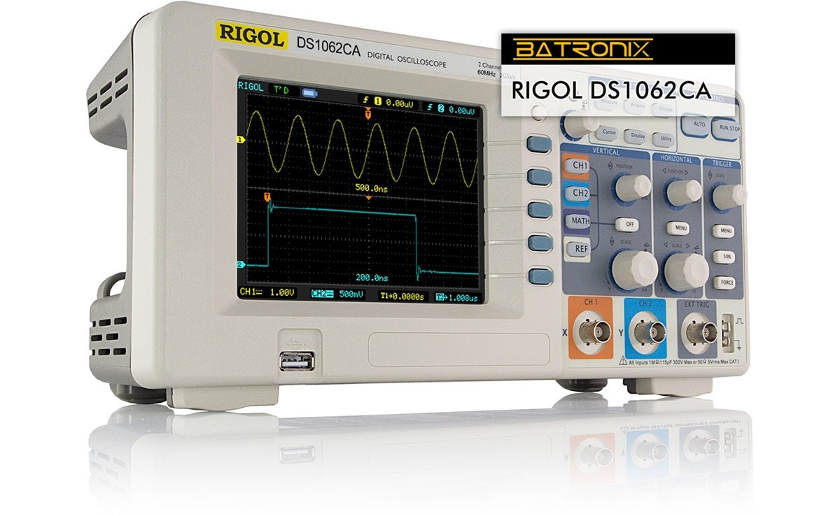
Signal Processing, Computer-Assisted
Spectroscopy, Fourier Transform Infrared
Mathematics
Algorithms
Models, Biological
A new filtering algorithm for medical magnetic resonance and computer tomography images. (1/2483)
Inner views of tubular structures based on computer tomography (CT) and magnetic resonance (MR) data sets may be created by virtual endoscopy. After a preliminary segmentation procedure for selecting the organ to be represented, the virtual endoscopy is a new postprocessing technique using surface or volume rendering of the data sets. In the case of surface rendering, the segmentation is based on a grey level thresholding technique. To avoid artifacts owing to the noise created in the imaging process, and to restore spurious resolution degradations, a robust Wiener filter was applied. This filter working in Fourier space approximates the noise spectrum by a simple function that is proportional to the square root of the signal amplitude. Thus, only points with tiny amplitudes consisting mostly of noise are suppressed. Further artifacts are avoided by the correct selection of the threshold range. Afterwards, the lumen and the inner walls of the tubular structures are well represented and allow one to distinguish between harmless fluctuations and medically significant structures. (+info)Wavelet transform to quantify heart rate variability and to assess its instantaneous changes. (2/2483)
Heart rate variability is a recognized parameter for assessing autonomous nervous system activity. Fourier transform, the most commonly used method to analyze variability, does not offer an easy assessment of its dynamics because of limitations inherent in its stationary hypothesis. Conversely, wavelet transform allows analysis of nonstationary signals. We compared the respective yields of Fourier and wavelet transforms in analyzing heart rate variability during dynamic changes in autonomous nervous system balance induced by atropine and propranolol. Fourier and wavelet transforms were applied to sequences of heart rate intervals in six subjects receiving increasing doses of atropine and propranolol. At the lowest doses of atropine administered, heart rate variability increased, followed by a progressive decrease with higher doses. With the first dose of propranolol, there was a significant increase in heart rate variability, which progressively disappeared after the last dose. Wavelet transform gave significantly better quantitative analysis of heart rate variability than did Fourier transform during autonomous nervous system adaptations induced by both agents and provided novel temporally localized information. (+info)A new tool for measuring the suckling stimulus during breastfeeding in humans: the orokinetogram and the Fourier series. (3/2483)
The Fourier series was used to analyse the oral movements recorded by the orokinetogram during breastfeeding in human babies. This is a new method that allows recording of oral movements without introducing any extrinsic element between the nipple and the mouth of the baby. The advantage of displaying suckling activity after fast Fourier transform (FFT) is that this algorithm allows storage, quantification and frequency analysis of the oral movements throughout a suckling bout, which enables the total oral activity to be measured. Two types of oral movements are found: slow high amplitude (SHA) and fast low amplitude (FLA). FLA movements may be derived from peristaltic movements of the tongue that result in tickling stimuli to the mechanoreceptors of the nipple and milk expression. The frequency bandwidth of oral movements is wider (0-8 Hz) than has been described previously (0-3 Hz) and this is due to the presence of the FLA oral movements. An indirect measurement of the energy of oral movements during suckling is obtained by the pattern of energy distribution used in each individual frequency band by oral movements. This pattern changes in relation to the periods of continuous and intermittent suckling activity. SHA and FLA oral movements are more intense during continuous suckling. Statistical analysis showed a correlation between the energy of SHA and FLA waves throughout the suckling bout, and also that the highest level of energy during suckling activity is displayed during the first 2 min. The novel tools described in this paper allow investigation of the role of suckling stimulus in reflex hormone release and other mother-infant interactions. (+info)Effects of nicorandil on aortic input impedance: a comparative study with nitroglycerin. (4/2483)
A study of aortic input impedance was performed to evaluate the effects of nicorandil on the systemic circulation, and the effects were compared with those of nitroglycerin. Sixteen patients with coronary artery disease were divided into 2 age-matched groups. Aortic input impedance was obtained from Fourier analysis of aortic pressure and flow signals at baseline conditions, after intravenous administration of either 4 mg (Group 1) or 8 mg (Group 2) nicorandil, and 20 min after 0.3 mg sublingual nitroglycerin. In Group 1, the first harmonic impedance modulus (Z1, 304+/-140 dyne x s x cm(-5)) and the average of the first to third harmonics (Z1-3, 207+/-99 dyne x s x cm(-5)), indices of wave reflection, significantly decreased (24.4% (p<0.05) and 24.7% (p<0.01), respectively) after nicorandil, and 41.3% (p<0.01) and 33.9% (p<0.01) after nitroglycerin. The effects between the 2 vasodilators were not significantly different. In Group 2, Z1 and Z1-3 (275+/-138 and 196+/-93 dyne x s x cm(-5), respectively) also decreased after administration of nicorandil (28.4% (p<0.01) and 35.9% (p<0.01), respectively), and after administration of nitroglycerin (23.9% (p<0.01) and 28.7% (p<0.01), respectively), without any significant difference between the 2 drugs. Characteristic impedance and total peripheral resistance (R) in both groups remained unchanged except for R after 8 mg nicorandil (from 1830+/-415 to 1433+/-428 dyne x s x cm(-5); p<0.01). Like nitroglycerin, both doses of nicorandil reduced wave reflection. The reduction in R after 8 mg nicorandil is related to decreased tone in the resistance arteries, probably due to potassium channel opener effects. (+info)Oxytocin-induced Ca2+ responses in human myometrial cells. (5/2483)
Complex spatiotemporal changes in intracellular Ca2+ were monitored in an immortalized human myometrial cell line (PHM1-41) and first-passage human myometrial cells after oxytocin stimulation (1. 0-1000 nM). Laser cytometry revealed intracellular Ca2+ oscillations in both culture systems starting at 1.0 nM, which were followed by repetitive Ca2+ transients by 10-15 min that lasted for at least 90 min. The amplitude of the initial Ca2+ spike was dose dependent, while the frequency of Ca2+ oscillations identified by Fast Fourier Transform (FFT) tended to increase with dose. Removal of oxytocin resulted in termination of oscillations. Analysis of the sources of the Ca2+ involved in oscillations indicated that the major contribution to oscillation frequencies of +info)Automated MAD and MIR structure solution. (6/2483)
Obtaining an electron-density map from X-ray diffraction data can be difficult and time-consuming even after the data have been collected, largely because MIR and MAD structure determinations currently require many subjective evaluations of the qualities of trial heavy-atom partial structures before a correct heavy-atom solution is obtained. A set of criteria for evaluating the quality of heavy-atom partial solutions in macromolecular crystallography have been developed. These have allowed the conversion of the crystal structure-solution process into an optimization problem and have allowed its automation. The SOLVE software has been used to solve MAD data sets with as many as 52 selenium sites in the asymmetric unit. The automated structure-solution process developed is a major step towards the fully automated structure-determination, model-building and refinement procedure which is needed for genomic scale structure determinations. (+info)Discrimination of solvent from protein regions in native Fouriers as a means of evaluating heavy-atom solutions in the MIR and MAD methods. (7/2483)
An automated examination of the native Fourier is tested as a means of evaluation of a heavy-atom solution in MAD and MIR methods for macromolecular crystallography. It is found that the presence of distinct regions of high and low density variation in electron-density maps is a good indicator of the correctness of a heavy-atom solution in the MIR and MAD methods. The method can be used to evaluate heavy-atom solutions during MAD and MIR structure solutions and to determine the handedness of the structure if anomalous data have been measured. (+info)Evidence of noncovalent dimerization of calmodulin. (8/2483)
Calcium-binding proteins, such as S-100, dimerize readily, and this phenomenon plays an important role in their regulation of target enzymes [Krebs, J., Quadroni, M. & Van Eldik, L.J. (1995) Nat. Struct. Biol. 2, 711-714; Kilby, P.M., Van Eldik, L.J. & Roberts, G. C. (1996) Structure 4, 1041-1052]. We have investigated by Fourier-transform ion cyclotron resonance (FTICR) MS the conformational states of the calcium-binding protein calmodulin, and present clear evidence for a calmodulin dimer formed as a result of noncovalent interactions between folded monomers. Ultra-high-resolution electrospray ionization (ESI) mass spectra for calmodulin, obtained with a 9.4 T FTICR mass spectrometer, are presented. With the use of denaturing solutions (1 : 1 acetonitrile/water + 1% formic acid), relatively high charge states (20 < z < 10) of monomeric calmodulin ions were detected, whereas when calmodulin was electrosprayed from buffer, monomers ions with only 5-10 charges were detected. CD measurements for calmodulin in buffered solution revealed that its alpha-helical content was significantly higher than that for calmodulin in acetonitrile/water solutions, consistent with a proposition that changes in charge state distributions observed in the MS experiments reflect differing states of calmodulin folding. Under buffered conditions, noncovalently bound calmodulin dimers were observed by ESI FTICR MS. Analytical ultracentrifugation experiments carried out in the same solution conditions as those used in the MS experiments were consistent with the proposed calmodulin dimer-monomer equilibrium. The ultra-high mass resolution achieved with the 9.4 T FTICR mass spectrometer allowed unequivocal identification of the noncovalent, as opposed to covalent, character of the calmodulin dimer. (+info)I'm sorry for any confusion, but Fourier Analysis is not a medical term. It is a mathematical concept used in various scientific fields, including physics, engineering, and signal processing.
Fourier Analysis is a method to decompose functions into sinusoidal components (sines and cosines) of different frequencies. This allows for the representation of a function or a signal as a sum of these frequency components. It's particularly useful in analyzing periodic functions, understanding signals, and solving partial differential equations.
If you have any medical terms you would like me to define, please let me know!
Optical phenomena refer to the various observable patterns and effects that occur due to the interaction of light with the environment or with structures in our eye. These can include natural phenomena such as rainbows, mirages, and halos around the sun or moon, as well as visual artifacts created by the eye itself, such as afterimages, floaters, and flashes of light. Some optical phenomena are caused by the refraction, reflection, or interference of light waves, while others may result from abnormalities in the eye's structure or function. Understanding these phenomena can provide insight into the properties of light and the functioning of the visual system.
Technetium is not a medical term itself, but it is a chemical element with the symbol Tc and atomic number 43. However, in the field of nuclear medicine, which is a branch of medicine that uses small amounts of radioactive material to diagnose or treat diseases, Technetium-99m (a radioisotope of technetium) is commonly used for various diagnostic procedures.
Technetium-99m is a metastable nuclear isomer of technetium-99, and it emits gamma rays that can be detected outside the body to create images of internal organs or tissues. It has a short half-life of about 6 hours, which makes it ideal for diagnostic imaging since it decays quickly and reduces the patient's exposure to radiation.
Technetium-99m is used in a variety of medical procedures, such as bone scans, lung scans, heart scans, liver-spleen scans, brain scans, and kidney scans, among others. It can be attached to different pharmaceuticals or molecules that target specific organs or tissues, allowing healthcare professionals to assess their function or identify any abnormalities.
Gated Blood-Pool Imaging (GBPI) is a type of nuclear medicine test that uses radioactive material and a specialized camera to create detailed images of the heart and its function. In this procedure, a small amount of radioactive tracer is injected into the patient's bloodstream, which then accumulates in the heart muscle and the blood pool within the heart chambers.
The term "gated" refers to the use of an electrocardiogram (ECG) signal to synchronize the image acquisition with the heart's contractions. This allows for the visualization of the heart's motion during different phases of the cardiac cycle, providing valuable information about the size, shape, and contraction of the heart chambers, as well as the movement of the walls of the heart.
GBPI is often used to assess patients with known or suspected heart disease, such as valvular abnormalities, cardiomyopathies, or congenital heart defects. It can help diagnose and evaluate the severity of these conditions, guide treatment decisions, and monitor the effectiveness of therapy.
Computer-assisted signal processing is a medical term that refers to the use of computer algorithms and software to analyze, interpret, and extract meaningful information from biological signals. These signals can include physiological data such as electrocardiogram (ECG) waves, electromyography (EMG) signals, electroencephalography (EEG) readings, or medical images.
The goal of computer-assisted signal processing is to automate the analysis of these complex signals and extract relevant features that can be used for diagnostic, monitoring, or therapeutic purposes. This process typically involves several steps, including:
1. Signal acquisition: Collecting raw data from sensors or medical devices.
2. Preprocessing: Cleaning and filtering the data to remove noise and artifacts.
3. Feature extraction: Identifying and quantifying relevant features in the signal, such as peaks, troughs, or patterns.
4. Analysis: Applying statistical or machine learning algorithms to interpret the extracted features and make predictions about the underlying physiological state.
5. Visualization: Presenting the results in a clear and intuitive way for clinicians to review and use.
Computer-assisted signal processing has numerous applications in healthcare, including:
* Diagnosing and monitoring cardiac arrhythmias or other heart conditions using ECG signals.
* Assessing muscle activity and function using EMG signals.
* Monitoring brain activity and diagnosing neurological disorders using EEG readings.
* Analyzing medical images to detect abnormalities, such as tumors or fractures.
Overall, computer-assisted signal processing is a powerful tool for improving the accuracy and efficiency of medical diagnosis and monitoring, enabling clinicians to make more informed decisions about patient care.
Fourier Transform Infrared (FTIR) spectroscopy is a type of infrared spectroscopy that uses the Fourier transform mathematical technique to convert the raw data obtained from an interferometer into a more interpretable spectrum. This technique allows for the simultaneous collection of a wide range of wavelengths, resulting in increased sensitivity and speed compared to traditional dispersive infrared spectroscopy.
FTIR spectroscopy measures the absorption or transmission of infrared radiation by a sample as a function of frequency, providing information about the vibrational modes of the molecules present in the sample. This can be used for identification and quantification of chemical compounds, analysis of molecular structure, and investigation of chemical interactions and reactions.
In summary, FTIR spectroscopy is a powerful analytical technique that uses infrared radiation to study the vibrational properties of molecules, with increased sensitivity and speed due to the use of Fourier transform mathematical techniques and an interferometer.
I'm sorry for any confusion, but there seems to be a misunderstanding. Mathematics is not a medical term; it is a branch of science dedicated to the study of numbers, shapes, and structures. However, mathematics does have many applications in medicine, such as in modeling disease spread, analyzing medical images, or designing clinical trials. If you have any questions related to mathematics in a medical context, I'd be happy to help clarify those for you!
An algorithm is not a medical term, but rather a concept from computer science and mathematics. In the context of medicine, algorithms are often used to describe step-by-step procedures for diagnosing or managing medical conditions. These procedures typically involve a series of rules or decision points that help healthcare professionals make informed decisions about patient care.
For example, an algorithm for diagnosing a particular type of heart disease might involve taking a patient's medical history, performing a physical exam, ordering certain diagnostic tests, and interpreting the results in a specific way. By following this algorithm, healthcare professionals can ensure that they are using a consistent and evidence-based approach to making a diagnosis.
Algorithms can also be used to guide treatment decisions. For instance, an algorithm for managing diabetes might involve setting target blood sugar levels, recommending certain medications or lifestyle changes based on the patient's individual needs, and monitoring the patient's response to treatment over time.
Overall, algorithms are valuable tools in medicine because they help standardize clinical decision-making and ensure that patients receive high-quality care based on the latest scientific evidence.
Biological models, also known as physiological models or organismal models, are simplified representations of biological systems, processes, or mechanisms that are used to understand and explain the underlying principles and relationships. These models can be theoretical (conceptual or mathematical) or physical (such as anatomical models, cell cultures, or animal models). They are widely used in biomedical research to study various phenomena, including disease pathophysiology, drug action, and therapeutic interventions.
Examples of biological models include:
1. Mathematical models: These use mathematical equations and formulas to describe complex biological systems or processes, such as population dynamics, metabolic pathways, or gene regulation networks. They can help predict the behavior of these systems under different conditions and test hypotheses about their underlying mechanisms.
2. Cell cultures: These are collections of cells grown in a controlled environment, typically in a laboratory dish or flask. They can be used to study cellular processes, such as signal transduction, gene expression, or metabolism, and to test the effects of drugs or other treatments on these processes.
3. Animal models: These are living organisms, usually vertebrates like mice, rats, or non-human primates, that are used to study various aspects of human biology and disease. They can provide valuable insights into the pathophysiology of diseases, the mechanisms of drug action, and the safety and efficacy of new therapies.
4. Anatomical models: These are physical representations of biological structures or systems, such as plastic models of organs or tissues, that can be used for educational purposes or to plan surgical procedures. They can also serve as a basis for developing more sophisticated models, such as computer simulations or 3D-printed replicas.
Overall, biological models play a crucial role in advancing our understanding of biology and medicine, helping to identify new targets for therapeutic intervention, develop novel drugs and treatments, and improve human health.
 Fourier analysis
 Mid-infrared supercontinuum-based Fourier transform spectroscopy for plasma analysis | Scientific Reports
 Fourier Theory without Complex Analysis | School of Mathematics and Statistics
 FFT (Fast Fourier Transform) Waveform Analysis
 Fourier Transform Infrared Absorption Spectroscopy for Quantitative Analysis of Gas Mixtures for Homeland Security Applications...
 Fourier Analysis
 Surface profile determination by additive-subtractive phase-modulated ESPI with Fourier analysis | (1996) | Wang | ...
 Newest 'fourier-analysis' Questions - Mathematics Stack Exchange
 PalaeoMath: Part 24 - The Centre Cannot Hold I: Z-R Fourier Analysis | The Palaeontological Association
 ap.analysis of pdes - Positivity of the Fourier transform: prove or disprove that $\operatorname{Re}(\overline{\widehat{u}}(\xi...
 soft question - Why does Fourier analysis of Boolean functions 'work'? - Theoretical Computer Science Stack Exchange
 A fractional Fourier transform analysis of a bubble excited by an ultrasonic chirp - Strathprints
 Reconstructability analysis with Fourier transforms
| Emerald Insight
Reconstructability analysis with Fourier transforms
| Emerald Insight Analysis of Retinal Nerve Fiber Layer Data Obtained by Optical Coherence Tomograph Using Fourier Based Analysis | IOVS | ARVO...
![2008.10871] Analysis of the Feshbach-Schur method for the Fourier Spectral discretizations of Schr{ö}dinger operators](data:image/png;base64,iVBORw0KGgoAAAANSUhEUgAAABAAAAAQCAYAAAAf8/9hAAACA0lEQVQ4jY3TXWjNcRgH8M//f3YwaTt5GYltWVMKeYkLiUJSnChLmqQkXKC8RSlckQsl5QYXSmspSg4JV3Nhd4qi5n0u197nHJuds7+L/Y92MPne/N6el+/z/T1PkKmthyl4gA84joH0l/f+B2XxmsVjnMM3nMvU1v8KkrnbBAFWYgBvId2wSxgbRbiG2ziMS0hlauuLziF24h42jGUQQhwkFzN4ioO4rFCYakF9GQ7ECV7ESaQbdpWUUEQHTmA29kokJmpp/WzmjKOi6CVOomesQ6K4ae7t1piaRj7fiTZRtB6rZXNrVM/5KFWxj6AN0tsb/xBRsZTMretUTX/j/uN3nrfO9eoNn9q79A90SSRoby+hnBh7yNxt4sET5tedNa9mt2iET18ZHKwWBNNF0SOVlYXm3u5SEUvQkA5E0RRhyNpVrFhSfKn8m33pRc1cgiAShhf1D7RIJtm0jqULX8t9PyMMBv0YLnEJSkqormNkhOH8ArOq7ti8YZFli4miNsnkDoXCa6kK6fVbxmEQhpSVVSmfdFlv3yIPn2UM/WgWBPPl81dE0Rw9fcXmwphfiGeiHOexURA06ew6ZmpqSDbXi/24gEPoH0/ErdiDmzhiwoQO2VwfTuEqGow2U7LI4pcGMYPlRgemCf2/DdNknDHa5qdx4w8R/4U4SHlcxkJsQ/YnnK+wsTK7TvYAAAAASUVORK5CYII=) 2008.10871] Analysis of the Feshbach-Schur method for the Fourier Spectral discretizations of Schr{ö}dinger operators
2008.10871] Analysis of the Feshbach-Schur method for the Fourier Spectral discretizations of Schr{ö}dinger operators Mathematical Analysis of Computer Generated Binary Fourier Transform Holograms - MADOC
 MATLAB and Simulink Courseware - MATLAB & Simulink
 Fourier Analysis - Aurora Vision
 Did Joseph Fourier discover the greenhouse effect? - Rwmansiononpeachtree.com
 Real-time implementation of discrete fourier transform phase analysis and fault tolerant control for PMSM in electric vehicles ...
 Fourier Analysis - Reuben Cohn-Gordon
 Fourier Analysis Questions and Answers - Matchmaticians
 MATH 382 Fourier Analysis 2023-24 - Catalog