Foramen Ovale, Patent
Foramen Ovale
Heart Septal Defects, Atrial
Embolism, Paradoxical
Plasmodium ovale
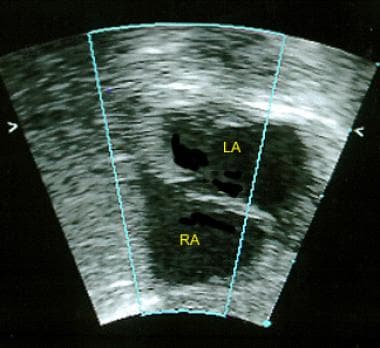
Echocardiography, Transesophageal
Atrial Septum
Septal Occluder Device
Heart Septum
Heart Aneurysm
Cardiac Catheterization
Migraine with Aura
Embolism
Intracranial Embolism
Heart Septal Defects
Stroke
Decompression Sickness
Echocardiography
Prostheses and Implants
Ultrasonography, Doppler, Transcranial
Migraine Disorders
Ischemic Attack, Transient
Embolism, Air
Secondary Prevention
High Pressure Neurological Syndrome
Diving
Sphenoid Bone
Vena Cava, Inferior
Treatment Outcome
Mandibular Nerve
Intracranial Embolism and Thrombosis
Skull Base
Echocardiography, Doppler, Color
Fetal Heart
Plasmodium malariae
Follow-Up Studies
Common Dolphins
Thromboembolism
Cerebrovascular Disorders
Echoencephalography
Prospective Studies
Carcinoid Heart Disease
Embolectomy
Tooth Apex
Foreign-Body Migration
Cardiac Tamponade
Respiratory Paralysis
Brain Ischemia
Heart Valves
Plasmodium
Magnetic Resonance Imaging
Heart Diseases
Radiography, Interventional
Cardiac-Gated Imaging Techniques
Risk Factors
Gelatin
Retrospective Studies
Echocardiography, Three-Dimensional
Intellectual Property
Ultrasonography, Prenatal
Malaria
Catheterization
Tomography, X-Ray Computed
Hypoplastic Left Heart Syndrome
Parietal Bone
Heart Defects, Congenital
Absorbable Implants
Arnold-Chiari Malformation
Syringomyelia
Mandible
Echocardiography, Doppler
Sensitivity and Specificity
Esophagus
Prevalence
Cerebral Infarction
Intention to Treat Analysis
Platelet Aggregation Inhibitors
Maxillary Nerve
Postoperative Complications
Glomus Jugulare Tumor
Single-Blind Method
Predictive Value of Tests
Patents
Ultrasonography, Interventional
Aspirin
Profound hypoxaemia corrected by PFO closure device in carcinoid heart disease. (1/233)
A 66-year-old man with known metastatic carcinoid tumor presented with increasing dyspnoea, right heart failure and marked hypoxaemia which did not correct with oxygen. Echocardiography demonstrated severe tricuspid regurgitation, moderate pulmonary regurgitation and marked right heart dilatation. The inter-atrial septum was aneurysmal, with a large patent foramen ovale (PFO) with continuous right to left shunting. Cardiac catheterization demonstrated oxygen saturations of 96% in the pulmonary veins and 74% in the left atrium with a significant right to left shunt. During percutaneous closure of the PFO, anaesthetic induction resulted in marked systemic hypotension and worsening hypoxia related to systemic vasodilatation and increased shunting. PFO flow was temporarily obstructed with a sizing balloon resulting in a rapid increase in arterial oxygen saturation from 60% to >90%, but marked systemic hypotension due to acute left ventricular preload reduction, requiring volume replacement and adrenaline. Following deployment of a PFO occluder device, prominent pulsatile splaying of the right and left discs was noted due to the severe tricuspid regurgitation, resulting in some residual inter-atrial shunting. Arterial oxygen saturation was 83%, increasing to 92% at day 4 post-procedure as tissue organization occurred within the device, and the patient reported improvement in dyspnoea. (+info)Patent foramen ovale and ischemic stroke in young people: statistical association or causal relation? (2/233)
OBJECTIVES: To determine if there are evidences of a causal relation between patent foramen ovale (PFO) x cryptogenic ischemic stroke (IS) in the young population and to analyze this relation in terms of causal criteria. METHODS: A total of 168 young patients with IS was retrospectively evaluated and divided into two groups: cryptogenic and with a defined cause. As a routine procedure, the patients underwent investigation of the PFO by means of transesophageal echocardiogram and/or transcranial Doppler sonography, both of them associated with the bubble test. Multivariate analysis was performed after demonstration of univariate statistical association between PFO x IS. RESULTS: After multivariate analysis, the association between PFO x cryptogenic IS was still statistically significant with odds ratio (adjusted OR = 3.3; 95% CI: 1.5-7.4). The total number of cerebral lesions also presented a significant association with cryptogenic IS (adjusted OR = 0.4; 95% CI: 0.2-0.9). The association between PFO and cryptogenic IS met all the causality criteria. CONCLUSION: The causal relation between PFO and cryptogenic IS in the young population is highly probable. This fact should be considered in the therapeutic decision. (+info)Eustachian valve interfering with transcatheter closure of patent foramen ovale. (3/233)
A prominent Eustachian valve (EV) is a common finding in patients with a patent foramen ovale (PFO). Its presence might compromise transcatheter closure of the PFO. (+info)A dumbbell thrombus entrapped in patent foramen ovale. (4/233)
A 75-year-old lady came to emergency room due to dizziness and presyncopal attacks during exertion since two days prior to admission. Transesophageal echocardiography revealed a thrombus like mass in right atrium traversing patent foramen ovale and extending to left atrium. Spiral chest CT scan showed bilateral pulmonary thromboemboli. Operative and pathological findings confirmed the diagnosis. (+info)Pulmonary embolism and patent foramen ovale thrombosis: the key role of TEE. (5/233)
This is a case report of a 35 young man with Klinefelter Syndrome presented breathlessness, palpitations and chest pain. It shows a rare case of a thrombus located through the PFO, in patient with pulmonary and paradoxical embolism, which takes back to exciting hypothesis on thrombus growth. A thrombus, which has grown 'in situ' or trapped through the patent foramen ovale, may be a cause of relapsing pulmonary or systemic embolism during anticoagulation therapy. To prevent recurrent paradoxical embolism, percutaneous closure of PFO is recommended, but in this case, thrombus was trapped through the PFO and the patient was referred to the surgeon. We believe that under these circumstances the clinician should be informed of the presence of PFO in critical pulmonary embolism; this case points out the key role of TEE to face a diagnostic and therapeutic scenarios. (+info)Successful interventional closure of a patent foramen ovale in a pediatric patient supported with a biventricular assist device. (6/233)
We report on a 16-year-old boy after an event of cardiac arrest and initial treatment with a veno-arterial extracorporeal membrane oxygenator (ECMO). After a short stabilisation period a biventricular assist device (BVAD, Thoratec) was implanted. Although the BVAD was functioning well, the patient showed persisting hypoxemia. Transthoracic echocardiography revealed a patent foramen ovale with a high right-to-left shunt due to low aspiration pressures of the BVAD. The patient was successfully treated by interventional closure of the PFO with a 27-mm Amplatzer septal occluder and could easily be weaned from the respirator. Meanwhile the boy has successfully undergone heart transplantation. PFO has to be considered as a cause of arterial hypoxemia in patients supported with ventricular assist devices. The diagnosis of a PFO may be missed under ECMO-treatment. Interventional closure of a PFO can successfully be performed even if the patient is supported with a BVAD. (+info)Migraineurs with patent foramen ovale have larger right-to-left shunt despite similar atrial septal characteristics. (7/233)
The objective of the study was to assess differences in proportion of large right-to-left shunt (RLS) and atrial septal characteristics between migraineurs and non-migraineurs referred for transcatheter closure of patent foramen ovale (PF0). This retrospective study took place in a large metropolitan medical centre. The patients were migraineurs with aura (n=52), migraineurs without aura (n=19) and non-migraineurs (n=149). RLS was evaluated before closure using bilateral power m-mode transcranial Doppler at rest and after calibrated, sustained Valsalva manoeuvre, and graded with a validated 0-5 scale. Intracardiac echocardiography was used to assess atrial septal characteristics. Migraineurs had a higher proportion of large RLS (Grade IV or V) than nonmigraineurs at rest and after calibrated Valsalva (rest, p=0.04; Valsalva, p=0.01). Atrial septal characteristics were similar between groups. Migraine is associated with larger RLS at rest and strain; however migraine status does not predict PFO characteristics. (+info)Patent foramen ovale and cryptogenic stroke in older patients. (8/233)
BACKGROUND: Studies to date have shown an association between the presence of patent foramen ovale and cryptogenic stroke in patients younger than 55 years of age. This association has not been established in patients 55 years of age or older. METHODS: We prospectively examined 503 consecutive patients who had had a stroke, and we compared the 227 patients with cryptogenic stroke and the 276 control patients with stroke of known cause. We examined the prevalences of patent foramen ovale and of patent foramen ovale with concomitant atrial septal aneurysm in all patients, using transesophageal echocardiography. We also compared data for the 131 younger patients (< 55 years of age) and those for the 372 older patients (> or = 55 years of age). RESULTS: The prevalence of patent foramen ovale was significantly greater among patients with cryptogenic stroke than among those with stroke of known cause, for both younger patients (43.9% vs. 14.3%; odds ratio, 4.70; 95% confidence interval [CI], 1.89 to 11.68; P<0.001) and older patients (28.3% vs. 11.9%; odds ratio, 2.92; 95% CI, 1.70 to 5.01; P<0.001). Even stronger was the association between the presence of patent foramen ovale with concomitant atrial septal aneurysm and cryptogenic stroke, as compared with stroke of known cause, among both younger patients (13.4% vs. 2.0%; odds ratio, 7.36; 95% CI, 1.01 to 326.60; P=0.049) and older patients (15.2% vs. 4.4%; odds ratio, 3.88; 95% CI, 1.78 to 8.46; P<0.001). Multivariate analysis adjusted for age, plaque thickness, and presence or absence of coronary artery disease and hypertension showed that the presence of patent foramen ovale was independently associated with cryptogenic stroke in both the younger group (odds ratio, 3.70; 95% CI, 1.42 to 9.65; P=0.008) and the older group (odds ratio, 3.00; 95% CI, 1.73 to 5.23; P<0.001). CONCLUSIONS: There is an association between the presence of patent foramen ovale and cryptogenic stroke in both older patients and younger patients. These data suggest that paradoxical embolism is a cause of stroke in both age groups. (+info)Patent Foramen Ovale (PFO) is a medical condition where the foramen ovale, an opening between the left and right atria of the heart in a fetus, does not close completely after birth. This results in a small flap-like opening that allows blood to pass from the right atrium to the left atrium. While this condition is typically harmless in itself, it can potentially allow blood clots to pass from the right side of the heart to the left, which could then travel to the brain and cause a stroke. Patent Foramen Ovale is usually an incidental finding during tests for other conditions.
The foramen ovale is a fetal cardiovascular structure that usually closes after birth. It's a flap-like opening between the right and left atria (the upper chambers) of the heart. This opening allows oxygen-rich blood from the mother to bypass the fetal lungs and go directly to the fetal brain and body.
After birth, when the newborn starts breathing and blood pressure in the lungs increases, the pressure in the left atrium also rises, causing the flap to close and seal the foramen ovale. In about 25% of adults, this flap doesn't close completely, resulting in a condition known as a patent foramen ovale (PFO), which is usually asymptomatic but can rarely lead to complications such as stroke or migraine with aura.
Atrial septal defect (ASD) is a type of congenital heart defect that involves the septum, which is the wall that separates the two upper chambers of the heart (atria). An ASD is a hole or abnormal opening in the atrial septum, allowing oxygen-rich blood to leak into the oxygen-poor blood chambers in the heart. This leads to an overload of blood in the right side of the heart, which can cause enlargement of the heart and increased work for the right ventricle.
ASDs can vary in size, and small defects may not cause any symptoms or require treatment. Larger defects, however, can result in symptoms such as shortness of breath, fatigue, and heart rhythm abnormalities. Over time, if left untreated, ASDs can lead to complications like pulmonary hypertension, atrial fibrillation, and stroke.
Treatment for ASD typically involves surgical closure of the defect or catheter-based procedures using devices to close the hole. The choice of treatment depends on factors such as the size and location of the defect, the patient's age and overall health, and the presence of any coexisting conditions.
Paradoxical embolism is a medical condition that occurs when a blood clot or other material (embolus) from a vein passes through an abnormal connection between the right and left sides of the heart and lodges in an artery in the systemic circulation. This is considered "paradoxical" because the embolus originates from the venous system but bypasses the lungs and travels directly to the arterial system.
Under normal circumstances, blood flows from the body's veins into the right atrium of the heart, then through the tricuspid valve into the right ventricle, where it is pumped through the pulmonary artery into the lungs for oxygenation. The now oxygen-rich blood returns to the left atrium via the pulmonary veins, passes through the mitral valve into the left ventricle, and is then pumped out to the body's arteries.
However, in certain conditions such as a patent foramen ovale (PFO) or an atrial septal defect (ASD), there can be an abnormal communication between the right and left atria. This allows for the possibility of a paradoxical embolism to occur when a clot or other material from the venous system passes through this connection into the arterial system, bypassing filtration and oxygenation in the lungs.
Paradoxical embolism can lead to serious consequences, such as stroke, transient ischemic attack (TIA), or tissue damage in various organs, depending on where the embolus lodges. Treatment typically involves addressing the underlying cause of the paradoxical embolism and may include anticoagulation therapy, surgical closure of the abnormal connection, or other interventions as necessary.
A patent, in the context of medicine and healthcare, generally refers to a government-granted exclusive right for an inventor to manufacture, use, or sell their invention for a certain period of time, typically 20 years from the filing date. In the medical field, patents may cover a wide range of inventions, including new drugs, medical devices, diagnostic methods, and even genetic sequences.
The purpose of patents is to provide incentives for innovation by allowing inventors to profit from their inventions. However, patents can also have significant implications for access to medical technologies and healthcare costs. For example, a patent on a life-saving drug may give the patent holder the exclusive right to manufacture and sell the drug, potentially limiting access and driving up prices.
It's worth noting that the patent system is complex and varies from country to country. In some cases, there may be ways to challenge or circumvent patents in order to increase access to medical technologies, such as through compulsory licensing or generic substitution.
"Plasmodium ovale" is a species of protozoan parasites that are transmitted to humans through the bites of infected female Anopheles mosquitoes. This parasite causes a type of malaria known as "ovale malaria," which is generally milder than other forms of malaria caused by Plasmodium falciparum or Plasmodium vivax.
The life cycle of Plasmodium ovale involves two hosts: the mosquito and humans. When an infected mosquito bites a human, the parasites are injected into the skin along with the mosquito's saliva. The parasites then enter the liver where they multiply and form dormant stages called hypnozoites. After a period of time (usually several weeks to months), the hypnozoites become activated and begin to infect red blood cells, leading to the symptoms of malaria.
The symptoms of ovale malaria are similar to those of other forms of malaria and include fever, chills, headache, muscle and joint pain, and fatigue. However, ovale malaria is less likely to cause severe complications or death than falciparum malaria. Diagnosis of ovale malaria is typically made through microscopic examination of blood smears or by using rapid diagnostic tests (RDTs) that detect parasite antigens in the blood. Treatment usually involves the use of antimalarial drugs such as chloroquine or primaquine.
Transesophageal echocardiography (TEE) is a type of echocardiogram, which is a medical test that uses sound waves to create detailed images of the heart. In TEE, a special probe containing a transducer is passed down the esophagus (the tube that connects the mouth to the stomach) to obtain views of the heart from behind. This allows for more detailed images of the heart structures and function compared to a standard echocardiogram, which uses a probe placed on the chest. TEE is often used in patients with poor image quality from a standard echocardiogram or when more detailed images are needed to diagnose or monitor certain heart conditions. It is typically performed by a trained cardiologist or sonographer under the direction of a cardiologist.
The foramen magnum is the largest opening in the human skull, located at the base of the skull, through which the spinal cord connects to the brain. It is a crucial structure for the transmission of nerve impulses between the brain and the rest of the body. The foramen magnum also provides passage for blood vessels that supply the brainstem and upper spinal cord.
The atrial septum is the wall of tissue that divides the right and left atria, which are the upper chambers of the heart. This septum ensures that oxygen-rich blood in the left atrium is kept separate from oxygen-poor blood in the right atrium. Defects or abnormalities in the atrial septum, such as a hole or a gap, can result in various heart conditions, including septal defects and congenital heart diseases.
A septal occluder device is a type of medical implant used to close defects or holes in the heart, specifically within the septum, which is the wall that separates the two sides of the heart. The device typically consists of two disc-shaped components connected by a waist, resembling a button or an umbrella.
The procedure for implanting a septal occluder device involves inserting it through a catheter, which is introduced into a vein in the leg and guided to the heart. Once in position, the discs of the device expand and are pressed against the septum on both sides of the hole, effectively closing it. Over time, tissue grows over the device, permanently sealing the defect.
Septal occluder devices are commonly used to treat atrial septal defects (ASD) and patent foramen ovale (PFO), which are two types of congenital heart defects that can cause symptoms such as shortness of breath, fatigue, and heart palpitations. The use of these devices has revolutionized the treatment of these conditions, allowing for less invasive procedures and faster recovery times compared to traditional surgical methods.
The heart septum is the thick, muscular wall that divides the right and left sides of the heart. It consists of two main parts: the atrial septum, which separates the right and left atria (the upper chambers of the heart), and the ventricular septum, which separates the right and left ventricles (the lower chambers of the heart). A normal heart septum ensures that oxygen-rich blood from the lungs does not mix with oxygen-poor blood from the body. Any defect or abnormality in the heart septum is called a septal defect, which can lead to various congenital heart diseases.
A heart aneurysm, also known as a ventricular aneurysm, is a localized bulging or ballooning of the heart muscle in the left ventricle, which is the main pumping chamber of the heart. This condition typically occurs following a myocardial infarction (heart attack), where blood flow to a portion of the heart muscle is blocked, leading to tissue death and weakness in the heart wall. As a result, the weakened area may stretch and form a sac-like bulge or aneurysm.
Heart aneurysms can vary in size and may cause complications such as blood clots, arrhythmias (irregular heartbeats), or heart failure. In some cases, they may be asymptomatic and discovered during routine imaging tests. The diagnosis of a heart aneurysm is typically made through echocardiography, cardiac MRI, or cardiac CT scans. Treatment options depend on the size, location, and symptoms of the aneurysm and may include medications, surgical repair, or implantation of a device to support heart function.
The Valsalva maneuver is a medical procedure that involves forced exhalation against a closed airway, typically by closing one's mouth, pinching the nose shut, and then blowing. This maneuver increases the pressure in the chest and affects the heart's filling and pumping capabilities, as well as the pressures within the ears and eyes.
It is often used during medical examinations to test for conditions such as heart murmurs or to help clear the ears during changes in air pressure (like when scuba diving or flying). It can also be used to help diagnose or monitor conditions related to the autonomic nervous system, such as orthostatic hypotension or dysautonomia.
However, it's important to perform the Valsalva maneuver correctly and under medical supervision, as improper technique or overdoing it can lead to adverse effects like increased heart rate, changes in blood pressure, or even damage to the eardrum.
Cardiac catheterization is a medical procedure used to diagnose and treat cardiovascular conditions. In this procedure, a thin, flexible tube called a catheter is inserted into a blood vessel in the arm or leg and threaded up to the heart. The catheter can be used to perform various diagnostic tests, such as measuring the pressure inside the heart chambers and assessing the function of the heart valves.
Cardiac catheterization can also be used to treat certain cardiovascular conditions, such as narrowed or blocked arteries. In these cases, a balloon or stent may be inserted through the catheter to open up the blood vessel and improve blood flow. This procedure is known as angioplasty or percutaneous coronary intervention (PCI).
Cardiac catheterization is typically performed in a hospital cardiac catheterization laboratory by a team of healthcare professionals, including cardiologists, radiologists, and nurses. The procedure may be done under local anesthesia with sedation or general anesthesia, depending on the individual patient's needs and preferences.
Overall, cardiac catheterization is a valuable tool in the diagnosis and treatment of various heart conditions, and it can help improve symptoms, reduce complications, and prolong life for many patients.
"Migraine with Aura" is a neurological condition that is formally defined by the International Classification of Headache Disorders (ICHD) as follows:
"An migraine attack with focal neurological symptoms that usually develop gradually over 5 to 20 minutes and last for less than 60 minutes. Motor weakness is not a feature of the aura."
The symptoms of an aura may include visual disturbances such as flickering lights, zigzag lines, or blind spots; sensory disturbances such as tingling or numbness in the face, arms, or legs; and speech or language difficulties. These symptoms are caused by abnormal electrical activity in the brain and typically precede or accompany a migraine headache, although they can also occur without a headache.
It's important to note that not all people who experience migraines will have an aura, and some people may have an aura without a headache. If you are experiencing symptoms of a migraine with aura or any other type of headache, it is recommended that you consult with a healthcare professional for proper diagnosis and treatment.
An embolism is a medical condition that occurs when a substance, such as a blood clot or an air bubble, blocks a blood vessel. This can happen in any part of the body, but it is particularly dangerous when it affects the brain (causing a stroke) or the lungs (causing a pulmonary embolism). Embolisms can cause serious harm by preventing oxygen and nutrients from reaching the tissues and organs that need them. They are often the result of underlying medical conditions, such as heart disease or deep vein thrombosis, and may require immediate medical attention to prevent further complications.
An intracranial embolism is a medical condition that occurs when a blood clot or other foreign material (embolus) forms elsewhere in the body and travels to the blood vessels within the brain. This embolus then blocks the flow of blood in the cerebral arteries, leading to potential damage or death of brain tissue. Common sources of intracranial emboli include heart conditions such as atrial fibrillation, valvular heart disease, or following a heart attack; or from large-vessel atherosclerosis in the carotid arteries. Symptoms can vary depending on the location and size of the obstruction, but may include sudden weakness or numbness, confusion, difficulty speaking, vision loss, severe headache, or even loss of consciousness. Immediate medical attention is required to diagnose and treat intracranial embolism, often involving anticoagulation therapy, endovascular procedures, or surgery.
A heart septal defect is a type of congenital heart defect, which means it is present at birth. It involves an abnormal opening in the septum, the wall that separates the two sides of the heart. This opening allows oxygen-rich blood to leak into the oxygen-poor blood chambers in the heart.
There are several types of heart septal defects, including:
1. Atrial Septal Defect (ASD): A hole in the atrial septum, the wall between the two upper chambers of the heart (the right and left atria).
2. Ventricular Septal Defect (VSD): A hole in the ventricular septum, the wall between the two lower chambers of the heart (the right and left ventricles).
3. Atrioventricular Septal Defect (AVSD): A combination of an ASD and a VSD, often accompanied by malformation of the mitral and/or tricuspid valves.
The severity of a heart septal defect depends on the size of the opening and its location in the septum. Small defects may cause no symptoms and may close on their own over time. Larger defects can lead to complications, such as heart failure, pulmonary hypertension, or infective endocarditis, and may require medical or surgical intervention.
Balloon occlusion is a medical procedure that involves the use of a small, deflated balloon at the end of a catheter, which can be inserted into a blood vessel or other tubular structure in the body. Once the balloon is in position, it is inflated with a fluid or gas to create a blockage or obstruction in the vessel. This can be used for various medical purposes, such as:
1. Controlling bleeding: By inflating the balloon in a blood vessel, doctors can temporarily stop the flow of blood to a specific area, allowing them to treat injuries or abnormalities that are causing excessive bleeding.
2. Vessel narrowing or blockage assessment: Balloon occlusion can be used to assess the severity of narrowing or blockages in blood vessels. By inflating the balloon and measuring the pressure differences upstream and downstream, doctors can determine the extent of the obstruction and plan appropriate treatment.
3. Embolization therapy: In some cases, balloon occlusion is used to deliver embolic agents (such as coils, particles, or glue) that block off blood flow to specific areas. This can be useful in treating conditions like tumors, arteriovenous malformations, or aneurysms.
4. Temporary vessel occlusion during surgery: During certain surgical procedures, it may be necessary to temporarily stop the flow of blood to a specific area. Balloon occlusion can be used to achieve this quickly and safely.
5. Assisting in the placement of stents or other devices: Balloon occlusion can help position and deploy stents or other medical devices by providing temporary support or blocking off blood flow during the procedure.
It is important to note that balloon occlusion procedures carry potential risks, such as vessel injury, infection, or embolism (the blockage of a blood vessel by a clot or foreign material). These risks should be carefully weighed against the benefits when considering this type of treatment.
A stroke, also known as cerebrovascular accident (CVA), is a serious medical condition that occurs when the blood supply to part of the brain is interrupted or reduced, leading to deprivation of oxygen and nutrients to brain cells. This can result in the death of brain tissue and cause permanent damage or temporary impairment to cognitive functions, speech, memory, movement, and other body functions controlled by the affected area of the brain.
Strokes can be caused by either a blockage in an artery that supplies blood to the brain (ischemic stroke) or the rupture of a blood vessel in the brain (hemorrhagic stroke). A transient ischemic attack (TIA), also known as a "mini-stroke," is a temporary disruption of blood flow to the brain that lasts only a few minutes and does not cause permanent damage.
Symptoms of a stroke may include sudden weakness or numbness in the face, arm, or leg; difficulty speaking or understanding speech; vision problems; loss of balance or coordination; severe headache with no known cause; and confusion or disorientation. Immediate medical attention is crucial for stroke patients to receive appropriate treatment and prevent long-term complications.
Decompression sickness (DCS), also known as "the bends," is a medical condition that results from dissolved gases coming out of solution in the body's tissues and forming bubbles during decompression. This typically occurs when a person who has been exposed to increased pressure at depth, such as scuba divers or compressed air workers, ascends too quickly.
The elevated pressure at depth causes nitrogen to dissolve into the blood and tissues of the body. As the diver ascends and the pressure decreases, the dissolved gases form bubbles, which can cause symptoms ranging from joint pain and rashes to paralysis and death. The risk of DCS is influenced by several factors, including depth, duration of exposure, rate of ascent, and individual susceptibility.
Prevention of DCS involves following established dive tables or using a personal decompression computer to calculate safe ascent rates and decompression stops. Additionally, proper hydration, fitness, and avoiding alcohol and tobacco before diving can reduce the risk of DCS. Treatment typically involves administering oxygen and recompression therapy in a hyperbaric chamber.
Echocardiography is a medical procedure that uses sound waves to produce detailed images of the heart's structure, function, and motion. It is a non-invasive test that can help diagnose various heart conditions, such as valve problems, heart muscle damage, blood clots, and congenital heart defects.
During an echocardiogram, a transducer (a device that sends and receives sound waves) is placed on the chest or passed through the esophagus to obtain images of the heart. The sound waves produced by the transducer bounce off the heart structures and return to the transducer, which then converts them into electrical signals that are processed to create images of the heart.
There are several types of echocardiograms, including:
* Transthoracic echocardiography (TTE): This is the most common type of echocardiogram and involves placing the transducer on the chest.
* Transesophageal echocardiography (TEE): This type of echocardiogram involves passing a specialized transducer through the esophagus to obtain images of the heart from a closer proximity.
* Stress echocardiography: This type of echocardiogram is performed during exercise or medication-induced stress to assess how the heart functions under stress.
* Doppler echocardiography: This type of echocardiogram uses sound waves to measure blood flow and velocity in the heart and blood vessels.
Echocardiography is a valuable tool for diagnosing and managing various heart conditions, as it provides detailed information about the structure and function of the heart. It is generally safe, non-invasive, and painless, making it a popular choice for doctors and patients alike.
Prostheses: Artificial substitutes or replacements for missing body parts, such as limbs, eyes, or teeth. They are designed to restore the function, appearance, or mobility of the lost part. Prosthetic devices can be categorized into several types, including:
1. External prostheses: Devices that are attached to the outside of the body, like artificial arms, legs, hands, and feet. These may be further classified into:
a. Cosmetic or aesthetic prostheses: Primarily designed to improve the appearance of the affected area.
b. Functional prostheses: Designed to help restore the functionality and mobility of the lost limb.
2. Internal prostheses: Implanted artificial parts that replace missing internal organs, bones, or tissues, such as heart valves, hip joints, or intraocular lenses.
Implants: Medical devices or substances that are intentionally placed inside the body to replace or support a missing or damaged biological structure, deliver medication, monitor physiological functions, or enhance bodily functions. Examples of implants include:
1. Orthopedic implants: Devices used to replace or reinforce damaged bones, joints, or cartilage, such as knee or hip replacements.
2. Cardiovascular implants: Devices that help support or regulate heart function, like pacemakers, defibrillators, and artificial heart valves.
3. Dental implants: Artificial tooth roots that are placed into the jawbone to support dental prostheses, such as crowns, bridges, or dentures.
4. Neurological implants: Devices used to stimulate nerves, brain structures, or spinal cord tissues to treat various neurological conditions, like deep brain stimulators for Parkinson's disease or cochlear implants for hearing loss.
5. Ophthalmic implants: Artificial lenses that are placed inside the eye to replace a damaged or removed natural lens, such as intraocular lenses used in cataract surgery.
Transcranial Doppler ultrasonography is a non-invasive diagnostic technique that uses high-frequency sound waves to visualize and measure the velocity of blood flow in the cerebral arteries located in the skull. This imaging modality employs the Doppler effect, which describes the change in frequency of sound waves as they reflect off moving red blood cells. By measuring the frequency shift of the reflected ultrasound waves, the velocity and direction of blood flow can be determined.
Transcranial Doppler ultrasonography is primarily used to assess cerebrovascular circulation and detect abnormalities such as stenosis (narrowing), occlusion (blockage), or embolism (obstruction) in the intracranial arteries. It can also help monitor patients with conditions like sickle cell disease, vasospasm following subarachnoid hemorrhage, and evaluate the effectiveness of treatments such as thrombolysis or angioplasty. The procedure is typically performed by placing a transducer on the patient's skull after applying a coupling gel, and it does not involve radiation exposure or contrast agents.
A migraine disorder is a neurological condition characterized by recurrent headaches that often involve one side of the head and are accompanied by various symptoms such as nausea, vomiting, sensitivity to light and sound, and visual disturbances. Migraines can last from several hours to days and can be severely debilitating. The exact cause of migraines is not fully understood, but they are believed to result from a combination of genetic and environmental factors that affect the brain and blood vessels. There are different types of migraines, including migraine without aura, migraine with aura, chronic migraine, and others, each with its own specific set of symptoms and diagnostic criteria. Treatment typically involves a combination of lifestyle changes, medications, and behavioral therapies to manage symptoms and prevent future attacks.
Cardiac surgical procedures are operations that are performed on the heart or great vessels (the aorta and vena cava) by cardiothoracic surgeons. These surgeries are often complex and require a high level of skill and expertise. Some common reasons for cardiac surgical procedures include:
1. Coronary artery bypass grafting (CABG): This is a surgery to improve blood flow to the heart in patients with coronary artery disease. During the procedure, a healthy blood vessel from another part of the body is used to create a detour around the blocked or narrowed portion of the coronary artery.
2. Valve repair or replacement: The heart has four valves that control blood flow through and out of the heart. If one or more of these valves become damaged or diseased, they may need to be repaired or replaced. This can be done using artificial valves or valves from animal or human donors.
3. Aneurysm repair: An aneurysm is a weakened area in the wall of an artery that can bulge out and potentially rupture. If an aneurysm occurs in the aorta, it may require surgical repair to prevent rupture.
4. Heart transplantation: In some cases, heart failure may be so severe that a heart transplant is necessary. This involves removing the diseased heart and replacing it with a healthy donor heart.
5. Arrhythmia surgery: Certain types of abnormal heart rhythms (arrhythmias) may require surgical treatment. One such procedure is called the Maze procedure, which involves creating a pattern of scar tissue in the heart to disrupt the abnormal electrical signals that cause the arrhythmia.
6. Congenital heart defect repair: Some people are born with structural problems in their hearts that require surgical correction. These may include holes between the chambers of the heart or abnormal blood vessels.
Cardiac surgical procedures carry risks, including bleeding, infection, stroke, and death. However, for many patients, these surgeries can significantly improve their quality of life and longevity.
A Transient Ischemic Attack (TIA), also known as a "mini-stroke," is a temporary period of symptoms similar to those you'd get if you were having a stroke. A TIA doesn't cause permanent damage and is often caused by a temporary decrease in blood supply to part of your brain, which may last as little as five minutes.
Like an ischemic stroke, a TIA occurs when a clot or debris blocks blood flow to part of your nervous system. However, unlike a stroke, a TIA doesn't leave lasting damage because the blockage is temporary.
Symptoms of a TIA can include sudden onset of weakness, numbness or paralysis in your face, arm or leg, typically on one side of your body. You could also experience slurred or garbled speech, or difficulty understanding others. Other symptoms can include blindness in one or both eyes, dizziness, or a severe headache with no known cause.
Even though TIAs usually last only a few minutes, they are a serious condition and should not be ignored. If you suspect you or someone else is experiencing a TIA, seek immediate medical attention. TIAs can be a warning sign that a full-blown stroke is imminent.
An air embolism is a medical condition that occurs when one or more air bubbles enter the bloodstream and block or obstruct blood vessels. This can lead to various symptoms depending on the severity and location of the obstruction, including shortness of breath, chest pain, confusion, stroke, or even death.
Air embolisms can occur in a variety of ways, such as during certain medical procedures (e.g., when air is accidentally introduced into a vein or artery), trauma to the lungs or blood vessels, scuba diving, or mountain climbing. Treatment typically involves administering oxygen and supportive care, as well as removing the source of the air bubbles if possible. In severe cases, hyperbaric oxygen therapy may be used to help reduce the size of the air bubbles and improve outcomes.
The heart atria are the upper chambers of the heart that receive blood from the veins and deliver it to the lower chambers, or ventricles. There are two atria in the heart: the right atrium receives oxygen-poor blood from the body and pumps it into the right ventricle, which then sends it to the lungs to be oxygenated; and the left atrium receives oxygen-rich blood from the lungs and pumps it into the left ventricle, which then sends it out to the rest of the body. The atria contract before the ventricles during each heartbeat, helping to fill the ventricles with blood and prepare them for contraction.
Secondary prevention in a medical context refers to actions taken to detect and treat a disease or condition early in its course, before it causes significant symptoms or complications. This is often done through screening, monitoring, and early intervention in high-risk individuals who have previously been identified as having a higher likelihood of developing the disease based on their personal or family medical history, lifestyle factors, or other risk factors.
The goal of secondary prevention is to reduce the burden of disease, improve outcomes, and prevent or delay complications. Examples of secondary prevention measures include regular mammograms and breast exams for women with a family history of breast cancer, cholesterol screening for people with a history of heart disease, and colonoscopies for individuals at high risk for colorectal cancer.
Secondary prevention is an important component of overall preventive healthcare, as it can help to reduce the incidence and severity of diseases, improve quality of life, and reduce healthcare costs.
High pressure neurological syndrome (HPNS) is not a specific medical condition but rather a group of symptoms that can occur in deep sea divers during rapid decompression or ascent from great depths. It is caused by the increased pressure of nitrogen gas dissolved in the blood and tissues, which can affect the nervous system and cause various neurological symptoms.
HPNS is characterized by a range of symptoms including tremors, myoclonic jerks (involuntary muscle twitches), visual disturbances, nausea, dizziness, disorientation, and decreased mental performance. In severe cases, it can lead to seizures, loss of consciousness, and even death.
The exact mechanisms underlying HPNS are not fully understood, but it is believed that the high pressure causes changes in the membranes of nerve cells, leading to altered ion channel function and neurotransmitter release. The symptoms of HPNS can be minimized by using a slow decompression rate or by using gas mixtures that reduce the amount of nitrogen in the breathing gas.
The term "diving" is generally not used in the context of medical definitions. However, when referring to diving in relation to a medical or physiological context, it usually refers to the act of submerging the body underwater, typically for activities such as swimming, snorkeling, or scuba diving.
In a medical or physiological sense, diving can have specific effects on the human body due to changes in pressure, temperature, and exposure to water. Some of these effects include:
* Changes in lung volume and gas exchange due to increased ambient pressure at depth.
* Decompression sickness (DCS) or nitrogen narcosis, which can occur when dissolved gases form bubbles in the body during ascent from a dive.
* Hypothermia, which can occur if the water is cold and the diver is not adequately insulated.
* Barotrauma, which can occur due to pressure differences between the middle ear or sinuses and the surrounding environment.
* Other medical conditions such as seizures or heart problems can also be exacerbated by diving.
It's important for divers to undergo proper training and certification, follow safe diving practices, and monitor their health before and after dives to minimize the risks associated with diving.
The sphenoid bone is a complex, irregularly shaped bone located in the middle cranial fossa and forms part of the base of the skull. It articulates with several other bones, including the frontal, parietal, temporal, ethmoid, palatine, and zygomatic bones. The sphenoid bone has two main parts: the body and the wings.
The body of the sphenoid bone is roughly cuboid in shape and contains several important structures, such as the sella turcica, which houses the pituitary gland, and the sphenoid sinuses, which are air-filled cavities within the bone. The greater wings of the sphenoid bone extend laterally from the body and form part of the skull's lateral walls. They contain the superior orbital fissure, through which important nerves and blood vessels pass between the cranial cavity and the orbit of the eye.
The lesser wings of the sphenoid bone are thin, blade-like structures that extend anteriorly from the body and form part of the floor of the anterior cranial fossa. They contain the optic canal, which transmits the optic nerve and ophthalmic artery between the brain and the orbit of the eye.
Overall, the sphenoid bone plays a crucial role in protecting several important structures within the skull, including the pituitary gland, optic nerves, and ophthalmic arteries.
A pulmonary embolism (PE) is a medical condition that occurs when a blood clot, often formed in the deep veins of the legs (deep vein thrombosis), breaks off and travels to the lungs, blocking one or more pulmonary arteries. This blockage can lead to various symptoms such as shortness of breath, chest pain, rapid heart rate, and coughing up blood. In severe cases, it can cause life-threatening complications like low oxygen levels, hypotension, and even death if not promptly diagnosed and treated with anticoagulant medications or thrombolytic therapy to dissolve the clot.
The inferior vena cava (IVC) is the largest vein in the human body that carries deoxygenated blood from the lower extremities, pelvis, and abdomen to the right atrium of the heart. It is formed by the union of the left and right common iliac veins at the level of the fifth lumbar vertebra. The inferior vena cava is a retroperitoneal structure, meaning it lies behind the peritoneum, the lining that covers the abdominal cavity. It ascends through the posterior abdominal wall and passes through the central tendon of the diaphragm to enter the thoracic cavity.
The inferior vena cava is composed of three parts:
1. The infrarenal portion, which lies below the renal veins
2. The renal portion, which receives blood from the renal veins
3. The suprahepatic portion, which lies above the liver and receives blood from the hepatic veins before draining into the right atrium of the heart.
The inferior vena cava plays a crucial role in maintaining venous return to the heart and contributing to cardiovascular function.
The Ductus Arteriosus is a fetal blood vessel that connects the pulmonary trunk (the artery that carries blood from the heart to the lungs) and the aorta (the largest artery in the body, which carries oxygenated blood from the heart to the rest of the body). This vessel allows most of the blood from the right ventricle of the fetal heart to bypass the lungs, as the fetus receives oxygen through the placenta rather than breathing air.
After birth, with the first breaths, the blood oxygen level increases and the pressure in the lungs rises. As a result, the circulation in the newborn's body changes, and the Ductus Arteriosus is no longer needed. Within the first few days or weeks of life, this vessel usually closes spontaneously, turning into a fibrous cord called the Ligamentum Arteriosum.
Persistent Patency of the Ductus Arteriosus (PDA) occurs when the Ductus Arteriosus does not close after birth, which can lead to various complications such as heart failure and pulmonary hypertension. This condition is often seen in premature infants and may require medical intervention or surgical closure of the vessel.
Treatment outcome is a term used to describe the result or effect of medical treatment on a patient's health status. It can be measured in various ways, such as through symptoms improvement, disease remission, reduced disability, improved quality of life, or survival rates. The treatment outcome helps healthcare providers evaluate the effectiveness of a particular treatment plan and make informed decisions about future care. It is also used in clinical research to compare the efficacy of different treatments and improve patient care.
Recurrence, in a medical context, refers to the return of symptoms or signs of a disease after a period of improvement or remission. It indicates that the condition has not been fully eradicated and may require further treatment. Recurrence is often used to describe situations where a disease such as cancer comes back after initial treatment, but it can also apply to other medical conditions. The likelihood of recurrence varies depending on the type of disease and individual patient factors.
Contrast media are substances that are administered to a patient in order to improve the visibility of internal body structures or processes in medical imaging techniques such as X-rays, CT scans, MRI scans, and ultrasounds. These media can be introduced into the body through various routes, including oral, rectal, or intravenous administration.
Contrast media work by altering the appearance of bodily structures in imaging studies. For example, when a patient undergoes an X-ray examination, contrast media can be used to highlight specific organs, tissues, or blood vessels, making them more visible on the resulting images. In CT and MRI scans, contrast media can help to enhance the differences between normal and abnormal tissues, allowing for more accurate diagnosis and treatment planning.
There are several types of contrast media available, each with its own specific properties and uses. Some common examples include barium sulfate, which is used as a contrast medium in X-ray studies of the gastrointestinal tract, and iodinated contrast media, which are commonly used in CT scans to highlight blood vessels and other structures.
While contrast media are generally considered safe, they can sometimes cause adverse reactions, ranging from mild symptoms such as nausea or hives to more serious complications such as anaphylaxis or kidney damage. As a result, it is important for healthcare providers to carefully evaluate each patient's medical history and individual risk factors before administering contrast media.
The mandibular nerve is a branch of the trigeminal nerve (the fifth cranial nerve), which is responsible for sensations in the face and motor functions such as biting and chewing. The mandibular nerve provides both sensory and motor innervation to the lower third of the face, below the eye and nose down to the chin.
More specifically, it carries sensory information from the lower teeth, lower lip, and parts of the oral cavity, as well as the skin over the jaw and chin. It also provides motor innervation to the muscles of mastication (chewing), which include the masseter, temporalis, medial pterygoid, and lateral pterygoid muscles.
Damage to the mandibular nerve can result in numbness or loss of sensation in the lower face and mouth, as well as weakness or difficulty with chewing and biting.
1. Intracranial Embolism: This is a medical condition that occurs when a blood clot or other particle (embolus) formed elsewhere in the body, travels through the bloodstream and lodges itself in the intracranial blood vessels, blocking the flow of blood to a part of the brain. This can lead to various neurological symptoms such as weakness, numbness, speech difficulties, or even loss of consciousness, depending on the severity and location of the blockage.
2. Intracranial Thrombosis: This is a medical condition that occurs when a blood clot (thrombus) forms within the intracranial blood vessels. The clot can partially or completely obstruct the flow of blood, leading to various symptoms such as headache, confusion, seizures, or neurological deficits, depending on the severity and location of the thrombosis. Intracranial thrombosis can occur due to various factors including atherosclerosis, hypertension, diabetes, and other medical conditions that increase the risk of blood clot formation.
The skull base is the lower part of the skull that forms the floor of the cranial cavity and the roof of the facial skeleton. It is a complex anatomical region composed of several bones, including the frontal, sphenoid, temporal, occipital, and ethmoid bones. The skull base supports the brain and contains openings for blood vessels and nerves that travel between the brain and the face or neck. The skull base can be divided into three regions: the anterior cranial fossa, middle cranial fossa, and posterior cranial fossa, which house different parts of the brain.
Echocardiography, Doppler, color is a type of ultrasound test that uses sound waves to create detailed moving images of the heart and its blood vessels. In this technique, color Doppler is used to visualize the direction and speed of blood flow through the heart and great vessels. The movement of the red blood cells causes a change in frequency of the reflected sound waves (Doppler shift), which can be used to calculate the velocity and direction of the blood flow. By adding color to the Doppler image, it becomes easier for the interpreting physician to understand the complex three-dimensional motion of blood through the heart. This test is often used to diagnose and monitor various heart conditions, including valve disorders, congenital heart defects, and cardiac muscle diseases.
Fluoroscopy is a type of medical imaging that uses X-rays to obtain real-time moving images of the internal structures of the body. A continuous X-ray beam is passed through the body part being examined, and the resulting fluoroscopic images are transmitted to a monitor, allowing the medical professional to view the structure and movement of the internal organs and bones in real time.
Fluoroscopy is often used to guide minimally invasive procedures such as catheterization, stent placement, or joint injections. It can also be used to diagnose and monitor a variety of medical conditions, including gastrointestinal disorders, musculoskeletal injuries, and cardiovascular diseases.
It is important to note that fluoroscopy involves exposure to ionizing radiation, and the risks associated with this exposure should be carefully weighed against the benefits of the procedure. Medical professionals are trained to use the lowest possible dose of radiation necessary to obtain the desired diagnostic information.
"Device Removal" in a medical context generally refers to the surgical or nonsurgical removal of a medical device that has been previously implanted in a patient's body. The purpose of removing the device may vary, depending on the individual case. Some common reasons for device removal include infection, malfunction, rejection, or when the device is no longer needed.
Examples of medical devices that may require removal include pacemakers, implantable cardioverter-defibrillators (ICDs), artificial joints, orthopedic hardware, breast implants, cochlear implants, and intrauterine devices (IUDs). The procedure for device removal will depend on the type of device, its location in the body, and the reason for its removal.
It is important to note that device removal carries certain risks, such as bleeding, infection, damage to surrounding tissues, or complications related to anesthesia. Therefore, the decision to remove a medical device should be made carefully, considering both the potential benefits and risks of the procedure.
The fetal heart is the cardiovascular organ that develops in the growing fetus during pregnancy. It starts to form around 22 days after conception and continues to develop throughout the first trimester. By the end of the eighth week of gestation, the fetal heart has developed enough to pump blood throughout the body.
The fetal heart is similar in structure to the adult heart but has some differences. It is smaller and more compact, with a four-chambered structure that includes two atria and two ventricles. The fetal heart also has unique features such as the foramen ovale, which is a hole between the right and left atria that allows blood to bypass the lungs, and the ductus arteriosus, a blood vessel that connects the pulmonary artery to the aorta and diverts blood away from the lungs.
The fetal heart is responsible for pumping oxygenated blood from the placenta to the rest of the body and returning deoxygenated blood back to the placenta for re-oxygenation. The rate of the fetal heartbeat is faster than that of an adult, typically ranging from 120 to 160 beats per minute. Fetal heart rate monitoring is a common method used during pregnancy and childbirth to assess the health and well-being of the developing fetus.
"Plasmodium malariae" is a species of protozoan parasite that causes malaria in humans. It's one of the five Plasmodium species known to cause malaria in humans, with the other four being P. falciparum, P. vivax, P. ovale, and P. knowlesi.
P. malariae is transmitted through the bites of infected Anopheles mosquitoes. Once inside the human body, the parasites travel to the liver where they multiply and then infect red blood cells. The infection caused by P. malariae can persist for several years, even a lifetime, if not treated properly.
The symptoms of P. malariae infection include fever, chills, headache, muscle and joint pain, and anemia. However, the severity of these symptoms is generally less than that caused by P. falciparum, which is the most deadly form of malaria.
It's worth noting that while P. malariae can be effectively treated with antimalarial drugs such as chloroquine and primaquine, drug resistance has been reported in some areas, making accurate diagnosis and treatment even more critical for controlling the spread of this disease.
Follow-up studies are a type of longitudinal research that involve repeated observations or measurements of the same variables over a period of time, in order to understand their long-term effects or outcomes. In medical context, follow-up studies are often used to evaluate the safety and efficacy of medical treatments, interventions, or procedures.
In a typical follow-up study, a group of individuals (called a cohort) who have received a particular treatment or intervention are identified and then followed over time through periodic assessments or data collection. The data collected may include information on clinical outcomes, adverse events, changes in symptoms or functional status, and other relevant measures.
The results of follow-up studies can provide important insights into the long-term benefits and risks of medical interventions, as well as help to identify factors that may influence treatment effectiveness or patient outcomes. However, it is important to note that follow-up studies can be subject to various biases and limitations, such as loss to follow-up, recall bias, and changes in clinical practice over time, which must be carefully considered when interpreting the results.
Prosthesis implantation is a surgical procedure where an artificial device or component, known as a prosthesis, is placed inside the body to replace a missing or damaged body part. The prosthesis can be made from various materials such as metal, plastic, or ceramic and is designed to perform the same function as the original body part.
The implantation procedure involves making an incision in the skin to create a pocket where the prosthesis will be placed. The prosthesis is then carefully positioned and secured in place using screws, cement, or other fixation methods. In some cases, tissue from the patient's own body may be used to help anchor the prosthesis.
Once the prosthesis is in place, the incision is closed with sutures or staples, and the area is bandaged. The patient will typically need to undergo rehabilitation and physical therapy to learn how to use the new prosthesis and regain mobility and strength.
Prosthesis implantation is commonly performed for a variety of reasons, including joint replacement due to arthritis or injury, dental implants to replace missing teeth, and breast reconstruction after mastectomy. The specific procedure and recovery time will depend on the type and location of the prosthesis being implanted.
Common dolphins refer to several species of dolphins that belong to the genus Delphinus. The two most widely recognized and studied species are the Short-beaked Common Dolphin (Delphinus delphis) and the Long-beaked Common Dolphin (Delphinus capensis). These intelligent and social marine mammals are known for their distinctive hourglass or crisscross patterns on their sides. They prefer warm and temperate offshore waters and are often found in large groups called pods. Adult common dolphins typically measure between 6 to 8 feet long and weigh around 200-650 pounds, depending on the species.
The occipital bone is the single, posterior cranial bone that forms the base of the skull and encloses the brain. It articulates with the parietal bones anteriorly and the temporal bones laterally. The occipital bone also contains several important structures such as the foramen magnum, through which the spinal cord connects to the brain, and the external and internal occipital protuberances, which serve as attachment points for neck muscles.
Thromboembolism is a medical condition that refers to the obstruction of a blood vessel by a thrombus (blood clot) that has formed elsewhere in the body and then been transported by the bloodstream to a narrower vessel, where it becomes lodged. This process can occur in various parts of the body, leading to different types of thromboembolisms:
1. Deep Vein Thrombosis (DVT): A thrombus forms in the deep veins, usually in the legs or pelvis, and then breaks off and travels to the lungs, causing a pulmonary embolism.
2. Pulmonary Embolism (PE): A thrombus formed elsewhere, often in the deep veins of the legs, dislodges and travels to the lungs, blocking one or more pulmonary arteries. This can lead to shortness of breath, chest pain, and potentially life-threatening complications if not treated promptly.
3. Cerebral Embolism: A thrombus formed in another part of the body, such as the heart or carotid artery, dislodges and travels to the brain, causing a stroke or transient ischemic attack (TIA).
4. Arterial Thromboembolism: A thrombus forms in an artery and breaks off, traveling to another part of the body and blocking blood flow to an organ or tissue, leading to potential damage or loss of function. Examples include mesenteric ischemia (intestinal damage due to blocked blood flow) and retinal artery occlusion (vision loss due to blocked blood flow in the eye).
Prevention, early detection, and appropriate treatment are crucial for managing thromboembolism and reducing the risk of severe complications.
Cerebrovascular disorders are a group of medical conditions that affect the blood vessels of the brain. These disorders can be caused by narrowing, blockage, or rupture of the blood vessels, leading to decreased blood flow and oxygen supply to the brain. The most common types of cerebrovascular disorders include:
1. Stroke: A stroke occurs when a blood vessel in the brain becomes blocked or bursts, causing a lack of oxygen and nutrients to reach brain cells. This can lead to permanent damage or death of brain tissue.
2. Transient ischemic attack (TIA): Also known as a "mini-stroke," a TIA occurs when blood flow to the brain is temporarily blocked, often by a blood clot. Symptoms may last only a few minutes to a few hours and typically resolve on their own. However, a TIA is a serious warning sign that a full-blown stroke may occur in the future.
3. Aneurysm: An aneurysm is a weakened or bulging area in the wall of a blood vessel. If left untreated, an aneurysm can rupture and cause bleeding in the brain.
4. Arteriovenous malformation (AVM): An AVM is a tangled mass of abnormal blood vessels that connect arteries and veins. This can lead to bleeding in the brain or stroke.
5. Carotid stenosis: Carotid stenosis occurs when the carotid arteries, which supply blood to the brain, become narrowed or blocked due to plaque buildup. This can increase the risk of stroke.
6. Vertebrobasilar insufficiency: This condition occurs when the vertebral and basilar arteries, which supply blood to the back of the brain, become narrowed or blocked. This can lead to symptoms such as dizziness, vertigo, and difficulty swallowing.
Cerebrovascular disorders are a leading cause of disability and death worldwide. Risk factors for these conditions include age, high blood pressure, smoking, diabetes, high cholesterol, and family history. Treatment may involve medications, surgery, or lifestyle changes to reduce the risk of further complications.
Anticoagulants are a class of medications that work to prevent the formation of blood clots in the body. They do this by inhibiting the coagulation cascade, which is a series of chemical reactions that lead to the formation of a clot. Anticoagulants can be given orally, intravenously, or subcutaneously, depending on the specific drug and the individual patient's needs.
There are several different types of anticoagulants, including:
1. Heparin: This is a naturally occurring anticoagulant that is often used in hospitalized patients who require immediate anticoagulation. It works by activating an enzyme called antithrombin III, which inhibits the formation of clots.
2. Low molecular weight heparin (LMWH): LMWH is a form of heparin that has been broken down into smaller molecules. It has a longer half-life than standard heparin and can be given once or twice daily by subcutaneous injection.
3. Direct oral anticoagulants (DOACs): These are newer oral anticoagulants that work by directly inhibiting specific clotting factors in the coagulation cascade. Examples include apixaban, rivaroxaban, and dabigatran.
4. Vitamin K antagonists: These are older oral anticoagulants that work by inhibiting the action of vitamin K, which is necessary for the formation of clotting factors. Warfarin is an example of a vitamin K antagonist.
Anticoagulants are used to prevent and treat a variety of conditions, including deep vein thrombosis (DVT), pulmonary embolism (PE), atrial fibrillation, and prosthetic heart valve thrombosis. It is important to note that anticoagulants can increase the risk of bleeding, so they must be used with caution and regular monitoring of blood clotting times may be required.
Echoencephalography (EEG) is a type of neurosonology technique that uses ultrasound to assess the structures of the brain and detect any abnormalities. It is also known as brain ultrasound or transcranial Doppler ultrasound. This non-invasive procedure involves placing a small ultrasound probe on the skull, which emits sound waves that travel through the skull and bounce back (echo) when they reach the brain tissue. The resulting echoes are then analyzed to create images of the brain's structures, including the ventricles, cerebral arteries, and other blood vessels.
EEG is often used in infants and young children, as their skulls are still thin enough to allow for clear ultrasound imaging. It can help diagnose conditions such as hydrocephalus (fluid buildup in the brain), intracranial hemorrhage (bleeding in the brain), stroke, and other neurological disorders. EEG is a safe and painless procedure that does not require any radiation or contrast agents, making it an attractive alternative to other imaging techniques such as CT or MRI scans. However, its use is limited in older children and adults due to the thickening of the skull bones, which can make it difficult to obtain clear images.
Prospective studies, also known as longitudinal studies, are a type of cohort study in which data is collected forward in time, following a group of individuals who share a common characteristic or exposure over a period of time. The researchers clearly define the study population and exposure of interest at the beginning of the study and follow up with the participants to determine the outcomes that develop over time. This type of study design allows for the investigation of causal relationships between exposures and outcomes, as well as the identification of risk factors and the estimation of disease incidence rates. Prospective studies are particularly useful in epidemiology and medical research when studying diseases with long latency periods or rare outcomes.
Carcinoid heart disease is a rare complication that occurs in some people with carcinoid tumors, which are slow-growing tumors that typically originate in the digestive tract. These tumors can release hormones and other substances into the bloodstream, which can cause various symptoms. In carcinoid heart disease, these substances cause fibrous plaques to form on the heart valves, leading to thickening and stiffening of the valve leaflets. This can result in leakage or obstruction of the heart valves, causing symptoms such as shortness of breath, fatigue, and fluid retention. Carcinoid heart disease is most commonly affects the tricuspid and pulmonary valves, which are located on the right side of the heart. If left untreated, carcinoid heart disease can lead to serious complications, including heart failure. Treatment typically involves a combination of medications to manage symptoms and control the growth of the tumor, as well as surgery to repair or replace damaged heart valves.
An embolectomy is a surgical procedure to remove an embolus, which is a blockage in a blood vessel caused by a clot or air bubble that has traveled from another part of the body. During an embolectomy, the surgeon makes an incision in the affected blood vessel and removes the embolus using specialized surgical instruments. This procedure is often performed as an emergency treatment to restore blood flow and prevent tissue damage in the affected area of the body.
Neurosurgical procedures are operations that are performed on the brain, spinal cord, and peripheral nerves. These procedures are typically carried out by neurosurgeons, who are medical doctors with specialized training in the diagnosis and treatment of disorders of the nervous system. Neurosurgical procedures can be used to treat a wide range of conditions, including traumatic injuries, tumors, aneurysms, vascular malformations, infections, degenerative diseases, and congenital abnormalities.
Some common types of neurosurgical procedures include:
* Craniotomy: A procedure in which a bone flap is temporarily removed from the skull to gain access to the brain. This type of procedure may be performed to remove a tumor, repair a blood vessel, or relieve pressure on the brain.
* Spinal fusion: A procedure in which two or more vertebrae in the spine are fused together using bone grafts and metal hardware. This is often done to stabilize the spine and alleviate pain caused by degenerative conditions or spinal deformities.
* Microvascular decompression: A procedure in which a blood vessel that is causing pressure on a nerve is repositioned or removed. This type of procedure is often used to treat trigeminal neuralgia, a condition that causes severe facial pain.
* Deep brain stimulation: A procedure in which electrodes are implanted in specific areas of the brain and connected to a battery-operated device called a neurostimulator. The neurostimulator sends electrical impulses to the brain to help alleviate symptoms of movement disorders such as Parkinson's disease or dystonia.
* Stereotactic radiosurgery: A non-invasive procedure that uses focused beams of radiation to treat tumors, vascular malformations, and other abnormalities in the brain or spine. This type of procedure is often used for patients who are not good candidates for traditional surgery due to age, health status, or location of the lesion.
Neurosurgical procedures can be complex and require a high degree of skill and expertise. Patients considering neurosurgical treatment should consult with a qualified neurosurgeon to discuss their options and determine the best course of action for their individual situation.
The tooth apex is the tip or the narrowed end of the root of a tooth. It is the portion that is located deepest within the jawbone and it contains dental pulp tissue, which includes nerves and blood vessels. The apex plays an essential role in the development and maintenance of a tooth, as well as in the process of root canal treatment, where instruments and materials are introduced through it to clean and fill the root canals. It is also a crucial landmark in endodontic surgery and dental imaging.
Anoxia is a medical condition that refers to the absence or complete lack of oxygen supply in the body or a specific organ, tissue, or cell. This can lead to serious health consequences, including damage or death of cells and tissues, due to the vital role that oxygen plays in supporting cellular metabolism and energy production.
Anoxia can occur due to various reasons, such as respiratory failure, cardiac arrest, severe blood loss, carbon monoxide poisoning, or high altitude exposure. Prolonged anoxia can result in hypoxic-ischemic encephalopathy, a serious condition that can cause brain damage and long-term neurological impairments.
Medical professionals use various diagnostic tests, such as blood gas analysis, pulse oximetry, and electroencephalography (EEG), to assess oxygen levels in the body and diagnose anoxia. Treatment for anoxia typically involves addressing the underlying cause, providing supplemental oxygen, and supporting vital functions, such as breathing and circulation, to prevent further damage.
Foreign-body migration is a medical condition that occurs when a foreign object, such as a surgical implant, tissue graft, or trauma-induced fragment, moves from its original position within the body to a different location. This displacement can cause various complications and symptoms depending on the type of foreign body, the location it migrated to, and the individual's specific physiological response.
Foreign-body migration may result from insufficient fixation or anchoring of the object during implantation, inadequate wound healing, infection, or an inflammatory reaction. Symptoms can include pain, swelling, redness, or infection at the new location, as well as potential damage to surrounding tissues and organs. Diagnosis typically involves imaging techniques like X-rays, CT scans, or MRIs to locate the foreign body, followed by a surgical procedure to remove it and address any resulting complications.
Cardiac tamponade is a serious medical condition that occurs when there is excessive fluid or blood accumulation in the pericardial sac, which surrounds the heart. This accumulation puts pressure on the heart, preventing it from filling properly and reducing its ability to pump blood effectively. As a result, cardiac output decreases, leading to symptoms such as low blood pressure, shortness of breath, chest pain, and a rapid pulse. If left untreated, cardiac tamponade can be life-threatening, requiring emergency medical intervention to drain the fluid and relieve the pressure on the heart.
Respiratory paralysis is a condition characterized by the inability to breathe effectively due to the failure or weakness of the muscles involved in respiration. This can include the diaphragm, intercostal muscles, and other accessory muscles.
In medical terms, it's often associated with conditions that affect the neuromuscular junction, such as botulism, myasthenia gravis, or spinal cord injuries. It can also occur as a complication of general anesthesia, sedative drugs, or certain types of poisoning.
Respiratory paralysis is a serious condition that requires immediate medical attention, as it can lead to lack of oxygen (hypoxia) and buildup of carbon dioxide (hypercapnia) in the body, which can be life-threatening if not treated promptly.
Brain ischemia is the medical term used to describe a reduction or interruption of blood flow to the brain, leading to a lack of oxygen and glucose delivery to brain tissue. This can result in brain damage or death of brain cells, known as infarction. Brain ischemia can be caused by various conditions such as thrombosis (blood clot formation), embolism (obstruction of a blood vessel by a foreign material), or hypoperfusion (reduced blood flow). The severity and duration of the ischemia determine the extent of brain damage. Symptoms can range from mild, such as transient ischemic attacks (TIAs or "mini-strokes"), to severe, including paralysis, speech difficulties, loss of consciousness, and even death. Immediate medical attention is required for proper diagnosis and treatment to prevent further damage and potential long-term complications.
Heart valves are specialized structures in the heart that ensure unidirectional flow of blood through its chambers during the cardiac cycle. There are four heart valves: the tricuspid valve and the mitral (bicuspid) valve, located between the atria and ventricles, and the pulmonic (pulmonary) valve and aortic valve, located between the ventricles and the major blood vessels leaving the heart.
The heart valves are composed of thin flaps of tissue called leaflets or cusps, which are supported by a fibrous ring. The aortic and pulmonic valves have three cusps each, while the tricuspid and mitral valves have three and two cusps, respectively.
The heart valves open and close in response to pressure differences across them, allowing blood to flow forward into the ventricles during diastole (filling phase) and preventing backflow of blood into the atria during systole (contraction phase). A properly functioning heart valve ensures efficient pumping of blood by the heart and maintains normal blood circulation throughout the body.
"Plasmodium" is a genus of protozoan parasites that are the causative agents of malaria in humans and other animals. There are several species within this genus, including Plasmodium falciparum, P. vivax, P. ovale, P. malariae, and P. knowlesi, among others.
These parasites have a complex life cycle that involves two hosts: an Anopheles mosquito and a vertebrate host (such as humans). When a person is bitten by an infected mosquito, the parasites enter the bloodstream and infect red blood cells, where they multiply and cause the symptoms of malaria.
Plasmodium species are transmitted through the bites of infected female Anopheles mosquitoes, which become infected after taking a blood meal from an infected person. The parasites then develop in the mosquito's midgut, eventually making their way to the salivary glands, where they can be transmitted to another human through the mosquito's bite.
Malaria is a serious and sometimes fatal disease that affects millions of people worldwide, particularly in tropical and subtropical regions. It is characterized by fever, chills, headache, muscle and joint pain, and anemia, among other symptoms. Prompt diagnosis and treatment are essential to prevent severe illness and death from malaria.
Thrombosis is the formation of a blood clot (thrombus) inside a blood vessel, obstructing the flow of blood through the circulatory system. When a clot forms in an artery, it can cut off the supply of oxygen and nutrients to the tissues served by that artery, leading to damage or tissue death. If a thrombus forms in the heart, it can cause a heart attack. If a thrombus breaks off and travels through the bloodstream, it can lodge in a smaller vessel, causing blockage and potentially leading to damage in the organ that the vessel supplies. This is known as an embolism.
Thrombosis can occur due to various factors such as injury to the blood vessel wall, abnormalities in blood flow, or changes in the composition of the blood. Certain medical conditions, medications, and lifestyle factors can increase the risk of thrombosis. Treatment typically involves anticoagulant or thrombolytic therapy to dissolve or prevent further growth of the clot, as well as addressing any underlying causes.
Dyspnea is defined as difficulty or discomfort in breathing, often described as shortness of breath. It can range from mild to severe, and may occur during rest, exercise, or at any time. Dyspnea can be caused by various medical conditions, including heart and lung diseases, anemia, and neuromuscular disorders. It is important to seek medical attention if experiencing dyspnea, as it can be a sign of a serious underlying condition.
Medical Definition:
Magnetic Resonance Imaging (MRI) is a non-invasive diagnostic imaging technique that uses a strong magnetic field and radio waves to create detailed cross-sectional or three-dimensional images of the internal structures of the body. The patient lies within a large, cylindrical magnet, and the scanner detects changes in the direction of the magnetic field caused by protons in the body. These changes are then converted into detailed images that help medical professionals to diagnose and monitor various medical conditions, such as tumors, injuries, or diseases affecting the brain, spinal cord, heart, blood vessels, joints, and other internal organs. MRI does not use radiation like computed tomography (CT) scans.
Heart disease is a broad term for a class of diseases that involve the heart or blood vessels. It's often used to refer to conditions that include:
1. Coronary artery disease (CAD): This is the most common type of heart disease. It occurs when the arteries that supply blood to the heart become hardened and narrowed due to the buildup of cholesterol and other substances, which can lead to chest pain (angina), shortness of breath, or a heart attack.
2. Heart failure: This condition occurs when the heart is unable to pump blood efficiently to meet the body's needs. It can be caused by various conditions, including coronary artery disease, high blood pressure, and cardiomyopathy.
3. Arrhythmias: These are abnormal heart rhythms, which can be too fast, too slow, or irregular. They can lead to symptoms such as palpitations, dizziness, and fainting.
4. Valvular heart disease: This involves damage to one or more of the heart's four valves, which control blood flow through the heart. Damage can be caused by various conditions, including infection, rheumatic fever, and aging.
5. Cardiomyopathy: This is a disease of the heart muscle that makes it harder for the heart to pump blood efficiently. It can be caused by various factors, including genetics, viral infections, and drug abuse.
6. Pericardial disease: This involves inflammation or other problems with the sac surrounding the heart (pericardium). It can cause chest pain and other symptoms.
7. Congenital heart defects: These are heart conditions that are present at birth, such as a hole in the heart or abnormal blood vessels. They can range from mild to severe and may require medical intervention.
8. Heart infections: The heart can become infected by bacteria, viruses, or parasites, leading to various symptoms and complications.
It's important to note that many factors can contribute to the development of heart disease, including genetics, lifestyle choices, and certain medical conditions. Regular check-ups and a healthy lifestyle can help reduce the risk of developing heart disease.
Interventional radiography is a subspecialty of radiology that uses imaging guidance (such as X-ray fluoroscopy, ultrasound, CT, or MRI) to perform minimally invasive diagnostic and therapeutic procedures. These procedures typically involve the insertion of needles, catheters, or other small instruments through the skin or a natural body opening, allowing for targeted treatment with reduced risk, trauma, and recovery time compared to traditional open surgeries.
Examples of interventional radiography procedures include:
1. Angiography: Imaging of blood vessels to diagnose and treat conditions like blockages, narrowing, or aneurysms.
2. Biopsy: The removal of tissue samples for diagnostic purposes.
3. Drainage: The removal of fluid accumulations (e.g., abscesses, cysts) or the placement of catheters to drain fluids continuously.
4. Embolization: The blocking of blood vessels to control bleeding, tumor growth, or reduce the size of an aneurysm.
5. Stenting and angioplasty: The widening of narrowed or blocked vessels using stents (small mesh tubes) or balloon catheters.
6. Radiofrequency ablation: The use of heat to destroy tumors or abnormal tissues.
7. Cryoablation: The use of extreme cold to destroy tumors or abnormal tissues.
Interventional radiologists are medical doctors who have completed specialized training in both diagnostic imaging and interventional procedures, allowing them to provide comprehensive care for patients requiring image-guided treatments.
Cardiac-gated imaging techniques are medical diagnostic procedures that involve synchronizing the acquisition of data with the electrical activity of the heart, typically the R-wave of the electrocardiogram (ECG). This allows for the capture of images during specific phases of the cardiac cycle, reducing motion artifacts and improving image quality. These techniques are commonly used in various imaging modalities such as echocardiography, cardiac magnetic resonance imaging (MRI), and nuclear medicine studies like myocardial perfusion imaging. By obtaining images at specific points in the cardiac cycle, these techniques help assess heart function, wall motion abnormalities, valve function, and myocardial perfusion, ultimately aiding in the diagnosis and management of various cardiovascular diseases.
Medical Definition:
"Risk factors" are any attribute, characteristic or exposure of an individual that increases the likelihood of developing a disease or injury. They can be divided into modifiable and non-modifiable risk factors. Modifiable risk factors are those that can be changed through lifestyle choices or medical treatment, while non-modifiable risk factors are inherent traits such as age, gender, or genetic predisposition. Examples of modifiable risk factors include smoking, alcohol consumption, physical inactivity, and unhealthy diet, while non-modifiable risk factors include age, sex, and family history. It is important to note that having a risk factor does not guarantee that a person will develop the disease, but rather indicates an increased susceptibility.
Gelatin is not strictly a medical term, but it is often used in medical contexts. Medically, gelatin is recognized as a protein-rich substance that is derived from collagen, which is found in the skin, bones, and connective tissue of animals. It is commonly used in the production of various medical and pharmaceutical products such as capsules, wound dressings, and drug delivery systems due to its biocompatibility and ability to form gels.
In a broader sense, gelatin is a translucent, colorless, flavorless food ingredient that is derived from collagen through a process called hydrolysis. It is widely used in the food industry as a gelling agent, thickener, stabilizer, and texturizer in various foods such as candies, desserts, marshmallows, and yogurts.
It's worth noting that while gelatin has many uses, it may not be suitable for vegetarians or those with dietary restrictions since it is derived from animal products.
Retrospective studies, also known as retrospective research or looking back studies, are a type of observational study that examines data from the past to draw conclusions about possible causal relationships between risk factors and outcomes. In these studies, researchers analyze existing records, medical charts, or previously collected data to test a hypothesis or answer a specific research question.
Retrospective studies can be useful for generating hypotheses and identifying trends, but they have limitations compared to prospective studies, which follow participants forward in time from exposure to outcome. Retrospective studies are subject to biases such as recall bias, selection bias, and information bias, which can affect the validity of the results. Therefore, retrospective studies should be interpreted with caution and used primarily to generate hypotheses for further testing in prospective studies.
Three-dimensional echocardiography (3DE) is a type of cardiac ultrasound that uses advanced technologies to create a real-time, detailed 3D image of the heart. This imaging technique provides a more comprehensive view of the heart's structure and function compared to traditional 2D echocardiography. By visualizing the heart from multiple angles, 3DE can help physicians better assess complex cardiac conditions, plan treatments, and monitor their effectiveness.
In a 3DE examination, a transducer (a handheld device that emits and receives sound waves) is placed on the chest to capture ultrasound data. This data is then processed by specialized software to create a 3D model of the heart. The procedure is non-invasive and typically takes less than an hour to complete.
Three-dimensional echocardiography has several clinical applications, including:
1. Evaluation of cardiac morphology and function in congenital heart disease
2. Assessment of valvular structure and function, such as mitral or aortic valve regurgitation or stenosis
3. Guidance during interventional procedures like transcatheter aortic valve replacement (TAVR)
4. Quantification of left ventricular volumes, ejection fraction, and mass
5. Assessment of right ventricular size and function
6. Detection and monitoring of cardiac tumors or other masses
7. Pre-surgical planning for complex heart surgeries
Overall, 3DE offers a more accurate and detailed view of the heart, allowing healthcare providers to make informed decisions about patient care and improve outcomes.
I'm sorry for any confusion, but "Intellectual Property" is not a term that has a specific medical definition. Instead, it is a legal and business concept. Intellectual property refers to creations of the mind, such as inventions, literary and artistic works, symbols, names, images, and designs used in commerce. It is protected by law through various types of intellectual property rights, such as patents, trademarks, copyrights, and trade secrets.
However, in a broader context, protecting intellectual property can have implications for medical research and development, innovation, and collaboration. For instance, patent protection encourages biomedical companies to invest in the development of new drugs and therapies by providing them with exclusive rights to manufacture and sell their inventions for a certain period. Similarly, trademark protection helps ensure that medical products and services are reliably and distinctly identified, while copyright protection can apply to written works like medical research articles or educational materials.
Prenatal ultrasonography, also known as obstetric ultrasound, is a medical diagnostic procedure that uses high-frequency sound waves to create images of the developing fetus, placenta, and amniotic fluid inside the uterus. It is a non-invasive and painless test that is widely used during pregnancy to monitor the growth and development of the fetus, detect any potential abnormalities or complications, and determine the due date.
During the procedure, a transducer (a small handheld device) is placed on the mother's abdomen and moved around to capture images from different angles. The sound waves travel through the mother's body and bounce back off the fetus, producing echoes that are then converted into electrical signals and displayed as images on a screen.
Prenatal ultrasonography can be performed at various stages of pregnancy, including early pregnancy to confirm the pregnancy and detect the number of fetuses, mid-pregnancy to assess the growth and development of the fetus, and late pregnancy to evaluate the position of the fetus and determine if it is head down or breech. It can also be used to guide invasive procedures such as amniocentesis or chorionic villus sampling.
Overall, prenatal ultrasonography is a valuable tool in modern obstetrics that helps ensure the health and well-being of both the mother and the developing fetus.
Malaria is not a medical definition itself, but it is a disease caused by parasites that are transmitted to people through the bites of infected female Anopheles mosquitoes. Here's a simple definition:
Malaria: A mosquito-borne infectious disease caused by Plasmodium parasites, characterized by cycles of fever, chills, and anemia. It can be fatal if not promptly diagnosed and treated. The five Plasmodium species known to cause malaria in humans are P. falciparum, P. vivax, P. ovale, P. malariae, and P. knowlesi.
Catheterization is a medical procedure in which a catheter (a flexible tube) is inserted into the body to treat various medical conditions or for diagnostic purposes. The specific definition can vary depending on the area of medicine and the particular procedure being discussed. Here are some common types of catheterization:
1. Urinary catheterization: This involves inserting a catheter through the urethra into the bladder to drain urine. It is often performed to manage urinary retention, monitor urine output in critically ill patients, or assist with surgical procedures.
2. Cardiac catheterization: A procedure where a catheter is inserted into a blood vessel, usually in the groin or arm, and guided to the heart. This allows for various diagnostic tests and treatments, such as measuring pressures within the heart chambers, assessing blood flow, or performing angioplasty and stenting of narrowed coronary arteries.
3. Central venous catheterization: A catheter is inserted into a large vein, typically in the neck, chest, or groin, to administer medications, fluids, or nutrition, or to monitor central venous pressure.
4. Peritoneal dialysis catheterization: A catheter is placed into the abdominal cavity for individuals undergoing peritoneal dialysis, a type of kidney replacement therapy.
5. Neurological catheterization: In some cases, a catheter may be inserted into the cerebrospinal fluid space (lumbar puncture) or the brain's ventricular system (ventriculostomy) to diagnose or treat various neurological conditions.
These are just a few examples of catheterization procedures in medicine. The specific definition and purpose will depend on the medical context and the particular organ or body system involved.
Equipment design, in the medical context, refers to the process of creating and developing medical equipment and devices, such as surgical instruments, diagnostic machines, or assistive technologies. This process involves several stages, including:
1. Identifying user needs and requirements
2. Concept development and brainstorming
3. Prototyping and testing
4. Design for manufacturing and assembly
5. Safety and regulatory compliance
6. Verification and validation
7. Training and support
The goal of equipment design is to create safe, effective, and efficient medical devices that meet the needs of healthcare providers and patients while complying with relevant regulations and standards. The design process typically involves a multidisciplinary team of engineers, clinicians, designers, and researchers who work together to develop innovative solutions that improve patient care and outcomes.
X-ray computed tomography (CT or CAT scan) is a medical imaging method that uses computer-processed combinations of many X-ray images taken from different angles to produce cross-sectional (tomographic) images (virtual "slices") of the body. These cross-sectional images can then be used to display detailed internal views of organs, bones, and soft tissues in the body.
The term "computed tomography" is used instead of "CT scan" or "CAT scan" because the machines take a series of X-ray measurements from different angles around the body and then use a computer to process these data to create detailed images of internal structures within the body.
CT scanning is a noninvasive, painless medical test that helps physicians diagnose and treat medical conditions. CT imaging provides detailed information about many types of tissue including lung, bone, soft tissue and blood vessels. CT examinations can be performed on every part of the body for a variety of reasons including diagnosis, surgical planning, and monitoring of therapeutic responses.
In computed tomography (CT), an X-ray source and detector rotate around the patient, measuring the X-ray attenuation at many different angles. A computer uses this data to construct a cross-sectional image by the process of reconstruction. This technique is called "tomography". The term "computed" refers to the use of a computer to reconstruct the images.
CT has become an important tool in medical imaging and diagnosis, allowing radiologists and other physicians to view detailed internal images of the body. It can help identify many different medical conditions including cancer, heart disease, lung nodules, liver tumors, and internal injuries from trauma. CT is also commonly used for guiding biopsies and other minimally invasive procedures.
In summary, X-ray computed tomography (CT or CAT scan) is a medical imaging technique that uses computer-processed combinations of many X-ray images taken from different angles to produce cross-sectional images of the body. It provides detailed internal views of organs, bones, and soft tissues in the body, allowing physicians to diagnose and treat medical conditions.
Hypoplastic Left Heart Syndrome (HLHS) is a congenital heart defect in which the left side of the heart is underdeveloped. This includes the mitral valve, left ventricle, aortic valve, and aorta. The left ventricle is too small or absent, and the aorta is narrowed or poorly formed. As a result, blood cannot be adequately pumped to the body. Oxygen-rich blood from the lungs mixes with oxygen-poor blood in the heart, and the body does not receive enough oxygen-rich blood. HLHS is a serious condition that requires immediate medical attention and often surgical intervention.
Equipment safety in a medical context refers to the measures taken to ensure that medical equipment is free from potential harm or risks to patients, healthcare providers, and others who may come into contact with the equipment. This includes:
1. Designing and manufacturing the equipment to meet safety standards and regulations.
2. Properly maintaining and inspecting the equipment to ensure it remains safe over time.
3. Providing proper training for healthcare providers on how to use the equipment safely.
4. Implementing safeguards, such as alarms and warnings, to alert users of potential hazards.
5. Conducting regular risk assessments to identify and address any potential safety concerns.
6. Reporting and investigating any incidents or accidents involving the equipment to determine their cause and prevent future occurrences.
Prosthesis design is a specialized field in medical device technology that involves creating and developing artificial substitutes to replace a missing body part, such as a limb, tooth, eye, or internal organ. The design process typically includes several stages: assessment of the patient's needs, selection of appropriate materials, creation of a prototype, testing and refinement, and final fabrication and fitting of the prosthesis.
The goal of prosthesis design is to create a device that functions as closely as possible to the natural body part it replaces, while also being comfortable, durable, and aesthetically pleasing for the patient. The design process may involve collaboration between medical professionals, engineers, and designers, and may take into account factors such as the patient's age, lifestyle, occupation, and overall health.
Prosthesis design can be highly complex, particularly for advanced devices such as robotic limbs or implantable organs. These devices often require sophisticated sensors, actuators, and control systems to mimic the natural functions of the body part they replace. As a result, prosthesis design is an active area of research and development in the medical field, with ongoing efforts to improve the functionality, comfort, and affordability of these devices for patients.
The parietal bone is one of the four flat bones that form the skull's cranial vault, which protects the brain. There are two parietal bones in the skull, one on each side, located posterior to the frontal bone and temporal bone, and anterior to the occipital bone. Each parietal bone has a squamous part, which forms the roof and sides of the skull, and a smaller, wing-like portion called the mastoid process. The parietal bones contribute to the formation of the coronal and lambdoid sutures, which are fibrous joints that connect the bones in the skull.
Congenital heart defects (CHDs) are structural abnormalities in the heart that are present at birth. They can affect any part of the heart's structure, including the walls of the heart, the valves inside the heart, and the major blood vessels that lead to and from the heart.
Congenital heart defects can range from mild to severe and can cause various symptoms depending on the type and severity of the defect. Some common symptoms of CHDs include cyanosis (a bluish tint to the skin, lips, and fingernails), shortness of breath, fatigue, poor feeding, and slow growth in infants and children.
There are many different types of congenital heart defects, including:
1. Septal defects: These are holes in the walls that separate the four chambers of the heart. The two most common septal defects are atrial septal defect (ASD) and ventricular septal defect (VSD).
2. Valve abnormalities: These include narrowed or leaky valves, which can affect blood flow through the heart.
3. Obstruction defects: These occur when blood flow is blocked or restricted due to narrowing or absence of a part of the heart's structure. Examples include pulmonary stenosis and coarctation of the aorta.
4. Cyanotic heart defects: These cause a lack of oxygen in the blood, leading to cyanosis. Examples include tetralogy of Fallot and transposition of the great arteries.
The causes of congenital heart defects are not fully understood, but genetic factors and environmental influences during pregnancy may play a role. Some CHDs can be detected before birth through prenatal testing, while others may not be diagnosed until after birth or later in childhood. Treatment for CHDs may include medication, surgery, or other interventions to improve blood flow and oxygenation of the body's tissues.
Absorbable implants are medical devices that are designed to be placed inside the body during a surgical procedure, where they provide support, stabilization, or other functions, and then gradually break down and are absorbed by the body over time. These implants are typically made from materials such as polymers, proteins, or ceramics that have been engineered to degrade at a controlled rate, allowing them to be resorbed and eliminated from the body without the need for a second surgical procedure to remove them.
Absorbable implants are often used in orthopedic, dental, and plastic surgery applications, where they can help promote healing and support tissue regeneration. For example, absorbable screws or pins may be used to stabilize fractured bones during the healing process, after which they will gradually dissolve and be absorbed by the body. Similarly, absorbable membranes may be used in dental surgery to help guide the growth of new bone and gum tissue around an implant, and then be resorbed over time.
It's important to note that while absorbable implants offer several advantages over non-absorbable materials, such as reduced risk of infection and improved patient comfort, they may also have some limitations. For example, the mechanical properties of absorbable materials may not be as strong as those of non-absorbable materials, which could affect their performance in certain applications. Additionally, the degradation products of absorbable implants may cause local inflammation or other adverse reactions in some patients. As with any medical device, the use of absorbable implants should be carefully considered and discussed with a qualified healthcare professional.
Posture is the position or alignment of body parts supported by the muscles, especially the spine and head in relation to the vertebral column. It can be described as static (related to a stationary position) or dynamic (related to movement). Good posture involves training your body to stand, walk, sit, and lie in positions where the least strain is placed on supporting muscles and ligaments during movement or weight-bearing activities. Poor posture can lead to various health issues such as back pain, neck pain, headaches, and respiratory problems.
Arnold-Chiari malformation is a structural abnormality of the brain and skull base, specifically the cerebellum and brainstem. It is characterized by the descent of the cerebellar tonsils and sometimes parts of the brainstem through the foramen magnum (the opening at the base of the skull) into the upper spinal canal. This can cause pressure on the brainstem and cerebellum, potentially leading to a range of symptoms such as headaches, neck pain, unsteady gait, swallowing difficulties, hearing or balance problems, and in severe cases, neurological deficits. There are four types of Arnold-Chiari malformations, with type I being the most common and least severe form. Types II, III, and IV are progressively more severe and involve varying degrees of hindbrain herniation and associated neural tissue damage. Surgical intervention is often required to alleviate symptoms and prevent further neurological deterioration.
Syringomyelia is a medical condition characterized by the formation of a fluid-filled cavity or cavities (syrinx) within the spinal cord. This syrinx can lead to various symptoms depending on its size and location, which may include pain, muscle weakness, numbness, and stiffness in the neck, back, shoulders, arms, or legs. In some cases, it may also affect bladder and bowel function, sexual performance, and the ability to maintain normal body temperature. Syringomyelia is often associated with Chiari malformation, a condition where the lower part of the brain extends into the spinal canal. However, other conditions such as spinal cord injuries, tumors, or infections may also cause syringomyelia.
The mandible, also known as the lower jaw, is the largest and strongest bone in the human face. It forms the lower portion of the oral cavity and plays a crucial role in various functions such as mastication (chewing), speaking, and swallowing. The mandible is a U-shaped bone that consists of a horizontal part called the body and two vertical parts called rami.
The mandible articulates with the skull at the temporomandibular joints (TMJs) located in front of each ear, allowing for movements like opening and closing the mouth, protrusion, retraction, and side-to-side movement. The mandible contains the lower teeth sockets called alveolar processes, which hold the lower teeth in place.
In medical terminology, the term "mandible" refers specifically to this bone and its associated structures.
Doppler echocardiography is a type of ultrasound test that uses high-frequency sound waves to produce detailed images of the heart and its blood vessels. It measures the direction and speed of blood flow in the heart and major blood vessels leading to and from the heart. This helps to evaluate various conditions such as valve problems, congenital heart defects, and heart muscle diseases.
In Doppler echocardiography, a small handheld device called a transducer is placed on the chest, which emits sound waves that bounce off the heart and blood vessels. The transducer then picks up the returning echoes, which are processed by a computer to create moving images of the heart.
The Doppler effect is used to measure the speed and direction of blood flow. This occurs when the frequency of the sound waves changes as they bounce off moving objects, such as red blood cells. By analyzing these changes, the ultrasound machine can calculate the velocity and direction of blood flow in different parts of the heart.
Doppler echocardiography is a non-invasive test that does not require any needles or dyes. It is generally safe and painless, although patients may experience some discomfort from the pressure applied by the transducer on the chest. The test usually takes about 30 to 60 minutes to complete.
Sensitivity and specificity are statistical measures used to describe the performance of a diagnostic test or screening tool in identifying true positive and true negative results.
* Sensitivity refers to the proportion of people who have a particular condition (true positives) who are correctly identified by the test. It is also known as the "true positive rate" or "recall." A highly sensitive test will identify most or all of the people with the condition, but may also produce more false positives.
* Specificity refers to the proportion of people who do not have a particular condition (true negatives) who are correctly identified by the test. It is also known as the "true negative rate." A highly specific test will identify most or all of the people without the condition, but may also produce more false negatives.
In medical testing, both sensitivity and specificity are important considerations when evaluating a diagnostic test. High sensitivity is desirable for screening tests that aim to identify as many cases of a condition as possible, while high specificity is desirable for confirmatory tests that aim to rule out the condition in people who do not have it.
It's worth noting that sensitivity and specificity are often influenced by factors such as the prevalence of the condition in the population being tested, the threshold used to define a positive result, and the reliability and validity of the test itself. Therefore, it's important to consider these factors when interpreting the results of a diagnostic test.
The esophagus is the muscular tube that connects the throat (pharynx) to the stomach. It is located in the midline of the neck and chest, passing through the diaphragm to enter the abdomen and join the stomach. The main function of the esophagus is to transport food and liquids from the mouth to the stomach for digestion.
The esophagus has a few distinct parts: the upper esophageal sphincter (a ring of muscle that separates the esophagus from the throat), the middle esophagus, and the lower esophageal sphincter (another ring of muscle that separates the esophagus from the stomach). The lower esophageal sphincter relaxes to allow food and liquids to enter the stomach and then contracts to prevent stomach contents from flowing back into the esophagus.
The walls of the esophagus are made up of several layers, including mucosa (a moist tissue that lines the inside of the tube), submucosa (a layer of connective tissue), muscle (both voluntary and involuntary types), and adventitia (an outer layer of connective tissue).
Common conditions affecting the esophagus include gastroesophageal reflux disease (GERD), Barrett's esophagus, esophageal cancer, esophageal strictures, and eosinophilic esophagitis.
Prevalence, in medical terms, refers to the total number of people in a given population who have a particular disease or condition at a specific point in time, or over a specified period. It is typically expressed as a percentage or a ratio of the number of cases to the size of the population. Prevalence differs from incidence, which measures the number of new cases that develop during a certain period.
Cerebral infarction, also known as a "stroke" or "brain attack," is the sudden death of brain cells caused by the interruption of their blood supply. It is most commonly caused by a blockage in one of the blood vessels supplying the brain (an ischemic stroke), but can also result from a hemorrhage in or around the brain (a hemorrhagic stroke).
Ischemic strokes occur when a blood clot or other particle blocks a cerebral artery, cutting off blood flow to a part of the brain. The lack of oxygen and nutrients causes nearby brain cells to die. Hemorrhagic strokes occur when a weakened blood vessel ruptures, causing bleeding within or around the brain. This bleeding can put pressure on surrounding brain tissues, leading to cell death.
Symptoms of cerebral infarction depend on the location and extent of the affected brain tissue but may include sudden weakness or numbness in the face, arm, or leg; difficulty speaking or understanding speech; vision problems; loss of balance or coordination; and severe headache with no known cause. Immediate medical attention is crucial for proper diagnosis and treatment to minimize potential long-term damage or disability.
In the field of medicine, "time factors" refer to the duration of symptoms or time elapsed since the onset of a medical condition, which can have significant implications for diagnosis and treatment. Understanding time factors is crucial in determining the progression of a disease, evaluating the effectiveness of treatments, and making critical decisions regarding patient care.
For example, in stroke management, "time is brain," meaning that rapid intervention within a specific time frame (usually within 4.5 hours) is essential to administering tissue plasminogen activator (tPA), a clot-busting drug that can minimize brain damage and improve patient outcomes. Similarly, in trauma care, the "golden hour" concept emphasizes the importance of providing definitive care within the first 60 minutes after injury to increase survival rates and reduce morbidity.
Time factors also play a role in monitoring the progression of chronic conditions like diabetes or heart disease, where regular follow-ups and assessments help determine appropriate treatment adjustments and prevent complications. In infectious diseases, time factors are crucial for initiating antibiotic therapy and identifying potential outbreaks to control their spread.
Overall, "time factors" encompass the significance of recognizing and acting promptly in various medical scenarios to optimize patient outcomes and provide effective care.
Cerebral arteries refer to the blood vessels that supply oxygenated blood to the brain. These arteries branch off from the internal carotid arteries and the vertebral arteries, which combine to form the basilar artery. The major cerebral arteries include:
1. Anterior cerebral artery (ACA): This artery supplies blood to the frontal lobes of the brain, including the motor and sensory cortices responsible for movement and sensation in the lower limbs.
2. Middle cerebral artery (MCA): The MCA is the largest of the cerebral arteries and supplies blood to the lateral surface of the brain, including the temporal, parietal, and frontal lobes. It is responsible for providing blood to areas involved in motor function, sensory perception, speech, memory, and vision.
3. Posterior cerebral artery (PCA): The PCA supplies blood to the occipital lobe, which is responsible for visual processing, as well as parts of the temporal and parietal lobes.
4. Anterior communicating artery (ACoA) and posterior communicating arteries (PComAs): These are small arteries that connect the major cerebral arteries, forming an important circulatory network called the Circle of Willis. The ACoA connects the two ACAs, while the PComAs connect the ICA with the PCA and the basilar artery.
These cerebral arteries play a crucial role in maintaining proper brain function by delivering oxygenated blood to various regions of the brain. Any damage or obstruction to these arteries can lead to serious neurological conditions, such as strokes or transient ischemic attacks (TIAs).
Intention-to-treat (ITT) analysis is a principle used in the design and interpretation of clinical trials, where all participants who are randomly assigned to a treatment group, regardless of whether they receive or complete the intended intervention, are included in the final analysis. The primary aim of ITT analysis is to mirror real-world scenarios as closely as possible by preserving the original randomization scheme and accounting for potential confounding factors, such as dropouts, protocol deviations, or crossovers between treatment groups.
In an ITT analysis, participants are analyzed in their originally assigned groups, even if they do not receive the intended intervention or switch to another group during the study. This approach helps maintain the balance of prognostic factors across treatment groups and reduces bias that may arise from selective exclusion of non-compliant or non-adherent individuals.
The ITT principle is particularly important in superiority trials, where the primary goal is to demonstrate a significant difference between two interventions. By including all participants in the analysis, researchers can minimize potential biases and maintain statistical power, ensuring that the results are more generalizable to the broader target population. However, it is essential to recognize that ITT analyses may underestimate treatment effects compared to per-protocol or as-treated analyses, which only include participants who adhere to their assigned intervention.
In summary, an intention-to-treat analysis is a medical research principle in clinical trials where all randomly assigned participants are analyzed in their original groups, regardless of whether they receive or complete the intended intervention. This approach helps maintain statistical power and reduce bias, providing results that are more generalizable to real-world scenarios.
Platelet aggregation inhibitors are a class of medications that prevent platelets (small blood cells involved in clotting) from sticking together and forming a clot. These drugs work by interfering with the ability of platelets to adhere to each other and to the damaged vessel wall, thereby reducing the risk of thrombosis (blood clot formation).
Platelet aggregation inhibitors are often prescribed for people who have an increased risk of developing blood clots due to various medical conditions such as atrial fibrillation, coronary artery disease, peripheral artery disease, stroke, or a history of heart attack. They may also be used in patients undergoing certain medical procedures, such as angioplasty and stenting, to prevent blood clot formation in the stents.
Examples of platelet aggregation inhibitors include:
1. Aspirin: A nonsteroidal anti-inflammatory drug (NSAID) that irreversibly inhibits the enzyme cyclooxygenase, which is involved in platelet activation and aggregation.
2. Clopidogrel (Plavix): A P2Y12 receptor antagonist that selectively blocks ADP-induced platelet activation and aggregation.
3. Prasugrel (Effient): A third-generation thienopyridine P2Y12 receptor antagonist, similar to clopidogrel but with faster onset and greater potency.
4. Ticagrelor (Brilinta): A direct-acting P2Y12 receptor antagonist that does not require metabolic activation and has a reversible binding profile.
5. Dipyridamole (Persantine): An antiplatelet agent that inhibits platelet aggregation by increasing cyclic adenosine monophosphate (cAMP) levels in platelets, which leads to decreased platelet reactivity.
6. Iloprost (Ventavis): A prostacyclin analogue that inhibits platelet aggregation and causes vasodilation, often used in the treatment of pulmonary arterial hypertension.
7. Cilostazol (Pletal): A phosphodiesterase III inhibitor that increases cAMP levels in platelets, leading to decreased platelet activation and aggregation, as well as vasodilation.
8. Ticlopidine (Ticlid): An older P2Y12 receptor antagonist with a slower onset of action and more frequent side effects compared to clopidogrel or prasugrel.
Fibrinolytic agents are medications that dissolve or break down blood clots by activating plasminogen, which is converted into plasmin. Plasmin is a proteolytic enzyme that degrades fibrin, the structural protein in blood clots. Fibrinolytic agents are used medically to treat conditions such as acute ischemic stroke, deep vein thrombosis, pulmonary embolism, and myocardial infarction (heart attack) by restoring blood flow in occluded vessels. Examples of fibrinolytic agents include alteplase, reteplase, and tenecteplase. It is important to note that these medications carry a risk of bleeding complications and should be administered with caution.
A newborn infant is a baby who is within the first 28 days of life. This period is also referred to as the neonatal period. Newborns require specialized care and attention due to their immature bodily systems and increased vulnerability to various health issues. They are closely monitored for signs of well-being, growth, and development during this critical time.
The maxillary nerve, also known as the second division of the trigeminal nerve (cranial nerve V2), is a primary sensory nerve that provides innervation to the skin of the lower eyelid, side of the nose, part of the cheek, upper lip, and roof of the mouth. It also supplies sensory fibers to the mucous membranes of the nasal cavity, maxillary sinus, palate, and upper teeth. Furthermore, it contributes motor innervation to the muscles involved in chewing (muscles of mastication), specifically the tensor veli palatini and tensor tympani. The maxillary nerve originates from the trigeminal ganglion and passes through the foramen rotundum in the skull before reaching its target areas.
Postoperative complications refer to any unfavorable condition or event that occurs during the recovery period after a surgical procedure. These complications can vary in severity and may include, but are not limited to:
1. Infection: This can occur at the site of the incision or inside the body, such as pneumonia or urinary tract infection.
2. Bleeding: Excessive bleeding (hemorrhage) can lead to a drop in blood pressure and may require further surgical intervention.
3. Blood clots: These can form in the deep veins of the legs (deep vein thrombosis) and can potentially travel to the lungs (pulmonary embolism).
4. Wound dehiscence: This is when the surgical wound opens up, which can lead to infection and further complications.
5. Pulmonary issues: These include atelectasis (collapsed lung), pneumonia, or respiratory failure.
6. Cardiovascular problems: These include abnormal heart rhythms (arrhythmias), heart attack, or stroke.
7. Renal failure: This can occur due to various reasons such as dehydration, blood loss, or the use of certain medications.
8. Pain management issues: Inadequate pain control can lead to increased stress, anxiety, and decreased mobility.
9. Nausea and vomiting: These can be caused by anesthesia, opioid pain medication, or other factors.
10. Delirium: This is a state of confusion and disorientation that can occur in the elderly or those with certain medical conditions.
Prompt identification and management of these complications are crucial to ensure the best possible outcome for the patient.
A Glomus Jugulare Tumor is a rare, usually benign, slow-growing tumor that develops from the glomus body, a small collection of modified blood vessels involved in temperature regulation, located near the jugular bulb in the skull. This type of tumor can cause symptoms such as hearing loss, pulsatile tinnitus (a rhythmic sound in the ear), and cranial nerve palsies due to its proximity to critical structures in the head and neck. Treatment typically involves surgical removal or radiation therapy.
A single-blind method in medical research is a study design where the participants are unaware of the group or intervention they have been assigned to, but the researchers conducting the study know which participant belongs to which group. This is done to prevent bias from the participants' expectations or knowledge of their assignment, while still allowing the researchers to control the study conditions and collect data.
In a single-blind trial, the participants do not know whether they are receiving the active treatment or a placebo (a sham treatment that looks like the real thing but has no therapeutic effect), whereas the researcher knows which participant is receiving which intervention. This design helps to ensure that the participants' responses and outcomes are not influenced by their knowledge of the treatment assignment, while still allowing the researchers to assess the effectiveness or safety of the intervention being studied.
Single-blind methods are commonly used in clinical trials and other medical research studies where it is important to minimize bias and control for confounding variables that could affect the study results.
The Predictive Value of Tests, specifically the Positive Predictive Value (PPV) and Negative Predictive Value (NPV), are measures used in diagnostic tests to determine the probability that a positive or negative test result is correct.
Positive Predictive Value (PPV) is the proportion of patients with a positive test result who actually have the disease. It is calculated as the number of true positives divided by the total number of positive results (true positives + false positives). A higher PPV indicates that a positive test result is more likely to be a true positive, and therefore the disease is more likely to be present.
Negative Predictive Value (NPV) is the proportion of patients with a negative test result who do not have the disease. It is calculated as the number of true negatives divided by the total number of negative results (true negatives + false negatives). A higher NPV indicates that a negative test result is more likely to be a true negative, and therefore the disease is less likely to be present.
The predictive value of tests depends on the prevalence of the disease in the population being tested, as well as the sensitivity and specificity of the test. A test with high sensitivity and specificity will generally have higher predictive values than a test with low sensitivity and specificity. However, even a highly sensitive and specific test can have low predictive values if the prevalence of the disease is low in the population being tested.
Atrial function in a medical context refers to the role and performance of the two upper chambers of the heart, known as the atria. The main functions of the atria are to receive blood from the veins and help pump it into the ventricles, which are the lower pumping chambers of the heart.
The atria contract in response to electrical signals generated by the sinoatrial node, which is the heart's natural pacemaker. This contraction helps to fill the ventricles with blood before they contract and pump blood out to the rest of the body. Atrial function can be assessed through various diagnostic tests, such as echocardiograms or electrocardiograms (ECGs), which can help identify any abnormalities in atrial structure or electrical activity that may affect heart function.
The skull is the bony structure that encloses and protects the brain, the eyes, and the ears. It is composed of two main parts: the cranium, which contains the brain, and the facial bones. The cranium is made up of several fused flat bones, while the facial bones include the upper jaw (maxilla), lower jaw (mandible), cheekbones, nose bones, and eye sockets (orbits).
The skull also provides attachment points for various muscles that control chewing, moving the head, and facial expressions. Additionally, it contains openings for blood vessels, nerves, and the spinal cord to pass through. The skull's primary function is to protect the delicate and vital structures within it from injury and trauma.
I believe there may be some confusion in your question. "Patents" are not a medical term, but rather a legal concept related to the exclusive rights granted to inventors for their inventions. In the context of medicine and healthcare, patents might apply to new drugs, medical devices, or other innovative treatments that have been developed.
A patent is a form of intellectual property right that gives its owner the legal right to exclude others from making, using, selling, and importing an invention for a limited period of time, usually 20 years from the filing date of the application. In order to be eligible for a patent, an invention must meet certain criteria, such as being novel, non-obvious, and useful.
In the medical field, patents can play a critical role in incentivizing innovation and investment in research and development. At the same time, they can also raise concerns about access to affordable healthcare, particularly when patents are held by companies that have significant market power.
The hard palate is the anterior, bony part of the roof of the mouth, forming a vertical partition between the oral and nasal cavities. It is composed of the maxilla and palatine bones, and provides attachment for the muscles of the soft palate, which functions in swallowing, speaking, and breathing. The hard palate also contains taste buds that contribute to our ability to taste food.
Interventional ultrasonography is a medical procedure that involves the use of real-time ultrasound imaging to guide minimally invasive diagnostic and therapeutic interventions. This technique combines the advantages of ultrasound, such as its non-ionizing nature (no radiation exposure), relatively low cost, and portability, with the ability to perform precise and targeted procedures.
In interventional ultrasonography, a specialized physician called an interventional radiologist or an interventional sonographer uses high-frequency sound waves to create detailed images of internal organs and tissues. These images help guide the placement of needles, catheters, or other instruments used during the procedure. Common interventions include biopsies (tissue sampling), fluid drainage, tumor ablation, and targeted drug delivery.
The real-time visualization provided by ultrasonography allows for increased accuracy and safety during these procedures, minimizing complications and reducing recovery time compared to traditional surgical approaches. Additionally, interventional ultrasonography can be performed on an outpatient basis, further contributing to its appeal as a less invasive alternative in many clinical scenarios.
Aspirin is the common name for acetylsalicylic acid, which is a medication used to relieve pain, reduce inflammation, and lower fever. It works by inhibiting the activity of an enzyme called cyclooxygenase (COX), which is involved in the production of prostaglandins, hormone-like substances that cause inflammation and pain. Aspirin also has an antiplatelet effect, which means it can help prevent blood clots from forming. This makes it useful for preventing heart attacks and strokes.
Aspirin is available over-the-counter in various forms, including tablets, capsules, and chewable tablets. It is also available in prescription strengths for certain medical conditions. As with any medication, aspirin should be taken as directed by a healthcare provider, and its use should be avoided in children and teenagers with viral infections due to the risk of Reye's syndrome, a rare but serious condition that can affect the liver and brain.
 Foramen ovale (heart)
Air embolism
Fossa ovalis (heart)
Migraine treatment
Foramen secundum
Atrium (heart)
Prevention of migraine attacks
Glossary of underwater diving terminology: P-S
Moritz Litten
Barry A. Love
Divers Alert Network
Lutembacher's syndrome
History of decompression research and development
Valsalva maneuver
Congenital heart defect
Deep vein thrombosis
Relative Atrial Index
Jonathan L. Halperin
Splenic infarction
Embolic stroke of undetermined source
Head injury
Jimmy Osmond
Migrainous infarction
Atrial septal defect
Idiopathic disease
Hypoxemia
TBX20
Outline of cardiology
Interatrial septum
Tell Me Everything You Don't Remember
 Patent foramen ovale: MedlinePlus Medical Encyclopedia
 Patent foramen ovale - Symptoms & causes - Mayo Clinic
 Patent Foramen Ovale (PFO) (for Parents) - Children's Health System - Alabama
 Pediatric Patent Foramen Ovale Atrial Septal Defects: Background, Pathophysiology, Epidemiology
 Patent Foramen Ovale | Boston Children's Hospital
Patent foramen ovale: the current state of play | Heart
 The presence of a large patent foramen ovale reduces acute and chronic success in atrial fibrillation ablation
 Patent Foramen Ovale | SCUBADOC - Diving Medicine Online
 Patent Foramen Ovale Closure among Patients with Hypercoagulable States Maintained on Antithrombotic Therapy | Cardiology |...
Efficacy of percutaneous closure of patent foramen ovale: comparison among three commonly used occluders | Heart
 Mayo Clinic Q and A: Treatment for patent foramen ovale - Mayo Clinic News Network
 Evaluating Therapies to Prevent Future Stroke in Patients with Patent Foramen Ovale-Related Strokes -- The SCOPE Study | PCORI
 Patent Foramen Ovale and Elective Posterior Fossa Craniotomy in the Park-bench Position: How Should We Proceed?
An index to identify stroke-related vs incidental patent foramen ovale in cryptogenic stroke | Neurology
Clinical outcomes of secondary prevention strategies for young patients with cryptogenic stroke and patent foramen ovale -...
 Vista de Current status of transcatheter closure of patent foramen ovale
 Patent Foramen Ovale
Patent foramen ovale<...
 Patent Foramen Ovale | Patient Handouts | 5MinuteConsult
 PATENT FORAMEN OVALE | Simone Super Energy
 Patent Foramen Ovale Closure - Northern Heart Specialists
 Patent Foramen Ovale, Old Patients, and Atrial Fibrillation
 Cryptogenicstroke | Patent Foramen Ovale and Stroke | PFO Closure
Transcatheter Procedure for Patent Foramen Ovale (PFO) - Sentara
 Potential Role of Patent Foramen Ovale in Exacerbating Hypoxemia in Chronic Pulmonary Disease | Texas Heart Institute Journal
Foramen ovale (heart) - Wikipedia
UCLA Patent Foramen Ovale Clinical Trials for 2023 - Los Angeles
 Closure of Patent Foramen Ovale for Migraine - NHS BNSSG ICB
Obstructive sleep apnea and patent foramen ovale - Fingerprint - Mayo Clinic
Transesophageal Echocardiography for the Detection of Patent Foramen Ovale | Thoracic Key
Cryptogenic stroke6
- A couple of years ago, the author and colleagues published a subgroup analysis of Device Closure Versus Medical Therapy for Cryptogenic Stroke Patients With High-Risk Patent Foramen Ovale (DEFENSE-PFO) study, which included the patients with old (≥60 yrs) age [ 1 ]. (j-stroke.org)
- Patent foramen ovale and cryptogenic stroke. (ox.ac.uk)
- In April 2019, the Norwegian Institute of Public Health (NIPH) has released a health technology assessment report for patent foramen ovale (PFO) closure, antiplatelet therapy or anticoagulation therapy alone for management of cryptogenic stroke. (mtrconsult.com)
- Monitoring for atrial fibrillation prior to patent foramen ovale closure after cryptogenic stroke. (ox.ac.uk)
- AbstractAbstractBackground: Patients who had a cryptogenic stroke (CS) suspected to be causally related to a patent foramen ovale (PFO) are candidates for percutaneous PFO closure. (ox.ac.uk)
- One is about patent foramen ovale (PFO) and cryptogenic stroke, and the other one is about proton pump inhibitors and dementia. (medscape.com)
Stroke8
- However, your health care team may recommend a patent foramen ovale be closed if you have had a stroke and no other cause of the stroke can be identified. (mayoclinic.org)
- Patent foramen ovale closure in old stroke patients: a subgroup analysis of the DEFENSE-PFO Trial. (j-stroke.org)
- Patent foramen ovale (PFO) is an embryonic defect, seen in up to 25% of adults,¹ that has been associated with increased risk of ischemic stroke of unknown cause. (pfostroke.com)
- Is patent foramen ovale a modifiable risk factor for stroke recurrence? (pfostroke.com)
- Deepak R. Talreja, M.D., discusses patent foramen ovale (PFO), the risk of stroke and the treatments that exist. (sentaraproviderresources.com)
- Patent foramen ovale has been linked with paradoxical embolic stroke, platypnea-orthodeoxia syndrome, decompression illness, exacerbation of obstructive sleep apnea, obesity hypoventilation syndrome, chronic pulmonary disease, and migraine headache with aura. (allenpress.com)
- Transesophageal echocardiography revealed a patent foramen ovale (PFO) and a large fluctuating thrombus in the right atrium on the tip of the CVC, thus allowing a diagnosis of ischemic stroke from paradoxical embolism. (unich.it)
- Ischemic Stroke Related to a Patent Foramen Ovale Occurs in the Morning Hours. (medscape.com)
Paradoxical2
- Patients with persistent or transient elevation of right atrial pressure can experience a paradoxical embolus through a patent foramen ovale. (medscape.com)
- Philip Foster et al, in the Journal of the Aerospace Medical Association, has an elegant article "Patent Foramen Ovale and paradoxical Systemic Embolism: A Bibliographic Review" in which is presented in a single document a summary of the original findings and views from authors in this field. (scuba-doc.com)
Fossa ovalis3
- Anatomically, a thick muscular ridge, the limbus of the fossa ovalis, borders the foramen ovale. (medscape.com)
- The septum primum is forced against the LA side of the septum secundum, and the 2 septa eventually fuse, leaving behind the fossa ovalis, a depression that marks the site of the foramen ovale. (allenpress.com)
- In time the septa eventually fuse, leaving a remnant of the foramen ovale, the fossa ovalis. (wikipedia.org)
Closure of patent1
- Request for a referral for closure of patent foramen ovale for the treatment of migraine. (icb.nhs.uk)
Fetal circulation2
- The foramen ovale is essential for proper fetal circulation, directing oxygenated, nutrient-rich blood from the placenta, preferentially to the developing fetal brain. (medscape.com)
- In fetal circulation, the foramen ovale functions as a one-way valve that enables blood to flow from the right atrium (RA) to the left atrium (LA) so that oxygenated blood from the placenta can bypass the unaerated lungs and directly enter the fetus' systemic circulation. (allenpress.com)
Persistent4
- Persistent left atrial enlargement associated with specific cardiac lesions, such as mitral valve stenosis, mitral valve regurgitation, patent ductus arteriosus , or ventricular septal defect , can render the foramen ovale "incompetent. (medscape.com)
- PFO (Patent foramen ovale) is a persistent opening in the wall of the heart which did not close completely after birth (opening required before birth for transfer of oxygenated blood via the umbilical cord). (scuba-doc.com)
- A patient who has persistent low blood oxygen due to shunt through a patent foramen ovale also may benefit from closure. (mayoclinic.org)
- Previous reports show a high association of inner-ear DCS with persistent foramen ovale (PFO), suggesting that a moderate-to-severe right-to-left shunt might represent a major predisposing factor, and more properly de fi ning it as an event from arterial gas embolism (AGE). (who.int)
PFOs1
- Some studies have found that patent foramen ovales (PFOs) are more common in people with unexplained strokes and migraines with aura. (mayoclinic.org)
Congenital3
- If a newborn has congenital heart defects, the foramen ovale is more likely to stay open. (kidshealth.org)
- Some congenital heart lesions depend on the foramen ovale for obligatory left-to-right (mitral atresia) or right-to-left ( tricuspid atresia , total anomalous pulmonary venous return ) shunting to maintain adequate cardiac output. (medscape.com)
- Patent foramen ovale (PFO) is a congenital defect in the partition between two atria, which may cause right-to-left shunt (RLS), leading to neurological chronic diseases with episodic manifestations (NCDEMs), such as migraine and epilepsy . (bvsalud.org)
Flap4
- The pressure of the blood pumping through the heart usually forces the flap opening of the foramen ovale to close. (mayoclinic.org)
- In most people, the flap that closes off the foramen ovale gradually seals itself in place so it's permanently closed. (kidshealth.org)
- As a fetus develops, there is a small oval flap, called the foramen ovale, in the thin wall between the heart's upper chambers - the atria - that allows oxygen-rich blood from the mother through the umbilicus to flow from the right to the left atrium of the heart of the fetus. (mayoclinic.org)
- Closing a patent foramen ovale typically involves placing a device in the hole to hold the flap together. (mayoclinic.org)
Percutaneous3
- Percutaneous device closure was shown to effectively prevent recurrent strokes in patients with patent foramen ovale (PFO). (karger.com)
- The purpose of this study in patients with obstructive sleep apnoea (OSA) and concomitant patent foramen ovale (PFO) is to assess the impact of percutaneous PFO closure on nocturnal hypoxemia and apnea/hypopnea, pulmonary and systemic artery pressure, endothelial function and arterial stiffness. (medthority.com)
- The Coherex FlatStent EF device is designed to close Patent Foramen Ovale (PFO), a common structural heart defect found in approximately 20 percent of the worldwide population, using a percutaneous catheter-based approach. (salesandmarketingnetwork.com)
Pulmonary6
- Patent foramen ovale has been associated with multiple pulmonary diseases, such as pulmonary hypertension, platypnea-orthodeoxia syndrome, and chronic obstructive pulmonary disease. (allenpress.com)
- A connection between patent foramen ovale and chronic pulmonary disease was first described more than 2 decades ago in case reports associating patent foramen ovale with more severe hypoxemia than that expected based on the severity of the primary pulmonary disease. (allenpress.com)
- It has been suggested that patients with both chronic pulmonary disease and patent foramen ovale are subject to severe hypoxemia because of the right-to-left shunt. (allenpress.com)
- Furthermore, investigators have reported improved systemic oxygenation after patent foramen ovale closure in some patients with chronic pulmonary disease. (allenpress.com)
- This review focuses on the association between chronic pulmonary disease and patent foramen ovale and on the dynamics of a right-to-left shunt, and it considers the potential benefit of patent foramen ovale closure in patients who have hypoxemia that is excessive in relation to the degree of their pulmonary disease. (allenpress.com)
- We report on a 44-year-old female with an atrial thrombus causing pulmonary embolism and transiting through a patent foramen ovale. (authorea.com)
Prevalence1
- Divers who suffer decompression sickness have a patent foramen ovale (PFO) prevalence twice that of the population in general. (dan.org)
Cardiac4
- If closure is required, cardiac catheterization can be used to place a device through a long, thin tube guided through blood vessels to the heart to close the foramen ovale. (kidshealth.org)
- The foramen ovale is a normal cardiac structure found in all newborns and can be best described as a "door" between the right and left atria. (medscape.com)
- Several echocardiography and postmortem studies indicate that the foramen remains competent in 30% of patients with otherwise normal cardiac anatomy. (medscape.com)
- Other changes with no certain causal relationship are regarded as potential minor cardiac embolic sources (e.g. atrial septal aneurysm, patent foramen ovale, mitral annular calcification, mitral valve prolapse, protruding atheroma of the aorta). (lu.se)
Closes4
- The foramen ovale usually closes 6 months to a year after the baby's birth. (kidshealth.org)
- In most individuals, the foramen ovale closes at birth. (wikipedia.org)
- citation needed] The foramen ovale often closes at birth. (wikipedia.org)
- Normally, the foramen ovale closes soon after an infant is born. (salesandmarketingnetwork.com)
Interatrial1
- The foramen ovale is an interatrial communication that permits blood from the inferior vena cava to freely enter the left atrium in utero. (medscape.com)
Cardiologist1
- Dr. Fred Bove , a Temple University cardiologist, did a search of the literature for patent foramen ovale in relation to diving and diving risks. (scuba-doc.com)
Stays open2
- It's unclear why the foramen ovale stays open in some people. (mayoclinic.org)
- If the foramen ovale stays open after birth, it's called a patent (PAY-tent, which means "open") foramen ovale (PFO). (kidshealth.org)
Right atrial1
- Right-to-left shunting can occur through a patent foramen ovale, especially in conditions associated with elevated right atrial pressure such as tricuspid atresia, tricuspid valve stenosis or right ventricular hypoplasia with decreased right ventricle compliance. (medscape.com)
Medical therapy1
- In some patients for whom medical therapy has failed, closure of the patent foramen ovale (PMO) can be used to treat migraine. (icb.nhs.uk)
Bypass the lungs3
- Before birth, the foramen ovale allows blood flow to bypass the lungs (a fetus gets the oxygen it needs from the placenta, not the lungs). (kidshealth.org)
- All babies have this opening (called a foramen ovale) before birth to allow blood to bypass the lungs. (childrenshospital.org)
- A foramen ovale is a tunnel-like opening between the upper chambers of the heart that allows blood to bypass the lungs while a fetus is in the womb. (salesandmarketingnetwork.com)
Migraine1
- Multisite and multitimepoint proteomics reveal that patent foramen ovale closure improves migraine and epilepsy by reducing right-to-left shunt-induced hypoxia. (bvsalud.org)
Complications1
- A patent foramen ovale, also called a patent foramen ovale (PFO) usually doesn't cause complications. (mayoclinic.org)
Womb1
- As a baby grows in the womb, an opening called the foramen ovale (foh-RAY-mun oh-VAY-lee) sits between the upper heart chambers. (mayoclinic.org)
Oval1
- The foramen ovale (from Latin 'oval hole') forms in the late fourth week of gestation, as a small passageway between the septum secundum and the ostium secundum. (wikipedia.org)
Patients2
- Accordingly, the benefit of patent foramen ovale (PFO) closure was more significant in old patients than in young ones. (j-stroke.org)
- Patent foramen ovale closure: opportunity closed in old patients? (j-stroke.org)
Left atrium3
- The foramen ovale (feh-RAY-men oh-VAL-ee) is a normal opening between the upper two chambers (the right atrium and left atrium) of an unborn baby's heart . (kidshealth.org)
- In the fetal heart, the foramen ovale (/fəˈreɪmən oʊˈvæli, -mɛn-, -ˈvɑː-, -ˈveɪ-/), also foramen Botalli, or the ostium secundum of Born, allows blood to enter the left atrium from the right atrium. (wikipedia.org)
- The first is the foramen ovale (the valve present between them called eustachian valve) which shunts blood from the right atrium to the left atrium. (wikipedia.org)
Incidental1
- A patent foramen ovale may also be an incidental finding. (wikipedia.org)
Echocardiogram4
- A patent foramen ovale most often is seen on an echocardiogram (ultrasound of the heart) being done for other reasons. (kidshealth.org)
- This 2-dimensional echocardiogram in an infant (subcostal long-axis view) shows a patent foramen ovale. (medscape.com)
- After undergoing an echocardiogram, I was surprised to be told that I have a patent foramen ovale. (mayoclinic.org)
- A subsequent echocardiogram demonstrated supravalvular and peripheral pulmonic stenosis, a small patent ductus arteriosis, and a patent foramen ovale. (cdc.gov)
Occurs2
- Patent foramen ovale occurs in about 1 in 4 people. (mayoclinic.org)
- However, if this opening fails to close naturally after birth, the opening is said to remain patent and the condition is called a Patent Foramen Ovale (PFO) a common heart defect that occurs in approximately 20 percent of the population. (salesandmarketingnetwork.com)
Close6
- A patent foramen ovale (PFO) is a hole in the heart that didn't close the way it should after birth. (mayoclinic.org)
- When the foramen ovale doesn't close, it's called a patent foramen ovale. (mayoclinic.org)
- A patent foramen ovale is an opening between the two upper chambers of the heart that did not close completely after birth. (mayoclinic.org)
- When the opening does not close, it is called a patent foramen ovale. (mayoclinic.org)
- A patent foramen ovale, or PFO, is a small hole between the top two chambers of the heart that didn't close correctly after birth. (pcori.org)
- Some small ASDs, often just a stretched patent foramen ovale, close spontaneously during the first few years of life. (msdmanuals.com)
Seals2
- During the first years of life, the foramen ovale seals shut and becomes a true wall that separates the right and left atria. (medscape.com)
- Shunting generally resolves by age 1 year as the foramen ovale seals shut. (medscape.com)
Lungs3
- A foramen ovale allows blood to go around the lungs. (medlineplus.gov)
- Rarely, a patent foramen ovale can cause a significant amount of blood to go around the lungs. (mayoclinic.org)
- There is also the potential for a patent foramen ovale to allow low-oxygen venous blood to shunt through the heart and back into the body without picking up more oxygen in the lungs. (mayoclinic.org)
Aorta1
- The patient was diagnosed with POS caused by a patent foramen ovale (PFO), dilation and elongation of the ascending aorta, and pectus excavatum. (biomedcentral.com)
Typically2
- To understand more about patent foramen ovale, it may be helpful to know how the heart typically works. (mayoclinic.org)
- As mentioned, no treatment is typically required for a patent foramen ovale. (mayoclinic.org)
Heart6
- Patent foramen ovale (PFO) is a hole between the left and right atria (upper chambers) of the heart. (medlineplus.gov)
- A patent foramen ovale is a small, flaplike opening in the wall between the upper chambers of the heart. (mayoclinic.org)
- The blood then goes across the foramen ovale and into the left upper heart chamber. (mayoclinic.org)
- They may go through a patent foramen ovale and into the left side of the heart. (mayoclinic.org)
- The catheter is guided into place in the heart, where the device is inserted through the patent foramen ovale. (mayoclinic.org)
- A less common approach involves a surgeon closing a patent foramen ovale by opening the heart and stitching the hole shut. (mayoclinic.org)
Symptoms1
- What are the symptoms of patent foramen ovale? (childrenshospital.org)
Shut1
- However, in a significant proportion of people, the foramen ovale does not seal shut and remains a potential trapdoor between the two atria. (medscape.com)
Blood1
- For example, it is possible for a small blood clot from a lower vein to go through a patent foramen ovale. (mayoclinic.org)
People3
- Most people never need treatment for patent foramen ovale. (mayoclinic.org)
- But in about 25 percent of people, the hole remains open (patent), resulting in a PFO. (childrenshospital.org)
- 3-8 Most people with a patent foramen ovale (PFO) remain asymptomatic. (allenpress.com)
Structural1
- However, some inborn structural disorders, like a condition known as patent foramen ovale, may not become obvious until after an affected individual has taken up diving - and may result in an increased risk of certain diving injuries. (dan.org)
Small patent1
- However, adult autopsy studies have shown that 20-34% of adults from the third to ninth decades of life have at least a small patent foramen ovale. (medscape.com)
Birth1
- A foramen ovale is normal until birth. (kidshealth.org)
Decades1
- Incidence and size of patent foramen ovale during the first 10 decades of life: an autopsy study of 965 normal hearts. (medscape.com)
Treatment3
- What are the treatment options for patent foramen ovale? (childrenshospital.org)
- When is treatment for a patent foramen ovale necessary? (mayoclinic.org)
- In most cases, no treatment is necessary for a patent foramen ovale , which is sometimes called a PFO. (mayoclinic.org)