Fibrin
Carboxypeptidase U
Serum Globulins
Blood Coagulation
Clot Retraction
Fibrinogen
Plasminogen
Tissue Plasminogen Activator
alpha-2-Antiplasmin
Fibrinolysin
Fibrin Fibrinogen Degradation Products
Factor XIIIa
Antifibrinolytic Agents
Coagulation assessment in normal pregnancy: thrombelastography with citrated non activated samples. (1/7)
BACKGROUND: Thrombelastography (TEG) provides an effective and convenient means of whole blood coagulation monitoring. TEG evaluates the elastic properties of whole blood and provides a global assessment of hemostatic function. Previous studies performed TEG on native blood sample, but no data are available with citrated samples in healthy pregnant women at term. The aim of this study was to investigate the effect of pregnancy on coagulation assessed by TEG and establish normal ranges of TEG values in pregnant women at term comparing them with healthy non pregnant young women. METHODS: We enrolled pregnant women at term undergoing elective cesarean section or labour induction (PREG group) and healthy non-pregnant women (CTRL group). Women with fever or inflammatory syndrome, defined as C-reactive protein (CRP) >5 mg/L and with a platelet count <150.000/mm(3) have been excluded. For each women hemochrome and standard coagulation test were assessed. At the same time we performed a thrombelastographic test with Hemoscope TEG((R)) after sample recalcification without using any activator. RESULTS: One hundred thirty patients were studied, 65 for each group. There were no differences between groups regarding demographic data. Hemoglobin, platelet count, International Normalized Ratio and Activated Partial Thromboplastin Time Ratio were lower and fibrinogen was higher in PREG group. All TEG parameters resulted as being significantly different between the groups with a hypercoagulable pattern in PREG group compared to CTRL group. CONCLUSION: The main findings of this study confirm the hypercoagulability status of pregnant women at term. This coagulation pattern is well represented by thrombelastographic trace obtained by recalcified citrate blood sample. (+info)Impaired thrombolysis: a novel cardiovascular risk factor in end-stage renal disease. (2/7)
(+info)Plasma clot lysis time and its association with cardiovascular risk factors in black Africans. (3/7)
(+info)Prediction of recurrent venous thromboembolism by clot lysis time: a prospective cohort study. (4/7)
(+info)Evidence that fibrinogen gamma' regulates plasma clot structure and lysis and relationship to cardiovascular risk factors in black Africans. (5/7)
(+info)Plasminogen controls inflammation and pathogenesis of influenza virus infections via fibrinolysis. (6/7)
(+info)A comparative study of the antioxidant, antimicrobial, cytotoxic and thrombolytic potential of the fruits and leaves of Spondias dulcis. (7/7)
(+info)Fibrin clot lysis time, also known as fibrinolytic time, is the measurement of the amount of time it takes for a blood clot to dissolve or lyse. This is typically measured in a laboratory setting using specialized tests such as the thromboelastography (TEG) or rotational thromboelastometry (ROTEM) assays. These tests measure the viscoelastic properties of a clot and can provide information about the rate of fibrinolysis, which is the natural process by which the body breaks down and removes blood clots.
Increased fibrin clot lysis time may indicate an impaired fibrinolytic system, which can lead to an increased risk of thrombosis or blood clot formation. Decreased fibrin clot lysis time may indicate an overactive fibrinolytic system, which can lead to an increased risk of bleeding.
It's important to note that the fibrin clot lysis time is just one factor among many that are considered when evaluating a patient's coagulation status and risk of thrombosis or bleeding. Other factors, such as platelet function, coagulation factor levels, and the presence of inhibitors or anticoagulants, must also be taken into account.
Fibrin is defined as a protein that is formed from fibrinogen during the clotting of blood. It plays an essential role in the formation of blood clots, also known as a clotting or coagulation cascade. When an injury occurs and bleeding starts, fibrin threads form a net-like structure that entraps platelets and red blood cells to create a stable clot, preventing further loss of blood.
The process of forming fibrin from fibrinogen is initiated by thrombin, another protein involved in the coagulation cascade. Thrombin cleaves fibrinogen into fibrin monomers, which then polymerize to form long strands of fibrin. These strands cross-link with each other through a process catalyzed by factor XIIIa, forming a stable clot that protects the wound and promotes healing.
It is important to note that abnormalities in fibrin formation or breakdown can lead to bleeding disorders or thrombotic conditions, respectively. Proper regulation of fibrin production and degradation is crucial for maintaining healthy hemostasis and preventing excessive clotting or bleeding.
Fibrinolysis is the natural process in the body that leads to the dissolution of blood clots. It is a vital part of hemostasis, the process that regulates bleeding and wound healing. Fibrinolysis occurs when plasminogen activators convert plasminogen to plasmin, an enzyme that breaks down fibrin, the insoluble protein mesh that forms the structure of a blood clot. This process helps to prevent excessive clotting and maintains the fluidity of the blood. In medical settings, fibrinolysis can also refer to the therapeutic use of drugs that stimulate this process to dissolve unwanted or harmful blood clots, such as those that cause deep vein thrombosis or pulmonary embolism.
Carboxypeptidase U is also known as thiol protease or thiol carboxypeptidase. It is a type of enzyme that belongs to the peptidase family, specifically the serine proteases. This enzyme plays a role in the regulation of blood pressure by cleaving and inactivating bradykinin, a potent vasodilator peptide. Carboxypeptidase U is primarily produced in the kidneys and is released into the circulation in response to various stimuli, such as renin and angiotensin II. It functions by removing the C-terminal arginine residue from bradykinin, thereby reducing its biological activity and helping to maintain blood pressure homeostasis.
Serum globulins are a group of proteins present in the liquid portion of blood, known as serum. They are produced by the immune system in response to foreign substances such as bacteria, viruses, and allergens. Serum globulins include several types of immunoglobulins (antibodies), complement components, and other proteins involved in the immune response.
The serum globulin level is often measured as part of a complete blood count (CBC) or a protein electrophoresis test. An elevated serum globulin level may indicate an ongoing infection, inflammation, or an autoimmune disorder. Conversely, a decreased level may suggest a liver or kidney disease, or a malnutrition condition. It is important to note that the interpretation of serum globulin levels should be done in conjunction with other laboratory and clinical findings.
Blood coagulation, also known as blood clotting, is a complex process that occurs in the body to prevent excessive bleeding when a blood vessel is damaged. This process involves several different proteins and chemical reactions that ultimately lead to the formation of a clot.
The coagulation cascade is initiated when blood comes into contact with tissue factor, which is exposed after damage to the blood vessel wall. This triggers a series of enzymatic reactions that activate clotting factors, leading to the formation of a fibrin clot. Fibrin is a protein that forms a mesh-like structure that traps platelets and red blood cells to form a stable clot.
Once the bleeding has stopped, the coagulation process is regulated and inhibited to prevent excessive clotting. The fibrinolytic system degrades the clot over time, allowing for the restoration of normal blood flow.
Abnormalities in the blood coagulation process can lead to bleeding disorders or thrombotic disorders such as deep vein thrombosis and pulmonary embolism.
Blood coagulation tests, also known as coagulation studies or clotting tests, are a series of medical tests used to evaluate the blood's ability to clot. These tests measure the functioning of various clotting factors and regulatory proteins involved in the coagulation cascade, which is a complex process that leads to the formation of a blood clot to prevent excessive bleeding.
The most commonly performed coagulation tests include:
1. Prothrombin Time (PT): Measures the time it takes for a sample of plasma to clot after the addition of calcium and tissue factor, which activates the extrinsic pathway of coagulation. The PT is reported in seconds and can be converted to an International Normalized Ratio (INR) to monitor anticoagulant therapy.
2. Activated Partial Thromboplastin Time (aPTT): Measures the time it takes for a sample of plasma to clot after the addition of calcium, phospholipid, and a contact activator, which activates the intrinsic pathway of coagulation. The aPTT is reported in seconds and is used to monitor heparin therapy.
3. Thrombin Time (TT): Measures the time it takes for a sample of plasma to clot after the addition of thrombin, which directly converts fibrinogen to fibrin. The TT is reported in seconds and can be used to detect the presence of fibrin degradation products or abnormalities in fibrinogen function.
4. Fibrinogen Level: Measures the amount of fibrinogen, a protein involved in clot formation, present in the blood. The level is reported in grams per liter (g/L) and can be used to assess bleeding risk or the effectiveness of fibrinogen replacement therapy.
5. D-dimer Level: Measures the amount of D-dimer, a protein fragment produced during the breakdown of a blood clot, present in the blood. The level is reported in micrograms per milliliter (µg/mL) and can be used to diagnose or exclude venous thromboembolism (VTE), such as deep vein thrombosis (DVT) or pulmonary embolism (PE).
These tests are important for the diagnosis, management, and monitoring of various bleeding and clotting disorders. They can help identify the underlying cause of abnormal bleeding or clotting, guide appropriate treatment decisions, and monitor the effectiveness of therapy. It is essential to interpret these test results in conjunction with a patient's clinical presentation and medical history.
Clot retraction is the process that occurs during blood clotting where the platelets in the blood contract and pull together the edges of the clot, causing it to shrink. This process helps to seal off injured blood vessels and prevent further bleeding. Clot retraction also aids in the healing process by helping to remove damaged tissue and debris from the wound site. The proteins in the blood, called fibrin, form a mesh that traps red and white blood cells and platelets, creating a clot. As the platelets contract, they pull on the fibrin mesh, causing it to tighten and the clot to shrink. This process is an important part of the body's natural healing response to injury.
Fibrinogen is a soluble protein present in plasma, synthesized by the liver. It plays an essential role in blood coagulation. When an injury occurs, fibrinogen gets converted into insoluble fibrin by the action of thrombin, forming a fibrin clot that helps to stop bleeding from the injured site. Therefore, fibrinogen is crucial for hemostasis, which is the process of stopping bleeding and starting the healing process after an injury.
Plasminogen is a glycoprotein that is present in human plasma, and it is the inactive precursor of the enzyme plasmin. Plasmin is a serine protease that plays a crucial role in the dissolution of blood clots by degrading fibrin, one of the major components of a blood clot.
Plasminogen can be activated to form plasmin through the action of various activators, such as tissue plasminogen activator (tPA) and urokinase-type plasminogen activator (uPA). Once activated, plasmin can break down fibrin and other proteins, helping to prevent excessive clotting and promoting the normal turnover of extracellular matrix components.
Abnormalities in plasminogen activation have been implicated in various diseases, including thrombosis, fibrosis, and cancer. Therefore, understanding the regulation and function of plasminogen is important for developing therapies to treat these conditions.
Tissue Plasminogen Activator (tPA) is a thrombolytic enzyme, which means it dissolves blood clots. It is naturally produced by the endothelial cells that line the interior surface of blood vessels. tPA activates plasminogen, a zymogen, to convert it into plasmin, a protease that breaks down fibrin, the structural protein in blood clots. This enzyme is used medically as a thrombolytic drug under various brand names, such as Activase and Alteplase, to treat conditions like acute ischemic stroke, pulmonary embolism, and deep vein thrombosis by dissolving the clots and restoring blood flow.
Alpha-2-antiplasmin (α2AP) is a protein found in the blood plasma that inhibits fibrinolysis, the process by which blood clots are broken down. It does this by irreversibly binding to and inhibiting plasmin, an enzyme that degrades fibrin clots.
Alpha-2-antiplasmin is one of the most important regulators of fibrinolysis, helping to maintain a balance between clot formation and breakdown. Deficiencies or dysfunction in alpha-2-antiplasmin can lead to an increased risk of bleeding due to uncontrolled plasmin activity.
Fibrinolysin is defined as a proteolytic enzyme that dissolves or breaks down fibrin, a protein involved in the clotting of blood. This enzyme is produced by certain cells, such as endothelial cells that line the interior surface of blood vessels, and is an important component of the body's natural mechanism for preventing excessive blood clotting and maintaining blood flow.
Fibrinolysin works by cleaving specific bonds in the fibrin molecule, converting it into soluble degradation products that can be safely removed from the body. This process is known as fibrinolysis, and it helps to maintain the balance between clotting and bleeding in the body.
In medical contexts, fibrinolysin may be used as a therapeutic agent to dissolve blood clots that have formed in the blood vessels, such as those that can occur in deep vein thrombosis or pulmonary embolism. It is often administered in combination with other medications that help to enhance its activity and specificity for fibrin.
Fibrin(ogen) degradation products (FDPs) are a group of proteins that result from the breakdown of fibrinogen and fibrin, which are key components of blood clots. This process occurs during the normal physiological process of fibrinolysis, where clots are dissolved to maintain blood flow.
FDPs can be measured in the blood as a marker for the activation of the coagulation and fibrinolytic systems. Elevated levels of FDPs may indicate the presence of a disorder that causes abnormal clotting or bleeding, such as disseminated intravascular coagulation (DIC), deep vein thrombosis (DVT), pulmonary embolism (PE), or certain types of cancer.
It is important to note that FDPs are not specific to any particular disorder and their measurement should be interpreted in conjunction with other clinical and laboratory findings.
Factor XIIIa is a protein involved in the blood clotting process. It is a activated form of Factor XIII, which is a protransglutaminase enzyme that plays a role in stabilizing blood clots. Factor XIIIa cross-links fibrin molecules in the clot to form a more stable and insoluble clot. This action helps prevent further bleeding from the site of injury.
Factor XIIIa is formed when thrombin, another protein involved in blood clotting, cleaves and activates Factor XIII. Once activated, Factor XIIIa catalyzes the formation of covalent bonds between fibrin molecules, creating a mesh-like structure that strengthens the clot.
Deficiencies or dysfunctions in Factor XIIIa can lead to bleeding disorders, including factor XIII deficiency, which is a rare but serious condition characterized by prolonged bleeding and an increased risk of spontaneous hemorrhage.
Fibrinolytic agents are medications that dissolve or break down blood clots by activating plasminogen, which is converted into plasmin. Plasmin is a proteolytic enzyme that degrades fibrin, the structural protein in blood clots. Fibrinolytic agents are used medically to treat conditions such as acute ischemic stroke, deep vein thrombosis, pulmonary embolism, and myocardial infarction (heart attack) by restoring blood flow in occluded vessels. Examples of fibrinolytic agents include alteplase, reteplase, and tenecteplase. It is important to note that these medications carry a risk of bleeding complications and should be administered with caution.
Antifibrinolytic agents are a class of medications that inhibit the breakdown of blood clots. They work by blocking the action of enzymes called plasminogen activators, which convert plasminogen to plasmin, the main enzyme responsible for breaking down fibrin, a protein that forms the framework of a blood clot.
By preventing the conversion of plasminogen to plasmin, antifibrinolytic agents help to stabilize existing blood clots and prevent their premature dissolution. These medications are often used in clinical settings where excessive bleeding is a concern, such as during or after surgery, childbirth, or trauma.
Examples of antifibrinolytic agents include tranexamic acid, aminocaproic acid, and epsilon-aminocaproic acid. While these medications can be effective in reducing bleeding, they also carry the risk of thromboembolic events, such as deep vein thrombosis or pulmonary embolism, due to their pro-coagulant effects. Therefore, they should be used with caution and only under the close supervision of a healthcare provider.
Factor XIII, also known as fibrin stabilizing factor, is a protein involved in the clotting process of blood. It is a transglutaminase enzyme that cross-links fibrin molecules to form a stable clot. Factor XIII becomes activated during the coagulation cascade, and its activity helps strengthen the clot and protect it from premature degradation by proteolytic enzymes. A deficiency in Factor XIII can lead to a bleeding disorder characterized by prolonged bleeding after injury or surgery.
 Overall hemostatic potential
Dysfibrinogenemia
Disseminated intravascular coagulation
Purpura
Fibrinolysis
Mannan-binding lectin
Plasma gelsolin
Fibrin
Congenital hypofibrinogenemia
Hyperfibrinolysis
Thromboelastometry
Coagulation
Thrombolysis
Diagnostic microbiology
Désiré Collen
Thromboelastography
Epizootic hemorrhagic disease virus
Complete blood count
Katerina Akassoglou
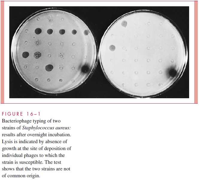
Staphylococcus aureus
Xenotransplantation
Cell culture
Epstein-Barr virus-associated lymphoproliferative diseases
 Fibrinolysis Shutdown Correlation with Thromboembolic Events in Severe COVID-19 Infection
Overall hemostatic potential - Wikipedia
 Prolonged Prothrombotic Effects of Antecedent Hypoglycemia in Individuals With Type 2 Diabetes - White Rose Research Online
Statistical Services Consulting Unit - Research output - University of Hertfordshire (Research Profiles)
 High Altitude Pulmonary Hypertension: Effects of Altitude, Effects of Altitude on Pulmonary Pressures, Adaptation and...
 Abnormal plasma clot structure and stability distinguish bleeding risk in patients with severe factor XI deficiency<...
 Euglobulin Lysis Time | Cleveland Clinic Laboratories
 Thieme E-Journals - Seminars in Thrombosis and Hemostasis / Abstract
 Pesquisa | Portal Regional da BVS
 A Quantitative Investigation of Selected Reactions in the Fibrinolytic Cascade
 Prothrombin time. Medical search
In Silico Analysis of Fibrinolytic Activity of Subtilisin E
Thrombolytic Therapy: Background, Thrombolytic Agents, Thrombolytic Therapy for Acute Myocardial Infarction
 Fibrin Gel Ultrastructure<...
 heparin sodium in 5% dextrose injection 50 units/ml and 100 units/ml Clinical Pharmacology
| Pfizer Medical Information - US
heparin sodium in 5% dextrose injection 50 units/ml and 100 units/ml Clinical Pharmacology
| Pfizer Medical Information - US
heparin sodium injection CARPUJECT () Use in Specific Populations | Pfizer Medical Information - US
 DeCS
DeCS Coagulation Tests in Hepatic Disease in Small Animals - Digestive System - MSD Veterinary Manual
DeCS 2013 - New terms
DeCS 2013 - New terms
DeCS 2013 - New terms
DeCS 2013 - New terms
DeCS 2013 - New terms
DeCS 2013 - New terms
Reticulocytosis | Profiles RNS
 Hypercoagulable Disorders
 Cryoprecipitate transfusion in trauma patients attenuates hyperfibrinolysis and restores normal clot structure and stability:...
 PPT - Methicillin Resistant Staphylococcus Aureus PowerPoint presentation | free to download - id: 3bd84d-ODk5Z
 Thrombin@Fe3O4 nanoparticles for use as a hemostatic agent in internal bleeding | Scientific Reports
Fibrinolysis14
- Fibrinolysis shutdown, as evidenced by elevated d-dimer and complete failure of clot lysis at 30 minutes on thromboelastography predicts thromboembolic events and need for hemodialysis in critically ill patients with COVID-19. (nih.gov)
- This curve represents the balance between fibrin formation induced by thrombin or tissue factor and fibrinolysis induced by t-PA. (wikipedia.org)
- The assay is novel in terms of its combined evaluation of both fibrin generation and fibrinolysis. (wikipedia.org)
- Clot formation and fibrinolysis were measured by turbidity and fibrin network structure by laser scanning confocal microscopy. (tau.ac.il)
- Previous work has shown that thrombin activatable fibrinolysis inhibitor (TAFI) was unable to prolong lysis of purified clots in the presence of Lys-plasminogen (Lys-Pg), indicating a possible mechanism for fibrinolysis to circumvent prolongation mediated by activated TAFI (TAFIa). (queensu.ca)
- This ASA research Project with the UPH university focuses on discovering the fibrinolysis activity and its mechanism of action from Subtilisin, the enzyme that gives natto its unique characteristic in degrading blood clot lysis. (sph.ac.id)
- The diffusional access from outside to proteases involved in fibrinolysis is not yet fully understood For this reason, further knowledge of fibrin network architecture and of the packing arrangement of protofibrils would be desirable. (unicatt.it)
- In normal haemostasis once the body has activated the clotting process, there are regulatory feedback mechanisms , which limit and control the clotting process (natural anticoagulants), prevent the complete blocking of the vessel by excessive clot formation and by removal of the fibrin clot once the injury has healed (fibrinolysis). (labtestsonline.org.uk)
- In addition to conventional laboratory parameters, rotational thromboelastometry (ROTEM) provides evidence for net coagulation capacity and insight into clot formation time, clot firmness and fibrinolysis in the critically ill patients [ 12 ]. (biomedcentral.com)
- prevents reaccumulation of a clot after spontaneous fibrinolysis. (medscape.com)
- Major final results incorporated resolution of plasma fibrin clot components, such as clog permeability (Ks) along with performance regarding fibrinolysis making use of clog lysis period (CLT), as well as thrombin technology (prothrombin fragmented phrases 1 + 2) as well as endogenous thrombin possible (ETP) along with fibrinolysis inhibitor quantities. (hif-signal.com)
- It is poorly understood how changes in the fibrin structure and molecular interactions influence the biochemical regulation and behavior of internal fibrinolysis. (rutgers.edu)
- We used a combination of experiments and mathematical modeling to study fibrin structure and molecular interactions that restrict internal fibrinolysis. (rutgers.edu)
- Analysis of simulations and experiments indicate that fibrinolysis is driven by pore expansion of the fibrin network. (rutgers.edu)
Thrombin12
- The OHP assay measures total fibrin generation in the presence of thrombin or tissue factor and tissue plasminogen activator (t-PA). (wikipedia.org)
- Clotting time of PLASMA mixed with a THROMBIN solution. (lookformedical.com)
- During thrombus formation, circulating prothrombin is activated to the active clotting factor, thrombin, by activated platelets. (medscape.com)
- Fibrinogen is activated to fibrin by the newly activated thrombin. (medscape.com)
- The thrombin-catalyzed polymerization process is usually modelled through the occurrence of a number of distinct steps that lead to the formation of fibrin monomers, which subsequently undergo polymerization to produce oligomers called protofibrils. (unicatt.it)
- Once active thrombosis has developed, larger amounts of heparin can inhibit further coagulation by inactivating thrombin and preventing the conversion of fibrinogen to fibrin. (pfizermedicalinformation.com)
- In 126 normotensive acute PE patients (aged 58±14 years) we assessed 3 genetic polymorphisms FGA rs6050 C___2892877_10, FGB rs1800790 C___7429790_20 and factor XIII rs5958 C___1639938_20 in relation to fibrin clot permeability (K s ), clot lysis time (CLT), and endogenous thrombin potential (ETP). (isth.org)
- Fibrinogen B Arg448Lys and FXIII Val34Leu polymorphisms are associated with formation of more compact fibrin networks in acute PE patients, suggesting that despite heightened thrombin generation and inflammation typical of acute thromboembolism the 2 common generic variants have detectable effect on clot properties, similar to those reported in subjects at risk of venous thrombosis. (isth.org)
- This study geared towards evaluating the changes throughout fibrin clog qualities as well as thrombin technology induced through a pair of various COH practices lengthy together with gonadotropin-releasing hormonal agonist (GnRH-a) as well as GnRH antagonist (GnRH-ant). (hif-signal.com)
- In response to vascular injury, circulating platelets adhere, aggregate, and provide cell surface phospholipids for the assembly of blood clotting enzyme complexes, thrombin activation, and fibrin formation. (medscape.com)
- Thrombin is generated, and fibrin is formed. (medscape.com)
- This increase in the rate of thrombin formation with large doses of rFVIIa permits the formation of fibrin, which is less susceptible to lysis. (medscape.com)
Platelet7
- Fibrin clot properties, platelet reactivity, and inflammatory markers were measured at baseline, end of and after recovery from the initial clamp, day 1, and day 7 using validated assays and electron microscopy. (whiterose.ac.uk)
- RESULTS: Euglycemic hyperinsulinemia reduced platelet reactivity, decreased fibrin clot density, and improved fibrinolytic efficiency in both groups. (whiterose.ac.uk)
- Results: Non-bleeders and bleeders had similarly low FXI levels, normal prothrombin times, normal levels of fibrinogen, factor VIII, von Willebrand factor and factor XIII, and normal platelet number and function. (tau.ac.il)
- The time required for the appearance of FIBRIN strands following the mixing of PLASMA with phospholipid platelet substitute (e.g., crude cephalins, soybean phosphatides). (lookformedical.com)
- During the cascade process, the body sequentially activates coagulation factors , which are proteins that create a net of fibrin threads, which weave them through the platelet plug making a stable firm fibrin blood clot, whilst still allowing blood to flow through the damage vessel. (labtestsonline.org.uk)
- The QStat Cartridge is intended for in vitro diagnostic use by trained professionals at the point-of-care and in clinical laboratories to evaluate the viscoelastic properties of whole blood by means of the following functional parameters: Clot Time (CT), Clot Stiffness (CS), Fibrinogen Contribution to Clot Stiffness (FCS), Platelet Contribution to Clot Stiffness (PCS), and Clot Stability to Lysis (CSL). (hemosonics.com)
- Clotted samples or those containing clots, fibrin strands, or platelet clumps. (crlcorp.com)
Coagulation10
- The process of the interaction of BLOOD COAGULATION FACTORS that results in an insoluble FIBRIN clot. (lookformedical.com)
- Because most veterinarians in private first-opinion practices rely on traditional coagulation assessments (prothrombin time [PT] and activated partial thromboplastin time [aPTT]), and fibrinogen, with occasional measurement of antithrombin (AT), protein C (PC), D-dimers, and fibrin degradation products (FDPs), it is important to acknowledge the complexities that these may not reflect. (msdvetmanual.com)
- Disseminated intravascular coagulation (DIC) is a life-threatening, acute, acquired condition that causes tiny clots throughout the body,often associated with sepsis. (labtestsonline.org.uk)
- Maximum lysis, especially following stimulation of the extrinsic coagulation system, was inversely associated with an enhanced risk of thromboembolic complications. (biomedcentral.com)
- In each patient, intrinsically (contact activation, INTEM) and extrinsically (tissue factor activation, EXTEM) activated test assays were performed to analyze the clot dynamics in both coagulation pathways. (biomedcentral.com)
- The QStat Cartridge is a multi-channel cartridge that provides semi-quantitative indications of the coagulation and clot lysis state of a 3.2% citrated venous whole blood sample using the Quantra ® Hemostasis Analyzer. (hemosonics.com)
- The QStat Cartridge includes tests to assess coagulation via the intrinsic and extrinsic pathways and includes a test with tranexamic acid to evaluate clot lysis characteristics. (hemosonics.com)
- The QStat Cartridge is indicated for the evaluation of blood coagulation and clot lysis in patients age 18 years and older to assess possible hypocoagulable and hypercoagulable conditions in trauma and liver transplantation procedures. (hemosonics.com)
- Urokinase for injection acts directly on the endogenous fibrinolytic system and catalyzes the lysis of plasminogen into fibrinolytic enzyme, which not only degrades fibrinogen clots, but also degrades fibrinogen, coagulation factor Ⅴ and coagulation factor in blood circulation, so as to play a role in thrombolysis. (kangyuan.com.cn)
- The pathophysiology of trauma-induced coagulopathy consists of coagulation activation, hyperfibrino(geno)lysis, and consumption coagulopathy. (biomedcentral.com)
Fibrinolytic activity4
- Increased fibrinolytic activity is suggested by fibrin clot lysis that occurs in less than 3 hours. (clevelandcliniclabs.com)
- In healthy homeostasis, blood clotting events are tracked by fibrinolytic activity, preventing obstructive blood flow. (sph.ac.id)
- defined as the difference between MCF and the lowest clot amplitude after MCF, reflecting fibrinolytic activity (Fig. 1 ). (biomedcentral.com)
- Fibrinolytic activity is defined as the capacity of plasmin, the activated form of plasminogen, to dissolve a fibrin clot into fibrin degradation products (see Figure 3 ). (touchoncology.com)
Failure of clot lysis1
- It has been hypothesized that patients with severe COVID-19 have elevated levels of antifibrinolytic proteins, which create a hypofibrinolytic state, with subsequent failure of clot lysis. (thieme-connect.com)
Degradation6
- 10 ] Laboratory profiles seen in patients with severe COVID-19 (elevated D-dimers and fibrin degradation products [FDPs], and prolonged prothrombin time) are also consistent with a hypercoagulable state. (thieme-connect.com)
- High molecular weight fibrin degradation products (HMW-FDPs), a soluble fibrin surrogate that models Pn modified fibrin, treated with TAFIa decreased the catalytic efficiency (kcat/Km) of 5IAF-Glu-Pg cleavage by 417-fold and of 5IAF-Lys-Pg cleavage by 55-fold. (queensu.ca)
- It is a measure of the conversion of FIBRINOGEN to FIBRIN, which is prolonged by AFIBRINOGENEMIA, abnormal fibrinogen, or the presence of inhibitory substances, e.g., fibrin-fibrinogen degradation products, or HEPARIN. (lookformedical.com)
- However, the decrease in plasma fibrin or fibrinogen levels and the increase in their degradation products can last for 12 to 24 hours. (kangyuan.com.cn)
- A high concentration of hemoglobin, bilirubin, triglycerides and fibrin degradation products might affect PLG measurement. (medscape.com)
- Fibrin-bound tissue plasminogen activator (tPA) converts nearby plasminogen into active plasmin, which is bound to the fibrin network, breaking it down into fibrin degradation products and releasing the entrapped blood cells. (rutgers.edu)
Deep Vein Throm2
- For patient education information, see DVT (Blood Clot in the Leg, Deep Vein Thrombosis) . (medscape.com)
- Innohep (tinzaparin) is a blood thinner ( anticoagulant ) used together with warfarin ( Coumadin ) to treat a type of blood clot called deep vein thrombosis , or DVT. (rxlist.com)
Tissue plasminog2
- Clot formation was triggered by recalcification and addition of tissue factor and phospholipids in the absence or presence of tissue plasminogen activator and/or thrombomodulin. (tau.ac.il)
- Compared with non-bleeders, bleeders exhibited lower fibrin network density and lower clot stability in the presence of tissue plasminogen activator. (tau.ac.il)
Plasmin5
- Fibrin-bound plasminogen will be converted by thrombolytic drugs to plasmin, the rate-limiting step in thrombolysis. (medscape.com)
- Plasmin lyses clots by breaking down the fibrinogen and fibrin contained in a clot. (medscape.com)
- The ability of these substances to catalyze the conversion of plasminogen to plasmin is affected only slightly by the presence or absence of local fibrin clot. (medscape.com)
- Methods Clot lysis, plasmin generation, atomic force microscopy and confocal microscopy were utilised to investigate clot strength and structure in FEISTY patient plasma. (figshare.com)
- Conclusions In summary, our data indicate that cryo transfusion restores key fibrinolytic regulators and limits plasmin generation to form stronger clots in an ex vivo laboratory study. (figshare.com)
Enzyme2
- To identify the interaction of enzyme and its substrate by using in vitro method, excessive time, energy, and money, will be required. (sph.ac.id)
- We show that this effect is strongly influenced by the ratio of fibrin:lytic enzyme when compared to absolute enzyme concentration. (rutgers.edu)
HEPARIN6
- Heparin inhibits reactions that lead to the clotting of blood and the formation of fibrin clots both in vitro and in vivo. (pfizermedicalinformation.com)
- Heparin also prevents the formation of a stable fibrin clot by inhibiting the activation of the fibrin stabilizing factor. (pfizermedicalinformation.com)
- Bleeding time is usually unaffected by heparin. (pfizermedicalinformation.com)
- In a published study conducted in rats and rabbits, pregnant animals received heparin intravenously during organogenesis at a dose of 10,000 units/kg/day, approximately 10 times the maximum human daily dose based on body weight. (pfizermedicalinformation.com)
- The heparin effect was determined by comparing the clotting time of the INTEM with the clotting time of the HEPTEM, where heparinase is added. (biomedcentral.com)
- Heparin is a medication that helps prevent formation of these clots. (petplace.com)
Plasma5
- Conclusions: Plasma clot structure and stability assays distinguished non-bleeders from bleeders. (tau.ac.il)
- Clotting time of PLASMA recalcified in the presence of excess TISSUE THROMBOPLASTIN. (lookformedical.com)
- The structure of fibrin gel depends upon the polymerization conditions of fibrinogen, a glycoprotein present in the plasma of vertebrates. (unicatt.it)
- To our knowledge, no reports assessed the impact of fibrinogen and factor XIII (FXIII) polymorphisms on plasma fibrin clot features in acute PE. (isth.org)
- Older patients who received unpurified plasma‐derived clotting factor concentrates may have signs and symptoms of infectious disease (eg, hepatitis, HIV infection). (medscape.com)
Platelets3
- In the FIBTEM, platelets are inactivated with cytochalasin D to enable isolated evaluation of fibrinogen in clot firmness. (biomedcentral.com)
- Acute clots, which are mostly red cells and platelets in a fibrin mesh, turn into yellow, chronic adherent clots made of collagen, elastin, inflammatory cells, and even occasionally calcifications [7] . (sts.org)
- The QStat Cartridge directly compares changes in clot stiffness in both the presence and absence of tranexamic acid, automatically correcting for clot retraction caused by interaction between platelets/fibrin and the measuring device. (hemosonics.com)
Anticoagulants2
- Genetic risk factors such as factor V Leiden, prothrombin 20210A or deficiencies of the natural anticoagulants antithrombin, protein-S or protein-C are all known to be associated with an increased capacity to form a blood clot. (touchoncology.com)
- Pulmonary embolism treatment is with anticoagulants and, sometimes, clot dissolution with systemic or catheter-directed thrombolysis or by removal of the clot via catheter suction thrombectomy or surgical resection. (msdmanuals.com)
Permeability1
- The chief factor responsible for clot lysis rate is the intrinsic permeability of the fibrin network and of the individual fibers to proteolytic agents. (unicatt.it)
Maximum amplitude1
- viscoelastic measurements showed an elevated maximum amplitude and low lysis of clot at 30 minutes. (nih.gov)
Thrombolytic5
- hence, the importance of time for thrombolytic therapy. (medscape.com)
- The history of thrombolytic therapy began in 1933, when it was discovered that filtrates of broth cultures of certain streptococcal strains (beta-hemolytic streptococci) could dissolve a fibrin clot. (medscape.com)
- With the advent of thrombolytic agents that favor clot lysis, treatment of patients suffering from thromboembolic diseases is greatly improved. (unicatt.it)
- For the first time characteristic fibrils distance are related to the water trapped among fibrils and thus to space available to thrombolytic agents diffusion. (unicatt.it)
- 3. Urokinase for injection showed obvious correlation between thrombolytic effect and drug dose and time window of administration. (kangyuan.com.cn)
Thromboembolic3
- A complete lack of lysis of clot at 30 minutes was seen in 57% of patients and predicted venous thromboembolic events with an area under the receiver operating characteristic curve of 0.742 (p = 0.021). (nih.gov)
- Combining values for maximum lysis with D-dimer concentrations revealed high sensitivity and specificity of thromboembolic risk prediction. (biomedcentral.com)
- At the same time, proteolytic enzymes stimulate the healing and repair processes, reduce the risk of thromboembolic complications during prolonged immobilization, and prevent the development of trophic disorders and purulent complications. (pillbuys.com)
Pulmonary5
- However, a DVT can become life-threatening if the clot breaks free and travels to other parts of the body through the bloodstream, particularly if it becomes lodged in the arteries of lung, called a pulmonary embolism PE or to the brain causing strokes. (labtestsonline.org.uk)
- As time progresses, PA pressure rises and flow decreases as a result of macrovascular obstruction, small vessel arteriopathy, and vasoconstriction of the pulmonary arteries. (sts.org)
- The inability to lyse these pulmonary emboli suggests that perhaps a hypercoagulable disorder puts these patients at risk. (sts.org)
- Klajmon A, Chmiel J, Ząbczyk M, Natorska J, Undas A. Fibrinogen B Arg448Lys and Factor XIII Val34Leu Polymorphisms Are Associated with Prothrombotic Fibrin Clot Properties in Patients with Acute Pulmonary Embolism [abstract]. (isth.org)
- This condition sometimes occurs with a blood clot in lungs ( pulmonary embolism , or PE). (rxlist.com)
Stability2
- This study will determine differences in clot strength and fibrinolytic stability within individuals and between treatment arms. (figshare.com)
- This is the first study to investigate differences in clot stability and structure between cryo and Fg-C and demonstrates that the additional factors in cryo allow formation of a stronger and more stable clot. (figshare.com)
Confocal microscopy1
- Plasminogen activator inhibitor 1 activity and antigen levels and Factor XIII antigen were increased post-treatment with cryo, but not Fg-C. Confocal microscopy analysis of fibrin clots revealed that cryo transfusion restored fibrin structure similar to those observed in control clots. (figshare.com)
Acute2
- Prothrombotic fibrin clot properties in acute PE are associated with eightfold increased risk of death. (isth.org)
- We investigated whether a fibrinogen alpha chain ( FGA ) Thr312Ala, fibrinogen beta chain ( FGB ) Arg448Lys and FXIII Val34Leu polymorphisms are associated with fibrin clot properties assessed on admission in acute PE patients. (isth.org)
Thrombus1
- A hypercoagulable disorder, also known as thrombophilia, is an inherited or acquired condition that increases the risk of developing inappropriate or excessive thrombus (blood clot) formation. (labtestsonline.org.uk)
Degrades1
- Though information about subtilisin hydrolyzing raw materials has been reported, the mechanism of how it degrades fibrin is still limited. (sph.ac.id)
Formation4
- These mediators promote clot formation. (medscape.com)
- If the clotting process activates inappropriately, or feedback mechanisms fail to work effectively to limit formation or removal of fibrin clot, then there can be inappropriate and/or excessive blood clot formation. (labtestsonline.org.uk)
- Thrombosis can occur within veins or arteries, however the mechanism of clot formation is different, with venous thrombosis associated with sluggish movement of blood (stasis) or imbalance of the clotting progress and feedback mechanism, whereas arterial thrombosis more commonly results from the rupture of an atherosclerotic plaque due to build-up of cholesterol in the arterial wall. (labtestsonline.org.uk)
- Complications of IMHA include the formation of blood clots. (petplace.com)
Mechanism4
- In conclusion, an additional mechanism was identified whereby TAFIa can prolong clot lysis by increasing the rate of tPA inhibition by PAI-1 by eliminating the protective effects of Pn-modified fibrin and Pg. (queensu.ca)
- Laboratory tests for evaluating the individual's clotting mechanism. (lookformedical.com)
- The activation of the clotting mechanism may arise from any of a number of disorders. (lookformedical.com)
- This innovative research discovered the fibrinolytic characteristic, the mechanism of Subtilisin that was extracted from Bacillus Subtilis G8, and it has also displayed a strong interaction between Subtilisin and Fibrin. (sph.ac.id)
Bleeding4
- In this paper, magnetic hemostatic nanoparticles are shown for the first time to assist in minimally invasive treatment of internal bleeding, implying the introduction directly into the circulatory system followed by localization in the bleeding zone due to the application of an external magnetic field. (nature.com)
- However, since this product increases plasminogen activity and reduces unbound plasminogen and fibrin-bound plasminogen in circulation, there may be a serious risk of bleeding. (kangyuan.com.cn)
- Blood clots are critical in cessation of bleeding following injury. (rutgers.edu)
- An improved understanding of effective internal lysis can aid in development of better monitoring techniques to avoid thrombotic or bleeding risk, as well as in the design of novel enzymatic treatments to overcome the innate challenges with internal lysis. (rutgers.edu)
Thrombolysis1
- Thrombolysis, also called fibrinolytic therapy , is the breakdown ( lysis ) of blood clots formed in blood vessels , using medication. (wikimili.com)
Polymerization1
- The most important test used to distinguish S. aureus from other staphylococci is the production of coagulase, which nonenzymatically binds to prothrombin, forming a com-plex that initiates the polymerization of fibrin. (brainkart.com)
Vessel5
- Blood clotting is normal response to blood vessel or tissue injury. (labtestsonline.org.uk)
- For instance, a model vessel system with circulating blood at the puncture of the vessel wall and the application of a permanent magnetic field yielded a hemostasis time by a factor of 6.5 shorter than that observed for the control sample. (nature.com)
- For this small cohort of patients, the clot becomes adherent to the vessel wall. (sts.org)
- When adjusted to time to therapy and vessel occluded, these results lessened but remained significant. (ajnr.org)
- Time to achieving vessel patency has been shown to be crucial in achieving a better clinical outcome ( 2 - 4 ). (ajnr.org)
Anticoagulant1
- The sample should be mixed immediately by gentle inversion at least six times to ensure adequate mixing of the anticoagulant with the blood. (clevelandcliniclabs.com)
Protein1
- The RCSB database [6] in crystallographic form was used to identify the residues for the 3D Structure of the Fibrin protein. (sph.ac.id)
Specimens2
- Specimens that are hemolyzed, clotted, diluted with IV fluids or have thawed before arriving in the performing laboratory will be rejected. (clevelandcliniclabs.com)
- All specimens are checked visually for obvious clots prior to sampling on the analyzer. (crlcorp.com)
Anticoagulation1
- With anticoagulation, the vast majority of these patients will lyse the clot over the following few weeks [4] [5] . (sts.org)
0.0011
- In type 2 diabetes, clot lysis times and clot maximum absorbance increased up to day 7 (P = 0.002 and 0.001 vs. euglycemia, respectively), but clots from control subjects without diabetes showed limited changes. (whiterose.ac.uk)
Medication1
- Innohep may interact with dextran, aspirin and other salicylates, and other medication used to prevent blood clots . (rxlist.com)
Activity1
- It is one of the important enzymes for natto's blood clot lysis activity. (sph.ac.id)
Activation2
- Because TAFIa can suppress Lys-Pg activation but cannot attenuate Lys-Pn inhibition by AP, the Glu- to Lys-Pg/Pn conversion is able to act as a fibrinolytic switch to ultimately lyse the clot. (queensu.ca)
- OBJECTIVE: To determine the incidence and magnitude of fibrinolytic activation in trauma patients and its relationship to clot lysis as measured by thromboelastometry. (ox.ac.uk)
Enzymes1
- The complex retains and slows down the elimination of proteolytic enzymes of the drug from the body, increases the time of their circulation in the vascular bed and, accordingly, the therapeutic effect. (pillbuys.com)
Hypercoagulable state1
- Several genetic and acquired risk factors for venous thrombosis are associated with a hypercoagulable state (i.e. an increased capacity to form a blood clot). (touchoncology.com)