Femur Head
Femur Head Necrosis
Head
Head and Neck Neoplasms
Head Movements
Femoral Neoplasms
Femoral Neck Fractures
Sperm Head
Bone Density
Tibia
Fracture Fixation, Intramedullary
Head Injuries, Closed
Craniocerebral Trauma
Bone and Bones
Bone Nails
Biomechanical Phenomena
Fracture Healing
Fracture Fixation, Internal
Hip Joint
Hip Fractures
Weight-Bearing
Periprosthetic Fractures
Epiphyses
Fractures, Spontaneous
Bone Plates
Osteoporosis
Bone Remodeling
Fracture Fixation
Leg Length Inequality
Absorptiometry, Photon
Carcinoma, Squamous Cell
Bone Development
Head Protective Devices
Bone Lengthening
Tomography, X-Ray Computed
Rotation
Bone Diseases, Metabolic
Traction
Bony Callus
Prosthesis Failure
Bone Cements
Fractures, Ununited
Range of Motion, Articular
Stress, Mechanical
Humeral Head
External Fixators
Compressive Strength
Finite Element Analysis
Retrospective Studies
Treatment Outcome
Hip
Pediculus
Calcification, Physiologic
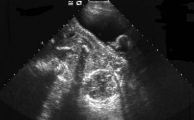
Ultrasonography, Prenatal
Reoperation
Osseointegration
Parietal Bone
Bone Density Conservation Agents
Torsion, Mechanical
Gestational Age
Cementation
Lice Infestations
Hip Dislocation, Congenital
Follow-Up Studies
Bone Diseases, Developmental
Acetabulum
Models, Anatomic
Alendronate
Biometry
Prostheses and Implants
Magnetic Resonance Imaging
Orthopedic Procedures
Fibula
Bone Malalignment
Radius
Osteoarthritis, Hip
Tensile Strength
Pregnancy Trimester, Second
Leg Bones
Ilizarov Technique
Fractures, Stress
Postoperative Complications
Cartilage, Articular
Legg-Calve-Perthes Disease
Reconstructive Surgical Procedures
Anthropometry
Body Weight
Reflex, Vestibulo-Ocular
Periosteum
Fetal Weight
Pregnancy
Bone Regeneration
Prospective Studies
Imaging, Three-Dimensional
Chondrosarcoma
Skull Fractures
Mechanical considerations in impaction bone grafting. (1/715)
In impaction grafting of contained bone defects after revision joint arthroplasty the graft behaves as a friable aggregate and its resistance to complex forces depends on grading, normal load and compaction. Bone mills in current use produce a distribution of particle sizes more uniform than is desirable for maximising resistance to shear stresses. We have performed experiments in vitro using morsellised allograft bone from the femoral head which have shown that its mechanical properties improve with increasing normal load and with increasing shear strains (strain hardening). The mechanical strength also increases with increasing compaction energy, and with the addition of bioglass particles to make good the deficiency in small and very small fragments. Donor femoral heads may be milled while frozen without affecting the profile of the particle size. Osteoporotic femoral heads provide a similar grading of sizes, although fewer particles are obtained from each specimen. Our findings have implications for current practice and for the future development of materials and techniques. (+info)The pathogenesis of Perthes' disease. (2/715)
It has been shown that in the puppy, two infarcts separated by an interval of four weeks produce a disorder of long duration which results in flattening and broadening of the femoral head and which reproduces the radiological changes seen in Perthes' disease in man. The histological appearances produced by two infarcts are characteristic. In this study the histological appearance of fifty-seven femoral head biopsy specimens in Perthes' disease in man have been studied. In 51 per cent of hips histopathological changes characteristic of double infarction were present, and there were grounds for postulating that double infarction might eventually occur in all cases. The findings support the concept that the deformation of the femoral head and the chronicity of Perthes' disease in man may be due at least as much or even more to repeated episodes of infarction and the ensuing abnormalities of growth as to mechanical factors related to weight-bearing. (+info)The influence of weight-bearing on the measurement of polyethylene wear in THA. (3/715)
We have studied the influence of weight-bearing on the measurement of wear of the polyethylene acetabular component in total hip arthroplasty using two techniques. The measured vertical wear was significantly greater when radiographs were taken weight-bearing rather than with the patient supine (p = 0.001, method 1; p = 0.007, method 2). Calculations of rates of linear wear of the acetabular component were significantly underestimated (p < 0.05) when radiographs were taken supine. There are two reasons for this. First, a change in pelvic orientation when bearing weight ensures that the thinnest polyethylene is brought into relief, and secondly, the head of the femoral component assumes the position of maximal displacement along its wear path. Interpretation of previous studies on both linear and volumetric polyethylene wear in total hip arthroplasty should be reassessed in the light of these findings. (+info)Accuracy of EBRA-FCA in the measurement of migration of femoral components of total hip replacement. Einzel-Bild-Rontgen-Analyse-femoral component analysis. (4/715)
Several methods of measuring the migration of the femoral component after total hip replacement have been described, but they use different reference lines, and have differing accuracies, some unproven. Statistical comparison of different studies is rarely possible. We report a study of the EBRA-FCA method (femoral component analysis using Einzel-Bild-Rontgen-Analyse) to determine its accuracy using three independent assessments, including a direct comparison with the results of roentgen stereophotogrammetric analysis (RSA). The accuracy of EBRA-FCA was better than +/- 1.5 mm (95% percentile) with a Cronbach's coefficient alpha for interobserver reliability of 0.84; a very good result. The method had a specificity of 100% and a sensitivity of 78% compared with RSA for the detection of migration of over 1 mm. This is accurate enough to assess the stability of a prosthesis within a relatively limited period. The best reference line for downward migration is between the greater trochanter and the shoulder of the stem, as confirmed by two experimental analyses and a computer-assisted design. (+info)The prediction of failure of the stem in THR by measurement of early migration using EBRA-FCA. Einzel-Bild-Roentgen-Analyse-femoral component analysis. (5/715)
We report the ten-year results for three designs of stem in 240 total hip replacements, for which subsidence had been measured on plain radiographs at regular intervals. Accurate migration patterns could be determined by the method of Einzel-Bild-Roentgen-Analyse-femoral component analysis (EBRA-FCA) for 158 hips (66%). Of these, 108 stems (68%) remained stable throughout, and five (3%) started to migrate after a median of 54 months. Initial migration of at least 1 mm was seen in 45 stems (29%) during the first two years, but these then became stable. We revised 17 stems for aseptic loosening, and 12 for other reasons. Revision for aseptic loosening could be predicted by EBRA-FCA with a sensitivity of 69%, a specificity of 80%, and an accuracy of 79% by the use of a threshold of subsidence of 1.5 mm during the first two years. Similar observations over a five-year period allowed the long-term outcome to be predicted with an accuracy of 91%. We discuss the importance of four different patterns of subsidence and confirm that the early measurement of migration by a reasonably accurate method can help to predict long-term outcome. Such methods should be used to evaluate new and modified designs of prosthesis. (+info)The pathology of bone allograft. (6/715)
We analysed the histological findings in 1146 osteoarthritic femoral heads which would have been considered suitable for bone-bank donation to determine whether pathological lesions, other than osteoarthritis, were present. We found that 91 femoral heads (8%) showed evidence of disease. The most common conditions noted were chondrocalcinosis (63 cases), avascular necrosis (13), osteomas (6) and malignant tumours (one case of low-grade chondrosarcoma and two of well-differentiated lymphocytic lymphoma). There were two with metabolic bone disease (Paget's disease and hyperparathyroid bone disease) and four with inflammatory (rheumatoid-like) arthritis. Our findings indicate that occult pathological conditions are common and it is recommended that histological examination of this regularly used source of bone allograft should be included as part of the screening protocol for bone-bank collection. (+info)Histopathology of retrieved allografts of the femoral head. (7/715)
From November 1994 to March 1997, we harvested 137 grafts of the femoral head from 125 patients for donation during total hip arthroplasty according to the guidelines of the American Associations of Tissue Banks (AATB) and the European Association of Musculo-Skeletal transplantation (EAMST). In addition to the standards recommended by these authorities, we performed histopathological examination of a core biopsy of the retrieved bone allograft and of the synovium. Of the 137 allografts, 48 (35.0%) fulfilled all criteria and were free for donation; 31 (22.6%) were not regarded as suitable for transplantation because the serological retests at six months were not yet complete and 58 (42.3%) were discarded because of incomplete data. Of those discarded, five showed abnormal histopathological findings; three were highly suspicious of low-grade B-cell lymphoma, one of monoclonal plasmacytosis and the other of non-specific inflammation of bone marrow. However, according to the standards of the AATB or EAMST they all met the criteria and were eligible for transplantation. Our findings indicate that the incidence of abnormal histopathology in these retrieved allografts was 3.6%. Since it is essential to confirm the quality of donor bones in bone banking, we advise that histopathological screening of donor bone should be performed to exclude abnormal allografts. (+info)Decrease in the mesenchymal stem-cell pool in the proximal femur in corticosteroid-induced osteonecrosis. (8/715)
We have evaluated bone-marrow activity in the proximal femur of patients with corticosteroid-induced osteonecrosis and compared it with that of patients with osteonecrosis related to sickle-cell disease and with a control group without osteonecrosis. Bone marrow was obtained by puncture of the femoral head outside the area of necrosis and in the intertrochanteric region. The activity of stromal cells was assessed by culturing fibroblast colony-forming units (FCFUs). We found a decrease in the number of FCFUs outside the area of osteonecrosis in the upper end of the femur of patients with corticosteroid-induced osteonecrosis compared with the other groups. We suggest that glucocorticosteroids may also have an adverse effect on bone by decreasing the number of progenitors. The possible relevance of this finding to osteonecrosis is discussed. (+info)The femur is the medical term for the thigh bone, which is the longest and strongest bone in the human body. It connects the hip bone to the knee joint and plays a crucial role in supporting the weight of the body and allowing movement during activities such as walking, running, and jumping. The femur is composed of a rounded head, a long shaft, and two condyles at the lower end that articulate with the tibia and patella to form the knee joint.
The femoral head is the rounded, ball-like top portion of the femur (thigh bone) that fits into the hip socket (acetabulum) to form the hip joint. It has a smooth, articular cartilage surface that allows for smooth and stable articulation with the pelvis. The femoral head is connected to the femoral neck, which is a narrower section of bone that angles downward and leads into the shaft of the femur. Together, the femoral head and neck provide stability and range of motion to the hip joint.
Femoral head necrosis, also known as avascular necrosis of the femoral head, is a medical condition that results from the interruption of blood flow to the femoral head, which is the rounded end of the thigh bone that fits into the hip joint. This lack of blood supply can cause the bone tissue to die, leading to the collapse of the femoral head and eventually resulting in hip joint damage or arthritis.
The condition can be caused by a variety of factors, including trauma, alcohol abuse, corticosteroid use, radiation therapy, and certain medical conditions such as sickle cell disease and lupus. Symptoms may include pain in the hip or groin, limited range of motion, and difficulty walking. Treatment options depend on the severity and progression of the necrosis and may include medication, physical therapy, or surgical intervention.
In medical terms, the "head" is the uppermost part of the human body that contains the brain, skull, face, eyes, nose, mouth, and ears. It is connected to the rest of the body by the neck and is responsible for many vital functions such as sight, hearing, smell, taste, touch, and thought processing. The head also plays a crucial role in maintaining balance, speech, and eating.
Head and neck neoplasms refer to abnormal growths or tumors in the head and neck region, which can be benign (non-cancerous) or malignant (cancerous). These tumors can develop in various sites, including the oral cavity, nasopharynx, oropharynx, larynx, hypopharynx, paranasal sinuses, salivary glands, and thyroid gland.
Benign neoplasms are slow-growing and generally do not spread to other parts of the body. However, they can still cause problems if they grow large enough to press on surrounding tissues or structures. Malignant neoplasms, on the other hand, can invade nearby tissues and organs and may also metastasize (spread) to other parts of the body.
Head and neck neoplasms can have various symptoms depending on their location and size. Common symptoms include difficulty swallowing, speaking, or breathing; pain in the mouth, throat, or ears; persistent coughing or hoarseness; and swelling or lumps in the neck or face. Early detection and treatment of head and neck neoplasms are crucial for improving outcomes and reducing the risk of complications.
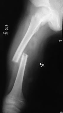
A femoral fracture is a medical term that refers to a break in the thigh bone, which is the longest and strongest bone in the human body. The femur extends from the hip joint to the knee joint and is responsible for supporting the weight of the upper body and allowing movement of the lower extremity. Femoral fractures can occur due to various reasons such as high-energy trauma, low-energy trauma in individuals with weak bones (osteoporosis), or as a result of a direct blow to the thigh.
Femoral fractures can be classified into different types based on their location, pattern, and severity. Some common types of femoral fractures include:
1. Transverse fracture: A break that occurs straight across the bone.
2. Oblique fracture: A break that occurs at an angle across the bone.
3. Spiral fracture: A break that occurs in a helical pattern around the bone.
4. Comminuted fracture: A break that results in multiple fragments of the bone.
5. Open or compound fracture: A break in which the bone pierces through the skin.
6. Closed or simple fracture: A break in which the bone does not pierce through the skin.
Femoral fractures can cause severe pain, swelling, bruising, and difficulty walking or bearing weight on the affected leg. Diagnosis typically involves a physical examination, medical history, and imaging tests such as X-rays or CT scans. Treatment may involve surgical intervention, including the use of metal rods, plates, or screws to stabilize the bone, followed by rehabilitation and physical therapy to restore mobility and strength.
The "femur neck" is the narrow, upper part of the femur (thigh bone) where it connects to the pelvis. It is the region through which the femoral head articulates with the acetabulum to form the hip joint. The femur neck is a common site for fractures, especially in older adults with osteoporosis.
Head movements refer to the voluntary or involuntary motion of the head in various directions. These movements can occur in different planes, including flexion (moving the head forward), extension (moving the head backward), rotation (turning the head to the side), and lateral bending (leaning the head to one side).
Head movements can be a result of normal physiological processes, such as when nodding in agreement or shaking the head to indicate disagreement. They can also be caused by neurological conditions, such as abnormal head movements in patients with Parkinson's disease or cerebellar disorders. Additionally, head movements may occur in response to sensory stimuli, such as turning the head toward a sound.
In a medical context, an examination of head movements can provide important clues about a person's neurological function and help diagnose various conditions affecting the brain and nervous system.
Femoral neoplasms refer to abnormal growths or tumors that develop in the femur, which is the long thigh bone in the human body. These neoplasms can be benign (non-cancerous) or malignant (cancerous). Benign femoral neoplasms are slow-growing and rarely spread to other parts of the body, while malignant neoplasms are aggressive and can invade nearby tissues and organs, as well as metastasize (spread) to distant sites.
There are various types of femoral neoplasms, including osteochondromas, enchondromas, chondrosarcomas, osteosarcomas, and Ewing sarcomas, among others. The specific type of neoplasm is determined by the cell type from which it arises and its behavior.
Symptoms of femoral neoplasms may include pain, swelling, stiffness, or weakness in the thigh, as well as a palpable mass or limited mobility. Diagnosis typically involves imaging studies such as X-rays, CT scans, or MRI, as well as biopsy to determine the type and grade of the tumor. Treatment options may include surgery, radiation therapy, chemotherapy, or a combination of these approaches, depending on the type, size, location, and stage of the neoplasm.
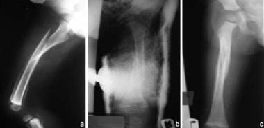
A femoral neck fracture is a type of hip fracture that occurs in the narrow, vertical section of bone just below the ball of the femur (thigh bone) that connects to the hip socket. This area is called the femoral neck. Femoral neck fractures can be categorized into different types based on their location and the direction of the fractured bone.
These fractures are typically caused by high-energy trauma, such as car accidents or falls from significant heights, in younger individuals. However, in older adults, particularly those with osteoporosis, femoral neck fractures can also result from low-energy trauma, like a simple fall from standing height.
Femoral neck fractures are often serious and require prompt medical attention. Treatment usually involves surgery to realign and stabilize the broken bone fragments, followed by rehabilitation to help regain mobility and strength. Potential complications of femoral neck fractures include avascular necrosis (loss of blood flow to the femoral head), nonunion or malunion (improper healing), and osteoarthritis in the hip joint.
A sperm head is the anterior (front) part of a spermatozoon, which contains the genetic material (DNA). It is covered by a protein layer called the acrosome, which plays a crucial role in fertilization. The sperm head is followed by the midpiece and the tail, which provide mobility to the sperm for its journey towards the egg.
The diaphysis refers to the shaft or middle portion of a long bone in the body. It is the part that is typically cylindrical in shape and contains the medullary cavity, which is filled with yellow marrow. The diaphysis is primarily composed of compact bone tissue, which provides strength and support for weight-bearing and movement.
In contrast to the diaphysis, the ends of long bones are called epiphyses, and they are covered with articular cartilage and contain spongy bone tissue filled with red marrow, which is responsible for producing blood cells. The area where the diaphysis meets the epiphysis is known as the metaphysis, and it contains growth plates that are responsible for the longitudinal growth of bones during development.
Bone density refers to the amount of bone mineral content (usually measured in grams) in a given volume of bone (usually measured in cubic centimeters). It is often used as an indicator of bone strength and fracture risk. Bone density is typically measured using dual-energy X-ray absorptiometry (DXA) scans, which provide a T-score that compares the patient's bone density to that of a young adult reference population. A T-score of -1 or above is considered normal, while a T-score between -1 and -2.5 indicates osteopenia (low bone mass), and a T-score below -2.5 indicates osteoporosis (porous bones). Regular exercise, adequate calcium and vitamin D intake, and medication (if necessary) can help maintain or improve bone density and prevent fractures.
The tibia, also known as the shin bone, is the larger of the two bones in the lower leg and part of the knee joint. It supports most of the body's weight and is a major insertion point for muscles that flex the foot and bend the leg. The tibia articulates with the femur at the knee joint and with the fibula and talus bone at the ankle joint. Injuries to the tibia, such as fractures, are common in sports and other activities that put stress on the lower leg.
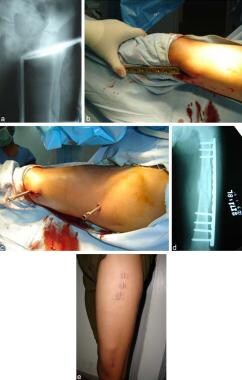
Intramedullary fracture fixation is a surgical technique used to stabilize and align bone fractures. In this procedure, a metal rod or nail is inserted into the marrow cavity (intramedullary canal) of the affected bone, spanning the length of the fracture. The rod is then secured to the bone using screws or other fixation devices on either side of the fracture. This provides stability and helps maintain proper alignment during the healing process.
The benefits of intramedullary fixation include:
1. Load sharing: The intramedullary rod shares some of the load bearing capacity with the bone, which can help reduce stress on the healing bone.
2. Minimal soft tissue dissection: Since the implant is inserted through the medullary canal, there is less disruption to the surrounding muscles, tendons, and ligaments compared to other fixation methods.
3. Biomechanical stability: Intramedullary fixation provides rotational and bending stiffness, which helps maintain proper alignment of the fracture fragments during healing.
4. Early mobilization: Patients with intramedullary fixation can often begin weight bearing and rehabilitation exercises earlier than those with other types of fixation, leading to faster recovery times.
Common indications for intramedullary fracture fixation include long bone fractures in the femur, tibia, humerus, and fibula, as well as certain pelvic and spinal fractures. However, the choice of fixation method depends on various factors such as patient age, fracture pattern, location, and associated injuries.
A closed head injury is a type of traumatic brain injury (TBI) that occurs when there is no penetration or breakage of the skull. The brain is encased in the skull and protected by cerebrospinal fluid, but when the head experiences a sudden impact or jolt, the brain can move back and forth within the skull, causing it to bruise, tear blood vessels, or even cause nerve damage. This type of injury can result from various incidents such as car accidents, sports injuries, falls, or any other event that causes the head to suddenly stop or change direction quickly.
Closed head injuries can range from mild (concussion) to severe (diffuse axonal injury, epidural hematoma, subdural hematoma), and symptoms may not always be immediately apparent. They can include headache, dizziness, nausea, vomiting, confusion, memory loss, difficulty concentrating, mood changes, sleep disturbances, and in severe cases, loss of consciousness, seizures, or even coma. It is essential to seek medical attention immediately if you suspect a closed head injury, as prompt diagnosis and treatment can significantly improve the outcome.
Craniocerebral trauma, also known as traumatic brain injury (TBI), is a type of injury that occurs to the head and brain. It can result from a variety of causes, including motor vehicle accidents, falls, sports injuries, violence, or other types of trauma. Craniocerebral trauma can range in severity from mild concussions to severe injuries that cause permanent disability or death.
The injury typically occurs when there is a sudden impact to the head, causing the brain to move within the skull and collide with the inside of the skull. This can result in bruising, bleeding, swelling, or tearing of brain tissue, as well as damage to blood vessels and nerves. In severe cases, the skull may be fractured or penetrated, leading to direct injury to the brain.
Symptoms of craniocerebral trauma can vary widely depending on the severity and location of the injury. They may include headache, dizziness, confusion, memory loss, difficulty speaking or understanding speech, changes in vision or hearing, weakness or numbness in the limbs, balance problems, and behavioral or emotional changes. In severe cases, the person may lose consciousness or fall into a coma.
Treatment for craniocerebral trauma depends on the severity of the injury. Mild injuries may be treated with rest, pain medication, and close monitoring, while more severe injuries may require surgery, intensive care, and rehabilitation. Prevention is key to reducing the incidence of craniocerebral trauma, including measures such as wearing seat belts and helmets, preventing falls, and avoiding violent situations.
"Bone" is the hard, dense connective tissue that makes up the skeleton of vertebrate animals. It provides support and protection for the body's internal organs, and serves as a attachment site for muscles, tendons, and ligaments. Bone is composed of cells called osteoblasts and osteoclasts, which are responsible for bone formation and resorption, respectively, and an extracellular matrix made up of collagen fibers and mineral crystals.
Bones can be classified into two main types: compact bone and spongy bone. Compact bone is dense and hard, and makes up the outer layer of all bones and the shafts of long bones. Spongy bone is less dense and contains large spaces, and makes up the ends of long bones and the interior of flat and irregular bones.
The human body has 206 bones in total. They can be further classified into five categories based on their shape: long bones, short bones, flat bones, irregular bones, and sesamoid bones.
I believe you are referring to "bone pins" or "bone nails" rather than "bone nails." These terms are used in the medical field to describe surgical implants made of metal or biocompatible materials that are used to stabilize and hold together fractured bones during the healing process. They can also be used in spinal fusion surgery to provide stability and promote bone growth between vertebrae.
Bone pins or nails typically have a threaded or smooth shaft, with a small diameter that allows them to be inserted into the medullary canal of long bones such as the femur or tibia. They may also have a head or eyelet on one end that allows for attachment to external fixation devices or other surgical instruments.
The use of bone pins and nails has revolutionized orthopedic surgery, allowing for faster healing times, improved stability, and better functional outcomes for patients with fractures or spinal deformities.
Biomechanics is the application of mechanical laws to living structures and systems, particularly in the field of medicine and healthcare. A biomechanical phenomenon refers to a observable event or occurrence that involves the interaction of biological tissues or systems with mechanical forces. These phenomena can be studied at various levels, from the molecular and cellular level to the tissue, organ, and whole-body level.
Examples of biomechanical phenomena include:
1. The way that bones and muscles work together to produce movement (known as joint kinematics).
2. The mechanical behavior of biological tissues such as bone, cartilage, tendons, and ligaments under various loads and stresses.
3. The response of cells and tissues to mechanical stimuli, such as the way that bone tissue adapts to changes in loading conditions (known as Wolff's law).
4. The biomechanics of injury and disease processes, such as the mechanisms of joint injury or the development of osteoarthritis.
5. The use of mechanical devices and interventions to treat medical conditions, such as orthopedic implants or assistive devices for mobility impairments.
Understanding biomechanical phenomena is essential for developing effective treatments and prevention strategies for a wide range of medical conditions, from musculoskeletal injuries to neurological disorders.
The humerus is the long bone in the upper arm that extends from the shoulder joint (glenohumeral joint) to the elbow joint. It articulates with the glenoid cavity of the scapula to form the shoulder joint and with the radius and ulna bones at the elbow joint. The proximal end of the humerus has a rounded head that provides for movement in multiple planes, making it one of the most mobile joints in the body. The greater and lesser tubercles are bony prominences on the humeral head that serve as attachment sites for muscles that move the shoulder and arm. The narrow shaft of the humerus provides stability and strength for weight-bearing activities, while the distal end forms two articulations: one with the ulna (trochlea) and one with the radius (capitulum). Together, these structures allow for a wide range of motion in the shoulder and elbow joints.
Fracture healing is the natural process by which a broken bone repairs itself. When a fracture occurs, the body responds by initiating a series of biological and cellular events aimed at restoring the structural integrity of the bone. This process involves the formation of a hematoma (a collection of blood) around the fracture site, followed by the activation of inflammatory cells that help to clean up debris and prepare the area for repair.
Over time, specialized cells called osteoblasts begin to lay down new bone matrix, or osteoid, along the edges of the broken bone ends. This osteoid eventually hardens into new bone tissue, forming a bridge between the fracture fragments. As this process continues, the callus (a mass of newly formed bone and connective tissue) gradually becomes stronger and more compact, eventually remodeling itself into a solid, unbroken bone.
The entire process of fracture healing can take several weeks to several months, depending on factors such as the severity of the injury, the patient's age and overall health, and the location of the fracture. In some cases, medical intervention may be necessary to help promote healing or ensure proper alignment of the bone fragments. This may include the use of casts, braces, or surgical implants such as plates, screws, or rods.
A hip prosthesis, also known as a total hip replacement, is a surgical implant designed to replace the damaged or diseased components of the human hip joint. The procedure involves replacing the femoral head (the ball at the top of the thigh bone) and the acetabulum (the socket in the pelvis) with artificial parts, typically made from materials such as metal, ceramic, or plastic.
The goal of a hip prosthesis is to relieve pain, improve joint mobility, and restore function, allowing patients to return to their normal activities and enjoy an improved quality of life. The procedure is most commonly performed in individuals with advanced osteoarthritis, rheumatoid arthritis, or other degenerative conditions that have caused significant damage to the hip joint.
There are several different types of hip prostheses available, each with its own unique design and set of benefits and risks. The choice of prosthesis will depend on a variety of factors, including the patient's age, activity level, overall health, and specific medical needs. In general, however, all hip prostheses are designed to provide a durable, long-lasting solution for patients suffering from debilitating joint pain and stiffness.
Fracture fixation, internal, is a surgical procedure where a fractured bone is fixed using metal devices such as plates, screws, or rods that are implanted inside the body. This technique helps to maintain the alignment and stability of the broken bone while it heals. The implants may be temporarily or permanently left inside the body, depending on the nature and severity of the fracture. Internal fixation allows for early mobilization and rehabilitation, which can result in a faster recovery and improved functional outcome.
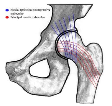
The hip joint, also known as the coxal joint, is a ball-and-socket type synovial joint that connects the femur (thigh bone) to the pelvis. The "ball" is the head of the femur, while the "socket" is the acetabulum, a concave surface on the pelvic bone.
The hip joint is surrounded by a strong fibrous capsule and is reinforced by several ligaments, including the iliofemoral, ischiofemoral, and pubofemoral ligaments. The joint allows for flexion, extension, abduction, adduction, medial and lateral rotation, and circumduction movements, making it one of the most mobile joints in the body.
The hip joint is also supported by various muscles, including the gluteus maximus, gluteus medius, gluteus minimus, iliopsoas, and other hip flexors and extensors. These muscles provide stability and strength to the joint, allowing for weight-bearing activities such as walking, running, and jumping.
A hip fracture is a medical condition referring to a break in the upper part of the femur (thigh) bone, which forms the hip joint. The majority of hip fractures occur due to falls or direct trauma to the area. They are more common in older adults, particularly those with osteoporosis, a condition that weakens bones and makes them more prone to breaking. Hip fractures can significantly impact mobility and quality of life, often requiring surgical intervention and rehabilitation.
"Weight-bearing" is a term used in the medical field to describe the ability of a body part or limb to support the weight or pressure exerted upon it, typically while standing, walking, or performing other physical activities. In a clinical setting, healthcare professionals often use the term "weight-bearing exercise" to refer to physical activities that involve supporting one's own body weight, such as walking, jogging, or climbing stairs. These exercises can help improve bone density, muscle strength, and overall physical function, particularly in individuals with conditions affecting the bones, joints, or muscles.
In addition, "weight-bearing" is also used to describe the positioning of a body part during medical imaging studies, such as X-rays or MRIs. For example, a weight-bearing X-ray of the foot or ankle involves taking an image while the patient stands on the affected limb, allowing healthcare providers to assess any alignment or stability issues that may not be apparent in a non-weight-bearing position.
Periprosthetic fractures are defined as fractures that occur in close proximity to a prosthetic joint, such as those found in total hip or knee replacements. These types of fractures typically occur as a result of low-energy trauma, and can be caused by a variety of factors including osteoporosis, bone weakness, or loosening of the prosthetic implant.
Periprosthetic fractures are classified based on the location of the fracture in relation to the prosthesis, as well as the stability of the implant. Treatment options for periprosthetic fractures may include non-surgical management, such as immobilization with a brace or cast, or surgical intervention, such as open reduction and internal fixation (ORIF) or revision arthroplasty.
The management of periprosthetic fractures can be complex and requires careful consideration of various factors, including the patient's age, overall health status, bone quality, and functional needs. As such, these types of fractures are typically managed by orthopedic surgeons with experience in joint replacement surgery and fracture care.
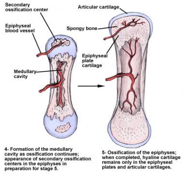
The epiphyses are the rounded ends of long bones in the body, which articulate with other bones to form joints. They are separated from the main shaft of the bone (diaphysis) by a growth plate called the physis or epiphyseal plate. The epiphyses are made up of spongy bone and covered with articular cartilage, which allows for smooth movement between bones. During growth, the epiphyseal plates produce new bone cells that cause the bone to lengthen until they eventually fuse during adulthood, at which point growth stops.
Spontaneous fractures are bone breaks that occur without any identifiable trauma or injury. They are typically caused by underlying medical conditions that weaken the bones, making them more susceptible to breaking under normal stress or weight. The most common cause of spontaneous fractures is osteoporosis, a condition characterized by weak and brittle bones. Other potential causes include various bone diseases, certain cancers, long-term use of corticosteroids, and genetic disorders affecting bone strength.
It's important to note that while the term "spontaneous" implies that the fracture occurred without any apparent cause, it is usually the result of an underlying medical condition. Therefore, if you experience a spontaneous fracture, seeking medical attention is crucial to diagnose and manage the underlying cause to prevent future fractures and related complications.
Hip arthroplasty, also known as hip replacement surgery, is a medical procedure where the damaged or diseased joint surfaces of the hip are removed and replaced with artificial components. These components typically include a metal or ceramic ball that replaces the head of the femur (thigh bone), and a polyethylene or ceramic socket that replaces the acetabulum (hip socket) in the pelvis.
The goal of hip arthroplasty is to relieve pain, improve joint mobility, and restore function to the hip joint. This procedure is commonly performed in patients with advanced osteoarthritis, rheumatoid arthritis, hip fractures, or other conditions that cause significant damage to the hip joint.
There are several types of hip replacement surgeries, including traditional total hip arthroplasty, partial (hemi) hip arthroplasty, and resurfacing hip arthroplasty. The choice of procedure depends on various factors, such as the patient's age, activity level, overall health, and the extent of joint damage.
After surgery, patients typically require rehabilitation to regain strength, mobility, and function in the affected hip. With proper care and follow-up, most patients can expect significant pain relief and improved quality of life following hip arthroplasty.
Bone plates are medical devices used in orthopedic surgery to stabilize and hold together fractured or broken bones during the healing process. They are typically made of surgical-grade stainless steel, titanium, or other biocompatible materials. The plate is shaped to fit the contour of the bone and is held in place with screws that are inserted through the plate and into the bone on either side of the fracture. This provides stability and alignment to the broken bones, allowing them to heal properly. Bone plates can be used to treat a variety of fractures, including those that are complex or unstable. After healing is complete, the bone plate may be left in place or removed, depending on the individual's needs and the surgeon's recommendation.
Osteoporosis is a systemic skeletal disease characterized by low bone mass, deterioration of bone tissue, and disruption of bone architecture, leading to increased risk of fractures, particularly in the spine, wrist, and hip. It mainly affects older people, especially postmenopausal women, due to hormonal changes that reduce bone density. Osteoporosis can also be caused by certain medications, medical conditions, or lifestyle factors such as smoking, alcohol abuse, and a lack of calcium and vitamin D in the diet. The diagnosis is often made using bone mineral density testing, and treatment may include medication to slow bone loss, promote bone formation, and prevent fractures.
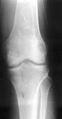
The knee joint, also known as the tibiofemoral joint, is the largest and one of the most complex joints in the human body. It is a synovial joint that connects the thighbone (femur) to the shinbone (tibia). The patella (kneecap), which is a sesamoid bone, is located in front of the knee joint and helps in the extension of the leg.
The knee joint is made up of three articulations: the femorotibial joint between the femur and tibia, the femoropatellar joint between the femur and patella, and the tibiofibular joint between the tibia and fibula. These articulations are surrounded by a fibrous capsule that encloses the synovial membrane, which secretes synovial fluid to lubricate the joint.
The knee joint is stabilized by several ligaments, including the medial and lateral collateral ligaments, which provide stability to the sides of the joint, and the anterior and posterior cruciate ligaments, which prevent excessive forward and backward movement of the tibia relative to the femur. The menisci, which are C-shaped fibrocartilaginous structures located between the femoral condyles and tibial plateaus, also help to stabilize the joint by absorbing shock and distributing weight evenly across the articular surfaces.
The knee joint allows for flexion, extension, and a small amount of rotation, making it essential for activities such as walking, running, jumping, and sitting.
Osteotomy is a surgical procedure in which a bone is cut to shorten, lengthen, or change its alignment. It is often performed to correct deformities or to realign bones that have been damaged by trauma or disease. The bone may be cut straight across (transverse osteotomy) or at an angle (oblique osteotomy). After the bone is cut, it can be realigned and held in place with pins, plates, or screws until it heals. This procedure is commonly performed on bones in the leg, such as the femur or tibia, but can also be done on other bones in the body.
Bone remodeling is the normal and continuous process by which bone tissue is removed from the skeleton (a process called resorption) and new bone tissue is formed (a process called formation). This ongoing cycle allows bones to repair microdamage, adjust their size and shape in response to mechanical stress, and maintain mineral homeostasis. The cells responsible for bone resorption are osteoclasts, while the cells responsible for bone formation are osteoblasts. These two cell types work together to maintain the structural integrity and health of bones throughout an individual's life.
During bone remodeling, the process can be divided into several stages:
1. Activation: The initiation of bone remodeling is triggered by various factors such as microdamage, hormonal changes, or mechanical stress. This leads to the recruitment and activation of osteoclast precursor cells.
2. Resorption: Osteoclasts attach to the bone surface and create a sealed compartment called a resorption lacuna. They then secrete acid and enzymes that dissolve and digest the mineralized matrix, creating pits or cavities on the bone surface. This process helps remove old or damaged bone tissue and releases calcium and phosphate ions into the bloodstream.
3. Reversal: After resorption is complete, the osteoclasts undergo apoptosis (programmed cell death), and mononuclear cells called reversal cells appear on the resorbed surface. These cells prepare the bone surface for the next stage by cleaning up debris and releasing signals that attract osteoblast precursors.
4. Formation: Osteoblasts, derived from mesenchymal stem cells, migrate to the resorbed surface and begin producing a new organic matrix called osteoid. As the osteoid mineralizes, it forms a hard, calcified structure that gradually replaces the resorbed bone tissue. The osteoblasts may become embedded within this newly formed bone as they differentiate into osteocytes, which are mature bone cells responsible for maintaining bone homeostasis and responding to mechanical stress.
5. Mineralization: Over time, the newly formed bone continues to mineralize, becoming stronger and more dense. This process helps maintain the structural integrity of the skeleton and ensures adequate calcium storage.
Throughout this continuous cycle of bone remodeling, hormones, growth factors, and mechanical stress play crucial roles in regulating the balance between resorption and formation. Disruptions to this delicate equilibrium can lead to various bone diseases, such as osteoporosis, where excessive resorption results in weakened bones and increased fracture risk.
Fracture fixation is a surgical procedure in orthopedic trauma surgery where a fractured bone is stabilized using various devices and techniques to promote proper healing and alignment. The goal of fracture fixation is to maintain the broken bone ends in correct anatomical position and length, allowing for adequate stability during the healing process.
There are two main types of fracture fixation:
1. Internal fixation: In this method, metal implants like plates, screws, or intramedullary rods are inserted directly into the bone to hold the fragments in place. These implants can be either removed or left in the body once healing is complete, depending on the type and location of the fracture.
2. External fixation: This technique involves placing pins or screws through the skin and into the bone above and below the fracture site. These pins are then connected to an external frame that maintains alignment and stability. External fixators are typically used when there is significant soft tissue damage, infection, or when internal fixation is not possible due to the complexity of the fracture.
The choice between internal and external fixation depends on various factors such as the type and location of the fracture, patient's age and overall health, surgeon's preference, and potential complications. Both methods aim to provide a stable environment for bone healing while minimizing the risk of malunion, nonunion, or deformity.
'Leg length inequality' (LLIS) is a condition where there is a discrepancy in the lengths of an individual's lower extremities, specifically the bones of the thigh (femur) and/or the leg (tibia/fibula). This discrepancy can be congenital or acquired due to various causes such as fractures, infections, or surgical procedures. The inequality can lead to functional scoliosis, lower back pain, and other musculoskeletal issues. It is typically diagnosed through physical examination and imaging studies like X-rays, and may be treated with various methods including orthotics, shoe lifts, or in some cases, surgical intervention.
Photon Absorptiometry is a medical technique used to measure the absorption of photons (light particles) by tissues or materials. In clinical practice, it is often used as a non-invasive method for measuring bone mineral density (BMD). This technique uses a low-energy X-ray beam or gamma ray to penetrate the tissue and then measures the amount of radiation absorbed by the bone. The amount of absorption is related to the density and thickness of the bone, allowing for an assessment of BMD. It can be used to diagnose osteoporosis and monitor treatment response in patients with bone diseases. There are two types of photon absorptiometry: single-photon absorptiometry (SPA) and dual-photon absorptiometry (DPA). SPA uses one energy level, while DPA uses two different energy levels to measure BMD, providing more precise measurements.
Bone transplantation, also known as bone grafting, is a surgical procedure in which bone or bone-like material is transferred from one part of the body to another or from one person to another. The graft may be composed of cortical (hard outer portion) bone, cancellous (spongy inner portion) bone, or a combination of both. It can be taken from different sites in the same individual (autograft), from another individual of the same species (allograft), or from an animal source (xenograft). The purpose of bone transplantation is to replace missing bone, provide structural support, and stimulate new bone growth. This procedure is commonly used in orthopedic, dental, and maxillofacial surgeries to repair bone defects caused by trauma, tumors, or congenital conditions.
Squamous cell carcinoma is a type of skin cancer that begins in the squamous cells, which are flat, thin cells that form the outer layer of the skin (epidermis). It commonly occurs on sun-exposed areas such as the face, ears, lips, and backs of the hands. Squamous cell carcinoma can also develop in other areas of the body including the mouth, lungs, and cervix.
This type of cancer usually develops slowly and may appear as a rough or scaly patch of skin, a red, firm nodule, or a sore or ulcer that doesn't heal. While squamous cell carcinoma is not as aggressive as some other types of cancer, it can metastasize (spread) to other parts of the body if left untreated, making early detection and treatment important.
Risk factors for developing squamous cell carcinoma include prolonged exposure to ultraviolet (UV) radiation from the sun or tanning beds, fair skin, a history of sunburns, a weakened immune system, and older age. Prevention measures include protecting your skin from the sun by wearing protective clothing, using a broad-spectrum sunscreen with an SPF of at least 30, avoiding tanning beds, and getting regular skin examinations.
Bone development, also known as ossification, is the process by which bone tissue is formed and grows. This complex process involves several different types of cells, including osteoblasts, which produce new bone matrix, and osteoclasts, which break down and resorb existing bone tissue.
There are two main types of bone development: intramembranous and endochondral ossification. Intramembranous ossification occurs when bone tissue forms directly from connective tissue, while endochondral ossification involves the formation of a cartilage model that is later replaced by bone.
During fetal development, most bones develop through endochondral ossification, starting as a cartilage template that is gradually replaced by bone tissue. However, some bones, such as those in the skull and clavicles, develop through intramembranous ossification.
Bone development continues after birth, with new bone tissue being laid down and existing tissue being remodeled throughout life. This ongoing process helps to maintain the strength and integrity of the skeleton, allowing it to adapt to changing mechanical forces and repair any damage that may occur.
A cadaver is a deceased body that is used for medical research or education. In the field of medicine, cadavers are often used in anatomy lessons, surgical training, and other forms of medical research. The use of cadavers allows medical professionals to gain a deeper understanding of the human body and its various systems without causing harm to living subjects. Cadavers may be donated to medical schools or obtained through other means, such as through consent of the deceased or their next of kin. It is important to handle and treat cadavers with respect and dignity, as they were once living individuals who deserve to be treated with care even in death.
Prosthesis design is a specialized field in medical device technology that involves creating and developing artificial substitutes to replace a missing body part, such as a limb, tooth, eye, or internal organ. The design process typically includes several stages: assessment of the patient's needs, selection of appropriate materials, creation of a prototype, testing and refinement, and final fabrication and fitting of the prosthesis.
The goal of prosthesis design is to create a device that functions as closely as possible to the natural body part it replaces, while also being comfortable, durable, and aesthetically pleasing for the patient. The design process may involve collaboration between medical professionals, engineers, and designers, and may take into account factors such as the patient's age, lifestyle, occupation, and overall health.
Prosthesis design can be highly complex, particularly for advanced devices such as robotic limbs or implantable organs. These devices often require sophisticated sensors, actuators, and control systems to mimic the natural functions of the body part they replace. As a result, prosthesis design is an active area of research and development in the medical field, with ongoing efforts to improve the functionality, comfort, and affordability of these devices for patients.
Head protective devices are equipment designed to protect the head from potential injuries or trauma. These devices often include helmets, hard hats, and bump caps. They are engineered to absorb the impact force, shield the head from sharp objects, or prevent contact with harmful substances. The specific design and construction of these devices vary depending on their intended use, such as for construction, sports, military, or healthcare purposes. It's important to choose and use a head protective device that is appropriate for the specific activity and follows established safety guidelines.
Bone lengthening is a surgical procedure that involves cutting and then gradually stretching the bone apart, allowing new bone to grow in its place. This process is also known as distraction osteogenesis. The goal of bone lengthening is to increase the length of a bone, either to improve function or to correct a deformity.
The procedure typically involves making an incision in the skin over the bone and using specialized tools to cut through the bone. Once the bone is cut, a device called an external fixator is attached to the bone on either side of the cut. The external fixator is then gradually adjusted over time to slowly stretch the bone apart, creating a gap between the two ends of the bone. As the bone is stretched, new bone tissue begins to grow in the space between the two ends, eventually filling in the gap and lengthening the bone.
Bone lengthening can be used to treat a variety of conditions, including limb length discrepancies, congenital deformities, and injuries that result in bone loss. It is typically performed by an orthopedic surgeon and may require several months of follow-up care to ensure proper healing and growth of the new bone tissue.
X-ray computed tomography (CT or CAT scan) is a medical imaging method that uses computer-processed combinations of many X-ray images taken from different angles to produce cross-sectional (tomographic) images (virtual "slices") of the body. These cross-sectional images can then be used to display detailed internal views of organs, bones, and soft tissues in the body.
The term "computed tomography" is used instead of "CT scan" or "CAT scan" because the machines take a series of X-ray measurements from different angles around the body and then use a computer to process these data to create detailed images of internal structures within the body.
CT scanning is a noninvasive, painless medical test that helps physicians diagnose and treat medical conditions. CT imaging provides detailed information about many types of tissue including lung, bone, soft tissue and blood vessels. CT examinations can be performed on every part of the body for a variety of reasons including diagnosis, surgical planning, and monitoring of therapeutic responses.
In computed tomography (CT), an X-ray source and detector rotate around the patient, measuring the X-ray attenuation at many different angles. A computer uses this data to construct a cross-sectional image by the process of reconstruction. This technique is called "tomography". The term "computed" refers to the use of a computer to reconstruct the images.
CT has become an important tool in medical imaging and diagnosis, allowing radiologists and other physicians to view detailed internal images of the body. It can help identify many different medical conditions including cancer, heart disease, lung nodules, liver tumors, and internal injuries from trauma. CT is also commonly used for guiding biopsies and other minimally invasive procedures.
In summary, X-ray computed tomography (CT or CAT scan) is a medical imaging technique that uses computer-processed combinations of many X-ray images taken from different angles to produce cross-sectional images of the body. It provides detailed internal views of organs, bones, and soft tissues in the body, allowing physicians to diagnose and treat medical conditions.
In the context of medicine, particularly in anatomy and physiology, "rotation" refers to the movement of a body part around its own axis or the long axis of another structure. This type of motion is three-dimensional and can occur in various planes. A common example of rotation is the movement of the forearm bones (radius and ulna) around each other during pronation and supination, which allows the hand to be turned palm up or down. Another example is the rotation of the head during mastication (chewing), where the mandible moves in a circular motion around the temporomandibular joint.
Metabolic bone diseases are a group of conditions that affect the bones and are caused by disorders in the body's metabolism. These disorders can result in changes to the bone structure, density, and strength, leading to an increased risk of fractures and other complications. Some common examples of metabolic bone diseases include:
1. Osteoporosis: a condition characterized by weak and brittle bones that are more likely to break, often as a result of age-related bone loss or hormonal changes.
2. Paget's disease of bone: a chronic disorder that causes abnormal bone growth and deformities, leading to fragile and enlarged bones.
3. Osteomalacia: a condition caused by a lack of vitamin D or problems with the body's ability to absorb it, resulting in weak and soft bones.
4. Hyperparathyroidism: a hormonal disorder that causes too much parathyroid hormone to be produced, leading to bone loss and other complications.
5. Hypoparathyroidism: a hormonal disorder that results in low levels of parathyroid hormone, causing weak and brittle bones.
6. Renal osteodystrophy: a group of bone disorders that occur as a result of chronic kidney disease, including osteomalacia, osteoporosis, and high turnover bone disease.
Treatment for metabolic bone diseases may include medications to improve bone density and strength, dietary changes, exercise, and lifestyle modifications. In some cases, surgery may be necessary to correct bone deformities or fractures.
Traction, in medical terms, refers to the application of a pulling force to distract or align parts of the body, particularly bones, joints, or muscles, with the aim of immobilizing, reducing displacement, or realigning them. This is often achieved through the use of various devices such as tongs, pulleys, weights, or specialized traction tables. Traction may be applied manually or mechanically and can be continuous or intermittent, depending on the specific medical condition being treated. Common indications for traction include fractures, dislocations, spinal cord injuries, and certain neurological conditions.
A comminuted fracture is a type of bone break where the bone is shattered into three or more pieces. This type of fracture typically occurs after high-energy trauma, such as a car accident or a fall from a great height. Commminuted fractures can also occur in bones that are weakened by conditions like osteoporosis or cancer. Because of the severity and complexity of comminuted fractures, they often require extensive treatment, which may include surgery to realign and stabilize the bone fragments using metal screws, plates, or rods.
Bony callus is a medical term that refers to the specialized tissue that forms in response to a bone fracture. It is a crucial part of the natural healing process, as it helps to stabilize and protect the broken bone while it mends.
When a bone is fractured, the body responds by initiating an inflammatory response, which triggers the production of various cells and signaling molecules that promote healing. As part of this process, specialized cells called osteoblasts begin to produce new bone tissue at the site of the fracture. This tissue is initially soft and pliable, allowing it to bridge the gap between the broken ends of the bone.
Over time, this soft callus gradually hardens and calcifies, forming a bony callus that helps to stabilize the fracture and provide additional support as the bone heals. The bony callus is typically composed of a mixture of woven bone (which is less organized than normal bone) and more structured lamellar bone (which is similar in structure to normal bone).
As the bone continues to heal, the bony callus may be gradually remodeled and reshaped by osteoclasts, which are specialized cells that break down and remove excess or unwanted bone tissue. This process helps to restore the bone's original shape and strength, allowing it to function normally again.
It is worth noting that excessive bony callus formation can sometimes lead to complications, such as stiffness, pain, or decreased range of motion in the affected limb. In some cases, surgical intervention may be necessary to remove or reduce the size of the bony callus and promote proper healing.
Prosthesis failure is a term used to describe a situation where a prosthetic device, such as an artificial joint or limb, has stopped functioning or failed to meet its intended purpose. This can be due to various reasons, including mechanical failure, infection, loosening of the device, or a reaction to the materials used in the prosthesis.
Mechanical failure can occur due to wear and tear, manufacturing defects, or improper use of the prosthetic device. Infection can also lead to prosthesis failure, particularly in cases where the prosthesis is implanted inside the body. The immune system may react to the presence of the foreign material, leading to inflammation and infection.
Loosening of the prosthesis can also cause it to fail over time, as the device becomes less stable and eventually stops working properly. Additionally, some people may have a reaction to the materials used in the prosthesis, leading to tissue damage or other complications that can result in prosthesis failure.
In general, prosthesis failure can lead to decreased mobility, pain, and the need for additional surgeries or treatments to correct the problem. It is important for individuals with prosthetic devices to follow their healthcare provider's instructions carefully to minimize the risk of prosthesis failure and ensure that the device continues to function properly over time.
Bone cements are medical-grade materials used in orthopedic and trauma surgery to fill gaps between bone surfaces and implants, such as artificial joints or screws. They serve to mechanically stabilize the implant and provide a smooth, load-bearing surface. The two most common types of bone cement are:
1. Polymethylmethacrylate (PMMA) cement: This is a two-component system consisting of powdered PMMA and liquid methyl methacrylate monomer. When mixed together, they form a dough-like consistency that hardens upon exposure to air. PMMA cement has been widely used for decades in joint replacement surgeries, such as hip or knee replacements.
2. Calcium phosphate (CP) cement: This is a two-component system consisting of a powdered CP compound and an aqueous solution. When mixed together, they form a paste that hardens through a chemical reaction at body temperature. CP cement has lower mechanical strength compared to PMMA but demonstrates better biocompatibility, bioactivity, and the ability to resorb over time.
Both types of bone cements have advantages and disadvantages, and their use depends on the specific surgical indication and patient factors.
X-ray microtomography, often referred to as micro-CT, is a non-destructive imaging technique used to visualize and analyze the internal structure of objects with high spatial resolution. It is based on the principles of computed tomography (CT), where multiple X-ray images are acquired at different angles and then reconstructed into cross-sectional slices using specialized software. These slices can be further processed to create 3D visualizations, allowing researchers and clinicians to examine the internal structure and composition of samples in great detail. Micro-CT is widely used in materials science, biology, medicine, and engineering for various applications such as material characterization, bone analysis, and defect inspection.
Ununited fracture is a medical term used to describe a fractured bone that has failed to heal properly. This condition is also known as a nonunion fracture. In a normal healing process, the broken ends of the bone will grow together, or "unite," over time as new bone tissue forms. However, in some cases, the bones may not reconnect due to various reasons such as infection, poor blood supply, excessive motion at the fracture site, or inadequate stabilization of the fracture.
Ununited fractures can cause significant pain, swelling, and deformity in the affected area. They may also lead to a decreased range of motion, weakness, and instability in the joint near the fracture. Treatment for ununited fractures typically involves surgical intervention to promote bone healing, such as bone grafting or internal fixation with screws or plates. In some cases, electrical stimulation or ultrasound therapy may also be used to help promote bone growth and healing.
Articular Range of Motion (AROM) is a term used in physiotherapy and orthopedics to describe the amount of movement available in a joint, measured in degrees of a circle. It refers to the range through which synovial joints can actively move without causing pain or injury. AROM is assessed by measuring the degree of motion achieved by active muscle contraction, as opposed to passive range of motion (PROM), where the movement is generated by an external force.
Assessment of AROM is important in evaluating a patient's functional ability and progress, planning treatment interventions, and determining return to normal activities or sports participation. It is also used to identify any restrictions in joint mobility that may be due to injury, disease, or surgery, and to monitor the effectiveness of rehabilitation programs.
Bone neoplasms are abnormal growths or tumors that develop in the bone. They can be benign (non-cancerous) or malignant (cancerous). Benign bone neoplasms do not spread to other parts of the body and are rarely a threat to life, although they may cause problems if they grow large enough to press on surrounding tissues or cause fractures. Malignant bone neoplasms, on the other hand, can invade and destroy nearby tissue and may spread (metastasize) to other parts of the body.
There are many different types of bone neoplasms, including:
1. Osteochondroma - a benign tumor that develops from cartilage and bone
2. Enchondroma - a benign tumor that forms in the cartilage that lines the inside of the bones
3. Chondrosarcoma - a malignant tumor that develops from cartilage
4. Osteosarcoma - a malignant tumor that develops from bone cells
5. Ewing sarcoma - a malignant tumor that develops in the bones or soft tissues around the bones
6. Giant cell tumor of bone - a benign or occasionally malignant tumor that develops from bone tissue
7. Fibrosarcoma - a malignant tumor that develops from fibrous tissue in the bone
The symptoms of bone neoplasms vary depending on the type, size, and location of the tumor. They may include pain, swelling, stiffness, fractures, or limited mobility. Treatment options depend on the type and stage of the tumor but may include surgery, radiation therapy, chemotherapy, or a combination of these treatments.
Mechanical stress, in the context of physiology and medicine, refers to any type of force that is applied to body tissues or organs, which can cause deformation or displacement of those structures. Mechanical stress can be either external, such as forces exerted on the body during physical activity or trauma, or internal, such as the pressure changes that occur within blood vessels or other hollow organs.
Mechanical stress can have a variety of effects on the body, depending on the type, duration, and magnitude of the force applied. For example, prolonged exposure to mechanical stress can lead to tissue damage, inflammation, and chronic pain. Additionally, abnormal or excessive mechanical stress can contribute to the development of various musculoskeletal disorders, such as tendinitis, osteoarthritis, and herniated discs.
In order to mitigate the negative effects of mechanical stress, the body has a number of adaptive responses that help to distribute forces more evenly across tissues and maintain structural integrity. These responses include changes in muscle tone, joint positioning, and connective tissue stiffness, as well as the remodeling of bone and other tissues over time. However, when these adaptive mechanisms are overwhelmed or impaired, mechanical stress can become a significant factor in the development of various pathological conditions.
The humeral head is the rounded, articular surface at the proximal end of the humerus bone in the human body. It forms the upper part of the shoulder joint and articulates with the glenoid fossa of the scapula to form the glenohumeral joint, allowing for a wide range of motion in the arm. The humeral head is covered with cartilage that helps to provide a smooth, lubricated surface for movement and shock absorption.
An external fixator is a type of orthopedic device used in the treatment of severe fractures or deformities of bones. It consists of an external frame that is attached to the bone with pins or wires that pass through the skin and into the bone. This provides stability to the injured area while allowing for alignment and adjustment of the bone during the healing process.
External fixators are typically used in cases where traditional casting or internal fixation methods are not feasible, such as when there is extensive soft tissue damage, infection, or when a limb needs to be gradually stretched or shortened. They can also be used in reconstructive surgery for bone defects or deformities.
The external frame of the fixator is made up of bars and clamps that are adjustable, allowing for precise positioning and alignment of the bones. The pins or wires that attach to the bone are carefully inserted through small incisions in the skin, and are held in place by the clamps on the frame.
External fixators can be used for a period of several weeks to several months, depending on the severity of the injury and the individual's healing process. During this time, the patient may require regular adjustments and monitoring by an orthopedic surgeon or other medical professional. Once the bone has healed sufficiently, the external fixator can be removed in a follow-up procedure.
Compressive strength is a measure of the maximum compressive load that a material or structure can withstand before failure or deformation. It is typically expressed in units of pressure, such as pounds per square inch (psi) or megapascals (MPa). Compressive strength is an important property in the design and analysis of structures and materials, as it helps to ensure their safety and durability under compressive loads.
In medical terminology, compressive strength may refer to the ability of biological tissues, such as bone or cartilage, to withstand compressive forces without deforming or failing. For example, osteoporosis is a condition characterized by reduced bone density and compressive strength, which can increase the risk of fractures in affected individuals. Similarly, degenerative changes in articular cartilage can lead to decreased compressive strength and joint pain or stiffness.
I'm sorry for any confusion, but "Finite Element Analysis" (FEA) is not a medical term. It is a computational technique used in engineering and physical sciences. FEA is a computerized method for predicting how a product reacts to real-world forces, vibration, heat, fluid flow, and other physical effects. It's a way that engineers can simulate the performance of a product or system before it is built, which can help reduce costs, improve quality, and shorten the development time.
However, in a medical context, FEA might be used in the field of biomechanical engineering to analyze the mechanical behavior of biological systems, such as bones, joints, or soft tissues, under various loads and conditions. This can help researchers and clinicians better understand the mechanisms of injury, disease, or the effects of treatment, and develop more effective prevention, diagnostic, or therapeutic strategies.
Retrospective studies, also known as retrospective research or looking back studies, are a type of observational study that examines data from the past to draw conclusions about possible causal relationships between risk factors and outcomes. In these studies, researchers analyze existing records, medical charts, or previously collected data to test a hypothesis or answer a specific research question.
Retrospective studies can be useful for generating hypotheses and identifying trends, but they have limitations compared to prospective studies, which follow participants forward in time from exposure to outcome. Retrospective studies are subject to biases such as recall bias, selection bias, and information bias, which can affect the validity of the results. Therefore, retrospective studies should be interpreted with caution and used primarily to generate hypotheses for further testing in prospective studies.
Treatment outcome is a term used to describe the result or effect of medical treatment on a patient's health status. It can be measured in various ways, such as through symptoms improvement, disease remission, reduced disability, improved quality of life, or survival rates. The treatment outcome helps healthcare providers evaluate the effectiveness of a particular treatment plan and make informed decisions about future care. It is also used in clinical research to compare the efficacy of different treatments and improve patient care.
In medical terms, the hip is a ball-and-socket joint where the rounded head of the femur (thigh bone) fits into the cup-shaped socket, also known as the acetabulum, of the pelvis. This joint allows for a wide range of movement in the lower extremities and supports the weight of the upper body during activities such as walking, running, and jumping. The hip joint is surrounded by strong ligaments, muscles, and tendons that provide stability and enable proper functioning.
Bone screws are medical devices used in orthopedic and trauma surgery to affix bone fracture fragments or to attach bones to other bones or to metal implants such as plates, rods, or artificial joints. They are typically made of stainless steel or titanium alloys and have a threaded shaft that allows for purchase in the bone when tightened. The head of the screw may have a hexagonal or star-shaped design to allow for precise tightening with a screwdriver. Bone screws come in various shapes, sizes, and designs, including fully threaded, partially threaded, cannulated (hollow), and headless types, depending on their intended use and location in the body.
"Pediculus" is the medical term for a type of small, wingless parasitic insect that can be found in human hair and on the body. There are two main species that affect humans:
1. Pediculus humanus capitis - also known as the head louse, it primarily lives on the scalp and is responsible for causing head lice infestations.
2. Pediculus humanus corporis - also known as the body louse, it typically lives in clothing and on the body, particularly in seams and folds of clothing, and can cause body lice infestations.
Both species of Pediculus feed on human blood and can cause itching and skin irritation. They are primarily spread through close personal contact and sharing of items such as hats, combs, and clothing.
Physiologic calcification is the normal deposit of calcium salts in body tissues and organs. It is a natural process that occurs as part of the growth and development of the human body, as well as during the repair and remodeling of tissues.
Calcium is an essential mineral that plays a critical role in many bodily functions, including bone formation, muscle contraction, nerve impulse transmission, and blood clotting. In order to maintain proper levels of calcium in the body, excess calcium that is not needed for these functions may be deposited in various tissues as a normal part of the aging process.
Physiologic calcification typically occurs in areas such as the walls of blood vessels, the lungs, and the heart valves. While these calcifications are generally harmless, they can sometimes lead to complications, particularly if they occur in large amounts or in sensitive areas. For example, calcification of the coronary arteries can increase the risk of heart disease, while calcification of the lung tissue can cause respiratory symptoms.
It is important to note that pathologic calcification, on the other hand, refers to the abnormal deposit of calcium salts in tissues and organs, which can be caused by various medical conditions such as chronic kidney disease, hyperparathyroidism, and certain infections. Pathologic calcification is not a normal process and can lead to serious health complications if left untreated.
In the field of medicine, "time factors" refer to the duration of symptoms or time elapsed since the onset of a medical condition, which can have significant implications for diagnosis and treatment. Understanding time factors is crucial in determining the progression of a disease, evaluating the effectiveness of treatments, and making critical decisions regarding patient care.
For example, in stroke management, "time is brain," meaning that rapid intervention within a specific time frame (usually within 4.5 hours) is essential to administering tissue plasminogen activator (tPA), a clot-busting drug that can minimize brain damage and improve patient outcomes. Similarly, in trauma care, the "golden hour" concept emphasizes the importance of providing definitive care within the first 60 minutes after injury to increase survival rates and reduce morbidity.
Time factors also play a role in monitoring the progression of chronic conditions like diabetes or heart disease, where regular follow-ups and assessments help determine appropriate treatment adjustments and prevent complications. In infectious diseases, time factors are crucial for initiating antibiotic therapy and identifying potential outbreaks to control their spread.
Overall, "time factors" encompass the significance of recognizing and acting promptly in various medical scenarios to optimize patient outcomes and provide effective care.
Prenatal ultrasonography, also known as obstetric ultrasound, is a medical diagnostic procedure that uses high-frequency sound waves to create images of the developing fetus, placenta, and amniotic fluid inside the uterus. It is a non-invasive and painless test that is widely used during pregnancy to monitor the growth and development of the fetus, detect any potential abnormalities or complications, and determine the due date.
During the procedure, a transducer (a small handheld device) is placed on the mother's abdomen and moved around to capture images from different angles. The sound waves travel through the mother's body and bounce back off the fetus, producing echoes that are then converted into electrical signals and displayed as images on a screen.
Prenatal ultrasonography can be performed at various stages of pregnancy, including early pregnancy to confirm the pregnancy and detect the number of fetuses, mid-pregnancy to assess the growth and development of the fetus, and late pregnancy to evaluate the position of the fetus and determine if it is head down or breech. It can also be used to guide invasive procedures such as amniocentesis or chorionic villus sampling.
Overall, prenatal ultrasonography is a valuable tool in modern obstetrics that helps ensure the health and well-being of both the mother and the developing fetus.
A reoperation is a surgical procedure that is performed again on a patient who has already undergone a previous operation for the same or related condition. Reoperations may be required due to various reasons, such as inadequate initial treatment, disease recurrence, infection, or complications from the first surgery. The nature and complexity of a reoperation can vary widely depending on the specific circumstances, but it often carries higher risks and potential complications compared to the original operation.
Osteogenesis is the process of bone formation or development. It involves the differentiation and maturation of osteoblasts, which are bone-forming cells that synthesize and deposit the organic matrix of bone tissue, composed mainly of type I collagen. This organic matrix later mineralizes to form the inorganic crystalline component of bone, primarily hydroxyapatite.
There are two primary types of osteogenesis: intramembranous and endochondral. Intramembranous osteogenesis occurs directly within connective tissue, where mesenchymal stem cells differentiate into osteoblasts and form bone tissue without an intervening cartilage template. This process is responsible for the formation of flat bones like the skull and clavicles.
Endochondral osteogenesis, on the other hand, involves the initial development of a cartilaginous model or template, which is later replaced by bone tissue. This process forms long bones, such as those in the limbs, and occurs through several stages involving chondrocyte proliferation, hypertrophy, and calcification, followed by invasion of blood vessels and osteoblasts to replace the cartilage with bone tissue.
Abnormalities in osteogenesis can lead to various skeletal disorders and diseases, such as osteogenesis imperfecta (brittle bone disease), achondroplasia (a form of dwarfism), and cleidocranial dysplasia (a disorder affecting skull and collarbone development).
Osseointegration is a direct structural and functional connection between living bone and the surface of an implant. It's a process where the bone grows in and around the implant, which is typically made of titanium or another biocompatible material. This process provides a solid foundation for dental prosthetics, such as crowns, bridges, or dentures, or for orthopedic devices like artificial limbs. The success of osseointegration depends on various factors, including the patient's overall health, the quality and quantity of available bone, and the surgical technique used for implant placement.
The parietal bone is one of the four flat bones that form the skull's cranial vault, which protects the brain. There are two parietal bones in the skull, one on each side, located posterior to the frontal bone and temporal bone, and anterior to the occipital bone. Each parietal bone has a squamous part, which forms the roof and sides of the skull, and a smaller, wing-like portion called the mastoid process. The parietal bones contribute to the formation of the coronal and lambdoid sutures, which are fibrous joints that connect the bones in the skull.
Bone density conservation agents, also known as anti-resorptive agents or bone-sparing drugs, are a class of medications that help to prevent the loss of bone mass and reduce the risk of fractures. They work by inhibiting the activity of osteoclasts, the cells responsible for breaking down and reabsorbing bone tissue during the natural remodeling process.
Examples of bone density conservation agents include:
1. Bisphosphonates (e.g., alendronate, risedronate, ibandronate, zoledronic acid) - These are the most commonly prescribed class of bone density conservation agents. They bind to hydroxyapatite crystals in bone tissue and inhibit osteoclast activity, thereby reducing bone resorption.
2. Denosumab (Prolia) - This is a monoclonal antibody that targets RANKL (Receptor Activator of Nuclear Factor-κB Ligand), a key signaling molecule involved in osteoclast differentiation and activation. By inhibiting RANKL, denosumab reduces osteoclast activity and bone resorption.
3. Selective estrogen receptor modulators (SERMs) (e.g., raloxifene) - These medications act as estrogen agonists or antagonists in different tissues. In bone tissue, SERMs mimic the bone-preserving effects of estrogen by inhibiting osteoclast activity and reducing bone resorption.
4. Hormone replacement therapy (HRT) - Estrogen hormone replacement therapy has been shown to preserve bone density in postmenopausal women; however, its use is limited due to increased risks of breast cancer, cardiovascular disease, and thromboembolic events.
5. Calcitonin - This hormone, secreted by the thyroid gland, inhibits osteoclast activity and reduces bone resorption. However, it has largely been replaced by other more effective bone density conservation agents.
These medications are often prescribed for individuals at high risk of fractures due to conditions such as osteoporosis or metabolic disorders that affect bone health. It is essential to follow the recommended dosage and administration guidelines to maximize their benefits while minimizing potential side effects. Regular monitoring of bone density, blood calcium levels, and other relevant parameters is also necessary during treatment with these medications.
Mechanical torsion in a medical context refers to the twisting or rotational deformation of a body or structure due to an applied torque or force. This can occur in various biological structures, such as blood vessels, intestines, or muscles, leading to impaired function, pain, or even tissue necrosis if severe or prolonged.
For example, in the case of the gastrointestinal tract, torsion can cause a segment of the bowel to twist around its own axis, cutting off blood flow and causing ischemia or necrosis. This is a surgical emergency that requires prompt intervention to prevent further complications. Similarly, in the eye, torsion can refer to the rotation of the eyeball within the orbit, which can cause double vision or other visual disturbances.
Gestational age is the length of time that has passed since the first day of the last menstrual period (LMP) in pregnant women. It is the standard unit used to estimate the age of a pregnancy and is typically expressed in weeks. This measure is used because the exact date of conception is often not known, but the start of the last menstrual period is usually easier to recall.
It's important to note that since ovulation typically occurs around two weeks after the start of the LMP, gestational age is approximately two weeks longer than fetal age, which is the actual time elapsed since conception. Medical professionals use both gestational and fetal age to track the development and growth of the fetus during pregnancy.
In the medical field, cementation refers to the process of using a type of dental cement or bonding agent to attach a dental restoration (such as a crown, bridge, or false tooth) to a natural tooth or implant. The cement helps to create a strong and secure attachment, while also helping to seal the restoration and prevent the entry of bacteria and saliva.
Dental cement can be made from various materials, including glass ionomers, resin-modified glass ionomers, zinc phosphate, and polycarboxylate cements. The choice of cement depends on several factors, such as the type of restoration being attached, the location in the mouth, and the patient's individual needs and preferences.
Cementation is an important step in many dental procedures, as it helps to ensure the longevity and success of the restoration. Proper technique and material selection are crucial for achieving a successful cementation that will last for years to come.
A lice infestation, also known as pediculosis, is a condition characterized by the presence and multiplication of parasitic insects called lice on a person's body. The three main types of lice that can infest humans are:
1. Head lice (Pediculus humanus capitis): These lice primarily live on the scalp, neck, and behind the ears, feeding on human blood. They lay their eggs (nits) on hair shafts close to the scalp. Head lice infestations are most common in children aged 3-12 years old.
2. Body lice (Pediculus humanus corporis): These lice typically live and lay eggs on clothing, particularly seams and collars, near the body's warmest areas. They move to the skin to feed on blood, usually at night. Body lice infestations are more common in people who experience homelessness or overcrowded living conditions with limited access to clean clothing and hygiene facilities.
3. Pubic lice (Pthirus pubis): Also known as crab lice, these lice primarily live in coarse body hair, such as the pubic area, armpits, eyelashes, eyebrows, beard, or mustache. They feed on human blood and lay eggs on hair shafts close to the skin. Pubic lice infestations are typically sexually transmitted but can also occur through close personal contact with an infected individual or sharing contaminated items like bedding or clothing.
Symptoms of a lice infestation may include intense itching, tickling sensations, and visible red bumps or sores on the skin caused by lice bites. In some cases, secondary bacterial infections can occur due to scratching. Diagnosis is usually made through visual identification of lice or nits on the body or clothing. Treatment typically involves topical medications, such as shampoos, creams, or lotions, and thorough cleaning of bedding, clothing, and personal items to prevent reinfestation.
A bone fracture is a medical condition in which there is a partial or complete break in the continuity of a bone due to external or internal forces. Fractures can occur in any bone in the body and can vary in severity from a small crack to a shattered bone. The symptoms of a bone fracture typically include pain, swelling, bruising, deformity, and difficulty moving the affected limb. Treatment for a bone fracture may involve immobilization with a cast or splint, surgery to realign and stabilize the bone, or medication to manage pain and prevent infection. The specific treatment approach will depend on the location, type, and severity of the fracture.
Congenital hip dislocation, also known as developmental dysplasia of the hip (DDH), is a condition where the hip joint fails to develop normally in utero or during early infancy. In a healthy hip, the head of the femur (thigh bone) fits snugly into the acetabulum (hip socket). However, in congenital hip dislocation, the femoral head is not held firmly in place within the acetabulum due to abnormal development or laxity of the ligaments that support the joint.
There are two types of congenital hip dislocations:
1. Teratologic dislocation: This type is present at birth and occurs due to abnormalities in the development of the hip joint during fetal growth. The femoral head may be completely outside the acetabulum or partially dislocated.
2. Developmental dysplasia: This type develops after birth, often within the first few months of life, as a result of ligamentous laxity and shallow acetabulum. In some cases, it can progress to a complete hip dislocation if left untreated.
Risk factors for congenital hip dislocation include family history, breech presentation during delivery, and female gender. Early diagnosis and treatment are crucial to prevent long-term complications such as pain, limited mobility, and osteoarthritis. Treatment options may include bracing, closed reduction, or surgical intervention, depending on the severity and age of the child at diagnosis.
Follow-up studies are a type of longitudinal research that involve repeated observations or measurements of the same variables over a period of time, in order to understand their long-term effects or outcomes. In medical context, follow-up studies are often used to evaluate the safety and efficacy of medical treatments, interventions, or procedures.
In a typical follow-up study, a group of individuals (called a cohort) who have received a particular treatment or intervention are identified and then followed over time through periodic assessments or data collection. The data collected may include information on clinical outcomes, adverse events, changes in symptoms or functional status, and other relevant measures.
The results of follow-up studies can provide important insights into the long-term benefits and risks of medical interventions, as well as help to identify factors that may influence treatment effectiveness or patient outcomes. However, it is important to note that follow-up studies can be subject to various biases and limitations, such as loss to follow-up, recall bias, and changes in clinical practice over time, which must be carefully considered when interpreting the results.
Developmental bone diseases are a group of medical conditions that affect the growth and development of bones. These diseases are present at birth or develop during childhood and adolescence, when bones are growing rapidly. They can result from genetic mutations, hormonal imbalances, or environmental factors such as poor nutrition.
Some examples of developmental bone diseases include:
1. Osteogenesis imperfecta (OI): Also known as brittle bone disease, OI is a genetic disorder that affects the body's production of collagen, a protein necessary for healthy bones. People with OI have fragile bones that break easily and may also experience other symptoms such as blue sclerae (whites of the eyes), hearing loss, and joint laxity.
2. Achondroplasia: This is the most common form of dwarfism, caused by a genetic mutation that affects bone growth. People with achondroplasia have short limbs and a large head relative to their body size.
3. Rickets: A condition caused by vitamin D deficiency or an inability to absorb or use vitamin D properly. This leads to weak, soft bones that can bow or bend easily, particularly in children.
4. Fibrous dysplasia: A rare bone disorder where normal bone is replaced with fibrous tissue, leading to weakened bones and deformities.
5. Scoliosis: An abnormal curvature of the spine that can develop during childhood or adolescence. While not strictly a developmental bone disease, scoliosis can be caused by various underlying conditions such as cerebral palsy, muscular dystrophy, or spina bifida.
Treatment for developmental bone diseases varies depending on the specific condition and its severity. Treatment may include medication, physical therapy, bracing, or surgery to correct deformities and improve function. Regular follow-up with a healthcare provider is essential to monitor growth, manage symptoms, and prevent complications.
The acetabulum is the cup-shaped cavity in the pelvic bone (specifically, the os coxa) where the head of the femur bone articulates to form the hip joint. It provides a stable and flexible connection between the lower limb and the trunk, allowing for a wide range of movements such as flexion, extension, abduction, adduction, rotation, and circumduction. The acetabulum is lined with articular cartilage, which facilitates smooth and frictionless movement of the hip joint. Its stability is further enhanced by various ligaments, muscles, and the labrum, a fibrocartilaginous rim that deepens the socket and increases its contact area with the femoral head.
Anatomic models are three-dimensional representations of body structures used for educational, training, or demonstration purposes. They can be made from various materials such as plastic, wax, or rubber and may depict the entire body or specific regions, organs, or systems. These models can be used to provide a visual aid for understanding anatomy, physiology, and pathology, and can be particularly useful in situations where actual human specimens are not available or practical to use. They may also be used for surgical planning and rehearsal, as well as in medical research and product development.
Alendronate is a medication that falls under the class of bisphosphonates. It is commonly used in the treatment and prevention of osteoporosis in postmenopausal women and men, as well as in the management of glucocorticoid-induced osteoporosis and Paget's disease of bone.
Alendronate works by inhibiting the activity of osteoclasts, which are cells responsible for breaking down and reabsorbing bone tissue. By reducing the activity of osteoclasts, alendronate helps to slow down bone loss and increase bone density, thereby reducing the risk of fractures.
The medication is available in several forms, including tablets and oral solutions, and is typically taken once a week for osteoporosis prevention and treatment. It is important to follow the dosing instructions carefully, as improper administration can reduce the drug's effectiveness or increase the risk of side effects. Common side effects of alendronate include gastrointestinal symptoms such as heartburn, stomach pain, and nausea.
Biometry, also known as biometrics, is the scientific study of measurements and statistical analysis of living organisms. In a medical context, biometry is often used to refer to the measurement and analysis of physical characteristics or features of the human body, such as height, weight, blood pressure, heart rate, and other physiological variables. These measurements can be used for a variety of purposes, including diagnosis, treatment planning, monitoring disease progression, and research.
In addition to physical measurements, biometry may also refer to the use of statistical methods to analyze biological data, such as genetic information or medical images. This type of analysis can help researchers and clinicians identify patterns and trends in large datasets, and make predictions about health outcomes or treatment responses.
Overall, biometry is an important tool in modern medicine, as it allows healthcare professionals to make more informed decisions based on data and evidence.
Bone resorption is the process by which bone tissue is broken down and absorbed into the body. It is a normal part of bone remodeling, in which old or damaged bone tissue is removed and new tissue is formed. However, excessive bone resorption can lead to conditions such as osteoporosis, in which bones become weak and fragile due to a loss of density. This process is carried out by cells called osteoclasts, which break down the bone tissue and release minerals such as calcium into the bloodstream.
Prostheses: Artificial substitutes or replacements for missing body parts, such as limbs, eyes, or teeth. They are designed to restore the function, appearance, or mobility of the lost part. Prosthetic devices can be categorized into several types, including:
1. External prostheses: Devices that are attached to the outside of the body, like artificial arms, legs, hands, and feet. These may be further classified into:
a. Cosmetic or aesthetic prostheses: Primarily designed to improve the appearance of the affected area.
b. Functional prostheses: Designed to help restore the functionality and mobility of the lost limb.
2. Internal prostheses: Implanted artificial parts that replace missing internal organs, bones, or tissues, such as heart valves, hip joints, or intraocular lenses.
Implants: Medical devices or substances that are intentionally placed inside the body to replace or support a missing or damaged biological structure, deliver medication, monitor physiological functions, or enhance bodily functions. Examples of implants include:
1. Orthopedic implants: Devices used to replace or reinforce damaged bones, joints, or cartilage, such as knee or hip replacements.
2. Cardiovascular implants: Devices that help support or regulate heart function, like pacemakers, defibrillators, and artificial heart valves.
3. Dental implants: Artificial tooth roots that are placed into the jawbone to support dental prostheses, such as crowns, bridges, or dentures.
4. Neurological implants: Devices used to stimulate nerves, brain structures, or spinal cord tissues to treat various neurological conditions, like deep brain stimulators for Parkinson's disease or cochlear implants for hearing loss.
5. Ophthalmic implants: Artificial lenses that are placed inside the eye to replace a damaged or removed natural lens, such as intraocular lenses used in cataract surgery.
A closed fracture, also known as a simple fracture, is a type of bone break where the skin remains intact and there is no open wound. The bone may be broken in such a way that it does not pierce the skin, but still requires medical attention for proper diagnosis, treatment, and healing. Closed fractures can range from hairline cracks to complete breaks and can occur due to various reasons, including trauma, overuse, or weakened bones. It is important to seek immediate medical care if a closed fracture is suspected, as improper healing can lead to long-term complications such as decreased mobility, chronic pain, or deformity.
Medical Definition:
Magnetic Resonance Imaging (MRI) is a non-invasive diagnostic imaging technique that uses a strong magnetic field and radio waves to create detailed cross-sectional or three-dimensional images of the internal structures of the body. The patient lies within a large, cylindrical magnet, and the scanner detects changes in the direction of the magnetic field caused by protons in the body. These changes are then converted into detailed images that help medical professionals to diagnose and monitor various medical conditions, such as tumors, injuries, or diseases affecting the brain, spinal cord, heart, blood vessels, joints, and other internal organs. MRI does not use radiation like computed tomography (CT) scans.
Orthopedic procedures are surgical or nonsurgical methods used to treat musculoskeletal conditions, including injuries, deformities, or diseases of the bones, joints, muscles, ligaments, and tendons. These procedures can range from simple splinting or casting to complex surgeries such as joint replacements, spinal fusions, or osteotomies (cutting and repositioning bones). The primary goal of orthopedic procedures is to restore function, reduce pain, and improve the quality of life for patients.
The fibula is a slender bone located in the lower leg of humans and other vertebrates. It runs parallel to the larger and more robust tibia, and together they are known as the bones of the leg or the anterior tibial segment. The fibula is the lateral bone in the leg, positioned on the outside of the tibia.
In humans, the fibula extends from the knee joint proximally to the ankle joint distally. Its proximal end, called the head of the fibula, articulates with the lateral condyle of the tibia and forms part of the inferior aspect of the knee joint. The narrowed portion below the head is known as the neck of the fibula.
The shaft of the fibula, also called the body of the fibula, is a long, thin structure that descends from the neck and serves primarily for muscle attachment rather than weight-bearing functions. The distal end of the fibula widens to form the lateral malleolus, which is an important bony landmark in the ankle region. The lateral malleolus articulates with the talus bone of the foot and forms part of the ankle joint.
The primary functions of the fibula include providing attachment sites for muscles that act on the lower leg, ankle, and foot, as well as contributing to the stability of the ankle joint through its articulation with the talus bone. Fractures of the fibula can occur due to various injuries, such as twisting or rotational forces applied to the ankle or direct trauma to the lateral aspect of the lower leg.
Bone malalignment is a term used to describe the abnormal alignment or positioning of bones in relation to each other. This condition can occur as a result of injury, deformity, surgery, or disease processes that affect the bones and joints. Bone malalignment can cause pain, stiffness, limited mobility, and an increased risk of further injury. In some cases, bone malalignment may require treatment such as bracing, physical therapy, or surgery to correct the alignment and improve function.
The radius is one of the two bones in the forearm in humans and other vertebrates. In humans, it runs from the lateral side of the elbow to the thumb side of the wrist. It is responsible for rotation of the forearm and articulates with the humerus at the elbow and the carpals at the wrist. Any medical condition or injury that affects the radius can impact the movement and function of the forearm and hand.
Osteoarthritis (OA) of the hip is a degenerative joint disease that affects the articular cartilage and subchondral bone of the hip joint. It is characterized by the progressive loss of cartilage, remodeling of bone, osteophyte formation (bone spurs), cysts, and mild to moderate inflammation. The degenerative process can lead to pain, stiffness, limited range of motion, and crepitus (grating or crackling sound) during movement.
In the hip joint, OA typically affects the femoral head and acetabulum. As the articular cartilage wears away, the underlying bone becomes exposed and can lead to bone-on-bone contact, which is painful. The body responds by attempting to repair the damage through remodeling of the subchondral bone and formation of osteophytes. However, these changes can further limit joint mobility and exacerbate symptoms.
Risk factors for OA of the hip include age, obesity, genetics, previous joint injury or surgery, and repetitive stress on the joint. Treatment options may include pain management (such as NSAIDs, physical therapy, and injections), lifestyle modifications (such as weight loss and exercise), and, in severe cases, surgical intervention (such as hip replacement).
Tensile strength is a material property that measures the maximum amount of tensile (pulling) stress that a material can withstand before failure, such as breaking or fracturing. It is usually measured in units of force per unit area, such as pounds per square inch (psi) or pascals (Pa). In the context of medical devices or biomaterials, tensile strength may be used to describe the mechanical properties of materials used in implants, surgical tools, or other medical equipment. High tensile strength is often desirable in these applications to ensure that the material can withstand the stresses and forces it will encounter during use.
Ovariectomy is a surgical procedure in which one or both ovaries are removed. It is also known as "ovary removal" or "oophorectomy." This procedure is often performed as a treatment for various medical conditions, including ovarian cancer, endometriosis, uterine fibroids, and pelvic pain. Ovariectomy can also be part of a larger surgical procedure called an hysterectomy, in which the uterus is also removed.
In some cases, an ovariectomy may be performed as a preventative measure for individuals at high risk of developing ovarian cancer. This is known as a prophylactic ovariectomy. After an ovariectomy, a person will no longer have menstrual periods and will be unable to become pregnant naturally. Hormone replacement therapy may be recommended in some cases to help manage symptoms associated with the loss of hormones produced by the ovaries.
The second trimester of pregnancy is the period between the completion of 12 weeks (the end of the first trimester) and 26 weeks (the beginning of the third trimester) of gestational age. It is often considered the most comfortable period for many pregnant women as the risk of miscarriage decreases significantly, and the symptoms experienced during the first trimester, such as nausea and fatigue, typically improve.
During this time, the uterus expands above the pubic bone, allowing more space for the growing fetus. The fetal development in the second trimester includes significant growth in size and weight, formation of all major organs, and the beginning of movement sensations that the mother can feel. Additionally, the fetus starts to hear, swallow and kick, and the skin is covered with a protective coating called vernix.
Prenatal care during this period typically includes regular prenatal appointments to monitor the mother's health and the baby's growth and development. These appointments may include measurements of the uterus, fetal heart rate monitoring, and screening tests for genetic disorders or other potential issues.
Osteonecrosis is a medical condition characterized by the death of bone tissue due to the disruption of blood supply. Also known as avascular necrosis, this process can lead to the collapse of the bone and adjacent joint surfaces, resulting in pain, limited mobility, and potential deformity if left untreated. Osteonecrosis most commonly affects the hips, shoulders, and knees, but it can occur in any bone. The condition may be caused by trauma, corticosteroid use, alcohol abuse, certain medical conditions (like sickle cell disease or lupus), or for no apparent reason (idiopathic).
'Leg bones' is a general term that refers to the bones in the leg portion of the lower extremity. In humans, this would specifically include:
1. Femur: This is the thigh bone, the longest and strongest bone in the human body. It connects the hip bone to the knee.
2. Patella: This is the kneecap, a small triangular bone located at the front of the knee joint.
3. Tibia and Fibula: These are the bones of the lower leg. The tibia, or shin bone, is the larger of the two and bears most of the body's weight. It connects the knee to the ankle. The fibula, a slender bone, runs parallel to the tibia on its outside.
Please note that in medical terminology, 'leg bones' doesn't include the bones of the foot (tarsal bones, metatarsal bones, and phalanges), which are often collectively referred to as the 'foot bones'.
The Ilizarov technique is a surgical method used for limb lengthening and reconstruction. It involves the use of an external fixation device, which consists of rings connected by adjustable rods and wires that are attached to the bone. This apparatus allows for gradual distraction (slow, steady stretching) of the bone, allowing new bone tissue to grow in the gap created by the distraction. The Ilizarov technique can be used to treat various conditions such as limb length discrepancies, bone deformities, and nonunions (failed healing of a fracture). It is named after its developer, Gavriil Abramovich Ilizarov, a Soviet orthopedic surgeon.
The ilium is the largest and broadest of the three parts that make up the hip bone or coxal bone. It is the uppermost portion of the pelvis and forms the side of the waist. The ilium has a curved, fan-like shape and articulates with the sacrum at the back to form the sacroiliac joint. The large, concave surface on the top of the ilium is called the iliac crest, which can be felt as a prominent ridge extending from the front of the hip to the lower back. This region is significant in orthopedics and physical examinations for its use in assessing various medical conditions and performing certain maneuvers during the physical examination.
Stress fractures are defined as small cracks or severe bruising in bones that occur from repetitive stress or overuse. They most commonly occur in weight-bearing bones, such as the legs and feet, but can also occur in the arms, hips, and back. Stress fractures differ from regular fractures because they typically do not result from a single, traumatic event. Instead, they are caused by repeated stress on the bone that results in microscopic damage over time. Athletes, military personnel, and individuals who engage in high-impact activities or have weak bones (osteoporosis) are at increased risk of developing stress fractures. Symptoms may include pain, swelling, tenderness, and difficulty walking or bearing weight on the affected bone.
Postoperative complications refer to any unfavorable condition or event that occurs during the recovery period after a surgical procedure. These complications can vary in severity and may include, but are not limited to:
1. Infection: This can occur at the site of the incision or inside the body, such as pneumonia or urinary tract infection.
2. Bleeding: Excessive bleeding (hemorrhage) can lead to a drop in blood pressure and may require further surgical intervention.
3. Blood clots: These can form in the deep veins of the legs (deep vein thrombosis) and can potentially travel to the lungs (pulmonary embolism).
4. Wound dehiscence: This is when the surgical wound opens up, which can lead to infection and further complications.
5. Pulmonary issues: These include atelectasis (collapsed lung), pneumonia, or respiratory failure.
6. Cardiovascular problems: These include abnormal heart rhythms (arrhythmias), heart attack, or stroke.
7. Renal failure: This can occur due to various reasons such as dehydration, blood loss, or the use of certain medications.
8. Pain management issues: Inadequate pain control can lead to increased stress, anxiety, and decreased mobility.
9. Nausea and vomiting: These can be caused by anesthesia, opioid pain medication, or other factors.
10. Delirium: This is a state of confusion and disorientation that can occur in the elderly or those with certain medical conditions.
Prompt identification and management of these complications are crucial to ensure the best possible outcome for the patient.
Articular cartilage is the smooth, white tissue that covers the ends of bones where they come together to form joints. It provides a cushion between bones and allows for smooth movement by reducing friction. Articular cartilage also absorbs shock and distributes loads evenly across the joint, protecting the bones from damage. It is avascular, meaning it does not have its own blood supply, and relies on the surrounding synovial fluid for nutrients. Over time, articular cartilage can wear down or become damaged due to injury or disease, leading to conditions such as osteoarthritis.
Fetal development is the process in which a fertilized egg grows and develops into a fetus, which is a developing human being from the end of the eighth week after conception until birth. This complex process involves many different stages, including:
1. Fertilization: The union of a sperm and an egg to form a zygote.
2. Implantation: The movement of the zygote into the lining of the uterus, where it will begin to grow and develop.
3. Formation of the embryo: The development of the basic structures of the body, including the neural tube (which becomes the brain and spinal cord), heart, gastrointestinal tract, and sensory organs.
4. Differentiation of tissues and organs: The process by which different cells and tissues become specialized to perform specific functions.
5. Growth and maturation: The continued growth and development of the fetus, including the formation of bones, muscles, and other tissues.
Fetal development is a complex and highly regulated process that involves the interaction of genetic and environmental factors. Proper nutrition, prenatal care, and avoidance of harmful substances such as tobacco, alcohol, and drugs are important for ensuring healthy fetal development.
Legg-Calve-Perthes disease is a childhood hip disorder that occurs when the blood supply to the ball part of the thigh bone (femoral head) is disrupted. This causes the bone tissue to die, leading to its collapse and deformity. The femoral head then regenerates itself, but often not as round and smooth as it should be, which can lead to hip problems in later life.
The disease is named after three doctors who independently described it: Arthur Legg, Jacques Calve, and Georg Perthes. It typically affects children between the ages of 4 and 10, more commonly boys than girls. Symptoms may include limping, pain in the hip or knee, reduced range of motion in the hip, and muscle wasting. Treatment often involves rest, physical therapy, and sometimes surgery to realign or reshape the femoral head.
Multiple trauma, also known as polytrauma, is a medical term used to describe severe injuries to the body that are sustained in more than one place or region. It often involves damage to multiple organ systems and can be caused by various incidents such as traffic accidents, falls from significant heights, high-energy collisions, or violent acts.
The injuries sustained in multiple trauma may include fractures, head injuries, internal bleeding, chest and abdominal injuries, and soft tissue injuries. These injuries can lead to a complex medical situation requiring immediate and ongoing care from a multidisciplinary team of healthcare professionals, including emergency physicians, trauma surgeons, critical care specialists, nurses, rehabilitation therapists, and mental health providers.
Multiple trauma is a serious condition that can result in long-term disability or even death if not treated promptly and effectively.
Reconstructive surgical procedures are a type of surgery aimed at restoring the form and function of body parts that are defective or damaged due to various reasons such as congenital abnormalities, trauma, infection, tumors, or disease. These procedures can involve the transfer of tissue from one part of the body to another, manipulation of bones, muscles, and tendons, or use of prosthetic materials to reconstruct the affected area. The goal is to improve both the physical appearance and functionality of the body part, thereby enhancing the patient's quality of life. Examples include breast reconstruction after mastectomy, cleft lip and palate repair, and treatment of severe burns.
Anthropometry is the scientific study of measurements and proportions of the human body. It involves the systematic measurement and analysis of various physical characteristics, such as height, weight, blood pressure, waist circumference, and other body measurements. These measurements are used in a variety of fields, including medicine, ergonomics, forensics, and fashion design, to assess health status, fitness level, or to design products and environments that fit the human body. In a medical context, anthropometry is often used to assess growth and development, health status, and disease risk factors in individuals and populations.
Bone diseases is a broad term that refers to various medical conditions that affect the bones. These conditions can be categorized into several groups, including:
1. Developmental and congenital bone diseases: These are conditions that affect bone growth and development before or at birth. Examples include osteogenesis imperfecta (brittle bone disease), achondroplasia (dwarfism), and cleidocranial dysostosis.
2. Metabolic bone diseases: These are conditions that affect the body's ability to maintain healthy bones. They are often caused by hormonal imbalances, vitamin deficiencies, or problems with mineral metabolism. Examples include osteoporosis, osteomalacia, and Paget's disease of bone.
3. Inflammatory bone diseases: These are conditions that cause inflammation in the bones. They can be caused by infections, autoimmune disorders, or other medical conditions. Examples include osteomyelitis, rheumatoid arthritis, and ankylosing spondylitis.
4. Degenerative bone diseases: These are conditions that cause the bones to break down over time. They can be caused by aging, injury, or disease. Examples include osteoarthritis, avascular necrosis, and diffuse idiopathic skeletal hyperostosis (DISH).
5. Tumors and cancers of the bone: These are conditions that involve abnormal growths in the bones. They can be benign or malignant. Examples include osteosarcoma, chondrosarcoma, and Ewing sarcoma.
6. Fractures and injuries: While not strictly a "disease," fractures and injuries are common conditions that affect the bones. They can result from trauma, overuse, or weakened bones. Examples include stress fractures, compound fractures, and dislocations.
Overall, bone diseases can cause a wide range of symptoms, including pain, stiffness, deformity, and decreased mobility. Treatment for these conditions varies depending on the specific diagnosis but may include medication, surgery, physical therapy, or lifestyle changes.
Posture is the position or alignment of body parts supported by the muscles, especially the spine and head in relation to the vertebral column. It can be described as static (related to a stationary position) or dynamic (related to movement). Good posture involves training your body to stand, walk, sit, and lie in positions where the least strain is placed on supporting muscles and ligaments during movement or weight-bearing activities. Poor posture can lead to various health issues such as back pain, neck pain, headaches, and respiratory problems.
Body weight is the measure of the force exerted on a scale or balance by an object's mass, most commonly expressed in units such as pounds (lb) or kilograms (kg). In the context of medical definitions, body weight typically refers to an individual's total weight, which includes their skeletal muscle, fat, organs, and bodily fluids.
Healthcare professionals often use body weight as a basic indicator of overall health status, as it can provide insights into various aspects of a person's health, such as nutritional status, metabolic function, and risk factors for certain diseases. For example, being significantly underweight or overweight can increase the risk of developing conditions like malnutrition, diabetes, heart disease, and certain types of cancer.
It is important to note that body weight alone may not provide a complete picture of an individual's health, as it does not account for factors such as muscle mass, bone density, or body composition. Therefore, healthcare professionals often use additional measures, such as body mass index (BMI), waist circumference, and blood tests, to assess overall health status more comprehensively.
A vestibulo-ocular reflex (VOR) is a automatic motion of the eyes that helps to stabilize images on the retina during head movement. It is mediated by the vestibular system, which includes the semicircular canals and otolith organs in the inner ear.
When the head moves, the movement is detected by the vestibular system, which sends signals to the oculomotor nuclei in the brainstem. These nuclei then generate an eye movement that is equal and opposite to the head movement, allowing the eyes to remain fixed on a target while the head is moving. This reflex helps to maintain visual stability during head movements and is essential for activities such as reading, walking, and driving.
The VOR can be tested clinically by having the patient follow a target with their eyes while their head is moved passively. If the VOR is functioning properly, the eyes should remain fixed on the target despite the head movement. Abnormalities in the VOR can indicate problems with the vestibular system or the brainstem.
The periosteum is a highly vascularized and innervated tissue that surrounds the outer surface of bones, except at the articular surfaces. It consists of two layers: an outer fibrous layer containing blood vessels, nerves, and fibroblasts; and an inner cellular layer called the cambium or osteogenic layer, which contains progenitor cells capable of bone formation and repair.
The periosteum plays a crucial role in bone growth, remodeling, and healing by providing a source of osteoprogenitor cells and blood supply. It also contributes to the sensation of pain in response to injury or inflammation of the bone. Additionally, the periosteum can respond to mechanical stress by activating bone formation, making it an essential component in orthopedic treatments such as distraction osteogenesis.
A hip dislocation is a medical emergency that occurs when the head of the femur (thighbone) slips out of its socket in the pelvis. This can happen due to high-energy trauma, such as a car accident or a severe fall. Hip dislocations can also occur in people with certain health conditions that make their hips more prone to displacement, such as developmental dysplasia of the hip.
There are two main types of hip dislocations: posterior and anterior. In a posterior dislocation, the femur head moves out of the back of the socket, which is the most common type. In an anterior dislocation, the femur head moves out of the front of the socket. Both types of hip dislocations can cause severe pain, swelling, and difficulty moving the affected leg.
Immediate medical attention is necessary for a hip dislocation to realign the bones and prevent further damage. Treatment typically involves sedation or anesthesia to relax the muscles around the joint, followed by a closed reduction procedure to gently guide the femur head back into the socket. In some cases, surgery may be required to repair any associated injuries, such as fractures or damaged ligaments. After treatment, physical therapy and rehabilitation are usually necessary to restore strength, mobility, and function to the affected hip joint.
A growth plate, also known as an epiphyseal plate or physis, is a layer of cartilaginous tissue found near the ends of long bones in children and adolescents. This region is responsible for the longitudinal growth of bones during development. The growth plate contains actively dividing cells that differentiate into chondrocytes, which produce and deposit new matrix, leading to bone elongation. Once growth is complete, usually in late adolescence or early adulthood, the growth plates ossify (harden) and are replaced by solid bone, transforming into the epiphyseal line.
Fetal weight is the calculated weight of a fetus during pregnancy, typically estimated through ultrasound measurements. It is a crucial indicator of fetal growth and development throughout pregnancy. The weight is determined by measuring various parameters such as the head circumference, abdominal circumference, and femur length, which are then used in conjunction with specific formulas to estimate the fetal weight. Regular monitoring of fetal weight helps healthcare providers assess fetal health, identify potential growth restrictions or abnormalities, and determine appropriate delivery timing. Low fetal weight can indicate intrauterine growth restriction (IUGR), while high fetal weight might suggest macrosomia, both of which may require specialized care and management.
Pregnancy is a physiological state or condition where a fertilized egg (zygote) successfully implants and grows in the uterus of a woman, leading to the development of an embryo and finally a fetus. This process typically spans approximately 40 weeks, divided into three trimesters, and culminates in childbirth. Throughout this period, numerous hormonal and physical changes occur to support the growing offspring, including uterine enlargement, breast development, and various maternal adaptations to ensure the fetus's optimal growth and well-being.
Bone regeneration is the biological process of new bone formation that occurs after an injury or removal of a portion of bone. This complex process involves several stages, including inflammation, migration and proliferation of cells, matrix deposition, and mineralization, leading to the restoration of the bone's structure and function.
The main cells involved in bone regeneration are osteoblasts, which produce new bone matrix, and osteoclasts, which resorb damaged or old bone tissue. The process is tightly regulated by various growth factors, hormones, and signaling molecules that promote the recruitment, differentiation, and activity of these cells.
Bone regeneration can occur naturally in response to injury or surgical intervention, such as fracture repair or dental implant placement. However, in some cases, bone regeneration may be impaired due to factors such as age, disease, or trauma, leading to delayed healing or non-union of the bone. In these situations, various strategies and techniques, including the use of bone grafts, scaffolds, and growth factors, can be employed to enhance and support the bone regeneration process.
Prospective studies, also known as longitudinal studies, are a type of cohort study in which data is collected forward in time, following a group of individuals who share a common characteristic or exposure over a period of time. The researchers clearly define the study population and exposure of interest at the beginning of the study and follow up with the participants to determine the outcomes that develop over time. This type of study design allows for the investigation of causal relationships between exposures and outcomes, as well as the identification of risk factors and the estimation of disease incidence rates. Prospective studies are particularly useful in epidemiology and medical research when studying diseases with long latency periods or rare outcomes.
Three-dimensional (3D) imaging in medicine refers to the use of technologies and techniques that generate a 3D representation of internal body structures, organs, or tissues. This is achieved by acquiring and processing data from various imaging modalities such as X-ray computed tomography (CT), magnetic resonance imaging (MRI), ultrasound, or confocal microscopy. The resulting 3D images offer a more detailed visualization of the anatomy and pathology compared to traditional 2D imaging techniques, allowing for improved diagnostic accuracy, surgical planning, and minimally invasive interventions.
In 3D imaging, specialized software is used to reconstruct the acquired data into a volumetric model, which can be manipulated and viewed from different angles and perspectives. This enables healthcare professionals to better understand complex anatomical relationships, detect abnormalities, assess disease progression, and monitor treatment response. Common applications of 3D imaging include neuroimaging, orthopedic surgery planning, cancer staging, dental and maxillofacial reconstruction, and interventional radiology procedures.
A knee prosthesis, also known as a knee replacement or artificial knee joint, is a medical device used to replace the damaged or diseased weight-bearing surfaces of the knee joint. It typically consists of three components: the femoral component (made of metal) that fits over the end of the thighbone (femur), the tibial component (often made of metal and plastic) that fits into the top of the shinbone (tibia), and a patellar component (usually made of plastic) that replaces the damaged surface of the kneecap.
The primary goal of knee prosthesis is to relieve pain, restore function, and improve quality of life for individuals with advanced knee joint damage due to conditions such as osteoarthritis, rheumatoid arthritis, or traumatic injuries. The procedure to implant a knee prosthesis is called knee replacement surgery or total knee arthroplasty (TKA).
Chondrosarcoma is a type of cancer that develops in the cartilaginous tissue, which is the flexible and smooth connective tissue found in various parts of the body such as the bones, ribs, and nose. It is characterized by the production of malignant cartilage cells that can invade surrounding tissues and spread to other parts of the body (metastasis).
Chondrosarcomas are typically slow-growing tumors but can be aggressive in some cases. They usually occur in adults over the age of 40, and men are more commonly affected than women. The most common sites for chondrosarcoma development include the bones of the pelvis, legs, and arms.
Treatment for chondrosarcoma typically involves surgical removal of the tumor, along with radiation therapy or chemotherapy in some cases. The prognosis for chondrosarcoma depends on several factors, including the size and location of the tumor, the grade of malignancy, and whether it has spread to other parts of the body.
A skull fracture is a break in one or more of the bones that form the skull. It can occur from a direct blow to the head, penetrating injuries like gunshot wounds, or from strong rotational forces during an accident. There are several types of skull fractures, including:
1. Linear Skull Fracture: This is the most common type, where there's a simple break in the bone without any splintering, depression, or displacement. It often doesn't require treatment unless it's near a sensitive area like an eye or ear.
2. Depressed Skull Fracture: In this type, a piece of the skull is pushed inward toward the brain. Surgery may be needed to relieve pressure on the brain and repair the fracture.
3. Diastatic Skull Fracture: This occurs along the suture lines (the fibrous joints between the skull bones) that haven't fused yet, often seen in infants and young children.
4. Basilar Skull Fracture: This involves fractures at the base of the skull. It can be serious due to potential injury to the cranial nerves and blood vessels located in this area.
5. Comminuted Skull Fracture: In this severe type, the bone is shattered into many pieces. These fractures usually require extensive surgical repair.
Symptoms of a skull fracture can include pain, swelling, bruising, bleeding (if there's an open wound), and in some cases, clear fluid draining from the ears or nose (cerebrospinal fluid leak). Severe fractures may cause brain injury, leading to symptoms like confusion, loss of consciousness, seizures, or neurological deficits. Immediate medical attention is necessary for any suspected skull fracture.
 Head of femur
Head of Femur (band)
Ligament of head of femur
Upper extremity of femur
Hysterical Stars
Femoral head
Hip dysplasia
Yarasuchus
Orrorin
Pygarctia flavidorsalis
Slipped capital femoral epiphysis
Lagosuchus
Quadriceps
Glossary of dinosaur anatomy
Trochanteric anastomosis
Pachylemur
John Charnley
Diodorus scytobrachion
Chindesaurus
Ringodom or Proctor
Kembawacela
Acetabular fracture
Bachitherium
Massospondylus
Fascia lata
Erliansaurus
Seznec affair
Richard Manuel
Ascending branch of medial circumflex femoral artery
Charly Manson
Head of femur - Wikipedia
 Buy Didactic Spine W/ Femur Heads
 Anatomical Teaching Models | Plastic Spinal Column | Vertebrae Model | Flexible Spine Model with Femur Heads
 Didactic Flexible Spine Model w/Femur Heads- Anatomy Models and Anatomical Charts
 Intralesional autologous mesenchymal stem cells in management of osteonecrosis of femur: a preliminary study
 Pediatric Femur Fractures: Practice Essentials, Anatomy, Pathophysiology
 Head femur break in broiler - Veterinaria Digital
Human Mini Spinal Column Model With Femur Head
Products - tagged "Head Of Femur" - Medical Art Works
 Femur Length to Head Circumference Ratio | Medicalalgorithms.com
 Pelvic Skeleton Model, with Femur Heads
- Dr Wong Anatomy
Pelvic Skeleton Model, with Femur Heads
- Dr Wong Anatomy
 66fit Anatomical Flexible Vertebral Column With Pelvis & Femur Heads
 Axis Scientific Femur Bone
 4.5mm Femoral Head/Distal Femur Locking Plate Manufacturer & Supplier | India
 Treatment of Avulsion of/by the ligament of the head of the femur
 Deluxe Flexible Spine Model with Femur Heads, Painted Muscles & Sacral Opening - 3B Smart Anatomy - Medirehabook
 UPDATE: Denton officer shot in head & femur, still in critical condition; suspects caught | DFW Scanner
 Coloured X-ray of femur fracture in osteoporosis available as Framed Prints, Photos, Wall Art and Photo Gifts
 S72.092C Other fracture of head and neck of left femur, initial encounter for open fracture type IIIA, IIIB, or IIIC | eORIF
 On-line Systematic Catalog of Plant Bugs (Insecta: Heteroptera: Miridae)- Phytocoris guttulatus
 Anita Eccleston Trio - Jazz : !earshot : reviews
Anita Eccleston Trio - Jazz : !earshot : reviews
Old World Vulture - Old World Vulture : !earshot : reviews
Glenn Chatten - Where You Need To Be : !earshot : reviews
Human Spine Models
 Christopher the Conquered Archives - Hear Nebraska
Axis Scientific - Vertebral Models - Vertebrae Sections - Anatomy Warehouse
Femoral Head Avascular Necrosis Treatment & Management: Acute Phase, Recovery Phase, Return to Play
 Aconitum Napellus modalities etc - ABC Homeopathy
Pelvis7
- The round femur head articulates with the pelvis at the hip socket (centre). (mediastorehouse.com)
- The round head of the femur can be seen articulating with the pelvis at the center of the image. (mediastorehouse.com)
- The upper end of the femur (thigh bone) meets the pelvis to create the joint. (nih.gov)
- The "ball" at the end of the femur is called the femoral head and fits into the "socket" (the acetabulum) in the pelvis. (nih.gov)
- The hip is the joint between the upper end of the thighbone (femur) and its socket in the pelvis. (harvard.edu)
- The head of the femur - The rounded surface at the very end of the bone that fits into a socket in the pelvis. (harvard.edu)
- Whether you notice them depends on the bone structure of your pelvis and femur. (healthline.com)
Fracture6
- This print showcases a coloured X-ray of an elderly woman's hip, revealing a femur fracture caused by osteoporosis. (mediastorehouse.com)
- Similarly, a displaced femoral neck fracture can damage the fragile retinacular vessels, which supply the femoral head and result in femoral head necrosis. (medscape.com)
- Treatment of non-union, femoral neck fracture, and trochanteric fractures of the proximal femur with head involvement, unmanageable by other techniques. (fda.gov)
- An injury to the hip, such as a dislocation or fracture, may limit the blood supply to the femoral head. (orthoinfo.org)
- Femur fracture. (appropedia.org)
- Femoral neck hip fractures are particularly problematic because the fracture often disrupts the blood supply to the head of the bone. (msdmanuals.com)
Fractures9
- Pediatric femoral fractures are injuries that may involve the proximal femur, the femoral shaft, or the distal femur. (medscape.com)
- In the treatment of these injuries, it is important to keep in mind that pediatric femur fractures differ from adult femur fractures in several key respects (see below), and these differences affect management. (medscape.com)
- The prevalence of Cushingoid features (P less than .005), ocular complications (cataracts and glaucoma, P less than .025), and skeletal complications (compression fractures of vertebrae, aseptic necrosis of the femoral head, and osteopenia, P less than .005) in the study group was significantly higher than in the control group, as was the prevalence of total complications (P less than .005). (nih.gov)
- The most common cause of fatal falls was head trauma and femur fractures for non-fatal. (bmj.com)
- When a hip fractures (breaks), the injury is always in the femur. (harvard.edu)
- Hip fractures may occur in the round upper end (head) of the thighbone, in the narrow part of the thighbone just below the head (neck), or in the bumps in the broader area just below the neck. (msdmanuals.com)
- Most hip fractures occur just below the head of the thighbone. (msdmanuals.com)
- Hip fractures can also occur in the head of the femur or below the large bumps (called subtrochanteric fractures). (msdmanuals.com)
- Intertrochanteric hip fractures rarely interrupt the blood supply to the head of the femur. (msdmanuals.com)
Abdominal circumference9
- We used ultrasound measures of biparietal diameter (BPD), head circumference, femur length, and abdominal circumference (AC), in addition to birth weight, from 9,446 pregnancies that were delivered at the Beth Israel Deaconess Medical Center from 2011-2016. (nih.gov)
- During a sonogram, measurements are taken of the fetus' head circumference, abdominal circumference and length of the femur (thigh bone). (nih.gov)
- 1. [Accuracy of ultrasonic fetal weight estimation using head and abdominal circumference and femur length]. (nih.gov)
- 2. Construction of fetal charts for biparietal diameter, fetal abdominal circumference and femur length in Bangladeshi population. (nih.gov)
- 8. Regression formula for estimation of fetal weight with use of abdominal circumference and femur length: a prospective study. (nih.gov)
- 10. Fetal biparietal diameter, head circumference, abdominal circumference and femur length. (nih.gov)
- These measurements include: head circumference (HC), biparietal diameter (BPD), abdominal circumference (AC), and femur length (FL). (nih.gov)
- Gestational age, race, abdominal circumference and femur length must be entered for the calculation of EFW. (nih.gov)
- This study aimed to develop growth centiles at different gestational weeks for fetal biparietal diameter, abdominal circumference, femur length and head circumference in a Pakistani cohort. (who.int)
Proximal femur2
- [ 9 ] In addition, the valgus positioning of the femoral component was reported to increase compressive stress in the proximal femur and, thus, promote bone remodeling. (medscape.com)
- Cite this: Bone Mineral Density of the Proximal Femur After Hip Resurfacing Arthroplasty - Medscape - May 01, 2011. (medscape.com)
Bone14
- The purpose of the present prospective study was to quantify the changes in bone remodeling in the upper part of the femur during the first postoperative year after BHR using dual energy X-ray absorptiometry (DEXA). (medscape.com)
- Coloured X-ray of the hip of an elderly woman with a fractured femur (lower right, pink/green) caused by osteoporosis (brittle bone disease). (mediastorehouse.com)
- The femur (thigh bone) is fractured at the neck between the head and shaft of the bone. (mediastorehouse.com)
- Remove the head of your thigh (femur) bone. (medlineplus.gov)
- Avascular necrosis (AVN) of the femoral head is a pathologic process that results from interruption of blood supply to the bone. (medscape.com)
- Ischemic insult to the femoral head results in infarcted subchondral bone. (medscape.com)
- Total hip replacement, in which both the head of the femur and its socket are replaced, is done to restore movement to hips damaged by osteoarthritis, late-stage degenerative bone and cartilage disease, or other injuries and disease ( 1 ). (cdc.gov)
- Diet consumption, body weight, uterine weight, urine deoxypyridinoline, and bone mineral density of the femur and lumbar vertebrae were measured. (nih.gov)
- So the long bone of the thigh, your femur, at the very top of the femur is where I continue pointing at my hip joint. (pilatesanytime.com)
- isolated bone lesions with osteolysis (different bones: ribs, femur heads etc. (muni.cz)
- The neck of the femur - A somewhat horizontal stretch of bone at the top of the femur that gives this bone the shape of an inverted "L. (harvard.edu)
- Bones vary widely in size, ranging from the tiny inner ear bones that are responsible for transmitting mechanical sound waves to the sensory organs to the large (nearly 2 ft long) femur bone that is strong enough to withstand 30 times one's body weight. (medscape.com)
- The hip joint consists of the round upper end (head) of the thighbone (femur) and part of the pelvic bone. (msdmanuals.com)
- The round head of the thighbone fits into the cup-shaped cavity in the pelvic bone, making a ball-and-socket joint. (msdmanuals.com)
Vertebral Column1
- The Axis Scientific Painted Flexible Vertebral Column with Femur Heads is an incredible model of the human vertebral column with skeletal detail. (anatomywarehouse.com)
Necrosis2
- Femur head necrosis (FHN) is a challenging clinical disease with unclear underlying mechanism, which pathologically is associated with disordered metabolism. (nih.gov)
- [ 1 ] (See also the Medscape Reference article Imaging in Avascular Necrosis of the Femoral Head . (medscape.com)
Biparietal diameter1
- 14. [Discrepancy of ultrasound biometric parameters of the head (HC--head circumference, BPD--biparietal diameter) and femur length in relation to sex of the fetus and duration of pregnancy]. (nih.gov)
Preparing the femoral head1
- For example, excessive valgus positioning of the stem or poor operation technique when preparing the femoral head may result in failures. (medscape.com)
Acetabulum1
- Next, the surgeon replaces the head of the femur and acetabulum with new, artificial parts. (nih.gov)
Greater trochanter2
- For some people, the skin in this area is more tightly connected to the greater trochanter of the femur , causing the appearance of indentations. (healthline.com)
- Also, the size, positioning, and angle of the greater trochanter and head of the femur can influence the visibility of hip dips ( 4 , 5 , 6 ). (healthline.com)
Neck8
- The main part of the femoral head and the femoral neck is saved during the arthroplasty, which it is thought to eliminate the problems of proximal femoral stress shielding and osteolysis and to decrease the dislocation rate. (medscape.com)
- WE 700-715 Head and Trunk was changed to WE 700-708 Head and Neck . (nih.gov)
- At WE 700, the caption was changed from Head and trunk to Head and neck . (nih.gov)
- neck, and trochanteric portion of the femur. (nih.gov)
- Use femoral head and the slanting neck as landmark. (acep.org)
- True effusion crosses the femoral head and extends along the femoral neck while cartilage does not. (acep.org)
- The Head & Neck Section of this forum has some great individuals and their feedback is most welcomed. (cancer.org)
- Below the head, the thighbone narrows, forming the neck of the thighbone. (msdmanuals.com)
Trochanters1
- Between or below the greater and lesser trochanters - The femur naturally bends at the lower boundary of the hip, angling toward the knee. (harvard.edu)
Thighbone2
- A metal or ceramic ball that will replace the round head (top) of your thighbone. (medlineplus.gov)
- The ball is the femoral head, which is the upper end of the femur (thighbone). (orthoinfo.org)
Lesion1
- MRI found soft tissue sarcoma in his left gluteal region along with a lesion on his femur head. (cancer.org)
Joint1
- Joint space narrowing is the end result of untreated femoral head AVN. (medscape.com)
Trauma2
- Emergency responders found our client lying on the ground, having sustained trauma to all of his extremities and his head. (gjel.com)
- 1 Your patient has clear fluid coming from their ears following significant head trauma. (appropedia.org)
Patient's2
- 7 When performing a head-to-toe assessment, the patient's back should only be observed if they have a complaint relating to the area or had a mechanism of injury that could have injured the area. (appropedia.org)
- No, the patient's mentation dismisses the possibility of head injury. (appropedia.org)
Cartilage3
- Lula underwent surgery at the Sirio-Libanes Hospital on Friday for arthrosis of the right hip caused by the extremely painful wear and tear of the cartilage on the head of the femur. (inquirer.net)
- Cartilage over femoral head may be mistaken as effusion as it is hypoechoic. (acep.org)
- Long bones are formed from a cartilage model precursor by endochondral ossification (see the image below) and can range in size from a phalanx to a femur. (medscape.com)
Replaces1
- This ball replaces the damaged femoral head that was removed. (orthoinfo.org)
Length2
- 5. Incorporation of femur length leads to underestimation of fetal weight in asymmetric preterm growth restriction. (nih.gov)
- An anatomical short leg is caused by a difference in the length and/or size of the structures between the femur head and the ground. (footlevelers.com)
Extends1
- the pain comes from the head of femur, extends along the anterior surface of thigh to knee. (abchomeopathy.com)
Articular1
- an articular surface that encompasses the head of the femur. (eskeletons.org)
Juvenile1
- Possibly from an apatosaurus, this sauropod femur makes an excellant comparison piece to our other sauropod and juvenile specimens. (angelfire.com)
Tightly1
- This entails the use of either an AP lumbopelvis view taken at a distance of 72 inches or 80 inches (instead of 40 inches) or a tightly collimated spot view of the femur heads from 40 inches. (footlevelers.com)
Left1
- Muscle origins (red) and insertions (blue) are painted on left innominate, femur and. (anatomywarehouse.com)
Model1
- This beautiful representation of a human spine, along with rib cage and femur heads, is the perfect model cast from a specifically selected human skeleton material to ensure that the details and bony landmarks are not compromised. (anatomywarehouse.com)
Osteonecrosis2
- Osteonecrosis of the femoral head was first described in 1738 by Munro. (medscape.com)
- This study examined whether use of a specific questionnaire sheet for nontraumatic osteonecrosis of the femoral head (ONFH) could affect the subclassification of ONFH compared with a conventional medical interview. (medscimonit.com)
Radial1
- and radial spoke head 3. (mcw.edu)
Include1
- Early radiographic findings in femoral head AVN include femoral head lucency and subchondral sclerosis. (medscape.com)
Measures2
- This apatosaurus femur measures 59 inches long (1.5 meters). (angelfire.com)
- Surgical treatment of AVN can be broadly categorized as either prophylactic measures (to retard progression) or reconstruction procedures (after femoral head collapse). (medscape.com)
Secondary3
- Family physician having AVN of femoral head secondary to steriods was depressed due to pain and disability. (eswtindia.com)
- In approximately 1835, Cruveilhier depicted femoral head morphologic changes secondary to interruption of blood flow. (medscape.com)
- A functional short leg develops secondary to a difference in the supporting structural alignment between the femur head and the ground. (footlevelers.com)
Arthroplasty1
- The arthroplasty, carried out under general anesthetic, involved removing the head of the femur to implant a prosthesis. (inquirer.net)
Title1
- Head of femur can refer to: Femoral head Head of Femur (band) This disambiguation page lists articles associated with the title Head of femur. (wikipedia.org)
Result1
- Traumatic AVN is simply a result of mechanical disruption of blood flow to the femoral head. (medscape.com)
Additional1
- Additional information (such as inclusion of the head circumference) will allow for more accurate calculation of EFW velocity. (nih.gov)
Appearance1
- The embryonic development of the femur begins during week 4 of gestation, with the appearance of the limb bud. (medscape.com)
Stem1
- The damaged femoral head is removed and replaced with a metal stem that is placed into the hollow center of the femur. (orthoinfo.org)
Process1
- AVN of the hip is poorly understood, but this process is the final common pathway of traumatic or nontraumatic factors that compromise the already precarious circulation of the femoral head. (medscape.com)
Falls1
- Head of Femur - Fronted by guitarist/vocalist Matt Focht with contributions by a plethora of local heroes, the band plays gorgeous indie that falls somewhere between folk rock and prog. (lazy-i.com)