Facial Dermatoses
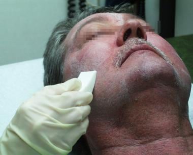
Chemexfoliation
Hand Dermatoses
Skin Diseases
Skin Diseases, Vesiculobullous
Linear IgA Bullous Dermatosis
Sweet Syndrome
Facial Nerve
Facial Paralysis
Dermatitis, Occupational
Facial Muscles
Disseminated superficial actinic porokeratosis like drug eruption: a case report. (1/192)
We report a 54-year-old male patient who developed an unusual form of generalized drug eruption. He had pain and breathlessness on the left chest wall. He had history of taking several drugs at private clinics under a diagnosis of herpes zoster. Two weeks later he had a generalized skin eruption. Examination showed multiple variable sized, mild pruritic, erythematous macules and papules on the face and upper extremities. Skin lesions take the form of a clinically consistent with disseminated superficial actinic porokeratosis (DSAP). Methylprednisolone 16 mg, astemisole 10 mg, oxatomide 60 mg was prescribed. Topical corticosteroid cream was applied. Within two months, his eruption had cleared almost completely. The pathogenetic mechanisms of this case are unclear, but drug and UV light have been considered. (+info)Anti-elongation factor-1alpha autoantibody in adult atopic dermatitis patients. (2/192)
Adult atopic dermatitis (AD) patients develop severe facial lesions, which sometimes distribute in sun-exposed areas similar to the rash of systemic lupus erythematosus. To declare autoimmunity in the pathogenesis of AD, we investigated serum antinuclear antibody (ANA) in 256 adult AD patients and identified its ligands. A high titer of ANA was found in 31.3% of AD patients and 75% of the ANA showed a homogenous pattern. Sixty-five percent of ANA(+) sera reacted to a 52 kDa protein (p52) in HeLa cell immunoblots. By screening the HeLa cell cDNA expression library with anti-p52 sera, a clearly positive clone was isolated. The sequence of this cDNA was identical to human elongation factor (hEF)-1alpha. The eluate of IgG bound to hEF-1alpha-glutathione S-transferase (GST) fusion protein recognized a band at 52 kDa in a HeLa cell immunoblot, and stained Hep-2 cell nuclei and cytoplasma as reported in hEF-1alpha distribution. The anti-p52 AD sera recognized the hEF-1alpha-GST fusion protein. The anti-hEF-1alpha antibody-positive AD patients were characterized by higher facial involvement and lower white blood cell counts compared with antibody-negative patients. The present results suggest the possible involvement of autoimmunity in the pathogenesis of adult AD. (+info)A retarded rate of DNA chain growth in Bloom's syndrome. (3/192)
The cytogenetic observation that homologous chromatid interchange occurs in Bloom's syndrome more often than normal prompted an investigation of DNA replication in that rare genetic disorder. Using DNA fiber autoradiography, an estimation was made of the rate of one component of ongoing DNA replication, DNA chain growth. The rate in Bloom's syndrome dermal fibroblasts in tissue culture was found to be significantly slower than that in normal control cells. (The rate was found to be normal in Fanconi's anemia cells.) The explanation for the retarded chain growth may be either that an enzyme concerned directly with semiconservative DNA replication is defective or that a defective enzyme not itself concerned directly with replication results in disturbed cellular metabolism which in turn affects replication. (+info)Epidemiology of the incidence of oro-facial noma: a study of cases in Dakar, Senegal, 1981-1993. (4/192)
Oro-facial noma is an oral gangrene occurring in early childhood in extremely poor areas. As many as 70-90% of those with noma die, and to date, there is no satisfactory treatment to fight this disease. Within the context of the World Health Organization international program against noma, a 13-year retrospective study based on clinical records was carried out in Dakar, Senegal in an attempt to understand the epidemiology of noma. Between 1981 and 1993, 199 cases of noma were identified, among them; 36.7% were acute cases and 63.3% showed sequelae. Chronic sequelae of noma were seen in patients 2-41 years of age, but the acute phase of noma was found only in young children (77.7% in those 1-4 years of age, maximum age = 9 years, mean age +/- SD age = 3.4 +/- 1.9 years). A total of 73.1% of the cases with acute disease were reported in the Dakar, Diourbel and Kaolack regions during the dry season (57.0% of the cases). The lesions of progressive noma were localized mainly on the upper lip (42.4%) and the cheek (31.1%). A total of 96.9% of the patients with acute diseases were had poor general health with serious associated diseases; only 20.0% had a good vital prognosis. The development of epidemiologic surveillance programs for noma should be a public health priority in Senegal. (+info)Is demodex really non-pathogenic? (5/192)
Although usually considered a non-pathogenic parasite in parasitological textbooks, Demodex folliculorum has been implicated as a causative agent for some dermatological conditions, such as rosacea-like eruptions and some types of blepharitis. Several anecdotal reports have demonstrated unequivocal tissue damage directly related to the presence of the parasite. However, this seems to be exceedingly rare, in contrast with the marked prevalence of this infestation. We have had the opportunity to observe one of such cases. A 38-year-old woman presented with rosacea-like papular lesions in her right cheek. Histopathological examination revealed granulomatous dermal inflammation with a well-preserved mite phagocytized by a multinucleated giant cell. This finding may be taken as an evidence for the pathogenicity of the parasite, inasmuch as it does not explain how such a common parasite is able to produce such a rare disease. (+info)Elastin peptides induce migration and terminal differentiation of cultured keratinocytes via 67 kDa elastin receptor in vitro: 67 kDa elastin receptor is expressed in the keratinocytes eliminating elastic materials in elastosis perforans serpiginosa. (6/192)
To delineate the molecular mechanism of transepidermal elimination of dermal elastic materials in elastosis perforans serpiginosa, the interaction between elastin and cultured keratinocytes was studied in vitro. Synthetic elastin peptide VGVAPG elicited chemotactic responses to the cultured keratinocytes at the dose of 10-9 M. Treatment of keratinocytes with 10-6 or 10-5 M elastin peptides resulted in the suppression of cell growth and the increased expression of involucrin and transglutaminase-1, markers of terminal differentiation. When cultured keratinocytes were treated with the elastin peptides, the expression of 67 kDa elastin receptor was increased. The induction of terminal differentiation by elastin peptides was attenuated by the treatment with the combination of anti-67 kDa elastin receptor antibody. The results indicate that elastin is a potent inducer of migration and terminal differentiation of cultured keratinocytes, which is mediated by the 67 kDa elastin receptor. In the lesional skins of patients with elastosis perforans serpiginosa, the 67 kDa elastin receptor was specifically expressed in the epidermis immediately surrounding the elastic materials that were being eliminated. The elastin receptor may be involved in the interaction between keratinocytes and elastin in elastosis perforans serpiginosa. (+info)Effects of electric field reduction in visual display units on skin symptoms. (7/192)
OBJECTIVES: This study investigated the facial skin complaints of office workers before and after the static electric fields of a visual display unit were reduced. METHODS: On the basis of a screening survey of 4556 office workers in 11 companies, 120 of 227 subjects reporting facial skin complaints were randomly selected to this double blind intervention study. Antistatic measures were used to reduce the static electric fields of the visual display unit in the intervention group but not in the control group, which worked with a visual display unit resembling that of the intervention group. Electric fields, dust concentration, health complaints, and psychological behavior tests were recorded before and after the intervention. RESULTS: The intervention group reported statistically significantly fewer facial skin complaints than the control group. In the intervention group, among those with an office dust concentration of >58 microg/m3, a median reduction of 1.5 skin index points (scale 0-8) was achieved, whereas there was no change in the control group. In the regression model "group category" was still a significant variable after control for background factors. In addition, further linear regression analyses indicated that several static electric field parameters were predictors of the skin complaint reduction. CONCLUSIONS: This field trial indicates that removing static electric fields from visual display units can probably help reduce the facial skin complaints of workers in offices with high dust concentrations. (+info)Periorbital dermatitis as a side effect of topical dorzolamide. (8/192)
AIM: To report periorbital dermatitis as a late side effect of topical dorzolamide hydrochloride (Trusopt), a drug used to reduce intraocular pressure. METHODS: A retrospective study of 14 patients who developed periorbital dermatitis while using topical dorzolamide hydrochloride was undertaken. Six patients underwent patch testing for sensitivity to Trusopt, dorzolamide hydrochloride, and the preservative benzalkonium chloride. RESULTS: The periorbital dermatitis occurred after a mean period of 20.4 weeks of commencing dorzolamide hydrochloride therapy. 13 patients had used preserved topical beta blocker treatment for a mean period of 34.2 months without complication before the introduction of dorzolamide. In eight (57.1%) the dermatitis resolved completely after discontinuing dorzolamide but in six (42.9%) resolution of the dermatitis did not occur until the concomitant preserved beta blocker was stopped and substituted with preservative free drops. Patch testing for sensitivity to Trusopt, dorzolamide hydrochloride, and benzalkonium chloride was negative. CONCLUSION: These findings suggest that dorzolamide can cause severe periorbital dermatitis. Although the dermatitis may resolve when dorzolamide is discontinued, this does not always occur and in some patients all topical medication containing benzalkonium chloride needs to be stopped. (+info)Facial dermatoses refer to various skin conditions that affect the face. These can include a wide range of disorders, such as:
1. Acne vulgaris: A common skin condition characterized by the formation of comedones (blackheads and whiteheads) and inflammatory papules, pustules, and nodules. It primarily affects the face, neck, chest, and back.
2. Rosacea: A chronic skin condition that causes redness, flushing, and visible blood vessels on the face, along with bumps or pimples and sometimes eye irritation.
3. Seborrheic dermatitis: A common inflammatory skin disorder that causes a red, itchy, and flaky rash, often on the scalp, face, and eyebrows. It can also affect other oily areas of the body, like the sides of the nose and behind the ears.
4. Atopic dermatitis (eczema): A chronic inflammatory skin condition that causes red, itchy, and scaly patches on the skin. While it can occur anywhere on the body, it frequently affects the face, especially in infants and young children.
5. Psoriasis: An autoimmune disorder that results in thick, scaly, silvery, or red patches on the skin. It can affect any part of the body, including the face.
6. Contact dermatitis: A skin reaction caused by direct contact with an allergen or irritant, resulting in redness, itching, and inflammation. The face can be affected when allergens or irritants come into contact with the skin through cosmetics, skincare products, or other substances.
7. Lupus erythematosus: An autoimmune disorder that can cause a butterfly-shaped rash on the cheeks and nose, along with other symptoms like joint pain, fatigue, and photosensitivity.
8. Perioral dermatitis: A inflammatory skin condition that causes redness, small bumps, and dryness around the mouth, often mistaken for acne. It can also affect the skin around the nose and eyes.
9. Vitiligo: An autoimmune disorder that results in the loss of pigmentation in patches of skin, which can occur on the face and other parts of the body.
10. Tinea faciei: A fungal infection that affects the facial skin, causing red, scaly, or itchy patches. It is also known as ringworm of the face.
These are just a few examples of skin conditions that can affect the face. If you experience any unusual symptoms or changes in your skin, it's essential to consult a dermatologist for proper diagnosis and treatment.
Chemexfoliation is a medical term that refers to the use of chemical agents to exfoliate or remove the outer layers of the skin. It is also known as chempeel, derma peeling, or chemabrasion. This procedure is commonly used in dermatology and cosmetic surgery to improve the appearance of the skin, reduce fine lines and wrinkles, treat acne, uneven pigmentation, and sun damage.
During a chemexfoliation procedure, a chemical solution is applied to the skin, which causes the outer layers to blister and eventually peel off. The type of chemical agent used depends on the individual's skin type and the desired outcome. Commonly used chemicals include alpha-hydroxy acids (AHAs), beta-hydroxy acids (BHAs), trichloroacetic acid (TCA), and phenol.
After the procedure, the skin may be red, swollen, and sensitive for several days. It is important to avoid sun exposure and use a broad-spectrum sunscreen to protect the new skin. Multiple treatments may be necessary to achieve the desired results. Chemexfoliation should only be performed by a qualified healthcare professional in a controlled medical setting.
Hand dermatoses is a general term used to describe various inflammatory skin conditions that affect the hands. These conditions can cause symptoms such as redness, swelling, itching, blistering, scaling, and cracking of the skin on the hands. Common examples of hand dermatoses include:
1. Irritant contact dermatitis: A reaction that occurs when the skin comes into contact with irritants such as chemicals, soaps, or detergents.
2. Allergic contact dermatitis: A reaction that occurs when the skin comes into contact with allergens, such as nickel, rubber, or poison ivy.
3. Atopic dermatitis (eczema): A chronic skin condition characterized by dry, itchy, and inflamed skin.
4. Psoriasis: A chronic skin condition characterized by red, scaly patches that can occur anywhere on the body, including the hands.
5. Dyshidrotic eczema: A type of eczema that causes small blisters to form on the sides of the fingers, palms, and soles of the feet.
6. Lichen planus: An inflammatory skin condition that can cause purple or white patches to form on the hands and other parts of the body.
7. Scabies: A contagious skin condition caused by mites that burrow into the skin and lay eggs, causing intense itching and a rash.
Treatment for hand dermatoses depends on the specific diagnosis and may include topical creams or ointments, oral medications, phototherapy, or avoidance of triggers.
Skin diseases, also known as dermatological conditions, refer to any medical condition that affects the skin, which is the largest organ of the human body. These diseases can affect the skin's function, appearance, or overall health. They can be caused by various factors, including genetics, infections, allergies, environmental factors, and aging.
Skin diseases can present in many different forms, such as rashes, blisters, sores, discolorations, growths, or changes in texture. Some common examples of skin diseases include acne, eczema, psoriasis, dermatitis, fungal infections, viral infections, bacterial infections, and skin cancer.
The symptoms and severity of skin diseases can vary widely depending on the specific condition and individual factors. Some skin diseases are mild and can be treated with over-the-counter medications or topical creams, while others may require more intensive treatments such as prescription medications, light therapy, or even surgery.
It is important to seek medical attention if you experience any unusual or persistent changes in your skin, as some skin diseases can be serious or indicative of other underlying health conditions. A dermatologist is a medical doctor who specializes in the diagnosis and treatment of skin diseases.
Vesiculobullous skin diseases are a group of disorders characterized by the formation of blisters (vesicles) and bullae (larger blisters) on the skin. These blisters form when there is a separation between the epidermis (outer layer of the skin) and the dermis (layer beneath the epidermis) due to damage in the area where they join, known as the dermo-epidermal junction.
There are several types of vesiculobullous diseases, each with its own specific causes and symptoms. Some of the most common types include:
1. Pemphigus vulgaris: an autoimmune disorder where the immune system mistakenly attacks proteins that help to hold the skin together, causing blisters to form.
2. Bullous pemphigoid: another autoimmune disorder, but in this case, the immune system attacks a different set of proteins, leading to large blisters and inflammation.
3. Dermatitis herpetiformis: a skin condition associated with celiac disease, where gluten ingestion triggers an immune response that leads to the formation of itchy blisters.
4. Pemphigoid gestationis: a rare autoimmune disorder that occurs during pregnancy and causes blisters on the abdomen and other parts of the body.
5. Epidermolysis bullosa: a group of inherited disorders where there is a fragile skin structure, leading to blistering and wound formation after minor trauma or friction.
Treatment for vesiculobullous diseases depends on the specific diagnosis and may include topical or systemic medications, such as corticosteroids, immunosuppressants, or antibiotics, as well as wound care and prevention of infection.
Linear IgA Bullous Dermatosis (LABD) is an autoimmune blistering disorder characterized by the production of autoantibodies against the 97-kDa component of the basement membrane zone, leading to the formation of tense blisters and erosions. It can occur in both children and adults, with different subtypes and clinical presentations.
In LABD, there is a linear deposition of IgA along the basement membrane zone on direct immunofluorescence (DIF) studies, which helps to distinguish it from other autoimmune blistering disorders like bullous pemphigoid or pemphigus vulgaris.
The condition can be idiopathic or associated with medications, infections, or underlying medical conditions such as inflammatory bowel disease or hematologic malignancies. Treatment typically involves systemic corticosteroids and other immunosuppressive agents to control the blister formation and prevent complications.
Sweet syndrome, also known as acute febrile neutrophilic dermatosis, is a skin condition characterized by the rapid onset of painful, red, and swollen skin lesions. The lesions are often accompanied by fever and elevated white blood cell count, particularly an increase in neutrophils.
The medical definition of Sweet syndrome includes the following criteria:
1. Abrupt onset of painful, erythematous (red), and edematous (swollen) papules, plaques, or nodules.
2. Fever greater than 38°C (100.4°F).
3. Leukocytosis with a predominance of neutrophils in the peripheral blood.
4. Histopathological evidence of a dense dermal infiltrate of neutrophils without evidence of vasculitis.
5. Rapid response to systemic corticosteroids.
Sweet syndrome can be associated with various medical conditions, such as infections, malignancies, and inflammatory diseases, or it can occur without an identifiable underlying cause (idiopathic).
The facial nerve, also known as the seventh cranial nerve (CN VII), is a mixed nerve that carries both sensory and motor fibers. Its functions include controlling the muscles involved in facial expressions, taste sensation from the anterior two-thirds of the tongue, and secretomotor function to the lacrimal and salivary glands.
The facial nerve originates from the brainstem and exits the skull through the internal acoustic meatus. It then passes through the facial canal in the temporal bone before branching out to innervate various structures of the face. The main branches of the facial nerve include:
1. Temporal branch: Innervates the frontalis, corrugator supercilii, and orbicularis oculi muscles responsible for eyebrow movements and eyelid closure.
2. Zygomatic branch: Supplies the muscles that elevate the upper lip and wrinkle the nose.
3. Buccal branch: Innervates the muscles of the cheek and lips, allowing for facial expressions such as smiling and puckering.
4. Mandibular branch: Controls the muscles responsible for lower lip movement and depressing the angle of the mouth.
5. Cervical branch: Innervates the platysma muscle in the neck, which helps to depress the lower jaw and wrinkle the skin of the neck.
Damage to the facial nerve can result in various symptoms, such as facial weakness or paralysis, loss of taste sensation, and dry eyes or mouth due to impaired secretion.
Facial paralysis is a loss of facial movement due to damage or dysfunction of the facial nerve (cranial nerve VII). This nerve controls the muscles involved in facial expressions, such as smiling, frowning, and closing the eyes. Damage to one side of the facial nerve can cause weakness or paralysis on that side of the face.
Facial paralysis can result from various conditions, including:
1. Bell's palsy - an idiopathic (unknown cause) inflammation of the facial nerve
2. Trauma - skull fractures, facial injuries, or surgical trauma to the facial nerve
3. Infections - Lyme disease, herpes zoster (shingles), HIV/AIDS, or bacterial infections like meningitis
4. Tumors - benign or malignant growths that compress or invade the facial nerve
5. Stroke - damage to the brainstem where the facial nerve originates
6. Congenital conditions - some people are born with facial paralysis due to genetic factors or birth trauma
Symptoms of facial paralysis may include:
* Inability to move one or more parts of the face, such as the eyebrows, eyelids, mouth, or cheeks
* Drooping of the affected side of the face
* Difficulty closing the eye on the affected side
* Changes in saliva and tear production
* Altered sense of taste
* Pain around the ear or jaw
* Speech difficulties due to weakened facial muscles
Treatment for facial paralysis depends on the underlying cause. In some cases, such as Bell's palsy, spontaneous recovery may occur within a few weeks to months. However, physical therapy, medications, and surgical interventions might be necessary in other situations to improve function and minimize complications.
A facial expression is a result of the contraction or relaxation of muscles in the face that change the physical appearance of an individual's face to convey various emotions, intentions, or physical sensations. Facial expressions can be voluntary or involuntary and are a form of non-verbal communication that plays a crucial role in social interaction and conveying a person's state of mind.
The seven basic facial expressions of emotion, as proposed by Paul Ekman, include happiness, sadness, fear, disgust, surprise, anger, and contempt. These facial expressions are universally recognized across cultures and can be detected through the interpretation of specific muscle movements in the face, known as action units, which are measured and analyzed in fields such as psychology, neurology, and computer vision.
Occupational dermatitis is a specific type of contact dermatitis that results from exposure to certain substances or conditions in the workplace. It can be caused by direct contact with chemicals, irritants, or allergens present in the work environment. This condition typically affects the skin on the hands and forearms but can also involve other areas of the body, depending on the nature of the exposure.
There are two main types of occupational dermatitis:
1. Irritant contact dermatitis (ICD): This type occurs when the skin comes into direct contact with an irritating substance, leading to redness, swelling, itching, and sometimes blistering. Common irritants include solvents, detergents, oils, and other industrial chemicals.
2. Allergic contact dermatitis (ACD): This type is a result of an allergic reaction to a specific substance. The immune system identifies the allergen as harmful and mounts a response, causing skin inflammation. Common allergens include latex, metals (such as nickel), and certain plants (like poison ivy).
Prevention measures for occupational dermatitis include using appropriate personal protective equipment (PPE) like gloves, masks, and aprons, as well as practicing good hygiene, such as washing hands regularly and avoiding touching the face with contaminated hands. If you suspect you have developed occupational dermatitis, consult a healthcare professional for proper diagnosis and treatment.
Facial muscles, also known as facial nerves or cranial nerve VII, are a group of muscles responsible for various expressions and movements of the face. These muscles include:
1. Orbicularis oculi: muscle that closes the eyelid and raises the upper eyelid
2. Corrugator supercilii: muscle that pulls the eyebrows down and inward, forming wrinkles on the forehead
3. Frontalis: muscle that raises the eyebrows and forms horizontal wrinkles on the forehead
4. Procerus: muscle that pulls the medial ends of the eyebrows downward, forming vertical wrinkles between the eyebrows
5. Nasalis: muscle that compresses or dilates the nostrils
6. Depressor septi: muscle that pulls down the tip of the nose
7. Levator labii superioris alaeque nasi: muscle that raises the upper lip and flares the nostrils
8. Levator labii superioris: muscle that raises the upper lip
9. Zygomaticus major: muscle that raises the corner of the mouth, producing a smile
10. Zygomaticus minor: muscle that raises the nasolabial fold and corner of the mouth
11. Risorius: muscle that pulls the angle of the mouth laterally, producing a smile
12. Depressor anguli oris: muscle that pulls down the angle of the mouth
13. Mentalis: muscle that raises the lower lip and forms wrinkles on the chin
14. Buccinator: muscle that retracts the cheek and helps with chewing
15. Platysma: muscle that depresses the corner of the mouth and wrinkles the skin of the neck.
These muscles are innervated by the facial nerve, which arises from the brainstem and exits the skull through the stylomastoid foramen. Damage to the facial nerve can result in facial paralysis or weakness on one or both sides of the face.
Foot dermatoses refer to various skin conditions that affect the feet. These can include inflammatory conditions like eczema and psoriasis, infectious diseases such as athlete's foot (tinea pedis), fungal infections, bacterial infections, viral infections (like plantar warts caused by HPV), and autoimmune blistering disorders. Additionally, contact dermatitis from irritants or allergens can also affect the feet. Proper diagnosis is essential to determine the best course of treatment for each specific condition.
Leg dermatoses is a general term that refers to various skin conditions affecting the legs. This can include a wide range of inflammatory, infectious, or degenerative diseases that cause symptoms such as redness, itching, scaling, blistering, or pigmentation changes on the leg skin. Examples of specific leg dermatoses include stasis dermatitis, venous eczema, contact dermatitis, lichen planus, psoriasis, and cellulitis among others. Accurate diagnosis usually requires a thorough examination and sometimes a biopsy to determine the specific type of dermatosis and appropriate treatment.