Facial Bones
Frontal Bone
Bone and Bones
Fibrous Dysplasia of Bone
Skull Fractures
Facial Nerve
Facial Paralysis
Bone Remodeling
Facial Muscles
Bone Density
Tomography, X-Ray Computed
Facial Nerve Diseases
Bone Marrow
Face
Bone Development
Bone Marrow Cells
Facial Dermatoses
Bone Regeneration
Facial Neoplasms
Facial Pain
Bone Matrix
Bone Marrow Transplantation
Bone Substitutes
Bone Diseases, Metabolic
Temporal Bone
Bone Morphogenetic Proteins
Arrested eruption of the permanent lower second molar. (1/528)
The incidence of retention/impaction of the permanent lower second molar (M2inf) lies between 0.6/1000 and 3/1000. Therefore, the purpose of the present study was to investigate the craniofacial morphology, the frequency of dental anomalies and the inclination of the affected M2inf and the adjacent first molar in patients with arrested eruption of M2inf. The overall goal was to elucidate the aetiology of arrested tooth eruption and to present the characteristics of these patients in order to improve diagnosis and treatment planning. Radiographic material (profile radiographs and orthopantomograms) from 19 patients (nine females and 10 males; 13-19 years of age at the time of referral) were analysed. The ages of the patients when profile radiographs were taken for cephalometric analysis varied from 8 to 16 years. The study shows that this group of patients, compared with a reference group, had an increased sagittal jaw relationship (Class II). Specifically, the mandibular prognathism was less, the mandibular gonial angle smaller, the mandibular alveolar prognathism enlarged and the maxillary incisor inclination less than in the reference group. Furthermore, this group of patients had a more frequent occurrence of morphological tooth anomalies, such as root deflections, invaginations, and taurodontism. However, none of the patients with arrested eruption of M2inf had agenesis of the lower third molar. The study did not reveal an association between the degree of inclination of the M2inf and that of the first molar in the same region. The results of this investigation show that conditions such as the craniofacial morphology and deviations in the dentition are associated with arrested eruption of M2inf. Therefore, it is important to evaluate these conditions in future diagnosis and treatment planning of patients with arrested eruption of M2inf. (+info)The robust australopithecine face: a morphogenetic perspective. (2/528)
The robust australopithecines were a side branch of human evolution. They share a number of unique craniodental features that suggest their monophyletic origin. However, virtually all of these traits appear to reflect a singular pattern of nasomaxillary modeling derived from their unusual dental proportions. Therefore, recent cladistic analyses have not resolved the phylogenetic history of these early hominids. Efforts to increase cladistic resolution by defining traits at greater levels of anatomical detail have instead introduced substantial phyletic error. (+info)Evaluation of CSF leaks: high-resolution CT compared with contrast-enhanced CT and radionuclide cisternography. (3/528)
BACKGROUND AND PURPOSE: Radiologic evaluation of CSF leaks is a diagnostic challenge that often involves multiple imaging studies with the associated expense and patient discomfort. We evaluated the use of screening noncontrast high-resolution CT in identifying the presence and site of CSF rhinorrhea and otorrhea and compared it with contrast-enhanced CT cisternography and radionuclide cisternography. METHODS: We retrospectively reviewed the imaging studies and medical records of all patients who were evaluated for CSF leak during a 7-year period. Forty-two patients with rhinorrhea and/or otorrhea underwent high-resolution CT of the face or temporal bone and then had CT cisternography and radionuclide cisternography via lumbar puncture. The results of the three studies were compared and correlated with the surgical findings in 21 patients. RESULTS: High-resolution CT showed bone defects in 30 of 42 patients (71%) with CSF leak. High-resolution, radionuclide cisternography and CT cisternography did not show bone defects or CSF leak for 12 patients (29%) who had clinical evidence of CSF leak. Among the 30 patients with bone defects, 20 (66%) had positive results of their radionuclide cisternography and/or CT cisternography. For the 21 patients who underwent surgical exploration and repair, intraoperative findings correlated with the defects revealed by high-resolution CT in all cases. High-resolution CT identified significantly more patients with CSF leak than did radionuclide cisternography and CT cisternography, with a moderate degree of agreement. CONCLUSION: Noncontrast high-resolution CT showed a defect in 70% of the patients with CSF leak. No radionuclide cisternography or CT cisternography study produced positive results without previous visualization of a defect on high-resolution CT. CT cisternography and radionuclide cisternography may be reserved for patients in whom initial high-resolution CT does not identify a bone defect or for patients with multiple fractures or postoperative defects. (+info)Comparison of cervicovertebral dimensions in Australian Aborigines and Caucasians. (4/528)
Cervicovertebral dimensions were compared in a group of 30 male and 30 female young adult Australian Aborigines from the Northern Territory, and a control sample consisting of 60 Caucasian dental students from Adelaide, matched for sex and age. Thirty-six variables, 22 cervical and 14 craniofacial, were derived from standardized lateral roentgenograms with the use of a computerized cephalometric system. Vertebral body height and length were significantly greater in Aboriginal males than females for C3 to C7, while dorsal arch height of C1 and C2 displayed the greatest dimensional variability in both sexes. The antero-posterior length of C1, dens height, and body heights of C3 and C4 were significantly shorter in Aborigines than Caucasians for both males and females. Total length of the column from C2 to C6 was approximately 12 per cent shorter in the Aborigines compared with Caucasians. The height of the posterior arch of C1 was significantly correlated with one or both posterior cranial base lengths in Aborigines and Caucasians. Associations were also noted between mandibular lengths and posterior arch heights of the upper two vertebrae. The results confirm and clarify several previous observations on the relative shortness of the cervical spine in Australian Aboriginals. They also indicate some associations between dimensions of the cervical vertebrae and craniofacial lengths, particularly those representing the posterior cranial base and the mandible. (+info)Effect of low-dose testosterone treatment on craniofacial growth in boys with delayed puberty. (5/528)
Craniofacial growth was investigated in boys treated with low-dose testosterone for delayed puberty (> 14 years old; testicular volume < 4 ml; n = 7) and compared with controls (12-14 years; n = 37). Cephalometric radiographs, statural height and pubertal stage were recorded at the start of the study and after 1 year. Craniofacial growth was assessed by nine linear measurements. At the beginning of the study, statural height, mandibular ramus length, upper anterior face height, and total cranial base length were significantly shorter in the delayed puberty boys than in the controls. After 1 year, the growth rate of the statural height, total mandibular length, ramus length, and upper and total anterior face height was significantly higher in the treated boys than in the untreated height-matched controls (n = 7). The craniofacial measurements were similar in the treated boys as compared with the controls. These results show that statural height and craniofacial dimensions are low in boys with delayed puberty. Low doses of testosterone accelerate statural and craniofacial growth, particularly in the delayed components, thus leading towards a normalization of facial dimensions. (+info)Mandibular shape and skeletal divergency. (6/528)
Pre-treatment lateral cephalograms of 41 skeletal Class I girls aged 11 to 15 were divided according to MP-SN angle: lower than 28 degrees (hypodivergent, 10 girls), between 31 and 34 degrees (normodivergent, 18 girls), or larger than 37 degrees (hyperdivergent, 13 girls). The mandibular outlines were traced and digitized, and differences in shape were quantified using the elliptic Fourier series. Size differences were measured from the areas enclosed by the mandibular outlines. Shape differences were assessed by calculating a morphological distance (MD) between the size-independent mean mathematical reconstructions of the mandibular outlines of the three divergency classes. Mandibular shape was different in the three classes: large variations were found in hyperdivergent girls versus normodivergent girls (MD = 4.61), while smaller differences were observed in hypodivergent girls (MD versus normodivergent 2.91). Mean size-independent mandibular shapes were superimposed on an axis passing through the centres of gravity of the condyle and of the chin. Normodivergent and hyperdivergent mandibles differed mostly at gonion, the coronoid process, sigmoid notch, alveolar process, posterior border of the ramus, and along the mandibular plane. A significant size effect was also found, with smaller mandibles in the hyperdivergent girls. (+info)Linear and angular changes in dento-facial dimensions in the third decade. (7/528)
The object of the study was to examine changes in dento-facial dimensions and relationships during the third decade of life, and consisted of a prospective cephalometric study. The data used consisted of 90 degree left lateral cephalometric radiographs of 21 males and 26 females at ages 18 years (T1) and 21 years (T2), and for 15 of the males and 22 of the females at 28 years (T3). Various dimensions representative of dento-facial morphology were measured and the changes in dimensions over time were calculated and tested for significance with the one sample t-test. In general, skeletal and dental relationships remained relatively stable. Face height and jaw length dimensions increased by small amounts. (+info)Differential responses to parathyroid hormone-related protein (PTHrP) deficiency in the various craniofacial cartilages. (8/528)
PTHrP null mutant mice exhibit skeletal abnormalities both in the craniofacial region and limbs. In the growth plate cartilage of the null mutant, a diminished number of proliferating chondrocytes and accelerated chondrocytic differentiation are observed. In order to examine the effect of PTHrP deficiency on the craniofacial morphology and highlight the differential feature of the composing cartilages, we examined the various cartilages in the craniofacial region of neonatal PTHrP deficient mice. The major part of the cartilaginous anterior cranial base appeared to be normal in the homozygous PTHrP deficient mice. However, acceleration of chondrocytic differentiation and endochondral bone formation was observed in the posterior part of the anterior cranial base and in the cranial base synchondroses. Ectopic bone formation was observed in the soft tissue-running mid-portion of the Meckel's cartilage, where the cartilage degenerates and converts to ligament in the course of normal development. The zonal structure of the mandibular condylar cartilage was scarcely affected, but the whole condyle was reduced in size. These results suggest the effect of PTHrP deficiency varies widely between the craniofacial cartilages, according to the differential features of each cartilage. (+info)The facial bones, also known as the facial skeleton, are a series of bones that make up the framework of the face. They include:
1. Frontal bone: This bone forms the forehead and the upper part of the eye sockets.
2. Nasal bones: These two thin bones form the bridge of the nose.
3. Maxilla bones: These are the largest bones in the facial skeleton, forming the upper jaw, the bottom of the eye sockets, and the sides of the nose. They also contain the upper teeth.
4. Zygomatic bones (cheekbones): These bones form the cheekbones and the outer part of the eye sockets.
5. Palatine bones: These bones form the back part of the roof of the mouth, the side walls of the nasal cavity, and contribute to the formation of the eye socket.
6. Inferior nasal conchae: These are thin, curved bones that form the lateral walls of the nasal cavity and help to filter and humidify air as it passes through the nose.
7. Lacrimal bones: These are the smallest bones in the skull, located at the inner corner of the eye socket, and help to form the tear duct.
8. Mandible (lower jaw): This is the only bone in the facial skeleton that can move. It holds the lower teeth and forms the chin.
These bones work together to protect vital structures such as the eyes, brain, and nasal passages, while also providing attachment points for muscles that control chewing, expression, and other facial movements.
Facial injuries refer to any damage or trauma caused to the face, which may include the bones of the skull that form the face, teeth, salivary glands, muscles, nerves, and skin. Facial injuries can range from minor cuts and bruises to severe fractures and disfigurement. They can be caused by a variety of factors such as accidents, falls, sports-related injuries, physical assaults, or animal attacks.
Facial injuries can affect one or more areas of the face, including the forehead, eyes, nose, cheeks, ears, mouth, and jaw. Common types of facial injuries include lacerations (cuts), contusions (bruises), abrasions (scrapes), fractures (broken bones), and burns.
Facial injuries can have significant psychological and emotional impacts on individuals, in addition to physical effects. Treatment for facial injuries may involve simple first aid, suturing of wounds, splinting or wiring of broken bones, reconstructive surgery, or other medical interventions. It is essential to seek prompt medical attention for any facial injury to ensure proper healing and minimize the risk of complications.
The frontal bone is the bone that forms the forehead and the upper part of the eye sockets (orbits) in the skull. It is a single, flat bone that has a prominent ridge in the middle called the superior sagittal sinus, which contains venous blood. The frontal bone articulates with several other bones, including the parietal bones at the sides and back, the nasal bones in the center of the face, and the zygomatic (cheek) bones at the lower sides of the orbits.
A mandibular fracture is a break or crack in the lower jaw (mandible) bone. It can occur at any point along the mandible, but common sites include the condyle (the rounded end near the ear), the angle (the curved part of the jaw), and the symphysis (the area where the two halves of the jaw meet in the front). Mandibular fractures are typically caused by trauma, such as a direct blow to the face or a fall. Symptoms may include pain, swelling, bruising, difficulty chewing or speaking, and malocclusion (misalignment) of the teeth. Treatment usually involves immobilization with wires or screws to allow the bone to heal properly.
"Bone" is the hard, dense connective tissue that makes up the skeleton of vertebrate animals. It provides support and protection for the body's internal organs, and serves as a attachment site for muscles, tendons, and ligaments. Bone is composed of cells called osteoblasts and osteoclasts, which are responsible for bone formation and resorption, respectively, and an extracellular matrix made up of collagen fibers and mineral crystals.
Bones can be classified into two main types: compact bone and spongy bone. Compact bone is dense and hard, and makes up the outer layer of all bones and the shafts of long bones. Spongy bone is less dense and contains large spaces, and makes up the ends of long bones and the interior of flat and irregular bones.
The human body has 206 bones in total. They can be further classified into five categories based on their shape: long bones, short bones, flat bones, irregular bones, and sesamoid bones.
Fibrous Dysplasia of Bone is a rare, benign bone disorder that is characterized by the replacement of normal bone tissue with fibrous (scar-like) and immature bone tissue. This results in weakened bones that are prone to fractures, deformities, and pain. The condition can affect any bone in the body but most commonly involves the long bones of the legs, arms, and skull. It can occur as an isolated finding or as part of a genetic disorder called McCune-Albright syndrome. The exact cause of fibrous dysplasia is not fully understood, but it is believed to result from a genetic mutation that occurs during early bone development. There is no cure for fibrous dysplasia, and treatment typically focuses on managing symptoms and preventing complications.
A skull fracture is a break in one or more of the bones that form the skull. It can occur from a direct blow to the head, penetrating injuries like gunshot wounds, or from strong rotational forces during an accident. There are several types of skull fractures, including:
1. Linear Skull Fracture: This is the most common type, where there's a simple break in the bone without any splintering, depression, or displacement. It often doesn't require treatment unless it's near a sensitive area like an eye or ear.
2. Depressed Skull Fracture: In this type, a piece of the skull is pushed inward toward the brain. Surgery may be needed to relieve pressure on the brain and repair the fracture.
3. Diastatic Skull Fracture: This occurs along the suture lines (the fibrous joints between the skull bones) that haven't fused yet, often seen in infants and young children.
4. Basilar Skull Fracture: This involves fractures at the base of the skull. It can be serious due to potential injury to the cranial nerves and blood vessels located in this area.
5. Comminuted Skull Fracture: In this severe type, the bone is shattered into many pieces. These fractures usually require extensive surgical repair.
Symptoms of a skull fracture can include pain, swelling, bruising, bleeding (if there's an open wound), and in some cases, clear fluid draining from the ears or nose (cerebrospinal fluid leak). Severe fractures may cause brain injury, leading to symptoms like confusion, loss of consciousness, seizures, or neurological deficits. Immediate medical attention is necessary for any suspected skull fracture.
The skull is the bony structure that encloses and protects the brain, the eyes, and the ears. It is composed of two main parts: the cranium, which contains the brain, and the facial bones. The cranium is made up of several fused flat bones, while the facial bones include the upper jaw (maxilla), lower jaw (mandible), cheekbones, nose bones, and eye sockets (orbits).
The skull also provides attachment points for various muscles that control chewing, moving the head, and facial expressions. Additionally, it contains openings for blood vessels, nerves, and the spinal cord to pass through. The skull's primary function is to protect the delicate and vital structures within it from injury and trauma.
The facial nerve, also known as the seventh cranial nerve (CN VII), is a mixed nerve that carries both sensory and motor fibers. Its functions include controlling the muscles involved in facial expressions, taste sensation from the anterior two-thirds of the tongue, and secretomotor function to the lacrimal and salivary glands.
The facial nerve originates from the brainstem and exits the skull through the internal acoustic meatus. It then passes through the facial canal in the temporal bone before branching out to innervate various structures of the face. The main branches of the facial nerve include:
1. Temporal branch: Innervates the frontalis, corrugator supercilii, and orbicularis oculi muscles responsible for eyebrow movements and eyelid closure.
2. Zygomatic branch: Supplies the muscles that elevate the upper lip and wrinkle the nose.
3. Buccal branch: Innervates the muscles of the cheek and lips, allowing for facial expressions such as smiling and puckering.
4. Mandibular branch: Controls the muscles responsible for lower lip movement and depressing the angle of the mouth.
5. Cervical branch: Innervates the platysma muscle in the neck, which helps to depress the lower jaw and wrinkle the skin of the neck.
Damage to the facial nerve can result in various symptoms, such as facial weakness or paralysis, loss of taste sensation, and dry eyes or mouth due to impaired secretion.
Facial paralysis is a loss of facial movement due to damage or dysfunction of the facial nerve (cranial nerve VII). This nerve controls the muscles involved in facial expressions, such as smiling, frowning, and closing the eyes. Damage to one side of the facial nerve can cause weakness or paralysis on that side of the face.
Facial paralysis can result from various conditions, including:
1. Bell's palsy - an idiopathic (unknown cause) inflammation of the facial nerve
2. Trauma - skull fractures, facial injuries, or surgical trauma to the facial nerve
3. Infections - Lyme disease, herpes zoster (shingles), HIV/AIDS, or bacterial infections like meningitis
4. Tumors - benign or malignant growths that compress or invade the facial nerve
5. Stroke - damage to the brainstem where the facial nerve originates
6. Congenital conditions - some people are born with facial paralysis due to genetic factors or birth trauma
Symptoms of facial paralysis may include:
* Inability to move one or more parts of the face, such as the eyebrows, eyelids, mouth, or cheeks
* Drooping of the affected side of the face
* Difficulty closing the eye on the affected side
* Changes in saliva and tear production
* Altered sense of taste
* Pain around the ear or jaw
* Speech difficulties due to weakened facial muscles
Treatment for facial paralysis depends on the underlying cause. In some cases, such as Bell's palsy, spontaneous recovery may occur within a few weeks to months. However, physical therapy, medications, and surgical interventions might be necessary in other situations to improve function and minimize complications.
A facial expression is a result of the contraction or relaxation of muscles in the face that change the physical appearance of an individual's face to convey various emotions, intentions, or physical sensations. Facial expressions can be voluntary or involuntary and are a form of non-verbal communication that plays a crucial role in social interaction and conveying a person's state of mind.
The seven basic facial expressions of emotion, as proposed by Paul Ekman, include happiness, sadness, fear, disgust, surprise, anger, and contempt. These facial expressions are universally recognized across cultures and can be detected through the interpretation of specific muscle movements in the face, known as action units, which are measured and analyzed in fields such as psychology, neurology, and computer vision.
Bone remodeling is the normal and continuous process by which bone tissue is removed from the skeleton (a process called resorption) and new bone tissue is formed (a process called formation). This ongoing cycle allows bones to repair microdamage, adjust their size and shape in response to mechanical stress, and maintain mineral homeostasis. The cells responsible for bone resorption are osteoclasts, while the cells responsible for bone formation are osteoblasts. These two cell types work together to maintain the structural integrity and health of bones throughout an individual's life.
During bone remodeling, the process can be divided into several stages:
1. Activation: The initiation of bone remodeling is triggered by various factors such as microdamage, hormonal changes, or mechanical stress. This leads to the recruitment and activation of osteoclast precursor cells.
2. Resorption: Osteoclasts attach to the bone surface and create a sealed compartment called a resorption lacuna. They then secrete acid and enzymes that dissolve and digest the mineralized matrix, creating pits or cavities on the bone surface. This process helps remove old or damaged bone tissue and releases calcium and phosphate ions into the bloodstream.
3. Reversal: After resorption is complete, the osteoclasts undergo apoptosis (programmed cell death), and mononuclear cells called reversal cells appear on the resorbed surface. These cells prepare the bone surface for the next stage by cleaning up debris and releasing signals that attract osteoblast precursors.
4. Formation: Osteoblasts, derived from mesenchymal stem cells, migrate to the resorbed surface and begin producing a new organic matrix called osteoid. As the osteoid mineralizes, it forms a hard, calcified structure that gradually replaces the resorbed bone tissue. The osteoblasts may become embedded within this newly formed bone as they differentiate into osteocytes, which are mature bone cells responsible for maintaining bone homeostasis and responding to mechanical stress.
5. Mineralization: Over time, the newly formed bone continues to mineralize, becoming stronger and more dense. This process helps maintain the structural integrity of the skeleton and ensures adequate calcium storage.
Throughout this continuous cycle of bone remodeling, hormones, growth factors, and mechanical stress play crucial roles in regulating the balance between resorption and formation. Disruptions to this delicate equilibrium can lead to various bone diseases, such as osteoporosis, where excessive resorption results in weakened bones and increased fracture risk.
Facial muscles, also known as facial nerves or cranial nerve VII, are a group of muscles responsible for various expressions and movements of the face. These muscles include:
1. Orbicularis oculi: muscle that closes the eyelid and raises the upper eyelid
2. Corrugator supercilii: muscle that pulls the eyebrows down and inward, forming wrinkles on the forehead
3. Frontalis: muscle that raises the eyebrows and forms horizontal wrinkles on the forehead
4. Procerus: muscle that pulls the medial ends of the eyebrows downward, forming vertical wrinkles between the eyebrows
5. Nasalis: muscle that compresses or dilates the nostrils
6. Depressor septi: muscle that pulls down the tip of the nose
7. Levator labii superioris alaeque nasi: muscle that raises the upper lip and flares the nostrils
8. Levator labii superioris: muscle that raises the upper lip
9. Zygomaticus major: muscle that raises the corner of the mouth, producing a smile
10. Zygomaticus minor: muscle that raises the nasolabial fold and corner of the mouth
11. Risorius: muscle that pulls the angle of the mouth laterally, producing a smile
12. Depressor anguli oris: muscle that pulls down the angle of the mouth
13. Mentalis: muscle that raises the lower lip and forms wrinkles on the chin
14. Buccinator: muscle that retracts the cheek and helps with chewing
15. Platysma: muscle that depresses the corner of the mouth and wrinkles the skin of the neck.
These muscles are innervated by the facial nerve, which arises from the brainstem and exits the skull through the stylomastoid foramen. Damage to the facial nerve can result in facial paralysis or weakness on one or both sides of the face.
Bone density refers to the amount of bone mineral content (usually measured in grams) in a given volume of bone (usually measured in cubic centimeters). It is often used as an indicator of bone strength and fracture risk. Bone density is typically measured using dual-energy X-ray absorptiometry (DXA) scans, which provide a T-score that compares the patient's bone density to that of a young adult reference population. A T-score of -1 or above is considered normal, while a T-score between -1 and -2.5 indicates osteopenia (low bone mass), and a T-score below -2.5 indicates osteoporosis (porous bones). Regular exercise, adequate calcium and vitamin D intake, and medication (if necessary) can help maintain or improve bone density and prevent fractures.
X-ray computed tomography (CT or CAT scan) is a medical imaging method that uses computer-processed combinations of many X-ray images taken from different angles to produce cross-sectional (tomographic) images (virtual "slices") of the body. These cross-sectional images can then be used to display detailed internal views of organs, bones, and soft tissues in the body.
The term "computed tomography" is used instead of "CT scan" or "CAT scan" because the machines take a series of X-ray measurements from different angles around the body and then use a computer to process these data to create detailed images of internal structures within the body.
CT scanning is a noninvasive, painless medical test that helps physicians diagnose and treat medical conditions. CT imaging provides detailed information about many types of tissue including lung, bone, soft tissue and blood vessels. CT examinations can be performed on every part of the body for a variety of reasons including diagnosis, surgical planning, and monitoring of therapeutic responses.
In computed tomography (CT), an X-ray source and detector rotate around the patient, measuring the X-ray attenuation at many different angles. A computer uses this data to construct a cross-sectional image by the process of reconstruction. This technique is called "tomography". The term "computed" refers to the use of a computer to reconstruct the images.
CT has become an important tool in medical imaging and diagnosis, allowing radiologists and other physicians to view detailed internal images of the body. It can help identify many different medical conditions including cancer, heart disease, lung nodules, liver tumors, and internal injuries from trauma. CT is also commonly used for guiding biopsies and other minimally invasive procedures.
In summary, X-ray computed tomography (CT or CAT scan) is a medical imaging technique that uses computer-processed combinations of many X-ray images taken from different angles to produce cross-sectional images of the body. It provides detailed internal views of organs, bones, and soft tissues in the body, allowing physicians to diagnose and treat medical conditions.
Facial nerve diseases refer to a group of medical conditions that affect the function of the facial nerve, also known as the seventh cranial nerve. This nerve is responsible for controlling the muscles of facial expression, and it also carries sensory information from the taste buds in the front two-thirds of the tongue, and regulates saliva flow and tear production.
Facial nerve diseases can cause a variety of symptoms, depending on the specific location and extent of the nerve damage. Common symptoms include:
* Facial weakness or paralysis on one or both sides of the face
* Drooping of the eyelid and corner of the mouth
* Difficulty closing the eye or keeping it closed
* Changes in taste sensation or dryness of the mouth and eyes
* Abnormal sensitivity to sound (hyperacusis)
* Twitching or spasms of the facial muscles
Facial nerve diseases can be caused by a variety of factors, including:
* Infections such as Bell's palsy, Ramsay Hunt syndrome, and Lyme disease
* Trauma or injury to the face or skull
* Tumors that compress or invade the facial nerve
* Neurological conditions such as multiple sclerosis or Guillain-Barre syndrome
* Genetic disorders such as Moebius syndrome or hemifacial microsomia
Treatment for facial nerve diseases depends on the underlying cause and severity of the symptoms. In some cases, medication, physical therapy, or surgery may be necessary to restore function and relieve symptoms.
Bone resorption is the process by which bone tissue is broken down and absorbed into the body. It is a normal part of bone remodeling, in which old or damaged bone tissue is removed and new tissue is formed. However, excessive bone resorption can lead to conditions such as osteoporosis, in which bones become weak and fragile due to a loss of density. This process is carried out by cells called osteoclasts, which break down the bone tissue and release minerals such as calcium into the bloodstream.
Bone marrow is the spongy tissue found inside certain bones in the body, such as the hips, thighs, and vertebrae. It is responsible for producing blood-forming cells, including red blood cells, white blood cells, and platelets. There are two types of bone marrow: red marrow, which is involved in blood cell production, and yellow marrow, which contains fatty tissue.
Red bone marrow contains hematopoietic stem cells, which can differentiate into various types of blood cells. These stem cells continuously divide and mature to produce new blood cells that are released into the circulation. Red blood cells carry oxygen throughout the body, white blood cells help fight infections, and platelets play a crucial role in blood clotting.
Bone marrow also serves as a site for immune cell development and maturation. It contains various types of immune cells, such as lymphocytes, macrophages, and dendritic cells, which help protect the body against infections and diseases.
Abnormalities in bone marrow function can lead to several medical conditions, including anemia, leukopenia, thrombocytopenia, and various types of cancer, such as leukemia and multiple myeloma. Bone marrow aspiration and biopsy are common diagnostic procedures used to evaluate bone marrow health and function.
In medical terms, the face refers to the front part of the head that is distinguished by the presence of the eyes, nose, and mouth. It includes the bones of the skull (frontal bone, maxilla, zygoma, nasal bones, lacrimal bones, palatine bones, inferior nasal conchae, and mandible), muscles, nerves, blood vessels, skin, and other soft tissues. The face plays a crucial role in various functions such as breathing, eating, drinking, speaking, seeing, smelling, and expressing emotions. It also serves as an important identifier for individuals, allowing them to be recognized by others.
Bone neoplasms are abnormal growths or tumors that develop in the bone. They can be benign (non-cancerous) or malignant (cancerous). Benign bone neoplasms do not spread to other parts of the body and are rarely a threat to life, although they may cause problems if they grow large enough to press on surrounding tissues or cause fractures. Malignant bone neoplasms, on the other hand, can invade and destroy nearby tissue and may spread (metastasize) to other parts of the body.
There are many different types of bone neoplasms, including:
1. Osteochondroma - a benign tumor that develops from cartilage and bone
2. Enchondroma - a benign tumor that forms in the cartilage that lines the inside of the bones
3. Chondrosarcoma - a malignant tumor that develops from cartilage
4. Osteosarcoma - a malignant tumor that develops from bone cells
5. Ewing sarcoma - a malignant tumor that develops in the bones or soft tissues around the bones
6. Giant cell tumor of bone - a benign or occasionally malignant tumor that develops from bone tissue
7. Fibrosarcoma - a malignant tumor that develops from fibrous tissue in the bone
The symptoms of bone neoplasms vary depending on the type, size, and location of the tumor. They may include pain, swelling, stiffness, fractures, or limited mobility. Treatment options depend on the type and stage of the tumor but may include surgery, radiation therapy, chemotherapy, or a combination of these treatments.
Bone development, also known as ossification, is the process by which bone tissue is formed and grows. This complex process involves several different types of cells, including osteoblasts, which produce new bone matrix, and osteoclasts, which break down and resorb existing bone tissue.
There are two main types of bone development: intramembranous and endochondral ossification. Intramembranous ossification occurs when bone tissue forms directly from connective tissue, while endochondral ossification involves the formation of a cartilage model that is later replaced by bone.
During fetal development, most bones develop through endochondral ossification, starting as a cartilage template that is gradually replaced by bone tissue. However, some bones, such as those in the skull and clavicles, develop through intramembranous ossification.
Bone development continues after birth, with new bone tissue being laid down and existing tissue being remodeled throughout life. This ongoing process helps to maintain the strength and integrity of the skeleton, allowing it to adapt to changing mechanical forces and repair any damage that may occur.
Bone marrow cells are the types of cells found within the bone marrow, which is the spongy tissue inside certain bones in the body. The main function of bone marrow is to produce blood cells. There are two types of bone marrow: red and yellow. Red bone marrow is where most blood cell production takes place, while yellow bone marrow serves as a fat storage site.
The three main types of bone marrow cells are:
1. Hematopoietic stem cells (HSCs): These are immature cells that can differentiate into any type of blood cell, including red blood cells, white blood cells, and platelets. They have the ability to self-renew, meaning they can divide and create more hematopoietic stem cells.
2. Red blood cell progenitors: These are immature cells that will develop into mature red blood cells, also known as erythrocytes. Red blood cells carry oxygen from the lungs to the body's tissues and carbon dioxide back to the lungs.
3. Myeloid and lymphoid white blood cell progenitors: These are immature cells that will develop into various types of white blood cells, which play a crucial role in the body's immune system by fighting infections and diseases. Myeloid progenitors give rise to granulocytes (neutrophils, eosinophils, and basophils), monocytes, and megakaryocytes (which eventually become platelets). Lymphoid progenitors differentiate into B cells, T cells, and natural killer (NK) cells.
Bone marrow cells are essential for maintaining a healthy blood cell count and immune system function. Abnormalities in bone marrow cells can lead to various medical conditions, such as anemia, leukopenia, leukocytosis, thrombocytopenia, or thrombocytosis, depending on the specific type of blood cell affected. Additionally, bone marrow cells are often used in transplantation procedures to treat patients with certain types of cancer, such as leukemia and lymphoma, or other hematologic disorders.
Bone diseases is a broad term that refers to various medical conditions that affect the bones. These conditions can be categorized into several groups, including:
1. Developmental and congenital bone diseases: These are conditions that affect bone growth and development before or at birth. Examples include osteogenesis imperfecta (brittle bone disease), achondroplasia (dwarfism), and cleidocranial dysostosis.
2. Metabolic bone diseases: These are conditions that affect the body's ability to maintain healthy bones. They are often caused by hormonal imbalances, vitamin deficiencies, or problems with mineral metabolism. Examples include osteoporosis, osteomalacia, and Paget's disease of bone.
3. Inflammatory bone diseases: These are conditions that cause inflammation in the bones. They can be caused by infections, autoimmune disorders, or other medical conditions. Examples include osteomyelitis, rheumatoid arthritis, and ankylosing spondylitis.
4. Degenerative bone diseases: These are conditions that cause the bones to break down over time. They can be caused by aging, injury, or disease. Examples include osteoarthritis, avascular necrosis, and diffuse idiopathic skeletal hyperostosis (DISH).
5. Tumors and cancers of the bone: These are conditions that involve abnormal growths in the bones. They can be benign or malignant. Examples include osteosarcoma, chondrosarcoma, and Ewing sarcoma.
6. Fractures and injuries: While not strictly a "disease," fractures and injuries are common conditions that affect the bones. They can result from trauma, overuse, or weakened bones. Examples include stress fractures, compound fractures, and dislocations.
Overall, bone diseases can cause a wide range of symptoms, including pain, stiffness, deformity, and decreased mobility. Treatment for these conditions varies depending on the specific diagnosis but may include medication, surgery, physical therapy, or lifestyle changes.
Facial asymmetry refers to a condition in which the facial features are not identical or proportionate on both sides of a vertical line drawn down the middle of the face. This can include differences in the size, shape, or positioning of facial features such as the eyes, ears, nose, cheeks, and jaw. Facial asymmetry can be mild and barely noticeable, or it can be more severe and affect a person's appearance and/or functionality of the mouth and jaw.
Facial asymmetry can be present at birth (congenital) or can develop later in life due to various factors such as injury, surgery, growth disorders, nerve damage, or tumors. In some cases, facial asymmetry may not cause any medical problems and may only be of cosmetic concern. However, in other cases, it may indicate an underlying medical condition that requires treatment.
Depending on the severity and cause of the facial asymmetry, treatment options may include cosmetic procedures such as fillers or surgery, orthodontic treatment, physical therapy, or medication to address any underlying conditions.
Facial dermatoses refer to various skin conditions that affect the face. These can include a wide range of disorders, such as:
1. Acne vulgaris: A common skin condition characterized by the formation of comedones (blackheads and whiteheads) and inflammatory papules, pustules, and nodules. It primarily affects the face, neck, chest, and back.
2. Rosacea: A chronic skin condition that causes redness, flushing, and visible blood vessels on the face, along with bumps or pimples and sometimes eye irritation.
3. Seborrheic dermatitis: A common inflammatory skin disorder that causes a red, itchy, and flaky rash, often on the scalp, face, and eyebrows. It can also affect other oily areas of the body, like the sides of the nose and behind the ears.
4. Atopic dermatitis (eczema): A chronic inflammatory skin condition that causes red, itchy, and scaly patches on the skin. While it can occur anywhere on the body, it frequently affects the face, especially in infants and young children.
5. Psoriasis: An autoimmune disorder that results in thick, scaly, silvery, or red patches on the skin. It can affect any part of the body, including the face.
6. Contact dermatitis: A skin reaction caused by direct contact with an allergen or irritant, resulting in redness, itching, and inflammation. The face can be affected when allergens or irritants come into contact with the skin through cosmetics, skincare products, or other substances.
7. Lupus erythematosus: An autoimmune disorder that can cause a butterfly-shaped rash on the cheeks and nose, along with other symptoms like joint pain, fatigue, and photosensitivity.
8. Perioral dermatitis: A inflammatory skin condition that causes redness, small bumps, and dryness around the mouth, often mistaken for acne. It can also affect the skin around the nose and eyes.
9. Vitiligo: An autoimmune disorder that results in the loss of pigmentation in patches of skin, which can occur on the face and other parts of the body.
10. Tinea faciei: A fungal infection that affects the facial skin, causing red, scaly, or itchy patches. It is also known as ringworm of the face.
These are just a few examples of skin conditions that can affect the face. If you experience any unusual symptoms or changes in your skin, it's essential to consult a dermatologist for proper diagnosis and treatment.
Bone regeneration is the biological process of new bone formation that occurs after an injury or removal of a portion of bone. This complex process involves several stages, including inflammation, migration and proliferation of cells, matrix deposition, and mineralization, leading to the restoration of the bone's structure and function.
The main cells involved in bone regeneration are osteoblasts, which produce new bone matrix, and osteoclasts, which resorb damaged or old bone tissue. The process is tightly regulated by various growth factors, hormones, and signaling molecules that promote the recruitment, differentiation, and activity of these cells.
Bone regeneration can occur naturally in response to injury or surgical intervention, such as fracture repair or dental implant placement. However, in some cases, bone regeneration may be impaired due to factors such as age, disease, or trauma, leading to delayed healing or non-union of the bone. In these situations, various strategies and techniques, including the use of bone grafts, scaffolds, and growth factors, can be employed to enhance and support the bone regeneration process.
Facial neoplasms refer to abnormal growths or tumors that develop in the tissues of the face. These growths can be benign (non-cancerous) or malignant (cancerous). Facial neoplasms can occur in any of the facial structures, including the skin, muscles, bones, nerves, and glands.
Benign facial neoplasms are typically slow-growing and do not spread to other parts of the body. Examples include papillomas, hemangiomas, and neurofibromas. While these tumors are usually harmless, they can cause cosmetic concerns or interfere with normal facial function.
Malignant facial neoplasms, on the other hand, can be aggressive and invasive. They can spread to other parts of the face, as well as to distant sites in the body. Common types of malignant facial neoplasms include basal cell carcinoma, squamous cell carcinoma, and melanoma.
Treatment for facial neoplasms depends on several factors, including the type, size, location, and stage of the tumor. Treatment options may include surgery, radiation therapy, chemotherapy, or a combination of these approaches. It is important to seek medical attention promptly if you notice any unusual growths or changes in the skin or tissues of your face.
Facial pain is a condition characterized by discomfort or pain felt in any part of the face. It can result from various causes, including nerve damage or irritation, injuries, infections, dental problems, migraines, or sinus congestion. The pain can range from mild to severe and may be sharp, dull, constant, or intermittent. In some cases, facial pain can also be associated with other symptoms such as headaches, redness, swelling, or changes in sensation. Accurate diagnosis and treatment of the underlying cause are essential for effective management of facial pain.
Bone matrix refers to the non-cellular component of bone that provides structural support and functions as a reservoir for minerals, such as calcium and phosphate. It is made up of organic and inorganic components. The organic component consists mainly of type I collagen fibers, which provide flexibility and tensile strength to the bone. The inorganic component is primarily composed of hydroxyapatite crystals, which give bone its hardness and compressive strength. Bone matrix also contains other proteins, growth factors, and signaling molecules that regulate bone formation, remodeling, and repair.
Bone transplantation, also known as bone grafting, is a surgical procedure in which bone or bone-like material is transferred from one part of the body to another or from one person to another. The graft may be composed of cortical (hard outer portion) bone, cancellous (spongy inner portion) bone, or a combination of both. It can be taken from different sites in the same individual (autograft), from another individual of the same species (allograft), or from an animal source (xenograft). The purpose of bone transplantation is to replace missing bone, provide structural support, and stimulate new bone growth. This procedure is commonly used in orthopedic, dental, and maxillofacial surgeries to repair bone defects caused by trauma, tumors, or congenital conditions.
Bone marrow transplantation (BMT) is a medical procedure in which damaged or destroyed bone marrow is replaced with healthy bone marrow from a donor. Bone marrow is the spongy tissue inside bones that produces blood cells. The main types of BMT are autologous, allogeneic, and umbilical cord blood transplantation.
In autologous BMT, the patient's own bone marrow is used for the transplant. This type of BMT is often used in patients with lymphoma or multiple myeloma who have undergone high-dose chemotherapy or radiation therapy to destroy their cancerous bone marrow.
In allogeneic BMT, bone marrow from a genetically matched donor is used for the transplant. This type of BMT is often used in patients with leukemia, lymphoma, or other blood disorders who have failed other treatments.
Umbilical cord blood transplantation involves using stem cells from umbilical cord blood as a source of healthy bone marrow. This type of BMT is often used in children and adults who do not have a matched donor for allogeneic BMT.
The process of BMT typically involves several steps, including harvesting the bone marrow or stem cells from the donor, conditioning the patient's body to receive the new bone marrow or stem cells, transplanting the new bone marrow or stem cells into the patient's body, and monitoring the patient for signs of engraftment and complications.
BMT is a complex and potentially risky procedure that requires careful planning, preparation, and follow-up care. However, it can be a life-saving treatment for many patients with blood disorders or cancer.
Bone substitutes are materials that are used to replace missing or damaged bone in the body. They can be made from a variety of materials, including natural bone from other parts of the body or from animals, synthetic materials, or a combination of both. The goal of using bone substitutes is to provide structural support and promote the growth of new bone tissue.
Bone substitutes are often used in dental, orthopedic, and craniofacial surgery to help repair defects caused by trauma, tumors, or congenital abnormalities. They can also be used to augment bone volume in procedures such as spinal fusion or joint replacement.
There are several types of bone substitutes available, including:
1. Autografts: Bone taken from another part of the patient's body, such as the hip or pelvis.
2. Allografts: Bone taken from a deceased donor and processed to remove any cells and infectious materials.
3. Xenografts: Bone from an animal source, typically bovine or porcine, that has been processed to remove any cells and infectious materials.
4. Synthetic bone substitutes: Materials such as calcium phosphate ceramics, bioactive glass, and polymer-based materials that are designed to mimic the properties of natural bone.
The choice of bone substitute material depends on several factors, including the size and location of the defect, the patient's medical history, and the surgeon's preference. It is important to note that while bone substitutes can provide structural support and promote new bone growth, they may not have the same strength or durability as natural bone. Therefore, they may not be suitable for all applications, particularly those that require high load-bearing capacity.
A bone fracture is a medical condition in which there is a partial or complete break in the continuity of a bone due to external or internal forces. Fractures can occur in any bone in the body and can vary in severity from a small crack to a shattered bone. The symptoms of a bone fracture typically include pain, swelling, bruising, deformity, and difficulty moving the affected limb. Treatment for a bone fracture may involve immobilization with a cast or splint, surgery to realign and stabilize the bone, or medication to manage pain and prevent infection. The specific treatment approach will depend on the location, type, and severity of the fracture.
Metabolic bone diseases are a group of conditions that affect the bones and are caused by disorders in the body's metabolism. These disorders can result in changes to the bone structure, density, and strength, leading to an increased risk of fractures and other complications. Some common examples of metabolic bone diseases include:
1. Osteoporosis: a condition characterized by weak and brittle bones that are more likely to break, often as a result of age-related bone loss or hormonal changes.
2. Paget's disease of bone: a chronic disorder that causes abnormal bone growth and deformities, leading to fragile and enlarged bones.
3. Osteomalacia: a condition caused by a lack of vitamin D or problems with the body's ability to absorb it, resulting in weak and soft bones.
4. Hyperparathyroidism: a hormonal disorder that causes too much parathyroid hormone to be produced, leading to bone loss and other complications.
5. Hypoparathyroidism: a hormonal disorder that results in low levels of parathyroid hormone, causing weak and brittle bones.
6. Renal osteodystrophy: a group of bone disorders that occur as a result of chronic kidney disease, including osteomalacia, osteoporosis, and high turnover bone disease.
Treatment for metabolic bone diseases may include medications to improve bone density and strength, dietary changes, exercise, and lifestyle modifications. In some cases, surgery may be necessary to correct bone deformities or fractures.
The temporal bone is a paired bone that is located on each side of the skull, forming part of the lateral and inferior walls of the cranial cavity. It is one of the most complex bones in the human body and has several important structures associated with it. The main functions of the temporal bone include protecting the middle and inner ear, providing attachment for various muscles of the head and neck, and forming part of the base of the skull.
The temporal bone is divided into several parts, including the squamous part, the petrous part, the tympanic part, and the styloid process. The squamous part forms the lateral portion of the temporal bone and articulates with the parietal bone. The petrous part is the most medial and superior portion of the temporal bone and contains the inner ear and the semicircular canals. The tympanic part forms the lower and anterior portions of the temporal bone and includes the external auditory meatus or ear canal. The styloid process is a long, slender projection that extends downward from the inferior aspect of the temporal bone and serves as an attachment site for various muscles and ligaments.
The temporal bone plays a crucial role in hearing and balance, as it contains the structures of the middle and inner ear, including the oval window, round window, cochlea, vestibule, and semicircular canals. The stapes bone, one of the three bones in the middle ear, is entirely encased within the petrous portion of the temporal bone. Additionally, the temporal bone contains important structures for facial expression and sensation, including the facial nerve, which exits the skull through the stylomastoid foramen, a small opening in the temporal bone.
Bone Morphogenetic Proteins (BMPs) are a group of growth factors that play crucial roles in the development, growth, and repair of bones and other tissues. They belong to the Transforming Growth Factor-β (TGF-β) superfamily and were first discovered when researchers found that certain proteins extracted from demineralized bone matrix had the ability to induce new bone formation.
BMPs stimulate the differentiation of mesenchymal stem cells into osteoblasts, which are the cells responsible for bone formation. They also promote the recruitment and proliferation of these cells, enhancing the overall process of bone regeneration. In addition to their role in bone biology, BMPs have been implicated in various other biological processes, including embryonic development, wound healing, and the regulation of fat metabolism.
There are several types of BMPs (BMP-2, BMP-4, BMP-7, etc.) that exhibit distinct functions and expression patterns. Due to their ability to stimulate bone formation, recombinant human BMPs have been used in clinical applications, such as spinal fusion surgery and non-healing fracture treatment. However, the use of BMPs in medicine has been associated with certain risks and complications, including uncontrolled bone growth, inflammation, and cancer development, which necessitates further research to optimize their therapeutic potential.
 Sphenoid bone
Orbital x-ray
Facial skeleton
1990-91 Houston Rockets season
Facial trauma
Herpetoskylax
Gypsy horse
Seong Sil Kim v. New York City Transit Authority
Brendan Marrocco
Harry Reid
Voice change
Neurocranium
Errol Spence Jr.
Plastic surgery
Korean beauty standards
Death of Sean Kennedy
Dolores del Río
Atavism
Chin augmentation
Lalpet
OPS-301
Short face
Maxillary hypoplasia
Australasian gannet
Head and neck anatomy
Nose
Police brutality by country
Inferior nasal concha
Vomer
Peștera Muierilor
 Facial Bone Anatomy: Overview, Mandible, Maxilla
 Cranial imaging in autosomal recessive osteopetrosis. Part I. Facial bones and calvarium
 Facial Bone Structure A Novel Marker for Left-Handedness - Neuroscience News
 Facial Bone Aging Process - Ocean Clinic - Marbella Madrid
 Facial Bones - The Definitive Guide | Biology Dictionary
 Alveolar Bone Grafting - Cyprus Facial Surgery
 Bone Grafting - Clark Oral & Facial Surgery
AIPGMEE Anatomy Topic 07 Facial bones | Incus Quiz Private Limited
Facial Bone Anatomy: Overview, Mandible, Maxilla
Facial Bone Anatomy: Overview, Mandible, Maxilla
 Bone Graft Melbourne - Victorian Oral and Facial Surgeons
 Bone Shaving & Facial Contouring Surgery. - Best Cosmetic Surgeons
 Risks and complications to the facial bones after bisphosphonates use
 The Significance of Facial Bones in Cosmetology: A Comprehensive Examination - MedQueen Network
 Bone Grafting<span role="presentation" class="dropdown-menu-toggle">...
Facial Trauma Orange County CA | Broken Teeth | Facial Bone Repair
Hottest Asian elated boys alongside bone-tired facial JAV video
 The Next Breakthrough for Facial Rejuvenation - The 'Bone Lift' - SW1 Clinic
 October 1977 - Volume 60 - Issue 4 : Plastic and Reconstructive Surgery
 Chadetta's Bone Graft & Extraction at Northlake Oral & Facial Surgery in Slidell, LA
Sphenoid bone - Wikipedia
 A Case of Malignant Small Round Cell Tumor of Temporal Bone with Facial Paralysis
 A Lovely Exfoliating Cleanser I'm Loving: Feather and Bone Face Gems Facial Wash - MyStyleSpot
 Pagets Disease of Bone
Diagnostic performance of brain computed tomography to detect facial bone fractures - Fingerprint - Ewha Womans University
 for Newsletter Archive | Beautiful On Raw
 "The Influence of the Facial Pattern on Bone Density Pre and Post Ortho" by Kristen M. Knecht
 What is a growth hormone deficiency? What are the symptoms? Learn more
 Before and After Photos New York, NY - Plastic Surgery Gallery Dr. John Sherman
Fractures9
- Typically, facial injuries are classified as either soft tissue injuries (skin and gums), bone injuries (fractures), or injuries to special regions (such as the eyes, facial nerves, or the salivary glands). (occore.com)
- Fractures of the bones of the face are treated in a manner similar to the fractures in other parts of the body. (occore.com)
- Since a cast cannot be placed on the face, other means have been developed to stabilize facial fractures. (occore.com)
- The treatment of facial fractures should be accomplished in a thorough and predictable manner. (occore.com)
- Oral surgeons usually are involved in treating fractures in the supporting bone or in replanting teeth that have been displaced or knocked out. (occore.com)
- Bone scintigraphy has a limited role in the detection of subtle pathologic fractures. (medscape.com)
- Facial fractures are broken bones in the face. (childrenshospital.org)
- Doctors use computed tomography (CT) to diagnose temporal bone fractures. (msdmanuals.com)
- Temporal bone fractures can cause various injuries to the middle and inner ear. (msdmanuals.com)
Mandible12
- The mandible is a U-shaped bone. (medscape.com)
- This affects the facial bones, particularly the maxilla and mandible, causing them to shrink and lose their original shape and contour. (oceanclinic.net)
- This resorption affects the bones of the eye sockets (orbits), cheeks (zygomatic bones), and jawline (mandible), leading to a reduction in facial projection and volume. (oceanclinic.net)
- This can be attributed to differential bone loss in various facial regions, including the orbits, zygomatic arches, and mandible. (oceanclinic.net)
- The facial bones number between twelve (not counting the mandible) and seventeen bones according to various sources. (biologydictionary.net)
- Certain bones of the facial skeleton hold significant relevance in the cosmetology industry, including the zygomatic bones, lacrimal bones, nasal bones, maxillae bones, and the mandible. (medqueen.net)
- The mandible, the largest and strongest bone of the face, forms the lower jaw. (medqueen.net)
- The most frequently affected sites of the polyostotic form are the skull, the mandible, the pelvic bones, and the femur. (medscape.com)
- The facial skeleton consists of 14 stationary bones and a mobile lower jawbone (mandible). (dermascope.com)
- Mandible - the largest bone of the facial skeleton and forms the lower jaw (jawbone). (dermascope.com)
- The TMJ connects the lower jaw (mandible) to the skull (temporal bone) in front of the ear. (emedicinehealth.com)
- While some consider the facial bones to comprise the hyoid (HYOID BONE), palatine (HARD PALATE), and zygomatic (ZYGOMA) bones, MANDIBLE, and MAXILLA, others include also the lacrimal and nasal bones, inferior nasal concha, and vomer but exclude the hyoid bone. (bvsalud.org)
Skeleton11
- It is the only mobile bone of the facial skeleton, and, since it houses the lower teeth, its motion is essential for mastication. (medscape.com)
- The form of your face is largely due to the shape of your facial skeleton. (biologydictionary.net)
- As a beautician, an understanding of the facial skeleton, comprising 14 bones, is pivotal to delivering superior aesthetic services, enhancing both the client's natural beauty and their overall experience. (medqueen.net)
- Furthermore, understanding the facial skeleton provides a roadmap for beauty treatments like facial massages, where each stroke's direction and pressure can significantly affect the outcome. (medqueen.net)
- It stands to reason that every aspiring and practicing beautician should endeavor to grasp the intricacies of the facial skeleton, integrating this knowledge into their practice, and elevating their career to new heights. (medqueen.net)
- pterygoid notch pterygoid fossa scaphoid fossa pterygoid hamulus pterygoid canal pterygospinous process sella turcica The sphenoid articulates with the frontal, parietal, ethmoid, temporal, zygomatic, palatine, vomer, and occipital bones and helps to connect the neurocranium to the facial skeleton. (wikipedia.org)
- Computed tomography (CT) scanning may be required to assess complex regions such as the spine, pelvis, chest, and facial skeleton. (medscape.com)
- Lacrimal - the two smallest and most delicate bones of the facial skeleton that form the inner, bottom wall of the eye socket. (dermascope.com)
- The remaining facial bones are not directly linked to aesthetic contact, yet remain critical in the overall formation of the facial skeleton. (dermascope.com)
- The embryonic skeleton is mainly made up of cartilage which is gradually replaced by bone. (learninggnm.com)
- The facial skeleton, consisting of bones situated between the cranial base and the mandibular region. (bvsalud.org)
Resorption7
- The process of bone resorption becomes more prevalent with age. (oceanclinic.net)
- Over time, the balance between bone resorption and formation may be disrupted, leading to facial asymmetry. (oceanclinic.net)
- The reshaping and resorption of the facial bones contribute to the loss of distinct facial contours, such as a well-defined jawline and prominent cheekbones. (oceanclinic.net)
- The gradual loss of bone density, bone resorption, and facial asymmetry contribute to wrinkles, sagging skin, loss of facial contours, and altered proportions. (oceanclinic.net)
- The bisphosphonates (BFs) as a drug-related inhibition of bone resorption consecrates its to the treatment of patients with osteoporosis and cancer with bone metastases, which culminated with its expanded use. (bvsalud.org)
- However, understanding the complex changes taking place at the deeper layers also mean that to fully restore the appearance of an aging face, we need to not just replace the volume loss but in certain cases, augment bone resorption which occurs with age . (sw1clinic.com)
- Bone resorption over the years causes the overlying structures to loose support. (sw1clinic.com)
Fracture4
- Facial bones were the most common location of the fracture (69.0% in 2012 and 76.2% in 2013). (cdc.gov)
- A temporal bone fracture may cause facial paralysis, hearing loss, bruising behind the ear, and bleeding from the ear. (msdmanuals.com)
- Some people have facial paralysis on the side of the fracture. (msdmanuals.com)
- Car accidents were the most common cause, described in 22.31% of medical records, and the main fracture, present in 85.83% of cases, was of the bones of the nose. (bvsalud.org)
Computed tomography2
Nasal7
- articulate with the maxilla, ethmoid, and frontal bone and with the inferior nasal concha. (biologydictionary.net)
- articulate with the highest number of other bones: the frontal, ethmoid, nasal, zygomatic, lacrimal, palatine, and vomer bones and with the inferior nasal concha and each other. (biologydictionary.net)
- Firstly, the facial bones protect the soft tissues that lie underneath, such as the mucous membranes and sensory cells of the nasal cavity, the oral cavity, the eyes, and - if the ethmoid is included - the pituitary gland of the brain. (biologydictionary.net)
- The nasal bones are the two bones that form the bridge of the nose. (medqueen.net)
- Vomer - the bone that forms the lower part of the nasal septum. (dermascope.com)
- Inferior Nasal Conchae - delicate spongy bones that help to divide the nasal cavity. (dermascope.com)
- Polyps may cause destruction of the nasal bones or other facial bones. (medscape.com)
Soft tissue5
- This shows expansile soft tissue densities and diffuse erosive destruction on the left temporal bone. (ejao.org)
- CT scans may identify soft-tissue masses and bone destruction and suggest malignant transformation. (medscape.com)
- Buccal bone thickness and mid-facial soft tissue recession after various surgical approaches for immediate implant placement: A systematic review and network meta-analysis of controlled trials. (bvsalud.org)
- Outcome measures included implant survival (primary outcome), buccal bone thickness (BBT) reduction, and mid-facial soft tissue recession (MSTR). (bvsalud.org)
- adding STA improves the stability of the mid-facial soft tissue level (moderate confidence) but at the expense of BBT (low confidence). (bvsalud.org)
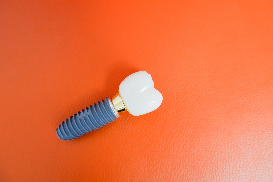
Graft13
- What Is A Bone Graft? (clarkomfs.com)
- A bone graft is a procedure performed to restore lost bone along the dental ridge once decay, infection, poor stimulation, or extraction has led to an empty socket or lost bone density. (clarkomfs.com)
- Who Needs A Bone Graft? (clarkomfs.com)
- While the idea of a bone graft may seem scary, you can rest assured that the procedure is relatively common and has impressively high success rates! (clarkomfs.com)
- Once x-rays have been taken to confirm a treatment plan and define the location needing the bone graft, the surgery can begin. (clarkomfs.com)
- A bone graft is a common procedure in oral surgery and is sometimes part of the pre-prosthetic process. (vofs.com.au)
- A bone graft is done to coax the patient's body into rebuilding the degenerated bone tissue to prepare for the next step: dental implants. (vofs.com.au)
- Bone fragments - Because a bone graft is made up of many small pieces of bone, it is normal to discover small fragments of bone in your mouth a few days after surgery. (vofs.com.au)
- Smoking while you are recovering increases the risk of infection as well as the risk that the bone graft may fail. (vofs.com.au)
- Autogenous bone grafts are advantageous in that the graft material is your own live bone, meaning it contains living cellular elements that enhances bone growth, also eliminating the risk of your body rejecting the graft material since it comes from you. (mnofs.com)
- Graft composites consist of other bone graft materials and growth factors to achieve the benefits of a variety of substances. (mnofs.com)
- Drs. Kademani and associates will determine which type of bone graft material is best suited to your particular needs. (mnofs.com)
- In addition, special membranes may be utilized that dissolve under the gum to protect the bone graft, as well as encourage bone regeneration. (mnofs.com)
Surgery10
- A good place to start is visiting Clark Oral & Facial Surgery for a bone grafting procedure! (clarkomfs.com)
- When you choose Clark Oral & Facial Surgery for your surgical needs, you're choosing a team that is here for you from beginning to end! (clarkomfs.com)
- Bone Shaving & Facial Contouring Surgery. (bestcosmeticsurgeons.com)
- or most commonly, V-line surgery, this permanent surgical procedure removes bone from the lower jaw, to change its shape and size. (bestcosmeticsurgeons.com)
- Researchers at Rutgers Medical School claim that the use of osteoporosis drugs and mechanical tools and devices typically used in craniofacial surgery, and even orthodontics, can help mitigate the effects of changes in facial bones. (sw1clinic.com)
- I came to Northlake Oral & Facial Surgery to get extractions and bone grafts. (northlakeofs.com)
- I'm extremely happy that I came to Northlake Oral & Facial Surgery. (northlakeofs.com)
- The uptick in interest about facial bones could be due to news coverage regarding reconstructive surgery for the young girl who was disfigured by a dog bite and whose mother used that disfigurement to perpetrate a hoax against KFC. (medscape.com)
- A child whose middle ear bones (called ossicles) are damaged might need more surgery to improve hearing. (kidshealth.org)
- Permanent damage to the facial nerve from surgery is very rare. (kidshealth.org)
Marrow6
- Cranial imaging studies (radiographs, computed tomographic [CT] scans, magnetic resonance [MR] images, and bone marrow scintigrams) in 13 infants and children with autosomally recessive osteopetrosis were reviewed to characterize patterns of facial and calvarial involvement at presentation and with progression of disease. (nih.gov)
- In three older children, a "hair-on-end" appearance was seen, which, at bone marrow scintigraphy, corresponded to areas of marked hematopoietic activity. (nih.gov)
- Some combinations may include: collagen/ceramic composite, which closely resembles the composition of natural bone, DBM combined with bone marrow cells, which aid in the growth of new bone, or a collagen/ceramic/autograft composite. (mnofs.com)
- The red bone marrow inside of bones produces most of the blood cells, including erythrocytes (red blood cells), leukocytes (white blood cells), and thrombocytes (platelets). (learninggnm.com)
- Most bones of the limbs contain mainly yellow bone marrow composed for the most part of fat. (learninggnm.com)
- However, if the body suffers large amounts of blood loss, yellow marrow is converted into red bone marrow to ensure blood cell production. (learninggnm.com)
Anatomy3
- Facial bone anatomy is complex, yet elegant, in its suitability to serve a multitude of functions. (medscape.com)
- Your answer could list 14 facial bones, 12 facial bones, or even 17 facial bones depending on which anatomy textbook you consult. (biologydictionary.net)
- For more in-depth clinical information, see Facial Bone Anatomy . (medscape.com)
Zygomatic bones1
- A robust understanding of the zygomatic bones' structure is essential for several beautician tasks, such as applying blush or bronzer, contouring, and even determining the best hairstyles that complement the client's face shape. (medqueen.net)
Temporal12
- Inferiorly, the condylar process has a narrow neck that widens to a globular head that articulates with the glenoid fossa of the temporal bone. (medscape.com)
- forms a synovial joint with the temporal fossa of the temporal bone (temporomandibular joint). (biologydictionary.net)
- We present a rare case of a rapidly growing temporal bone malignant small round cell tumor which initially showed facial paralysis. (ejao.org)
- Malignant small round cell tumor (MSRCT) of temporal bone is very rare, with rapid aggressive patterns. (ejao.org)
- The authors will describe a case of MSRCT of the temporal bone which showed negative results in all immunohistochemical studies. (ejao.org)
- On imaging study, temporal magnetic resonance imaging showed a partial fluid signal at the left mastoid air cells and abnormal enhancement at the left facial nerve around the genu area ( Fig. 1 ). (ejao.org)
- Temporal bone magnetic resonance image. (ejao.org)
- Photomicrograph of left temporal bone tumor taken intraoperatively. (ejao.org)
- When you open your mouth, the rounded ends of the lower jaw (condyles) glide along the joint socket of the temporal bone. (emedicinehealth.com)
- To keep this motion smooth, a soft disk of cartilage lies between the condyle and the temporal bone. (emedicinehealth.com)
- The muscle is anchored within the petrous temporal bone and emerges anteriorly into the mesotympanum from the hollow of the pyramidal process . (radiopaedia.org)
- The temporal bone is located on the sides and base of the skull. (msdmanuals.com)
Skeletal6
- Fibrous dysplasia (FD) is a skeletal developmental anomaly of the bone-forming mesenchyme that manifests as a defect in osteoblastic differentiation and maturation. (medscape.com)
- Balance tightness of the skeletal and facial muscles. (udemy.com)
- Recent studies have indicated that the skeletal morphology does change with age with a slight increase or decrease of height and length of various skeletal facial structures. (dermascope.com)
- The skeletal system includes all bones and joints of the human body. (learninggnm.com)
- Together with the skeletal muscles , the bones and joints allow controlled physical movements. (learninggnm.com)
- The bones, skeletal muscles , lymph vessels with lymph nodes , blood vessels , connective tissue , and fat tissue share the same brain relays and therefore the same biological conflict, namely a self-devaluation conflict. (learninggnm.com)
Paralysis4
- A 5-year old boy visited outpatient clinics for left idiopathic acute facial paralysis (House-Brackmann grade IV). (ejao.org)
- Facial nerve paralysis happens when a child cannot move muscles that control smiling and blinking, among other facial movements. (childrenshospital.org)
- Muscle weakness - Paralysis can occasionally spread and affect chewing or facial expressions. (cdhp.org)
- Facial paralysis can develop immediately or after some time and can be mild or severe. (msdmanuals.com)
Lower jaw2
- People with slender jaws typically have a lower jaw which bites a bit backward, giving them a convex facial profile and what's commonly called an overbite. (neurosciencenews.com)
- Certain facial muscles that control chewing are also attached to the lower jaw. (emedicinehealth.com)
Palatine1
- Palatine - two bones that form the roof of the mouth. (dermascope.com)
Upper jaw4
- Additionally, the loss of bone volume in the upper jaw can contribute to drooping of the upper eyelids. (oceanclinic.net)
- These two bones form the upper jaw. (medqueen.net)
- Sinus bone grafts are also performed to replace bone in the posterior upper jaw. (mnofs.com)
- Maxillae - two bones that form the upper jaw. (dermascope.com)
Paget's Dise6
- Paget's disease of bone is a chronic bone disorder. (medlineplus.gov)
- Who is at risk for Paget's disease of bone? (medlineplus.gov)
- What are the symptoms of Paget's disease of bone? (medlineplus.gov)
- What other problems can Paget's disease of bone cause? (medlineplus.gov)
- How is Paget's disease of bone diagnosed? (medlineplus.gov)
- What are the treatments for Paget's disease of bone? (medlineplus.gov)
Injuries9
- Bone grafting can repair implant sites with inadequate bone structure due to previous extractions, gum disease, or injuries. (mnofs.com)
- The dental specialist performs the proper treatment of facial injuries. (occore.com)
- There are a number of possible causes of facial trauma such as motor vehicle accidents, accidental falls, sports injuries, interpersonal violence, and work-related injuries. (occore.com)
- Types of facial injuries can range from injuries of teeth to extremely severe injuries of the skin and bones of the face. (occore.com)
- In addition to the obvious concern of providing a repair that yields the best cosmetic result possible, care is taken to inspect for and treat injuries to structures such as facial nerves, salivary glands, and salivary ducts (or outflow channels). (occore.com)
- His collar bone was broke in 5 pieces with severe facial injuries. (snopes.com)
- Bite marks and long gouges caused by teeth raking bone are both fairly common, but both types of injuries show signs of healing - unlike the marks found on prey, these bites weren't inflicted to kill. (scienceblogs.com)
- All affect facial bone, but all of them produce very different injuries. (scienceblogs.com)
- Victims of oral and maxillofacial trauma treated at the Hospital de Clínicas da Universidade Federal do Triângulo Mineiro are predominantly men in their third decade of life, involved in automobile accidents, with injuries to the bones of the nose that were treated electively. (bvsalud.org)
Dental implants5
- Bone grafting is most often performed as a preliminary procedure before dental implants are placed. (clarkomfs.com)
- While this is most often utilized before getting dental implants, bone grafting can be used in other circumstances as well. (clarkomfs.com)
- This results in the bone not being suitable for dental implants. (vofs.com.au)
- This often leaves a condition in which there is poor quality and quantity of bone suitable for the placement of dental implants. (mnofs.com)
- This often results in poor quality and quantity of bone suitable for the placement of dental implants as well as long term shifting of remaining teeth and changes to facial structure. (mnofs.com)
Cranial2
- These multiple bones also feature foramina (holes) that allow important cranial nerves and blood supply to cross from the inside to the outside surfaces and vice versa. (biologydictionary.net)
- There are eight cranial bones and 14 facial bones. (dermascope.com)
Muscles10
- The facial bones support the many muscles and soft tissues of the face, head, and neck. (biologydictionary.net)
- Literally memorizing the muscles, bones, arteries and veins in a desperate attempt to pass the test, and hopefully retain enough memorization to make it through the state boards. (dermascope.com)
- The muscles and bones of our face are not only the core foundation, but the structural format for our skin and facial features. (dermascope.com)
- The facial bones create the structural foundation for the face and the origination point for the facial muscles. (dermascope.com)
- These 14 bones form the basic shape of the face, and are responsible for providing attachments for muscles that make the jaw move and control facial expressions. (dermascope.com)
- The muscles of the face are subcutaneous (just under the skin), striated muscles that voluntarily control facial expression. (dermascope.com)
- These muscles originate on bone and insert on the skin of the face. (dermascope.com)
- Fortunately for the sake of learning, the names of facial muscles coincide with the related facial bones. (dermascope.com)
- Teeth grinding and clenching are habits that may be diagnosed in people who complain of pain in the temporomandibular joint or have facial pain that includes the muscles involved in chewing (myofascial pain). (emedicinehealth.com)
- and damage to the ossicles (the chain of small bones that connects the eardrum to the inner ear), the cochlea (the organ of hearing), the vestibular apparatus (the organ of balance in the inner ear), or the nerve that controls muscles of the face (facial nerve). (msdmanuals.com)
Grafting8
- To learn more about bone grafting, please read below! (clarkomfs.com)
- Bone grafting may also be used to augment the dental ridge following an extraction. (clarkomfs.com)
- Any person that has bone loss in their jaw can benefit from a bone grafting procedure. (clarkomfs.com)
- With our bone grafting procedure, you can find peace in knowing that if you experience bone loss or tooth loss, we can bring your smile back! (clarkomfs.com)
- What is Bone Grafting? (mnofs.com)
- With bone grafting we now have the opportunity to not only replace bone where it is missing, but we also have the ability to promote new bone growth in that location. (mnofs.com)
- Both allogenic and xenogenic bone grafting have an advantage of not requiring a second procedure to harvest your own bone, as with autografts. (mnofs.com)
- Each bone grafting option has its own risks and benefits. (mnofs.com)
Maxilla1
- articulate with the frontal, ethmoid, and maxilla bones, as well as with each other. (biologydictionary.net)
Sphenoid bone4
- The sphenoid bone is an unpaired bone of the neurocranium. (wikipedia.org)
- The sphenoid bone is one of the seven bones that articulate to form the orbit. (wikipedia.org)
- It is divided into the following parts: a median portion, known as the body of sphenoid bone, containing the sella turcica, which houses the pituitary gland as well as the paired paranasal sinuses, the sphenoidal sinuses two greater wings on the lateral side of the body and two lesser wings from the anterior side. (wikipedia.org)
- about the fourth year they fuse with the labyrinths of the ethmoid bone, and between the ninth and twelfth years they unite with the sphenoid bone. (wikipedia.org)
Malar bones2
- Often referred to as cheekbones or malar bones, these form the prominence of the cheeks and the outer part of the eye socket. (medqueen.net)
- Zygomatic - two bones that form the cheekbones (malar bones) and the bottom of the eye socket. (dermascope.com)
Ethmoid bone1
- Articulates with ethmoid bone anteriorly and basilar part of occipital bone posteriorly. (wikipedia.org)
Extractions1
- this includes tooth decay, gum disease, tooth extractions, aging, or facial trauma - even wearing dentures or having misaligned teeth can result in weakening of the bone structure! (clarkomfs.com)
Tissue6
- Osteoclasts, specialized cells responsible for breaking down old bone tissue, become more active, resulting in a net loss of bone mass. (oceanclinic.net)
- Using either synthetic material, bone tissue from a donor, or bone from your own body, this material is packed into the empty space and covered with a protective membrane to heal, solidify, and fuse to the surrounding bone. (clarkomfs.com)
- Once the area is thoroughly cleaned, we will create a small incision in the gum tissue above the area lacking bone. (clarkomfs.com)
- The bone is either obtained from a tissue bank or your own bone is taken from the jaw, hip or tibia (below the knee). (mnofs.com)
- Osseous changes are characterized by normal bone being replaced and distorted by poorly organized, structurally unsound, fibrous tissue. (medscape.com)
- The bone tissue stores several minerals, specifically calcium and phosphorus that keep the bones strong. (learninggnm.com)
Structures2
- Further research in this field may provide insights into innovative approaches to rejuvenate the facial structures and enhance overall facial aesthetics, improving the quality of life for individuals as they age. (oceanclinic.net)
- Beyond the surface aesthetics lie the foundational structures of the human body that significantly contribute to an individual's appearance - the facial bones. (medqueen.net)
Surgeries2
- Procedures such as facelifts, brow lifts, and eyelid surgeries can address more significant facial sagging and restore a more youthful appearance by repositioning tissues and tightening the skin. (oceanclinic.net)
- The following information was by the World Health Organization (WHO), trauma was selected for patients: age and sex, trauma mechanism, present in the fourth and eighth positions, respectively fractured bones, other traumatized body regions, represented by interpersonal violence and traffic number of surgeries, surgical procedure adopted, accidents. (bvsalud.org)
Skull and facial bones4
- This article delves into the scientific understanding of how the skull and facial bones undergo changes over time and explores their subsequent effects on the skin and overall appearance of the face. (oceanclinic.net)
- As individuals age, several structural changes occur in the skull and facial bones, resulting from a combination of intrinsic and extrinsic factors. (oceanclinic.net)
- The changes in the skull and facial bones associated with aging have significant repercussions on the skin and overall appearance of the face. (oceanclinic.net)
- The aging process impacts not only the skin but also the underlying skull and facial bones, leading to noticeable changes in facial appearance. (oceanclinic.net)
Oral2
- Oral and maxillofacial surgeons are trained, skilled, and uniquely qualified to manage and treat facial trauma. (occore.com)
- Drs. Grant or Mergelmeyer is a well-trained oral and maxillofacial surgeon and is proficient at diagnosing and treating all types of facial lacerations. (occore.com)
Nerves1
- Nervous system problems , since the bones can cause pressure on the brain, spinal cord, or nerves. (medlineplus.gov)
Articulates1
- Each bone articulates (joins) with neighboring bones. (biologydictionary.net)
Fibrous dysplasia2
- In fibrous dysplasia, the features on a bone scan are nonspecific for diagnostic purposes. (medscape.com)
- The usual appearance of fibrous dysplasia includes a lucent lesion in the diaphysis or metaphysis, with endosteal scalloping and with or without bone expansion and the absence of periosteal reaction. (medscape.com)
Nerve5
- This shows partial fluid signal at left mastoid air cells (arrow head) and abnormal enhancement at Lt. facial nerve around genu area (arrow). (ejao.org)
- Cholesteatoma can dissolve the bone over the facial nerve, which passes by the middle ear bones. (kidshealth.org)
- It is supplied by a small branch from the facial nerve . (radiopaedia.org)
- Hearing loss Hearing Loss and Deafness may result from damage to the three tiny bones (called the ossicles) that connect the eardrum to the inner ear or from damage to the cochlea or the nerve that leads to the cochlea. (msdmanuals.com)
- by an audiologist or electrodiagnostic testing of the nerve responsible for controlling facial movement. (msdmanuals.com)
Osteoporosis2
- The natural aging process leads to a decrease in bone density and volume, known as osteoporosis. (oceanclinic.net)
- The researchers went on to say that using osteoporosis medication may help to build up bone density in the face for a younger look. (sw1clinic.com)
Face10
- and provide a frame on which the soft tissues of the face can act to facilitate eating, facial expression, breathing, and speech. (medscape.com)
- Lower face variability was described as one of six facial phenotypes and related to handedness using logistic regression models while adjusting for sex, ancestry, geography, and income. (neurosciencenews.com)
- The changes in facial bone structure can affect the proportions of the face, such as the ratio of the lower face to the upper face. (oceanclinic.net)
- This is because, as already mentioned, some bones of the face are also part of the neurocranium (the bony covering of the brain). (biologydictionary.net)
- To receive optimum points, a diagram with labeled facial bones will show that you understand how each bone contributes to the face. (biologydictionary.net)
- The facial bones have many functions, as each individual bone supports different areas of the face. (biologydictionary.net)
- To understand how to restore an aging face, first you need to understand what constitutes facial aging in the first place. (sw1clinic.com)
- While it's not exactly news that the bones play a significant role in how the face ages, what is pretty interesting, and definitely new, is that for the first time ever there may be a way to treat aging bones. (sw1clinic.com)
- This lends the face a shortened appearance, making the facial proportions less than ideal. (sw1clinic.com)
- I have never heard of Face Gems from Feather and Bone.It sounds really nice and I will have to check it out.Thank you for the great review. (mystylespot.net)
Joints4
- The outer surface of the bones is covered by the periosteum , except the joints and sites attached to ligaments and tendons, which are capped with cartilage. (learninggnm.com)
- In the cerebral medulla , the bones and joints (incl. (learninggnm.com)
- the bones and joints of the left side of the body are controlled from the right cerebral hemisphere. (learninggnm.com)
- The biological conflict linked to the bones and joints is a severe self-devaluation conflict or loss of self-worth . (learninggnm.com)
Grafts2
Jawbone1
- If bone loss is discovered within the jawbone, this provides a solid foundation for implants to be anchored to so that they remain stable and secure. (clarkomfs.com)
Patient's1
- More importantly, the patient's facial appearance should be minimally affected. (occore.com)
Cartilage5
- The greater part of the bone is ossified in cartilage. (wikipedia.org)
- The cartilage is covered by the perichondrium, the equivalent to the periosteum lining the bones. (learninggnm.com)
- The bones as well as the cartilage, tendons, and ligaments originate from the new mesoderm and are therefore controlled from the cerebral medulla. (learninggnm.com)
- The degenerative joint disease causes a slowly progressive loss of cartilage and formation of new bone at the surface of the joint. (emedicinehealth.com)
- As it progresses, the disease can cause the destruction of cartilage, erode bone, and eventually cause joint deformity. (emedicinehealth.com)
Deformity1
- Other causes of TMJ syndrome include infection of the joint, cancer , and bone deformity that occurs at birth. (emedicinehealth.com)
Orbital bones1
- The thinning of the orbital bones can cause a hollowing effect around the eyes, leading to the formation of under-eye bags and a sunken appearance. (oceanclinic.net)
Eardrum to the inner ear1
- A cholesteatoma (kuh-less-tee-uh-TOE-muh) is a growth behind the eardrum, in the middle part of the ear where tiny bones relay sound waves from the eardrum to the inner ear. (kidshealth.org)
Complications1
- RÉSUMÉ La présente étude vise à examiner la qualité de vie de patients atteints de thalassémie majeure en fonction de l'âge, du sexe, des résultats scolaires, et de la gravité et des complications de la maladie. (who.int)
Proportions1
- Similarly, knowledge of the skull's shape can help in creating hairstyles that balance and complement the client's facial proportions. (medqueen.net)
Reduction1
- This study focuses on the clinical use of BFs, the risks it represents to the facial bones, considered its characteristics as well as the importance of diagnosing patients who use these drugs, in order to conduct them on the proper dental treatment to be held initially, and thus, to contribute to the reduction of complication that means an unforeseeable and devastating consequences for the patients. (bvsalud.org)
Femur2
- Femur right, D-max (Gy): Whole bone in PVT? (cancercentrum.se)
- Femur left, D-max (Gy): Whole bone in PVT? (cancercentrum.se)
Stabilize1
- When an arm or a leg is fractured, a cast is often applied to stabilize the bone to allow for proper healing. (occore.com)
Allograft2
- Allogenic bone, or allograft, is dead bone harvested from a cadaver, then processed using a freeze-dry method to extract the water via a vacuum. (mnofs.com)
- This product is processed allograft bone, containing collagen, proteins, and growth factors that are extracted from the allograft bone. (mnofs.com)
Multiple bones1
- Virtually any bone, a single bone or multiple bones, may be affected, and virtually any bone in the body can be affected. (medscape.com)
Typically1
- Other populations, such as the Eskimos, were in the 19th century described as tuberculosis-resistant, having robust facial features, and typically depicted in art as showing right-hand dominance with tools and instruments. (neurosciencenews.com)
Destruction1
- With increased growth, polyps can cause bony destruction because they can exert pressure on bone. (medscape.com)
Cheekbones1
- With the growth and readjustment of bones, studies have shown a noticeable shift of the forehead moving forward and the cheekbones moving back! (dermascope.com)
Neurocranium1
- The ethmoid and sphenoid bones are also part of the neurocranium. (biologydictionary.net)
Lift1
- Which is The Best Type of Facial Thread Lift? (sw1clinic.com)
Inferior1
- On the anterior inferior midline region of the hemimandible body is a triangular thickening of bone termed the mental protuberance. (medscape.com)