External Fixators
Ilizarov Technique
Osteogenesis, Distraction
Bone Lengthening
Tibial Fractures
Leg Length Inequality
Fracture Fixation
Fractures, Open
Bone Nails
Fracture Healing
Bone Diseases, Developmental
Tibia
New concepts and advances of immobilization of long bones. (1/342)
OBJECTIVE: To present some new concepts in the treatment of fractures and bone defects of long bones with internal fixation. METHODS: Animal experiments, mechanical tests and clinical analyses were done. RESULTS AND CONCLUSIONS: Reduction of fracture should be perfect, bone defect can be reconstructed by intramedullary and extramedullary bone graft. Relatively rigid fixation at the early stage and elastic fixation at the later stage are beneficial not only for fracture healing, but also for bone remodeling. In order to avoid complications including non-union, immobilization syndrome of the bone and joint, and implant failure, radiographs should be taken periodically; if there is any bone resorption, weight-bearing should be restricted. (+info)Percutaneous autologous bone marrow grafting on the site of tibial delayed union. (2/342)
Six months after injury, 150 mL of autogenous bone marrow was applied percutaneously at the site of delayed union to stimulate the healing of a tibial delayed union fracture in a 44 year-old man. Five months following the procedure, the fracture gaps and bone defects were completely filled with callus, the external fixator was removed, and the patient started using normal leg loading. (+info)External fixation in proximal tibial osteotomy: a comparison of three methods. (3/342)
During a period of 6 years (1990-1996), 154 patients with unilateral gonarthrosis underwent proximal tibial osteotomy using 3 different methods of external fixation: (1) closing wedge osteotomy and bilateral fixation; (2) closing wedge osteotomy with unilateral fixation, and (3) opening wedge osteotomy with unilateral fixation. The most common complications were pin-tract infection (25%), temporary nerve palsy (10%), and loss of alignment (17%). At least one complication developed in 33% of patients in this study, indicating that the use and technique of external fixation in proximal tibial osteotomy can be problematic. (+info)Muscle recovery after immobilisation by external fixation. (4/342)
We immobilised the right hindlimbs of six-month-old female Wistar rats for four weeks using a biplanar external fixation bridging the knee. The untreated left limbs served as a control group. An additional group of rats was allowed to recover for four weeks after removal of the frame. Immobilisation caused reduction in the wet weights of approximately 50% in the gastrocnemius, quadriceps, soleus and plantaris muscles; this was not restored completely after remobilisation. There was an increase in the activity of acid phosphatase of approximately 85% in the gastrocnemius and quadriceps muscles whereas that of creatine phosphokinase was reduced by about 40%. These values returned to nearly normal after remobilisation. Histological and ultrastructural examination showed a marked myopathy of the gastrocnemius muscle after immobilisation although the morphology was largely restored after remobilisation. We conclude that after four weeks of remobilisation, hind-limb muscles do not return to preimmobilisation weights, although biochemical activities and ultrastructural appearance are largely restored. (+info)Distal femoral fractures after knee arthroplasty. (5/342)
In seven patients who sustained eight distal femoral fractures following knee arthroplasty all fractures were treated operatively. Seven with open reduction internal fixation and one with external fixation. Seven of eight fractures had an unsatisfactory result. This was due not only to bone quality and fracture comminution, but also to technical problems and choice of implant. Revision surgery is often required. (+info)Joint distraction in treatment of osteoarthritis (II): effects on cartilage in a canine model. (6/342)
OBJECTIVE: From a clinical point of view, joint distraction as a treatment for osteoarthritis (OA) of hip and ankle has been demonstrated to be very promising. Pain, joint mobility and functional ability, the most important factors for a patient with severe OA, all improved. Although radiographic joint space enlargement in a significant number of patients suggested cartilage repair, actual cartilage repair remains difficult to evaluate. Therefore the present study was initiated to evaluate the actual effects of joint distraction on cartilage. METHODS: For this purpose a canine model for OA, anterior cruciate ligament transection (ACLT) was used. Sixteen weeks after ACLT articulating Ilizarov joint distraction of the knee was carried out. Absence of mechanical contact between articular surfaces and presence of intra-articular intermittent fluid pressure, characteristics of Ilizarov joint distraction, were confirmed. Twenty-five weeks after ACLT joint tissue of the dogs was analyzed. RESULTS: Biochemical analysis showed that after joint distraction the abnormal cartilage proteoglycan (PG) metabolism, characteristic for OA, had changed to a level found in control joints. Moreover, a mild degree of inflammation, present after ACLT, was reduced upon joint distraction. PG-content and histological cartilage degeneration had not (yet) improved within the time of treatment. DISCUSSION: Results suggest that the promising clinical results of Ilizarov joint distraction in patients with OA are accompanied by changes in cartilage metabolism. A change in proteoglycan turnover, indicating normalization of overall chondrocyte function, might in the long term, with normal joint use, lead to actual repair of cartilage. (+info)Failure of reduction with an external fixator in the management of injuries of the pelvic ring. Long-term evaluation of 110 patients. (7/342)
We reviewed 110 patients with an unstable fracture of the pelvic ring who had been treated with a trapezoidal external fixator after a mean follow-up of 4.1 years. There were eight open-book (type B1, B3-1) injuries, 62 lateral compression (type B2, B3-2) and 40 rotationally and vertically unstable (type C1-C3) injuries. The rate of complications was high with loss of reduction in 57%, malunion in 58%, nonunion in 5%, infection at the pin site in 24%, loosening of the pins in 2%, injury to the lateral femoral cutaneous nerve in 2%, and pressure sores in 3%. The external fixator failed to give and maintain a proper reduction in six of the eight open-book injuries, in 20 of the 62 lateral compression injuries, and in 38 of the 40 type-C injuries. Poor functional results were usually associated with failure of reduction and an unsatisfactory radiological appearance. In type-C injuries more than 10 mm of residual vertical displacement of the injury to the posterior pelvic ring was significantly related to poor outcome. In 14 patients in this unsatisfactory group poor functional results were also affected by associated nerve injuries. In lateral compression injuries the degree of displacement of fractures of the pubic rami caused by internal rotation of the hemipelvis was an important prognostic factor. External fixation may be useful in the acute phase of resuscitation but it is of limited value in the definitive treatment of an unstable type-C injury and in type-B open-book injuries. It is usually unnecessary in minimally displaced lateral compression injuries. (+info)Leg lengthening over an intramedullary nail. (8/342)
Distraction osteogenesis is widely used for leg lengthening, but often requires a long period of external fixation which carries risks of pin-track sepsis, malalignment, stiffness of the joint and late fracture of the regenerate. We present the results of 20 cases in which, in an attempt to reduce the rate of complications, a combination of external fixation and intramedullary nailing was used. The mean gain in length was 4.7 cm (2 to 8.6). The mean time of external fixation was 20 days per centimetre gain in length. All distracted segments healed spontaneously without refracture or malalignment. There were three cases of deep infection, two of which occurred in patients who had had previous open fractures of the bone which was being lengthened. All resolved with appropriate treatment. This method allows early rehabilitation, with a rapid return of knee movement. There is a lower rate of complications than occurs when external fixation is used on its own. The time of external fixation is shorter than in other methods of leg lengthening. The high risk of infection calls for caution. (+info)An external fixator is a type of orthopedic device used in the treatment of severe fractures or deformities of bones. It consists of an external frame that is attached to the bone with pins or wires that pass through the skin and into the bone. This provides stability to the injured area while allowing for alignment and adjustment of the bone during the healing process.
External fixators are typically used in cases where traditional casting or internal fixation methods are not feasible, such as when there is extensive soft tissue damage, infection, or when a limb needs to be gradually stretched or shortened. They can also be used in reconstructive surgery for bone defects or deformities.
The external frame of the fixator is made up of bars and clamps that are adjustable, allowing for precise positioning and alignment of the bones. The pins or wires that attach to the bone are carefully inserted through small incisions in the skin, and are held in place by the clamps on the frame.
External fixators can be used for a period of several weeks to several months, depending on the severity of the injury and the individual's healing process. During this time, the patient may require regular adjustments and monitoring by an orthopedic surgeon or other medical professional. Once the bone has healed sufficiently, the external fixator can be removed in a follow-up procedure.
The Ilizarov technique is a surgical method used for limb lengthening and reconstruction. It involves the use of an external fixation device, which consists of rings connected by adjustable rods and wires that are attached to the bone. This apparatus allows for gradual distraction (slow, steady stretching) of the bone, allowing new bone tissue to grow in the gap created by the distraction. The Ilizarov technique can be used to treat various conditions such as limb length discrepancies, bone deformities, and nonunions (failed healing of a fracture). It is named after its developer, Gavriil Abramovich Ilizarov, a Soviet orthopedic surgeon.
Osteogenesis, distraction refers to a surgical procedure and controlled rehabilitation process used in orthopedic surgery, oral and maxillofacial surgery, and neurosurgery to lengthen bones or correct bone deformities. The term "osteogenesis" means bone formation, while "distraction" refers to the gradual separation of bone segments.
In this procedure, a surgeon first cuts the bone (osteotomy) and then applies an external or internal distraction device that slowly moves apart the cut ends of the bone. Over time, new bone forms in the gap between the separated bone segments through a process called distraction osteogenesis. This results in increased bone length or correction of deformities.
Distraction osteogenesis is often used to treat various conditions such as limb length discrepancies, craniofacial deformities, and spinal deformities. The procedure requires careful planning, precise surgical technique, and close postoperative management to ensure optimal outcomes.
Bone lengthening is a surgical procedure that involves cutting and then gradually stretching the bone apart, allowing new bone to grow in its place. This process is also known as distraction osteogenesis. The goal of bone lengthening is to increase the length of a bone, either to improve function or to correct a deformity.
The procedure typically involves making an incision in the skin over the bone and using specialized tools to cut through the bone. Once the bone is cut, a device called an external fixator is attached to the bone on either side of the cut. The external fixator is then gradually adjusted over time to slowly stretch the bone apart, creating a gap between the two ends of the bone. As the bone is stretched, new bone tissue begins to grow in the space between the two ends, eventually filling in the gap and lengthening the bone.
Bone lengthening can be used to treat a variety of conditions, including limb length discrepancies, congenital deformities, and injuries that result in bone loss. It is typically performed by an orthopedic surgeon and may require several months of follow-up care to ensure proper healing and growth of the new bone tissue.
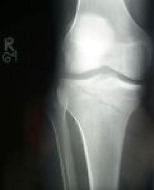
A tibial fracture is a medical term that refers to a break in the shin bone, which is called the tibia. The tibia is the larger of the two bones in the lower leg and is responsible for supporting much of your body weight. Tibial fractures can occur in various ways, such as from high-energy trauma like car accidents or falls, or from low-energy trauma in individuals with weakened bones due to osteoporosis or other medical conditions.
Tibial fractures can be classified into different types based on the location, pattern, and severity of the break. Some common types of tibial fractures include:
1. Transverse fracture: A straight break that goes across the bone.
2. Oblique fracture: A diagonal break that slopes across the bone.
3. Spiral fracture: A break that spirals around the bone, often caused by twisting or rotational forces.
4. Comminuted fracture: A break where the bone is shattered into multiple pieces.
5. Open fracture: A break in which the bone pierces through the skin, increasing the risk of infection.
6. Closed fracture: A break in which the bone does not pierce through the skin.
Tibial fractures can cause symptoms such as pain, swelling, bruising, deformity, and difficulty walking or bearing weight on the affected leg. Treatment for tibial fractures may include immobilization with a cast or brace, surgery to realign and stabilize the bone with plates, screws, or rods, and rehabilitation to restore strength, mobility, and function to the injured limb.
'Leg length inequality' (LLIS) is a condition where there is a discrepancy in the lengths of an individual's lower extremities, specifically the bones of the thigh (femur) and/or the leg (tibia/fibula). This discrepancy can be congenital or acquired due to various causes such as fractures, infections, or surgical procedures. The inequality can lead to functional scoliosis, lower back pain, and other musculoskeletal issues. It is typically diagnosed through physical examination and imaging studies like X-rays, and may be treated with various methods including orthotics, shoe lifts, or in some cases, surgical intervention.
Fracture fixation is a surgical procedure in orthopedic trauma surgery where a fractured bone is stabilized using various devices and techniques to promote proper healing and alignment. The goal of fracture fixation is to maintain the broken bone ends in correct anatomical position and length, allowing for adequate stability during the healing process.
There are two main types of fracture fixation:
1. Internal fixation: In this method, metal implants like plates, screws, or intramedullary rods are inserted directly into the bone to hold the fragments in place. These implants can be either removed or left in the body once healing is complete, depending on the type and location of the fracture.
2. External fixation: This technique involves placing pins or screws through the skin and into the bone above and below the fracture site. These pins are then connected to an external frame that maintains alignment and stability. External fixators are typically used when there is significant soft tissue damage, infection, or when internal fixation is not possible due to the complexity of the fracture.
The choice between internal and external fixation depends on various factors such as the type and location of the fracture, patient's age and overall health, surgeon's preference, and potential complications. Both methods aim to provide a stable environment for bone healing while minimizing the risk of malunion, nonunion, or deformity.
An open fracture, also known as a compound fracture, is a type of bone injury in which the bone breaks and penetrates through the skin, creating an open wound. This condition exposes the fractured bone to the external environment, increasing the risk of infection and complicating the healing process. Open fractures can result from high-energy trauma such as car accidents, falls from significant heights, or industrial incidents. Immediate medical attention is crucial for proper treatment and prevention of infection.
I believe you are referring to "bone pins" or "bone nails" rather than "bone nails." These terms are used in the medical field to describe surgical implants made of metal or biocompatible materials that are used to stabilize and hold together fractured bones during the healing process. They can also be used in spinal fusion surgery to provide stability and promote bone growth between vertebrae.
Bone pins or nails typically have a threaded or smooth shaft, with a small diameter that allows them to be inserted into the medullary canal of long bones such as the femur or tibia. They may also have a head or eyelet on one end that allows for attachment to external fixation devices or other surgical instruments.
The use of bone pins and nails has revolutionized orthopedic surgery, allowing for faster healing times, improved stability, and better functional outcomes for patients with fractures or spinal deformities.
Fracture healing is the natural process by which a broken bone repairs itself. When a fracture occurs, the body responds by initiating a series of biological and cellular events aimed at restoring the structural integrity of the bone. This process involves the formation of a hematoma (a collection of blood) around the fracture site, followed by the activation of inflammatory cells that help to clean up debris and prepare the area for repair.
Over time, specialized cells called osteoblasts begin to lay down new bone matrix, or osteoid, along the edges of the broken bone ends. This osteoid eventually hardens into new bone tissue, forming a bridge between the fracture fragments. As this process continues, the callus (a mass of newly formed bone and connective tissue) gradually becomes stronger and more compact, eventually remodeling itself into a solid, unbroken bone.
The entire process of fracture healing can take several weeks to several months, depending on factors such as the severity of the injury, the patient's age and overall health, and the location of the fracture. In some cases, medical intervention may be necessary to help promote healing or ensure proper alignment of the bone fragments. This may include the use of casts, braces, or surgical implants such as plates, screws, or rods.
Developmental bone diseases are a group of medical conditions that affect the growth and development of bones. These diseases are present at birth or develop during childhood and adolescence, when bones are growing rapidly. They can result from genetic mutations, hormonal imbalances, or environmental factors such as poor nutrition.
Some examples of developmental bone diseases include:
1. Osteogenesis imperfecta (OI): Also known as brittle bone disease, OI is a genetic disorder that affects the body's production of collagen, a protein necessary for healthy bones. People with OI have fragile bones that break easily and may also experience other symptoms such as blue sclerae (whites of the eyes), hearing loss, and joint laxity.
2. Achondroplasia: This is the most common form of dwarfism, caused by a genetic mutation that affects bone growth. People with achondroplasia have short limbs and a large head relative to their body size.
3. Rickets: A condition caused by vitamin D deficiency or an inability to absorb or use vitamin D properly. This leads to weak, soft bones that can bow or bend easily, particularly in children.
4. Fibrous dysplasia: A rare bone disorder where normal bone is replaced with fibrous tissue, leading to weakened bones and deformities.
5. Scoliosis: An abnormal curvature of the spine that can develop during childhood or adolescence. While not strictly a developmental bone disease, scoliosis can be caused by various underlying conditions such as cerebral palsy, muscular dystrophy, or spina bifida.
Treatment for developmental bone diseases varies depending on the specific condition and its severity. Treatment may include medication, physical therapy, bracing, or surgery to correct deformities and improve function. Regular follow-up with a healthcare provider is essential to monitor growth, manage symptoms, and prevent complications.
Osteotomy is a surgical procedure in which a bone is cut to shorten, lengthen, or change its alignment. It is often performed to correct deformities or to realign bones that have been damaged by trauma or disease. The bone may be cut straight across (transverse osteotomy) or at an angle (oblique osteotomy). After the bone is cut, it can be realigned and held in place with pins, plates, or screws until it heals. This procedure is commonly performed on bones in the leg, such as the femur or tibia, but can also be done on other bones in the body.
The tibia, also known as the shin bone, is the larger of the two bones in the lower leg and part of the knee joint. It supports most of the body's weight and is a major insertion point for muscles that flex the foot and bend the leg. The tibia articulates with the femur at the knee joint and with the fibula and talus bone at the ankle joint. Injuries to the tibia, such as fractures, are common in sports and other activities that put stress on the lower leg.
The femur is the medical term for the thigh bone, which is the longest and strongest bone in the human body. It connects the hip bone to the knee joint and plays a crucial role in supporting the weight of the body and allowing movement during activities such as walking, running, and jumping. The femur is composed of a rounded head, a long shaft, and two condyles at the lower end that articulate with the tibia and patella to form the knee joint.
Equipment design, in the medical context, refers to the process of creating and developing medical equipment and devices, such as surgical instruments, diagnostic machines, or assistive technologies. This process involves several stages, including:
1. Identifying user needs and requirements
2. Concept development and brainstorming
3. Prototyping and testing
4. Design for manufacturing and assembly
5. Safety and regulatory compliance
6. Verification and validation
7. Training and support
The goal of equipment design is to create safe, effective, and efficient medical devices that meet the needs of healthcare providers and patients while complying with relevant regulations and standards. The design process typically involves a multidisciplinary team of engineers, clinicians, designers, and researchers who work together to develop innovative solutions that improve patient care and outcomes.
 Octopod External Fixator