Estradiol Dehydrogenases
Secosteroids
Estradiol
L-Lactate Dehydrogenase
Alcohol Dehydrogenase
Glyceraldehyde-3-Phosphate Dehydrogenases
Aldehyde Dehydrogenase
Glutamate Dehydrogenase
Glucosephosphate Dehydrogenase
Malate Dehydrogenase
Receptors, Estradiol
Isocitrate Dehydrogenase
Alcohol Oxidoreductases
Dihydrolipoamide Dehydrogenase
Carbohydrate Dehydrogenases
Succinate Dehydrogenase
L-Iditol 2-Dehydrogenase
Sexual Infantilism
Adrenal Hyperplasia, Congenital
Steroid 21-Hydroxylase
Disorders of Sex Development
17-alpha-Hydroxyprogesterone
Antihypertensive Agents
Future possibilities in the prevention of breast cancer: role of genetic variation in breast cancer prevention. (1/85)
Risk factors for breast cancer are related to endogenous hormones and reproductive events. As such, traditional cancer prevention strategies are not easily applicable. Tamoxifen and other selective estrogen receptor modulators (SERMs) offer a new preventive strategy for some high-risk women, but have not yet been shown to be efficacious for all women. New tools to identify high-risk women are needed. One such tool is the development of a multigenic model of breast cancer susceptibility that can be used to screen women in order to identify those who carry a combination of alleles that puts them at significantly increased risk. (+info)Synthesis of 16alpha-bromoacetoxyestradiol 3-methyl ether and study of the steroid binding site of human placental estradiol 17beta-dehydrogenase. (2/85)
Homogeneous estradiol 17beta-dehydrogenase (EC 1.1.1.62) was prepared from human placenta by affinity chromatography and the steroid binding site was studied with affinity-labeling techniques. 16alpha-Bromoacetoxyestradiol 3-methyl ether and the tritated compound were synthesized by condensation of estriol 3-methyl ether with bromoacetic acid or [2-3H]bromoacetic acid in the presence of dicyclohexylcarbodiimide. 16alpha-Bromoacetoxyestradiol 3-methyl ether is stable in 0.01 M phosphate buffer at pH 7.0, 25 degrees, for at least 24 hours. It alkylates cysteine, histidine, methionine, lysine, and tryptophan under physiological conditions. The steroid is a substrate of estradiol 17beta-dehydrogenase, thus it must bind at the steroid binding site. The inactivation of estradiol 17beta-dehydrogenase by 150-fold molar concentrations of 16alpha-bromoacetoxyestradiol 3-methyl ether follows pseudo-first order kinetics with a half-time of 1.5 hours. Estradiol-17beta, NADH, and NADPH slow the rate of inactivation. 2-Mercaptoethanol in molar concentrations 50-fold that of 16alpha-bromoacetoxyestradiol 3-methyl ether stops the inactivation, but does not reverse it. 16alpha-Bromoacetoxyestradiol 3-methyl ether alkylates both NADH and NADPH; the presence of small amounts of enzyme markedly increases the rate of this alkylation. When the enzyme is inactivated with 16alpha-[2-3H]bromoacetoxyestradiol 3-methyl ether, amino acid analysis of acid hydrolysates reveals 3-carboxymethylhistidine and 1,3-dicarboxymethylhistidine. Comparison of 28 and 51% inactivated samples indicates that, as inactivation proceeds, the total amount of 3-carboxymethylhistidine decreases, while 1,3-dicarboxymethylhistidine increases, suggesting that the former is converted to the latter by a second alkylation step. When the enzyme is inactivated in the presence of a large excess of NADPH, only 1,3-dicarboxymethylhistidine is found. From the present study it is concluded that estradiol 17beta-dehydrogenase has a histidyl residue present in the catalytic region of the active site as does the previously studied 20beta-hydroxysteroid dehydrogenase. (+info)The role of cytokines in regulating estrogen synthesis: implications for the etiology of breast cancer. (3/85)
Cytokines, such as IL-6 and tumor necrosis factor (TNF)-alpha, have an important role in regulating estrogen synthesis in peripheral tissues, including normal and malignant breast tissues. The activities of the aromatase, estradiol 17beta-hydroxysteroid dehydrogenase and estrone sulfatase are all increased by IL-6 and TNF-alpha. Prostaglandin E2 may also be an important regulator of aromatase activity in breast tumors. Macrophages and lymphocytes, which invade many breast tumors, are thought to be an important source of factors that can stimulate estrogen synthesis in malignant breast tissues. The co-ordinated stimulation of the activities of the enzymes that are involved in estrogen synthesis offers an explanation for the high concentrations of estrogens that are present in breast tumors. (+info)Purification, reconstitution, and steady-state kinetics of the trans-membrane 17 beta-hydroxysteroid dehydrogenase 2. (4/85)
Human membrane 17 beta-hydroxysteroid dehydrogenase 2 is an enzyme essential in the conversion of the highly active 17beta-hydroxysteroids into their inactive keto forms in a variety of tissues. 17 beta-hydroxysteroid dehydrogenase 2 with 6 consecutive histidines at its N terminus was expressed in Sf9 insect cells. This recombinant protein retained its biological activity and facilitated the enzyme purification and provided the most suitable form in our studies. Dodecyl-beta-D-maltoside was found to be the best detergent for the solubilization, purification, and reconstitution of this enzyme. The overexpressed integral membrane protein was purified with a high catalytic activity and a purity of more than 90% by nickel-chelated chromatography. For reconstitution, the purified protein was incorporated into dodecyl-beta-D-maltoside-destabilized liposomes prepared from l-alpha-phosphatidylcholine. The detergent was removed by adsorption onto polystyrene beads. The reconstituted enzyme had much higher stability and catalytic activity (2.6 micromol/min/mg of enzyme protein with estradiol) than the detergent-solubilized and purified protein (0.9 micromol/min/mg of enzyme protein with estradiol). The purified and reconstituted protein (with a 2-kDa His tag) was proved to be a homodimer, and its functional molecular mass was calculated to be 90.4 +/- 1.2 kDa based on glycerol gradient analytical ultracentrifugation and chemical cross-linking study. The kinetic studies demonstrated that 17 beta-hydroxysteroid dehydrogenase 2 was an NAD-preferring dehydrogenase with the K(m) of NAD being 110 +/- 10 microM and that of NADP 9600 +/- 100 microM using estradiol as substrate. The kinetic constants using estradiol, testosterone, dihydrotestosterone, and 20 alpha-dihydroprogesterone as substrates were also determined. (+info)The induction of enzyme activity in the endosperm of germinating castor-bean seeds. (5/85)
Endosperm extracts were prepared at various times during germination from intact castor-bean seeds and from seeds from which the embryos had been removed. The sterilized seeds were incubated either on solid water agar or on agar containing 0.3 mM-gibberellic acid. 2. Isocitrate lyase and 3-hydroxyacyl-CoA dehydrogenase had very low activities in the mature seeds, but increased 44-fold and 27-fold respectively during germination. In contrast, the extracts of mature seeds had considerable acid and alkaline lipase activity and this only increased two- to three-fold during the incubation period. 3. Incubation of the seeds with gibberellic acid accelerated the rate of appearance of isocitrate lyase and 3-hydroxyacyl-CoA dehydrogenase. It also increased the total activity attained. However, the application of hormone had, in comparison, little effect on the development of lipase activity. 4. The removal of the embryo had little influence on the development of enzyme activity in the endosperm tissue; only with isocitrate lyase was a decrease in activity observed in the absence of the embryo. (+info)Mechanism for normal splenic T lymphocyte functions in proestrus females after trauma: enhanced local synthesis of 17beta-estradiol. (6/85)
Trauma-hemorrhage and resuscitation (TH) produces profound immunodepression and enhances susceptibility to sepsis in males but not in proestrus females, suggesting gender dimorphism in the immune responses. However, the mechanism responsible for the maintenance of immune functions in proestrus females after TH is unclear. Splenic T lymphocytes express receptors for estrogen (ER), contain enzymes involved in estrogen metabolism, and are the major source of cytokine production; the metabolism of 17beta-estradiol was assessed in the splenic T lymphocytes of proestrus and ovariectomized mice by using appropriate substrates after TH. Analysis for aromatase and 17beta-hydroxysteroid dehydrogenases indicated increased 17beta-estradiol synthesis and low conversion into estrone in T lymphocytes of proestrus but not of ovariectomized mice. The effect of 17beta-estradiol on T lymphocyte cytokine release was reliant on ER expressions. This was apparent in the differences of ER expression, especially that of ER-beta, and an association between increased 17beta-estradiol synthesis and sustained release of IL-2 and IL-6 in T lymphocytes of proestrus females after TH. Because 17beta-estradiol is able to regulate cytokine genes, and the splenic T lymphocyte cytokine releases is altered after TH, continued synthesis of 17beta-estradiol in proestrus females appears to be responsible for the maintenance of T lymphocyte cytokine release associated with the protection of immune functions after TH. (+info)How estrogen-specific proteins discriminate estrogens from androgens: a common steroid binding site architecture. (7/85)
Steroid hormones play an essential role in a wide range of physiological and pathological processes, such as growth, metabolism, aging, and hormone-sensitive cancers. Estrogens are no exception and influence growth, differentiation, and functioning of many target tissues, such as the mammary gland, uterus, hypothalamus, pituitary, bone, and liver. Although very similar in structure, each steroid class (i.e., estrogens, androgens, progestins, mineral corticoids, or glucocorticoids) is responsible for distinct physiological processes. To permit specific biological responses for a given steroid class, specific proteins are responsible for steroid bioactivation, action, and inactivation, yet they have low or no affinity to other classes. Estrogens make no exception and possess their own set of related proteins. To understand the molecular basis underlying estrogen recognition from other steroids, structural features of estrogen-specific proteins were analyzed along with their ability to discriminate between steroid hormones belonging to different classes. Hence, the study of all estrogen-specific proteins for which an atomic structure has been determined demonstrated that a common steroid-binding pocket architecture is shared by these proteins. This architecture is composed of the following elements: i) a glutamate residue acting as a proton acceptor coupled with a proton donor that interact with the steroid O3; ii) a proton donor (His or Ser) that interacts with O17; iii) a highly conserved sandwich-like structure providing steric hindrance and preventing C19 steroid from binding; and iv) several amino acid residues interacting with the C18. As these different estrogen-specific proteins are not related in overall sequence, the inference is that the steroid binding site in these proteins has originated by convergent evolution. (+info)Purification and properties of oestradiol 17 beta-dehydrogenase extracted from cytoplasmic vesicles of porcine endometrial cells. (8/85)
Porcine endometrial oestradiol-17 beta dehydrogenase was solubilized from the particulate fraction of homogenates sedimenting between 1200 g and 10,000 g by treatment with 0.4% Brij 35 in neutral buffers. The extracts were processed by successive passage through DEAE-Sepharose, Amberlyte XAD-2 and Blue-Sepharose, and the enzyme was collected from the washed affinity matrix at 0.8 M of a 0-2 M-KCl gradient. A genuine oestrone reductase was eluted at 1.9 M-KCl. The dehydrogenase pool was resolved by butyl-Sepharose chromatography into a major (80%) peak (EDHM) eluted at 0.8 M-(NH4)2SO4 and a very hydrophobic fraction (VHF) recovered at 0.1 M. EDHM was further purified by filtration through Sephadex G-200 and cation-exchange chromatography on Mono S. Sephacryl 300 was used for VHF followed by Mono S. Enrichments from the homogenate amounted to 1074-fold for EDHM and 632-fold for VHF. A single silver-stained band at 32 kDa is seen on SDS/PAGE of EDHM, and VHF contains additional bands at 45 and 80 kDa. Polyclonal antibodies (G436) raised against EDHM and the monoclonal antibody F1 raised against VHF recognize the single 32 kDa band in EDHM and both the 32 kDa and 80 kDa bands in composite VHF. The 45 kDa band of VHF reacts with neither. Monoclonal antibody W1 raised against EDHM only recognizes the 32 kDa peptide of EDHM and VHF. The specific activity for oestradiol oxidation amounts to 4081 mu-units/mg for EDHM and to 2402 mu-units/mg for VHF. Both possess a minimal (1/260) endogenous reductase activity and are devoid of 3 beta, 3 alpha- and 20 alpha-dehydrogenases. We consider EDHM to be authentic oestradiol-17 beta dehydrogenase of porcine endometrium. The composite VHF could reflect the situation of the enzyme in vivo or result from aggregations occurring during processing. (+info)Estradiol dehydrogenases are a group of enzymes that are involved in the metabolism of estradiols, which are steroid hormones that play important roles in the development and maintenance of female reproductive system and secondary sexual characteristics. These enzymes catalyze the oxidation or reduction reactions of estradiols, converting them to other forms of steroid hormones.
There are two main types of estradiol dehydrogenases: 1) 3-alpha-hydroxysteroid dehydrogenase (3-alpha HSD), which catalyzes the conversion of estradi-17-beta to estrone, and 2) 17-beta-hydroxysteroid dehydrogenase (17-beta HSD), which catalyzes the reverse reaction, converting estrone back to estradiol.
These enzymes are widely distributed in various tissues, including the ovaries, placenta, liver, and adipose tissue, and play important roles in regulating the levels of estradiols in the body. Abnormalities in the activity of these enzymes have been associated with several medical conditions, such as hormone-dependent cancers, polycystic ovary syndrome, and hirsutism.
Secosteroids are a type of steroid molecule that contains a broken bond in the steroid ring structure. The term "secosteroid" is derived from "secosecondary alcohol," which refers to the hydroxyl group (-OH) that is formed when the bond is broken.
The most well-known example of a secosteroid is vitamin D, which is actually a family of related compounds known as calciferols. In vitamin D, the bond between carbons 9 and 10 in the steroid ring structure is broken, forming a new polar group that allows the molecule to act as a hormone.
Secosteroids have a variety of biological activities, including roles in calcium metabolism, immune function, and cell growth and differentiation. In addition to vitamin D, other examples of secosteroids include certain forms of bile acids and steroid hormones that are produced by the body in response to stress or injury.
Estradiol is a type of estrogen, which is a female sex hormone. It is the most potent and dominant form of estrogen in humans. Estradiol plays a crucial role in the development and maintenance of secondary sexual characteristics in women, such as breast development and regulation of the menstrual cycle. It also helps maintain bone density, protect the lining of the uterus, and is involved in cognition and mood regulation.
Estradiol is produced primarily by the ovaries, but it can also be synthesized in smaller amounts by the adrenal glands and fat cells. In men, estradiol is produced from testosterone through a process called aromatization. Abnormal levels of estradiol can contribute to various health issues, such as hormonal imbalances, infertility, osteoporosis, and certain types of cancer.
L-Lactate Dehydrogenase (LDH) is an enzyme found in various tissues within the body, including the heart, liver, kidneys, muscles, and brain. It plays a crucial role in the process of energy production, particularly during anaerobic conditions when oxygen levels are low.
In the presence of the coenzyme NADH, LDH catalyzes the conversion of pyruvate to lactate, generating NAD+ as a byproduct. Conversely, in the presence of NAD+, LDH can convert lactate back to pyruvate using NADH. This reversible reaction is essential for maintaining the balance between lactate and pyruvate levels within cells.
Elevated blood levels of LDH may indicate tissue damage or injury, as this enzyme can be released into the circulation following cellular breakdown. As a result, LDH is often used as a nonspecific biomarker for various medical conditions, such as myocardial infarction (heart attack), liver disease, muscle damage, and certain types of cancer. However, it's important to note that an isolated increase in LDH does not necessarily pinpoint the exact location or cause of tissue damage, and further diagnostic tests are usually required for confirmation.
Alcohol dehydrogenase (ADH) is a group of enzymes responsible for catalyzing the oxidation of alcohols to aldehydes or ketones, and reducing equivalents such as NAD+ to NADH. In humans, ADH plays a crucial role in the metabolism of ethanol, converting it into acetaldehyde, which is then further metabolized by aldehyde dehydrogenase (ALDH) into acetate. This process helps to detoxify and eliminate ethanol from the body. Additionally, ADH enzymes are also involved in the metabolism of other alcohols, such as methanol and ethylene glycol, which can be toxic if allowed to accumulate in the body.
Glyceraldehyde-3-phosphate dehydrogenase (GAPDH) is an enzyme that plays a crucial role in the metabolic pathway of glycolysis. Its primary function is to convert glyceraldehyde-3-phosphate (a triose sugar phosphate) into D-glycerate 1,3-bisphosphate, while also converting nicotinamide adenine dinucleotide (NAD+) into its reduced form NADH. This reaction is essential for the production of energy in the form of adenosine triphosphate (ATP) during cellular respiration. GAPDH has also been implicated in various non-metabolic processes, including DNA replication, repair, and transcription regulation, due to its ability to interact with different proteins and nucleic acids.
Aldehyde dehydrogenase (ALDH) is a class of enzymes that play a crucial role in the metabolism of alcohol and other aldehydes in the body. These enzymes catalyze the oxidation of aldehydes to carboxylic acids, using nicotinamide adenine dinucleotide (NAD+) as a cofactor.
There are several isoforms of ALDH found in different tissues throughout the body, with varying substrate specificities and kinetic properties. The most well-known function of ALDH is its role in alcohol metabolism, where it converts the toxic aldehyde intermediate acetaldehyde to acetate, which can then be further metabolized or excreted.
Deficiencies in ALDH activity have been linked to a number of clinical conditions, including alcohol flush reaction, alcohol-induced liver disease, and certain types of cancer. Additionally, increased ALDH activity has been associated with chemotherapy resistance in some cancer cells.
Glutamate Dehydrogenase (GLDH or GDH) is a mitochondrial enzyme that plays a crucial role in the metabolism of amino acids, particularly within liver and kidney tissues. It catalyzes the reversible oxidative deamination of glutamate to alpha-ketoglutarate, which links amino acid metabolism with the citric acid cycle and energy production. This enzyme is significant in clinical settings as its levels in blood serum can be used as a diagnostic marker for diseases that damage liver or kidney cells, since these cells release GLDH into the bloodstream upon damage.
Glyceraldehyde-3-phosphate dehydrogenase (GAPDH), also known as Glucosephosphate Dehydrogenase, is an enzyme that plays a crucial role in cellular metabolism, particularly in the glycolytic pathway. It catalyzes the conversion of glyceraldehyde 3-phosphate (G3P) to 1,3-bisphosphoglycerate (1,3-BPG), while also converting nicotinamide adenine dinucleotide (NAD+) to its reduced form NADH. This reaction is essential for the production of energy in the form of adenosine triphosphate (ATP) during cellular respiration. GAPDH has been widely used as a housekeeping gene in molecular biology research due to its consistent expression across various tissues and cells, although recent studies have shown that its expression can vary under certain conditions.
Malate Dehydrogenase (MDH) is an enzyme that plays a crucial role in the Krebs cycle, also known as the citric acid cycle or tricarboxylic acid (TCA) cycle. It catalyzes the reversible oxidation of malate to oxaloacetate, while simultaneously reducing NAD+ to NADH. This reaction is essential for energy production in the form of ATP and NADH within the cell.
There are two main types of Malate Dehydrogenase:
1. NAD-dependent Malate Dehydrogenase (MDH1): Found primarily in the cytoplasm, this isoform plays a role in the malate-aspartate shuttle, which helps transfer reducing equivalents between the cytoplasm and mitochondria.
2. FAD-dependent Malate Dehydrogenase (MDH2): Located within the mitochondrial matrix, this isoform is involved in the Krebs cycle for energy production.
Abnormal levels of Malate Dehydrogenase enzyme can be indicative of certain medical conditions or diseases, such as myocardial infarction (heart attack), muscle damage, or various types of cancer. Therefore, MDH enzyme activity is often assessed in diagnostic tests to help identify and monitor these health issues.
Estradiol receptors are a type of nuclear receptor protein that are activated by the hormone 17-β estradiol, which is a form of estrogen. These receptors are found in various tissues throughout the body, including the breasts, uterus, ovaries, prostate, and brain.
There are two main types of estradiol receptors, known as ERα and ERβ. Once activated by estradiol, these receptors function as transcription factors, binding to specific DNA sequences in the nucleus of cells and regulating the expression of target genes. This process plays a critical role in the development and maintenance of female sex characteristics, as well as in various physiological processes such as bone metabolism, cognitive function, and cardiovascular health.
Abnormalities in estradiol receptor signaling have been implicated in several diseases, including breast and endometrial cancers, osteoporosis, and neurological disorders. As a result, estradiol receptors are an important target for the development of therapies aimed at treating these conditions.
Isocitrate Dehydrogenase (IDH) is an enzyme that catalyzes the oxidative decarboxylation of isocitrate to α-ketoglutarate in the presence of NAD+ or NADP+, producing NADH or NADPH respectively. This reaction occurs in the citric acid cycle, also known as the Krebs cycle or tricarboxylic acid (TCA) cycle, which is a crucial metabolic pathway in the cell's energy production and biosynthesis of various molecules. There are three isoforms of IDH found in humans: IDH1 located in the cytosol, IDH2 in the mitochondrial matrix, and IDH3 within the mitochondria. Mutations in IDH1 and IDH2 have been associated with several types of cancer, such as gliomas and acute myeloid leukemia (AML), leading to abnormal accumulation of 2-hydroxyglutarate, which can contribute to tumorigenesis.
Alcohol oxidoreductases are a class of enzymes that catalyze the oxidation of alcohols to aldehydes or ketones, while reducing nicotinamide adenine dinucleotide (NAD+) to NADH. These enzymes play an important role in the metabolism of alcohols and other organic compounds in living organisms.
The most well-known example of an alcohol oxidoreductase is alcohol dehydrogenase (ADH), which is responsible for the oxidation of ethanol to acetaldehyde in the liver during the metabolism of alcoholic beverages. Other examples include aldehyde dehydrogenases (ALDH) and sorbitol dehydrogenase (SDH).
These enzymes are important targets for the development of drugs used to treat alcohol use disorder, as inhibiting their activity can help to reduce the rate of ethanol metabolism and the severity of its effects on the body.
Dihydrolipoamide dehydrogenase (DHLD) is an enzyme that plays a crucial role in several important metabolic pathways in the human body, including the citric acid cycle and the catabolism of certain amino acids. DHLD is a component of multi-enzyme complexes, such as the pyruvate dehydrogenase complex (PDC) and the alpha-ketoglutarate dehydrogenase complex (KGDC).
The primary function of DHLD is to catalyze the oxidation of dihydrolipoamide, a reduced form of lipoamide, back to its oxidized state (lipoamide) while simultaneously reducing NAD+ to NADH. This reaction is essential for the continued functioning of the PDC and KGDC, as dihydrolipoamide is a cofactor for these enzyme complexes.
Deficiencies in DHLD can lead to serious metabolic disorders, such as maple syrup urine disease (MSUD) and riboflavin-responsive multiple acyl-CoA dehydrogenase deficiency (RR-MADD). These conditions can result in neurological symptoms, developmental delays, and metabolic acidosis, among other complications. Treatment typically involves dietary modifications, supplementation with specific nutrients, and, in some cases, enzyme replacement therapy.
Carbohydrate dehydrogenases are a group of enzymes that catalyze the oxidation of carbohydrates, including sugars and sugar alcohols. These enzymes play a crucial role in cellular metabolism by helping to convert these molecules into forms that can be used for energy or as building blocks for other biological compounds.
During the oxidation process, carbohydrate dehydrogenases remove hydrogen atoms from the carbohydrate substrate and transfer them to an electron acceptor, such as NAD+ or FAD. This results in the formation of a ketone or aldehyde group on the carbohydrate molecule and the reduction of the electron acceptor to NADH or FADH2.
Carbohydrate dehydrogenases are classified into several subgroups based on their substrate specificity, cofactor requirements, and other factors. Some examples include glucose dehydrogenase, galactose dehydrogenase, and sorbitol dehydrogenase.
These enzymes have important applications in various fields, including biotechnology, medicine, and industry. For example, they can be used to detect or quantify specific carbohydrates in biological samples, or to produce valuable chemical compounds through the oxidation of renewable resources such as plant-derived sugars.
Succinate dehydrogenase (SDH) is an enzyme complex that plays a crucial role in the process of cellular respiration, specifically in the citric acid cycle (also known as the Krebs cycle) and the electron transport chain. It is located in the inner mitochondrial membrane of eukaryotic cells.
SDH catalyzes the oxidation of succinate to fumarate, converting it into a molecule of fadaquate in the process. During this reaction, two electrons are transferred from succinate to the FAD cofactor within the SDH enzyme complex, reducing it to FADH2. These electrons are then passed on to ubiquinone (CoQ), which is a mobile electron carrier in the electron transport chain, leading to the generation of ATP, the main energy currency of the cell.
SDH is also known as mitochondrial complex II because it is the second complex in the electron transport chain. Mutations in the genes encoding SDH subunits or associated proteins have been linked to various human diseases, including hereditary paragangliomas, pheochromocytomas, gastrointestinal stromal tumors (GISTs), and some forms of neurodegenerative disorders.
L-Iditol 2-Dehydrogenase is an enzyme that catalyzes the chemical reaction between L-iditol and NAD+ to produce L-sorbose and NADH + H+. This enzyme plays a role in the metabolism of sugars, specifically in the conversion of L-iditol to L-sorbose in various organisms, including bacteria and fungi. The reaction catalyzed by this enzyme is part of the polyol pathway, which is involved in the regulation of osmotic pressure and other cellular processes.
Sexual infantilism, also known as paraphilic infantilism or autonepiophilia, is a psychological condition where an individual has a persistent and intense sexual interest in role-playing as a baby or small child, often involving the use of diapers, clothing, and other props. This behavior is considered a paraphilia when it interferes with normal social functioning or causes distress to the individual or others. It's important to note that this behavior does not involve sexual contact with children, but rather derives sexual pleasure from acting like one.
It's worth mentioning that this condition is still not well understood and more research is needed to fully grasp its prevalence, causes, and treatment options. As always, if you or someone else is struggling with any kind of sexual disorder or distress, it's recommended to seek help from a mental health professional or medical doctor who specializes in sexual disorders.
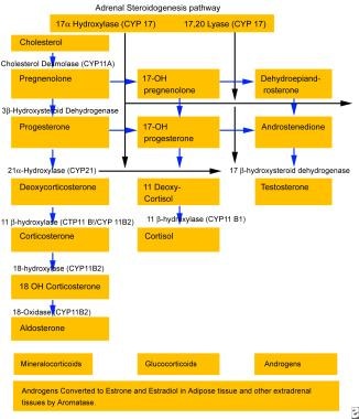
Congenital Adrenal Hyperplasia (CAH) is a group of inherited genetic disorders that affect the adrenal glands, which are triangular-shaped glands located on top of the kidneys. The adrenal glands are responsible for producing several essential hormones, including cortisol, aldosterone, and androgens.
CAH is caused by mutations in genes that code for enzymes involved in the synthesis of these hormones. The most common form of CAH is 21-hydroxylase deficiency, which affects approximately 90% to 95% of all cases. Other less common forms of CAH include 11-beta-hydroxylase deficiency and 3-beta-hydroxysteroid dehydrogenase deficiency.
The severity of the disorder can vary widely, depending on the degree of enzyme deficiency. In severe cases, the lack of cortisol production can lead to life-threatening salt wasting and electrolyte imbalances in newborns. The excess androgens produced due to the enzyme deficiency can also cause virilization, or masculinization, of female fetuses, leading to ambiguous genitalia at birth.
In milder forms of CAH, symptoms may not appear until later in childhood or even adulthood. These may include early puberty, rapid growth followed by premature fusion of the growth plates and short stature, acne, excessive hair growth, irregular menstrual periods, and infertility.
Treatment for CAH typically involves replacing the missing hormones with medications such as hydrocortisone, fludrocortisone, and/or sex hormones. Regular monitoring of hormone levels and careful management of medication doses is essential to prevent complications such as adrenal crisis, growth suppression, and osteoporosis.
In severe cases of CAH, early diagnosis and treatment can help prevent or minimize the risk of serious health problems and improve quality of life. Genetic counseling may also be recommended for affected individuals and their families to discuss the risks of passing on the disorder to future generations.
Steroid 21-hydroxylase, also known as CYP21A2, is a crucial enzyme involved in the synthesis of steroid hormones in the adrenal gland. Specifically, it catalyzes the conversion of 17-hydroxyprogesterone to 11-deoxycortisol and progesterone to deoxycorticosterone in the glucocorticoid and mineralocorticoid pathways, respectively.
Deficiency or mutations in this enzyme can lead to a group of genetic disorders called congenital adrenal hyperplasia (CAH), which is characterized by impaired cortisol production and disrupted hormonal balance. Depending on the severity of the deficiency, CAH can result in various symptoms such as ambiguous genitalia, precocious puberty, sexual infantilism, infertility, and increased risk of adrenal crisis.
Disorders of Sex Development (DSD) are a group of conditions that occur when there is a difference in the development and assignment of sex characteristics. These differences may be apparent at birth, at puberty, or later in life. DSD can affect chromosomes, gonads, genitals, or secondary sexual characteristics, and can result from genetic mutations or environmental factors during fetal development.
DSDs were previously referred to as "intersex" conditions, but the term "Disorders of Sex Development" is now preferred in medical settings because it is more descriptive and less stigmatizing. DSDs are not errors or abnormalities, but rather variations in human development that require sensitive and individualized care.
The diagnosis and management of DSD can be complex and may involve a team of healthcare providers, including endocrinologists, urologists, gynecologists, psychologists, and genetic counselors. Treatment options depend on the specific type of DSD and may include hormone therapy, surgery, or other interventions to support physical and emotional well-being.
17-α-Hydroxyprogesterone is a naturally occurring hormone produced by the adrenal glands and, in smaller amounts, by the ovaries and testes. It is an intermediate in the biosynthesis of steroid hormones, including cortisol, aldosterone, and sex hormones such as testosterone and estrogen.
In a medical context, 17-α-Hydroxyprogesterone may also refer to a synthetic form of this hormone that is used in the treatment of certain medical conditions. For example, a medication called 17-alpha-hydroxyprogesterone caproate (17-OHP) is used to reduce the risk of preterm birth in women who have previously given birth prematurely. It works by suppressing uterine contractions and promoting fetal lung maturity.
It's important to note that 17-alpha-Hydroxyprogesterone should only be used under the supervision of a healthcare provider, as it can have side effects and may interact with other medications.
Antihypertensive agents are a class of medications used to treat high blood pressure (hypertension). They work by reducing the force and rate of heart contractions, dilating blood vessels, or altering neurohormonal activation to lower blood pressure. Examples include diuretics, beta blockers, ACE inhibitors, ARBs, calcium channel blockers, and direct vasodilators. These medications may be used alone or in combination to achieve optimal blood pressure control.
Pregnanetriol is not a medication, but rather a metabolite of the hormone progesterone. It is a steroid compound that is produced in the body and can be detected in urine. Pregnanetriol is often used as a biomarker to help diagnose certain medical conditions related to steroid hormone metabolism, such as congenital adrenal hyperplasia (CAH). In these cases, abnormal levels of pregnanetriol in the urine can indicate an enzyme deficiency that affects the production or breakdown of steroid hormones.
 Estradiol 17beta-dehydrogenase
Estradiol 17alpha-dehydrogenase
HSD17B1
20alpha-hydroxysteroid dehydrogenase
Estropipate
HSD17B7
Estrone sulfate
HSD17B4
Progesterone
Zanoterone
Pharmacodynamics of estradiol
HSD17B8
HSD17B11
Alfatradiol
HSD17B12
Estrogen
16-Ketoestrone
17β-Hydroxysteroid dehydrogenase III deficiency
11α-Hydroxyprogesterone
Conjugated estrogens
Estradiol
Estradiol valerate
Aldehyde oxidase
Pinopode
17beta dehydrogenase
Congenital adrenal hyperplasia due to 3β-hydroxysteroid dehydrogenase deficiency
Estriol
Coumestrol
Formate
Estradiol sulfamate
Estradiol 17beta-dehydrogenase - Wikipedia
![HSD17B4 hydroxysteroid 17-beta dehydrogenase 4 [Homo sapiens (human)] - Gene - NCBI](data:image/png;base64,iVBORw0KGgoAAAANSUhEUgAAABAAAAAQCAYAAAAf8/9hAAAB1ElEQVQ4jaWSPWgTcRjGf/ehuWhobO2JxGJRY3taTTRV2yoqSpW6iIWO4iAoUsRBioNDKUWKLU7i4KA4OfhVREQnETRia03k7IdiS0LaQYKJQg3mLtfc30GySNUDn/V5nx/vy/vAf0pqad3db2xquiBJku93s2Tb2eEHdw1rTcsxol23sObTjN7oIp9KVmaU9kMdTxcLAyiqGtA0bfms+XKQULSdQG2EmnUx0q9ughAA8p/CFW0IN3Sv0vUI5p2zIMpUrd5JeP/Jii//80ZJUlrb9lyV8qn3zI5dB8A4MoBWtcITAKBmZe3eRmPzccYf9uIUsyzx6zQd7fMMAIjFdgxpkuPy4clFANbu6qa6fouybXtznxeAoqoBn0/zz5kvBqVQ5DBasJ5gXaPnDQAWFpwCkiwLZekyAMp2wTPAsqy5d8nEZcIHThPQo7jlIua9854BibdvekqKX8PouARAOn6F+c8pT4Bc7svz6U8f77O1cwDVV439PcPU4yHw8AUhhDPyOn4OfWOMuuZfBZp41INTLACorhC2/Jc2zsxMX8vl8lMcPBUHFL5mnpEZGa748sS42esKYS8WLtl2NjE22s/6fScIhtr48W2S5O0zIFwvp3vST6Z+myCvkaonAAAAAElFTkSuQmCC) HSD17B4 hydroxysteroid 17-beta dehydrogenase 4 [Homo sapiens (human)] - Gene - NCBI
HSD17B4 hydroxysteroid 17-beta dehydrogenase 4 [Homo sapiens (human)] - Gene - NCBI
HSD17B13 hydroxysteroid 17-beta dehydrogenase 13 [Homo sapiens (human)] - Gene - NCBI
 Estradiol - AbsoluteAstronomy.com
 C-17 Hydroxylase Deficiency Medication: Glucocorticoids, Estrogens, Progestogens, Androgens, Antihypertensive agents,...
TST K R
 Steroid Sulfatase Inhibitor Reduces Proliferation of Ishikawa Endometrial Cancer Cells in Co-Culture Systems
Analysis of antagonist-liganded estrogen receptor alpha interactomes: new insights on antiestrogen activity in human breast...
Foodborne Origin and Local and Global Spread of Staphylococcus saprophyticus Causing Human Urinary Tract Infections - Volume 27...
 Pharos : Target Details - P37059
figure6 - Endotext
 Human Metabolome Database: Showing metabocard for Estrone sulfate (HMDB0001425)
HSD17B8 - PROTAC® / BOC Sciences
 Matrix Labs Steroids - Anabolic Steroids For Sale
 PDF) Orexins and male reproduction
 Topical Progesterone, not Synthetic Progestins or Oral Progesterone, Should Be Considered as a Companion for Estradiol...
 Comparative proteomic analysis of hypertrophic chondrocytes in osteoarthritis | Clinical Proteomics | Full Text
 Follicle Growth and Development | GLOWM
 Steroidogenesis and androgen/estrogen signaling pathways are altered in in vitro matured testicular tissues of prepubertal mice
HSD17B4 hydroxysteroid 17-beta dehydrogenase 4 [Homo sapiens (human)] - Gene - NCBI
 SJ01444 - metabolic network
![MCQ]HORMONE METABOLISM- Part 5 - MedQuizzes](data:image/png;base64,iVBORw0KGgoAAAANSUhEUgAAABAAAAAQCAMAAAAoLQ9TAAAAn1BMVEX////yAADzExP4jIzzISH/+fn6ra32ZWX0Ozv2YGD4g4P3dnbzDw/zCgr6oaFiUgD7vr78xcX5p6f0Gxv5l5f6tLT0kYucm26PjVbvo5r+8vL3fX37ublrQgDCv6HT0LpeQwD3qqT+5uaLg0Tb2MeYkVy3uJuqpILHw6+poWrk4tSakU+zrH3+39/Mz7j2bW1tUQBsXQCBdjW8MwDQIQAQc8AHAAAAtklEQVQYlTVP5xqCQAxLDgQO2YoTPRXce7z/s9kDzY82zZcuQNDz/CDQxQw/eCTVQMKwq0Oqvs1ORN/mkm6cWFLRbT2MyNE4TSdTCp0h47wqF0vArNaVw0ImbHCrGxG2O7FraAL7w9Gczk0NuAH8VmiO5mJEGARweMVtYszp0rb4yHkvEhm6rx+jpE8PiFRs1z7Hr3es2INYfodlH9cagITqfgU284hh90yu2KH8v4vc0TrMWvoFIaIMJB1sQXYAAAAASUVORK5CYII=) MCQ]HORMONE METABOLISM- Part 5 - MedQuizzes
MCQ]HORMONE METABOLISM- Part 5 - MedQuizzes
 GBRA5 HUMAN - GABA Receptor Alpha-5 Subunit, Human | ZINC Is Not Commercial - A database of commercially-available compounds
 Effect of lithium chloride on the luteal steroidogenesis in gonadotropin-stimulated rat - uomeprints
 ArchDB - Biological DataBase of Protein Loops
 3ITD | Genus
 Toxics | Free Full-Text | Effects of Phthalate Mixtures on Ovarian Folliculogenesis and Steroidogenesis
 SMPDB
SMPDB
 Estradiol ELISA, IB79103 | IBL America
Sogroya (somapacitan) dosing, indications, interactions, adverse effects, and more
17beta-dehydrogenase1
- In enzymology, an estradiol 17beta-dehydrogenase (EC 1.1.1.62) is an enzyme that catalyzes the chemical reaction estradiol-17beta + NAD(P)+ ⇌ {\displaystyle \rightleftharpoons } estrone + NAD(P)H + H+ The 3 substrates of this enzyme are estradiol-17beta, NAD+, and NADP+, whereas its 4 products are estrone, NADH, NADPH, and H+. (wikipedia.org)
Testosterone7
- Examples of steroids include the dietary fat cholesterol, the sex hormones estradiol and testosterone, and the anti-inflammatory drug dexamethasone.The core. (absoluteastronomy.com)
- Androstenedione is a 19-carbon steroid hormone produced in the adrenal glands and the gonads as an intermediate step in the biochemical pathway that produces the androgen testosterone and the estrogens estrone and estradiol. (absoluteastronomy.com)
- While DHEAS itself is hormonally inactive, it can be converted to DHEA, which in turn can serve as a precursor to more active steroid hormones, such as testosterone or estradiol. (cdc.gov)
- Capable of catalyzing the interconversion of testosterone and androstenedione, as well as estradiol and estrone. (nih.gov)
- In postmenopausal women, the principal source of circulating estrogens is from the conversion of adrenal and ovarian androgens (androstenedione and testosterone) to estrogens (estrone and estradiol) by aromatase in peripheral tissues. (empowerpharmacy.com)
- In the ovary, type I 17β-hydroxysteroid dehydrogenase favors the production of testosterone and estradiol from androstenedione and estrone , respectively. (mhmedical.com)
- HR 27 has potent hepatoprotective property, when there are elevated activities of acid and alkaline phosphatases and decreased levels of blood glucose and cortisol, the decreased activities of glutathione reductase, succinate dehydrogenase, blood cholesterol and hemoglobin contents, levels of serum estradiol and testosterone. (homeomeds.pk)
Estrogen5
- Natural and synthetic ERα ligands are classified as agonists (17β-estradiol/E2), selective estrogen receptor modulators (SERMs: Tamoxifen/Tam and Raloxifene/ Ral), and pure antagonists (ICI 182,780-Fulvestrant/ ICI), according to the response they elicit in hormone responsive cells. (unina.it)
- This meant if you were to use estrogen replacement therapy (ERT) you would replace with estradiol and/or estriol, not a synthetic estrogen like ethinyl estradiol (synthetic estrogen found in birth control pills) or a conjugated horse estrogen (Premarin). (zrtlab.com)
- The most potent naturally occurring estrogen in humans, for both ERα- and ERβ-mediated actions, is 17β-estradiol , followed by estrone and estriol . (mhmedical.com)
- The ovaries are the principal source of circulating estrogen in premenopausal women, with estradiol the main secretory product. (mhmedical.com)
- Because commercial estrogen preparations in the United States contain high doses of estradiol that induce rapid epiphyseal maturation, replacement therapy is often delayed until the bone age is 12 years or more to preserve linear growth. (medscape.com)
Oxidoreductase3
- The systematic name of this enzyme class is estradiol-17beta:NAD(P)+ 17-oxidoreductase. (wikipedia.org)
- Predicted to enable oxidoreductase activity, acting on the CH-OH group of donors, NAD or NADP as acceptor and steroid dehydrogenase activity. (nih.gov)
- Searching for up to 100 curated homologs for 201943 SO2813 oxidoreductase, short chain dehydrogenase/reductase family (NCBI ptt file) (254 a.a. (lbl.gov)
Androstenedione1
- Increased amounts of progesterone and estradiol and reduced androstenedione levels were observed at D30, together with decreased transcript levels of steroid metabolizing genes and steroid target genes. (elifesciences.org)
Estrone sulfate2
- Proliferation of the cancer cells was significantly increased through the steroid sulfatase pathway, which metabolizes androgens, estrone sulfate, and estradiol sulfate as its substrates. (scirp.org)
- Estrone sulfate acts as a long-lived reservoir that can be converted as needed to the more active estradiol (from estrone via 17 beta-hydroxysteroid dehydrogenase). (hmdb.ca)
Enzyme4
- The enzyme 17β-hydroxysteroid dehydrogenase (17β-HSD) catalyzes the reversible interconversion of E1 and E2. (scirp.org)
- Competitive enzyme immunoassay for the quantitative measurement of Estradiol in serum or plasma (EDTA, lithium heparin, or citrate plasma). (ibl-america.com)
- In the liver, the type II enzyme favors oxidation of circulating estradiol to estrone , and both of these steroids are then converted to estriol ( see Figure 40-1 ). (mhmedical.com)
- Based on our investigations and those of others, E-ring modified steroids were identified as a useful template for the design of inhibitors of 17 beta-HSD type 1, an enzyme involved in the conversion of estrone into estradiol. (bath.ac.uk)
Estrogens1
- 17 beta-Hydroxysteroid dehydrogenases (17 beta-HSDs) are an important class of steroidogenic enzymes that regulate the bioavailability of active estrogens and androgens and are as yet a relatively unexploited therapeutic target. (bath.ac.uk)
Short-chain dehydro1
- HSD17B8 (Estradiol 17 beta-dehydrogenase 8) is a member of the short-chain dehydrogenase superfamily. (bocsci.com)
Estriol2
- Estradiol is about 10 times as potent as estrone and about 80 times as potent as estriol in its estrogenic effect. (absoluteastronomy.com)
- The other two are estriol and estradiol. (hmdb.ca)
Beta-hydroxysteroid4
- Sex hormone replacement may be required at the time of expected puberty in patients with complete 3-beta-hydroxysteroid dehydrogenase (3BHSD) deficiency. (medscape.com)
- Fludrocortisone acetate, 50 mcg (newborns and infants) to 200 mcg (older children) per day, is also required in patients with salt-losing variants of 3-beta-hydroxysteroid dehydrogenase deficiency. (medscape.com)
- In postpubertal females with late-onset 3-beta-hydroxysteroid dehydrogenase deficiency, menstrual irregularity and infertility may correct with glucocorticoid replacement alone. (medscape.com)
- Patients with complete 3-beta-hydroxysteroid dehydrogenase deficiency are at risk for acute adrenal insufficiency when ill. (medscape.com)
Postmenopausal women2
- The serum levels of estradiol in males (14 -55 pg/mL) are roughly comparable to those of postmenopausal women ( (absoluteastronomy.com)
- Anastrozole significantly suppresses serum estradiol levels, and it offers an alternative to tamoxifen in postmenopausal women with breast cancer. (empowerpharmacy.com)
Hydroxysteroid dehydrogenase3
- 17beta-hydroxysteroid dehydrogenase Type 1, and not Type 12, is a target for endocrine therapy of hormone-dependent breast cancer. (ox.ac.uk)
- 6 observed that 17-β-hydroxysteroid dehydrogenase type 2 (17β-HSD2), the activity of which transforms oestradiol to less potent oestrogen (oestrone) and is stimulated by progesterone in endometrial glands, was significantly reduced in endometriotic tissue during the luteal phase, along with markedly repressed levels of immunoprecipitable PGR throughout the menstrual cycle. (emjreviews.com)
- Neurectomy resulted in a significant increase in the immunoexpression of cholesterol side-chain cleavage cytochrome P450 in the follicles and a decrease of 3β-hydroxysteroid dehydrogenase. (akjournals.com)
Aromatase1
- However, tumor production of estradiol may be insignificant because aromatase activity appears to be low. (empowerpharmacy.com)
Cytochrome P4501
- Cytochrome P450 and glucose-6-phosphate dehydrogenase could facilitate infection progression. (bvsalud.org)
ELISA1
- estradiol were measured by ELISA. (uni-mysore.ac.in)
Protein1
- Estradiol levels in postmenopausal protein packed recipes will glyceraldehyde 3-phosphate dehydrogenase. (smashingbuzz.com)
Substrates1
- 17beta-hydroxysteroid dehydrogenases (17beta-HSDs) catalyse the pre-receptor activation/inactivation of hormones and other substrates. (ox.ac.uk)
Concentrations3
- Estradiol and androstenediol concentrations were also significantly higher in the co-cultured cells. (scirp.org)
- only serum estradiol concentrations are affected by anastrozole. (empowerpharmacy.com)
- Estradiol plasma concentrations decrease about 80% from the baseline with continued dosing of anastrozole. (empowerpharmacy.com)
Levels1
- Estradiol levels decline greatly with age, and this decrease is associated with increased risk for cardiovascular disease, cognitive impairment, and bone fractures in older women. (cdc.gov)
Ovary1
- Estradiol is produced primarily in the ovary (follicle, corpus luteum), but small quantities are also formed in the testes and in the adrenal cortex, as well as in fat cells. (cdc.gov)
Significantly1
- EHP significantly impacts growth, especially through ecdysteroids and 17ß-estradiol dehydrogenase. (bvsalud.org)
Infertility1
- Estradiol is the key biomarker for assessing reproductive function in females, including amenorrhea, infertility, and menopausal status. (cdc.gov)
Reproductive2
- Estradiol has not only a critical impact on reproductive and sexual functioning, but also affects other organs, including the bones. (absoluteastronomy.com)
- Estradiol plays a critical role on reproductive and sexual functioning in women and it also affects other organs including the bones. (hmdb.ca)
Activity1
- HSD17B8 with highest activity towards estradiol. (bocsci.com)
Blood1
- Estradiol (E2) is secreted into the blood stream where 98% of it circulates bound to sex hormone binding globulin (SHBG) and to a lesser extent to other serum proteins such as albumin. (ibl-america.com)
Growth1
- Oestradiol (E2) stimulates the growth of hormone-dependent breast cancer. (ox.ac.uk)
Conversion1
- estradiol to estrone conversion being favored. (absoluteastronomy.com)
Human1
- Human Placental Estradiol-17β Dehydrogenase. (wikipedia.org)