Erythropoietin
Receptors, Erythropoietin
Erythropoiesis
Erythroid Precursor Cells
Hematocrit
Hemoglobins
Erythroblasts
Reticulocyte Count
Hematinics
Leukemia, Erythroblastic, Acute
Janus Kinase 2
Spleen Focus-Forming Viruses
Kidney Failure, Chronic
Anemia, Neonatal
Interleukin-3
Renal Dialysis
Colony-Forming Units Assay
STAT5 Transcription Factor
Injections, Subcutaneous
Iron
Polycythemia Vera
Blood Transfusion
Erythrocytes
Doping in Sports
Milk Proteins
Hypoxia-Inducible Factor 1
Cells, Cultured
Phenylhydrazines
Bone Marrow
Bone Marrow Cells
Friend murine leukemia virus
Reticulocytes
Stem Cell Factor
Signal Transduction
Hematopoiesis
Phlebotomy
Erythrocyte Volume
Hypoxia-Inducible Factor 1, alpha Subunit
Cell Division
Anemia, Hypochromic
RNA, Messenger
Erythroid Cells
Neuroprotective Agents
Ferritins
Cell Differentiation
Bloodletting
Dose-Response Relationship, Drug
Cobalt
GATA1 Transcription Factor
Erythrocyte Transfusion
Hypoxia-Inducible Factor-Proline Dioxygenases
Iron Radioisotopes
Hematopoietic Cell Growth Factors
Kidney
Molecular Sequence Data
Human, rat, and mouse kidney cells express functional erythropoietin receptors. (1/3892)
BACKGROUND: Erythropoietin (EPO), secreted by fibroblast-like cells in the renal interstitium, controls erythropoiesis by regulating the survival, proliferation, and differentiation of erythroid progenitor cells. We examined whether renal cells that are exposed to EPO express EPO receptors (EPO-R) through which analogous cytokine responses might be elicited. METHODS: Normal human and rat kidney tissue and defined cell lines of human, rat, and mouse kidney were screened, using reverse transcription-polymerase chain reaction, nucleotide sequencing, ligand binding, and Western blotting, for the expression of EPO-R. EPO's effects on DNA synthesis and cell proliferation were also examined. RESULTS: EPO-R transcripts were readily detected in cortex, medulla, and papilla of human and rat kidney, in mesangial (human, rat), proximal tubular (human, mouse), and medullary collecting duct cells (human). Nucleotide sequences of EPO-R cDNAs from renal cells were identical to those of erythroid precursor cells. Specific 125I-EPO binding revealed a single class of high- to intermediate-affinity EPO-Rs in each tested cell line (kD 96 pm to 1. 4 nm; Bmax 0.3 to 7.0 fmol/mg protein). Western blots of murine proximal tubular cell membranes revealed an EPO-R protein of approximately 68 kDa. EPO stimulated DNA synthesis and cell proliferation dose dependently. CONCLUSION: This is the first direct demonstration, to our knowledge, that renal cells possess EPO-Rs through which EPO stimulates mitogenesis. This suggests currently unrecognized cytokine functions for EPO in the kidney, which may prove beneficial in the repair of an injured kidney while being potentially detrimental in renal malignancies. (+info)Role of cytokine signaling molecules in erythroid differentiation of mouse fetal liver hematopoietic cells: functional analysis of signaling molecules by retrovirus-mediated expression. (2/3892)
Erythropoietin (EPO) and its cell surface receptor (EPOR) play a central role in proliferation, differentiation, and survival of erythroid progenitors. Signals induced by EPO have been studied extensively by using erythroid as well as nonerythroid cell lines, and various controversial results have been reported as to the role of signaling molecules in erythroid differentiation. Here we describe a novel approach to analyze the EPO signaling by using primary mouse fetal liver hematopoietic cells to avoid possible artifacts due to established cell lines. Our strategy is based on high-titer retrovirus vectors with a bicistronic expression system consisting of an internal ribosome entry site (IRES) and green fluorescent protein (GFP). By placing the cDNA for a signaling molecule in front of IRES-GFP, virus-infected cells can be viably sorted by fluorescence-activated cell sorter, and the effect of expression of the signaling molecule can be assessed. By using this system, expression of cell-survival genes such as Bcl-2 and Bcl-XL was found to enhance erythroid colony formation from colony-forming unit-erythroid (CFU-E) in response to EPO. However, their expression was not sufficient for erythroid colony formation from CFU-E alone, indicating that EPO induces signals for erythroid differentiation. To examine the role of EPOR tyrosine residues in erythroid differentiation, we introduced a chimeric EGFR-EPOR receptor, which has the extracellular domain of the EGF receptor and the intracellular domain of the EPOR, as well as a mutant EGFR-EPOR in which all the cytoplasmic tyrosine residues are replaced with phenylalanine, and found that tyrosine residues of EPOR are essential for erythroid colony formation from CFU-E. We further analyzed the function of the downstream signaling molecules by expressing modified signaling molecules and found that both JAK2/STAT5 and Ras, two major signaling pathways activated by EPOR, are involved in full erythroid differentiation. (+info)Iron depletion by phlebotomy with recombinant erythropoietin prior to allogeneic transplantation to prevent liver toxicity. (3/3892)
Iron overload may induce liver toxicity after hematopoietic stem cell transplantation (HSCT), but it is not known if iron depletion prior to HSCT can reduce the risk of severe toxicity in this setting. We used subcutaneous recombinant erythropoietin (EPO) (25 UI/kg) three times a week and phlebotomy once a week, to prevent liver toxicity in a patient with advanced acute leukemia and liver disease due to severe iron overload, previous drug toxicity and hepatitis C viral infection. Over the 9 months prior to allogeneic HSCT, 34 phlebotomies were carried out. Serum ferritin dropped from 2964 to 239 microg/l and the ALT dropped to near normal values. At allogeneic HSCT no liver toxicity was observed, suggesting that iron depletion in the pretransplant period may contribute to reducing transplant-related toxicity in selected cases. (+info)Association of plasma fibrinogen concentration with vascular access failure in hemodialysis patients. (4/3892)
BACKGROUND: Elevated plasma fibrinogen is an important risk factor for coronary artery disease in the general population and patients with chronic renal failure. High plasma fibrinogen may trigger thrombus formation in arteriovenous fistulas. We performed a prospective, cohort study to evaluate the association of plasma fibrinogen concentration with vascular access failure in patients undergoing long-term haemodialysis. METHODS: Between September 1989 and October 1995, 144 patients underwent a vascular access operation. In March 1997, 102 patients (56 M, 46 F) who had been followed up for more than 18 months (median; 37 months, range; 18-102 months) were included in the study. The median age of the patients was 52 years (range; 19-78 years). In 35 patients, renal disease was secondary to diabetes mellitus. The type of vascular access was a polytetrafluoroethylene (PTFE) graft in 17 patients. Seventy-seven patients received recombinant human erythropoietin (r-HuEPO) therapy during the follow-up period. Plasma fibrinogen, albumin, total cholesterol, hematocrit, platelets and creatinine were measured at the time of operation. Vascular access failure was defined as the occurrence of complications requiring transluminal angioplasty, thrombolytic therapy or surgical repair. RESULTS: Thirty-eight patients had at least one vascular access failure and the incidence was 0.3 (range; 0-2.4) episodes per patient-year. The survival rate of vascular access was 78% (native fistula; 80%, PTFE graft; 71%) after 12 months and 70% (native fistula; 73%, PTFE graft; 51%) after 24 months. Older age, a PTFE graft, r-HuEPO therapy, higher hematocrit, lower albumin and higher fibrinogen levels were significantly associated with vascular access failure, whereas gender, diabetes mellitus, total cholesterol and platelet count were not. Plasma fibrinogen was inversely correlated with albumin (r=-0.38, P=0.001). The cumulative vascular access survival was significantly lower in patients with high plasma fibrinogen levels (> or = 460 mg/dl) compared with patients with low levels (< 460 mg/dl) (P=0.007). Independent risk factors for vascular access failure analysed by Cox's proportional hazards model were older age (RR; 1.36 by 10-year increment), higher fibrinogen level (RR; 1.20 by 100 mg/dl increment), PTFE graft (RR; 2.28) and r-HuEPO therapy (RR; 3.79). CONCLUSION: High plasma fibrinogen level is an independent risk factor for vascular access failure in haemodialysis patients. (+info)Advances in the biological therapy and gene therapy of malignant disease. (5/3892)
Biological and gene therapy of cancer have become important components of clinical cancer research. Advances in this area are based on evidence for the presence of tumor antigens, antitumor immune responses, evasion of host control by tumors, and the recognition of host defense failure in cancer patients. These mechanisms are being corrected or exploited in the development of biological and gene therapy. Over the last decade, 9 biological therapies have received Food and Drug Administration approval, and another 12 appear promising and will likely be approved in the next few years. Our approach to gene therapy has been to allogenize tumors by the direct intratumoral injection of HLA-B7/beta2-microglobulin genes as plasmid DNA in a cationic lipid into patients with malignant melanoma. In four Phase I studies, we found a 36% response by the local injected tumor and a 19% systemic antitumor response. In other cancers, gene transfer, expression, and an intratumoral T-cell response were seen, but no clinical response was seen. A variety of follow-up studies with HLA-B7 and other genes are planned. Evasion of host control is now a major target of gene therapy. Strategies to overcome this include up-regulation of MHC and introduction of cell adhesion molecules into tumor cells, suppression of transforming growth factor and interleukin 10 production by tumor cells, and blockade of the fas ligand-fas interaction between tumor cells and attacking lymphocytes. With these approaches, it seems likely that gene therapy may become the fifth major modality of cancer treatment in the next decade. (+info)Identification of the poly(C) binding protein in the complex associated with the 3' untranslated region of erythropoietin messenger RNA. (6/3892)
Hypoxia regulates expression of erythropoietin (EPO), a glycoprotein that stimulates erythrocytosis, at the level of transcription and also possibly at the level of messenger RNA (mRNA) stability. A pyrimidine-rich region within the EPO mRNA 3' untranslated region was implicated in regulation of EPO mRNA stability element and shown to bind protein factors. In the present study we wished to identify the protein factor binding to the pyrimidine-rich sequence in the EPO mRNA stability element. Using mobility shift assays, ultraviolet light cross-linking, and sodium dodecyl sulfate-polyacrylamide gel electrophoresis (SDS-PAGE), and electroelution of protein factors from the gel slices corresponding to the ribonucleoprotein complexes, we found that two isoforms of a 40 kD poly(C) binding protein (PCBP, also known as alphaCP or hnRNPE), PCBP1, and PCBP2 are present in that complex. In Hep3B or HepG2 cells hypoxia induces neither expression of PCBP nor formation of the ribonucleoprotein complex associated with EPO mRNA that involves PCBP. (+info)Erythropoietin depresses nitric oxide synthase expression by human endothelial cells. (7/3892)
We have recently shown that erythropoietin (EPO)-induced hypertension is unrelated to the rise in hematocrit and is marked by elevated cytosolic [Ca+2] and nitric oxide (NO) resistance. The present study was done to determine the effect of EPO on NO production and endothelial NO synthase (eNOS) expression by endothelial cells. Human coronary artery endothelial cells were cultured to subconfluence and then were incubated for 24 hours in the presence of either EPO (0, 5, and 20 U/mL) alone or EPO plus the calcium channel blocker felodipine. The experiments were carried out with quiescent (0.5% FCS) and proliferating (5% FCS) cells. Total nitrate and nitrite, eNOS protein, DNA synthesis (thymidine incorporation), and cell proliferation (cell count) were determined. In addition, NO production in response to acetylcholine stimulation was tested. EPO resulted in a dose-dependent inhibition of basal and acetylcholine-stimulated NO production and eNOS protein expression and also led to a significant dose-dependent stimulation of DNA synthesis in endothelial cells. The inhibitory effects of EPO on NO production and eNOS expression were reversed by felodipine. Thus, EPO downregulates basal and acetylcholine-stimulated NO production, depresses eNOS expression, and stimulates proliferation in isolated human endothelial cells. The suppressive effects of EPO on NO production and on eNOS expression are reversed by calcium channel blockade. (+info)Expression of the erythropoietin receptor by trophoblast cellsin the human placenta. (8/3892)
Nonclassical sites of erythropoietin (EPO) and erythropoietin receptor (EPO-R) expression have been described that suggest new physiological roles for this hormone unrelated to erythropoiesis. The recent finding of EPO expression by trophoblast cells in the human placenta prompted us to consider whether these cells also express EPO-R. With use of immunocytochemistry, EPO-R was identified in villous and extravillous cytotrophoblast cells, as well as in the syncytiotrophoblast at all gestational ages. EPO-R was also expressed by cells within the villous core, including endothelial cells of fetoplacental blood vessels. Placental tissues and isolated and immunopurified trophoblast cells, as well as trophoblast-derived choriocarcinoma Jar cells, expressed immunoreactive EPO-R on Western blot. EPO-R mRNA was also detected in the same placental tissues and trophoblast cells by nested-primer reverse transcription-polymerase chain reaction. Finally, EPO-R was functional insofar as the receptor was phosphorylated on tyrosine residues in response to exogenous EPO treatment of cultured trophoblast or Jar cells. Thus, the present findings support the hypothesis that trophoblast cells of the human placenta express EPO-R. In view of these results, taken together with previous work demonstrating EPO expression by the same cells, an autocrine role for this hormone in the survival, proliferation, or differentiation of placental trophoblast cells is proposed. (+info)Erythropoietin (EPO) is a hormone that is primarily produced by the kidneys and plays a crucial role in the production of red blood cells in the body. It works by stimulating the bone marrow to produce more red blood cells, which are essential for carrying oxygen to various tissues and organs.
EPO is a glycoprotein that is released into the bloodstream in response to low oxygen levels in the body. When the kidneys detect low oxygen levels, they release EPO, which then travels to the bone marrow and binds to specific receptors on immature red blood cells called erythroblasts. This binding triggers a series of events that promote the maturation and proliferation of erythroblasts, leading to an increase in the production of red blood cells.
In addition to its role in regulating red blood cell production, EPO has also been shown to have neuroprotective effects and may play a role in modulating the immune system. Abnormal levels of EPO have been associated with various medical conditions, including anemia, kidney disease, and certain types of cancer.
EPO is also used as a therapeutic agent for the treatment of anemia caused by chronic kidney disease, chemotherapy, or other conditions that affect red blood cell production. Recombinant human EPO (rhEPO) is a synthetic form of the hormone that is produced using genetic engineering techniques and is commonly used in clinical practice to treat anemia. However, misuse of rhEPO for performance enhancement in sports has been a subject of concern due to its potential to enhance oxygen-carrying capacity and improve endurance.
Erythropoietin receptors are cell surface proteins found on immature red blood cell precursors in the bone marrow. They bind to the hormone erythropoietin (EPO), which is produced by the kidneys in response to low oxygen levels in the blood. When EPO binds to its receptor, it activates a signaling pathway that promotes the survival, proliferation, and differentiation of red blood cell precursors, leading to increased production of red blood cells. This process is critical for maintaining adequate oxygen delivery to tissues in the body. Mutations in the erythropoietin receptor gene can lead to various blood disorders, including anemia and polycythemia.
Erythropoiesis is the process of forming and developing red blood cells (erythrocytes) in the body. It occurs in the bone marrow and is regulated by the hormone erythropoietin (EPO), which is produced by the kidneys. Erythropoiesis involves the differentiation and maturation of immature red blood cell precursors called erythroblasts into mature red blood cells, which are responsible for carrying oxygen to the body's tissues. Disorders that affect erythropoiesis can lead to anemia or other blood-related conditions.
Anemia is a medical condition characterized by a lower than normal number of red blood cells or lower than normal levels of hemoglobin in the blood. Hemoglobin is an important protein in red blood cells that carries oxygen from the lungs to the rest of the body. Anemia can cause fatigue, weakness, shortness of breath, and a pale complexion because the body's tissues are not getting enough oxygen.
Anemia can be caused by various factors, including nutritional deficiencies (such as iron, vitamin B12, or folate deficiency), blood loss, chronic diseases (such as kidney disease or rheumatoid arthritis), inherited genetic disorders (such as sickle cell anemia or thalassemia), and certain medications.
There are different types of anemia, classified based on the underlying cause, size and shape of red blood cells, and the level of hemoglobin in the blood. Treatment for anemia depends on the underlying cause and may include dietary changes, supplements, medication, or blood transfusions.
Polycythemia is a medical condition characterized by an abnormal increase in the total red blood cell (RBC) mass or hematocrit (the percentage of RBCs in the blood). This results in a higher-than-normal viscosity of the blood, which can lead to various complications such as impaired circulation, increased risk of blood clots, and reduced oxygen supply to the tissues.
There are two main types of polycythemia: primary and secondary. Primary polycythemia, also known as polycythemia vera, is a rare myeloproliferative neoplasm caused by genetic mutations that lead to excessive production of RBCs in the bone marrow. Secondary polycythemia, on the other hand, is a reactive condition triggered by various factors such as chronic hypoxia (low oxygen levels), high altitude, smoking, or certain medical conditions like sleep apnea, heart disease, or kidney tumors.
Symptoms of polycythemia may include fatigue, headaches, dizziness, shortness of breath, itching, and a bluish or reddish tint to the skin (cyanosis). Treatment depends on the underlying cause and severity of the condition and may involve phlebotomy, medications to reduce RBC production, and management of associated complications.
Recombinant proteins are artificially created proteins produced through the use of recombinant DNA technology. This process involves combining DNA molecules from different sources to create a new set of genes that encode for a specific protein. The resulting recombinant protein can then be expressed, purified, and used for various applications in research, medicine, and industry.
Recombinant proteins are widely used in biomedical research to study protein function, structure, and interactions. They are also used in the development of diagnostic tests, vaccines, and therapeutic drugs. For example, recombinant insulin is a common treatment for diabetes, while recombinant human growth hormone is used to treat growth disorders.
The production of recombinant proteins typically involves the use of host cells, such as bacteria, yeast, or mammalian cells, which are engineered to express the desired protein. The host cells are transformed with a plasmid vector containing the gene of interest, along with regulatory elements that control its expression. Once the host cells are cultured and the protein is expressed, it can be purified using various chromatography techniques.
Overall, recombinant proteins have revolutionized many areas of biology and medicine, enabling researchers to study and manipulate proteins in ways that were previously impossible.
Erythroid precursor cells, also known as erythroblasts or normoblasts, are early stage cells in the process of producing mature red blood cells (erythrocytes) in the bone marrow. These cells are derived from hematopoietic stem cells and undergo a series of maturation stages, including proerythroblast, basophilic erythroblast, polychromatophilic erythroblast, and orthochromatic erythroblast, before becoming reticulocytes and then mature red blood cells. During this maturation process, the cells lose their nuclei and become enucleated, taking on the biconcave shape and flexible membrane that allows them to move through small blood vessels and deliver oxygen to tissues throughout the body.
Hematocrit is a medical term that refers to the percentage of total blood volume that is made up of red blood cells. It is typically measured as part of a complete blood count (CBC) test. A high hematocrit may indicate conditions such as dehydration, polycythemia, or living at high altitudes, while a low hematocrit may be a sign of anemia, bleeding, or overhydration. It is important to note that hematocrit values can vary depending on factors such as age, gender, and pregnancy status.
Hemoglobin (Hb or Hgb) is the main oxygen-carrying protein in the red blood cells, which are responsible for delivering oxygen throughout the body. It is a complex molecule made up of four globin proteins and four heme groups. Each heme group contains an iron atom that binds to one molecule of oxygen. Hemoglobin plays a crucial role in the transport of oxygen from the lungs to the body's tissues, and also helps to carry carbon dioxide back to the lungs for exhalation.
There are several types of hemoglobin present in the human body, including:
* Hemoglobin A (HbA): This is the most common type of hemoglobin, making up about 95-98% of total hemoglobin in adults. It consists of two alpha and two beta globin chains.
* Hemoglobin A2 (HbA2): This makes up about 1.5-3.5% of total hemoglobin in adults. It consists of two alpha and two delta globin chains.
* Hemoglobin F (HbF): This is the main type of hemoglobin present in fetal life, but it persists at low levels in adults. It consists of two alpha and two gamma globin chains.
* Hemoglobin S (HbS): This is an abnormal form of hemoglobin that can cause sickle cell disease when it occurs in the homozygous state (i.e., both copies of the gene are affected). It results from a single amino acid substitution in the beta globin chain.
* Hemoglobin C (HbC): This is another abnormal form of hemoglobin that can cause mild to moderate hemolytic anemia when it occurs in the homozygous state. It results from a different single amino acid substitution in the beta globin chain than HbS.
Abnormal forms of hemoglobin, such as HbS and HbC, can lead to various clinical disorders, including sickle cell disease, thalassemia, and other hemoglobinopathies.
Erythroblasts are immature red blood cells that are produced in the bone marrow. They are also known as normoblasts and are a stage in the development of red blood cells, or erythrocytes. Erythroblasts are larger than mature red blood cells and have a nucleus, which is lost during the maturation process. These cells are responsible for producing hemoglobin, the protein that carries oxygen in the blood. Abnormal increases or decreases in the number of erythroblasts can be indicative of certain medical conditions, such as anemia or leukemia.
A reticulocyte count is a laboratory test that measures the percentage of reticulocytes in the peripheral blood. Reticulocytes are immature red blood cells produced in the bone marrow and released into the bloodstream. They contain residual ribosomal RNA, which gives them a reticular or net-like appearance under a microscope when stained with certain dyes.
The reticulocyte count is often used as an indicator of the rate of red blood cell production in the bone marrow. A higher than normal reticulocyte count may indicate an increased production of red blood cells, which can be seen in conditions such as hemolysis, blood loss, or response to treatment of anemia. A lower than normal reticulocyte count may suggest a decreased production of red blood cells, which can be seen in conditions such as bone marrow suppression, aplastic anemia, or vitamin deficiencies.
The reticulocyte count is usually expressed as a percentage of the total number of red blood cells, but it can also be reported as an absolute reticulocyte count (the actual number of reticulocytes per microliter of blood). The normal range for the reticulocyte count varies depending on the laboratory and the population studied.
Hematinics are a class of medications and dietary supplements that are used to enhance the production of red blood cells or hemoglobin in the body. They typically contain iron, vitamin B12, folic acid, or other nutrients that are essential for the synthesis of hemoglobin and the formation of red blood cells.
Iron is a critical component of hematinics because it plays a central role in the production of hemoglobin, which is the protein in red blood cells that carries oxygen throughout the body. Vitamin B12 and folic acid are also important nutrients for red blood cell production, as they help to regulate the growth and division of red blood cells in the bone marrow.
Hematinics are often prescribed to treat anemia, which is a condition characterized by a low red blood cell count or abnormally low levels of hemoglobin in the blood. Anemia can be caused by a variety of factors, including nutritional deficiencies, chronic diseases, and inherited genetic disorders.
Examples of hematinics include ferrous sulfate (an iron supplement), cyanocobalamin (vitamin B12), and folic acid. These medications are available in various forms, such as tablets, capsules, and liquids, and can be taken orally or by injection. It is important to follow the dosage instructions carefully and to inform your healthcare provider of any other medications you are taking, as hematinics can interact with certain drugs and may cause side effects.
Erythrocyte count, also known as red blood cell (RBC) count, is a laboratory test that measures the number of red blood cells in a sample of blood. Red blood cells are important because they carry oxygen from the lungs to the rest of the body. A low erythrocyte count may indicate anemia, while a high count may be a sign of certain medical conditions such as polycythemia. The normal range for erythrocyte count varies depending on a person's age, sex, and other factors.
Erythroblastic Leukemia, Acute (also known as Acute Erythroid Leukemia or AEL) is a subtype of acute myeloid leukemia (AML), which is a type of cancer affecting the blood and bone marrow. In this condition, there is an overproduction of erythroblasts (immature red blood cells) in the bone marrow, leading to their accumulation and interference with normal blood cell production. This results in a decrease in the number of functional red blood cells, white blood cells, and platelets in the body. Symptoms may include fatigue, weakness, frequent infections, and easy bruising or bleeding. AEL is typically treated with chemotherapy and sometimes requires stem cell transplantation.
Janus Kinase 2 (JAK2) is a tyrosine kinase enzyme that plays a crucial role in intracellular signal transduction. It is named after the Roman god Janus, who is depicted with two faces, as JAK2 has two similar phosphate-transferring domains. JAK2 is involved in various cytokine receptor-mediated signaling pathways and contributes to hematopoiesis, immune function, and cell growth.
Mutations in the JAK2 gene have been associated with several myeloproliferative neoplasms (MPNs), including polycythemia vera, essential thrombocythemia, and primary myelofibrosis. The most common mutation is JAK2 V617F, which results in a constitutively active enzyme that promotes uncontrolled cell proliferation and survival, contributing to the development of these MPNs.
"Spleen Focus-Forming Virus" (SFFV) is not a widely used medical term, but it is a term from the field of virology. SFFV is a type of retrovirus that primarily infects mice and causes erythroleukemia, a cancer of the blood-forming organs. The virus is called "Spleen Focus-Forming" because when it infects mice, it initially replicates in the spleen and forms distinct foci or clusters of infected cells.
The virus contains an oncogene called v-abl, which is a cancer-causing gene that contributes to the development of leukemia in infected animals. SFFV is closely related to another retrovirus called Friend Virus (FV), and together they are referred to as the FV complex. These viruses have been extensively studied as models for retroviral-induced leukemogenesis and have provided valuable insights into the mechanisms of cancer development.
Chronic kidney failure, also known as chronic kidney disease (CKD) stage 5 or end-stage renal disease (ESRD), is a permanent loss of kidney function that occurs gradually over a period of months to years. It is defined as a glomerular filtration rate (GFR) of less than 15 ml/min, which means the kidneys are filtering waste and excess fluids at less than 15% of their normal capacity.
CKD can be caused by various underlying conditions such as diabetes, hypertension, glomerulonephritis, polycystic kidney disease, and recurrent kidney infections. Over time, the damage to the kidneys can lead to a buildup of waste products and fluids in the body, which can cause a range of symptoms including fatigue, weakness, shortness of breath, nausea, vomiting, and confusion.
Treatment for chronic kidney failure typically involves managing the underlying condition, making lifestyle changes such as following a healthy diet, and receiving supportive care such as dialysis or a kidney transplant to replace lost kidney function.
Neonatal anemia is a condition characterized by a lower-than-normal number of red blood cells or lower-than-normal levels of hemoglobin in the blood of a newborn infant. Hemoglobin is the protein in red blood cells that carries oxygen to the body's tissues.
There are several types and causes of neonatal anemia, including:
1. Anemia of prematurity: This is the most common type of anemia in newborns, especially those born before 34 weeks of gestation. It occurs due to a decrease in red blood cell production and a shorter lifespan of red blood cells in premature infants.
2. Hemolytic anemia: This type of anemia is caused by the destruction of red blood cells at a faster rate than they can be produced. It can result from various factors, such as incompatibility between the mother's and baby's blood types, genetic disorders like G6PD deficiency, or infections.
3. Fetomaternal hemorrhage: This condition occurs when there is a significant transfer of fetal blood into the maternal circulation during pregnancy or childbirth, leading to anemia in the newborn.
4. Iron-deficiency anemia: Although rare in newborns, iron-deficiency anemia can occur if the mother has low iron levels during pregnancy, and the infant does not receive adequate iron supplementation after birth.
5. Anemia due to nutritional deficiencies: Rarely, neonatal anemia may result from a lack of essential vitamins or minerals like folate, vitamin B12, or copper in the newborn's diet.
Symptoms of neonatal anemia can vary but may include pallor, lethargy, poor feeding, rapid heartbeat, and difficulty breathing. Diagnosis typically involves a complete blood count (CBC) to measure red blood cell count, hemoglobin levels, and other parameters. Treatment depends on the underlying cause of anemia and may include iron supplementation, transfusions, or management of any underlying conditions.
Interleukin-3 (IL-3) is a type of cytokine, which is a small signaling protein that modulates the immune response, cell growth, and differentiation. IL-3 is primarily produced by activated T cells and mast cells. It plays an essential role in the survival, proliferation, and differentiation of hematopoietic stem cells, which give rise to all blood cell types. Specifically, IL-3 supports the development of myeloid lineage cells, including basophils, eosinophils, mast cells, megakaryocytes, and erythroid progenitors.
IL-3 binds to its receptor, the interleukin-3 receptor (IL-3R), which consists of two subunits: CD123 (the alpha chain) and CD131 (the beta chain). The binding of IL-3 to its receptor triggers a signaling cascade within the cell that ultimately leads to changes in gene expression, promoting cell growth and differentiation. Dysregulation of IL-3 production or signaling has been implicated in several hematological disorders, such as leukemia and myelodysplastic syndromes.
Renal dialysis is a medical procedure that is used to artificially remove waste products, toxins, and excess fluids from the blood when the kidneys are no longer able to perform these functions effectively. This process is also known as hemodialysis.
During renal dialysis, the patient's blood is circulated through a special machine called a dialyzer or an artificial kidney, which contains a semi-permeable membrane that filters out waste products and excess fluids from the blood. The cleaned blood is then returned to the patient's body.
Renal dialysis is typically recommended for patients with advanced kidney disease or kidney failure, such as those with end-stage renal disease (ESRD). It is a life-sustaining treatment that helps to maintain the balance of fluids and electrolytes in the body, prevent the buildup of waste products and toxins, and control blood pressure.
There are two main types of renal dialysis: hemodialysis and peritoneal dialysis. Hemodialysis is the most common type and involves using a dialyzer to filter the blood outside the body. Peritoneal dialysis, on the other hand, involves placing a catheter in the abdomen and using the lining of the abdomen (peritoneum) as a natural filter to remove waste products and excess fluids from the body.
Overall, renal dialysis is an essential treatment option for patients with kidney failure, helping them to maintain their quality of life and prolong their survival.
Hematopoietic stem cells (HSCs) are immature, self-renewing cells that give rise to all the mature blood and immune cells in the body. They are capable of both producing more hematopoietic stem cells (self-renewal) and differentiating into early progenitor cells that eventually develop into red blood cells, white blood cells, and platelets. HSCs are found in the bone marrow, umbilical cord blood, and peripheral blood. They have the ability to repair damaged tissues and offer significant therapeutic potential for treating various diseases, including hematological disorders, genetic diseases, and cancer.
A Colony-Forming Units (CFU) assay is a type of laboratory test used to measure the number of viable, or living, cells in a sample. It is commonly used to enumerate bacteria, yeast, and other microorganisms. The test involves placing a known volume of the sample onto a nutrient-agar plate, which provides a solid growth surface for the cells. The plate is then incubated under conditions that allow the cells to grow and form colonies. Each colony that forms on the plate represents a single viable cell from the original sample. By counting the number of colonies and multiplying by the known volume of the sample, the total number of viable cells in the sample can be calculated. This information is useful in a variety of applications, including monitoring microbial populations, assessing the effectiveness of disinfection procedures, and studying microbial growth and survival.
Stat5 (Signal Transducer and Activator of Transcription 5) is a transcription factor that plays a crucial role in various cellular processes, including growth, survival, and differentiation. It exists in two closely related isoforms, Stat5a and Stat5b, which are encoded by separate genes but share significant sequence homology and functional similarity.
When activated through phosphorylation by receptor or non-receptor tyrosine kinases, Stat5 forms homodimers or heterodimers that translocate to the nucleus. Once in the nucleus, these dimers bind to specific DNA sequences called Stat-binding elements (SBEs) in the promoter regions of target genes, leading to their transcriptional activation or repression.
Stat5 is involved in various physiological and pathological conditions, such as hematopoiesis, lactation, immune response, and cancer progression. Dysregulation of Stat5 signaling has been implicated in several malignancies, including leukemias, lymphomas, and breast cancer, making it an attractive therapeutic target for these diseases.
Anoxia is a medical condition that refers to the absence or complete lack of oxygen supply in the body or a specific organ, tissue, or cell. This can lead to serious health consequences, including damage or death of cells and tissues, due to the vital role that oxygen plays in supporting cellular metabolism and energy production.
Anoxia can occur due to various reasons, such as respiratory failure, cardiac arrest, severe blood loss, carbon monoxide poisoning, or high altitude exposure. Prolonged anoxia can result in hypoxic-ischemic encephalopathy, a serious condition that can cause brain damage and long-term neurological impairments.
Medical professionals use various diagnostic tests, such as blood gas analysis, pulse oximetry, and electroencephalography (EEG), to assess oxygen levels in the body and diagnose anoxia. Treatment for anoxia typically involves addressing the underlying cause, providing supplemental oxygen, and supporting vital functions, such as breathing and circulation, to prevent further damage.
Subcutaneous injection is a route of administration where a medication or vaccine is delivered into the subcutaneous tissue, which lies between the skin and the muscle. This layer contains small blood vessels, nerves, and connective tissues that help to absorb the medication slowly and steadily over a period of time. Subcutaneous injections are typically administered using a short needle, at an angle of 45-90 degrees, and the dose is injected slowly to minimize discomfort and ensure proper absorption. Common sites for subcutaneous injections include the abdomen, thigh, or upper arm. Examples of medications that may be given via subcutaneous injection include insulin, heparin, and some vaccines.
In the context of medicine, iron is an essential micromineral and key component of various proteins and enzymes. It plays a crucial role in oxygen transport, DNA synthesis, and energy production within the body. Iron exists in two main forms: heme and non-heme. Heme iron is derived from hemoglobin and myoglobin in animal products, while non-heme iron comes from plant sources and supplements.
The recommended daily allowance (RDA) for iron varies depending on age, sex, and life stage:
* For men aged 19-50 years, the RDA is 8 mg/day
* For women aged 19-50 years, the RDA is 18 mg/day
* During pregnancy, the RDA increases to 27 mg/day
* During lactation, the RDA for breastfeeding mothers is 9 mg/day
Iron deficiency can lead to anemia, characterized by fatigue, weakness, and shortness of breath. Excessive iron intake may result in iron overload, causing damage to organs such as the liver and heart. Balanced iron levels are essential for maintaining optimal health.
Polycythemia Vera is a type of myeloproliferative neoplasm, a group of rare blood cancers. In Polycythemia Vera, the body produces too many red blood cells, leading to an increased risk of blood clots and thickening of the blood, which can cause various symptoms such as fatigue, headache, dizziness, and itching. It can also lead to enlargement of the spleen. The exact cause of Polycythemia Vera is not known, but it is associated with genetic mutations in the JAK2 gene in most cases. It is a progressive disease that can lead to complications such as bleeding, thrombosis, and transformation into acute leukemia if left untreated.
A blood transfusion is a medical procedure in which blood or its components are transferred from one individual (donor) to another (recipient) through a vein. The donated blood can be fresh whole blood, packed red blood cells, platelets, plasma, or cryoprecipitate, depending on the recipient's needs. Blood transfusions are performed to replace lost blood due to severe bleeding, treat anemia, support patients undergoing major surgeries, or manage various medical conditions such as hemophilia, thalassemia, and leukemia. The donated blood must be carefully cross-matched with the recipient's blood type to minimize the risk of transfusion reactions.
Erythrocytes, also known as red blood cells (RBCs), are the most common type of blood cell in circulating blood in mammals. They are responsible for transporting oxygen from the lungs to the body's tissues and carbon dioxide from the tissues to the lungs.
Erythrocytes are formed in the bone marrow and have a biconcave shape, which allows them to fold and bend easily as they pass through narrow blood vessels. They do not have a nucleus or mitochondria, which makes them more flexible but also limits their ability to reproduce or repair themselves.
In humans, erythrocytes are typically disc-shaped and measure about 7 micrometers in diameter. They contain the protein hemoglobin, which binds to oxygen and gives blood its red color. The lifespan of an erythrocyte is approximately 120 days, after which it is broken down in the liver and spleen.
Abnormalities in erythrocyte count or function can lead to various medical conditions, such as anemia, polycythemia, and sickle cell disease.
Doping in sports is the use of prohibited substances or methods to improve athletic performance. The World Anti-Doping Agency (WADA) defines doping as "the occurrence of one or more of the following anti-doping rule violations":
1. Presence of a prohibited substance in an athlete's sample
2. Use or attempted use of a prohibited substance or method
3. Evading, refusing, or failing to submit to sample collection
4. Whereabouts failures (three missed tests or filing failures within a 12-month period)
5. Tampering or attempted tampering with any part of the doping control process
6. Possession, trafficking, or administration of a prohibited substance or method
7. Complicity in an anti-doping rule violation
8. Prohibited association with a person who has been serving a period of ineligibility for an anti-doping rule violation
Doping is considered unethical and harmful to the integrity of sports, as it provides an unfair advantage to those who engage in it. It can also have serious health consequences for athletes. Various international and national organizations, including WADA and the United States Anti-Doping Agency (USADA), work to prevent doping in sports through education, testing, and enforcement of anti-doping rules.
Milk proteins are a complex mixture of proteins that are naturally present in milk, consisting of casein and whey proteins. Casein makes up about 80% of the total milk protein and is divided into several types including alpha-, beta-, gamma- and kappa-casein. Whey proteins account for the remaining 20% and include beta-lactoglobulin, alpha-lactalbumin, bovine serum albumin, and immunoglobulins. These proteins are important sources of essential amino acids and play a crucial role in the nutrition of infants and young children. Additionally, milk proteins have various functional properties that are widely used in the food industry for their gelling, emulsifying, and foaming abilities.
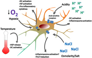
Hypoxia-Inducible Factor 1 (HIF-1) is a transcription factor that plays a crucial role in the cellular response to low oxygen levels, also known as hypoxia. It is a heterodimeric protein composed of two subunits: HIF-1α and HIF-1β.
Under normoxic conditions (adequate oxygen supply), HIF-1α is constantly produced but rapidly degraded by proteasomes due to the action of prolyl hydroxylases, which mark it for destruction in the presence of oxygen. However, under hypoxic conditions, the activity of prolyl hydroxylases is inhibited, leading to the stabilization and accumulation of HIF-1α.
Once stabilized, HIF-1α translocates to the nucleus and forms a complex with HIF-1β. This complex then binds to hypoxia-responsive elements (HREs) in the promoter regions of various genes involved in angiogenesis, glucose metabolism, erythropoiesis, cell survival, and other processes that help cells adapt to low oxygen levels.
By activating these target genes, HIF-1 plays a critical role in regulating the body's response to hypoxia, including promoting the formation of new blood vessels (angiogenesis), enhancing anaerobic metabolism, and inhibiting cell proliferation and apoptosis under low oxygen conditions. Dysregulation of HIF-1 has been implicated in several diseases, such as cancer, cardiovascular disease, and ischemic disorders.
"Cells, cultured" is a medical term that refers to cells that have been removed from an organism and grown in controlled laboratory conditions outside of the body. This process is called cell culture and it allows scientists to study cells in a more controlled and accessible environment than they would have inside the body. Cultured cells can be derived from a variety of sources, including tissues, organs, or fluids from humans, animals, or cell lines that have been previously established in the laboratory.
Cell culture involves several steps, including isolation of the cells from the tissue, purification and characterization of the cells, and maintenance of the cells in appropriate growth conditions. The cells are typically grown in specialized media that contain nutrients, growth factors, and other components necessary for their survival and proliferation. Cultured cells can be used for a variety of purposes, including basic research, drug development and testing, and production of biological products such as vaccines and gene therapies.
It is important to note that cultured cells may behave differently than they do in the body, and results obtained from cell culture studies may not always translate directly to human physiology or disease. Therefore, it is essential to validate findings from cell culture experiments using additional models and ultimately in clinical trials involving human subjects.
A cell line is a culture of cells that are grown in a laboratory for use in research. These cells are usually taken from a single cell or group of cells, and they are able to divide and grow continuously in the lab. Cell lines can come from many different sources, including animals, plants, and humans. They are often used in scientific research to study cellular processes, disease mechanisms, and to test new drugs or treatments. Some common types of human cell lines include HeLa cells (which come from a cancer patient named Henrietta Lacks), HEK293 cells (which come from embryonic kidney cells), and HUVEC cells (which come from umbilical vein endothelial cells). It is important to note that cell lines are not the same as primary cells, which are cells that are taken directly from a living organism and have not been grown in the lab.
Phenylhydrazines are organic compounds that contain a phenyl group (a benzene ring with a hydrogen atom substituted by a hydroxy group) and a hydrazine group (-NH-NH2). They are aromatic amines that have been used in various chemical reactions, including the formation of azos and hydrazones. In medicine, phenylhydrazines were once used as vasodilators to treat angina pectoris, but their use has largely been discontinued due to their toxicity and potential carcinogenicity.
Bone marrow is the spongy tissue found inside certain bones in the body, such as the hips, thighs, and vertebrae. It is responsible for producing blood-forming cells, including red blood cells, white blood cells, and platelets. There are two types of bone marrow: red marrow, which is involved in blood cell production, and yellow marrow, which contains fatty tissue.
Red bone marrow contains hematopoietic stem cells, which can differentiate into various types of blood cells. These stem cells continuously divide and mature to produce new blood cells that are released into the circulation. Red blood cells carry oxygen throughout the body, white blood cells help fight infections, and platelets play a crucial role in blood clotting.
Bone marrow also serves as a site for immune cell development and maturation. It contains various types of immune cells, such as lymphocytes, macrophages, and dendritic cells, which help protect the body against infections and diseases.
Abnormalities in bone marrow function can lead to several medical conditions, including anemia, leukopenia, thrombocytopenia, and various types of cancer, such as leukemia and multiple myeloma. Bone marrow aspiration and biopsy are common diagnostic procedures used to evaluate bone marrow health and function.
Bone marrow cells are the types of cells found within the bone marrow, which is the spongy tissue inside certain bones in the body. The main function of bone marrow is to produce blood cells. There are two types of bone marrow: red and yellow. Red bone marrow is where most blood cell production takes place, while yellow bone marrow serves as a fat storage site.
The three main types of bone marrow cells are:
1. Hematopoietic stem cells (HSCs): These are immature cells that can differentiate into any type of blood cell, including red blood cells, white blood cells, and platelets. They have the ability to self-renew, meaning they can divide and create more hematopoietic stem cells.
2. Red blood cell progenitors: These are immature cells that will develop into mature red blood cells, also known as erythrocytes. Red blood cells carry oxygen from the lungs to the body's tissues and carbon dioxide back to the lungs.
3. Myeloid and lymphoid white blood cell progenitors: These are immature cells that will develop into various types of white blood cells, which play a crucial role in the body's immune system by fighting infections and diseases. Myeloid progenitors give rise to granulocytes (neutrophils, eosinophils, and basophils), monocytes, and megakaryocytes (which eventually become platelets). Lymphoid progenitors differentiate into B cells, T cells, and natural killer (NK) cells.
Bone marrow cells are essential for maintaining a healthy blood cell count and immune system function. Abnormalities in bone marrow cells can lead to various medical conditions, such as anemia, leukopenia, leukocytosis, thrombocytopenia, or thrombocytosis, depending on the specific type of blood cell affected. Additionally, bone marrow cells are often used in transplantation procedures to treat patients with certain types of cancer, such as leukemia and lymphoma, or other hematologic disorders.
Friend murine leukemia virus (F-MuLV) is a type of retrovirus that specifically infects mice. It was first discovered by Charlotte Friend in the 1950s and has since been widely used as a model system to study retroviral pathogenesis, oncogenesis, and immune responses.
F-MuLV is a complex retrovirus that contains several accessory genes, including gag, pol, env, and others. The virus can cause leukemia and other malignancies in susceptible mice, particularly when it is transmitted from mother to offspring through the milk.
The virus is also known to induce immunosuppression, which makes infected mice more susceptible to other infections and diseases. F-MuLV has been used extensively in laboratory research to investigate various aspects of retroviral biology, including viral entry, replication, gene expression, and host immune responses.
It is important to note that Friend murine leukemia virus only infects mice and is not known to cause any disease in humans or other animals.
Reticulocytes are immature red blood cells that still contain remnants of organelles, such as ribosomes and mitochondria, which are typically found in developing cells. These organelles are involved in the process of protein synthesis and energy production, respectively. Reticulocytes are released from the bone marrow into the bloodstream, where they continue to mature into fully developed red blood cells called erythrocytes.
Reticulocytes can be identified under a microscope by their staining characteristics, which reveal a network of fine filaments or granules known as the reticular apparatus. This apparatus is composed of residual ribosomal RNA and other proteins that have not yet been completely eliminated during the maturation process.
The percentage of reticulocytes in the blood can be used as a measure of bone marrow function and erythropoiesis, or red blood cell production. An increased reticulocyte count may indicate an appropriate response to blood loss, hemolysis, or other conditions that cause anemia, while a decreased count may suggest impaired bone marrow function or a deficiency in erythropoietin, the hormone responsible for stimulating red blood cell production.
Stem Cell Factor (SCF), also known as Kit Ligand or Steel Factor, is a growth factor that plays a crucial role in the regulation of hematopoiesis, which is the process of producing various blood cells. It is a glycoprotein that binds to the c-Kit receptor found on hematopoietic stem cells and progenitor cells, promoting their survival, proliferation, and differentiation into mature blood cells.
SCF is involved in the development and function of several types of blood cells, including red blood cells, white blood cells, and platelets. It also plays a role in the maintenance and self-renewal of hematopoietic stem cells, which are essential for the continuous production of new blood cells throughout an individual's lifetime.
In addition to its role in hematopoiesis, SCF has been implicated in various other biological processes, such as melanogenesis, gametogenesis, and tissue repair and regeneration. Dysregulation of SCF signaling has been associated with several diseases, including certain types of cancer, bone marrow failure disorders, and autoimmune diseases.
Signal transduction is the process by which a cell converts an extracellular signal, such as a hormone or neurotransmitter, into an intracellular response. This involves a series of molecular events that transmit the signal from the cell surface to the interior of the cell, ultimately resulting in changes in gene expression, protein activity, or metabolism.
The process typically begins with the binding of the extracellular signal to a receptor located on the cell membrane. This binding event activates the receptor, which then triggers a cascade of intracellular signaling molecules, such as second messengers, protein kinases, and ion channels. These molecules amplify and propagate the signal, ultimately leading to the activation or inhibition of specific cellular responses.
Signal transduction pathways are highly regulated and can be modulated by various factors, including other signaling molecules, post-translational modifications, and feedback mechanisms. Dysregulation of these pathways has been implicated in a variety of diseases, including cancer, diabetes, and neurological disorders.
Hematopoiesis is the process of forming and developing blood cells. It occurs in the bone marrow and includes the production of red blood cells (erythropoiesis), white blood cells (leukopoiesis), and platelets (thrombopoiesis). This process is regulated by various growth factors, hormones, and cytokines. Hematopoiesis begins early in fetal development and continues throughout a person's life. Disorders of hematopoiesis can result in conditions such as anemia, leukopenia, leukocytosis, thrombocytopenia, or thrombocytosis.
Phlebotomy is a medical term that refers to the process of making an incision in a vein, usually in the arm, in order to draw blood. It is also commonly known as venipuncture. This procedure is performed by healthcare professionals for various purposes such as diagnostic testing, blood donation, or therapeutic treatments like phlebotomy for patients with hemochromatosis (a condition where the body absorbs too much iron from food).
The person who performs this procedure is called a phlebotomist. They must be trained in the proper techniques to ensure that the process is safe and relatively pain-free for the patient, and that the blood sample is suitable for laboratory testing.
Erythrocyte volume, also known as red cell volume or hematocrit, is the proportion of whole blood that is made up of erythrocytes or red blood cells. It is typically expressed as a percentage and can be measured using a centrifuge to separate the components of a blood sample by density.
The erythrocyte volume is an important clinical parameter because it can provide information about a person's health status, such as their hydration level, altitude acclimatization, and the presence of certain medical conditions like anemia or polycythemia. Changes in erythrocyte volume can also have significant effects on the body's oxygen-carrying capacity and overall cardiovascular function.
Hypoxia-Inducible Factor 1 (HIF-1) is a transcription factor that plays a crucial role in the body's response to low oxygen levels, also known as hypoxia. HIF-1 is a heterodimeric protein composed of two subunits: an alpha subunit (HIF-1α) and a beta subunit (HIF-1β).
The alpha subunit, HIF-1α, is the regulatory subunit that is subject to oxygen-dependent degradation. Under normal oxygen conditions (normoxia), HIF-1α is constantly produced in the cell but is rapidly degraded by proteasomes due to hydroxylation of specific proline residues by prolyl hydroxylase domain-containing proteins (PHDs). This hydroxylation reaction requires oxygen as a substrate, and under hypoxic conditions, the activity of PHDs is inhibited, leading to the stabilization and accumulation of HIF-1α.
Once stabilized, HIF-1α translocates to the nucleus, where it heterodimerizes with HIF-1β and binds to hypoxia-responsive elements (HREs) in the promoter regions of target genes. This binding results in the activation of gene transcription programs that promote cellular adaptation to low oxygen levels. These adaptive responses include increased erythropoiesis, angiogenesis, glucose metabolism, and pH regulation, among others.
Therefore, HIF-1α is a critical regulator of the body's response to hypoxia, and its dysregulation has been implicated in various pathological conditions, including cancer, cardiovascular disease, and neurodegenerative disorders.
Cell division is the process by which a single eukaryotic cell (a cell with a true nucleus) divides into two identical daughter cells. This complex process involves several stages, including replication of DNA, separation of chromosomes, and division of the cytoplasm. There are two main types of cell division: mitosis and meiosis.
Mitosis is the type of cell division that results in two genetically identical daughter cells. It is a fundamental process for growth, development, and tissue repair in multicellular organisms. The stages of mitosis include prophase, prometaphase, metaphase, anaphase, and telophase, followed by cytokinesis, which divides the cytoplasm.
Meiosis, on the other hand, is a type of cell division that occurs in the gonads (ovaries and testes) during the production of gametes (sex cells). Meiosis results in four genetically unique daughter cells, each with half the number of chromosomes as the parent cell. This process is essential for sexual reproduction and genetic diversity. The stages of meiosis include meiosis I and meiosis II, which are further divided into prophase, prometaphase, metaphase, anaphase, and telophase.
In summary, cell division is the process by which a single cell divides into two daughter cells, either through mitosis or meiosis. This process is critical for growth, development, tissue repair, and sexual reproduction in multicellular organisms.
Hypochromic anemia is a type of anemia characterized by the presence of red blood cells that have lower than normal levels of hemoglobin and appear paler in color than normal. Hemoglobin is a protein in red blood cells that carries oxygen from the lungs to the rest of the body. In hypochromic anemia, there may be a decrease in the production or increased destruction of red blood cells, leading to a reduced number of red blood cells and insufficient oxygen supply to the tissues.
Hypochromic anemia can result from various underlying medical conditions, including iron deficiency, thalassemia, chronic inflammation, lead poisoning, and certain infections or chronic diseases. Treatment for hypochromic anemia depends on the underlying cause and may include iron supplements, dietary changes, medications, or blood transfusions.
Messenger RNA (mRNA) is a type of RNA (ribonucleic acid) that carries genetic information copied from DNA in the form of a series of three-base code "words," each of which specifies a particular amino acid. This information is used by the cell's machinery to construct proteins, a process known as translation. After being transcribed from DNA, mRNA travels out of the nucleus to the ribosomes in the cytoplasm where protein synthesis occurs. Once the protein has been synthesized, the mRNA may be degraded and recycled. Post-transcriptional modifications can also occur to mRNA, such as alternative splicing and addition of a 5' cap and a poly(A) tail, which can affect its stability, localization, and translation efficiency.
Erythroid cells are a type of blood cell that develops in the bone marrow and mature into red blood cells (RBCs), also known as erythrocytes. These cells play a crucial role in the body's oxygen-carrying capacity by transporting oxygen from the lungs to the body's tissues and carbon dioxide from the tissues to the lungs.
The development of erythroid cells begins with hematopoietic stem cells, which can differentiate into various types of blood cells. Through a series of maturation stages, including proerythroblasts, basophilic erythroblasts, polychromatophilic erythroblasts, and orthochromatic erythroblasts, these cells gradually lose their nuclei and organelles to become reticulocytes. Reticulocytes are immature RBCs that still contain some residual ribosomes and are released into the bloodstream. Over time, they mature into fully functional RBCs, which have a biconcave shape and a flexible membrane that allows them to navigate through small blood vessels.
Erythroid cells are essential for maintaining adequate oxygenation of body tissues, and their production is tightly regulated by various hormones and growth factors, such as erythropoietin (EPO), which stimulates the proliferation and differentiation of erythroid progenitor cells. Abnormalities in erythroid cell development or function can lead to various blood disorders, including anemia, polycythemia, and myelodysplastic syndromes.
Cell hypoxia, also known as cellular hypoxia or tissue hypoxia, refers to a condition in which the cells or tissues in the body do not receive an adequate supply of oxygen. Oxygen is essential for the production of energy in the form of ATP (adenosine triphosphate) through a process called oxidative phosphorylation. When the cells are deprived of oxygen, they switch to anaerobic metabolism, which produces lactic acid as a byproduct and can lead to acidosis.
Cell hypoxia can result from various conditions, including:
1. Low oxygen levels in the blood (hypoxemia) due to lung diseases such as chronic obstructive pulmonary disease (COPD), pneumonia, or high altitude.
2. Reduced blood flow to tissues due to cardiovascular diseases such as heart failure, peripheral artery disease, or shock.
3. Anemia, which reduces the oxygen-carrying capacity of the blood.
4. Carbon monoxide poisoning, which binds to hemoglobin and prevents it from carrying oxygen.
5. Inadequate ventilation due to trauma, drug overdose, or other causes that can lead to respiratory failure.
Cell hypoxia can cause cell damage, tissue injury, and organ dysfunction, leading to various clinical manifestations depending on the severity and duration of hypoxia. Treatment aims to correct the underlying cause and improve oxygen delivery to the tissues.
Neuroprotective agents are substances that protect neurons or nerve cells from damage, degeneration, or death caused by various factors such as trauma, inflammation, oxidative stress, or excitotoxicity. These agents work through different mechanisms, including reducing the production of free radicals, inhibiting the release of glutamate (a neurotransmitter that can cause cell damage in high concentrations), promoting the growth and survival of neurons, and preventing apoptosis (programmed cell death). Neuroprotective agents have been studied for their potential to treat various neurological disorders, including stroke, traumatic brain injury, Parkinson's disease, Alzheimer's disease, and multiple sclerosis. However, more research is needed to fully understand their mechanisms of action and to develop effective therapies.
Ferritin is a protein in iron-metabolizing cells that stores iron in a water-soluble form. It is found inside the cells (intracellular) and is released into the bloodstream when the cells break down or die. Measuring the level of ferritin in the blood can help determine the amount of iron stored in the body. High levels of ferritin may indicate hemochromatosis, inflammation, liver disease, or other conditions. Low levels of ferritin may indicate anemia, iron deficiency, or other conditions.
Cell differentiation is the process by which a less specialized cell, or stem cell, becomes a more specialized cell type with specific functions and structures. This process involves changes in gene expression, which are regulated by various intracellular signaling pathways and transcription factors. Differentiation results in the development of distinct cell types that make up tissues and organs in multicellular organisms. It is a crucial aspect of embryonic development, tissue repair, and maintenance of homeostasis in the body.
Bloodletting is a medical procedure that was commonly used in the past to balance the four humors of the body, which were believed to be blood, phlegm, black bile, and yellow bile. The procedure involved withdrawing blood from a patient through various methods such as venesection (making an incision in a vein), leeches, or cupping.
The theory behind bloodletting was that if one humor became overabundant, it could cause disease or illness. By removing some of the excess humor, practitioners believed they could restore balance and promote healing. Bloodletting was used to treat a wide variety of conditions, including fever, inflammation, and pain.
While bloodletting is no longer practiced in modern medicine, it was once a common treatment for many different ailments. The practice dates back to ancient times and was used by various cultures throughout history, including the Greeks, Romans, Egyptians, and Chinese. However, its effectiveness as a medical treatment has been called into question, and it is now considered an outdated and potentially harmful procedure.
A dose-response relationship in the context of drugs refers to the changes in the effects or symptoms that occur as the dose of a drug is increased or decreased. Generally, as the dose of a drug is increased, the severity or intensity of its effects also increases. Conversely, as the dose is decreased, the effects of the drug become less severe or may disappear altogether.
The dose-response relationship is an important concept in pharmacology and toxicology because it helps to establish the safe and effective dosage range for a drug. By understanding how changes in the dose of a drug affect its therapeutic and adverse effects, healthcare providers can optimize treatment plans for their patients while minimizing the risk of harm.
The dose-response relationship is typically depicted as a curve that shows the relationship between the dose of a drug and its effect. The shape of the curve may vary depending on the drug and the specific effect being measured. Some drugs may have a steep dose-response curve, meaning that small changes in the dose can result in large differences in the effect. Other drugs may have a more gradual dose-response curve, where larger changes in the dose are needed to produce significant effects.
In addition to helping establish safe and effective dosages, the dose-response relationship is also used to evaluate the potential therapeutic benefits and risks of new drugs during clinical trials. By systematically testing different doses of a drug in controlled studies, researchers can identify the optimal dosage range for the drug and assess its safety and efficacy.
In the field of medicine, "time factors" refer to the duration of symptoms or time elapsed since the onset of a medical condition, which can have significant implications for diagnosis and treatment. Understanding time factors is crucial in determining the progression of a disease, evaluating the effectiveness of treatments, and making critical decisions regarding patient care.
For example, in stroke management, "time is brain," meaning that rapid intervention within a specific time frame (usually within 4.5 hours) is essential to administering tissue plasminogen activator (tPA), a clot-busting drug that can minimize brain damage and improve patient outcomes. Similarly, in trauma care, the "golden hour" concept emphasizes the importance of providing definitive care within the first 60 minutes after injury to increase survival rates and reduce morbidity.
Time factors also play a role in monitoring the progression of chronic conditions like diabetes or heart disease, where regular follow-ups and assessments help determine appropriate treatment adjustments and prevent complications. In infectious diseases, time factors are crucial for initiating antibiotic therapy and identifying potential outbreaks to control their spread.
Overall, "time factors" encompass the significance of recognizing and acting promptly in various medical scenarios to optimize patient outcomes and provide effective care.
Cobalt is a chemical element with the symbol Co and atomic number 27. It is a hard, silver-white, lustrous, and brittle metal that is found naturally only in chemically combined form, except for small amounts found in meteorites. Cobalt is used primarily in the production of magnetic, wear-resistant, and high-strength alloys, as well as in the manufacture of batteries, magnets, and pigments.
In a medical context, cobalt is sometimes used in the form of cobalt-60, a radioactive isotope, for cancer treatment through radiation therapy. Cobalt-60 emits gamma rays that can be directed at tumors to destroy cancer cells. Additionally, small amounts of cobalt are present in some vitamin B12 supplements and fortified foods, as cobalt is an essential component of vitamin B12. However, exposure to high levels of cobalt can be harmful and may cause health effects such as allergic reactions, lung damage, heart problems, and neurological issues.
GATA1 (Global Architecture of Tissue/stage-specific Transcription Factors 1) is a transcription factor that belongs to the GATA family, which recognizes and binds to the (A/T)GATA(A/G) motif in the DNA. It plays a crucial role in the development and differentiation of hematopoietic cells, particularly erythroid, megakaryocytic, eosinophilic, and mast cell lineages.
GATA1 regulates gene expression by binding to specific DNA sequences and recruiting other co-factors that modulate chromatin structure and transcriptional activity. Mutations in the GATA1 gene can lead to various blood disorders such as congenital dyserythropoietic anemia type II, Diamond-Blackfan anemia, acute megakaryoblastic leukemia (AMKL), and myelodysplastic syndrome.
In summary, GATA1 Transcription Factor is a protein that binds to specific DNA sequences in the genome and regulates gene expression, playing a critical role in hematopoietic cell development and differentiation.
An erythrocyte transfusion, also known as a red blood cell (RBC) transfusion, is the process of transferring compatible red blood cells from a donor to a recipient. This procedure is typically performed to increase the recipient's oxygen-carrying capacity, usually in situations where there is significant blood loss, anemia, or impaired red blood cell production.
During the transfusion, the donor's red blood cells are collected, typed, and tested for compatibility with the recipient's blood to minimize the risk of a transfusion reaction. Once compatible units are identified, they are infused into the recipient's circulation through a sterile intravenous (IV) line. The recipient's body will eventually eliminate the donated red blood cells within 100-120 days as part of its normal turnover process.
Erythrocyte transfusions can be lifesaving in various clinical scenarios, such as trauma, surgery, severe anemia due to chronic diseases, and hematologic disorders. However, they should only be used when necessary, as there are potential risks associated with the procedure, including allergic reactions, transmission of infectious diseases, transfusion-related acute lung injury (TRALI), and iron overload in cases of multiple transfusions.
Hypoxia-Inducible Factor (HIF) is a transcription factor that plays a crucial role in the body's response to low oxygen levels (hypoxia). HIF is composed of two subunits: an alpha subunit and a beta subunit. Under normal oxygen conditions, the alpha subunit is constantly being broken down by prolyl hydroxylase domain-containing proteins, which are a type of enzyme known as HIF-Proline Dioxygenases (HIF-PDOs).
HIF-PDOs post-translationally modify the HIF alpha subunit by adding a hydroxyl group to specific proline residues. This modification marks the HIF alpha subunit for degradation by the proteasome, a complex that breaks down unneeded or damaged proteins in the cell. However, under hypoxic conditions, the activity of HIF-PDOs is inhibited, leading to the stabilization and accumulation of HIF alpha subunits.
Once stabilized, HIF alpha subunits dimerize with HIF beta subunits and translocate to the nucleus where they bind to hypoxia response elements (HREs) in the DNA. This binding induces the expression of genes involved in various cellular responses to hypoxia, such as angiogenesis, metabolic reprogramming, and erythropoiesis. Therefore, HIF-PDOs play a critical role in regulating the body's response to low oxygen levels by controlling the stability and activity of HIF.
"Iron radioisotopes" refer to specific forms of the element iron that have unstable nuclei and emit radiation. These isotopes are often used in medical imaging and treatment procedures due to their ability to be detected by specialized equipment. Common iron radioisotopes include Iron-52, Iron-55, Iron-59, and Iron-60. They can be used as tracers to study the distribution, metabolism, or excretion of iron in the body, or for targeted radiation therapy in conditions such as cancer.
Hematopoietic cell growth factors are a group of glycoproteins that stimulate the proliferation, differentiation, and survival of hematopoietic cells, which are the precursor cells that give rise to all blood cells. These growth factors include colony-stimulating factors (CSFs) such as granulocyte-colony stimulating factor (G-CSF), granulocyte-macrophage colony-stimulating factor (GM-CSF), and macrophage colony-stimulating factor (M-CSF), as well as erythropoietin (EPO) and thrombopoietin (TPO).
G-CSF primarily stimulates the production of neutrophils, a type of white blood cell that plays a crucial role in the immune response to bacterial infections. GM-CSF stimulates the production of both granulocytes and monocytes/macrophages, while M-CSF specifically stimulates the production of monocytes/macrophages. EPO stimulates the production of red blood cells, while TPO stimulates the production of platelets.
Hematopoietic cell growth factors are used clinically to treat a variety of conditions associated with impaired hematopoiesis, such as chemotherapy-induced neutropenia, aplastic anemia, and congenital disorders of hematopoiesis. They can also be used to mobilize hematopoietic stem cells from the bone marrow into the peripheral blood for collection and transplantation.
A kidney, in medical terms, is one of two bean-shaped organs located in the lower back region of the body. They are essential for maintaining homeostasis within the body by performing several crucial functions such as:
1. Regulation of water and electrolyte balance: Kidneys help regulate the amount of water and various electrolytes like sodium, potassium, and calcium in the bloodstream to maintain a stable internal environment.
2. Excretion of waste products: They filter waste products from the blood, including urea (a byproduct of protein metabolism), creatinine (a breakdown product of muscle tissue), and other harmful substances that result from normal cellular functions or external sources like medications and toxins.
3. Endocrine function: Kidneys produce several hormones with important roles in the body, such as erythropoietin (stimulates red blood cell production), renin (regulates blood pressure), and calcitriol (activated form of vitamin D that helps regulate calcium homeostasis).
4. pH balance regulation: Kidneys maintain the proper acid-base balance in the body by excreting either hydrogen ions or bicarbonate ions, depending on whether the blood is too acidic or too alkaline.
5. Blood pressure control: The kidneys play a significant role in regulating blood pressure through the renin-angiotensin-aldosterone system (RAAS), which constricts blood vessels and promotes sodium and water retention to increase blood volume and, consequently, blood pressure.
Anatomically, each kidney is approximately 10-12 cm long, 5-7 cm wide, and 3 cm thick, with a weight of about 120-170 grams. They are surrounded by a protective layer of fat and connected to the urinary system through the renal pelvis, ureters, bladder, and urethra.
Molecular sequence data refers to the specific arrangement of molecules, most commonly nucleotides in DNA or RNA, or amino acids in proteins, that make up a biological macromolecule. This data is generated through laboratory techniques such as sequencing, and provides information about the exact order of the constituent molecules. This data is crucial in various fields of biology, including genetics, evolution, and molecular biology, allowing for comparisons between different organisms, identification of genetic variations, and studies of gene function and regulation.
Transferrin receptors are membrane-bound proteins found on the surface of many cell types, including red and white blood cells, as well as various tissues such as the liver, brain, and placenta. These receptors play a crucial role in iron homeostasis by regulating the uptake of transferrin, an iron-binding protein, into the cells.
Transferrin binds to two ferric ions (Fe3+) in the bloodstream, forming a complex known as holo-transferrin. This complex then interacts with the transferrin receptors on the cell surface, leading to endocytosis of the transferrin-receptor complex into the cell. Once inside the cell, the acidic environment within the endosome causes the release of iron ions from the transferrin molecule, which can then be transported into the cytoplasm for use in various metabolic processes.
After releasing the iron, the apo-transferrin (iron-free transferrin) is recycled back to the cell surface and released back into the bloodstream, where it can bind to more ferric ions and repeat the cycle. This process helps maintain appropriate iron levels within the body and ensures that cells have access to the iron they need for essential functions such as DNA synthesis, energy production, and oxygen transport.
In summary, transferrin receptors are membrane-bound proteins responsible for recognizing and facilitating the uptake of transferrin-bound iron into cells, playing a critical role in maintaining iron homeostasis within the body.
 Erythropoietin
Erythropoietin receptor
Erythropoietin in neuroprotection
Continuous erythropoietin receptor activator
Epileptogenesis
Eugene Goldwasser
Rejuvenation
Doping at the 1999 Tour de France
Doping at the 1998 Tour de France
Daprodustat
List of stripped Olympic medals
Zidovudine
Amgen
Epitestosterone
Tan Chorh Chuan
List of Olympic Games scandals and controversies
Soviet Union
1980 Summer Olympics
Doping in sport
Polycythemia
Endocrine gland
Doping at the Olympic Games
Altitude
Megakaryocyte
Renal cell carcinoma
Hereditary haemochromatosis
UFC 174
UFC Fight Night: Cejudo vs. Dillashaw
List of recombinant proteins
Assessment of kidney function
Erythropoietin - Wikipedia
 erythropoietin - Search Results - PubMed
 Erythropoietin test
Epo MGI Mouse Gene Detail - MGI:95407 - erythropoietin
Zebrafish Kit ligands cooperate with erythropoietin to promote erythroid cell expansion
 Man Trying To Build Muscle Stock Photo - Download Image Now - Erythropoietin, Adult, Aggression - iStock
 cellular response to erythropoietin Antibodies | Invitrogen
...
cellular response to erythropoietin Antibodies | Invitrogen
...
 Mechanical Stretch Promotes the Osteogenic Differentiation of Bone Mesenchymal Stem Cells Induced by Erythropoietin
 6I4X: Crystal structure of SOCS2:Elongin C:Elongin B in complex with erythropoietin receptor peptide
Erythropoietin-Behandlung der tumorassoziierten Anämie bei Patienten mit multiplem Myelom | Onkologie | Karger Publishers
 Human Erythropoietin/EPO Antibody MAB287-100: R&D Systems
 Erythropoiesis from human embryonic stem cells through erythropoietin-independent AKT signaling. - CIRM
 Systematic Review of Controlled Trials on Erythropoietin to Support Evidence-Based Guidelines
 Association between recombinant human erythropoietin and quality of life and exercise capacity of patients receiving...
Erythropoietin Is a Paracrine Mediator of Ischemic Tolerance in the Brain: Evidence from an In Vitro Model | Journal of...
 Erythropoietin benefits from controlled sugar-coating | Research | Chemistry World
 What is Recombinant human erythropoietin injection? - wren-clothing.com
 Erythropoietin to improve outcomes for critically ill trauma patients | Health Research Council of New Zealand
Erythropoietin Response to Anaemia in Children with Sickle Cell Disease and Fanconi's Hypoproliferative Anaemia | Citedby...
 Erythropoietin drugs market is Booming Across the Globe and Witness Huge Growth by Key Players to 2028 | Amgen Inc., Roche,...
Neutral protease activity and erythropoietin production in the rat after cobalt administration. | Journal of Pharmacology and...
Pharmacokinetic/Pharmacodynamic Analysis of Paradoxal Regulation of Erythropoietin Production in Acute Anemia | Journal of...
 An integrative 'Omics' solution to the detection of recombinant human erythropoietin and blood doping | British Journal of...
 Molecular Vision: Aqueous humor erythropoietin levels in open-angle glaucoma patients with and without TTR V30M familial...
Systemic endogenous erythropoietin and associated disorders in extremely preterm newborns | ADC Fetal & Neonatal Edition
 The effect of common hematologic abnormalities on the ability of blood models to detect erythropoietin abuse by athletes
...
The effect of common hematologic abnormalities on the ability of blood models to detect erythropoietin abuse by athletes
...
2007-P: Age-Dependent Metabolic Response to Erythropoietin in Mice | Diabetes | American Diabetes Association
 Endogenous erythropoietin signaling facilitates skeletal muscle repair and recovery following pharmacologically induced damage ...
 Erythropoietin - MedicalRecords.com
Does Erythropoietin Improve Cognitive Symptoms in Patients with Cancer? A Systematic Review<...
Recombinant13
- Exogenous erythropoietin, recombinant human erythropoietin (rhEPO), is produced by recombinant DNA technology in cell culture and are collectively called erythropoiesis-stimulating agents (ESA): two examples are epoetin alfa and epoetin beta. (wikipedia.org)
- BACKGROUND: Among patients with chronic kidney disease (CKD), the use of recombinant human erythropoietin and its derivatives for the treatment of anemia has been linked to a possibly increased risk of stroke, myocardial infarction, and other adverse events. (nih.gov)
- 323. The Neutralization Dose (ND 50 ) is typically 0.5-1.5 µg/mL in the presence of 0.3 units/mL Recombinant Human Erythropoietin/EPO (Tissue Culture Grade). (rndsystems.com)
- Recombinant Human Erythropoietin/EPO (Tissue Culture Grade) (Catalog # 287-TC ) stimulates proliferation in the TF-1 human erythroleukemic cell line in a dose-dependent manner (orange line). (rndsystems.com)
- Proliferation elicited by Recombinant Human Erythropoietin/EPO (Tissue Culture Grade) (0.3 units/mL) is neutralized (green line) by increasing concentrations of Mouse Anti-Human Erythropoietin/EPO Monoclonal Antibody (Catalog # MAB287). (rndsystems.com)
- Association between recombinant human erythropoietin and quality of life and exercise capacity of patients receiving haemodialysis. (bmj.com)
- Commercial erythropoietin preparations contain a mixture of glycoforms, an inherent effect of the recombinant DNA technology used. (chemistryworld.com)
- What is Recombinant human erythropoietin injection? (wren-clothing.com)
- High level utilization of recombinant DNA innovation empowers creation of erythropoietin for in-vitro conditions/manufactured variations, which are alluded to as recombinant human erythropoietin (rHuEPO). (medgadget.com)
- BACKGROUND AND OBJECTIVES: Algorithms that combine scores from multiple blood parameters are demonstrably effective in highlighting recombinant human erythropoietin (rHuEPO) administration, and have been used to deter rHuEPO use by athletes. (haematologica.org)
- We have examined whether the secretion of erythropoietin (Epo) from genetically modified cells could represent an alternative to repeated injections of the recombinant hormone for treating chronic anemias responsive to Epo. (pasteur.fr)
- Since the availability of recombinant human erythropoietin to patients in 1989, anemia and transfusion requirements have become relatively rare in patients on hemodialysis. (medicinehack.com)
- Since recombinant human erythropoietin (rHuEPO) induces a range of cytoprotective cellular responses, rHuEPO treatment may reduce PD fluid (PDF)-induced damage. (bvsalud.org)
RHuEPO1
- In dieser klinischen Studie behandelten wir 13 Patienten mit tumorassoziierter chronischer Anämie und multiplem Myelom dreimal wöchentlich mit rekombinantem humanem Erythropoietin (rHuEPO). (karger.com)
Anemia11
- Erythropoietin is a glycoprotein hormone that stimulates red blood cell (RBC) production in bone marrow via binding to the cell-surface receptor on hematopoietic progenitor cells, and it has been widely used for treating anemia [ 13 ]. (hindawi.com)
- To support evidence-based clinical guidelines on erythropoietin use for anemia in oncology, we conducted systematic reviews of controlled trials on four patient groups. (cancernetwork.com)
- The kidney hormone erythropoietin controls red blood cell production and is a common treatment for anemia, but it also reduces inflammation. (hrc.govt.nz)
- The regulatory mechanism responsible for a paradoxal, rapid drop in the erythropoietin (EPO) plasma level seen 2 to 4 days after acute, phlebotomy-induced anemia was investigated in seven adult sheep. (aspetjournals.org)
- Synthetic (man-made) erythropoietin may be used during cancer treatment to prevent anemia or to treat anemia caused by chemotherapy or bone marrow transplant. (medicalrecords.com)
- Erythropoietin also can be used to treat anemia caused by chronic kidney disease and kidney failure. (medicalrecords.com)
- Anemia secondary to AZT and low erythropoietin levels in HIV-infected pts. (hopkinsguides.com)
- Anemia in diabetic patients - Erythropoietin treatment? (medicinehack.com)
- If you encounter a normochromic and normocytic anemia in a diabetic patient, do not forget that it may be a case of erythropoietin deficiency. (medicinehack.com)
- In cases of decreased renal mass, the level of erythropoietin falls and does not increase much in response to hypoxia (anemia). (medicinehack.com)
- In some conditions, erythropoietin testing can be used in the assessment and differentiation of anemia, especially in patients receiving erythropoietin replacement therapy with an inadequate response. (medscape.com)
Glycoprotein hormone2
- Red cell production in vertebrates is controlled by a glycoprotein hormone known as erythropoietin (Ep), which is produced by the kidney in response to hypoxia and acts on the marrow to selectively stimulate erythropoiesis. (nih.gov)
- Erythropoietin (EPO) is a glycoprotein hormone, naturally produced by the peritubular cells of the kidney, that stimulates red blood cell production. (wren-clothing.com)
Effects of erythropoietin1
- The effects of erythropoietin (EPO) on the behaviors of bone marrow mesenchymal stem cells (BMSCs) subjected to mechanical stretch remain unclear. (hindawi.com)
Sensitive to erythropoietin2
- The burst-forming unit-erythroid (BFU-E) cells start erythropoietin receptor expression and are sensitive to erythropoietin. (wikipedia.org)
- With corresponding decreased levels of erythropoietin in older children with the same degree of severe malarial anaemia, conceivably, the bone marrows of younger children with acute malaria may be less sensitive to erythropoietin. (biomedcentral.com)
Erythroid7
- Subsequent stage, the colony-forming unit-erythroid (CFU-E), expresses maximal erythropoietin receptor density and is completely dependent on erythropoietin for further differentiation. (wikipedia.org)
- High level erythropoietin receptor expression is localized to erythroid progenitor cells. (wikipedia.org)
- To further address the role of kitlga and kitlgb in hematopoietic development in vivo, we performed gain-of-function experiments in zebrafish embryos, showing that both ligands cooperate with erythropoietin (Epo) to promote erythroid cell expansion. (nih.gov)
- Erythropoietin (EPO) a glycoprotein produced primarily by the kidney, is the principal factor that regulates erythropoiesis by stimulating the proliferation and differentiation of erythroid progenitor cells. (rndsystems.com)
- We present here evidence that ic-MPL dimerization induces erythropoietin (EPO)-independent erythroid differentiation from hESC by inducing the generation of erythroid progenitors and by promoting more efficient erythroid maturation with increased RBC enucleation as well as increased gamma:epsilon globin ratio and production of beta-globin protein. (ca.gov)
- Erythropoietin acts by binding to its cell surface receptor on erythroid progenitor cells to stimulate erythrocyte production. (uzh.ch)
- Erythropoietin (Epo) regulates the proliferation and differentiation of erythroid precursors. (drugbank.com)
Proliferation7
- Measured by its ability to neutralize Erythropoietin/EPO-induced proliferation in the TF‑1 human erythroleukemic cell line. (rndsystems.com)
- Cell Proliferation Induced by Erythropoietin/EPO and Neutralization by Human Erythropoietin/EPO Antibody. (rndsystems.com)
- Mice with erythropoietin receptor restricted to hematopoietic tissue were used to assess contributions of endogenous erythropoietin to promote skeletal myoblast proliferation and survival and wound healing in a mouse model of cardiotoxin induced muscle injury. (uzh.ch)
- In contrast, mice with chronic elevated circulating erythropoietin had more Pax-7(+) satellite cells and myoblasts with increased proliferation and survival in culture, decreased muscle injury, and accelerated recovery of maximum load tolerated by isolated muscle. (uzh.ch)
- Erythropoietin promoted proliferation, survival, and wound recovery in myoblasts via the phosphoinositide 3-kinase/AKT pathway. (uzh.ch)
- Therefore, endogenous and exogenous erythropoietin contribute to increasing satellite cell number following muscle injury, improve myoblast proliferation and survival, and promote repair and regeneration in this mouse induced muscle injury model independent of its effect on erythrocyte production. (uzh.ch)
- Objective: The erythropoietin receptor (EpoR) stimulates erythro- cyte proliferation after erythropoietin binding. (edu.au)
Hormone erythropoietin1
- In a recent randomized controlled clinical trial , researchers looked at the hormone erythropoietin (EPO) effects on wound healing after gingival graft surgery. (todaysrdh.com)
Endogenous erythropoietin2
- Objective To explore the association between concentrations of endogenous erythropoietin (EPO) in blood the first 2 weeks of life and neonatal disorders in extremely low gestational age newborns (ELGANs). (bmj.com)
- Skeletal muscle myoblasts also produced endogenous erythropoietin that increased at low O(2). (uzh.ch)
Differentiation1
- Under hypoxic conditions, the kidney will produce and secrete erythropoietin to increase the production of red blood cells by targeting CFU-E, proerythroblast and basophilic erythroblast subsets in the differentiation. (wikipedia.org)
EpoR1
- Erythropoietin has been shown to exert its effects by binding to the erythropoietin receptor (EpoR). (wikipedia.org)
Stimulates the production1
- These cells possess specific regulatory mechanisms, called hypoxia-inducible factors (HIFs), which, under hypoxic conditions, stimulate the production of erythropoietin, which stimulates the production of erythrocytes. (medscape.com)
Cytokine2
- Erythropoietin (EPO) is a cytokine that has been shown to play a role in neuroprotection and is endogenously produced in the eye. (molvis.org)
- Erythropoietin (EPO), the cytokine required for erythrocyte production, regulates glucose homeostasis and fat mass accumulation in animal models. (diabetesjournals.org)
Renal4
- The generation of erythropoietin purportedly involves the interaction of an enzyme, of renal origin, and a circulating plasma protein substrate. (aspetjournals.org)
- Cathepsins A and B and plasma protease were sensitive to the same enzymatic inhibitory agents, and all three enzymes demonstrated activity patterns similar to that of the renal erythropoietic factor [enzyme(s) presumed responsible for generating erythropoietin] in rats treated with cobalt. (aspetjournals.org)
- Regulation of renal erythropoietin (EPO) production is based on an intrarenal oxygen sensor. (uni-regensburg.de)
- Erythropoietin is secreted by fibroblasts in the renal cortex. (medscape.com)
Kidneys4
- What would happen if the kidneys could not produce erythropoietin hormone? (wren-clothing.com)
- What does erythropoietin do in the kidneys? (wren-clothing.com)
- Erythropoietin is a hormone made mainly in the kidneys that stimulates the bone marrow to produce new red blood cells. (medicalrecords.com)
- In adults, about 85-90% of the erythropoietin comes from the kidneys and 10-15% from the liver. (medicinehack.com)
Injection5
- What is erythropoietin injection used for? (wren-clothing.com)
- What is the cost of erythropoietin injection? (wren-clothing.com)
- Is erythropoietin injection safe? (wren-clothing.com)
- We will perform a study involving 2500 adults with severe injuries in five countries (New Zealand, Australia, Republic of Ireland, United Kingdom and Finland) who will receive either erythropoietin or a dummy injection under the skin, in addition to all usual treatment. (hrc.govt.nz)
- https://www.wada-ama.org/en/questions-answers/blood-doping) EPO (erythropoietin) is naturally produced by the body and can also be administered via injection. (cram.com)
Chemotherapy1
- Some evidence supports erythropoietin as a preventive intervention to maintain normal cognitive function during and after chemotherapy. (kcl.ac.uk)
19891
- Bioengineered erythropoietin has been used clinically and misused athletically since it first appeared on the market in 1989. (chemistryworld.com)
Human2
Receptor expression1
- Erythropoietin receptor expression in nonhematopoietic tissue, including skeletal muscle progenitor cells, raises the possibility of a role for erythropoietin beyond erythropoiesis. (uzh.ch)
Kidney3
- Erythropoietin is produced by interstitial fibroblasts in the kidney in close association with the peritubular capillary and proximal convoluted tubule. (wikipedia.org)
- Erythropoietin (EPO) is produced by the kidney and used to make red blood cells. (wren-clothing.com)
- Ascend in pervasiveness of malignant growth, HIV, iron deficiency, and kidney sicknesses, flood in degree of mindfulness about the advantages of EPO therapeutics, and commercialization for erythropoietin biosimilars drive the development of the worldwide erythropoietin drugs market. (medgadget.com)
Antibodies1
- Our range of Erythropoietin antibodies (also known as EPO, Hematopoietin or Hemopoietin) come in a number of formats and to a range of target species. (bio-rad-antibodies.com)
Erythropoietic factor1
- Erythropoietin is the primary erythropoietic factor that cooperates with various other growth factors (e.g. (wikipedia.org)
Blood4
- Erythropoietin is an essential hormone for red blood cell production. (wikipedia.org)
- Erythropoietin has its primary effect on red blood cell progenitors and precursors (which are found in the bone marrow in humans) by promoting their survival through protecting these cells from apoptosis, or cell death. (wikipedia.org)
- The erythropoietin test measures the amount of a hormone called erythropoietin (EPO) in blood. (ucsfhealth.org)
- Erythropoietin is a hormone responsible for regulating red blood cell production. (chemistryworld.com)
Purportedly1
- Age purportedly has a direct effect on background levels of erythropoietin. (biomedcentral.com)
Hematopoietin1
- Erythropoietin/hematopoietin is a glycoprotein chemical that triggers creation of red platelets in the bone marrow through erythropoiesis. (medgadget.com)
Erythropoiesis-stimul1
- All of the erythropoietin preparations are now referred to collectively as erythropoiesis stimulating-agents (ESAs). (medicinehack.com)
Production4
- We also found that the synthetic protein acts to increase red cell production independently of the well known growth factor erythropoietin. (ca.gov)
- Neutral protease activity and erythropoietin production in the rat after cobalt administration. (aspetjournals.org)
- In this study we correlated the levels of erythropoietin (EPO), as an indicator of stimulation of haemoglobin production, to the levels of monocyte acquired haemozoin in children with both severe and uncomplicated malaria. (biomedcentral.com)
- Erythropoietin, a member of class 1 cytokines, is a 30.4-kDa glycoprotein composed of 4 chains of carbohydrate and a protein chain composed of 193 amino acids that undergo a cleavage process, resulting in the production of mature erythropoietin (165 amino acids). (medscape.com)
Synthetic1
- Researchers in New Zealand have developed a method for glycosylating synthetic erythropoietin. (chemistryworld.com)
Mice1
- These data show that erythropoietin treatment can stimulate cell oxygen consumption and can contribute to regulation of metabolism and body weight in mice. (wren-clothing.com)
Synthesis1
- Yasuhiro Kajihara from Osaka University, Japan, who researches glycoprotein synthesis and has worked on producing homogenous erythropoietin analogues, comments that while click chemistry has been used to produce PEGylated erythropoietin, previous reports required tough chemical syntheses. (chemistryworld.com)
Systematic Review1
- In 1998, the American Society of Hematology (ASH) and theAmerican Society of Clinical Oncology (ASCO) jointly nominated the topic'Uses of Erythropoietin in Oncology' to AHRQ for an Evidence-basedPractice Center systematic review and evidence report. (cancernetwork.com)
Protein3
- By careful optimisation of the exact nature of saccharides introduced onto the erythropoietin protein backbone it should be possible to mimic the exact biological activity of native erythropoietin. (chemistryworld.com)
- Her team harnessed the combined power of peptide ligation to build the erythropoietin protein and click chemistry to decorate it with sugars. (chemistryworld.com)
- Brimble's team also studied the relationship between glycosylation and protein folding using their erythropoietin analogues and demonstrated how oligosaccharides affect erythropoietin folding and aggregation, which impacts biological activity. (chemistryworld.com)
19911
- Above is a diagram showing the change in hemoglobin level (blue line) with respect to different dosage of erythropoietin from 1991 till 2009 in an attempt to keep the hemoglobin in the target range. (medicinehack.com)
Apoptosis1
- Erythropoietin prevents dialysis fluid-induced apoptosis of mesothelial cells. (bvsalud.org)
Pathways1
- Involvement of SH2-containing phosphotyrosine phosphatase Syp in erythropoietin receptor signal transduction pathways. (drugbank.com)
Transfusion1
- N = 1,927) demonstrated reduced odds of transfusion after erythropoietin, but higher-quality trials reported smaller odds reductions. (cancernetwork.com)
Anemic1
- The role of erythropoietin stimulating agents in anemic patients with heart failure: solved and unresolved questions. (medscape.com)
Precursors1
- Precursors of red cells, the proerythroblasts and basophilic erythroblasts also express erythropoietin receptor and are therefore affected by it. (wikipedia.org)
Cognitive9
- Does Erythropoietin Improve Cognitive Symptoms in Patients with Cancer? (kcl.ac.uk)
- Aim: To review the effectiveness of erythropoietin to improve cognitive symptoms such as memory or attention problem and related symptoms in patients with cancer. (kcl.ac.uk)
- All study designs, cancer patients with symptoms of cognitive impairment (memory or attention problem and related symptoms) receiving erythropoietin were included. (kcl.ac.uk)
- Prevalence of cognitive impairment before receiving erythropoietin was within the normal range for cognition, bar one study including elderly patients with cancer. (kcl.ac.uk)
- Significant improvements in cognitive function were seen in elderly patients receiving high doses of erythropoietin and cognitive domains, particularly memory, in women with breast cancer. (kcl.ac.uk)
- Conclusion: Erythropoietin may improve cognitive function in cancer patients receiving erythropoietin but further evidence is required. (kcl.ac.uk)
- Dive into the research topics of 'Does Erythropoietin Improve Cognitive Symptoms in Patients with Cancer? (kcl.ac.uk)
- Significantimprovements in cognitive function were seen inelderly patients receiving high doses of erythropoietin andcognitive domains, particularly memory, in women withbreast cancer. (kcl.ac.uk)
- Participants with solid malignancy reportedimproved quality of life.Conclusion: Erythropoietin may improve cognitive functionin cancer patients receiving erythropoietin but furtherevidence is required. (kcl.ac.uk)
Patients2
- Une anémie a été diagnostiquée chez 75 patients (37,5 %) en tout : 45 avaient une anémie hypochrome microcytaire, 18 une anémie normochrome normocytaire et 12 une anémie hyperchrome macrocytaire. (who.int)
- Sur les 75 patients, 41 (54,7 %) avaient une carence en fer, 14 (18,7 %) avaient une carence en folates et 14 (18,7 %) avaient une thalassémie mineure. (who.int)
Detect1
- ProTec Bioseparation has succeeded in producing a novel gel matrix ( ProPhyl Air ), which makes it possible to detect Erythropoietin (EPO) by means of isoelectric focusing directly in the gel with a conventional Coomassie dye. (protec-biosep.de)
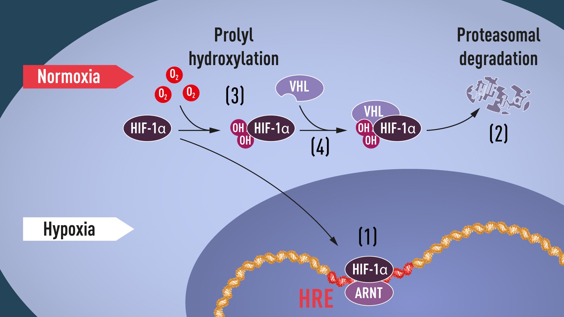


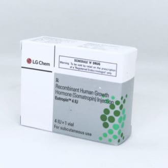

![MELDONIUM [MILDRONATE] comprar nos Estados Unidos da América, preço favorável para MELDONIUM [MILDRONATE] na farmácia 24hoursppc](https://24hoursppc.org/wp-content/uploads/2020/01/mildronate.png)