Epidermolysis Bullosa
Epidermolysis Bullosa Dystrophica
Epidermolysis Bullosa Simplex
Epidermolysis Bullosa, Junctional
Epidermolysis Bullosa Acquisita
Collagen Type VII
Plectin
Keratin-14
Skin
Pylorus
Non-Fibrillar Collagens
Integrin beta4
Hemidesmosomes
Keratin-5
Keratins
Skin Diseases, Vesiculobullous
Keratinocytes
Encyclopedias as Topic
Minnesota
Group Purchasing
Bibliometrics
Publications
Research
Peer Review
Dominant dystrophic epidermolysis bullosa (Pasini) caused by a novel glycine substitution mutation in the type VII collagen gene (COL7A1). (1/105)
A 12 y old girl with the albopapuloid variant (Pasini) of dominant dystrophic epidermolysis bullosa is studied. The albopapuloid lesions developed within the first year of life, contained milia and were associated with pruritus. Mutation detection of the COL7A1 gene revealed a G-->A transition at nucleotide position 6110 in the mutant allele converting a glycine to glutamic acid (G2037E). This report adds to the expanding database on COL7A1 mutations in dystrophic epidermolysis bullosa. (+info)The eye in epidermolysis bullosa. (2/105)
AIMS: To describe the ophthalmic findings in a large cohort of epidermolysis bullosa (EB) patients managed in one large specialist centre. METHODS: A case note review of consecutive patients seen at Great Ormond Street Children's Hospital. Data on the dermatological disease, ophthalmic history, and examination were collected and coded onto a data sheet. RESULTS: 181 patients: 50 (28%) simplex EB; 15 (8%) junctional EB; 28 (15%) autosomal dominant dystrophic EB; 72 (40%) autosomal recessive dystrophic EB; nine patients (5%) with dystrophic EB whose inheritance could not be ascertained; and seven cases (4%) of EB that could not be classified. Ocular problems were found in 12% (n = 6) of simplex patients and 40% (n = 6) of those with junctional disease. One patient (of 28) in the autosomal dominant dystrophic group had ocular involvement and 51% (37/72) of patients in the autosomal recessive dystrophic group had ophthalmic complications: corneal (25/72), lid ectropions (3/72), lid blisters (5/72), and symblepharon (3/72). CONCLUSION: Ophthalmic complications are common in EB overall but the incidence varies widely with subtype. Ophthalmic complications are the most severe in the dystrophic recessive and junctional subtypes where there is a need for extra vigilance. The major treatment modality was use of ocular lubricants. (+info)Allelic heterogeneity of dominant and recessive COL7A1 mutations underlying epidermolysis bullosa pruriginosa. (3/105)
The inherited mechanobullous disease, dystrophic epidermolysis bullosa, is caused by type VII collagen gene (COL7A1) mutations. We studied six unrelated patients with a distinct clinical subtype of this disease, epidermolysis bullosa pruriginosa, characterized by pruritus, excoriated prurigo nodules, and skin fragility. Mutation analysis using polymerase chain reaction amplification of genomic DNA, heteroduplex analysis and direct nucleotide sequencing demonstrated pathogenetic COL7A1 mutations in each case. Four patients had a glycine substitution mutation on one COL7A1 allele (G1791E, G2242R, G2369S, and G2713R), a fifth was a compound heterozygote for a splice site mutation (5532 + 1G-to-A) and a single base pair deletion (7786delG), and a sixth patient was heterozygous for an out-of-frame deletion mutation (6863del16). This study shows that the molecular pathology in patients with the distinctive clinical features of epidermolysis bullosa pruriginosa is heterogeneous and suggests that other factors, in addition to the inherent COL7A1 mutation(s), may be responsible for an epidermolysis bullosa pruriginosa phenotype. (+info)Moderation of phenotypic severity in dystrophic and junctional forms of epidermolysis bullosa through in-frame skipping of exons containing non-sense or frameshift mutations. (4/105)
Non-sense mutations on both alleles of either the type VII collagen gene (COL7A1) or the genes encoding laminin 5 (LAMA3, LAMB3, or LAMC2) usually result in clinically severe forms of recessive dystrophic or junctional epidermolysis bullosa, respectively. In this study we assessed two unrelated families whose mutations in genomic DNA predicted severe recessive dystrophic epidermolysis bullosa or junctional epidermolysis bullosa phenotypes but in whom the manifestations were milder than expected. The recessive dystrophic epidermolysis bullosa patients had a homozygous single base-pair frameshift mutation in exon 19 of COL7A1 (2470insG). Clinically, there was generalized blistering but only mild scarring. Skin biopsy revealed positive type VII collagen immunoreactivity and recognizable anchoring fibrils. The junctional epidermolysis bullosa patients were compound heterozygotes for a frameshift/non-sense combination of mutations in exons 3 and 17 of LAMB3 (29insC/Q834X). These patients did not have the lethal form of junctional epidermolysis bullosa but, as adults, displayed the milder generalized atrophic benign epidermolysis bullosa variant. There was undetectable laminin 5 staining at the dermal-epidermal junction using an antibody to the beta3 chain, but faintly positive alpha3 and gamma2 chain labeling, and there was variable hypoplasia of hemidesmosomes. To explain the milder recessive dystrophic epidermolysis bullosa and junctional epidermolysis bullosa phenotypes in these families, reverse transcription-polymerase chain reaction, using RNA extracted from frozen skin, was able to provide evidence for some rescue of mutant mRNA transcripts with restoration of the open- reading frame. In the recessive dystrophic epidermolysis bullosa patients, transcripts containing in-frame skipping of exon 19 of COL7A1 in the cDNA were detected, and in the junctional epidermolysis bullosa patients transcripts with in-frame skipping of exon 17 of LAMB3 were identified. The truncated proteins encoded by these transcripts are expected to lack certain critical domains involved in cell-matrix attachment, but may still be able to contribute to adhesion thereby moderating the severity of the skin blistering. This study shows the limitations in predicting phenotype in epidermolysis bullosa solely based on mutation analysis of genomic DNA and emphasizes the importance of immunohistochemistry, electron microscopy, and mRNA assessment as parallel investigations. (+info)Comparative mutation detection screening of the type VII collagen gene (COL7A1) using the protein truncation test, fluorescent chemical cleavage of mismatch, and conformation sensitive gel electrophoresis. (5/105)
Mutations in the type VII collagen gene, COL7A1, give rise to the blistering skin disease, dystrophic epidermolysis bullosa. We have developed two new mutation detection strategies for the screening of COL7A1 mutations in patients with dystrophic epidermolysis bullosa and compared them with an established protocol using conformational sensitive gel electrophoresis. The first strategy consisted of an RNA based protein truncation test that amplified the entire coding region in only four overlapping nested reverse transcriptase-polymerase chain reaction assays. These fragments were transcribed and translated in vitro and analyzed using sodium dodecyl sulfate-polyacrylamide gel electrophoresis. We have used the protein truncation test procedure to characterize 15 truncating mutations in 13 patients with severe recessive dystrophic epidermolysis bullosa yielding a detection sensitivity of 58%. The second strategy was a DNA-based fluorescent chemical cleavage of mismatch (fl-CCM) procedure that amplified the COL7A1 gene in 21 polymerase chain reaction assays. Mismatches, formed between patient and control DNA, were identified using chemical modification and cleavage of the DNA. We have compared fl-CCM with conformational sensitive gel electrophoresis by screening a total of 50 dominant and recessive dystrophic epidermolysis bullosa patients. The detection sensitivity for fl-CCM was 81% compared with 75% for conformational sensitive gel electrophoresis (p = 0.37 chi2-test). Using a combination of the three techniques we have screened 93 dystrophic epidermolysis bullosa patients yielding an overall sensitivity of 87%, detecting 79 different mutations, 57 of which have not been reported previously. Comparing all three approaches, we believe that no single method is consistently better than the others, but that the fl-CCM procedure is a sensitive, semiautomated, high throughput system that can be recommended for COL7A1 mutation detection. (+info)Targeted inactivation of the type VII collagen gene (Col7a1) in mice results in severe blistering phenotype: a model for recessive dystrophic epidermolysis bullosa. (6/105)
Dystrophic forms of epidermolysis bullosa (DEB) are associated with mutations in the type VII collagen gene (Col7a1) which encodes the major component of anchoring fibrils. To develop a DEB animal model, type VII collagen deficient mice were generated by targeted homologous recombination. The targeting vector replaced exons 46-69 of Col7a1 with the neomycin-resistance gene, in reverse transcriptional orientation, resulting in elimination of most of the collagenous domain 1. Col7a1 heterozygous (+/-) mice were phenotypically normal. Mating of Col7a1 +/- mice revealed that Col7a1 null (-/-) mice, which were born with extensive cutaneous blistering, died during the first two weeks of life probably due to complications arising from the blistering. Transmission electron microscopy revealed subepidermal blistering below the lamina densa and absence of anchoring fibrils. Immunohistochemical staining with anti-human type VII collagen antibody stained the dermal-epidermal junction in control mice, but did not stain the skin of Col7a1 null mice. Collectively, the DEB mice recapitulate the clinical, genetic, immunohistochemical and ultrastructural characteristics of recessive DEB in humans. These mice provide an animal model to study the pathomechanisms of DEB and serve as a system to test therapeutic approaches, including gene replacement, towards the cure of this devastating skin disease. (+info)Reduced anchoring fibril formation and collagen VII immunoreactivity in feline dystrophic epidermolysis bullosa. (7/105)
Dystrophic epidermolysis bullosa was diagnosed in a cat with juvenile-onset epithelial sloughing of the oral mucosa, footpads, and haired skin. Dermoepidermal separation occurred in the absence of inflammation or cytolysis of basal epidermal cells. Collagen IV-specific immunostaining corroborated the fact that clefting took place below the epidermal basement membrane. Ultrastructural examination revealed that the proband's anchoring fibrils exhibited a filamentous morphology and were decreased in number compared with those in a normal cat. Finally, the attenuated immunoreactivity for collagen VII in our patient led us to suspect that its encoding gene, COL7A1, could be mutated in this case of feline dystrophic epidermolysis bullosa. (+info)Development and characterization of a recombinant truncated type VII collagen "minigene". Implication for gene therapy of dystrophic epidermolysis bullosa. (8/105)
Dystrophic epidermolysis bullosa (DEB) is an inherited mechano-bullous disorder of skin caused by mutations in the type VII collagen gene. The lack of therapy for DEB provides an impetus to develop gene therapy strategies. However, the full-length 9-kilobase type VII collagen cDNA exceeds the cloning capacity of current viral delivery vectors. In this study, we produced a recombinant type VII minicollagen containing the intact noncollagenous domains, NC1 and NC2, and part of the central collagenous domain using stably transfected human 293 cell clones and purified large quantities of the recombinant minicollagen VII from culture media. Minicollagen VII was secreted as correctly-folded, disulfide-bonded, helical trimers resistant to protease degradation. Purified minicollagen VII bound to fibronectin, laminin-5, type I collagen, and type IV collagen. Furthermore, retroviral-mediated transduction of the minigene construct into DEB keratinocytes (in which type VII collagen was absent) resulted in persistent synthesis and secretion of a 230-kDa recombinant minicollagen VII. In comparison with parent DEB keratinocytes, the gene-corrected DEB keratinocytes demonstrated enhanced cell-substratum adhesion, increased proliferative potential, and reduced cell motility, features that reversed the DEB phenotype toward normal. We conclude that the use of the minicollagen VII may provide a strategy to correct the cellular manifestations of gene defects in DEB. (+info)Epidermolysis Bullosa (EB) is a group of rare inherited skin disorders that are characterized by the development of blisters, erosions, and scarring following minor trauma or friction. The condition results from a genetic defect that affects the structural proteins responsible for anchoring the epidermis (outer layer of the skin) to the dermis (inner layer of the skin).
There are several types of EB, which vary in severity and clinical presentation. These include:
1. Epidermolysis Bullosa Simplex (EBS): This is the most common form of EB, and it typically affects the skin's superficial layers. Blistering tends to occur after minor trauma or friction, and healing usually occurs without scarring. There are several subtypes of EBS, which vary in severity.
2. Junctional Epidermolysis Bullosa (JEB): This form of EB affects the deeper layers of the skin, and blistering can occur spontaneously or following minor trauma. Healing often results in scarring, and affected individuals may also experience nail loss, dental abnormalities, and fragile mucous membranes.
3. Dystrophic Epidermolysis Bullosa (DEB): DEB affects the deeper layers of the skin, and blistering can lead to significant scarring, contractures, and fusion of fingers and toes. There are two main subtypes of DEB: recessive DEB (RDEB), which is more severe and associated with a higher risk of skin cancer, and dominant DEB (DDEB), which tends to be milder.
4. Kindler Syndrome: This is a rare form of EB that affects both the epidermis and dermis. Blistering can occur spontaneously or following minor trauma, and affected individuals may experience photosensitivity, poikiloderma (a mottled skin appearance), and oral and gastrointestinal abnormalities.
Treatment for EB typically focuses on managing symptoms, preventing blister formation and infection, and promoting wound healing. There is currently no cure for EB, but research is ongoing to develop new therapies and treatments.
Epidermolysis Bullosa Dystrophica (EBD) is a type of inherited skin disorder that belongs to the group of conditions known as Epidermolysis Bullosa. This condition is characterized by the development of fragile, blistering skin that can be caused by minor trauma or friction.
In EBD, the blisters form in the upper layer of the skin (epidermis) and the underlying layer (dermis), leading to scarring and tissue damage. The symptoms of EBD can range from mild to severe and may include:
* Blistering of the skin that can be triggered by friction, heat, or other factors
* Formation of scars, particularly on the hands and feet
* Thickening of the skin (hyperkeratosis)
* Nail abnormalities, such as ridged or brittle nails
* Mouth sores and blisters
* Dental problems, including tooth decay and gum disease
EBD is caused by mutations in the genes that provide instructions for making proteins that help to anchor the skin's layers together. As a result, the skin becomes fragile and prone to blistering.
There are several subtypes of EBD, each with its own specific genetic cause and symptoms. Treatment typically involves wound care, prevention of infection, and management of pain. In severe cases, surgery may be necessary to treat complications such as scarring or contractures.
Epidermolysis Bullosa Simplex (EBS) is a group of genetic skin disorders characterized by the development of blisters and erosions on the skin following minor trauma or friction. It is caused by mutations in genes that encode proteins responsible for anchoring the epidermis (outer layer of the skin) to the dermis (inner layer of the skin).
There are several subtypes of EBS, which vary in severity and clinical presentation. The most common form is called "Dowling-Meara" EBS, which is characterized by blistering at or near birth, widespread blistering, and scarring. Other forms of EBS include "Weber-Cockayne" EBS, which is characterized by localized blistering and healing with minimal scarring, and "Kobner" EBS, which is characterized by blistering in response to heat or physical trauma.
Treatment for EBS typically involves wound care, prevention of infection, and pain management. In some cases, protein therapy or bone marrow transplantation may be considered as a treatment option. It's important to note that the prognosis for individuals with EBS varies depending on the severity and subtype of the disorder.
Junctional Epidermolysis Bullosa (JEB) is a rare genetic skin disorder characterized by the presence of blisters and erosions on the skin and mucous membranes. It results from a defect in one of the proteins that anchors the epidermis (the outermost layer of the skin) to the dermis (the underlying layer of connective tissue). This defect causes the layers to separate easily, leading to blistering with minor friction or trauma.
JEB is usually apparent at birth or within the first few months of life. The severity of the condition can vary widely, even among members of the same family. There are several subtypes of JEB, each caused by mutations in different genes. These include:
1. Herlitz JEB: This is the most severe form, often lethal in infancy. It's characterized by widespread blistering over the entire body, including the mucous membranes, and severe growth retardation.
2. Non-Herlitz JEB: Less severe than Herlitz JEB, this form can still cause significant disability. Blistering tends to be localized to specific areas of the body, such as the hands, feet, and knees.
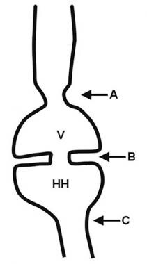
3. JEB with Pyloric Atresia: This subtype includes gastrointestinal abnormalities like pyloric atresia (a blockage in the lower part of the stomach), in addition to skin fragility.
Treatment for JEB typically focuses on managing symptoms and preventing complications. This may involve wound care, prevention of infection, pain management, nutritional support, and physical therapy. There is currently no cure for JEB.
Epidermolysis Bullosa Acquisita (EBA) is a rare autoimmune blistering disorder characterized by the production of autoantibodies against type VII collagen, a protein that plays a crucial role in anchoring the epidermis to the dermis. This results in the formation of blisters and erosions on the skin and mucous membranes, particularly in areas subjected to friction or trauma.
EBA can be classified into two main forms: the mechanobullous form and the inflammatory form. The mechanobullous form is characterized by spontaneous blistering and mechanical fragility of the skin, while the inflammatory form presents with inflammation and erosions in the mucous membranes.
The onset of EBA can occur at any age, but it is more common in adults, particularly those over 40 years old. The diagnosis of EBA is based on clinical presentation, direct immunofluorescence (DIF) studies, and detection of autoantibodies against type VII collagen.
Treatment of EBA typically involves a combination of wound care, prevention of infection, and immunosuppressive therapy to control the production of autoantibodies. The prognosis of EBA varies depending on the severity and extent of skin and mucous membrane involvement, as well as the response to treatment.
Collagen type VII is a type of collagen that is a major component of the anchoring fibrils, which are structures that help to attach the epidermis (the outermost layer of the skin) to the dermis (the layer of skin directly below the epidermis). Collagen type VII is composed of three identical chains that are encoded by the COL7A1 gene. Mutations in this gene can lead to a group of inherited blistering disorders known as autosomal recessive dystrophic epidermolysis bullosa, which is characterized by fragile skin and mucous membranes that blister and tear easily, often from minor trauma or friction.
A blister is a small fluid-filled bubble that forms on the skin due to friction, burns, or contact with certain chemicals or irritants. Blisters are typically filled with a clear fluid called serum, which is a component of blood. They can also be filled with blood (known as blood blisters) if the blister is caused by a more severe injury.
Blisters act as a natural protective barrier for the underlying skin and tissues, preventing infection and promoting healing. It's generally recommended to leave blisters intact and avoid breaking them, as doing so can increase the risk of infection and delay healing. If a blister is particularly large or painful, medical attention may be necessary to prevent complications.
Plectin is a large cytolinker protein that plays a crucial role in the structural organization and stability of the cell. It has the ability to interact with various components of the cytoskeleton, including intermediate filaments, microtubules, and actin filaments, thereby providing a critical link between these structures. Plectin is widely expressed in many tissues and is involved in maintaining the integrity and functionality of cells under both physiological and pathological conditions. Mutations in the gene encoding plectin have been associated with several human diseases, including epidermolysis bullosa, muscular dystrophy, and neuropathies.
Keratin-14 is a type of keratin protein that is specifically expressed in the suprabasal layers of stratified epithelia, including the epidermis. It is a component of the intermediate filament cytoskeleton and plays an important role in maintaining the structural integrity and stability of epithelial cells. Mutations in the gene encoding keratin-14 have been associated with several genetic skin disorders, such as epidermolysis bullosa simplex and white sponge nevus.
In medical terms, the skin is the largest organ of the human body. It consists of two main layers: the epidermis (outer layer) and dermis (inner layer), as well as accessory structures like hair follicles, sweat glands, and oil glands. The skin plays a crucial role in protecting us from external factors such as bacteria, viruses, and environmental hazards, while also regulating body temperature and enabling the sense of touch.
The pylorus is the lower, narrow part of the stomach that connects to the first part of the small intestine (duodenum). It consists of the pyloric canal, which is a short muscular tube, and the pyloric sphincter, a circular muscle that controls the passage of food from the stomach into the duodenum. The pylorus regulates the entry of chyme (partially digested food) into the small intestine by adjusting the size and frequency of the muscular contractions that push the chyme through the pyloric sphincter. This process helps in further digestion and absorption of nutrients in the small intestine.
Non-fibrillar collagens are a type of collagen that do not form fibrous structures, unlike the more common fibrillar collagens. They are a group of structurally diverse collagens that play important roles in various biological processes such as cell adhesion, migration, and differentiation. Non-fibrillar collagens include types IV, VI, VIII, X, XII, XIV, XVI, XIX, XXI, and XXVIII. They are often found in basement membranes and other specialized extracellular matrix structures.
Type IV collagen is a major component of the basement membrane and forms a network-like structure that provides a scaffold for other matrix components. Type VI collagen has a beaded filament structure and is involved in the organization of the extracellular matrix. Type VIII collagen is found in the eyes and helps to maintain the structural integrity of the eye. Type X collagen is associated with cartilage development and bone formation. Type XII and XIV collagens are fibril-associated collagens that help to regulate the organization and diameter of fibrillar collagens. The other non-fibrillar collagens have various functions, including cell adhesion, migration, and differentiation.
Overall, non-fibrillar collagens are important structural components of the extracellular matrix and play critical roles in various biological processes.
Integrin beta4, also known as ITGB4 or CD104, is a type of integrin subunit that forms part of the integrin receptor along with an alpha subunit. Integrins are transmembrane proteins involved in cell-cell and cell-extracellular matrix (ECM) adhesion, signal transduction, and regulation of various cellular processes such as proliferation, differentiation, and migration.
Integrin beta4 is unique among the integrin subunits because it has a large cytoplasmic domain that can interact with several intracellular signaling molecules, making it an important regulator of cell behavior. Integrin beta4 is widely expressed in various tissues, including epithelial cells, endothelial cells, and hematopoietic cells.
Integrin beta4 forms heterodimers with integrin alpha6 to form the receptor for laminins, which are major components of the basement membrane. This receptor is involved in maintaining the integrity of epithelial tissues and regulating cell migration during development, tissue repair, and cancer progression. Mutations in ITGB4 have been associated with several human diseases, including epidermolysis bullosa, a group of inherited skin disorders characterized by fragile skin and blistering.
Hemidesmosomes are specialized structures found in the cell membranes of epithelial cells that help to anchor them to the underlying basement membrane. They are composed of several proteins, including integrins and collagen type XVII, which interact with both intracellular keratin filaments and extracellular matrix components such as laminin-332. Hemidesmosomes play a crucial role in maintaining the integrity and stability of epithelial tissues by providing strong adhesive bonds between the epithelial cells and the underlying basement membrane, which is essential for normal tissue function and homeostasis. Mutations in genes encoding hemidesmosomal proteins can lead to various inherited skin blistering disorders, such as epidermolysis bullosa.
Keratin 5 is a type of keratin protein that is primarily expressed in the basal layer of epithelial tissues, including the skin, hair follicles, and nails. It forms heterodimers with keratin 14 and plays a crucial role in maintaining the structural integrity and stability of these tissues. Mutations in the gene that encodes keratin 5 (KRT5) can lead to several genetic disorders, such as epidermolysis bullosa simplex, which is characterized by blistering of the skin and mucous membranes.
Keratins are a type of fibrous structural proteins that constitute the main component of the integumentary system, which includes the hair, nails, and skin of vertebrates. They are also found in other tissues such as horns, hooves, feathers, and reptilian scales. Keratins are insoluble proteins that provide strength, rigidity, and protection to these structures.
Keratins are classified into two types: soft keratins (Type I) and hard keratins (Type II). Soft keratins are found in the skin and simple epithelial tissues, while hard keratins are present in structures like hair, nails, horns, and hooves.
Keratin proteins have a complex structure consisting of several domains, including an alpha-helical domain, beta-pleated sheet domain, and a non-repetitive domain. These domains provide keratin with its unique properties, such as resistance to heat, chemicals, and mechanical stress.
In summary, keratins are fibrous structural proteins that play a crucial role in providing strength, rigidity, and protection to various tissues in the body.
Recessive genes refer to the alleles (versions of a gene) that will only be expressed when an individual has two copies of that particular allele, one inherited from each parent. If an individual inherits one recessive allele and one dominant allele for a particular gene, the dominant allele will be expressed and the recessive allele will have no effect on the individual's phenotype (observable traits).
Recessive genes can still play a role in determining an individual's genetic makeup and can be passed down through generations even if they are not expressed. If two carriers of a recessive gene have children, there is a 25% chance that their offspring will inherit two copies of the recessive allele and exhibit the associated recessive trait.
Examples of genetic disorders caused by recessive genes include cystic fibrosis, sickle cell anemia, and albinism.
Vesiculobullous skin diseases are a group of disorders characterized by the formation of blisters (vesicles) and bullae (larger blisters) on the skin. These blisters form when there is a separation between the epidermis (outer layer of the skin) and the dermis (layer beneath the epidermis) due to damage in the area where they join, known as the dermo-epidermal junction.
There are several types of vesiculobullous diseases, each with its own specific causes and symptoms. Some of the most common types include:
1. Pemphigus vulgaris: an autoimmune disorder where the immune system mistakenly attacks proteins that help to hold the skin together, causing blisters to form.
2. Bullous pemphigoid: another autoimmune disorder, but in this case, the immune system attacks a different set of proteins, leading to large blisters and inflammation.
3. Dermatitis herpetiformis: a skin condition associated with celiac disease, where gluten ingestion triggers an immune response that leads to the formation of itchy blisters.
4. Pemphigoid gestationis: a rare autoimmune disorder that occurs during pregnancy and causes blisters on the abdomen and other parts of the body.
5. Epidermolysis bullosa: a group of inherited disorders where there is a fragile skin structure, leading to blistering and wound formation after minor trauma or friction.
Treatment for vesiculobullous diseases depends on the specific diagnosis and may include topical or systemic medications, such as corticosteroids, immunosuppressants, or antibiotics, as well as wound care and prevention of infection.
Keratinocytes are the predominant type of cells found in the epidermis, which is the outermost layer of the skin. These cells are responsible for producing keratin, a tough protein that provides structural support and protection to the skin. Keratinocytes undergo constant turnover, with new cells produced in the basal layer of the epidermis and older cells moving upward and eventually becoming flattened and filled with keratin as they reach the surface of the skin, where they are then shed. They also play a role in the immune response and can release cytokines and other signaling molecules to help protect the body from infection and injury.
An encyclopedia is a comprehensive reference work containing articles on various topics, usually arranged in alphabetical order. In the context of medicine, a medical encyclopedia is a collection of articles that provide information about a wide range of medical topics, including diseases and conditions, treatments, tests, procedures, and anatomy and physiology. Medical encyclopedias may be published in print or electronic formats and are often used as a starting point for researching medical topics. They can provide reliable and accurate information on medical subjects, making them useful resources for healthcare professionals, students, and patients alike. Some well-known examples of medical encyclopedias include the Merck Manual and the Stedman's Medical Dictionary.
I'm sorry for any confusion, but "Minnesota" is not a medical term or concept. It is a state located in the Midwestern United States, known for its cold winters, beautiful lakes, and friendly people. If you have any questions about medical terms or concepts, I would be happy to help!
Group purchasing in a healthcare context refers to the practice where multiple healthcare organizations, such as hospitals or clinics, join together to negotiate and purchase medical supplies, pharmaceuticals, and other goods or services from vendors at a reduced price. By pooling their resources and purchasing power, these organizations can secure better pricing, terms, and contractual agreements than they might be able to obtain individually. This collaborative approach can help healthcare organizations reduce costs, improve operational efficiency, and ensure access to high-quality products and services.
Bibliometrics is the use of statistical methods to analyze books, articles, and other publications. In the field of information science, bibliometrics is often used to measure the impact of scholarly works or authors by counting the number of times that a work has been cited in other publications. This can help researchers identify trends and patterns in research output and collaboration, as well as assess the influence of individual researchers or institutions.
Bibliometric analyses may involve a variety of statistical measures, such as citation counts, author productivity, journal impact factors, and collaborative networks. These measures can be used to evaluate the performance of individual researchers, departments, or institutions, as well as to identify areas of research strength or weakness.
It is important to note that while bibliometrics can provide useful insights into research trends and impact, they should not be the sole basis for evaluating the quality or significance of scholarly work. Other factors, such as the rigor of the research design, the clarity of the writing, and the relevance of the findings to the field, are also important considerations.
'Diseases in Twins' is a field of study that focuses on the similarities and differences in the occurrence, development, and outcomes of diseases among twins. This research can provide valuable insights into the genetic and environmental factors that contribute to various medical conditions.
Twins can be classified into two types: monozygotic (identical) and dizygotic (fraternal). Monozygotic twins share 100% of their genes, while dizygotic twins share about 50%, similar to non-twin siblings. By comparing the concordance rates (the likelihood of both twins having the same disease) between monozygotic and dizygotic twins, researchers can estimate the heritability of a particular disease.
Studying diseases in twins also helps understand the role of environmental factors. When both twins develop the same disease, but they are discordant for certain risk factors (e.g., one twin smokes and the other does not), it suggests that the disease may have a stronger genetic component. On the other hand, when both twins share similar risk factors and develop the disease, it implies that environmental factors play a significant role.
Diseases in Twins research has contributed to our understanding of various medical conditions, including infectious diseases, cancer, mental health disorders, and developmental disorders. This knowledge can lead to better prevention strategies, early detection methods, and more targeted treatments for these diseases.
In the context of medicine, "publications" typically refers to the dissemination of research findings or other medical information through various forms of media. This can include:
1. Peer-reviewed journals: These are scientific or medical publications that undergo a rigorous review process by experts in the field before they are accepted for publication. They represent some of the most reliable sources of medical information.
2. Conference proceedings: Medical conferences often publish abstracts, presentations, or posters from the event. These can provide early insights into ongoing research and new developments in the field.
3. Books and book chapters: Medical texts and reference books are a common form of publication, offering comprehensive overviews of specific topics or conditions.
4. Online platforms: Websites, blogs, and social media platforms have become increasingly popular ways to share medical information. While these can be valuable resources, it's important to critically evaluate the quality and reliability of the information presented.
5. News articles and press releases: Media outlets may report on new medical research or developments, although these should also be approached with caution as they may not always accurately represent the findings or context of the original research.
It's worth noting that all publications should be evaluated based on their source, methodology, and relevance to the specific question or issue at hand.
Research, in the context of medicine, is a systematic and rigorous process of collecting, analyzing, and interpreting information in order to increase our understanding, develop new knowledge, or evaluate current practices and interventions. It can involve various methodologies such as observational studies, experiments, surveys, or literature reviews. The goal of medical research is to advance health care by identifying new treatments, improving diagnostic techniques, and developing prevention strategies. Medical research is typically conducted by teams of researchers including clinicians, scientists, and other healthcare professionals. It is subject to ethical guidelines and regulations to ensure that it is conducted responsibly and with the best interests of patients in mind.
Peer review is a process in which experts in a field assess the quality and validity of scientific research, scholarly articles, or other professional works prior to publication. In the context of medical research, peer review typically involves one or more researchers with similar expertise evaluating a manuscript or study proposal to ensure that it meets established standards for design, methodology, analysis, and interpretation of results. The goal of peer review is to maintain the integrity and credibility of the scientific record by identifying and correcting errors, biases, or other shortcomings in the research before it is published. Peer review is a standard practice in medical publishing and is considered an essential component of the scientific process.
 Epidermolysis bullosa dystrophica
List of OMIM disorder codes
Protein replacement therapy
Histology of the vocal cords
Anchoring fibrils
List of diseases (E)
EBD
List of MeSH codes (C16)
Antisense therapy
Deb
List of MeSH codes (C17)
Epidermolysis bullosa dystrophica - Wikipedia
 Dystrophic epidermolysis bullosa: MedlinePlus Genetics
 Human COL7A1-corrected induced pluripotent stem cells for the treatment of recessive dystrophic epidermolysis bullosa
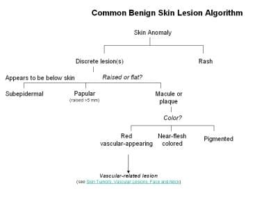
 Benign Skin Lesions: Overview, Defining the Lesion, Papules and Plaques
 SMART: FN3 domain annotation
SMART: FN3 domain annotation
 MMP1 | M10: Matrix metallopeptidase | IUPHAR/BPS Guide to PHARMACOLOGY
 Sentinel Lymph Node Biopsy in Nonmelanoma Skin Cancer Patients
 Bruce R Blazar - Research output
- Experts@Minnesota
Bruce R Blazar - Research output
- Experts@Minnesota
 Psychosocial recommendations for the care of children and adults with epidermolysis bullosa and their family: evidence based...
 Recessive generalized dystrophic epidermolysis bullosa: dental service protocol and case report
 Collagen. Medical search
Bio2Vec
 Genetic test kit - Linea Vita
 dermatosis
Rashida Pramanik - Research output - King's College London
 Elisabeth Divera Alida Cathari Meester - Research output
- Universidad de Monterrey
Elisabeth Divera Alida Cathari Meester - Research output
- Universidad de Monterrey
 Kprp Mouse Gene Details | keratinocyte expressed, proline-rich | International Mouse Phenotyping Consortium
University of Minnesota Twin Cities - Research output
- Experts@Minnesota
 Browse Items · Pediatric Palliative Care Library
Browse Items · Pediatric Palliative Care Library
 Laboratorio de Genetica Clinica SL - Labs - NIH Genetic Testing Registry (GTR) - NCBI
 Kuhny
Kuhny
 Prenatal diagnosis of dominant and recessive dystrophic epidermolysis bullosa: Application and limitations in the use of kf-1...
 Epidermolysis bullosa, dystrofisk - Sjelden
Hair Diseases | Profiles RNS
 Vitamin E Treatment of Dermolytic Bullous Dermatoses | JAMA Dermatology | JAMA Network
Recessive dystrophic epidermolysis bullosa results in painful small fibre neuropathy. - Oxford Neuroscience
 DeCS
DeCSDystrophic16
- Epidermolysis bullosa dystrophica or dystrophic EB (DEB) is an inherited disease affecting the skin and other organs. (wikipedia.org)
- In May 2023, the FDA approved Vyjuvek for the treatment of wounds in people with dystrophic epidermolysis bullosa with mutation(s) in the collagen type VII alpha 1 chain (COL7A1) gene. (wikipedia.org)
- 1], Recessive dystrophic epidermolysis bullosa results in painful small fibre neuropathy. (wikipedia.org)
- Dystrophic epidermolysis bullosa is one of the major forms of a group of conditions called epidermolysis bullosa. (medlineplus.gov)
- The signs and symptoms of dystrophic epidermolysis bullosa vary widely among affected individuals. (medlineplus.gov)
- Researchers classify dystrophic epidermolysis bullosa into major types based on the inheritance pattern and features of the condition. (medlineplus.gov)
- Recessive dystrophic epidermolysis bullosa severe generalized (RDEB-sev gen) is the classic form of the condition and is the most severe. (medlineplus.gov)
- Other types of recessive dystrophic epidermolysis bullosa fall along a spectrum referred to as RDEB-generalized and localized (RDEB-gen and -loc). (medlineplus.gov)
- Another major type of this condition is known as dominant dystrophic epidermolysis bullosa (DDEB). (medlineplus.gov)
- Considered together, the prevalence of recessive and dominant dystrophic epidermolysis bullosa is estimated to be 3.3 per million people. (medlineplus.gov)
- Mutations in the COL7A1 gene cause all forms of dystrophic epidermolysis bullosa. (medlineplus.gov)
- Mutations that allow a small amount of normal or partially functional type VII collagen to be produced lead to milder forms of the dystrophic epidermolysis bullosa. (medlineplus.gov)
- The present article reports on an dental service protocol for patients with recessive generalized dystrophic epidermolysis bullosa, as well as offers some considerations concerning the appropriate dental treatment. (bvsalud.org)
- In children with severe generalized recessive dystrophic epidermolysis bullosa (RDEB), esophageal scarring leads to esophageal strictures with dysphagia, followed by malnutrition and delayed development. (omeka.net)
- Dystrophic epidermolysis bullosa (DEB) comprises four major and several rare sub-types with the three most common being intermediate dominant DEB, severe recessive DEB and intermediate recessive DEB. (sjelden.no)
- Recessive dystrophic epidermolysis bullosa (RDEB) is a rare condition in which mutations of proteins of the dermo-epidermal junction lead to cycles of blistering followed by regeneration of the skin. (ox.ac.uk)
Recessive1
- I am studying epidermal neoplasms associated with autosomal recessive epidermolysis bullosa dystrophica and have written all over the world for information regarding such cases. (jamanetwork.com)
Acquisita18
- citation needed] In the absence of mutations of the COL7A1 gene, an autoimmune response against type VII collagen can result in an acquired form of epidermolysis bullosa called epidermolysis bullosa acquisita. (wikipedia.org)
- Epidermolysis bullosa acquisita (EBA) is a chronic autoimmune subepidermal blistering disease of the skin and mucus membranes. (medscape.com)
- Epidermolysis bullosa acquisita is caused by antibodies targeting type VII collagen, the major component of anchoring fibrils that connect the basement membrane to dermal structures. (medscape.com)
- A second clinical presentation, the inflammatory form of epidermolysis bullosa acquisita, involves a generalized vesiculobullous eruption primarily on the trunk and flexural areas. (medscape.com)
- Epidermolysis bullosa acquisita is rare in humans. (medscape.com)
- In animals, epidermolysis bullosa acquisita has been reported in dogs only. (medscape.com)
- In canine epidermolysis bullosa acquisita, the immunoglobulin G (IgG) autoantibodies also target the type VII collagen noncollagenous (NC1) domain, which shares greater than 80% homology in amino acid sequence with the human NC1 domain. (medscape.com)
- Various murine models have contributed to the understanding of the pathogenic role of antitype VII collagen antibodies and pathophysiology of epidermolysis bullosa acquisita. (medscape.com)
- 5, 6] More recently, affinity-purified antitype VII collagen autoantibodies from epidermolysis bullosa acquisita patients have induced blisters in an adult hairless mouse strain (SKH1), further supporting a pathogenic role of antitype collagen VII autoantibodies. (medscape.com)
- Immunization of type VII collagen in athymic nude SJL mice did not induce an autoimmune response, whereas the repletion of T cells from type VII collagen-immunized wild-type mice to the thymic mice showed autoantibody production and resulted in a blistering disease phenotype, supporting the role of T cells in the induction of epidermolysis bullosa acquisita. (medscape.com)
- The fact that epidermolysis bullosa acquisita is responsive to rituximab antibody to CD20 supports the role of B cells. (medscape.com)
- Epidermolysis bullosa acquisita (EBA) is a rare disease, with an incidence of 0.26 case per million population, which is 5% of all blistering diseases. (medscape.com)
- The race distribution of epidermolysis bullosa acquisita (EBA) is not known. (medscape.com)
- Epidermolysis bullosa acquisita (EBA) can occur at any age. (medscape.com)
- however, children with epidermolysis bullosa acquisita have been reported, including one child with the onset of epidermolysis bullosa acquisita at age 3 months. (medscape.com)
- Patients with epidermolysis bullosa acquisita (EBA), if treated and cared for properly, should expect to live a normal life span. (medscape.com)
- Epidermolysis bullosa acquisita is a chronic inflammatory disease with periods of partial remissions and exacerbations. (medscape.com)
- however, epidermolysis bullosa acquisita is relatively unresponsive to treatment and can cause significant morbidity. (medscape.com)
Junctional1
- There exist other types of inherited epidermolysis bullosa, junctional epidermolysis bullosa and epidermolysis bullosa simplex, which are not related to type VII collagen deficiency. (wikipedia.org)
Patients1
- I saw one of the first patients alledged to have the dystropic type of epidermolysis bullosa who in reality had the nondystrophic Weber-Cockayne type of epidermolysis bullosa. (jamanetwork.com)
Clinical1
- A case of epidermolysis bullosa dystrophica in a neonate with characteristic clinical manifestation that occurred within the first 24 hours of life is presented. (home.pl)
Formation3
- Epidermolysis Bullosa (EB) is a group of rare genetic disorders, the primary manifestation is the formation of blisters and erosions in response to mechanical trauma [ 1 ]. (biomedcentral.com)
- Epidermolysis bullosa is a mucocutaneous disease characterized by the formation of bullae on the skin and mucosa, which grow either spontaneously or from minor traumas. (bvsalud.org)
- A group of inherited epidermolysis bullosa (EB) characterized by cutaneous and mucosal fragility resulting in blisters and superficial ulcerations that develop below the lamina densa of the cutaneous basement membrane and that heal with significant scarring and milia formation. (sjelden.no)
Rare1
- Epidermolysis Bullosa (EB) is a group of rare genetic disorders resulting in skin fragility and other symptoms. (biomedcentral.com)
Treatment1
- I read, with interest, the paper by Michaelson et al on vitamin E treatment of epidermolysis bullosa ( Arch Dermatol 109:67, 1974). (jamanetwork.com)
Case1
- Epidermolysis bullosa dystrophica - a case presentation. (home.pl)
Dystrophic epiderm21
- In May 2023, the FDA approved Vyjuvek for the treatment of wounds in people with dystrophic epidermolysis bullosa with mutation(s) in the collagen type VII alpha 1 chain (COL7A1) gene. (wikipedia.org)
- 1], Recessive dystrophic epidermolysis bullosa results in painful small fibre neuropathy. (wikipedia.org)
- Dystrophic epidermolysis bullosa is one of the major forms of a group of conditions called epidermolysis bullosa. (medlineplus.gov)
- The signs and symptoms of dystrophic epidermolysis bullosa vary widely among affected individuals. (medlineplus.gov)
- Researchers classify dystrophic epidermolysis bullosa into major types based on the inheritance pattern and features of the condition. (medlineplus.gov)
- Recessive dystrophic epidermolysis bullosa severe generalized (RDEB-sev gen) is the classic form of the condition and is the most severe. (medlineplus.gov)
- Other types of recessive dystrophic epidermolysis bullosa fall along a spectrum referred to as RDEB-generalized and localized (RDEB-gen and -loc). (medlineplus.gov)
- Another major type of this condition is known as dominant dystrophic epidermolysis bullosa (DDEB). (medlineplus.gov)
- Considered together, the prevalence of recessive and dominant dystrophic epidermolysis bullosa is estimated to be 3.3 per million people. (medlineplus.gov)
- Mutations in the COL7A1 gene cause all forms of dystrophic epidermolysis bullosa. (medlineplus.gov)
- Mutations that allow a small amount of normal or partially functional type VII collagen to be produced lead to milder forms of the dystrophic epidermolysis bullosa. (medlineplus.gov)
- Autosomal dominant and recessive forms of dystrophic epidermolysis bullosa (DEB) result from mutations in the type VII collagen gene (COL7A1). (nih.gov)
- 6. Morphological and morphometric analysis of cutaneous squamous cell carcinoma in patients with recessive dystrophic epidermolysis bullosa: a retrospective study. (nih.gov)
- 8. Electrochemotherapy, a local treatment for squamous cell carcinoma in patients with recessive dystrophic epidermolysis bullosa. (nih.gov)
- 9. The potential of gene therapy for recessive dystrophic epidermolysis bullosa. (nih.gov)
- 10. Squamous cell carcinoma secondary to recessive dystrophic epidermolysis bullosa. (nih.gov)
- 12. Cetuximab therapy of metastasizing cutaneous squamous cell carcinoma in a patient with severe recessive dystrophic epidermolysis bullosa. (nih.gov)
- 15. Variable Outcome of Immunotherapy in Advanced Multiple Cutaneous Squamous Cell Carcinomas in Two Patients with Recessive Dystrophic Epidermolysis Bullosa. (nih.gov)
- All forms of dystrophic epidermolysis bullosa result from mutations in COLLAGEN TYPE VII , a major component fibrils of BASEMENT MEMBRANE and EPIDERMIS . (nih.gov)
- The present article reports on an dental service protocol for patients with recessive generalized dystrophic epidermolysis bullosa, as well as offers some considerations concerning the appropriate dental treatment. (bvsalud.org)
- Seven novel COL7A1 mutations identified in patients with recessive dystrophic epidermolysis bullosa from Mexico. (jefferson.edu)
Junctional epiderm1
- There exist other types of inherited epidermolysis bullosa, junctional epidermolysis bullosa and epidermolysis bullosa simplex, which are not related to type VII collagen deficiency. (wikipedia.org)
COL7A11
- citation needed] In the absence of mutations of the COL7A1 gene, an autoimmune response against type VII collagen can result in an acquired form of epidermolysis bullosa called epidermolysis bullosa acquisita. (wikipedia.org)
Inherited disease1
- Epidermolysis bullosa dystrophica or dystrophic EB (DEB) is an inherited disease affecting the skin and other organs. (wikipedia.org)
Retrospective Study1
- 14. Cutaneous Squamous Cell Carcinoma in Epidermolysis Bullosa: a 28-year Retrospective Study. (nih.gov)
Hereditary1
- Some rare diseases with oral manifestations warrant fixed/implant-supported restorations - a case report Epidermolysis bullosa dystrophica (DEB) describes a form of hereditary epidermolysis bullosa (EB), which is characterized by fragility of the skin and mucous membranes and manifests itself with blistering. (implant-register.com)
Phenotype-genotype correlations1
- 18. Ophthalmologic Approach in Epidermolysis Bullosa: A Cross-Sectional Study With Phenotype-Genotype Correlations. (nih.gov)
Severe1
- Form of epidermolysis bullosa characterized by atrophy of blistered areas, severe scarring, and nail changes. (nih.gov)
Skin3
- Epidermolysis bullosa cause the skin to be very fragile and to blister easily. (medlineplus.gov)
- 7. Treatment of skin cancers in epidermolysis bullosa. (nih.gov)
- Epidermolysis bullosa is a mucocutaneous disease characterized by the formation of bullae on the skin and mucosa, which grow either spontaneously or from minor traumas. (bvsalud.org)
Reference2
- Epidermolysis bullosa Reference, Genetics Home. (wikipedia.org)
- 13. Multidisciplinary care for patients with epidermolysis bullosa from birth to adolescence: experience of one Italian reference center. (nih.gov)
Cases1
- 5. Inherited epidermolysis bullosa and squamous cell carcinoma: a systematic review of 117 cases. (nih.gov)