Enteric Nervous System
Hirschsprung Disease
Myenteric Plexus
Submucous Plexus
Neural Crest
Central Nervous System
Nervous System
Gastrointestinal Tract
Proto-Oncogene Proteins c-ret
Digestive System
Peripheral Nervous System
SOXE Transcription Factors
Endothelin-3
Neurons
Neuroglia
Glial Cell Line-Derived Neurotrophic Factor
Ganglia
Intestines
Colon
Ileum
Glial Cell Line-Derived Neurotrophic Factor Receptors
Quail
Digestive System Physiological Phenomena
Megacolon
Vasoactive Intestinal Peptide
Serotonin 5-HT4 Receptor Agonists
Immunohistochemistry
Intestine, Small
Central Nervous System Diseases
Peristalsis
Vagus Nerve
Interstitial Cells of Cajal
Guinea Pigs
Cell Movement
Intestinal Pseudo-Obstruction
Neurofilament Proteins
Jejunum
Receptor Protein-Tyrosine Kinases
High Mobility Group Proteins
Gene Expression Regulation, Developmental
Chick Embryo
Ileus
Celiac Plexus
Intestine, Large
Stem Cells
Central Nervous System Neoplasms
Brain
Receptors, Serotonin, 5-HT4
In Situ Hybridization
Neurotransmitter Agents
Coturnix
Ubiquitin Thiolesterase
Nerve Tissue Proteins
Neurogenesis
Nerve Growth Factors
Mice, Transgenic
Receptor, Endothelin B
Choline O-Acetyltransferase
Manduca
Autonomic Nervous System
Intestinal Mucosa
Gastrointestinal Diseases
Hexamethonium
Mice, Inbred C57BL
Stomach
NADPH Dehydrogenase
Calbindin 2
Cell Differentiation
Irritable Bowel Syndrome
Tetrodotoxin
Ganglia, Autonomic
Serotonin
Muscle, Smooth
Gastrointestinal Transit
Sympathetic Nervous System
Nervous System Physiological Phenomena
Signal Transduction
Zebrafish
Cell Count
Drosophila Proteins
Neurturin
Gastroparesis
Nitric Oxide Synthase Type I
Myoelectric Complex, Migrating
Cells, Cultured
Mice, Knockout
Rats, Sprague-Dawley
Neuropeptides
Embryo, Nonmammalian
Zebrafish Proteins
Central Nervous System Infections
Tyrosine 3-Monooxygenase
Disease Models, Animal
Substance P
Body Patterning
Hu Paraneoplastic Encephalomyelitis Antigens
Embryo, Mammalian
Nervous System Diseases
Nervous System Neoplasms
Mutation
Synaptic Transmission
Inhibition of in vitro enteric neuronal development by endothelin-3: mediation by endothelin B receptors. (1/604)
The terminal colon is aganglionic in mice lacking endothelin-3 or its receptor, endothelin B. To analyze the effects of endothelin-3/endothelin B on the differentiation of enteric neurons, E11-13 mouse gut was dissociated, and positive and negative immunoselection with antibodies to p75(NTR )were used to isolate neural crest- and non-crest-derived cells. mRNA encoding endothelin B was present in both the crest-and non-crest-derived cells, but that encoding preproendothelin-3 was detected only in the non-crest-derived population. The crest- and non-crest-derived cells were exposed in vitro to endothelin-3, IRL 1620 (an endothelin B agonist), and/or BQ 788 (an endothelin B antagonist). Neurons and glia developed only in cultures of crest-derived cells, and did so even when endothelin-3 was absent and BQ 788 was present. Endothelin-3 inhibited neuronal development, an effect that was mimicked by IRL 1620 and blocked by BQ 788. Endothelin-3 failed to stimulate the incorporation of [3H]thymidine or bromodeoxyuridine. Smooth muscle development in non-crest-derived cell cultures was promoted by endothelin-3 and inhibited by BQ 788. In contrast, transcription of laminin alpha1, a smooth muscle-derived promoter of neuronal development, was inhibited by endothelin-3, but promoted by BQ 788. Neurons did not develop in explants of the terminal bowel of E12 ls/ls (endothelin-3-deficient) mice, but could be induced to do so by endothelin-3 if a source of neural precursors was present. We suggest that endothelin-3/endothelin B normally prevents the premature differentiation of crest-derived precursors migrating to and within the fetal bowel, enabling the precursor population to persist long enough to finish colonizing the bowel. (+info)IL-1beta and IL-6 excite neurons and suppress nicotinic and noradrenergic neurotransmission in guinea pig enteric nervous system. (2/604)
Conventional intracellular microelectrodes and injection of biocytin were used to study the actions of IL-1beta and IL-6 on electrical and synaptic behavior in morphologically identified guinea pig small intestinal submucous neurons. Exposure to nanomolar concentrations of either IL-1beta or IL-6 stimulated neuronal excitability. The excitatory action consisted of depolarization of the membrane potential, decreased membrane conductance, and increased discharge of action potentials. Excitatory action of IL-1beta was suppressed by the natural IL-1beta human receptor antagonist. Electrical stimulation of sympathetic postganglionic axons evoked inhibitory postsynaptic potentials (IPSPs), and stimulation of cholinergic axons evoked nicotinic fast excitatory postsynaptic potentials (EPSPs). Both kinds of synaptic potentials occurred in neurons with uniaxonal morphology believed to be secretomotor neurons. Either IL-1beta or IL-6 suppressed the noradrenergic IPSPs and the fast EPSPs, and the two acted synergistically when applied in combination. Suppression of the IPSP resulted from presynaptic inhibition of the release of norepinephrine from sympathetic nerves. The results suggest that the presence of either or both inflammatory cytokines will release the sympathetic brake from secretomotor neurons to the intestinal crypts and from nicotinic synapses in the integrative microcircuits, where norepinephrine is known to have a presynaptic inhibitory action. This, in concert with excitation of secretomotor neurons, may lead to neurogenic secretory diarrhea. (+info)Rectal biopsy for diagnosis of intestinal neuronal dysplasia in children: a prospective multicentre study on interobserver variation and clinical outcome. (3/604)
BACKGROUND: Intestinal neuronal dysplasia (IND) of the colonic submucous plexus is considered to be a congenital malformation of the enteric nervous system causing symptoms resembling those of Hirschsprung's disease. In contrast with the established diagnosis of aganglionosis using enzyme histochemistry, controversy exists over the diagnostic criteria of IND on rectal biopsies previously defined by a consensus report and the causal relation between morphological findings and clinical symptoms. AIMS: The interobserver variability was prospectively investigated with respect to final diagnoses and several histological features in rectal biopsy specimens from children suspected of having colonic motility disturbances. METHODS: 377 biopsy specimens from 108 children aged 4 days to 15 years were independently coded without knowledge of clinical symptoms by three experienced pathologists for 20 histological features, and a final diagnosis was given for every case. Interobserver variation for the different items and the final diagnosis were analysed using Cohen's kappa statistic. Clinical data at biopsy and outcome after 12 months were related to morphological findings. RESULTS: The three pathologists agreed completely with respect to the diagnosis Hirschsprung's disease (kappa = 1), but in only 14% of the children without aganglionosis. In 15 (17%) of the 87 children without aganglionosis, at least one pathologist judged the case as normal, while another diagnosed IND. kappa values were close to the zero value expected by chance for the diagnoses normal and IND. Young age was related to the presence of several morphological features-for example, acetylcholine esterase staining and presence of giant ganglia. Children with chronic constipation diagnosed as having IND, given no other specific diagnosis by any of the pathologists, were significantly younger (median 8.8 months) and had a higher cure rate after one year (60%) than constipated patients considered by all observers to have no histological abnormalities (median 6.1 years, cure rate 23%). CONCLUSIONS: In contrast with Hirschsprung's disease, there is a high interobserver variation with regard to the different morphological features and final diagnosis of IND, based on the criteria and conditions of the previous consensus report. The high frequency of histological "abnormalities" in young infants suggests that some of the features may represent a normal variant of postnatal development rather than a pathological process. Investigations using more refined and morphometric methods in rectal specimens from infants and children without bowel disease are needed to define the normal range of morphological appearance at different ages. These preliminary data indicate that, with current knowledge, rectal biopsy for diagnostic purposes should only be performed in constipated children for diagnosis of Hirschsprung's disease. (+info)Signalling by the RET receptor tyrosine kinase and its role in the development of the mammalian enteric nervous system. (4/604)
RET is a member of the receptor tyrosine kinase (RTK) superfamily, which can transduce signalling by glial cell line-derived neurotrophic factor (GDNF) and neurturin (NTN) in cultured cells. In order to determine whether in addition to being sufficient, RET is also necessary for signalling by these growth factors, we studied the response to GDNF and NTN of primary neuronal cultures (peripheral sensory and central dopaminergic neurons) derived from wild-type and RET-deficient mice. Our experiments show that absence of a functional RET receptor abrogates the biological responses of neuronal cells to both GDNF and NTN. Despite the established role of the RET signal transduction pathway in the development of the mammalian enteric nervous system (ENS), very little is known regarding its cellular mechanism(s) of action. Here, we have studied the effects of GDNF and NTN on cultures of neural crest (NC)-derived cells isolated from the gut of rat embryos. Our findings suggest that GDNF and NTN promote the survival of enteric neurons as well as the survival, proliferation and differentiation of multipotential ENS progenitors present in the gut of E12.5-13.5 rat embryos. However, the effects of these growth factors are stage-specific, since similar ENS cultures established from later stage embryos (E14. 5-15.5), show markedly diminished response to GDNF and NTN. To examine whether the in vitro effects of RET activation reflect the in vivo function(s) of this receptor, the extent of programmed cell death was examined in the gut of wild-type and RET-deficient mouse embryos by TUNEL histochemistry. Our experiments show that a subpopulation of enteric NC undergoes apoptotic cell death specifically in the foregut of embryos lacking the RET receptor. We suggest that normal function of the RET RTK is required in vivo during early stages of ENS histogenesis for the survival of undifferentiated enteric NC and their derivatives. (+info)Peptidyl inhibitors of shaker-type Kv1 channels elicit twitches in guinea pig ileum by blocking kv1.1 at enteric nervous system and enhancing acetylcholine release. (5/604)
Potent and selective peptidyl blockers of the Shaker-type (Kv1) voltage-gated potassium channels were used to determine the role of these channels in regulating the spontaneous motility of smooth muscle preparations. Margatoxin (MgTX), kaliotoxin, and agitoxin-2 at 1 to 10 nM and agitoxin-1 at 50 to 100 nM induce twitches in guinea pig ileum strips. These twitches are abolished by tetrodotoxin (TTX, 0.5 microM), atropine (1 microM), hexamethonium (10 microM), or nifedipine (0.1 microM). It is proposed that blockade of Kv1 channels by MgTX, kaliotoxin, or the agitoxins increases excitability of intramural nerve plexuses in the ileum, promoting release of acetylcholine from excitatory motor nerve terminals. This, in turn, leads to Ca2+-dependent action potentials and twitching of the muscle fibers. MgTX does not induce twitches in several other guinea pig and/or rat vascular, genitourinary, or gastrointestinal smooth muscles, although small increases in spontaneous myogenic activity may be seen in detrusor muscle exposed to >30 nM MgTX. This effect is not reversed by TTX or atropine. The TTX- and atropine-sensitive twitches of guinea pig ileum are also induced by nanomolar concentrations of alpha-dendrotoxin, a selective blocker of Shaker Kv1.1 and 1.2 subtypes, or stichodactylatoxin, a peptide isolated from sea anemone that displays high affinity for Kv1.1 and 1.3, but not by charybdotoxin, which blocks Kv1.2 and 1.3 but not 1.1. The data taken together suggest that high-affinity blockade of Kv1.1 underlies the ability of MgTX, kaliotoxin, agitoxin-1, agitoxin-2, alpha-dendrotoxin, and stichodactylatoxin to elicit TTX-sensitive twitches in guinea pig ileum. (+info)Immune-epithelial interactions in host defense. (6/604)
Over the past 15 years, it has become very clear that the immune system can have profound effects on epithelial function. Acute immune-mediated changes in epithelial physiology are beneficial to host defense against enteric pathogens. For example, ion secretion washes out noxious luminal contents and increased permeability allows phagocytic cells and antibodies to enter the gut lumen. However, ongoing immune activation results in chronic effects that may be pathophysiologic. Responses are mediated by soluble immune mediators that act directly on the epithelium, or indirectly via nerves that also serve to amplify the epithelial response. Here, we will review some of the recent advances that have been made in the field of immunophysiology. The effect of mast cells on transport functions of the epithelium will be reviewed, with emphasis on the consequence of interactions between mast cells and nerves. The use of in vitro coculture systems has recently provided considerable information on the effects of neutrophils, eosinophils, monocytes, and lymphocytes on epithelial functions; the contribution of each immunocyte will be highlighted. Finally, we will describe evidence for the active participation of the epithelium in mucosal immune activation, including pathogen or cytokine induced epithelial cytokine synthesis or secretion and adhesion molecule expression. (+info)A role for fasciclin II in the guidance of neuronal migration. (7/604)
The insect cell adhesion receptor fasciclin II is expressed by specific subsets of neural and non-neural cells during embryogenesis and has been shown to control growth cone motility and axonal fasciculation. Here we demonstrate a role for fasciclin II in the guidance of migratory neurons. In the developing enteric nervous system of the moth Manduca sexta, an identified set of neurons (the EP cells) undergoes a stereotyped sequence of migration along the visceral muscle bands of the midgut prior to their differentiation. Probes specific for Manduca fasciclin II show that while the EP cells express fasciclin II throughout embryogenesis, their muscle band pathways express fasciclin II only during the migratory period. Manipulations of fasciclin II in embryonic culture using blocking antibodies, recombinant fasciclin II fragments, and enzymatic removal of glycosyl phosphatidylinositol-linked fasciclin II produced concentration-dependent reductions in the extent of EP cell migration. These results support a novel role for fasciclin II, indicating that this homophilic adhesion molecule is required for the promotion or guidance of neuronal migration. (+info)Fundamentals of neurogastroenterology. (8/604)
Current concepts and basic principles of neurogastroenterology in relation to functional gastrointestinal disorders are reviewed. Neurogastroenterology is emphasized as a new and advancing subspecialty of clinical gastroenterology and digestive science. As such, it embraces the investigative sciences dealing with functions, malfunctions, and malformations in the brain and spinal cord, and the sympathetic, parasympathetic and enteric divisions of the autonomic innervation of the digestive tract. Somatomotor systems are included insofar as pharyngeal phases of swallowing and pelvic floor involvement in defecation, continence, and pelvic pain are concerned. Inclusion of basic physiology of smooth muscle, mucosal epithelium, and the enteric immune system in the neurogastroenterologic domain relates to requirements for compatibility with neural control mechanisms. Psychologic and psychiatric relations to functional gastrointestinal disorders are included because they are significant components of neurogastroenterology, especially in relation to projections of discomfort and pain to the digestive tract. (+info)The enteric nervous system (ENS) is a part of the autonomic nervous system that directly controls the gastrointestinal tract, including the stomach, small intestine, colon, and rectum. It is sometimes referred to as the "second brain" because it can operate independently of the central nervous system (CNS).
The ENS contains around 500 million neurons that are organized into two main plexuses: the myenteric plexus, which lies between the longitudinal and circular muscle layers of the gut, and the submucosal plexus, which is located in the submucosa. These plexuses contain various types of neurons that are responsible for regulating gastrointestinal motility, secretion, and blood flow.
The ENS can communicate with the CNS through afferent nerve fibers that transmit information about the state of the gut to the brain, and efferent nerve fibers that carry signals from the brain back to the ENS. However, the ENS is also capable of functioning independently of the CNS, allowing it to regulate gastrointestinal functions in response to local stimuli such as food intake, inflammation, or infection.
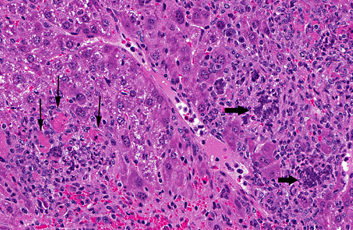
Hirschsprung disease is a gastrointestinal disorder that affects the large intestine, specifically the section known as the colon. This condition is congenital, meaning it is present at birth. It occurs due to the absence of ganglion cells (nerve cells) in the bowel's muscular wall, which are responsible for coordinating muscle contractions that move food through the digestive tract.
The affected segment of the colon cannot relax and propel the contents within it, leading to various symptoms such as constipation, intestinal obstruction, or even bowel perforation in severe cases. Common diagnostic methods include rectal suction biopsy, anorectal manometry, and contrast enema studies. Treatment typically involves surgical removal of the aganglionic segment and reattachment of the normal colon to the anus (known as a pull-through procedure).
The myenteric plexus, also known as Auerbach's plexus, is a component of the enteric nervous system located in the wall of the gastrointestinal tract. It is a network of nerve cells (neurons) and supporting cells (neuroglia) that lies between the inner circular layer and outer longitudinal muscle layers of the digestive system's muscularis externa.
The myenteric plexus plays a crucial role in controlling gastrointestinal motility, secretion, and blood flow, primarily through its intrinsic nerve circuits called reflex arcs. These reflex arcs regulate peristalsis (the coordinated muscle contractions that move food through the digestive tract) and segmentation (localized contractions that mix and churn the contents within a specific region of the gut).
Additionally, the myenteric plexus receives input from both the sympathetic and parasympathetic divisions of the autonomic nervous system, allowing for central nervous system regulation of gastrointestinal functions. Dysfunction in the myenteric plexus has been implicated in various gastrointestinal disorders, such as irritable bowel syndrome, achalasia, and intestinal pseudo-obstruction.
The submucosal plexus, also known as Meissner's plexus, is a component of the autonomic nervous system located in the submucosa layer of the gastrointestinal tract. It is a network of nerve fibers and ganglia that primarily regulates local reflexes and secretions, contributing to the control of gut motility, blood flow, and mucosal transport.
Meissner's plexus is part of the enteric nervous system (ENS), which can operate independently from the central nervous system (CNS). The ENS consists of two interconnected plexuses: Meissner's submucosal plexus and Auerbach's myenteric plexus.
Meissner's plexus is responsible for regulating functions such as absorption, secretion, vasodilation, and local immune responses in the gastrointestinal tract. Dysfunction of this plexus can lead to various gastrointestinal disorders, including irritable bowel syndrome (IBS) and other motility-related conditions.
The neural crest is a transient, multipotent embryonic cell population that originates from the ectoderm (outermost layer) of the developing neural tube (precursor to the central nervous system). These cells undergo an epithelial-to-mesenchymal transition and migrate throughout the embryo, giving rise to a diverse array of cell types and structures.
Neural crest cells differentiate into various tissues, including:
1. Peripheral nervous system (PNS) components: sensory neurons, sympathetic and parasympathetic ganglia, and glial cells (e.g., Schwann cells).
2. Facial bones and cartilage, as well as connective tissue of the skull.
3. Melanocytes, which are pigment-producing cells in the skin.
4. Smooth muscle cells in major blood vessels, heart, gastrointestinal tract, and other organs.
5. Secretory cells in endocrine glands (e.g., chromaffin cells of the adrenal medulla).
6. Parts of the eye, such as the cornea and iris stroma.
7. Dental tissues, including dentin, cementum, and dental pulp.
Due to their wide-ranging contributions to various tissues and organs, neural crest cells play a crucial role in embryonic development and organogenesis. Abnormalities in neural crest cell migration or differentiation can lead to several congenital disorders, such as neurocristopathies.
The Central Nervous System (CNS) is the part of the nervous system that consists of the brain and spinal cord. It is called the "central" system because it receives information from, and sends information to, the rest of the body through peripheral nerves, which make up the Peripheral Nervous System (PNS).
The CNS is responsible for processing sensory information, controlling motor functions, and regulating various autonomic processes like heart rate, respiration, and digestion. The brain, as the command center of the CNS, interprets sensory stimuli, formulates thoughts, and initiates actions. The spinal cord serves as a conduit for nerve impulses traveling to and from the brain and the rest of the body.
The CNS is protected by several structures, including the skull (which houses the brain) and the vertebral column (which surrounds and protects the spinal cord). Despite these protective measures, the CNS remains vulnerable to injury and disease, which can have severe consequences due to its crucial role in controlling essential bodily functions.
The nervous system is a complex, highly organized network of specialized cells called neurons and glial cells that communicate with each other via electrical and chemical signals to coordinate various functions and activities in the body. It consists of two main parts: the central nervous system (CNS), including the brain and spinal cord, and the peripheral nervous system (PNS), which includes all the nerves and ganglia outside the CNS.
The primary function of the nervous system is to receive, process, and integrate information from both internal and external environments and then respond by generating appropriate motor outputs or behaviors. This involves sensing various stimuli through specialized receptors, transmitting this information through afferent neurons to the CNS for processing, integrating this information with other inputs and memories, making decisions based on this processed information, and finally executing responses through efferent neurons that control effector organs such as muscles and glands.
The nervous system can be further divided into subsystems based on their functions, including the somatic nervous system, which controls voluntary movements and reflexes; the autonomic nervous system, which regulates involuntary physiological processes like heart rate, digestion, and respiration; and the enteric nervous system, which is a specialized subset of the autonomic nervous system that controls gut functions. Overall, the nervous system plays a critical role in maintaining homeostasis, regulating behavior, and enabling cognition and consciousness.
The gastrointestinal (GI) tract, also known as the digestive tract, is a continuous tube that starts at the mouth and ends at the anus. It is responsible for ingesting, digesting, absorbing, and excreting food and waste materials. The GI tract includes the mouth, esophagus, stomach, small intestine (duodenum, jejunum, ileum), large intestine (cecum, colon, rectum, anus), and accessory organs such as the liver, gallbladder, and pancreas. The primary function of this system is to process and extract nutrients from food while also protecting the body from harmful substances, pathogens, and toxins.
Proto-oncogene proteins c-RET are a group of gene products that play crucial roles in the development and functioning of the nervous system, as well as in other tissues. The c-RET proto-oncogene encodes a receptor tyrosine kinase, which is a type of enzyme that helps transmit signals from the outside to the inside of cells. This receptor is activated by binding to its ligands, leading to the activation of various signaling pathways that regulate cell growth, differentiation, and survival.
Mutations in the c-RET proto-oncogene can lead to its overactivation, resulting in the conversion of this gene into an oncogene. Oncogenes are genes that have the potential to cause cancer when they are mutated or abnormally expressed. Activating mutations in c-RET have been implicated in several types of human cancers, including multiple endocrine neoplasia type 2 (MEN2), papillary thyroid carcinoma, and certain types of lung and kidney cancers. These mutations can lead to the constitutive activation of c-RET, resulting in uncontrolled cell growth and tumor formation.
Gastrointestinal motility refers to the coordinated muscular contractions and relaxations that propel food, digestive enzymes, and waste products through the gastrointestinal tract. This process involves the movement of food from the mouth through the esophagus into the stomach, where it is mixed with digestive enzymes and acids to break down food particles.
The contents are then emptied into the small intestine, where nutrients are absorbed, and the remaining waste products are moved into the large intestine for further absorption of water and electrolytes and eventual elimination through the rectum and anus.
Gastrointestinal motility is controlled by a complex interplay between the autonomic nervous system, hormones, and local reflexes. Abnormalities in gastrointestinal motility can lead to various symptoms such as bloating, abdominal pain, nausea, vomiting, diarrhea, or constipation.
The digestive system is a complex group of organs and glands that process food. It converts the food we eat into nutrients, which the body uses for energy, growth, and cell repair. The digestive system also eliminates waste from the body. It is made up of the gastrointestinal tract (GI tract) and other organs that help the body break down and absorb food.
The GI tract includes the mouth, esophagus, stomach, small intestine, large intestine, and anus. Other organs that are part of the digestive system include the liver, pancreas, gallbladder, and salivary glands.
The process of digestion begins in the mouth, where food is chewed and mixed with saliva. The food then travels down the esophagus and into the stomach, where it is broken down further by stomach acids. The digested food then moves into the small intestine, where nutrients are absorbed into the bloodstream. The remaining waste material passes into the large intestine, where it is stored until it is eliminated through the anus.
The liver, pancreas, and gallbladder play important roles in the digestive process as well. The liver produces bile, a substance that helps break down fats in the small intestine. The pancreas produces enzymes that help digest proteins, carbohydrates, and fats. The gallbladder stores bile until it is needed in the small intestine.
Overall, the digestive system is responsible for breaking down food, absorbing nutrients, and eliminating waste. It plays a critical role in maintaining our health and well-being.
The Peripheral Nervous System (PNS) is that part of the nervous system which lies outside of the brain and spinal cord. It includes all the nerves and ganglia ( clusters of neurons) outside of the central nervous system (CNS). The PNS is divided into two components: the somatic nervous system and the autonomic nervous system.
The somatic nervous system is responsible for transmitting sensory information from the skin, muscles, and joints to the CNS, and for controlling voluntary movements of the skeletal muscles.
The autonomic nervous system, on the other hand, controls involuntary actions, such as heart rate, digestion, respiratory rate, salivation, perspiration, pupillary dilation, and sexual arousal. It is further divided into the sympathetic and parasympathetic systems, which generally have opposing effects and maintain homeostasis in the body.
Damage to the peripheral nervous system can result in various medical conditions such as neuropathies, neuritis, plexopathies, and radiculopathies, leading to symptoms like numbness, tingling, pain, weakness, or loss of reflexes in the affected area.
SOXE transcription factors are a subgroup of the SOX (SRY-related HMG box) family of proteins, which are involved in various developmental processes, including cell fate specification and differentiation. The SOXE group includes SOX8, SOX9, and SOX10, all of which contain a conserved high mobility group (HMG) box DNA-binding domain. They play crucial roles in the development of several tissues, such as the nervous system, skeletal system, and urogenital system.
SOXE transcription factors are known to regulate gene expression by binding to specific DNA sequences, often acting in combination with other transcription factors to control various cellular processes. Dysregulation of SOXE transcription factors has been implicated in several human diseases, including cancer and neurodevelopmental disorders.
Endothelin-3 (ET-3) is a member of the endothelin family, which are small peptides with potent vasoconstrictor properties. ET-3 is primarily produced by neurons in the central and peripheral nervous system, and it plays important roles in the development and regulation of various physiological functions, including cardiovascular function, neurotransmission, and cell proliferation.
ET-3 exerts its effects by binding to specific G protein-coupled receptors, known as endothelin A (ETA) and endothelin B (ETB) receptors. These receptors are widely distributed throughout the body, including in the cardiovascular, respiratory, gastrointestinal, and genitourinary systems.
In addition to its role as a potent vasoconstrictor, ET-3 has been implicated in various pathological conditions, such as hypertension, heart failure, pulmonary arterial hypertension, and cancer. In recent years, there has been growing interest in the potential therapeutic use of endothelin receptor antagonists to treat these conditions.
Neurons, also known as nerve cells or neurocytes, are specialized cells that constitute the basic unit of the nervous system. They are responsible for receiving, processing, and transmitting information and signals within the body. Neurons have three main parts: the dendrites, the cell body (soma), and the axon. The dendrites receive signals from other neurons or sensory receptors, while the axon transmits these signals to other neurons, muscles, or glands. The junction between two neurons is called a synapse, where neurotransmitters are released to transmit the signal across the gap (synaptic cleft) to the next neuron. Neurons vary in size, shape, and structure depending on their function and location within the nervous system.
Neuroglia, also known as glial cells or simply glia, are non-neuronal cells that provide support and protection for neurons in the nervous system. They maintain homeostasis, form myelin sheaths around nerve fibers, and provide structural support. They also play a role in the immune response of the central nervous system. Some types of neuroglia include astrocytes, oligodendrocytes, microglia, and ependymal cells.
Glial Cell Line-Derived Neurotrophic Factor (GDNF) is a protein that plays a crucial role in the survival, development, and function of certain neurons in the nervous system. It is a member of the transforming growth factor-β (TGF-β) superfamily and was initially identified for its ability to support the survival and differentiation of midbrain dopaminergic neurons, which are critical for movement control and motivation. GDNF also supports other types of neurons, including motor neurons and sensory neurons. It exerts its effects by binding to a receptor complex consisting of GFRα1 and RET tyrosine kinase receptors, activating intracellular signaling pathways that promote neuronal survival, growth, and synaptic plasticity. GDNF has been investigated as a potential therapeutic agent for various neurodegenerative disorders, including Parkinson's disease and amyotrophic lateral sclerosis (ALS).
A ganglion is a cluster of neuron cell bodies in the peripheral nervous system. Ganglia are typically associated with nerves and serve as sites for sensory processing, integration, and relay of information between the periphery and the central nervous system (CNS). The two main types of ganglia are sensory ganglia, which contain pseudounipolar neurons that transmit sensory information to the CNS, and autonomic ganglia, which contain multipolar neurons that control involuntary physiological functions.
Examples of sensory ganglia include dorsal root ganglia (DRG), which are associated with spinal nerves, and cranial nerve ganglia, such as the trigeminal ganglion. Autonomic ganglia can be further divided into sympathetic and parasympathetic ganglia, which regulate different aspects of the autonomic nervous system.
It's worth noting that in anatomy, "ganglion" refers to a group of nerve cell bodies, while in clinical contexts, "ganglion" is often used to describe a specific type of cystic structure that forms near joints or tendons, typically in the wrist or foot. These ganglia are not related to the peripheral nervous system's ganglia but rather are fluid-filled sacs that may cause discomfort or pain due to their size or location.
The intestines, also known as the bowel, are a part of the digestive system that extends from the stomach to the anus. They are responsible for the further breakdown and absorption of nutrients from food, as well as the elimination of waste products. The intestines can be divided into two main sections: the small intestine and the large intestine.
The small intestine is a long, coiled tube that measures about 20 feet in length and is lined with tiny finger-like projections called villi, which increase its surface area and enhance nutrient absorption. The small intestine is where most of the digestion and absorption of nutrients takes place.
The large intestine, also known as the colon, is a wider tube that measures about 5 feet in length and is responsible for absorbing water and electrolytes from digested food, forming stool, and eliminating waste products from the body. The large intestine includes several regions, including the cecum, colon, rectum, and anus.
Together, the intestines play a critical role in maintaining overall health and well-being by ensuring that the body receives the nutrients it needs to function properly.
The colon, also known as the large intestine, is a part of the digestive system in humans and other vertebrates. It is an organ that eliminates waste from the body and is located between the small intestine and the rectum. The main function of the colon is to absorb water and electrolytes from digested food, forming and storing feces until they are eliminated through the anus.
The colon is divided into several regions, including the cecum, ascending colon, transverse colon, descending colon, sigmoid colon, rectum, and anus. The walls of the colon contain a layer of muscle that helps to move waste material through the organ by a process called peristalsis.
The inner surface of the colon is lined with mucous membrane, which secretes mucus to lubricate the passage of feces. The colon also contains a large population of bacteria, known as the gut microbiota, which play an important role in digestion and immunity.
The ileum is the third and final segment of the small intestine, located between the jejunum and the cecum (the beginning of the large intestine). It plays a crucial role in nutrient absorption, particularly for vitamin B12 and bile salts. The ileum is characterized by its thin, lined walls and the presence of Peyer's patches, which are part of the immune system and help surveil for pathogens.
Glial cell line-derived neurotrophic factor (GDNF) receptors are a group of proteins found on the surface of certain cells in the body that bind to GDNF and transmit signals into the cell, thereby activating various cellular responses. GDNF is a type of signaling protein called a neurotrophic factor, which supports the survival and development of neurons (nerve cells).
The GDNF receptor complex consists of two main components: the Ret tyrosine kinase receptor and a glycosylphosphatidylinositol (GPI)-anchored coreceptor called GDNF family receptor alpha (GFRα). There are four different GFRα isoforms (GFRα1, GFRα2, GFRα3, and GFRα4) that can form complexes with Ret and bind to different members of the GDNF ligand family.
When GDNF binds to the GFRα-Ret complex, it induces a conformational change leading to Ret autophosphorylation and activation of various downstream signaling pathways, including Ras/MAPK, PI3K/Akt, and PLCγ. These signaling cascades ultimately regulate cell survival, proliferation, differentiation, and migration, depending on the cellular context.
GDNF receptors are widely expressed in various tissues, but they have crucial roles in the nervous system, where they support neuronal survival, promote axon growth and guidance, and maintain synaptic plasticity. Dysregulation of GDNF signaling has been implicated in several neurological disorders, such as Parkinson's disease, Huntington's disease, and amyotrophic lateral sclerosis (ALS).
I believe there may be some confusion in your question. "Quail" is typically used to refer to a group of small birds that belong to the family Phasianidae and the subfamily Perdicinae. There is no established medical definition for "quail."
However, if you're referring to the verb "to quail," it means to shrink back, draw back, or cower, often due to fear or intimidation. In a medical context, this term could be used metaphorically to describe a patient's psychological response to a threatening situation, such as receiving a difficult diagnosis. But again, "quail" itself is not a medical term.
The digestive system is a complex network of organs and glands that work together to break down food into nutrients, which are then absorbed and utilized by the body for energy, growth, and cell repair. The physiological phenomena associated with the digestive system include:
1. Ingestion: This is the process of taking in food through the mouth.
2. Mechanical digestion: This involves the physical breakdown of food into smaller pieces through processes such as chewing, churning, and segmentation.
3. Chemical digestion: This involves the chemical breakdown of food molecules into simpler forms that can be absorbed by the body. This is achieved through the action of enzymes produced by the mouth, stomach, pancreas, and small intestine.
4. Motility: This refers to the movement of food through the digestive tract, which is achieved through a series of coordinated muscle contractions called peristalsis.
5. Secretion: This involves the production and release of various digestive juices and enzymes by glands such as the salivary glands, gastric glands, pancreas, and liver.
6. Absorption: This is the process of absorbing nutrients from the digested food into the bloodstream through the walls of the small intestine.
7. Defecation: This is the final process of eliminating undigested food and waste products from the body through the rectum and anus.
Overall, the coordinated functioning of these physiological phenomena ensures the proper digestion and absorption of nutrients, maintaining the health and well-being of the individual.
Megacolon is a medical condition characterized by an abnormal dilation and/or hypomotility (decreased ability to move) of the colon, resulting in a significantly enlarged colon. It can be congenital or acquired. Congenital megacolon, also known as Hirschsprung's disease, is present at birth and occurs due to the absence of ganglion cells in the distal portion of the colon. Acquired megacolon, on the other hand, can develop in adults due to various causes such as chronic constipation, neurological disorders, or certain medications.
In both cases, the affected individual may experience symptoms like severe constipation, abdominal distention, and fecal impaction. If left untreated, megacolon can lead to complications such as perforation of the colon, sepsis, and even death. Treatment options depend on the underlying cause but may include medication, surgery, or a combination of both.
Vasoactive Intestinal Peptide (VIP) is a 28-amino acid polypeptide hormone that has potent vasodilatory, secretory, and neurotransmitter effects. It is widely distributed throughout the body, including in the gastrointestinal tract, where it is synthesized and released by nerve cells (neurons) in the intestinal mucosa. VIP plays a crucial role in regulating various physiological functions such as intestinal secretion, motility, and blood flow. It also has immunomodulatory effects and may play a role in neuroprotection. High levels of VIP are found in the brain, where it acts as a neurotransmitter or neuromodulator and is involved in various cognitive functions such as learning, memory, and social behavior.
Serotonin 5-HT4 receptor agonists are a class of medications that selectively bind to and activate serotonin 5-HT4 receptors. These receptors are found in various parts of the body, including the gastrointestinal tract, brain, and heart.
When serotonin 5-HT4 receptor agonists bind to these receptors, they stimulate a range of physiological responses, such as increasing gastrointestinal motility, improving cognitive function, and regulating cardiac function. These drugs have been used in the treatment of various conditions, including constipation, irritable bowel syndrome, and depression.
Examples of serotonin 5-HT4 receptor agonists include prucalopride, cisapride, mosapride, and tegaserod. However, some of these drugs have been withdrawn from the market due to safety concerns, such as cardiac arrhythmias. Therefore, it is essential to use these medications under the close supervision of a healthcare provider.
Immunohistochemistry (IHC) is a technique used in pathology and laboratory medicine to identify specific proteins or antigens in tissue sections. It combines the principles of immunology and histology to detect the presence and location of these target molecules within cells and tissues. This technique utilizes antibodies that are specific to the protein or antigen of interest, which are then tagged with a detection system such as a chromogen or fluorophore. The stained tissue sections can be examined under a microscope, allowing for the visualization and analysis of the distribution and expression patterns of the target molecule in the context of the tissue architecture. Immunohistochemistry is widely used in diagnostic pathology to help identify various diseases, including cancer, infectious diseases, and immune-mediated disorders.
The small intestine is the portion of the gastrointestinal tract that extends from the pylorus of the stomach to the beginning of the large intestine (cecum). It plays a crucial role in the digestion and absorption of nutrients from food. The small intestine is divided into three parts: the duodenum, jejunum, and ileum.
1. Duodenum: This is the shortest and widest part of the small intestine, approximately 10 inches long. It receives chyme (partially digested food) from the stomach and begins the process of further digestion with the help of various enzymes and bile from the liver and pancreas.
2. Jejunum: The jejunum is the middle section, which measures about 8 feet in length. It has a large surface area due to the presence of circular folds (plicae circulares), finger-like projections called villi, and microvilli on the surface of the absorptive cells (enterocytes). These structures increase the intestinal surface area for efficient absorption of nutrients, electrolytes, and water.
3. Ileum: The ileum is the longest and final section of the small intestine, spanning about 12 feet. It continues the absorption process, mainly of vitamin B12, bile salts, and any remaining nutrients. At the end of the ileum, there is a valve called the ileocecal valve that prevents backflow of contents from the large intestine into the small intestine.
The primary function of the small intestine is to absorb the majority of nutrients, electrolytes, and water from ingested food. The mucosal lining of the small intestine contains numerous goblet cells that secrete mucus, which protects the epithelial surface and facilitates the movement of chyme through peristalsis. Additionally, the small intestine hosts a diverse community of microbiota, which contributes to various physiological functions, including digestion, immunity, and protection against pathogens.
Central nervous system (CNS) diseases refer to medical conditions that primarily affect the brain and spinal cord. The CNS is responsible for controlling various functions in the body, including movement, sensation, cognition, and behavior. Therefore, diseases of the CNS can have significant impacts on a person's quality of life and overall health.
There are many different types of CNS diseases, including:
1. Infectious diseases: These are caused by viruses, bacteria, fungi, or parasites that infect the brain or spinal cord. Examples include meningitis, encephalitis, and polio.
2. Neurodegenerative diseases: These are characterized by progressive loss of nerve cells in the brain or spinal cord. Examples include Alzheimer's disease, Parkinson's disease, and Huntington's disease.
3. Structural diseases: These involve damage to the physical structure of the brain or spinal cord, such as from trauma, tumors, or stroke.
4. Functional diseases: These affect the function of the nervous system without obvious structural damage, such as multiple sclerosis and epilepsy.
5. Genetic disorders: Some CNS diseases are caused by genetic mutations, such as spinal muscular atrophy and Friedreich's ataxia.
Symptoms of CNS diseases can vary widely depending on the specific condition and the area of the brain or spinal cord that is affected. They may include muscle weakness, paralysis, seizures, loss of sensation, difficulty with coordination and balance, confusion, memory loss, changes in behavior or mood, and pain. Treatment for CNS diseases depends on the specific condition and may involve medications, surgery, rehabilitation therapy, or a combination of these approaches.
Peristalsis is an involuntary muscular movement that occurs in the digestive tract, including the esophagus, stomach, and intestines. It is characterized by alternate contraction and relaxation of the smooth muscles in the walls of these organs, which creates a wave-like motion that helps propel food, fluids, and waste through the digestive system.
The process of peristalsis begins with a narrowing or constriction of the muscle in one area of the digestive tract, followed by a relaxation of the muscle in the adjacent area. This creates a localized contraction that moves along the length of the organ, pushing its contents forward. The wave of contractions continues to move along the digestive tract until it reaches the anus, where waste is eliminated from the body.
Peristalsis plays a crucial role in maintaining proper digestion and absorption of nutrients, as well as in the elimination of waste products from the body. Disorders that affect peristalsis, such as gastrointestinal motility disorders, can lead to symptoms such as abdominal pain, bloating, constipation, or diarrhea.
The vagus nerve, also known as the 10th cranial nerve (CN X), is the longest of the cranial nerves and extends from the brainstem to the abdomen. It has both sensory and motor functions and plays a crucial role in regulating various bodily functions such as heart rate, digestion, respiratory rate, speech, and sweating, among others.
The vagus nerve is responsible for carrying sensory information from the internal organs to the brain, and it also sends motor signals from the brain to the muscles of the throat and voice box, as well as to the heart, lungs, and digestive tract. The vagus nerve helps regulate the body's involuntary responses, such as controlling heart rate and blood pressure, promoting relaxation, and reducing inflammation.
Dysfunction in the vagus nerve can lead to various medical conditions, including gastroparesis, chronic pain, and autonomic nervous system disorders. Vagus nerve stimulation (VNS) is a therapeutic intervention that involves delivering electrical impulses to the vagus nerve to treat conditions such as epilepsy, depression, and migraine headaches.
Interstitial Cells of Cajal (ICCs) are specialized cells found in the walls of the gastrointestinal tract, as well as in other organs such as the urinary and vascular systems. They play a crucial role in regulating the motility of the digestive system by acting as pacemakers and mediators of nerve impulses that control muscle contractions. ICCs have a unique morphology, characterized by numerous extensions and a large number of mitochondria, which allow them to generate electrical signals and communicate with surrounding cells. They are named after Santiago Ramón y Cajal, the Spanish histologist who first described these cells in the late 19th century.
I must clarify that the term "Guinea Pigs" is not typically used in medical definitions. However, in colloquial or informal language, it may refer to people who are used as the first to try out a new medical treatment or drug. This is known as being a "test subject" or "in a clinical trial."
In the field of scientific research, particularly in studies involving animals, guinea pigs are small rodents that are often used as experimental subjects due to their size, cost-effectiveness, and ease of handling. They are not actually pigs from Guinea, despite their name's origins being unclear. However, they do not exactly fit the description of being used in human medical experiments.
Cell movement, also known as cell motility, refers to the ability of cells to move independently and change their location within tissue or inside the body. This process is essential for various biological functions, including embryonic development, wound healing, immune responses, and cancer metastasis.
There are several types of cell movement, including:
1. **Crawling or mesenchymal migration:** Cells move by extending and retracting protrusions called pseudopodia or filopodia, which contain actin filaments. This type of movement is common in fibroblasts, immune cells, and cancer cells during tissue invasion and metastasis.
2. **Amoeboid migration:** Cells move by changing their shape and squeezing through tight spaces without forming protrusions. This type of movement is often observed in white blood cells (leukocytes) as they migrate through the body to fight infections.
3. **Pseudopodial extension:** Cells extend pseudopodia, which are temporary cytoplasmic projections containing actin filaments. These protrusions help the cell explore its environment and move forward.
4. **Bacterial flagellar motion:** Bacteria use a whip-like structure called a flagellum to propel themselves through their environment. The rotation of the flagellum is driven by a molecular motor in the bacterial cell membrane.
5. **Ciliary and ependymal movement:** Ciliated cells, such as those lining the respiratory tract and fallopian tubes, have hair-like structures called cilia that beat in coordinated waves to move fluids or mucus across the cell surface.
Cell movement is regulated by a complex interplay of signaling pathways, cytoskeletal rearrangements, and adhesion molecules, which enable cells to respond to environmental cues and navigate through tissues.
Intestinal pseudo-obstruction, also known as paralytic ileus or functional obstruction, is a gastrointestinal motility disorder characterized by the absence of mechanical obstruction in the intestines, but with symptoms mimicking a mechanical small bowel obstruction. These symptoms may include abdominal distention, cramping, nausea, vomiting, and constipation or difficulty passing stools.
The condition is caused by impaired intestinal motility due to dysfunction of the nerves or muscles that control the movement of food and waste through the digestive system. It can be a chronic or acute condition and may occur as a primary disorder or secondary to other medical conditions, such as surgery, trauma, infections, metabolic disorders, neurological diseases, or certain medications.
Diagnosis of intestinal pseudo-obstruction typically involves imaging studies, such as X-rays or CT scans, to rule out mechanical obstruction and confirm the presence of dilated bowel loops. Manometry and other specialized tests may also be used to assess intestinal motility. Treatment options include medications to stimulate intestinal motility, dietary modifications, and in severe cases, surgery or intravenous nutrition.
Neurofilament proteins (NFs) are type IV intermediate filament proteins that are specific to neurons. They are the major structural components of the neuronal cytoskeleton and play crucial roles in maintaining the structural integrity, stability, and diameter of axons. Neurofilaments are composed of three subunits: light (NFL), medium (NFM), and heavy (NFH) neurofilament proteins, which differ in their molecular weights. These subunits assemble into heteropolymers to form the neurofilament core, while the C-terminal tails of NFH and NFM extend outward from the core, interacting with other cellular components and participating in various neuronal functions. Increased levels of neurofilament proteins, particularly NFL, in cerebrospinal fluid (CSF) and blood are considered biomarkers for axonal damage and neurodegeneration in several neurological disorders, such as Alzheimer's disease, Parkinson's disease, amyotrophic lateral sclerosis (ALS), and multiple sclerosis (MS).
The jejunum is the middle section of the small intestine, located between the duodenum and the ileum. It is responsible for the majority of nutrient absorption that occurs in the small intestine, particularly carbohydrates, proteins, and some fats. The jejunum is characterized by its smooth muscle structure, which allows it to contract and mix food with digestive enzymes and absorb nutrients through its extensive network of finger-like projections called villi.
The jejunum is also lined with microvilli, which further increase the surface area available for absorption. Additionally, the jejunum contains numerous lymphatic vessels called lacteals, which help to absorb fats and fat-soluble vitamins into the bloodstream. Overall, the jejunum plays a critical role in the digestion and absorption of nutrients from food.
Receptor Protein-Tyrosine Kinases (RTKs) are a type of transmembrane receptors found on the cell surface that play a crucial role in signal transduction and regulation of various cellular processes, including cell growth, differentiation, metabolism, and survival. They are called "tyrosine kinases" because they possess an intrinsic enzymatic activity that catalyzes the transfer of a phosphate group from ATP to tyrosine residues on target proteins, thereby modulating their function.
RTKs are composed of three main domains: an extracellular domain that binds to specific ligands (growth factors, hormones, or cytokines), a transmembrane domain that spans the cell membrane, and an intracellular domain with tyrosine kinase activity. Upon ligand binding, RTKs undergo conformational changes that lead to their dimerization or oligomerization, which in turn activates their tyrosine kinase activity. Activated RTKs then phosphorylate specific tyrosine residues on downstream signaling proteins, initiating a cascade of intracellular signaling events that ultimately result in the appropriate cellular response.
Dysregulation of RTK signaling has been implicated in various human diseases, including cancer, diabetes, and developmental disorders. As such, RTKs are important targets for therapeutic intervention in these conditions.
High mobility group proteins (HMG proteins) are a family of nuclear proteins that are characterized by their ability to bind to DNA and influence its structure and function. They are named "high mobility" because of their rapid movement in gel electrophoresis. HMG proteins are involved in various nuclear processes, including chromatin remodeling, transcription regulation, and DNA repair.
There are three main classes of HMG proteins: HMGA, HMGB, and HMGN. Each class has distinct structural features and functions. For example, HMGA proteins have a unique "AT-hook" domain that allows them to bind to the minor groove of AT-rich DNA sequences, while HMGB proteins have two "HMG-box" domains that enable them to bend and unwind DNA.
HMG proteins play important roles in many physiological and pathological processes, such as embryonic development, inflammation, and cancer. Dysregulation of HMG protein function has been implicated in various diseases, including neurodegenerative disorders, diabetes, and cancer. Therefore, understanding the structure, function, and regulation of HMG proteins is crucial for developing new therapeutic strategies for these diseases.
Developmental gene expression regulation refers to the processes that control the activation or repression of specific genes during embryonic and fetal development. These regulatory mechanisms ensure that genes are expressed at the right time, in the right cells, and at appropriate levels to guide proper growth, differentiation, and morphogenesis of an organism.
Developmental gene expression regulation is a complex and dynamic process involving various molecular players, such as transcription factors, chromatin modifiers, non-coding RNAs, and signaling molecules. These regulators can interact with cis-regulatory elements, like enhancers and promoters, to fine-tune the spatiotemporal patterns of gene expression during development.
Dysregulation of developmental gene expression can lead to various congenital disorders and developmental abnormalities. Therefore, understanding the principles and mechanisms governing developmental gene expression regulation is crucial for uncovering the etiology of developmental diseases and devising potential therapeutic strategies.
A chick embryo refers to the developing organism that arises from a fertilized chicken egg. It is often used as a model system in biological research, particularly during the stages of development when many of its organs and systems are forming and can be easily observed and manipulated. The study of chick embryos has contributed significantly to our understanding of various aspects of developmental biology, including gastrulation, neurulation, organogenesis, and pattern formation. Researchers may use various techniques to observe and manipulate the chick embryo, such as surgical alterations, cell labeling, and exposure to drugs or other agents.
Ileus is a condition characterized by a lack of intestinal motility or paralysis of the bowel, leading to obstruction of the digestive tract. It is not caused by a physical blockage but rather by a disruption of the normal muscular contractions (peristalsis) that move food through the intestines. This can result in abdominal distention, vomiting, and absence of bowel movements or gas passage. Ileus can be a complication of various surgical procedures, intra-abdominal infections, or other medical conditions. It is essential to diagnose and treat ileus promptly to prevent further complications such as tissue damage, sepsis, or even death.
The celiac plexus, also known as the solar plexus or autonomic plexus, is a complex network of nerves located in the abdomen, near the stomach and other digestive organs. It plays a crucial role in regulating various automatic functions of the body, such as digestion, absorption, and secretion.
The celiac plexus is formed by the union of several splanchnic nerves that arise from the spinal cord and pass through the diaphragm to reach the abdomen. These nerves carry sensory information from the organs in the abdomen to the brain, as well as motor impulses that control the function of these organs.
In some medical procedures, such as celiac plexus block or neurolysis, the celiac plexus may be targeted to relieve chronic pain associated with conditions like pancreatitis, cancer, or abdominal surgery. These procedures involve injecting anesthetic or neurolytic agents into the area around the celiac plexus to interrupt nerve signals and reduce pain.
The large intestine, also known as the colon, is the lower part of the gastrointestinal tract that extends from the cecum, where it joins the small intestine, to the anus. It is called "large" because it has a larger diameter compared to the small intestine and is responsible for several important functions in the digestive process.
The large intestine measures about 1.5 meters (5 feet) long in adults and consists of four main regions: the ascending colon, transverse colon, descending colon, and sigmoid colon. The primary function of the large intestine is to absorb water and electrolytes from undigested food materials, compact the remaining waste into feces, and store it until it is eliminated through defecation.
The large intestine also contains a diverse population of bacteria that aid in digestion by breaking down complex carbohydrates, producing vitamins like vitamin K and some B vitamins, and competing with harmful microorganisms to maintain a healthy balance within the gut. Additionally, the large intestine plays a role in immune function and helps protect the body from pathogens through the production of mucus, antimicrobial substances, and the activation of immune cells.
Nitrergic neurons are specialized cells within the nervous system that release nitric oxide (NO) as their primary neurotransmitter. Nitric oxide is a small, gaseous molecule that plays an essential role in various physiological processes, including neurotransmission, vasodilation, and immune response.
In the context of the nervous system, nitrergic neurons are involved in several functions:
1. Neurotransmission: Nitric oxide acts as a retrograde messenger, transmitting signals backward across synapses to modulate the activity of presynaptic neurons. This unique mode of communication allows for fine-tuning of neural circuits and contributes to various cognitive processes, such as learning and memory.
2. Vasodilation: Nitrergic neurons are present in blood vessel walls, where they release nitric oxide to cause vasodilation. This process helps regulate blood flow and pressure in different organs and tissues.
3. Immune response: Nitrergic neurons can interact with immune cells, releasing nitric oxide to modulate their activity and contribute to the body's defense mechanisms.
4. Gastrointestinal motility: In the gastrointestinal tract, nitrergic neurons are involved in regulating smooth muscle contractility and relaxation, which influences gut motility and secretion.
5. Reproductive system function: Nitrergic neurons play a role in the regulation of sexual behavior, penile erection, and sperm motility in the male reproductive system.
It is important to note that nitrergic neurons can be found throughout the nervous system, including the central and peripheral nervous systems, and are involved in various physiological processes. Dysfunction of these neurons has been implicated in several pathological conditions, such as neurodegenerative diseases, cardiovascular disorders, and gastrointestinal motility dysfunctions.
According to the National Institutes of Health (NIH), stem cells are "initial cells" or "precursor cells" that have the ability to differentiate into many different cell types in the body. They can also divide without limit to replenish other cells for as long as the person or animal is still alive.
There are two main types of stem cells: embryonic stem cells, which come from human embryos, and adult stem cells, which are found in various tissues throughout the body. Embryonic stem cells have the ability to differentiate into all cell types in the body, while adult stem cells have more limited differentiation potential.
Stem cells play an essential role in the development and repair of various tissues and organs in the body. They are currently being studied for their potential use in the treatment of a wide range of diseases and conditions, including cancer, diabetes, heart disease, and neurological disorders. However, more research is needed to fully understand the properties and capabilities of these cells before they can be used safely and effectively in clinical settings.
Central nervous system (CNS) neoplasms refer to a group of abnormal growths or tumors that develop within the brain or spinal cord. These tumors can be benign or malignant, and their growth can compress or disrupt the normal functioning of surrounding brain or spinal cord tissue.
Benign CNS neoplasms are slow-growing and rarely spread to other parts of the body. However, they can still cause significant problems if they grow large enough to put pressure on vital structures within the brain or spinal cord. Malignant CNS neoplasms, on the other hand, are aggressive tumors that can invade and destroy surrounding tissue. They may also spread to other parts of the CNS or, rarely, to other organs in the body.
CNS neoplasms can arise from various types of cells within the brain or spinal cord, including nerve cells, glial cells (which provide support and insulation for nerve cells), and supportive tissues such as blood vessels. The specific type of CNS neoplasm is often used to help guide treatment decisions and determine prognosis.
Symptoms of CNS neoplasms can vary widely depending on the location and size of the tumor, but may include headaches, seizures, weakness or paralysis, vision or hearing changes, balance problems, memory loss, and changes in behavior or personality. Treatment options for CNS neoplasms may include surgery, radiation therapy, chemotherapy, or a combination of these approaches.
The brain is the central organ of the nervous system, responsible for receiving and processing sensory information, regulating vital functions, and controlling behavior, movement, and cognition. It is divided into several distinct regions, each with specific functions:
1. Cerebrum: The largest part of the brain, responsible for higher cognitive functions such as thinking, learning, memory, language, and perception. It is divided into two hemispheres, each controlling the opposite side of the body.
2. Cerebellum: Located at the back of the brain, it is responsible for coordinating muscle movements, maintaining balance, and fine-tuning motor skills.
3. Brainstem: Connects the cerebrum and cerebellum to the spinal cord, controlling vital functions such as breathing, heart rate, and blood pressure. It also serves as a relay center for sensory information and motor commands between the brain and the rest of the body.
4. Diencephalon: A region that includes the thalamus (a major sensory relay station) and hypothalamus (regulates hormones, temperature, hunger, thirst, and sleep).
5. Limbic system: A group of structures involved in emotional processing, memory formation, and motivation, including the hippocampus, amygdala, and cingulate gyrus.
The brain is composed of billions of interconnected neurons that communicate through electrical and chemical signals. It is protected by the skull and surrounded by three layers of membranes called meninges, as well as cerebrospinal fluid that provides cushioning and nutrients.
'Receptors, Serotonin, 5-HT4' refer to a specific type of serotonin receptor found in various parts of the body, including the central and peripheral nervous systems. These receptors are activated by the neurotransmitter serotonin (5-hydroxytryptamine or 5-HT) and play an essential role in regulating several physiological functions, such as gastrointestinal motility, cognition, mood, and memory.
The 5-HT4 receptor is a G protein-coupled receptor (GPCR), which means it consists of seven transmembrane domains that span the cell membrane. When serotonin binds to the 5-HT4 receptor, it activates a signaling cascade within the cell, leading to various downstream effects.
The 5-HT4 receptor has been a target for drug development, particularly in treating gastrointestinal disorders such as constipation and irritable bowel syndrome (IBS). Additionally, some evidence suggests that 5-HT4 receptors may play a role in the treatment of depression, anxiety, and cognitive impairment. However, further research is needed to fully understand the therapeutic potential of targeting this receptor.
In situ hybridization (ISH) is a molecular biology technique used to detect and localize specific nucleic acid sequences, such as DNA or RNA, within cells or tissues. This technique involves the use of a labeled probe that is complementary to the target nucleic acid sequence. The probe can be labeled with various types of markers, including radioisotopes, fluorescent dyes, or enzymes.
During the ISH procedure, the labeled probe is hybridized to the target nucleic acid sequence in situ, meaning that the hybridization occurs within the intact cells or tissues. After washing away unbound probe, the location of the labeled probe can be visualized using various methods depending on the type of label used.
In situ hybridization has a wide range of applications in both research and diagnostic settings, including the detection of gene expression patterns, identification of viral infections, and diagnosis of genetic disorders.
Neurotransmitter agents are substances that affect the synthesis, storage, release, uptake, degradation, or reuptake of neurotransmitters, which are chemical messengers that transmit signals across a chemical synapse from one neuron to another. These agents can be either agonists, which mimic the action of a neurotransmitter and bind to its receptor, or antagonists, which block the action of a neurotransmitter by binding to its receptor without activating it. They are used in medicine to treat various neurological and psychiatric disorders, such as depression, anxiety, and Parkinson's disease.
"Coturnix" is a genus of birds that includes several species of quails. The most common species is the Common Quail (Coturnix coturnix), which is also known as the European Quail or the Eurasian Quail. This small ground-dwelling bird is found throughout Europe, Asia, and parts of Africa, and it is known for its distinctive call and its migratory habits. Other species in the genus Coturnix include the Rain Quail (Coturnix coromandelica), the Stubble Quail (Coturnix pectoralis), and the Harlequin Quail (Coturnix delegorguei). These birds are all similar in appearance and behavior, with small, round bodies, short wings, and strong legs that are adapted for running and scratching in leaf litter. They are also known for their cryptic coloration, which helps them blend in with their surroundings and avoid predators. Quails are popular game birds and are also kept as pets and for ornamental purposes in some parts of the world.
I'm sorry for any confusion, but "Ubiquitin Thiolesterase" is not a widely recognized medical term or a well-defined concept in the field of medicine. Ubiquitination, however, is a post-translational modification that plays a crucial role in various cellular processes, including protein degradation and regulation of signaling pathways.
Ubiquitin Thiolesterase could potentially refer to an enzyme that catalyzes the hydrolysis of a thioester bond between ubiquitin and a target protein. This process would be part of the ubiquitination cascade, where ubiquitin is transferred from one protein to another through various intermediates, including thioester bonds. However, I would recommend consulting primary literature or speaking with an expert in the field for more precise information on this topic.
Nerve tissue proteins are specialized proteins found in the nervous system that provide structural and functional support to nerve cells, also known as neurons. These proteins include:
1. Neurofilaments: These are type IV intermediate filaments that provide structural support to neurons and help maintain their shape and size. They are composed of three subunits - NFL (light), NFM (medium), and NFH (heavy).
2. Neuronal Cytoskeletal Proteins: These include tubulins, actins, and spectrins that provide structural support to the neuronal cytoskeleton and help maintain its integrity.
3. Neurotransmitter Receptors: These are specialized proteins located on the postsynaptic membrane of neurons that bind neurotransmitters released by presynaptic neurons, triggering a response in the target cell.
4. Ion Channels: These are transmembrane proteins that regulate the flow of ions across the neuronal membrane and play a crucial role in generating and transmitting electrical signals in neurons.
5. Signaling Proteins: These include enzymes, receptors, and adaptor proteins that mediate intracellular signaling pathways involved in neuronal development, differentiation, survival, and death.
6. Adhesion Proteins: These are cell surface proteins that mediate cell-cell and cell-matrix interactions, playing a crucial role in the formation and maintenance of neural circuits.
7. Extracellular Matrix Proteins: These include proteoglycans, laminins, and collagens that provide structural support to nerve tissue and regulate neuronal migration, differentiation, and survival.
Neurogenesis is the process by which new neurons (nerve cells) are generated in the brain. It occurs throughout life in certain areas of the brain, such as the hippocampus and subventricular zone, although the rate of neurogenesis decreases with age. Neurogenesis involves the proliferation, differentiation, and integration of new neurons into existing neural circuits. This process plays a crucial role in learning, memory, and recovery from brain injury or disease.
Nerve Growth Factors (NGFs) are a family of proteins that play an essential role in the growth, maintenance, and survival of certain neurons (nerve cells). They were first discovered by Rita Levi-Montalcini and Stanley Cohen in 1956. NGF is particularly crucial for the development and function of the peripheral nervous system, which connects the central nervous system to various organs and tissues throughout the body.
NGF supports the differentiation and survival of sympathetic and sensory neurons during embryonic development. In adults, NGF continues to regulate the maintenance and repair of these neurons, contributing to neuroplasticity – the brain's ability to adapt and change over time. Additionally, NGF has been implicated in pain transmission and modulation, as well as inflammatory responses.
Abnormal levels or dysfunctional NGF signaling have been associated with various medical conditions, including neurodegenerative diseases (e.g., Alzheimer's and Parkinson's), chronic pain disorders, and certain cancers (e.g., small cell lung cancer). Therefore, understanding the role of NGF in physiological and pathological processes may provide valuable insights into developing novel therapeutic strategies for these conditions.
Transgenic mice are genetically modified rodents that have incorporated foreign DNA (exogenous DNA) into their own genome. This is typically done through the use of recombinant DNA technology, where a specific gene or genetic sequence of interest is isolated and then introduced into the mouse embryo. The resulting transgenic mice can then express the protein encoded by the foreign gene, allowing researchers to study its function in a living organism.
The process of creating transgenic mice usually involves microinjecting the exogenous DNA into the pronucleus of a fertilized egg, which is then implanted into a surrogate mother. The offspring that result from this procedure are screened for the presence of the foreign DNA, and those that carry the desired genetic modification are used to establish a transgenic mouse line.
Transgenic mice have been widely used in biomedical research to model human diseases, study gene function, and test new therapies. They provide a valuable tool for understanding complex biological processes and developing new treatments for a variety of medical conditions.
The Endothelin B (ETB) receptor is a type of G protein-coupled receptor that binds to endothelin, a potent vasoconstrictor peptide. ETB receptors are expressed in various tissues, including vascular endothelial cells and smooth muscle cells. When endothelin binds to the ETB receptor, it can cause both vasodilation and vasoconstriction, depending on the location of the receptor. In endothelial cells, activation of ETB receptors leads to the production of nitric oxide, a potent vasodilator. However, in vascular smooth muscle cells, activation of ETB receptors can cause vasoconstriction by increasing intracellular calcium levels.
ETB receptors have also been implicated in various physiological and pathophysiological processes, including cardiovascular function, kidney function, and neurotransmission. In the cardiovascular system, ETB receptors play a role in regulating blood pressure and vascular remodeling. In the kidneys, they are involved in the regulation of sodium and water balance. Additionally, ETB receptors have been implicated in the development of pulmonary hypertension, heart failure, and chronic kidney disease.
Overall, Endothelin B receptors play a critical role in regulating various physiological processes, and their dysregulation has been associated with several pathological conditions.
Choline O-Acetyltransferase (COAT, ChAT) is an enzyme that plays a crucial role in the synthesis of the neurotransmitter acetylcholine. It catalyzes the transfer of an acetyl group from acetyl CoA to choline, resulting in the formation of acetylcholine. Acetylcholine is a vital neurotransmitter involved in various physiological processes such as memory, cognition, and muscle contraction. COAT is primarily located in cholinergic neurons, which are nerve cells that use acetylcholine to transmit signals to other neurons or muscles. Inhibition of ChAT can lead to a decrease in acetylcholine levels and may contribute to neurological disorders such as Alzheimer's disease and myasthenia gravis.
"Manduca" is not a term commonly used in medical definitions. However, it does refer to a genus of moths, also known as the "hawk moths." While there are no direct medical applications or definitions associated with this term, it's worth noting that some species of hawk moths have been used in scientific research. For instance, the tobacco hornworm (Manduca sexta) is a popular model organism for studying insect physiology and genetics.
In a broader context, understanding the biology and behavior of Manduca can contribute to fields like ecology, entomology, and environmental science, which in turn can have indirect implications for human health, agriculture, and conservation. However, it is not a term that would be used in a medical context for diagnosing or treating diseases.
The Autonomic Nervous System (ANS) is a part of the peripheral nervous system that operates largely below the level of consciousness and controls visceral functions. It is divided into two main subdivisions: the sympathetic and parasympathetic nervous systems, which generally have opposing effects and maintain homeostasis in the body.
The Sympathetic Nervous System (SNS) prepares the body for stressful or emergency situations, often referred to as the "fight or flight" response. It increases heart rate, blood pressure, respiratory rate, and metabolic rate, while also decreasing digestive activity. This response helps the body respond quickly to perceived threats.
The Parasympathetic Nervous System (PNS), on the other hand, promotes the "rest and digest" state, allowing the body to conserve energy and restore itself after the stress response has subsided. It decreases heart rate, blood pressure, and respiratory rate, while increasing digestive activity and promoting relaxation.
These two systems work together to maintain balance in the body by adjusting various functions based on internal and external demands. Disorders of the Autonomic Nervous System can lead to a variety of symptoms, such as orthostatic hypotension, gastroparesis, and cardiac arrhythmias, among others.
The intestinal mucosa is the innermost layer of the intestines, which comes into direct contact with digested food and microbes. It is a specialized epithelial tissue that plays crucial roles in nutrient absorption, barrier function, and immune defense. The intestinal mucosa is composed of several cell types, including absorptive enterocytes, mucus-secreting goblet cells, hormone-producing enteroendocrine cells, and immune cells such as lymphocytes and macrophages.
The surface of the intestinal mucosa is covered by a single layer of epithelial cells, which are joined together by tight junctions to form a protective barrier against harmful substances and microorganisms. This barrier also allows for the selective absorption of nutrients into the bloodstream. The intestinal mucosa also contains numerous lymphoid follicles, known as Peyer's patches, which are involved in immune surveillance and defense against pathogens.
In addition to its role in absorption and immunity, the intestinal mucosa is also capable of producing hormones that regulate digestion and metabolism. Dysfunction of the intestinal mucosa can lead to various gastrointestinal disorders, such as inflammatory bowel disease, celiac disease, and food allergies.
Gastrointestinal diseases refer to a group of conditions that affect the gastrointestinal (GI) tract, which includes the organs from the mouth to the anus, responsible for food digestion, absorption, and elimination of waste. These diseases can affect any part of the GI tract, causing various symptoms such as abdominal pain, bloating, diarrhea, constipation, nausea, vomiting, and weight loss.
Common gastrointestinal diseases include:
1. Gastroesophageal reflux disease (GERD) - a condition where stomach acid flows back into the esophagus, causing heartburn and other symptoms.
2. Peptic ulcers - sores that develop in the lining of the stomach or duodenum, often caused by bacterial infection or long-term use of nonsteroidal anti-inflammatory drugs (NSAIDs).
3. Inflammatory bowel disease (IBD) - a group of chronic inflammatory conditions of the intestine, including Crohn's disease and ulcerative colitis.
4. Irritable bowel syndrome (IBS) - a functional gastrointestinal disorder characterized by abdominal pain, bloating, and altered bowel habits.
5. Celiac disease - an autoimmune disorder where the ingestion of gluten leads to damage in the small intestine.
6. Diverticular disease - a condition that affects the colon, causing diverticula (small pouches) to form and potentially become inflamed or infected.
7. Constipation - a common gastrointestinal symptom characterized by infrequent bowel movements, hard stools, and difficulty passing stools.
8. Diarrhea - a common gastrointestinal symptom characterized by loose, watery stools and frequent bowel movements.
9. Food intolerances and allergies - adverse reactions to specific foods or food components that can cause various gastrointestinal symptoms.
10. Gastrointestinal infections - caused by bacteria, viruses, parasites, or fungi that can lead to a range of symptoms, including diarrhea, vomiting, and abdominal pain.
Hexamethonium is defined as a ganglionic blocker, which is a type of medication that blocks the activity at the junction between two nerve cells (neurons) called the neurotransmitter receptor site. It is a non-depolarizing neuromuscular blocking agent, which means it works by binding to and inhibiting the action of the nicotinic acetylcholine receptors at the motor endplate, where the nerve meets the muscle.
Hexamethonium was historically used in anesthesia practice as a adjunct to provide muscle relaxation during surgical procedures. However, its use has largely been replaced by other neuromuscular blocking agents that have a faster onset and shorter duration of action. It is still used in research settings to study the autonomic nervous system and for the treatment of hypertensive emergencies in some cases.
It's important to note that the use of Hexamethonium requires careful monitoring and management, as it can have significant effects on cardiovascular function and other body systems.
C57BL/6 (C57 Black 6) is an inbred strain of laboratory mouse that is widely used in biomedical research. The term "inbred" refers to a strain of animals where matings have been carried out between siblings or other closely related individuals for many generations, resulting in a population that is highly homozygous at most genetic loci.
The C57BL/6 strain was established in 1920 by crossing a female mouse from the dilute brown (DBA) strain with a male mouse from the black strain. The resulting offspring were then interbred for many generations to create the inbred C57BL/6 strain.
C57BL/6 mice are known for their robust health, longevity, and ease of handling, making them a popular choice for researchers. They have been used in a wide range of biomedical research areas, including studies of cancer, immunology, neuroscience, cardiovascular disease, and metabolism.
One of the most notable features of the C57BL/6 strain is its sensitivity to certain genetic modifications, such as the introduction of mutations that lead to obesity or impaired glucose tolerance. This has made it a valuable tool for studying the genetic basis of complex diseases and traits.
Overall, the C57BL/6 inbred mouse strain is an important model organism in biomedical research, providing a valuable resource for understanding the genetic and molecular mechanisms underlying human health and disease.
In anatomical terms, the stomach is a muscular, J-shaped organ located in the upper left portion of the abdomen. It is part of the gastrointestinal tract and plays a crucial role in digestion. The stomach's primary functions include storing food, mixing it with digestive enzymes and hydrochloric acid to break down proteins, and slowly emptying the partially digested food into the small intestine for further absorption of nutrients.
The stomach is divided into several regions, including the cardia (the area nearest the esophagus), the fundus (the upper portion on the left side), the body (the main central part), and the pylorus (the narrowed region leading to the small intestine). The inner lining of the stomach, called the mucosa, is protected by a layer of mucus that prevents the digestive juices from damaging the stomach tissue itself.
In medical contexts, various conditions can affect the stomach, such as gastritis (inflammation of the stomach lining), peptic ulcers (sores in the stomach or duodenum), gastroesophageal reflux disease (GERD), and stomach cancer. Symptoms related to the stomach may include abdominal pain, bloating, nausea, vomiting, heartburn, and difficulty swallowing.
NADPH Dehydrogenase (also known as Nicotinamide Adenine Dinucleotide Phosphate Hydrogen Dehydrogenase) is an enzyme that plays a crucial role in the electron transport chain within the mitochondria of cells. It catalyzes the oxidation of NADPH to NADP+, which is a vital step in the process of cellular respiration where energy is produced in the form of ATP (Adenosine Triphosphate).
There are multiple forms of this enzyme, including both membrane-bound and soluble varieties. The membrane-bound NADPH Dehydrogenase is a complex I protein found in the inner mitochondrial membrane, while the soluble form is located in the cytosol.
Mutations in genes encoding for this enzyme can lead to various medical conditions, such as mitochondrial disorders and neurological diseases.
Calbindin 2 is a calcium-binding protein that belongs to the calbindin family and is found in various tissues, including the brain and intestines. It has a molecular weight of approximately 28 kDa and plays a crucial role in regulating intracellular calcium levels, neurotransmitter release, and protecting neurons from excitotoxicity. Calbindin 2 is also known as calbindin D-28k or calbindin-D9k, depending on the species and its molecular weight. It has multiple isoforms generated by alternative splicing and is involved in various physiological processes, including muscle contraction, hormone secretion, and cell proliferation. In the nervous system, calbindin 2 is expressed in specific populations of neurons and glial cells, where it functions as a neuroprotective agent and modulates synaptic plasticity.
Cell differentiation is the process by which a less specialized cell, or stem cell, becomes a more specialized cell type with specific functions and structures. This process involves changes in gene expression, which are regulated by various intracellular signaling pathways and transcription factors. Differentiation results in the development of distinct cell types that make up tissues and organs in multicellular organisms. It is a crucial aspect of embryonic development, tissue repair, and maintenance of homeostasis in the body.
Irritable Bowel Syndrome (IBS) is a functional gastrointestinal disorder characterized by recurrent abdominal pain, bloating, and altered bowel habits in the absence of any structural or biochemical abnormalities. The symptoms can vary from person to person, ranging from mild to severe.
The exact cause of IBS is not known, but it's thought to involve a combination of factors such as muscle contractions in the intestine, abnormalities in the nervous system, inflammation in the intestines, severe infection, or changes in bacteria in the gut.
It's important to note that while IBS can cause great discomfort and distress, it does not lead to serious complications such as changes in bowel tissue or increased risk of colorectal cancer. However, it can significantly affect a person's quality of life and daily activities.
Tetrodotoxin (TTX) is a potent neurotoxin that is primarily found in certain species of pufferfish, blue-ringed octopuses, and other marine animals. It blocks voltage-gated sodium channels in nerve cell membranes, leading to muscle paralysis and potentially respiratory failure. TTX has no known antidote, and medical treatment focuses on supportive care for symptoms. Exposure can occur through ingestion, inhalation, or skin absorption, depending on the route of toxicity.
Autonomic ganglia are collections of neurons located outside the central nervous system (CNS) that are a part of the autonomic nervous system (ANS). The ANS is responsible for controlling various involuntary physiological functions such as heart rate, digestion, respiratory rate, pupillary response, urination, and sexual arousal.
Autonomic ganglia receive inputs from preganglionic neurons, whose cell bodies are located in the CNS, and send outputs to effector organs through postganglionic neurons. The autonomic ganglia can be divided into two main subsystems: the sympathetic and parasympathetic systems.
Sympathetic ganglia are typically located close to the spinal cord and receive inputs from preganglionic neurons whose cell bodies are located in the thoracic and lumbar regions of the spinal cord. The postganglionic neurons of the sympathetic system release noradrenaline (also known as norepinephrine) as their primary neurotransmitter, which acts on effector organs to produce a range of responses such as increasing heart rate and blood pressure, dilating pupils, and promoting glucose mobilization.
Parasympathetic ganglia are typically located closer to the target organs and receive inputs from preganglionic neurons whose cell bodies are located in the brainstem and sacral regions of the spinal cord. The postganglionic neurons of the parasympathetic system release acetylcholine as their primary neurotransmitter, which acts on effector organs to produce a range of responses such as decreasing heart rate and blood pressure, constricting pupils, and promoting digestion and urination.
Overall, autonomic ganglia play a critical role in regulating various physiological functions that are essential for maintaining homeostasis in the body.
Serotonin, also known as 5-hydroxytryptamine (5-HT), is a monoamine neurotransmitter that is found primarily in the gastrointestinal (GI) tract, blood platelets, and the central nervous system (CNS) of humans and other animals. It is produced by the conversion of the amino acid tryptophan to 5-hydroxytryptophan (5-HTP), and then to serotonin.
In the CNS, serotonin plays a role in regulating mood, appetite, sleep, memory, learning, and behavior, among other functions. It also acts as a vasoconstrictor, helping to regulate blood flow and blood pressure. In the GI tract, it is involved in peristalsis, the contraction and relaxation of muscles that moves food through the digestive system.
Serotonin is synthesized and stored in serotonergic neurons, which are nerve cells that use serotonin as their primary neurotransmitter. These neurons are found throughout the brain and spinal cord, and they communicate with other neurons by releasing serotonin into the synapse, the small gap between two neurons.
Abnormal levels of serotonin have been linked to a variety of disorders, including depression, anxiety, schizophrenia, and migraines. Medications that affect serotonin levels, such as selective serotonin reuptake inhibitors (SSRIs), are commonly used to treat these conditions.
Smooth muscle, also known as involuntary muscle, is a type of muscle that is controlled by the autonomic nervous system and functions without conscious effort. These muscles are found in the walls of hollow organs such as the stomach, intestines, bladder, and blood vessels, as well as in the eyes, skin, and other areas of the body.
Smooth muscle fibers are shorter and narrower than skeletal muscle fibers and do not have striations or sarcomeres, which give skeletal muscle its striped appearance. Smooth muscle is controlled by the autonomic nervous system through the release of neurotransmitters such as acetylcholine and norepinephrine, which bind to receptors on the smooth muscle cells and cause them to contract or relax.
Smooth muscle plays an important role in many physiological processes, including digestion, circulation, respiration, and elimination. It can also contribute to various medical conditions, such as hypertension, gastrointestinal disorders, and genitourinary dysfunction, when it becomes overactive or underactive.
Gastrointestinal transit refers to the movement of food, digestive secretions, and waste products through the gastrointestinal tract, from the mouth to the anus. This process involves several muscles and nerves that work together to propel the contents through the stomach, small intestine, large intestine, and rectum.
The transit time can vary depending on factors such as the type and amount of food consumed, hydration levels, and overall health. Abnormalities in gastrointestinal transit can lead to various conditions, including constipation, diarrhea, and malabsorption. Therefore, maintaining normal gastrointestinal transit is essential for proper digestion, nutrient absorption, and overall health.
The sympathetic nervous system (SNS) is a part of the autonomic nervous system that operates largely below the level of consciousness, and it functions to produce appropriate physiological responses to perceived danger. It's often associated with the "fight or flight" response. The SNS uses nerve impulses to stimulate target organs, causing them to speed up (e.g., increased heart rate), prepare for action, or otherwise respond to stressful situations.
The sympathetic nervous system is activated due to stressful emotional or physical situations and it prepares the body for immediate actions. It dilates the pupils, increases heart rate and blood pressure, accelerates breathing, and slows down digestion. The primary neurotransmitter involved in this system is norepinephrine (also known as noradrenaline).
A "mutant strain of mice" in a medical context refers to genetically engineered mice that have specific genetic mutations introduced into their DNA. These mutations can be designed to mimic certain human diseases or conditions, allowing researchers to study the underlying biological mechanisms and test potential therapies in a controlled laboratory setting.
Mutant strains of mice are created through various techniques, including embryonic stem cell manipulation, gene editing technologies such as CRISPR-Cas9, and radiation-induced mutagenesis. These methods allow scientists to introduce specific genetic changes into the mouse genome, resulting in mice that exhibit altered physiological or behavioral traits.
These strains of mice are widely used in biomedical research because their short lifespan, small size, and high reproductive rate make them an ideal model organism for studying human diseases. Additionally, the mouse genome has been well-characterized, and many genetic tools and resources are available to researchers working with these animals.
Examples of mutant strains of mice include those that carry mutations in genes associated with cancer, neurodegenerative disorders, metabolic diseases, and immunological conditions. These mice provide valuable insights into the pathophysiology of human diseases and help advance our understanding of potential therapeutic interventions.
'Nervous system physiological phenomena' refer to the functions, activities, and processes that occur within the nervous system in a healthy or normal state. This includes:
1. Neuronal Activity: The transmission of electrical signals (action potentials) along neurons, which allows for communication between different cells and parts of the nervous system.
2. Neurotransmission: The release and binding of neurotransmitters to receptors on neighboring cells, enabling the transfer of information across the synapse or junction between two neurons.
3. Sensory Processing: The conversion of external stimuli into electrical signals by sensory receptors, followed by the transmission and interpretation of these signals within the central nervous system (brain and spinal cord).
4. Motor Function: The generation and execution of motor commands, allowing for voluntary movement and control of muscles and glands.
5. Autonomic Function: The regulation of internal organs and glands through the sympathetic and parasympathetic divisions of the autonomic nervous system, maintaining homeostasis within the body.
6. Cognitive Processes: Higher brain functions such as perception, attention, memory, language, learning, and emotion, which are supported by complex neural networks and interactions.
7. Sleep-Wake Cycle: The regulation of sleep and wakefulness through interactions between the brainstem, thalamus, hypothalamus, and basal forebrain, ensuring proper rest and recovery.
8. Development and Plasticity: The growth, maturation, and adaptation of the nervous system throughout life, including processes such as neuronal migration, synaptogenesis, and neural plasticity.
9. Endocrine Regulation: The interaction between the nervous system and endocrine system, with the hypothalamus playing a key role in controlling hormone release and maintaining homeostasis.
10. Immune Function: The communication between the nervous system and immune system, allowing for the coordination of responses to infection, injury, or stress.
Signal transduction is the process by which a cell converts an extracellular signal, such as a hormone or neurotransmitter, into an intracellular response. This involves a series of molecular events that transmit the signal from the cell surface to the interior of the cell, ultimately resulting in changes in gene expression, protein activity, or metabolism.
The process typically begins with the binding of the extracellular signal to a receptor located on the cell membrane. This binding event activates the receptor, which then triggers a cascade of intracellular signaling molecules, such as second messengers, protein kinases, and ion channels. These molecules amplify and propagate the signal, ultimately leading to the activation or inhibition of specific cellular responses.
Signal transduction pathways are highly regulated and can be modulated by various factors, including other signaling molecules, post-translational modifications, and feedback mechanisms. Dysregulation of these pathways has been implicated in a variety of diseases, including cancer, diabetes, and neurological disorders.
A zebrafish is a freshwater fish species belonging to the family Cyprinidae and the genus Danio. Its name is derived from its distinctive striped pattern that resembles a zebra's. Zebrafish are often used as model organisms in scientific research, particularly in developmental biology, genetics, and toxicology studies. They have a high fecundity rate, transparent embryos, and a rapid development process, making them an ideal choice for researchers. However, it is important to note that providing a medical definition for zebrafish may not be entirely accurate or relevant since they are primarily used in biological research rather than clinical medicine.
"Cell count" is a medical term that refers to the process of determining the number of cells present in a given volume or sample of fluid or tissue. This can be done through various laboratory methods, such as counting individual cells under a microscope using a specialized grid called a hemocytometer, or using automated cell counters that use light scattering and electrical impedance techniques to count and classify different types of cells.
Cell counts are used in a variety of medical contexts, including hematology (the study of blood and blood-forming tissues), microbiology (the study of microscopic organisms), and pathology (the study of diseases and their causes). For example, a complete blood count (CBC) is a routine laboratory test that includes a white blood cell (WBC) count, red blood cell (RBC) count, hemoglobin level, hematocrit value, and platelet count. Abnormal cell counts can indicate the presence of various medical conditions, such as infections, anemia, or leukemia.
'Drosophila proteins' refer to the proteins that are expressed in the fruit fly, Drosophila melanogaster. This organism is a widely used model system in genetics, developmental biology, and molecular biology research. The study of Drosophila proteins has contributed significantly to our understanding of various biological processes, including gene regulation, cell signaling, development, and aging.
Some examples of well-studied Drosophila proteins include:
1. HSP70 (Heat Shock Protein 70): A chaperone protein involved in protein folding and protection from stress conditions.
2. TUBULIN: A structural protein that forms microtubules, important for cell division and intracellular transport.
3. ACTIN: A cytoskeletal protein involved in muscle contraction, cell motility, and maintenance of cell shape.
4. BETA-GALACTOSIDASE (LACZ): A reporter protein often used to monitor gene expression patterns in transgenic flies.
5. ENDOGLIN: A protein involved in the development of blood vessels during embryogenesis.
6. P53: A tumor suppressor protein that plays a crucial role in preventing cancer by regulating cell growth and division.
7. JUN-KINASE (JNK): A signaling protein involved in stress response, apoptosis, and developmental processes.
8. DECAPENTAPLEGIC (DPP): A member of the TGF-β (Transforming Growth Factor Beta) superfamily, playing essential roles in embryonic development and tissue homeostasis.
These proteins are often studied using various techniques such as biochemistry, genetics, molecular biology, and structural biology to understand their functions, interactions, and regulation within the cell.
Neurturin is a type of protein called a neurotrophic factor, which supports the survival and development of certain cells in the body, particularly nerve cells. It is a member of the glial cell line-derived neurotrophic factor (GDNF) family ligands (GFLs). Neurturin plays a crucial role in the development and maintenance of the nervous system, including promoting the growth and survival of sensory and sympathetic neurons. It binds to a receptor called RET, which is found on the surface of these nerve cells, and activates signaling pathways that help keep the cells alive and functioning properly. Mutations in the gene that encodes neurturin have been associated with certain inherited neurological disorders.
Gastroparesis is a gastrointestinal disorder that affects the stomach's normal motility, resulting in the delayed emptying of food from the stomach into the small intestine. The term "gastroparesis" literally means "stomach paralysis," although the stomach doesn't actually become paralyzed in this condition. Instead, the muscles of the stomach wall become weakened or damaged, leading to a decrease in their ability to contract and push food through the digestive tract effectively.
The causes of gastroparesis can vary, but some common reasons include diabetes (both type 1 and type 2), viral infections, surgery involving the vagus nerve (which controls stomach muscle contractions), certain medications (such as narcotics, antidepressants, and high blood pressure drugs), gastroesophageal reflux disease (GERD), scleroderma, Parkinson's disease, multiple sclerosis, and Amyloidosis.
Symptoms of gastroparesis may include nausea, vomiting, feeling full quickly after starting to eat, bloating, heartburn, abdominal pain, lack of appetite, and unintended weight loss. These symptoms can significantly impact a person's quality of life and make it difficult for them to maintain proper nutrition.
Diagnosis typically involves a thorough medical history, physical examination, and various tests such as upper endoscopy, gastric emptying studies (such as the scintigraphy scan), and manometry to assess stomach muscle function. Treatment options may include dietary modifications, medications to stimulate stomach contractions or reduce symptoms like nausea and vomiting, botulinum toxin injections, electrical stimulation of the stomach muscles, or, in severe cases, feeding tubes or surgery.
Nitric Oxide Synthase Type I, also known as NOS1 or neuronal nitric oxide synthase (nNOS), is an enzyme that catalyzes the production of nitric oxide (NO) from L-arginine. It is primarily expressed in the nervous system, particularly in neurons, and plays a crucial role in the regulation of neurotransmission, synaptic plasticity, and cerebral blood flow. NOS1 is calcium-dependent and requires several cofactors for its activity, including NADPH, FAD, FMN, and calmodulin. It is involved in various physiological and pathological processes, such as learning and memory, seizure susceptibility, and neurodegenerative disorders.
A myoelectric complex is a group of electromyographic (EMG) signals that are recorded from muscles during a specific physiological process. These signals can provide information about the electrical activity of the muscle and its functional state.
A migrating myoelectric complex (MMC), also known as a migrating motor complex, is a pattern of muscle contractions that occurs in the gastrointestinal (GI) tract during periods of fasting. These complexes are responsible for cleaning out the GI tract and preparing it for the next meal.
An MMC typically consists of four phases: phase I, which is a period of quiescence; phase II, which is characterized by irregular muscle contractions; phase III, which is a period of strong, rhythmic contractions that sweep through the GI tract; and phase IV, which is a transition phase back to phase I.
The term "migrating" refers to the fact that these complexes move along the GI tract at a rate of about 1-2 cm/min. This allows them to effectively clean out the entire length of the GI tract during periods of fasting.
It is important to note that dysfunction of MMCs has been implicated in various gastrointestinal disorders, such as gastroparesis and irritable bowel syndrome (IBS).
"Cells, cultured" is a medical term that refers to cells that have been removed from an organism and grown in controlled laboratory conditions outside of the body. This process is called cell culture and it allows scientists to study cells in a more controlled and accessible environment than they would have inside the body. Cultured cells can be derived from a variety of sources, including tissues, organs, or fluids from humans, animals, or cell lines that have been previously established in the laboratory.
Cell culture involves several steps, including isolation of the cells from the tissue, purification and characterization of the cells, and maintenance of the cells in appropriate growth conditions. The cells are typically grown in specialized media that contain nutrients, growth factors, and other components necessary for their survival and proliferation. Cultured cells can be used for a variety of purposes, including basic research, drug development and testing, and production of biological products such as vaccines and gene therapies.
It is important to note that cultured cells may behave differently than they do in the body, and results obtained from cell culture studies may not always translate directly to human physiology or disease. Therefore, it is essential to validate findings from cell culture experiments using additional models and ultimately in clinical trials involving human subjects.
A "knockout" mouse is a genetically engineered mouse in which one or more genes have been deleted or "knocked out" using molecular biology techniques. This allows researchers to study the function of specific genes and their role in various biological processes, as well as potential associations with human diseases. The mice are generated by introducing targeted DNA modifications into embryonic stem cells, which are then used to create a live animal. Knockout mice have been widely used in biomedical research to investigate gene function, disease mechanisms, and potential therapeutic targets.
Sprague-Dawley rats are a strain of albino laboratory rats that are widely used in scientific research. They were first developed by researchers H.H. Sprague and R.C. Dawley in the early 20th century, and have since become one of the most commonly used rat strains in biomedical research due to their relatively large size, ease of handling, and consistent genetic background.
Sprague-Dawley rats are outbred, which means that they are genetically diverse and do not suffer from the same limitations as inbred strains, which can have reduced fertility and increased susceptibility to certain diseases. They are also characterized by their docile nature and low levels of aggression, making them easier to handle and study than some other rat strains.
These rats are used in a wide variety of research areas, including toxicology, pharmacology, nutrition, cancer, and behavioral studies. Because they are genetically diverse, Sprague-Dawley rats can be used to model a range of human diseases and conditions, making them an important tool in the development of new drugs and therapies.
Neuropeptides are small protein-like molecules that are used by neurons to communicate with each other and with other cells in the body. They are produced in the cell body of a neuron, processed from larger precursor proteins, and then transported to the nerve terminal where they are stored in secretory vesicles. When the neuron is stimulated, the vesicles fuse with the cell membrane and release their contents into the extracellular space.
Neuropeptides can act as neurotransmitters or neuromodulators, depending on their target receptors and the duration of their effects. They play important roles in a variety of physiological processes, including pain perception, appetite regulation, stress response, and social behavior. Some neuropeptides also have hormonal functions, such as oxytocin and vasopressin, which are produced in the hypothalamus and released into the bloodstream to regulate reproductive and cardiovascular function, respectively.
There are hundreds of different neuropeptides that have been identified in the nervous system, and many of them have multiple functions and interact with other signaling molecules to modulate neural activity. Dysregulation of neuropeptide systems has been implicated in various neurological and psychiatric disorders, such as chronic pain, addiction, depression, and anxiety.
A nonmammalian embryo refers to the developing organism in animals other than mammals, from the fertilized egg (zygote) stage until hatching or birth. In nonmammalian species, the developmental stages and terminology differ from those used in mammals. The term "embryo" is generally applied to the developing organism up until a specific stage of development that is characterized by the formation of major organs and structures. After this point, the developing organism is referred to as a "larva," "juvenile," or other species-specific terminology.
The study of nonmammalian embryos has played an important role in our understanding of developmental biology and evolutionary developmental biology (evo-devo). By comparing the developmental processes across different animal groups, researchers can gain insights into the evolutionary origins and diversification of body plans and structures. Additionally, nonmammalian embryos are often used as model systems for studying basic biological processes, such as cell division, gene regulation, and pattern formation.
Zebrafish proteins refer to the diverse range of protein molecules that are produced by the organism Danio rerio, commonly known as the zebrafish. These proteins play crucial roles in various biological processes such as growth, development, reproduction, and response to environmental stimuli. They are involved in cellular functions like enzymatic reactions, signal transduction, structural support, and regulation of gene expression.
Zebrafish is a popular model organism in biomedical research due to its genetic similarity with humans, rapid development, and transparent embryos that allow for easy observation of biological processes. As a result, the study of zebrafish proteins has contributed significantly to our understanding of protein function, structure, and interaction in both zebrafish and human systems.
Some examples of zebrafish proteins include:
* Transcription factors that regulate gene expression during development
* Enzymes involved in metabolic pathways
* Structural proteins that provide support to cells and tissues
* Receptors and signaling molecules that mediate communication between cells
* Heat shock proteins that assist in protein folding and protect against stress
The analysis of zebrafish proteins can be performed using various techniques, including biochemical assays, mass spectrometry, protein crystallography, and computational modeling. These methods help researchers to identify, characterize, and understand the functions of individual proteins and their interactions within complex networks.
Central nervous system (CNS) infections refer to infectious processes that affect the brain, spinal cord, and their surrounding membranes, known as meninges. These infections can be caused by various microorganisms, including bacteria, viruses, fungi, and parasites. Examples of CNS infections are:
1. Meningitis: Inflammation of the meninges, usually caused by bacterial or viral infections. Bacterial meningitis is a medical emergency that requires immediate treatment.
2. Encephalitis: Inflammation of the brain parenchyma, often caused by viral infections. Some viruses associated with encephalitis include herpes simplex virus, enteroviruses, and arboviruses.
3. Meningoencephalitis: A combined inflammation of both the brain and meninges, commonly seen in certain viral infections or when bacterial pathogens directly invade the brain.
4. Brain abscess: A localized collection of pus within the brain caused by a bacterial or fungal infection.
5. Spinal epidural abscess: An infection in the space surrounding the spinal cord, usually caused by bacteria.
6. Myelitis: Inflammation of the spinal cord, which can result from viral, bacterial, or fungal infections.
7. Rarely, parasitic infections like toxoplasmosis and cysticercosis can also affect the CNS.
Symptoms of CNS infections may include fever, headache, stiff neck, altered mental status, seizures, focal neurological deficits, or meningeal signs (e.g., Brudzinski's and Kernig's signs). The specific symptoms depend on the location and extent of the infection, as well as the causative organism. Prompt diagnosis and treatment are crucial to prevent long-term neurological complications or death.
Tyrosine 3-Monooxygenase (also known as Tyrosinase or Tyrosine hydroxylase) is an enzyme that plays a crucial role in the synthesis of catecholamines, which are neurotransmitters and hormones in the body. This enzyme catalyzes the conversion of the amino acid L-tyrosine to 3,4-dihydroxyphenylalanine (L-DOPA) by adding a hydroxyl group to the 3rd carbon atom of the tyrosine molecule.
The reaction is as follows:
L-Tyrosine + O2 + pterin (co-factor) -> L-DOPA + pterin (oxidized) + H2O
This enzyme requires molecular oxygen and a co-factor such as tetrahydrobiopterin to carry out the reaction. Tyrosine 3-Monooxygenase is found in various tissues, including the brain and adrenal glands, where it helps regulate the production of catecholamines like dopamine, norepinephrine, and epinephrine. Dysregulation of this enzyme has been implicated in several neurological disorders, such as Parkinson's disease.
Animal disease models are specialized animals, typically rodents such as mice or rats, that have been genetically engineered or exposed to certain conditions to develop symptoms and physiological changes similar to those seen in human diseases. These models are used in medical research to study the pathophysiology of diseases, identify potential therapeutic targets, test drug efficacy and safety, and understand disease mechanisms.
The genetic modifications can include knockout or knock-in mutations, transgenic expression of specific genes, or RNA interference techniques. The animals may also be exposed to environmental factors such as chemicals, radiation, or infectious agents to induce the disease state.
Examples of animal disease models include:
1. Mouse models of cancer: Genetically engineered mice that develop various types of tumors, allowing researchers to study cancer initiation, progression, and metastasis.
2. Alzheimer's disease models: Transgenic mice expressing mutant human genes associated with Alzheimer's disease, which exhibit amyloid plaque formation and cognitive decline.
3. Diabetes models: Obese and diabetic mouse strains like the NOD (non-obese diabetic) or db/db mice, used to study the development of type 1 and type 2 diabetes, respectively.
4. Cardiovascular disease models: Atherosclerosis-prone mice, such as ApoE-deficient or LDLR-deficient mice, that develop plaque buildup in their arteries when fed a high-fat diet.
5. Inflammatory bowel disease models: Mice with genetic mutations affecting intestinal barrier function and immune response, such as IL-10 knockout or SAMP1/YitFc mice, which develop colitis.
Animal disease models are essential tools in preclinical research, but it is important to recognize their limitations. Differences between species can affect the translatability of results from animal studies to human patients. Therefore, researchers must carefully consider the choice of model and interpret findings cautiously when applying them to human diseases.
Gastric emptying is the process by which the stomach empties its contents into the small intestine. In medical terms, it refers to the rate and amount of food that leaves the stomach and enters the duodenum, which is the first part of the small intestine. This process is regulated by several factors, including the volume and composition of the meal, hormonal signals, and neural mechanisms. Abnormalities in gastric emptying can lead to various gastrointestinal symptoms and disorders, such as gastroparesis, where the stomach's ability to empty food is delayed.
Substance P is an undecapeptide neurotransmitter and neuromodulator, belonging to the tachykinin family of peptides. It is widely distributed in the central and peripheral nervous systems and is primarily found in sensory neurons. Substance P plays a crucial role in pain transmission, inflammation, and various autonomic functions. It exerts its effects by binding to neurokinin 1 (NK-1) receptors, which are expressed on the surface of target cells. Apart from nociception and inflammation, Substance P is also involved in regulating emotional behaviors, smooth muscle contraction, and fluid balance.
"Body patterning" is a general term that refers to the process of forming and organizing various tissues and structures into specific patterns during embryonic development. This complex process involves a variety of molecular mechanisms, including gene expression, cell signaling, and cell-cell interactions. It results in the creation of distinct body regions, such as the head, trunk, and limbs, as well as the organization of internal organs and systems.
In medical terminology, "body patterning" may refer to specific developmental processes or abnormalities related to embryonic development. For example, in genetic disorders such as Poland syndrome or Holt-Oram syndrome, mutations in certain genes can lead to abnormal body patterning, resulting in the absence or underdevelopment of certain muscles, bones, or other structures.
It's important to note that "body patterning" is not a formal medical term with a specific definition, but rather a general concept used in developmental biology and genetics.
Hu paraneoplastic encephalomyelitis antigens are a group of neuronal intracellular antigens associated with paraneoplastic neurological disorders (PNDs). PNDs are a group of rare, degenerative conditions that affect the nervous system and can occur in patients with cancer. The Hu antigens are part of a family of proteins known as onconeural antigens, which are expressed in both cancer cells and normal neurons.
The Hu antigens include three main proteins: HuD, HuC, and Rb/p75. These proteins are involved in the regulation of gene expression and are found in the nucleus and cytoplasm of neuronal cells. In patients with PNDs associated with Hu antigens, the immune system mistakenly recognizes these antigens as foreign and mounts an immune response against them. This leads to inflammation and damage to the nervous system, resulting in various neurological symptoms such as muscle weakness, sensory loss, and autonomic dysfunction.
Paraneoplastic encephalomyelitis is a specific type of PND that affects both the brain (encephalitis) and spinal cord (myelitis). It is often associated with small cell lung cancer but can also occur in other types of cancer. The presence of Hu antibodies in the blood or cerebrospinal fluid is a useful diagnostic marker for this condition, although not all patients with Hu-associated PNDs will have detectable Hu antibodies.
A mammalian embryo is the developing offspring of a mammal, from the time of implantation of the fertilized egg (blastocyst) in the uterus until the end of the eighth week of gestation. During this period, the embryo undergoes rapid cell division and organ differentiation to form a complex structure with all the major organs and systems in place. This stage is followed by fetal development, which continues until birth. The study of mammalian embryos is important for understanding human development, evolution, and reproductive biology.
Nervous system diseases, also known as neurological disorders, refer to a group of conditions that affect the nervous system, which includes the brain, spinal cord, nerves, and muscles. These diseases can affect various functions of the body, such as movement, sensation, cognition, and behavior. They can be caused by genetics, infections, injuries, degeneration, or tumors. Examples of nervous system diseases include Alzheimer's disease, Parkinson's disease, multiple sclerosis, epilepsy, migraine, stroke, and neuroinfections like meningitis and encephalitis. The symptoms and severity of these disorders can vary widely, ranging from mild to severe and debilitating.
Nervous system neoplasms are abnormal growths or tumors that occur within the nervous system, which includes the brain, spinal cord, and peripheral nerves. These tumors can be benign (non-cancerous) or malignant (cancerous), and their growth can compress or infiltrate surrounding tissues, leading to various neurological symptoms. The causes of nervous system neoplasms are not fully understood but may involve genetic factors, exposure to certain chemicals or radiation, and certain viral infections. Treatment options depend on the type, location, and size of the tumor and can include surgery, radiation therapy, chemotherapy, or a combination of these approaches.
A mutation is a permanent change in the DNA sequence of an organism's genome. Mutations can occur spontaneously or be caused by environmental factors such as exposure to radiation, chemicals, or viruses. They may have various effects on the organism, ranging from benign to harmful, depending on where they occur and whether they alter the function of essential proteins. In some cases, mutations can increase an individual's susceptibility to certain diseases or disorders, while in others, they may confer a survival advantage. Mutations are the driving force behind evolution, as they introduce new genetic variability into populations, which can then be acted upon by natural selection.
Synaptic transmission is the process by which a neuron communicates with another cell, such as another neuron or a muscle cell, across a junction called a synapse. It involves the release of neurotransmitters from the presynaptic terminal of the neuron, which then cross the synaptic cleft and bind to receptors on the postsynaptic cell, leading to changes in the electrical or chemical properties of the target cell. This process is critical for the transmission of signals within the nervous system and for controlling various physiological functions in the body.
 Enteric nervous system - Wikipedia
 Optogenetic Control of the Enteric Nervous System | Nick Spencer
 Alpha-synuclein immunoreactivity patterns in the enteric nervous system. - SENS Research Foundation
 Enteric glia: the glue of the enteric nervous system | HSTalks
 Somatostatin as an Active Substance in the Mammalian Enteric Nervous System. - International Association for the Study of Pain ...
 Frontiers | Label-free, fast, 2-photon volume imaging of the organization of neurons and glia in the enteric nervous system
 The sympathetic and enteric nervous systems in necdin-null mice
 Enteric Nervous System | Encyclopedia MDPI
Download The Enteric Nervous System
Enteric Nervous System - McMaster Experts
 Enteric Nervous System Archives - The Rainbow Diet
 The enteric nervous system is our second brain | Enterosys
 Stamp & Hao laboratory: Plasticity of the enteric nervous system
 Glial development - Latest research and news | Nature
 The enteric nervous system relays psychological stress to intestinal inflammation - Peeref
 The enteric nervous system relays psychological stress to intestinal inflammation | TU Dresden
DI-fusion The enteric nervous system of P2Y13 receptor null mice is...
 The Gut Brain Axis and the Enteric Nervous System - SGD Beauty and Body
 Unlocking the Secrets of the Second Brain: Understanding the Enteric Nervous System - LOTFY Healthpedia
 White paper on guidelines concerning enteric nervous system stem cell therapy for enteric neuropathies
Immune-mediated genetic pathways resulting in pulmonary function impairment increase lung cancer susceptibility | Nature...
 Transcription and signaling regulators in developing neuronal subtypes of mouse and human enteric nervous system
 Sensing Luminal Contents By The Enteric Nervous System: Consequences For Gut Immunity. - Kenneth Rainin Foundation
 Platelet-activating factor in the enteric nervous system of the guinea pig small intestine<...
 TagTeam :: Intestinal distension orchestrates neuronal activity in the enteric nervous system of adult mice - pubmed: wnt1 -...
 Stress, Anxiety, and IBS: Stress Relief, Anxiety Treatment, and More
Wellness + Wisdom with Josh Trent: SOLOCAST | The Gut Is The Second Brain: Exploring The Enteric Nervous System + How To Heal...
Localisation and activation of the neurokinin 1 receptor in the enteric nervous system of the mouse distal colon<...
 Additional file 5: of TLR2 and TLR9 modulate enteric nervous system inflammatory responses to lipopolysaccharide<...
 SciELO - Brazil - Is the positive c-kit immunostaining associated with the presence of cells analogous to the intersticial...
Glia7
- I'm going to discuss enteric glia, the 'glue' of the enteric nervous system. (hstalks.com)
- Glue' refers to the term glia, which were originally thought to be just largely supportive elements within the nervous system. (hstalks.com)
- But we now know the glia play a much more important role than simply the holding the nervous system together, as it were. (hstalks.com)
- Whilst we know far less about how the glia of the enteric nervous system work relative to what we know about astrocytes or oligodendrocytes within the brain, I'd like to share with you some of our understanding of the role of enteric glia within this important part of the autonomic nervous system. (hstalks.com)
- Gastrointestinal function relies on the co-ordinated activity of neural circuits in the enteric nervous system, a network of neurons and glia located within the wall of the gut. (edu.au)
- We find that chronically elevated levels of glucocorticoids drive the generation of an inflammatory subset of enteric glia that promotes monocyte- and TNF-mediated inflammation via CSF1. (tu-dresden.de)
- There is some evidence for age-associated degeneration of neurons and glia of the enteric nervous system, although enteric neuronal losses are likely not to be nearly as extensive as previously believed. (open.ac.uk)
Autonomic15
- The enteric nervous system (ENS) or intrinsic nervous system is one of the main divisions of the autonomic nervous system (ANS) and consists of a mesh-like system of neurons that governs the function of the gastrointestinal tract. (wikipedia.org)
- although it receives considerable innervation from the autonomic nervous system, it can and does operate independently of the brain and the spinal cord. (wikipedia.org)
- The ENS is the largest component of our autonomic nervous system and is unique in the fact that it can function independently of any neural inputs (or commands) from the central nervous system (the brain and spinal cord). (adinstruments.com)
- Background The automatic nervous system performs many functions that are abnormal in PWS: feeding, drinking, thermoregulation, intestinal motility, reproduction, reaction to stress and infection and together with the autonomic system of the brain, emotion and other complex behaviors. (fpwr.org)
- The cells (neurons) of the autonomic nervous system extend processes to other important organs such as the salivary glands and the intestine. (fpwr.org)
- However, we have not sufficiently examined the autonomic nervous system in these mice to know the extent of the defects or how they arise. (fpwr.org)
- We propose that autonomic nervous system deficiencies in PWS occur during to prenatal development, cause problems in the autonomic nervous system in PWS children and adults, and happen because the necdin protein is not present when it is needed for the proper development of these neurons. (fpwr.org)
- Relevance Statement We will identify defects selected regions of the autonomic nervous system (the superior cervical ganglia, other sympathetic chain ganglia, the neuroendocrine cells of the adrenal medulla, the sympathetic innervation of target organs including the salivary glands and the intestinal tract, and enteric nervous system). (fpwr.org)
- The trk and Ret neurotrophic signaling pathways are essential for the normal development of the autonomic nervous system. (fpwr.org)
- This is the ENS, a part of the autonomic nervous system that governs the function of the gastrointestinal system. (khaledlotfy.com)
- The enteric nervous system (ENS), which is regarded as our second brain, is the part of the autonomic nervous system that controls the digestive tract. (medscape.com)
- The autonomic nervous system (ANS) is a very complex, multifaceted neural network that maintains internal physiologic homeostasis. (medscape.com)
- Given the complex nature of this system, a stepwise approach to autonomic disorders is required for proper understanding. (medscape.com)
- The goal for this article remains focused at step III on the anatomy of the autonomic nervous system, as follows. (medscape.com)
- Because the autonomic nervous system maintains internal physiologic homeostasis, disorders of this system can be present with both central as well as peripheral nervous system localization. (medscape.com)
Intestinal9
- Then, we demonstrate the rapid acquisition of detailed 3-D image stacks from unlabeled mouse ileum and colon, across the whole intestinal wall and including both the myenteric and submucosal enteric nervous plexuses using a new spinning-disk two-photon (2P) microscope. (frontiersin.org)
- The intestinal tract has its own nervous system, the enteric nervous system. (fpwr.org)
- Here, we discover a critical role for the enteric nervous system (ENS) in mediating the aggravating effect of chronic stress on intestinal inflammation. (tu-dresden.de)
- Physiological levels of distension locally silence or synchronize neurons, dynamically regulating the excitability of enteric neural circuits based on the content of the intestinal lumen. (harvard.edu)
- Here it is shown that intestinal distension modifies intracellular calcium levels in the underlying enteric neuronal network, locally and reversibly silencing neurons in the distended regions. (harvard.edu)
- Changes to the microbiota and intestinal immune system during aging are likely to contribute to wider aging of the organism and are increasingly important areas of analysis. (open.ac.uk)
- A lack of enteric nerves prevents stool from being moved through the intestine normally, leading to severe constipation or intestinal blockage. (medlineplus.gov)
- Conversely, INTESTINAL MICROBIOTA associate with the CENTRAL NERVOUS SYSTEM through the axis via microorganism-derived products (e.g. (bvsalud.org)
- Anatomic gastrointestinal abnormalities such as enteric fistulas or intestinal perforation can sequester sufficient quantities of ingested elemental mercury to allow significant oxidation and subsequent absorption. (cdc.gov)
Glial cells4
- In this presentation, I'm going to discuss with you enteric glial cells, both from an anatomical and functional standpoint, within the enteric nervous system of the gut. (hstalks.com)
- During development, enteric neurons arise from neural crest cells that emigrate from the caudal hindbrain and migrate into and along the developing gut, differentiating to form all the different subtypes of enteric neurons and glial cells. (edu.au)
- In addition, enteric glial cells are a highly plastic population of cells in the gut, and we are investigating the potential of enteric glial cells as a source of neural stem cells. (edu.au)
- Glial development is the biological process by which glial cells - which comprise most of the non-neuronal cells present in the nervous system and include oligodendrocytes, astrocytes and Schwann cells - are produced during development. (nature.com)
20231
- Enteric Nervous System" Encyclopedia , https://encyclopedia.pub/entry/6362 (accessed December 05, 2023). (encyclopedia.pub)
Peripheral nervou6
- Somatostatin (SOM) is an active substance which most commonly occurs in endocrine cells, as well as in the central and peripheral nervous system. (iasp-pain.org)
- It is the largest and most complex part of the peripheral nervous system, being organized into distinct neuron networks within the gut wall, where individual small ganglia are interconnected by dense fiber bundles. (encyclopedia.pub)
- They are distinguished by their location in the body and include the central nervous system (CNS) and the peripheral nervous system (PNS). (healthline.com)
- ZFHX3 has known functions in neuronal development and differentiation n both the central and peripheral nervous system. (lu.se)
- Besides nerve localization in the peripheral nervous system, it occurs in diseases of the presynaptic neuromuscular junction such as botulism and myasthenic syndrome. (medscape.com)
- Загальні відомості про захворювання периферичної нервової системи The peripheral nervous system refers to parts of the nervous system outside the brain and spinal cord. (msdmanuals.com)
Examined enteric neuronal1
- We examined enteric neuronal diversity and gastric function in Wnt1-Cre x Sox6fl/fl mice, which do not express the Sox6 gene in the ENS. (figshare.com)
Subtypes3
- BACKGROUND & AIMS: The enteric nervous system (ENS) regulates gastrointestinal function via different subtypes of neurons, organized into fine-tuned neural circuits. (figshare.com)
- Further studies of these candidate determinants might elucidate the mechanisms by which enteric stem cells differentiate into neuronal subtypes and form distinct connectivity patterns during ENS development. (figshare.com)
- Information regarding the distribution of NK 1 R in subtypes of mouse enteric neurons is lacking and is the focus of the present study. (nyu.edu)
Gastrointestinal tract10
- Buried deep within the lining of your gastrointestinal tract there is a complex network of over 100 million neurons , more commonly known as our Enteric Nervous System (ENS). (adinstruments.com)
- The enteric nervous system (ENS), sometimes referred to as a "second brain" is a quasi-autonomous nervous system, made up of interconnected plexuses organized in a mesh-like network lining the gastrointestinal tract. (frontiersin.org)
- The gastrointestinal tract has the function of digesting food, absorbing the nutrients, and forming a barrier against harmful agents, but it is also an immune-hormonal system. (encyclopedia.pub)
- These cells can express receptors for cytokines, neuropeptides, and neurotrophins, exerting functions in both the ENS and the immune system, and participating in the modulation of motility and secretion functions of the gastrointestinal tract. (encyclopedia.pub)
- The third hierarchical level is located in the spinal tract between T5 and L2, for the SS, and S2 and S4/S5 for the parasympathetic system, through the tractus solitaires nucleus in the brain stem and the dorsal motor nucleus of VN, whose effect is dominant in the upper gastrointestinal tract, mediated by cholinergic inputs [ 10 ] . (encyclopedia.pub)
- Our research is focused on the mechanisms controlling the development of the enteric nervous system, its plasticity in adulthood, and the potential of cell therapy for treating diseases of the gastrointestinal tract. (edu.au)
- The enteric nervous system (ENS) regulates the motor, secretory and defensive functions of the gastrointestinal tract. (harvard.edu)
- KEY POINTS: How the enteric nervous system of the gastrointestinal tract responds to luminal distension remains to be fully elucidated. (harvard.edu)
- PURPOSE: Interstitial cells of Cajal were identified in the gastrointestinal tract of several species, with close relation to the enteric nervous system. (scielo.br)
- An interactive network between the GASTROINTESTINAL TRACT (gut) and the brain principally mediated through the ENTERIC NERVOUS SYSTEM. (bvsalud.org)
Pathogens include1
- Other recently identified pathogens include the enteric adenoviruses, calicivirus, astrovirus, and the Norwalk family of agents. (cdc.gov)
Immune system5
- Furthermore, reverse causality remains a concern since pulmonary symptoms may be early manifestations of lung cancer or acquired lung diseases in patients whose immune system has already been compromised by undiagnosed cancer. (nature.com)
- IBS may be triggered by the immune system, which is affected by stress. (webmd.com)
- Scientists are now developing ways to "hack" into the immune system, gaining the ability to control brain cells with the flash of a light. (healthline.com)
- Only the latter can tell us why particular systems can experience anything, why they are conscious, and why other systems - such as the enteric nervous system or the immune system - are not. (scholarpedia.org)
- Are rarely fatal, but most commonly occur in people who are very young or old or have a weakened immune system. (cdc.gov)
Nerves6
- Nick's lab - the Visceral Neurophysiology Laboratory, primarily focuses on understanding how intrinsic nerves within the gut wall (i.e., the Enteric Nervous System) lead to the propulsion of content along the gut. (adinstruments.com)
- While a lot of us have heard of the Central Nervous system, which consists of the spinal cord, the brain and the nerves, the enteric nervous system is different. (sgdbodybeauty.com)
- In particular, endothelin 3 and endothelin receptor type B are essential for the formation of nerves in the intestine (enteric nerves) and for the production of specialized cells called melanocytes. (medlineplus.gov)
- Changes in the EDNRB gene disrupt the normal function of endothelin receptor type B, preventing it from playing its usual role in the development of enteric nerves. (medlineplus.gov)
- Because the receptor is necessary for the formation of enteric nerves and melanocytes, these cell types do not form normally during embryonic development. (medlineplus.gov)
- Missing enteric nerves in certain parts of the intestine cause the signs and symptoms of Hirschsprung disease. (medlineplus.gov)
Hirschsprung's2
- For example, Hirschsprung's disease is a congenital disorder where enteric neurons are missing from the caudal region of the bowel. (edu.au)
- Heanue TA, Pachnis V. Enteric nervous system development and Hirschsprung's disease: advances in genetic and stem cell studies. (medscape.com)
Microbiota1
- the organisms of the microbiota also contribute to this brain-gut-enteric microbiota axis. (open.ac.uk)
Second brain3
- The enteric nervous system has been described as a "second brain" for several reasons. (wikipedia.org)
- In conclusion, the ENS, our body's "second brain," plays a vital role in maintaining the smooth functioning of our digestive system. (khaledlotfy.com)
- What the enteric nervous system (ENS) is and why it is our second brain. (libsyn.com)
Regulates2
- It regulates most of the functions of the stomach and intestine and it is characterized by complex organization and a high degree of independence from the central nervous system. (iasp-pain.org)
- The enteric nervous system automatically regulates bowel movements as a part of digestion. (healthline.com)
Network of neurons1
- Its intricate network of neurons and neurotransmitters, along with the gut microbiome, form a complex system that ensures our body's nourishment and wellbeing. (khaledlotfy.com)
Neuropathies1
- information required for development of cell-based therapies and models of enteric neuropathies. (figshare.com)
Intrinsic2
- Functional aspects of this system, such as peristaltic movements, substance transport, and local blood flow, are regulated by an intrinsic network of neuronal ganglia known as the enteric nervous system (ENS). (encyclopedia.pub)
- Another nervous intrinsic plexus in the GI tract (Meissner plexus or submucosal plexus) helps to modify mucosal absorption and secretion without influencing motility. (medscape.com)
Sympathetic and parasympathetic nervous1
- It is capable of acting independently of the sympathetic and parasympathetic nervous systems, although it may be influenced by them. (wikipedia.org)
Spinal cord2
- The enteric nervous system is capable of operating independently of the brain and spinal cord, but does rely on innervation from the vagus nerve and prevertebral ganglia in healthy subjects. (wikipedia.org)
- The enteric nervous system in humans consists of some 500 million neurons (including the various types of Dogiel cells), 0.5% of the number of neurons in the brain, five times as many as the one hundred million neurons in the human spinal cord, and about 2⁄3 as many as in the whole nervous system of a cat. (wikipedia.org)
Somatic nervou2
- Moreover, in line with the somatic nervous system, pretarget sorting via heterotypic axonal interactions is revealed to play critical roles in patterning extrinsic efferent trajectories to the gut. (jneurosci.org)
- The body's voluntary (somatic) nervous system controls things a person is aware of and can control consciously, such as moving their head, arms, legs, or other body parts. (healthline.com)
Axis2
- The Gut Brain Axis and the Enteric Nervous System plays an amazing role in the body and in our health. (sgdbodybeauty.com)
- Want to read more on the Gut Brain Axis and the Enteric Nervous System? (sgdbodybeauty.com)
Neuronal loss2
- The enteric nervous system of P2Y13 receptor null mice is resistant against high-fat-diet- and palmitic-acid-induced neuronal loss. (ac.be)
- It therefore remains to be determined whether vagal nerve damage alone can explain gastrointestinal disorders or whether dysfunction of enteric neurons without neuronal loss is occurring. (medscape.com)
Anatomy2
- The anatomy of the plexus is similar to the anatomy of the central nervous system. (wikipedia.org)
- Important new studies and progress in research on bowel motility and motility disorders are covered in detail as well as new aspects concerning the embryology, functional anatomy of the enteric nervous system. (chipsbooks.com)
Humans1
- The data come from CDC's National Antimicrobial Resistance Monitoring System (NARMS) and are also available through NARMS Now , an interactive web tool containing data on selected enteric bacteria isolated from humans. (cdc.gov)
Digestion1
- The Enteric nervous system helps to control gut functions, digestion, swallowing and similar functions. (sgdbodybeauty.com)
Progenitors1
- Barlow A, de Graaff E, Pachnis V. Enteric nervous system progenitors are coordinately controlled by the G protein-coupled receptor EDNRB and the receptor tyrosine kinase RET. (medlineplus.gov)
Parts of the nervous system1
- One of the parts of the nervous system where the presence of SOM has been confirmed is the enteric nervous system (ENS), located in the wall of the gastrointestinal (GI) tract. (iasp-pain.org)
Afferent neurons1
- In vertebrates, the enteric nervous system includes efferent neurons, afferent neurons, and interneurons, all of which make the enteric nervous system capable of carrying reflexes and acting as an integrating center in the absence of CNS input. (wikipedia.org)
Motility1
- Gastrointestinal motility disorders arise from many different causes, including defects in the enteric nervous system. (edu.au)
Neurotransmission2
- They have important metabolic and functional roles in terms of neurotransmission, and the enteric nervous system is no exception. (hstalks.com)
- In adulthood, the enteric nervous system is not a static organisation, but exhibits plasticity in many ways, including neurotransmission during the 24-hour circadian cycle. (edu.au)
Viral1
- Stools were screened for enteric viral adenovirus 40/41, rotavirus and norovirus pathogens by means of enzyme immuno-assay (EIA) and enteric bacterial Escherichia coli spp. (bvsalud.org)
Brain1
- A lot of us don't realise that our gut has a huge impact on our nervous system, and our brain, and vice versa. (sgdbodybeauty.com)
Receptor1
- DI-fusion The enteric nervous system of P2Y13 receptor null mice is. (ac.be)
Nerve6
- However, studies have shown that the system is operable with a severed vagus nerve. (wikipedia.org)
- It normally communicates with the central nervous system (CNS) through the parasympathetic (e.g., via the vagus nerve) and sympathetic (e.g., via the prevertebral ganglia) nervous systems. (wikipedia.org)
- However, vertebrate studies show that when the vagus nerve is severed, the enteric nervous system continues to function. (wikipedia.org)
- The vagus nerve is one of the superhighways of the central nervous system, and is involved in a lot of the automatic processes that go on in the body. (sgdbodybeauty.com)
- They orchestrate the flow of nerve impulses, ensuring each section of the digestive system plays its part at just the right time. (khaledlotfy.com)
- Why vagus nerve stimulation (VNS) through the ear makes a shift in your nervous system, and why you need to be in communion with your body to see results. (libsyn.com)
Deficiency1
- Additionally, glucocorticoids cause transcriptional immaturity in enteric neurons, acetylcholine deficiency, and dysmotility via TGF-β2. (tu-dresden.de)
Plexus1
- The plexus includes sensory receptors, such as chemoreceptors and mechanoreceptors, that are used to provide sensory input to the interneurons in the enteric nervous system. (wikipedia.org)
Circuits2
- How intact enteric neural circuits respond to changes in the gut lumen is not well understood. (harvard.edu)
- Physiological levels of distension dynamically regulate the excitability of enteric neuronal circuits. (harvard.edu)
Acetylcholine3
- The enteric nervous system also makes use of more than 30 neurotransmitters, most of which are identical to the ones found in CNS, such as acetylcholine, dopamine, and serotonin. (wikipedia.org)
- The neurotransmitter for preganglionic sympathetic and parasympathetic nervous system (PNS) as well as postganglionic parasympathetic nervous system is acetylcholine (ACh). (medscape.com)
- The neurotransmitter for the postganglionic sympathetic nervous system (innervating sweat glands) is also acetylcholine, whereas that for the remaining postganglionic sympathetic nervous system is norepinephrine (NE). (medscape.com)
Interactions1
- The Enteric Network: Interactions between the Immune and Nervous Systems of the Gut. (bvsalud.org)
Tract1
- The gastrointestinal (GI) tract is innervated both intrinsically by the enteric nervous system (ENS), and extrinsically by visceral sensory afferent and visceral motor efferent (sympathetic and parasympathetic) fibers. (jneurosci.org)
Secretion1
- The neurons of the enteric nervous system control the motor functions of the system, in addition to the secretion of gastrointestinal enzymes. (wikipedia.org)
Immunity1
- Sensing Luminal Contents By The Enteric Nervous System: Consequences For Gut Immunity. (krfoundation.org)