Endothelial Cells
Umbilical Veins
Cells, Cultured
Human Umbilical Vein Endothelial Cells
Endothelium
Endothelium, Vascular
Neovascularization, Physiologic
Cattle
Vascular Endothelial Growth Factor A
Cell Movement
Neovascularization, Pathologic
Antigens, CD31
Vascular Cell Adhesion Molecule-1
Intercellular Adhesion Molecule-1
E-Selectin
Signal Transduction
RNA, Messenger
Nitric Oxide Synthase Type III
Vascular Endothelial Growth Factor Receptor-2
Cell Adhesion Molecules
Nitric Oxide
Endothelial Growth Factors
Blood Vessels
Microvessels
Vascular Endothelial Growth Factors
Capillary Permeability
Cell Division
Angiogenesis Inhibitors
Lymphokines
Tumor Necrosis Factor-alpha
Stress, Mechanical
Apoptosis
Gene Expression Regulation
von Willebrand Factor
Receptor, TIE-2
Immunohistochemistry
Pulmonary Artery
Dose-Response Relationship, Drug
Phosphorylation
Up-Regulation
Blotting, Western
Fibroblast Growth Factor 2
Coculture Techniques
Mice, Inbred C57BL
Receptors, Vascular Endothelial Growth Factor
Cell Survival
Epoprostenol
Reverse Transcriptase Polymerase Chain Reaction
Swine
Enzyme Activation
Enzyme Inhibitors
Nitric Oxide Synthase
Cell Communication
Culture Media, Conditioned
Blood-Brain Barrier
Angiopoietin-1
Receptors, Cell Surface
Antigens, CD
RNA, Small Interfering
Gene Expression
Corneal Endothelial Cell Loss
Transfection
Pericytes
Endothelium, Lymphatic
Interleukin-8
Receptors, Growth Factor
NF-kappa B
Monocytes
Molecular Sequence Data
Leukocytes
Thromboplastin
Umbilical Cord
P-Selectin
Caveolin 1
Angiogenesis Inducing Agents
Laminin
Flow Cytometry
Neutrophils
Reactive Oxygen Species
Angiopoietin-2
Mice, Knockout
Membrane Proteins
Inflammation
Fluorescent Antibody Technique
Interleukin-1
Thrombomodulin
Weibel-Palade Bodies
Collagen
Lung
Calcium
Microscopy, Fluorescence
Receptor Protein-Tyrosine Kinases
Lipoproteins, LDL
Bradykinin
Extracellular Matrix
Rabbits
Proto-Oncogene Proteins c-akt
Microscopy, Electron
Actins
Aorta, Thoracic
Cornea
Down-Regulation
Vascular Endothelial Growth Factor Receptor-1
Base Sequence
Intercellular Signaling Peptides and Proteins
Intercellular Junctions
Brain
Transendothelial and Transepithelial Migration
Integrin alphaVbeta3
Caveolae
Endothelin-1
Disease Models, Animal
Lipopolysaccharides
Oxidative Stress
Drug Combinations
Cell Differentiation
Microscopy, Electron, Scanning
Microscopy, Confocal
Skin
Cell Count
Rats, Sprague-Dawley
Cytoskeleton
Protein Binding
Cadherins
Chemokine CCL2
Fibronectins
Cell Membrane
Blood Platelets
Hydrogen Peroxide
Allantois
Models, Biological
Arteriosclerosis
Phosphatidylinositol 3-Kinases
Cytokines
Stem Cells
Superoxides
Chorioallantoic Membrane
Scavenger Receptors, Class E
Amino Acid Sequence
Receptor, TIE-1
Growth Substances
Integrins
Receptors, Vitronectin
Antibodies
RNA Interference
Mice, Nude
Atherosclerosis
Receptor, PAR-1
Enzyme-Linked Immunosorbent Assay
Claudin-5
Mitogen-Activated Protein Kinases
Hemorheology
Chemotaxis
Shear Strength
Lysophospholipids
Thrombospondin 1
Cell Membrane Permeability
Paracrine Communication
Cell Line, Transformed
Glycoproteins
Blotting, Northern
Proto-Oncogene Proteins
Ischemia
Thrombospondins
Angiogenic Proteins
p38 Mitogen-Activated Protein Kinases
Fibroblasts
Receptors, Thrombin
NADPH Oxidase
Plasminogen Activator Inhibitor 1
DNA Primers
Heparin
Transcription, Genetic
Mice, Transgenic
Protein Kinase C
Venules
Transgenic mice with overexpression of human scavenger receptor A on endothelial cells. (1/13004)
OBJECTIVES: To establish a new transgenic mouse model for determining the function and role of human scavenger receptor A (SR-A) in atherosclerosis in vivo. METHODS: Human scavenger receptor minigene-driven mouse tie-1 promoter was constructed and confirmed by endonuclease digestion and sequence analysis. Transgenic mice were generated via the microinjection method. PCR and Southern blot were used to screen the positive transgenic mice. RT-PCR and immunohistochemical analysis were used to detect the level and location of human SR-AI expression in transgenic mice. The activity of human SR-AI was determined by morphologic observation of aortic endothelial cells of transgenic mice under transmission electron microscopy. RESULTS: The electrophoresis assay showed the expected 4 fragments of 0.9 kb, 1.1 kb, 1.2 kb and 4.2 kb in the Sma I digest and 2 fragments of 0.8 kb and 6.7 kb in Bgl II digest of plasmids pTie-1/hSR-A. The fragment sequence of tie-1 promoter and human SR-A cDNA in plasmids pTie-1/hSR-A was correct and no ATG before the translation initiation sites of human SR-A was found by sequence analysis. 561 injected and surviving embryos with the purified human SR-A minigene were implanted into the oviducts of 19 ICR pseudopregnant mice. Among the 54 surviving pups from 13 foster mothers, 7 were identified by PCR and Southern blot analysis. The results of RT-PCR and immunohistochemical analysis showed human SR-A was specifically expressed on vascular endothelial cells of the aorta and renal artery, as well as hepatic sinusoidal endothelial cells in transgenic mice. Transmission electron microscope (TEM) of aorta of transgenic mice showed that a large number of vesicles, multivesicle bodies and swollen mitochondria filled the plasma of endothelial cells. CONCLUSIONS: A transgenic mouse model with overexpression of human SR-A in endothelial cells was successfully established. The transgene was integrated and transmitted into the chromosome of transgenic mice. Tie-1 promoter controlled the transgene to express in endothelial cells in mice. Pinocytic activity of aortic endothelial cells in transgenic mice was higher than that of C57BL/6J mice. Our studies will provide a new transgenic model for investigation of atherosclerosis and functions of human SR-A. (+info)Effects of tetrandrine on cardiovascular electrophysiologic properties. (2/13004)
Tetrandrine (Tet) is one of the best characterized Ca2+ channel blocker of plant origin. It can affect cardiovascular electrophysiologic properties in following field: inhibit the contractility, +/-dt/dpmax, and automaticity of myocardium, prolong the FRP, and exert concentration-dependent negative inotropic and chronotropic effects without altering cardiac excitability. Tet directly blocks both T-type and L-type calcium current in ventricular cells and vascular smooth muscle cells, but it does not shift the I-V relationship curve of ICa. All its effects would be beneficial in the treatment of angina, arrhythmias, and other cardiovascular disorders. Tet also directly inhibits the activity of BKCa channel in endothelial cell line and also inhibits Ca2+-release-activated channels in vessel endothelial cells, which might significantly contribute to the change of endothelial cell activity. (+info)Effect of Korea red ginseng on cerebral blood flow and superoxide production. (3/13004)
AIM: To investigate the effects of Korea red ginseng (KRG) on the cerebral perfusion rate in the rats and the generation of superoxide anion in the endothelial cells. METHODS: The cerebral perfusion rate was measured using laser-doppler flowmetry before and after the administration of crude saponin (CS) and saponin-free fraction (SFF) of KRG in the anesthetized rats. The superoxide generation was measured by the method based on lucigenin-enhanced chemiluminescence in the cultured endothelial cells. RESULTS: The relative cerebral perfusion rate (rCBF) was significantly increased by the intraperitoneal injection of CS (100 mg/kg) in the rats, but SFF had no effect on the rCBF. Chronic treatment with CS for 7 d significantly inhibited the decrease of forebrain cerebral blood flow induced by clamping both carotid arteries in the rats. Furthermore, CS (0.1 g/L) significantly suppressed NADPH-induced superoxide generation in the human umbilical vein endothelial cells (P <0.01). CONCLUSION: The present study demonstrated that crude saponin fraction of KRG enhanced cerebral blood flow in rats. Furthermore, crude saponin fraction of KRG abrogated the NADPH-driven superoxide generation in endothelial cells. (+info)Enhancement of fibrinolytic activity of bovine aortic endothelial cells by ginsenoside Rb2. (4/13004)
AIM: The effect of ginsenoside Rb2 purified from Panax ginseng on fibrinolytic activity of bovine aortic endothelial cells (BAEC) was investigated. METHODS: Cellular plasminogen activator (PA) level of the lysates was measured by the chromogenic substrate S-2403. Fibrin underlay technique was carried out to observe fibrinolysis by growing endothelial cells in the culture medium. Cell viability was then determined by measurement of the activity of mitochondrial dehydrogenase. The ability of Rb2 of potentiating cellular PA activity was investigated by measuring the amounts of PA and PA inhibitor-1 (PAI-1) in the culture medium using zymography and reverse zymography. Changes in the expression of urokinase-type PA (uPA), uPA receptor, and PAI-1 mRNA in BAEC after treatment with Rb2 were analyzed by Northern blot. RESULTS: Rb2 enhanced cellular PA activity in a concentration-and time-dependent manner. Treatment of BAEC with Rb2 10 mg/L for 9 h resulted in a 3.5-fold increase of PA activity without a marked cytotoxic effect, as shown by LDH levels in culture. Increased PA levels caused the increase in surface plasmin levels as observed by fibrin underlay technique. Rb2 greatly or moderately increased the amount of urokinase-type PA (uPA) or its inhibitor (PAI-1), present in the culture medium, whereas saponin did not influence mRNA levels of uPA, its surface receptor, and PAI-1, suggesting that Rb2 may stimulate the secretion of uPA without enhancing its gene expression. The enhancement of PA levels by retinoic acid alone, a stimulator of PA synthesis, was potentiated by the simultaneous addition of ginsenoside Rb2 1 mg/L. Therefore, Rb2 might exert a strong synergism in the synthesis of cellular PA in BAEC. CONCLUSION: Ginsenoside Rb2 enhanced the PA activity levels in BAEC as well as the surface plasmin activity of BAEC. Rb2 may stimulate the secretion of uPA without enhancing the gene expression of uPA, uPA receptor (uPAR), and PAI-1. (+info)Effect of matrine on cold ischemia and reperfusion injury of sinusoidal endothelial cells in rat orthotopic liver transplantation. (5/13004)
AIM: To study the mechanism and prevention of matrine (Mat) on cold ischemia/reperfusion injury of sinusoidal endothelial cells (SEC) in rat orthotopic liver transplantation (OLT). METHODS: One hundred and twenty-six syngeneic SD rats were randomly divided into four groups (n=18): untreated group, 40 mg/kg treated group, 80 mg/kg treated group, and pseudo-treated group. After 5 h of preservation in Ringer's (LR) solution, orthotopic implantation of the donor liver was performed. At 1, 2, and 4 h after reperfusion of the portal vein, 6 rats were killed in each group to collect the serum and the median lobe of liver for assay. RESULTS: The level of hylluronic acid (HA) and intercellular adhesion molecule-1 (ICAM-1) decreased significantly in both treated groups at different times post-transplantation, and their pathological changes of SEC were ameliorated, too. CONCLUSION: Matrine can prevent SEC from ischemia and reperfusion injury in rat orthotopic liver transplantation. (+info)Endothelial cell proliferation in male reproductive organs of adult rat is high and regulated by testicular factors. (6/13004)
Endothelial cells in the intact adult are, apart from those in the female reproductive organs, believed to be quiescent. Systematic examination of endothelial cell proliferation in male reproductive organs has not been performed and was therefore the aim of the present study. Intact adult rats were either pulse labeled or long-term labeled with bromodeoxyuridine to label proliferating cells. The roles of Leydig cells and testosterone were examined after castration or treatment with the Leydig cell toxin ethane dimethane sulfonate (EDS) and testosterone substitution. After perfusion fixation, all blood vessels remained open and were easily identified. In all male reproductive organs studied, particularly in the testis and epididymis, endothelial cell proliferation was considerably higher than in other tissues such as the liver, brain, and muscle. Proliferating endothelial cells were observed in all types of blood vessels in male reproductive organs, but other characteristics of new blood vessel formation were not seen. High endothelial cell proliferation may reflect a continuous high turnover of endothelial cells rather than classical angiogenesis. In the epididymis, the ventral and dorsolateral prostate lobes, and the seminal vesicles, endothelial cell proliferation decreased after testosterone withdrawal and increased following testosterone treatment. In the testis, endothelial cell proliferation was decreased after Leydig cell depletion but remained low after testosterone substitution. High, hormonally regulated endothelial cell proliferation is not unique to the female but is also seen in the male reproductive organs. (+info)Chemokine receptor expression in human endometrium. (7/13004)
Chemokines play a role in endometrial physiology and pathology and may affect endometrial receptivity and menstrual shedding. Chemokines exert their effect by binding to their relevant receptors, the expression levels of which may modulate their action. In the present study, we examined the expression of chemokine receptors CXCR1 and CXCR2 (receptors for interleukin-8) and CCR5 (receptor for RANTES [regulated-on-activation, normal-T-cell-expressed and -secreted], macrophage inflammatory protein [MIP]-1alpha, and MIP-1beta) in human endometrium. Human endometria (n = 35) were grouped according to the menstrual cycle phase and examined by immunohistochemistry for CXCR1, CXCR2, and CCR5. In both epithelial and stromal cells, CXCR1 and CXCR2 immunoreactivity was detected. Staining was most prominent at the apical and basal aspects of epithelial cells. Intense CCR5 immunostaining was observed in epithelial and stromal compartments throughout the menstrual cycle. Epithelial and stromal staining for CXCR1 reached a peak at the midsecretory phase, during which it was significantly higher than the level of staining during the proliferative phase (P < 0.05). Immunostaining for CXCR2 and CCR5 showed no significant variation across the menstrual cycle. Expression of interleukin-8 and RANTES in endometrium, together with the presence of their receptors, suggests that autocrine and paracrine interactions involving these chemokines may participate in endometrial physiology. (+info)Differentiation of endothelial progenitor cells from human umbilical cord blood CD 34+ cells in vitro. (8/13004)
AIM: To study the time course of the expression of stem cell marker and endothelial cell markers on human cord blood CD34+ cells during in vitro differentiation process of endothelial progenitor cells (EPC). METHODS: CD34+ cells were selected and enriched from human cord blood by magnetically activated cell sorting (MACS), and cultured in dishes coated with or without fibronectin (Fn). Endothelial cells were identified by staining the cells with anti Flk-1 and vWF antibodies. The percentage of AC133+ cells in adherent CD34+ cell population was analyzed by fluorescence-activated cell sorting (FACS). RESULTS: The expression of Flk-1 and vWF on adherent CD34+ cells increased during the culture time, with 27.0 % positive for Flk-1 and negative for vWF at d 3, and 100 % positive for both Flk-1 and vWF at d 7. When cells were cultured in Fn-treated dishes, the percentages of Flk-1 and vWF positive cells increased to 34 % and 47 %, respectively at d 3, and 100 % at d 7. In contrast, the percentages of AC133+ cells among the adherent cell population decreased rapidly, and similar changes occurred in cells cultured in the presence of Fn. CONCLUSION: The gradual appearance of endothelial cell markers and the disappearance of stem cell marker characterized the in vitro differentiation of endothelial progenitor cells. Fibronectin accelerated the differentiation process of EPC. (+info)Endothelial cells are the type of cells that line the inner surface of blood vessels, lymphatic vessels, and heart chambers. They play a crucial role in maintaining vascular homeostasis by controlling vasomotor tone, coagulation, platelet activation, and inflammation. Endothelial cells also regulate the transport of molecules between the blood and surrounding tissues, and contribute to the maintenance of the structural integrity of the vasculature. They are flat, elongated cells with a unique morphology that allows them to form a continuous, nonthrombogenic lining inside the vessels. Endothelial cells can be isolated from various tissues and cultured in vitro for research purposes.
The umbilical veins are blood vessels in the umbilical cord that carry oxygenated and nutrient-rich blood from the mother to the developing fetus during pregnancy. There are typically two umbilical veins, one of which usually degenerates and becomes obliterated, leaving a single functional vein. This remaining vein is known as the larger umbilical vein or the venous duct. It enters the fetal abdomen through the umbilicus and passes through the liver, where it branches off to form the portal sinus. Ultimately, the blood from the umbilical vein mixes with the blood from the inferior vena cava and is pumped to the heart through the right atrium.
It's important to note that after birth, the umbilical veins are no longer needed and undergo involution, becoming the ligamentum teres in the adult.
"Cells, cultured" is a medical term that refers to cells that have been removed from an organism and grown in controlled laboratory conditions outside of the body. This process is called cell culture and it allows scientists to study cells in a more controlled and accessible environment than they would have inside the body. Cultured cells can be derived from a variety of sources, including tissues, organs, or fluids from humans, animals, or cell lines that have been previously established in the laboratory.
Cell culture involves several steps, including isolation of the cells from the tissue, purification and characterization of the cells, and maintenance of the cells in appropriate growth conditions. The cells are typically grown in specialized media that contain nutrients, growth factors, and other components necessary for their survival and proliferation. Cultured cells can be used for a variety of purposes, including basic research, drug development and testing, and production of biological products such as vaccines and gene therapies.
It is important to note that cultured cells may behave differently than they do in the body, and results obtained from cell culture studies may not always translate directly to human physiology or disease. Therefore, it is essential to validate findings from cell culture experiments using additional models and ultimately in clinical trials involving human subjects.
Human Umbilical Vein Endothelial Cells (HUVECs) are a type of primary cells that are isolated from the umbilical cord vein of human placenta. These cells are naturally equipped with endothelial properties and functions, making them an essential tool in biomedical research. HUVECs line the interior surface of blood vessels and play a crucial role in the regulation of vascular function, including angiogenesis (the formation of new blood vessels), coagulation, and permeability. Due to their accessibility and high proliferation rate, HUVECs are widely used in various research areas such as vascular biology, toxicology, drug development, and gene therapy.
The endothelium is the thin, delicate tissue that lines the interior surface of blood vessels and lymphatic vessels. It is a single layer of cells called endothelial cells that are in contact with the blood or lymph fluid. The endothelium plays an essential role in maintaining vascular homeostasis by regulating blood flow, coagulation, platelet activation, immune function, and angiogenesis (the formation of new blood vessels). It also acts as a barrier between the vessel wall and the circulating blood or lymph fluid. Dysfunction of the endothelium has been implicated in various cardiovascular diseases, diabetes, inflammation, and cancer.
The endothelium is a thin layer of simple squamous epithelial cells that lines the interior surface of blood vessels, lymphatic vessels, and heart chambers. The vascular endothelium, specifically, refers to the endothelial cells that line the blood vessels. These cells play a crucial role in maintaining vascular homeostasis by regulating vasomotor tone, coagulation, platelet activation, inflammation, and permeability of the vessel wall. They also contribute to the growth and repair of the vascular system and are involved in various pathological processes such as atherosclerosis, hypertension, and diabetes.
Physiologic neovascularization is the natural and controlled formation of new blood vessels in the body, which occurs as a part of normal growth and development, as well as in response to tissue repair and wound healing. This process involves the activation of endothelial cells, which line the interior surface of blood vessels, and their migration, proliferation, and tube formation to create new capillaries. Physiologic neovascularization is tightly regulated by a balance of pro-angiogenic and anti-angiogenic factors, ensuring that it occurs only when and where it is needed. It plays crucial roles in various physiological processes, such as embryonic development, tissue regeneration, and wound healing.
"Cattle" is a term used in the agricultural and veterinary fields to refer to domesticated animals of the genus *Bos*, primarily *Bos taurus* (European cattle) and *Bos indicus* (Zebu). These animals are often raised for meat, milk, leather, and labor. They are also known as bovines or cows (for females), bulls (intact males), and steers/bullocks (castrated males). However, in a strict medical definition, "cattle" does not apply to humans or other animals.
Cell adhesion refers to the binding of cells to extracellular matrices or to other cells, a process that is fundamental to the development, function, and maintenance of multicellular organisms. Cell adhesion is mediated by various cell surface receptors, such as integrins, cadherins, and immunoglobulin-like cell adhesion molecules (Ig-CAMs), which interact with specific ligands in the extracellular environment. These interactions lead to the formation of specialized junctions, such as tight junctions, adherens junctions, and desmosomes, that help to maintain tissue architecture and regulate various cellular processes, including proliferation, differentiation, migration, and survival. Disruptions in cell adhesion can contribute to a variety of diseases, including cancer, inflammation, and degenerative disorders.
The aorta is the largest artery in the human body, which originates from the left ventricle of the heart and carries oxygenated blood to the rest of the body. It can be divided into several parts, including the ascending aorta, aortic arch, and descending aorta. The ascending aorta gives rise to the coronary arteries that supply blood to the heart muscle. The aortic arch gives rise to the brachiocephalic, left common carotid, and left subclavian arteries, which supply blood to the head, neck, and upper extremities. The descending aorta travels through the thorax and abdomen, giving rise to various intercostal, visceral, and renal arteries that supply blood to the chest wall, organs, and kidneys.
Vascular Endothelial Growth Factor A (VEGFA) is a specific isoform of the vascular endothelial growth factor (VEGF) family. It is a well-characterized signaling protein that plays a crucial role in angiogenesis, the process of new blood vessel formation from pre-existing vessels. VEGFA stimulates the proliferation and migration of endothelial cells, which line the interior surface of blood vessels, thereby contributing to the growth and development of new vasculature. This protein is essential for physiological processes such as embryonic development and wound healing, but it has also been implicated in various pathological conditions, including cancer, age-related macular degeneration, and diabetic retinopathy. The regulation of VEGFA expression and activity is critical to maintaining proper vascular function and homeostasis.
The endothelium of the cornea is the thin, innermost layer of cells that lines the inner surface of the cornea, which is the clear, dome-shaped structure at the front of the eye. This single layer of specialized cells is essential for maintaining the transparency and proper hydration of the cornea, allowing light to pass through it and focus on the retina.
The endothelial cells are hexagonal in shape and have tight junctions between them, creating a semi-permeable barrier that controls the movement of water and solutes between the corneal stroma (the middle layer of the cornea) and the anterior chamber (the space between the cornea and the iris). The endothelial cells actively pump excess fluid out of the cornea, maintaining a delicate balance of hydration that is critical for corneal clarity.
Damage to or dysfunction of the corneal endothelium can result in corneal edema (swelling), cloudiness, and loss of vision. Factors contributing to endothelial damage include aging, eye trauma, intraocular surgery, and certain diseases such as Fuchs' dystrophy and glaucoma.
Capillaries are the smallest blood vessels in the body, with diameters that range from 5 to 10 micrometers. They form a network of tiny tubes that connect the arterioles (small branches of arteries) and venules (small branches of veins), allowing for the exchange of oxygen, carbon dioxide, nutrients, and waste products between the blood and the surrounding tissues.
Capillaries are composed of a single layer of endothelial cells that surround a hollow lumen through which blood flows. The walls of capillaries are extremely thin, allowing for easy diffusion of molecules between the blood and the surrounding tissue. This is essential for maintaining the health and function of all body tissues.
Capillaries can be classified into three types based on their structure and function: continuous, fenestrated, and sinusoidal. Continuous capillaries have a continuous layer of endothelial cells with tight junctions that restrict the passage of large molecules. Fenestrated capillaries have small pores or "fenestrae" in the endothelial cell walls that allow for the passage of larger molecules, such as proteins and lipids. Sinusoidal capillaries are found in organs with high metabolic activity, such as the liver and spleen, and have large, irregular spaces between the endothelial cells that allow for the exchange of even larger molecules.
Overall, capillaries play a critical role in maintaining the health and function of all body tissues by allowing for the exchange of nutrients, oxygen, and waste products between the blood and surrounding tissues.
Cell movement, also known as cell motility, refers to the ability of cells to move independently and change their location within tissue or inside the body. This process is essential for various biological functions, including embryonic development, wound healing, immune responses, and cancer metastasis.
There are several types of cell movement, including:
1. **Crawling or mesenchymal migration:** Cells move by extending and retracting protrusions called pseudopodia or filopodia, which contain actin filaments. This type of movement is common in fibroblasts, immune cells, and cancer cells during tissue invasion and metastasis.
2. **Amoeboid migration:** Cells move by changing their shape and squeezing through tight spaces without forming protrusions. This type of movement is often observed in white blood cells (leukocytes) as they migrate through the body to fight infections.
3. **Pseudopodial extension:** Cells extend pseudopodia, which are temporary cytoplasmic projections containing actin filaments. These protrusions help the cell explore its environment and move forward.
4. **Bacterial flagellar motion:** Bacteria use a whip-like structure called a flagellum to propel themselves through their environment. The rotation of the flagellum is driven by a molecular motor in the bacterial cell membrane.
5. **Ciliary and ependymal movement:** Ciliated cells, such as those lining the respiratory tract and fallopian tubes, have hair-like structures called cilia that beat in coordinated waves to move fluids or mucus across the cell surface.
Cell movement is regulated by a complex interplay of signaling pathways, cytoskeletal rearrangements, and adhesion molecules, which enable cells to respond to environmental cues and navigate through tissues.
Pathologic neovascularization is the abnormal growth of new blood vessels in previously avascular tissue or excessive growth within existing vasculature, which occurs as a result of hypoxia, inflammation, or angiogenic stimuli. These newly formed vessels are often disorganized, fragile, and lack proper vessel hierarchy, leading to impaired blood flow and increased vascular permeability. Pathologic neovascularization can be observed in various diseases such as cancer, diabetic retinopathy, age-related macular degeneration, and chronic inflammation. This process contributes to disease progression by promoting tumor growth, metastasis, and edema formation, ultimately leading to tissue damage and organ dysfunction.
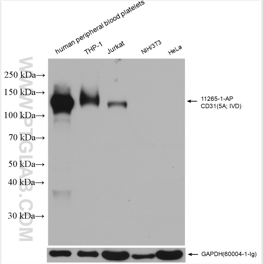
CD31 (also known as PECAM-1 or Platelet Endothelial Cell Adhesion Molecule-1) is a type of protein that is found on the surface of certain cells in the body, including platelets, endothelial cells (which line the blood vessels), and some immune cells.
CD31 functions as a cell adhesion molecule, meaning it helps cells stick together and interact with each other. It plays important roles in various physiological processes, such as the regulation of leukocyte migration, angiogenesis (the formation of new blood vessels), hemostasis (the process that stops bleeding), and thrombosis (the formation of a blood clot inside a blood vessel).
As an antigen, CD31 is used in immunological techniques to identify and characterize cells expressing this protein. Antigens are substances that can be recognized by the immune system and stimulate an immune response. In the case of CD31, antibodies specific to this protein can be used to detect its presence on the surface of cells, providing valuable information for research and diagnostic purposes.
Vascular Cell Adhesion Molecule-1 (VCAM-1) is a glycoprotein expressed on the surface of endothelial cells that plays a crucial role in the inflammatory response. It is involved in the recruitment and adhesion of leukocytes to the site of inflammation. VCAM-1 interacts with integrins on the surface of leukocytes, particularly very late antigen-4 (VLA-4), to facilitate this adhesion process. This interaction leads to the activation of signaling pathways that promote the migration of leukocytes across the endothelial barrier and into the surrounding tissue, where they can contribute to the immune response and resolution of inflammation. Increased expression of VCAM-1 has been associated with various inflammatory diseases, including atherosclerosis, rheumatoid arthritis, and multiple sclerosis.
Microcirculation is the circulation of blood in the smallest blood vessels, including arterioles, venules, and capillaries. It's responsible for the delivery of oxygen and nutrients to the tissues and the removal of waste products. The microcirculation plays a crucial role in maintaining tissue homeostasis and is regulated by various physiological mechanisms such as autonomic nervous system activity, local metabolic factors, and hormones.
Impairment of microcirculation can lead to tissue hypoxia, inflammation, and organ dysfunction, which are common features in several diseases, including diabetes, hypertension, sepsis, and ischemia-reperfusion injury. Therefore, understanding the structure and function of the microcirculation is essential for developing new therapeutic strategies to treat these conditions.
Intercellular Adhesion Molecule-1 (ICAM-1), also known as CD54, is a transmembrane glycoprotein expressed on the surface of various cell types including endothelial cells, fibroblasts, and immune cells. ICAM-1 plays a crucial role in the inflammatory response and the immune system by mediating the adhesion of leukocytes (white blood cells) to the endothelium, allowing them to migrate into surrounding tissues during an immune response or inflammation.
ICAM-1 contains five immunoglobulin-like domains in its extracellular region and binds to several integrins present on leukocytes, such as LFA-1 (lymphocyte function-associated antigen 1) and Mac-1 (macrophage-1 antigen). This interaction facilitates the firm adhesion of leukocytes to the endothelium, which is a critical step in the extravasation process.
In addition to its role in inflammation and immunity, ICAM-1 has been implicated in several pathological conditions, including atherosclerosis, cancer, and autoimmune diseases. Increased expression of ICAM-1 on endothelial cells is associated with the recruitment of immune cells to sites of injury or infection, making it an important target for therapeutic interventions in various inflammatory disorders.
E-Selectin, also known as Endothelial Leukocyte Adhesion Molecule 1 (ELAM-1), is a type of cell adhesion molecule mainly expressed on the surface of endothelial cells in response to inflammatory cytokines. It plays a crucial role in the initial recruitment and attachment of leukocytes (white blood cells) to the site of inflammation or injury, facilitating their transendothelial migration into the surrounding tissue. E-Selectin recognizes specific carbohydrate structures on the surface of leukocytes, contributing to the specificity of this adhesive interaction during the inflammatory response.
Signal transduction is the process by which a cell converts an extracellular signal, such as a hormone or neurotransmitter, into an intracellular response. This involves a series of molecular events that transmit the signal from the cell surface to the interior of the cell, ultimately resulting in changes in gene expression, protein activity, or metabolism.
The process typically begins with the binding of the extracellular signal to a receptor located on the cell membrane. This binding event activates the receptor, which then triggers a cascade of intracellular signaling molecules, such as second messengers, protein kinases, and ion channels. These molecules amplify and propagate the signal, ultimately leading to the activation or inhibition of specific cellular responses.
Signal transduction pathways are highly regulated and can be modulated by various factors, including other signaling molecules, post-translational modifications, and feedback mechanisms. Dysregulation of these pathways has been implicated in a variety of diseases, including cancer, diabetes, and neurological disorders.
Messenger RNA (mRNA) is a type of RNA (ribonucleic acid) that carries genetic information copied from DNA in the form of a series of three-base code "words," each of which specifies a particular amino acid. This information is used by the cell's machinery to construct proteins, a process known as translation. After being transcribed from DNA, mRNA travels out of the nucleus to the ribosomes in the cytoplasm where protein synthesis occurs. Once the protein has been synthesized, the mRNA may be degraded and recycled. Post-transcriptional modifications can also occur to mRNA, such as alternative splicing and addition of a 5' cap and a poly(A) tail, which can affect its stability, localization, and translation efficiency.
Nitric Oxide Synthase Type III (NOS-III), also known as endothelial Nitric Oxide Synthase (eNOS), is an enzyme responsible for the production of nitric oxide (NO) in the endothelium, the lining of blood vessels. This enzyme catalyzes the conversion of L-arginine to L-citrulline, producing NO as a byproduct. The release of NO from eNOS plays an important role in regulating vascular tone and homeostasis, including the relaxation of smooth muscle cells in the blood vessel walls, inhibition of platelet aggregation, and modulation of immune function. Mutations or dysfunction in NOS-III can contribute to various cardiovascular diseases such as hypertension, atherosclerosis, and erectile dysfunction.
Vascular Endothelial Growth Factor Receptor-2 (VEGFR-2) is a tyrosine kinase receptor that is primarily expressed on vascular endothelial cells. It is a crucial regulator of angiogenesis, the process of new blood vessel formation from pre-existing vessels. VEGFR-2 is activated by binding to its ligand, Vascular Endothelial Growth Factor-A (VEGF-A), leading to receptor dimerization and autophosphorylation. This activation triggers a cascade of intracellular signaling events that promote endothelial cell proliferation, migration, survival, and vascular permeability, all essential steps in the angiogenic process.
VEGFR-2 plays a significant role in physiological and pathological conditions associated with angiogenesis, such as embryonic development, wound healing, tumor growth, and retinopathies. Inhibition of VEGFR-2 signaling has been an attractive target for anti-angiogenic therapies in various diseases, including cancer and age-related macular degeneration.
Cell adhesion molecules (CAMs) are a type of protein found on the surface of cells that mediate the attachment or adhesion of cells to either other cells or to the extracellular matrix (ECM), which is the network of proteins and carbohydrates that provides structural and biochemical support to surrounding cells.
CAMs play crucial roles in various biological processes, including tissue development, differentiation, repair, and maintenance of tissue architecture and function. They are also involved in cell signaling, migration, and regulation of the immune response.
There are several types of CAMs, classified based on their structure and function, such as immunoglobulin-like CAMs (IgCAMs), cadherins, integrins, and selectins. Dysregulation of CAMs has been implicated in various diseases, including cancer, inflammation, and neurological disorders.
Nitric oxide (NO) is a molecule made up of one nitrogen atom and one oxygen atom. In the body, it is a crucial signaling molecule involved in various physiological processes such as vasodilation, immune response, neurotransmission, and inhibition of platelet aggregation. It is produced naturally by the enzyme nitric oxide synthase (NOS) from the amino acid L-arginine. Inhaled nitric oxide is used medically to treat pulmonary hypertension in newborns and adults, as it helps to relax and widen blood vessels, improving oxygenation and blood flow.
Endothelial growth factors (ECGFs or EGFs) are a group of signaling proteins that stimulate the growth, proliferation, and survival of endothelial cells, which line the interior surface of blood vessels. These growth factors play crucial roles in various physiological processes, including angiogenesis (the formation of new blood vessels), wound healing, and vascular development during embryogenesis.
One of the most well-studied EGFs is the vascular endothelial growth factor (VEGF) family, which consists of several members like VEGFA, VEGFB, VEGFC, VEGFD, and placental growth factor (PlGF). These factors bind to specific receptors on the surface of endothelial cells, leading to a cascade of intracellular signaling events that ultimately result in cell proliferation, migration, and survival.
Other EGFs include fibroblast growth factors (FGFs), hepatocyte growth factor (HGF), platelet-derived growth factor (PDGF), and transforming growth factor-beta (TGF-β). Dysregulation of endothelial growth factors has been implicated in various pathological conditions, such as cancer, diabetic retinopathy, age-related macular degeneration, and cardiovascular diseases. Therefore, understanding the functions and regulation of EGFs is essential for developing novel therapeutic strategies to treat these disorders.
Blood vessels are the part of the circulatory system that transport blood throughout the body. They form a network of tubes that carry blood to and from the heart, lungs, and other organs. The main types of blood vessels are arteries, veins, and capillaries. Arteries carry oxygenated blood away from the heart to the rest of the body, while veins return deoxygenated blood back to the heart. Capillaries connect arteries and veins and facilitate the exchange of oxygen, nutrients, and waste materials between the blood and the body's tissues.
Microvessels are the smallest blood vessels in the body, including capillaries, venules, and arterioles. They form a crucial part of the circulatory system, responsible for delivering oxygen and nutrients to tissues and organs while removing waste products. Capillaries, the tiniest microvessels, facilitate the exchange of substances between blood and tissue cells through their thin walls. Overall, microvessels play a vital role in maintaining proper organ function and overall health.
A cell line is a culture of cells that are grown in a laboratory for use in research. These cells are usually taken from a single cell or group of cells, and they are able to divide and grow continuously in the lab. Cell lines can come from many different sources, including animals, plants, and humans. They are often used in scientific research to study cellular processes, disease mechanisms, and to test new drugs or treatments. Some common types of human cell lines include HeLa cells (which come from a cancer patient named Henrietta Lacks), HEK293 cells (which come from embryonic kidney cells), and HUVEC cells (which come from umbilical vein endothelial cells). It is important to note that cell lines are not the same as primary cells, which are cells that are taken directly from a living organism and have not been grown in the lab.
Vascular Endothelial Growth Factors (VEGFs) are a family of signaling proteins that stimulate the growth and development of new blood vessels, a process known as angiogenesis. They play crucial roles in both physiological and pathological conditions, such as embryonic development, wound healing, and tumor growth. Specifically, VEGFs bind to specific receptors on the surface of endothelial cells, which line the interior surface of blood vessels, triggering a cascade of intracellular signaling events that promote cell proliferation, migration, and survival. Dysregulation of VEGF signaling has been implicated in various diseases, including cancer, age-related macular degeneration, and diabetic retinopathy.
Capillary permeability refers to the ability of substances to pass through the walls of capillaries, which are the smallest blood vessels in the body. These tiny vessels connect the arterioles and venules, allowing for the exchange of nutrients, waste products, and gases between the blood and the surrounding tissues.
The capillary wall is composed of a single layer of endothelial cells that are held together by tight junctions. The permeability of these walls varies depending on the size and charge of the molecules attempting to pass through. Small, uncharged molecules such as water, oxygen, and carbon dioxide can easily diffuse through the capillary wall, while larger or charged molecules such as proteins and large ions have more difficulty passing through.
Increased capillary permeability can occur in response to inflammation, infection, or injury, allowing larger molecules and immune cells to enter the surrounding tissues. This can lead to swelling (edema) and tissue damage if not controlled. Decreased capillary permeability, on the other hand, can lead to impaired nutrient exchange and tissue hypoxia.
Overall, the permeability of capillaries is a critical factor in maintaining the health and function of tissues throughout the body.
Cell division is the process by which a single eukaryotic cell (a cell with a true nucleus) divides into two identical daughter cells. This complex process involves several stages, including replication of DNA, separation of chromosomes, and division of the cytoplasm. There are two main types of cell division: mitosis and meiosis.
Mitosis is the type of cell division that results in two genetically identical daughter cells. It is a fundamental process for growth, development, and tissue repair in multicellular organisms. The stages of mitosis include prophase, prometaphase, metaphase, anaphase, and telophase, followed by cytokinesis, which divides the cytoplasm.
Meiosis, on the other hand, is a type of cell division that occurs in the gonads (ovaries and testes) during the production of gametes (sex cells). Meiosis results in four genetically unique daughter cells, each with half the number of chromosomes as the parent cell. This process is essential for sexual reproduction and genetic diversity. The stages of meiosis include meiosis I and meiosis II, which are further divided into prophase, prometaphase, metaphase, anaphase, and telophase.
In summary, cell division is the process by which a single cell divides into two daughter cells, either through mitosis or meiosis. This process is critical for growth, development, tissue repair, and sexual reproduction in multicellular organisms.
Angiogenesis inhibitors are a class of drugs that block the growth of new blood vessels (angiogenesis). They work by targeting specific molecules involved in the process of angiogenesis, such as vascular endothelial growth factor (VEGF) and its receptors. By blocking these molecules, angiogenesis inhibitors can prevent the development of new blood vessels that feed tumors, thereby slowing or stopping their growth.
Angiogenesis inhibitors are used in the treatment of various types of cancer, including colon, lung, breast, kidney, and ovarian cancer. They may be given alone or in combination with other cancer treatments, such as chemotherapy or radiation therapy. Some examples of angiogenesis inhibitors include bevacizumab (Avastin), sorafenib (Nexavar), sunitinib (Sutent), and pazopanib (Votrient).
It's important to note that while angiogenesis inhibitors can be effective in treating cancer, they can also have serious side effects, such as high blood pressure, bleeding, and damage to the heart or kidneys. Therefore, it's essential that patients receive careful monitoring and management of these potential side effects while undergoing treatment with angiogenesis inhibitors.
Lymphokines are a type of cytokines that are produced and released by activated lymphocytes, a type of white blood cell, in response to an antigenic stimulation. They play a crucial role in the regulation of immune responses and inflammation. Lymphokines can mediate various biological activities such as chemotaxis, activation, proliferation, and differentiation of different immune cells including lymphocytes, monocytes, macrophages, and eosinophils. Examples of lymphokines include interleukins (ILs), interferons (IFNs), tumor necrosis factor (TNF), and colony-stimulating factors (CSFs).
Tumor Necrosis Factor-alpha (TNF-α) is a cytokine, a type of small signaling protein involved in immune response and inflammation. It is primarily produced by activated macrophages, although other cell types such as T-cells, natural killer cells, and mast cells can also produce it.
TNF-α plays a crucial role in the body's defense against infection and tissue injury by mediating inflammatory responses, activating immune cells, and inducing apoptosis (programmed cell death) in certain types of cells. It does this by binding to its receptors, TNFR1 and TNFR2, which are found on the surface of many cell types.
In addition to its role in the immune response, TNF-α has been implicated in the pathogenesis of several diseases, including autoimmune disorders such as rheumatoid arthritis, inflammatory bowel disease, and psoriasis, as well as cancer, where it can promote tumor growth and metastasis.
Therapeutic agents that target TNF-α, such as infliximab, adalimumab, and etanercept, have been developed to treat these conditions. However, these drugs can also increase the risk of infections and other side effects, so their use must be carefully monitored.
Mechanical stress, in the context of physiology and medicine, refers to any type of force that is applied to body tissues or organs, which can cause deformation or displacement of those structures. Mechanical stress can be either external, such as forces exerted on the body during physical activity or trauma, or internal, such as the pressure changes that occur within blood vessels or other hollow organs.
Mechanical stress can have a variety of effects on the body, depending on the type, duration, and magnitude of the force applied. For example, prolonged exposure to mechanical stress can lead to tissue damage, inflammation, and chronic pain. Additionally, abnormal or excessive mechanical stress can contribute to the development of various musculoskeletal disorders, such as tendinitis, osteoarthritis, and herniated discs.
In order to mitigate the negative effects of mechanical stress, the body has a number of adaptive responses that help to distribute forces more evenly across tissues and maintain structural integrity. These responses include changes in muscle tone, joint positioning, and connective tissue stiffness, as well as the remodeling of bone and other tissues over time. However, when these adaptive mechanisms are overwhelmed or impaired, mechanical stress can become a significant factor in the development of various pathological conditions.
Apoptosis is a programmed and controlled cell death process that occurs in multicellular organisms. It is a natural process that helps maintain tissue homeostasis by eliminating damaged, infected, or unwanted cells. During apoptosis, the cell undergoes a series of morphological changes, including cell shrinkage, chromatin condensation, and fragmentation into membrane-bound vesicles called apoptotic bodies. These bodies are then recognized and engulfed by neighboring cells or phagocytic cells, preventing an inflammatory response. Apoptosis is regulated by a complex network of intracellular signaling pathways that involve proteins such as caspases, Bcl-2 family members, and inhibitors of apoptosis (IAPs).
Cell proliferation is the process by which cells increase in number, typically through the process of cell division. In the context of biology and medicine, it refers to the reproduction of cells that makes up living tissue, allowing growth, maintenance, and repair. It involves several stages including the transition from a phase of quiescence (G0 phase) to an active phase (G1 phase), DNA replication in the S phase, and mitosis or M phase, where the cell divides into two daughter cells.
Abnormal or uncontrolled cell proliferation is a characteristic feature of many diseases, including cancer, where deregulated cell cycle control leads to excessive and unregulated growth of cells, forming tumors that can invade surrounding tissues and metastasize to distant sites in the body.
'Gene expression regulation' refers to the processes that control whether, when, and where a particular gene is expressed, meaning the production of a specific protein or functional RNA encoded by that gene. This complex mechanism can be influenced by various factors such as transcription factors, chromatin remodeling, DNA methylation, non-coding RNAs, and post-transcriptional modifications, among others. Proper regulation of gene expression is crucial for normal cellular function, development, and maintaining homeostasis in living organisms. Dysregulation of gene expression can lead to various diseases, including cancer and genetic disorders.
Von Willebrand factor (vWF) is a large multimeric glycoprotein that plays a crucial role in hemostasis, the process which leads to the cessation of bleeding and the formation of a blood clot. It was named after Erik Adolf von Willebrand, a Finnish physician who first described the disorder associated with its deficiency, known as von Willebrand disease (vWD).
The primary functions of vWF include:
1. Platelet adhesion and aggregation: vWF mediates the initial attachment of platelets to damaged blood vessel walls by binding to exposed collagen fibers and then interacting with glycoprotein Ib (GPIb) receptors on the surface of platelets, facilitating platelet adhesion. Subsequently, vWF also promotes platelet-platelet interactions (aggregation) through its interaction with platelet glycoprotein IIb/IIIa (GPIIb/IIIa) receptors under high shear stress conditions found in areas of turbulent blood flow, such as arterioles and the capillary bed.
2. Transport and stabilization of coagulation factor VIII: vWF serves as a carrier protein for coagulation factor VIII (FVIII), protecting it from proteolytic degradation and maintaining its stability in circulation. This interaction between vWF and FVIII is essential for the proper functioning of the coagulation cascade, particularly in the context of vWD, where impaired FVIII function can lead to bleeding disorders.
3. Wound healing: vWF contributes to wound healing by promoting platelet adhesion and aggregation at the site of injury, which facilitates the formation of a provisional fibrin-based clot that serves as a scaffold for tissue repair and regeneration.
In summary, von Willebrand factor is a vital hemostatic protein involved in platelet adhesion, aggregation, coagulation factor VIII stabilization, and wound healing. Deficiencies or dysfunctions in vWF can lead to bleeding disorders such as von Willebrand disease.
TEC (Tyrosine kinase with Immunoglobulin-like and EGF homology domains-2) or TIE-2 is a type of receptor tyrosine kinase that plays a crucial role in the regulation of angiogenesis, lymphangiogenesis, and vascular maintenance. It is primarily expressed on the surface of endothelial cells, which line the interior surface of blood vessels.
The TIE-2 receptor binds to its ligand, angiopoietin-1 (Ang1), promoting vessel stability and quiescence by reducing endothelial cell permeability and enhancing their survival. Angiopoietin-2 (Ang2) can also bind to the TIE-2 receptor but with lower affinity than Ang1, acting as a context-dependent agonist or antagonist. In the presence of VEGF (Vascular Endothelial Growth Factor), Ang2 functions as an antagonist, inducing vascular instability and increasing endothelial cell permeability, which contributes to angiogenesis during development and in pathological conditions like tumor growth, inflammation, and ischemia.
Abnormal TIE-2 signaling has been implicated in several diseases, including cancer, atherosclerosis, and diabetic retinopathy. Targeting the TIE-2 signaling pathway presents an attractive therapeutic strategy for treating these conditions.
Immunohistochemistry (IHC) is a technique used in pathology and laboratory medicine to identify specific proteins or antigens in tissue sections. It combines the principles of immunology and histology to detect the presence and location of these target molecules within cells and tissues. This technique utilizes antibodies that are specific to the protein or antigen of interest, which are then tagged with a detection system such as a chromogen or fluorophore. The stained tissue sections can be examined under a microscope, allowing for the visualization and analysis of the distribution and expression patterns of the target molecule in the context of the tissue architecture. Immunohistochemistry is widely used in diagnostic pathology to help identify various diseases, including cancer, infectious diseases, and immune-mediated disorders.
The pulmonary artery is a large blood vessel that carries deoxygenated blood from the right ventricle of the heart to the lungs for oxygenation. It divides into two main branches, the right and left pulmonary arteries, which further divide into smaller vessels called arterioles, and then into a vast network of capillaries in the lungs where gas exchange occurs. The thin walls of these capillaries allow oxygen to diffuse into the blood and carbon dioxide to diffuse out, making the blood oxygen-rich before it is pumped back to the left side of the heart through the pulmonary veins. This process is crucial for maintaining proper oxygenation of the body's tissues and organs.
A dose-response relationship in the context of drugs refers to the changes in the effects or symptoms that occur as the dose of a drug is increased or decreased. Generally, as the dose of a drug is increased, the severity or intensity of its effects also increases. Conversely, as the dose is decreased, the effects of the drug become less severe or may disappear altogether.
The dose-response relationship is an important concept in pharmacology and toxicology because it helps to establish the safe and effective dosage range for a drug. By understanding how changes in the dose of a drug affect its therapeutic and adverse effects, healthcare providers can optimize treatment plans for their patients while minimizing the risk of harm.
The dose-response relationship is typically depicted as a curve that shows the relationship between the dose of a drug and its effect. The shape of the curve may vary depending on the drug and the specific effect being measured. Some drugs may have a steep dose-response curve, meaning that small changes in the dose can result in large differences in the effect. Other drugs may have a more gradual dose-response curve, where larger changes in the dose are needed to produce significant effects.
In addition to helping establish safe and effective dosages, the dose-response relationship is also used to evaluate the potential therapeutic benefits and risks of new drugs during clinical trials. By systematically testing different doses of a drug in controlled studies, researchers can identify the optimal dosage range for the drug and assess its safety and efficacy.
Phosphorylation is the process of adding a phosphate group (a molecule consisting of one phosphorus atom and four oxygen atoms) to a protein or other organic molecule, which is usually done by enzymes called kinases. This post-translational modification can change the function, localization, or activity of the target molecule, playing a crucial role in various cellular processes such as signal transduction, metabolism, and regulation of gene expression. Phosphorylation is reversible, and the removal of the phosphate group is facilitated by enzymes called phosphatases.
Up-regulation is a term used in molecular biology and medicine to describe an increase in the expression or activity of a gene, protein, or receptor in response to a stimulus. This can occur through various mechanisms such as increased transcription, translation, or reduced degradation of the molecule. Up-regulation can have important functional consequences, for example, enhancing the sensitivity or response of a cell to a hormone, neurotransmitter, or drug. It is a normal physiological process that can also be induced by disease or pharmacological interventions.
Western blotting is a laboratory technique used in molecular biology to detect and quantify specific proteins in a mixture of many different proteins. This technique is commonly used to confirm the expression of a protein of interest, determine its size, and investigate its post-translational modifications. The name "Western" blotting distinguishes this technique from Southern blotting (for DNA) and Northern blotting (for RNA).
The Western blotting procedure involves several steps:
1. Protein extraction: The sample containing the proteins of interest is first extracted, often by breaking open cells or tissues and using a buffer to extract the proteins.
2. Separation of proteins by electrophoresis: The extracted proteins are then separated based on their size by loading them onto a polyacrylamide gel and running an electric current through the gel (a process called sodium dodecyl sulfate-polyacrylamide gel electrophoresis or SDS-PAGE). This separates the proteins according to their molecular weight, with smaller proteins migrating faster than larger ones.
3. Transfer of proteins to a membrane: After separation, the proteins are transferred from the gel onto a nitrocellulose or polyvinylidene fluoride (PVDF) membrane using an electric current in a process called blotting. This creates a replica of the protein pattern on the gel but now immobilized on the membrane for further analysis.
4. Blocking: The membrane is then blocked with a blocking agent, such as non-fat dry milk or bovine serum albumin (BSA), to prevent non-specific binding of antibodies in subsequent steps.
5. Primary antibody incubation: A primary antibody that specifically recognizes the protein of interest is added and allowed to bind to its target protein on the membrane. This step may be performed at room temperature or 4°C overnight, depending on the antibody's properties.
6. Washing: The membrane is washed with a buffer to remove unbound primary antibodies.
7. Secondary antibody incubation: A secondary antibody that recognizes the primary antibody (often coupled to an enzyme or fluorophore) is added and allowed to bind to the primary antibody. This step may involve using a horseradish peroxidase (HRP)-conjugated or alkaline phosphatase (AP)-conjugated secondary antibody, depending on the detection method used later.
8. Washing: The membrane is washed again to remove unbound secondary antibodies.
9. Detection: A detection reagent is added to visualize the protein of interest by detecting the signal generated from the enzyme-conjugated or fluorophore-conjugated secondary antibody. This can be done using chemiluminescent, colorimetric, or fluorescent methods.
10. Analysis: The resulting image is analyzed to determine the presence and quantity of the protein of interest in the sample.
Western blotting is a powerful technique for identifying and quantifying specific proteins within complex mixtures. It can be used to study protein expression, post-translational modifications, protein-protein interactions, and more. However, it requires careful optimization and validation to ensure accurate and reproducible results.
Fibroblast Growth Factor 2 (FGF-2), also known as basic fibroblast growth factor, is a protein involved in various biological processes such as cell growth, proliferation, and differentiation. It plays a crucial role in wound healing, embryonic development, and angiogenesis (the formation of new blood vessels). FGF-2 is produced and secreted by various cells, including fibroblasts, and exerts its effects by binding to specific receptors on the cell surface, leading to activation of intracellular signaling pathways. It has been implicated in several diseases, including cancer, where it can contribute to tumor growth and progression.
In the field of medicine, "time factors" refer to the duration of symptoms or time elapsed since the onset of a medical condition, which can have significant implications for diagnosis and treatment. Understanding time factors is crucial in determining the progression of a disease, evaluating the effectiveness of treatments, and making critical decisions regarding patient care.
For example, in stroke management, "time is brain," meaning that rapid intervention within a specific time frame (usually within 4.5 hours) is essential to administering tissue plasminogen activator (tPA), a clot-busting drug that can minimize brain damage and improve patient outcomes. Similarly, in trauma care, the "golden hour" concept emphasizes the importance of providing definitive care within the first 60 minutes after injury to increase survival rates and reduce morbidity.
Time factors also play a role in monitoring the progression of chronic conditions like diabetes or heart disease, where regular follow-ups and assessments help determine appropriate treatment adjustments and prevent complications. In infectious diseases, time factors are crucial for initiating antibiotic therapy and identifying potential outbreaks to control their spread.
Overall, "time factors" encompass the significance of recognizing and acting promptly in various medical scenarios to optimize patient outcomes and provide effective care.
Coculture techniques refer to a type of experimental setup in which two or more different types of cells or organisms are grown and studied together in a shared culture medium. This method allows researchers to examine the interactions between different cell types or species under controlled conditions, and to study how these interactions may influence various biological processes such as growth, gene expression, metabolism, and signal transduction.
Coculture techniques can be used to investigate a wide range of biological phenomena, including the effects of host-microbe interactions on human health and disease, the impact of different cell types on tissue development and homeostasis, and the role of microbial communities in shaping ecosystems. These techniques can also be used to test the efficacy and safety of new drugs or therapies by examining their effects on cells grown in coculture with other relevant cell types.
There are several different ways to establish cocultures, depending on the specific research question and experimental goals. Some common methods include:
1. Mixed cultures: In this approach, two or more cell types are simply mixed together in a culture dish or flask and allowed to grow and interact freely.
2. Cell-layer cultures: Here, one cell type is grown on a porous membrane or other support structure, while the second cell type is grown on top of it, forming a layered coculture.
3. Conditioned media cultures: In this case, one cell type is grown to confluence and its culture medium is collected and then used to grow a second cell type. This allows the second cell type to be exposed to any factors secreted by the first cell type into the medium.
4. Microfluidic cocultures: These involve growing cells in microfabricated channels or chambers, which allow for precise control over the spatial arrangement and flow of nutrients, waste products, and signaling molecules between different cell types.
Overall, coculture techniques provide a powerful tool for studying complex biological systems and gaining insights into the mechanisms that underlie various physiological and pathological processes.
C57BL/6 (C57 Black 6) is an inbred strain of laboratory mouse that is widely used in biomedical research. The term "inbred" refers to a strain of animals where matings have been carried out between siblings or other closely related individuals for many generations, resulting in a population that is highly homozygous at most genetic loci.
The C57BL/6 strain was established in 1920 by crossing a female mouse from the dilute brown (DBA) strain with a male mouse from the black strain. The resulting offspring were then interbred for many generations to create the inbred C57BL/6 strain.
C57BL/6 mice are known for their robust health, longevity, and ease of handling, making them a popular choice for researchers. They have been used in a wide range of biomedical research areas, including studies of cancer, immunology, neuroscience, cardiovascular disease, and metabolism.
One of the most notable features of the C57BL/6 strain is its sensitivity to certain genetic modifications, such as the introduction of mutations that lead to obesity or impaired glucose tolerance. This has made it a valuable tool for studying the genetic basis of complex diseases and traits.
Overall, the C57BL/6 inbred mouse strain is an important model organism in biomedical research, providing a valuable resource for understanding the genetic and molecular mechanisms underlying human health and disease.
Vascular endothelial growth factor (VEGF) receptors are a type of cell surface receptor that play crucial roles in the process of angiogenesis, which is the formation of new blood vessels from pre-existing ones. These receptors bind to VEGF proteins, leading to a cascade of intracellular signaling events that ultimately result in the proliferation, migration, and survival of endothelial cells, which line the interior surface of blood vessels. There are three main types of VEGF receptors: VEGFR-1, VEGFR-2, and VEGFR-3. These receptors have distinct roles in angiogenesis, with VEGFR-2 being the primary mediator of this process. Dysregulation of VEGF signaling has been implicated in various diseases, including cancer, age-related macular degeneration, and diabetic retinopathy, making VEGF receptors important targets for therapeutic intervention.
Retinal vessels refer to the blood vessels that are located in the retina, which is the light-sensitive tissue that lines the inner surface of the eye. The retina contains two types of blood vessels: arteries and veins.
The central retinal artery supplies oxygenated blood to the inner layers of the retina, while the central retinal vein drains deoxygenated blood from the retina. These vessels can be visualized during a routine eye examination using an ophthalmoscope, which allows healthcare professionals to assess their health and any potential abnormalities.
Retinal vessels are essential for maintaining the health and function of the retina, and any damage or changes to these vessels can affect vision and lead to various eye conditions such as diabetic retinopathy, retinal vein occlusion, and hypertensive retinopathy.
Thrombin is a serine protease enzyme that plays a crucial role in the coagulation cascade, which is a complex series of biochemical reactions that leads to the formation of a blood clot (thrombus) to prevent excessive bleeding during an injury. Thrombin is formed from its precursor protein, prothrombin, through a process called activation, which involves cleavage by another enzyme called factor Xa.
Once activated, thrombin converts fibrinogen, a soluble plasma protein, into fibrin, an insoluble protein that forms the structural framework of a blood clot. Thrombin also activates other components of the coagulation cascade, such as factor XIII, which crosslinks and stabilizes the fibrin network, and platelets, which contribute to the formation and growth of the clot.
Thrombin has several regulatory mechanisms that control its activity, including feedback inhibition by antithrombin III, a plasma protein that inactivates thrombin and other serine proteases, and tissue factor pathway inhibitor (TFPI), which inhibits the activation of factor Xa, thereby preventing further thrombin formation.
Overall, thrombin is an essential enzyme in hemostasis, the process that maintains the balance between bleeding and clotting in the body. However, excessive or uncontrolled thrombin activity can lead to pathological conditions such as thrombosis, atherosclerosis, and disseminated intravascular coagulation (DIC).
Cell survival refers to the ability of a cell to continue living and functioning normally, despite being exposed to potentially harmful conditions or treatments. This can include exposure to toxins, radiation, chemotherapeutic drugs, or other stressors that can damage cells or interfere with their normal processes.
In scientific research, measures of cell survival are often used to evaluate the effectiveness of various therapies or treatments. For example, researchers may expose cells to a particular drug or treatment and then measure the percentage of cells that survive to assess its potential therapeutic value. Similarly, in toxicology studies, measures of cell survival can help to determine the safety of various chemicals or substances.
It's important to note that cell survival is not the same as cell proliferation, which refers to the ability of cells to divide and multiply. While some treatments may promote cell survival, they may also inhibit cell proliferation, making them useful for treating diseases such as cancer. Conversely, other treatments may be designed to specifically target and kill cancer cells, even if it means sacrificing some healthy cells in the process.
Epoprostenol is a medication that belongs to a class of drugs called prostaglandins. It is a synthetic analog of a natural substance in the body called prostacyclin, which widens blood vessels and has anti-platelet effects. Epoprostenol is used to treat pulmonary arterial hypertension (PAH), a condition characterized by high blood pressure in the arteries that supply blood to the lungs.
Epoprostenol works by relaxing the smooth muscle in the walls of the pulmonary arteries, which reduces the resistance to blood flow and lowers the pressure within these vessels. This helps improve symptoms such as shortness of breath, fatigue, and chest pain, and can also prolong survival in people with PAH.
Epoprostenol is administered continuously through a small pump that delivers the medication directly into the bloodstream. It is a potent vasodilator, which means it can cause a sudden drop in blood pressure if not given carefully. Therefore, it is usually started in a hospital setting under close medical supervision.
Common side effects of epoprostenol include headache, flushing, jaw pain, nausea, vomiting, diarrhea, and muscle or joint pain. More serious side effects can include bleeding, infection at the site of the catheter, and an allergic reaction to the medication.
Reverse Transcriptase Polymerase Chain Reaction (RT-PCR) is a laboratory technique used in molecular biology to amplify and detect specific DNA sequences. This technique is particularly useful for the detection and quantification of RNA viruses, as well as for the analysis of gene expression.
The process involves two main steps: reverse transcription and polymerase chain reaction (PCR). In the first step, reverse transcriptase enzyme is used to convert RNA into complementary DNA (cDNA) by reading the template provided by the RNA molecule. This cDNA then serves as a template for the PCR amplification step.
In the second step, the PCR reaction uses two primers that flank the target DNA sequence and a thermostable polymerase enzyme to repeatedly copy the targeted cDNA sequence. The reaction mixture is heated and cooled in cycles, allowing the primers to anneal to the template, and the polymerase to extend the new strand. This results in exponential amplification of the target DNA sequence, making it possible to detect even small amounts of RNA or cDNA.
RT-PCR is a sensitive and specific technique that has many applications in medical research and diagnostics, including the detection of viruses such as HIV, hepatitis C virus, and SARS-CoV-2 (the virus that causes COVID-19). It can also be used to study gene expression, identify genetic mutations, and diagnose genetic disorders.
"Swine" is a common term used to refer to even-toed ungulates of the family Suidae, including domestic pigs and wild boars. However, in a medical context, "swine" often appears in the phrase "swine flu," which is a strain of influenza virus that typically infects pigs but can also cause illness in humans. The 2009 H1N1 pandemic was caused by a new strain of swine-origin influenza A virus, which was commonly referred to as "swine flu." It's important to note that this virus is not transmitted through eating cooked pork products; it spreads from person to person, mainly through respiratory droplets produced when an infected person coughs or sneezes.
Enzyme activation refers to the process by which an enzyme becomes biologically active and capable of carrying out its specific chemical or biological reaction. This is often achieved through various post-translational modifications, such as proteolytic cleavage, phosphorylation, or addition of cofactors or prosthetic groups to the enzyme molecule. These modifications can change the conformation or structure of the enzyme, exposing or creating a binding site for the substrate and allowing the enzymatic reaction to occur.
For example, in the case of proteolytic cleavage, an inactive precursor enzyme, known as a zymogen, is cleaved into its active form by a specific protease. This is seen in enzymes such as trypsin and chymotrypsin, which are initially produced in the pancreas as inactive precursors called trypsinogen and chymotrypsinogen, respectively. Once they reach the small intestine, they are activated by enteropeptidase, a protease that cleaves a specific peptide bond, releasing the active enzyme.
Phosphorylation is another common mechanism of enzyme activation, where a phosphate group is added to a specific serine, threonine, or tyrosine residue on the enzyme by a protein kinase. This modification can alter the conformation of the enzyme and create a binding site for the substrate, allowing the enzymatic reaction to occur.
Enzyme activation is a crucial process in many biological pathways, as it allows for precise control over when and where specific reactions take place. It also provides a mechanism for regulating enzyme activity in response to various signals and stimuli, such as hormones, neurotransmitters, or changes in the intracellular environment.
Enzyme inhibitors are substances that bind to an enzyme and decrease its activity, preventing it from catalyzing a chemical reaction in the body. They can work by several mechanisms, including blocking the active site where the substrate binds, or binding to another site on the enzyme to change its shape and prevent substrate binding. Enzyme inhibitors are often used as drugs to treat various medical conditions, such as high blood pressure, abnormal heart rhythms, and bacterial infections. They can also be found naturally in some foods and plants, and can be used in research to understand enzyme function and regulation.
Nitric Oxide Synthase (NOS) is a group of enzymes that catalyze the production of nitric oxide (NO) from L-arginine. There are three distinct isoforms of NOS, each with different expression patterns and functions:
1. Neuronal Nitric Oxide Synthase (nNOS or NOS1): This isoform is primarily expressed in the nervous system and plays a role in neurotransmission, synaptic plasticity, and learning and memory processes.
2. Inducible Nitric Oxide Synthase (iNOS or NOS2): This isoform is induced by various stimuli such as cytokines, lipopolysaccharides, and hypoxia in a variety of cells including immune cells, endothelial cells, and smooth muscle cells. iNOS produces large amounts of NO, which functions as a potent effector molecule in the immune response, particularly in the defense against microbial pathogens.
3. Endothelial Nitric Oxide Synthase (eNOS or NOS3): This isoform is constitutively expressed in endothelial cells and produces low levels of NO that play a crucial role in maintaining vascular homeostasis by regulating vasodilation, inhibiting platelet aggregation, and preventing smooth muscle cell proliferation.
Overall, NOS plays an essential role in various physiological processes, including neurotransmission, immune response, cardiovascular function, and respiratory regulation. Dysregulation of NOS activity has been implicated in several pathological conditions such as hypertension, atherosclerosis, neurodegenerative diseases, and inflammatory disorders.
Cell communication, also known as cell signaling, is the process by which cells exchange and transmit signals between each other and their environment. This complex system allows cells to coordinate their functions and maintain tissue homeostasis. Cell communication can occur through various mechanisms including:
1. Autocrine signaling: When a cell releases a signal that binds to receptors on the same cell, leading to changes in its behavior or function.
2. Paracrine signaling: When a cell releases a signal that binds to receptors on nearby cells, influencing their behavior or function.
3. Endocrine signaling: When a cell releases a hormone into the bloodstream, which then travels to distant target cells and binds to specific receptors, triggering a response.
4. Synaptic signaling: In neurons, communication occurs through the release of neurotransmitters that cross the synapse and bind to receptors on the postsynaptic cell, transmitting electrical or chemical signals.
5. Contact-dependent signaling: When cells physically interact with each other, allowing for the direct exchange of signals and information.
Cell communication is essential for various physiological processes such as growth, development, differentiation, metabolism, immune response, and tissue repair. Dysregulation in cell communication can contribute to diseases, including cancer, diabetes, and neurological disorders.
Conditioned culture media refers to a type of growth medium that has been previously used to culture and maintain the cells of an organism. The conditioned media contains factors secreted by those cells, such as hormones, nutrients, and signaling molecules, which can affect the behavior and growth of other cells that are introduced into the media later on.
When the conditioned media is used for culturing a new set of cells, it can provide a more physiologically relevant environment than traditional culture media, as it contains factors that are specific to the original cell type. This can be particularly useful in studies that aim to understand cell-cell interactions and communication, or to mimic the natural microenvironment of cells in the body.
It's important to note that conditioned media should be handled carefully and used promptly after preparation, as the factors it contains can degrade over time and affect the quality of the results.
The Blood-Brain Barrier (BBB) is a highly specialized, selective interface between the central nervous system (CNS) and the circulating blood. It is formed by unique endothelial cells that line the brain's capillaries, along with tight junctions, astrocytic foot processes, and pericytes, which together restrict the passage of substances from the bloodstream into the CNS. This barrier serves to protect the brain from harmful agents and maintain a stable environment for proper neural function. However, it also poses a challenge in delivering therapeutics to the CNS, as most large and hydrophilic molecules cannot cross the BBB.
Angiopoietin-1 (ANG-1) is a protein that plays a crucial role in the development and maintenance of blood vessels. It is a member of the angiopoietin family, which includes several growth factors involved in the regulation of angiogenesis, the formation of new blood vessels from pre-existing ones.
ANG-1 primarily binds to the Tie2 receptor, which is predominantly expressed on vascular endothelial cells. The ANG-1/Tie2 signaling pathway promotes vascular stability, integrity, and maturation by enhancing endothelial cell survival, migration, and adhesion. It also inhibits vascular leakage and inflammation, contributing to the overall homeostasis of the vasculature.
In addition to its role in physiological conditions, ANG-1 has been implicated in various pathological processes such as tumor angiogenesis, ischemia, and fibrosis. Modulation of the ANG-1/Tie2 signaling pathway has emerged as a potential therapeutic strategy for treating several diseases associated with abnormal vascular function.
Cell surface receptors, also known as membrane receptors, are proteins located on the cell membrane that bind to specific molecules outside the cell, known as ligands. These receptors play a crucial role in signal transduction, which is the process of converting an extracellular signal into an intracellular response.
Cell surface receptors can be classified into several categories based on their structure and mechanism of action, including:
1. Ion channel receptors: These receptors contain a pore that opens to allow ions to flow across the cell membrane when they bind to their ligands. This ion flux can directly activate or inhibit various cellular processes.
2. G protein-coupled receptors (GPCRs): These receptors consist of seven transmembrane domains and are associated with heterotrimeric G proteins that modulate intracellular signaling pathways upon ligand binding.
3. Enzyme-linked receptors: These receptors possess an intrinsic enzymatic activity or are linked to an enzyme, which becomes activated when the receptor binds to its ligand. This activation can lead to the initiation of various signaling cascades within the cell.
4. Receptor tyrosine kinases (RTKs): These receptors contain intracellular tyrosine kinase domains that become activated upon ligand binding, leading to the phosphorylation and activation of downstream signaling molecules.
5. Integrins: These receptors are transmembrane proteins that mediate cell-cell or cell-matrix interactions by binding to extracellular matrix proteins or counter-receptors on adjacent cells. They play essential roles in cell adhesion, migration, and survival.
Cell surface receptors are involved in various physiological processes, including neurotransmission, hormone signaling, immune response, and cell growth and differentiation. Dysregulation of these receptors can contribute to the development of numerous diseases, such as cancer, diabetes, and neurological disorders.
CD (cluster of differentiation) antigens are cell-surface proteins that are expressed on leukocytes (white blood cells) and can be used to identify and distinguish different subsets of these cells. They are important markers in the field of immunology and hematology, and are commonly used to diagnose and monitor various diseases, including cancer, autoimmune disorders, and infectious diseases.
CD antigens are designated by numbers, such as CD4, CD8, CD19, etc., which refer to specific proteins found on the surface of different types of leukocytes. For example, CD4 is a protein found on the surface of helper T cells, while CD8 is found on cytotoxic T cells.
CD antigens can be used as targets for immunotherapy, such as monoclonal antibody therapy, in which antibodies are designed to bind to specific CD antigens and trigger an immune response against cancer cells or infected cells. They can also be used as markers to monitor the effectiveness of treatments and to detect minimal residual disease (MRD) after treatment.
It's important to note that not all CD antigens are exclusive to leukocytes, some can be found on other cell types as well, and their expression can vary depending on the activation state or differentiation stage of the cells.
Small interfering RNA (siRNA) is a type of short, double-stranded RNA molecule that plays a role in the RNA interference (RNAi) pathway. The RNAi pathway is a natural cellular process that regulates gene expression by targeting and destroying specific messenger RNA (mRNA) molecules, thereby preventing the translation of those mRNAs into proteins.
SiRNAs are typically 20-25 base pairs in length and are generated from longer double-stranded RNA precursors called hairpin RNAs or dsRNAs by an enzyme called Dicer. Once generated, siRNAs associate with a protein complex called the RNA-induced silencing complex (RISC), which uses one strand of the siRNA (the guide strand) to recognize and bind to complementary sequences in the target mRNA. The RISC then cleaves the target mRNA, leading to its degradation and the inhibition of protein synthesis.
SiRNAs have emerged as a powerful tool for studying gene function and have shown promise as therapeutic agents for a variety of diseases, including viral infections, cancer, and genetic disorders. However, their use as therapeutics is still in the early stages of development, and there are challenges associated with delivering siRNAs to specific cells and tissues in the body.
Gene expression is the process by which the information encoded in a gene is used to synthesize a functional gene product, such as a protein or RNA molecule. This process involves several steps: transcription, RNA processing, and translation. During transcription, the genetic information in DNA is copied into a complementary RNA molecule, known as messenger RNA (mRNA). The mRNA then undergoes RNA processing, which includes adding a cap and tail to the mRNA and splicing out non-coding regions called introns. The resulting mature mRNA is then translated into a protein on ribosomes in the cytoplasm through the process of translation.
The regulation of gene expression is a complex and highly controlled process that allows cells to respond to changes in their environment, such as growth factors, hormones, and stress signals. This regulation can occur at various stages of gene expression, including transcriptional activation or repression, RNA processing, mRNA stability, and translation. Dysregulation of gene expression has been implicated in many diseases, including cancer, genetic disorders, and neurological conditions.
Corneal endothelial cell loss refers to the decrease in the number of corneal endothelial cells, which is a layer of cells that line the inner surface of the cornea. These cells are essential for maintaining the clarity and health of the cornea, as they help to pump fluids out of the cornea and maintain its transparency.
Corneal endothelial cell loss can occur due to various reasons such as aging, eye trauma, surgery (such as cataract surgery), diseases (such as Fuchs' dystrophy), or inherited conditions. When the number of endothelial cells decreases below a certain threshold, it can lead to corneal swelling, cloudiness, and vision loss.
The rate of corneal endothelial cell loss varies from person to person, but on average, people lose about 0.6% of their endothelial cells per year. Factors such as age, certain medical conditions, and previous eye surgery can increase the rate of cell loss. In some cases, corneal transplantation may be necessary to replace damaged or lost endothelial cells and restore vision.
Transfection is a term used in molecular biology that refers to the process of deliberately introducing foreign genetic material (DNA, RNA or artificial gene constructs) into cells. This is typically done using chemical or physical methods, such as lipofection or electroporation. Transfection is widely used in research and medical settings for various purposes, including studying gene function, producing proteins, developing gene therapies, and creating genetically modified organisms. It's important to note that transfection is different from transduction, which is the process of introducing genetic material into cells using viruses as vectors.
Pericytes are specialized cells that surround the endothelial cells which line the blood capillaries. They play an important role in the regulation of capillary diameter, blood flow, and the formation of new blood vessels (angiogenesis). Pericytes also contribute to the maintenance of the blood-brain barrier, immune surveillance, and the clearance of waste products from the brain. They are often referred to as "mural cells" or "rouleaux cells" and can be found in various tissues throughout the body.
The endothelium is a thin layer of cells that lines the interior surface of blood vessels and lymphatic vessels. The lymphatic endothelium, specifically, is the type of endothelial cell that forms the walls of lymphatic vessels. These vessels are an important part of the immune system and play a crucial role in transporting fluid, waste products, and immune cells throughout the body.
The lymphatic endothelium helps to regulate the movement of fluids and cells between the tissues and the bloodstream. It also contains specialized structures called valves that help to ensure the unidirectional flow of lymph fluid towards the heart. Dysfunction of the lymphatic endothelium has been implicated in a variety of diseases, including lymphedema, inflammation, and cancer metastasis.
Interleukin-8 (IL-8) is a type of cytokine, which is a small signaling protein involved in immune response and inflammation. IL-8 is also known as neutrophil chemotactic factor or NCF because it attracts neutrophils, a type of white blood cell, to the site of infection or injury.
IL-8 is produced by various cells including macrophages, epithelial cells, and endothelial cells in response to bacterial or inflammatory stimuli. It acts by binding to specific receptors called CXCR1 and CXCR2 on the surface of neutrophils, which triggers a series of intracellular signaling events leading to neutrophil activation, migration, and degranulation.
IL-8 plays an important role in the recruitment of neutrophils to the site of infection or tissue damage, where they can phagocytose and destroy invading microorganisms. However, excessive or prolonged production of IL-8 has been implicated in various inflammatory diseases such as chronic obstructive pulmonary disease (COPD), rheumatoid arthritis, and cancer.
Growth factor receptors are a type of cell surface receptor that bind to specific growth factors, which are signaling molecules that play crucial roles in regulating various cellular processes such as growth, differentiation, and survival. These receptors have an extracellular domain that can recognize and bind to the growth factor and an intracellular domain that can transduce the signal into the cell through a series of biochemical reactions.
There are several types of growth factors, including fibroblast growth factors (FGFs), epidermal growth factors (EGFs), vascular endothelial growth factors (VEGFs), and transforming growth factors (TGFs). Each type of growth factor has its own specific receptor or family of receptors.
Once a growth factor binds to its receptor, it triggers a cascade of intracellular signaling events that ultimately lead to changes in gene expression, protein synthesis, and other cellular responses. These responses can include the activation of enzymes, the regulation of ion channels, and the modulation of cytoskeletal dynamics.
Abnormalities in growth factor receptor signaling have been implicated in various diseases, including cancer, developmental disorders, and autoimmune diseases. For example, mutations in growth factor receptors can lead to uncontrolled cell growth and division, which is a hallmark of cancer. Therefore, understanding the structure and function of growth factor receptors has important implications for the development of new therapies for these diseases.
NF-κB (Nuclear Factor kappa-light-chain-enhancer of activated B cells) is a protein complex that plays a crucial role in regulating the immune response to infection and inflammation, as well as in cell survival, differentiation, and proliferation. It is composed of several subunits, including p50, p52, p65 (RelA), c-Rel, and RelB, which can form homodimers or heterodimers that bind to specific DNA sequences called κB sites in the promoter regions of target genes.
Under normal conditions, NF-κB is sequestered in the cytoplasm by inhibitory proteins known as IκBs (inhibitors of κB). However, upon stimulation by various signals such as cytokines, bacterial or viral products, and stress, IκBs are phosphorylated, ubiquitinated, and degraded, leading to the release and activation of NF-κB. Activated NF-κB then translocates to the nucleus, where it binds to κB sites and regulates the expression of target genes involved in inflammation, immunity, cell survival, and proliferation.
Dysregulation of NF-κB signaling has been implicated in various pathological conditions such as cancer, chronic inflammation, autoimmune diseases, and neurodegenerative disorders. Therefore, targeting NF-κB signaling has emerged as a potential therapeutic strategy for the treatment of these diseases.
A smooth muscle within the vascular system refers to the involuntary, innervated muscle that is found in the walls of blood vessels. These muscles are responsible for controlling the diameter of the blood vessels, which in turn regulates blood flow and blood pressure. They are called "smooth" muscles because their individual muscle cells do not have the striations, or cross-striped patterns, that are observed in skeletal and cardiac muscle cells. Smooth muscle in the vascular system is controlled by the autonomic nervous system and by hormones, and can contract or relax slowly over a period of time.
Recombinant proteins are artificially created proteins produced through the use of recombinant DNA technology. This process involves combining DNA molecules from different sources to create a new set of genes that encode for a specific protein. The resulting recombinant protein can then be expressed, purified, and used for various applications in research, medicine, and industry.
Recombinant proteins are widely used in biomedical research to study protein function, structure, and interactions. They are also used in the development of diagnostic tests, vaccines, and therapeutic drugs. For example, recombinant insulin is a common treatment for diabetes, while recombinant human growth hormone is used to treat growth disorders.
The production of recombinant proteins typically involves the use of host cells, such as bacteria, yeast, or mammalian cells, which are engineered to express the desired protein. The host cells are transformed with a plasmid vector containing the gene of interest, along with regulatory elements that control its expression. Once the host cells are cultured and the protein is expressed, it can be purified using various chromatography techniques.
Overall, recombinant proteins have revolutionized many areas of biology and medicine, enabling researchers to study and manipulate proteins in ways that were previously impossible.
Monocytes are a type of white blood cell that are part of the immune system. They are large cells with a round or oval shape and a nucleus that is typically indented or horseshoe-shaped. Monocytes are produced in the bone marrow and then circulate in the bloodstream, where they can differentiate into other types of immune cells such as macrophages and dendritic cells.
Monocytes play an important role in the body's defense against infection and tissue damage. They are able to engulf and digest foreign particles, microorganisms, and dead or damaged cells, which helps to clear them from the body. Monocytes also produce cytokines, which are signaling molecules that help to coordinate the immune response.
Elevated levels of monocytes in the bloodstream can be a sign of an ongoing infection, inflammation, or other medical conditions such as cancer or autoimmune disorders.
Molecular sequence data refers to the specific arrangement of molecules, most commonly nucleotides in DNA or RNA, or amino acids in proteins, that make up a biological macromolecule. This data is generated through laboratory techniques such as sequencing, and provides information about the exact order of the constituent molecules. This data is crucial in various fields of biology, including genetics, evolution, and molecular biology, allowing for comparisons between different organisms, identification of genetic variations, and studies of gene function and regulation.
Leukocytes, also known as white blood cells (WBCs), are a crucial component of the human immune system. They are responsible for protecting the body against infections and foreign substances. Leukocytes are produced in the bone marrow and circulate throughout the body in the bloodstream and lymphatic system.
There are several types of leukocytes, including:
1. Neutrophils - These are the most abundant type of leukocyte and are primarily responsible for fighting bacterial infections. They contain enzymes that can destroy bacteria.
2. Lymphocytes - These are responsible for producing antibodies and destroying virus-infected cells, as well as cancer cells. There are two main types of lymphocytes: B-lymphocytes and T-lymphocytes.
3. Monocytes - These are the largest type of leukocyte and help to break down and remove dead or damaged tissues, as well as microorganisms.
4. Eosinophils - These play a role in fighting parasitic infections and are also involved in allergic reactions and inflammation.
5. Basophils - These release histamine and other chemicals that cause inflammation in response to allergens or irritants.
An abnormal increase or decrease in the number of leukocytes can indicate an underlying medical condition, such as an infection, inflammation, or a blood disorder.
Thromboplastin is a substance that activates the coagulation cascade, leading to the formation of a clot (thrombus). It's primarily found in damaged or injured tissues and blood vessels, as well as in platelets (thrombocytes). There are two types of thromboplastin:
1. Extrinsic thromboplastin (also known as tissue factor): This is a transmembrane glycoprotein that is primarily found in subendothelial cells and released upon injury to the blood vessels. It initiates the extrinsic pathway of coagulation by binding to and activating Factor VII, ultimately leading to the formation of thrombin and fibrin clots.
2. Intrinsic thromboplastin (also known as plasma thromboplastin or factor III): This term is used less frequently and refers to a labile phospholipid component present in platelet membranes, which plays a role in the intrinsic pathway of coagulation.
In clinical settings, the term "thromboplastin" often refers to reagents used in laboratory tests like the prothrombin time (PT) and activated partial thromboplastin time (aPTT). These reagents contain a source of tissue factor and calcium ions to initiate and monitor the coagulation process.
The umbilical cord is a flexible, tube-like structure that connects the developing fetus to the placenta in the uterus during pregnancy. It arises from the abdomen of the fetus and transports essential nutrients, oxygen, and blood from the mother's circulation to the growing baby. Additionally, it carries waste products, such as carbon dioxide, from the fetus back to the placenta for elimination. The umbilical cord is primarily composed of two arteries (the umbilical arteries) and one vein (the umbilical vein), surrounded by a protective gelatinous substance called Wharton's jelly, and enclosed within a fibrous outer covering known as the umbilical cord coating. Following birth, the umbilical cord is clamped and cut, leaving behind the stump that eventually dries up and falls off, resulting in the baby's belly button.
P-Selectin is a type of cell adhesion molecule, specifically a member of the selectin family, that is involved in the inflammatory response. It is primarily expressed on the surface of activated platelets and endothelial cells. P-Selectin plays a crucial role in the initial interaction between leukocytes (white blood cells) and the vascular endothelium, which is an essential step in the recruitment of leukocytes to sites of inflammation or injury. This process helps to mediate the rolling and adhesion of leukocytes to the endothelial surface, facilitating their extravasation into the surrounding tissue. P-Selectin's function is regulated by its interaction with specific ligands on the surface of leukocytes, such as PSGL-1 (P-Selectin Glycoprotein Ligand-1).
Caveolin 1 is a protein that is a key component of caveolae, which are specialized invaginations of the plasma membrane found in many cell types. Caveolae play important roles in various cellular processes, including endocytosis, cholesterol homeostasis, and signal transduction.
Caveolin 1 is a structural protein that helps to form and maintain the shape of caveolae. It also plays a role in regulating the activity of various signaling molecules that are associated with caveolae, including G proteins, receptor tyrosine kinases, and Src family kinases.
Mutations in the gene that encodes caveolin 1 have been linked to several genetic disorders, including muscular dystrophy, cardiac arrhythmias, and cancer. Additionally, changes in the expression or localization of caveolin 1 have been implicated in a variety of diseases, including diabetes, neurodegenerative disorders, and infectious diseases.
Angiogenesis inducing agents are substances or drugs that stimulate the growth of new blood vessels, a process known as angiogenesis. This process is essential for the growth and development of tissues and organs in the body, including wound healing and the formation of blood vessels in the placenta during pregnancy. However, abnormal angiogenesis can also contribute to various diseases, such as cancer, diabetic retinopathy, and age-related macular degeneration.
Angiogenesis inducing agents are being studied for their potential therapeutic benefits in a variety of medical conditions. For example, they may be used to promote wound healing or tissue repair after injury or surgery. In cancer treatment, angiogenesis inhibitors are often used to block the growth of new blood vessels and prevent tumors from growing and spreading. However, angiogenesis inducing agents can have the opposite effect and may potentially be used to enhance the delivery of drugs to tumors or improve the effectiveness of other cancer treatments.
Examples of angiogenesis inducing agents include certain growth factors, such as vascular endothelial growth factor (VEGF), fibroblast growth factor (FGF), and platelet-derived growth factor (PDGF). These substances can be administered as drugs to stimulate angiogenesis in specific contexts. Other substances, such as hypoxia-inducible factors (HIFs) and prostaglandins, can also induce angiogenesis under certain conditions.
Laminin is a family of proteins that are an essential component of the basement membrane, which is a specialized type of extracellular matrix. Laminins are large trimeric molecules composed of three different chains: α, β, and γ. There are five different α chains, three different β chains, and three different γ chains that can combine to form at least 15 different laminin isoforms.
Laminins play a crucial role in maintaining the structure and integrity of basement membranes by interacting with other components of the extracellular matrix, such as collagen IV, and cell surface receptors, such as integrins. They are involved in various biological processes, including cell adhesion, differentiation, migration, and survival.
Laminin dysfunction has been implicated in several human diseases, including cancer, diabetic nephropathy, and muscular dystrophy.
Flow cytometry is a medical and research technique used to measure physical and chemical characteristics of cells or particles, one cell at a time, as they flow in a fluid stream through a beam of light. The properties measured include:
* Cell size (light scatter)
* Cell internal complexity (granularity, also light scatter)
* Presence or absence of specific proteins or other molecules on the cell surface or inside the cell (using fluorescent antibodies or other fluorescent probes)
The technique is widely used in cell counting, cell sorting, protein engineering, biomarker discovery and monitoring disease progression, particularly in hematology, immunology, and cancer research.
Neutrophils are a type of white blood cell that are part of the immune system's response to infection. They are produced in the bone marrow and released into the bloodstream where they circulate and are able to move quickly to sites of infection or inflammation in the body. Neutrophils are capable of engulfing and destroying bacteria, viruses, and other foreign substances through a process called phagocytosis. They are also involved in the release of inflammatory mediators, which can contribute to tissue damage in some cases. Neutrophils are characterized by the presence of granules in their cytoplasm, which contain enzymes and other proteins that help them carry out their immune functions.
Reactive Oxygen Species (ROS) are highly reactive molecules containing oxygen, including peroxides, superoxide, hydroxyl radical, and singlet oxygen. They are naturally produced as byproducts of normal cellular metabolism in the mitochondria, and can also be generated by external sources such as ionizing radiation, tobacco smoke, and air pollutants. At low or moderate concentrations, ROS play important roles in cell signaling and homeostasis, but at high concentrations, they can cause significant damage to cell structures, including lipids, proteins, and DNA, leading to oxidative stress and potential cell death.
Coronary vessels refer to the network of blood vessels that supply oxygenated blood and nutrients to the heart muscle, also known as the myocardium. The two main coronary arteries are the left main coronary artery and the right coronary artery.
The left main coronary artery branches off into the left anterior descending artery (LAD) and the left circumflex artery (LCx). The LAD supplies blood to the front of the heart, while the LCx supplies blood to the side and back of the heart.
The right coronary artery supplies blood to the right lower part of the heart, including the right atrium and ventricle, as well as the back of the heart.
Coronary vessel disease (CVD) occurs when these vessels become narrowed or blocked due to the buildup of plaque, leading to reduced blood flow to the heart muscle. This can result in chest pain, shortness of breath, or a heart attack.
Angiopoietin-2 (Ang-2) is a protein that is involved in the regulation of blood vessel formation and maintenance. It is a member of the angiopoietin family, which includes Ang-1, Ang-2, Ang-3, and Ang-4. These proteins bind to the Tie receptor tyrosine kinases (Tie1 and Tie2) on the surface of endothelial cells, which line the interior of blood vessels.
Ang-2 is primarily produced by endothelial cells and has context-dependent roles in angiogenesis, which is the growth of new blood vessels from pre-existing ones. In general, Ang-2 is thought to act as an antagonist of Ang-1, which promotes vessel stability and maturation.
Ang-2 can destabilize existing blood vessels by binding to Tie2 receptors and blocking the stabilizing effects of Ang-1. This can lead to increased vascular permeability and inflammation. However, in the presence of pro-angiogenic factors such as VEGF (vascular endothelial growth factor), Ang-2 can also promote the formation of new blood vessels by stimulating endothelial cell migration and proliferation.
Abnormal regulation of Ang-2 has been implicated in various diseases, including cancer, diabetic retinopathy, and age-related macular degeneration. In these conditions, increased levels of Ang-2 can contribute to the development of abnormal blood vessels, which can lead to tissue damage and loss of function.
A "knockout" mouse is a genetically engineered mouse in which one or more genes have been deleted or "knocked out" using molecular biology techniques. This allows researchers to study the function of specific genes and their role in various biological processes, as well as potential associations with human diseases. The mice are generated by introducing targeted DNA modifications into embryonic stem cells, which are then used to create a live animal. Knockout mice have been widely used in biomedical research to investigate gene function, disease mechanisms, and potential therapeutic targets.
Membrane proteins are a type of protein that are embedded in the lipid bilayer of biological membranes, such as the plasma membrane of cells or the inner membrane of mitochondria. These proteins play crucial roles in various cellular processes, including:
1. Cell-cell recognition and signaling
2. Transport of molecules across the membrane (selective permeability)
3. Enzymatic reactions at the membrane surface
4. Energy transduction and conversion
5. Mechanosensation and signal transduction
Membrane proteins can be classified into two main categories: integral membrane proteins, which are permanently associated with the lipid bilayer, and peripheral membrane proteins, which are temporarily or loosely attached to the membrane surface. Integral membrane proteins can further be divided into three subcategories based on their topology:
1. Transmembrane proteins, which span the entire width of the lipid bilayer with one or more alpha-helices or beta-barrels.
2. Lipid-anchored proteins, which are covalently attached to lipids in the membrane via a glycosylphosphatidylinositol (GPI) anchor or other lipid modifications.
3. Monotopic proteins, which are partially embedded in the membrane and have one or more domains exposed to either side of the bilayer.
Membrane proteins are essential for maintaining cellular homeostasis and are targets for various therapeutic interventions, including drug development and gene therapy. However, their structural complexity and hydrophobicity make them challenging to study using traditional biochemical methods, requiring specialized techniques such as X-ray crystallography, nuclear magnetic resonance (NMR) spectroscopy, and single-particle cryo-electron microscopy (cryo-EM).
Inflammation is a complex biological response of tissues to harmful stimuli, such as pathogens, damaged cells, or irritants. It is characterized by the following signs: rubor (redness), tumor (swelling), calor (heat), dolor (pain), and functio laesa (loss of function). The process involves the activation of the immune system, recruitment of white blood cells, and release of inflammatory mediators, which contribute to the elimination of the injurious stimuli and initiation of the healing process. However, uncontrolled or chronic inflammation can also lead to tissue damage and diseases.
The Fluorescent Antibody Technique (FAT) is a type of immunofluorescence assay used in laboratory medicine and pathology for the detection and localization of specific antigens or antibodies in tissues, cells, or microorganisms. In this technique, a fluorescein-labeled antibody is used to selectively bind to the target antigen or antibody, forming an immune complex. When excited by light of a specific wavelength, the fluorescein label emits light at a longer wavelength, typically visualized as green fluorescence under a fluorescence microscope.
The FAT is widely used in diagnostic microbiology for the identification and characterization of various bacteria, viruses, fungi, and parasites. It has also been applied in the diagnosis of autoimmune diseases and certain cancers by detecting specific antibodies or antigens in patient samples. The main advantage of FAT is its high sensitivity and specificity, allowing for accurate detection and differentiation of various pathogens and disease markers. However, it requires specialized equipment and trained personnel to perform and interpret the results.
Interleukin-1 (IL-1) is a type of cytokine, which are proteins that play a crucial role in cell signaling. Specifically, IL-1 is a pro-inflammatory cytokine that is involved in the regulation of immune and inflammatory responses in the body. It is produced by various cells, including monocytes, macrophages, and dendritic cells, in response to infection or injury.
IL-1 exists in two forms, IL-1α and IL-1β, which have similar biological activities but are encoded by different genes. Both forms of IL-1 bind to the same receptor, IL-1R, and activate intracellular signaling pathways that lead to the production of other cytokines, chemokines, and inflammatory mediators.
IL-1 has a wide range of biological effects, including fever induction, activation of immune cells, regulation of hematopoiesis (the formation of blood cells), and modulation of bone metabolism. Dysregulation of IL-1 production or activity has been implicated in various inflammatory diseases, such as rheumatoid arthritis, gout, and inflammatory bowel disease. Therefore, IL-1 is an important target for the development of therapies aimed at modulating the immune response and reducing inflammation.
Thrombomodulin is a protein that is found on the surface of endothelial cells, which line the interior surface of blood vessels. It plays an important role in the regulation of blood coagulation (clotting) and the activation of natural anticoagulant pathways. Thrombomodulin binds to thrombin, a protein involved in blood clotting, and changes its function from promoting coagulation to inhibiting it. This interaction also activates protein C, an important anticoagulant protein, which helps to prevent the excessive formation of blood clots. Thrombomodulin also has anti-inflammatory properties and is involved in the maintenance of the integrity of the endothelial cell lining.
Weibel-Palade bodies are rod-shaped, membrane-bound organelles found in the cytoplasm of endothelial cells, which line the interior surface of blood vessels. They were first described by Edwin Weibel and George Palade in 1964. These organelles are unique to endothelial cells and serve as storage sites for von Willebrand factor (vWF) and other proteins involved in hemostasis, inflammation, and vasomotor functions.
The main components of Weibel-Palade bodies include:
1. Von Willebrand factor (vWF): A multimeric glycoprotein that plays a crucial role in platelet adhesion and aggregation at the site of vascular injury, as well as mediating the transport of coagulation factors VIII and V.
2. P-selectin: A cell adhesion molecule that facilitates leukocyte rolling and recruitment to sites of inflammation.
3. Endothelial nitric oxide synthase (eNOS): An enzyme responsible for the production of nitric oxide, a potent vasodilator that regulates vascular tone and blood flow.
4. Angiopoietin-2: A growth factor involved in angiogenesis and vascular remodeling.
5. Tissue plasminogen activator (tPA): A serine protease that plays a role in fibrinolysis, the process of breaking down blood clots.
Upon stimulation by various agonists such as thrombin, histamine, or vascular endothelial growth factor (VEGF), Weibel-Palade bodies undergo exocytosis, releasing their contents into the extracellular space. This process contributes to hemostatic responses, inflammatory reactions, and modulation of vascular tone.
Collagen is the most abundant protein in the human body, and it is a major component of connective tissues such as tendons, ligaments, skin, and bones. Collagen provides structure and strength to these tissues and helps them to withstand stretching and tension. It is made up of long chains of amino acids, primarily glycine, proline, and hydroxyproline, which are arranged in a triple helix structure. There are at least 16 different types of collagen found in the body, each with slightly different structures and functions. Collagen is important for maintaining the integrity and health of tissues throughout the body, and it has been studied for its potential therapeutic uses in various medical conditions.
A lung is a pair of spongy, elastic organs in the chest that work together to enable breathing. They are responsible for taking in oxygen and expelling carbon dioxide through the process of respiration. The left lung has two lobes, while the right lung has three lobes. The lungs are protected by the ribcage and are covered by a double-layered membrane called the pleura. The trachea divides into two bronchi, which further divide into smaller bronchioles, leading to millions of tiny air sacs called alveoli, where the exchange of gases occurs.
Calcium is an essential mineral that is vital for various physiological processes in the human body. The medical definition of calcium is as follows:
Calcium (Ca2+) is a crucial cation and the most abundant mineral in the human body, with approximately 99% of it found in bones and teeth. It plays a vital role in maintaining structural integrity, nerve impulse transmission, muscle contraction, hormonal secretion, blood coagulation, and enzyme activation.
Calcium homeostasis is tightly regulated through the interplay of several hormones, including parathyroid hormone (PTH), calcitonin, and vitamin D. Dietary calcium intake, absorption, and excretion are also critical factors in maintaining optimal calcium levels in the body.
Hypocalcemia refers to low serum calcium levels, while hypercalcemia indicates high serum calcium levels. Both conditions can have detrimental effects on various organ systems and require medical intervention to correct.
Fluorescence microscopy is a type of microscopy that uses fluorescent dyes or proteins to highlight and visualize specific components within a sample. In this technique, the sample is illuminated with high-energy light, typically ultraviolet (UV) or blue light, which excites the fluorescent molecules causing them to emit lower-energy, longer-wavelength light, usually visible light in the form of various colors. This emitted light is then collected by the microscope and detected to produce an image.
Fluorescence microscopy has several advantages over traditional brightfield microscopy, including the ability to visualize specific structures or molecules within a complex sample, increased sensitivity, and the potential for quantitative analysis. It is widely used in various fields of biology and medicine, such as cell biology, neuroscience, and pathology, to study the structure, function, and interactions of cells and proteins.
There are several types of fluorescence microscopy techniques, including widefield fluorescence microscopy, confocal microscopy, two-photon microscopy, and total internal reflection fluorescence (TIRF) microscopy, each with its own strengths and limitations. These techniques can provide valuable insights into the behavior of cells and proteins in health and disease.
Cell hypoxia, also known as cellular hypoxia or tissue hypoxia, refers to a condition in which the cells or tissues in the body do not receive an adequate supply of oxygen. Oxygen is essential for the production of energy in the form of ATP (adenosine triphosphate) through a process called oxidative phosphorylation. When the cells are deprived of oxygen, they switch to anaerobic metabolism, which produces lactic acid as a byproduct and can lead to acidosis.
Cell hypoxia can result from various conditions, including:
1. Low oxygen levels in the blood (hypoxemia) due to lung diseases such as chronic obstructive pulmonary disease (COPD), pneumonia, or high altitude.
2. Reduced blood flow to tissues due to cardiovascular diseases such as heart failure, peripheral artery disease, or shock.
3. Anemia, which reduces the oxygen-carrying capacity of the blood.
4. Carbon monoxide poisoning, which binds to hemoglobin and prevents it from carrying oxygen.
5. Inadequate ventilation due to trauma, drug overdose, or other causes that can lead to respiratory failure.
Cell hypoxia can cause cell damage, tissue injury, and organ dysfunction, leading to various clinical manifestations depending on the severity and duration of hypoxia. Treatment aims to correct the underlying cause and improve oxygen delivery to the tissues.
Receptor Protein-Tyrosine Kinases (RTKs) are a type of transmembrane receptors found on the cell surface that play a crucial role in signal transduction and regulation of various cellular processes, including cell growth, differentiation, metabolism, and survival. They are called "tyrosine kinases" because they possess an intrinsic enzymatic activity that catalyzes the transfer of a phosphate group from ATP to tyrosine residues on target proteins, thereby modulating their function.
RTKs are composed of three main domains: an extracellular domain that binds to specific ligands (growth factors, hormones, or cytokines), a transmembrane domain that spans the cell membrane, and an intracellular domain with tyrosine kinase activity. Upon ligand binding, RTKs undergo conformational changes that lead to their dimerization or oligomerization, which in turn activates their tyrosine kinase activity. Activated RTKs then phosphorylate specific tyrosine residues on downstream signaling proteins, initiating a cascade of intracellular signaling events that ultimately result in the appropriate cellular response.
Dysregulation of RTK signaling has been implicated in various human diseases, including cancer, diabetes, and developmental disorders. As such, RTKs are important targets for therapeutic intervention in these conditions.
Low-density lipoproteins (LDL), also known as "bad cholesterol," are a type of lipoprotein that carry cholesterol and other fats from the liver to cells throughout the body. High levels of LDL in the blood can lead to the buildup of cholesterol in the walls of the arteries, which can increase the risk of heart disease and stroke.
Lipoproteins are complex particles composed of proteins (apolipoproteins) and lipids (cholesterol, triglycerides, and phospholipids) that are responsible for transporting fat molecules around the body in the bloodstream. LDL is one type of lipoprotein, along with high-density lipoproteins (HDL), very low-density lipoproteins (VLDL), and chylomicrons.
LDL particles are smaller than HDL particles and can easily penetrate the artery walls, leading to the formation of plaques that can narrow or block the arteries. Therefore, maintaining healthy levels of LDL in the blood is essential for preventing cardiovascular disease.
Bradykinin is a naturally occurring peptide in the human body, consisting of nine amino acids. It is a potent vasodilator and increases the permeability of blood vessels, causing a local inflammatory response. Bradykinin is formed from the breakdown of certain proteins, such as kininogen, by enzymes called kininases or proteases, including kallikrein. It plays a role in several physiological processes, including pain transmission, blood pressure regulation, and the immune response. In some pathological conditions, such as hereditary angioedema, bradykinin levels can increase excessively, leading to symptoms like swelling, redness, and pain.
The extracellular matrix (ECM) is a complex network of biomolecules that provides structural and biochemical support to cells in tissues and organs. It is composed of various proteins, glycoproteins, and polysaccharides, such as collagens, elastin, fibronectin, laminin, and proteoglycans. The ECM plays crucial roles in maintaining tissue architecture, regulating cell behavior, and facilitating communication between cells. It provides a scaffold for cell attachment, migration, and differentiation, and helps to maintain the structural integrity of tissues by resisting mechanical stresses. Additionally, the ECM contains various growth factors, cytokines, and chemokines that can influence cellular processes such as proliferation, survival, and differentiation. Overall, the extracellular matrix is essential for the normal functioning of tissues and organs, and its dysregulation can contribute to various pathological conditions, including fibrosis, cancer, and degenerative diseases.
Cell culture is a technique used in scientific research to grow and maintain cells from plants, animals, or humans in a controlled environment outside of their original organism. This environment typically consists of a sterile container called a cell culture flask or plate, and a nutrient-rich liquid medium that provides the necessary components for the cells' growth and survival, such as amino acids, vitamins, minerals, and hormones.
There are several different types of cell culture techniques used in research, including:
1. Adherent cell culture: In this technique, cells are grown on a flat surface, such as the bottom of a tissue culture dish or flask. The cells attach to the surface and spread out, forming a monolayer that can be observed and manipulated under a microscope.
2. Suspension cell culture: In suspension culture, cells are grown in liquid medium without any attachment to a solid surface. These cells remain suspended in the medium and can be agitated or mixed to ensure even distribution of nutrients.
3. Organoid culture: Organoids are three-dimensional structures that resemble miniature organs and are grown from stem cells or other progenitor cells. They can be used to study organ development, disease processes, and drug responses.
4. Co-culture: In co-culture, two or more different types of cells are grown together in the same culture dish or flask. This technique is used to study cell-cell interactions and communication.
5. Conditioned medium culture: In this technique, cells are grown in a medium that has been conditioned by previous cultures of other cells. The conditioned medium contains factors secreted by the previous cells that can influence the growth and behavior of the new cells.
Cell culture techniques are widely used in biomedical research to study cellular processes, develop drugs, test toxicity, and investigate disease mechanisms. However, it is important to note that cell cultures may not always accurately represent the behavior of cells in a living organism, and results from cell culture experiments should be validated using other methods.
Monoclonal antibodies are a type of antibody that are identical because they are produced by a single clone of cells. They are laboratory-produced molecules that act like human antibodies in the immune system. They can be designed to attach to specific proteins found on the surface of cancer cells, making them useful for targeting and treating cancer. Monoclonal antibodies can also be used as a therapy for other diseases, such as autoimmune disorders and inflammatory conditions.
Monoclonal antibodies are produced by fusing a single type of immune cell, called a B cell, with a tumor cell to create a hybrid cell, or hybridoma. This hybrid cell is then able to replicate indefinitely, producing a large number of identical copies of the original antibody. These antibodies can be further modified and engineered to enhance their ability to bind to specific targets, increase their stability, and improve their effectiveness as therapeutic agents.
Monoclonal antibodies have several mechanisms of action in cancer therapy. They can directly kill cancer cells by binding to them and triggering an immune response. They can also block the signals that promote cancer growth and survival. Additionally, monoclonal antibodies can be used to deliver drugs or radiation directly to cancer cells, increasing the effectiveness of these treatments while minimizing their side effects on healthy tissues.
Monoclonal antibodies have become an important tool in modern medicine, with several approved for use in cancer therapy and other diseases. They are continuing to be studied and developed as a promising approach to treating a wide range of medical conditions.
I believe there may be some confusion in your question. "Rabbits" is a common name used to refer to the Lagomorpha species, particularly members of the family Leporidae. They are small mammals known for their long ears, strong legs, and quick reproduction.
However, if you're referring to "rabbits" in a medical context, there is a term called "rabbit syndrome," which is a rare movement disorder characterized by repetitive, involuntary movements of the fingers, resembling those of a rabbit chewing. It is also known as "finger-chewing chorea." This condition is usually associated with certain medications, particularly antipsychotics, and typically resolves when the medication is stopped or adjusted.
Protein-kinase B, also known as AKT, is a group of intracellular proteins that play a crucial role in various cellular processes such as glucose metabolism, apoptosis, cell proliferation, transcription, and cell migration. The AKT family includes three isoforms: AKT1, AKT2, and AKT3, which are encoded by the genes PKBalpha, PKBbeta, and PKBgamma, respectively.
Proto-oncogene proteins c-AKT refer to the normal, non-mutated forms of these proteins that are involved in the regulation of cell growth and survival under physiological conditions. However, when these genes are mutated or overexpressed, they can become oncogenes, leading to uncontrolled cell growth and cancer development.
Activation of c-AKT occurs through a signaling cascade that begins with the binding of extracellular ligands such as insulin-like growth factor 1 (IGF-1) or epidermal growth factor (EGF) to their respective receptors on the cell surface. This triggers a series of phosphorylation events that ultimately lead to the activation of c-AKT, which then phosphorylates downstream targets involved in various cellular processes.
In summary, proto-oncogene proteins c-AKT are normal intracellular proteins that play essential roles in regulating cell growth and survival under physiological conditions. However, their dysregulation can contribute to cancer development and progression.
Electron microscopy (EM) is a type of microscopy that uses a beam of electrons to create an image of the sample being examined, resulting in much higher magnification and resolution than light microscopy. There are several types of electron microscopy, including transmission electron microscopy (TEM), scanning electron microscopy (SEM), and reflection electron microscopy (REM).
In TEM, a beam of electrons is transmitted through a thin slice of the sample, and the electrons that pass through the sample are focused to form an image. This technique can provide detailed information about the internal structure of cells, viruses, and other biological specimens, as well as the composition and structure of materials at the atomic level.
In SEM, a beam of electrons is scanned across the surface of the sample, and the electrons that are scattered back from the surface are detected to create an image. This technique can provide information about the topography and composition of surfaces, as well as the structure of materials at the microscopic level.
REM is a variation of SEM in which the beam of electrons is reflected off the surface of the sample, rather than scattered back from it. This technique can provide information about the surface chemistry and composition of materials.
Electron microscopy has a wide range of applications in biology, medicine, and materials science, including the study of cellular structure and function, disease diagnosis, and the development of new materials and technologies.
Actin is a type of protein that forms part of the contractile apparatus in muscle cells, and is also found in various other cell types. It is a globular protein that polymerizes to form long filaments, which are important for many cellular processes such as cell division, cell motility, and the maintenance of cell shape. In muscle cells, actin filaments interact with another type of protein called myosin to enable muscle contraction. Actins can be further divided into different subtypes, including alpha-actin, beta-actin, and gamma-actin, which have distinct functions and expression patterns in the body.
The thoracic aorta is the segment of the largest artery in the human body (the aorta) that runs through the chest region (thorax). The thoracic aorta begins at the aortic arch, where it branches off from the ascending aorta, and extends down to the diaphragm, where it becomes the abdominal aorta.
The thoracic aorta is divided into three parts: the ascending aorta, the aortic arch, and the descending aorta. The ascending aorta rises from the left ventricle of the heart and is about 2 inches (5 centimeters) long. The aortic arch curves backward and to the left, giving rise to the brachiocephalic trunk, the left common carotid artery, and the left subclavian artery. The descending thoracic aorta runs downward through the chest, passing through the diaphragm to become the abdominal aorta.
The thoracic aorta supplies oxygenated blood to the upper body, including the head, neck, arms, and chest. It plays a critical role in maintaining blood flow and pressure throughout the body.
The cornea is the clear, dome-shaped surface at the front of the eye. It plays a crucial role in focusing vision. The cornea protects the eye from harmful particles and microorganisms, and it also serves as a barrier against UV light. Its transparency allows light to pass through and get focused onto the retina. The cornea does not contain blood vessels, so it relies on tears and the fluid inside the eye (aqueous humor) for nutrition and oxygen. Any damage or disease that affects its clarity and shape can significantly impact vision and potentially lead to blindness if left untreated.
Down-regulation is a process that occurs in response to various stimuli, where the number or sensitivity of cell surface receptors or the expression of specific genes is decreased. This process helps maintain homeostasis within cells and tissues by reducing the ability of cells to respond to certain signals or molecules.
In the context of cell surface receptors, down-regulation can occur through several mechanisms:
1. Receptor internalization: After binding to their ligands, receptors can be internalized into the cell through endocytosis. Once inside the cell, these receptors may be degraded or recycled back to the cell surface in smaller numbers.
2. Reduced receptor synthesis: Down-regulation can also occur at the transcriptional level, where the expression of genes encoding for specific receptors is decreased, leading to fewer receptors being produced.
3. Receptor desensitization: Prolonged exposure to a ligand can lead to a decrease in receptor sensitivity or affinity, making it more difficult for the cell to respond to the signal.
In the context of gene expression, down-regulation refers to the decreased transcription and/or stability of specific mRNAs, leading to reduced protein levels. This process can be induced by various factors, including microRNA (miRNA)-mediated regulation, histone modification, or DNA methylation.
Down-regulation is an essential mechanism in many physiological processes and can also contribute to the development of several diseases, such as cancer and neurodegenerative disorders.
Vascular Endothelial Growth Factor Receptor-1 (VEGFR-1), also known as Flt-1 (Fms-like tyrosine kinase-1), is a receptor tyrosine kinase that plays a crucial role in the regulation of angiogenesis, vasculogenesis, and lymphangiogenesis. It is primarily expressed on vascular endothelial cells, hematopoietic stem cells, and monocytes/macrophages. VEGFR-1 binds to several ligands, including Vascular Endothelial Growth Factor-A (VEGF-A), VEGF-B, and Placental Growth Factor (PlGF). The binding of these ligands to VEGFR-1 triggers intracellular signaling cascades that modulate various cellular responses, such as proliferation, migration, survival, and vascular permeability. While VEGFR-1 is known to have a role in promoting angiogenesis under certain conditions, it primarily acts as a negative regulator of angiogenesis by sequestering VEGF-A, preventing its binding to the more proangiogenic VEGFR-2 receptor. Dysregulation of VEGFR-1 signaling has been implicated in various pathological conditions, including cancer, inflammation, and vascular diseases.
A base sequence in the context of molecular biology refers to the specific order of nucleotides in a DNA or RNA molecule. In DNA, these nucleotides are adenine (A), guanine (G), cytosine (C), and thymine (T). In RNA, uracil (U) takes the place of thymine. The base sequence contains genetic information that is transcribed into RNA and ultimately translated into proteins. It is the exact order of these bases that determines the genetic code and thus the function of the DNA or RNA molecule.
Intercellular signaling peptides and proteins are molecules that mediate communication and interaction between different cells in living organisms. They play crucial roles in various biological processes, including cell growth, differentiation, migration, and apoptosis (programmed cell death). These signals can be released into the extracellular space, where they bind to specific receptors on the target cell's surface, triggering intracellular signaling cascades that ultimately lead to a response.
Peptides are short chains of amino acids, while proteins are larger molecules made up of one or more polypeptide chains. Both can function as intercellular signaling molecules by acting as ligands for cell surface receptors or by being cleaved from larger precursor proteins and released into the extracellular space. Examples of intercellular signaling peptides and proteins include growth factors, cytokines, chemokines, hormones, neurotransmitters, and their respective receptors.
These molecules contribute to maintaining homeostasis within an organism by coordinating cellular activities across tissues and organs. Dysregulation of intercellular signaling pathways has been implicated in various diseases, such as cancer, autoimmune disorders, and neurodegenerative conditions. Therefore, understanding the mechanisms underlying intercellular signaling is essential for developing targeted therapies to treat these disorders.
Arteries are blood vessels that carry oxygenated blood away from the heart to the rest of the body. They have thick, muscular walls that can withstand the high pressure of blood being pumped out of the heart. Arteries branch off into smaller vessels called arterioles, which further divide into a vast network of tiny capillaries where the exchange of oxygen, nutrients, and waste occurs between the blood and the body's cells. After passing through the capillary network, deoxygenated blood collects in venules, then merges into veins, which return the blood back to the heart.
Intercellular junctions are specialized areas of contact between two or more adjacent cells in multicellular organisms. They play crucial roles in maintaining tissue structure and function by regulating the movement of ions, molecules, and even larger cellular structures from one cell to another. There are several types of intercellular junctions, including:
1. Tight Junctions (Zonulae Occludentes): These are the most apical structures in epithelial and endothelial cells, forming a virtually impermeable barrier to prevent the paracellular passage of solutes and water between the cells. They create a tight seal by connecting the transmembrane proteins of adjacent cells, such as occludin and claudins.
2. Adherens Junctions: These are located just below the tight junctions and help maintain cell-to-cell adhesion and tissue integrity. Adherens junctions consist of cadherin proteins that form homophilic interactions with cadherins on adjacent cells, as well as intracellular adaptor proteins like catenins, which connect to the actin cytoskeleton.
3. Desmosomes: These are another type of cell-to-cell adhesion structure, primarily found in tissues that experience mechanical stress, such as the skin and heart. Desmosomes consist of cadherin proteins (desmocadherins) that interact with each other and connect to intermediate filaments (keratin in epithelial cells) via plakoglobin and desmoplakin.
4. Gap Junctions: These are specialized channels that directly connect the cytoplasm of adjacent cells, allowing for the exchange of small molecules, ions, and second messengers. Gap junctions consist of connexin proteins that form hexameric structures called connexons in the plasma membrane of each cell. When two connexons align, they create a continuous pore or channel between the cells.
In summary, intercellular junctions are essential for maintaining tissue structure and function by regulating paracellular transport, cell-to-cell adhesion, and intercellular communication.
The brain is the central organ of the nervous system, responsible for receiving and processing sensory information, regulating vital functions, and controlling behavior, movement, and cognition. It is divided into several distinct regions, each with specific functions:
1. Cerebrum: The largest part of the brain, responsible for higher cognitive functions such as thinking, learning, memory, language, and perception. It is divided into two hemispheres, each controlling the opposite side of the body.
2. Cerebellum: Located at the back of the brain, it is responsible for coordinating muscle movements, maintaining balance, and fine-tuning motor skills.
3. Brainstem: Connects the cerebrum and cerebellum to the spinal cord, controlling vital functions such as breathing, heart rate, and blood pressure. It also serves as a relay center for sensory information and motor commands between the brain and the rest of the body.
4. Diencephalon: A region that includes the thalamus (a major sensory relay station) and hypothalamus (regulates hormones, temperature, hunger, thirst, and sleep).
5. Limbic system: A group of structures involved in emotional processing, memory formation, and motivation, including the hippocampus, amygdala, and cingulate gyrus.
The brain is composed of billions of interconnected neurons that communicate through electrical and chemical signals. It is protected by the skull and surrounded by three layers of membranes called meninges, as well as cerebrospinal fluid that provides cushioning and nutrients.
Transendothelial migration (TEM) and transepithelial migration (TRM) are terms used to describe the movement of cells, typically leukocytes (white blood cells), across endothelial or epithelial cell layers. These processes play a crucial role in immune surveillance and inflammation.
Transendothelial migration refers specifically to the movement of cells across the endothelium, which is the layer of cells that lines the interior surface of blood vessels. This process allows leukocytes to leave the bloodstream and enter surrounding tissues during an immune response. TEM can be further divided into two main steps:
1. Adhesion: The initial attachment of leukocytes to the endothelium, mediated by adhesion molecules expressed on both the leukocyte and endothelial cell surfaces.
2. Diapedesis: The transmigration step where leukocytes squeeze between adjacent endothelial cells and move through the basement membrane to reach the underlying tissue.
Transepithelial migration, on the other hand, refers to the movement of cells across an epithelium, which is a layer of cells that forms a barrier between a body cavity or lumen (such as the gut or airways) and the underlying tissue. TRM can be observed in various physiological processes like wound healing and immune cell trafficking, but it also plays a role in pathological conditions such as cancer metastasis. Similar to TEM, TRM can be divided into several steps:
1. Adhesion: The initial attachment of cells to the epithelium, facilitated by adhesion molecules and receptors.
2. Polarization: Cells become polarized, forming protrusions that help them navigate through the tight junctions between epithelial cells.
3. Diapedesis: The transmigration step where cells move across the epithelium, often involving the disassembly and reassembly of tight junctions between epithelial cells.
4. Re-epithelialization: After cell migration is complete, the epithelial layer needs to be restored by re-establishing tight junctions and maintaining barrier integrity.
Integrin αVβ3 is a type of integrin, which is a heterodimeric transmembrane receptor that mediates cell-cell and cell-extracellular matrix (ECM) interactions. Integrins play crucial roles in various biological processes, including cell adhesion, migration, proliferation, differentiation, and survival.
Integrin αVβ3 is composed of two subunits, αV and β3, which are non-covalently associated to form a functional receptor. This integrin can bind to various ECM proteins containing the arginine-glycine-aspartic acid (RGD) motif, such as vitronectin, fibronectin, fibrinogen, and osteopontin.
Integrin αVβ3 is widely expressed in different cell types, including endothelial cells, smooth muscle cells, macrophages, and various tumor cells. It has been implicated in several physiological and pathological processes, such as angiogenesis, wound healing, bone remodeling, and tumor metastasis.
In the context of cancer, integrin αVβ3 has been shown to promote tumor growth, invasion, and metastasis by enhancing cell migration, survival, and resistance to apoptosis. Therefore, targeting integrin αVβ3 with therapeutic agents has emerged as a promising strategy for cancer treatment.
Proteoglycans are complex, highly negatively charged macromolecules that are composed of a core protein covalently linked to one or more glycosaminoglycan (GAG) chains. They are a major component of the extracellular matrix (ECM) and play crucial roles in various biological processes, including cell signaling, regulation of growth factor activity, and maintenance of tissue structure and function.
The GAG chains, which can vary in length and composition, are long, unbranched polysaccharides that are composed of repeating disaccharide units containing a hexuronic acid (either glucuronic or iduronic acid) and a hexosamine (either N-acetylglucosamine or N-acetylgalactosamine). These GAG chains can be sulfated to varying degrees, which contributes to the negative charge of proteoglycans.
Proteoglycans are classified into four major groups based on their core protein structure and GAG composition: heparan sulfate/heparin proteoglycans, chondroitin/dermatan sulfate proteoglycans, keratan sulfate proteoglycans, and hyaluronan-binding proteoglycans. Each group has distinct functions and is found in specific tissues and cell types.
In summary, proteoglycans are complex macromolecules composed of a core protein and one or more GAG chains that play important roles in the ECM and various biological processes, including cell signaling, growth factor regulation, and tissue structure maintenance.
Caveolae are small, flask-shaped invaginations of the plasma membrane that are abundant in many cell types, including endothelial cells, adipocytes, and muscle cells. They are characterized by the presence of caveolin proteins, which play a crucial role in their formation and function.
Caveolae have been implicated in various cellular processes, such as endocytosis, signal transduction, cholesterol homeostasis, and mechanoprotection. They can also serve as platforms for the assembly of signaling complexes and the regulation of various enzymatic activities.
The invaginated structure of caveolae allows them to interact with extracellular molecules and intracellular proteins, facilitating the exchange of materials between the plasma membrane and the cytosol. Dysregulation of caveolae function has been linked to several diseases, including cardiovascular disorders, cancer, and neurological conditions.
In the context of medicine and pharmacology, "kinetics" refers to the study of how a drug moves throughout the body, including its absorption, distribution, metabolism, and excretion (often abbreviated as ADME). This field is called "pharmacokinetics."
1. Absorption: This is the process of a drug moving from its site of administration into the bloodstream. Factors such as the route of administration (e.g., oral, intravenous, etc.), formulation, and individual physiological differences can affect absorption.
2. Distribution: Once a drug is in the bloodstream, it gets distributed throughout the body to various tissues and organs. This process is influenced by factors like blood flow, protein binding, and lipid solubility of the drug.
3. Metabolism: Drugs are often chemically modified in the body, typically in the liver, through processes known as metabolism. These changes can lead to the formation of active or inactive metabolites, which may then be further distributed, excreted, or undergo additional metabolic transformations.
4. Excretion: This is the process by which drugs and their metabolites are eliminated from the body, primarily through the kidneys (urine) and the liver (bile).
Understanding the kinetics of a drug is crucial for determining its optimal dosing regimen, potential interactions with other medications or foods, and any necessary adjustments for special populations like pediatric or geriatric patients, or those with impaired renal or hepatic function.
Endothelin-1 is a small peptide (21 amino acids) and a potent vasoconstrictor, which means it narrows blood vessels. It is primarily produced by the endothelial cells that line the interior surface of blood vessels. Endothelin-1 plays a crucial role in regulating vascular tone, cell growth, and inflammation. Its dysregulation has been implicated in various cardiovascular diseases, such as hypertension and heart failure. It exerts its effects by binding to specific G protein-coupled receptors (ETA and ETB) on the surface of target cells.
Animal disease models are specialized animals, typically rodents such as mice or rats, that have been genetically engineered or exposed to certain conditions to develop symptoms and physiological changes similar to those seen in human diseases. These models are used in medical research to study the pathophysiology of diseases, identify potential therapeutic targets, test drug efficacy and safety, and understand disease mechanisms.
The genetic modifications can include knockout or knock-in mutations, transgenic expression of specific genes, or RNA interference techniques. The animals may also be exposed to environmental factors such as chemicals, radiation, or infectious agents to induce the disease state.
Examples of animal disease models include:
1. Mouse models of cancer: Genetically engineered mice that develop various types of tumors, allowing researchers to study cancer initiation, progression, and metastasis.
2. Alzheimer's disease models: Transgenic mice expressing mutant human genes associated with Alzheimer's disease, which exhibit amyloid plaque formation and cognitive decline.
3. Diabetes models: Obese and diabetic mouse strains like the NOD (non-obese diabetic) or db/db mice, used to study the development of type 1 and type 2 diabetes, respectively.
4. Cardiovascular disease models: Atherosclerosis-prone mice, such as ApoE-deficient or LDLR-deficient mice, that develop plaque buildup in their arteries when fed a high-fat diet.
5. Inflammatory bowel disease models: Mice with genetic mutations affecting intestinal barrier function and immune response, such as IL-10 knockout or SAMP1/YitFc mice, which develop colitis.
Animal disease models are essential tools in preclinical research, but it is important to recognize their limitations. Differences between species can affect the translatability of results from animal studies to human patients. Therefore, researchers must carefully consider the choice of model and interpret findings cautiously when applying them to human diseases.
Lipopolysaccharides (LPS) are large molecules found in the outer membrane of Gram-negative bacteria. They consist of a hydrophilic polysaccharide called the O-antigen, a core oligosaccharide, and a lipid portion known as Lipid A. The Lipid A component is responsible for the endotoxic activity of LPS, which can trigger a powerful immune response in animals, including humans. This response can lead to symptoms such as fever, inflammation, and septic shock, especially when large amounts of LPS are introduced into the bloodstream.
Oxidative stress is defined as an imbalance between the production of reactive oxygen species (free radicals) and the body's ability to detoxify them or repair the damage they cause. This imbalance can lead to cellular damage, oxidation of proteins, lipids, and DNA, disruption of cellular functions, and activation of inflammatory responses. Prolonged or excessive oxidative stress has been linked to various health conditions, including cancer, cardiovascular diseases, neurodegenerative disorders, and aging-related diseases.
A drug combination refers to the use of two or more drugs in combination for the treatment of a single medical condition or disease. The rationale behind using drug combinations is to achieve a therapeutic effect that is superior to that obtained with any single agent alone, through various mechanisms such as:
* Complementary modes of action: When different drugs target different aspects of the disease process, their combined effects may be greater than either drug used alone.
* Synergistic interactions: In some cases, the combination of two or more drugs can result in a greater-than-additive effect, where the total response is greater than the sum of the individual responses to each drug.
* Antagonism of adverse effects: Sometimes, the use of one drug can mitigate the side effects of another, allowing for higher doses or longer durations of therapy.
Examples of drug combinations include:
* Highly active antiretroviral therapy (HAART) for HIV infection, which typically involves a combination of three or more antiretroviral drugs to suppress viral replication and prevent the development of drug resistance.
* Chemotherapy regimens for cancer treatment, where combinations of cytotoxic agents are used to target different stages of the cell cycle and increase the likelihood of tumor cell death.
* Fixed-dose combination products, such as those used in the treatment of hypertension or type 2 diabetes, which combine two or more active ingredients into a single formulation for ease of administration and improved adherence to therapy.
However, it's important to note that drug combinations can also increase the risk of adverse effects, drug-drug interactions, and medication errors. Therefore, careful consideration should be given to the selection of appropriate drugs, dosing regimens, and monitoring parameters when using drug combinations in clinical practice.
Cell differentiation is the process by which a less specialized cell, or stem cell, becomes a more specialized cell type with specific functions and structures. This process involves changes in gene expression, which are regulated by various intracellular signaling pathways and transcription factors. Differentiation results in the development of distinct cell types that make up tissues and organs in multicellular organisms. It is a crucial aspect of embryonic development, tissue repair, and maintenance of homeostasis in the body.
Scanning electron microscopy (SEM) is a type of electron microscopy that uses a focused beam of electrons to scan the surface of a sample and produce a high-resolution image. In SEM, a beam of electrons is scanned across the surface of a specimen, and secondary electrons are emitted from the sample due to interactions between the electrons and the atoms in the sample. These secondary electrons are then detected by a detector and used to create an image of the sample's surface topography. SEM can provide detailed images of the surface of a wide range of materials, including metals, polymers, ceramics, and biological samples. It is commonly used in materials science, biology, and electronics for the examination and analysis of surfaces at the micro- and nanoscale.
Confocal microscopy is a powerful imaging technique used in medical and biological research to obtain high-resolution, contrast-rich images of thick samples. This super-resolution technology provides detailed visualization of cellular structures and processes at various depths within a specimen.
In confocal microscopy, a laser beam focused through a pinhole illuminates a small spot within the sample. The emitted fluorescence or reflected light from this spot is then collected by a detector, passing through a second pinhole that ensures only light from the focal plane reaches the detector. This process eliminates out-of-focus light, resulting in sharp images with improved contrast compared to conventional widefield microscopy.
By scanning the laser beam across the sample in a raster pattern and collecting fluorescence at each point, confocal microscopy generates optical sections of the specimen. These sections can be combined to create three-dimensional reconstructions, allowing researchers to study cellular architecture and interactions within complex tissues.
Confocal microscopy has numerous applications in medical research, including studying protein localization, tracking intracellular dynamics, analyzing cell morphology, and investigating disease mechanisms at the cellular level. Additionally, it is widely used in clinical settings for diagnostic purposes, such as analyzing skin lesions or detecting pathogens in patient samples.
In medical terms, the skin is the largest organ of the human body. It consists of two main layers: the epidermis (outer layer) and dermis (inner layer), as well as accessory structures like hair follicles, sweat glands, and oil glands. The skin plays a crucial role in protecting us from external factors such as bacteria, viruses, and environmental hazards, while also regulating body temperature and enabling the sense of touch.
"Cell count" is a medical term that refers to the process of determining the number of cells present in a given volume or sample of fluid or tissue. This can be done through various laboratory methods, such as counting individual cells under a microscope using a specialized grid called a hemocytometer, or using automated cell counters that use light scattering and electrical impedance techniques to count and classify different types of cells.
Cell counts are used in a variety of medical contexts, including hematology (the study of blood and blood-forming tissues), microbiology (the study of microscopic organisms), and pathology (the study of diseases and their causes). For example, a complete blood count (CBC) is a routine laboratory test that includes a white blood cell (WBC) count, red blood cell (RBC) count, hemoglobin level, hematocrit value, and platelet count. Abnormal cell counts can indicate the presence of various medical conditions, such as infections, anemia, or leukemia.
Sprague-Dawley rats are a strain of albino laboratory rats that are widely used in scientific research. They were first developed by researchers H.H. Sprague and R.C. Dawley in the early 20th century, and have since become one of the most commonly used rat strains in biomedical research due to their relatively large size, ease of handling, and consistent genetic background.
Sprague-Dawley rats are outbred, which means that they are genetically diverse and do not suffer from the same limitations as inbred strains, which can have reduced fertility and increased susceptibility to certain diseases. They are also characterized by their docile nature and low levels of aggression, making them easier to handle and study than some other rat strains.
These rats are used in a wide variety of research areas, including toxicology, pharmacology, nutrition, cancer, and behavioral studies. Because they are genetically diverse, Sprague-Dawley rats can be used to model a range of human diseases and conditions, making them an important tool in the development of new drugs and therapies.
The cytoskeleton is a complex network of various protein filaments that provides structural support, shape, and stability to the cell. It plays a crucial role in maintaining cellular integrity, intracellular organization, and enabling cell movement. The cytoskeleton is composed of three major types of protein fibers: microfilaments (actin filaments), intermediate filaments, and microtubules. These filaments work together to provide mechanical support, participate in cell division, intracellular transport, and help maintain the cell's architecture. The dynamic nature of the cytoskeleton allows cells to adapt to changing environmental conditions and respond to various stimuli.
Protein binding, in the context of medical and biological sciences, refers to the interaction between a protein and another molecule (known as the ligand) that results in a stable complex. This process is often reversible and can be influenced by various factors such as pH, temperature, and concentration of the involved molecules.
In clinical chemistry, protein binding is particularly important when it comes to drugs, as many of them bind to proteins (especially albumin) in the bloodstream. The degree of protein binding can affect a drug's distribution, metabolism, and excretion, which in turn influence its therapeutic effectiveness and potential side effects.
Protein-bound drugs may be less available for interaction with their target tissues, as only the unbound or "free" fraction of the drug is active. Therefore, understanding protein binding can help optimize dosing regimens and minimize adverse reactions.
Cadherins are a type of cell adhesion molecule that play a crucial role in the development and maintenance of intercellular junctions. They are transmembrane proteins that mediate calcium-dependent homophilic binding between adjacent cells, meaning that they bind to identical cadherin molecules on neighboring cells.
There are several types of cadherins, including classical cadherins, desmosomal cadherins, and protocadherins, each with distinct functions and localization in tissues. Classical cadherins, also known as type I cadherins, are the most well-studied and are essential for the formation of adherens junctions, which help to maintain cell-to-cell contact and tissue architecture.
Desmosomal cadherins, on the other hand, are critical for the formation and maintenance of desmosomes, which are specialized intercellular junctions that provide mechanical strength and stability to tissues. Protocadherins are a diverse family of cadherin-related proteins that have been implicated in various developmental processes, including neuronal connectivity and tissue patterning.
Mutations in cadherin genes have been associated with several human diseases, including cancer, neurological disorders, and heart defects. Therefore, understanding the structure, function, and regulation of cadherins is essential for elucidating their roles in health and disease.
Chemokine (C-C motif) ligand 2, also known as monocyte chemoattractant protein-1 (MCP-1), is a small signaling protein that belongs to the chemokine family. Chemokines are a group of cytokines, or regulatory proteins, that play important roles in immune responses and inflammation by recruiting various immune cells to sites of infection or injury.
CCL2 specifically acts as a chemoattractant for monocytes, memory T cells, and dendritic cells, guiding them to migrate towards the source of infection or tissue damage. It does this by binding to its receptor, CCR2, which is expressed on the surface of these immune cells.
CCL2 has been implicated in several pathological conditions, including atherosclerosis, rheumatoid arthritis, and various cancers, where it contributes to the recruitment of immune cells that can exacerbate tissue damage or promote tumor growth and metastasis. Therefore, targeting CCL2 or its signaling pathways has emerged as a potential therapeutic strategy for these diseases.
Fibronectin is a high molecular weight glycoprotein that is found in many tissues and body fluids, including plasma, connective tissue, and the extracellular matrix. It is composed of two similar subunits that are held together by disulfide bonds. Fibronectin plays an important role in cell adhesion, migration, and differentiation by binding to various cell surface receptors, such as integrins, and other extracellular matrix components, such as collagen and heparan sulfate proteoglycans.
Fibronectin has several isoforms that are produced by alternative splicing of a single gene transcript. These isoforms differ in their biological activities and can be found in different tissues and developmental stages. Fibronectin is involved in various physiological processes, such as wound healing, tissue repair, and embryonic development, and has been implicated in several pathological conditions, including fibrosis, tumor metastasis, and thrombosis.
A cell membrane, also known as the plasma membrane, is a thin semi-permeable phospholipid bilayer that surrounds all cells in animals, plants, and microorganisms. It functions as a barrier to control the movement of substances in and out of the cell, allowing necessary molecules such as nutrients, oxygen, and signaling molecules to enter while keeping out harmful substances and waste products. The cell membrane is composed mainly of phospholipids, which have hydrophilic (water-loving) heads and hydrophobic (water-fearing) tails. This unique structure allows the membrane to be flexible and fluid, yet selectively permeable. Additionally, various proteins are embedded in the membrane that serve as channels, pumps, receptors, and enzymes, contributing to the cell's overall functionality and communication with its environment.
Membrane glycoproteins are proteins that contain oligosaccharide chains (glycans) covalently attached to their polypeptide backbone. They are integral components of biological membranes, spanning the lipid bilayer and playing crucial roles in various cellular processes.
The glycosylation of these proteins occurs in the endoplasmic reticulum (ER) and Golgi apparatus during protein folding and trafficking. The attached glycans can vary in structure, length, and composition, which contributes to the diversity of membrane glycoproteins.
Membrane glycoproteins can be classified into two main types based on their orientation within the lipid bilayer:
1. Type I (N-linked): These glycoproteins have a single transmembrane domain and an extracellular N-terminus, where the oligosaccharides are predominantly attached via asparagine residues (Asn-X-Ser/Thr sequon).
2. Type II (C-linked): These glycoproteins possess two transmembrane domains and an intracellular C-terminus, with the oligosaccharides linked to tryptophan residues via a mannose moiety.
Membrane glycoproteins are involved in various cellular functions, such as:
* Cell adhesion and recognition
* Receptor-mediated signal transduction
* Enzymatic catalysis
* Transport of molecules across membranes
* Cell-cell communication
* Immunological responses
Some examples of membrane glycoproteins include cell surface receptors (e.g., growth factor receptors, cytokine receptors), adhesion molecules (e.g., integrins, cadherins), and transporters (e.g., ion channels, ABC transporters).
Blood platelets, also known as thrombocytes, are small, colorless cell fragments in our blood that play an essential role in normal blood clotting. They are formed in the bone marrow from large cells called megakaryocytes and circulate in the blood in an inactive state until they are needed to help stop bleeding. When a blood vessel is damaged, platelets become activated and change shape, releasing chemicals that attract more platelets to the site of injury. These activated platelets then stick together to form a plug, or clot, that seals the wound and prevents further blood loss. In addition to their role in clotting, platelets also help to promote healing by releasing growth factors that stimulate the growth of new tissue.
Hydrogen peroxide (H2O2) is a colorless, odorless, clear liquid with a slightly sweet taste, although drinking it is harmful and can cause poisoning. It is a weak oxidizing agent and is used as an antiseptic and a bleaching agent. In diluted form, it is used to disinfect wounds and kill bacteria and viruses on the skin; in higher concentrations, it can be used to bleach hair or remove stains from clothing. It is also used as a propellant in rocketry and in certain industrial processes. Chemically, hydrogen peroxide is composed of two hydrogen atoms and two oxygen atoms, and it is structurally similar to water (H2O), with an extra oxygen atom. This gives it its oxidizing properties, as the additional oxygen can be released and used to react with other substances.
The allantois is a fetal membranous structure in mammals, including humans, that arises from the posterior end of the embryonic hindgut during early development. It plays an essential role in the exchange of waste products and nutrients between the developing fetus and the mother's uterus.
The allantois serves as a reservoir for urinary waste produced by the fetal kidneys, which are the primitive metanephros at this stage. As the allantois grows, it extends toward the chorion, another fetal membrane lining the uterine wall. The point where these two structures meet forms the allantoic bud, which eventually develops into the umbilical cord.
In some non-mammalian vertebrates, like birds and reptiles, the allantois plays a significant role in gas exchange and calcium transport for eggshell formation. However, in humans and other mammals, its primary function is to form part of the umbilical cord, which connects the developing fetus to the placenta, allowing for nutrient and waste exchange between the mother and the fetus.
After birth, the remnants of the allantois become a small fibrous structure called the urachus or median umbilical ligament, which extends from the bladder to the umbilicus. This structure usually obliterates during infancy but may persist as a variant anatomical feature in some individuals.
Biological models, also known as physiological models or organismal models, are simplified representations of biological systems, processes, or mechanisms that are used to understand and explain the underlying principles and relationships. These models can be theoretical (conceptual or mathematical) or physical (such as anatomical models, cell cultures, or animal models). They are widely used in biomedical research to study various phenomena, including disease pathophysiology, drug action, and therapeutic interventions.
Examples of biological models include:
1. Mathematical models: These use mathematical equations and formulas to describe complex biological systems or processes, such as population dynamics, metabolic pathways, or gene regulation networks. They can help predict the behavior of these systems under different conditions and test hypotheses about their underlying mechanisms.
2. Cell cultures: These are collections of cells grown in a controlled environment, typically in a laboratory dish or flask. They can be used to study cellular processes, such as signal transduction, gene expression, or metabolism, and to test the effects of drugs or other treatments on these processes.
3. Animal models: These are living organisms, usually vertebrates like mice, rats, or non-human primates, that are used to study various aspects of human biology and disease. They can provide valuable insights into the pathophysiology of diseases, the mechanisms of drug action, and the safety and efficacy of new therapies.
4. Anatomical models: These are physical representations of biological structures or systems, such as plastic models of organs or tissues, that can be used for educational purposes or to plan surgical procedures. They can also serve as a basis for developing more sophisticated models, such as computer simulations or 3D-printed replicas.
Overall, biological models play a crucial role in advancing our understanding of biology and medicine, helping to identify new targets for therapeutic intervention, develop novel drugs and treatments, and improve human health.
Arteriosclerosis is a general term that describes the hardening and stiffening of the artery walls. It's a progressive condition that can occur as a result of aging, or it may be associated with certain risk factors such as high blood pressure, high cholesterol, diabetes, smoking, and a sedentary lifestyle.
The process of arteriosclerosis involves the buildup of plaque, made up of fat, cholesterol, calcium, and other substances, in the inner lining of the artery walls. Over time, this buildup can cause the artery walls to thicken and harden, reducing the flow of oxygen-rich blood to the body's organs and tissues.
Arteriosclerosis can affect any of the body's arteries, but it is most commonly found in the coronary arteries that supply blood to the heart, the cerebral arteries that supply blood to the brain, and the peripheral arteries that supply blood to the limbs. When arteriosclerosis affects the coronary arteries, it can lead to heart disease, angina, or heart attack. When it affects the cerebral arteries, it can lead to stroke or transient ischemic attack (TIA). When it affects the peripheral arteries, it can cause pain, numbness, or weakness in the limbs, and in severe cases, gangrene and amputation.
Phosphatidylinositol 3-Kinases (PI3Ks) are a family of enzymes that play a crucial role in intracellular signal transduction. They phosphorylate the 3-hydroxyl group of the inositol ring in phosphatidylinositol and its derivatives, which results in the production of second messengers that regulate various cellular processes such as cell growth, proliferation, differentiation, motility, and survival.
PI3Ks are divided into three classes based on their structure and substrate specificity. Class I PI3Ks are further subdivided into two categories: class IA and class IB. Class IA PI3Ks are heterodimers consisting of a catalytic subunit (p110α, p110β, or p110δ) and a regulatory subunit (p85α, p85β, p55γ, or p50γ). They are primarily activated by receptor tyrosine kinases and G protein-coupled receptors. Class IB PI3Ks consist of a catalytic subunit (p110γ) and a regulatory subunit (p101 or p84/87). They are mainly activated by G protein-coupled receptors.
Dysregulation of PI3K signaling has been implicated in various human diseases, including cancer, diabetes, and autoimmune disorders. Therefore, PI3Ks have emerged as important targets for drug development in these areas.
Cytokines are a broad and diverse category of small signaling proteins that are secreted by various cells, including immune cells, in response to different stimuli. They play crucial roles in regulating the immune response, inflammation, hematopoiesis, and cellular communication.
Cytokines mediate their effects by binding to specific receptors on the surface of target cells, which triggers intracellular signaling pathways that ultimately result in changes in gene expression, cell behavior, and function. Some key functions of cytokines include:
1. Regulating the activation, differentiation, and proliferation of immune cells such as T cells, B cells, natural killer (NK) cells, and macrophages.
2. Coordinating the inflammatory response by recruiting immune cells to sites of infection or tissue damage and modulating their effector functions.
3. Regulating hematopoiesis, the process of blood cell formation in the bone marrow, by controlling the proliferation, differentiation, and survival of hematopoietic stem and progenitor cells.
4. Modulating the development and function of the nervous system, including neuroinflammation, neuroprotection, and neuroregeneration.
Cytokines can be classified into several categories based on their structure, function, or cellular origin. Some common types of cytokines include interleukins (ILs), interferons (IFNs), tumor necrosis factors (TNFs), chemokines, colony-stimulating factors (CSFs), and transforming growth factors (TGFs). Dysregulation of cytokine production and signaling has been implicated in various pathological conditions, such as autoimmune diseases, chronic inflammation, cancer, and neurodegenerative disorders.
According to the National Institutes of Health (NIH), stem cells are "initial cells" or "precursor cells" that have the ability to differentiate into many different cell types in the body. They can also divide without limit to replenish other cells for as long as the person or animal is still alive.
There are two main types of stem cells: embryonic stem cells, which come from human embryos, and adult stem cells, which are found in various tissues throughout the body. Embryonic stem cells have the ability to differentiate into all cell types in the body, while adult stem cells have more limited differentiation potential.
Stem cells play an essential role in the development and repair of various tissues and organs in the body. They are currently being studied for their potential use in the treatment of a wide range of diseases and conditions, including cancer, diabetes, heart disease, and neurological disorders. However, more research is needed to fully understand the properties and capabilities of these cells before they can be used safely and effectively in clinical settings.
Superoxides are partially reduced derivatives of oxygen that contain one extra electron, giving them an overall charge of -1. They are highly reactive and unstable, with the most common superoxide being the hydroxyl radical (•OH-) and the superoxide anion (O2-). Superoxides are produced naturally in the body during metabolic processes, particularly within the mitochondria during cellular respiration. They play a role in various physiological processes, but when produced in excess or not properly neutralized, they can contribute to oxidative stress and damage to cells and tissues, potentially leading to the development of various diseases such as cancer, atherosclerosis, and neurodegenerative disorders.
The chorioallantoic membrane (CAM) is a highly vascularized extraembryonic membrane in birds, such as chickens and quails, that forms during the development of the embryo. It is a fusion of the chorion and allantois, which have important functions in gas exchange and waste removal, respectively. The CAM provides a rich source of blood vessels and serves as a site for nutrient and waste transport between the developing embryo and the external environment.
The CAM has been widely used as a model system in various biological research areas, including angiogenesis, tumor biology, and drug development. Its accessibility, robust vascularization, and immune tolerance make it an attractive platform for studying vasculature-related processes and screening potential therapeutic compounds.
In the context of scientific research, the CAM is often manipulated by creating a window in the eggshell, allowing direct observation and experimental access to the membrane. Researchers can then perform various assays, such as grafting tumor cells or applying test compounds, to investigate angiogenesis, tumor growth, and drug responses.
Scavenger receptors, class E (SR-E), also known as CD36 and scavenger receptor family member 8 (SCARF8), are a group of membrane-bound receptors found on the surface of various cell types, including macrophages, platelets, and endothelial cells. They play a crucial role in the recognition and clearance of damaged or modified self-molecules, as well as foreign substances, from the body.
SR-E receptors have a wide range of ligands, such as oxidized low-density lipoprotein (oxLDL), apoptotic cells, bacteria, and long-chain fatty acids. The binding of these ligands to SR-E triggers various intracellular signaling pathways that regulate cellular processes like phagocytosis, foam cell formation, inflammation, and lipid metabolism.
Dysregulation of SR-E receptors has been implicated in several diseases, including atherosclerosis, diabetes, obesity, and Alzheimer's disease. Therefore, understanding the function and regulation of these receptors can provide valuable insights into the pathogenesis of various disorders and potentially lead to the development of novel therapeutic strategies.
An amino acid sequence is the specific order of amino acids in a protein or peptide molecule, formed by the linking of the amino group (-NH2) of one amino acid to the carboxyl group (-COOH) of another amino acid through a peptide bond. The sequence is determined by the genetic code and is unique to each type of protein or peptide. It plays a crucial role in determining the three-dimensional structure and function of proteins.
To my knowledge, there is no widely recognized medical definition for a "TIE-1 receptor" in the context of general medicine or clinical practice. The term "TIE-1" refers to a type of gene and its corresponding protein that are part of the angiopoietin/TIE signaling pathway, which plays crucial roles in blood vessel development and maintenance. However, this is more of a research concept and is not typically mentioned in medical textbooks or clinical practice guidelines.
Therefore, I would recommend consulting relevant scientific literature or consulting with a basic science or molecular biology expert for a more detailed and accurate definition of "TIE-1 receptor" and its functions.
Wound healing is a complex and dynamic process that occurs after tissue injury, aiming to restore the integrity and functionality of the damaged tissue. It involves a series of overlapping phases: hemostasis, inflammation, proliferation, and remodeling.
1. Hemostasis: This initial phase begins immediately after injury and involves the activation of the coagulation cascade to form a clot, which stabilizes the wound and prevents excessive blood loss.
2. Inflammation: Activated inflammatory cells, such as neutrophils and monocytes/macrophages, infiltrate the wound site to eliminate pathogens, remove debris, and release growth factors that promote healing. This phase typically lasts for 2-5 days post-injury.
3. Proliferation: In this phase, various cell types, including fibroblasts, endothelial cells, and keratinocytes, proliferate and migrate to the wound site to synthesize extracellular matrix (ECM) components, form new blood vessels (angiogenesis), and re-epithelialize the wounded area. This phase can last up to several weeks depending on the size and severity of the wound.
4. Remodeling: The final phase of wound healing involves the maturation and realignment of collagen fibers, leading to the restoration of tensile strength in the healed tissue. This process can continue for months to years after injury, although the tissue may never fully regain its original structure and function.
It is important to note that wound healing can be compromised by several factors, including age, nutrition, comorbidities (e.g., diabetes, vascular disease), and infection, which can result in delayed healing or non-healing chronic wounds.
Growth substances, in the context of medical terminology, typically refer to natural hormones or chemically synthesized agents that play crucial roles in controlling and regulating cell growth, differentiation, and division. They are also known as "growth factors" or "mitogens." These substances include:
1. Proteins: Examples include insulin-like growth factors (IGFs), transforming growth factor-beta (TGF-β), platelet-derived growth factor (PDGF), and fibroblast growth factors (FGFs). They bind to specific receptors on the cell surface, activating intracellular signaling pathways that promote cell proliferation, differentiation, and survival.
2. Steroids: Certain steroid hormones, such as androgens and estrogens, can also act as growth substances by binding to nuclear receptors and influencing gene expression related to cell growth and division.
3. Cytokines: Some cytokines, like interleukins (ILs) and hematopoietic growth factors (HGFs), contribute to the regulation of hematopoiesis, immune responses, and inflammation, thus indirectly affecting cell growth and differentiation.
These growth substances have essential roles in various physiological processes, such as embryonic development, tissue repair, and wound healing. However, abnormal or excessive production or response to these growth substances can lead to pathological conditions, including cancer, benign tumors, and other proliferative disorders.
Integrins are a type of cell-adhesion molecule that play a crucial role in cell-cell and cell-extracellular matrix (ECM) interactions. They are heterodimeric transmembrane receptors composed of non-covalently associated α and β subunits, which form more than 24 distinct integrin heterodimers in humans.
Integrins bind to specific ligands, such as ECM proteins (e.g., collagen, fibronectin, laminin), cell surface molecules, and soluble factors, through their extracellular domains. The intracellular domains of integrins interact with the cytoskeleton and various signaling proteins, allowing them to transduce signals from the ECM into the cell (outside-in signaling) and vice versa (inside-out signaling).
These molecular interactions are essential for numerous biological processes, including cell adhesion, migration, proliferation, differentiation, survival, and angiogenesis. Dysregulation of integrin function has been implicated in various pathological conditions, such as cancer, fibrosis, inflammation, and autoimmune diseases.
Vitronectin receptors, also known as integrin αvβ3 or integrin avb3, are a type of cell surface receptor that bind to the protein vitronectin. These receptors are heterodimeric transmembrane proteins composed of αv and β3 subunits. They play important roles in various biological processes including cell adhesion, migration, proliferation, and survival. Vitronectin receptors are widely expressed in many different cell types, including endothelial cells, smooth muscle cells, and platelets. In addition to vitronectin, these receptors can also bind to other extracellular matrix proteins such as fibronectin, von Willebrand factor, and osteopontin. They are also involved in the regulation of angiogenesis, wound healing, and bone metabolism.
Antibodies are proteins produced by the immune system in response to the presence of a foreign substance, such as a bacterium or virus. They are capable of identifying and binding to specific antigens (foreign substances) on the surface of these invaders, marking them for destruction by other immune cells. Antibodies are also known as immunoglobulins and come in several different types, including IgA, IgD, IgE, IgG, and IgM, each with a unique function in the immune response. They are composed of four polypeptide chains, two heavy chains and two light chains, that are held together by disulfide bonds. The variable regions of the heavy and light chains form the antigen-binding site, which is specific to a particular antigen.
RNA interference (RNAi) is a biological process in which RNA molecules inhibit the expression of specific genes. This process is mediated by small RNA molecules, including microRNAs (miRNAs) and small interfering RNAs (siRNAs), that bind to complementary sequences on messenger RNA (mRNA) molecules, leading to their degradation or translation inhibition.
RNAi plays a crucial role in regulating gene expression and defending against foreign genetic elements, such as viruses and transposons. It has also emerged as an important tool for studying gene function and developing therapeutic strategies for various diseases, including cancer and viral infections.
"Nude mice" is a term used in the field of laboratory research to describe a strain of mice that have been genetically engineered to lack a functional immune system. Specifically, nude mice lack a thymus gland and have a mutation in the FOXN1 gene, which results in a failure to develop a mature T-cell population. This means that they are unable to mount an effective immune response against foreign substances or organisms.
The name "nude" refers to the fact that these mice also have a lack of functional hair follicles, resulting in a hairless or partially hairless phenotype. This feature is actually a secondary consequence of the same genetic mutation that causes their immune deficiency.
Nude mice are commonly used in research because their weakened immune system makes them an ideal host for transplanted tumors, tissues, and cells from other species, including humans. This allows researchers to study the behavior of these foreign substances in a living organism without the complication of an immune response. However, it's important to note that because nude mice lack a functional immune system, they must be kept in sterile conditions and are more susceptible to infection than normal mice.
Atherosclerosis is a medical condition characterized by the buildup of plaques, made up of fat, cholesterol, calcium, and other substances found in the blood, on the inner walls of the arteries. This process gradually narrows and hardens the arteries, reducing the flow of oxygen-rich blood to various parts of the body. Atherosclerosis can affect any artery in the body, including those that supply blood to the heart (coronary arteries), brain, limbs, and other organs. The progressive narrowing and hardening of the arteries can lead to serious complications such as coronary artery disease, carotid artery disease, peripheral artery disease, and aneurysms, which can result in heart attacks, strokes, or even death if left untreated.
The exact cause of atherosclerosis is not fully understood, but it is believed to be associated with several risk factors, including high blood pressure, high cholesterol levels, smoking, diabetes, obesity, physical inactivity, and a family history of the condition. Atherosclerosis can often progress without any symptoms for many years, but as the disease advances, it can lead to various signs and symptoms depending on which arteries are affected. Treatment typically involves lifestyle changes, medications, and, in some cases, surgical procedures to restore blood flow.
Protease-activated receptor 1 (PAR-1) is a type of G protein-coupled receptor that is activated by proteolytic cleavage rather than by binding to a ligand in the traditional sense. PAR-1 is expressed on the surface of various cell types, including endothelial cells, smooth muscle cells, and platelets.
When activated by proteases such as thrombin or trypsin, PAR-1 undergoes a conformational change that allows it to interact with G proteins and initiate intracellular signaling pathways. These pathways can lead to a variety of cellular responses, including platelet activation, smooth muscle contraction, and inflammation.
PAR-1 has been implicated in several physiological processes, including hemostasis, thrombosis, and vascular remodeling, as well as in the pathophysiology of various diseases, such as atherosclerosis, cancer, and Alzheimer's disease. Therefore, PAR-1 is an important target for the development of therapeutic agents for these conditions.
Endostatin is a naturally occurring protein that inhibits the growth of new blood vessels, a process known as angiogenesis. It is derived from collagen type XVIII, which is found in the basement membrane of blood vessels. Endostatin has been studied for its potential use in treating various diseases, including cancer, because tumors need to form new blood vessels to grow and spread. By inhibiting this process, endostatin may be able to slow or stop tumor growth. It has also been investigated for its potential role in the treatment of age-related macular degeneration, a leading cause of blindness, due to its ability to inhibit the growth of new blood vessels in the eye.
Oligopeptides are defined in medicine and biochemistry as short chains of amino acids, typically containing fewer than 20 amino acid residues. These small peptides are important components in various biological processes, such as serving as signaling molecules, enzyme inhibitors, or structural elements in some proteins. They can be found naturally in foods and may also be synthesized for use in medical research and therapeutic applications.
Lymphatic vessels are thin-walled, valved structures that collect and transport lymph, a fluid derived from the interstitial fluid surrounding the cells, throughout the lymphatic system. They play a crucial role in immune function and maintaining fluid balance in the body. The primary function of lymphatic vessels is to return excess interstitial fluid, proteins, waste products, and immune cells to the bloodstream via the subclavian veins near the heart.
There are two types of lymphatic vessels:
1. Lymphatic capillaries: These are the smallest lymphatic vessels, found in most body tissues except for the central nervous system (CNS). They have blind ends and are highly permeable to allow the entry of interstitial fluid, proteins, and other large molecules.
2. Larger lymphatic vessels: These include precollecting vessels, collecting vessels, and lymphatic trunks. Precollecting vessels have valves that prevent backflow of lymph and merge to form larger collecting vessels. Collecting vessels contain smooth muscle in their walls, which helps to propel the lymph forward. They also have valves at regular intervals to ensure unidirectional flow towards the heart. Lymphatic trunks are large vessels that collect lymph from various regions of the body and eventually drain into the two main lymphatic ducts: the thoracic duct and the right lymphatic duct.
Overall, lymphatic vessels play a vital role in maintaining fluid balance, immune surveillance, and waste removal in the human body.
An Enzyme-Linked Immunosorbent Assay (ELISA) is a type of analytical biochemistry assay used to detect and quantify the presence of a substance, typically a protein or peptide, in a liquid sample. It takes its name from the enzyme-linked antibodies used in the assay.
In an ELISA, the sample is added to a well containing a surface that has been treated to capture the target substance. If the target substance is present in the sample, it will bind to the surface. Next, an enzyme-linked antibody specific to the target substance is added. This antibody will bind to the captured target substance if it is present. After washing away any unbound material, a substrate for the enzyme is added. If the enzyme is present due to its linkage to the antibody, it will catalyze a reaction that produces a detectable signal, such as a color change or fluorescence. The intensity of this signal is proportional to the amount of target substance present in the sample, allowing for quantification.
ELISAs are widely used in research and clinical settings to detect and measure various substances, including hormones, viruses, and bacteria. They offer high sensitivity, specificity, and reproducibility, making them a reliable choice for many applications.
Claudin-5 is a protein that is a member of the claudin family, which are tight junction proteins. Tight junctions are specialized structures found in epithelial and endothelial cells that help to form a barrier between different cellular compartments. Claudin-5 is specifically expressed in endothelial cells and plays an important role in the formation of tight junctions in the blood-brain barrier, which helps to regulate the movement of molecules between the blood and the brain. Mutations in the gene that encodes claudin-5 have been associated with various neurological disorders.
Mitogen-Activated Protein Kinases (MAPKs) are a family of serine/threonine protein kinases that play crucial roles in various cellular processes, including proliferation, differentiation, transformation, and apoptosis, in response to diverse stimuli such as mitogens, growth factors, hormones, cytokines, and environmental stresses. They are highly conserved across eukaryotes and consist of a three-tiered kinase module composed of MAPK kinase kinases (MAP3Ks), MAPK kinases (MKKs or MAP2Ks), and MAPKs.
Activation of MAPKs occurs through a sequential phosphorylation and activation cascade, where MAP3Ks phosphorylate and activate MKKs, which in turn phosphorylate and activate MAPKs at specific residues (Thr-X-Tyr or Ser-Pro motifs). Once activated, MAPKs can further phosphorylate and regulate various downstream targets, including transcription factors and other protein kinases.
There are four major groups of MAPKs in mammals: extracellular signal-regulated kinases (ERK1/2), c-Jun N-terminal kinases (JNK1/2/3), p38 MAPKs (p38α/β/γ/δ), and ERK5/BMK1. Each group of MAPKs has distinct upstream activators, downstream targets, and cellular functions, allowing for a high degree of specificity in signal transduction and cellular responses. Dysregulation of MAPK signaling pathways has been implicated in various human diseases, including cancer, diabetes, neurodegenerative disorders, and inflammatory diseases.
Hemorheology is the study of the flow properties of blood and its components, including red blood cells, white blood cells, platelets, and plasma. Specifically, it examines how these components interact with each other and with the walls of blood vessels to affect the flow characteristics of blood under different conditions. Hemorheological factors can influence blood viscosity, which is a major determinant of peripheral vascular resistance and cardiac workload. Abnormalities in hemorheology have been implicated in various diseases such as atherosclerosis, hypertension, diabetes, and sickle cell disease.
Chemotaxis is a term used in biology and medicine to describe the movement of an organism or cell towards or away from a chemical stimulus. This process plays a crucial role in various biological phenomena, including immune responses, wound healing, and the development and progression of diseases such as cancer.
In chemotaxis, cells can detect and respond to changes in the concentration of specific chemicals, known as chemoattractants or chemorepellents, in their environment. These chemicals bind to receptors on the cell surface, triggering a series of intracellular signaling events that ultimately lead to changes in the cytoskeleton and directed movement of the cell towards or away from the chemical gradient.
For example, during an immune response, white blood cells called neutrophils use chemotaxis to migrate towards sites of infection or inflammation, where they can attack and destroy invading pathogens. Similarly, cancer cells can use chemotaxis to migrate towards blood vessels and metastasize to other parts of the body.
Understanding chemotaxis is important for developing new therapies and treatments for a variety of diseases, including cancer, infectious diseases, and inflammatory disorders.
Shear strength is a property of a material that describes its ability to withstand forces that cause internal friction and sliding of one portion of the material relative to another. In the context of human tissues, shear strength is an important factor in understanding how tissues respond to various stresses and strains, such as those experienced during physical activities or injuries.
For example, in the case of bones, shear strength is a critical factor in determining their ability to resist fractures under different types of loading conditions. Similarly, in soft tissues like ligaments and tendons, shear strength plays a crucial role in maintaining the integrity of these structures during movement and preventing excessive deformation or injury.
It's worth noting that measuring the shear strength of human tissues can be challenging due to their complex structure and anisotropic properties. As such, researchers often use specialized techniques and equipment to quantify these properties under controlled conditions in the lab.
Lysophospholipids are a type of glycerophospholipid, which is a major component of cell membranes. They are characterized by having only one fatty acid chain attached to the glycerol backbone, as opposed to two in regular phospholipids. This results in a more polar and charged molecule, which can play important roles in cell signaling and regulation.
Lysophospholipids can be derived from the breakdown of regular phospholipids through the action of enzymes such as phospholipase A1 or A2. They can also be synthesized de novo in the cell. Some lysophospholipids, such as lysophosphatidic acid (LPA) and sphingosine-1-phosphate (S1P), have been found to act as signaling molecules that bind to specific G protein-coupled receptors and regulate various cellular processes, including proliferation, survival, and migration.
Abnormal levels of lysophospholipids have been implicated in several diseases, such as cancer, inflammation, and neurological disorders. Therefore, understanding the biology of lysophospholipids has important implications for developing new therapeutic strategies.
Thrombospondin-1 (TSP-1) is a multifunctional glycoprotein that is involved in various biological processes, including cell adhesion, migration, proliferation, differentiation, and angiogenesis. It is primarily produced by platelets, endothelial cells, and smooth muscle cells. TSP-1 is a large molecule composed of several domains, including an N-terminal domain that binds to calcium, a region that interacts with various extracellular matrix proteins, and a C-terminal domain that mediates its interaction with cell surface receptors.
TSP-1 plays a critical role in the regulation of coagulation and thrombosis by interacting with components of the coagulation cascade and promoting platelet aggregation. It also has anti-angiogenic properties, as it can inhibit the proliferation and migration of endothelial cells and induce their apoptosis. TSP-1 has been implicated in several pathological conditions, including atherosclerosis, tumor growth and metastasis, and fibrosis.
Cell membrane permeability refers to the ability of various substances, such as molecules and ions, to pass through the cell membrane. The cell membrane, also known as the plasma membrane, is a thin, flexible barrier that surrounds all cells, controlling what enters and leaves the cell. Its primary function is to protect the cell's internal environment and maintain homeostasis.
The permeability of the cell membrane depends on its structure, which consists of a phospholipid bilayer interspersed with proteins. The hydrophilic (water-loving) heads of the phospholipids face outward, while the hydrophobic (water-fearing) tails face inward, creating a barrier that is generally impermeable to large, polar, or charged molecules.
However, specific proteins within the membrane, called channels and transporters, allow certain substances to cross the membrane. Channels are protein structures that span the membrane and provide a pore for ions or small uncharged molecules to pass through. Transporters, on the other hand, are proteins that bind to specific molecules and facilitate their movement across the membrane, often using energy in the form of ATP.
The permeability of the cell membrane can be influenced by various factors, such as temperature, pH, and the presence of certain chemicals or drugs. Changes in permeability can have significant consequences for the cell's function and survival, as they can disrupt ion balances, nutrient uptake, waste removal, and signal transduction.
Paracrine communication is a form of cell-to-cell communication in which a cell releases a signaling molecule, known as a paracrine factor, that acts on nearby cells within the local microenvironment. This type of communication allows for the coordination and regulation of various cellular processes, including growth, differentiation, and survival.
Paracrine factors can be released from a cell through various mechanisms, such as exocytosis or diffusion through the extracellular matrix. Once released, these factors bind to specific receptors on the surface of nearby cells, triggering intracellular signaling pathways that lead to changes in gene expression and cell behavior.
Paracrine communication is an important mechanism for maintaining tissue homeostasis and coordinating responses to injury or disease. For example, during wound healing, paracrine signals released by immune cells can recruit other cells to the site of injury and stimulate their proliferation and differentiation to promote tissue repair.
It's worth noting that paracrine communication should be distinguished from autocrine signaling, where a cell releases a signaling molecule that binds back to its own receptors, and endocrine signaling, where a hormone is released into the bloodstream and travels to distant target cells.
A "cell line, transformed" is a type of cell culture that has undergone a stable genetic alteration, which confers the ability to grow indefinitely in vitro, outside of the organism from which it was derived. These cells have typically been immortalized through exposure to chemical or viral carcinogens, or by introducing specific oncogenes that disrupt normal cell growth regulation pathways.
Transformed cell lines are widely used in scientific research because they offer a consistent and renewable source of biological material for experimentation. They can be used to study various aspects of cell biology, including signal transduction, gene expression, drug discovery, and toxicity testing. However, it is important to note that transformed cells may not always behave identically to their normal counterparts, and results obtained using these cells should be validated in more physiologically relevant systems when possible.
Glycoproteins are complex proteins that contain oligosaccharide chains (glycans) covalently attached to their polypeptide backbone. These glycans are linked to the protein through asparagine residues (N-linked) or serine/threonine residues (O-linked). Glycoproteins play crucial roles in various biological processes, including cell recognition, cell-cell interactions, cell adhesion, and signal transduction. They are widely distributed in nature and can be found on the outer surface of cell membranes, in extracellular fluids, and as components of the extracellular matrix. The structure and composition of glycoproteins can vary significantly depending on their function and location within an organism.
Northern blotting is a laboratory technique used in molecular biology to detect and analyze specific RNA molecules (such as mRNA) in a mixture of total RNA extracted from cells or tissues. This technique is called "Northern" blotting because it is analogous to the Southern blotting method, which is used for DNA detection.
The Northern blotting procedure involves several steps:
1. Electrophoresis: The total RNA mixture is first separated based on size by running it through an agarose gel using electrical current. This separates the RNA molecules according to their length, with smaller RNA fragments migrating faster than larger ones.
2. Transfer: After electrophoresis, the RNA bands are denatured (made single-stranded) and transferred from the gel onto a nitrocellulose or nylon membrane using a technique called capillary transfer or vacuum blotting. This step ensures that the order and relative positions of the RNA fragments are preserved on the membrane, similar to how they appear in the gel.
3. Cross-linking: The RNA is then chemically cross-linked to the membrane using UV light or heat treatment, which helps to immobilize the RNA onto the membrane and prevent it from washing off during subsequent steps.
4. Prehybridization: Before adding the labeled probe, the membrane is prehybridized in a solution containing blocking agents (such as salmon sperm DNA or yeast tRNA) to minimize non-specific binding of the probe to the membrane.
5. Hybridization: A labeled nucleic acid probe, specific to the RNA of interest, is added to the prehybridization solution and allowed to hybridize (form base pairs) with its complementary RNA sequence on the membrane. The probe can be either a DNA or an RNA molecule, and it is typically labeled with a radioactive isotope (such as ³²P) or a non-radioactive label (such as digoxigenin).
6. Washing: After hybridization, the membrane is washed to remove unbound probe and reduce background noise. The washing conditions (temperature, salt concentration, and detergent concentration) are optimized based on the stringency required for specific hybridization.
7. Detection: The presence of the labeled probe is then detected using an appropriate method, depending on the type of label used. For radioactive probes, this typically involves exposing the membrane to X-ray film or a phosphorimager screen and analyzing the resulting image. For non-radioactive probes, detection can be performed using colorimetric, chemiluminescent, or fluorescent methods.
8. Data analysis: The intensity of the signal is quantified and compared to controls (such as housekeeping genes) to determine the relative expression level of the RNA of interest. This information can be used for various purposes, such as identifying differentially expressed genes in response to a specific treatment or comparing gene expression levels across different samples or conditions.
Proto-oncogene proteins are normal cellular proteins that play crucial roles in various cellular processes, such as signal transduction, cell cycle regulation, and apoptosis (programmed cell death). They are involved in the regulation of cell growth, differentiation, and survival under physiological conditions.
When proto-oncogene proteins undergo mutations or aberrations in their expression levels, they can transform into oncogenic forms, leading to uncontrolled cell growth and division. These altered proteins are then referred to as oncogene products or oncoproteins. Oncogenic mutations can occur due to various factors, including genetic predisposition, environmental exposures, and aging.
Examples of proto-oncogene proteins include:
1. Ras proteins: Involved in signal transduction pathways that regulate cell growth and differentiation. Activating mutations in Ras genes are found in various human cancers.
2. Myc proteins: Regulate gene expression related to cell cycle progression, apoptosis, and metabolism. Overexpression of Myc proteins is associated with several types of cancer.
3. EGFR (Epidermal Growth Factor Receptor): A transmembrane receptor tyrosine kinase that regulates cell proliferation, survival, and differentiation. Mutations or overexpression of EGFR are linked to various malignancies, such as lung cancer and glioblastoma.
4. Src family kinases: Intracellular tyrosine kinases that regulate signal transduction pathways involved in cell proliferation, survival, and migration. Dysregulation of Src family kinases is implicated in several types of cancer.
5. Abl kinases: Cytoplasmic tyrosine kinases that regulate various cellular processes, including cell growth, differentiation, and stress responses. Aberrant activation of Abl kinases, as seen in chronic myelogenous leukemia (CML), leads to uncontrolled cell proliferation.
Understanding the roles of proto-oncogene proteins and their dysregulation in cancer development is essential for developing targeted cancer therapies that aim to inhibit or modulate these aberrant signaling pathways.
Ischemia is the medical term used to describe a lack of blood flow to a part of the body, often due to blocked or narrowed blood vessels. This can lead to a shortage of oxygen and nutrients in the tissues, which can cause them to become damaged or die. Ischemia can affect many different parts of the body, including the heart, brain, legs, and intestines. Symptoms of ischemia depend on the location and severity of the blockage, but they may include pain, cramping, numbness, weakness, or coldness in the affected area. In severe cases, ischemia can lead to tissue death (gangrene) or organ failure. Treatment for ischemia typically involves addressing the underlying cause of the blocked blood flow, such as through medication, surgery, or lifestyle changes.
Thrombospondins (TSPs) are a family of multifunctional glycoproteins that are involved in various biological processes, including cell adhesion, migration, proliferation, differentiation, and angiogenesis. They were initially identified as calcium-binding proteins that are secreted by platelets during blood clotting (thrombosis), hence the name thrombospondin.
There are five members in the TSP family, designated as TSP-1 to TSP-5, and they share a common structure consisting of several domains, including an N-terminal domain, a series of type 1 repeats, a type 2 (von Willebrand factor C) repeat, a type 3 repeat, and a C-terminal domain.
TSP-1 and TSP-2 are secreted proteins that have been extensively studied for their roles in the regulation of angiogenesis, the process of new blood vessel formation. They bind to various extracellular matrix components, growth factors, and cell surface receptors, and can either promote or inhibit angiogenesis depending on the context.
TSP-3 to TSP-5 are expressed in a variety of tissues and play roles in cell adhesion, migration, and differentiation. They have been implicated in various pathological conditions, including cancer, fibrosis, and neurodegenerative diseases.
Overall, thrombospondins are important regulators of extracellular matrix dynamics and cell-matrix interactions, and their dysregulation has been associated with a variety of diseases.
Angiogenic proteins are a group of molecules that play a crucial role in the formation of new blood vessels, a process known as angiogenesis. These proteins can stimulate the growth, survival, and migration of endothelial cells, which line the interior surface of blood vessels. By promoting the development of new blood vessels, angiogenic proteins help supply oxygen and nutrients to tissues, facilitating wound healing, tissue repair, and regeneration.
However, an imbalance in angiogenic proteins can contribute to various pathological conditions. Overexpression or dysregulation of these proteins has been associated with several diseases, including cancer, diabetic retinopathy, age-related macular degeneration, and rheumatoid arthritis. In contrast, a deficiency in angiogenic proteins can lead to ischemic disorders, such as peripheral artery disease and coronary artery disease.
Some examples of angiogenic proteins are:
1. Vascular Endothelial Growth Factor (VEGF): One of the most potent and well-studied angiogenic factors, VEGF stimulates endothelial cell proliferation, migration, and survival. It is overexpressed in various malignancies, contributing to tumor growth and metastasis.
2. Fibroblast Growth Factor (FGF): A family of growth factors that includes FGF1, FGF2, and others. They promote angiogenesis by stimulating endothelial cell proliferation, migration, and differentiation.
3. Angiopoietins: A group of proteins that include Angiopoietin-1 (Ang-1) and Angiopoietin-2 (Ang-2). Ang-1 primarily acts as a stabilizer of blood vessels by promoting endothelial cell survival and maturation, while Ang-2 can destabilize existing vessels and promote the formation of new ones.
4. Platelet-Derived Growth Factor (PDGF): A protein that plays a role in recruiting pericytes, supporting cells that help maintain the stability of blood vessels. PDGF also contributes to angiogenesis by stimulating endothelial cell proliferation and migration.
5. Hepatocyte Growth Factor (HGF): A pleiotropic factor that promotes angiogenesis by stimulating endothelial cell motility, proliferation, and survival. It also plays a role in the recruitment of endothelial progenitor cells to sites of neovascularization.
6. Transforming Growth Factor-β (TGF-β): A family of cytokines that includes TGF-β1, TGF-β2, and TGF-β3. They regulate various cellular processes, including angiogenesis, by modulating endothelial cell function and extracellular matrix remodeling.
7. Vascular Endothelial Growth Factor (VEGF) receptors: Tyrosine kinase receptors that mediate the effects of VEGF on endothelial cells. They include VEGFR-1, VEGFR-2, and VEGFR-3, which have distinct roles in angiogenesis and lymphangiogenesis.
8. Tie receptors: Receptor tyrosine kinases that bind to Angiopoietins and regulate endothelial cell survival, migration, and vascular remodeling. They include Tie-1 and Tie-2, which have distinct roles in angiogenesis and vascular maturation.
9. Eph receptors: Receptor tyrosine kinases that bind to ephrins and regulate cell-cell interactions, migration, and axonal guidance. They also play a role in angiogenesis by modulating endothelial cell function and vascular patterning.
10. Notch receptors: Transmembrane proteins that mediate cell-cell communication and regulate various developmental processes, including angiogenesis. They include Notch-1, Notch-2, Notch-3, and Notch-4, which have distinct roles in endothelial cell differentiation, migration, and vascular morphogenesis.
These factors and their receptors form complex signaling networks that regulate angiogenesis in a context-dependent manner. Dysregulation of these pathways can lead to aberrant angiogenesis and contribute to the pathogenesis of various diseases, including cancer, diabetic retinopathy, and age-related macular degeneration. Therefore, understanding the molecular mechanisms that control angiogenesis is crucial for developing novel therapeutic strategies to treat these conditions.
p38 Mitogen-Activated Protein Kinases (p38 MAPKs) are a family of conserved serine-threonine protein kinases that play crucial roles in various cellular processes, including inflammation, immune response, differentiation, apoptosis, and stress responses. They are activated by diverse stimuli such as cytokines, ultraviolet radiation, heat shock, osmotic stress, and lipopolysaccharides (LPS).
Once activated, p38 MAPKs phosphorylate and regulate several downstream targets, including transcription factors and other protein kinases. This regulation leads to the expression of genes involved in inflammation, cell cycle arrest, and apoptosis. Dysregulation of p38 MAPK signaling has been implicated in various diseases, such as cancer, neurodegenerative disorders, and autoimmune diseases. Therefore, p38 MAPKs are considered promising targets for developing new therapeutic strategies to treat these conditions.
Fibroblasts are specialized cells that play a critical role in the body's immune response and wound healing process. They are responsible for producing and maintaining the extracellular matrix (ECM), which is the non-cellular component present within all tissues and organs, providing structural support and biochemical signals for surrounding cells.
Fibroblasts produce various ECM proteins such as collagens, elastin, fibronectin, and laminins, forming a complex network of fibers that give tissues their strength and flexibility. They also help in the regulation of tissue homeostasis by controlling the turnover of ECM components through the process of remodeling.
In response to injury or infection, fibroblasts become activated and start to proliferate rapidly, migrating towards the site of damage. Here, they participate in the inflammatory response, releasing cytokines and chemokines that attract immune cells to the area. Additionally, they deposit new ECM components to help repair the damaged tissue and restore its functionality.
Dysregulation of fibroblast activity has been implicated in several pathological conditions, including fibrosis (excessive scarring), cancer (where they can contribute to tumor growth and progression), and autoimmune diseases (such as rheumatoid arthritis).
Thrombin receptors are a type of G protein-coupled receptor (GPCR) that play a crucial role in hemostasis and thrombosis. They are activated by the protease thrombin, which is generated during the coagulation cascade. There are two main types of thrombin receptors: protease-activated receptor 1 (PAR-1) and PAR-4.
PAR-1 is expressed on various cell types including platelets, endothelial cells, and smooth muscle cells, while PAR-4 is primarily expressed on platelets. Activation of these receptors triggers a variety of intracellular signaling pathways that lead to diverse cellular responses such as platelet activation, aggregation, and secretion; vasoconstriction; and inflammation.
Dysregulation of thrombin receptor signaling has been implicated in several pathological conditions, including arterial and venous thrombosis, atherosclerosis, and cancer. Therefore, thrombin receptors are considered important therapeutic targets for the treatment of these disorders.
NADPH oxidase is an enzyme complex that plays a crucial role in the production of reactive oxygen species (ROS) in various cell types. The primary function of NADPH oxidase is to catalyze the transfer of electrons from NADPH to molecular oxygen, resulting in the formation of superoxide radicals. This enzyme complex consists of several subunits, including two membrane-bound components (gp91phox and p22phox) and several cytosolic components (p47phox, p67phox, p40phox, and rac1 or rac2). Upon activation, these subunits assemble to form a functional enzyme complex that generates ROS, which serve as important signaling molecules in various cellular processes. However, excessive or uncontrolled production of ROS by NADPH oxidase has been implicated in the pathogenesis of several diseases, such as cardiovascular disorders, neurodegenerative diseases, and cancer.
Plasminogen Activator Inhibitor 1 (PAI-1) is a protein involved in the regulation of fibrinolysis, which is the body's natural process of breaking down blood clots. PAI-1 inhibits tissue plasminogen activator (tPA) and urokinase-type plasminogen activator (uPA), two enzymes that convert plasminogen to plasmin, which degrades fibrin clots. Therefore, PAI-1 acts as a natural antagonist of the fibrinolytic system, promoting clot formation and stability. Increased levels of PAI-1 have been associated with thrombotic disorders, such as deep vein thrombosis and pulmonary embolism.
A cell line that is derived from tumor cells and has been adapted to grow in culture. These cell lines are often used in research to study the characteristics of cancer cells, including their growth patterns, genetic changes, and responses to various treatments. They can be established from many different types of tumors, such as carcinomas, sarcomas, and leukemias. Once established, these cell lines can be grown and maintained indefinitely in the laboratory, allowing researchers to conduct experiments and studies that would not be feasible using primary tumor cells. It is important to note that tumor cell lines may not always accurately represent the behavior of the original tumor, as they can undergo genetic changes during their time in culture.
DNA primers are short single-stranded DNA molecules that serve as a starting point for DNA synthesis. They are typically used in laboratory techniques such as the polymerase chain reaction (PCR) and DNA sequencing. The primer binds to a complementary sequence on the DNA template through base pairing, providing a free 3'-hydroxyl group for the DNA polymerase enzyme to add nucleotides and synthesize a new strand of DNA. This allows for specific and targeted amplification or analysis of a particular region of interest within a larger DNA molecule.
Heparin is defined as a highly sulfated glycosaminoglycan (a type of polysaccharide) that is widely present in many tissues, but is most commonly derived from the mucosal tissues of mammalian lungs or intestinal mucosa. It is an anticoagulant that acts as an inhibitor of several enzymes involved in the blood coagulation cascade, primarily by activating antithrombin III which then neutralizes thrombin and other clotting factors.
Heparin is used medically to prevent and treat thromboembolic disorders such as deep vein thrombosis, pulmonary embolism, and certain types of heart attacks. It can also be used during hemodialysis, cardiac bypass surgery, and other medical procedures to prevent the formation of blood clots.
It's important to note that while heparin is a powerful anticoagulant, it does not have any fibrinolytic activity, meaning it cannot dissolve existing blood clots. Instead, it prevents new clots from forming and stops existing clots from growing larger.
Genetic transcription is the process by which the information in a strand of DNA is used to create a complementary RNA molecule. This process is the first step in gene expression, where the genetic code in DNA is converted into a form that can be used to produce proteins or functional RNAs.
During transcription, an enzyme called RNA polymerase binds to the DNA template strand and reads the sequence of nucleotide bases. As it moves along the template, it adds complementary RNA nucleotides to the growing RNA chain, creating a single-stranded RNA molecule that is complementary to the DNA template strand. Once transcription is complete, the RNA molecule may undergo further processing before it can be translated into protein or perform its functional role in the cell.
Transcription can be either "constitutive" or "regulated." Constitutive transcription occurs at a relatively constant rate and produces essential proteins that are required for basic cellular functions. Regulated transcription, on the other hand, is subject to control by various intracellular and extracellular signals, allowing cells to respond to changing environmental conditions or developmental cues.
Transgenic mice are genetically modified rodents that have incorporated foreign DNA (exogenous DNA) into their own genome. This is typically done through the use of recombinant DNA technology, where a specific gene or genetic sequence of interest is isolated and then introduced into the mouse embryo. The resulting transgenic mice can then express the protein encoded by the foreign gene, allowing researchers to study its function in a living organism.
The process of creating transgenic mice usually involves microinjecting the exogenous DNA into the pronucleus of a fertilized egg, which is then implanted into a surrogate mother. The offspring that result from this procedure are screened for the presence of the foreign DNA, and those that carry the desired genetic modification are used to establish a transgenic mouse line.
Transgenic mice have been widely used in biomedical research to model human diseases, study gene function, and test new therapies. They provide a valuable tool for understanding complex biological processes and developing new treatments for a variety of medical conditions.
Protein Kinase C (PKC) is a family of serine-threonine kinases that play crucial roles in various cellular signaling pathways. These enzymes are activated by second messengers such as diacylglycerol (DAG) and calcium ions (Ca2+), which result from the activation of cell surface receptors like G protein-coupled receptors (GPCRs) and receptor tyrosine kinases (RTKs).
Once activated, PKC proteins phosphorylate downstream target proteins, thereby modulating their activities. This regulation is involved in numerous cellular processes, including cell growth, differentiation, apoptosis, and membrane trafficking. There are at least 10 isoforms of PKC, classified into three subfamilies based on their second messenger requirements and structural features: conventional (cPKC; α, βI, βII, and γ), novel (nPKC; δ, ε, η, and θ), and atypical (aPKC; ζ and ι/λ). Dysregulation of PKC signaling has been implicated in several diseases, such as cancer, diabetes, and neurological disorders.
Venules are very small blood vessels that carry oxygen-depleted blood from capillaries to veins. They have a diameter of 8-50 micrometers and are an integral part of the microcirculation system in the body. Venules merge together to form veins, which then transport the low-oxygen blood back to the heart.
6-Ketoprostaglandin F1 alpha, also known as prostaglandin H1A, is a stable metabolite of prostaglandin F2alpha (PGF2alpha). It is a type of eicosanoid, which is a signaling molecule made by the enzymatic or non-enzymatic oxidation of arachidonic acid or other polyunsaturated fatty acids. Prostaglandins are a subclass of eicosanoids and have diverse hormone-like effects in various tissues, including smooth muscle contraction, vasodilation, and modulation of inflammation.
6-Ketoprostaglandin F1 alpha is formed by the oxidation of PGF2alpha by 15-hydroxyprostaglandin dehydrogenase (15-PGDH), an enzyme that metabolizes prostaglandins and thromboxanes. It has been used as a biomarker for the measurement of PGF2alpha production in research settings, but it does not have any known physiological activity.
Angiostatin is a naturally occurring inhibitor of angiogenesis, which is the process of new blood vessel formation. It is a proteolytic fragment of plasminogen, a glycoprotein present in plasma. Angiostatin works by binding to and inhibiting the activity of endothelial cell surface receptors that are necessary for angiogenesis, such as the ATP-binding cassette transporter protein ABCB1.
Angiostatin has been shown to have anti-tumor effects in preclinical models, as tumor growth and metastasis depend on the formation of new blood vessels to supply nutrients and oxygen. Inhibition of angiogenesis by angiostatin can therefore starve tumors and prevent their growth and spread. Angiostatin has also been studied in clinical trials for the treatment of cancer, although its efficacy as a therapeutic agent remains to be established.
Vasodilation is the widening or increase in diameter of blood vessels, particularly the involuntary relaxation of the smooth muscle in the tunica media (middle layer) of the arteriole walls. This results in an increase in blood flow and a decrease in vascular resistance. Vasodilation can occur due to various physiological and pathophysiological stimuli, such as local metabolic demands, neural signals, or pharmacological agents. It plays a crucial role in regulating blood pressure, tissue perfusion, and thermoregulation.
Anoxia is a medical condition that refers to the absence or complete lack of oxygen supply in the body or a specific organ, tissue, or cell. This can lead to serious health consequences, including damage or death of cells and tissues, due to the vital role that oxygen plays in supporting cellular metabolism and energy production.
Anoxia can occur due to various reasons, such as respiratory failure, cardiac arrest, severe blood loss, carbon monoxide poisoning, or high altitude exposure. Prolonged anoxia can result in hypoxic-ischemic encephalopathy, a serious condition that can cause brain damage and long-term neurological impairments.
Medical professionals use various diagnostic tests, such as blood gas analysis, pulse oximetry, and electroencephalography (EEG), to assess oxygen levels in the body and diagnose anoxia. Treatment for anoxia typically involves addressing the underlying cause, providing supplemental oxygen, and supporting vital functions, such as breathing and circulation, to prevent further damage.
Vascular Endothelial Growth Factor Receptor-3 (VEGFR-3) is a type of receptor tyrosine kinase that is primarily expressed in lymphatic endothelial cells. It is a crucial regulator of lymphangiogenesis, which is the formation of new lymphatic vessels from pre-existing ones. VEGFR-3 binds to its ligands, including VEGF-C and VEGF-D, leading to the activation of downstream signaling pathways that promote cell survival, proliferation, migration, and differentiation of lymphatic endothelial cells.
VEGFR-3 also plays a role in angiogenesis, which is the formation of new blood vessels from pre-existing ones. However, its functions in angiogenesis are less well understood compared to its roles in lymphangiogenesis. Dysregulation of VEGFR-3 signaling has been implicated in various pathological conditions, including cancer, inflammation, and lymphatic disorders.
Caveolins are a group of proteins that are the main structural components of caveolae, which are small invaginations or "caves" found in the plasma membrane of many cell types. These proteins play important roles in various cellular processes such as endocytosis, cholesterol homeostasis, and signal transduction.
There are three main caveolin isoforms: caveolin-1, caveolin-2, and caveolin-3. Caveolin-1 is the most well-studied and is expressed in many cell types, while caveolin-2 and caveolin-3 have more restricted expression patterns. Caveolin-1 and caveolin-2 are co-expressed in many cells and can form hetero-oligomers, while caveolin-3 primarily forms homo-oligomers.
Caveolins have a number of functional domains that allow them to interact with various proteins and lipids. For example, the C-terminal domain of caveolin-1 contains a binding site for cholesterol, which helps to regulate the formation and stability of caveolae. Additionally, the N-terminal domain of caveolin-1 contains a binding site for various signaling proteins, allowing it to act as a scaffolding protein that organizes signaling complexes within caveolae.
Mutations in caveolin genes have been associated with several human diseases, including muscular dystrophy, cardiovascular disease, and cancer.
The Fluorescent Antibody Technique (FAT), Indirect is a type of immunofluorescence assay used to detect the presence of specific antigens in a sample. In this method, the sample is first incubated with a primary antibody that binds to the target antigen. After washing to remove unbound primary antibodies, a secondary fluorescently labeled antibody is added, which recognizes and binds to the primary antibody. This indirect labeling approach allows for amplification of the signal, making it more sensitive than direct methods. The sample is then examined under a fluorescence microscope to visualize the location and amount of antigen based on the emitted light from the fluorescent secondary antibody. It's commonly used in diagnostic laboratories for detection of various bacteria, viruses, and other antigens in clinical specimens.
Stress fibers are specialized cytoskeletal structures composed primarily of actin filaments, along with myosin II and other associated proteins. They are called "stress" fibers because they are thought to provide cells with the ability to resist and respond to mechanical stresses. These structures play a crucial role in maintaining cell shape, facilitating cell migration, and mediating cell-cell and cell-matrix adhesions. Stress fibers form bundles that span the length of the cell and connect to focal adhesion complexes at their ends, allowing for the transmission of forces between the extracellular matrix and the cytoskeleton. They are dynamic structures that can undergo rapid assembly and disassembly in response to various stimuli, including changes in mechanical stress, growth factor signaling, and cellular differentiation.
Antioxidants are substances that can prevent or slow damage to cells caused by free radicals, which are unstable molecules that the body produces as a reaction to environmental and other pressures. Antioxidants are able to neutralize free radicals by donating an electron to them, thus stabilizing them and preventing them from causing further damage to the cells.
Antioxidants can be found in a variety of foods, including fruits, vegetables, nuts, and grains. Some common antioxidants include vitamins C and E, beta-carotene, and selenium. Antioxidants are also available as dietary supplements.
In addition to their role in protecting cells from damage, antioxidants have been studied for their potential to prevent or treat a number of health conditions, including cancer, heart disease, and age-related macular degeneration. However, more research is needed to fully understand the potential benefits and risks of using antioxidant supplements.
BALB/c is an inbred strain of laboratory mouse that is widely used in biomedical research. The strain was developed at the Institute of Cancer Research in London by Henry Baldwin and his colleagues in the 1920s, and it has since become one of the most commonly used inbred strains in the world.
BALB/c mice are characterized by their black coat color, which is determined by a recessive allele at the tyrosinase locus. They are also known for their docile and friendly temperament, making them easy to handle and work with in the laboratory.
One of the key features of BALB/c mice that makes them useful for research is their susceptibility to certain types of tumors and immune responses. For example, they are highly susceptible to developing mammary tumors, which can be induced by chemical carcinogens or viral infection. They also have a strong Th2-biased immune response, which makes them useful models for studying allergic diseases and asthma.
BALB/c mice are also commonly used in studies of genetics, neuroscience, behavior, and infectious diseases. Because they are an inbred strain, they have a uniform genetic background, which makes it easier to control for genetic factors in experiments. Additionally, because they have been bred in the laboratory for many generations, they are highly standardized and reproducible, making them ideal subjects for scientific research.
Immunoblotting, also known as western blotting, is a laboratory technique used in molecular biology and immunogenetics to detect and quantify specific proteins in a complex mixture. This technique combines the electrophoretic separation of proteins by gel electrophoresis with their detection using antibodies that recognize specific epitopes (protein fragments) on the target protein.
The process involves several steps: first, the protein sample is separated based on size through sodium dodecyl sulfate-polyacrylamide gel electrophoresis (SDS-PAGE). Next, the separated proteins are transferred onto a nitrocellulose or polyvinylidene fluoride (PVDF) membrane using an electric field. The membrane is then blocked with a blocking agent to prevent non-specific binding of antibodies.
After blocking, the membrane is incubated with a primary antibody that specifically recognizes the target protein. Following this, the membrane is washed to remove unbound primary antibodies and then incubated with a secondary antibody conjugated to an enzyme such as horseradish peroxidase (HRP) or alkaline phosphatase (AP). The enzyme catalyzes a colorimetric or chemiluminescent reaction that allows for the detection of the target protein.
Immunoblotting is widely used in research and clinical settings to study protein expression, post-translational modifications, protein-protein interactions, and disease biomarkers. It provides high specificity and sensitivity, making it a valuable tool for identifying and quantifying proteins in various biological samples.
Promoter regions in genetics refer to specific DNA sequences located near the transcription start site of a gene. They serve as binding sites for RNA polymerase and various transcription factors that regulate the initiation of gene transcription. These regulatory elements help control the rate of transcription and, therefore, the level of gene expression. Promoter regions can be composed of different types of sequences, such as the TATA box and CAAT box, and their organization and composition can vary between different genes and species.
Sphingosine is not a medical term per se, but rather a biological compound with importance in the field of medicine. It is a type of sphingolipid, a class of lipids that are crucial components of cell membranes. Sphingosine itself is a secondary alcohol with an amino group and two long-chain hydrocarbons.
Medically, sphingosine is significant due to its role as a precursor in the synthesis of other sphingolipids, such as ceramides, sphingomyelins, and gangliosides, which are involved in various cellular processes like signal transduction, cell growth, differentiation, and apoptosis (programmed cell death).
Moreover, sphingosine-1-phosphate (S1P), a derivative of sphingosine, is an important bioactive lipid mediator that regulates various physiological functions, including immune response, vascular maturation, and neuronal development. Dysregulation of S1P signaling has been implicated in several diseases, such as cancer, inflammation, and cardiovascular disorders.
In summary, sphingosine is a crucial biological compound with medical relevance due to its role as a precursor for various sphingolipids involved in cellular processes and as a precursor for the bioactive lipid mediator S1P.
Rac1 (Ras-related C3 botulinum toxin substrate 1) is a GTP-binding protein, which belongs to the Rho family of small GTPases. These proteins function as molecular switches that regulate various cellular processes such as actin cytoskeleton organization, gene expression, cell proliferation, and differentiation.
Rac1 cycles between an inactive GDP-bound state and an active GTP-bound state. When Rac1 is in its active form (GTP-bound), it interacts with various downstream effectors to modulate the actin cytoskeleton dynamics, cell adhesion, and motility. Activation of Rac1 has been implicated in several cellular responses, including cell migration, membrane ruffling, and filopodia formation.
Rac1 GTP-binding protein plays a crucial role in many physiological processes, such as embryonic development, angiogenesis, and wound healing. However, dysregulation of Rac1 activity has been associated with various pathological conditions, including cancer, inflammation, and neurological disorders.
A peptide fragment is a short chain of amino acids that is derived from a larger peptide or protein through various biological or chemical processes. These fragments can result from the natural breakdown of proteins in the body during regular physiological processes, such as digestion, or they can be produced experimentally in a laboratory setting for research or therapeutic purposes.
Peptide fragments are often used in research to map the structure and function of larger peptides and proteins, as well as to study their interactions with other molecules. In some cases, peptide fragments may also have biological activity of their own and can be developed into drugs or diagnostic tools. For example, certain peptide fragments derived from hormones or neurotransmitters may bind to receptors in the body and mimic or block the effects of the full-length molecule.
Cell separation is a process used to separate and isolate specific cell types from a heterogeneous mixture of cells. This can be accomplished through various physical or biological methods, depending on the characteristics of the cells of interest. Some common techniques for cell separation include:
1. Density gradient centrifugation: In this method, a sample containing a mixture of cells is layered onto a density gradient medium and then centrifuged. The cells are separated based on their size, density, and sedimentation rate, with denser cells settling closer to the bottom of the tube and less dense cells remaining near the top.
2. Magnetic-activated cell sorting (MACS): This technique uses magnetic beads coated with antibodies that bind to specific cell surface markers. The labeled cells are then passed through a column placed in a magnetic field, which retains the magnetically labeled cells while allowing unlabeled cells to flow through.
3. Fluorescence-activated cell sorting (FACS): In this method, cells are stained with fluorochrome-conjugated antibodies that recognize specific cell surface or intracellular markers. The stained cells are then passed through a laser beam, which excites the fluorophores and allows for the detection and sorting of individual cells based on their fluorescence profile.
4. Filtration: This simple method relies on the physical size differences between cells to separate them. Cells can be passed through filters with pore sizes that allow smaller cells to pass through while retaining larger cells.
5. Enzymatic digestion: In some cases, cells can be separated by enzymatically dissociating tissues into single-cell suspensions and then using various separation techniques to isolate specific cell types.
These methods are widely used in research and clinical settings for applications such as isolating immune cells, stem cells, or tumor cells from biological samples.
"Wistar rats" are a strain of albino rats that are widely used in laboratory research. They were developed at the Wistar Institute in Philadelphia, USA, and were first introduced in 1906. Wistar rats are outbred, which means that they are genetically diverse and do not have a fixed set of genetic characteristics like inbred strains.
Wistar rats are commonly used as animal models in biomedical research because of their size, ease of handling, and relatively low cost. They are used in a wide range of research areas, including toxicology, pharmacology, nutrition, cancer, cardiovascular disease, and behavioral studies. Wistar rats are also used in safety testing of drugs, medical devices, and other products.
Wistar rats are typically larger than many other rat strains, with males weighing between 500-700 grams and females weighing between 250-350 grams. They have a lifespan of approximately 2-3 years. Wistar rats are also known for their docile and friendly nature, making them easy to handle and work with in the laboratory setting.
Histamine is defined as a biogenic amine that is widely distributed throughout the body and is involved in various physiological functions. It is derived primarily from the amino acid histidine by the action of histidine decarboxylase. Histamine is stored in granules (along with heparin and proteases) within mast cells and basophils, and is released upon stimulation or degranulation of these cells.
Once released into the tissues and circulation, histamine exerts a wide range of pharmacological actions through its interaction with four types of G protein-coupled receptors (H1, H2, H3, and H4 receptors). Histamine's effects are diverse and include modulation of immune responses, contraction and relaxation of smooth muscle, increased vascular permeability, stimulation of gastric acid secretion, and regulation of neurotransmission.
Histamine is also a potent mediator of allergic reactions and inflammation, causing symptoms such as itching, sneezing, runny nose, and wheezing. Antihistamines are commonly used to block the actions of histamine at H1 receptors, providing relief from these symptoms.
Protein-Serine-Threonine Kinases (PSTKs) are a type of protein kinase that catalyzes the transfer of a phosphate group from ATP to the hydroxyl side chains of serine or threonine residues on target proteins. This phosphorylation process plays a crucial role in various cellular signaling pathways, including regulation of metabolism, gene expression, cell cycle progression, and apoptosis. PSTKs are involved in many physiological and pathological processes, and their dysregulation has been implicated in several diseases, such as cancer, diabetes, and neurodegenerative disorders.
Immunoenzyme techniques are a group of laboratory methods used in immunology and clinical chemistry that combine the specificity of antibody-antigen reactions with the sensitivity and amplification capabilities of enzyme reactions. These techniques are primarily used for the detection, quantitation, or identification of various analytes (such as proteins, hormones, drugs, viruses, or bacteria) in biological samples.
In immunoenzyme techniques, an enzyme is linked to an antibody or antigen, creating a conjugate. This conjugate then interacts with the target analyte in the sample, forming an immune complex. The presence and amount of this immune complex can be visualized or measured by detecting the enzymatic activity associated with it.
There are several types of immunoenzyme techniques, including:
1. Enzyme-linked Immunosorbent Assay (ELISA): A widely used method for detecting and quantifying various analytes in a sample. In ELISA, an enzyme is attached to either the capture antibody or the detection antibody. After the immune complex formation, a substrate is added that reacts with the enzyme, producing a colored product that can be measured spectrophotometrically.
2. Immunoblotting (Western blot): A method used for detecting specific proteins in a complex mixture, such as a protein extract from cells or tissues. In this technique, proteins are separated by gel electrophoresis and transferred to a membrane, where they are probed with an enzyme-conjugated antibody directed against the target protein.
3. Immunohistochemistry (IHC): A method used for detecting specific antigens in tissue sections or cells. In IHC, an enzyme-conjugated primary or secondary antibody is applied to the sample, and the presence of the antigen is visualized using a chromogenic substrate that produces a colored product at the site of the antigen-antibody interaction.
4. Immunofluorescence (IF): A method used for detecting specific antigens in cells or tissues by employing fluorophore-conjugated antibodies. The presence of the antigen is visualized using a fluorescence microscope.
5. Enzyme-linked immunosorbent assay (ELISA): A method used for detecting and quantifying specific antigens or antibodies in liquid samples, such as serum or culture supernatants. In ELISA, an enzyme-conjugated detection antibody is added after the immune complex formation, and a substrate is added that reacts with the enzyme to produce a colored product that can be measured spectrophotometrically.
These techniques are widely used in research and diagnostic laboratories for various applications, including protein characterization, disease diagnosis, and monitoring treatment responses.
The glycocalyx is a complex, thin layer of sugars, proteoglycans, and glycoproteins that covers the exterior surface of many cell types, including the endothelial cells that line the interior of blood vessels. It plays crucial roles in various biological processes such as cell adhesion, recognition, signaling, and protection against mechanical stress and pathogens. The glycocalyx also contributes to the regulation of vascular permeability, coagulation, and inflammation. Damage to the endothelial glycocalyx has been implicated in several diseases, including cardiovascular disorders and diabetes.
Plasminogen inactivators are substances that inhibit the activity of plasminogen, a proenzyme that is converted into the active enzyme plasmin. Plasmin plays a crucial role in the breakdown of blood clots by cleaving fibrin, the protein that forms the structural framework of a clot.
There are two main types of plasminogen activators: tissue plasminogen activator (tPA) and urokinase-type plasminogen activator (uPA). These enzymes convert plasminogen into plasmin, thereby promoting fibrinolysis, or the dissolution of blood clots. Plasminogen inactivators, on the other hand, inhibit this process by blocking the conversion of plasminogen to plasmin.
Plasminogen activator inhibitor-1 (PAI-1) and plasminogen activator inhibitor-2 (PAI-2) are two main types of plasminogen inactivators. PAI-1 is a serine protease inhibitor that inactivates tPA and uPA by forming covalent complexes with them. PAI-1 is produced by various cells, including endothelial cells, hepatocytes, and adipocytes. Elevated levels of PAI-1 have been associated with an increased risk of thrombosis and cardiovascular disease.
PAI-2 is another serine protease inhibitor that primarily inhibits uPA. It is produced by various cells, including monocytes, macrophages, and trophoblasts. PAI-2 has been shown to play a role in pregnancy, immune response, and cancer.
Overall, plasminogen inactivators are important regulators of the fibrinolytic system, which helps maintain blood flow and prevent excessive clotting or bleeding. Dysregulation of this system can lead to various pathological conditions, such as thrombosis, hemorrhage, and cancer.
Retinal neovascularization is a medical condition characterized by the growth of new, abnormal blood vessels on the surface of the retina, which is the light-sensitive tissue located at the back of the eye. This condition typically occurs in response to an insufficient supply of oxygen and nutrients to the retina, often due to damage or disease, such as diabetic retinopathy or retinal vein occlusion.
The new blood vessels that form during neovascularization are fragile and prone to leakage, which can cause fluid and protein to accumulate in the retina, leading to distorted vision, hemorrhages, and potentially blindness if left untreated. Retinal neovascularization is a serious eye condition that requires prompt medical attention and management to prevent further vision loss.
The chorion is the outermost fetal membrane that surrounds the developing conceptus (the embryo or fetus and its supporting structures). It forms early in pregnancy as an extraembryonic structure, meaning it arises from cells that will not become part of the actual body of the developing organism. The chorion plays a crucial role in pregnancy by contributing to the formation of the placenta, which provides nutrients and oxygen to the growing embryo/fetus and removes waste products.
One of the most important functions of the chorion is to produce human chorionic gonadotropin (hCG), a hormone that signals the presence of pregnancy and maintains the corpus luteum, a temporary endocrine structure in the ovary that produces progesterone during early pregnancy. Progesterone is essential for preparing the uterus for implantation and maintaining the pregnancy.
The chorion consists of two layers: an inner cytotrophoblast layer and an outer syncytiotrophoblast layer. The cytotrophoblast layer is made up of individual cells, while the syncytiotrophoblast layer is a multinucleated mass of fused cytotrophoblast cells. These layers interact with the maternal endometrium (the lining of the uterus) to form the placenta and facilitate exchange between the mother and the developing fetus.
In summary, the chorion is a vital extraembryonic structure in pregnancy that contributes to the formation of the placenta, produces hCG, and interacts with the maternal endometrium to support fetal development.
Endothelin is a type of peptide (small protein) that is produced by the endothelial cells, which line the interior surface of blood vessels. Endothelins are known to be potent vasoconstrictors, meaning they cause the narrowing of blood vessels, and thus increase blood pressure. There are three major types of endothelin molecules, known as Endothelin-1, Endothelin-2, and Endothelin-3. These endothelins bind to specific receptors (ETA, ETB) on the surface of smooth muscle cells in the blood vessel walls, leading to contraction and subsequent vasoconstriction. Additionally, endothelins have been implicated in various physiological and pathophysiological processes such as regulation of cell growth, inflammation, and fibrosis.
Arginine is an α-amino acid that is classified as a semi-essential or conditionally essential amino acid, depending on the developmental stage and health status of the individual. The adult human body can normally synthesize sufficient amounts of arginine to meet its needs, but there are certain circumstances, such as periods of rapid growth or injury, where the dietary intake of arginine may become necessary.
The chemical formula for arginine is C6H14N4O2. It has a molecular weight of 174.20 g/mol and a pKa value of 12.48. Arginine is a basic amino acid, which means that it contains a side chain with a positive charge at physiological pH levels. The side chain of arginine is composed of a guanidino group, which is a functional group consisting of a nitrogen atom bonded to three methyl groups.
In the body, arginine plays several important roles. It is a precursor for the synthesis of nitric oxide, a molecule that helps regulate blood flow and immune function. Arginine is also involved in the detoxification of ammonia, a waste product produced by the breakdown of proteins. Additionally, arginine can be converted into other amino acids, such as ornithine and citrulline, which are involved in various metabolic processes.
Foods that are good sources of arginine include meat, poultry, fish, dairy products, nuts, seeds, and legumes. Arginine supplements are available and may be used for a variety of purposes, such as improving exercise performance, enhancing wound healing, and boosting immune function. However, it is important to consult with a healthcare provider before taking arginine supplements, as they can interact with certain medications and have potential side effects.
'Tumor cells, cultured' refers to the process of removing cancerous cells from a tumor and growing them in controlled laboratory conditions. This is typically done by isolating the tumor cells from a patient's tissue sample, then placing them in a nutrient-rich environment that promotes their growth and multiplication.
The resulting cultured tumor cells can be used for various research purposes, including the study of cancer biology, drug development, and toxicity testing. They provide a valuable tool for researchers to better understand the behavior and characteristics of cancer cells outside of the human body, which can lead to the development of more effective cancer treatments.
It is important to note that cultured tumor cells may not always behave exactly the same way as they do in the human body, so findings from cell culture studies must be validated through further research, such as animal models or clinical trials.
Cyclic guanosine monophosphate (cGMP) is a important second messenger molecule that plays a crucial role in various biological processes within the human body. It is synthesized from guanosine triphosphate (GTP) by the enzyme guanylyl cyclase.
Cyclic GMP is involved in regulating diverse physiological functions, such as smooth muscle relaxation, cardiovascular function, and neurotransmission. It also plays a role in modulating immune responses and cellular growth and differentiation.
In the medical field, changes in cGMP levels or dysregulation of cGMP-dependent pathways have been implicated in various disease states, including pulmonary hypertension, heart failure, erectile dysfunction, and glaucoma. Therefore, pharmacological agents that target cGMP signaling are being developed as potential therapeutic options for these conditions.
Hemangioendothelioma is a rare type of vascular tumor, which means it arises from the endothelial cells that line the blood vessels. It can occur in various parts of the body, but it most commonly involves the soft tissues and bones. Hemangioendotheliomas are often classified as borderline malignant tumors because they can behave either indolently (like a benign tumor) or aggressively (like a malignant tumor), depending on their specific type and location.
There are several subtypes of hemangioendothelioma, including:
1. Epithelioid hemangioendothelioma: This subtype typically affects young adults and can involve various organs, such as the liver, lungs, or soft tissues. It tends to have a more indolent course but can metastasize in some cases.
2. Kaposiform hemangioendothelioma: This is an aggressive subtype that usually occurs in infants and children. It often involves the skin and soft tissues, causing local invasion and consumptive coagulopathy (Kasabach-Merritt phenomenon).
3. Retiform hemangioendothelioma: A rare and low-grade malignant tumor that typically affects the skin and subcutaneous tissue of adults. It has a favorable prognosis with a low risk of metastasis.
4. Papillary intralymphatic angioendothelioma (PILA): This is a rare, slow-growing tumor that usually occurs in the head and neck region of children and young adults. It has an excellent prognosis with no reported cases of metastasis or recurrence after complete surgical resection.
Treatment for hemangioendotheliomas typically involves surgical excision when possible. Other treatment options, such as radiation therapy, chemotherapy, or targeted therapies, may be considered depending on the tumor's location, size, and behavior. Regular follow-up is essential to monitor for potential recurrence or metastasis.
Mitogen-Activated Protein Kinase 3 (MAPK3), also known as extracellular signal-regulated kinase 1 (ERK1), is a serine/threonine protein kinase that plays a crucial role in intracellular signal transduction pathways. It is involved in the regulation of various cellular processes, including proliferation, differentiation, and survival, in response to extracellular stimuli such as growth factors, hormones, and stress.
MAPK3 is activated through a phosphorylation cascade that involves the activation of upstream MAPK kinases (MKK or MEK). Once activated, MAPK3 can phosphorylate and activate various downstream targets, including transcription factors, to regulate gene expression. Dysregulation of MAPK3 signaling has been implicated in several diseases, including cancer and neurological disorders.
Gene expression regulation, enzymologic refers to the biochemical processes and mechanisms that control the transcription and translation of specific genes into functional proteins or enzymes. This regulation is achieved through various enzymatic activities that can either activate or repress gene expression at different levels, such as chromatin remodeling, transcription factor activation, mRNA processing, and protein degradation.
Enzymologic regulation of gene expression involves the action of specific enzymes that catalyze chemical reactions involved in these processes. For example, histone-modifying enzymes can alter the structure of chromatin to make genes more or less accessible for transcription, while RNA polymerase and its associated factors are responsible for transcribing DNA into mRNA. Additionally, various enzymes are involved in post-transcriptional modifications of mRNA, such as splicing, capping, and tailing, which can affect the stability and translation of the transcript.
Overall, the enzymologic regulation of gene expression is a complex and dynamic process that allows cells to respond to changes in their environment and maintain proper physiological function.
Mitogen-Activated Protein Kinase 1 (MAPK1), also known as Extracellular Signal-Regulated Kinase 2 (ERK2), is a protein kinase that plays a crucial role in intracellular signal transduction pathways. It is a member of the MAPK family, which regulates various cellular processes such as proliferation, differentiation, apoptosis, and stress response.
MAPK1 is activated by a cascade of phosphorylation events initiated by upstream activators like MAPKK (Mitogen-Activated Protein Kinase Kinase) in response to various extracellular signals such as growth factors, hormones, and mitogens. Once activated, MAPK1 phosphorylates downstream targets, including transcription factors and other protein kinases, thereby modulating their activities and ultimately influencing gene expression and cellular responses.
MAPK1 is widely expressed in various tissues and cells, and its dysregulation has been implicated in several pathological conditions, including cancer, inflammation, and neurodegenerative diseases. Therefore, understanding the regulation and function of MAPK1 signaling pathways has important implications for developing therapeutic strategies to treat these disorders.
Peptides are short chains of amino acid residues linked by covalent bonds, known as peptide bonds. They are formed when two or more amino acids are joined together through a condensation reaction, which results in the elimination of a water molecule and the formation of an amide bond between the carboxyl group of one amino acid and the amino group of another.
Peptides can vary in length from two to about fifty amino acids, and they are often classified based on their size. For example, dipeptides contain two amino acids, tripeptides contain three, and so on. Oligopeptides typically contain up to ten amino acids, while polypeptides can contain dozens or even hundreds of amino acids.
Peptides play many important roles in the body, including serving as hormones, neurotransmitters, enzymes, and antibiotics. They are also used in medical research and therapeutic applications, such as drug delivery and tissue engineering.
Protein C is a vitamin K-dependent protease that functions as an important regulator of coagulation and inflammation. It is a plasma protein produced in the liver that, when activated, degrades clotting factors Va and VIIIa to limit thrombus formation and prevent excessive blood clotting. Protein C also has anti-inflammatory properties by inhibiting the release of pro-inflammatory cytokines and reducing endothelial cell activation. Inherited or acquired deficiencies in Protein C can lead to an increased risk of thrombosis, a condition characterized by abnormal blood clot formation within blood vessels.
A phenotype is the physical or biochemical expression of an organism's genes, or the observable traits and characteristics resulting from the interaction of its genetic constitution (genotype) with environmental factors. These characteristics can include appearance, development, behavior, and resistance to disease, among others. Phenotypes can vary widely, even among individuals with identical genotypes, due to differences in environmental influences, gene expression, and genetic interactions.
Biological factors are the aspects related to living organisms, including their genes, evolution, physiology, and anatomy. These factors can influence an individual's health status, susceptibility to diseases, and response to treatments. Biological factors can be inherited or acquired during one's lifetime and can interact with environmental factors to shape a person's overall health. Examples of biological factors include genetic predisposition, hormonal imbalances, infections, and chronic medical conditions.
Fibrin is defined as a protein that is formed from fibrinogen during the clotting of blood. It plays an essential role in the formation of blood clots, also known as a clotting or coagulation cascade. When an injury occurs and bleeding starts, fibrin threads form a net-like structure that entraps platelets and red blood cells to create a stable clot, preventing further loss of blood.
The process of forming fibrin from fibrinogen is initiated by thrombin, another protein involved in the coagulation cascade. Thrombin cleaves fibrinogen into fibrin monomers, which then polymerize to form long strands of fibrin. These strands cross-link with each other through a process catalyzed by factor XIIIa, forming a stable clot that protects the wound and promotes healing.
It is important to note that abnormalities in fibrin formation or breakdown can lead to bleeding disorders or thrombotic conditions, respectively. Proper regulation of fibrin production and degradation is crucial for maintaining healthy hemostasis and preventing excessive clotting or bleeding.
Blood coagulation, also known as blood clotting, is a complex process that occurs in the body to prevent excessive bleeding when a blood vessel is damaged. This process involves several different proteins and chemical reactions that ultimately lead to the formation of a clot.
The coagulation cascade is initiated when blood comes into contact with tissue factor, which is exposed after damage to the blood vessel wall. This triggers a series of enzymatic reactions that activate clotting factors, leading to the formation of a fibrin clot. Fibrin is a protein that forms a mesh-like structure that traps platelets and red blood cells to form a stable clot.
Once the bleeding has stopped, the coagulation process is regulated and inhibited to prevent excessive clotting. The fibrinolytic system degrades the clot over time, allowing for the restoration of normal blood flow.
Abnormalities in the blood coagulation process can lead to bleeding disorders or thrombotic disorders such as deep vein thrombosis and pulmonary embolism.
CD18 is a type of protein called an integrin that is found on the surface of many different types of cells in the human body, including white blood cells (leukocytes). It plays a crucial role in the immune system by helping these cells to migrate through blood vessel walls and into tissues where they can carry out their various functions, such as fighting infection and inflammation.
CD18 forms a complex with another protein called CD11b, and together they are known as Mac-1 or CR3 (complement receptor 3). This complex is involved in the recognition and binding of various molecules, including bacterial proteins and fragments of complement proteins, which help to trigger an immune response.
CD18 has been implicated in a number of diseases, including certain types of cancer, inflammatory bowel disease, and rheumatoid arthritis. Mutations in the gene that encodes CD18 can lead to a rare disorder called leukocyte adhesion deficiency (LAD) type 1, which is characterized by recurrent bacterial infections and impaired wound healing.
Oxidized low-density lipoprotein (oxidized LDL) receptors are proteins found on the surface of certain cells, such as immune cells and endothelial cells, that bind to and help remove oxidized LDL particles from the bloodstream.
Low-density lipoprotein (LDL), also known as "bad" cholesterol, can become oxidized when it is exposed to oxidative stress or inflammation in the body. Oxidized LDL is more easily taken up by immune cells and can contribute to the development of atherosclerosis, a buildup of plaque in the arteries that can lead to heart disease and stroke.
There are several types of oxidized LDL receptors, including scavenger receptors and lectin-like oxidized low-density lipoprotein receptor-1 (LOX-1). These receptors play a role in the clearance of oxidized LDL from the bloodstream and help to regulate the immune response to oxidized LDL.
It is important to note that while oxidized LDL receptors play a role in the removal of oxidized LDL, high levels of oxidized LDL in the bloodstream can still contribute to the development of atherosclerosis and other cardiovascular diseases. Therefore, it is important to maintain healthy levels of LDL cholesterol and reduce exposure to oxidative stress and inflammation through lifestyle choices such as a healthy diet, regular exercise, and avoiding smoking.
Tetradecanoylphorbol acetate (TPA) is defined as a pharmacological agent that is a derivative of the phorbol ester family. It is a potent tumor promoter and activator of protein kinase C (PKC), a group of enzymes that play a role in various cellular processes such as signal transduction, proliferation, and differentiation. TPA has been widely used in research to study PKC-mediated signaling pathways and its role in cancer development and progression. It is also used in topical treatments for skin conditions such as psoriasis.
Rheology is not a term that is specific to medicine, but rather it is a term used in the field of physics to describe the flow and deformation of matter. It specifically refers to the study of how materials flow or deform under various stresses or strains. This concept can be applied to various medical fields such as studying the flow properties of blood (hematology), understanding the movement of tissues and organs during surgical procedures, or analyzing the mechanical behavior of biological materials like bones and cartilages.
Lymphangiogenesis is the formation of new lymphatic vessels from pre-existing ones. It is a complex biological process that involves the growth, differentiation, and remodeling of lymphatic endothelial cells, which line the interior surface of lymphatic vessels. Lymphangiogenesis plays crucial roles in various physiological processes, including tissue drainage, immune surveillance, and lipid absorption. However, it can also contribute to pathological conditions such as cancer metastasis, inflammation, and fibrosis when it is dysregulated.
The process of lymphangiogenesis is regulated by a variety of growth factors, receptors, and signaling molecules, including vascular endothelial growth factor (VEGF)-C, VEGF-D, and their receptor VEGFR-3, as well as other factors such as angiopoietins, integrins, and matrix metalloproteinases. Understanding the mechanisms of lymphangiogenesis has important implications for developing novel therapies for a range of diseases associated with abnormal lymphatic vessel growth and function.
A kidney glomerulus is a functional unit in the nephron of the kidney. It is a tuft of capillaries enclosed within a structure called Bowman's capsule, which filters waste and excess fluids from the blood. The glomerulus receives blood from an afferent arteriole and drains into an efferent arteriole.
The process of filtration in the glomerulus is called ultrafiltration, where the pressure within the glomerular capillaries drives plasma fluid and small molecules (such as ions, glucose, amino acids, and waste products) through the filtration membrane into the Bowman's space. Larger molecules, like proteins and blood cells, are retained in the blood due to their larger size. The filtrate then continues down the nephron for further processing, eventually forming urine.
Veins are blood vessels that carry deoxygenated blood from the tissues back to the heart. They have a lower pressure than arteries and contain valves to prevent the backflow of blood. Veins have a thin, flexible wall with a larger lumen compared to arteries, allowing them to accommodate more blood volume. The color of veins is often blue or green due to the absorption characteristics of light and the reduced oxygen content in the blood they carry.
Oxidation-Reduction (redox) reactions are a type of chemical reaction involving a transfer of electrons between two species. The substance that loses electrons in the reaction is oxidized, and the substance that gains electrons is reduced. Oxidation and reduction always occur together in a redox reaction, hence the term "oxidation-reduction."
In biological systems, redox reactions play a crucial role in many cellular processes, including energy production, metabolism, and signaling. The transfer of electrons in these reactions is often facilitated by specialized molecules called electron carriers, such as nicotinamide adenine dinucleotide (NAD+/NADH) and flavin adenine dinucleotide (FAD/FADH2).
The oxidation state of an element in a compound is a measure of the number of electrons that have been gained or lost relative to its neutral state. In redox reactions, the oxidation state of one or more elements changes as they gain or lose electrons. The substance that is oxidized has a higher oxidation state, while the substance that is reduced has a lower oxidation state.
Overall, oxidation-reduction reactions are fundamental to the functioning of living organisms and are involved in many important biological processes.
A chick embryo refers to the developing organism that arises from a fertilized chicken egg. It is often used as a model system in biological research, particularly during the stages of development when many of its organs and systems are forming and can be easily observed and manipulated. The study of chick embryos has contributed significantly to our understanding of various aspects of developmental biology, including gastrulation, neurulation, organogenesis, and pattern formation. Researchers may use various techniques to observe and manipulate the chick embryo, such as surgical alterations, cell labeling, and exposure to drugs or other agents.
Smooth muscle myocytes are specialized cells that make up the contractile portion of non-striated, or smooth, muscles. These muscles are found in various organs and structures throughout the body, including the walls of blood vessels, the digestive system, the respiratory system, and the reproductive system.
Smooth muscle myocytes are smaller than their striated counterparts (skeletal and cardiac muscle cells) and have a single nucleus. They lack the distinctive banding pattern seen in striated muscles and instead have a uniform appearance of actin and myosin filaments. Smooth muscle myocytes are controlled by the autonomic nervous system, which allows them to contract and relax involuntarily.
These cells play an essential role in many physiological processes, such as regulating blood flow, moving food through the digestive tract, and facilitating childbirth. They can also contribute to various pathological conditions, including hypertension, atherosclerosis, and gastrointestinal disorders.
Tight junctions, also known as zonula occludens, are specialized types of intercellular junctions that occur in epithelial and endothelial cells. They are located near the apical side of the lateral membranes of adjacent cells, where they form a continuous belt-like structure that seals off the space between the cells.
Tight junctions are composed of several proteins, including occludin, claudins, and junctional adhesion molecules (JAMs), which interact to form a network of strands that create a tight barrier. This barrier regulates the paracellular permeability of ions, solutes, and water, preventing their uncontrolled movement across the epithelial or endothelial layer.
Tight junctions also play an important role in maintaining cell polarity by preventing the mixing of apical and basolateral membrane components. Additionally, they are involved in various signaling pathways that regulate cell proliferation, differentiation, and survival.
Cell-derived microparticles (CDMs), also known as microvesicles or microparticles, are small membrane-bound particles that are released from the cell surface upon activation or apoptosis of various cell types, including platelets, leukocytes, endothelial cells, and red blood cells. CDMs range in size from 0.1 to 1.0 micrometers in diameter and contain a variety of bioactive molecules, such as lipids, proteins, and nucleic acids, which can be transferred to neighboring or distant cells, thereby modulating their function.
CDMs have been implicated in various physiological and pathological processes, including coagulation, inflammation, immune response, angiogenesis, and cancer progression. They have also emerged as potential biomarkers for various diseases, such as cardiovascular disease, sepsis, and cancer, due to their distinct molecular signature and abundance in body fluids, such as blood, urine, and cerebrospinal fluid.
The mechanisms of CDM formation and release are complex and involve several cellular processes, including cytoskeletal rearrangement, membrane budding, and vesicle shedding. The molecular composition of CDMs reflects their cellular origin and activation state, and can be analyzed by various techniques, such as flow cytometry, proteomics, and transcriptomics, to gain insights into their biological functions and clinical relevance.
Macrophages are a type of white blood cell that are an essential part of the immune system. They are large, specialized cells that engulf and destroy foreign substances, such as bacteria, viruses, parasites, and fungi, as well as damaged or dead cells. Macrophages are found throughout the body, including in the bloodstream, lymph nodes, spleen, liver, lungs, and connective tissues. They play a critical role in inflammation, immune response, and tissue repair and remodeling.
Macrophages originate from monocytes, which are a type of white blood cell produced in the bone marrow. When monocytes enter the tissues, they differentiate into macrophages, which have a larger size and more specialized functions than monocytes. Macrophages can change their shape and move through tissues to reach sites of infection or injury. They also produce cytokines, chemokines, and other signaling molecules that help coordinate the immune response and recruit other immune cells to the site of infection or injury.
Macrophages have a variety of surface receptors that allow them to recognize and respond to different types of foreign substances and signals from other cells. They can engulf and digest foreign particles, bacteria, and viruses through a process called phagocytosis. Macrophages also play a role in presenting antigens to T cells, which are another type of immune cell that helps coordinate the immune response.
Overall, macrophages are crucial for maintaining tissue homeostasis, defending against infection, and promoting wound healing and tissue repair. Dysregulation of macrophage function has been implicated in a variety of diseases, including cancer, autoimmune disorders, and chronic inflammatory conditions.
Adherens junctions are specialized types of cell-cell contacts that play a crucial role in maintaining the integrity and stability of tissues. They are composed of transmembrane cadherin proteins, which connect to the actin cytoskeleton inside the cell through intracellular adaptor proteins such as catenins.
The cadherins on opposing cells interact with each other to form adhesive bonds that help to anchor the cells together and regulate various cellular processes, including cell growth, differentiation, and migration. Adherens junctions are essential for many physiological processes, such as embryonic development, wound healing, and tissue homeostasis, and their dysfunction has been implicated in a variety of diseases, including cancer and degenerative disorders.
A hemangioma is a benign (noncancerous) vascular tumor or growth that originates from blood vessels. It is characterized by an overgrowth of endothelial cells, which line the interior surface of blood vessels. Hemangiomas can occur in various parts of the body, but they are most commonly found on the skin and mucous membranes.
Hemangiomas can be classified into two main types:
1. Capillary hemangioma (also known as strawberry hemangioma): This type is more common and typically appears during the first few weeks of life. It grows rapidly for several months before gradually involuting (or shrinking) on its own, usually within the first 5 years of life. Capillary hemangiomas can be superficial, appearing as a bright red, raised lesion on the skin, or deep, forming a bluish, compressible mass beneath the skin.
2. Cavernous hemangioma: This type is less common and typically appears during infancy or early childhood. It consists of large, dilated blood vessels and can occur in various organs, including the skin, liver, brain, and gastrointestinal tract. Cavernous hemangiomas on the skin appear as a rubbery, bluish mass that does not typically involute like capillary hemangiomas.
Most hemangiomas do not require treatment, especially if they are small and not causing any significant problems. However, in cases where hemangiomas interfere with vital functions, impair vision or hearing, or become infected, various treatments may be considered, such as medication (e.g., corticosteroids, propranolol), laser therapy, surgical excision, or embolization.
Electric impedance is a measure of opposition to the flow of alternating current (AC) in an electrical circuit or component, caused by both resistance (ohmic) and reactance (capacitive and inductive). It is expressed as a complex number, with the real part representing resistance and the imaginary part representing reactance. The unit of electric impedance is the ohm (Ω).
In the context of medical devices, electric impedance may be used to measure various physiological parameters, such as tissue conductivity or fluid composition. For example, bioelectrical impedance analysis (BIA) uses electrical impedance to estimate body composition, including fat mass and lean muscle mass. Similarly, electrical impedance tomography (EIT) is a medical imaging technique that uses electric impedance to create images of internal organs and tissues.
Vasculitis is a group of disorders characterized by inflammation of the blood vessels, which can cause changes in the vessel walls including thickening, narrowing, or weakening. These changes can restrict blood flow, leading to organ and tissue damage. The specific symptoms and severity of vasculitis depend on the size and location of the affected blood vessels and the extent of inflammation. Vasculitis can affect any organ system in the body, and its causes can vary, including infections, autoimmune disorders, or exposure to certain medications or chemicals.
Chemotaxis, Leukocyte is the movement of leukocytes (white blood cells) towards a higher concentration of a particular chemical substance, known as a chemotactic factor. This process plays a crucial role in the immune system's response to infection and injury.
When there is an infection or tissue damage, certain cells release chemotactic factors, which are small molecules or proteins that can attract leukocytes to the site of inflammation. Leukocytes have receptors on their surface that can detect these chemotactic factors and move towards them through a process called chemotaxis.
Once they reach the site of inflammation, leukocytes can help eliminate pathogens or damaged cells by phagocytosis (engulfing and destroying) or releasing toxic substances that kill the invading microorganisms. Chemotaxis is an essential part of the immune system's defense mechanisms and helps to maintain tissue homeostasis and prevent the spread of infection.
Plasminogen activators are a group of enzymes that play a crucial role in the body's fibrinolytic system, which is responsible for breaking down and removing blood clots. These enzymes activate plasminogen, a zymogen (inactive precursor) found in circulation, converting it into plasmin - a protease that degrades fibrin, the insoluble protein mesh that forms the structural basis of a blood clot.
There are two main types of plasminogen activators:
1. Tissue Plasminogen Activator (tPA): This is a serine protease primarily produced by endothelial cells lining blood vessels. tPA has a higher affinity for fibrin-bound plasminogen and is therefore more specific in activating plasmin at the site of a clot, helping to localize fibrinolysis and minimize bleeding risks.
2. Urokinase Plasminogen Activator (uPA): This is another serine protease found in various tissues and body fluids, including urine. uPA can be produced by different cell types, such as macrophages and fibroblasts. Unlike tPA, uPA does not have a strong preference for fibrin-bound plasminogen and can activate plasminogen in a more general manner, which might contribute to its role in processes like tissue remodeling and cancer progression.
Plasminogen activators are essential for maintaining vascular homeostasis by ensuring the proper removal of blood clots and preventing excessive fibrin accumulation. They have also been implicated in various pathological conditions, including thrombosis, hemorrhage, and tumor metastasis.
Nitric oxide (NO) donors are pharmacological agents that release nitric oxide in the body when they are metabolized. Nitric oxide is a molecule that plays an important role as a signaling messenger in the cardiovascular, nervous, and immune systems. It helps regulate blood flow, relax smooth muscle, inhibit platelet aggregation, and modulate inflammatory responses.
NO donors can be used medically to treat various conditions, such as hypertension, angina, heart failure, and pulmonary hypertension, by promoting vasodilation and improving blood flow. Some examples of NO donors include nitroglycerin, isosorbide dinitrate, sodium nitroprusside, and molsidomine. These drugs work by releasing nitric oxide slowly over time, which then interacts with the enzyme soluble guanylate cyclase to produce cyclic guanosine monophosphate (cGMP), leading to relaxation of smooth muscle and vasodilation.
It is important to note that NO donors can have side effects, such as headache, dizziness, and hypotension, due to their vasodilatory effects. Therefore, they should be used under the guidance of a healthcare professional.
Medical definitions of "oxidants" refer to them as oxidizing agents or substances that can gain electrons and be reduced. They are capable of accepting electrons from other molecules in chemical reactions, leading to the production of oxidation products. In biological systems, oxidants play a crucial role in various cellular processes such as energy production and immune responses. However, an imbalance between oxidant and antioxidant levels can lead to a state of oxidative stress, which has been linked to several diseases, including cancer, cardiovascular disease, and neurodegenerative disorders. Examples of oxidants include reactive oxygen species (ROS), such as superoxide anion, hydrogen peroxide, and hydroxyl radical, as well as reactive nitrogen species (RNS), such as nitric oxide and peroxynitrite.
Caspase-3 is a type of protease enzyme that plays a central role in the execution-phase of cell apoptosis, or programmed cell death. It's also known as CPP32 (CPP for ced-3 protease precursor) or apopain. Caspase-3 is produced as an inactive protein that is activated when cleaved by other caspases during the early stages of apoptosis. Once activated, it cleaves a variety of cellular proteins, including structural proteins, enzymes, and signal transduction proteins, leading to the characteristic morphological and biochemical changes associated with apoptotic cell death. Caspase-3 is often referred to as the "death protease" because of its crucial role in executing the cell death program.
Recombinant fusion proteins are artificially created biomolecules that combine the functional domains or properties of two or more different proteins into a single protein entity. They are generated through recombinant DNA technology, where the genes encoding the desired protein domains are linked together and expressed as a single, chimeric gene in a host organism, such as bacteria, yeast, or mammalian cells.
The resulting fusion protein retains the functional properties of its individual constituent proteins, allowing for novel applications in research, diagnostics, and therapeutics. For instance, recombinant fusion proteins can be designed to enhance protein stability, solubility, or immunogenicity, making them valuable tools for studying protein-protein interactions, developing targeted therapies, or generating vaccines against infectious diseases or cancer.
Examples of recombinant fusion proteins include:
1. Etaglunatide (ABT-523): A soluble Fc fusion protein that combines the heavy chain fragment crystallizable region (Fc) of an immunoglobulin with the extracellular domain of the human interleukin-6 receptor (IL-6R). This fusion protein functions as a decoy receptor, neutralizing IL-6 and its downstream signaling pathways in rheumatoid arthritis.
2. Etanercept (Enbrel): A soluble TNF receptor p75 Fc fusion protein that binds to tumor necrosis factor-alpha (TNF-α) and inhibits its proinflammatory activity, making it a valuable therapeutic option for treating autoimmune diseases like rheumatoid arthritis, ankylosing spondylitis, and psoriasis.
3. Abatacept (Orencia): A fusion protein consisting of the extracellular domain of cytotoxic T-lymphocyte antigen 4 (CTLA-4) linked to the Fc region of an immunoglobulin, which downregulates T-cell activation and proliferation in autoimmune diseases like rheumatoid arthritis.
4. Belimumab (Benlysta): A monoclonal antibody that targets B-lymphocyte stimulator (BLyS) protein, preventing its interaction with the B-cell surface receptor and inhibiting B-cell activation in systemic lupus erythematosus (SLE).
5. Romiplostim (Nplate): A fusion protein consisting of a thrombopoietin receptor agonist peptide linked to an immunoglobulin Fc region, which stimulates platelet production in patients with chronic immune thrombocytopenia (ITP).
6. Darbepoetin alfa (Aranesp): A hyperglycosylated erythropoiesis-stimulating protein that functions as a longer-acting form of recombinant human erythropoietin, used to treat anemia in patients with chronic kidney disease or cancer.
7. Palivizumab (Synagis): A monoclonal antibody directed against the F protein of respiratory syncytial virus (RSV), which prevents RSV infection and is administered prophylactically to high-risk infants during the RSV season.
8. Ranibizumab (Lucentis): A recombinant humanized monoclonal antibody fragment that binds and inhibits vascular endothelial growth factor A (VEGF-A), used in the treatment of age-related macular degeneration, diabetic retinopathy, and other ocular disorders.
9. Cetuximab (Erbitux): A chimeric monoclonal antibody that binds to epidermal growth factor receptor (EGFR), used in the treatment of colorectal cancer and head and neck squamous cell carcinoma.
10. Adalimumab (Humira): A fully humanized monoclonal antibody that targets tumor necrosis factor-alpha (TNF-α), used in the treatment of various inflammatory diseases, including rheumatoid arthritis, psoriasis, and Crohn's disease.
11. Bevacizumab (Avastin): A recombinant humanized monoclonal antibody that binds to VEGF-A, used in the treatment of various cancers, including colorectal, lung, breast, and kidney cancer.
12. Trastuzumab (Herceptin): A humanized monoclonal antibody that targets HER2/neu receptor, used in the treatment of breast cancer.
13. Rituximab (Rituxan): A chimeric monoclonal antibody that binds to CD20 antigen on B cells, used in the treatment of non-Hodgkin's lymphoma and rheumatoid arthritis.
14. Palivizumab (Synagis): A humanized monoclonal antibody that binds to the F protein of respiratory syncytial virus, used in the prevention of respiratory syncytial virus infection in high-risk infants.
15. Infliximab (Remicade): A chimeric monoclonal antibody that targets TNF-α, used in the treatment of various inflammatory diseases, including Crohn's disease, ulcerative colitis, rheumatoid arthritis, and ankylosing spondylitis.
16. Natalizumab (Tysabri): A humanized monoclonal antibody that binds to α4β1 integrin, used in the treatment of multiple sclerosis and Crohn's disease.
17. Adalimumab (Humira): A fully human monoclonal antibody that targets TNF-α, used in the treatment of various inflammatory diseases, including rheumatoid arthritis, psoriatic arthritis, ankylosing spondylitis, Crohn's disease, and ulcerative colitis.
18. Golimumab (Simponi): A fully human monoclonal antibody that targets TNF-α, used in the treatment of rheumatoid arthritis, psoriatic arthritis, ankylosing spondylitis, and ulcerative colitis.
19. Certolizumab pegol (Cimzia): A PEGylated Fab' fragment of a humanized monoclonal antibody that targets TNF-α, used in the treatment of rheumatoid arthritis, psoriatic arthritis, ankylosing spondylitis, and Crohn's disease.
20. Ustekinumab (Stelara): A fully human monoclonal antibody that targets IL-12 and IL-23, used in the treatment of psoriasis, psoriatic arthritis, and Crohn's disease.
21. Secukinumab (Cosentyx): A fully human monoclonal antibody that targets IL-17A, used in the treatment of psoriasis, psoriatic arthritis, and ankylosing spondylitis.
22. Ixekizumab (Taltz): A fully human monoclonal antibody that targets IL-17A, used in the treatment of psoriasis and psoriatic arthritis.
23. Brodalumab (Siliq): A fully human monoclonal antibody that targets IL-17 receptor A, used in the treatment of psoriasis.
24. Sarilumab (Kevzara): A fully human monoclonal antibody that targets the IL-6 receptor, used in the treatment of rheumatoid arthritis.
25. Tocilizumab (Actemra): A humanized monoclonal antibody that targets the IL-6 receptor, used in the treatment of rheumatoid arthritis, systemic juvenile idiopathic arthritis, polyarticular juvenile idiopathic arthritis, giant cell arteritis, and chimeric antigen receptor T-cell-induced cytokine release syndrome.
26. Siltuximab (Sylvant): A chimeric monoclonal antibody that targets IL-6, used in the treatment of multicentric Castleman disease.
27. Satralizumab (Enspryng): A humanized monoclonal antibody that targets IL-6 receptor alpha, used in the treatment of neuromyelitis optica spectrum disorder.
28. Sirukumab (Plivensia): A human monoclonal antibody that targets IL-6, used in the treatment
Inflammation mediators are substances that are released by the body in response to injury or infection, which contribute to the inflammatory response. These mediators include various chemical factors such as cytokines, chemokines, prostaglandins, leukotrienes, and histamine, among others. They play a crucial role in regulating the inflammatory process by attracting immune cells to the site of injury or infection, increasing blood flow to the area, and promoting the repair and healing of damaged tissues. However, an overactive or chronic inflammatory response can also contribute to the development of various diseases and conditions, such as autoimmune disorders, cardiovascular disease, and cancer.
Interleukin-6 (IL-6) is a cytokine, a type of protein that plays a crucial role in communication between cells, especially in the immune system. It is produced by various cells including T-cells, B-cells, fibroblasts, and endothelial cells in response to infection, injury, or inflammation.
IL-6 has diverse effects on different cell types. In the immune system, it stimulates the growth and differentiation of B-cells into plasma cells that produce antibodies. It also promotes the activation and survival of T-cells. Moreover, IL-6 plays a role in fever induction by acting on the hypothalamus to raise body temperature during an immune response.
In addition to its functions in the immune system, IL-6 has been implicated in various physiological processes such as hematopoiesis (the formation of blood cells), bone metabolism, and neural development. However, abnormal levels of IL-6 have also been associated with several diseases, including autoimmune disorders, chronic inflammation, and cancer.
Indole is not strictly a medical term, but it is a chemical compound that can be found in the human body and has relevance to medical and biological research. Indoles are organic compounds that contain a bicyclic structure consisting of a six-membered benzene ring fused to a five-membered pyrrole ring.
In the context of medicine, indoles are particularly relevant due to their presence in certain hormones and other biologically active molecules. For example, the neurotransmitter serotonin contains an indole ring, as does the hormone melatonin. Indoles can also be found in various plant-based foods, such as cruciferous vegetables (e.g., broccoli, kale), and have been studied for their potential health benefits.
Some indoles, like indole-3-carbinol and diindolylmethane, are found in these vegetables and can have anti-cancer properties by modulating estrogen metabolism, reducing inflammation, and promoting cell death (apoptosis) in cancer cells. However, it is essential to note that further research is needed to fully understand the potential health benefits and risks associated with indoles.
Cell shape refers to the physical form or configuration of a cell, which is determined by the cytoskeleton (the internal framework of the cell) and the extracellular matrix (the external environment surrounding the cell). The shape of a cell can vary widely depending on its type and function. For example, some cells are spherical, such as red blood cells, while others are elongated or irregularly shaped. Changes in cell shape can be indicative of various physiological or pathological processes, including development, differentiation, migration, and disease.
Tie receptors (also known as Tyrosine Kinase with Immunoglobulin-like and EGF-like domains) are a family of transmembrane receptors that play crucial roles in regulating various cellular processes, including cell survival, proliferation, differentiation, and migration. They are composed of an extracellular domain containing immunoglobulin-like and EGF-like motifs, a single transmembrane region, and an intracellular tyrosine kinase domain. Upon ligand binding, Tie receptors undergo dimerization and autophosphorylation, leading to the activation of downstream signaling pathways that control vascular development, angiogenesis, and maintenance of vascular integrity. There are two main members of this family: Tie1 and Tie2 (also known as Tek). While Tie2 is widely expressed in endothelial cells and has well-established ligands (Angiopoietin-1 and -2), Tie1 is predominantly found in endothelial cells and its function and ligand remain less clear. Dysregulation of Tie receptors has been implicated in various vascular disorders, such as tumor angiogenesis and vascular leakage.
Heme Oxygenase-1 (HO-1) is an inducible enzyme that catalyzes the degradation of heme into biliverdin, iron, and carbon monoxide. It is a rate-limiting enzyme in the oxidative degradation of heme. HO-1 is known to play a crucial role in cellular defense against oxidative stress and inflammation. It is primarily located in the microsomes of many tissues, including the spleen, liver, and brain. Induction of HO-1 has been shown to have cytoprotective effects, while deficiency in HO-1 has been associated with several pathological conditions, such as vascular diseases, neurodegenerative disorders, and cancer.
Zonula Occludens-1 (ZO-1) protein is a tight junction (TJ) protein, which belongs to the membrane-associated guanylate kinase (MAGUK) family. It plays a crucial role in the formation and maintenance of tight junctions, which are complex structures that form a barrier between neighboring cells in epithelial and endothelial tissues.
Tight junctions are composed of several proteins, including transmembrane proteins and cytoplasmic plaque proteins. ZO-1 is one of the major cytoplasmic plaque proteins that interact with both transmembrane proteins (such as occludin and claudins) and other cytoskeletal proteins to form a network of protein interactions that maintain the integrity of tight junctions.
ZO-1 has multiple domains, including PDZ domains, SH3 domains, and a guanylate kinase-like domain, which allow it to interact with various binding partners. It is involved in regulating paracellular permeability, cell polarity, and signal transduction pathways that control cell proliferation, differentiation, and survival.
Mutations or dysfunction of ZO-1 protein have been implicated in several human diseases, including inflammatory bowel disease, cancer, and neurological disorders.
A biological marker, often referred to as a biomarker, is a measurable indicator that reflects the presence or severity of a disease state, or a response to a therapeutic intervention. Biomarkers can be found in various materials such as blood, tissues, or bodily fluids, and they can take many forms, including molecular, histologic, radiographic, or physiological measurements.
In the context of medical research and clinical practice, biomarkers are used for a variety of purposes, such as:
1. Diagnosis: Biomarkers can help diagnose a disease by indicating the presence or absence of a particular condition. For example, prostate-specific antigen (PSA) is a biomarker used to detect prostate cancer.
2. Monitoring: Biomarkers can be used to monitor the progression or regression of a disease over time. For instance, hemoglobin A1c (HbA1c) levels are monitored in diabetes patients to assess long-term blood glucose control.
3. Predicting: Biomarkers can help predict the likelihood of developing a particular disease or the risk of a negative outcome. For example, the presence of certain genetic mutations can indicate an increased risk for breast cancer.
4. Response to treatment: Biomarkers can be used to evaluate the effectiveness of a specific treatment by measuring changes in the biomarker levels before and after the intervention. This is particularly useful in personalized medicine, where treatments are tailored to individual patients based on their unique biomarker profiles.
It's important to note that for a biomarker to be considered clinically valid and useful, it must undergo rigorous validation through well-designed studies, including demonstrating sensitivity, specificity, reproducibility, and clinical relevance.
In situ hybridization (ISH) is a molecular biology technique used to detect and localize specific nucleic acid sequences, such as DNA or RNA, within cells or tissues. This technique involves the use of a labeled probe that is complementary to the target nucleic acid sequence. The probe can be labeled with various types of markers, including radioisotopes, fluorescent dyes, or enzymes.
During the ISH procedure, the labeled probe is hybridized to the target nucleic acid sequence in situ, meaning that the hybridization occurs within the intact cells or tissues. After washing away unbound probe, the location of the labeled probe can be visualized using various methods depending on the type of label used.
In situ hybridization has a wide range of applications in both research and diagnostic settings, including the detection of gene expression patterns, identification of viral infections, and diagnosis of genetic disorders.
Carrier proteins, also known as transport proteins, are a type of protein that facilitates the movement of molecules across cell membranes. They are responsible for the selective and active transport of ions, sugars, amino acids, and other molecules from one side of the membrane to the other, against their concentration gradient. This process requires energy, usually in the form of ATP (adenosine triphosphate).
Carrier proteins have a specific binding site for the molecule they transport, and undergo conformational changes upon binding, which allows them to move the molecule across the membrane. Once the molecule has been transported, the carrier protein returns to its original conformation, ready to bind and transport another molecule.
Carrier proteins play a crucial role in maintaining the balance of ions and other molecules inside and outside of cells, and are essential for many physiological processes, including nerve impulse transmission, muscle contraction, and nutrient uptake.
Transforming Growth Factor-beta (TGF-β) is a type of cytokine, which is a cell signaling protein involved in the regulation of various cellular processes, including cell growth, differentiation, and apoptosis (programmed cell death). TGF-β plays a critical role in embryonic development, tissue homeostasis, and wound healing. It also has been implicated in several pathological conditions such as fibrosis, cancer, and autoimmune diseases.
TGF-β exists in multiple isoforms (TGF-β1, TGF-β2, and TGF-β3) that are produced by many different cell types, including immune cells, epithelial cells, and fibroblasts. The protein is synthesized as a precursor molecule, which is cleaved to release the active TGF-β peptide. Once activated, TGF-β binds to its receptors on the cell surface, leading to the activation of intracellular signaling pathways that regulate gene expression and cell behavior.
In summary, Transforming Growth Factor-beta (TGF-β) is a multifunctional cytokine involved in various cellular processes, including cell growth, differentiation, apoptosis, embryonic development, tissue homeostasis, and wound healing. It has been implicated in several pathological conditions such as fibrosis, cancer, and autoimmune diseases.
Platelet membrane glycoproteins are specialized proteins found on the surface of platelets, which are small blood cells responsible for clotting. These glycoproteins play crucial roles in various processes related to hemostasis and thrombosis, including platelet adhesion, activation, and aggregation.
There are several key platelet membrane glycoproteins, such as:
1. Glycoprotein (GP) Ia/IIa (also known as integrin α2β1): This glycoprotein mediates the binding of platelets to collagen fibers in the extracellular matrix, facilitating platelet adhesion and activation.
2. GP IIb/IIIa (also known as integrin αIIbβ3): This is the most abundant glycoprotein on the platelet surface and functions as a receptor for fibrinogen, von Willebrand factor, and other adhesive proteins. Upon activation, GP IIb/IIIa undergoes conformational changes that enable it to bind these ligands, leading to platelet aggregation and clot formation.
3. GPIb-IX-V: This glycoprotein complex is involved in the initial tethering and adhesion of platelets to von Willebrand factor (vWF) in damaged blood vessels. It consists of four subunits: GPIbα, GPIbβ, GPIX, and GPV.
4. GPVI: This glycoprotein is essential for platelet activation upon contact with collagen. It associates with the Fc receptor γ-chain (FcRγ) to form a signaling complex that triggers intracellular signaling pathways, leading to platelet activation and aggregation.
Abnormalities in these platelet membrane glycoproteins can lead to bleeding disorders or thrombotic conditions. For example, mutations in GPIIb/IIIa can result in Glanzmann's thrombasthenia, a severe bleeding disorder characterized by impaired platelet aggregation. On the other hand, increased expression or activation of these glycoproteins may contribute to the development of arterial thrombosis and cardiovascular diseases.
Blood coagulation factors, also known as clotting factors, are a group of proteins that play a crucial role in the blood coagulation process. They are essential for maintaining hemostasis, which is the body's ability to stop bleeding after injury.
There are 13 known blood coagulation factors, and they are designated by Roman numerals I through XIII. These factors are produced in the liver and are normally present in an inactive form in the blood. When there is an injury to a blood vessel, the coagulation process is initiated, leading to the activation of these factors in a specific order.
The coagulation cascade involves two pathways: the intrinsic and extrinsic pathways. The intrinsic pathway is activated when there is damage to the blood vessel itself, while the extrinsic pathway is activated by tissue factor released from damaged tissues. Both pathways converge at the common pathway, leading to the formation of a fibrin clot.
Blood coagulation factors work together in a complex series of reactions that involve activation, binding, and proteolysis. When one factor is activated, it activates the next factor in the cascade, and so on. This process continues until a stable fibrin clot is formed.
Deficiencies or abnormalities in blood coagulation factors can lead to bleeding disorders such as hemophilia or thrombosis. Hemophilia is a genetic disorder that affects one or more of the coagulation factors, leading to excessive bleeding and difficulty forming clots. Thrombosis, on the other hand, occurs when there is an abnormal formation of blood clots in the blood vessels, which can lead to serious complications such as stroke or pulmonary embolism.
In situ nick-end labeling (ISEL, also known as TUNEL) is a technique used in pathology and molecular biology to detect DNA fragmentation, which is a characteristic of apoptotic cells (cells undergoing programmed cell death). The method involves labeling the 3'-hydroxyl termini of double or single stranded DNA breaks in situ (within tissue sections or individual cells) using modified nucleotides that are coupled to a detectable marker, such as a fluorophore or an enzyme. This technique allows for the direct visualization and quantification of apoptotic cells within complex tissues or cell populations.
The liver is a large, solid organ located in the upper right portion of the abdomen, beneath the diaphragm and above the stomach. It plays a vital role in several bodily functions, including:
1. Metabolism: The liver helps to metabolize carbohydrates, fats, and proteins from the food we eat into energy and nutrients that our bodies can use.
2. Detoxification: The liver detoxifies harmful substances in the body by breaking them down into less toxic forms or excreting them through bile.
3. Synthesis: The liver synthesizes important proteins, such as albumin and clotting factors, that are necessary for proper bodily function.
4. Storage: The liver stores glucose, vitamins, and minerals that can be released when the body needs them.
5. Bile production: The liver produces bile, a digestive juice that helps to break down fats in the small intestine.
6. Immune function: The liver plays a role in the immune system by filtering out bacteria and other harmful substances from the blood.
Overall, the liver is an essential organ that plays a critical role in maintaining overall health and well-being.
Platelet-activating factor (PAF) is a potent phospholipid mediator that plays a significant role in various inflammatory and immune responses. It is a powerful lipid signaling molecule released mainly by activated platelets, neutrophils, monocytes, endothelial cells, and other cell types during inflammation or injury.
PAF has a molecular structure consisting of an alkyl chain linked to a glycerol moiety, a phosphate group, and an sn-2 acetyl group. This unique structure allows PAF to bind to its specific G protein-coupled receptor (PAF-R) on the surface of target cells, triggering various intracellular signaling cascades that result in cell activation, degranulation, and aggregation.
The primary functions of PAF include:
1. Platelet activation and aggregation: PAF stimulates platelets to aggregate, release their granules, and activate the coagulation cascade, which can lead to thrombus formation.
2. Neutrophil and monocyte activation: PAF activates these immune cells, leading to increased adhesion, degranulation, and production of reactive oxygen species (ROS) and pro-inflammatory cytokines.
3. Vasodilation and increased vascular permeability: PAF can cause vasodilation by acting on endothelial cells, leading to an increase in blood flow and facilitating the extravasation of immune cells into inflamed tissues.
4. Bronchoconstriction: In the respiratory system, PAF can induce bronchoconstriction and recruitment of inflammatory cells, contributing to asthma symptoms.
5. Neurotransmission modulation: PAF has been implicated in neuroinflammation and may play a role in neuronal excitability, synaptic plasticity, and cognitive functions.
Dysregulated PAF signaling has been associated with several pathological conditions, including atherosclerosis, sepsis, acute respiratory distress syndrome (ARDS), ischemia-reperfusion injury, and neuroinflammatory disorders. Therefore, targeting the PAF pathway may provide therapeutic benefits in these diseases.
Adenoviridae is a family of viruses that includes many species that can cause various types of illnesses in humans and animals. These viruses are non-enveloped, meaning they do not have a lipid membrane, and have an icosahedral symmetry with a diameter of approximately 70-90 nanometers.
The genome of Adenoviridae is composed of double-stranded DNA, which contains linear chromosomes ranging from 26 to 45 kilobases in length. The family is divided into five genera: Mastadenovirus, Aviadenovirus, Atadenovirus, Siadenovirus, and Ichtadenovirus.
Human adenoviruses are classified under the genus Mastadenovirus and can cause a wide range of illnesses, including respiratory infections, conjunctivitis, gastroenteritis, and upper respiratory tract infections. Some serotypes have also been associated with more severe diseases such as hemorrhagic cystitis, hepatitis, and meningoencephalitis.
Adenoviruses are highly contagious and can be transmitted through respiratory droplets, fecal-oral route, or by contact with contaminated surfaces. They can also be spread through contaminated water sources. Infections caused by adenoviruses are usually self-limiting, but severe cases may require hospitalization and supportive care.
Extracellular signal-regulated mitogen-activated protein kinases (ERKs or Extracellular signal-regulated kinases) are a subfamily of the MAPK (mitogen-activated protein kinase) family, which are serine/threonine protein kinases that regulate various cellular processes such as proliferation, differentiation, migration, and survival in response to extracellular signals.
ERKs are activated by a cascade of phosphorylation events initiated by the binding of growth factors, hormones, or other extracellular molecules to their respective receptors. This activation results in the formation of a complex signaling pathway that involves the sequential activation of several protein kinases, including Ras, Raf, MEK (MAPK/ERK kinase), and ERK.
Once activated, ERKs translocate to the nucleus where they phosphorylate and activate various transcription factors, leading to changes in gene expression that ultimately result in the appropriate cellular response. Dysregulation of the ERK signaling pathway has been implicated in a variety of diseases, including cancer, diabetes, and neurological disorders.
The Descemet membrane is the thin, transparent basement membrane that is produced by the corneal endothelial cells. It is located between the corneal stroma and the corneal endothelium, which is the innermost layer of the cornea. The Descemet membrane provides structural support for the corneal endothelium and helps to maintain the proper hydration and clarity of the cornea. It is named after the French physician Jean Descemet, who first described it in 1752.
CD146, also known as Melanoma Cell Adhesion Molecule (MCAM), is a type of transmembrane glycoprotein that functions as an adhesion molecule. It is found on various cell types, including endothelial cells, smooth muscle cells, and some cancer cells.
As an antigen, CD146 can be recognized by the immune system and may play a role in the immune response. In the context of cancer, CD146 has been shown to contribute to tumor progression and metastasis, and may be a target for immunotherapy. However, it's important to note that the specific medical definition of 'antigens, CD146' may vary depending on the context and the source. For more detailed information, it is recommended to consult scientific literature or speak with a medical professional.
Ephrin-B2 is a type of protein that belongs to the ephrin family and is primarily involved in the development and function of the nervous system. It is a membrane-bound ligand for Eph receptor tyrosine kinases, and their interactions play crucial roles in cell-cell communication during embryogenesis and adult tissue homeostasis.
Ephrin-B2 is specifically a glycosylphosphatidylinositol (GPI)-anchored protein that is expressed on the cell membrane of various cell types, including endothelial cells, neurons, and some immune cells. Its interactions with Eph receptors, which are transmembrane proteins, lead to bidirectional signaling across the contacting cell membranes. This process regulates various aspects of cell behavior, such as adhesion, migration, repulsion, and proliferation.
In the context of the cardiovascular system, ephrin-B2 is essential for the development and maintenance of blood vessels. It is involved in the formation of arterial-venous boundaries, vascular branching, and remodeling. Mutations or dysregulation of ephrin-B2 have been implicated in various diseases, including cancer, where it can contribute to tumor angiogenesis and metastasis.
Gene expression profiling is a laboratory technique used to measure the activity (expression) of thousands of genes at once. This technique allows researchers and clinicians to identify which genes are turned on or off in a particular cell, tissue, or organism under specific conditions, such as during health, disease, development, or in response to various treatments.
The process typically involves isolating RNA from the cells or tissues of interest, converting it into complementary DNA (cDNA), and then using microarray or high-throughput sequencing technologies to determine which genes are expressed and at what levels. The resulting data can be used to identify patterns of gene expression that are associated with specific biological states or processes, providing valuable insights into the underlying molecular mechanisms of diseases and potential targets for therapeutic intervention.
In recent years, gene expression profiling has become an essential tool in various fields, including cancer research, drug discovery, and personalized medicine, where it is used to identify biomarkers of disease, predict patient outcomes, and guide treatment decisions.
Calcimycin is a ionophore compound that is produced by the bacterium Streptomyces chartreusensis. It is also known as Calcineurin A inhibitor because it can bind to and inhibit the activity of calcineurin, a protein phosphatase. In medical research, calcimycin is often used to study calcium signaling in cells.
It has been also used in laboratory studies for its antiproliferative and pro-apoptotic effects on certain types of cancer cells. However, it is not approved for use as a drug in humans.
RhoA (Ras Homolog Family Member A) is a small GTPase protein that acts as a molecular switch, cycling between an inactive GDP-bound state and an active GTP-bound state. It plays a crucial role in regulating various cellular processes such as actin cytoskeleton organization, gene expression, cell cycle progression, and cell migration.
RhoA GTP-binding protein becomes activated when it binds to GTP, and this activation leads to the recruitment of downstream effectors that mediate its functions. The activity of RhoA is tightly regulated by several proteins, including guanine nucleotide exchange factors (GEFs) that promote the exchange of GDP for GTP, GTPase-activating proteins (GAPs) that stimulate the intrinsic GTPase activity of RhoA to hydrolyze GTP to GDP and return it to an inactive state, and guanine nucleotide dissociation inhibitors (GDIs) that sequester RhoA in the cytoplasm and prevent its association with the membrane.
Mutations or dysregulation of RhoA GTP-binding protein have been implicated in various human diseases, including cancer, neurological disorders, and cardiovascular diseases.
Tissue distribution, in the context of pharmacology and toxicology, refers to the way that a drug or xenobiotic (a chemical substance found within an organism that is not naturally produced by or expected to be present within that organism) is distributed throughout the body's tissues after administration. It describes how much of the drug or xenobiotic can be found in various tissues and organs, and is influenced by factors such as blood flow, lipid solubility, protein binding, and the permeability of cell membranes. Understanding tissue distribution is important for predicting the potential effects of a drug or toxin on different parts of the body, and for designing drugs with improved safety and efficacy profiles.
Deoxyribonucleic acid (DNA) is the genetic material present in the cells of organisms where it is responsible for the storage and transmission of hereditary information. DNA is a long molecule that consists of two strands coiled together to form a double helix. Each strand is made up of a series of four nucleotide bases - adenine (A), guanine (G), cytosine (C), and thymine (T) - that are linked together by phosphate and sugar groups. The sequence of these bases along the length of the molecule encodes genetic information, with A always pairing with T and C always pairing with G. This base-pairing allows for the replication and transcription of DNA, which are essential processes in the functioning and reproduction of all living organisms.
Mitogen-activated protein kinase (MAPK) signaling system is a crucial pathway for the transmission and regulation of various cellular responses in eukaryotic cells. It plays a significant role in several biological processes, including proliferation, differentiation, apoptosis, inflammation, and stress response. The MAPK cascade consists of three main components: MAP kinase kinase kinase (MAP3K or MEKK), MAP kinase kinase (MAP2K or MEK), and MAP kinase (MAPK).
The signaling system is activated by various extracellular stimuli, such as growth factors, cytokines, hormones, and stress signals. These stimuli initiate a phosphorylation cascade that ultimately leads to the activation of MAPKs. The activated MAPKs then translocate into the nucleus and regulate gene expression by phosphorylating various transcription factors and other regulatory proteins.
There are four major MAPK families: extracellular signal-regulated kinases (ERK1/2), c-Jun N-terminal kinases (JNK1/2/3), p38 MAPKs (p38α/β/γ/δ), and ERK5. Each family has distinct functions, substrates, and upstream activators. Dysregulation of the MAPK signaling system can lead to various diseases, including cancer, diabetes, cardiovascular diseases, and neurological disorders. Therefore, understanding the molecular mechanisms underlying this pathway is crucial for developing novel therapeutic strategies.
Corneal neovascularization is a medical condition that refers to the growth of new, abnormal blood vessels in the cornea, which is the clear, dome-shaped surface at the front of the eye. The cornea typically receives its nutrients from tears and oxygen in the air, so it does not have its own blood vessels. However, when the cornea is damaged or inflamed, it may trigger the growth of new blood vessels from the surrounding tissue into the cornea to promote healing.
Corneal neovascularization can occur due to various eye conditions such as infection, injury, inflammation, degenerative diseases, or contact lens wear. Excessive growth of blood vessels in the cornea can interfere with vision, cause scarring, and increase the risk of corneal transplant rejection. Treatment for corneal neovascularization depends on the underlying cause and may include topical medications, surgery, or other therapies to reduce inflammation, prevent further growth of blood vessels, and preserve vision.
Transcription factors are proteins that play a crucial role in regulating gene expression by controlling the transcription of DNA to messenger RNA (mRNA). They function by binding to specific DNA sequences, known as response elements, located in the promoter region or enhancer regions of target genes. This binding can either activate or repress the initiation of transcription, depending on the properties and interactions of the particular transcription factor. Transcription factors often act as part of a complex network of regulatory proteins that determine the precise spatiotemporal patterns of gene expression during development, differentiation, and homeostasis in an organism.
Autocrine communication is a type of cell signaling in which a cell produces and releases a chemical messenger (such as a hormone or growth factor) that binds to receptors on the same cell, thereby affecting its own behavior or function. This process allows the cell to regulate its own activities in response to internal or external stimuli. Autocrine communication plays important roles in various physiological and pathological processes, including tissue repair, immune responses, and cancer progression.
Lysophosphatidylcholines (LPCs) are a type of glycerophospholipids, which are major components of cell membranes. They are formed by the hydrolysis of phosphatidylcholines, another type of glycerophospholipids, catalyzed by the enzyme phospholipase A2. LPCs contain a single fatty acid chain attached to a glycerol backbone and a choline headgroup.
In medical terms, LPCs have been implicated in various physiological and pathological processes, such as cell signaling, membrane remodeling, and inflammation. Elevated levels of LPCs have been found in several diseases, including cardiovascular disease, neurodegenerative disorders, and cancer. They can also serve as biomarkers for the diagnosis and prognosis of these conditions.
Rho-associated kinases (ROCKs) are serine/threonine kinases that are involved in the regulation of various cellular processes, including actin cytoskeleton organization, cell migration, and gene expression. They are named after their association with the small GTPase RhoA, which activates them upon binding.
ROCKs exist as two isoforms, ROCK1 and ROCK2, which share a high degree of sequence homology and have similar functions. They contain several functional domains, including a kinase domain, a coiled-coil region that mediates protein-protein interactions, and a Rho-binding domain (RBD) that binds to active RhoA.
Once activated by RhoA, ROCKs phosphorylate a variety of downstream targets, including myosin light chain (MLC), LIM kinase (LIMK), and moesin, leading to the regulation of actomyosin contractility, stress fiber formation, and focal adhesion turnover. Dysregulation of ROCK signaling has been implicated in various pathological conditions, such as cancer, cardiovascular diseases, neurological disorders, and fibrosis. Therefore, ROCKs have emerged as promising therapeutic targets for the treatment of these diseases.
Neuropilin-1 (NRP-1) is a cell surface glycoprotein receptor that has been identified as having roles in both nervous system development and cancer biology. It was initially described as a receptor for semaphorins, which are guidance cues involved in axon pathfinding during neuronal development. However, it is now known to also function as a co-receptor for vascular endothelial growth factor (VEGF), playing critical roles in angiogenesis and lymphangiogenesis.
NRP-1 contains several distinct domains that allow it to interact with various ligands and coreceptors, including a extracellular domain containing two complement-binding protein-like domains, a membrane-proximal MAM (meprin A5, reversion-inducing cysteine-rich protein, and KAZAL) domain, and an intracellular domain.
In cancer biology, NRP-1 has been found to be overexpressed in many tumor types, where it contributes to tumor growth, progression, and metastasis by promoting angiogenesis, lymphangiogenesis, and tumor cell survival, migration, and invasion. Therefore, NRP-1 is considered a promising therapeutic target for cancer treatment.
The basement membrane is a thin, specialized layer of extracellular matrix that provides structural support and separates epithelial cells (which line the outer surfaces of organs and blood vessels) from connective tissue. It is composed of two main layers: the basal lamina, which is produced by the epithelial cells, and the reticular lamina, which is produced by the connective tissue. The basement membrane plays important roles in cell adhesion, migration, differentiation, and survival.
The basal lamina is composed mainly of type IV collagen, laminins, nidogens, and proteoglycans, while the reticular lamina contains type III collagen, fibronectin, and other matrix proteins. The basement membrane also contains a variety of growth factors and cytokines that can influence cell behavior.
Defects in the composition or organization of the basement membrane can lead to various diseases, including kidney disease, eye disease, and skin blistering disorders.
Vascular diseases are medical conditions that affect the circulatory system, specifically the blood vessels (arteries, veins, and capillaries). These diseases can include conditions such as:
1. Atherosclerosis: The buildup of fats, cholesterol, and other substances in and on the walls of the arteries, which can restrict blood flow.
2. Peripheral Artery Disease (PAD): A condition caused by atherosclerosis where there is narrowing or blockage of the peripheral arteries, most commonly in the legs. This can lead to pain, numbness, and cramping.
3. Coronary Artery Disease (CAD): Atherosclerosis of the coronary arteries that supply blood to the heart muscle. This can lead to chest pain, shortness of breath, or a heart attack.
4. Carotid Artery Disease: Atherosclerosis of the carotid arteries in the neck that supply blood to the brain. This can increase the risk of stroke.
5. Cerebrovascular Disease: Conditions that affect blood flow to the brain, including stroke and transient ischemic attack (TIA or "mini-stroke").
6. Aneurysm: A weakened area in the wall of a blood vessel that causes it to bulge outward and potentially rupture.
7. Deep Vein Thrombosis (DVT): A blood clot that forms in the deep veins, usually in the legs, which can cause pain, swelling, and increased risk of pulmonary embolism if the clot travels to the lungs.
8. Varicose Veins: Swollen, twisted, and often painful veins that have filled with an abnormal collection of blood, usually appearing in the legs.
9. Vasculitis: Inflammation of the blood vessels, which can cause damage and narrowing, leading to reduced blood flow.
10. Raynaud's Phenomenon: A condition where the small arteries that supply blood to the skin become narrowed, causing decreased blood flow, typically in response to cold temperatures or stress.
These are just a few examples of vascular conditions that fall under the umbrella term "cerebrovascular disease." Early diagnosis and treatment can significantly improve outcomes for many of these conditions.
Protein-Tyrosine Kinases (PTKs) are a type of enzyme that plays a crucial role in various cellular functions, including signal transduction, cell growth, differentiation, and metabolism. They catalyze the transfer of a phosphate group from ATP to the tyrosine residues of proteins, thereby modifying their activity, localization, or interaction with other molecules.
PTKs can be divided into two main categories: receptor tyrosine kinases (RTKs) and non-receptor tyrosine kinases (NRTKs). RTKs are transmembrane proteins that become activated upon binding to specific ligands, such as growth factors or hormones. NRTKs, on the other hand, are intracellular enzymes that can be activated by various signals, including receptor-mediated signaling and intracellular messengers.
Dysregulation of PTK activity has been implicated in several diseases, such as cancer, diabetes, and inflammatory disorders. Therefore, PTKs are important targets for drug development and therapy.
Platelet adhesiveness refers to the ability of platelets, which are small blood cells that help your body form clots to prevent excessive bleeding, to stick to other cells or surfaces. This process is crucial in hemostasis, the process of stopping bleeding after injury to a blood vessel.
When the endothelium (the lining of blood vessels) is damaged, subendothelial structures are exposed, which can trigger platelet adhesion. Platelets then change shape and release chemical signals that cause other platelets to clump together, forming a platelet plug. This plug helps to seal the damaged vessel and prevent further bleeding.
Platelet adhesiveness is influenced by several factors, including the presence of von Willebrand factor (vWF), a protein in the blood that helps platelets bind to damaged vessels, and the expression of glycoprotein receptors on the surface of platelets. Abnormalities in platelet adhesiveness can lead to bleeding disorders or thrombotic conditions.
Protein transport, in the context of cellular biology, refers to the process by which proteins are actively moved from one location to another within or between cells. This is a crucial mechanism for maintaining proper cell function and regulation.
Intracellular protein transport involves the movement of proteins within a single cell. Proteins can be transported across membranes (such as the nuclear envelope, endoplasmic reticulum, Golgi apparatus, or plasma membrane) via specialized transport systems like vesicles and transport channels.
Intercellular protein transport refers to the movement of proteins from one cell to another, often facilitated by exocytosis (release of proteins in vesicles) and endocytosis (uptake of extracellular substances via membrane-bound vesicles). This is essential for communication between cells, immune response, and other physiological processes.
It's important to note that any disruption in protein transport can lead to various diseases, including neurological disorders, cancer, and metabolic conditions.
The dermis is the layer of skin located beneath the epidermis, which is the outermost layer of the skin. It is composed of connective tissue and provides structure and support to the skin. The dermis contains blood vessels, nerves, hair follicles, sweat glands, and oil glands. It is also responsible for the production of collagen and elastin, which give the skin its strength and flexibility. The dermis can be further divided into two layers: the papillary dermis, which is the upper layer and contains finger-like projections called papillae that extend upwards into the epidermis, and the reticular dermis, which is the lower layer and contains thicker collagen bundles. Together, the epidermis and dermis make up the true skin.
Gene knockdown techniques are methods used to reduce the expression or function of specific genes in order to study their role in biological processes. These techniques typically involve the use of small RNA molecules, such as siRNAs (small interfering RNAs) or shRNAs (short hairpin RNAs), which bind to and promote the degradation of complementary mRNA transcripts. This results in a decrease in the production of the protein encoded by the targeted gene.
Gene knockdown techniques are often used as an alternative to traditional gene knockout methods, which involve completely removing or disrupting the function of a gene. Knockdown techniques allow for more subtle and reversible manipulation of gene expression, making them useful for studying genes that are essential for cell survival or have redundant functions.
These techniques are widely used in molecular biology research to investigate gene function, genetic interactions, and disease mechanisms. However, it is important to note that gene knockdown can have off-target effects and may not completely eliminate the expression of the targeted gene, so results should be interpreted with caution.
Corneal transplantation, also known as keratoplasty, is a surgical procedure in which all or part of a damaged or diseased cornea is replaced with healthy corneal tissue from a deceased donor. The cornea is the clear, dome-shaped surface at the front of the eye that plays an important role in focusing vision. When it becomes cloudy or misshapen due to injury, infection, or inherited conditions, vision can become significantly impaired.
During the procedure, the surgeon carefully removes a circular section of the damaged cornea and replaces it with a similarly sized piece of donor tissue. The new cornea is then stitched into place using very fine sutures that are typically removed several months after surgery.
Corneal transplantation has a high success rate, with more than 90% of procedures resulting in improved vision. However, as with any surgical procedure, there are risks involved, including infection, rejection of the donor tissue, and bleeding. Regular follow-up care is essential to monitor for any signs of complications and ensure proper healing.
Plasminogen is a glycoprotein that is present in human plasma, and it is the inactive precursor of the enzyme plasmin. Plasmin is a serine protease that plays a crucial role in the dissolution of blood clots by degrading fibrin, one of the major components of a blood clot.
Plasminogen can be activated to form plasmin through the action of various activators, such as tissue plasminogen activator (tPA) and urokinase-type plasminogen activator (uPA). Once activated, plasmin can break down fibrin and other proteins, helping to prevent excessive clotting and promoting the normal turnover of extracellular matrix components.
Abnormalities in plasminogen activation have been implicated in various diseases, including thrombosis, fibrosis, and cancer. Therefore, understanding the regulation and function of plasminogen is important for developing therapies to treat these conditions.
Metalloendopeptidases are a type of enzymes that cleave peptide bonds in proteins, specifically at interior positions within the polypeptide chain. They require metal ions as cofactors for their catalytic activity, typically zinc (Zn2+) or cobalt (Co2+). These enzymes play important roles in various biological processes such as protein degradation, processing, and signaling. Examples of metalloendopeptidases include thermolysin, matrix metalloproteinases (MMPs), and neutrophil elastase.
Thymidine phosphorylase (TP) is an enzyme that plays a role in the metabolism of nucleosides, specifically thymidine. The medical definition of thymidine phosphorylase is:
An enzyme that catalyzes the conversion of thymidine to thymine and deoxyribose-1-phosphate. Thymidine phosphorylase has been identified as a key enzyme in the angiogenic (formation of new blood vessels) pathway, where it facilitates the release of pro-angiogenic factors such as vascular endothelial growth factor (VEGF).
In addition to its role in nucleoside metabolism and angiogenesis, thymidine phosphorylase has been implicated in cancer biology. Increased levels of thymidine phosphorylase have been found in various human cancers, including colorectal, breast, lung, and pancreatic cancers. These high levels of thymidine phosphorylase are associated with poor prognosis and increased angiogenesis, contributing to tumor growth and metastasis.
Thus, thymidine phosphorylase is a crucial enzyme in nucleoside metabolism, angiogenesis, and cancer biology, making it an important target for the development of novel anti-cancer therapies.
I'm sorry for any confusion, but "Pyridines" is not a medical term. It is a chemical term that refers to a class of organic compounds with the chemical structure of a six-membered ring containing one nitrogen atom and five carbon atoms (heterocyclic aromatic compound).
In a biological or medical context, pyridine derivatives can be found in various natural and synthetic substances. For example, some medications contain pyridine rings as part of their chemical structure. However, "Pyridines" itself is not a medical term or condition.
Urokinase-type plasminogen activator (uPA) is a serine protease enzyme that plays a crucial role in the degradation of the extracellular matrix and cell migration. It catalyzes the conversion of plasminogen to plasmin, which then breaks down various proteins in the extracellular matrix, leading to tissue remodeling and repair.
uPA is synthesized as a single-chain molecule, pro-uPA, which is activated by cleavage into two chains, forming the mature and active enzyme. uPA binds to its specific receptor, uPAR, on the cell surface, where it exerts its proteolytic activity.
Abnormal regulation of uPA and uPAR has been implicated in various pathological conditions, including cancer, where they contribute to tumor invasion and metastasis. Therefore, uPA is a potential target for therapeutic intervention in cancer and other diseases associated with excessive extracellular matrix degradation.
Culture media is a substance that is used to support the growth of microorganisms or cells in an artificial environment, such as a petri dish or test tube. It typically contains nutrients and other factors that are necessary for the growth and survival of the organisms being cultured. There are many different types of culture media, each with its own specific formulation and intended use. Some common examples include blood agar, which is used to culture bacteria; Sabouraud dextrose agar, which is used to culture fungi; and Eagle's minimum essential medium, which is used to culture animal cells.
Cellular mechanotransduction is the process by which cells convert mechanical stimuli into biochemical signals, resulting in changes in cell behavior and function. This complex process involves various molecular components, including transmembrane receptors, ion channels, cytoskeletal proteins, and signaling molecules. Mechanical forces such as tension, compression, or fluid flow can activate these components, leading to alterations in gene expression, protein synthesis, and cell shape or movement. Cellular mechanotransduction plays a crucial role in various physiological processes, including tissue development, homeostasis, and repair, as well as in pathological conditions such as fibrosis and cancer progression.
The blood-retinal barrier (BRB) is a specialized physiological barrier in the eye that helps regulate the movement of molecules between the retina and the bloodstream. It is made up of tight junctions between the endothelial cells of retinal blood vessels and between the pigment epithelium cells of the retina, which restrict the paracellular diffusion of solutes.
The BRB plays a crucial role in maintaining the health and function of the retina by preventing harmful substances from entering the retina while allowing essential nutrients and oxygen to reach the retinal tissues. Disruption of the BRB has been implicated in various retinal diseases, including diabetic retinopathy, age-related macular degeneration, and retinal vein occlusion.
Kupffer cells are specialized macrophages that reside in the liver, particularly in the sinusoids of the liver's blood circulation system. They play a crucial role in the immune system by engulfing and destroying bacteria, microorganisms, and other particles that enter the liver via the portal vein. Kupffer cells also contribute to the clearance of damaged red blood cells, iron metabolism, and the regulation of inflammation in the liver. They are named after the German pathologist Karl Wilhelm von Kupffer who first described them in 1876.
Tissue Plasminogen Activator (tPA) is a thrombolytic enzyme, which means it dissolves blood clots. It is naturally produced by the endothelial cells that line the interior surface of blood vessels. tPA activates plasminogen, a zymogen, to convert it into plasmin, a protease that breaks down fibrin, the structural protein in blood clots. This enzyme is used medically as a thrombolytic drug under various brand names, such as Activase and Alteplase, to treat conditions like acute ischemic stroke, pulmonary embolism, and deep vein thrombosis by dissolving the clots and restoring blood flow.
A ligand, in the context of biochemistry and medicine, is a molecule that binds to a specific site on a protein or a larger biomolecule, such as an enzyme or a receptor. This binding interaction can modify the function or activity of the target protein, either activating it or inhibiting it. Ligands can be small molecules, like hormones or neurotransmitters, or larger structures, like antibodies. The study of ligand-protein interactions is crucial for understanding cellular processes and developing drugs, as many therapeutic compounds function by binding to specific targets within the body.
Biological transport refers to the movement of molecules, ions, or solutes across biological membranes or through cells in living organisms. This process is essential for maintaining homeostasis, regulating cellular functions, and enabling communication between cells. There are two main types of biological transport: passive transport and active transport.
Passive transport does not require the input of energy and includes:
1. Diffusion: The random movement of molecules from an area of high concentration to an area of low concentration until equilibrium is reached.
2. Osmosis: The diffusion of solvent molecules (usually water) across a semi-permeable membrane from an area of lower solute concentration to an area of higher solute concentration.
3. Facilitated diffusion: The assisted passage of polar or charged substances through protein channels or carriers in the cell membrane, which increases the rate of diffusion without consuming energy.
Active transport requires the input of energy (in the form of ATP) and includes:
1. Primary active transport: The direct use of ATP to move molecules against their concentration gradient, often driven by specific transport proteins called pumps.
2. Secondary active transport: The coupling of the movement of one substance down its electrochemical gradient with the uphill transport of another substance, mediated by a shared transport protein. This process is also known as co-transport or counter-transport.
Heterologous transplantation is a type of transplantation where an organ or tissue is transferred from one species to another. This is in contrast to allogeneic transplantation, where the donor and recipient are of the same species, or autologous transplantation, where the donor and recipient are the same individual.
In heterologous transplantation, the immune systems of the donor and recipient are significantly different, which can lead to a strong immune response against the transplanted organ or tissue. This is known as a graft-versus-host disease (GVHD), where the immune cells in the transplanted tissue attack the recipient's body.
Heterologous transplantation is not commonly performed in clinical medicine due to the high risk of rejection and GVHD. However, it may be used in research settings to study the biology of transplantation and to develop new therapies for transplant rejection.
Heparin sulfate is not exactly referred to as "heparitin sulfate" in medical terminology. The correct term is heparan sulfate, which is a type of glycosaminoglycan (GAG), a long unbranched chain of repeating disaccharide units composed of a hexuronic acid and a hexosamine.
Heparan sulfate is found on the cell surface and in the extracellular matrix, where it plays crucial roles in various biological processes, including cell signaling, regulation of growth factor activity, and control of blood coagulation. It is also an important component of the proteoglycans, which are complex molecules that help to maintain the structural integrity and function of tissues and organs.
Like heparin, heparan sulfate has a high negative charge due to the presence of sulfate groups, which allows it to bind to and interact with various proteins and growth factors. However, heparan sulfate has a more diverse structure than heparin, with variations in the pattern of sulfation along the chain, which leads to specificity in its interactions with different proteins.
Defects in heparan sulfate biosynthesis or function have been implicated in various human diseases, including certain forms of cancer, developmental disorders, and infectious diseases.
Phosphoproteins are proteins that have been post-translationally modified by the addition of a phosphate group (-PO3H2) onto specific amino acid residues, most commonly serine, threonine, or tyrosine. This process is known as phosphorylation and is mediated by enzymes called kinases. Phosphoproteins play crucial roles in various cellular processes such as signal transduction, cell cycle regulation, metabolism, and gene expression. The addition or removal of a phosphate group can activate or inhibit the function of a protein, thereby serving as a switch to control its activity. Phosphoproteins can be detected and quantified using techniques such as Western blotting, mass spectrometry, and immunofluorescence.
Cycloheximide is an antibiotic that is primarily used in laboratory settings to inhibit protein synthesis in eukaryotic cells. It is derived from the actinobacteria species Streptomyces griseus. In medical terms, it is not used as a therapeutic drug in humans due to its significant side effects, including liver toxicity and potential neurotoxicity. However, it remains a valuable tool in research for studying protein function and cellular processes.
The antibiotic works by binding to the 60S subunit of the ribosome, thereby preventing the transfer RNA (tRNA) from delivering amino acids to the growing polypeptide chain during translation. This inhibition of protein synthesis can be lethal to cells, making cycloheximide a useful tool in studying cellular responses to protein depletion or misregulation.
In summary, while cycloheximide has significant research applications due to its ability to inhibit protein synthesis in eukaryotic cells, it is not used as a therapeutic drug in humans because of its toxic side effects.
Glucose is a simple monosaccharide (or single sugar) that serves as the primary source of energy for living organisms. It's a fundamental molecule in biology, often referred to as "dextrose" or "grape sugar." Glucose has the molecular formula C6H12O6 and is vital to the functioning of cells, especially those in the brain and nervous system.
In the body, glucose is derived from the digestion of carbohydrates in food, and it's transported around the body via the bloodstream to cells where it can be used for energy. Cells convert glucose into a usable form through a process called cellular respiration, which involves a series of metabolic reactions that generate adenosine triphosphate (ATP)—the main currency of energy in cells.
Glucose is also stored in the liver and muscles as glycogen, a polysaccharide (multiple sugar) that can be broken down back into glucose when needed for energy between meals or during physical activity. Maintaining appropriate blood glucose levels is crucial for overall health, and imbalances can lead to conditions such as diabetes mellitus.
Chemokines are a family of small signaling proteins that are involved in immune regulation and inflammation. They mediate their effects by interacting with specific cell surface receptors, leading to the activation and migration of various types of immune cells. Chemokines can be divided into four subfamilies based on the arrangement of conserved cysteine residues near the N-terminus: CXC, CC, C, and CX3C.
CXC chemokines are characterized by the presence of a single amino acid (X) between the first two conserved cysteine residues. They play important roles in the recruitment and activation of neutrophils, which are critical effector cells in the early stages of inflammation. CXC chemokines can be further divided into two subgroups based on the presence or absence of a specific amino acid sequence (ELR motif) near the N-terminus: ELR+ and ELR-.
ELR+ CXC chemokines, such as IL-8, are potent chemoattractants for neutrophils and play important roles in the recruitment of these cells to sites of infection or injury. They bind to and activate the CXCR1 and CXCR2 receptors on the surface of neutrophils, leading to their migration towards the source of the chemokine.
ELR- CXC chemokines, such as IP-10 and MIG, are involved in the recruitment of T cells and other immune cells to sites of inflammation. They bind to and activate different receptors, such as CXCR3, on the surface of these cells, leading to their migration towards the source of the chemokine.
Overall, CXC chemokines play important roles in the regulation of immune responses and inflammation, and dysregulation of their expression or activity has been implicated in a variety of diseases, including cancer, autoimmune disorders, and infectious diseases.
Enzyme induction is a process by which the activity or expression of an enzyme is increased in response to some stimulus, such as a drug, hormone, or other environmental factor. This can occur through several mechanisms, including increasing the transcription of the enzyme's gene, stabilizing the mRNA that encodes the enzyme, or increasing the translation of the mRNA into protein.
In some cases, enzyme induction can be a beneficial process, such as when it helps the body to metabolize and clear drugs more quickly. However, in other cases, enzyme induction can have negative consequences, such as when it leads to the increased metabolism of important endogenous compounds or the activation of harmful procarcinogens.
Enzyme induction is an important concept in pharmacology and toxicology, as it can affect the efficacy and safety of drugs and other xenobiotics. It is also relevant to the study of drug interactions, as the induction of one enzyme by a drug can lead to altered metabolism and effects of another drug that is metabolized by the same enzyme.
Thrombosis is the formation of a blood clot (thrombus) inside a blood vessel, obstructing the flow of blood through the circulatory system. When a clot forms in an artery, it can cut off the supply of oxygen and nutrients to the tissues served by that artery, leading to damage or tissue death. If a thrombus forms in the heart, it can cause a heart attack. If a thrombus breaks off and travels through the bloodstream, it can lodge in a smaller vessel, causing blockage and potentially leading to damage in the organ that the vessel supplies. This is known as an embolism.
Thrombosis can occur due to various factors such as injury to the blood vessel wall, abnormalities in blood flow, or changes in the composition of the blood. Certain medical conditions, medications, and lifestyle factors can increase the risk of thrombosis. Treatment typically involves anticoagulant or thrombolytic therapy to dissolve or prevent further growth of the clot, as well as addressing any underlying causes.
Medical Definition:
Superoxide dismutase (SOD) is an enzyme that catalyzes the dismutation of superoxide radicals (O2-) into oxygen (O2) and hydrogen peroxide (H2O2). This essential antioxidant defense mechanism helps protect the body's cells from damage caused by reactive oxygen species (ROS), which are produced during normal metabolic processes and can lead to oxidative stress when their levels become too high.
There are three main types of superoxide dismutase found in different cellular locations:
1. Copper-zinc superoxide dismutase (CuZnSOD or SOD1) - Present mainly in the cytoplasm of cells.
2. Manganese superoxide dismutase (MnSOD or SOD2) - Located within the mitochondrial matrix.
3. Extracellular superoxide dismutase (EcSOD or SOD3) - Found in the extracellular spaces, such as blood vessels and connective tissues.
Imbalances in SOD levels or activity have been linked to various pathological conditions, including neurodegenerative diseases, cancer, and aging-related disorders.
Intracellular signaling peptides and proteins are molecules that play a crucial role in transmitting signals within cells, which ultimately lead to changes in cell behavior or function. These signals can originate from outside the cell (extracellular) or within the cell itself. Intracellular signaling molecules include various types of peptides and proteins, such as:
1. G-protein coupled receptors (GPCRs): These are seven-transmembrane domain receptors that bind to extracellular signaling molecules like hormones, neurotransmitters, or chemokines. Upon activation, they initiate a cascade of intracellular signals through G proteins and secondary messengers.
2. Receptor tyrosine kinases (RTKs): These are transmembrane receptors that bind to growth factors, cytokines, or hormones. Activation of RTKs leads to autophosphorylation of specific tyrosine residues, creating binding sites for intracellular signaling proteins such as adapter proteins, phosphatases, and enzymes like Ras, PI3K, and Src family kinases.
3. Second messenger systems: Intracellular second messengers are small molecules that amplify and propagate signals within the cell. Examples include cyclic adenosine monophosphate (cAMP), cyclic guanosine monophosphate (cGMP), diacylglycerol (DAG), inositol triphosphate (IP3), calcium ions (Ca2+), and nitric oxide (NO). These second messengers activate or inhibit various downstream effectors, leading to changes in cellular responses.
4. Signal transduction cascades: Intracellular signaling proteins often form complex networks of interacting molecules that relay signals from the plasma membrane to the nucleus. These cascades involve kinases (protein kinases A, B, C, etc.), phosphatases, and adapter proteins, which ultimately regulate gene expression, cell cycle progression, metabolism, and other cellular processes.
5. Ubiquitination and proteasome degradation: Intracellular signaling pathways can also control protein stability by modulating ubiquitin-proteasome degradation. E3 ubiquitin ligases recognize specific substrates and conjugate them with ubiquitin molecules, targeting them for proteasomal degradation. This process regulates the abundance of key signaling proteins and contributes to signal termination or amplification.
In summary, intracellular signaling pathways involve a complex network of interacting proteins that relay signals from the plasma membrane to various cellular compartments, ultimately regulating gene expression, metabolism, and other cellular processes. Dysregulation of these pathways can contribute to disease development and progression, making them attractive targets for therapeutic intervention.
Polymerase Chain Reaction (PCR) is a laboratory technique used to amplify specific regions of DNA. It enables the production of thousands to millions of copies of a particular DNA sequence in a rapid and efficient manner, making it an essential tool in various fields such as molecular biology, medical diagnostics, forensic science, and research.
The PCR process involves repeated cycles of heating and cooling to separate the DNA strands, allow primers (short sequences of single-stranded DNA) to attach to the target regions, and extend these primers using an enzyme called Taq polymerase, resulting in the exponential amplification of the desired DNA segment.
In a medical context, PCR is often used for detecting and quantifying specific pathogens (viruses, bacteria, fungi, or parasites) in clinical samples, identifying genetic mutations or polymorphisms associated with diseases, monitoring disease progression, and evaluating treatment effectiveness.
NG-Nitroarginine Methyl Ester (L-NAME) is not a medication, but rather a research chemical used in scientific studies. It is an inhibitor of nitric oxide synthase, an enzyme that synthesizes nitric oxide, a molecule involved in the relaxation of blood vessels.
Therefore, L-NAME is often used in experiments to investigate the role of nitric oxide in various physiological and pathophysiological processes. It is important to note that the use of L-NAME in humans is not approved for therapeutic purposes due to its potential side effects, which can include hypertension, decreased renal function, and decreased cerebral blood flow.
SCID mice is an acronym for Severe Combined Immunodeficiency mice. These are genetically modified mice that lack a functional immune system due to the mutation or knockout of several key genes required for immunity. This makes them ideal for studying the human immune system, infectious diseases, and cancer, as well as testing new therapies and treatments in a controlled environment without the risk of interference from the mouse's own immune system. SCID mice are often used in xenotransplantation studies, where human cells or tissues are transplanted into the mouse to study their behavior and interactions with the human immune system.
Proteins are complex, large molecules that play critical roles in the body's functions. They are made up of amino acids, which are organic compounds that are the building blocks of proteins. Proteins are required for the structure, function, and regulation of the body's tissues and organs. They are essential for the growth, repair, and maintenance of body tissues, and they play a crucial role in many biological processes, including metabolism, immune response, and cellular signaling. Proteins can be classified into different types based on their structure and function, such as enzymes, hormones, antibodies, and structural proteins. They are found in various foods, especially animal-derived products like meat, dairy, and eggs, as well as plant-based sources like beans, nuts, and grains.
Kaposi sarcoma (KS) is a type of cancer that causes abnormal growths in the skin, lymph nodes, or other organs. It is caused by the Kaposi sarcoma-associated herpesvirus (KSHV), also known as human herpesvirus 8 (HHV8). There are several forms of KS, including:
1. Classic KS: This form primarily affects older men of Mediterranean, Middle Eastern, or Ashkenazi Jewish descent. It tends to progress slowly and mainly involves the skin.
2. Endemic KS: Found in parts of Africa, this form predominantly affects children and young adults, regardless of their HIV status.
3. Immunosuppression-associated KS: This form is more aggressive and occurs in people with weakened immune systems due to organ transplantation or other causes.
4. Epidemic KS (AIDS-related KS): This is the most common form of KS, seen primarily in people with HIV/AIDS. The widespread use of antiretroviral therapy (ART) has significantly reduced its incidence.
KS lesions can appear as red, purple, or brown spots on the skin and may also affect internal organs such as the lungs, lymph nodes, or gastrointestinal tract. Symptoms vary depending on the location of the lesions but often include fever, fatigue, weight loss, and swelling in the legs or abdomen. Treatment options depend on the extent and severity of the disease and may involve local therapies (e.g., radiation, topical treatments), systemic therapies (e.g., chemotherapy, immunotherapy), or a combination of these approaches.
U937 cells are a type of human histiocytic lymphoma cell line that is commonly used in scientific research and studies. They are derived from the peripheral blood of a patient with histiocytic lymphoma, which is a rare type of cancer that affects the immune system's cells called histiocytes.
U937 cells have a variety of uses in research, including studying the mechanisms of cancer cell growth and proliferation, testing the effects of various drugs and treatments on cancer cells, and investigating the role of different genes and proteins in cancer development and progression. These cells are easy to culture and maintain in the laboratory, making them a popular choice for researchers in many fields.
It is important to note that while U937 cells can provide valuable insights into the behavior of cancer cells, they do not necessarily reflect the complexity and diversity of human cancers. Therefore, findings from studies using these cells should be validated in more complex models or clinical trials before being applied to patient care.
Tyrosine is an non-essential amino acid, which means that it can be synthesized by the human body from another amino acid called phenylalanine. Its name is derived from the Greek word "tyros," which means cheese, as it was first isolated from casein, a protein found in cheese.
Tyrosine plays a crucial role in the production of several important substances in the body, including neurotransmitters such as dopamine, norepinephrine, and epinephrine, which are involved in various physiological processes, including mood regulation, stress response, and cognitive functions. It also serves as a precursor to melanin, the pigment responsible for skin, hair, and eye color.
In addition, tyrosine is involved in the structure of proteins and is essential for normal growth and development. Some individuals may require tyrosine supplementation if they have a genetic disorder that affects tyrosine metabolism or if they are phenylketonurics (PKU), who cannot metabolize phenylalanine, which can lead to elevated tyrosine levels in the blood. However, it is important to consult with a healthcare professional before starting any supplementation regimen.
Matrix metalloproteinase 2 (MMP-2), also known as gelatinase A, is an enzyme that belongs to the matrix metalloproteinase family. MMPs are involved in the breakdown of extracellular matrix components, and MMP-2 is responsible for degrading type IV collagen, a major component of the basement membrane. This enzyme plays a crucial role in various physiological processes, including tissue remodeling, wound healing, and angiogenesis. However, its dysregulation has been implicated in several pathological conditions, such as cancer, arthritis, and cardiovascular diseases. MMP-2 is synthesized as an inactive proenzyme and requires activation by other proteases or chemical modifications before it can exert its proteolytic activity.
In the context of medical and biological sciences, a "binding site" refers to a specific location on a protein, molecule, or cell where another molecule can attach or bind. This binding interaction can lead to various functional changes in the original protein or molecule. The other molecule that binds to the binding site is often referred to as a ligand, which can be a small molecule, ion, or even another protein.
The binding between a ligand and its target binding site can be specific and selective, meaning that only certain ligands can bind to particular binding sites with high affinity. This specificity plays a crucial role in various biological processes, such as signal transduction, enzyme catalysis, or drug action.
In the case of drug development, understanding the location and properties of binding sites on target proteins is essential for designing drugs that can selectively bind to these sites and modulate protein function. This knowledge can help create more effective and safer therapeutic options for various diseases.
Hemostatics are substances or agents that promote bleeding cessation or prevent the spread of bleeding. They can act in various ways, such as by stimulating the body's natural clotting mechanisms, constricting blood vessels to reduce blood flow, or forming a physical barrier to block the bleeding site.
Hemostatics are often used in medical settings to manage wounds, injuries, and surgical procedures. They can be applied directly to the wound as a powder, paste, or gauze, or they can be administered systemically through intravenous injection. Examples of hemostatic agents include fibrin sealants, collagen-based products, thrombin, and oxidized regenerated cellulose.
It's important to note that while hemostatics can be effective in controlling bleeding, they should be used with caution and only under the guidance of a healthcare professional. Inappropriate use or overuse of hemostatic agents can lead to complications such as excessive clotting, thrombosis, or tissue damage.
"Pyrroles" is not a medical term in and of itself, but "pyrrole" is an organic compound that contains one nitrogen atom and four carbon atoms in a ring structure. In the context of human health, "pyrroles" often refers to a group of compounds called pyrrol derivatives or pyrrole metabolites.
In clinical settings, "pyrroles" is sometimes used to refer to a urinary metabolite called "pyrrole-protein conjugate," which contains a pyrrole ring and is excreted in the urine. Elevated levels of this compound have been associated with certain psychiatric and behavioral disorders, such as schizophrenia and mood disorders. However, the relationship between pyrroles and these conditions is not well understood, and more research is needed to establish a clear medical definition or diagnostic criteria for "pyrrole disorder" or "pyroluria."
The lymphatic system is a complex network of organs, tissues, vessels, and cells that work together to defend the body against infectious diseases and also play a crucial role in the immune system. It is made up of:
1. Lymphoid Organs: These include the spleen, thymus, lymph nodes, tonsils, adenoids, and Peyer's patches (in the intestines). They produce and mature immune cells.
2. Lymphatic Vessels: These are thin tubes that carry clear fluid called lymph towards the heart.
3. Lymph: This is a clear-to-white fluid that contains white blood cells, mainly lymphocytes, which help fight infections.
4. Other tissues and cells: These include bone marrow where immune cells are produced, and lymphocytes (T cells and B cells) which are types of white blood cells that help protect the body from infection and disease.
The primary function of the lymphatic system is to transport lymph throughout the body, collecting waste products, bacteria, viruses, and other foreign substances from the tissues, and filtering them out through the lymph nodes. The lymphatic system also helps in the absorption of fats and fat-soluble vitamins from food in the digestive tract.
Fibrinolysis is the natural process in the body that leads to the dissolution of blood clots. It is a vital part of hemostasis, the process that regulates bleeding and wound healing. Fibrinolysis occurs when plasminogen activators convert plasminogen to plasmin, an enzyme that breaks down fibrin, the insoluble protein mesh that forms the structure of a blood clot. This process helps to prevent excessive clotting and maintains the fluidity of the blood. In medical settings, fibrinolysis can also refer to the therapeutic use of drugs that stimulate this process to dissolve unwanted or harmful blood clots, such as those that cause deep vein thrombosis or pulmonary embolism.
Cellular aging, also known as cellular senescence, is a natural process that occurs as cells divide and grow older. Over time, cells accumulate damage to their DNA, proteins, and lipids due to various factors such as genetic mutations, oxidative stress, and epigenetic changes. This damage can impair the cell's ability to function properly and can lead to changes associated with aging, such as decreased tissue repair and regeneration, increased inflammation, and increased risk of age-related diseases.
Cellular aging is characterized by several features, including:
1. Shortened telomeres: Telomeres are the protective caps on the ends of chromosomes that shorten each time a cell divides. When telomeres become too short, the cell can no longer divide and becomes senescent or dies.
2. Epigenetic changes: Epigenetic modifications refer to chemical changes to DNA and histone proteins that affect gene expression without changing the underlying genetic code. As cells age, they accumulate epigenetic changes that can alter gene expression and contribute to cellular aging.
3. Oxidative stress: Reactive oxygen species (ROS) are byproducts of cellular metabolism that can damage DNA, proteins, and lipids. Accumulated ROS over time can lead to oxidative stress, which is associated with cellular aging.
4. Inflammation: Senescent cells produce pro-inflammatory cytokines, chemokines, and matrix metalloproteinases that contribute to a low-grade inflammation known as inflammaging. This chronic inflammation can lead to tissue damage and increase the risk of age-related diseases.
5. Genomic instability: DNA damage accumulates with age, leading to genomic instability and an increased risk of mutations and cancer.
Understanding cellular aging is crucial for developing interventions that can delay or prevent age-related diseases and improve healthy lifespan.
Isoenzymes, also known as isoforms, are multiple forms of an enzyme that catalyze the same chemical reaction but differ in their amino acid sequence, structure, and/or kinetic properties. They are encoded by different genes or alternative splicing of the same gene. Isoenzymes can be found in various tissues and organs, and they play a crucial role in biological processes such as metabolism, detoxification, and cell signaling. Measurement of isoenzyme levels in body fluids (such as blood) can provide valuable diagnostic information for certain medical conditions, including tissue damage, inflammation, and various diseases.
Antisense oligonucleotides (ASOs) are short synthetic single stranded DNA-like molecules that are designed to complementarily bind to a specific RNA sequence through base-pairing, with the goal of preventing the translation of the target RNA into protein or promoting its degradation.
The antisense oligonucleotides work by hybridizing to the targeted messenger RNA (mRNA) molecule and inducing RNase H-mediated degradation, sterically blocking ribosomal translation, or modulating alternative splicing of the pre-mRNA.
ASOs have shown promise as therapeutic agents for various genetic diseases, viral infections, and cancers by specifically targeting disease-causing genes. However, their clinical application is still facing challenges such as off-target effects, stability, delivery, and potential immunogenicity.
Chemokine (C-X-C motif) ligand 1 (CX3CL1), also known as fractalkine, is a protein that belongs to the chemokine family. Chemokines are a group of small signaling proteins involved in immune responses and inflammation. CX3CL1 is unique among chemokines because it exists both as a soluble protein and as a membrane-bound protein on the surface of certain cells.
As a chemoattractant, CX3CL1 plays a crucial role in recruiting immune cells, particularly T cells and monocytes/macrophages, to sites of infection or injury. The interaction between CX3CL1 and its receptor, CX3CR1, expressed on the surface of these immune cells, mediates their migration and activation.
In addition to its role in immunity and inflammation, CX3CL1 has been implicated in various physiological and pathological processes, such as neuronal development, neuroinflammation, and neurodegenerative disorders like Alzheimer's disease and Parkinson's disease.
High Molecular Weight Kininogen (HMWK) is a glycoprotein that is synthesized in the liver and circulates in the bloodstream. It is a precursor to bradykinin, a potent vasodilator and inflammatory mediator. HMWK plays a crucial role in the coagulation cascade as well as in the activation of the contact system, which includes the intrinsic pathway of coagulation and the fibrinolytic system.
HMWK is called "high molecular weight" because it has a larger molecular weight than its counterpart, low molecular weight kininogen (LMWK). HMWK is involved in several physiological processes, including blood coagulation, inflammation, and innate immunity. It is also a target for various proteases, such as thrombin, factor XIa, and plasma kallikrein, which can cleave it to release bradykinin and other bioactive peptides.
In summary, High Molecular Weight Kininogen (HMWK) is a glycoprotein that plays a critical role in blood coagulation, inflammation, and innate immunity. It serves as a precursor to bradykinin and is involved in the activation of the contact system.
Leukocyte rolling is a crucial step in the process of leukocytes (white blood cells) migrating from the bloodstream to the site of infection or inflammation, which is known as extravasation. This phenomenon is mediated by the interaction between selectins on the surface of endothelial cells and their ligands on leukocytes.
The multi-step adhesion cascade begins with leukocyte rolling, where leukocytes move along the vessel wall in a slow, rolling motion. This is facilitated by the transient interactions between selectins (P-selectin, E-selectin, and L-selectin) on endothelial cells and their ligands (PSGL-1, CD44, and others) on leukocytes. These interactions are weak and short-lived but sufficient to reduce the leukocyte's velocity and enable it to roll along the vessel wall.
Leukocyte rolling allows the leukocytes to come in close contact with the endothelium, where they can receive further signals that promote their activation and firm adhesion. This process is critical for the immune response to infection and inflammation, as it enables the recruitment of effector cells to the site of injury or infection.
Cricetinae is a subfamily of rodents that includes hamsters, gerbils, and relatives. These small mammals are characterized by having short limbs, compact bodies, and cheek pouches for storing food. They are native to various parts of the world, particularly in Europe, Asia, and Africa. Some species are popular pets due to their small size, easy care, and friendly nature. In a medical context, understanding the biology and behavior of Cricetinae species can be important for individuals who keep them as pets or for researchers studying their physiology.
Vasodilator agents are pharmacological substances that cause the relaxation or widening of blood vessels by relaxing the smooth muscle in the vessel walls. This results in an increase in the diameter of the blood vessels, which decreases vascular resistance and ultimately reduces blood pressure. Vasodilators can be further classified based on their site of action:
1. Systemic vasodilators: These agents cause a generalized relaxation of the smooth muscle in the walls of both arteries and veins, resulting in a decrease in peripheral vascular resistance and preload (the volume of blood returning to the heart). Examples include nitroglycerin, hydralazine, and calcium channel blockers.
2. Arterial vasodilators: These agents primarily affect the smooth muscle in arterial vessel walls, leading to a reduction in afterload (the pressure against which the heart pumps blood). Examples include angiotensin-converting enzyme (ACE) inhibitors, angiotensin receptor blockers (ARBs), and direct vasodilators like sodium nitroprusside.
3. Venous vasodilators: These agents primarily affect the smooth muscle in venous vessel walls, increasing venous capacitance and reducing preload. Examples include nitroglycerin and other organic nitrates.
Vasodilator agents are used to treat various cardiovascular conditions such as hypertension, heart failure, angina, and pulmonary arterial hypertension. It is essential to monitor their use carefully, as excessive vasodilation can lead to orthostatic hypotension, reflex tachycardia, or fluid retention.
In the context of medicine and physiology, permeability refers to the ability of a tissue or membrane to allow the passage of fluids, solutes, or gases. It is often used to describe the property of the capillary walls, which control the exchange of substances between the blood and the surrounding tissues.
The permeability of a membrane can be influenced by various factors, including its molecular structure, charge, and the size of the molecules attempting to pass through it. A more permeable membrane allows for easier passage of substances, while a less permeable membrane restricts the movement of substances.
In some cases, changes in permeability can have significant consequences for health. For example, increased permeability of the blood-brain barrier (a specialized type of capillary that regulates the passage of substances into the brain) has been implicated in a number of neurological conditions, including multiple sclerosis, Alzheimer's disease, and traumatic brain injury.
Prostaglandin-Endoperoxide Synthases (PTGS), also known as Cyclooxygenases (COX), are a group of enzymes that catalyze the conversion of arachidonic acid into prostaglandin G2 and H2, which are further metabolized to produce various prostaglandins and thromboxanes. These lipid mediators play crucial roles in several physiological processes such as inflammation, pain, fever, and blood clotting. There are two major isoforms of PTGS: PTGS-1 (COX-1) and PTGS-2 (COX-2). While COX-1 is constitutively expressed in most tissues and involved in homeostatic functions, COX-2 is usually induced during inflammation and tissue injury. Nonsteroidal anti-inflammatory drugs (NSAIDs) exert their therapeutic effects by inhibiting these enzymes, thereby reducing the production of prostaglandins and thromboxanes.
Peroxynitrous acid (ONOOH) is a highly reactive nitrogen species formed from the reaction between nitric oxide (NO) and superoxide radical (O2-). It is an unstable compound that quickly decomposes to form other reactive species, such as nitrogen dioxide (NO2) and hydroxyl radical (HO•), which can cause significant damage to biological molecules, including proteins, lipids, and DNA. Peroxynitrous acid has been implicated in the pathogenesis of various diseases, including neurodegenerative disorders, cardiovascular disease, and cancer.
Hypoxia-Inducible Factor 1 (HIF-1) is a transcription factor that plays a crucial role in the body's response to low oxygen levels, also known as hypoxia. HIF-1 is a heterodimeric protein composed of two subunits: an alpha subunit (HIF-1α) and a beta subunit (HIF-1β).
The alpha subunit, HIF-1α, is the regulatory subunit that is subject to oxygen-dependent degradation. Under normal oxygen conditions (normoxia), HIF-1α is constantly produced in the cell but is rapidly degraded by proteasomes due to hydroxylation of specific proline residues by prolyl hydroxylase domain-containing proteins (PHDs). This hydroxylation reaction requires oxygen as a substrate, and under hypoxic conditions, the activity of PHDs is inhibited, leading to the stabilization and accumulation of HIF-1α.
Once stabilized, HIF-1α translocates to the nucleus, where it heterodimerizes with HIF-1β and binds to hypoxia-responsive elements (HREs) in the promoter regions of target genes. This binding results in the activation of gene transcription programs that promote cellular adaptation to low oxygen levels. These adaptive responses include increased erythropoiesis, angiogenesis, glucose metabolism, and pH regulation, among others.
Therefore, HIF-1α is a critical regulator of the body's response to hypoxia, and its dysregulation has been implicated in various pathological conditions, including cancer, cardiovascular disease, and neurodegenerative disorders.
Extracellular matrix (ECM) proteins are a group of structural and functional molecules that provide support, organization, and regulation to the cells in tissues and organs. The ECM is composed of a complex network of proteins, glycoproteins, and carbohydrates that are secreted by the cells and deposited outside of them.
ECM proteins can be classified into several categories based on their structure and function, including:
1. Collagens: These are the most abundant ECM proteins and provide strength and stability to tissues. They form fibrils that can withstand high tensile forces.
2. Proteoglycans: These are complex molecules made up of a core protein and one or more glycosaminoglycan (GAG) chains. The GAG chains attract water, making proteoglycans important for maintaining tissue hydration and resilience.
3. Elastin: This is an elastic protein that allows tissues to stretch and recoil, such as in the lungs and blood vessels.
4. Fibronectins: These are large glycoproteins that bind to cells and ECM components, providing adhesion, migration, and signaling functions.
5. Laminins: These are large proteins found in basement membranes, which provide structural support for epithelial and endothelial cells.
6. Tenascins: These are large glycoproteins that modulate cell adhesion and migration, and regulate ECM assembly and remodeling.
Together, these ECM proteins create a microenvironment that influences cell behavior, differentiation, and function. Dysregulation of ECM proteins has been implicated in various diseases, including fibrosis, cancer, and degenerative disorders.
Cell size refers to the volume or spatial dimensions of a cell, which can vary widely depending on the type and function of the cell. In general, eukaryotic cells (cells with a true nucleus) tend to be larger than prokaryotic cells (cells without a true nucleus). The size of a cell is determined by various factors such as genetic makeup, the cell's role in the organism, and its environment.
The study of cell size and its relationship to cell function is an active area of research in biology, with implications for our understanding of cellular processes, evolution, and disease. For example, changes in cell size have been linked to various pathological conditions, including cancer and neurodegenerative disorders. Therefore, measuring and analyzing cell size can provide valuable insights into the health and function of cells and tissues.
Nitric Oxide Synthase Type II (NOS2), also known as Inducible Nitric Oxide Synthase (iNOS), is an enzyme that catalyzes the production of nitric oxide (NO) from L-arginine. Unlike other isoforms of NOS, NOS2 is not constitutively expressed and its expression can be induced by various stimuli such as cytokines, lipopolysaccharides, and bacterial products. Once induced, NOS2 produces large amounts of NO, which plays a crucial role in the immune response against invading pathogens. However, excessive or prolonged production of NO by NOS2 has been implicated in various pathological conditions such as inflammation, septic shock, and neurodegenerative disorders.
Platelet-Derived Growth Factor (PDGF) is a dimeric protein with potent mitogenic and chemotactic properties that plays an essential role in wound healing, blood vessel growth, and cellular proliferation and differentiation. It is released from platelets during the process of blood clotting and binds to specific receptors on the surface of target cells, including fibroblasts, smooth muscle cells, and glial cells. PDGF exists in several isoforms, which are generated by alternative splicing of a single gene, and have been implicated in various physiological and pathological processes, such as tissue repair, atherosclerosis, and tumor growth.
Trihexosylceramides are a type of glycosphingolipids, which are complex lipids found in animal tissues. They consist of a ceramide molecule (a sphingosine and fatty acid) with three hexose sugars attached to it in a specific sequence, typically glucose-galactose-galactose.
Trihexosylceramides are further classified into two types based on the type of ceramide they contain: lactosylceramide (Gal-Glc-Cer) and isoglobotrihexosylceramide (GalNAcβ1-4Galβ1-4Glc-Cer).
These lipids are important components of the cell membrane and play a role in various biological processes, including cell recognition, signal transduction, and cell adhesion. Abnormal accumulation of trihexosylceramides has been implicated in certain diseases, such as Gaucher disease and Tay-Sachs disease, which are caused by deficiencies in enzymes involved in their breakdown.
Fluorescent dyes are substances that emit light upon excitation by absorbing light of a shorter wavelength. In a medical context, these dyes are often used in various diagnostic tests and procedures to highlight or mark certain structures or substances within the body. For example, fluorescent dyes may be used in imaging techniques such as fluorescence microscopy or fluorescence angiography to help visualize cells, tissues, or blood vessels. These dyes can also be used in flow cytometry to identify and sort specific types of cells. The choice of fluorescent dye depends on the specific application and the desired properties, such as excitation and emission spectra, quantum yield, and photostability.
"Serum-free culture media" refers to a type of nutrient medium used in cell culture and tissue engineering that does not contain fetal bovine serum (FBS) or other animal serums. Instead, it is supplemented with defined, chemically-defined components such as hormones, growth factors, vitamins, and amino acids.
The use of serum-free media offers several advantages over traditional media formulations that contain serum. For example, it reduces the risk of contamination with adventitious agents, such as viruses and prions, that may be present in animal serums. Additionally, it allows for greater control over the culture environment, as the concentration and composition of individual components can be carefully regulated. This is particularly important in applications where precise control over cell behavior is required, such as in the production of therapeutic proteins or in stem cell research.
However, serum-free media may not be suitable for all cell types, as some cells require the complex mixture of growth factors and other components found in animal serums to survive and proliferate. Therefore, it is important to carefully evaluate the needs of each specific cell type when selecting a culture medium.
Neoplasms are abnormal growths of cells or tissues in the body that serve no physiological function. They can be benign (non-cancerous) or malignant (cancerous). Benign neoplasms are typically slow growing and do not spread to other parts of the body, while malignant neoplasms are aggressive, invasive, and can metastasize to distant sites.
Neoplasms occur when there is a dysregulation in the normal process of cell division and differentiation, leading to uncontrolled growth and accumulation of cells. This can result from genetic mutations or other factors such as viral infections, environmental exposures, or hormonal imbalances.
Neoplasms can develop in any organ or tissue of the body and can cause various symptoms depending on their size, location, and type. Treatment options for neoplasms include surgery, radiation therapy, chemotherapy, immunotherapy, and targeted therapy, among others.
Endocytosis is the process by which cells absorb substances from their external environment by engulfing them in membrane-bound structures, resulting in the formation of intracellular vesicles. This mechanism allows cells to take up large molecules, such as proteins and lipids, as well as small particles, like bacteria and viruses. There are two main types of endocytosis: phagocytosis (cell eating) and pinocytosis (cell drinking). Phagocytosis involves the engulfment of solid particles, while pinocytosis deals with the uptake of fluids and dissolved substances. Other specialized forms of endocytosis include receptor-mediated endocytosis and caveolae-mediated endocytosis, which allow for the specific internalization of molecules through the interaction with cell surface receptors.
Complementary DNA (cDNA) is a type of DNA that is synthesized from a single-stranded RNA molecule through the process of reverse transcription. In this process, the enzyme reverse transcriptase uses an RNA molecule as a template to synthesize a complementary DNA strand. The resulting cDNA is therefore complementary to the original RNA molecule and is a copy of its coding sequence, but it does not contain non-coding regions such as introns that are present in genomic DNA.
Complementary DNA is often used in molecular biology research to study gene expression, protein function, and other genetic phenomena. For example, cDNA can be used to create cDNA libraries, which are collections of cloned cDNA fragments that represent the expressed genes in a particular cell type or tissue. These libraries can then be screened for specific genes or gene products of interest. Additionally, cDNA can be used to produce recombinant proteins in heterologous expression systems, allowing researchers to study the structure and function of proteins that may be difficult to express or purify from their native sources.
Oxygen is a colorless, odorless, tasteless gas that constitutes about 21% of the earth's atmosphere. It is a crucial element for human and most living organisms as it is vital for respiration. Inhaled oxygen enters the lungs and binds to hemoglobin in red blood cells, which carries it to tissues throughout the body where it is used to convert nutrients into energy and carbon dioxide, a waste product that is exhaled.
Medically, supplemental oxygen therapy may be provided to patients with conditions such as chronic obstructive pulmonary disease (COPD), pneumonia, heart failure, or other medical conditions that impair the body's ability to extract sufficient oxygen from the air. Oxygen can be administered through various devices, including nasal cannulas, face masks, and ventilators.
Protein kinase inhibitors (PKIs) are a class of drugs that work by interfering with the function of protein kinases. Protein kinases are enzymes that play a crucial role in many cellular processes by adding a phosphate group to specific proteins, thereby modifying their activity, localization, or interaction with other molecules. This process of adding a phosphate group is known as phosphorylation and is a key mechanism for regulating various cellular functions, including signal transduction, metabolism, and cell division.
In some diseases, such as cancer, protein kinases can become overactive or mutated, leading to uncontrolled cell growth and division. Protein kinase inhibitors are designed to block the activity of these dysregulated kinases, thereby preventing or slowing down the progression of the disease. These drugs can be highly specific, targeting individual protein kinases or families of kinases, making them valuable tools for targeted therapy in cancer and other diseases.
Protein kinase inhibitors can work in various ways to block the activity of protein kinases. Some bind directly to the active site of the enzyme, preventing it from interacting with its substrates. Others bind to allosteric sites, changing the conformation of the enzyme and making it inactive. Still, others target upstream regulators of protein kinases or interfere with their ability to form functional complexes.
Examples of protein kinase inhibitors include imatinib (Gleevec), which targets the BCR-ABL kinase in chronic myeloid leukemia, and gefitinib (Iressa), which inhibits the EGFR kinase in non-small cell lung cancer. These drugs have shown significant clinical benefits in treating these diseases and have become important components of modern cancer therapy.
CHO cells, or Chinese Hamster Ovary cells, are a type of immortalized cell line that are commonly used in scientific research and biotechnology. They were originally derived from the ovaries of a female Chinese hamster (Cricetulus griseus) in the 1950s.
CHO cells have several characteristics that make them useful for laboratory experiments. They can grow and divide indefinitely under appropriate conditions, which allows researchers to culture large quantities of them for study. Additionally, CHO cells are capable of expressing high levels of recombinant proteins, making them a popular choice for the production of therapeutic drugs, vaccines, and other biologics.
In particular, CHO cells have become a workhorse in the field of biotherapeutics, with many approved monoclonal antibody-based therapies being produced using these cells. The ability to genetically modify CHO cells through various methods has further expanded their utility in research and industrial applications.
It is important to note that while CHO cells are widely used in scientific research, they may not always accurately represent human cell behavior or respond to drugs and other compounds in the same way as human cells do. Therefore, results obtained using CHO cells should be validated in more relevant systems when possible.
CD29, also known as integrin β1, is a type of cell surface protein called an integrin that forms heterodimers with various α subunits to form different integrin receptors. These integrin receptors play important roles in various biological processes such as cell adhesion, migration, and signaling.
CD29/integrin β1 is widely expressed on many types of cells including leukocytes, endothelial cells, epithelial cells, and fibroblasts. It can bind to several extracellular matrix proteins such as collagen, laminin, and fibronectin, and mediate cell-matrix interactions. CD29/integrin β1 also participates in intracellular signaling pathways that regulate cell survival, proliferation, differentiation, and migration.
CD29/integrin β1 can function as an antigen, which is a molecule capable of inducing an immune response. Antibodies against CD29/integrin β1 have been found in some autoimmune diseases such as rheumatoid arthritis and systemic lupus erythematosus (SLE). These antibodies can contribute to the pathogenesis of these diseases by activating complement, inducing inflammation, and damaging tissues.
Therefore, CD29/integrin β1 is an important molecule in both physiological and pathological processes, and its functions as an antigen have been implicated in some autoimmune disorders.
A mammalian embryo is the developing offspring of a mammal, from the time of implantation of the fertilized egg (blastocyst) in the uterus until the end of the eighth week of gestation. During this period, the embryo undergoes rapid cell division and organ differentiation to form a complex structure with all the major organs and systems in place. This stage is followed by fetal development, which continues until birth. The study of mammalian embryos is important for understanding human development, evolution, and reproductive biology.
Caspases are a family of protease enzymes that play essential roles in programmed cell death, also known as apoptosis. These enzymes are produced as inactive precursors and are activated when cells receive signals to undergo apoptosis. Once activated, caspases cleave specific protein substrates, leading to the characteristic morphological changes and DNA fragmentation associated with apoptotic cell death. Caspases also play roles in other cellular processes, including inflammation and differentiation. There are two types of caspases: initiator caspases (caspase-2, -8, -9, and -10) and effector caspases (caspase-3, -6, and -7). Initiator caspases are activated in response to various apoptotic signals and then activate the effector caspases, which carry out the proteolytic cleavage of cellular proteins. Dysregulation of caspase activity has been implicated in a variety of diseases, including neurodegenerative disorders, ischemic injury, and cancer.
Fuchs' Endothelial Dystrophy is a medical condition that affects the eye's cornea. It is a slowly progressing disorder that causes the endothelium, a thin layer of cells lining the inner surface of the cornea, to deteriorate and eventually fail to function properly. This results in swelling of the cornea, leading to cloudy vision, distorted vision, and sensitivity to light.
The condition is typically inherited and tends to affect both eyes. It is more common in women than in men and usually becomes apparent after the age of 50. There is no cure for Fuchs' Endothelial Dystrophy, but treatments such as corneal transplantation can help improve vision and alleviate symptoms.
Protein isoforms are different forms or variants of a protein that are produced from a single gene through the process of alternative splicing, where different exons (or parts of exons) are included in the mature mRNA molecule. This results in the production of multiple, slightly different proteins that share a common core structure but have distinct sequences and functions. Protein isoforms can also arise from genetic variations such as single nucleotide polymorphisms or mutations that alter the protein-coding sequence of a gene. These differences in protein sequence can affect the stability, localization, activity, or interaction partners of the protein isoform, leading to functional diversity and specialization within cells and organisms.
Calcium signaling is the process by which cells regulate various functions through changes in intracellular calcium ion concentrations. Calcium ions (Ca^2+^) are crucial second messengers that play a critical role in many cellular processes, including muscle contraction, neurotransmitter release, gene expression, and programmed cell death (apoptosis).
Intracellular calcium levels are tightly regulated by a complex network of channels, pumps, and exchangers located on the plasma membrane and intracellular organelles such as the endoplasmic reticulum (ER) and mitochondria. These proteins control the influx, efflux, and storage of calcium ions within the cell.
Calcium signaling is initiated when an external signal, such as a hormone or neurotransmitter, binds to a specific receptor on the plasma membrane. This interaction triggers the opening of ion channels, allowing extracellular Ca^2+^ to flow into the cytoplasm. In some cases, this influx of calcium ions is sufficient to activate downstream targets directly. However, in most instances, the increase in intracellular Ca^2+^ serves as a trigger for the release of additional calcium from internal stores, such as the ER.
The release of calcium from the ER is mediated by ryanodine receptors (RyRs) and inositol trisphosphate receptors (IP3Rs), which are activated by specific second messengers generated in response to the initial external signal. The activation of these channels leads to a rapid increase in cytoplasmic Ca^2+^, creating a transient intracellular calcium signal known as a "calcium spark" or "calcium puff."
These localized increases in calcium concentration can then propagate throughout the cell as waves of elevated calcium, allowing for the spatial and temporal coordination of various cellular responses. The duration and amplitude of these calcium signals are finely tuned by the interplay between calcium-binding proteins, pumps, and exchangers, ensuring that appropriate responses are elicited in a controlled manner.
Dysregulation of intracellular calcium signaling has been implicated in numerous pathological conditions, including neurodegenerative diseases, cardiovascular disorders, and cancer. Therefore, understanding the molecular mechanisms governing calcium homeostasis and signaling is crucial for the development of novel therapeutic strategies targeting these diseases.
Organ specificity, in the context of immunology and toxicology, refers to the phenomenon where a substance (such as a drug or toxin) or an immune response primarily affects certain organs or tissues in the body. This can occur due to various reasons such as:
1. The presence of specific targets (like antigens in the case of an immune response or receptors in the case of drugs) that are more abundant in these organs.
2. The unique properties of certain cells or tissues that make them more susceptible to damage.
3. The way a substance is metabolized or cleared from the body, which can concentrate it in specific organs.
For example, in autoimmune diseases, organ specificity describes immune responses that are directed against antigens found only in certain organs, such as the thyroid gland in Hashimoto's disease. Similarly, some toxins or drugs may have a particular affinity for liver cells, leading to liver damage or specific drug interactions.
CD34 is a type of antigen that is found on the surface of certain cells in the human body. Specifically, CD34 antigens are present on hematopoietic stem cells, which are immature cells that can develop into different types of blood cells. These stem cells are found in the bone marrow and are responsible for producing red blood cells, white blood cells, and platelets.
CD34 antigens are a type of cell surface marker that is used in medical research and clinical settings to identify and isolate hematopoietic stem cells. They are also used in the development of stem cell therapies and transplantation procedures. CD34 antigens can be detected using various laboratory techniques, such as flow cytometry or immunohistochemistry.
It's important to note that while CD34 is a useful marker for identifying hematopoietic stem cells, it is not exclusive to these cells and can also be found on other cell types, such as endothelial cells that line blood vessels. Therefore, additional markers are often used in combination with CD34 to more specifically identify and isolate hematopoietic stem cells.
Focal adhesion protein-tyrosine kinases (FAKs) are a group of non-receptor tyrosine kinases that play crucial roles in the regulation of various cellular processes, including cell adhesion, migration, proliferation, and survival. They are primarily localized at focal adhesions, which are specialized structures formed at the sites of integrin-mediated attachment of cells to the extracellular matrix (ECM).
FAKs consist of two major domains: an N-terminal FERM (4.1 protein, ezrin, radixin, moesin) domain and a C-terminal kinase domain. The FERM domain is responsible for the interaction with various proteins, including integrins, growth factor receptors, and cytoskeletal components, while the kinase domain possesses enzymatic activity that phosphorylates tyrosine residues on target proteins.
FAKs are activated in response to various extracellular signals, such as ECM stiffness, growth factors, and integrin engagement. Once activated, FAKs initiate a cascade of intracellular signaling events that ultimately regulate cell behavior. Dysregulation of FAK signaling has been implicated in several pathological conditions, including cancer, fibrosis, and cardiovascular diseases.
In summary, focal adhesion protein-tyrosine kinases are essential regulators of cellular processes that localize to focal adhesions and modulate intracellular signaling pathways in response to extracellular cues.
Vitronectin is a glycoprotein found in various biological fluids, including blood plasma. It has multiple functions in the body, such as participating in blood clotting (as a cofactor for the protease thrombin), inhibiting the complement system, and binding to cell surfaces and the extracellular matrix. Vitronectin can also interact with several other molecules, including heparin, collagen, and the cytoskeleton. It is involved in various biological processes, such as cell adhesion, migration, and protection against apoptosis (programmed cell death).
Adenosine Triphosphate (ATP) is a high-energy molecule that stores and transports energy within cells. It is the main source of energy for most cellular processes, including muscle contraction, nerve impulse transmission, and protein synthesis. ATP is composed of a base (adenine), a sugar (ribose), and three phosphate groups. The bonds between these phosphate groups contain a significant amount of energy, which can be released when the bond between the second and third phosphate group is broken, resulting in the formation of adenosine diphosphate (ADP) and inorganic phosphate. This process is known as hydrolysis and can be catalyzed by various enzymes to drive a wide range of cellular functions. ATP can also be regenerated from ADP through various metabolic pathways, such as oxidative phosphorylation or substrate-level phosphorylation, allowing for the continuous supply of energy to cells.
Chemokines are a family of small cytokines, or signaling proteins, that are secreted by cells and play an important role in the immune system. They are chemotactic, meaning they can attract and guide the movement of various immune cells to specific locations within the body. Chemokines do this by binding to G protein-coupled receptors on the surface of target cells, initiating a signaling cascade that leads to cell migration.
There are four main subfamilies of chemokines, classified based on the arrangement of conserved cysteine residues near the amino terminus: CXC, CC, C, and CX3C. Different chemokines have specific roles in inflammation, immune surveillance, hematopoiesis, and development. Dysregulation of chemokine function has been implicated in various diseases, including autoimmune disorders, infections, and cancer.
In summary, Chemokines are a group of signaling proteins that play a crucial role in the immune system by directing the movement of immune cells to specific locations within the body, thus helping to coordinate the immune response.
Chromones are a type of chemical compound that contain a benzopyran ring, which is a structural component made up of a benzene ring fused to a pyran ring. They can be found in various plants and have been used in medicine for their anti-inflammatory, antimicrobial, and antitussive (cough suppressant) properties. Some chromones are also known to have estrogenic activity and have been studied for their potential use in hormone replacement therapy. Additionally, some synthetic chromones have been developed as drugs for the treatment of asthma and other respiratory disorders.
Arachidonic acid is a type of polyunsaturated fatty acid that is found naturally in the body and in certain foods. It is an essential fatty acid, meaning that it cannot be produced by the human body and must be obtained through the diet. Arachidonic acid is a key component of cell membranes and plays a role in various physiological processes, including inflammation and blood clotting.
In the body, arachidonic acid is released from cell membranes in response to various stimuli, such as injury or infection. Once released, it can be converted into a variety of bioactive compounds, including prostaglandins, thromboxanes, and leukotrienes, which mediate various physiological responses, including inflammation, pain, fever, and blood clotting.
Arachidonic acid is found in high concentrations in animal products such as meat, poultry, fish, and eggs, as well as in some plant sources such as certain nuts and seeds. It is also available as a dietary supplement. However, it is important to note that excessive intake of arachidonic acid can contribute to the development of inflammation and other health problems, so it is recommended to consume this fatty acid in moderation as part of a balanced diet.
A kidney, in medical terms, is one of two bean-shaped organs located in the lower back region of the body. They are essential for maintaining homeostasis within the body by performing several crucial functions such as:
1. Regulation of water and electrolyte balance: Kidneys help regulate the amount of water and various electrolytes like sodium, potassium, and calcium in the bloodstream to maintain a stable internal environment.
2. Excretion of waste products: They filter waste products from the blood, including urea (a byproduct of protein metabolism), creatinine (a breakdown product of muscle tissue), and other harmful substances that result from normal cellular functions or external sources like medications and toxins.
3. Endocrine function: Kidneys produce several hormones with important roles in the body, such as erythropoietin (stimulates red blood cell production), renin (regulates blood pressure), and calcitriol (activated form of vitamin D that helps regulate calcium homeostasis).
4. pH balance regulation: Kidneys maintain the proper acid-base balance in the body by excreting either hydrogen ions or bicarbonate ions, depending on whether the blood is too acidic or too alkaline.
5. Blood pressure control: The kidneys play a significant role in regulating blood pressure through the renin-angiotensin-aldosterone system (RAAS), which constricts blood vessels and promotes sodium and water retention to increase blood volume and, consequently, blood pressure.
Anatomically, each kidney is approximately 10-12 cm long, 5-7 cm wide, and 3 cm thick, with a weight of about 120-170 grams. They are surrounded by a protective layer of fat and connected to the urinary system through the renal pelvis, ureters, bladder, and urethra.
Phase-contrast microscopy is a type of optical microscopy that allows visualization of transparent or translucent specimens, such as living cells and their organelles, by increasing the contrast between areas with different refractive indices within the sample. This technique works by converting phase shifts in light passing through the sample into changes in amplitude, which can then be observed as differences in brightness and contrast.
In a phase-contrast microscope, a special condenser and objective are used to create an optical path difference between the direct and diffracted light rays coming from the specimen. The condenser introduces a phase shift for the diffracted light, while the objective contains a phase ring that compensates for this shift in the direct light. This results in the direct light appearing brighter than the diffracted light, creating contrast between areas with different refractive indices within the sample.
Phase-contrast microscopy is particularly useful for observing unstained living cells and their dynamic processes, such as cell division, motility, and secretion, without the need for stains or dyes that might affect their viability or behavior.
Chemotactic factors are substances that attract or repel cells, particularly immune cells, by stimulating directional movement in response to a chemical gradient. These factors play a crucial role in the body's immune response and inflammation process. They include:
1. Chemokines: A family of small signaling proteins that direct the migration of immune cells to sites of infection or tissue damage.
2. Cytokines: A broad category of signaling molecules that mediate and regulate immunity, inflammation, and hematopoiesis. Some cytokines can also act as chemotactic factors.
3. Complement components: Cleavage products of the complement system can attract immune cells to the site of infection or tissue injury.
4. Growth factors: Certain growth factors, like colony-stimulating factors (CSFs), can stimulate the migration and proliferation of specific cell types.
5. Lipid mediators: Products derived from arachidonic acid metabolism, such as leukotrienes and prostaglandins, can also act as chemotactic factors.
6. Formyl peptides: Bacterial-derived formylated peptides can attract and activate neutrophils during an infection.
7. Extracellular matrix (ECM) components: Fragments of ECM proteins, like collagen and fibronectin, can serve as chemotactic factors for immune cells.
These factors help orchestrate the immune response by guiding the movement of immune cells to specific locations in the body where they are needed.
Cytoprotection refers to the protection of cells, particularly from harmful agents or damaging conditions. This can be achieved through various mechanisms, such as:
1. Activation of cellular defense pathways that help cells resist damage.
2. Inhibition of oxidative stress and inflammation, which can cause cellular damage.
3. Enhancement of cell repair processes, enabling cells to recover from damage more effectively.
4. Prevention of apoptosis (programmed cell death) or promotion of cell survival signals.
In the medical context, cytoprotective agents are often used to protect tissues and organs from injury due to various factors like chemotherapy, radiation therapy, ischemia-reperfusion injury, or inflammation. These agents can include antioxidants, anti-inflammatory drugs, growth factors, and other compounds that help maintain cellular integrity and function.
Occludin is a protein that is a component of tight junctions, which are structures that form a barrier between adjacent cells in epithelial and endothelial tissues. Tight junctions help to regulate the movement of molecules between cells and play a crucial role in maintaining the integrity of these tissues.
Occludin is composed of four transmembrane domains, two extracellular loops, and intracellular N- and C-termini. The extracellular loops interact with other tight junction proteins to form the intercellular seal, while the intracellular domains interact with various signaling molecules and cytoskeletal components to regulate the assembly and disassembly of tight junctions.
Mutations in the gene that encodes occludin have been associated with various human diseases, including inflammatory bowel disease, liver cirrhosis, and skin disorders. Additionally, changes in occludin expression and localization have been implicated in the development of cancer and neurological disorders.
Cyclooxygenase-2 (COX-2) is an enzyme involved in the synthesis of prostaglandins, which are hormone-like substances that play a role in inflammation, pain, and fever. COX-2 is primarily expressed in response to stimuli such as cytokines and growth factors, and its expression is associated with the development of inflammation.
COX-2 inhibitors are a class of nonsteroidal anti-inflammatory drugs (NSAIDs) that selectively block the activity of COX-2, reducing the production of prostaglandins and providing analgesic, anti-inflammatory, and antipyretic effects. These medications are often used to treat pain and inflammation associated with conditions such as arthritis, menstrual cramps, and headaches.
It's important to note that while COX-2 inhibitors can be effective in managing pain and inflammation, they may also increase the risk of cardiovascular events such as heart attack and stroke, particularly when used at high doses or for extended periods. Therefore, it's essential to use these medications under the guidance of a healthcare provider and to follow their instructions carefully.
Heparan sulfate proteoglycans (HSPGs) are complex molecules composed of a core protein to which one or more heparan sulfate (HS) glycosaminoglycan chains are covalently attached. They are widely distributed in animal tissues and play crucial roles in various biological processes, including cell-cell communication, growth factor signaling, viral infection, and cancer metastasis.
The HS chains are long, linear polysaccharides composed of repeating disaccharide units of glucosamine and uronic acid (either glucuronic or iduronic acid). These chains contain sulfate groups at various positions, which give them a negative charge and allow them to interact with numerous proteins, growth factors, and enzymes.
HSPGs can be found on the cell surface (syndecans and glypicans) or in the extracellular matrix (perlecans and agrin). They act as co-receptors for many signaling molecules, such as fibroblast growth factors (FGFs), wingless-type MMTV integration site family members (WNTs), and hedgehog proteins. By modulating the activity of these signaling pathways, HSPGs help regulate various cellular functions, including proliferation, differentiation, migration, and adhesion.
Dysregulation of HSPGs has been implicated in several diseases, such as cancer, fibrosis, and viral infections (e.g., HIV and herpes simplex virus). Therefore, understanding the structure and function of HSPGs is essential for developing new therapeutic strategies to target these diseases.
DNA-binding proteins are a type of protein that have the ability to bind to DNA (deoxyribonucleic acid), the genetic material of organisms. These proteins play crucial roles in various biological processes, such as regulation of gene expression, DNA replication, repair and recombination.
The binding of DNA-binding proteins to specific DNA sequences is mediated by non-covalent interactions, including electrostatic, hydrogen bonding, and van der Waals forces. The specificity of binding is determined by the recognition of particular nucleotide sequences or structural features of the DNA molecule.
DNA-binding proteins can be classified into several categories based on their structure and function, such as transcription factors, histones, and restriction enzymes. Transcription factors are a major class of DNA-binding proteins that regulate gene expression by binding to specific DNA sequences in the promoter region of genes and recruiting other proteins to modulate transcription. Histones are DNA-binding proteins that package DNA into nucleosomes, the basic unit of chromatin structure. Restriction enzymes are DNA-binding proteins that recognize and cleave specific DNA sequences, and are widely used in molecular biology research and biotechnology applications.
Flavonoids are a type of plant compounds with antioxidant properties that are beneficial to health. They are found in various fruits, vegetables, grains, and wine. Flavonoids have been studied for their potential to prevent chronic diseases such as heart disease and cancer due to their ability to reduce inflammation and oxidative stress.
There are several subclasses of flavonoids, including:
1. Flavanols: Found in tea, chocolate, grapes, and berries. They have been shown to improve blood flow and lower blood pressure.
2. Flavones: Found in parsley, celery, and citrus fruits. They have anti-inflammatory and antioxidant properties.
3. Flavanonols: Found in citrus fruits, onions, and tea. They have been shown to improve blood flow and reduce inflammation.
4. Isoflavones: Found in soybeans and legumes. They have estrogen-like effects and may help prevent hormone-related cancers.
5. Anthocyanidins: Found in berries, grapes, and other fruits. They have antioxidant properties and may help improve vision and memory.
It is important to note that while flavonoids have potential health benefits, they should not be used as a substitute for medical treatment or a healthy lifestyle. It is always best to consult with a healthcare professional before starting any new supplement regimen.
SRC-family kinases (SFKs) are a group of non-receptor tyrosine kinases that play important roles in various cellular processes, including cell proliferation, differentiation, survival, and migration. They are named after the founding member, SRC, which was first identified as an oncogene in Rous sarcoma virus.
SFKs share a common structure, consisting of an N-terminal unique domain, a SH3 domain, a SH2 domain, a catalytic kinase domain, and a C-terminal regulatory tail with a negative regulatory tyrosine residue (Y527 in human SRC). In their inactive state, SFKs are maintained in a closed conformation through intramolecular interactions between the SH3 domain, SH2 domain, and the phosphorylated C-terminal tyrosine.
Upon activation by various signals, such as growth factors, cytokines, or integrin engagement, SFKs are activated through a series of events that involve dephosphorylation of the regulatory tyrosine residue, recruitment to membrane receptors via their SH2 and SH3 domains, and trans-autophosphorylation of the activation loop in the kinase domain.
Once activated, SFKs can phosphorylate a wide range of downstream substrates, including other protein kinases, adaptor proteins, and cytoskeletal components, thereby regulating various signaling pathways that control cell behavior. Dysregulation of SFK activity has been implicated in various diseases, including cancer, inflammation, and neurological disorders.
An animal model in medicine refers to the use of non-human animals in experiments to understand, predict, and test responses and effects of various biological and chemical interactions that may also occur in humans. These models are used when studying complex systems or processes that cannot be easily replicated or studied in human subjects, such as genetic manipulation or exposure to harmful substances. The choice of animal model depends on the specific research question being asked and the similarities between the animal's and human's biological and physiological responses. Examples of commonly used animal models include mice, rats, rabbits, guinea pigs, and non-human primates.
The cell nucleus is a membrane-bound organelle found in the eukaryotic cells (cells with a true nucleus). It contains most of the cell's genetic material, organized as DNA molecules in complex with proteins, RNA molecules, and histones to form chromosomes.
The primary function of the cell nucleus is to regulate and control the activities of the cell, including growth, metabolism, protein synthesis, and reproduction. It also plays a crucial role in the process of mitosis (cell division) by separating and protecting the genetic material during this process. The nuclear membrane, or nuclear envelope, surrounding the nucleus is composed of two lipid bilayers with numerous pores that allow for the selective transport of molecules between the nucleoplasm (nucleus interior) and the cytoplasm (cell exterior).
The cell nucleus is a vital structure in eukaryotic cells, and its dysfunction can lead to various diseases, including cancer and genetic disorders.
Neoplasm transplantation is not a recognized or established medical procedure in the field of oncology. The term "neoplasm" refers to an abnormal growth of cells, which can be benign or malignant (cancerous). "Transplantation" typically refers to the surgical transfer of living cells, tissues, or organs from one part of the body to another or between individuals.
The concept of neoplasm transplantation may imply the transfer of cancerous cells or tissues from a donor to a recipient, which is not a standard practice due to ethical considerations and the potential harm it could cause to the recipient. In some rare instances, researchers might use laboratory animals to study the transmission and growth of human cancer cells, but this is done for scientific research purposes only and under strict regulatory guidelines.
In summary, there is no medical definition for 'Neoplasm Transplantation' as it does not represent a standard or ethical medical practice.
Focal Adhesion Kinase 1 (FAK1), also known as Protein Tyrosine Kinase 2 (PTK2), is a cytoplasmic tyrosine kinase that plays a crucial role in cellular processes such as cell adhesion, migration, and survival. It is recruited to focal adhesions, which are specialized structures that form at the sites of integrin-mediated attachment of the cell to the extracellular matrix (ECM).
FAK1 becomes activated through autophosphorylation upon integrin clustering and ECM binding. Once activated, FAK1 can phosphorylate various downstream substrates, leading to the activation of several signaling pathways that regulate cell behavior. These pathways include the Ras/MAPK, PI3K/AKT, and JNK signaling cascades, which are involved in cell proliferation, survival, and motility.
FAK1 has been implicated in various physiological and pathological processes, including embryonic development, wound healing, angiogenesis, and tumorigenesis. Dysregulation of FAK1 signaling has been associated with several diseases, such as cancer, fibrosis, and neurological disorders. Therefore, FAK1 is considered a potential therapeutic target for the treatment of these conditions.
Pregnancy is a physiological state or condition where a fertilized egg (zygote) successfully implants and grows in the uterus of a woman, leading to the development of an embryo and finally a fetus. This process typically spans approximately 40 weeks, divided into three trimesters, and culminates in childbirth. Throughout this period, numerous hormonal and physical changes occur to support the growing offspring, including uterine enlargement, breast development, and various maternal adaptations to ensure the fetus's optimal growth and well-being.
"Carcinoma, Lewis lung" is a term used to describe a specific type of lung cancer that was first discovered in strain C57BL/6J mice by Dr. Margaret R. Lewis in 1951. It is a spontaneously occurring undifferentiated carcinoma that originates from the lung epithelium and is highly invasive and metastatic, making it a popular model for studying cancer biology and testing potential therapies.
The Lewis lung carcinoma (LLC) cells are typically characterized by their rapid growth rate, ability to form tumors when implanted into syngeneic mice, and high levels of vascular endothelial growth factor (VEGF), which promotes angiogenesis and tumor growth.
It is important to note that while the LLC model has been useful for studying certain aspects of lung cancer, it may not fully recapitulate the complexity and heterogeneity of human lung cancers. Therefore, findings from LLC studies should be validated in more clinically relevant models before being translated into human therapies.
Tissue engineering is a branch of biomedical engineering that combines the principles of engineering, materials science, and biological sciences to develop functional substitutes for damaged or diseased tissues and organs. It involves the creation of living, three-dimensional structures that can restore, maintain, or improve tissue function. This is typically accomplished through the use of cells, scaffolds (biodegradable matrices), and biologically active molecules. The goal of tissue engineering is to develop biological substitutes that can ultimately restore normal function and structure in damaged tissues or organs.
A genetic vector is a vehicle, often a plasmid or a virus, that is used to introduce foreign DNA into a host cell as part of genetic engineering or gene therapy techniques. The vector contains the desired gene or genes, along with regulatory elements such as promoters and enhancers, which are needed for the expression of the gene in the target cells.
The choice of vector depends on several factors, including the size of the DNA to be inserted, the type of cell to be targeted, and the efficiency of uptake and expression required. Commonly used vectors include plasmids, adenoviruses, retroviruses, and lentiviruses.
Plasmids are small circular DNA molecules that can replicate independently in bacteria. They are often used as cloning vectors to amplify and manipulate DNA fragments. Adenoviruses are double-stranded DNA viruses that infect a wide range of host cells, including human cells. They are commonly used as gene therapy vectors because they can efficiently transfer genes into both dividing and non-dividing cells.
Retroviruses and lentiviruses are RNA viruses that integrate their genetic material into the host cell's genome. This allows for stable expression of the transgene over time. Lentiviruses, a subclass of retroviruses, have the advantage of being able to infect non-dividing cells, making them useful for gene therapy applications in post-mitotic tissues such as neurons and muscle cells.
Overall, genetic vectors play a crucial role in modern molecular biology and medicine, enabling researchers to study gene function, develop new therapies, and modify organisms for various purposes.
Molecular weight, also known as molecular mass, is the mass of a molecule. It is expressed in units of atomic mass units (amu) or daltons (Da). Molecular weight is calculated by adding up the atomic weights of each atom in a molecule. It is a useful property in chemistry and biology, as it can be used to determine the concentration of a substance in a solution, or to calculate the amount of a substance that will react with another in a chemical reaction.
Scavenger receptors, class F (SR-F), are a subclass of scavenger receptors that play a crucial role in the recognition and clearance of modified low-density lipoproteins (LDL) and other waste products in the body. These receptors are expressed mainly on the surface of macrophages, dendritic cells, and endothelial cells.
SR-F consists of two members: SR-FI and SR-FII. Both receptors contain multiple domains, including extracellular cysteine-rich domains, transmembrane regions, and intracellular signaling domains. These features enable them to bind various ligands, such as oxidized LDL, acetylated LDL, advanced glycation end products (AGEs), and apoptotic cells.
SR-F receptors have been implicated in several physiological processes, including lipid metabolism, immune response, inflammation, and atherosclerosis. Dysregulation of these receptors has been associated with the development of various diseases, such as cardiovascular disease, diabetes, and neurodegenerative disorders.
In summary, scavenger receptors, class F, are essential for maintaining homeostasis by recognizing and removing waste products in the body. Their dysfunction can contribute to several pathological conditions, making them an important area of research in biology and medicine.
Medical Definition:
Matrix metalloproteinase 9 (MMP-9), also known as gelatinase B or 92 kDa type IV collagenase, is a member of the matrix metalloproteinase family. These enzymes are involved in degrading and remodeling the extracellular matrix (ECM) components, playing crucial roles in various physiological and pathological processes such as wound healing, tissue repair, and tumor metastasis.
MMP-9 is secreted as an inactive zymogen and activated upon removal of its propeptide domain. It can degrade several ECM proteins, including type IV collagen, elastin, fibronectin, and gelatin. MMP-9 has been implicated in numerous diseases, such as cancer, rheumatoid arthritis, neurological disorders, and cardiovascular diseases. Its expression is regulated at the transcriptional, translational, and post-translational levels, and its activity can be controlled by endogenous inhibitors called tissue inhibitors of metalloproteinases (TIMPs).
Factor VIII is a protein in the blood that is essential for normal blood clotting. It is also known as antihemophilic factor (AHF). Deficiency or dysfunction of this protein results in hemophilia A, a genetic disorder characterized by prolonged bleeding and easy bruising. Factor VIII works together with other proteins to help form a clot and stop bleeding at the site of an injury. It acts as a cofactor for another clotting factor, IX, in the so-called intrinsic pathway of blood coagulation. Intravenous infusions of Factor VIII concentrate are used to treat and prevent bleeding episodes in people with hemophilia A.
A hindlimb, also known as a posterior limb, is one of the pair of extremities that are located distally to the trunk in tetrapods (four-legged vertebrates) and include mammals, birds, reptiles, and amphibians. In humans and other primates, hindlimbs are equivalent to the lower limbs, which consist of the thigh, leg, foot, and toes.
The primary function of hindlimbs is locomotion, allowing animals to move from one place to another. However, they also play a role in other activities such as balance, support, and communication. In humans, the hindlimbs are responsible for weight-bearing, standing, walking, running, and jumping.
In medical terminology, the term "hindlimb" is not commonly used to describe human anatomy. Instead, healthcare professionals use terms like lower limbs or lower extremities to refer to the same region of the body. However, in comparative anatomy and veterinary medicine, the term hindlimb is still widely used to describe the corresponding structures in non-human animals.
Electrophoresis, polyacrylamide gel (EPG) is a laboratory technique used to separate and analyze complex mixtures of proteins or nucleic acids (DNA or RNA) based on their size and electrical charge. This technique utilizes a matrix made of cross-linked polyacrylamide, a type of gel, which provides a stable and uniform environment for the separation of molecules.
In this process:
1. The polyacrylamide gel is prepared by mixing acrylamide monomers with a cross-linking agent (bis-acrylamide) and a catalyst (ammonium persulfate) in the presence of a buffer solution.
2. The gel is then poured into a mold and allowed to polymerize, forming a solid matrix with uniform pore sizes that depend on the concentration of acrylamide used. Higher concentrations result in smaller pores, providing better resolution for separating smaller molecules.
3. Once the gel has set, it is placed in an electrophoresis apparatus containing a buffer solution. Samples containing the mixture of proteins or nucleic acids are loaded into wells on the top of the gel.
4. An electric field is applied across the gel, causing the negatively charged molecules to migrate towards the positive electrode (anode) while positively charged molecules move toward the negative electrode (cathode). The rate of migration depends on the size, charge, and shape of the molecules.
5. Smaller molecules move faster through the gel matrix and will migrate farther from the origin compared to larger molecules, resulting in separation based on size. Proteins and nucleic acids can be selectively stained after electrophoresis to visualize the separated bands.
EPG is widely used in various research fields, including molecular biology, genetics, proteomics, and forensic science, for applications such as protein characterization, DNA fragment analysis, cloning, mutation detection, and quality control of nucleic acid or protein samples.
Immunoprecipitation (IP) is a research technique used in molecular biology and immunology to isolate specific antigens or antibodies from a mixture. It involves the use of an antibody that recognizes and binds to a specific antigen, which is then precipitated out of solution using various methods, such as centrifugation or chemical cross-linking.
In this technique, an antibody is first incubated with a sample containing the antigen of interest. The antibody specifically binds to the antigen, forming an immune complex. This complex can then be captured by adding protein A or G agarose beads, which bind to the constant region of the antibody. The beads are then washed to remove any unbound proteins, leaving behind the precipitated antigen-antibody complex.
Immunoprecipitation is a powerful tool for studying protein-protein interactions, post-translational modifications, and signal transduction pathways. It can also be used to detect and quantify specific proteins in biological samples, such as cells or tissues, and to identify potential biomarkers of disease.
Genetic transduction is a process in molecular biology that describes the transfer of genetic material from one bacterium to another by a viral vector called a bacteriophage (or phage). In this process, the phage infects one bacterium and incorporates a portion of the bacterial DNA into its own genetic material. When the phage then infects a second bacterium, it can transfer the incorporated bacterial DNA to the new host. This can result in the horizontal gene transfer (HGT) of traits such as antibiotic resistance or virulence factors between bacteria.
There are two main types of transduction: generalized and specialized. In generalized transduction, any portion of the bacterial genome can be packaged into the phage particle, leading to a random assortment of genetic material being transferred. In specialized transduction, only specific genes near the site where the phage integrates into the bacterial chromosome are consistently transferred.
It's important to note that genetic transduction is not to be confused with transformation or conjugation, which are other mechanisms of HGT in bacteria.
Integrin α5β1, also known as very late antigen-5 (VLA-5) or fibronectin receptor, is a heterodimeric transmembrane receptor protein composed of two subunits: α5 and β1. This integrin is widely expressed in various cell types, including endothelial cells, smooth muscle cells, and fibroblasts.
Integrin α5β1 plays a crucial role in mediating cell-matrix adhesion by binding to the arginine-glycine-aspartic acid (RGD) sequence present in the extracellular matrix protein fibronectin. The interaction between integrin α5β1 and fibronectin is essential for various biological processes, such as cell migration, proliferation, differentiation, and survival. Additionally, this integrin has been implicated in several pathological conditions, including tumor progression, angiogenesis, and fibrosis.
Annexin A2 is a protein found in various types of cells, including those that line the inside of blood vessels. It is a member of the annexin family of proteins, which are characterized by their ability to bind to calcium ions and membranes. Annexin A2 is involved in several cellular processes, including the regulation of ion channels, the modulation of enzyme activity, and the promotion of cell adhesion and migration. It also plays a role in the coagulation of blood, and has been implicated in the development and progression of various diseases, including cancer and cardiovascular disease.
The yolk sac is a structure that forms in the early stages of an embryo's development. It is a extra-embryonic membrane, which means it exists outside of the developing embryo, and it plays a critical role in providing nutrients to the growing embryo during the initial stages of development.
In more detail, the yolk sac is responsible for producing blood cells, contributing to the formation of the early circulatory system, and storing nutrients that are absorbed from the yolk material inside the egg or uterus. The yolk sac also has a role in the development of the gut and the immune system.
As the embryo grows and the placenta develops, the yolk sac's function becomes less critical, and it eventually degenerates. However, remnants of the yolk sac can sometimes persist and may be found in the developing fetus or newborn baby. In some cases, abnormalities in the development or regression of the yolk sac can lead to developmental problems or congenital disorders.
I-kappa B (IκB) proteins are a family of inhibitory proteins that play a crucial role in regulating the activity of nuclear factor kappa B (NF-κB), a key transcription factor involved in inflammation, immune response, and cell survival. In resting cells, NF-κB is sequestered in the cytoplasm by binding to IκB proteins, which prevents NF-κB from translocating into the nucleus and activating its target genes.
Upon stimulation of various signaling pathways, such as those triggered by proinflammatory cytokines, bacterial or viral components, and stress signals, IκB proteins become phosphorylated, ubiquitinated, and subsequently degraded by the 26S proteasome. This process allows NF-κB to dissociate from IκB, translocate into the nucleus, and bind to specific DNA sequences, leading to the expression of various genes involved in immune response, inflammation, cell growth, differentiation, and survival.
There are several members of the IκB protein family, including IκBα, IκBβ, IκBε, IκBγ, and Bcl-3. Each member has distinct functions and regulatory mechanisms in controlling NF-κB activity. Dysregulation of IκB proteins and NF-κB signaling has been implicated in various pathological conditions, such as chronic inflammation, autoimmune diseases, and cancer.
I'm sorry for any confusion, but "Morpholines" is not a medical term. It is a chemical term that refers to a class of heterocyclic organic compounds containing one nitrogen atom and one oxygen atom in the ring. They are widely used as intermediates in the synthesis of various pharmaceuticals, agrochemicals, and dyes. If you have any questions about a medical issue or term, I'd be happy to try to help answer those for you!
Interferon-gamma (IFN-γ) is a soluble cytokine that is primarily produced by the activation of natural killer (NK) cells and T lymphocytes, especially CD4+ Th1 cells and CD8+ cytotoxic T cells. It plays a crucial role in the regulation of the immune response against viral and intracellular bacterial infections, as well as tumor cells. IFN-γ has several functions, including activating macrophages to enhance their microbicidal activity, increasing the presentation of major histocompatibility complex (MHC) class I and II molecules on antigen-presenting cells, stimulating the proliferation and differentiation of T cells and NK cells, and inducing the production of other cytokines and chemokines. Additionally, IFN-γ has direct antiproliferative effects on certain types of tumor cells and can enhance the cytotoxic activity of immune cells against infected or malignant cells.
Vascular Endothelial Growth Factor C (VEGF-C) is a protein that belongs to the family of vascular endothelial growth factors. It plays a crucial role in angiogenesis, which is the formation of new blood vessels from pre-existing ones. Specifically, VEGF-C is a key regulator of lymphangiogenesis, which is the development of new lymphatic vessels.
VEGF-C stimulates the growth and proliferation of lymphatic endothelial cells, leading to an increase in the number and size of lymphatic vessels. This process is important for maintaining fluid balance in tissues and for the immune system's response to infection and inflammation.
Abnormal regulation of VEGF-C has been implicated in various diseases, including cancer, where it can promote tumor growth and metastasis by enhancing the formation of new blood vessels that supply nutrients and oxygen to the tumor. Inhibitors of VEGF-C have been developed as potential therapeutic agents for cancer treatment.
Free radical scavengers, also known as antioxidants, are substances that neutralize or stabilize free radicals. Free radicals are highly reactive atoms or molecules with unpaired electrons, capable of causing damage to cells and tissues in the body through a process called oxidative stress. Antioxidants donate an electron to the free radical, thereby neutralizing it and preventing it from causing further damage. They can be found naturally in foods such as fruits, vegetables, and nuts, or they can be synthesized and used as dietary supplements. Examples of antioxidants include vitamins C and E, beta-carotene, and selenium.
Green Fluorescent Protein (GFP) is not a medical term per se, but a scientific term used in the field of molecular biology. GFP is a protein that exhibits bright green fluorescence when exposed to light, particularly blue or ultraviolet light. It was originally discovered in the jellyfish Aequorea victoria.
In medical and biological research, scientists often use recombinant DNA technology to introduce the gene for GFP into other organisms, including bacteria, plants, and animals, including humans. This allows them to track the expression and localization of specific genes or proteins of interest in living cells, tissues, or even whole organisms.
The ability to visualize specific cellular structures or processes in real-time has proven invaluable for a wide range of research areas, from studying the development and function of organs and organ systems to understanding the mechanisms of diseases and the effects of therapeutic interventions.
Hemangiosarcoma is a type of cancer that arises from the cells that line the blood vessels (endothelial cells). It most commonly affects middle-aged to older dogs, but it can also occur in cats and other animals, as well as rarely in humans.
This cancer can develop in various parts of the body, including the skin, heart, spleen, liver, and lungs. Hemangiosarcomas of the skin tend to be more benign and have a better prognosis than those that arise internally.
Hemangiosarcomas are highly invasive and often metastasize (spread) to other organs, making them difficult to treat. The exact cause of hemangiosarcoma is not known, but exposure to certain chemicals, radiation, and viruses may increase the risk of developing this cancer. Treatment options typically include surgery, chemotherapy, and/or radiation therapy, depending on the location and stage of the tumor.
EphB4 is a type of receptor tyrosine kinase (RTK) that belongs to the Eph receptor family. These receptors are involved in cell-cell communication during development and tissue homeostasis. Specifically, EphB4 is a membrane-bound protein that interacts with its ligand, ephrin-B2, which is also a transmembrane protein, to mediate bidirectional signaling between neighboring cells.
The binding of ephrin-B2 to EphB4 triggers a variety of intracellular signaling events that regulate various cellular processes, including cell migration, adhesion, and repulsion. In the context of the cardiovascular system, EphB4 plays important roles in vascular development, angiogenesis, and arterial-venous specification.
Mutations or dysregulation of EphB4 have been implicated in various pathological conditions, such as cancer, atherosclerosis, and neurological disorders. Therefore, understanding the function and regulation of EphB4 has important implications for the development of novel therapeutic strategies for these diseases.
Oligonucleotide Array Sequence Analysis is a type of microarray analysis that allows for the simultaneous measurement of the expression levels of thousands of genes in a single sample. In this technique, oligonucleotides (short DNA sequences) are attached to a solid support, such as a glass slide, in a specific pattern. These oligonucleotides are designed to be complementary to specific target mRNA sequences from the sample being analyzed.
During the analysis, labeled RNA or cDNA from the sample is hybridized to the oligonucleotide array. The level of hybridization is then measured and used to determine the relative abundance of each target sequence in the sample. This information can be used to identify differences in gene expression between samples, which can help researchers understand the underlying biological processes involved in various diseases or developmental stages.
It's important to note that this technique requires specialized equipment and bioinformatics tools for data analysis, as well as careful experimental design and validation to ensure accurate and reproducible results.
Angiopoietins are a family of growth factors that play crucial roles in the development and maintenance of blood vessels. They bind to the Tie2 receptor tyrosine kinase, which is primarily expressed on vascular endothelial cells. The interaction between angiopoietins and Tie2 regulates various aspects of vascular biology, including vasculogenesis, angiogenesis, and vascular stability.
There are four main members in the angiopoietin family: Ang1, Ang2, Ang3 (also known as Ang4 in humans), and Ang4 (also known as Ang5 in mice). Among these, Ang1 and Ang2 have been studied most extensively.
Ang1 is produced by perivascular cells, such as smooth muscle cells and pericytes, and it acts as a stabilizing factor for blood vessels. It promotes vascular maturation and quiescence by enhancing endothelial cell survival, reducing vascular permeability, and increasing the association between endothelial cells and mural cells (pericytes or smooth muscle cells).
Ang2, on the other hand, is produced mainly by endothelial cells and has context-dependent functions. During embryonic development, Ang2 acts as a pro-angiogenic factor in conjunction with vascular endothelial growth factor (VEGF) to promote the formation of new blood vessels. However, in adult tissues, Ang2 is upregulated during pathological conditions like inflammation and tumor growth, where it destabilizes existing vasculature by antagonizing Ang1's effects on Tie2 signaling. This leads to increased vascular permeability, inflammation, and the initiation of angiogenesis.
In summary, angiopoietins are essential regulators of blood vessel development and homeostasis, with distinct functions for different family members in promoting or inhibiting various aspects of vascular biology.
Fibrinolysin is defined as a proteolytic enzyme that dissolves or breaks down fibrin, a protein involved in the clotting of blood. This enzyme is produced by certain cells, such as endothelial cells that line the interior surface of blood vessels, and is an important component of the body's natural mechanism for preventing excessive blood clotting and maintaining blood flow.
Fibrinolysin works by cleaving specific bonds in the fibrin molecule, converting it into soluble degradation products that can be safely removed from the body. This process is known as fibrinolysis, and it helps to maintain the balance between clotting and bleeding in the body.
In medical contexts, fibrinolysin may be used as a therapeutic agent to dissolve blood clots that have formed in the blood vessels, such as those that can occur in deep vein thrombosis or pulmonary embolism. It is often administered in combination with other medications that help to enhance its activity and specificity for fibrin.
Cytochalasin D is a toxin produced by certain fungi that inhibits the polymerization and elongation of actin filaments, which are crucial components of the cytoskeleton in cells. This results in the disruption of various cellular processes such as cell division, motility, and shape maintenance. It is often used in research to study actin dynamics and cellular structure.
Transmission electron microscopy (TEM) is a type of microscopy in which an electron beam is transmitted through a ultra-thin specimen, interacting with it as it passes through. An image is formed from the interaction of the electrons with the specimen; the image is then magnified and visualized on a fluorescent screen or recorded on an electronic detector (or photographic film in older models).
TEM can provide high-resolution, high-magnification images that can reveal the internal structure of specimens including cells, viruses, and even molecules. It is widely used in biological and materials science research to investigate the ultrastructure of cells, tissues and materials. In medicine, TEM is used for diagnostic purposes in fields such as virology and bacteriology.
It's important to note that preparing a sample for TEM is a complex process, requiring specialized techniques to create thin (50-100 nm) specimens. These include cutting ultrathin sections of embedded samples using an ultramicrotome, staining with heavy metal salts, and positive staining or negative staining methods.
Blood circulation, also known as cardiovascular circulation, refers to the process by which blood is pumped by the heart and circulated throughout the body through a network of blood vessels, including arteries, veins, and capillaries. This process ensures that oxygen and nutrients are delivered to cells and tissues, while waste products and carbon dioxide are removed.
The circulation of blood can be divided into two main parts: the pulmonary circulation and the systemic circulation. The pulmonary circulation involves the movement of blood between the heart and the lungs, where it picks up oxygen and releases carbon dioxide. The systemic circulation refers to the movement of blood between the heart and the rest of the body, delivering oxygen and nutrients to cells and tissues while picking up waste products for removal.
The heart plays a central role in blood circulation, acting as a pump that contracts and relaxes to move blood through the body. The contraction of the heart's left ventricle pushes oxygenated blood into the aorta, which then branches off into smaller arteries that carry blood throughout the body. The blood then flows through capillaries, where it exchanges oxygen and nutrients for waste products and carbon dioxide with surrounding cells and tissues. The deoxygenated blood is then collected in veins, which merge together to form larger vessels that eventually return the blood back to the heart's right atrium. From there, the blood is pumped into the lungs to pick up oxygen and release carbon dioxide, completing the cycle of blood circulation.
Cell death is the process by which cells cease to function and eventually die. There are several ways that cells can die, but the two most well-known and well-studied forms of cell death are apoptosis and necrosis.
Apoptosis is a programmed form of cell death that occurs as a normal and necessary process in the development and maintenance of healthy tissues. During apoptosis, the cell's DNA is broken down into small fragments, the cell shrinks, and the membrane around the cell becomes fragmented, allowing the cell to be easily removed by phagocytic cells without causing an inflammatory response.
Necrosis, on the other hand, is a form of cell death that occurs as a result of acute tissue injury or overwhelming stress. During necrosis, the cell's membrane becomes damaged and the contents of the cell are released into the surrounding tissue, causing an inflammatory response.
There are also other forms of cell death, such as autophagy, which is a process by which cells break down their own organelles and proteins to recycle nutrients and maintain energy homeostasis, and pyroptosis, which is a form of programmed cell death that occurs in response to infection and involves the activation of inflammatory caspases.
Cell death is an important process in many physiological and pathological processes, including development, tissue homeostasis, and disease. Dysregulation of cell death can contribute to the development of various diseases, including cancer, neurodegenerative disorders, and autoimmune diseases.
Rho GTP-binding proteins are a subfamily of the Ras superfamily of small GTPases, which function as molecular switches in various cellular signaling pathways. These proteins play crucial roles in regulating diverse cellular processes such as actin cytoskeleton dynamics, gene expression, cell cycle progression, and cell migration.
Rho GTP-binding proteins cycle between an active GTP-bound state and an inactive GDP-bound state. In the active state, they interact with various downstream effectors to regulate their respective cellular functions. Guanine nucleotide exchange factors (GEFs) activate Rho GTP-binding proteins by promoting the exchange of GDP for GTP, while GTPase-activating proteins (GAPs) inactivate them by enhancing their intrinsic GTP hydrolysis activity.
There are several members of the Rho GTP-binding protein family, including RhoA, RhoB, RhoC, Rac1, Rac2, Rac3, Cdc42, and Rnd proteins, each with distinct functions and downstream effectors. Dysregulation of Rho GTP-binding proteins has been implicated in various human diseases, including cancer, cardiovascular disease, neurological disorders, and inflammatory diseases.
Medical Definition of "Herpesvirus 8, Human" (HHV-8):
Human Herpesvirus 8 (HHV-8), also known as Kaposi's Sarcoma-associated Herpesvirus (KSHV), is a DNA virus from the family of Herpesviridae. It is the causative agent of several malignancies, including Kaposi's sarcoma (KS), primary effusion lymphoma (PEL), and multicentric Castleman's disease (MCD). HHV-8 is primarily transmitted through saliva, sexual contact, or organ transplantation. In immunocompromised individuals, such as those with HIV/AIDS, the risk of HHV-8-associated malignancies significantly increases. The virus establishes latency in infected cells and can periodically reactivate, causing inflammation and potentially leading to the development of cancer.
Annexin A5 is a protein that belongs to the annexin family, which are calcium-dependent phospholipid-binding proteins. Annexin A5 has high affinity for phosphatidylserine, a type of phospholipid that is usually located on the inner leaflet of the plasma membrane in healthy cells. However, when cells undergo apoptosis (programmed cell death), phosphatidylserine is exposed on the outer leaflet of the plasma membrane.
Annexin A5 can bind to exposed phosphatidylserine on the surface of apoptotic cells and is commonly used as a marker for detecting apoptosis in various experimental settings, including flow cytometry, immunohistochemistry, and imaging techniques. Annexin A5-based assays are widely used in research and clinical settings to study the mechanisms of apoptosis and to develop diagnostic tools for various diseases, such as cancer, neurodegenerative disorders, and cardiovascular diseases.
"Kringles" is not a term commonly used in medical literature. It is a term that originates from Scandinavian folklore, referring to a mythical figure who delivers gifts and sweets to children. However, in the context of biochemistry and cell biology, Kringle domains are structural motifs found in certain proteins.
Kringle domains are small, compact protein domains that contain approximately 80-100 amino acids, characterized by a distinctive pattern of disulfide bonds. These domains are named after the Danish pastry "kringle," which has a knot-like shape similar to the structure of these protein domains. Kringle domains are found in several proteins involved in blood coagulation, fibrinolysis, and inflammation, such as plasminogen, urokinase-type plasminogen activator (uPA), and tissue plasminogen activator (tPA). They play a role in protein-protein interactions, cell signaling, and protease activation.
Cyclic adenosine monophosphate (cAMP) is a key secondary messenger in many biological processes, including the regulation of metabolism, gene expression, and cellular excitability. It is synthesized from adenosine triphosphate (ATP) by the enzyme adenylyl cyclase and is degraded by the enzyme phosphodiesterase.
In the body, cAMP plays a crucial role in mediating the effects of hormones and neurotransmitters on target cells. For example, when a hormone binds to its receptor on the surface of a cell, it can activate a G protein, which in turn activates adenylyl cyclase to produce cAMP. The increased levels of cAMP then activate various effector proteins, such as protein kinases, which go on to regulate various cellular processes.
Overall, the regulation of cAMP levels is critical for maintaining proper cellular function and homeostasis, and abnormalities in cAMP signaling have been implicated in a variety of diseases, including cancer, diabetes, and neurological disorders.
Corneal edema is a medical condition characterized by the accumulation of fluid in the cornea, which is the clear, dome-shaped surface at the front of the eye. This buildup of fluid causes the cornea to swell and thicken, resulting in blurry or distorted vision. Corneal edema can be caused by various factors, including eye injuries, certain medications, eye surgeries, and diseases that affect the eye's ability to pump fluids out of the cornea. In some cases, corneal edema may resolve on its own or with treatment, but in severe cases, it may require a corneal transplant.
Immunologic receptors are specialized proteins found on the surface of immune cells that recognize and bind to specific molecules, known as antigens, on the surface of pathogens or infected cells. This binding triggers a series of intracellular signaling events that activate the immune cell and initiate an immune response.
There are several types of immunologic receptors, including:
1. T-cell receptors (TCRs): These receptors are found on the surface of T cells and recognize antigens presented in the context of major histocompatibility complex (MHC) molecules.
2. B-cell receptors (BCRs): These receptors are found on the surface of B cells and recognize free antigens in solution.
3. Pattern recognition receptors (PRRs): These receptors are found inside immune cells and recognize conserved molecular patterns associated with pathogens, such as lipopolysaccharides and flagellin.
4. Fc receptors: These receptors are found on the surface of various immune cells and bind to the constant region of antibodies, mediating effector functions such as phagocytosis and antibody-dependent cellular cytotoxicity (ADCC).
Immunologic receptors play a critical role in the recognition and elimination of pathogens and infected cells, and dysregulation of these receptors can lead to immune disorders and diseases.
The placenta is an organ that develops in the uterus during pregnancy and provides oxygen and nutrients to the growing baby through the umbilical cord. It also removes waste products from the baby's blood. The placenta attaches to the wall of the uterus, and the baby's side of the placenta contains many tiny blood vessels that connect to the baby's circulatory system. This allows for the exchange of oxygen, nutrients, and waste between the mother's and baby's blood. After the baby is born, the placenta is usually expelled from the uterus in a process called afterbirth.
Imidazoles are a class of heterocyclic organic compounds that contain a double-bonded nitrogen atom and two additional nitrogen atoms in the ring. They have the chemical formula C3H4N2. In a medical context, imidazoles are commonly used as antifungal agents. Some examples of imidazole-derived antifungals include clotrimazole, miconazole, and ketoconazole. These medications work by inhibiting the synthesis of ergosterol, a key component of fungal cell membranes, leading to increased permeability and death of the fungal cells. Imidazoles may also have anti-inflammatory, antibacterial, and anticancer properties.
Albumins are a type of protein found in various biological fluids, including blood plasma. The most well-known albumin is serum albumin, which is produced by the liver and is the most abundant protein in blood plasma. Serum albumin plays several important roles in the body, such as maintaining oncotic pressure (which helps to regulate fluid balance in the body), transporting various substances (such as hormones, fatty acids, and drugs), and acting as an antioxidant.
Albumins are soluble in water and have a molecular weight ranging from 65,000 to 69,000 daltons. They are composed of a single polypeptide chain that contains approximately 585 amino acid residues. The structure of albumin is characterized by a high proportion of alpha-helices and beta-sheets, which give it a stable, folded conformation.
In addition to their role in human physiology, albumins are also used as diagnostic markers in medicine. For example, low serum albumin levels may indicate liver disease, malnutrition, or inflammation, while high levels may be seen in dehydration or certain types of kidney disease. Albumins may also be used as a replacement therapy in patients with severe protein loss, such as those with nephrotic syndrome or burn injuries.
The retina is the innermost, light-sensitive layer of tissue in the eye of many vertebrates and some cephalopods. It receives light that has been focused by the cornea and lens, converts it into neural signals, and sends these to the brain via the optic nerve. The retina contains several types of photoreceptor cells including rods (which handle vision in low light) and cones (which are active in bright light and are capable of color vision).
In medical terms, any pathological changes or diseases affecting the retinal structure and function can lead to visual impairment or blindness. Examples include age-related macular degeneration, diabetic retinopathy, retinal detachment, and retinitis pigmentosa among others.
The myocardium is the middle layer of the heart wall, composed of specialized cardiac muscle cells that are responsible for pumping blood throughout the body. It forms the thickest part of the heart wall and is divided into two sections: the left ventricle, which pumps oxygenated blood to the rest of the body, and the right ventricle, which pumps deoxygenated blood to the lungs.
The myocardium contains several types of cells, including cardiac muscle fibers, connective tissue, nerves, and blood vessels. The muscle fibers are arranged in a highly organized pattern that allows them to contract in a coordinated manner, generating the force necessary to pump blood through the heart and circulatory system.
Damage to the myocardium can occur due to various factors such as ischemia (reduced blood flow), infection, inflammation, or genetic disorders. This damage can lead to several cardiac conditions, including heart failure, arrhythmias, and cardiomyopathy.
The choroid is a layer of the eye that contains blood vessels that supply oxygen and nutrients to the outer layers of the retina. It lies between the sclera (the white, protective coat of the eye) and the retina (the light-sensitive tissue at the back of the eye). The choroid is essential for maintaining the health and function of the retina, particularly the photoreceptor cells that detect light and transmit visual signals to the brain. Damage to the choroid can lead to vision loss or impairment.
"Rickettsia rickettsii" is a species of bacteria that causes Rocky Mountain spotted fever, a potentially severe and life-threatening tick-borne disease. The bacteria are transmitted to humans through the bite of infected ticks, most commonly the American dog tick, Rocky Mountain wood tick, and the brown dog tick.
The bacteria infect endothelial cells, which line the blood vessels, causing vasculitis (inflammation of the blood vessels) and leading to a range of symptoms such as fever, headache, muscle pain, rash, and in severe cases, organ failure and death if left untreated. Rocky Mountain spotted fever is treated with antibiotics, usually doxycycline, which can be effective in reducing the severity of the disease and preventing complications if started promptly.
A xenograft model antitumor assay is a type of preclinical cancer research study that involves transplanting human tumor cells or tissues into an immunodeficient mouse. This model allows researchers to study the effects of various treatments, such as drugs or immune therapies, on human tumors in a living organism.
In this assay, human tumor cells or tissues are implanted into the mouse, typically under the skin or in another organ, where they grow and form a tumor. Once the tumor has established, the mouse is treated with the experimental therapy, and the tumor's growth is monitored over time. The response of the tumor to the treatment is then assessed by measuring changes in tumor size or weight, as well as other parameters such as survival rate and metastasis.
Xenograft model antitumor assays are useful for evaluating the efficacy and safety of new cancer therapies before they are tested in human clinical trials. They provide valuable information on how the tumors respond to treatment, drug pharmacokinetics, and toxicity, which can help researchers optimize dosing regimens and identify potential side effects. However, it is important to note that xenograft models have limitations, such as differences in tumor biology between mice and humans, and may not always predict how well a therapy will work in human patients.
Epithelial cells are types of cells that cover the outer surfaces of the body, line the inner surfaces of organs and glands, and form the lining of blood vessels and body cavities. They provide a protective barrier against the external environment, regulate the movement of materials between the internal and external environments, and are involved in the sense of touch, temperature, and pain. Epithelial cells can be squamous (flat and thin), cuboidal (square-shaped and of equal height), or columnar (tall and narrow) in shape and are classified based on their location and function.
I cannot provide a specific medical definition for "Melanoma, Experimental," as it is not a standardized medical term. However, I can give you information about melanoma and experimental treatments related to this disease.
Melanoma is a type of cancer that develops from pigment-producing cells known as melanocytes. It usually occurs in the skin but can rarely occur in other parts of the body, such as the eyes or internal organs. Melanoma is characterized by the uncontrolled growth and multiplication of melanocytes, forming malignant tumors.
Experimental treatments for melanoma refer to novel therapeutic strategies that are currently being researched and tested in clinical trials. These experimental treatments may include:
1. Targeted therapies: Drugs that target specific genetic mutations or molecular pathways involved in melanoma growth and progression. Examples include BRAF and MEK inhibitors, such as vemurafenib, dabrafenib, and trametinib.
2. Immunotherapies: Treatments designed to enhance the immune system's ability to recognize and destroy cancer cells. These may include checkpoint inhibitors (e.g., ipilimumab, nivolumab, pembrolizumab), adoptive cell therapies (e.g., CAR T-cell therapy), and therapeutic vaccines.
3. Oncolytic viruses: Genetically modified viruses that can selectively infect and kill cancer cells while leaving healthy cells unharmed. Talimogene laherparepvec (T-VEC) is an example of an oncolytic virus approved for the treatment of advanced melanoma.
4. Combination therapies: The use of multiple experimental treatments in combination to improve efficacy and reduce the risk of resistance. For instance, combining targeted therapies with immunotherapies or different types of immunotherapies.
5. Personalized medicine approaches: Using genetic testing and biomarker analysis to identify the most effective treatment for an individual patient based on their specific tumor characteristics.
It is essential to consult with healthcare professionals and refer to clinical trial databases, such as ClinicalTrials.gov, for up-to-date information on experimental treatments for melanoma.
Hyaluronic acid is a glycosaminoglycan, a type of complex carbohydrate, that is naturally found in the human body. It is most abundant in the extracellular matrix of soft connective tissues, including the skin, eyes, and joints. Hyaluronic acid is known for its remarkable capacity to retain water, which helps maintain tissue hydration, lubrication, and elasticity. Its functions include providing structural support, promoting wound healing, and regulating cell growth and differentiation. In the medical field, hyaluronic acid is often used in various forms as a therapeutic agent for conditions like osteoarthritis, dry eye syndrome, and skin rejuvenation.
DNA fragmentation is the breaking of DNA strands into smaller pieces. This process can occur naturally during apoptosis, or programmed cell death, where the DNA is broken down and packaged into apoptotic bodies to be safely eliminated from the body. However, excessive or abnormal DNA fragmentation can also occur due to various factors such as oxidative stress, exposure to genotoxic agents, or certain medical conditions. This can lead to genetic instability, cellular dysfunction, and increased risk of diseases such as cancer. In the context of reproductive medicine, high levels of DNA fragmentation in sperm cells have been linked to male infertility and poor assisted reproductive technology outcomes.
Integrin αV (also known as ITGAV or CD51) is a subunit of a family of heterodimeric transmembrane receptors called integrins, which are involved in cell-cell and cell-extracellular matrix (ECM) interactions. Integrin αV combines with various β subunits (e.g., β1, β3, β5, β6, and β8) to form distinct integrin heterodimers, such as αVβ1, αVβ3, αVβ5, αVβ6, and αVβ8. These integrins recognize and bind to specific arginine-glycine-aspartic acid (RGD) sequences present in various ECM proteins, such as fibronectin, vitronectin, osteopontin, and collagens. Integrin αV plays crucial roles in cell adhesion, migration, proliferation, differentiation, survival, and angiogenesis, and has been implicated in several pathological processes, including cancer, fibrosis, and inflammation.
8,11,14-Eicosatrienoic acid is a type of fatty acid that contains 20 carbon atoms and three double bonds. The locations of these double bonds are at the 8th, 11th, and 14th carbon atoms, hence the name of the fatty acid. It is an omega-3 fatty acid, which means that the first double bond is located between the third and fourth carbon atoms from the methyl end of the molecule.
This particular fatty acid is not considered to be essential for human health, as it can be synthesized in the body from other fatty acids. It is a component of certain types of lipids found in animal tissues, including beef and lamb. It has been studied for its potential role in various physiological processes, such as inflammation and immune function, but its specific functions and effects on human health are not well understood.
Platelet activation is the process by which platelets (also known as thrombocytes) become biologically active and change from their inactive discoid shape to a spherical shape with pseudopodia, resulting in the release of chemical mediators that are involved in hemostasis and thrombosis. This process is initiated by various stimuli such as exposure to subendothelial collagen, von Willebrand factor, or thrombin during vascular injury, leading to platelet aggregation and the formation of a platelet plug to stop bleeding. Platelet activation also plays a role in inflammation, immune response, and wound healing.
Gene transfer techniques, also known as gene therapy, refer to medical procedures where genetic material is introduced into an individual's cells or tissues to treat or prevent diseases. This can be achieved through various methods:
1. **Viral Vectors**: The most common method uses modified viruses, such as adenoviruses, retroviruses, or lentiviruses, to carry the therapeutic gene into the target cells. The virus infects the cell and inserts the new gene into the cell's DNA.
2. **Non-Viral Vectors**: These include methods like electroporation (using electric fields to create pores in the cell membrane), gene guns (shooting gold particles coated with DNA into cells), or liposomes (tiny fatty bubbles that can enclose DNA).
3. **Direct Injection**: In some cases, the therapeutic gene can be directly injected into a specific tissue or organ.
The goal of gene transfer techniques is to supplement or replace a faulty gene with a healthy one, thereby correcting the genetic disorder. However, these techniques are still largely experimental and have their own set of challenges, including potential immune responses, issues with accurate targeting, and risks of mutations or cancer development.
Azepines are heterocyclic chemical compounds that contain a seven-membered ring with one nitrogen atom and six carbon atoms. The term "azepine" refers to the basic structure, and various substituted azepines exist with different functional groups attached to the carbon and nitrogen atoms.
Azepines are not typically used in medical contexts as a therapeutic agent or a target for drug design. However, some azepine derivatives have been investigated for their potential biological activities, such as anti-inflammatory, antiviral, and anticancer properties. These compounds may be the subject of ongoing research, but they are not yet established as medical treatments.
It's worth noting that while azepines themselves are not a medical term, some of their derivatives or analogs may have medical relevance. Therefore, it is essential to consult medical literature and databases for accurate and up-to-date information on the medical use of specific azepine compounds.
Mitogen receptors are a type of cell surface receptor that become activated in response to the binding of mitogens, which are substances that stimulate mitosis (cell division) and therefore promote growth and proliferation of cells. The activation of mitogen receptors triggers a series of intracellular signaling events that ultimately lead to the transcription of genes involved in cell cycle progression and cell division.
Mitogen receptors include receptor tyrosine kinases (RTKs), G protein-coupled receptors (GPCRs), and cytokine receptors, among others. RTKs are transmembrane proteins that have an intracellular tyrosine kinase domain, which becomes activated upon ligand binding and phosphorylates downstream signaling molecules. GPCRs are seven-transmembrane domain proteins that activate heterotrimeric G proteins upon ligand binding, leading to the activation of various intracellular signaling pathways. Cytokine receptors are typically composed of multiple subunits and activate Janus kinases (JAKs) and signal transducer and activator of transcription (STAT) proteins upon ligand binding.
Abnormal activation of mitogen receptors has been implicated in the development and progression of various diseases, including cancer, autoimmune disorders, and inflammatory conditions. Therefore, understanding the mechanisms underlying mitogen receptor signaling is crucial for the development of targeted therapies for these diseases.
Arterioles are small branches of arteries that play a crucial role in regulating blood flow and blood pressure within the body's circulatory system. They are the smallest type of blood vessels that have muscular walls, which allow them to contract or dilate in response to various physiological signals.
Arterioles receive blood from upstream arteries and deliver it to downstream capillaries, where the exchange of oxygen, nutrients, and waste products occurs between the blood and surrounding tissues. The contraction of arteriolar muscles can reduce the diameter of these vessels, causing increased resistance to blood flow and leading to a rise in blood pressure upstream. Conversely, dilation of arterioles reduces resistance and allows for greater blood flow at a lower pressure.
The regulation of arteriolar tone is primarily controlled by the autonomic nervous system, local metabolic factors, and various hormones. This fine-tuning of arteriolar diameter enables the body to maintain adequate blood perfusion to vital organs while also controlling overall blood pressure and distribution.
Focal adhesions are specialized structures found in cells that act as points of attachment between the intracellular cytoskeleton and the extracellular matrix (ECM). They are composed of a complex network of proteins, including integrins, talin, vinculin, paxillin, and various others.
Focal adhesions play a crucial role in cellular processes such as adhesion, migration, differentiation, and signal transduction. They form when integrin receptors in the cell membrane bind to specific ligands within the ECM, leading to the clustering of these receptors and the recruitment of various adaptor and structural proteins. This results in the formation of a stable linkage between the cytoskeleton and the ECM, which helps maintain cell shape, provide mechanical stability, and facilitate communication between the intracellular and extracellular environments.
Focal adhesions are highly dynamic structures that can undergo rapid assembly and disassembly in response to various stimuli, allowing cells to adapt and respond to changes in their microenvironment. Dysregulation of focal adhesion dynamics has been implicated in several pathological conditions, including cancer metastasis, fibrosis, and impaired wound healing.
"Blood physiological phenomena" is a broad term that refers to various functions, processes, and characteristics related to the blood in the body. Here are some definitions of specific blood-related physiological phenomena:
1. Hematopoiesis: The process of producing blood cells in the bone marrow. This includes the production of red blood cells (erythropoiesis), white blood cells (leukopoiesis), and platelets (thrombopoiesis).
2. Hemostasis: The body's response to stop bleeding or prevent excessive blood loss after injury. It involves a complex interplay between blood vessels, platelets, and clotting factors that work together to form a clot.
3. Osmoregulation: The regulation of water and electrolyte balance in the blood. This is achieved through various mechanisms such as thirst, urine concentration, and hormonal control.
4. Acid-base balance: The maintenance of a stable pH level in the blood. This involves the balance between acidic and basic components in the blood, which can be affected by factors such as respiration, metabolism, and kidney function.
5. Hemoglobin function: The ability of hemoglobin molecules in red blood cells to bind and transport oxygen from the lungs to tissues throughout the body.
6. Blood viscosity: The thickness or flowability of blood, which can affect its ability to circulate through the body. Factors that can influence blood viscosity include hematocrit (the percentage of red blood cells in the blood), plasma proteins, and temperature.
7. Immunological function: The role of white blood cells and other components of the immune system in protecting the body against infection and disease. This includes the production of antibodies, phagocytosis (the engulfing and destruction of foreign particles), and inflammation.
Acetylcholine is a neurotransmitter, a type of chemical messenger that transmits signals across a chemical synapse from one neuron (nerve cell) to another "target" neuron, muscle cell, or gland cell. It is involved in both peripheral and central nervous system functions.
In the peripheral nervous system, acetylcholine acts as a neurotransmitter at the neuromuscular junction, where it transmits signals from motor neurons to activate muscles. Acetylcholine also acts as a neurotransmitter in the autonomic nervous system, where it is involved in both the sympathetic and parasympathetic systems.
In the central nervous system, acetylcholine plays a role in learning, memory, attention, and arousal. Disruptions in cholinergic neurotransmission have been implicated in several neurological disorders, including Alzheimer's disease, Parkinson's disease, and myasthenia gravis.
Acetylcholine is synthesized from choline and acetyl-CoA by the enzyme choline acetyltransferase and is stored in vesicles at the presynaptic terminal of the neuron. When a nerve impulse arrives, the vesicles fuse with the presynaptic membrane, releasing acetylcholine into the synapse. The acetylcholine then binds to receptors on the postsynaptic membrane, triggering a response in the target cell. Acetylcholine is subsequently degraded by the enzyme acetylcholinesterase, which terminates its action and allows for signal transduction to be repeated.
Morphogenesis is a term used in developmental biology and refers to the process by which cells give rise to tissues and organs with specific shapes, structures, and patterns during embryonic development. This process involves complex interactions between genes, cells, and the extracellular environment that result in the coordinated movement and differentiation of cells into specialized functional units.
Morphogenesis is a dynamic and highly regulated process that involves several mechanisms, including cell proliferation, death, migration, adhesion, and differentiation. These processes are controlled by genetic programs and signaling pathways that respond to environmental cues and regulate the behavior of individual cells within a developing tissue or organ.
The study of morphogenesis is important for understanding how complex biological structures form during development and how these processes can go awry in disease states such as cancer, birth defects, and degenerative disorders.
Tertiary protein structure refers to the three-dimensional arrangement of all the elements (polypeptide chains) of a single protein molecule. It is the highest level of structural organization and results from interactions between various side chains (R groups) of the amino acids that make up the protein. These interactions, which include hydrogen bonds, ionic bonds, van der Waals forces, and disulfide bridges, give the protein its unique shape and stability, which in turn determines its function. The tertiary structure of a protein can be stabilized by various factors such as temperature, pH, and the presence of certain ions. Any changes in these factors can lead to denaturation, where the protein loses its tertiary structure and thus its function.
Bone marrow cells are the types of cells found within the bone marrow, which is the spongy tissue inside certain bones in the body. The main function of bone marrow is to produce blood cells. There are two types of bone marrow: red and yellow. Red bone marrow is where most blood cell production takes place, while yellow bone marrow serves as a fat storage site.
The three main types of bone marrow cells are:
1. Hematopoietic stem cells (HSCs): These are immature cells that can differentiate into any type of blood cell, including red blood cells, white blood cells, and platelets. They have the ability to self-renew, meaning they can divide and create more hematopoietic stem cells.
2. Red blood cell progenitors: These are immature cells that will develop into mature red blood cells, also known as erythrocytes. Red blood cells carry oxygen from the lungs to the body's tissues and carbon dioxide back to the lungs.
3. Myeloid and lymphoid white blood cell progenitors: These are immature cells that will develop into various types of white blood cells, which play a crucial role in the body's immune system by fighting infections and diseases. Myeloid progenitors give rise to granulocytes (neutrophils, eosinophils, and basophils), monocytes, and megakaryocytes (which eventually become platelets). Lymphoid progenitors differentiate into B cells, T cells, and natural killer (NK) cells.
Bone marrow cells are essential for maintaining a healthy blood cell count and immune system function. Abnormalities in bone marrow cells can lead to various medical conditions, such as anemia, leukopenia, leukocytosis, thrombocytopenia, or thrombocytosis, depending on the specific type of blood cell affected. Additionally, bone marrow cells are often used in transplantation procedures to treat patients with certain types of cancer, such as leukemia and lymphoma, or other hematologic disorders.
Chemokine (C-X-C motif) ligand 12 (CXCL12), also known as stromal cell-derived factor 1 (SDF-1), is a small signaling protein belonging to the chemokine family. Chemokines are a group of cytokines, or signaling molecules, that play important roles in immune responses and inflammation by recruiting and activating various immune cells.
CXCL12 is produced by several types of cells, including stromal cells, endothelial cells, and certain immune cells. It exerts its effects by binding to a specific receptor called C-X-C chemokine receptor type 4 (CXCR4), which is found on the surface of various cell types, including immune cells, stem cells, and some cancer cells.
The CXCL12-CXCR4 axis plays crucial roles in various physiological processes, such as embryonic development, tissue homeostasis, hematopoiesis (the formation of blood cells), and neurogenesis (the formation of neurons). Additionally, this signaling pathway has been implicated in several pathological conditions, including cancer metastasis, inflammatory diseases, and HIV infection.
In summary, Chemokine CXCL12 is a small signaling protein that binds to the CXCR4 receptor and plays essential roles in various physiological processes and pathological conditions.
Omega-N-Methylarginine (also known as NG, NG-dimethyl-L-arginine) is not a commonly used medical term and it's not a well-known compound in medicine. However, it is a form of methylated arginine that can be found in the body.
Methylated arginines are a group of compounds that are generated through the post-translational modification of proteins by enzymes called protein arginine methyltransferases (PRMTs). These modifications play important roles in various cellular processes, including gene expression and signal transduction.
Omega-N-Methylarginine is a specific type of methylated arginine that has two methyl groups attached to the nitrogen atom at the end of the side chain (omega position) of the amino acid arginine. It can be formed by the action of PRMTs on proteins, and it may have various biological functions in the body. However, its specific medical significance is not well-established, and more research is needed to fully understand its role in health and disease.
Cytosol refers to the liquid portion of the cytoplasm found within a eukaryotic cell, excluding the organelles and structures suspended in it. It is the site of various metabolic activities and contains a variety of ions, small molecules, and enzymes. The cytosol is where many biochemical reactions take place, including glycolysis, protein synthesis, and the regulation of cellular pH. It is also where some organelles, such as ribosomes and vesicles, are located. In contrast to the cytosol, the term "cytoplasm" refers to the entire contents of a cell, including both the cytosol and the organelles suspended within it.
Regional blood flow (RBF) refers to the rate at which blood flows through a specific region or organ in the body, typically expressed in milliliters per minute per 100 grams of tissue (ml/min/100g). It is an essential physiological parameter that reflects the delivery of oxygen and nutrients to tissues while removing waste products. RBF can be affected by various factors such as metabolic demands, neural regulation, hormonal influences, and changes in blood pressure or vascular resistance. Measuring RBF is crucial for understanding organ function, diagnosing diseases, and evaluating the effectiveness of treatments.
Organ culture techniques refer to the methods used to maintain or grow intact organs or pieces of organs under controlled conditions in vitro, while preserving their structural and functional characteristics. These techniques are widely used in biomedical research to study organ physiology, pathophysiology, drug development, and toxicity testing.
Organ culture can be performed using a variety of methods, including:
1. Static organ culture: In this method, the organs or tissue pieces are placed on a porous support in a culture dish and maintained in a nutrient-rich medium. The medium is replaced periodically to ensure adequate nutrition and removal of waste products.
2. Perfusion organ culture: This method involves perfusing the organ with nutrient-rich media, allowing for better distribution of nutrients and oxygen throughout the tissue. This technique is particularly useful for studying larger organs such as the liver or kidney.
3. Microfluidic organ culture: In this approach, microfluidic devices are used to create a controlled microenvironment for organ cultures. These devices allow for precise control over the flow of nutrients and waste products, as well as the application of mechanical forces.
Organ culture techniques can be used to study various aspects of organ function, including metabolism, secretion, and response to drugs or toxins. Additionally, these methods can be used to generate three-dimensional tissue models that better recapitulate the structure and function of intact organs compared to traditional two-dimensional cell cultures.
A neoplasm is a tumor or growth that is formed by an abnormal and excessive proliferation of cells, which can be benign or malignant. Neoplasm proteins are therefore any proteins that are expressed or produced in these neoplastic cells. These proteins can play various roles in the development, progression, and maintenance of neoplasms.
Some neoplasm proteins may contribute to the uncontrolled cell growth and division seen in cancer, such as oncogenic proteins that promote cell cycle progression or inhibit apoptosis (programmed cell death). Others may help the neoplastic cells evade the immune system, allowing them to proliferate undetected. Still others may be involved in angiogenesis, the formation of new blood vessels that supply the tumor with nutrients and oxygen.
Neoplasm proteins can also serve as biomarkers for cancer diagnosis, prognosis, or treatment response. For example, the presence or level of certain neoplasm proteins in biological samples such as blood or tissue may indicate the presence of a specific type of cancer, help predict the likelihood of cancer recurrence, or suggest whether a particular therapy will be effective.
Overall, understanding the roles and behaviors of neoplasm proteins can provide valuable insights into the biology of cancer and inform the development of new diagnostic and therapeutic strategies.
Biopterin is a type of pteridine compound that acts as a cofactor in various biological reactions, particularly in the metabolism of amino acids such as phenylalanine and tyrosine. It plays a crucial role in the production of neurotransmitters like dopamine, serotonin, and noradrenaline. Biopterin exists in two major forms: tetrahydrobiopterin (BH4) and dihydrobiopterin (BH2). BH4 is the active form that participates in enzymatic reactions, while BH2 is an oxidized form that can be reduced back to BH4 by the action of dihydrobiopterin reductase.
Deficiencies in biopterin metabolism have been linked to several neurological disorders, including phenylketonuria (PKU), dopamine-responsive dystonia, and certain forms of autism. In these conditions, the impaired synthesis or recycling of biopterin can lead to reduced levels of neurotransmitters, causing various neurological symptoms.
Integrin β3 is a subunit of certain integrin heterodimers, which are transmembrane receptors that mediate cell-cell and cell-extracellular matrix (ECM) adhesion. Integrin β3 combines with either integrin αv (to form the integrin αvβ3) or integrin αIIb (to form the integrin αIIbβ3). These integrins are involved in various cellular processes, including platelet aggregation, angiogenesis, and tumor metastasis.
Integrin αIIbβ3 is primarily expressed on platelets and mediates platelet aggregation by binding to fibrinogen, von Willebrand factor, and other adhesive proteins in the ECM. Integrin αvβ3 is widely expressed in various cell types and participates in diverse functions such as cell migration, proliferation, differentiation, and survival. It binds to a variety of ECM proteins, including fibronectin, vitronectin, and osteopontin, as well as to soluble ligands like vascular endothelial growth factor (VEGF) and transforming growth factor-β (TGF-β).
Dysregulation of integrin β3 has been implicated in several pathological conditions, such as thrombosis, atherosclerosis, tumor metastasis, and inflammatory diseases.
Chemokine (C-X-C motif) ligand 1 (CXCL1), also known as growth-regulated oncogene-alpha (GRO-α), is a small signaling protein belonging to the chemokine family. Chemokines are a group of cytokines, or cell signaling molecules, that play important roles in immune responses and inflammation by recruiting immune cells to sites of infection or tissue injury.
CXCL1 specifically binds to and activates the CXCR2 receptor, which is found on various types of immune cells, such as neutrophils, monocytes, and lymphocytes. The activation of the CXCR2 receptor by CXCL1 leads to a series of intracellular signaling events that result in the directed migration of these immune cells towards the site of chemokine production.
CXCL1 is involved in various physiological and pathological processes, including wound healing, angiogenesis, and tumor growth and metastasis. It has been implicated in several inflammatory diseases, such as rheumatoid arthritis, psoriasis, and atherosclerosis, as well as in cancer progression and metastasis.
Platelet aggregation is the clumping together of platelets (thrombocytes) in the blood, which is an essential step in the process of hemostasis (the stopping of bleeding) after injury to a blood vessel. When the inner lining of a blood vessel is damaged, exposure of subendothelial collagen and tissue factor triggers platelet activation. Activated platelets change shape, become sticky, and release the contents of their granules, which include ADP (adenosine diphosphate).
ADP then acts as a chemical mediator to attract and bind additional platelets to the site of injury, leading to platelet aggregation. This forms a plug that seals the damaged vessel and prevents further blood loss. Platelet aggregation is also a crucial component in the formation of blood clots (thrombosis) within blood vessels, which can have pathological consequences such as heart attacks and strokes if they obstruct blood flow to vital organs.
A precipitin test is a type of immunodiagnostic test used to detect and measure the presence of specific antibodies or antigens in a patient's serum. The test is based on the principle of antigen-antibody interaction, where the addition of an antigen to a solution containing its corresponding antibody results in the formation of an insoluble immune complex known as a precipitin.
In this test, a small amount of the patient's serum is added to a solution containing a known antigen or antibody. If the patient has antibodies or antigens that correspond to the added reagent, they will bind and form a visible precipitate. The size and density of the precipitate can be used to quantify the amount of antibody or antigen present in the sample.
Precipitin tests are commonly used in the diagnosis of various infectious diseases, autoimmune disorders, and allergies. They can also be used in forensic science to identify biological samples. However, they have largely been replaced by more modern immunological techniques such as enzyme-linked immunosorbent assays (ELISAs) and radioimmunoassays (RIAs).
G-protein-coupled receptors (GPCRs) are a family of membrane receptors that play an essential role in cellular signaling and communication. These receptors possess seven transmembrane domains, forming a structure that spans the lipid bilayer of the cell membrane. They are called "G-protein-coupled" because they interact with heterotrimeric G proteins upon activation, which in turn modulate various downstream signaling pathways.
When an extracellular ligand binds to a GPCR, it causes a conformational change in the receptor's structure, leading to the exchange of guanosine diphosphate (GDP) for guanosine triphosphate (GTP) on the associated G protein's α subunit. This exchange triggers the dissociation of the G protein into its α and βγ subunits, which then interact with various effector proteins to elicit cellular responses.
There are four main families of GPCRs, classified based on their sequence similarities and downstream signaling pathways:
1. Gq-coupled receptors: These receptors activate phospholipase C (PLC), which leads to the production of inositol trisphosphate (IP3) and diacylglycerol (DAG). IP3 induces calcium release from intracellular stores, while DAG activates protein kinase C (PKC).
2. Gs-coupled receptors: These receptors activate adenylyl cyclase, which increases the production of cyclic adenosine monophosphate (cAMP) and subsequently activates protein kinase A (PKA).
3. Gi/o-coupled receptors: These receptors inhibit adenylyl cyclase, reducing cAMP levels and modulating PKA activity. Additionally, they can activate ion channels or regulate other signaling pathways through the βγ subunits.
4. G12/13-coupled receptors: These receptors primarily activate RhoGEFs, which in turn activate RhoA and modulate cytoskeletal organization and cellular motility.
GPCRs are involved in various physiological processes, including neurotransmission, hormone signaling, immune response, and sensory perception. Dysregulation of GPCR function has been implicated in numerous diseases, making them attractive targets for drug development.
Chemokines are a family of small signaling proteins that play a crucial role in the immune system by recruiting immune cells to sites of infection or injury. They do this by binding to specific receptors on the surface of immune cells and guiding their movement towards the source of the chemokine.
CX3C is a subfamily of chemokines that contains only one member, called fractalkine (CX3CL1). Fractalkine is unique among chemokines because it exists in two forms: a soluble form and a membrane-bound form. The soluble form acts as a chemoattractant for immune cells, while the membrane-bound form functions as an adhesion molecule that helps to tether immune cells to the site of inflammation.
Fractalkine plays important roles in the immune response, including the recruitment and activation of immune cells such as natural killer (NK) cells, T cells, and monocytes/macrophages. It is also involved in the development and maintenance of the nervous system, where it helps to regulate the migration and differentiation of neural progenitor cells.
Abnormalities in fractalkine signaling have been implicated in a variety of diseases, including neurological disorders such as multiple sclerosis, Alzheimer's disease, and Parkinson's disease, as well as inflammatory conditions such as rheumatoid arthritis and atherosclerosis.
In a medical context, nitrites are typically referred to as organic compounds that contain a functional group with the formula R-N=O, where R represents an alkyl or aryl group. They are commonly used in medicine as vasodilators, which means they widen and relax blood vessels, improving blood flow and lowering blood pressure.
One example of a nitrite used medically is amyl nitrite, which was previously used to treat angina pectoris, a type of chest pain caused by reduced blood flow to the heart muscle. However, its use has largely been replaced by other medications due to safety concerns and the availability of more effective treatments.
It's worth noting that inorganic nitrites, such as sodium nitrite, are also used in medicine for various purposes, including as a preservative in food and as a medication to treat cyanide poisoning. However, these compounds have different chemical properties and uses than organic nitrites.
L-Lactate Dehydrogenase (LDH) is an enzyme found in various tissues within the body, including the heart, liver, kidneys, muscles, and brain. It plays a crucial role in the process of energy production, particularly during anaerobic conditions when oxygen levels are low.
In the presence of the coenzyme NADH, LDH catalyzes the conversion of pyruvate to lactate, generating NAD+ as a byproduct. Conversely, in the presence of NAD+, LDH can convert lactate back to pyruvate using NADH. This reversible reaction is essential for maintaining the balance between lactate and pyruvate levels within cells.
Elevated blood levels of LDH may indicate tissue damage or injury, as this enzyme can be released into the circulation following cellular breakdown. As a result, LDH is often used as a nonspecific biomarker for various medical conditions, such as myocardial infarction (heart attack), liver disease, muscle damage, and certain types of cancer. However, it's important to note that an isolated increase in LDH does not necessarily pinpoint the exact location or cause of tissue damage, and further diagnostic tests are usually required for confirmation.
Developmental gene expression regulation refers to the processes that control the activation or repression of specific genes during embryonic and fetal development. These regulatory mechanisms ensure that genes are expressed at the right time, in the right cells, and at appropriate levels to guide proper growth, differentiation, and morphogenesis of an organism.
Developmental gene expression regulation is a complex and dynamic process involving various molecular players, such as transcription factors, chromatin modifiers, non-coding RNAs, and signaling molecules. These regulators can interact with cis-regulatory elements, like enhancers and promoters, to fine-tune the spatiotemporal patterns of gene expression during development.
Dysregulation of developmental gene expression can lead to various congenital disorders and developmental abnormalities. Therefore, understanding the principles and mechanisms governing developmental gene expression regulation is crucial for uncovering the etiology of developmental diseases and devising potential therapeutic strategies.
Hepatocyte Growth Factor (HGF) is a paracrine growth factor that plays a crucial role in various biological processes, including embryonic development, tissue repair, and organ regeneration. It is primarily produced by mesenchymal cells and exerts its effects on epithelial cells, endothelial cells, and hepatocytes (liver parenchymal cells).
HGF has mitogenic, motogenic, and morphogenic properties, promoting cell proliferation, migration, and differentiation. It is particularly important in liver biology, where it stimulates the growth and regeneration of hepatocytes following injury or disease. HGF also exhibits anti-apoptotic effects, protecting cells from programmed cell death.
The receptor for HGF is a transmembrane tyrosine kinase called c-Met, which is expressed on the surface of various cell types, including hepatocytes and epithelial cells. Upon binding to its receptor, HGF activates several intracellular signaling pathways, such as the Ras/MAPK, PI3K/Akt, and JAK/STAT pathways, which ultimately regulate gene expression, cell survival, and cell cycle progression.
Dysregulation of HGF and c-Met signaling has been implicated in various pathological conditions, including cancer, fibrosis, and inflammatory diseases. Therefore, targeting this signaling axis represents a potential therapeutic strategy for these disorders.
Xanthine oxidase is an enzyme that catalyzes the oxidation of xanthine to uric acid, which is the last step in purine metabolism. It's a type of molybdenum-containing oxidoreductase that generates reactive oxygen species (ROS) during its reaction mechanism.
The enzyme exists in two interconvertible forms: an oxidized state and a reduced state. The oxidized form, called xanthine oxidase, reduces molecular oxygen to superoxide and hydrogen peroxide, while the reduced form, called xanthine dehydrogenase, reduces NAD+ to NADH.
Xanthine oxidase is found in various tissues, including the liver, intestines, and milk. An overproduction of uric acid due to increased activity of xanthine oxidase can lead to hyperuricemia, which may result in gout or kidney stones. Some medications and natural compounds are known to inhibit xanthine oxidase, such as allopurinol and febuxostat, which are used to treat gout and prevent the formation of uric acid stones in the kidneys.
Penetrating keratoplasty (PK) is a type of corneal transplant surgery where the entire thickness of the host's damaged or diseased cornea is removed and replaced with a similar full-thickness portion of a healthy donor's cornea. The procedure aims to restore visual function, alleviate pain, and improve the structural integrity of the eye. It is typically performed for conditions such as severe keratoconus, corneal scarring, or corneal ulcers that cannot be treated with other, less invasive methods. Following the surgery, patients may require extended recovery time and rigorous postoperative care to minimize the risk of complications and ensure optimal visual outcomes.
Glutathione is a tripeptide composed of three amino acids: cysteine, glutamic acid, and glycine. It is a vital antioxidant that plays an essential role in maintaining cellular health and function. Glutathione helps protect cells from oxidative stress by neutralizing free radicals, which are unstable molecules that can damage cells and contribute to aging and diseases such as cancer, heart disease, and dementia. It also supports the immune system, detoxifies harmful substances, and regulates various cellular processes, including DNA synthesis and repair.
Glutathione is found in every cell of the body, with particularly high concentrations in the liver, lungs, and eyes. The body can produce its own glutathione, but levels may decline with age, illness, or exposure to toxins. As such, maintaining optimal glutathione levels through diet, supplementation, or other means is essential for overall health and well-being.
Microfilament proteins are a type of structural protein that form part of the cytoskeleton in eukaryotic cells. They are made up of actin monomers, which polymerize to form long, thin filaments. These filaments are involved in various cellular processes such as muscle contraction, cell division, and cell motility. Microfilament proteins also interact with other cytoskeletal components like intermediate filaments and microtubules to maintain the overall shape and integrity of the cell. Additionally, they play a crucial role in the formation of cell-cell junctions and cell-matrix adhesions, which are essential for tissue structure and function.
Fibroblast Growth Factors (FGFs) are a family of growth factors that play crucial roles in various biological processes, including cell survival, proliferation, migration, and differentiation. They bind to specific tyrosine kinase receptors (FGFRs) on the cell surface, leading to intracellular signaling cascades that regulate gene expression and downstream cellular responses. FGFs are involved in embryonic development, tissue repair, and angiogenesis (the formation of new blood vessels). There are at least 22 distinct FGFs identified in humans, each with unique functions and patterns of expression. Some FGFs, like FGF1 and FGF2, have mitogenic effects on fibroblasts and other cell types, while others, such as FGF7 and FGF10, are essential for epithelial-mesenchymal interactions during organ development. Dysregulation of FGF signaling has been implicated in various pathological conditions, including cancer, fibrosis, and developmental disorders.
Lysosphingolipid receptors are a type of cell surface receptor that bind to lysosphingolipids, which are bioactive lipids derived from the degradation of sphingolipids. Sphingolipids are a class of lipids that play important roles in cell signaling and membrane structure.
Lysosphingolipids, such as lysosulfatide, lyso-Gb1 (lysoganglioside GM1), and lyso-PS (lysophosphatidylserine), have been implicated in various physiological and pathological processes, including cell proliferation, differentiation, inflammation, and apoptosis.
Lysosphingolipid receptors include several proteins, such as P2X7 receptor, G2A receptor, and Mas-related G protein-coupled receptor member X2 (MRGX2), that have been identified to interact with lysosphingolipids and mediate their downstream signaling.
Abnormal accumulation of lysosphingolipids has been associated with several diseases, including lysosomal storage disorders, neurodegenerative disorders, and cancer. Therefore, understanding the biology of lysosphingolipid receptors may provide insights into the development of new therapeutic strategies for these diseases.
Hydroxyeicosatetraenoic acids (HETEs) are a type of metabolite produced by the oxidation of arachidonic acid, a polyunsaturated fatty acid that is found in the membranes of cells in the human body. This oxidation process is catalyzed by enzymes called lipoxygenases (LOXs) and cytochrome P450 monooxygenases (CYP450).
HETEs are biologically active compounds that play a role in various physiological and pathophysiological processes, including inflammation, immune response, and cancer. They can act as signaling molecules, modulating the activity of various cell types, such as leukocytes, endothelial cells, and smooth muscle cells.
There are several different types of HETEs, depending on the position of the hydroxyl group (-OH) attached to the arachidonic acid molecule. For example, 5-HETE, 12-HETE, and 15-HETE are produced by 5-LOX, 12-LOX, and 15-LOX, respectively, while CYP450 can produce 20-HETE.
It's worth noting that HETEs have been implicated in various diseases, such as atherosclerosis, hypertension, and cancer, making them potential targets for therapeutic intervention. However, further research is needed to fully understand their roles and develop effective treatments.
'Cell lineage' is a term used in biology and medicine to describe the developmental history or relationship of a cell or group of cells to other cells, tracing back to the original progenitor or stem cell. It refers to the series of cell divisions and differentiation events that give rise to specific types of cells in an organism over time.
In simpler terms, cell lineage is like a family tree for cells, showing how they are related to each other through a chain of cell division and specialization events. This concept is important in understanding the development, growth, and maintenance of tissues and organs in living beings.
Tissue culture techniques refer to the methods used to maintain and grow cells, tissues or organs from multicellular organisms in an artificial environment outside of the living body, called an in vitro culture. These techniques are widely used in various fields such as biology, medicine, and agriculture for research, diagnostics, and therapeutic purposes.
The basic components of tissue culture include a sterile growth medium that contains nutrients, growth factors, and other essential components to support the growth of cells or tissues. The growth medium is often supplemented with antibiotics to prevent contamination by microorganisms. The cells or tissues are cultured in specialized containers called culture vessels, which can be plates, flasks, or dishes, depending on the type and scale of the culture.
There are several types of tissue culture techniques, including:
1. Monolayer Culture: In this technique, cells are grown as a single layer on a flat surface, allowing for easy observation and manipulation of individual cells.
2. Organoid Culture: This method involves growing three-dimensional structures that resemble the organization and function of an organ in vivo.
3. Co-culture: In co-culture, two or more cell types are grown together to study their interactions and communication.
4. Explant Culture: In this technique, small pieces of tissue are cultured to maintain the original structure and organization of the cells within the tissue.
5. Primary Culture: This refers to the initial culture of cells directly isolated from a living organism. These cells can be further subcultured to generate immortalized cell lines.
Tissue culture techniques have numerous applications, such as studying cell behavior, drug development and testing, gene therapy, tissue engineering, and regenerative medicine.
Amino acid oxidoreductases are a class of enzymes that catalyze the reversible oxidation and reduction reactions involving amino acids. They play a crucial role in the metabolism of amino acids by catalyzing the interconversion of L-amino acids to their corresponding α-keto acids, while simultaneously reducing a cofactor such as NAD(P)+ or FAD.
The reaction catalyzed by these enzymes can be represented as follows:
L-amino acid + H2O + Coenzyme (Oxidized) → α-keto acid + NH3 + Coenzyme (Reduced)
Amino acid oxidoreductases are classified into two main types based on their cofactor requirements and reaction mechanisms. The first type uses FAD as a cofactor and is called amino acid flavoprotein oxidoreductases. These enzymes typically catalyze the oxidative deamination of L-amino acids to form α-keto acids, ammonia, and reduced FAD. The second type uses pyridine nucleotides (NAD(P)+) as cofactors and is called amino acid pyridine nucleotide-dependent oxidoreductases. These enzymes catalyze the reversible interconversion of L-amino acids to their corresponding α-keto acids, while simultaneously reducing or oxidizing NAD(P)H/NAD(P)+.
Amino acid oxidoreductases are widely distributed in nature and play important roles in various biological processes, including amino acid catabolism, nitrogen metabolism, and the biosynthesis of various secondary metabolites. Dysregulation of these enzymes has been implicated in several diseases, including neurodegenerative disorders and cancer. Therefore, understanding the structure, function, and regulation of amino acid oxidoreductases is crucial for developing novel therapeutic strategies to treat these diseases.
Urokinase Plasminogen Activator Receptors (uPAR) are a type of cell surface receptor that play a role in several biological processes including cell migration, tissue remodeling, and angiogenesis. They bind to urokinase plasminogen activator (uPA), a serine protease that converts plasminogen to plasmin, leading to the degradation of extracellular matrix components.
The interaction between uPAR and uPA plays a crucial role in various physiological processes such as wound healing and tissue repair, but it has also been implicated in several pathological conditions, including cancer, where it contributes to tumor cell invasion and metastasis. The regulation of uPAR expression and activity is therefore an important area of research for the development of new therapeutic strategies.
Gene silencing is a process by which the expression of a gene is blocked or inhibited, preventing the production of its corresponding protein. This can occur naturally through various mechanisms such as RNA interference (RNAi), where small RNAs bind to and degrade specific mRNAs, or DNA methylation, where methyl groups are added to the DNA molecule, preventing transcription. Gene silencing can also be induced artificially using techniques such as RNAi-based therapies, antisense oligonucleotides, or CRISPR-Cas9 systems, which allow for targeted suppression of gene expression in research and therapeutic applications.
Paxillin is a adaptor protein that plays a crucial role in the organization of signaling complexes at focal adhesions, which are specialized structures formed at sites of integrin-mediated cell attachment to the extracellular matrix. It contains multiple binding sites for various proteins involved in signal transduction, cytoskeletal organization, and cell adhesion. Paxillin has been implicated in several biological processes such as cell migration, proliferation, differentiation, and survival, and its dysregulation has been associated with the development of various diseases including cancer.
Collagen type XVIII is a type of collagen that is found in the basement membrane, which is a thin layer of extracellular matrix that separates and supports epithelial and endothelial cells. It is a heterotrimeric protein composed of three different chains, alpha1(XVIII), alpha2(XVIII), and alpha3(XVIII). Collagen XVIII is thought to play a role in the maintenance and organization of the basement membrane, as well as in cell adhesion and migration. It also contains a number of distinct domains that are involved in various biological processes, including angiogenesis, tissue repair, and tumor growth. Mutations in the gene that encodes collagen XVIII have been associated with eye diseases such as Knobloch syndrome and familial exudative vitreoretinopathy.
Protein synthesis inhibitors are a class of medications or chemical substances that interfere with the process of protein synthesis in cells. Protein synthesis is the biological process by which cells create proteins, essential components for the structure, function, and regulation of tissues and organs. This process involves two main stages: transcription and translation.
Translation is the stage where the genetic information encoded in messenger RNA (mRNA) is translated into a specific sequence of amino acids, resulting in a protein molecule. Protein synthesis inhibitors work by targeting various components of the translation machinery, such as ribosomes, transfer RNAs (tRNAs), or translation factors, thereby preventing or disrupting the formation of new proteins.
These inhibitors have clinical applications in treating various conditions, including bacterial and viral infections, cancer, and autoimmune disorders. Some examples of protein synthesis inhibitors include:
1. Antibiotics: Certain antibiotics, like tetracyclines, macrolides, aminoglycosides, and chloramphenicol, target bacterial ribosomes and inhibit their ability to synthesize proteins, thereby killing or inhibiting the growth of bacteria.
2. Antiviral drugs: Protein synthesis inhibitors are used to treat viral infections by targeting various stages of the viral replication cycle, including protein synthesis. For example, ribavirin is an antiviral drug that can inhibit viral RNA-dependent RNA polymerase and mRNA capping, which are essential for viral protein synthesis.
3. Cancer therapeutics: Some chemotherapeutic agents target rapidly dividing cancer cells by interfering with their protein synthesis machinery. For instance, puromycin is an aminonucleoside antibiotic that can be incorporated into elongating polypeptide chains during translation, causing premature termination and inhibiting overall protein synthesis in cancer cells.
4. Immunosuppressive drugs: Protein synthesis inhibitors are also used as immunosuppressants to treat autoimmune disorders and prevent organ rejection after transplantation. For example, tacrolimus and cyclosporine bind to and inhibit the activity of calcineurin, a protein phosphatase that plays a crucial role in T-cell activation and cytokine production.
In summary, protein synthesis inhibitors are valuable tools for treating various diseases, including bacterial and viral infections, cancer, and autoimmune disorders. By targeting the protein synthesis machinery of pathogens or abnormal cells, these drugs can selectively inhibit their growth and proliferation while minimizing harm to normal cells.
The umbilical arteries are a pair of vessels that develop within the umbilical cord during fetal development. They carry oxygenated and nutrient-rich blood from the mother to the developing fetus through the placenta. These arteries arise from the internal iliac arteries in the fetus and pass through the umbilical cord to connect with the two umbilical veins within the placenta. After birth, the umbilical arteries become ligaments (the medial umbilical ligaments) that run along the inner abdominal wall.
A gelatin sponge, absorbable is a surgical implant material that is derived from animal collagen. It is prepared in the form of a sterile, compressed sponge which can be expanded with the addition of fluids. The sponge is designed to absorb and hold surgical drainage, promote healing by providing a framework for the growth of new tissue, and then gradually break down and be absorbed by the body over time. It is often used in neurosurgery, plastic surgery, and other surgical specialties for its hemostatic (bleeding control) and supportive properties.
Nitroso compounds are a class of chemical compounds that contain a nitroso functional group, which is composed of a nitrogen atom bonded to an oxygen atom with a single covalent bond. The general formula for nitroso compounds is R-N=O, where R represents an organic group such as an alkyl or aryl group.
Nitroso compounds are known to be reactive and can form under various physiological conditions. They have been implicated in the formation of carcinogenic substances and have been linked to DNA damage and mutations. In the medical field, nitroso compounds have been studied for their potential use as therapeutic agents, particularly in the treatment of cancer and cardiovascular diseases. However, their use is limited due to their potential toxicity and carcinogenicity.
It's worth noting that exposure to high levels of nitroso compounds can be harmful to human health, and may cause respiratory, dermal, and ocular irritation, as well as potential genotoxic effects. Therefore, handling and storage of nitroso compounds should be done with caution, following appropriate safety guidelines.
Gelatin is not strictly a medical term, but it is often used in medical contexts. Medically, gelatin is recognized as a protein-rich substance that is derived from collagen, which is found in the skin, bones, and connective tissue of animals. It is commonly used in the production of various medical and pharmaceutical products such as capsules, wound dressings, and drug delivery systems due to its biocompatibility and ability to form gels.
In a broader sense, gelatin is a translucent, colorless, flavorless food ingredient that is derived from collagen through a process called hydrolysis. It is widely used in the food industry as a gelling agent, thickener, stabilizer, and texturizer in various foods such as candies, desserts, marshmallows, and yogurts.
It's worth noting that while gelatin has many uses, it may not be suitable for vegetarians or those with dietary restrictions since it is derived from animal products.
CD44 is a type of protein found on the surface of some cells in the human body. It is a cell adhesion molecule and is involved in various biological processes such as cell-cell interaction, lymphocyte activation, and migration of cells. CD44 also acts as a receptor for hyaluronic acid, a component of the extracellular matrix.
As an antigen, CD44 can be recognized by certain immune cells, including T cells and B cells, and can play a role in the immune response. There are several isoforms of CD44 that exist due to alternative splicing of its mRNA, leading to differences in its structure and function.
CD44 has been studied in the context of cancer, where it can contribute to tumor growth, progression, and metastasis. In some cases, high levels of CD44 have been associated with poor prognosis in certain types of cancer. However, CD44 also has potential roles in tumor suppression and immune surveillance, making its overall role in cancer complex and context-dependent.
L-Selectin, also known as LECAM-1 (Leukocyte Cell Adhesion Molecule 1), is a type of cell adhesion molecule that is found on the surface of leukocytes (white blood cells). It plays an important role in the immune system by mediating the initial attachment and rolling of leukocytes along the endothelial lining of blood vessels, which is a critical step in the process of inflammation and immune response.
L-Selectin recognizes specific sugar structures called sialyl Lewis x (sLeX) and related structures on the surface of endothelial cells, allowing leukocytes to bind to them. This interaction helps to slow down the leukocytes and facilitate their extravasation from the blood vessels into the surrounding tissues, where they can carry out their immune functions.
L-Selectin is involved in a variety of immunological processes, including the recruitment of leukocytes to sites of infection or injury, the homing of lymphocytes to lymphoid organs, and the regulation of immune cell trafficking under homeostatic conditions.
Solubility is a fundamental concept in pharmaceutical sciences and medicine, which refers to the maximum amount of a substance (solute) that can be dissolved in a given quantity of solvent (usually water) at a specific temperature and pressure. Solubility is typically expressed as mass of solute per volume or mass of solvent (e.g., grams per liter, milligrams per milliliter). The process of dissolving a solute in a solvent results in a homogeneous solution where the solute particles are dispersed uniformly throughout the solvent.
Understanding the solubility of drugs is crucial for their formulation, administration, and therapeutic effectiveness. Drugs with low solubility may not dissolve sufficiently to produce the desired pharmacological effect, while those with high solubility might lead to rapid absorption and short duration of action. Therefore, optimizing drug solubility through various techniques like particle size reduction, salt formation, or solubilization is an essential aspect of drug development and delivery.
Hematopoietic stem cells (HSCs) are immature, self-renewing cells that give rise to all the mature blood and immune cells in the body. They are capable of both producing more hematopoietic stem cells (self-renewal) and differentiating into early progenitor cells that eventually develop into red blood cells, white blood cells, and platelets. HSCs are found in the bone marrow, umbilical cord blood, and peripheral blood. They have the ability to repair damaged tissues and offer significant therapeutic potential for treating various diseases, including hematological disorders, genetic diseases, and cancer.
The carotid arteries are a pair of vital blood vessels in the human body that supply oxygenated blood to the head and neck. Each person has two common carotid arteries, one on each side of the neck, which branch off from the aorta, the largest artery in the body.
The right common carotid artery originates from the brachiocephalic trunk, while the left common carotid artery arises directly from the aortic arch. As they ascend through the neck, they split into two main branches: the internal and external carotid arteries.
The internal carotid artery supplies oxygenated blood to the brain, eyes, and other structures within the skull, while the external carotid artery provides blood to the face, scalp, and various regions of the neck.
Maintaining healthy carotid arteries is crucial for overall cardiovascular health and preventing serious conditions like stroke, which can occur when the arteries become narrowed or blocked due to the buildup of plaque or fatty deposits (atherosclerosis). Regular check-ups with healthcare professionals may include monitoring carotid artery health through ultrasound or other imaging techniques.
Drug synergism is a pharmacological concept that refers to the interaction between two or more drugs, where the combined effect of the drugs is greater than the sum of their individual effects. This means that when these drugs are administered together, they produce an enhanced therapeutic response compared to when they are given separately.
Drug synergism can occur through various mechanisms, such as:
1. Pharmacodynamic synergism - When two or more drugs interact with the same target site in the body and enhance each other's effects.
2. Pharmacokinetic synergism - When one drug affects the metabolism, absorption, distribution, or excretion of another drug, leading to an increased concentration of the second drug in the body and enhanced therapeutic effect.
3. Physiochemical synergism - When two drugs interact physically, such as when one drug enhances the solubility or permeability of another drug, leading to improved absorption and bioavailability.
It is important to note that while drug synergism can result in enhanced therapeutic effects, it can also increase the risk of adverse reactions and toxicity. Therefore, healthcare providers must carefully consider the potential benefits and risks when prescribing combinations of drugs with known or potential synergistic effects.
Transcription Factor RelA, also known as NF-kB (nuclear factor kappa-light-chain-enhancer of activated B cells) p65, is a protein complex that plays a crucial role in regulating the immune response to infection and inflammation, as well as cell survival, differentiation, and proliferation.
RelA is one of the five subunits that make up the NF-kB protein complex, and it is responsible for the transcriptional activation of target genes. In response to various stimuli such as cytokines, bacterial or viral antigens, and stress signals, RelA can be activated by phosphorylation and then translocate into the nucleus where it binds to specific DNA sequences called kB sites in the promoter regions of target genes. This binding leads to the recruitment of coactivators and the initiation of transcription.
RelA has been implicated in a wide range of biological processes, including inflammation, immunity, cell growth, and apoptosis. Dysregulation of NF-kB signaling and RelA activity has been associated with various diseases, such as cancer, autoimmune disorders, and neurodegenerative diseases.
Reperfusion injury is a complex pathophysiological process that occurs when blood flow is restored to previously ischemic tissues, leading to further tissue damage. This phenomenon can occur in various clinical settings such as myocardial infarction (heart attack), stroke, or peripheral artery disease after an intervention aimed at restoring perfusion.
The restoration of blood flow leads to the generation of reactive oxygen species (ROS) and inflammatory mediators, which can cause oxidative stress, cellular damage, and activation of the immune system. This results in a cascade of events that may lead to microvascular dysfunction, capillary leakage, and tissue edema, further exacerbating the injury.
Reperfusion injury is an important consideration in the management of ischemic events, as interventions aimed at restoring blood flow must be carefully balanced with potential harm from reperfusion injury. Strategies to mitigate reperfusion injury include ischemic preconditioning (exposing the tissue to short periods of ischemia before a prolonged ischemic event), ischemic postconditioning (applying brief periods of ischemia and reperfusion after restoring blood flow), remote ischemic preconditioning (ischemia applied to a distant organ or tissue to protect the target organ), and pharmacological interventions that scavenge ROS, reduce inflammation, or improve microvascular function.
Hemostasis is the physiological process that occurs to stop bleeding (bleeding control) when a blood vessel is damaged. This involves the interaction of platelets, vasoconstriction, and blood clotting factors leading to the formation of a clot. The ultimate goal of hemostasis is to maintain the integrity of the vascular system while preventing excessive blood loss.
Regeneration in a medical context refers to the process of renewal, restoration, and growth that replaces damaged or missing cells, tissues, organs, or even whole limbs in some organisms. This complex biological process involves various cellular and molecular mechanisms, such as cell proliferation, differentiation, and migration, which work together to restore the structural and functional integrity of the affected area.
In human medicine, regeneration has attracted significant interest due to its potential therapeutic applications in treating various conditions, including degenerative diseases, trauma, and congenital disorders. Researchers are actively studying the underlying mechanisms of regeneration in various model organisms to develop novel strategies for promoting tissue repair and regeneration in humans.
Examples of regeneration in human medicine include liver regeneration after partial hepatectomy, where the remaining liver lobes can grow back to their original size within weeks, and skin wound healing, where keratinocytes migrate and proliferate to close the wound and restore the epidermal layer. However, the regenerative capacity of humans is limited compared to some other organisms, such as planarians and axolotls, which can regenerate entire body parts or even their central nervous system.
Notch receptors are a type of transmembrane receptor proteins that play crucial roles in cell-cell communication and regulation of various biological processes, including cell fate determination, differentiation, proliferation, and apoptosis. These receptors are highly conserved across species and are essential for normal development and tissue homeostasis.
The Notch signaling pathway is initiated when the extracellular domain of a Notch receptor on one cell interacts with its ligand (such as Delta or Jagged) on an adjacent cell. This interaction triggers a series of proteolytic cleavage events that release the intracellular domain of the Notch receptor, which then translocates to the nucleus and regulates gene expression by interacting with transcription factors like CSL (CBF1/RBP-Jκ/Su(H)/Lag-1).
There are four known Notch receptors in humans (Notch1-4) that share a similar structure, consisting of an extracellular domain containing multiple epidermal growth factor (EGF)-like repeats, a transmembrane domain, and an intracellular domain. Mutations or dysregulation of the Notch signaling pathway have been implicated in various human diseases, including cancer, cardiovascular disorders, and developmental abnormalities.
Advanced Glycosylation End Products (AGEs) are formed through the non-enzymatic glycation and oxidative modification of proteins, lipids, and nucleic acids. This process occurs when a sugar molecule, such as glucose, binds to a protein or lipid without the regulation of an enzyme, leading to the formation of a Schiff base. This then rearranges to form a more stable ketoamine, known as an Amadori product. Over time, these Amadori products can undergo further reactions, including oxidation, fragmentation, and cross-linking, resulting in the formation of AGEs.
AGEs can alter the structure and function of proteins and lipids, leading to damage in tissues and organs. They have been implicated in the development and progression of several age-related diseases, including diabetes, atherosclerosis, kidney disease, and Alzheimer's disease. AGEs can also contribute to inflammation and oxidative stress, which can further exacerbate tissue damage.
In summary, Advanced Glycosylation End Products (AGEs) are the result of non-enzymatic glycation and oxidation of proteins, lipids, and nucleic acids, leading to structural and functional changes in tissues and organs, and contributing to the development and progression of several age-related diseases.
Protein Kinase C-alpha (PKC-α) is a specific isoform of the Protein Kinase C (PKC) family, which are serine/threonine protein kinases that play crucial roles in various cellular processes such as proliferation, differentiation, and apoptosis. PKC-α is activated by diacylglycerol (DAG) and calcium ions (Ca2+). It is involved in signal transduction pathways related to cell growth, differentiation, and oncogenic transformation. Mutations or dysregulation of PKC-alpha have been implicated in several diseases including cancer, diabetes, and neurological disorders.
Acetylcysteine is a medication that is used for its antioxidant effects and to help loosen thick mucus in the lungs. It is commonly used to treat conditions such as chronic bronchitis, emphysema, and cystic fibrosis. Acetylcysteine is also known by the brand names Mucomyst and Accolate. It works by thinning and breaking down mucus in the airways, making it easier to cough up and clear the airways. Additionally, acetylcysteine is an antioxidant that helps to protect cells from damage caused by free radicals. It is available as a oral tablet, liquid, or inhaled medication.
Proto-oncogene proteins c-sis, also known as PDGFRB (platelet-derived growth factor receptor beta), are involved in the regulation of cell growth and division. They are encoded by the c-sis gene, which is a member of the PDGF receptor tyrosine kinase family.
The c-sis protein forms a heterodimer with the PDGFRα protein when it binds to its ligand, PDGF-BB. This leads to activation of several signaling pathways that promote cell proliferation and survival.
Mutations in the c-sis gene or overexpression of the c-sis protein can lead to the development of various types of cancer, making it an important oncogene. The activation of proto-oncogenes like c-sis can contribute to tumor growth, progression, and metastasis.
Myosin light chains are regulatory proteins that bind to the myosin head region of myosin molecules, which are involved in muscle contraction. There are two types of myosin light chains, essential and regulatory, that have different functions. The essential light chains are necessary for the assembly and stability of the myosin filaments, while the regulatory light chains control the calcium-sensitive activation of the myosin ATPase activity during muscle contraction. Phosphorylation of the regulatory light chains plays a critical role in regulating muscle contraction and relaxation.
I believe there might be a misunderstanding in your question. "Dogs" is not a medical term or condition. It is the common name for a domesticated carnivore of the family Canidae, specifically the genus Canis, which includes wolves, foxes, and other extant and extinct species of mammals. Dogs are often kept as pets and companions, and they have been bred in a wide variety of forms and sizes for different purposes, such as hunting, herding, guarding, assisting police and military forces, and providing companionship and emotional support.
If you meant to ask about a specific medical condition or term related to dogs, please provide more context so I can give you an accurate answer.
"Competitive binding" is a term used in pharmacology and biochemistry to describe the behavior of two or more molecules (ligands) competing for the same binding site on a target protein or receptor. In this context, "binding" refers to the physical interaction between a ligand and its target.
When a ligand binds to a receptor, it can alter the receptor's function, either activating or inhibiting it. If multiple ligands compete for the same binding site, they will compete to bind to the receptor. The ability of each ligand to bind to the receptor is influenced by its affinity for the receptor, which is a measure of how strongly and specifically the ligand binds to the receptor.
In competitive binding, if one ligand is present in high concentrations, it can prevent other ligands with lower affinity from binding to the receptor. This is because the higher-affinity ligand will have a greater probability of occupying the binding site and blocking access to the other ligands. The competition between ligands can be described mathematically using equations such as the Langmuir isotherm, which describes the relationship between the concentration of ligand and the fraction of receptors that are occupied by the ligand.
Competitive binding is an important concept in drug development, as it can be used to predict how different drugs will interact with their targets and how they may affect each other's activity. By understanding the competitive binding properties of a drug, researchers can optimize its dosage and delivery to maximize its therapeutic effect while minimizing unwanted side effects.
Thymidine is a pyrimidine nucleoside that consists of a thymine base linked to a deoxyribose sugar by a β-N1-glycosidic bond. It plays a crucial role in DNA replication and repair processes as one of the four nucleosides in DNA, along with adenosine, guanosine, and cytidine. Thymidine is also used in research and clinical settings for various purposes, such as studying DNA synthesis or as a component of antiviral and anticancer therapies.
Lymphocyte Function-Associated Antigen-1 (LFA-1) is a type of integrin, which is a family of cell surface proteins that are important for cell-cell adhesion and signal transduction. LFA-1 is composed of two subunits, called alpha-L (CD11a) and beta-2 (CD18), and it is widely expressed on various leukocytes, including T cells, B cells, and natural killer cells.
LFA-1 plays a crucial role in the immune system by mediating the adhesion of leukocytes to other cells, such as endothelial cells that line blood vessels, and extracellular matrix components. This adhesion is necessary for leukocyte migration from the bloodstream into tissues during inflammation or immune responses. LFA-1 also contributes to the activation of T cells and their interaction with antigen-presenting cells, such as dendritic cells and macrophages.
The binding of LFA-1 to its ligands, including intercellular adhesion molecule 1 (ICAM-1) and ICAM-2, triggers intracellular signaling pathways that regulate various cellular functions, such as cytoskeletal reorganization, gene expression, and cell survival. Dysregulation of LFA-1 function has been implicated in several immune-related diseases, including autoimmune disorders, inflammatory diseases, and cancer.
Perfusion, in medical terms, refers to the process of circulating blood through the body's organs and tissues to deliver oxygen and nutrients and remove waste products. It is a measure of the delivery of adequate blood flow to specific areas or tissues in the body. Perfusion can be assessed using various methods, including imaging techniques like computed tomography (CT) scans, magnetic resonance imaging (MRI), and perfusion scintigraphy.
Perfusion is critical for maintaining proper organ function and overall health. When perfusion is impaired or inadequate, it can lead to tissue hypoxia, acidosis, and cell death, which can result in organ dysfunction or failure. Conditions that can affect perfusion include cardiovascular disease, shock, trauma, and certain surgical procedures.
A "reporter gene" is a type of gene that is linked to a gene of interest in order to make the expression or activity of that gene detectable. The reporter gene encodes for a protein that can be easily measured and serves as an indicator of the presence and activity of the gene of interest. Commonly used reporter genes include those that encode for fluorescent proteins, enzymes that catalyze colorimetric reactions, or proteins that bind to specific molecules.
In the context of genetics and genomics research, a reporter gene is often used in studies involving gene expression, regulation, and function. By introducing the reporter gene into an organism or cell, researchers can monitor the activity of the gene of interest in real-time or after various experimental treatments. The information obtained from these studies can help elucidate the role of specific genes in biological processes and diseases, providing valuable insights for basic research and therapeutic development.
Immunoelectron microscopy (IEM) is a specialized type of electron microscopy that combines the principles of immunochemistry and electron microscopy to detect and localize specific antigens within cells or tissues at the ultrastructural level. This technique allows for the visualization and identification of specific proteins, viruses, or other antigenic structures with a high degree of resolution and specificity.
In IEM, samples are first fixed, embedded, and sectioned to prepare them for electron microscopy. The sections are then treated with specific antibodies that have been labeled with electron-dense markers, such as gold particles or ferritin. These labeled antibodies bind to the target antigens in the sample, allowing for their visualization under an electron microscope.
There are several different methods of IEM, including pre-embedding and post-embedding techniques. Pre-embedding involves labeling the antigens before embedding the sample in resin, while post-embedding involves labeling the antigens after embedding. Post-embedding techniques are generally more commonly used because they allow for better preservation of ultrastructure and higher resolution.
IEM is a valuable tool in many areas of research, including virology, bacteriology, immunology, and cell biology. It can be used to study the structure and function of viruses, bacteria, and other microorganisms, as well as the distribution and localization of specific proteins and antigens within cells and tissues.
A chemical stimulation in a medical context refers to the process of activating or enhancing physiological or psychological responses in the body using chemical substances. These chemicals can interact with receptors on cells to trigger specific reactions, such as neurotransmitters and hormones that transmit signals within the nervous system and endocrine system.
Examples of chemical stimulation include the use of medications, drugs, or supplements that affect mood, alertness, pain perception, or other bodily functions. For instance, caffeine can chemically stimulate the central nervous system to increase alertness and decrease feelings of fatigue. Similarly, certain painkillers can chemically stimulate opioid receptors in the brain to reduce the perception of pain.
It's important to note that while chemical stimulation can have therapeutic benefits, it can also have adverse effects if used improperly or in excessive amounts. Therefore, it's essential to follow proper dosing instructions and consult with a healthcare provider before using any chemical substances for stimulation purposes.
Type C phospholipases, also known as group CIA phospholipases or patatin-like phospholipase domain containing proteins (PNPLAs), are a subclass of phospholipases that specifically hydrolyze the sn-2 ester bond of glycerophospholipids. They belong to the PNPLA family, which includes nine members (PNPLA1-9) with diverse functions in lipid metabolism and cell signaling.
Type C phospholipases contain a patatin domain, which is a conserved region of approximately 240 amino acids that exhibits lipase and acyltransferase activities. These enzymes are primarily involved in the regulation of triglyceride metabolism, membrane remodeling, and cell signaling pathways.
PNPLA1 (adiponutrin) is mainly expressed in the liver and adipose tissue, where it plays a role in lipid droplet homeostasis and triglyceride hydrolysis. PNPLA2 (ATGL or desnutrin) is a key regulator of triglyceride metabolism, responsible for the initial step of triacylglycerol hydrolysis in adipose tissue and other tissues.
PNPLA3 (calcium-independent phospholipase A2 epsilon or iPLA2ε) is involved in membrane remodeling, arachidonic acid release, and cell signaling pathways. Mutations in PNPLA3 have been associated with an increased risk of developing nonalcoholic fatty liver disease (NAFLD), alcoholic liver disease, and hepatic steatosis.
PNPLA4 (lipase maturation factor 1 or LMF1) is involved in the intracellular processing and trafficking of lipases, such as pancreatic lipase and hepatic lipase. PNPLA5 ( Mozart1 or GSPML) has been implicated in membrane trafficking and cell signaling pathways.
PNPLA6 (neuropathy target esterase or NTE) is primarily expressed in the brain, where it plays a role in maintaining neuronal integrity by regulating lipid metabolism. Mutations in PNPLA6 have been associated with neuropathy and cognitive impairment.
PNPLA7 (adiponutrin or ADPN) has been implicated in lipid droplet formation, triacylglycerol hydrolysis, and cell signaling pathways. Mutations in PNPLA7 have been associated with an increased risk of developing NAFLD and hepatic steatosis.
PNPLA8 (diglyceride lipase or DGLα) is involved in the regulation of intracellular triacylglycerol metabolism, particularly in adipocytes and muscle cells. PNPLA9 (calcium-independent phospholipase A2 gamma or iPLA2γ) has been implicated in membrane remodeling, arachidonic acid release, and cell signaling pathways.
PNPLA10 (calcium-independent phospholipase A2 delta or iPLA2δ) is involved in the regulation of intracellular triacylglycerol metabolism, particularly in adipocytes and muscle cells. PNPLA11 (calcium-independent phospholipase A2 epsilon or iPLA2ε) has been implicated in membrane remodeling, arachidonic acid release, and cell signaling pathways.
PNPLA12 (calcium-independent phospholipase A2 zeta or iPLA2ζ) is involved in the regulation of intracellular triacylglycerol metabolism, particularly in adipocytes and muscle cells. PNPLA13 (calcium-independent phospholipase A2 eta or iPLA2η) has been implicated in membrane remodeling, arachidonic acid release, and cell signaling pathways.
PNPLA14 (calcium-independent phospholipase A2 theta or iPLA2θ) is involved in the regulation of intracellular triacylglycerol metabolism, particularly in adipocytes and muscle cells. PNPLA15 (calcium-independent phospholipase A2 iota or iPLA2ι) has been implicated in membrane remodeling, arachidonic acid release, and cell signaling pathways.
PNPLA16 (calcium-independent phospholipase A2 kappa or iPLA2κ) is involved in the regulation of intracellular triacylglycerol metabolism, particularly in adipocytes and muscle cells. PNPLA17 (calcium-independent phospholipase A2 lambda or iPLA2λ) has been implicated in membrane remodeling, arachidonic acid release, and cell signaling pathways.
PNPLA18 (calcium-independent phospholipase A2 mu or iPLA2μ) is involved in the regulation of intracellular triacylglycerol metabolism, particularly in adipocytes and muscle cells. PNPLA19 (calcium-independent phospholipase A2 nu or iPLA2ν) has been implicated in membrane remodeling, arachidonic acid release, and cell signaling pathways.
PNPLA20 (calcium-independent phospholipase A2 xi or iPLA2ξ) is involved in the regulation of intracellular triacylglycerol metabolism, particularly in adipocytes and muscle cells. PNPLA21 (calcium-independent phospholipase A2 omicron or iPLA2ο) has been implicated in membrane remodeling, arachidonic acid release, and cell signaling pathways.
PNPLA22 (calcium-independent phospholipase A2 pi or iPLA2π) is involved in the regulation of intracellular triacylglycerol metabolism, particularly in adipocytes and muscle cells. PNPLA23 (calcium-independent phospholipase A2 rho or iPLA2ρ) has been implicated in membrane remodeling, arachidonic acid release, and cell signaling pathways.
PNPLA24 (calcium-independent phospholipase A2 sigma or iPLA2σ) is involved in the regulation of intracellular triacylglycerol metabolism, particularly in adipocytes and muscle cells. PNPLA25 (calcium-independent phospholipase A2 tau or iPLA2τ) has been implicated in membrane remodeling, arachidonic acid release, and cell signaling pathways.
PNPLA26 (calcium-independent phospholipase A2 upsilon or iPLA2υ) is involved in the regulation of intracellular triacylglycerol metabolism, particularly in adipocytes and muscle cells. PNPLA27 (calcium-independent phospholipase A2 phi or iPLA2φ) has been implicated in membrane remodeling, arachidonic acid release, and cell signaling pathways.
PNPLA28 (calcium-independent phospholipase A2 chi or iPLA2χ) is involved in the regulation of intracellular triacylglycerol metabolism, particularly in adipocytes and muscle cells. PNPLA29 (calcium-independent phospholipase A2 psi or iPLA2ψ) has been implicated in membrane remodeling, arachidonic acid release, and cell signaling pathways.
PNPLA30 (calcium-independent phospholipase A2 omega or iPLA2ω) is involved in the regulation of intracellular triacylglycerol metabolism, particularly in adipocytes and muscle cells. PNPLA31 (calcium-independent phospholipase A2 pi or iPLA2π) has been implicated in membrane remodeling, arachidonic acid release, and cell signaling pathways.
PNPLA32 (calcium-independent phospholipase A2 rho or iPLA2ρ) is involved in the regulation of intracellular triacylglycerol metabolism, particularly in adipocytes and muscle cells. PNPLA33 (calcium-independent phospholipase A2 sigma or iPLA2σ) has been implicated in membrane remodeling, ar
Glycosaminoglycans (GAGs) are long, unbranched polysaccharides composed of repeating disaccharide units. They are a major component of the extracellular matrix and connective tissues in the body. GAGs are negatively charged due to the presence of sulfate and carboxyl groups, which allows them to attract positively charged ions and water molecules, contributing to their ability to retain moisture and maintain tissue hydration and elasticity.
GAGs can be categorized into four main groups: heparin/heparan sulfate, chondroitin sulfate/dermatan sulfate, keratan sulfate, and hyaluronic acid. These different types of GAGs have varying structures and functions in the body, including roles in cell signaling, inflammation, and protection against enzymatic degradation.
Heparin is a highly sulfated form of heparan sulfate that is found in mast cells and has anticoagulant properties. Chondroitin sulfate and dermatan sulfate are commonly found in cartilage and contribute to its resiliency and ability to withstand compressive forces. Keratan sulfate is found in corneas, cartilage, and bone, where it plays a role in maintaining the structure and function of these tissues. Hyaluronic acid is a large, nonsulfated GAG that is widely distributed throughout the body, including in synovial fluid, where it provides lubrication and shock absorption for joints.
A mutation is a permanent change in the DNA sequence of an organism's genome. Mutations can occur spontaneously or be caused by environmental factors such as exposure to radiation, chemicals, or viruses. They may have various effects on the organism, ranging from benign to harmful, depending on where they occur and whether they alter the function of essential proteins. In some cases, mutations can increase an individual's susceptibility to certain diseases or disorders, while in others, they may confer a survival advantage. Mutations are the driving force behind evolution, as they introduce new genetic variability into populations, which can then be acted upon by natural selection.
Neutrophil activation refers to the process by which neutrophils, a type of white blood cell, become activated in response to a signal or stimulus, such as an infection or inflammation. This activation triggers a series of responses within the neutrophil that enable it to carry out its immune functions, including:
1. Degranulation: The release of granules containing enzymes and other proteins that can destroy microbes.
2. Phagocytosis: The engulfment and destruction of microbes through the use of reactive oxygen species (ROS) and other toxic substances.
3. Formation of neutrophil extracellular traps (NETs): A process in which neutrophils release DNA and proteins to trap and kill microbes outside the cell.
4. Release of cytokines and chemokines: Signaling molecules that recruit other immune cells to the site of infection or inflammation.
Neutrophil activation is a critical component of the innate immune response, but excessive or uncontrolled activation can contribute to tissue damage and chronic inflammation.
Cytoskeletal proteins are a type of structural proteins that form the cytoskeleton, which is the internal framework of cells. The cytoskeleton provides shape, support, and structure to the cell, and plays important roles in cell division, intracellular transport, and maintenance of cell shape and integrity.
There are three main types of cytoskeletal proteins: actin filaments, intermediate filaments, and microtubules. Actin filaments are thin, rod-like structures that are involved in muscle contraction, cell motility, and cell division. Intermediate filaments are thicker than actin filaments and provide structural support to the cell. Microtubules are hollow tubes that are involved in intracellular transport, cell division, and maintenance of cell shape.
Cytoskeletal proteins are composed of different subunits that polymerize to form filamentous structures. These proteins can be dynamically assembled and disassembled, allowing cells to change their shape and move. Mutations in cytoskeletal proteins have been linked to various human diseases, including cancer, neurological disorders, and muscular dystrophies.
Molecular cloning is a laboratory technique used to create multiple copies of a specific DNA sequence. This process involves several steps:
1. Isolation: The first step in molecular cloning is to isolate the DNA sequence of interest from the rest of the genomic DNA. This can be done using various methods such as PCR (polymerase chain reaction), restriction enzymes, or hybridization.
2. Vector construction: Once the DNA sequence of interest has been isolated, it must be inserted into a vector, which is a small circular DNA molecule that can replicate independently in a host cell. Common vectors used in molecular cloning include plasmids and phages.
3. Transformation: The constructed vector is then introduced into a host cell, usually a bacterial or yeast cell, through a process called transformation. This can be done using various methods such as electroporation or chemical transformation.
4. Selection: After transformation, the host cells are grown in selective media that allow only those cells containing the vector to grow. This ensures that the DNA sequence of interest has been successfully cloned into the vector.
5. Amplification: Once the host cells have been selected, they can be grown in large quantities to amplify the number of copies of the cloned DNA sequence.
Molecular cloning is a powerful tool in molecular biology and has numerous applications, including the production of recombinant proteins, gene therapy, functional analysis of genes, and genetic engineering.
Arachidonic acids are a type of polyunsaturated fatty acid that is primarily found in the phospholipids of cell membranes. They contain 20 carbon atoms and four double bonds (20:4n-6), with the first double bond located at the sixth carbon atom from the methyl end.
Arachidonic acids are derived from linoleic acid, an essential fatty acid that cannot be synthesized by the human body and must be obtained through dietary sources such as meat, fish, and eggs. Once ingested, linoleic acid is converted to arachidonic acid in a series of enzymatic reactions.
Arachidonic acids play an important role in various physiological processes, including inflammation, immune response, and cell signaling. They serve as precursors for the synthesis of eicosanoids, which are signaling molecules that include prostaglandins, thromboxanes, and leukotrienes. These eicosanoids have diverse biological activities, such as modulating blood flow, platelet aggregation, and pain perception, among others.
However, excessive production of arachidonic acid-derived eicosanoids has been implicated in various pathological conditions, including inflammation, atherosclerosis, and cancer. Therefore, the regulation of arachidonic acid metabolism is an important area of research for the development of new therapeutic strategies.
Necrosis is the premature death of cells or tissues due to damage or injury, such as from infection, trauma, infarction (lack of blood supply), or toxic substances. It's a pathological process that results in the uncontrolled and passive degradation of cellular components, ultimately leading to the release of intracellular contents into the extracellular space. This can cause local inflammation and may lead to further tissue damage if not treated promptly.
There are different types of necrosis, including coagulative, liquefactive, caseous, fat, fibrinoid, and gangrenous necrosis, each with distinct histological features depending on the underlying cause and the affected tissues or organs.
Experimental neoplasms refer to abnormal growths or tumors that are induced and studied in a controlled laboratory setting, typically in animals or cell cultures. These studies are conducted to understand the fundamental mechanisms of cancer development, progression, and potential treatment strategies. By manipulating various factors such as genetic mutations, environmental exposures, and pharmacological interventions, researchers can gain valuable insights into the complex processes underlying neoplasm formation and identify novel targets for cancer therapy. It is important to note that experimental neoplasms may not always accurately represent human cancers, and further research is needed to translate these findings into clinically relevant applications.
Apyrase is an enzyme that catalyzes the hydrolysis of nucleoside triphosphates (like ATP or GTP) to nucleoside diphosphates (like ADP or GDP), releasing inorganic phosphate in the process. It can also hydrolyze nucleoside diphosphates to nucleoside monophosphates, releasing inorganic pyrophosphate.
This enzyme is widely distributed in nature and has been found in various organisms, including bacteria, plants, and animals. In humans, apyrases are present in different tissues, such as the brain, platelets, and red blood cells. They play essential roles in several biological processes, including signal transduction, metabolism regulation, and inflammatory response modulation.
There are two major classes of apyrases: type I (also known as nucleoside diphosphate kinase) and type II (also known as NTPDase). Type II apyrases have higher substrate specificity for nucleoside triphosphates, while type I apyrases can hydrolyze both nucleoside tri- and diphosphates.
In the medical field, apyrases are sometimes used in research to study platelet function or neurotransmission, as they can help regulate purinergic signaling by controlling extracellular levels of ATP and ADP. Additionally, some studies suggest that apyrase activity might be involved in certain pathological conditions, such as atherosclerosis, thrombosis, and neurological disorders.
Cytoplasm is the material within a eukaryotic cell (a cell with a true nucleus) that lies between the nuclear membrane and the cell membrane. It is composed of an aqueous solution called cytosol, in which various organelles such as mitochondria, ribosomes, endoplasmic reticulum, Golgi apparatus, lysosomes, and vacuoles are suspended. Cytoplasm also contains a variety of dissolved nutrients, metabolites, ions, and enzymes that are involved in various cellular processes such as metabolism, signaling, and transport. It is where most of the cell's metabolic activities take place, and it plays a crucial role in maintaining the structure and function of the cell.
Pulmonary circulation refers to the process of blood flow through the lungs, where blood picks up oxygen and releases carbon dioxide. This is a vital part of the overall circulatory system, which delivers nutrients and oxygen to the body's cells while removing waste products like carbon dioxide.
In pulmonary circulation, deoxygenated blood from the systemic circulation returns to the right atrium of the heart via the superior and inferior vena cava. The blood then moves into the right ventricle through the tricuspid valve and gets pumped into the pulmonary artery when the right ventricle contracts.
The pulmonary artery divides into smaller vessels called arterioles, which further branch into a vast network of tiny capillaries in the lungs. Here, oxygen from the alveoli diffuses into the blood, binding to hemoglobin in red blood cells, while carbon dioxide leaves the blood and is exhaled through the nose or mouth.
The now oxygenated blood collects in venules, which merge to form pulmonary veins. These veins transport the oxygen-rich blood back to the left atrium of the heart, where it enters the systemic circulation once again. This continuous cycle enables the body's cells to receive the necessary oxygen and nutrients for proper functioning while disposing of waste products.
A zebrafish is a freshwater fish species belonging to the family Cyprinidae and the genus Danio. Its name is derived from its distinctive striped pattern that resembles a zebra's. Zebrafish are often used as model organisms in scientific research, particularly in developmental biology, genetics, and toxicology studies. They have a high fecundity rate, transparent embryos, and a rapid development process, making them an ideal choice for researchers. However, it is important to note that providing a medical definition for zebrafish may not be entirely accurate or relevant since they are primarily used in biological research rather than clinical medicine.
Diabetic retinopathy is a diabetes complication that affects the eyes. It's caused by damage to the blood vessels of the light-sensitive tissue at the back of the eye (retina).
At first, diabetic retinopathy may cause no symptoms or only mild vision problems. Eventually, it can cause blindness. The condition usually affects both eyes.
There are two main stages of diabetic retinopathy:
1. Early diabetic retinopathy. This is when the blood vessels in the eye start to leak fluid or bleed. You might not notice any changes in your vision at this stage, but it's still important to get treatment because it can prevent the condition from getting worse.
2. Advanced diabetic retinopathy. This is when new, abnormal blood vessels grow on the surface of the retina. These vessels can leak fluid and cause severe vision problems, including blindness.
Diabetic retinopathy can be treated with laser surgery, injections of medication into the eye, or a vitrectomy (a surgical procedure to remove the gel-like substance that fills the center of the eye). It's important to get regular eye exams to detect diabetic retinopathy early and get treatment before it causes serious vision problems.
I couldn't find a medical definition specifically for "Diabetes Mellitus, Experimental." However, I can provide you with information about experimental diabetes research and its relevance to understanding the disease.
Experimental diabetes models are used in biomedical research to study the pathophysiology of diabetes mellitus and to test potential therapies or treatments. These models can be broadly categorized into two types: chemically-induced and genetically modified.
1. Chemically-induced diabetes models: These involve administering chemicals, such as alloxan or streptozotocin, to animals (commonly mice or rats) to destroy their pancreatic β-cells, which produce insulin. This results in hyperglycemia and symptoms similar to those seen in type 1 diabetes in humans.
2. Genetically modified diabetes models: These involve altering the genes of animals (commonly mice) to create a diabetes phenotype. Examples include non-obese diabetic (NOD) mice, which develop an autoimmune form of diabetes similar to human type 1 diabetes, and various strains of obese mice with insulin resistance, such as ob/ob or db/db mice, which model aspects of type 2 diabetes.
These experimental models help researchers better understand the mechanisms behind diabetes development and progression, identify new therapeutic targets, and test potential treatments before moving on to human clinical trials. However, it's essential to recognize that these models may not fully replicate all aspects of human diabetes, so findings from animal studies should be interpreted with caution.
Lymphocytes are a type of white blood cell that is an essential part of the immune system. They are responsible for recognizing and responding to potentially harmful substances such as viruses, bacteria, and other foreign invaders. There are two main types of lymphocytes: B-lymphocytes (B-cells) and T-lymphocytes (T-cells).
B-lymphocytes produce antibodies, which are proteins that help to neutralize or destroy foreign substances. When a B-cell encounters a foreign substance, it becomes activated and begins to divide and differentiate into plasma cells, which produce and secrete large amounts of antibodies. These antibodies bind to the foreign substance, marking it for destruction by other immune cells.
T-lymphocytes, on the other hand, are involved in cell-mediated immunity. They directly attack and destroy infected cells or cancerous cells. T-cells can also help to regulate the immune response by producing chemical signals that activate or inhibit other immune cells.
Lymphocytes are produced in the bone marrow and mature in either the bone marrow (B-cells) or the thymus gland (T-cells). They circulate throughout the body in the blood and lymphatic system, where they can be found in high concentrations in lymph nodes, the spleen, and other lymphoid organs.
Abnormalities in the number or function of lymphocytes can lead to a variety of immune-related disorders, including immunodeficiency diseases, autoimmune disorders, and cancer.
Post-translational protein processing refers to the modifications and changes that proteins undergo after their synthesis on ribosomes, which are complex molecular machines responsible for protein synthesis. These modifications occur through various biochemical processes and play a crucial role in determining the final structure, function, and stability of the protein.
The process begins with the translation of messenger RNA (mRNA) into a linear polypeptide chain, which is then subjected to several post-translational modifications. These modifications can include:
1. Proteolytic cleavage: The removal of specific segments or domains from the polypeptide chain by proteases, resulting in the formation of mature, functional protein subunits.
2. Chemical modifications: Addition or modification of chemical groups to the side chains of amino acids, such as phosphorylation (addition of a phosphate group), glycosylation (addition of sugar moieties), methylation (addition of a methyl group), acetylation (addition of an acetyl group), and ubiquitination (addition of a ubiquitin protein).
3. Disulfide bond formation: The oxidation of specific cysteine residues within the polypeptide chain, leading to the formation of disulfide bonds between them. This process helps stabilize the three-dimensional structure of proteins, particularly in extracellular environments.
4. Folding and assembly: The acquisition of a specific three-dimensional conformation by the polypeptide chain, which is essential for its function. Chaperone proteins assist in this process to ensure proper folding and prevent aggregation.
5. Protein targeting: The directed transport of proteins to their appropriate cellular locations, such as the nucleus, mitochondria, endoplasmic reticulum, or plasma membrane. This is often facilitated by specific signal sequences within the protein that are recognized and bound by transport machinery.
Collectively, these post-translational modifications contribute to the functional diversity of proteins in living organisms, allowing them to perform a wide range of cellular processes, including signaling, catalysis, regulation, and structural support.
RNA (Ribonucleic Acid) is a single-stranded, linear polymer of ribonucleotides. It is a nucleic acid present in the cells of all living organisms and some viruses. RNAs play crucial roles in various biological processes such as protein synthesis, gene regulation, and cellular signaling. There are several types of RNA including messenger RNA (mRNA), ribosomal RNA (rRNA), transfer RNA (tRNA), small nuclear RNA (snRNA), microRNA (miRNA), and long non-coding RNA (lncRNA). These RNAs differ in their structure, function, and location within the cell.
An Eye Bank is an organization that collects, stores, and distributes donated human eyes for corneal transplantation and other ocular medical research purposes. The eye bank's primary function is to ensure the quality of the donated tissue and make it available for those in need of sight-restoring procedures.
The cornea, the clear front part of the eye, can be surgically transplanted from a deceased donor to a recipient with corneal damage or disease, thereby improving or restoring their vision. The eye bank's role includes obtaining consent for donation, retrieving the eyes from the donor, evaluating the tissue for suitability, preserving it properly, and then allocating it to surgeons for transplantation.
Eye banks follow strict medical guidelines and adhere to ethical standards to ensure the safety and quality of the donated tissues. The process involves screening potential donors for infectious diseases and other conditions that may affect the quality or safety of the cornea. Once deemed suitable, the corneas are carefully removed, preserved in specific solutions, and stored until they are needed for transplantation.
In addition to corneal transplants, eye banks also support research and education in ophthalmology by providing human eye tissues for various studies aimed at advancing our understanding of eye diseases and developing new treatments.
JNK (c-Jun N-terminal kinase) Mitogen-Activated Protein Kinases are a subgroup of the Ser/Thr protein kinases that are activated by stress stimuli and play important roles in various cellular processes, including inflammation, apoptosis, and differentiation. They are involved in the regulation of gene expression through phosphorylation of transcription factors such as c-Jun. JNKs are activated by a variety of upstream kinases, including MAP2Ks (MKK4/SEK1 and MKK7), which are in turn activated by MAP3Ks (such as ASK1, MEKK1, MLKs, and TAK1). JNK signaling pathways have been implicated in various diseases, including cancer, neurodegenerative disorders, and inflammatory diseases.
Mitochondria are specialized structures located inside cells that convert the energy from food into ATP (adenosine triphosphate), which is the primary form of energy used by cells. They are often referred to as the "powerhouses" of the cell because they generate most of the cell's supply of chemical energy. Mitochondria are also involved in various other cellular processes, such as signaling, differentiation, and apoptosis (programmed cell death).
Mitochondria have their own DNA, known as mitochondrial DNA (mtDNA), which is inherited maternally. This means that mtDNA is passed down from the mother to her offspring through the egg cells. Mitochondrial dysfunction has been linked to a variety of diseases and conditions, including neurodegenerative disorders, diabetes, and aging.
Blocking antibodies are a type of antibody that binds to a specific antigen but does not cause the immune system to directly attack the antigen. Instead, blocking antibodies prevent the antigen from interacting with other molecules or receptors, effectively "blocking" its activity. This can be useful in therapeutic settings, where blocking antibodies can be used to inhibit the activity of harmful proteins or toxins.
For example, some blocking antibodies have been developed to target and block the activity of specific cytokines, which are signaling molecules involved in inflammation and immune responses. By blocking the interaction between the cytokine and its receptor, these antibodies can help to reduce inflammation and alleviate symptoms in certain autoimmune diseases or chronic inflammatory conditions.
It's important to note that while blocking antibodies can be useful for therapeutic purposes, they can also have unintended consequences if they block the activity of essential proteins or molecules. Therefore, careful consideration and testing are required before using blocking antibodies as a treatment.
The actin cytoskeleton is a complex, dynamic network of filamentous (threadlike) proteins that provides structural support and shape to cells, allows for cell movement and division, and plays a role in intracellular transport. Actin filaments are composed of actin monomers that polymerize to form long, thin fibers. These filaments can be organized into different structures, such as stress fibers, which provide tension and support, or lamellipodia and filopodia, which are involved in cell motility. The actin cytoskeleton is constantly remodeling in response to various intracellular and extracellular signals, allowing for changes in cell shape and behavior.
Diffusion chambers are devices used in tissue culture and microbiology to maintain a sterile environment while allowing for the exchange of nutrients, gases, or other molecules between two separate environments. In the context of cell or tissue culture, diffusion chambers are often used to maintain cells or tissues in a controlled environment while allowing them to interact with other cells, molecules, or drugs present in a separate compartment.
Culture diffusion chambers typically consist of two compartments separated by a semi-permeable membrane that allows for the passive diffusion of small molecules. One compartment contains the cells or tissues of interest, while the other compartment may contain various nutrients, growth factors, drugs, or other substances to be tested.
The use of diffusion chambers in cell and tissue culture has several advantages, including:
1. Maintaining a sterile environment for the cells or tissues being cultured.
2. Allowing for the exchange of nutrients, gases, or other molecules between the two compartments.
3. Enabling the study of cell-cell interactions and the effects of various substances on cell behavior without direct contact between the cells and the test substance.
4. Providing a means to culture sensitive or difficult-to-grow cells in a controlled environment.
Diffusion chambers are widely used in research settings, particularly in the fields of cell biology, tissue engineering, and drug development.
The endocardium is the innermost layer of tissue that lines the chambers of the heart and the valves between them. It is a thin, smooth membrane that is in contact with the blood within the heart. This layer helps to maintain the heart's internal environment, facilitates the smooth movement of blood through the heart, and provides a protective barrier against infection and other harmful substances. The endocardium is composed of simple squamous epithelial cells called endothelial cells, which are supported by a thin layer of connective tissue.
Hydroxymethylglutaryl-CoA (HMG-CoA) reductase inhibitors, also known as statins, are a class of cholesterol-lowering medications. They work by inhibiting the enzyme HMG-CoA reductase, which plays a central role in the production of cholesterol in the liver. By blocking this enzyme, the liver is stimulated to take up more low-density lipoprotein (LDL) cholesterol from the bloodstream, leading to a decrease in LDL cholesterol levels and a reduced risk of cardiovascular disease.
Examples of HMG-CoA reductase inhibitors include atorvastatin, simvastatin, pravastatin, rosuvastatin, and fluvastatin. These medications are commonly prescribed to individuals with high cholesterol levels, particularly those who are at risk for or have established cardiovascular disease.
It's important to note that while HMG-CoA reductase inhibitors can be effective in reducing LDL cholesterol levels and the risk of cardiovascular events, they should be used as part of a comprehensive approach to managing high cholesterol, which may also include lifestyle modifications such as dietary changes, exercise, and weight management.
Surface antigens are molecules found on the surface of cells that can be recognized by the immune system as being foreign or different from the host's own cells. Antigens are typically proteins or polysaccharides that are capable of stimulating an immune response, leading to the production of antibodies and activation of immune cells such as T-cells.
Surface antigens are important in the context of infectious diseases because they allow the immune system to identify and target infected cells for destruction. For example, viruses and bacteria often display surface antigens that are distinct from those found on host cells, allowing the immune system to recognize and attack them. In some cases, these surface antigens can also be used as targets for vaccines or other immunotherapies.
In addition to their role in infectious diseases, surface antigens are also important in the context of cancer. Tumor cells often display abnormal surface antigens that differ from those found on normal cells, allowing the immune system to potentially recognize and attack them. However, tumors can also develop mechanisms to evade the immune system, making it difficult to mount an effective response.
Overall, understanding the properties and behavior of surface antigens is crucial for developing effective immunotherapies and vaccines against infectious diseases and cancer.
Transcriptional activation is the process by which a cell increases the rate of transcription of specific genes from DNA to RNA. This process is tightly regulated and plays a crucial role in various biological processes, including development, differentiation, and response to environmental stimuli.
Transcriptional activation occurs when transcription factors (proteins that bind to specific DNA sequences) interact with the promoter region of a gene and recruit co-activator proteins. These co-activators help to remodel the chromatin structure around the gene, making it more accessible for the transcription machinery to bind and initiate transcription.
Transcriptional activation can be regulated at multiple levels, including the availability and activity of transcription factors, the modification of histone proteins, and the recruitment of co-activators or co-repressors. Dysregulation of transcriptional activation has been implicated in various diseases, including cancer and genetic disorders.
Proto-oncogene protein c-ets-1 is a transcription factor that regulates gene expression in various cellular processes, including cell growth, differentiation, and apoptosis. It belongs to the ETS family of transcription factors, which are characterized by a highly conserved DNA-binding domain known as the ETS domain. The c-ets-1 protein is encoded by the ETS1 gene located on chromosome 11 in humans.
In normal cells, c-ets-1 plays critical roles in development, tissue repair, and immune function. However, when its expression or activity is dysregulated, it can contribute to tumorigenesis and cancer progression. In particular, c-ets-1 has been implicated in the development of various types of leukemia and solid tumors, such as breast, prostate, and lung cancer.
The activation of c-ets-1 can occur through various mechanisms, including gene amplification, chromosomal translocation, or point mutations. Once activated, c-ets-1 can promote cell proliferation, survival, and migration, while also inhibiting apoptosis. These oncogenic properties make c-ets-1 a potential target for cancer therapy.
'Sus scrofa' is the scientific name for the wild boar, a species of suid that is native to much of Eurasia and North Africa. It is not a medical term or concept. If you have any questions related to medical terminology or health-related topics, I would be happy to help with those instead!
Fibrinogen is a soluble protein present in plasma, synthesized by the liver. It plays an essential role in blood coagulation. When an injury occurs, fibrinogen gets converted into insoluble fibrin by the action of thrombin, forming a fibrin clot that helps to stop bleeding from the injured site. Therefore, fibrinogen is crucial for hemostasis, which is the process of stopping bleeding and starting the healing process after an injury.
The cell cycle is a series of events that take place in a cell leading to its division and duplication. It consists of four main phases: G1 phase, S phase, G2 phase, and M phase.
During the G1 phase, the cell grows in size and synthesizes mRNA and proteins in preparation for DNA replication. In the S phase, the cell's DNA is copied, resulting in two complete sets of chromosomes. During the G2 phase, the cell continues to grow and produces more proteins and organelles necessary for cell division.
The M phase is the final stage of the cell cycle and consists of mitosis (nuclear division) and cytokinesis (cytoplasmic division). Mitosis results in two genetically identical daughter nuclei, while cytokinesis divides the cytoplasm and creates two separate daughter cells.
The cell cycle is regulated by various checkpoints that ensure the proper completion of each phase before progressing to the next. These checkpoints help prevent errors in DNA replication and division, which can lead to mutations and cancer.
The intracellular space refers to the interior of a cell, specifically the area enclosed by the plasma membrane that is occupied by organelles, cytoplasm, and other cellular structures. It excludes the extracellular space, which is the area outside the cell surrounded by the plasma membrane. The intracellular space is where various metabolic processes, such as protein synthesis, energy production, and waste removal, occur. It is essential for maintaining the cell's structure, function, and survival.
Video microscopy is a medical technique that involves the use of a microscope equipped with a video camera to capture and display real-time images of specimens on a monitor. This allows for the observation and documentation of dynamic processes, such as cell movement or chemical reactions, at a level of detail that would be difficult or impossible to achieve with the naked eye. Video microscopy can also be used in conjunction with image analysis software to measure various parameters, such as size, shape, and motion, of individual cells or structures within the specimen.
There are several types of video microscopy, including brightfield, darkfield, phase contrast, fluorescence, and differential interference contrast (DIC) microscopy. Each type uses different optical techniques to enhance contrast and reveal specific features of the specimen. For example, fluorescence microscopy uses fluorescent dyes or proteins to label specific structures within the specimen, allowing them to be visualized against a dark background.
Video microscopy is used in various fields of medicine, including pathology, microbiology, and neuroscience. It can help researchers and clinicians diagnose diseases, study disease mechanisms, develop new therapies, and understand fundamental biological processes at the cellular and molecular level.
The extracellular space is the region outside of cells within a tissue or organ, where various biological molecules and ions exist in a fluid medium. This space is filled with extracellular matrix (ECM), which includes proteins like collagen and elastin, glycoproteins, and proteoglycans that provide structural support and biochemical cues to surrounding cells. The ECM also contains various ions, nutrients, waste products, signaling molecules, and growth factors that play crucial roles in cell-cell communication, tissue homeostasis, and regulation of cell behavior. Additionally, the extracellular space includes the interstitial fluid, which is the fluid component of the ECM, and the lymphatic and vascular systems, through which cells exchange nutrients, waste products, and signaling molecules with the rest of the body. Overall, the extracellular space is a complex and dynamic microenvironment that plays essential roles in maintaining tissue structure, function, and homeostasis.
Proteinase-activated receptor 2 (PAR-2) is a type of G protein-coupled receptor that is widely expressed in various tissues, including the respiratory and gastrointestinal tracts, skin, and nervous system. PAR-2 can be activated by serine proteases such as trypsin, mast cell tryptase, and thrombin, which cleave the N-terminal extracellular domain of the receptor to expose a tethered ligand that binds to and activates the receptor.
Once activated, PAR-2 signaling can lead to a variety of cellular responses, including inflammation, pain, and altered ion channel activity. PAR-2 has been implicated in several physiological and pathophysiological processes, such as airway hyperresponsiveness, asthma, cough, gastrointestinal motility disorders, and skin disorders.
In summary, PAR-2 is a type of receptor that can be activated by serine proteases, leading to various cellular responses and involvement in several disease processes.
Angiotensin II is a potent vasoactive peptide hormone that plays a critical role in the renin-angiotensin-aldosterone system (RAAS), which is a crucial regulator of blood pressure and fluid balance in the body. It is formed from angiotensin I through the action of an enzyme called angiotensin-converting enzyme (ACE).
Angiotensin II has several physiological effects on various organs, including:
1. Vasoconstriction: Angiotensin II causes contraction of vascular smooth muscle, leading to an increase in peripheral vascular resistance and blood pressure.
2. Aldosterone release: Angiotensin II stimulates the adrenal glands to release aldosterone, a hormone that promotes sodium reabsorption and potassium excretion in the kidneys, thereby increasing water retention and blood volume.
3. Sympathetic nervous system activation: Angiotensin II activates the sympathetic nervous system, leading to increased heart rate and contractility, further contributing to an increase in blood pressure.
4. Thirst regulation: Angiotensin II stimulates the hypothalamus to increase thirst, promoting water intake and helping to maintain intravascular volume.
5. Cell growth and fibrosis: Angiotensin II has been implicated in various pathological processes, such as cell growth, proliferation, and fibrosis, which can contribute to the development of cardiovascular and renal diseases.
Angiotensin-converting enzyme inhibitors (ACEIs) and angiotensin receptor blockers (ARBs) are two classes of medications commonly used in clinical practice to target the RAAS by blocking the formation or action of angiotensin II, respectively. These drugs have been shown to be effective in managing hypertension, heart failure, and chronic kidney disease.
Genetic therapy, also known as gene therapy, is a medical intervention that involves the use of genetic material, such as DNA or RNA, to treat or prevent diseases. It works by introducing functional genes into cells to replace missing or faulty ones caused by genetic disorders or mutations. The introduced gene is incorporated into the recipient's genome, allowing for the production of a therapeutic protein that can help manage the disease symptoms or even cure the condition.
There are several approaches to genetic therapy, including:
1. Replacing a faulty gene with a healthy one
2. Inactivating or "silencing" a dysfunctional gene causing a disease
3. Introducing a new gene into the body to help fight off a disease, such as cancer
Genetic therapy holds great promise for treating various genetic disorders, including cystic fibrosis, muscular dystrophy, hemophilia, and certain types of cancer. However, it is still an evolving field with many challenges, such as efficient gene delivery, potential immune responses, and ensuring the safety and long-term effectiveness of the therapy.
Stromal cells, also known as stromal/stroma cells, are a type of cell found in various tissues and organs throughout the body. They are often referred to as the "connective tissue" or "supporting framework" of an organ because they play a crucial role in maintaining the structure and function of the tissue. Stromal cells include fibroblasts, adipocytes (fat cells), and various types of progenitor/stem cells. They produce and maintain the extracellular matrix, which is the non-cellular component of tissues that provides structural support and biochemical cues for other cells. Stromal cells also interact with immune cells and participate in the regulation of the immune response. In some contexts, "stromal cells" can also refer to cells found in the microenvironment of tumors, which can influence cancer growth and progression.
The saphenous vein is a term used in anatomical description to refer to the great or small saphenous veins, which are superficial veins located in the lower extremities of the human body.
The great saphenous vein (GSV) is the longest vein in the body and originates from the medial aspect of the foot, ascending along the medial side of the leg and thigh, and drains into the femoral vein at the saphenofemoral junction, located in the upper third of the thigh.
The small saphenous vein (SSV) is a shorter vein that originates from the lateral aspect of the foot, ascends along the posterior calf, and drains into the popliteal vein at the saphenopopliteal junction, located in the popliteal fossa.
These veins are often used as conduits for coronary artery bypass grafting (CABG) surgery due to their consistent anatomy and length.
Bovine Serum Albumin (BSA) is not a medical term per se, but a biochemical term. It is widely used in medical and biological research. Here's the definition:
Bovine Serum Albumin is a serum albumin protein derived from cows. It is often used as a stabilizer, an emulsifier, or a protein source in various laboratory and industrial applications, including biochemical experiments, cell culture media, and diagnostic kits. BSA has a high solubility in water and can bind to many different types of molecules, making it useful for preventing unwanted interactions between components in a solution. It also has a consistent composition and is relatively inexpensive compared to human serum albumin, which are factors that contribute to its widespread use.
Astrocytes are a type of star-shaped glial cell found in the central nervous system (CNS), including the brain and spinal cord. They play crucial roles in supporting and maintaining the health and function of neurons, which are the primary cells responsible for transmitting information in the CNS.
Some of the essential functions of astrocytes include:
1. Supporting neuronal structure and function: Astrocytes provide structural support to neurons by ensheathing them and maintaining the integrity of the blood-brain barrier, which helps regulate the entry and exit of substances into the CNS.
2. Regulating neurotransmitter levels: Astrocytes help control the levels of neurotransmitters in the synaptic cleft (the space between two neurons) by taking up excess neurotransmitters and breaking them down, thus preventing excessive or prolonged activation of neuronal receptors.
3. Providing nutrients to neurons: Astrocytes help supply energy metabolites, such as lactate, to neurons, which are essential for their survival and function.
4. Modulating synaptic activity: Through the release of various signaling molecules, astrocytes can modulate synaptic strength and plasticity, contributing to learning and memory processes.
5. Participating in immune responses: Astrocytes can respond to CNS injuries or infections by releasing pro-inflammatory cytokines and chemokines, which help recruit immune cells to the site of injury or infection.
6. Promoting neuronal survival and repair: In response to injury or disease, astrocytes can become reactive and undergo morphological changes that aid in forming a glial scar, which helps contain damage and promote tissue repair. Additionally, they release growth factors and other molecules that support the survival and regeneration of injured neurons.
Dysfunction or damage to astrocytes has been implicated in several neurological disorders, including Alzheimer's disease, Parkinson's disease, amyotrophic lateral sclerosis (ALS), and multiple sclerosis (MS).
Androstadienes are a class of steroid hormones that are derived from androstenedione, which is a weak male sex hormone. Androstadienes include various compounds such as androstadiene-3,17-dione and androstanedione, which are intermediate products in the biosynthesis of more potent androgens like testosterone and dihydrotestosterone.
Androstadienes are present in both males and females but are found in higher concentrations in men. They can be detected in various bodily fluids, including blood, urine, sweat, and semen. In addition to their role in steroid hormone synthesis, androstadienes have been studied for their potential use as biomarkers of physiological processes and disease states.
It's worth noting that androstadienes are sometimes referred to as "androstenes" in the literature, although this term can also refer to other related compounds.
I am not aware of a widely recognized or established medical term called "Blood-Air Barrier." It is possible that you may be referring to a concept or phenomenon that goes by a different name, or it could be a term that is specific to certain context or field within medicine.
In general, the terms "blood" and "air" refer to two distinct and separate compartments in the body, and there are various physiological barriers that prevent them from mixing with each other under normal circumstances. For example, the alveolar-capillary membrane in the lungs serves as a barrier that allows for the exchange of oxygen and carbon dioxide between the air in the alveoli and the blood in the capillaries, while preventing the two from mixing together.
If you could provide more context or clarify what you mean by "Blood-Air Barrier," I may be able to provide a more specific answer.
Integrin α4β1, also known as Very Late Antigen-4 (VLA-4), is a heterodimeric transmembrane receptor protein composed of two subunits, α4 and β1. It is involved in various cellular activities such as adhesion, migration, and signaling. This integrin plays a crucial role in the immune system by mediating the interaction between leukocytes (white blood cells) and the endothelial cells that line blood vessels. The activation of Integrin α4β1 allows leukocytes to roll along and then firmly adhere to the endothelium, followed by their migration into surrounding tissues, particularly during inflammation and immune responses. Additionally, Integrin α4β1 also interacts with extracellular matrix proteins such as fibronectin and helps regulate cell survival, proliferation, and differentiation in various cell types.
Antineoplastic agents are a class of drugs used to treat malignant neoplasms or cancer. These agents work by inhibiting the growth and proliferation of cancer cells, either by killing them or preventing their division and replication. Antineoplastic agents can be classified based on their mechanism of action, such as alkylating agents, antimetabolites, topoisomerase inhibitors, mitotic inhibitors, and targeted therapy agents.
Alkylating agents work by adding alkyl groups to DNA, which can cause cross-linking of DNA strands and ultimately lead to cell death. Antimetabolites interfere with the metabolic processes necessary for DNA synthesis and replication, while topoisomerase inhibitors prevent the relaxation of supercoiled DNA during replication. Mitotic inhibitors disrupt the normal functioning of the mitotic spindle, which is essential for cell division. Targeted therapy agents are designed to target specific molecular abnormalities in cancer cells, such as mutated oncogenes or dysregulated signaling pathways.
It's important to note that antineoplastic agents can also affect normal cells and tissues, leading to various side effects such as nausea, vomiting, hair loss, and myelosuppression (suppression of bone marrow function). Therefore, the use of these drugs requires careful monitoring and management of their potential adverse effects.
Proto-oncogene proteins c-bcl-2 are a group of proteins that play a role in regulating cell death (apoptosis). The c-bcl-2 gene produces one of these proteins, which helps to prevent cells from undergoing apoptosis. This protein is located on the membrane of mitochondria and endoplasmic reticulum and it can inhibit the release of cytochrome c, a key player in the activation of caspases, which are enzymes that trigger apoptosis.
In normal cells, the regulation of c-bcl-2 protein helps to maintain a balance between cell proliferation and cell death, ensuring proper tissue homeostasis. However, when the c-bcl-2 gene is mutated or its expression is dysregulated, it can contribute to cancer development by allowing cancer cells to survive and proliferate. High levels of c-bcl-2 protein have been found in many types of cancer, including leukemia, lymphoma, and carcinomas, and are often associated with a poor prognosis.
Fluorescein-5-isothiocyanate (FITC) is not a medical term per se, but a chemical compound commonly used in biomedical research and clinical diagnostics. Therefore, I will provide a general definition of this term:
Fluorescein-5-isothiocyanate (FITC) is a fluorescent dye with an absorption maximum at approximately 492-495 nm and an emission maximum at around 518-525 nm. It is widely used as a labeling reagent for various biological molecules, such as antibodies, proteins, and nucleic acids, to study their structure, function, and interactions in techniques like flow cytometry, immunofluorescence microscopy, and western blotting. The isothiocyanate group (-N=C=S) in the FITC molecule reacts with primary amines (-NH2) present in biological molecules to form a stable thiourea bond, enabling specific labeling of target molecules for detection and analysis.
Interleukin-8 (IL-8) receptors are a type of G protein-coupled receptor that bind to and are activated by the cytokine IL-8. There are two main types of IL-8 receptors, known as CXCR1 and CXCR2.
IL-8B, also known as CXCR2, is a gene that encodes for the Interleukin-8 receptor B. This receptor is found on the surface of various cells, including neutrophils, monocytes, and endothelial cells. It plays a crucial role in the immune response, particularly in the recruitment and activation of neutrophils to sites of infection or inflammation.
IL-8B has a high affinity for IL-8 and other related chemokines, such as CXCL1, CXCL5, and CXCL7. Upon binding to its ligand, IL-8B activates various signaling pathways that lead to the mobilization and migration of neutrophils towards the site of inflammation. This process is critical for the elimination of invading pathogens and the resolution of inflammation.
However, excessive or prolonged activation of IL-8B has been implicated in various pathological conditions, including chronic inflammation, cancer, and autoimmune diseases. Therefore, targeting IL-8B with therapeutic agents has emerged as a promising strategy for the treatment of these conditions.
Culture techniques are methods used in microbiology to grow and multiply microorganisms, such as bacteria, fungi, or viruses, in a controlled laboratory environment. These techniques allow for the isolation, identification, and study of specific microorganisms, which is essential for diagnostic purposes, research, and development of medical treatments.
The most common culture technique involves inoculating a sterile growth medium with a sample suspected to contain microorganisms. The growth medium can be solid or liquid and contains nutrients that support the growth of the microorganisms. Common solid growth media include agar plates, while liquid growth media are used for broth cultures.
Once inoculated, the growth medium is incubated at a temperature that favors the growth of the microorganisms being studied. During incubation, the microorganisms multiply and form visible colonies on the solid growth medium or turbid growth in the liquid growth medium. The size, shape, color, and other characteristics of the colonies can provide important clues about the identity of the microorganism.
Other culture techniques include selective and differential media, which are designed to inhibit the growth of certain types of microorganisms while promoting the growth of others, allowing for the isolation and identification of specific pathogens. Enrichment cultures involve adding specific nutrients or factors to a sample to promote the growth of a particular type of microorganism.
Overall, culture techniques are essential tools in microbiology and play a critical role in medical diagnostics, research, and public health.
Tunica intima, also known as the intima layer, is the innermost layer of a blood vessel, including arteries and veins. It is in direct contact with the flowing blood and is composed of simple squamous endothelial cells that form a continuous, non-keratinized, stratified epithelium. These cells play a crucial role in maintaining vascular homeostasis by regulating the passage of molecules and immune cells between the blood and the vessel wall, as well as contributing to the maintenance of blood fluidity and preventing coagulation.
The tunica intima is supported by a thin layer of connective tissue called the basement membrane, which provides structural stability and anchorage for the endothelial cells. Beneath the basement membrane lies a loose network of elastic fibers and collagen, known as the internal elastic lamina, that separates the tunica intima from the middle layer, or tunica media.
In summary, the tunica intima is the innermost layer of blood vessels, primarily composed of endothelial cells and a basement membrane, which regulates various functions to maintain vascular homeostasis.
Heterophile antigens are a type of antigen that can induce an immune response in multiple species, not just the one they originate from. They are called "heterophile" because they exhibit cross-reactivity with antibodies produced against different antigens from other species. A common example of heterophile antigens is the Forssman antigen, which can be found in various animals such as guinea pigs, rabbits, and humans.
Heterophile antibody tests are often used in diagnostic medicine to detect certain infections or autoimmune disorders. One well-known example is the Paul-Bunnell test, which was historically used to diagnose infectious mononucleosis (IM) caused by the Epstein-Barr virus (EBV). The test detects heterophile antibodies produced against EBV antigens that cross-react with sheep red blood cells. However, this test has been largely replaced by more specific and sensitive EBV antibody tests.
It is important to note that heterophile antibody tests can sometimes produce false positive results due to the presence of these cross-reactive antibodies in individuals who have not been infected with the targeted pathogen. Therefore, it is crucial to interpret test results cautiously and consider them alongside clinical symptoms, medical history, and other diagnostic findings.
Prostaglandins are naturally occurring, lipid-derived hormones that play various important roles in the human body. They are produced in nearly every tissue in response to injury or infection, and they have diverse effects depending on the site of release and the type of prostaglandin. Some of their functions include:
1. Regulation of inflammation: Prostaglandins contribute to the inflammatory response by increasing vasodilation, promoting fluid accumulation, and sensitizing pain receptors, which can lead to symptoms such as redness, heat, swelling, and pain.
2. Modulation of gastrointestinal functions: Prostaglandins protect the stomach lining from acid secretion and promote mucus production, maintaining the integrity of the gastric mucosa. They also regulate intestinal motility and secretion.
3. Control of renal function: Prostaglandins help regulate blood flow to the kidneys, maintain sodium balance, and control renin release, which affects blood pressure and fluid balance.
4. Regulation of smooth muscle contraction: Prostaglandins can cause both relaxation and contraction of smooth muscles in various tissues, such as the uterus, bronchioles, and vascular system.
5. Modulation of platelet aggregation: Some prostaglandins inhibit platelet aggregation, preventing blood clots from forming too quickly or becoming too large.
6. Reproductive system regulation: Prostaglandins are involved in the menstrual cycle, ovulation, and labor induction by promoting uterine contractions.
7. Neurotransmission: Prostaglandins can modulate neurotransmitter release and neuronal excitability, affecting pain perception, mood, and cognition.
Prostaglandins exert their effects through specific G protein-coupled receptors (GPCRs) found on the surface of target cells. There are several distinct types of prostaglandins (PGs), including PGD2, PGE2, PGF2α, PGI2 (prostacyclin), and thromboxane A2 (TXA2). Each type has unique functions and acts through specific receptors. Prostaglandins are synthesized from arachidonic acid, a polyunsaturated fatty acid derived from membrane phospholipids, by the action of cyclooxygenase (COX) enzymes. Nonsteroidal anti-inflammatory drugs (NSAIDs), such as aspirin and ibuprofen, inhibit COX activity, reducing prostaglandin synthesis and providing analgesic, anti-inflammatory, and antipyretic effects.
Antigens are substances (usually proteins) on the surface of cells, or viruses, bacteria, and other microorganisms, that can stimulate an immune response.
Differentiation in the context of myelomonocytic cells refers to the process by which these cells mature and develop into specific types of immune cells, such as monocytes, macrophages, and neutrophils.
Myelomonocytic cells are a type of white blood cell that originate from stem cells in the bone marrow. They give rise to two main types of immune cells: monocytes and granulocytes (which include neutrophils, eosinophils, and basophils).
Therefore, 'Antigens, Differentiation, Myelomonocytic' refers to the study or examination of how antigens affect the differentiation process of myelomonocytic cells into specific types of immune cells. This is an important area of research in immunology and hematology as it relates to understanding how the body responds to infections, inflammation, and cancer.
Neoplasm metastasis is the spread of cancer cells from the primary site (where the original or primary tumor formed) to other places in the body. This happens when cancer cells break away from the original (primary) tumor and enter the bloodstream or lymphatic system. The cancer cells can then travel to other parts of the body and form new tumors, called secondary tumors or metastases.
Metastasis is a key feature of malignant neoplasms (cancers), and it is one of the main ways that cancer can cause harm in the body. The metastatic tumors may continue to grow and may cause damage to the organs and tissues where they are located. They can also release additional cancer cells into the bloodstream or lymphatic system, leading to further spread of the cancer.
The metastatic tumors are named based on the location where they are found, as well as the type of primary cancer. For example, if a patient has a primary lung cancer that has metastasized to the liver, the metastatic tumor would be called a liver metastasis from lung cancer.
It is important to note that the presence of metastases can significantly affect a person's prognosis and treatment options. In general, metastatic cancer is more difficult to treat than cancer that has not spread beyond its original site. However, there are many factors that can influence a person's prognosis and response to treatment, so it is important for each individual to discuss their specific situation with their healthcare team.
Evans Blue is not a medical condition or diagnosis, but rather a dye that is used in medical research and tests. It is a dark blue dye that binds to albumin (a type of protein) in the bloodstream. This complex is too large to pass through the walls of capillaries, so it remains in the blood vessels and does not enter the surrounding tissues. As a result, Evans Blue can be used as a marker to visualize or measure the volume of the circulatory system.
In research settings, Evans Blue is sometimes used in studies involving the brain and nervous system. For example, it may be injected into the cerebrospinal fluid (the fluid that surrounds the brain and spinal cord) to help researchers see the distribution of this fluid in the brain. It can also be used to study blood-brain barrier function, as changes in the permeability of the blood-brain barrier can allow Evans Blue to leak into the brain tissue.
It is important to note that Evans Blue should only be used under the supervision of a trained medical professional, as it can be harmful if ingested or inhaled.
Descemet Stripping Endothelial Keratoplasty (DSEK) is a type of corneal transplant surgery that involves replacing the damaged endothelium (inner layer) of the cornea with healthy endothelial cells from a donor. In this procedure, the surgeon removes the patient's Descemet's membrane (a thin, clear tissue beneath the endothelium) along with the damaged endothelium. Then, a thin disc of donor tissue, which includes both the endothelium and a small portion of the adjacent corneal stroma, is inserted into the eye and positioned using an air bubble. The new endothelial cells help to pump excess fluid out of the cornea, allowing it to become clear again. DSEK typically results in faster visual recovery and lower rejection rates compared to traditional full-thickness corneal transplantation.
Anti-inflammatory agents are a class of drugs or substances that reduce inflammation in the body. They work by inhibiting the production of inflammatory mediators, such as prostaglandins and leukotrienes, which are released during an immune response and contribute to symptoms like pain, swelling, redness, and warmth.
There are two main types of anti-inflammatory agents: steroidal and nonsteroidal. Steroidal anti-inflammatory drugs (SAIDs) include corticosteroids, which mimic the effects of hormones produced by the adrenal gland. Nonsteroidal anti-inflammatory drugs (NSAIDs) are a larger group that includes both prescription and over-the-counter medications, such as aspirin, ibuprofen, naproxen, and celecoxib.
While both types of anti-inflammatory agents can be effective in reducing inflammation and relieving symptoms, they differ in their mechanisms of action, side effects, and potential risks. Long-term use of NSAIDs, for example, can increase the risk of gastrointestinal bleeding, kidney damage, and cardiovascular events. Corticosteroids can have significant side effects as well, particularly with long-term use, including weight gain, mood changes, and increased susceptibility to infections.
It's important to use anti-inflammatory agents only as directed by a healthcare provider, and to be aware of potential risks and interactions with other medications or health conditions.
GTP Cyclohydrolase is a crucial enzyme in the biosynthetic pathway of neurotransmitters and other biogenic amines. It catalyzes the conversion of GTP (guanosine triphosphate) to dihydroneopterin triphosphate, which is a key intermediate in the production of tetrahydrobiopterin (BH4). Tetrahydrobiopterin serves as a cofactor for various enzymes involved in the synthesis of neurotransmitters such as dopamine, serotonin, and noradrenaline.
There are two main isoforms of GTP Cyclohydrolase: GTPCH1 (GTP Cyclohydrolase 1) and GTPCH2 (GTP Cyclohydrolase 2). GTPCH1 is primarily expressed in the brain, kidneys, and lungs, while GTPCH2 is mainly found in the liver. Defects or mutations in the GTPCH1 gene can lead to a rare genetic disorder known as Dopa-Responsive Dystonia (DRD), which is characterized by symptoms such as muscle stiffness, involuntary movements, and Parkinsonism.
'Staining and labeling' are techniques commonly used in pathology, histology, cytology, and molecular biology to highlight or identify specific components or structures within tissues, cells, or molecules. These methods enable researchers and medical professionals to visualize and analyze the distribution, localization, and interaction of biological entities, contributing to a better understanding of diseases, cellular processes, and potential therapeutic targets.
Medical definitions for 'staining' and 'labeling' are as follows:
1. Staining: A process that involves applying dyes or stains to tissues, cells, or molecules to enhance their contrast and reveal specific structures or components. Stains can be categorized into basic stains (which highlight acidic structures) and acidic stains (which highlight basic structures). Common staining techniques include Hematoxylin and Eosin (H&E), which differentiates cell nuclei from the surrounding cytoplasm and extracellular matrix; special stains, such as PAS (Periodic Acid-Schiff) for carbohydrates or Masson's trichrome for collagen fibers; and immunostains, which use antibodies to target specific proteins.
2. Labeling: A process that involves attaching a detectable marker or tag to a molecule of interest, allowing its identification, quantification, or tracking within a biological system. Labels can be direct, where the marker is directly conjugated to the targeting molecule, or indirect, where an intermediate linker molecule is used to attach the label to the target. Common labeling techniques include fluorescent labels (such as FITC, TRITC, or Alexa Fluor), enzymatic labels (such as horseradish peroxidase or alkaline phosphatase), and radioactive labels (such as ³²P or ¹⁴C). Labeling is often used in conjunction with staining techniques to enhance the specificity and sensitivity of detection.
Together, staining and labeling provide valuable tools for medical research, diagnostics, and therapeutic development, offering insights into cellular and molecular processes that underlie health and disease.
Lymphocyte homing receptors are specialized molecules found on the surface of lymphocytes (white blood cells that include T-cells and B-cells), which play a crucial role in the immune system's response to infection and disease. These receptors facilitate the targeted migration and trafficking of lymphocytes from the bloodstream to specific secondary lymphoid organs, such as lymph nodes, spleen, and Peyer's patches in the intestines, where they can encounter antigens and mount an immune response.
The homing receptors consist of two main components: adhesion molecules and chemokine receptors. Adhesion molecules, such as selectins and integrins, mediate the initial attachment and rolling of lymphocytes along the endothelial cells that line the blood vessels in lymphoid organs. Chemokine receptors, on the other hand, interact with chemokines (a type of cytokine) that are secreted by the endothelial cells and stromal cells within the lymphoid organs. This interaction triggers a signaling cascade that activates integrins, leading to their firm adhesion to the endothelium and subsequent transmigration into the lymphoid tissue.
The specificity of this homing process is determined by the unique combination of adhesion molecules and chemokine receptors expressed on different subsets of lymphocytes, which allows them to home to distinct anatomical locations in response to various chemokine gradients. This targeted migration ensures that the immune system can effectively mount a rapid and localized response against pathogens while minimizing unnecessary inflammation in other parts of the body.
AMP-activated protein kinases (AMPK) are a group of heterotrimeric enzymes that play a crucial role in cellular energy homeostasis. They are composed of a catalytic subunit (α) and two regulatory subunits (β and γ). AMPK is activated under conditions of low energy charge, such as ATP depletion, hypoxia, or exercise, through an increase in the AMP:ATP ratio.
Once activated, AMPK phosphorylates and regulates various downstream targets involved in metabolic pathways, including glycolysis, fatty acid oxidation, and protein synthesis. This results in the inhibition of energy-consuming processes and the promotion of energy-producing processes, ultimately helping to restore cellular energy balance.
AMPK has been implicated in a variety of physiological processes, including glucose and lipid metabolism, autophagy, mitochondrial biogenesis, and inflammation. Dysregulation of AMPK activity has been linked to several diseases, such as diabetes, obesity, cancer, and neurodegenerative disorders. Therefore, AMPK is an attractive target for therapeutic interventions in these conditions.
Transforming Growth Factor-beta 1 (TGF-β1) is a cytokine that belongs to the TGF-β superfamily. It is a multifunctional protein involved in various cellular processes, including cell growth, differentiation, apoptosis, and extracellular matrix production. TGF-β1 plays crucial roles in embryonic development, tissue homeostasis, and repair, as well as in pathological conditions such as fibrosis and cancer. It signals through a heteromeric complex of type I and type II serine/threonine kinase receptors, leading to the activation of intracellular signaling pathways, primarily the Smad-dependent pathway. TGF-β1 has context-dependent functions, acting as a tumor suppressor in normal and early-stage cancer cells but promoting tumor progression and metastasis in advanced cancers.
Rac (Ras-related C3 botulinum toxin substrate) GTP-binding proteins are a subfamily of the Rho family of small GTPases, which function as molecular switches that regulate various cellular processes, including actin cytoskeleton organization, cell adhesion, and gene transcription.
Rac GTP-binding proteins cycle between an inactive GDP-bound state and an active GTP-bound state. When Rac is in its active state, it interacts with downstream effectors to regulate various signaling pathways that control cell behavior. Activation of Rac promotes the formation of lamellipodia and membrane ruffles, which are important for cell migration and invasion.
Rac GTP-binding proteins have been implicated in a variety of physiological and pathological processes, including embryonic development, immune function, and cancer. Dysregulation of Rac signaling has been associated with various diseases, such as inflammatory disorders, neurological disorders, and cancer. Therefore, understanding the regulation and function of Rac GTP-binding proteins is crucial for developing therapeutic strategies to target these diseases.
Cell transplantation is the process of transferring living cells from one part of the body to another or from one individual to another. In medicine, cell transplantation is often used as a treatment for various diseases and conditions, including neurodegenerative disorders, diabetes, and certain types of cancer. The goal of cell transplantation is to replace damaged or dysfunctional cells with healthy ones, thereby restoring normal function to the affected area.
In the context of medical research, cell transplantation may involve the use of stem cells, which are immature cells that have the ability to develop into many different types of specialized cells. Stem cell transplantation has shown promise in the treatment of a variety of conditions, including spinal cord injuries, stroke, and heart disease.
It is important to note that cell transplantation carries certain risks, such as immune rejection and infection. As such, it is typically reserved for cases where other treatments have failed or are unlikely to be effective.
Catalase is a type of enzyme that is found in many living organisms, including humans. Its primary function is to catalyze the decomposition of hydrogen peroxide (H2O2) into water (H2O) and oxygen (O2). This reaction helps protect cells from the harmful effects of hydrogen peroxide, which can be toxic at high concentrations.
The chemical reaction catalyzed by catalase can be represented as follows:
H2O2 + Catalase → H2O + O2 + Catalase
Catalase is a powerful antioxidant enzyme that plays an important role in protecting cells from oxidative damage. It is found in high concentrations in tissues that produce or are exposed to hydrogen peroxide, such as the liver, kidneys, and erythrocytes (red blood cells).
Deficiency in catalase activity has been linked to several diseases, including cancer, neurodegenerative disorders, and aging. On the other hand, overexpression of catalase has been shown to have potential therapeutic benefits in various disease models, such as reducing inflammation and oxidative stress.
Intracellular fluid (ICF) refers to the fluid that is contained within the cells of the body. It makes up about two-thirds of the total body water and is found in the cytosol, which is the liquid inside the cell's membrane. The intracellular fluid contains various ions, nutrients, waste products, and other molecules that are necessary for the proper functioning of the cell.
The main ions present in the ICF include potassium (K+), magnesium (Mg2+), and phosphate (HPO42-). The concentration of these ions inside the cell is different from their concentration outside the cell, which creates an electrochemical gradient that plays a crucial role in various physiological processes such as nerve impulse transmission, muscle contraction, and cell volume regulation.
Maintaining the balance of intracellular fluid is essential for normal cell function, and any disruption in this balance can lead to various health issues. Factors that can affect the ICF balance include changes in hydration status, electrolyte imbalances, and certain medical conditions such as kidney disease or heart failure.
"Venae Cavae" is a term that refers to the two large veins in the human body that return deoxygenated blood from the systemic circulation to the right atrium of the heart.
The "Superior Vena Cava" receives blood from the upper half of the body, including the head, neck, upper limbs, and chest, while the "Inferior Vena Cava" collects blood from the lower half of the body, including the abdomen and lower limbs.
Together, these veins play a crucial role in the circulatory system by ensuring that oxygen-depleted blood is efficiently returned to the heart for reoxygenation in the lungs.
Fibroblast Growth Factor Receptor 1 (FGFR1) is a type of receptor tyrosine kinase that plays a crucial role in various biological processes such as cell survival, proliferation, differentiation, and migration. It is a transmembrane protein that binds to fibroblast growth factors (FGFs), leading to the activation of intracellular signaling pathways.
FGFR1 is specifically involved in the regulation of embryonic development, tissue repair, and angiogenesis. Mutations in the FGFR1 gene have been associated with several human diseases, including various types of cancer, skeletal dysplasias, and developmental disorders.
In summary, Fibroblast Growth Factor Receptor 1 (FGFR1) is a cell surface receptor that binds to fibroblast growth factors (FGFs) and activates intracellular signaling pathways involved in various biological processes, including cell survival, proliferation, differentiation, and migration.
An amide is a functional group or a compound that contains a carbonyl group (a double-bonded carbon atom) and a nitrogen atom. The nitrogen atom is connected to the carbonyl carbon atom by a single bond, and it also has a lone pair of electrons. Amides are commonly found in proteins and peptides, where they form amide bonds (also known as peptide bonds) between individual amino acids.
The general structure of an amide is R-CO-NHR', where R and R' can be alkyl or aryl groups. Amides can be classified into several types based on the nature of R and R' substituents:
* Primary amides: R-CO-NH2
* Secondary amides: R-CO-NHR'
* Tertiary amides: R-CO-NR''R'''
Amides have several important chemical properties. They are generally stable and resistant to hydrolysis under neutral or basic conditions, but they can be hydrolyzed under acidic conditions or with strong bases. Amides also exhibit a characteristic infrared absorption band around 1650 cm-1 due to the carbonyl stretching vibration.
In addition to their prevalence in proteins and peptides, amides are also found in many natural and synthetic compounds, including pharmaceuticals, dyes, and polymers. They have a wide range of applications in chemistry, biology, and materials science.
Real-Time Polymerase Chain Reaction (RT-PCR) is a laboratory technique used in molecular biology to amplify and detect specific DNA sequences in real-time. It is a sensitive and specific method that allows for the quantification of target nucleic acids, such as DNA or RNA, through the use of fluorescent reporter molecules.
The RT-PCR process involves several steps: first, the template DNA is denatured to separate the double-stranded DNA into single strands. Then, primers (short sequences of DNA) specific to the target sequence are added and allowed to anneal to the template DNA. Next, a heat-stable enzyme called Taq polymerase adds nucleotides to the annealed primers, extending them along the template DNA until a new double-stranded DNA molecule is formed.
During each amplification cycle, fluorescent reporter molecules are added that bind specifically to the newly synthesized DNA. As more and more copies of the target sequence are generated, the amount of fluorescence increases in proportion to the number of copies present. This allows for real-time monitoring of the PCR reaction and quantification of the target nucleic acid.
RT-PCR is commonly used in medical diagnostics, research, and forensics to detect and quantify specific DNA or RNA sequences. It has been widely used in the diagnosis of infectious diseases, genetic disorders, and cancer, as well as in the identification of microbial pathogens and the detection of gene expression.
COS cells are a type of cell line that are commonly used in molecular biology and genetic research. The name "COS" is an acronym for "CV-1 in Origin," as these cells were originally derived from the African green monkey kidney cell line CV-1. COS cells have been modified through genetic engineering to express high levels of a protein called SV40 large T antigen, which allows them to efficiently take up and replicate exogenous DNA.
There are several different types of COS cells that are commonly used in research, including COS-1, COS-3, and COS-7 cells. These cells are widely used for the production of recombinant proteins, as well as for studies of gene expression, protein localization, and signal transduction.
It is important to note that while COS cells have been a valuable tool in scientific research, they are not without their limitations. For example, because they are derived from monkey kidney cells, there may be differences in the way that human genes are expressed or regulated in these cells compared to human cells. Additionally, because COS cells express SV40 large T antigen, they may have altered cell cycle regulation and other phenotypic changes that could affect experimental results. Therefore, it is important to carefully consider the choice of cell line when designing experiments and interpreting results.
Thiocarbamates are a group of chemical compounds that contain a functional group with the structure R-S-CO-NH-R', where R and R' represent organic groups. They are commonly used as herbicides, fungicides, and nematocides in agriculture due to their ability to inhibit certain enzymes in plants and pests.
In a medical context, thiocarbamates have been studied for their potential therapeutic effects, particularly as anti-cancer agents. Some thiocarbamate derivatives have been found to inhibit the growth of cancer cells by interfering with microtubule dynamics or by inducing apoptosis (programmed cell death). However, more research is needed to fully understand their mechanisms of action and potential side effects before they can be widely used in clinical settings.
Hyperglycemia is a medical term that refers to an abnormally high level of glucose (sugar) in the blood. Fasting hyperglycemia is defined as a fasting blood glucose level greater than or equal to 126 mg/dL (milligrams per deciliter) on two separate occasions. Alternatively, a random blood glucose level greater than or equal to 200 mg/dL in combination with symptoms of hyperglycemia (such as increased thirst, frequent urination, blurred vision, and fatigue) can also indicate hyperglycemia.
Hyperglycemia is often associated with diabetes mellitus, a chronic metabolic disorder characterized by high blood glucose levels due to insulin resistance or insufficient insulin production. However, hyperglycemia can also occur in other conditions such as stress, surgery, infection, certain medications, and hormonal imbalances.
Prolonged or untreated hyperglycemia can lead to serious complications such as diabetic ketoacidosis (DKA), hyperosmolar hyperglycemic state (HHS), and long-term damage to various organs such as the eyes, kidneys, nerves, and blood vessels. Therefore, it is essential to monitor blood glucose levels regularly and maintain them within normal ranges through proper diet, exercise, medication, and lifestyle modifications.
Selectins are a type of cell adhesion molecule that play a crucial role in the inflammatory response. They are involved in the initial attachment and rolling of white blood cells (such as neutrophils) along the walls of blood vessels, which is an essential step in the extravasation process that allows these cells to migrate from the bloodstream into surrounding tissues in order to respond to infection or injury.
There are three main types of selectins: E-selectin (expressed on endothelial cells), P-selectin (expressed on both endothelial cells and platelets), and L-selectin (expressed on leukocytes). These proteins recognize specific carbohydrate structures on the surface of white blood cells, allowing them to bind together and initiate the inflammatory cascade. Selectins have been implicated in various inflammatory diseases, including atherosclerosis, asthma, and rheumatoid arthritis, making them potential targets for therapeutic intervention.
Adrenomedullin is a hormone that is produced and released by the adrenal glands, specifically from the chromaffin cells in the adrenal medulla. It is a small peptide made up of 52 amino acids and has various physiological functions, including vasodilation, bronchodilation, and inhibition of cell growth.
Adrenomedullin acts as a potent vasodilator by binding to specific G protein-coupled receptors in the vascular smooth muscle cells, leading to relaxation of the blood vessels. It also has a role in regulating blood pressure and fluid balance in the body.
In addition to its effects on the cardiovascular system, adrenomedullin has been shown to have anti-inflammatory and neuroprotective properties. It is involved in various physiological processes such as wound healing, tissue repair, and angiogenesis (the formation of new blood vessels).
Abnormal levels of adrenomedullin have been implicated in several disease states, including hypertension, heart failure, sepsis, and cancer. Therefore, measuring adrenomedullin levels in the body can provide valuable diagnostic and prognostic information for these conditions.
Stilbenes are a type of chemical compound that consists of a 1,2-diphenylethylene backbone. They are phenolic compounds and can be found in various plants, where they play a role in the defense against pathogens and stress conditions. Some stilbenes have been studied for their potential health benefits, including their antioxidant and anti-inflammatory effects. One well-known example of a stilbene is resveratrol, which is found in the skin of grapes and in red wine.
It's important to note that while some stilbenes have been shown to have potential health benefits in laboratory studies, more research is needed to determine their safety and effectiveness in humans. It's always a good idea to talk to a healthcare provider before starting any new supplement regimen.
Histochemistry is the branch of pathology that deals with the microscopic localization of cellular or tissue components using specific chemical reactions. It involves the application of chemical techniques to identify and locate specific biomolecules within tissues, cells, and subcellular structures. This is achieved through the use of various staining methods that react with specific antigens or enzymes in the sample, allowing for their visualization under a microscope. Histochemistry is widely used in diagnostic pathology to identify different types of tissues, cells, and structures, as well as in research to study cellular and molecular processes in health and disease.
Iodine radioisotopes are radioactive isotopes of the element iodine, which decays and emits radiation in the form of gamma rays. Some commonly used iodine radioisotopes include I-123, I-125, I-131. These radioisotopes have various medical applications such as in diagnostic imaging, therapy for thyroid disorders, and cancer treatment.
For example, I-131 is commonly used to treat hyperthyroidism and differentiated thyroid cancer due to its ability to destroy thyroid tissue. On the other hand, I-123 is often used in nuclear medicine scans of the thyroid gland because it emits gamma rays that can be detected by a gamma camera, allowing for detailed images of the gland's structure and function.
It is important to note that handling and administering radioisotopes require specialized training and safety precautions due to their radiation-emitting properties.
Erythrocytes, also known as red blood cells (RBCs), are the most common type of blood cell in circulating blood in mammals. They are responsible for transporting oxygen from the lungs to the body's tissues and carbon dioxide from the tissues to the lungs.
Erythrocytes are formed in the bone marrow and have a biconcave shape, which allows them to fold and bend easily as they pass through narrow blood vessels. They do not have a nucleus or mitochondria, which makes them more flexible but also limits their ability to reproduce or repair themselves.
In humans, erythrocytes are typically disc-shaped and measure about 7 micrometers in diameter. They contain the protein hemoglobin, which binds to oxygen and gives blood its red color. The lifespan of an erythrocyte is approximately 120 days, after which it is broken down in the liver and spleen.
Abnormalities in erythrocyte count or function can lead to various medical conditions, such as anemia, polycythemia, and sickle cell disease.
L-Citrulline is a non-essential amino acid that plays a role in the urea cycle, which is the process by which the body eliminates toxic ammonia from the bloodstream. It is called "non-essential" because it can be synthesized by the body from other compounds, such as L-Ornithine and carbamoyl phosphate.
Citrulline is found in some foods, including watermelon, bitter melon, and certain types of sausage. It is also available as a dietary supplement. In the body, citrulline is converted to another amino acid called L-Arginine, which is involved in the production of nitric oxide, a molecule that helps dilate blood vessels and improve blood flow.
Citrulline has been studied for its potential benefits on various aspects of health, including exercise performance, cardiovascular function, and immune system function. However, more research is needed to confirm these potential benefits and establish safe and effective dosages.
"Newborn animals" refers to the very young offspring of animals that have recently been born. In medical terminology, newborns are often referred to as "neonates," and they are classified as such from birth until about 28 days of age. During this time period, newborn animals are particularly vulnerable and require close monitoring and care to ensure their survival and healthy development.
The specific needs of newborn animals can vary widely depending on the species, but generally, they require warmth, nutrition, hydration, and protection from harm. In many cases, newborns are unable to regulate their own body temperature or feed themselves, so they rely heavily on their mothers for care and support.
In medical settings, newborn animals may be examined and treated by veterinarians to ensure that they are healthy and receiving the care they need. This can include providing medical interventions such as feeding tubes, antibiotics, or other treatments as needed to address any health issues that arise. Overall, the care and support of newborn animals is an important aspect of animal medicine and conservation efforts.
Factor Xa is a serine protease that plays a crucial role in the coagulation cascade, which is a series of reactions that lead to the formation of a blood clot. It is one of the activated forms of Factor X, a pro-protein that is converted to Factor Xa through the action of other enzymes in the coagulation cascade.
Factor Xa functions as a key component of the prothrombinase complex, which also includes calcium ions, phospholipids, and activated Factor V (also known as Activated Protein C or APC). This complex is responsible for converting prothrombin to thrombin, which then converts fibrinogen to fibrin, forming a stable clot.
Inhibitors of Factor Xa are used as anticoagulants in the prevention and treatment of thromboembolic disorders such as deep vein thrombosis and pulmonary embolism. These drugs work by selectively inhibiting Factor Xa, thereby preventing the formation of the prothrombinase complex and reducing the risk of clot formation.
Dinoprostone is a prostaglandin E2 analog used in medical practice for the induction of labor and ripening of the cervix in pregnant women. It is available in various forms, including vaginal suppositories, gel, and tablets. Dinoprostone works by stimulating the contraction of uterine muscles and promoting cervical dilation, which helps in facilitating a successful delivery.
It's important to note that dinoprostone should only be administered under the supervision of a healthcare professional, as its use is associated with certain risks and side effects, including uterine hyperstimulation, fetal distress, and maternal infection. The dosage and duration of treatment are carefully monitored to minimize these risks and ensure the safety of both the mother and the baby.
Dextrans are a type of complex glucose polymers that are formed by the action of certain bacteria on sucrose. They are branched polysaccharides consisting of linear chains of α-1,6 linked D-glucopyranosyl units with occasional α-1,3 branches.
Dextrans have a wide range of applications in medicine and industry. In medicine, dextrans are used as plasma substitutes, volume expanders, and anticoagulants. They are also used as carriers for drugs and diagnostic agents, and in the manufacture of immunoadsorbents for the removal of toxins and pathogens from blood.
Dextrans can be derived from various bacterial sources, but the most common commercial source is Leuconostoc mesenteroides B-512(F) or L. dextranicum. The molecular weight of dextrans can vary widely, ranging from a few thousand to several million Daltons, depending on the method of preparation and purification.
Dextrans are generally biocompatible and non-toxic, but they can cause allergic reactions in some individuals. Therefore, their use as medical products requires careful monitoring and testing for safety and efficacy.
Phospholipases A are a group of enzymes that hydrolyze phospholipids into fatty acids and lysophospholipids by cleaving the ester bond at the sn-1 or sn-2 position of the glycerol backbone. There are three main types of Phospholipases A:
* Phospholipase A1 (PLA1): This enzyme specifically hydrolyzes the ester bond at the sn-1 position, releasing a free fatty acid and a lysophospholipid.
* Phospholipase A2 (PLA2): This enzyme specifically hydrolyzes the ester bond at the sn-2 position, releasing a free fatty acid (often arachidonic acid, which is a precursor for eicosanoids) and a lysophospholipid.
* Phospholipase A/B (PLA/B): This enzyme has both PLA1 and PLA2 activity and can hydrolyze the ester bond at either the sn-1 or sn-2 position.
Phospholipases A play important roles in various biological processes, including cell signaling, membrane remodeling, and host defense. They are also involved in several diseases, such as atherosclerosis, neurodegenerative disorders, and cancer.
I believe there may be some confusion in your question. "Quail" is typically used to refer to a group of small birds that belong to the family Phasianidae and the subfamily Perdicinae. There is no established medical definition for "quail."
However, if you're referring to the verb "to quail," it means to shrink back, draw back, or cower, often due to fear or intimidation. In a medical context, this term could be used metaphorically to describe a patient's psychological response to a threatening situation, such as receiving a difficult diagnosis. But again, "quail" itself is not a medical term.
Scavenger receptors are a class of cell surface receptors that play a crucial role in the recognition and clearance of various biomolecules, including modified self-molecules, pathogens, and apoptotic cells. These receptors are expressed mainly by phagocytic cells such as macrophages and dendritic cells, but they can also be found on other cell types, including endothelial cells and smooth muscle cells.
Scavenger receptors have broad specificity and can bind to a wide range of ligands, including oxidized low-density lipoprotein (oxLDL), polyanionic molecules, advanced glycation end products (AGEs), and pathogen-associated molecular patterns (PAMPs). The binding of ligands to scavenger receptors triggers various cellular responses, such as phagocytosis, endocytosis, signaling cascades, and the production of cytokines and chemokines.
Scavenger receptors are classified into several families based on their structural features and ligand specificity, including:
1. Class A (SR-A): This family includes SR-AI, SR-AII, and MARCO, which bind to oxLDL, bacteria, and apoptotic cells.
2. Class B (SR-B): This family includes SR-BI, CD36, and LIMPII, which bind to lipoproteins, phospholipids, and pathogens.
3. Class C (SR-C): This family includes DEC-205, MRC1, and LOX-1, which bind to various ligands, including apoptotic cells, bacteria, and oxLDL.
4. Class D (SR-D): This family includes SCARF1, which binds to PAMPs and damage-associated molecular patterns (DAMPs).
5. Class E (SR-E): This family includes CXCL16, which binds to chemokine CXCR6 and phosphatidylserine.
Scavenger receptors play a critical role in maintaining tissue homeostasis by removing damaged or altered molecules and cells, modulating immune responses, and regulating lipid metabolism. Dysregulation of scavenger receptor function has been implicated in various pathological conditions, including atherosclerosis, inflammation, infection, and cancer.
Hemangioblasts are stem cells that are believed to give rise to the endothelial cells that line blood vessels and the blood cells themselves. They are found in the embryonic yolk sac and fetal liver, and they express both endothelial and hematopoietic markers. In adults, hemangioblasts are thought to be involved in the process of vasculogenesis, or the formation of new blood vessels from pre-existing ones.
It's important to note that the existence of true hemangioblasts in adult organisms is still a topic of ongoing research and debate. Some studies suggest that cells with hemangioblastic potential may exist in certain adult tissues, but more research is needed to confirm this and to fully understand their role in vasculogenesis and other processes.
Growth Differentiation Factor 2 (GDF2), also known as Bone Morphogenetic Protein 9 (BMP9), is a protein that belongs to the transforming growth factor-beta (TGF-β) superfamily. It is a cytokine with important roles in various biological processes, including angiogenesis (the formation of new blood vessels), cardiovascular development, and skeletal muscle regeneration. GDF2/BMP9 is primarily produced by liver cells called hepatocytes and circulates in the bloodstream. It exerts its effects by binding to specific receptors on the cell surface, which triggers intracellular signaling pathways that regulate gene expression and ultimately influence cell behavior.
Bacterial adhesion is the initial and crucial step in the process of bacterial colonization, where bacteria attach themselves to a surface or tissue. This process involves specific interactions between bacterial adhesins (proteins, fimbriae, or pili) and host receptors (glycoproteins, glycolipids, or extracellular matrix components). The attachment can be either reversible or irreversible, depending on the strength of interaction. Bacterial adhesion is a significant factor in initiating biofilm formation, which can lead to various infectious diseases and medical device-associated infections.
T-lymphocytes, also known as T-cells, are a type of white blood cell that plays a key role in the adaptive immune system's response to infection. They are produced in the bone marrow and mature in the thymus gland. There are several different types of T-cells, including CD4+ helper T-cells, CD8+ cytotoxic T-cells, and regulatory T-cells (Tregs).
CD4+ helper T-cells assist in activating other immune cells, such as B-lymphocytes and macrophages. They also produce cytokines, which are signaling molecules that help coordinate the immune response. CD8+ cytotoxic T-cells directly kill infected cells by releasing toxic substances. Regulatory T-cells help maintain immune tolerance and prevent autoimmune diseases by suppressing the activity of other immune cells.
T-lymphocytes are important in the immune response to viral infections, cancer, and other diseases. Dysfunction or depletion of T-cells can lead to immunodeficiency and increased susceptibility to infections. On the other hand, an overactive T-cell response can contribute to autoimmune diseases and chronic inflammation.
Haemophilus somnus (also known as Histophilus somni) is not typically defined in a medical dictionary, but it is a gram-negative bacterium that can cause various diseases in animals, particularly in cattle. It is part of the Haemophilus genus and Pasteurellaceae family.
H. somnus can lead to respiratory illnesses, reproductive disorders (such as infertility, abortions, and stillbirths), and systemic infections like sepsis or joint inflammation (arthritis). The bacterium is often found in the upper respiratory tract of healthy cattle, but it can become pathogenic under stressful conditions or when the animal's immune system is weakened.
While Haemophilus somnus primarily affects animals and not humans, there have been rare cases where people working closely with infected animals (such as veterinarians, farmers, or slaughterhouse workers) may develop infections due to exposure. However, this is uncommon, and H. somnus does not typically pose a significant risk to human health.
Neutrophil infiltration is a pathological process characterized by the accumulation of neutrophils, a type of white blood cell, in tissue. It is a common feature of inflammation and occurs in response to infection, injury, or other stimuli that trigger an immune response. Neutrophils are attracted to the site of tissue damage by chemical signals called chemokines, which are released by damaged cells and activated immune cells. Once they reach the site of inflammation, neutrophils help to clear away damaged tissue and microorganisms through a process called phagocytosis. However, excessive or prolonged neutrophil infiltration can also contribute to tissue damage and may be associated with various disease states, including cancer, autoimmune disorders, and ischemia-reperfusion injury.
Thapsigargin is not a medical term per se, but it is a chemical compound that has been studied in the field of medicine and biology. Thapsigargin is a substance that is derived from the plant Thapsia garganica, also known as the "deadly carrot." It is a powerful inhibitor of the sarcoendoplasmic reticulum calcium ATPase (SERCA) pump, which is responsible for maintaining calcium homeostasis within cells.
Thapsigargin has been studied for its potential use in cancer therapy due to its ability to induce cell death in certain types of cancer cells. However, its use as a therapeutic agent is still being investigated and is not yet approved for medical use. It should be noted that thapsigargin can also have toxic effects on normal cells, so its therapeutic use must be carefully studied and optimized to minimize harm to healthy tissues.
Simvastatin is a medication that belongs to a class of drugs called statins, which are used to lower cholesterol levels in the blood. It works by inhibiting HMG-CoA reductase, an enzyme that plays a key role in the production of cholesterol in the body. By reducing the amount of cholesterol produced by the liver, simvastatin helps to lower the levels of LDL (low-density lipoprotein) or "bad" cholesterol and triglycerides in the blood, while increasing HDL (high-density lipoprotein) or "good" cholesterol.
Simvastatin is used to prevent cardiovascular diseases such as heart attacks and strokes in individuals with high cholesterol levels, particularly those who have other risk factors such as diabetes, hypertension, or a history of smoking. It is available in various strengths and forms, and is typically taken orally once a day, usually in the evening.
Like all medications, simvastatin can cause side effects, ranging from mild to severe. Common side effects include headache, muscle pain, and gastrointestinal symptoms such as nausea, constipation, or diarrhea. Rare but serious side effects may include liver damage, muscle breakdown (rhabdomyolysis), and increased risk of diabetes. It is important to follow the dosage instructions carefully and inform your healthcare provider of any pre-existing medical conditions or medications you are taking, as these may affect the safety and efficacy of simvastatin.
Cyclooxygenase-1 (COX-1) is a type of enzyme belonging to the cyclooxygenase family, which is responsible for the production of prostaglandins, thromboxanes, and prostacyclins. These are important signaling molecules that play a role in various physiological processes such as inflammation, pain perception, blood clotting, and gastric acid secretion.
COX-1 is constitutively expressed in most tissues, including the stomach, kidneys, and platelets, where it performs housekeeping functions. For example, in the stomach, COX-1 produces prostaglandins that protect the stomach lining from acid and digestive enzymes. In the kidneys, COX-1 helps regulate blood flow and sodium balance. In platelets, COX-1 produces thromboxane A2, which promotes blood clotting.
COX-1 is a target of nonsteroidal anti-inflammatory drugs (NSAIDs), such as aspirin, ibuprofen, and naproxen. These medications work by inhibiting the activity of COX enzymes, reducing the production of prostaglandins and thromboxanes, and thereby alleviating pain, inflammation, and fever. However, long-term use of NSAIDs can lead to side effects such as stomach ulcers and bleeding due to the inhibition of COX-1 in the stomach lining.
In medical and embryological terms, the mesoderm is one of the three primary germ layers in the very early stages of embryonic development. It forms between the ectoderm and endoderm during gastrulation, and it gives rise to a wide variety of cell types, tissues, and organs in the developing embryo.
The mesoderm contributes to the formation of structures such as:
1. The connective tissues (including tendons, ligaments, and most of the bones)
2. Muscular system (skeletal, smooth, and cardiac muscles)
3. Circulatory system (heart, blood vessels, and blood cells)
4. Excretory system (kidneys and associated structures)
5. Reproductive system (gonads, including ovaries and testes)
6. Dermis of the skin
7. Parts of the eye and inner ear
8. Several organs in the urogenital system
Dysfunctions or abnormalities in mesoderm development can lead to various congenital disorders and birth defects, highlighting its importance during embryogenesis.
12-Hydroxy-5,8,10,14-eicosatetraenoic acid (12-HETE) is a type of fatty acid that is produced in the body as a result of the metabolism of arachidonic acid, which is an omega-6 fatty acid that is found in the membranes of cells throughout the body.
12-HETE is synthesized by the enzyme 12-lipoxygenase (12-LOX), which adds a hydroxyl group (-OH) to the twelfth carbon atom of arachidonic acid. This lipid mediator plays a role in various physiological and pathophysiological processes, including inflammation, immune response, and cancer development.
Increased levels of 12-HETE have been found in several diseases, such as atherosclerosis, asthma, and cancer, suggesting that it may contribute to the development and progression of these conditions. However, further research is needed to fully understand the role of 12-HETE in human health and disease.
Membrane potential is the electrical potential difference across a cell membrane, typically for excitable cells such as nerve and muscle cells. It is the difference in electric charge between the inside and outside of a cell, created by the selective permeability of the cell membrane to different ions. The resting membrane potential of a typical animal cell is around -70 mV, with the interior being negative relative to the exterior. This potential is generated and maintained by the active transport of ions across the membrane, primarily through the action of the sodium-potassium pump. Membrane potentials play a crucial role in many physiological processes, including the transmission of nerve impulses and the contraction of muscle cells.
Adenosine is a purine nucleoside that is composed of a sugar (ribose) and the base adenine. It plays several important roles in the body, including serving as a precursor for the synthesis of other molecules such as ATP, NAD+, and RNA.
In the medical context, adenosine is perhaps best known for its use as a pharmaceutical agent to treat certain cardiac arrhythmias. When administered intravenously, it can help restore normal sinus rhythm in patients with paroxysmal supraventricular tachycardia (PSVT) by slowing conduction through the atrioventricular node and interrupting the reentry circuit responsible for the arrhythmia.
Adenosine can also be used as a diagnostic tool to help differentiate between narrow-complex tachycardias of supraventricular origin and those that originate from below the ventricles (such as ventricular tachycardia). This is because adenosine will typically terminate PSVT but not affect the rhythm of VT.
It's worth noting that adenosine has a very short half-life, lasting only a few seconds in the bloodstream. This means that its effects are rapidly reversible and generally well-tolerated, although some patients may experience transient symptoms such as flushing, chest pain, or shortness of breath.
Cyclic AMP (cAMP)-dependent protein kinases, also known as protein kinase A (PKA), are a family of enzymes that play a crucial role in intracellular signaling pathways. These enzymes are responsible for the regulation of various cellular processes, including metabolism, gene expression, and cell growth and differentiation.
PKA is composed of two regulatory subunits and two catalytic subunits. When cAMP binds to the regulatory subunits, it causes a conformational change that leads to the dissociation of the catalytic subunits. The freed catalytic subunits then phosphorylate specific serine and threonine residues on target proteins, thereby modulating their activity.
The cAMP-dependent protein kinases are activated in response to a variety of extracellular signals, such as hormones and neurotransmitters, that bind to G protein-coupled receptors (GPCRs) or receptor tyrosine kinases (RTKs). These signals lead to the activation of adenylyl cyclase, which catalyzes the conversion of ATP to cAMP. The resulting increase in intracellular cAMP levels triggers the activation of PKA and the downstream phosphorylation of target proteins.
Overall, cAMP-dependent protein kinases are essential regulators of many fundamental cellular processes and play a critical role in maintaining normal physiology and homeostasis. Dysregulation of these enzymes has been implicated in various diseases, including cancer, diabetes, and neurological disorders.
Chelating agents are substances that can bind and form stable complexes with certain metal ions, preventing them from participating in chemical reactions. In medicine, chelating agents are used to remove toxic or excessive amounts of metal ions from the body. For example, ethylenediaminetetraacetic acid (EDTA) is a commonly used chelating agent that can bind with heavy metals such as lead and mercury, helping to eliminate them from the body and reduce their toxic effects. Other chelating agents include dimercaprol (BAL), penicillamine, and deferoxamine. These agents are used to treat metal poisoning, including lead poisoning, iron overload, and copper toxicity.
LDL receptors (Low-Density Lipoprotein Receptors) are cell surface receptors that play a crucial role in the regulation of cholesterol homeostasis within the body. They are responsible for recognizing and binding to LDL particles, also known as "bad cholesterol," which are then internalized by the cell through endocytosis.
Once inside the cell, the LDL particles are broken down, releasing their cholesterol content, which can be used for various cellular processes such as membrane synthesis and hormone production. The LDL receptors themselves are recycled back to the cell surface, allowing for continued uptake of LDL particles.
Mutations in the LDL receptor gene can lead to a condition called familial hypercholesterolemia, which is characterized by high levels of LDL cholesterol in the blood and an increased risk of premature cardiovascular disease.
The Blood-Nerve Barrier (BNB) is a term that refers to the protective mechanism surrounding the nerves, primarily composed of the endothelial cells that make up the blood vessels within the nerve tissue. These cells are tightly joined together, forming a barrier that restricts the movement of substances from the bloodstream into the nerve tissue. This helps maintain a stable environment for the proper functioning of the nervous system.
The BNB is not as strict as the Blood-Brain Barrier (BBB), which surrounds the brain and has more specialized cells to regulate the entry of substances. Nevertheless, the BNB still plays an essential role in controlling the exchange of molecules between the bloodstream and nerve tissue, protecting the nerves from potentially harmful substances while allowing necessary nutrients to reach the nerve cells.
Trans-activators are proteins that increase the transcriptional activity of a gene or a set of genes. They do this by binding to specific DNA sequences and interacting with the transcription machinery, thereby enhancing the recruitment and assembly of the complexes needed for transcription. In some cases, trans-activators can also modulate the chromatin structure to make the template more accessible to the transcription machinery.
In the context of HIV (Human Immunodeficiency Virus) infection, the term "trans-activator" is often used specifically to refer to the Tat protein. The Tat protein is a viral regulatory protein that plays a critical role in the replication of HIV by activating the transcription of the viral genome. It does this by binding to a specific RNA structure called the Trans-Activation Response Element (TAR) located at the 5' end of all nascent HIV transcripts, and recruiting cellular cofactors that enhance the processivity and efficiency of RNA polymerase II, leading to increased viral gene expression.
ADAM (A Disintegrin And Metalloprotease) proteins are a family of type I transmembrane proteins that contain several distinct domains, including a prodomain, a metalloprotease domain, a disintegrin-like domain, a cysteine-rich domain, a transmembrane domain, and a cytoplasmic tail. These proteins are involved in various biological processes such as cell adhesion, migration, proteolysis, and signal transduction.
ADAM proteins have been found to play important roles in many physiological and pathological conditions, including fertilization, neurodevelopment, inflammation, and cancer metastasis. For example, ADAM12 is involved in the fusion of myoblasts during muscle development, while ADAM17 (also known as TACE) plays a crucial role in the shedding of membrane-bound proteins such as tumor necrosis factor-alpha and epidermal growth factor receptor ligands.
Abnormalities in ADAM protein function have been implicated in various diseases, including cancer, Alzheimer's disease, and arthritis. Therefore, understanding the structure and function of these proteins has important implications for the development of novel therapeutic strategies.
The Endothelin B (ETB) receptor is a type of G protein-coupled receptor that binds to endothelin, a potent vasoconstrictor peptide. ETB receptors are expressed in various tissues, including vascular endothelial cells and smooth muscle cells. When endothelin binds to the ETB receptor, it can cause both vasodilation and vasoconstriction, depending on the location of the receptor. In endothelial cells, activation of ETB receptors leads to the production of nitric oxide, a potent vasodilator. However, in vascular smooth muscle cells, activation of ETB receptors can cause vasoconstriction by increasing intracellular calcium levels.
ETB receptors have also been implicated in various physiological and pathophysiological processes, including cardiovascular function, kidney function, and neurotransmission. In the cardiovascular system, ETB receptors play a role in regulating blood pressure and vascular remodeling. In the kidneys, they are involved in the regulation of sodium and water balance. Additionally, ETB receptors have been implicated in the development of pulmonary hypertension, heart failure, and chronic kidney disease.
Overall, Endothelin B receptors play a critical role in regulating various physiological processes, and their dysregulation has been associated with several pathological conditions.
Pertussis toxin is an exotoxin produced by the bacterium Bordetella pertussis, which is responsible for causing whooping cough in humans. This toxin has several effects on the host organism, including:
1. Adenylyl cyclase activation: Pertussis toxin enters the host cell and modifies a specific G protein (Gαi), leading to the continuous activation of adenylyl cyclase. This results in increased levels of intracellular cAMP, which disrupts various cellular processes.
2. Inhibition of immune response: Pertussis toxin impairs the host's immune response by inhibiting the migration and function of immune cells like neutrophils and macrophages. It also interferes with antigen presentation and T-cell activation, making it difficult for the body to clear the infection.
3. Increased inflammation: The continuous activation of adenylyl cyclase by pertussis toxin leads to increased production of proinflammatory cytokines, contributing to the severe coughing fits and other symptoms associated with whooping cough.
Pertussis toxin is an essential virulence factor for Bordetella pertussis, and its effects contribute significantly to the pathogenesis of whooping cough. Vaccination against pertussis includes inactivated or genetically detoxified forms of pertussis toxin, which provide immunity without causing disease symptoms.
Pulmonary hypertension is a medical condition characterized by increased blood pressure in the pulmonary arteries, which are the blood vessels that carry blood from the right side of the heart to the lungs. This results in higher than normal pressures in the pulmonary circulation and can lead to various symptoms and complications.
Pulmonary hypertension is typically defined as a mean pulmonary artery pressure (mPAP) greater than or equal to 25 mmHg at rest, as measured by right heart catheterization. The World Health Organization (WHO) classifies pulmonary hypertension into five groups based on the underlying cause:
1. Pulmonary arterial hypertension (PAH): This group includes idiopathic PAH, heritable PAH, drug-induced PAH, and associated PAH due to conditions such as connective tissue diseases, HIV infection, portal hypertension, congenital heart disease, and schistosomiasis.
2. Pulmonary hypertension due to left heart disease: This group includes conditions that cause elevated left atrial pressure, such as left ventricular systolic or diastolic dysfunction, valvular heart disease, and congenital cardiovascular shunts.
3. Pulmonary hypertension due to lung diseases and/or hypoxia: This group includes chronic obstructive pulmonary disease (COPD), interstitial lung disease, sleep-disordered breathing, alveolar hypoventilation disorders, and high altitude exposure.
4. Chronic thromboembolic pulmonary hypertension (CTEPH): This group includes persistent obstruction of the pulmonary arteries due to organized thrombi or emboli.
5. Pulmonary hypertension with unclear and/or multifactorial mechanisms: This group includes hematologic disorders, systemic disorders, metabolic disorders, and other conditions that can cause pulmonary hypertension but do not fit into the previous groups.
Symptoms of pulmonary hypertension may include shortness of breath, fatigue, chest pain, lightheadedness, and syncope (fainting). Diagnosis typically involves a combination of medical history, physical examination, imaging studies, and invasive testing such as right heart catheterization. Treatment depends on the underlying cause but may include medications, oxygen therapy, pulmonary rehabilitation, and, in some cases, surgical intervention.
Sulfonamides are a group of synthetic antibacterial drugs that contain the sulfonamide group (SO2NH2) in their chemical structure. They are bacteriostatic agents, meaning they inhibit bacterial growth rather than killing them outright. Sulfonamides work by preventing the bacteria from synthesizing folic acid, which is essential for their survival.
The first sulfonamide drug was introduced in the 1930s and since then, many different sulfonamides have been developed with varying chemical structures and pharmacological properties. They are used to treat a wide range of bacterial infections, including urinary tract infections, respiratory tract infections, skin and soft tissue infections, and ear infections.
Some common sulfonamide drugs include sulfisoxazole, sulfamethoxazole, and trimethoprim-sulfamethoxazole (a combination of a sulfonamide and another antibiotic called trimethoprim). While sulfonamides are generally safe and effective when used as directed, they can cause side effects such as rash, nausea, and allergic reactions. It is important to follow the prescribing physician's instructions carefully and to report any unusual symptoms or side effects promptly.
Collagen Type IV is a type of collagen that forms the structural basis of basement membranes, which are thin, sheet-like structures that separate and support cells in many types of tissues. It is a major component of the basement membrane's extracellular matrix and provides strength and flexibility to this structure. Collagen Type IV is composed of three chains that form a distinctive, mesh-like structure. Mutations in the genes encoding Collagen Type IV can lead to a variety of inherited disorders affecting the kidneys, eyes, and ears.
Leukocyte adhesion receptors are a type of cell surface molecules found on the white blood cells (leukocytes), which play a crucial role in the immune system's response to infection and inflammation. These receptors mediate the adhesion of leukocytes to the endothelial cells that line the blood vessels, allowing them to migrate out of the bloodstream and into the surrounding tissues where they can carry out their immune functions.
There are several types of leukocyte adhesion receptors, including selectins, integrins, and immunoglobulin-like receptors. Selectins are involved in the initial capture and rolling of leukocytes along the endothelium, while integrins mediate their firm adhesion and subsequent transmigration into the tissues. Immunoglobulin-like receptors can either enhance or inhibit leukocyte activation and function.
Dysregulation of leukocyte adhesion receptors has been implicated in various inflammatory and immune-related diseases, such as atherosclerosis, arthritis, and cancer metastasis. Therefore, targeting these receptors with therapeutic agents has emerged as a promising strategy for the treatment of these conditions.
Corneal diseases are a group of disorders that affect the cornea, which is the clear, dome-shaped surface at the front of the eye. The cornea plays an important role in focusing vision, and any damage or disease can cause significant visual impairment or loss. Some common types of corneal diseases include:
1. Keratoconus: A progressive disorder in which the cornea thins and bulges outward into a cone shape, causing distorted vision.
2. Fuchs' dystrophy: A genetic disorder that affects the inner layer of the cornea called the endothelium, leading to swelling, cloudiness, and decreased vision.
3. Dry eye syndrome: A condition in which the eyes do not produce enough tears or the tears evaporate too quickly, causing discomfort, redness, and blurred vision.
4. Corneal ulcers: Open sores on the cornea that can be caused by infection, trauma, or other factors.
5. Herpes simplex keratitis: A viral infection of the cornea that can cause recurrent episodes of inflammation, scarring, and vision loss.
6. Corneal dystrophies: Inherited disorders that affect the structure and clarity of the cornea, leading to visual impairment or blindness.
7. Bullous keratopathy: A condition in which the endothelium fails to pump fluid out of the cornea, causing it to swell and form blisters.
8. Corneal trauma: Injury to the cornea caused by foreign objects, chemicals, or other factors that can lead to scarring, infection, and vision loss.
Treatment for corneal diseases varies depending on the specific condition and severity of the disease. Options may include eyedrops, medications, laser surgery, corneal transplantation, or other treatments.
Mitogens are substances that stimulate mitosis, or cell division, in particular, the proliferation of cells derived from the immune system. They are often proteins or glycoproteins found on the surface of certain bacteria, viruses, and other cells, which can bind to receptors on the surface of immune cells and trigger a signal transduction pathway that leads to cell division.
Mitogens are commonly used in laboratory research to study the growth and behavior of immune cells, as well as to assess the function of the immune system. For example, mitogens can be added to cultures of lymphocytes (a type of white blood cell) to stimulate their proliferation and measure their response to various stimuli.
Examples of mitogens include phytohemagglutinin (PHA), concanavalin A (ConA), and pokeweed mitogen (PWM). It's important to note that while mitogens can be useful tools in research, they can also have harmful effects if they are introduced into the body in large quantities or inappropriately, as they can stimulate an overactive immune response.
Vimentin is a type III intermediate filament protein that is expressed in various cell types, including mesenchymal cells, endothelial cells, and hematopoietic cells. It plays a crucial role in maintaining cell structure and integrity by forming part of the cytoskeleton. Vimentin is also involved in various cellular processes such as cell division, motility, and intracellular transport.
In addition to its structural functions, vimentin has been identified as a marker for epithelial-mesenchymal transition (EMT), a process that occurs during embryonic development and cancer metastasis. During EMT, epithelial cells lose their polarity and cell-cell adhesion properties and acquire mesenchymal characteristics, including increased migratory capacity and invasiveness. Vimentin expression is upregulated during EMT, making it a potential target for therapeutic intervention in cancer.
In diagnostic pathology, vimentin immunostaining is used to identify mesenchymal cells and to distinguish them from epithelial cells. It can also be used to diagnose certain types of sarcomas and carcinomas that express vimentin.
Nitrates are chemical compounds that consist of a nitrogen atom bonded to three oxygen atoms (NO3-). In the context of medical science, nitrates are often discussed in relation to their use as medications or their presence in food and water.
As medications, nitrates are commonly used to treat angina (chest pain) caused by coronary artery disease. Nitrates work by relaxing and widening blood vessels, which improves blood flow and reduces the workload on the heart. Some examples of nitrate medications include nitroglycerin, isosorbide dinitrate, and isosorbide mononitrate.
In food and water, nitrates are naturally occurring compounds that can be found in a variety of vegetables, such as spinach, beets, and lettuce. They can also be present in fertilizers and industrial waste, which can contaminate groundwater and surface water sources. While nitrates themselves are not harmful, they can be converted into potentially harmful compounds called nitrites under certain conditions, particularly in the digestive system of young children or in the presence of bacteria such as those found in unpasteurized foods. Excessive levels of nitrites can react with hemoglobin in the blood to form methemoglobin, which cannot transport oxygen effectively and can lead to a condition called methemoglobinemia.
Membrane microdomains, also known as lipid rafts, are specialized microenvironments within the cell membrane. They are characterized by the presence of sphingolipids, cholesterol, and specific proteins that cluster together, forming dynamic, heterogeneous, and highly organized domains. These microdomains are involved in various cellular processes such as signal transduction, membrane trafficking, and pathogen entry. However, it's important to note that the existence and function of membrane microdomains are still subjects of ongoing research and debate within the scientific community.
Semaphorins are a family of secreted and membrane-associated proteins that were originally identified as axon guidance molecules in the developing nervous system. They play crucial roles in various biological processes, including cell migration, axonal pathfinding, immune response, angiogenesis, and tumorigenesis. Semaphorins exert their functions by interacting with specific receptors, such as plexins and neuropilins, leading to the activation of intracellular signaling cascades that regulate cytoskeletal dynamics, cell adhesion, and other cellular responses. Dysregulation of semaphorin signaling has been implicated in several pathological conditions, including neurodevelopmental disorders, chronic inflammation, and cancer.
Fibroblast Growth Factor 1 (FGF-1), also known as acidic fibroblast growth factor, is defined medically as a protein with mitogenic and chemotactic properties that play an essential role in various biological processes such as embryonic development, wound healing, tissue repair, and angiogenesis. It is produced by many cell types, including fibroblasts, endothelial cells, and macrophages. FGF-1 binds to specific tyrosine kinase receptors (FGFRs) on the cell surface, leading to intracellular signaling cascades that regulate cell proliferation, differentiation, and survival. It is involved in several diseases, including cancer, fibrotic disorders, and neurological conditions.
Ionophores are compounds that have the ability to form complexes with ions and facilitate their transportation across biological membranes. They can be either organic or inorganic molecules, and they play important roles in various physiological processes, including ion homeostasis, signal transduction, and antibiotic activity. In medicine and research, ionophores are used as tools to study ion transport, modulate cellular functions, and as therapeutic agents, especially in the treatment of bacterial and fungal infections.
Hantavirus is an etiologic agent for several clinical syndromes, including hantavirus pulmonary syndrome (HPS) and hemorrhagic fever with renal syndrome (HFRS). It's a single-stranded RNA virus belonging to the family Bunyaviridae, genus Orthohantavirus.
These viruses are primarily transmitted to humans by inhalation of aerosolized excreta from infected rodents. The symptoms can range from flu-like illness to severe respiratory distress and renal failure, depending upon the specific hantavirus species. There are no known treatments for HFRS, but early recognition and supportive care can significantly improve outcomes. Ribavirin has been used in some cases of HPS with apparent benefit, although its general efficacy is not well-established
(References: CDC, NIH, WHO)
A "mutant strain of mice" in a medical context refers to genetically engineered mice that have specific genetic mutations introduced into their DNA. These mutations can be designed to mimic certain human diseases or conditions, allowing researchers to study the underlying biological mechanisms and test potential therapies in a controlled laboratory setting.
Mutant strains of mice are created through various techniques, including embryonic stem cell manipulation, gene editing technologies such as CRISPR-Cas9, and radiation-induced mutagenesis. These methods allow scientists to introduce specific genetic changes into the mouse genome, resulting in mice that exhibit altered physiological or behavioral traits.
These strains of mice are widely used in biomedical research because their short lifespan, small size, and high reproductive rate make them an ideal model organism for studying human diseases. Additionally, the mouse genome has been well-characterized, and many genetic tools and resources are available to researchers working with these animals.
Examples of mutant strains of mice include those that carry mutations in genes associated with cancer, neurodegenerative disorders, metabolic diseases, and immunological conditions. These mice provide valuable insights into the pathophysiology of human diseases and help advance our understanding of potential therapeutic interventions.
Pseudopodia are temporary projections or extensions of the cytoplasm in certain types of cells, such as white blood cells (leukocytes) and some amoebas. They are used for locomotion and engulfing particles or other cells through a process called phagocytosis.
In simpler terms, pseudopodia are like "false feet" that some cells use to move around and interact with their environment. The term comes from the Greek words "pseudes," meaning false, and "podos," meaning foot.
The mesenteric arteries are the arteries that supply oxygenated blood to the intestines. There are three main mesenteric arteries: the superior mesenteric artery, which supplies blood to the small intestine (duodenum to two-thirds of the transverse colon) and large intestine (cecum, ascending colon, and the first part of the transverse colon); the inferior mesenteric artery, which supplies blood to the distal third of the transverse colon, descending colon, sigmoid colon, and rectum; and the middle colic artery, which is a branch of the superior mesenteric artery that supplies blood to the transverse colon. These arteries are important in maintaining adequate blood flow to the intestines to support digestion and absorption of nutrients.
Dactinomycin is an antineoplastic antibiotic, which means it is used to treat cancer. It is specifically used to treat certain types of testicular cancer, Wilms' tumor (a type of kidney cancer that occurs in children), and some gestational trophoblastic tumors (a type of tumor that can develop in the uterus after pregnancy). Dactinomycin works by interfering with the DNA in cancer cells, which prevents them from dividing and growing. It is often used in combination with other chemotherapy drugs as part of a treatment regimen.
Dactinomycin is administered intravenously (through an IV) and its use is usually limited to hospitals or specialized cancer treatment centers due to the need for careful monitoring during administration. Common side effects include nausea, vomiting, and hair loss. More serious side effects can include bone marrow suppression, which can lead to an increased risk of infection, and tissue damage at the site where the drug is injected. Dactinomycin can also cause severe allergic reactions in some people.
It's important to note that dactinomycin should only be used under the supervision of a qualified healthcare professional, as its use requires careful monitoring and management of potential side effects.
Cardiovascular models are simplified representations or simulations of the human cardiovascular system used in medical research, education, and training. These models can be physical, computational, or mathematical and are designed to replicate various aspects of the heart, blood vessels, and blood flow. They can help researchers study the structure and function of the cardiovascular system, test new treatments and interventions, and train healthcare professionals in diagnostic and therapeutic techniques.
Physical cardiovascular models may include artificial hearts, blood vessels, or circulation systems made from materials such as plastic, rubber, or silicone. These models can be used to study the mechanics of heart valves, the effects of different surgical procedures, or the impact of various medical devices on blood flow.
Computational and mathematical cardiovascular models use algorithms and equations to simulate the behavior of the cardiovascular system. These models may range from simple representations of a single heart chamber to complex simulations of the entire circulatory system. They can be used to study the electrical activity of the heart, the biomechanics of blood flow, or the distribution of drugs in the body.
Overall, cardiovascular models play an essential role in advancing our understanding of the human body and improving patient care.
Cortactin is a protein that is involved in the regulation of the actin cytoskeleton, which is a network of fibers made up of actin proteins that provides structure and shape to cells. Cortactin plays a role in various cellular processes such as cell motility, adhesion, and membrane dynamics. It does this by interacting with other proteins and enzymes that are involved in the regulation of the actin cytoskeleton.
Cortactin is composed of several functional domains, including an N-terminal acidic region, a central repeating unit, and a C-terminal SH3 domain. The central repeating unit contains binding sites for actin filaments, while the SH3 domain interacts with other proteins that regulate actin dynamics. Cortactin also has a binding site for Arp2/3 complex, which is a protein complex that nucleates new actin filaments and promotes their branching.
Mutations in the gene encoding cortactin have been associated with certain types of cancer, such as breast cancer and leukemia, suggesting that cortactin may play a role in tumorigenesis. Additionally, cortactin has been implicated in various other diseases, including neurological disorders and infectious diseases.
Alkaloids are a type of naturally occurring organic compounds that contain mostly basic nitrogen atoms. They are often found in plants, and are known for their complex ring structures and diverse pharmacological activities. Many alkaloids have been used in medicine for their analgesic, anti-inflammatory, and therapeutic properties. Examples of alkaloids include morphine, quinine, nicotine, and caffeine.
CD36 is a type of protein found on the surface of certain cells in the human body, including platelets, white blood cells (monocytes and macrophages), and fat (adipose) cells. It is a type of scavenger receptor that plays a role in various biological processes, such as:
1. Fatty acid uptake and metabolism: CD36 helps facilitate the transport of long-chain fatty acids into cells for energy production and storage.
2. Inflammation and immune response: CD36 is involved in the recognition and clearance of foreign substances (pathogens) and damaged or dying cells, which can trigger an immune response.
3. Angiogenesis: CD36 has been implicated in the regulation of blood vessel formation (angiogenesis), particularly during wound healing and tumor growth.
4. Atherosclerosis: CD36 has been associated with the development and progression of atherosclerosis, a condition characterized by the buildup of fats, cholesterol, and other substances in and on the artery walls. This is due to its role in the uptake of oxidized low-density lipoprotein (oxLDL) by macrophages, leading to the formation of foam cells and the development of fatty streaks in the arterial wall.
5. Infectious diseases: CD36 has been identified as a receptor for various pathogens, including malaria parasites, HIV, and some bacteria, which can use this protein to gain entry into host cells.
As an antigen, CD36 is a molecule that can be targeted by the immune system to produce an immune response. Antibodies against CD36 have been found in various diseases, such as autoimmune disorders and certain infections. Modulation of CD36 activity has been suggested as a potential therapeutic strategy for several conditions, including atherosclerosis, diabetes, and infectious diseases.
I'm sorry for any confusion, but "ketocholesterols" is not a recognized term in medicine or biochemistry. Cholesterol is a type of lipid (fat) molecule that is an essential component of cell membranes and is also used to make certain hormones and vitamins. The term "ketone" refers to a type of chemical compound that can be produced by the body during fat metabolism, particularly in conditions of low carbohydrate availability or high energy demand.
There is no known connection between cholesterol and ketones in the body, so it is unlikely that a substance called "ketocholesterol" would exist. If you have any further questions about cholesterol or ketones, I'd be happy to help clarify!
Collagen type VIII is a less common type of collagen that is found in the eyes, specifically in the basement membrane of the cornea and the blood vessels of the eye. It is a network-forming collagen and is believed to play a role in maintaining the structural integrity and stability of these tissues. Mutations in the genes encoding for collagen type VIII have been associated with certain eye disorders, such as Fuchs' endothelial corneal dystrophy.
Here is a medical definition from the US National Library of Medicine:
"Collagen, type VIII, alpha-1 (COL8A1) is a gene that provides instructions for making one component of a type VIII collagen protein called collagen VIII alpha-1 chain. Collagen proteins are important building blocks for many tissues in the body, including tendons, ligaments, and the cornea, which is the clear outer covering of the eye.
Collagen VIII is found in the basement membrane, a thin layer of protein that surrounds many types of cells and helps to anchor them to surrounding tissue. In the eye, collagen VIII is produced by cells called endothelial cells, which line the inside surface of the cornea. Collagen VIII forms networks with other proteins that help maintain the structural integrity and stability of the cornea.
Mutations in the COL8A1 gene can cause Fuchs' endothelial corneal dystrophy, a progressive eye disorder characterized by the gradual clouding of the cornea." ()
Homocysteine is an amino acid that is formed in the body during the metabolism of another amino acid called methionine. It's an important intermediate in various biochemical reactions, including the synthesis of proteins, neurotransmitters, and other molecules. However, elevated levels of homocysteine in the blood (a condition known as hyperhomocysteinemia) have been linked to several health issues, such as cardiovascular disease, stroke, and cognitive decline.
Homocysteine can be converted back to methionine with the help of vitamin B12 and a cofactor called betaine, or it can be converted to another amino acid called cystathionine with the help of vitamin B6 and folate (vitamin B9). Imbalances in these vitamins and other factors can lead to an increase in homocysteine levels.
It is crucial to maintain normal homocysteine levels for overall health, as high levels may contribute to the development of various diseases. Regular monitoring and maintaining a balanced diet rich in folate, vitamin B6, and vitamin B12 can help regulate homocysteine levels and reduce the risk of related health issues.
In medical terms, "gels" are semi-solid colloidal systems in which a solid phase is dispersed in a liquid medium. They have a viscous consistency and can be described as a cross between a solid and a liquid. The solid particles, called the gel network, absorb and swell with the liquid component, creating a system that has properties of both solids and liquids.
Gels are widely used in medical applications such as wound dressings, drug delivery systems, and tissue engineering due to their unique properties. They can provide a moist environment for wounds to heal, control the release of drugs over time, and mimic the mechanical properties of natural tissues.
Carbazoles are aromatic organic compounds that consist of a tricyclic structure with two benzene rings fused to a five-membered ring containing two nitrogen atoms. The chemical formula for carbazole is C12H9N. Carbazoles are found in various natural sources, including coal tar and certain plants. They also have various industrial applications, such as in the production of dyes, pigments, and pharmaceuticals. In a medical context, carbazoles are not typically referred to as a single entity but rather as a class of compounds with potential therapeutic activity. Some carbazole derivatives have been studied for their anti-cancer, anti-inflammatory, and anti-microbial properties.
An Electrophoretic Mobility Shift Assay (EMSA) is a laboratory technique used to detect and analyze protein-DNA interactions. In this assay, a mixture of proteins and fluorescently or radioactively labeled DNA probes are loaded onto a native polyacrylamide gel matrix and subjected to an electric field. The negatively charged DNA probe migrates towards the positive electrode, and the rate of migration (mobility) is dependent on the size and charge of the molecule. When a protein binds to the DNA probe, it forms a complex that has a different size and/or charge than the unbound probe, resulting in a shift in its mobility on the gel.
The EMSA can be used to identify specific protein-DNA interactions, determine the binding affinity of proteins for specific DNA sequences, and investigate the effects of mutations or post-translational modifications on protein-DNA interactions. The technique is widely used in molecular biology research, including studies of gene regulation, DNA damage repair, and epigenetic modifications.
In summary, Electrophoretic Mobility Shift Assay (EMSA) is a laboratory technique that detects and analyzes protein-DNA interactions by subjecting a mixture of proteins and labeled DNA probes to an electric field in a native polyacrylamide gel matrix. The binding of proteins to the DNA probe results in a shift in its mobility on the gel, allowing for the detection and analysis of specific protein-DNA interactions.
Beta-catenin is a protein that plays a crucial role in gene transcription and cell-cell adhesion. It is a key component of the Wnt signaling pathway, which regulates various processes such as cell proliferation, differentiation, and migration during embryonic development and tissue homeostasis in adults.
In the absence of Wnt signals, beta-catenin forms a complex with other proteins, including adenomatous polyposis coli (APC) and axin, which targets it for degradation by the proteasome. When Wnt ligands bind to their receptors, this complex is disrupted, allowing beta-catenin to accumulate in the cytoplasm and translocate to the nucleus. In the nucleus, beta-catenin interacts with T cell factor/lymphoid enhancer-binding factor (TCF/LEF) transcription factors to activate the transcription of target genes involved in cell fate determination, survival, and proliferation.
Mutations in the genes encoding components of the Wnt signaling pathway, including beta-catenin, have been implicated in various human diseases, such as cancer, developmental disorders, and degenerative conditions.
Phospholipase A2 (PLA2) is a type of enzyme that catalyzes the hydrolysis of the sn-2 ester bond in glycerophospholipids, releasing free fatty acids, such as arachidonic acid, and lysophospholipids. These products are important precursors for the biosynthesis of various signaling molecules, including eicosanoids, platelet-activating factor (PAF), and lipoxins, which play crucial roles in inflammation, immunity, and other cellular processes.
Phospholipases A2 are classified into several groups based on their structure, mechanism of action, and cellular localization. The secreted PLA2s (sPLA2s) are found in extracellular fluids and are characterized by a low molecular weight, while the calcium-dependent cytosolic PLA2s (cPLA2s) are larger proteins that reside within cells.
Abnormal regulation or activity of Phospholipase A2 has been implicated in various pathological conditions, such as inflammation, neurodegenerative diseases, and cancer. Therefore, understanding the biology and function of these enzymes is essential for developing novel therapeutic strategies to target these disorders.
Transient Receptor Potential Canonical (TRPC) cation channels are a subfamily of the TRP superfamily of non-selective cation channels. They are widely expressed in various tissues and play crucial roles in many cellular processes, including sensory perception, cell proliferation, and migration. TRPC channels are permeable to both monovalent (sodium and potassium) and divalent (calcium and magnesium) cations, and their activation can lead to a rise in intracellular calcium concentration, which in turn regulates various downstream signaling pathways. TRPC channels can be activated by a variety of stimuli, including G protein-coupled receptors, receptor tyrosine kinases, and mechanical stress. Mutations in TRPC genes have been associated with several human diseases, including hereditary hearing loss, cardiovascular disorders, and neurological conditions.
Acetophenones are organic compounds that consist of a phenyl group (a benzene ring with a hydroxyl group replaced by a hydrogen atom) bonded to an acetyl group (a carbonyl group bonded to a methyl group). The chemical structure can be represented as CH3COC6H5.
Acetophenones are aromatic ketones and can be found in essential oils of various plants, as well as in some synthetic fragrances. They have a characteristic sweet, fruity odor and are used in the perfume industry. In addition to their use as fragrances, acetophenones have been studied for their potential medicinal properties, including anti-inflammatory, antimicrobial, and analgesic effects. However, more research is needed before they can be considered safe and effective for medical use.
Mononuclear leukocytes are a type of white blood cells (leukocytes) that have a single, large nucleus. They include lymphocytes (B-cells, T-cells, and natural killer cells), monocytes, and dendritic cells. These cells play important roles in the body's immune system, including defending against infection and disease, and participating in immune responses and surveillance. Mononuclear leukocytes can be found in the bloodstream as well as in tissues throughout the body. They are involved in both innate and adaptive immunity, providing specific and nonspecific defense mechanisms to protect the body from harmful pathogens and other threats.
"Pregnancy proteins" is not a standard medical term, but it may refer to specific proteins that are produced or have increased levels during pregnancy. Two common pregnancy-related proteins are:
1. Human Chorionic Gonadotropin (hCG): A hormone produced by the placenta shortly after fertilization. It is often detected in urine or blood tests to confirm pregnancy. Its primary function is to maintain the corpus luteum, which produces progesterone and estrogen during early pregnancy until the placenta takes over these functions.
2. Pregnancy-Specific beta-1 Glycoprotein (SP1): A protein produced by the placental trophoblasts during pregnancy. Its function is not well understood, but it may play a role in implantation, placentation, and protection against the mother's immune system. SP1 levels increase throughout pregnancy and are used as a marker for fetal growth and well-being.
These proteins have clinical significance in monitoring pregnancy progression, detecting potential complications, and diagnosing certain pregnancy-related conditions.
I'm happy to help! "Rats, Inbred Lew" is a specific strain of laboratory rats that have been inbred for research purposes. The "Lew" part of the name refers to the location where they were first developed, the Lewis Institute in Lake Bluff, Illinois, USA.
Inbreeding is a process of mating closely related individuals over many generations to create a genetically homogeneous population. This results in a high degree of genetic similarity among members of the strain, making them ideal for use as experimental models because any differences observed between individuals are more likely to be due to the experimental manipulation rather than genetic variation.
Inbred Lew rats have been widely used in biomedical research, particularly in studies related to hypertension and cardiovascular disease. They exhibit a number of unique characteristics that make them useful for these types of studies, including their susceptibility to developing high blood pressure when fed a high-salt diet or given certain drugs.
It's important to note that while inbred strains like Lew rats can be very useful tools for researchers, they are not perfect models for human disease. Because they have been bred in a controlled environment and selected for specific traits, they may not respond to experimental manipulations in the same way that humans or other animals would. Therefore, it's important to interpret findings from these studies with caution and consider multiple lines of evidence before drawing any firm conclusions.
Activin receptors, type II, are a subgroup of serine/threonine kinase receptors that play a crucial role in signal transduction pathways involved in various biological processes, including cell growth, differentiation, and apoptosis. There are two types of activin receptors, Type IIA (ACVR2A) and Type IIB (ACVR2B), which are single-pass transmembrane proteins with an extracellular domain that binds to activins and a cytoplasmic domain with kinase activity.
Activins are dimeric proteins that belong to the transforming growth factor-β (TGF-β) superfamily, and they play essential roles in regulating developmental processes, reproduction, and homeostasis. Activin receptors, type II, function as primary binding sites for activins, forming a complex with Type I activin receptors (ALK4, ALK5, or ALK7) to initiate downstream signaling cascades.
Once the activin-receptor complex is formed, the intracellular kinase domain of the Type II receptor phosphorylates and activates the Type I receptor, which in turn propagates the signal by recruiting and phosphorylating downstream effectors such as SMAD proteins. Activated SMADs then form a complex and translocate to the nucleus, where they regulate gene expression.
Dysregulation of activin receptors, type II, has been implicated in various pathological conditions, including cancer, fibrosis, and developmental disorders. Therefore, understanding their function and regulation is essential for developing novel therapeutic strategies to target these diseases.
Epithelium is the tissue that covers the outer surface of the body, lines the internal cavities and organs, and forms various glands. It is composed of one or more layers of tightly packed cells that have a uniform shape and size, and rest on a basement membrane. Epithelial tissues are avascular, meaning they do not contain blood vessels, and are supplied with nutrients by diffusion from the underlying connective tissue.
Epithelial cells perform a variety of functions, including protection, secretion, absorption, excretion, and sensation. They can be classified based on their shape and the number of cell layers they contain. The main types of epithelium are:
1. Squamous epithelium: composed of flat, scalelike cells that fit together like tiles on a roof. It forms the lining of blood vessels, air sacs in the lungs, and the outermost layer of the skin.
2. Cuboidal epithelium: composed of cube-shaped cells with equal height and width. It is found in glands, tubules, and ducts.
3. Columnar epithelium: composed of tall, rectangular cells that are taller than they are wide. It lines the respiratory, digestive, and reproductive tracts.
4. Pseudostratified epithelium: appears stratified or layered but is actually made up of a single layer of cells that vary in height. The nuclei of these cells appear at different levels, giving the tissue a stratified appearance. It lines the respiratory and reproductive tracts.
5. Transitional epithelium: composed of several layers of cells that can stretch and change shape to accommodate changes in volume. It is found in the urinary bladder and ureters.
Epithelial tissue provides a barrier between the internal and external environments, protecting the body from physical, chemical, and biological damage. It also plays a crucial role in maintaining homeostasis by regulating the exchange of substances between the body and its environment.
5'-Nucleotidase is an enzyme that is found on the outer surface of cell membranes, including those of liver cells and red blood cells. Its primary function is to catalyze the hydrolysis of nucleoside monophosphates, such as adenosine monophosphate (AMP) and guanosine monophosphate (GMP), to their corresponding nucleosides, such as adenosine and guanosine, by removing a phosphate group from the 5' position of the nucleotide.
Abnormal levels of 5'-Nucleotidase in the blood can be indicative of liver or bone disease. For example, elevated levels of this enzyme in the blood may suggest liver damage or injury, such as that caused by hepatitis, cirrhosis, or alcohol abuse. Conversely, low levels of 5'-Nucleotidase may be associated with certain types of anemia, including aplastic anemia and paroxysmal nocturnal hemoglobinuria.
Medical professionals may order a 5'-Nucleotidase test to help diagnose or monitor the progression of these conditions. It is important to note that other factors, such as medication use or muscle damage, can also affect 5'-Nucleotidase levels, so results must be interpreted in conjunction with other clinical findings and diagnostic tests.
Chemokine (C-X-C motif) ligand 10 (CXCL10), also known as interferon-gamma-inducible protein 10 (IP-10), is a small cytokine protein that belongs to the chemokine family. Chemokines are a group of signaling proteins that play crucial roles in immune responses and inflammation by recruiting various immune cells to the sites of infection or injury.
CXCL10 is primarily produced by several cell types, including monocytes, endothelial cells, and fibroblasts, in response to stimulation by interferon-gamma (IFN-γ), a cytokine that is critical for the activation of immune cells during an immune response. CXCL10 specifically binds to and activates its receptor, CXCR3, which is expressed on various immune cells such as T lymphocytes, natural killer (NK) cells, and monocytes.
The binding of CXCL10 to CXCR3 triggers a cascade of intracellular signaling events that result in the activation and migration of these immune cells towards the site of inflammation or infection. Consequently, CXCL10 plays essential roles in various physiological and pathological processes, including the recruitment of immune cells to sites of viral infections, tumor growth, and autoimmune diseases.
In summary, Chemokine CXCL10 is a crucial signaling protein that mediates immune cell trafficking and activation during inflammation and immune responses.
The mesentery is a continuous fold of the peritoneum, the double-layered serous membrane that lines the abdominal cavity, which attaches the stomach, small intestine, large intestine (colon), and rectum to the posterior wall of the abdomen. It provides blood vessels, nerves, and lymphatic vessels to these organs.
Traditionally, the mesentery was thought to consist of separate and distinct sections along the length of the intestines. However, recent research has shown that the mesentery is a continuous organ, with a single continuous tethering point to the posterior abdominal wall. This new understanding of the anatomy of the mesentery has implications for the study of various gastrointestinal diseases and disorders.
Phospholipid ethers are a type of phospholipid in which the traditional fatty acid chains are replaced by alkyl or alkenyl groups linked to the glycerol backbone via an ether bond. They are a significant component of lipoproteins and cell membranes, particularly in archaea, where they contribute to the stability and rigidity of the membrane at extreme temperatures and pressures.
The two main types of phospholipid ethers are plasmalogens and diether lipids. Plasmalogens contain a vinyl ether bond at the sn-1 position, while diether lipids have an ether bond at both the sn-1 and sn-2 positions. These unique structures give phospholipid ethers distinct chemical and biological properties compared to conventional phospholipids with ester-linked fatty acids.
HEK293 cells, also known as human embryonic kidney 293 cells, are a line of cells used in scientific research. They were originally derived from human embryonic kidney cells and have been adapted to grow in a lab setting. HEK293 cells are widely used in molecular biology and biochemistry because they can be easily transfected (a process by which DNA is introduced into cells) and highly express foreign genes. As a result, they are often used to produce proteins for structural and functional studies. It's important to note that while HEK293 cells are derived from human tissue, they have been grown in the lab for many generations and do not retain the characteristics of the original embryonic kidney cells.
Transcription Factor AP-1 (Activator Protein 1) is a heterodimeric transcription factor that belongs to the bZIP (basic region-leucine zipper) family. It is formed by the dimerization of Jun (c-Jun, JunB, JunD) and Fos (c-Fos, FosB, Fra1, Fra2) protein families, or alternatively by homodimers of Jun proteins. AP-1 plays a crucial role in regulating gene expression in various cellular processes such as proliferation, differentiation, and apoptosis. Its activity is tightly controlled through various signaling pathways, including the MAPK (mitogen-activated protein kinase) cascades, which lead to phosphorylation and activation of its components. Once activated, AP-1 binds to specific DNA sequences called TPA response elements (TREs) or AP-1 sites, thereby modulating the transcription of target genes involved in various cellular responses, such as inflammation, immune response, stress response, and oncogenic transformation.
Analysis of Variance (ANOVA) is a statistical technique used to compare the means of two or more groups and determine whether there are any significant differences between them. It is a way to analyze the variance in a dataset to determine whether the variability between groups is greater than the variability within groups, which can indicate that the groups are significantly different from one another.
ANOVA is based on the concept of partitioning the total variance in a dataset into two components: variance due to differences between group means (also known as "between-group variance") and variance due to differences within each group (also known as "within-group variance"). By comparing these two sources of variance, ANOVA can help researchers determine whether any observed differences between groups are statistically significant, or whether they could have occurred by chance.
ANOVA is a widely used technique in many areas of research, including biology, psychology, engineering, and business. It is often used to compare the means of two or more experimental groups, such as a treatment group and a control group, to determine whether the treatment had a significant effect. ANOVA can also be used to compare the means of different populations or subgroups within a population, to identify any differences that may exist between them.
Nitriles, in a medical context, refer to a class of organic compounds that contain a cyano group (-CN) bonded to a carbon atom. They are widely used in the chemical industry and can be found in various materials, including certain plastics and rubber products.
In some cases, nitriles can pose health risks if ingested, inhaled, or come into contact with the skin. Short-term exposure to high levels of nitriles can cause irritation to the eyes, nose, throat, and respiratory tract. Prolonged or repeated exposure may lead to more severe health effects, such as damage to the nervous system, liver, and kidneys.
However, it's worth noting that the medical use of nitriles is not very common. Some nitrile gloves are used in healthcare settings due to their resistance to many chemicals and because they can provide a better barrier against infectious materials compared to latex or vinyl gloves. But beyond this application, nitriles themselves are not typically used as medications or therapeutic agents.
Integrin α2β1, also known as very late antigen-2 (VLA-2) or laminin receptor, is a heterodimeric transmembrane receptor protein composed of α2 and β1 subunits. It belongs to the integrin family of adhesion molecules that play crucial roles in cell-cell and cell-extracellular matrix (ECM) interactions.
Integrin α2β1 is widely expressed on various cell types, including fibroblasts, endothelial cells, smooth muscle cells, and some hematopoietic cells. It functions as a receptor for several ECM proteins, such as collagens (type I, II, III, and V), laminin, and fibronectin. The binding of integrin α2β1 to these ECM components mediates cell adhesion, migration, proliferation, differentiation, and survival, thereby regulating various physiological and pathological processes, such as tissue repair, angiogenesis, inflammation, and tumor progression.
In addition, integrin α2β1 has been implicated in several diseases, including fibrosis, atherosclerosis, and cancer. Therefore, targeting this integrin with therapeutic strategies may provide potential benefits for treating these conditions.
Cell aggregation is the process by which individual cells come together and adhere to each other to form a group or cluster. This phenomenon can occur naturally during embryonic development, tissue repair, and wound healing, as well as in the formation of multicellular organisms such as slime molds. In some cases, cell aggregation may also be induced in the laboratory setting through the use of various techniques, including the use of cell culture surfaces that promote cell-to-cell adhesion or the addition of factors that stimulate the expression of adhesion molecules on the cell surface.
Cell aggregation can be influenced by a variety of factors, including the type and properties of the cells involved, as well as environmental conditions such as pH, temperature, and nutrient availability. The ability of cells to aggregate is often mediated by the presence of adhesion molecules on the cell surface, such as cadherins, integrins, and immunoglobulin-like cell adhesion molecules (Ig-CAMs). These molecules interact with each other and with extracellular matrix components to promote cell-to-cell adhesion and maintain the stability of the aggregate.
In some contexts, abnormal or excessive cell aggregation can contribute to the development of diseases such as cancer, fibrosis, and inflammatory disorders. For example, the aggregation of cancer cells can facilitate their invasion and metastasis, while the accumulation of fibrotic cells in tissues can lead to organ dysfunction and failure. Understanding the mechanisms that regulate cell aggregation is therefore an important area of research with potential implications for the development of new therapies and treatments for a variety of diseases.
Inbred strains of mice are defined as lines of mice that have been brother-sister mated for at least 20 consecutive generations. This results in a high degree of homozygosity, where the mice of an inbred strain are genetically identical to one another, with the exception of spontaneous mutations.
Inbred strains of mice are widely used in biomedical research due to their genetic uniformity and stability, which makes them useful for studying the genetic basis of various traits, diseases, and biological processes. They also provide a consistent and reproducible experimental system, as compared to outbred or genetically heterogeneous populations.
Some commonly used inbred strains of mice include C57BL/6J, BALB/cByJ, DBA/2J, and 129SvEv. Each strain has its own unique genetic background and phenotypic characteristics, which can influence the results of experiments. Therefore, it is important to choose the appropriate inbred strain for a given research question.
I apologize, but "sheep" is not a term that has a medical definition. It is a common animal with the scientific name Ovis aries. If you have any medical or health-related questions, I would be happy to try and help answer those for you.
Endotoxins are toxic substances that are associated with the cell walls of certain types of bacteria. They are released when the bacterial cells die or divide, and can cause a variety of harmful effects in humans and animals. Endotoxins are made up of lipopolysaccharides (LPS), which are complex molecules consisting of a lipid and a polysaccharide component.
Endotoxins are particularly associated with gram-negative bacteria, which have a distinctive cell wall structure that includes an outer membrane containing LPS. These toxins can cause fever, inflammation, and other symptoms when they enter the bloodstream or other tissues of the body. They are also known to play a role in the development of sepsis, a potentially life-threatening condition characterized by a severe immune response to infection.
Endotoxins are resistant to heat, acid, and many disinfectants, making them difficult to eliminate from contaminated environments. They can also be found in a variety of settings, including hospitals, industrial facilities, and agricultural operations, where they can pose a risk to human health.
Nitro-L-arginine or Nitroarginine is not a medical term per se, but it is a chemical compound that is sometimes used in medical research and experiments. It is a salt of nitric acid and L-arginine, an amino acid that is important for the functioning of the body.
Nitroarginine is known to inhibit the production of nitric oxide, a molecule that plays a role in various physiological processes such as blood flow regulation, immune response, and neurotransmission. As a result, nitroarginine has been used in research to study the effects of reduced nitric oxide levels on different systems in the body.
It's worth noting that nitroarginine is not approved for use as a medication in humans, and its use is generally limited to laboratory settings.
 Endothelial cell tropism
Scavenger endothelial cell
Endothelial stem cell
Endothelial cell anergy
Endothelial progenitor cell
Circulating endothelial cell
Liver sinusoidal endothelial cell
Endothelial colony forming cell
Tumor-associated endothelial cell
Human umbilical artery endothelial cells
Human umbilical vein endothelial cell
Endothelial NOS
Endothelial activation
BMP binding endothelial regulator
Vascular endothelial growth factor A
Vascular endothelial growth factor B
Vascular endothelial growth factor C
Congenital hereditary endothelial dystrophy
Vascular endothelial growth factor
Vascular endothelial growth inhibitor
Endothelial lipase
Endothelial protein C receptor
Complement receptor
Pre-Descemet's endothelial keratoplasty
Una Ryan
Inflammation
Philip Lazarovici
Coagulation
Fluorescence microscope
Tumor hypoxia
Anne Ridley
Endothelial cell tropism - Wikipedia
 Role of glomerular endothelial cell fenestrations | Nature Reviews Nephrology
 Heparin-endothelial cell interactions
 Endothelial Cells
 Ginkgolide B protects human umbilical vein endothelial cells against
 PDF) Antioxidant and Radical Scavenging Activity of Honey in Endothelial Cell Cultures (EA.hy926)
 Endothelial Progenitor Cell Migration-Enhancing Factors in the Secretome of Placental-Derived Mesenchymal Stem Cells
 The RhoGEF TEM4 Regulates Endothelial Cell Migration by Suppressing Actomyosin Contractility | PLOS ONE
 Vascular Regeneration by Endothelial Progenitor Cells in Health and Diseases | IntechOpen
 Methotrexate Reins in CD8 Cells But Does Not Improve Endothelial Function
Time-dependent specific molecular signatures of inflammation and remodelling are associated with trimethylamine-N-oxide (TMAO)...
 Molecular Vision: Inhibition of YAP ameliorates choroidal neovascularization via inhibiting endothelial cell proliferation
 WikiGenes - Pecam1 - platelet/endothelial cell adhesion molecule 1
JCI - Notch alters VEGF responsiveness in human and murine endothelial cells by direct regulation of VEGFR-3 expression
 Gene silencing of E-selectin block recruitment of endothelial progenitor cell to vascular endothelium under flow
 CIL:47601, Staphylococcus aureus, neutrophil, endothelial cell. CIL. Dataset
 Biomolecules | Free Full-Text | Endothelial-Cell-Derived Human Secretory Leukocyte Protease Inhibitor (SLPI) Protects...
 Influence of homocysteine on the interaction between circulating monocytes and endothelial cells
 Embryonic Stem Cell-Derived Endothelial Cells for Treatment of Hindlimb Ischemia | Protocol
 Effects Of Simvastatin On Endothelial Cell Contractility And Barrier Integrity
Cells | Free Full-Text | Serum of Post-COVID-19 Syndrome Patients with or without ME/CFS Differentially Affects Endothelial...
JCI - Advanced glycation endproducts interacting with their endothelial receptor induce expression of vascular cell adhesion...
Effect of black currant anthocyanins on the activation of endothelial nitric oxide synthase (eNOS) in vitro in human...
Systemic vascular endothelial cell dysfunction in normal pressure glaucoma | British Journal of Ophthalmology
 The role of KDR in the interactions between human gastric carcinoma cell and vascular endothelial cell
ATP increases the migration of microglia across the brain endothelial cell monolayer | Bioscience Reports | Portland Press
Elevated endothelial-cell-stimulating angiogenic factor activity in rodent glycolytic skeletal muscles | Clinical Science |...
 Human Liver Sinusoidal Endothelial Cells
UCN-01, a Protein Kinase C Inhibitor, Inhibits Endothelial Cell Proliferation and Angiogenic Hypoxic Response | Invasion and...
 Search: ce enriched:liver;Sinusoid Endothelial cells,Vascular Endothelial cells;High - The Human Protein Atlas
Dysfunction17
- There are a multitude of endothelial cell features that influence cell tropism and ultimately, contribute to endothelial cell activation and dysfunction as well as the continuation of the virus life cycle. (wikipedia.org)
- In severe cases, SARS-CoV-2 can cause endothelial dysfunction or injury. (wikipedia.org)
- Endothelial dysfunction is a critical initiating factor contributing to cardiovascular diseases, involving the gut microbiome-derived metabolite trimethylamine N-oxide (TMAO). (nature.com)
- This study aims to clarify the time-dependent molecular pathways by which TMAO mediates endothelial dysfunction through transcriptomics and metabolomics analyses in human microvascular endothelial cells (HMEC-1). (nature.com)
- The discrepancies between studies on TMAO-induced endothelial dysfunction may be attributed to different cell types and animal models used, including different metabolic backgrounds, as well as to distinct TMAO treatment concentrations and durations. (nature.com)
- The resulting increase in reactive oxygen species leads to decreased bioavailability of the endothelium-derived signaling molecule nitric oxide via oxidative inactivation and thereby induces endothelial dysfunction. (uni-muenchen.de)
- Hyperhomocysteinemia not only leads to endothelial dysfunction but also promotes the development and propagation of atherosclerotic lesion in atherosclerosis-prone animal models. (uni-muenchen.de)
- To date therapeutics to reverse increased vascular stiffness, and in turn prevent endothelial cell dysfunction leading to increased endothelium permeability, have been limited and unsuccessful. (cornell.edu)
- Recently, endothelial dysfunction (ED) has been demonstrated in these patients, but the mechanisms remain elusive. (mdpi.com)
- Conclusions: This study has identified dysfunction of the systemic vascular endothelial cell in patients with normal pressure glaucoma. (bmj.com)
- As endothelial dysfunction is a key feature of PAH, and as endothelial progenitor cells (EPC) may contribute to vascular repair in PAH, we suspected that prostacyclin therapy might enhance EPC numbers and functions. (harvard.edu)
- A decrease in the number and dysfunction of endothelial progenitor cells (EPC) may increase the risk for progression of cardiovascular disease (CVD) in type 1 diabetic patients with diabetic nephropathy (DN). (ku.dk)
- Endothelial dysfunction during diabetes has been previously reported to be at least in part attributed to increased oxidized low‑density lipoprotein (oxLDL) levels mediated by high glucose (HG) levels. (spandidos-publications.com)
- These results suggest that rosmarinic acid can exert a protective effect against oxLDL‑mediated endothelial dysfunction under HG conditions by reducing the interaction between monocytes and ECs in addition to preventing monocyte diapedesis. (spandidos-publications.com)
- A number of studies have previously suggested that vascular endothelial dysfunction caused by increased junctional permeability is a key event in the development and progression of atherosclerosis and atherosclerotic cardiovascular diseases ( 3 , 4 ). (spandidos-publications.com)
- Chronic metabolic disorders, such as hyperglycemia and dyslipidemia, promote endothelial dysfunction and eventually leakage, resulting in increased monocyte diapedesis ( 5 - 7 ). (spandidos-publications.com)
- Endothelial dysfunction during diabetes is closely associated with increased oxidized low-density lipoprotein (oxLDL) levels mediated by high glucose (HG) conditions ( 8 ). (spandidos-publications.com)
Endothelium12
- The endothelium contains a myriad of cell surface receptors associated with functions such as immune cell adherence and trafficking, blood clotting, vasodilation, and barrier permeability. (wikipedia.org)
- Endothelial cells (ECs) form the endothelium, a layer of single-cell thickness that coats the interior wall of all blood vessels including arteries, capillaries, and veins. (beckman.com)
- Nishiwaki, Y., Yoshida, M., Masuda, H. and Isobe, M. (2004) Recruitment of bone marrow-derived endothelial progenitor cells to vascular endothelium involves E-selectin dependent mechanism. (scirp.org)
- It has been shown that increased substrate stiffness, mimicking the stiffening that occurs naturally with age, results in increased cell contractility, cell -cell junction width, and endothelium permeability via Rho mediated contractility 2. (cornell.edu)
- As increased stiffness leads to increased permeability via and statins have been shown to decrease RhoA activity, this investigation examined the effect of simvastatin (Zocor®), a common statin, on endothelial cell morphology, cell traction forces, cell-cell junction widths, and endothelium permeability for endothelial cells seeded on 2.5, 5, and 10 kPa polyacrylamide substrates, mimicking the stiffness of healthy and aged vessels respectively. (cornell.edu)
- Our data show that simvastatin results in an elongated cell morphology, reduced endothelial cell contractility, enhanced cell-cell junction integrity, and decreased substratestiffness-dependent and thrombin-dependent increased endothelium permeability on physiologically relevant substrate stiffnesses. (cornell.edu)
- Yet, endothelium deletion of miR-181a2b2 did not affect body weight, lipid metabolism, anti-inflammatory Ly6C low or the pro-inflammatory Ly6C interm and Ly6C high fractions in circulating peripheral blood mononuclear cells (PBMCs), and pro-inflammatory or anti-inflammatory mediators in both bone marrow (BM) and PBMCs. (unl.edu)
- The endothelium appears to be damaged both by AF and various other vascular diseases (e.g. hypertension) that frequently co-exist with the arrhythmia, with similar disruption to endothelial repair (normally effected by endothelial progenitor cells). (bham.ac.uk)
- The orderly movement of an endothelial cell into the extracellular matrix to form an endothelium. (yeastrc.org)
- Endothelial inflammation increases the adhesiveness of monocytes to the endothelium in addition to increasing vascular permeability, promoting diabetic atherogenesis. (spandidos-publications.com)
- Corneal endothelium is a monolayer of cells that acts as the major pump to deturgesce the cornea and ensures clarity. (medscape.com)
- This injury appears to be a consequence of the host's immunological response to viral antigens that have penetrated the endothelium by means of the cells' own integrins. (medscape.com)
Angiogenesis10
- For several decades, neovascularization is believed to be accomplished by proliferation of mature endothelial cells residing in the local vessels through the process of angiogenesis [ 6 ]. (hindawi.com)
- These cells can be differentiated into mature endothelial cells, which are involved in processes of angiogenesis and vessel regeneration. (intechopen.com)
- This chapter provides an overview of the key role of hEPC in promoting angiogenesis and their potential use for cell therapy. (intechopen.com)
- Transcriptional coactivator Yes-associated protein (YAP) can promote the proliferation of multiple cancer cells, corneal endothelial cells, and vascular smooth muscle cells, which participate in angiogenesis. (molvis.org)
- Since hypoxia activates hypoxia-inducible factor (HIF-1)-dependent transcription in cancer cells that, in a paracrine fashion, drive tumor angiogenesis, we investigated the effects of UCN-01 on HIF-1-responsive promoter constructs. (karger.com)
- Interleukin-6, IL-6) in AF with particular reference to circulating progenitor cells (CPCs) as a novel marker of endothelial health/angiogenesis. (bham.ac.uk)
- However, its role has not been evaluated in hypoxic choroidal endothelial cells (CECs), the precursor of VEGF-mediated choroidal angiogenesis. (molvis.org)
- The functionality of the matured microvasculature networks was demonstrated through the enhancement of cell-cell adhesion, angiogenesis process, and. (lu.se)
- The functionality of the matured microvasculature networks was demonstrated through the enhancement of cell-cell adhesion, angiogenesis process, and perfusion tests with microparticles, FITC-dextran, and whole mouse blood. (lu.se)
- Endothelial progenitor cells (EPCs) are immature cells able to proliferate and contribute to endothelial repair, vascular homeostasis , neovascularization, and angiogenesis. (bvsalud.org)
Interaction between circulating monocytes and endot1
- Postea, Otilia Adina (2005): Influence of homocysteine on the interaction between circulating monocytes and endothelial cells. (uni-muenchen.de)
Monolayer10
- This monolayer of cells acts as a barrier to separate the vascular lumen from surrounding tissues and regulates the passage of fluid and nutrients between the two. (beckman.com)
- This monolayer of cells is an important endocrine organ, and has been shown to play a major role in regulating blood fluidity, the transfer of solutes and macromolecules, immune responses and inflammation. (beckman.com)
- In addition, incubation of monocytes with D,L- homocysteine and L-homocysteine resulted in significant increase in the number of adhering monocytes to unstimulated endothelial monolayer by upregulating the expression of beta-2 integrins. (uni-muenchen.de)
- Exposure of cultured human ECs to AGEs induced expression of VCAM-1, increased adhesivity of the monolayer for Molt-4 cells, and was associated with increased levels of VCAM-1 transcripts. (jci.org)
- The addition of microglia or ATP led to the disruption of the MBEC monolayer and significantly decreased barrier function as measured by trans-endothelial electrical resistance (TEER) and electric cell-substrate impedance sensing (ECIS). (portlandpress.com)
- In conclusion, we offer evidence that microglia migration across the brain endothelial cell monolayer is increased in the presence of ATP in a manner that involves MMP secretion. (portlandpress.com)
- Vascular endothelial cells, as a monolayer in direct contact with the flowing blood, bear the most of the wall shear stresses and have important homeostatic functions in response to stress [16] . (plos.org)
- Furthermore, monocyte transmigration through the endothelial monolayer was significantly increased by oxLDL treatment under HG conditions. (spandidos-publications.com)
- Subsequently, rosmarinic acid treatment significantly reversed oxLDL‑induced overexpression of adhesion molecules and monocyte‑EC adhesion, oxLDL‑induced endothelial junction hyperpermeability and monocyte transmigration through the endothelial monolayer under HG conditions, in a dose‑dependent manner. (spandidos-publications.com)
- Endothelial cells (ECs) form a semi-permeable monolayer that separates the bloodstream from underlying tissues and regulate the infiltration of blood cells and proteins through the vessel wall ( 1 , 2 ). (spandidos-publications.com)
VEGF4
- 4 In addition, vascular endothelial growth factor (VEGF) is an angiogenic factor that regulates endothelial survival, proliferation, differentiation, and vascular permeability, and has been shown to be an important candidate for cancer therapy as it is upregulated in many tumors. (beckman.com)
- YAP expression increased following laser exposure, in accordance with vascular endothelial growth factor (VEGF) expression. (molvis.org)
- Among them, VEGF-A can promote the division and proliferation of vascular endothelial cells and neovascularization and maintain the survival of new vessels. (molvis.org)
- Activated SIRT1 signals hypoxia inducible factor (HIF)-2α, which transactivates vascular endothelial growth factor (VEGF) and erythropoietin. (molvis.org)
Neovascularization6
- Therapeutic potentials of mesenchymal stem cells (MSCs) depend largely on their ability to secrete cytokines or factors that modulate immune response, enhance cell survival, and induce neovascularization in the target tissues. (hindawi.com)
- We studied the secretome profile of gestational tissue-derived MSCs and their effects on functions of endothelial progenitor cells (EPCs), another angiogenic cell type that plays an important role during the neovascularization. (hindawi.com)
- Mesenchymal stem cells (MSCs) are multipotent stem/progenitor cells which can differentiate to several mesodermal derivatives and possess an ability to secrete factors involved in neovascularization and immunomodulation [ 1 , 2 ]. (hindawi.com)
- Previous studies demonstrated that MSCs could ameliorate the pathology associated with ischemic heart disease, ischemic stroke, and peripheral vascular disease by releasing several critical factors that increase cell survival, provide an appropriate microenvironment for repairing damaged tissue, and induce neovascularization [ 3 - 5 ]. (hindawi.com)
- hEPCs have been used for cell-based therapies due to their capacity to contribute in the re-endothelialization of injured blood vessels and neovascularization in ischemic tissues. (intechopen.com)
- We conclude that UCN-01, at clinically relevant concentrations, exerts an anti-neovascularization effect by blocking two important steps in vessel formation: (1) the response of cancer cells to hypoxia, and (2) endothelial cell proliferation. (karger.com)
Inflammation9
- These findings have led to the suggestion that matrix-associated FGF represents an extracellular reservoir that may be released by the action of heparan sulfate degrading enzymes introduced during inflammation or tumor metastasis or by heparin that is released from mast cells. (nih.gov)
- In older HIV-positive people with or at risk of cardiovascular disease, 24 weeks of low-dose methotrexate (LDMTX) reduced CD8-cell numbers and activation but did not quell other markers of inflammation or improve endothelial function [1]. (natap.org)
- They hypothesized (1) that LDMTX would cause no more than a 15% increase in probability of "safety events" (including drops in CD4 count and virologic failure) compared with the background rate, and (2) that reducing inflammation would improve endothelial function assessed by flow-mediated vasodilation of the brachial artery. (natap.org)
- Levels of CD8 cells, an inflammation indicator, fell significantly more with LDMTX than with placebo at weeks 8, 12, and 24 (-103 versus -2 cells, P = 0.001). (natap.org)
- Ample evidence shows that pro-atherosclerotic disturbed flow induces sustained activation of atherogenic genes in endothelial cells to promote their oxidation, inflammation, cell cycle progression and proliferation, whereas pulsatile shear stress tends to maintain endothelial cells in a quiescent and less proliferative state with a low level of oxidation and inflammation [16] , [17] . (plos.org)
- Mechanistically, bulk RNA-seq and gene set enrichment analysis of ECs enriched from the aortic arch intima, as well as single cell RNA-seq from atherosclerotic lesions, revealed that endothelial miR-181a2b2 serves as a critical regulatory hub in controlling endothelial inflammation, cell adhesion, cell cycle, and immune response during atherosclerosis. (unl.edu)
- Endothelial miR-181a2b2 deficiency regulates multiple key pathways related to endothelial inflammation, cell adhesion, cell cycle, and immune response involved in the development of atherosclerosis. (unl.edu)
- These cases suggest that diseases associated with endothelial inflammation could be associated with TSPyV infection. (cdc.gov)
- Rabbit vascular endothelial adhesion molecules: ELAM-1 is most elevated in acute inflammation, whereas VCAM-1 and ICAM-1 predominate in chronic inflammation. (cdc.gov)
Form a semi-permeable1
- Endothelial cells form a semi-permeable barrier between the blood and surrounding tissues within all blood vessels in the body. (beckman.com)
Vitro6
- However, both in vitro and in vivo studies have revealed that a significant proportion of the cell-synthesized FGF is found outside the cells, localized in the extracellular matrix bound to heparin-like glycosaminoglycans. (nih.gov)
- Therefore, we investigated the effects of patients' sera on endothelia cells (ECs) in vitro. (mdpi.com)
- To study the migration of microglia across the BBB, we developed an in vitro co-culture system of mouse brain endothelial cells (MBECs) and Ra2 microglia using Transwell inserts. (portlandpress.com)
- We demonstrate that UCN-01, at concentrations lower than those necessary to inhibit cancer cell growth, inhibit proliferation of human endothelial cells in vitro. (karger.com)
- EPC number was assessed in in vitro cultures by fluorescent staining of attached cells. (ku.dk)
- The possibility to collect and to store these cells could represent a promising therapeutic tool for the treatment of ischemic complications without the need of in vitro expansion. (bvsalud.org)
Choroidal endothelial cells2
- The proliferation of choroidal endothelial cells (CECs) is one important step in the formation of new vessels. (molvis.org)
- Choroidal endothelial cells (RF/6A) were maintained in a semiconfluent state and hypoxia was induced by exposing the cells to cobalt chloride for 24 h. (molvis.org)
Induces3
- TMAO treatment for either 24H or 48H induces reduced cell viability and enhanced oxidative stress. (nature.com)
- UCN-01, a protein kinase C (PKC) inhibitor, induces growth arrest and apoptosis in cancer cells and was recently introduced in a phase I clinical trial. (karger.com)
- A series of caspases proteins have been shown to be activated by the death induced signal complex [a form of death ligand that binds to the corresponding death receptor on the cell surface ( 13 )], which in turn induces apoptosis. (spandidos-publications.com)
Effects on endothelial2
- Ginkgolide B exerts anti-apoptotic and anti-inflammatory effects on endothelial cells via PXR activation, suggesting that a PXR-mediated endothelial detoxification program may be important for protecting endothelial cells from xeno- and endobiotic-induced injuries. (greenmedinfo.com)
- We report that, in addition to direct inhibitory effects on endothelial cell growth, UCN-01 abrogates hypoxia-mediated transactivation of HIF-1-responsive promoters in a prostate cancer cell line. (karger.com)
Extracellular matrix2
- In addition, at 48H, BPs linked to inflammatory phenotypes were activated (confirming KEGG results), while BPs linked to extracellular matrix (ECM) structural organisation, endothelial cell proliferation, and collagen metabolism were repressed. (nature.com)
- Here, we propose a technique combining multi-material extrusion and ultrasound standing wave forces to create a network structure of human umbilical vein endothelial cells within a mixture of calcium alginate and decellularized extracellular matrix. (lu.se)
Markers of endothelial1
- Recently, we analyzed serum biomarkers by using samples from the Gulu outbreak and identified associations between cytokines/chemokines, acute-phase reactants, markers of coagulopathy, and markers of endothelial function and patient death, hemorrhage, and viremia. (cdc.gov)
EPCs7
- However, recent studies have demonstrated that other angiogenic cells called endothelial progenitor cells (EPCs) also play an important role during the formation of new vessels through the process of vasculogenesis [ 7 - 9 ]. (hindawi.com)
- Different phenotypes and subtypes of endothelial progenitor cells (EPCs), such as early and late EPCs, have been described according to their functionality. (intechopen.com)
- Thus, it has been shown that early EPCs release cytokines that promote tissue regeneration and neovasculogenesis, whereas late EPC and endothelial colony forming cells (ECFCs) contribute to the formation of blood vessels and stimulate tube formation. (intechopen.com)
- Endothelial progenitor cells (EPCs) are bone marrow-derived haematopoietic progenitor cells that have been implicated in vasculogenesis. (scirp.org)
- The percentage of CD34+ cells and EPCs among circulating total nuclear cells (TNCs) increased significantly by approximately 2-fold and 3-fold, respectively, after plerixafor treatment . (bvsalud.org)
- Consequently, the absolute number of CD34+ cells and EPCs were increased 4-fold after plerixafor treatment . (bvsalud.org)
- We have shown that plerixafor in combination with G-CSF allows the mobilization and collection of large amounts of EPCs along with CD34+ cells in lymphoid neoplasm patients . (bvsalud.org)
Human21
- Endothelial cells are found in various tissue types such as in the capillaries, veins, and arteries in the human body. (wikipedia.org)
- As endothelial cells line these blood vessels and critical networks that extend access to various human organ systems, the virus entry into these cells can be detrimental to virus spread across the host system and affect clinical course of disease. (wikipedia.org)
- Human umbilical vein endothelial cells (HUVECs) were treated with ginkgolide B. The expression of PXR, CYP3A4, MDR1, VCAM-1, E-selectin and caspase-3 were quantified with qRT-PCR and Western blot analysis. (greenmedinfo.com)
- Fluorescently labeled human acute monocytic leukemia cells (THP-1 cells) were used to examine cell adhesion. (greenmedinfo.com)
- Human endothelial progenitor cells (hEPCs) are adult stem cells, located in the bone marrow and peripheral blood. (intechopen.com)
- We demonstrated for the first time that gene silencing of endothelial E-selectin using siRNA transfection in human umbilical vein endothelial cells (HUVECs) causes inhibition of EPC adhesion under flow conditions. (scirp.org)
- Advanced glycation endproducts interacting with their endothelial receptor induce expression of vascular cell adhesion molecule-1 (VCAM-1) in cultured human endothelial cells and in mice. (jci.org)
- Human Liver Sinusoidal Endothelial cells were isolated from normal human liver tissue. (neuromics.com)
- Human Liver Sinusoidal Endothelial Cells are negative for HIV-1, HBV, HCV, and mycoplasma. (neuromics.com)
- Application of unidirectional pulsatile shear stress to human umbilical vein endothelial cells (HUVECs) decreased PDCD4 protein but not mRNA level. (plos.org)
- Programmed cell death 4 (PDCD4) is an important tumor suppressor in the development of various human cancers [1] and inhibits translation rather than transcription. (plos.org)
- Human Large Vessel Endothelial Cell Basal Medium (formerly "Medium 200") is a sterile, liquid tissue culture medium intended for use as one component in a complete culture environment for normal human large vessel endothelial cells. (thermofisher.com)
- Human Large Vessel Endothelial Cell Basal Medium is a basal culture medium containing essential and non-essential amino acids, vitamins, other organic compounds, trace minerals, and inorganic salts. (thermofisher.com)
- To support the plating and proliferation of human large vessel endothelial cells, this medium must be supplemented with LSGS (Cat. (thermofisher.com)
- However, the effects of NAC on LPS‑induced apoptosis of human umbilical vein endothelial cells (HUVECs) and the associated mechanisms are not well characterized. (spandidos-publications.com)
- Endothelial cell damage in human and rabbit corneas stored in K-Sol without antioxidants. (bmj.com)
- Cultured human arterial endothelial cells (HAECs) were exposed to hypoxia/deoxygenation (H/R). Cells were also treated with baicalin at serially diluted concentrations. (medscimonit.com)
- Melero-Martin JM , Khan ZA , Picard A , In vivo vasculogenic potential of human blood-derived endothelial progenitor cells . (avma.org)
- Methods: The human umbilical cord endothelial cells are cultured on the surface of SLA-Ti disks and culture plates. (atlantis-press.com)
- In this study, we assessed the effects of pristine MWCNT at an occupationally relevant dose on fibroblasts grown in a transwell co-culture system of apical Beas-2B human bronchial epithelial cells and basolateral WI-38 normal human lung fibroblasts. (cdc.gov)
- 1998. Protection of human upper respiratory tract cell lines against sulphur mustard toxicity by hexamethylenetetramine (HMT). (cdc.gov)
HUVECs1
- The results of CCK‑8 and flow cytometric analyses showed that pretreatment of HUVECs with NAC or p38MAPK inhibitor (SB203580) attenuated LPS‑induced decrease in cell viability and increase in cell apoptosis. (spandidos-publications.com)
Embryonic1
- The surgical procedure for delivery of embryonic stem cell-derived endothelial cells to the ischemic hindlimb is demonstrated, with non-invasive tracking by bioluminescence imaging. (jove.com)
Hypoxia3
- This pathway is basally active in mesenteric artery endothelial cells, but, after exposure to intermittent hypoxia to mimic sleep apnea, nuclear factor of activated T cells c3 nuclear translocation and CSE expression are decreased, concomitant with decreased CSE-dependent vasodilation. (unboundmedicine.com)
- Induction of hypoxia was confirmed by flow cytometric analysis and the levels of SIRT1 were noted in a hypoxic condition as well in the cells after blocking SIRT1 activity using sirtinol. (molvis.org)
- Hypoxia of CEC was induced by exposing the cells to cobalt chloride (Sigma-Aldrich, St. Louis MO) in serum media, as described previously [ 12 , 13 ], and the induction was confirmed by cytotoxicity and cell cycle analysis. (molvis.org)
Apoptosis10
- Cell apoptosis was analyzed with flow cytometry. (greenmedinfo.com)
- Low PDCD4 level was associated with reduced proliferation but not apoptosis or phosphorylation of endothelial nitric oxide synthase caused by pulsatile shear stress to help maintain the homeostasis of endothelial cells. (plos.org)
- However, few studies have investigated LPS-induced apoptosis of vascular endothelial cells. (spandidos-publications.com)
- Apoptosis refers to the self-regulated and orderly death of cells controlled by genes in order to maintain the stability of internal environment ( 8 ). (spandidos-publications.com)
- The apoptosis of endothelial cells is a key event that may impair the integrity of the vessel wall and lead to the formation of atherosclerotic plaques ( 9 ). (spandidos-publications.com)
- The death receptor pathway plays an important role in cell apoptosis ( 12 ). (spandidos-publications.com)
- Flow cytometry was used to detect cell apoptosis. (medscimonit.com)
- PKCδ-siRNA transfection increased cell viabilities and reduced cell apoptosis in HAECs exposed to H/R. Baicalin treatment preserved cell viabilities and reduced apoptosis of H/R-exposed HAECs in a concentration- dependent manner. (medscimonit.com)
- Baicalin treatment reduced phosphorylation levels of PKCδ and p53, as well as the expression levels of active caspase3 and bax in HAECs exposed to H/R. The treatment of PKC activator PEP005 impaired the protective effects of baicalin in increasing cell viabilities and reducing apoptosis in HAECs exposed to H/R. (medscimonit.com)
- Baicalin exerts vascular a protective effect on HAECs exposed to H/R by reducing cell apoptosis. (medscimonit.com)
Angiogenic factor1
- In order to investigate further the angiogenic mechanisms involved in muscle capillarization, endothelial-cellstimulating angiogenic factor activities in various rodent skeletal muscles were compared. (portlandpress.com)
Nitric3
- Elevated levels of homocysteine lead to increased generation of superoxide anion in endothelial cells by a biochemical mechanism involving nitric oxide synthase, and, to a lesser extent, by an increase in the chemical oxidation rate of homocysteine and other aminothiols in the circulation. (uni-muenchen.de)
- In addition to previous studies that indicated that a significant source of reactive oxygen species in homocysteine-treated endothelial cells might be endothelial nitric oxide synthase, experiments using inhibitors of nitric oxide synthase in THP-1 cells indicated that nitric oxide synthase-dependent generation of superoxide anion also occurs in homocysteine-incubated THP-1 cells. (uni-muenchen.de)
- The expression of caspase‑3, Bax, Bcl‑2, phosphorylated (p)‑p38MAPK/total (t‑)p38MAPK and p‑endothelial e nitric oxide synthase (eNOS)/t‑eNOS proteins were determined by western blotting. (spandidos-publications.com)
Proliferation of vascular1
- However, release of FGF into the circulation, where there has been 'compromise' in the vascular system, indicate a dramatic influence on the rate of proliferation of vascular cells at the injured sites. (nih.gov)
Reactive oxygen1
- Cell viability and reactive oxygen species (ROS) generation were also evaluated. (nature.com)
Phenotypes1
- Primordial endothelial cells specialize into tissue-specific phenotypes of arterial, venous, hemogenic, and lymphatic subtypes with distinct functions for vascular development. (beckman.com)
Inhibition2
- Native honey (1% w/v pH 7.4, 10(6) cells) showed strong quenching activity against lipophilic cumoxyl and cumoperoxyl radicals, with significant suppression/prevention of cell damage, complete inhibition of cell membrane oxidation, of intracellular ROS production and recovery of intracellular GSH. (researchgate.net)
- In accordance, inhibition of NF-kB translocation by a synthetic inhibitor Bay 11-7082 significantly diminished homocysteine-induced ICAM-1 expression and adhesion of monocytes to endothelial cells. (uni-muenchen.de)
Nuclear translocation3
- Ginkgolide B (30-300μmol/L) did not change the mRNA and protein levels of PXR in the cells, but dose-dependently increased nuclear translocation of PXR protein. (greenmedinfo.com)
- The increased endothelial expression of ICAM-1 seems to be mediated by increased activation of the nuclear transcription factor NF-kB, as shown by increased nuclear translocation of NF-kB in homocysteine-incubated endothelial cells. (uni-muenchen.de)
- In support of the role of increased oxidant stress for the above mentioned effects, treatment of endothelial cells with the superoxide scavengers MnTBAP or Tiron together with homocysteine abolished homocysteine-induced monocyte adhesion, ICAM-1 expression and the nuclear translocation of NF-kB. (uni-muenchen.de)
Protein10
- In response to the virus production in the cell, the host cell can release a protein such as cytokine like interferon (IFN) that will signal for an immune response. (wikipedia.org)
- TMPRSS2 is needed to cleave the spike protein for viral fusion to cell membrane. (wikipedia.org)
- Vascular cell adhesion molecule-1 (VCAM-1), an inducible cell-cell recognition protein on the endothelial cell surface (EC), has been associated with early stages of atherosclerosis. (jci.org)
- Cells were also treated with PKC activator PEP005 or specific siRNA against protein kinase Cδ (PKCδ). (medscimonit.com)
- The proliferation of endothelial cells is detected by MTT method and adhesion of endothelial cells observed by PI fluorescence staining and western-bolt detection of protein of VE-cadherin. (atlantis-press.com)
- Results: The proliferation of endothelial cells on the titanium surface is better than the culture plate surfaces, the adhesion cells on titanium surface are more numbers, cell morphology is more extended, titanium surface expression of VE-cadherin protein higher than the culture plate surface. (atlantis-press.com)
- ECs were treated with oxLDL under HG conditions in the presence or absence of ROS scavengers mitoTEMPO and NAC, p38 inhibitor SB203580, FOXO1 inhibitor AS1842856 or transfected with the TXNIP siRNA, before protein expression levels of intercellular adhesion molecule 1 (ICAM‑1), vascular cell adhesion molecule‑1 (VCAM‑1), phosphorylated vascular endothelial‑cadherin (VE‑cadhedrin), VE‑cadherin and zonula occludens‑1 (ZO‑1) were measured by western blotting. (spandidos-publications.com)
- In addition, oxLDL‑stimulated ECs under HG conditions exhibited increased phosphorylated VE‑cadherin protein levels and decreased ZO‑1 protein expression levels compared with those in untreated ECs, suggesting increased endothelial permeability. (spandidos-publications.com)
- The MATR3 gene provides instructions for making a protein called matrin 3, which is found in the nucleus of the cell as part of the nuclear matrix. (medlineplus.gov)
- Other studies suggest that the matrin 3 protein may be involved in cell survival. (medlineplus.gov)
Atherosclerosis2
- As the recruitment of circulating monocytes to the vessel wall plays a crucial role in the process of atherosclerosis, the purpose of this study was to examine the influence of homocysteine on the interaction of endothelial cells with monocytes. (uni-muenchen.de)
- Using a proprotein convertase subtilisin/kexin type 9 (PCSK9)-induced atherosclerosis mouse model, we demonstrated that endothelial cell (EC)-specific deletion of miR-181a2b2 significantly promoted atherosclerotic lesion formation, cell adhesion molecule expression, and the influx of lesional macrophages in the vessel wall. (unl.edu)
Peripheral blood1
- Methods We quantified CD34+ cells, CFU-Hill and ECFC (endothelial colony forming cells) in peripheral blood from children with idiopathic PAH (n = 27) or PAH secondary to congenital heart disease (n = 52). (harvard.edu)
Monolayers1
- Exposure of endothelial monolayers to D,L- and L-homocysteine resulted in a time- and dose-dependent increase in adherent THP-1 cells by upregulating ICAM-1 expression on endothelial cells. (uni-muenchen.de)
Corneal1
- The excrescences of corneal guttata increase in number and may become confluent, resulting in a beaten metal appearance of the endothelial surface. (medscape.com)
Homeostasis1
- Disruption of endothelial homeostasis (damage vs. repair) is thought to be central to this process. (bham.ac.uk)
Aortic1
- En face co-immunostaining of the mouse aortic arch revealed a low level of PDCD4 in endothelial cells undergoing pulsatile shear stress. (plos.org)
Exposure3
- Additionally, after laser exposure, YAP siRNA inhibited the increased expression of PCNA, which was colocalized with endothelial cells. (molvis.org)
- The enzyme that produces H2S, cystathionine γ-lyase (CSE), is decreased in rat mesenteric artery endothelial cells (EC) following in vivo IH exposure. (unboundmedicine.com)
- The observations that IH exposure decreases NFATc3 activation and CSE-dependent vasodilation support a role for NFAT in regulating endothelial H2S production.NEW & NOTEWORTHY This study identifies the calcium-regulated transcription factor nuclear factor of activated T cells as a novel regulator of cystathionine γ-lyase (CSE). (unboundmedicine.com)
HUVEC1
- To observe the proliferation and adhesion of umbilical cord endothelial cells (HUVEC) on the titanium (Ti) surface with large particles blasted-etched (SLA) treatments and further clarify the mechanism of action of each other between the Ti implant surface and the body. (atlantis-press.com)
Promote the proliferation1
- Conclusion: The SLA Ti surface can promote the proliferation and adhesion of endothelial cell and could promote the early healing process of osseointegration. (atlantis-press.com)
Monocytes1
- Incubation of THP-1 monocytes with Tiron abolished homocysteine-induced beta-2 integrin expression on these cells and adhesion to unstimulated endothelial cells. (uni-muenchen.de)
Blood vessels1
- The hemostatic system consists of platelets, coagulation factors, and the endothelial cells lining the blood vessels. (medscape.com)
CD312
- Immunofluorescent double staining was used to detect YAP cellular localization with CD31 (an endothelial cell marker) antibody. (molvis.org)
- Proliferating cell nuclear antigen (PCNA) expression in CNV mice without or with YAP siRNA intravitreal injection and the colocalization of PCNA and CD31 were measured with western blotting and immunofluorescent double staining, respectively. (molvis.org)
Permeability3
- The effects of oxLDL‑mediated inflammasome activation on endothelial permeability under HG conditions, in addition to the effects of rosmarinic acid on these oxLDL‑mediated processes, also remain poorly understood. (spandidos-publications.com)
- Therefore, the present study aimed to elucidate the mechanisms involved in oxLDL‑induced endothelial permeability and monocyte diapedesis under HG conditions, in addition to the potential effects of rosmarinic acid. (spandidos-publications.com)
- The basic pathophysiological lesion of HPS, and indeed of all Hantavirus infections, is a generalized increase in capillary permeability that results from endothelial damage. (medscape.com)
Physiology1
- Vascular Physiology Group, Department of Cell Biology and Physiology, University of New Mexico Health Sciences Center, Albuquerque, New Mexico. (unboundmedicine.com)
Survival2
- Up to now, the fabrication of micro-vascular networks is a complicated multitask involving several different factors such as time consuming, cells survival, micro-diameter vasculature and strict alignment. (lu.se)
- Matrin 3 as a key regulator of endothelial cell survival. (medlineplus.gov)
Thrombosis2
- This virus-induced endothelial responses can lead to thrombosis, congestion, and microangiopathy. (wikipedia.org)
- Under physiological circumstances, the resistance of the endothelial cell lining to interactions with platelets and coagulation factors prevents thrombosis. (medscape.com)
Expression of vascular1
- Moreover, ginkgolide B suppressed TNF-α-induced THP-1 cell adhesion and TNF-α-induced expression of vascular adhesion molecule 1 (VCAM-1) and E-selectin in the cells, which was also reversed by transfection with PXR siRNA. (greenmedinfo.com)
Infiltration1
- The spleen in patients with HPS shows infiltration of immunocompetent cells within the red pulp and the periarteriolar sheaths. (medscape.com)
Interactions1
- Muller, W.A. (2003) Leukocyte-endothelial-cell interactions in leukocyte transmigration and the inflammatory response. (scirp.org)
Atherosclerotic1
- We recently found low level of tumor suppressor programmed cell death 4 (PDCD4) associated with reduced atherosclerotic plaque area (unpublished). (plos.org)
Oxidative2
- The aim of this work was to study the protective activity of a honey of multifloral origin, standardized for total antioxidant power and analytically profiled (HPLC-MS) in antioxidants, in a cultured endothelial cell line (EA.hy926) subjected to oxidative stress. (researchgate.net)
- 4. These findings suggest that endothelial-cell-stimulating angiogenic activity in muscle is either inversely or not related to the local capillary density, which may be at or near a maximum in physiologically contracting, predominantly oxidative muscles. (portlandpress.com)
Damage1
- Endothelial damage seems to be an essential prerequisite to thrombogenesis in AF. (bham.ac.uk)
SiRNA2
- Ginkgolide B increased the expression of CYP3A4 and MDR1 in the cells, which was partially reversed by pretreatment with the selective PXR signaling antagonist sulforaphane, or transfection with PXR siRNA. (greenmedinfo.com)
- Short interfering RNA (siRNA) is a powerful technique that can suppress gene expression in a variety of cells including mammalian cells. (scirp.org)