SOXF Transcription Factors
Mesoderm
Gene Expression Regulation, Developmental
Gastrula
Germ Layers
Embryonic Induction
Viscera
GATA6 Transcription Factor
Body Patterning
GATA5 Transcription Factor
Nodal Protein
Hepatocyte Nuclear Factor 3-beta
SOX Transcription Factors
Digestive System
Cell Differentiation
Yolk Sac
Embryo, Nonmammalian
In Situ Hybridization
Embryonic Stem Cells
Homeodomain Proteins
Morphogenesis
Cell Lineage
GATA4 Transcription Factor
GATA Transcription Factors
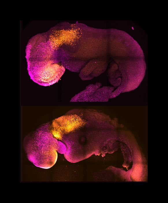
Embryo, Mammalian
Activins
Zebrafish Proteins
Xenopus Proteins
Transcription Factors
High Mobility Group Proteins
HMGB Proteins
Zebrafish
Blastocyst
Notochord
T-Box Domain Proteins
Otx Transcription Factors
Gastrulation
Branchial Region
Teratoma
Chick Embryo
Signal Transduction
Bone Morphogenetic Protein 4
Pancreas
Hepatocyte Nuclear Factor 3-gamma
Blastula
Fibroblast Growth Factor 4
Primitive Streak
Coturnix
Bone Morphogenetic Proteins
Tretinoin
Fibroblast Growth Factors
Embryoid Bodies
DNA-Binding Proteins
Quail
Urochordata
Left-Right Determination Factors
Organogenesis
Embryonic Structures
Genes, Homeobox
Trans-Activators
Teratocarcinoma
Stem Cells
Pharynx
Extraembryonic Membranes
Wnt Proteins
Xenopus
Head
Embryonal Carcinoma Stem Cells
Molecular Sequence Data
Sea Urchins
Hepatocyte Nuclear Factor 3-alpha
Carcinoma, Embryonal
Blastomeres
Organizers, Embryonic
Models, Biological
Fish swimbladder: an excellent mesodermal inductor in primary embryonic induction. (1/1642)
Swimbladder of the crucian carp, Carassius auratus, was found to be better as a vegatalizing tissue than other tissues, such as guinea-pig bone marrow, when presumptive ectoderm of Triturus gastrulae was used as reacting tissue. Swimbladder usually induced assemblies of highly organized mesodermal tissues, such as notochord, somites and pronephric tubules, some of which were covered by mesodermal epithelium without any epidermal covering. A special character of the effect of swimbladder was the rather frequent induction of solid balls of undifferentiated cells, which were identified as mesodermal or mesodermal and probably endodermal. These findings show that swimbladder has a strong and fast spreading vegetalizing effect on the responding presumptive ectoderm. (+info)derriere: a TGF-beta family member required for posterior development in Xenopus. (2/1642)
TGF-beta signaling plays a key role in induction of the Xenopus mesoderm and endoderm. Using a yeast-based selection scheme, we isolated derriere, a novel TGF-beta family member that is closely related to Vg1 and that is required for normal mesodermal patterning, particularly in posterior regions of the embryo. Unlike Vg1, derriere is expressed zygotically, with RNA localized to the future endoderm and mesoderm by late blastula, and to the posterior mesoderm by mid-gastrula. The derriere expression pattern appears to be identical to the zygotic expression domain of VegT (Xombi, Brat, Antipodean), and can be activated by VegT as well as fibroblast growth factor (FGF). In turn, derriere activates expression of itself, VegT and eFGF, suggesting that a regulatory loop exists between these genes. derriere is a potent mesoderm and endoderm inducer, acting in a dose-dependent fashion. When misexpressed ventrally, derriere induces a secondary axis lacking a head, an effect that is due to dorsalization of the ventral marginal zone. When misexpressed dorsally, derriere suppresses head formation. derriere can also posteriorize neurectoderm, but appears to do so indirectly. Together, these data suggest that derriere expression is compatible only with posterior fates. In order to assess the in vivo function of derriere, we constructed a dominant interfering Derriere protein (Cm-Derriere), which preferentially blocks Derriere activity relative to that of other TGFbeta family members. Cm-derriere expression in embryos leads to posterior truncation, including defects in blastopore lip formation, gastrulation and neural tube closure. Normal expression of anterior and hindbrain markers is observed; however, paraxial mesodermal gene expression is ablated. This phenotype can be rescued by wild-type derriere and by VegT. Our findings indicate that derriere plays a crucial role in mesodermal patterning and development of posterior regions in Xenopus. (+info)Goosecoid and mix.1 repress Brachyury expression and are required for head formation in Xenopus. (3/1642)
The Xenopus homologue of Brachyury, Xbra, is expressed in the presumptive mesoderm of the early gastrula. Induction of Xbra in animal pole tissue by activin occurs only in a narrow window of activin concentrations; if the level of inducer is too high, or too low, the gene is not expressed. Previously, we have suggested that the suppression of Xbra by high concentrations of activin is due to the action of genes such as goosecoid and Mix.1. Here, we examine the roles played by goosecoid and Mix.1 during normal development, first in the control of Xbra expression and then in the formation of the mesendoderm. Consistent with the model outlined above, inhibition of the function of either gene product leads to transient ectopic expression of Xbra. Such embryos later develop dorsoanterior defects and, in the case of interference with Mix.1, additional defects in heart and gut formation. Goosecoid, a transcriptional repressor, appears to act directly on transcription of Xbra. In contrast, Mix.1, which functions as a transcriptional activator, may act on Xbra indirectly, in part through activation of goosecoid. (+info)Uncoupling integrin adhesion and signaling: the betaPS cytoplasmic domain is sufficient to regulate gene expression in the Drosophila embryo. (4/1642)
Integrin cell surface receptors are ideally suited to coordinate cellular differentiation and tissue assembly during embryogenesis, as they can mediate both signaling and adhesion. We show that integrins regulate gene expression in the intact developing embryo by identifying two genes that require integrin function for their normal expression in Drosophila midgut endodermal cells. We determined the relative roles of integrin adhesion versus signaling in the regulation of these integrin target genes. We find that integrin-mediated adhesion is not required between the endodermal cells and the surrounding visceral mesoderm for integrin target gene expression. In addition, a chimeric protein that lacks integrin-adhesive function, but maintains the ability to signal, can substitute for the endogenous integrin and regulate integrin target genes. This chimera consists of an oligomeric extracellular domain fused to the integrin betaPS subunit cytoplasmic domain; a control monomeric extracellular domain fusion does not alter integrin target gene expression. Therefore, oligomerization of the 47-amino-acid betaPS intracellular domain is sufficient to initiate a signaling pathway that regulates gene expression in the developing embryo. (+info)Developmental competence of the gut endoderm: genetic potentiation by GATA and HNF3/fork head proteins. (5/1642)
A long-standing problem in developmental biology has been to understand how the embryonic germ layers gain the competence to differentiate into distinct cell types. Genetic studies have shown that members of the GATA and HNF3/fork head transcription factor families are essential for the formation and differentiation of gut endoderm tissues in worms, flies, and mammals. Recent in vivo footprinting studies have shown that GATA and HNF3 binding sites in chromatin are occupied on a silent gene in endoderm that has the potential to be activated solely in that germ layer. These and other data indicate that these evolutionarily conserved factors help impart the competence of a gene to be activated in development, a phenomenon called genetic potentiation. The mechanistic implications of genetic potentiation and its general significance are discussed. (+info)The type I serine/threonine kinase receptor ActRIA (ALK2) is required for gastrulation of the mouse embryo. (6/1642)
ActRIA (or ALK2), one of the type I receptors of the transforming growth factor-beta (TGF-beta) superfamily, can bind both activin and bone morphogenetic proteins (BMPs) in conjunction with the activin and BMP type II receptors, respectively. In mice, ActRIA is expressed primarily in the extraembryonic visceral endoderm before gastrulation and later in both embryonic and extraembryonic cells during gastrulation. To elucidate its function in mouse development, we disrupted the transmembrane domain of ActRIA by gene targeting. We showed that embryos homozygous for the mutation were arrested at the early gastrulation stage, displaying abnormal visceral endoderm morphology and severe disruption of mesoderm formation. To determine in which germ layer ActRIA functions during gastrulation, we performed reciprocal chimera analyses. (1) Homozygous mutant ES cells injected into wild-type blastocysts were able to contribute to all three definitive germ layers in chimeric embryos. However, a high contribution of mutant ES cells in chimeras disrupted normal development at the early somite stage. (2) Consistent with ActRIA expression in the extraembryonic cells, wild-type ES cells failed to rescue the gastrulation defect in chimeras in which the extraembryonic ectoderm and visceral endoderm were derived from homozygous mutant blastocysts. Furthermore, expression of HNF4, a key visceral endoderm-specific transcription regulatory factor, was significantly reduced in the mutant embryos. Together, our results indicate that ActRIA in extraembryonic cells plays a major role in early gastrulation, whereas ActRIA function is also required in embryonic tissues during later development in mice. (+info)Identification and characterization of maternally expressed genes with mRNAs that are segregated with the endoplasm of early ascidian embryos. (7/1642)
Endoderm cells of the ascidian embryo are specified autonomously dependent on maternal cytoplasmic information or determinants that are localized in the endoplasm. In the present study, we identified three maternally expressed genes (CsEndo-1, CsEndo-2 and CsEndo-3) by screening a cDNA library of Ciona savignyi fertilized egg mRNAs subtracted with gastrula mRNAs. CsEndo-1 encoded a protein with nuclear localization signals, CsEndo-3 predicted a protein containing both a potential transmembrane domain and the PDZ domain, and CsEndo-2 suggested a protein with no similarity to known proteins. The maternal transcripts of all of these genes were not concentrated during early stages of embryogenesis up to the 8-cell stage, but were concentrated at the endoplasmic region by the 16-cell stage and then segregated later with the endoplasm. At the 110-cell stage, the maternal transcript of CsEndo-1 was evident only in the primordial endoderm cells, while those of CsEndo-2 and CsEndo-3 were found in the primordial endoderm cells as well as the primordial notochord cells. All of the transcripts became barely detectable during gastrulation and neurulation. Later, zygotic expression of the three genes became evident again in the endoderm and notochord cells, suggesting developmental roles in the formation of these types of cell. Although we were not able to deduce their functions, this is the first report of maternal genes with mRNAs that are segregated with the endoplasm of ascidian embryos. (+info)Posterior fossa epithelial cyst: case report and review of the literature. (8/1642)
A 49-year old woman with progressive cranial nerve signs and hemiparesis was found at MR imaging and at surgery to have a cyst at the foramen magnum. Immunohistochemistry and electron microscopy showed an epithelial cyst of endodermal origin. MR findings were of an extraaxial mass, with short T1 and T2 times. Unless immunohistochemistry and electron microscopy are used in the final diagnosis of such cysts, all posterior fossa cysts lined by a single layer of epithelium should be described simply as epithelial cysts. (+info)Endoderm is the innermost of the three primary germ layers in a developing embryo, along with the ectoderm and mesoderm. The endoderm gives rise to several internal tissues and organs, most notably those found in the digestive system and respiratory system. Specifically, it forms the lining of the gut tube, which eventually becomes the epithelial lining of the gastrointestinal tract, liver, pancreas, lungs, and other associated structures.
During embryonic development, the endoderm arises from the inner cell mass of the blastocyst, following a series of cell divisions and migrations that help to establish the basic body plan of the organism. As the embryo grows and develops, the endoderm continues to differentiate into more specialized tissues and structures, playing a critical role in the formation of many essential bodily functions.
SOXF transcription factors are a subgroup of the SOX (SRY-related HMG box) family of proteins, which are involved in various developmental processes. The SOXF group includes SOX7, SOX17, and SOX18, all of which contain a conserved high mobility group (HMG) box DNA-binding domain. These transcription factors play crucial roles in the development of several organ systems, including the cardiovascular system, nervous system, and urogenital system. They are involved in cell fate determination, differentiation, and morphogenesis during embryonic development and have also been implicated in various disease processes, such as cancer.
In medical and embryological terms, the mesoderm is one of the three primary germ layers in the very early stages of embryonic development. It forms between the ectoderm and endoderm during gastrulation, and it gives rise to a wide variety of cell types, tissues, and organs in the developing embryo.
The mesoderm contributes to the formation of structures such as:
1. The connective tissues (including tendons, ligaments, and most of the bones)
2. Muscular system (skeletal, smooth, and cardiac muscles)
3. Circulatory system (heart, blood vessels, and blood cells)
4. Excretory system (kidneys and associated structures)
5. Reproductive system (gonads, including ovaries and testes)
6. Dermis of the skin
7. Parts of the eye and inner ear
8. Several organs in the urogenital system
Dysfunctions or abnormalities in mesoderm development can lead to various congenital disorders and birth defects, highlighting its importance during embryogenesis.
Developmental gene expression regulation refers to the processes that control the activation or repression of specific genes during embryonic and fetal development. These regulatory mechanisms ensure that genes are expressed at the right time, in the right cells, and at appropriate levels to guide proper growth, differentiation, and morphogenesis of an organism.
Developmental gene expression regulation is a complex and dynamic process involving various molecular players, such as transcription factors, chromatin modifiers, non-coding RNAs, and signaling molecules. These regulators can interact with cis-regulatory elements, like enhancers and promoters, to fine-tune the spatiotemporal patterns of gene expression during development.
Dysregulation of developmental gene expression can lead to various congenital disorders and developmental abnormalities. Therefore, understanding the principles and mechanisms governing developmental gene expression regulation is crucial for uncovering the etiology of developmental diseases and devising potential therapeutic strategies.
A gastrula is a stage in the early development of many animals, including humans, that occurs following fertilization and cleavage of the zygote. During this stage, the embryo undergoes a process called gastrulation, which involves a series of cell movements that reorganize the embryo into three distinct layers: the ectoderm, mesoderm, and endoderm. These germ layers give rise to all the different tissues and organs in the developing organism.
The gastrula is characterized by the presence of a central cavity called the archenteron, which will eventually become the gut or gastrointestinal tract. The opening of the archenteron is called the blastopore, which will give rise to either the mouth or anus, depending on the animal group.
In summary, a gastrula is a developmental stage in which an embryo undergoes gastrulation to form three germ layers and a central cavity, which will eventually develop into various organs and tissues of the body.
Germ layers refer to the primary layers of cells that form during embryonic development and give rise to the various tissues and organs in the body. In humans, there are three germ layers: the ectoderm, mesoderm, and endoderm. Each germ layer differentiates into distinct cell types and structures during the process of gastrulation. The ectoderm gives rise to the nervous system, sensory organs, and skin; the mesoderm forms muscles, bones, blood vessels, and the circulatory system; and the endoderm develops into the respiratory and digestive systems, including the lungs, liver, and pancreas.
Embryonic induction is a process that occurs during the development of a multicellular organism, where one group of cells in the embryo signals and influences the developmental fate of another group of cells. This interaction leads to the formation of specific structures or organs in the developing embryo. The signaling cells that initiate the process are called organizers, and they release signaling molecules known as morphogens that bind to receptors on the target cells and trigger a cascade of intracellular signals that ultimately lead to changes in gene expression and cell fate. Embryonic induction is a crucial step in the development of complex organisms and plays a key role in establishing the body plan and organizing the different tissues and organs in the developing embryo.
Viscera is a medical term that refers to the internal organs of the body, specifically those contained within the chest and abdominal cavities. These include the heart, lungs, liver, pancreas, spleen, kidneys, and intestines. In some contexts, it may also refer to the reproductive organs. The term viscera is often used in anatomical or surgical descriptions, and is derived from the Latin word "viscus," meaning "an internal organ."
GATA6 (GATA binding protein 6) is a transcription factor that belongs to the GATA family, which are characterized by their ability to bind to the DNA sequence (A/T)GATA(A/G). GATA6 plays crucial roles in the development and function of various tissues, particularly in the digestive system.
As a transcription factor, GATA6 regulates gene expression by binding to specific DNA sequences in the promoter or enhancer regions of target genes. This binding either activates or represses the transcription of these genes, thereby controlling cellular processes such as proliferation, differentiation, and survival.
In the context of the digestive system, GATA6 is essential for the development of the pancreas and small intestine. It promotes the differentiation of pancreatic progenitor cells into exocrine cells (such as acinar and ductal cells) and inhibits their differentiation into endocrine cells (such as β-cells). In the small intestine, GATA6 is involved in maintaining the identity and function of Paneth cells, which are specialized epithelial cells that play a role in innate immunity.
Mutations in the GATA6 gene have been associated with various human diseases, including pancreatic agenesis or hypoplasia, small intestinal atresia, and congenital diaphragmatic hernia. Additionally, altered GATA6 expression has been implicated in several types of cancer, such as pancreatic ductal adenocarcinoma and colorectal cancer.
"Body patterning" is a general term that refers to the process of forming and organizing various tissues and structures into specific patterns during embryonic development. This complex process involves a variety of molecular mechanisms, including gene expression, cell signaling, and cell-cell interactions. It results in the creation of distinct body regions, such as the head, trunk, and limbs, as well as the organization of internal organs and systems.
In medical terminology, "body patterning" may refer to specific developmental processes or abnormalities related to embryonic development. For example, in genetic disorders such as Poland syndrome or Holt-Oram syndrome, mutations in certain genes can lead to abnormal body patterning, resulting in the absence or underdevelopment of certain muscles, bones, or other structures.
It's important to note that "body patterning" is not a formal medical term with a specific definition, but rather a general concept used in developmental biology and genetics.
Ectoderm is the outermost of the three primary germ layers in a developing embryo, along with the endoderm and mesoderm. The ectoderm gives rise to the outer covering of the body, including the skin, hair, nails, glands, and the nervous system, which includes the brain, spinal cord, and peripheral nerves. It also forms the lining of the mouth, anus, nose, and ears. Essentially, the ectoderm is responsible for producing all the epidermal structures and the neural crest cells that contribute to various derivatives such as melanocytes, adrenal medulla, smooth muscle, and peripheral nervous system components.
GATA5 transcription factor is a protein that binds to specific DNA sequences, called GATA sites, in the regulatory regions of target genes and regulates their expression. The GATA5 protein contains two conserved domains, called zinc fingers, which mediate its binding to the GATA sites. GATA5 is mainly expressed in tissues derived from the endoderm, such as the gut, liver, and pancreas, where it plays critical roles in developmental processes, including cell fate determination, proliferation, and differentiation.
Mutations in the gene encoding GATA5 have been associated with congenital heart defects, suggesting that GATA5 is essential for normal cardiac development. In addition to its role in development, GATA5 has also been implicated in the pathogenesis of various diseases, including cancer, where it can act as a tumor suppressor or oncogene depending on the context.
A nodal protein, in the context of molecular biology and genetics, refers to a protein that plays a role in signal transmission within a cell at a node or junction point of a signaling pathway. These proteins are often involved in regulatory processes, such as activating or inhibiting downstream effectors in response to specific signals received by the cell. Nodal proteins can be activated or deactivated through various mechanisms, including phosphorylation, ubiquitination, and interactions with other signaling molecules.
In a more specific context, nodal proteins are also known as nodal factors, which are members of the transforming growth factor-beta (TGF-β) superfamily of signaling molecules that play critical roles in embryonic development and tissue homeostasis. Nodal is a secreted protein that acts as a morphogen, inducing different cellular responses depending on its concentration gradient. It is involved in establishing left-right asymmetry during embryonic development and regulates various processes such as cell proliferation, differentiation, and apoptosis.
In summary, nodal proteins can refer to any protein that functions at a node or junction point of a signaling pathway, but they are also specifically known as nodal factors, which are TGF-β superfamily members involved in embryonic development and tissue homeostasis.
Hepatocyte Nuclear Factor 3-beta (HNF-3β, also known as FOXA3) is a transcription factor that plays crucial roles in the development and function of various organs, including the liver, pancreas, and kidneys. It belongs to the forkhead box (FOX) family of proteins, which are characterized by a conserved DNA-binding domain known as the forkhead box or winged helix domain.
In the liver, HNF-3β is essential for the differentiation and maintenance of hepatocytes, the primary functional cells of the liver. It regulates the expression of several genes involved in liver-specific functions such as glucose metabolism, bile acid synthesis, and detoxification.
HNF-3β also has important roles in the pancreas, where it helps regulate the development and function of insulin-producing beta cells. In the kidneys, HNF-3β is involved in the differentiation and maintenance of the nephron, the functional unit responsible for filtering blood and maintaining water and electrolyte balance.
Mutations in the gene encoding HNF-3β have been associated with several genetic disorders, including maturity-onset diabetes of the young (MODY) and renal cysts and diabetes syndrome (RCAD).
SOX (SRY-related HMG box) transcription factors are a family of proteins that regulate gene expression during embryonic development and in adult tissues. They contain a highly conserved DNA-binding domain, the HMG box, which allows them to bind to specific DNA sequences and influence the transcription of nearby genes. SOX proteins play critical roles in various biological processes such as cell fate determination, differentiation, proliferation, and survival.
SOX transcription factors are classified into several groups (A-H) based on their sequence similarities and functional redundancies. Some well-known members of this family include SOX1, SOX2, SOX3, SOX4, SOX9, SOX10, and SOX17. These proteins often form complexes with other transcription factors or cofactors to modulate their target genes' expression.
Dysregulation of SOX transcription factors has been implicated in several human diseases, including cancer, developmental disorders, and degenerative conditions. For example, SOX2 overexpression is associated with certain types of tumors, while mutations in the SOX9 gene can cause campomelic dysplasia, a severe skeletal disorder.
The digestive system is a complex group of organs and glands that process food. It converts the food we eat into nutrients, which the body uses for energy, growth, and cell repair. The digestive system also eliminates waste from the body. It is made up of the gastrointestinal tract (GI tract) and other organs that help the body break down and absorb food.
The GI tract includes the mouth, esophagus, stomach, small intestine, large intestine, and anus. Other organs that are part of the digestive system include the liver, pancreas, gallbladder, and salivary glands.
The process of digestion begins in the mouth, where food is chewed and mixed with saliva. The food then travels down the esophagus and into the stomach, where it is broken down further by stomach acids. The digested food then moves into the small intestine, where nutrients are absorbed into the bloodstream. The remaining waste material passes into the large intestine, where it is stored until it is eliminated through the anus.
The liver, pancreas, and gallbladder play important roles in the digestive process as well. The liver produces bile, a substance that helps break down fats in the small intestine. The pancreas produces enzymes that help digest proteins, carbohydrates, and fats. The gallbladder stores bile until it is needed in the small intestine.
Overall, the digestive system is responsible for breaking down food, absorbing nutrients, and eliminating waste. It plays a critical role in maintaining our health and well-being.
Cell differentiation is the process by which a less specialized cell, or stem cell, becomes a more specialized cell type with specific functions and structures. This process involves changes in gene expression, which are regulated by various intracellular signaling pathways and transcription factors. Differentiation results in the development of distinct cell types that make up tissues and organs in multicellular organisms. It is a crucial aspect of embryonic development, tissue repair, and maintenance of homeostasis in the body.
The yolk sac is a structure that forms in the early stages of an embryo's development. It is a extra-embryonic membrane, which means it exists outside of the developing embryo, and it plays a critical role in providing nutrients to the growing embryo during the initial stages of development.
In more detail, the yolk sac is responsible for producing blood cells, contributing to the formation of the early circulatory system, and storing nutrients that are absorbed from the yolk material inside the egg or uterus. The yolk sac also has a role in the development of the gut and the immune system.
As the embryo grows and the placenta develops, the yolk sac's function becomes less critical, and it eventually degenerates. However, remnants of the yolk sac can sometimes persist and may be found in the developing fetus or newborn baby. In some cases, abnormalities in the development or regression of the yolk sac can lead to developmental problems or congenital disorders.
A nonmammalian embryo refers to the developing organism in animals other than mammals, from the fertilized egg (zygote) stage until hatching or birth. In nonmammalian species, the developmental stages and terminology differ from those used in mammals. The term "embryo" is generally applied to the developing organism up until a specific stage of development that is characterized by the formation of major organs and structures. After this point, the developing organism is referred to as a "larva," "juvenile," or other species-specific terminology.
The study of nonmammalian embryos has played an important role in our understanding of developmental biology and evolutionary developmental biology (evo-devo). By comparing the developmental processes across different animal groups, researchers can gain insights into the evolutionary origins and diversification of body plans and structures. Additionally, nonmammalian embryos are often used as model systems for studying basic biological processes, such as cell division, gene regulation, and pattern formation.
In situ hybridization (ISH) is a molecular biology technique used to detect and localize specific nucleic acid sequences, such as DNA or RNA, within cells or tissues. This technique involves the use of a labeled probe that is complementary to the target nucleic acid sequence. The probe can be labeled with various types of markers, including radioisotopes, fluorescent dyes, or enzymes.
During the ISH procedure, the labeled probe is hybridized to the target nucleic acid sequence in situ, meaning that the hybridization occurs within the intact cells or tissues. After washing away unbound probe, the location of the labeled probe can be visualized using various methods depending on the type of label used.
In situ hybridization has a wide range of applications in both research and diagnostic settings, including the detection of gene expression patterns, identification of viral infections, and diagnosis of genetic disorders.
Embryonic stem cells are a type of pluripotent stem cell that are derived from the inner cell mass of a blastocyst, which is a very early-stage embryo. These cells have the ability to differentiate into any cell type in the body, making them a promising area of research for regenerative medicine and the study of human development and disease. Embryonic stem cells are typically obtained from surplus embryos created during in vitro fertilization (IVF) procedures, with the consent of the donors. The use of embryonic stem cells is a controversial issue due to ethical concerns surrounding the destruction of human embryos.
Homeodomain proteins are a group of transcription factors that play crucial roles in the development and differentiation of cells in animals and plants. They are characterized by the presence of a highly conserved DNA-binding domain called the homeodomain, which is typically about 60 amino acids long. The homeodomain consists of three helices, with the third helix responsible for recognizing and binding to specific DNA sequences.
Homeodomain proteins are involved in regulating gene expression during embryonic development, tissue maintenance, and organismal growth. They can act as activators or repressors of transcription, depending on the context and the presence of cofactors. Mutations in homeodomain proteins have been associated with various human diseases, including cancer, congenital abnormalities, and neurological disorders.
Some examples of homeodomain proteins include PAX6, which is essential for eye development, HOX genes, which are involved in body patterning, and NANOG, which plays a role in maintaining pluripotency in stem cells.
Morphogenesis is a term used in developmental biology and refers to the process by which cells give rise to tissues and organs with specific shapes, structures, and patterns during embryonic development. This process involves complex interactions between genes, cells, and the extracellular environment that result in the coordinated movement and differentiation of cells into specialized functional units.
Morphogenesis is a dynamic and highly regulated process that involves several mechanisms, including cell proliferation, death, migration, adhesion, and differentiation. These processes are controlled by genetic programs and signaling pathways that respond to environmental cues and regulate the behavior of individual cells within a developing tissue or organ.
The study of morphogenesis is important for understanding how complex biological structures form during development and how these processes can go awry in disease states such as cancer, birth defects, and degenerative disorders.
'Cell lineage' is a term used in biology and medicine to describe the developmental history or relationship of a cell or group of cells to other cells, tracing back to the original progenitor or stem cell. It refers to the series of cell divisions and differentiation events that give rise to specific types of cells in an organism over time.
In simpler terms, cell lineage is like a family tree for cells, showing how they are related to each other through a chain of cell division and specialization events. This concept is important in understanding the development, growth, and maintenance of tissues and organs in living beings.
GATA4 is a transcription factor that belongs to the GATA family of zinc finger proteins, which are characterized by their ability to bind to DNA sequences containing the core motif (A/T)GATA(A/G). GATA4 specifically recognizes and binds to GATA motifs in the promoter and enhancer regions of target genes, where it can modulate their transcription.
GATA4 is widely expressed in various tissues, including the heart, gut, lungs, and gonads. In the heart, GATA4 plays critical roles during cardiac development, such as promoting cardiomyocyte differentiation and regulating heart tube formation. It also continues to be expressed in adult hearts, where it helps maintain cardiac function and can contribute to heart repair after injury.
Mutations in the GATA4 gene have been associated with congenital heart defects, suggesting its essential role in heart development. Additionally, GATA4 has been implicated in cancer progression, particularly in gastrointestinal and lung cancers, where it can act as an oncogene by promoting cell proliferation and survival.
GATA transcription factors are a group of proteins that regulate gene expression by binding to specific DNA sequences called GATA motifs. These transcription factors contain one or two conserved domains known as GATA-type zinc fingers, which recognize and bind to the consensus sequence (A/T)GATA(A/G). They are widely expressed in various tissues, including hematopoietic cells, endothelial cells, and neuronal cells. In hematopoiesis, GATA transcription factors play critical roles in cell fate determination, proliferation, and differentiation. For example, GATA-1 is essential for erythroid and megakaryocyte development, while GATA-2 is required for the development of hematopoietic stem cells and progenitor cells. Dysregulation of GATA transcription factors has been implicated in various diseases, including cancer and genetic disorders.
A mammalian embryo is the developing offspring of a mammal, from the time of implantation of the fertilized egg (blastocyst) in the uterus until the end of the eighth week of gestation. During this period, the embryo undergoes rapid cell division and organ differentiation to form a complex structure with all the major organs and systems in place. This stage is followed by fetal development, which continues until birth. The study of mammalian embryos is important for understanding human development, evolution, and reproductive biology.
Activins are a type of protein that belongs to the transforming growth factor-beta (TGF-β) superfamily. They are produced and released by various cells in the body, including those in the ovaries, testes, pituitary gland, and other tissues. Activins play important roles in regulating several biological processes, such as cell growth, differentiation, and apoptosis (programmed cell death).
Activins bind to specific receptors on the surface of cells, leading to the activation of intracellular signaling pathways that control gene expression. They are particularly well-known for their role in reproductive biology, where they help regulate follicle stimulation and hormone production in the ovaries and testes. Activins also have been implicated in various disease processes, including cancer, fibrosis, and inflammation.
There are three main isoforms of activin in humans: activin A, activin B, and inhibin A. While activins and inhibins share similar structures and functions, they have opposite effects on the activity of the pituitary gland. Activins stimulate the production of follicle-stimulating hormone (FSH), while inhibins suppress it. This delicate balance between activins and inhibins helps regulate reproductive function and other physiological processes in the body.
Zebrafish proteins refer to the diverse range of protein molecules that are produced by the organism Danio rerio, commonly known as the zebrafish. These proteins play crucial roles in various biological processes such as growth, development, reproduction, and response to environmental stimuli. They are involved in cellular functions like enzymatic reactions, signal transduction, structural support, and regulation of gene expression.
Zebrafish is a popular model organism in biomedical research due to its genetic similarity with humans, rapid development, and transparent embryos that allow for easy observation of biological processes. As a result, the study of zebrafish proteins has contributed significantly to our understanding of protein function, structure, and interaction in both zebrafish and human systems.
Some examples of zebrafish proteins include:
* Transcription factors that regulate gene expression during development
* Enzymes involved in metabolic pathways
* Structural proteins that provide support to cells and tissues
* Receptors and signaling molecules that mediate communication between cells
* Heat shock proteins that assist in protein folding and protect against stress
The analysis of zebrafish proteins can be performed using various techniques, including biochemical assays, mass spectrometry, protein crystallography, and computational modeling. These methods help researchers to identify, characterize, and understand the functions of individual proteins and their interactions within complex networks.
"Xenopus proteins" refer to the proteins that are expressed or isolated from the Xenopus species, which are primarily used as model organisms in biological and biomedical research. The most commonly used Xenopus species for research are the African clawed frogs, Xenopus laevis and Xenopus tropicalis. These proteins play crucial roles in various cellular processes and functions, and they serve as valuable tools to study different aspects of molecular biology, developmental biology, genetics, and biochemistry.
Some examples of Xenopus proteins that are widely studied include:
1. Xenopus Histones: These are the proteins that package DNA into nucleosomes, which are the fundamental units of chromatin in eukaryotic cells. They play a significant role in gene regulation and epigenetic modifications.
2. Xenopus Cyclins and Cyclin-dependent kinases (CDKs): These proteins regulate the cell cycle and control cell division, differentiation, and apoptosis.
3. Xenopus Transcription factors: These proteins bind to specific DNA sequences and regulate gene expression during development and in response to various stimuli.
4. Xenopus Signaling molecules: These proteins are involved in intracellular signaling pathways that control various cellular processes, such as cell growth, differentiation, migration, and survival.
5. Xenopus Cytoskeletal proteins: These proteins provide structural support to the cells and regulate their shape, motility, and organization.
6. Xenopus Enzymes: These proteins catalyze various biochemical reactions in the cell, such as metabolic pathways, DNA replication, transcription, and translation.
Overall, Xenopus proteins are essential tools for understanding fundamental biological processes and have contributed significantly to our current knowledge of molecular biology, genetics, and developmental biology.
Transcription factors are proteins that play a crucial role in regulating gene expression by controlling the transcription of DNA to messenger RNA (mRNA). They function by binding to specific DNA sequences, known as response elements, located in the promoter region or enhancer regions of target genes. This binding can either activate or repress the initiation of transcription, depending on the properties and interactions of the particular transcription factor. Transcription factors often act as part of a complex network of regulatory proteins that determine the precise spatiotemporal patterns of gene expression during development, differentiation, and homeostasis in an organism.
High mobility group proteins (HMG proteins) are a family of nuclear proteins that are characterized by their ability to bind to DNA and influence its structure and function. They are named "high mobility" because of their rapid movement in gel electrophoresis. HMG proteins are involved in various nuclear processes, including chromatin remodeling, transcription regulation, and DNA repair.
There are three main classes of HMG proteins: HMGA, HMGB, and HMGN. Each class has distinct structural features and functions. For example, HMGA proteins have a unique "AT-hook" domain that allows them to bind to the minor groove of AT-rich DNA sequences, while HMGB proteins have two "HMG-box" domains that enable them to bend and unwind DNA.
HMG proteins play important roles in many physiological and pathological processes, such as embryonic development, inflammation, and cancer. Dysregulation of HMG protein function has been implicated in various diseases, including neurodegenerative disorders, diabetes, and cancer. Therefore, understanding the structure, function, and regulation of HMG proteins is crucial for developing new therapeutic strategies for these diseases.
High Mobility Group Box (HMGB) proteins are a family of nuclear proteins that are highly conserved and expressed in eukaryotic cells. They play a crucial role in the regulation of gene expression, DNA repair, and maintenance of nucleosome structure. HMGB proteins contain two positively charged DNA-binding domains (HMG boxes) and a negatively charged acidic tail. These proteins can bind to DNA in a variety of ways, bending it and altering its structure, which in turn affects the binding of other proteins and the transcriptional activity of genes. HMGB proteins can also be released from cells under conditions of stress or injury, where they act as damage-associated molecular patterns (DAMPs) and contribute to the inflammatory response.
A zebrafish is a freshwater fish species belonging to the family Cyprinidae and the genus Danio. Its name is derived from its distinctive striped pattern that resembles a zebra's. Zebrafish are often used as model organisms in scientific research, particularly in developmental biology, genetics, and toxicology studies. They have a high fecundity rate, transparent embryos, and a rapid development process, making them an ideal choice for researchers. However, it is important to note that providing a medical definition for zebrafish may not be entirely accurate or relevant since they are primarily used in biological research rather than clinical medicine.
A blastocyst is a stage in the early development of a fertilized egg, or embryo, in mammals. It occurs about 5-6 days after fertilization and consists of an outer layer of cells called trophoblasts, which will eventually form the placenta, and an inner cell mass, which will give rise to the fetus. The blastocyst is characterized by a fluid-filled cavity called the blastocoel. This stage is critical for the implantation of the embryo into the uterine lining.
The notochord is a flexible, rod-shaped structure that is present in the embryos of chordates, including humans. It is composed of cells called chordocytes and is surrounded by a sheath. The notochord runs along the length of the body, providing support and flexibility. In human embryos, the notochord eventually becomes part of the discs between the vertebrae in the spine. An abnormal or absent notochord can lead to developmental problems with the spine and nervous system.
Embryonic and fetal development is the process of growth and development that occurs from fertilization of the egg (conception) to birth. The terms "embryo" and "fetus" are used to describe different stages of this development:
* Embryonic development: This stage begins at fertilization and continues until the end of the 8th week of pregnancy. During this time, the fertilized egg (zygote) divides and forms a blastocyst, which implants in the uterus and begins to develop into a complex structure called an embryo. The embryo consists of three layers of cells that will eventually form all of the organs and tissues of the body. During this stage, the basic structures of the body, including the nervous system, heart, and gastrointestinal tract, begin to form.
* Fetal development: This stage begins at the end of the 8th week of pregnancy and continues until birth. During this time, the embryo is called a fetus, and it grows and develops rapidly. The organs and tissues that were formed during the embryonic stage continue to mature and become more complex. The fetus also begins to move and kick, and it can hear and respond to sounds from outside the womb.
Overall, embryonic and fetal development is a complex and highly regulated process that involves the coordinated growth and differentiation of cells and tissues. It is a critical period of development that lays the foundation for the health and well-being of the individual throughout their life.
Embryonic development is the series of growth and developmental stages that occur during the formation and early growth of the embryo. In humans, this stage begins at fertilization (when the sperm and egg cell combine) and continues until the end of the 8th week of pregnancy. During this time, the fertilized egg (now called a zygote) divides and forms a blastocyst, which then implants into the uterus. The cells in the blastocyst begin to differentiate and form the three germ layers: the ectoderm, mesoderm, and endoderm. These germ layers will eventually give rise to all of the different tissues and organs in the body.
Embryonic development is a complex and highly regulated process that involves the coordinated interaction of genetic and environmental factors. It is characterized by rapid cell division, migration, and differentiation, as well as programmed cell death (apoptosis) and tissue remodeling. Abnormalities in embryonic development can lead to birth defects or other developmental disorders.
It's important to note that the term "embryo" is used to describe the developing organism from fertilization until the end of the 8th week of pregnancy in humans, after which it is called a fetus.
T-box domain proteins are a family of transcription factors that share a highly conserved DNA-binding domain, known as the T-box. The T-box domain is a DNA-binding motif that specifically recognizes and binds to T-box binding elements (TBEs) in the regulatory regions of target genes. These proteins play crucial roles during embryonic development, particularly in the formation of specific tissues and organs, such as the heart, limbs, and brain. Mutations in T-box domain proteins can lead to various congenital defects and developmental disorders. Some examples of T-box domain proteins include TBX1, TBX5, and TBX20.
OTX (Orthodenticle homeobox) transcription factors are a family of proteins that regulate gene expression during embryonic development, particularly in the eye, forebrain, and midbrain. They play crucial roles in the development and differentiation of these tissues, including the specification of eye field identity, the determination of dorsoventral patterning in the neural tube, and the regulation of neurogenesis.
OTX transcription factors contain a highly conserved DNA-binding domain called the homeodomain, which allows them to recognize and bind to specific DNA sequences. In humans, there are four known OTX transcription factors (OTX1, OTX2, OTX3, and CRX), each with distinct expression patterns and functions.
Mutations in OTX genes have been associated with various developmental disorders, such as microphthalmia, anophthalmia, and severe eye malformations, highlighting their importance in normal eye development. Additionally, OTX transcription factors have also been implicated in the pathogenesis of certain cancers, including medulloblastoma and retinoblastoma.
Gastrulation is a fundamental process in embryonic development, characterized by the transformation of a initially flat layer of cells called the blastula into a three-layered structure known as the gastrula. This complex series of cellular movements and rearrangements establishes the foundation for the formation of the three primary germ layers: the ectoderm, mesoderm, and endoderm. These germ layers further differentiate to give rise to all the diverse cell types and tissues in the developing organism, including the nervous system, muscles, bones, and internal organs.
The precise mechanisms of gastrulation vary among different animal groups; however, common features include:
1. Formation of a blastopore: A small indentation or opening that forms on the surface of the blastula, which eventually develops into the primitive gut or anus in the gastrula.
2. Invagination: The process by which cells at the blastopore fold inward and migrate towards the interior of the embryo, forming the endodermal layer.
3. Epiboly: A coordinated movement of cells that spreads over and encloses the yolk within the embryo, contributing to the formation of the ectodermal layer.
4. Delamination: The separation and migration of cells from the epiblast (the outer layer of the blastula) to form the mesodermal layer in between the ectoderm and endoderm.
Gastrulation is a critical period in embryonic development, as errors during this process can lead to severe congenital abnormalities or even embryonic lethality. A thorough understanding of gastrulation has important implications for regenerative medicine, stem cell research, and the study of evolutionary developmental biology (Evo-Devo).
Fetal proteins are a type of proteins that are produced by the fetus during pregnancy and can be detected in various biological samples, such as amniotic fluid or maternal blood. These proteins can provide valuable information about the health and development of the fetus. One commonly studied fetal protein is human chorionic gonadotropin (hCG), which is produced by the placenta and can be used as a marker for pregnancy and to detect potential complications, such as Down syndrome or spinal cord defects. Other examples of fetal proteins include alpha-fetoprotein (AFP) and human placental lactogen (hPL).
The branchial region, also known as the pharyngeal region or viscerocranium, is a term used in human anatomy to refer to the area of the developing embryo that gives rise to structures derived from the branchial (or pharyngeal) arches. The branchial arches are a series of paired, rod-like structures that appear early in embryonic development and give rise to various head and neck structures, including the bones and muscles of the face, jaws, and neck, as well as the associated nerves, blood vessels, and connective tissues.
The branchial region is divided into several subregions, each corresponding to a specific branchial arch. The first branchial arch gives rise to structures such as the mandible (lower jaw), maxilla (upper jaw), and muscles of mastication (chewing). The second branchial arch forms the stapes and styloid process in the ear, as well as some neck muscles. The third and fourth branchial arches contribute to the formation of the larynx, thyroid cartilage, and other structures in the neck.
Abnormalities in the development of the branchial region can lead to a variety of congenital defects, such as cleft palate, micrognathia (small jaw), and branchial cysts or sinuses. These conditions may require surgical intervention to correct.
A teratoma is a type of germ cell tumor, which is a broad category of tumors that originate from the reproductive cells. A teratoma contains developed tissues from all three embryonic germ layers: ectoderm, mesoderm, and endoderm. This means that a teratoma can contain various types of tissue such as hair, teeth, bone, and even more complex organs like eyes, thyroid, or neural tissue.
Teratomas are usually benign (non-cancerous), but they can sometimes be malignant (cancerous) and can spread to other parts of the body. They can occur anywhere in the body, but they're most commonly found in the ovaries and testicles. When found in these areas, they are typically removed surgically.
Teratomas can also occur in other locations such as the sacrum, coccyx (tailbone), mediastinum (the area between the lungs), and pineal gland (a small gland in the brain). These types of teratomas can be more complex to treat due to their location and potential to cause damage to nearby structures.
A chick embryo refers to the developing organism that arises from a fertilized chicken egg. It is often used as a model system in biological research, particularly during the stages of development when many of its organs and systems are forming and can be easily observed and manipulated. The study of chick embryos has contributed significantly to our understanding of various aspects of developmental biology, including gastrulation, neurulation, organogenesis, and pattern formation. Researchers may use various techniques to observe and manipulate the chick embryo, such as surgical alterations, cell labeling, and exposure to drugs or other agents.
Signal transduction is the process by which a cell converts an extracellular signal, such as a hormone or neurotransmitter, into an intracellular response. This involves a series of molecular events that transmit the signal from the cell surface to the interior of the cell, ultimately resulting in changes in gene expression, protein activity, or metabolism.
The process typically begins with the binding of the extracellular signal to a receptor located on the cell membrane. This binding event activates the receptor, which then triggers a cascade of intracellular signaling molecules, such as second messengers, protein kinases, and ion channels. These molecules amplify and propagate the signal, ultimately leading to the activation or inhibition of specific cellular responses.
Signal transduction pathways are highly regulated and can be modulated by various factors, including other signaling molecules, post-translational modifications, and feedback mechanisms. Dysregulation of these pathways has been implicated in a variety of diseases, including cancer, diabetes, and neurological disorders.
Bone Morphogenetic Protein 4 (BMP-4) is a growth factor that belongs to the transforming growth factor-beta (TGF-β) superfamily. It plays crucial roles in various biological processes, including embryonic development, cell growth, and differentiation. In the skeletal system, BMP-4 stimulates the formation of bone and cartilage by inducing the differentiation of mesenchymal stem cells into chondrocytes and osteoblasts. It also regulates the maintenance and repair of bones throughout life. An imbalance in BMP-4 signaling has been associated with several skeletal disorders, such as heterotopic ossification and osteoarthritis.
The pancreas is a glandular organ located in the abdomen, posterior to the stomach. It has both exocrine and endocrine functions. The exocrine portion of the pancreas consists of acinar cells that produce and secrete digestive enzymes into the duodenum via the pancreatic duct. These enzymes help in the breakdown of proteins, carbohydrates, and fats in food.
The endocrine portion of the pancreas consists of clusters of cells called islets of Langerhans, which include alpha, beta, delta, and F cells. These cells produce and secrete hormones directly into the bloodstream, including insulin, glucagon, somatostatin, and pancreatic polypeptide. Insulin and glucagon are critical regulators of blood sugar levels, with insulin promoting glucose uptake and storage in tissues and glucagon stimulating glycogenolysis and gluconeogenesis to raise blood glucose when it is low.
Hepatocyte Nuclear Factor 3-gamma (HNF-3γ, also known as FOXA3) is a member of the forkhead box (FOX) family of transcription factors. It plays crucial roles in the development and function of the liver, pancreas, and other organs. In the liver, HNF-3γ helps regulate the expression of genes involved in glucose and lipid metabolism, bile acid synthesis, and detoxification processes. Mutations in the HNF-3γ gene have been associated with various liver diseases, including monogenic forms of diabetes.
A blastula is a stage in the early development of many animals, including mammals. It is a hollow ball of cells that forms as a result of cleavage, which is the process of cell division during embryonic development. The blastula is typically characterized by the presence of a fluid-filled cavity called the blastocoel, which is surrounded by a single layer of cells known as the blastoderm.
In mammals, the blastula stage follows the morula stage, which is a solid mass of cells that results from cleavage of the fertilized egg. During further cell division and rearrangement, the cells in the morula become organized into an inner cell mass and an outer layer of cells, called the trophoblast. The inner cell mass will eventually give rise to the embryo proper, while the trophoblast will contribute to the formation of the placenta.
As the morula continues to divide and expand, it forms a cavity within the inner cell mass, which becomes the blastocoel. The single layer of cells surrounding the blastocoel is called the blastoderm. At this stage, the blastula is capable of further development through a process called gastrulation, during which the three germ layers of the embryo (ectoderm, mesoderm, and endoderm) are formed.
It's important to note that not all animals go through a blastula stage in their development. Some animals, such as insects and nematodes, have different patterns of early development that do not include a blastula stage.
A chimera, in the context of medicine and biology, is a single organism that is composed of cells with different genetics. This can occur naturally in some situations, such as when fraternal twins do not fully separate in utero and end up sharing some organs or tissues. The term "chimera" can also refer to an organism that contains cells from two different species, which can happen in certain types of genetic research or medical treatments. For example, a patient's cells might be genetically modified in a lab and then introduced into their body to treat a disease; if some of these modified cells mix with the patient's original cells, the result could be a chimera.
It's worth noting that the term "chimera" comes from Greek mythology, where it referred to a fire-breathing monster that was part lion, part goat, and part snake. In modern scientific usage, the term has a specific technical meaning related to genetics and organisms, but it may still evoke images of fantastical creatures for some people.
Fibroblast Growth Factor 4 (FGF4) is a growth factor that belongs to the fibroblast growth factor family. It plays a crucial role in various biological processes, including embryonic development, cell survival, proliferation, and differentiation. Specifically, FGF4 has been implicated in the development of the musculoskeletal system, where it helps regulate the growth and patterning of limbs and bones.
FGF4 exerts its effects by binding to specific receptors on the surface of target cells, known as fibroblast growth factor receptors (FGFRs). This interaction triggers a cascade of intracellular signaling events that ultimately lead to changes in gene expression and cell behavior.
In addition to its role in development, FGF4 has also been implicated in various pathological processes, including cancer. For example, elevated levels of FGF4 have been observed in certain types of tumors, where it may contribute to tumor growth and progression by promoting the survival and proliferation of cancer cells.
The Primitive Streak is a transient structure that forms in the epiblast (the outermost layer of cells) of a developing embryo during gastrulation, which is a critical phase of embryonic development in many animals, including humans. In human embryos, this process starts around 14-16 days after fertilization.
The Primitive Streak is the site of important events that establish the three primary germ layers of the developing embryo: the ectoderm, mesoderm, and endoderm. These germ layers give rise to all the different cell types and tissues in the body. The Primitive Streak itself is formed by a narrow band of cells that migrate inward from the epiblast, creating a linear groove on the surface of the embryo.
As gastrulation proceeds, cells continue to move through the Primitive Streak, undergoing an epithelial-to-mesenchymal transition (EMT) and differentiating into various cell types that will form the mesoderm and endoderm. The ectoderm remains on the exterior of the embryo and eventually forms the skin and nervous system.
The Primitive Streak is a crucial structure in early human development, as its formation and subsequent events set the stage for proper body plan establishment and organogenesis. Any abnormalities during this process can lead to severe birth defects or developmental disorders.
"Coturnix" is a genus of birds that includes several species of quails. The most common species is the Common Quail (Coturnix coturnix), which is also known as the European Quail or the Eurasian Quail. This small ground-dwelling bird is found throughout Europe, Asia, and parts of Africa, and it is known for its distinctive call and its migratory habits. Other species in the genus Coturnix include the Rain Quail (Coturnix coromandelica), the Stubble Quail (Coturnix pectoralis), and the Harlequin Quail (Coturnix delegorguei). These birds are all similar in appearance and behavior, with small, round bodies, short wings, and strong legs that are adapted for running and scratching in leaf litter. They are also known for their cryptic coloration, which helps them blend in with their surroundings and avoid predators. Quails are popular game birds and are also kept as pets and for ornamental purposes in some parts of the world.
Bone Morphogenetic Proteins (BMPs) are a group of growth factors that play crucial roles in the development, growth, and repair of bones and other tissues. They belong to the Transforming Growth Factor-β (TGF-β) superfamily and were first discovered when researchers found that certain proteins extracted from demineralized bone matrix had the ability to induce new bone formation.
BMPs stimulate the differentiation of mesenchymal stem cells into osteoblasts, which are the cells responsible for bone formation. They also promote the recruitment and proliferation of these cells, enhancing the overall process of bone regeneration. In addition to their role in bone biology, BMPs have been implicated in various other biological processes, including embryonic development, wound healing, and the regulation of fat metabolism.
There are several types of BMPs (BMP-2, BMP-4, BMP-7, etc.) that exhibit distinct functions and expression patterns. Due to their ability to stimulate bone formation, recombinant human BMPs have been used in clinical applications, such as spinal fusion surgery and non-healing fracture treatment. However, the use of BMPs in medicine has been associated with certain risks and complications, including uncontrolled bone growth, inflammation, and cancer development, which necessitates further research to optimize their therapeutic potential.
Tretinoin is a form of vitamin A that is used in the treatment of acne vulgaris, fine wrinkles, and dark spots caused by aging or sun damage. It works by increasing the turnover of skin cells, helping to unclog pores and promote the growth of new skin cells. Tretinoin is available as a cream, gel, or liquid, and is usually applied to the affected area once a day in the evening. Common side effects include redness, dryness, and peeling of the skin. It is important to avoid sunlight and use sunscreen while using tretinoin, as it can make the skin more sensitive to the sun.
Fibroblast Growth Factors (FGFs) are a family of growth factors that play crucial roles in various biological processes, including cell survival, proliferation, migration, and differentiation. They bind to specific tyrosine kinase receptors (FGFRs) on the cell surface, leading to intracellular signaling cascades that regulate gene expression and downstream cellular responses. FGFs are involved in embryonic development, tissue repair, and angiogenesis (the formation of new blood vessels). There are at least 22 distinct FGFs identified in humans, each with unique functions and patterns of expression. Some FGFs, like FGF1 and FGF2, have mitogenic effects on fibroblasts and other cell types, while others, such as FGF7 and FGF10, are essential for epithelial-mesenchymal interactions during organ development. Dysregulation of FGF signaling has been implicated in various pathological conditions, including cancer, fibrosis, and developmental disorders.
Embryoid bodies are aggregates of pluripotent stem cells, such as embryonic stem cells or induced pluripotent stem cells, that have been cultured in suspension and allowed to differentiate spontaneously into three-dimensional structures. These structures resemble early embryonic development and can contain cells from all three germ layers: ectoderm, mesoderm, and endoderm. Embryoid bodies are often used as a tool in stem cell research to study the processes of differentiation and organogenesis.
DNA-binding proteins are a type of protein that have the ability to bind to DNA (deoxyribonucleic acid), the genetic material of organisms. These proteins play crucial roles in various biological processes, such as regulation of gene expression, DNA replication, repair and recombination.
The binding of DNA-binding proteins to specific DNA sequences is mediated by non-covalent interactions, including electrostatic, hydrogen bonding, and van der Waals forces. The specificity of binding is determined by the recognition of particular nucleotide sequences or structural features of the DNA molecule.
DNA-binding proteins can be classified into several categories based on their structure and function, such as transcription factors, histones, and restriction enzymes. Transcription factors are a major class of DNA-binding proteins that regulate gene expression by binding to specific DNA sequences in the promoter region of genes and recruiting other proteins to modulate transcription. Histones are DNA-binding proteins that package DNA into nucleosomes, the basic unit of chromatin structure. Restriction enzymes are DNA-binding proteins that recognize and cleave specific DNA sequences, and are widely used in molecular biology research and biotechnology applications.
I believe there may be some confusion in your question. "Quail" is typically used to refer to a group of small birds that belong to the family Phasianidae and the subfamily Perdicinae. There is no established medical definition for "quail."
However, if you're referring to the verb "to quail," it means to shrink back, draw back, or cower, often due to fear or intimidation. In a medical context, this term could be used metaphorically to describe a patient's psychological response to a threatening situation, such as receiving a difficult diagnosis. But again, "quail" itself is not a medical term.
Urochordata is a phylum in the animal kingdom that includes sessile, marine organisms commonly known as tunicates or sea squirts. The name "Urochordata" means "tail-cord animals," which refers to the notochord, a flexible, rod-like structure found in the tails of these animals during their larval stage.
Tunicates are filter feeders that draw water into their bodies through a siphon and extract plankton and other organic particles for nutrition. They have a simple body plan, consisting of a protective outer covering called a tunic, an inner body mass with a muscular pharynx, and a tail-like structure called the post-anal tail.
Urochordates are of particular interest to biologists because they are considered to be the closest living relatives to vertebrates (animals with backbones), sharing a common ancestor with them around 550 million years ago. Despite their simple appearance, tunicates have complex developmental processes that involve the formation of notochords, dorsal nerve cords, and other structures that are similar to those found in vertebrate embryos.
Overall, Urochordata is a fascinating phylum that provides important insights into the evolutionary history of animals and their diverse body plans.
"Left-right determination factors" refer to the genetic and molecular mechanisms that establish the left-right asymmetry during embryonic development. These factors determine which side of the body will become the left and which will become the right. The process is critical for the proper development and function of various organs, including the heart, lungs, and gut.
In humans, the primary left-right determination factor is a gene called NODAL, which is expressed on the left side of the embryo and initiates a cascade of molecular events that lead to the establishment of left-right asymmetry. Other genes, such as PITX2 and LEFTY2, are also involved in this process and help to amplify and maintain the left-right asymmetry.
Defects in left-right determination factors can result in a variety of congenital abnormalities, including heterotaxy syndrome, in which the organs are arranged in mirror-image patterns or randomly on both sides of the body.
Organogenesis is the process of formation and development of organs during embryonic growth. It involves the complex interactions of cells, tissues, and signaling molecules that lead to the creation of specialized structures in the body. This process begins in the early stages of embryonic development, around week 4-8, and continues until birth. During organogenesis, the three primary germ layers (ectoderm, mesoderm, and endoderm) differentiate into various cell types and organize themselves into specific structures that will eventually form the functional organs of the body. Abnormalities in organogenesis can result in congenital disorders or birth defects.
Embryonic structures refer to the various parts and components that develop during the embryonic stage of prenatal development, which occurs from fertilization to the end of the 8th week of gestation. These structures include the primitive streak, notochord, neural tube, heart, somites, and limb buds, among others.
During this stage, the embryo undergoes rapid cell division, differentiation, and organization to form these structures, which will eventually develop into the various organs and systems of the human body. The embryonic structures are formed through a complex process of gene expression, signaling pathways, and interactions between cells and tissues.
Understanding the development of embryonic structures is crucial for understanding normal human development, as well as for identifying abnormalities or defects that may occur during this critical period. This knowledge can also inform medical interventions and treatments to address developmental issues and improve health outcomes for individuals throughout their lives.
Homeobox genes are a specific class of genes that play a crucial role in the development and regulation of an organism's body plan. They encode transcription factors, which are proteins that regulate the expression of other genes. The homeobox region within these genes contains a highly conserved sequence of about 180 base pairs that encodes a DNA-binding domain called the homeodomain. This domain is responsible for recognizing and binding to specific DNA sequences, thereby controlling the transcription of target genes.
Homeobox genes are particularly important during embryonic development, where they help establish the anterior-posterior axis and regulate the development of various organs and body segments. They also play a role in maintaining adult tissue homeostasis and have been implicated in certain diseases, including cancer. Mutations in homeobox genes can lead to developmental abnormalities and congenital disorders.
Some examples of homeobox gene families include HOX genes, PAX genes, and NKX genes, among others. These genes are highly conserved across species, indicating their fundamental role in the development and regulation of body plans throughout the animal kingdom.
Trans-activators are proteins that increase the transcriptional activity of a gene or a set of genes. They do this by binding to specific DNA sequences and interacting with the transcription machinery, thereby enhancing the recruitment and assembly of the complexes needed for transcription. In some cases, trans-activators can also modulate the chromatin structure to make the template more accessible to the transcription machinery.
In the context of HIV (Human Immunodeficiency Virus) infection, the term "trans-activator" is often used specifically to refer to the Tat protein. The Tat protein is a viral regulatory protein that plays a critical role in the replication of HIV by activating the transcription of the viral genome. It does this by binding to a specific RNA structure called the Trans-Activation Response Element (TAR) located at the 5' end of all nascent HIV transcripts, and recruiting cellular cofactors that enhance the processivity and efficiency of RNA polymerase II, leading to increased viral gene expression.
Teratocarcinoma is a rare type of cancer that contains both malignant germ cells (cells that give rise to sperm or eggs) and various types of benign, or noncancerous, tissue such as muscle, bone, and nerve tissue. It most commonly occurs in the ovaries or testicles but can also develop in other areas of the body, such as the mediastinum (the area between the lungs), retroperitoneum (the area behind the abdominal lining), and pineal gland (a small endocrine gland in the brain).
Teratocarcinomas are aggressive tumors that can spread quickly to other parts of the body if not treated promptly. They typically affect young adults, with a median age at diagnosis of around 20 years old. Treatment usually involves surgical removal of the tumor, followed by chemotherapy and/or radiation therapy to kill any remaining cancer cells.
It's important to note that Teratocarcinoma is different from Teratoma which is a type of germ cell tumor that can contain various types of tissue but it does not have malignant component.
According to the National Institutes of Health (NIH), stem cells are "initial cells" or "precursor cells" that have the ability to differentiate into many different cell types in the body. They can also divide without limit to replenish other cells for as long as the person or animal is still alive.
There are two main types of stem cells: embryonic stem cells, which come from human embryos, and adult stem cells, which are found in various tissues throughout the body. Embryonic stem cells have the ability to differentiate into all cell types in the body, while adult stem cells have more limited differentiation potential.
Stem cells play an essential role in the development and repair of various tissues and organs in the body. They are currently being studied for their potential use in the treatment of a wide range of diseases and conditions, including cancer, diabetes, heart disease, and neurological disorders. However, more research is needed to fully understand the properties and capabilities of these cells before they can be used safely and effectively in clinical settings.
The pharynx is a part of the digestive and respiratory systems that serves as a conduit for food and air. It is a musculo-membranous tube extending from the base of the skull to the level of the sixth cervical vertebra where it becomes continuous with the esophagus.
The pharynx has three regions: the nasopharynx, oropharynx, and laryngopharynx. The nasopharynx is the uppermost region, which lies above the soft palate and is connected to the nasal cavity. The oropharynx is the middle region, which includes the area between the soft palate and the hyoid bone, including the tonsils and base of the tongue. The laryngopharynx is the lowest region, which lies below the hyoid bone and connects to the larynx.
The primary function of the pharynx is to convey food from the oral cavity to the esophagus during swallowing and to allow air to pass from the nasal cavity to the larynx during breathing. It also plays a role in speech, taste, and immune defense.
Extraembryonic membranes are specialized structures that form around the developing embryo in utero and provide vital support and protection during fetal development. There are three main extraembryonic membranes: the amnion, the chorion, and the allantois.
The amnion is the innermost membrane that surrounds the embryo itself, forming a fluid-filled sac known as the amniotic cavity. This sac provides a protective cushion for the developing embryo and helps to regulate its temperature and moisture levels.
The chorion is the outermost of the extraembryonic membranes, and it forms the boundary between the developing fetus and the mother's uterine wall. The chorion contains blood vessels that exchange nutrients and waste products with the mother's circulation, allowing for the growth and development of the fetus.
The allantois is a small membranous sac that arises from the developing fetal gut and eventually becomes part of the umbilical cord. It serves as a reservoir for fetal urine and helps to exchange waste products between the fetal and maternal circulations.
Together, these extraembryonic membranes play a critical role in supporting fetal development and ensuring a healthy pregnancy.
The digestive system is a complex series of organs and glands that process food. Abnormalities in the digestive system can refer to a wide range of conditions that affect any part of the system, including the esophagus, stomach, small intestine, large intestine, liver, pancreas, and gallbladder. These abnormalities can be present at birth (congenital) or acquired later in life due to various factors such as infection, inflammation, injury, or disease.
Some examples of digestive system abnormalities include:
1. Gastroesophageal Reflux Disease (GERD): A condition where the stomach acid flows back into the esophagus, causing heartburn and damage to the esophageal lining.
2. Peptic Ulcers: Open sores that develop on the lining of the stomach or duodenum, often caused by bacterial infections or long-term use of nonsteroidal anti-inflammatory drugs (NSAIDs).
3. Inflammatory Bowel Disease (IBD): A group of chronic inflammatory conditions of the intestine, including Crohn's disease and ulcerative colitis.
4. Irritable Bowel Syndrome (IBS): A functional gastrointestinal disorder characterized by abdominal pain, bloating, and altered bowel habits.
5. Celiac Disease: An autoimmune disorder where the ingestion of gluten leads to damage in the small intestine.
6. Diverticulosis: The presence of small pouches or sacs that form on the lining of the intestine, which can become inflamed or infected (diverticulitis).
7. Hiatal Hernia: A condition where a portion of the stomach protrudes through the diaphragm into the chest cavity.
8. Hepatitis: Inflammation of the liver, often caused by viral infections or toxins.
9. Cirrhosis: A chronic liver disease characterized by scarring and loss of liver function, often due to long-term alcohol abuse or hepatitis.
10. Gallstones: Small, hard deposits that form in the gallbladder and can cause pain and inflammation.
These are just a few examples of gastrointestinal disorders, and there are many others. If you are experiencing symptoms such as abdominal pain, bloating, diarrhea, constipation, or difficulty swallowing, it is important to speak with your healthcare provider to determine the cause and develop an appropriate treatment plan.
Wnt proteins are a family of secreted signaling molecules that play crucial roles in the regulation of fundamental biological processes, including cell proliferation, differentiation, migration, and survival. They were first discovered in 1982 through genetic studies in Drosophila melanogaster (fruit flies) and have since been found to be highly conserved across various species, from invertebrates to humans.
Wnt proteins exert their effects by binding to specific receptors on the target cell surface, leading to the activation of several intracellular signaling pathways:
1. Canonical Wnt/β-catenin pathway: In the absence of Wnt ligands, β-catenin is continuously degraded by a destruction complex consisting of Axin, APC (Adenomatous polyposis coli), and GSK3β (Glycogen synthase kinase 3 beta). When Wnt proteins bind to their receptors Frizzled and LRP5/6, the formation of a "signalosome" complex leads to the inhibition of the destruction complex, allowing β-catenin to accumulate in the cytoplasm and translocate into the nucleus. Here, it interacts with TCF/LEF (T-cell factor/lymphoid enhancer-binding factor) transcription factors to regulate the expression of target genes involved in cell proliferation, differentiation, and survival.
2. Non-canonical Wnt pathways: These include the Wnt/Ca^2+^ pathway and the planar cell polarity (PCP) pathway. In the Wnt/Ca^2+^ pathway, Wnt ligands bind to Frizzled receptors and activate heterotrimeric G proteins, leading to an increase in intracellular Ca^2+^ levels and activation of downstream targets such as protein kinase C (PKC) and calcium/calmodulin-dependent protein kinase II (CAMKII). These signaling events ultimately regulate cell movement, adhesion, and gene expression. In the PCP pathway, Wnt ligands bind to Frizzled receptors and coreceptor complexes containing Ror2 or Ryk, leading to activation of small GTPases such as RhoA and Rac1, which control cytoskeletal organization and cell polarity.
Dysregulation of Wnt signaling has been implicated in various human diseases, including cancer, developmental disorders, and degenerative conditions. In cancer, aberrant activation of the canonical Wnt/β-catenin pathway contributes to tumor initiation, progression, and metastasis by promoting cell proliferation, survival, and epithelial-mesenchymal transition (EMT). Inhibitors targeting different components of the Wnt signaling pathway are currently being developed as potential therapeutic strategies for cancer treatment.
"Xenopus" is not a medical term, but it is a genus of highly invasive aquatic frogs native to sub-Saharan Africa. They are often used in scientific research, particularly in developmental biology and genetics. The most commonly studied species is Xenopus laevis, also known as the African clawed frog.
In a medical context, Xenopus might be mentioned when discussing their use in research or as a model organism to study various biological processes or diseases.
In medical terms, the "head" is the uppermost part of the human body that contains the brain, skull, face, eyes, nose, mouth, and ears. It is connected to the rest of the body by the neck and is responsible for many vital functions such as sight, hearing, smell, taste, touch, and thought processing. The head also plays a crucial role in maintaining balance, speech, and eating.
Embryonal carcinoma stem cells (ECSCs) are a type of cancer stem cell found in embryonal carcinomas, which are a rare form of germ cell tumor that primarily affect the testicles and ovaries. These stem cells are characterized by their ability to differentiate into various cell types, similar to embryonic stem cells. They are believed to play a key role in the development and progression of embryonal carcinomas, as they can self-renew and generate the heterogeneous population of cancer cells that make up the tumor.
Embryonal carcinoma stem cells have been studied extensively as a model system for understanding the biology of cancer stem cells and developing new therapies for germ cell tumors. They are known to express specific markers, such as Oct-4, Nanog, and Sox2, which are also expressed in embryonic stem cells and are involved in maintaining their pluripotency.
It is important to note that while embryonal carcinoma stem cells share some similarities with embryonic stem cells, they are distinct from them and have undergone malignant transformation, making them a target for cancer therapy.
Molecular sequence data refers to the specific arrangement of molecules, most commonly nucleotides in DNA or RNA, or amino acids in proteins, that make up a biological macromolecule. This data is generated through laboratory techniques such as sequencing, and provides information about the exact order of the constituent molecules. This data is crucial in various fields of biology, including genetics, evolution, and molecular biology, allowing for comparisons between different organisms, identification of genetic variations, and studies of gene function and regulation.
I'm happy to help! However, it seems there might be a bit of confusion here. "Sea urchins" is a common name that refers to a type of marine invertebrate animal, typically characterized by their round, spiny shells. They belong to the class Echinoidea within the phylum Echinodermata.
Medically speaking, there isn't a specific definition for "sea urchins." However, if you come into contact with sea urchins while swimming or diving and accidentally step on them, their spines can puncture your skin and potentially cause an infection. In this case, medical attention may be necessary to remove the embedded spines and treat any resulting infection.
If you were referring to a specific medical term related to sea urchins, could you please clarify? I'm here to help!
Hepatocyte Nuclear Factor 3-alpha (HNF-3α), also known as FoxA1, is a transcription factor that plays a crucial role in the development and function of the liver. It belongs to the forkhead box (Fox) family of proteins, which are characterized by a conserved DNA-binding domain called the forkhead box or winged helix domain.
HNF-3α is primarily expressed in the liver, pancreas, and intestine, where it regulates the expression of various genes involved in glucose and lipid metabolism, bile acid synthesis, and other liver-specific functions. It acts by binding to specific DNA sequences called FOX or HNF-3 response elements, thereby modulating the transcriptional activity of target genes.
Mutations in the gene encoding HNF-3α have been associated with several human diseases, including maturity-onset diabetes of the young (MODY) and liver dysfunction. In MODY, mutations in HNF-3α impair its ability to regulate glucose metabolism, leading to impaired insulin secretion and hyperglycemia. In the liver, HNF-3α plays a critical role in maintaining the differentiated state of hepatocytes and regulating their response to various hormonal and metabolic signals.
Embryonal carcinoma is a rare and aggressive type of cancer that arises from primitive germ cells. It typically occurs in the gonads (ovaries or testicles), but can also occur in other areas of the body such as the mediastinum, retroperitoneum, or sacrococcygeal region.
Embryonal carcinoma is called "embryonal" because the cancerous cells resemble those found in an embryo during early stages of development. These cells are capable of differentiating into various cell types, which can lead to a mix of cell types within the tumor.
Embryonal carcinoma is a highly malignant tumor that tends to grow and spread quickly. It can metastasize to other parts of the body, including the lungs, liver, brain, and bones. Treatment typically involves surgical removal of the tumor, followed by chemotherapy and/or radiation therapy to kill any remaining cancer cells.
Prognosis for embryonal carcinoma depends on several factors, including the stage of the disease at diagnosis, the location of the tumor, and the patient's overall health. In general, this type of cancer has a poor prognosis, with a high risk of recurrence even after treatment.
Blastomeres are early stage embryonic cells that result from the initial rounds of cell division in a fertilized egg, also known as a zygote. These cells are typically smaller and have a more simple organization compared to more mature cells. They are important for the normal development of the embryo and contribute to the formation of the blastocyst, which is an early stage embryonic structure that will eventually give rise to the fetus. The process of cell division that produces blastomeres is called cleavage.
Embryonic organizers are specialized cells or tissues in developing embryos that provide critical signals to guide the organization and development of surrounding cells and tissues. They play a crucial role in establishing the body plan and patterning of the organism during embryogenesis. A well-known example is the Spemann-Mangold organizer, first described in amphibians, which induces the formation of the neural tissue and organizes the surrounding tissues to form the body axis. Embryonic organizers have been identified in various animal models, including mammals, birds, and fish, and they are essential for normal embryonic development.
Pluripotent stem cells are a type of undifferentiated stem cell that have the ability to differentiate into any cell type of the three germ layers (endoderm, mesoderm, and ectoderm) of a developing embryo. These cells can give rise to all the cell types that make up the human body, with the exception of those that form the extra-embryonic tissues such as the placenta.
Pluripotent stem cells are characterized by their ability to self-renew, which means they can divide and produce more pluripotent stem cells, and differentiate, which means they can give rise to specialized cell types with specific functions. Pluripotent stem cells can be derived from embryos at the blastocyst stage of development or generated in the lab through a process called induced pluripotency, where adult cells are reprogrammed to have the properties of embryonic stem cells.
Pluripotent stem cells hold great promise for regenerative medicine and tissue engineering because they can be used to generate large numbers of specific cell types that can potentially replace or repair damaged or diseased tissues in the body. However, their use is still a subject of ethical debate due to concerns about the source of embryonic stem cells and the potential risks associated with their use in clinical applications.
Biological models, also known as physiological models or organismal models, are simplified representations of biological systems, processes, or mechanisms that are used to understand and explain the underlying principles and relationships. These models can be theoretical (conceptual or mathematical) or physical (such as anatomical models, cell cultures, or animal models). They are widely used in biomedical research to study various phenomena, including disease pathophysiology, drug action, and therapeutic interventions.
Examples of biological models include:
1. Mathematical models: These use mathematical equations and formulas to describe complex biological systems or processes, such as population dynamics, metabolic pathways, or gene regulation networks. They can help predict the behavior of these systems under different conditions and test hypotheses about their underlying mechanisms.
2. Cell cultures: These are collections of cells grown in a controlled environment, typically in a laboratory dish or flask. They can be used to study cellular processes, such as signal transduction, gene expression, or metabolism, and to test the effects of drugs or other treatments on these processes.
3. Animal models: These are living organisms, usually vertebrates like mice, rats, or non-human primates, that are used to study various aspects of human biology and disease. They can provide valuable insights into the pathophysiology of diseases, the mechanisms of drug action, and the safety and efficacy of new therapies.
4. Anatomical models: These are physical representations of biological structures or systems, such as plastic models of organs or tissues, that can be used for educational purposes or to plan surgical procedures. They can also serve as a basis for developing more sophisticated models, such as computer simulations or 3D-printed replicas.
Overall, biological models play a crucial role in advancing our understanding of biology and medicine, helping to identify new targets for therapeutic intervention, develop novel drugs and treatments, and improve human health.
 Endoderm
List of distinct cell types in the adult human body
Capillary
Stomochord
Pancreatic progenitor cell
Hydra viridissima
Cloacal membrane
Vasculogenesis
Germline development
Embryo
Gastrulation
Metamerism (biology)
Schizocoely
Parafollicular cell
Hyalin
Diploblasty
Cerebral organoid
CDX2
Myoepithelial cell
Ectoderm
MIXL1
Neoblast
Didier Stainier
Axial mesoderm
Embryoid body
WDR68
GATA6
AP-1 transcription factor
Lateral plate mesoderm
Omphalocele
Endoderm - Wikipedia
 Downstream Differentiation - hPSC-Derived Endoderm Cell Research - Product Portfolios
 Xenopus Anatomy Ontology: Summary for central endoderm
 Cells | Free Full-Text | Functional Genomic Screening in Human Pluripotent Stem Cells Reveals New Roadblocks in Early...
A computational model for understanding stem cell, trophectoderm and endoderm lineage determination
Likely foregut endoderm origin for a postzygotic mutation affecting the RNase IIIb domain of DICER1 | Journal of Medical...
 Repression of mesodermal fate by foxa, a key endoderm regulator of the sea urchin embryo | Development | The Company of...
Reciprocal endoderm-mesoderm interactions mediated by fgf24 and fgf10 govern pancreas development | Development | The Company...
Serum-free culture of mid-gestation mouse embryos: A tool for the study of endoderm-derived organs<...
 Endoderm - ThesBio: thesaurus em biodiversidade
endoderm ectoderm mesoderm Archives - Top Online Generals
 Endoderm Development and Transgenic Unit - Helmholtz Munich
 PDX1-EXPRESSING DORSAL AND VENTRAL FOREGUT ENDODERM - Patent application
Highly restricted expression at the ectoderm-endoderm boundary of PIHbox 9, a sea urchin homeobox gene related to the human HB9...
Heading forwards: anterior visceral endoderm migration in patterning the mouse embryo. - Oxford Neuroscience
 Active cell migration drives the unilateral movements of the anterior visceral endoderm
-
UCL Discovery
Active cell migration drives the unilateral movements of the anterior visceral endoderm
-
UCL Discovery
 Complete human day 14 post-implantation embryo models from naive ES cells | Nature
Make a list of organs formed by ectoderm and endoderm during embryonic development. - Biology Discussion
 The ELT-3 GATA factor specifies endoderm in Caenorhabditis angaria in an ancestral gene network - preLights
![ARC activity regulated cytoskeleton associated protein [Homo sapiens (human)] - Gene - NCBI](data:image/png;base64,iVBORw0KGgoAAAANSUhEUgAAABAAAAAQCAYAAAAf8/9hAAAB1ElEQVQ4jaWSPWgTcRjGf/ehuWhobO2JxGJRY3taTTRV2yoqSpW6iIWO4iAoUsRBioNDKUWKLU7i4KA4OfhVREQnETRia03k7IdiS0LaQYKJQg3mLtfc30GySNUDn/V5nx/vy/vAf0pqad3db2xquiBJku93s2Tb2eEHdw1rTcsxol23sObTjN7oIp9KVmaU9kMdTxcLAyiqGtA0bfms+XKQULSdQG2EmnUx0q9ughAA8p/CFW0IN3Sv0vUI5p2zIMpUrd5JeP/Jii//80ZJUlrb9lyV8qn3zI5dB8A4MoBWtcITAKBmZe3eRmPzccYf9uIUsyzx6zQd7fMMAIjFdgxpkuPy4clFANbu6qa6fouybXtznxeAoqoBn0/zz5kvBqVQ5DBasJ5gXaPnDQAWFpwCkiwLZekyAMp2wTPAsqy5d8nEZcIHThPQo7jlIua9854BibdvekqKX8PouARAOn6F+c8pT4Bc7svz6U8f77O1cwDVV439PcPU4yHw8AUhhDPyOn4OfWOMuuZfBZp41INTLACorhC2/Jc2zsxMX8vl8lMcPBUHFL5mnpEZGa748sS42esKYS8WLtl2NjE22s/6fScIhtr48W2S5O0zIFwvp3vST6Z+myCvkaonAAAAAElFTkSuQmCC) ARC activity regulated cytoskeleton associated protein [Homo sapiens (human)] - Gene - NCBI
ARC activity regulated cytoskeleton associated protein [Homo sapiens (human)] - Gene - NCBI
 The Digestive Canal of the American Alligator
 Human Pluripotent Stem Cell-Derived Micropatterned Ectoderm Allows Cell Sorting of Meso-Endoderm Lineages. | Front Bioeng...
A small-molecule screen reveals that HSP90? promotes the conversion of induced pluripotent stem cell-derived endoderm to a...
 A molecular signature for purified definitive endoderm guides differentiation and isolation of endoderm from mouse and human...
Pivotal roles for eomesodermin during axis formation, epithelium-to-mesenchyme transition and endoderm specification in the...
Inductive Effect of IDE1 on Differentiation of Human Induced Pluripotent Stem Cells (hiPSCs) Into Definitive Endoderm Cells by...
Nodal dependent differential localisation of dishevelled-2 demarcates regions of differing cell behaviour in the visceral...
 Kipnis Lecture: Anna-Katerina Hadjantonakis (Sloan Kettering Institute) - "From gastrulation to organogenesis, from single...
Yeats4 MGI Mouse Gene Detail - MGI:1927224 - YEATS domain containing 4
 A single-cell molecular map of mouse gastrulation and early organogenesis
Embryo8
- Endoderm is the innermost of the three primary germ layers in the very early embryo. (wikipedia.org)
- The foxa gene is an integral component of the endoderm specification subcircuit of the endomesoderm gene regulatory network in the Strongylocentrotus purpuratus embryo. (biologists.com)
- Heading forwards: anterior visceral endoderm migration in patterning the mouse embryo. (ox.ac.uk)
- The anterior visceral endoderm (AVE) of the mouse embryo is a specialised extra-embryonic tissue that is essential for anterior patterning of the embryo. (ucl.ac.uk)
- In the early embryo, the endoderm GRN is initiated by SKN-1, which activates a gene cascade driven by six GATA transcription factors - two MEDs, two ENDs, and two ELTs. (biologists.com)
- The anterior visceral endoderm (AVE), a signalling centre within the simple epithelium of the visceral endoderm (VE), is required for anterior-posterior axis specification in the mouse embryo. (ox.ac.uk)
- In the cranial half of the embryo, the midline endoderm joins with the ventral side of the notochordal process. (ehd.org)
- Around day 10, 3 germ layers (ectoderm, mesoderm, endoderm) are usually distinct in the embryo. (msdmanuals.com)
Mesoderm5
- Section through ovum imbedded in the uterine decidua Signaling pathway to inducing endoderm Ectoderm Germ layer Histogenesis Mesoderm Organogenesis Endodermal sinus tumor Gastrulation Cell differentiation Triploblasty List of human cell types derived from the germ layers This article incorporates text in the public domain from page 49 of the 20th edition of Gray's Anatomy (1918) Langman's Medical Embryology, 11th edition. (wikipedia.org)
- By gastrulation, mesoderm and endoderm have been defined, and have acquired competence to undergo the morphogenetic movements of gastrulation. (biologists.com)
- We firstly show that the endoderm, by expressing the fgf24 gene at early stages, triggers the patterning of the pancreatic lateral plate mesoderm. (biologists.com)
- These include embryonic disc and bilaminar disc formation, epiblast lumenogenesis, polarized amniogenesis, anterior-posterior symmetry breaking, primordial germ-cell specification, polarized yolk sac with visceral and parietal endoderm formation, extra-embryonic mesoderm expansion that defines a chorionic cavity and a connecting stalk, and a trophoblast-surrounding compartment demonstrating syncytium and lacunae formation. (nature.com)
- Teratomas are a germ cell tumors composed of two or more tissues which originate from ectoderm, endoderm or mesoderm. (medscape.com)
Definitive endoderm7
- See More Use our definitive endoderm (DE) differentiation kit to obtain functional DE cells that are capable of downstream differentiation to hepatic, intestinal, pancreatic and pulmonary cells. (stemcell.com)
- Embryonic definitive endoderm (DE) generates the epithelial compartment of vital organs such as liver, pancreas, and intestine. (uthscsa.edu)
- Marker analysis as well as fate-mapping and chimera studies demonstrate for the first time that Eomes is required for specification of the definitive endoderm lineage. (ox.ac.uk)
- Collectively, our experiments establish that Eomes is a key regulator of anteroposterior axis formation, EMT and definitive endoderm specification in the mouse. (ox.ac.uk)
- The cells were differentiated into definitive endoderm by using IDE1(induced definitive endoderm). (ac.ir)
- Expression of definitive endoderm markers including Sox17, FoxA2 and Gsc were confirmed by Immunocytochemistry attaining and RT-PCR analysis. (ac.ir)
- In the present study, cells morphology and cells viability were evaluated by using a scanning electron microscopy and MTT assay respectively.The results of this study demonstrated the positive effect of 3D cultures, by using suitable factors, on definitive endoderm differentiation.definitive endoderm by using IDE1(induced definitive endoderm). (ac.ir)
Visceral3
- In mammals, it is controlled by a specialized population of migratory extraembryonic epithelial cells, the anterior visceral endoderm (AVE). The AVE is a signalling centre that is responsible for several important patterning events during early development, including specifying the orientation of the A-P axis and the position of the heart with respect to the brain. (ox.ac.uk)
- Nodal dependent differential localisation of dishevelled-2 demarcates regions of differing cell behaviour in the visceral endoderm. (ox.ac.uk)
- We construct a molecular map of cellular differentiation from pluripotency towards all major embryonic lineages, and explore the complex events involved in the convergence of visceral and primitive streak-derived endoderm. (nih.gov)
Expressed in the embryonic endoderm2
- The authors also expanded their smFISH analysis to C. monodelphis and showed that elt-3 is expressed in the embryonic endoderm, as observed in C. portoensis and C. angaria, further strengthening the conclusion that ELT-3 plays a conserved role in endoderm specification in nematodes outside of the Elegans supergroup. (biologists.com)
- Here, we describe purification of mouse DE using fluorescence-activated cell sorting (FACS) and mice harboring a transgene encoding enhanced green fluorescent protein (eGFP) inserted into the Sox17 locus, which is expressed in the embryonic endoderm. (uthscsa.edu)
Differentiation6
- Here, we employed a human pancreatic differentiation platform complemented with an shRNA screen in human pluripotent stem cells (PSCs) to identify potential drivers of early endoderm and pancreatic development. (mdpi.com)
- In particular, an excess of Cdx2 over Oct4 determines the trophectoderm lineage whereas an excess of Gata-6 over Nanog determines differentiation into the endoderm lineage. (lu.se)
- The model also predicts that reprogramming the network from a differentiated state, in particular the endoderm state, into a stem cell state, is best achieved by over-expressing Nanog, rather than by suppression of differentiation genes such as Gata-6. (lu.se)
- Strikingly, the authors were able to move C. angaria network into C. elegans by knocking out the entire GATA cascade in C. elegans and replacing it with Can-ELT-3 and Can-ELT-2, which strongly supports the conclusion that Can-ELT-3 and Can-ELT-2 drive endoderm specification and differentiation, respectively. (biologists.com)
- Parallel studies of a new human SOX17-eGFP ES cell line allowed analysis of endoderm differentiation in vitro, leading to culture modifications that enhanced expression of an endoderm-like signature. (uthscsa.edu)
- Although many differentiation protocols in the literature have been successful in mimicking lung development from stem cells, there has not been an examination of how a specific mutation impacts the differentiation process including its effects on the early endoderm, as well as the proximal and distal lung epithelial cell populations in the lung organoids. (nature.com)
Foregut endoderm1
- The exquisite localisation of the variant confirms mosaicism and strongly suggests it arose in embryonic foregut endoderm between gestational days 16 and 26. (bmj.com)
Formed by ectoderm1
- Make a list of organs formed by ectoderm and endoderm during embryonic development. (biologydiscussion.com)
Lineage3
- Differentiate human pluripotent stem cells (hPSCs) to cells of the endoderm lineage with our specialized cell culture media. (stemcell.com)
- We are using various mouse models to address questions in the field of endoderm and pancreas development, endocrine lineage formation in gut and pancreas as well as beta-cell maturation and regeneration. (helmholtz-munich.de)
- We aim to understand how the lineage decision of pluripotent cells towards the endoderm is taken, how theses precursor cells form the actual germ layer and how they differentiate towards different subpopulations of the endoderm giving rise to various organs along the body axis. (helmholtz-munich.de)
Germ1
- The endoderm germ layer is giving rise to the epithelial lining of the respiratory and the gastrointestinal tract and can be therefore seen as stem cells of endoderm-derived organs such as liver, lung and pancreas. (helmholtz-munich.de)
Extraembryonic1
- and extraembryonic endoderm stem cells, which help to form the egg sac. (technologyreview.com)
Anterior2
- A sagittal section of approximately this stage, shown in figure 1 A , represents the foregut, fg , as a shallow enclosure of the anterior region of the entoderm, while the wide blastopore, blp , connects the region of the hindgut with the exterior. (gutenberg.org)
- Anterior endoderm that develops into the lung buds, and in turn, the lungs. (xenbase.org)
Organs1
- In humans, the endoderm can differentiate into distinguishable organs after 5 weeks of embryonic development. (wikipedia.org)
Epithelium1
- Pivotal roles for eomesodermin during axis formation, epithelium-to-mesenchyme transition and endoderm specification in the mouse. (ox.ac.uk)
Primitive1
- Posterior to the blastopore 2 the primitive streak, ps , is seen as a collection of scattered cells between the ectoderm and the entoderm, apparently formed by proliferation from the ventral side of the ectoderm. (gutenberg.org)
Tissues1
- The following chart shows the tissues produced by the endoderm. (wikipedia.org)
Pluripotent2
- Human Pluripotent Stem Cell-Derived Micropatterned Ectoderm Allows Cell Sorting of Meso-Endoderm Lineages. (bvsalud.org)
- promotes the conversion of induced pluripotent stem cell-derived endoderm to a hepatic fate and regulates HNF4A turnover. (musc.edu)
Developmental1
- The developmental capacity of nuclei taken from differentiating endoderm cells of Xenopus laevis. (springer.com)
Stem1
- However, purification of DE in mammals has not been achieved, limiting the molecular "definition" of endoderm, and hindering our understanding of DE development and attempts to produce endoderm from sources such as embryonic stem (ES) cells. (uthscsa.edu)
Sox171
- Comparison of patterns of signaling pathway activation in native mouse DE and endoderm-like cells generated from ES cells produced novel culture modifications that generated Sox17-eGFP + progeny whose gene expression resembled DE more closely than achieved with standard methods. (uthscsa.edu)
Embryos1
- By whole mount in situ hybridisation we observed a highly restricted expression in a few cells of the ectoderm-endoderm boundary of embryos at the prism stage. (northwestern.edu)
Blastopore3
- The portion of endoderm between dorsal and ventral blastopore lips, and extending towards to the blastocoel. (xenbase.org)
- The entoderm, which has the appearance of being thickened because of the fact that the notochord has not yet completely separated from it, is continuous, through the blastopore, with the ectoderm. (gutenberg.org)
- A sagittal section of this stage is shown in figure 2 A . The foregut is here more inclosed, and the notochord, nt , having separated from the entoderm, en , is seen as a distinct layer of cells extending from the foregut to the blastopore. (gutenberg.org)
Vitro1
- Investigating endoderm specification and formation in vivo will help to translate the findings to the cell culture dish to successfully generate endoderm derived cell types in vitro. (helmholtz-munich.de)
Yolk1
- Ventrally the canal communicates with the yolk sac cavity through a gap in the endoderm. (ehd.org)
Behavior2
- This pre-patterned ectoderm can be used to investigate the cell sorting behavior of hPSC-derived meso- endoderm cells , with an endoderm that segregates from the neural ectoderm . (bvsalud.org)
- 2011. Regulation of Lung Endoderm Progenitor Cell Behavior by miR302/367 . (umd.edu)
Formation1
- T-REX17 is a transiently expressed non-coding RNA essential for human endoderm formation. (mpg.de)
Develops3
- Cells migrating inward along the archenteron form the inner layer of the gastrula, which develops into the endoderm. (wikipedia.org)
- The embryonic endoderm develops into the interior linings of two tubes in the body, the digestive and respiratory tube. (wikipedia.org)
- A thickened area of midline endoderm known as the prochordal plate develops immediately in front of the notochordal process. (ehd.org)
Gene regulat3
- When I received and read this manuscript as an editor, I thought this manuscript may be a good fit for Development and sent it out for peer review since I was struck by what seemed like an example of evolution of a gene regulatory network that led to endoderm specification in two distant yet related species (in this case of the nematode Caenorhabhditis). (biologists.com)
- Using a combination of gene expression reporters, single molecule FISH analysis, and either genetic mutants or RNA interference to deplete gene function, the authors argued that the gene regulatory network that led to specification of the endoderm was not the same between C. elegans and C. angaria. (biologists.com)
- In fact C. angaria lacked some 'critical' gut specification genes, 'critical' as defined by the model nematode C. elegans, which led the group to identify a more basic gene regulatory network which drove endoderm specification in nematodes. (biologists.com)
Cells1
- The endoderm consists at first of flattened cells, which subsequently become columnar. (wikipedia.org)
Fate1
- Experiments in which blastomere transplantation is combined with foxa MASO treatment demonstrate that, in the normal endoderm, a crucial role of Foxa is to repress gcm expression in response to a Notch signal, and hence to repress mesodermal fate. (biologists.com)
Mesodermal1
- Branchial arches are mesodermal structures covered by ectoderm and lined with endoderm. (medscape.com)
Cell1
- In plant biology, endoderm corresponds to the innermost part of the cortex (bark) in young shoots and young roots often consisting of a single cell layer. (wikipedia.org)
Study1
- This study demonstrates an extensive rewiring of the endoderm GRN within the Caenorhabditis genus (Figure 2). (biologists.com)