Peptide Elongation Factors
Peptide Elongation Factor 1
Peptide Elongation Factor Tu
Peptide Elongation Factor 2
Peptide Elongation Factor G
Transcriptional Elongation Factors
GTP Phosphohydrolase-Linked Elongation Factors
Elongation Factor 2 Kinase
Peptide Chain Elongation, Translational
Positive Transcriptional Elongation Factor B
RNA, Transfer, Amino Acyl
Guanosine Triphosphate
Phosphatidylinositol 3-Kinases
Protein Kinases
Protein-Serine-Threonine Kinases
Ribosomes
MAP Kinase Signaling System
Molecular Sequence Data
Guanosine Diphosphate
RNA Polymerase II
Transcription Factors, General
Escherichia coli
Calcium-Calmodulin-Dependent Protein Kinases
Fusidic Acid
Amino Acid Sequence
Protein Binding
Transcription, Genetic
Protein Biosynthesis
RNA, Transfer, Phe
src-Family Kinases
Base Sequence
Poly U
RNA, Transfer
Mutation
Phosphorylation
Aurodox
Protein Kinase C
Guanine Nucleotides
Cyclin-Dependent Kinase 9
p38 Mitogen-Activated Protein Kinases
Cyclic AMP-Dependent Protein Kinases
Binding Sites
Mitogen-Activated Protein Kinase 1
Cyclin-Dependent Kinases
Saccharomyces cerevisiae
Artemia
Cyclin T
Peptide Biosynthesis
Transcription Factors
RNA, Messenger
Diphtheria Toxin
Ribosomal Proteins
p21-Activated Kinases
JNK Mitogen-Activated Protein Kinases
Protein Structure, Tertiary
Thermus thermophilus
Mitogen-Activated Protein Kinase Kinases
Sequence Homology, Amino Acid
Mitogen-Activated Protein Kinase 3
Ribosomal Protein S6 Kinases
Casein Kinase II
Saccharomyces cerevisiae Proteins
Protein-Tyrosine Kinases
CDC2 Protein Kinase
Models, Molecular
Cloning, Molecular
Creatine Kinase
MAP Kinase Kinase Kinases
HeLa Cells
Signal Transduction
eIF-2 Kinase
Casein Kinases
Thiostrepton
Pyruvate Kinase
Nuclear Proteins
Models, Biological
Phenylalanine
MAP Kinase Kinase 1
Receptor Protein-Tyrosine Kinases
Electrophoresis, Polyacrylamide Gel
Recombinant Fusion Proteins
Eukaryotic Initiation Factor-1
Thymidine Kinase
Protein Conformation
Extracellular Signal-Regulated MAP Kinases
MAP Kinase Kinase 4
Phosphoproteins
Substrate Specificity
Blotting, Western
Transfection
Phosphotransferases (Alcohol Group Acceptor)
Indenes
Promoter Regions, Genetic
Gene Expression Regulation
Dichlororibofuranosylbenzimidazole
Sequence Alignment
Mitogen-Activated Protein Kinases
1-Phosphatidylinositol 4-Kinase
CDC2-CDC28 Kinases
rho-Associated Kinases
AMP-Activated Protein Kinases
Serine
Glycogen Synthase Kinase 3
Enzyme Activation
Isoenzymes
tat Gene Products, Human Immunodeficiency Virus
I-kappa B Kinase
Aurora Kinases
DNA-Binding Proteins
Cells, Cultured
Thermus
Selenocysteine
Protein Kinase C-delta
Structure-Activity Relationship
Plasmids
Macromolecular Substances
Transcription Elongation, Genetic
Protein Kinase C-alpha
Adenosine Diphosphate Ribose
TOR Serine-Threonine Kinases
DNA Primers
DNA-Directed RNA Polymerases
Proto-Oncogene Proteins
RNA, Transfer, Amino Acid-Specific
Proto-Oncogene Proteins c-akt
Diacylglycerol Kinase
Cell Cycle Proteins
Nucleic Acid Conformation
Mutagenesis, Site-Directed
Cell Nucleus
Adenosine Diphosphate Sugars
Precipitin Tests
Reticulocytes
Rabbits
Protein Synthesis Inhibitors
Cell Division
Tyrosine
Peptides
Gene Products, tat
RNA
RNA-Binding Proteins
DNA
Proteins
Carrier Proteins
Focal Adhesion Kinase 1
Intracellular Signaling Peptides and Proteins
Adenosine Triphosphate
Transcription Factors, TFII
Apoptosis
Tosylphenylalanyl Chloromethyl Ketone
Actins
Puromycin
Ricin
Myosin-Light-Chain Kinase
Phenotype
Phosphoric Monoester Hydrolases
Cyclins
Immunoblotting
Arabidopsis
Focal Adhesion Protein-Tyrosine Kinases
Janus Kinase 2
Ribosomal Protein S6 Kinases, 90-kDa
Ribosomal Protein S6 Kinases, 70-kDa
RNA, Bacterial
Tumor Cells, Cultured
Nucleoside Diphosphate Sugars
Peptide Chain Termination, Translational
Hypocotyl
Cattle
Threonine
RNA, Small Interfering
Crystallography, X-Ray
Cell Cycle
Protein Kinase C-epsilon
Chromosomal Proteins, Non-Histone
MAP Kinase Kinase Kinase 1
Templates, Genetic
Trypsin
Calcium-Calmodulin-Dependent Protein Kinase Type 2
RNA Interference
Protein Processing, Post-Translational
Tetradecanoylphorbol Acetate
Cell-Free System
cAMP inhibits translation by inducing Ca2+/calmodulin-independent elongation factor 2 kinase activity in IPC-81 cells. (1/122)
Treatment of IPC-81 cells led to inhibition of protein synthesis, which was accompanied by an increase in the average size of polysomes and a decreased rate of elongation, indicating that it involved inhibition of peptide chain elongation. This inhibition was also associated with increased phosphorylation of elongation factor eEF2 (which inhibits its activity) and enhanced Ca2+/calmodulin-independent activity of eEF2 kinase. Previous work has shown that phosphorylation of eEF2 kinase by cAMP-dependent protein kinase (cAPK) in vitro induces such activator-independent activity, and the present data show that such a mechanism can occur in intact cells to link physiological levels of cAPK activation with inhibition of protein synthesis. (+info)Does prothymosin alpha affect the phosphorylation of elongation factor 2? (2/122)
Prothymosin alpha is a small, acidic, essential nuclear protein that plays a poorly defined role in the proliferation and survival of mammalian cells. Recently, Vega et al. proposed that exogenous prothymosin alpha can specifically increase the phosphorylation of eukaryotic elongation factor 2 (eEF-2) in extracts of NIH3T3 cells (Vega, F. V., Vidal, A., Hellman, U., Wernstedt, C., and Dominguez, F. (1998) J. Biol. Chem. 273, 10147-10152). Using similar lysates prepared by four methods (detergent lysis, Dounce homogenization, digitonin permeabilization, and sonication) and three preparations of prothymosin alpha, one of which was purified by gentle means (the native protein, and a histidine-tagged recombinant prothymosin alpha expressed either in bacteria or in COS cells), we failed to find a response. A reconstituted system composed of eEF-2, recombinant eEF-2 kinase, calmodulin, and calcium was also unaffected by prothymosin alpha. However, unlike our optimized buffer, Vega's system included a phosphatase inhibitor, 50 mM fluoride, which when evaluated in our laboratories severely reduced phosphorylation of all species. Under these conditions, any procedure that decreases the effective fluoride concentration will relieve the inhibition and appear to activate. Our data do not support a direct relationship between the function of prothymosin alpha and the phosphorylation of eEF-2. (+info)Activity and regulation by growth factors of calmodulin-dependent protein kinase III (elongation factor 2-kinase) in human breast cancer. (3/122)
Calmodulin-dependent protein kinase III (CaM kinase III, elongation factor-2 kinase) is a unique member of the Ca2+/CaM-dependent protein kinase family. Activation of CaM kinase III leads to the selective phosphorylation of elongation factor 2 (eEF-2) and transient inhibition of protein synthesis. Recent cloning and sequencing of CaM kinase III revealed that this enzyme represents a new superfamily of protein kinases. The activity of CaM kinase III is selectively activated in proliferating cells; inhibition of the kinase blocked cells in G0/G1-S and decreased viability. To determine the significance of CaM kinase III in breast cancer, we measured the activity of the kinase in human breast cancer cell lines as well as in fresh surgical specimens. The specific activity of CaM kinase III in human breast cancer cell lines was equal to or greater than that seen in a variety of cell lines with similar rates of proliferation. The specific activity of CaM kinase III was markedly increased in human breast tumour specimens compared with that of normal adjacent breast tissue. The activity of this enzyme was regulated by breast cancer mitogens. In serum-deprived MDA-MB-231 cells, the combination of insulin-like growth factor I (IGF-I) and epidermal growth factor (EGF) stimulated cell proliferation and activated CaM kinase III to activities observed in the presence of 10% serum. Inhibition of enzyme activity blocked cell proliferation induced by growth factors. In MCF-7 cells separated by fluorescence-activated cell sorting. CaM kinase III was increased in S-phase over that of other phases of the cell cycle. In summary, the activity of Ca2+/CaM-dependent protein kinase III is controlled by breast cancer mitogens and appears to be constitutively activated in human breast cancer. These results suggest that CaM kinase III may contribute an important link between growth factor/receptor interactions, protein synthesis and the induction of cellular proliferation in human breast cancer. (+info)Analysis of the domain structure of elongation factor-2 kinase by mutagenesis. (4/122)
A number of elongation factor-2 kinase (eEF-2K) mutants were constructed to investigate features of this kinase that may be important in its activity. Typical protein kinases possess a highly conserved lysine residue in subdomain II which follows the GXGXXG motif of subdomain I. Mutation of two lysine residues, K340 and K346, which follow the GXGXXG motif in eEF-2K had no effect on activity, showing that such a lysine residue is not important in eEF-2K activity. Mutation of a conserved pair of cysteine residues C-terminal to the GXGXXG sequence, however, completely inactivated eEF-2K. The eEF-2K CaM binding domain was localised to residues 77-99 which reside N-terminal to the catalytic domain. Tryptophan 84 is an important residue within this domain as mutation of this residue completely abolishes CaM binding and eEF-2K activity. Removal of approximately 130 residues from the C-terminus of eEF-2K completely abolished autokinase activity; however, removal of only 19 residues inhibited eEF-2 kinase activity but not autokinase activity, suggesting that a short region at the C-terminal end may be important in interacting with eEF-2. Likewise, removal of between 75 and 100 residues from the N-terminal end completely abolished eEF-2K activity. (+info)Modulation of molecular mechanisms involved in protein synthesis machinery as a new tool for the control of cell proliferation. (5/122)
In the past years, the attention of scientists has focused mainly on the study of the genetic information and alterations that regulate eukaryotic cell proliferation and that lead to neoplastic transformation. All therapeutic strategies against cancer are, to date, directed at DNA either with cytotoxic drugs or gene therapy. Little or no interest has been aroused by protein synthesis mechanisms. However, an increasing body of data is emerging about the involvement of translational processes and factors in control of cell proliferation, indicating that protein synthesis can be an additional target for anticancer strategies. In this paper we review the novel insights on the biochemical and molecular events leading to protein biosynthesis and we describe their involvement in cell proliferation and tumorigenesis. A possible mechanistic explanation is given by the interactions that occur between protein synthesis machinery and the proliferative signal transduction pathways and that are therefore suitable targets for indirect modulation of protein synthesis. We briefly describe the molecular tools used to block protein synthesis and the attempts made at increasing their efficacy. Finally, we propose a new multimodal strategy against cancer based on the simultaneous intervention on protein synthesis and signal transduction. (+info)Novel compounds, '1,3-selenazine derivatives' as specific inhibitors of eukaryotic elongation factor-2 kinase. (6/122)
The inhibitory activities of 5,6-dihydro-4H-1,3-selenazine derivatives on protein kinases were investigated. In a multiple protein kinase assay using a postnuclear fraction of v-src-transformed NIH3T3 cells, 4-ethyl-4-hydroxy-2-p-tolyl-5, 6-dihydro-4H-1,3-selenazine (TS-2) and 4-hydroxy-6-isopropyl-4-methyl-2-p-tolyl-5,6-dihydro-4H-1, 3-selenazine (TS-4) exhibited selective inhibitory activity against eukaryotic elongation factor-2 kinase (eEF-2K) over protein kinase A (PKA), protein kinase C (PKC) and protein tyrosine kinase (PTK). In further experiments using purified kinases, TS-2 (IC(50)=0.36 microM) and TS-4 (IC(50)=0.31 microM) inhibited eEF-2K about 25-fold more effectively than calmodulin-dependent protein kinase-I (CaMK-I), and about 6-fold (TS-2) or 33-fold (TS-4) more effectively than calmodulin-dependent protein kinase-II (CaMK-II), respectively. TS-2 and TS-4 showed much weaker inhibitory activity toward PKA and PKC, while TS-4, but not TS-2, moderately inhibited immunoprecipitated v-src kinase. TS-2 (10.7-fold) and TS-4 (12.5-fold) demonstrated more potent and more specific eEF-2K inhibitory activity than rottlerin, a previously identified eEF-2K inhibitor. TS-2 inhibited ATP or eEF-2 binding to eEF-2K in a competitive or non-competitive manner, respectively. In cultured v-src-transformed NIH3T3 cells, TS-2 also decreased phospho-eEF-2 protein level (IC(50)=4.7 microM) without changing the total eEF-2 protein level. Taken together, these results suggest that TS-2 and TS-4 are the first identified selective eEF-2K inhibitors and should be useful tools for studying the function of eEF-2K. (+info)beta-Adrenergic agonists increase phosphorylation of elongation factor 2 in cardiomyocytes without eliciting calcium-independent eEF2 kinase activity. (7/122)
The beta-adrenergic agonist isoproterenol increased the phosphorylation of elongation factor eEF2 in ventricular cardiomyocytes from adult rats (ARVC). Phosphorylation of eEF2 inhibits its activity, and protein synthesis was inhibited in ARVC concomitantly with increased eEF2 phosphorylation. eEF2 kinase activity in ARVC extracts was completely dependent upon Ca(2+)/calmodulin. In contrast to other cell types, however, treatments designed to raise intracellular cAMP failed to induce Ca(2+)/calmodulin-independent activity. Instead, they increased maximal eEF2 kinase activity. Similar data were obtained when partially purified ARVC eEF2 kinase was treated with cAMP-dependent protein kinase in vitro. These data suggest that ARVC possess a distinct isoform of eEF2 kinase. (+info)Phosphorylation of elongation factor-2 kinase on serine 499 by cAMP-dependent protein kinase induces Ca2+/calmodulin-independent activity. (8/122)
Elongation factor-2 kinase (eEF-2K) negatively regulates mRNA translation via the phosphorylation and inactivation of elongation factor-2 (eEF-2). We have shown previously that purified eEF-2K can be phosphorylated in vitro by cAMP-dependent protein kinase (PKA) and that this induces significant Ca(2+)/calmodulin (CaM)-independent eEF-2K activity [Redpath and Proud (1993) Biochem. J. 293, 31-34]. Furthermore, elevation of cAMP levels in adipocytes also increases the level of Ca(2+)/CaM-independent eEF-2K activity to a similar extent, providing a mechanistic link between elevated cAMP and the inhibition of protein synthesis [Diggle, Redpath, Heesom and Denton (1998) Biochem. J. 336, 525-529]. Here we describe the expression of glutathione S-transferase (GST)-eEF-2K fusion protein and the identification of two serine residues that are phosphorylated by PKA in vitro. Endoproteinase Arg-C digestion of GST-eEF-2K produced two phosphopeptides that were separated by HPLC and sequenced. (32)P Radioactivity release from these peptides indicated that the sites of phosphorylation were Ser-365 and Ser-499, both of which lie C-terminal to the catalytic domain. Mutation of these sites to non-phosphorylatable residues indicated that both sites need to be phosphorylated to induce Ca(2+)/CaM-independent eEF-2K activity in vitro. However, expression of Myc-tagged eEF-2K in HEK 293 cells, followed by treatment with chlorophenylthio-cAMP (CPT-cAMP), showed that Ser-499 phosphorylation alone induced Ca(2+)/CaM-independent eEF-2K activity in cells. Co-expression of wild-type eEF-2K with luciferase resulted in a 2-3-fold reduction in luciferase expression. Expression of eEF-2K S499D resulted in a 10-fold reduction in luciferase expression despite the fact that this mutant was expressed at very low levels. This indicates that eEF-2K S499D is constitutively active when expressed in cells, thus leading to the suppression of its own expression. Our data demonstrate an important role for the phosphorylation of Ser-499 in the activation of eEF-2K by PKA and the inhibition of protein synthesis. (+info)Peptide elongation factors are a group of proteins that play a crucial role in the process of protein synthesis in cells, specifically during the elongation stage of translation. They assist in the addition of amino acids to the growing polypeptide chain by facilitating the binding of aminoacyl-tRNAs (transfer RNAs with attached amino acids) to the ribosome, where protein synthesis occurs.
In prokaryotic cells, there are two main peptide elongation factors: EF-Tu and EF-G. EF-Tu forms a complex with aminoacyl-tRNA and delivers it to the ribosome's acceptor site (A-site), where the incoming amino acid is matched with the corresponding codon on the mRNA. Once the correct match is made, GTP hydrolysis occurs, releasing EF-Tu from the complex, allowing for peptide bond formation between the new amino acid and the growing polypeptide chain.
EF-G then enters the scene to facilitate translocation, the movement of the ribosome along the mRNA, which shifts the newly formed peptidyl-tRNA from the A-site to the P-site (peptidyl-tRNA site) and makes room for another aminoacyl-tRNA in the A-site. This process continues until protein synthesis is complete.
In eukaryotic cells, the equivalent proteins are called EF1α, EF1β, EF1γ, and EF2 (also known as eEF1A, eEF1B, eEF1G, and eEF2). The overall function remains similar to that in prokaryotes, but the specific mechanisms and protein names differ.
Peptide Elongation Factor 1 (PEF1) is not a commonly used medical term, but it is a term used in biochemistry and molecular biology. Here's the definition:
Peptide Elongation Factor 1 (also known as EF-Tu in prokaryotes or EFT1A/EFT1B in eukaryotes) is a protein involved in the elongation phase of protein synthesis, specifically during translation. It plays a crucial role in delivering aminoacyl-tRNAs to the ribosome, enabling the addition of new amino acids to the growing polypeptide chain.
In eukaryotic cells, EF1A and EF1B (also known as EF-Ts) form a complex that helps facilitate the binding of aminoacyl-tRNAs to the ribosome. In prokaryotic cells, EF-Tu forms a complex with GTP and aminoacyl-tRNA, which then binds to the ribosome. Once bound, GTP is hydrolyzed to GDP, causing a conformational change that releases the aminoacyl-tRNA into the acceptor site of the ribosome, allowing for peptide bond formation. The EF-Tu/GDP complex then dissociates from the ribosome and is recycled by another protein called EF-G (EF-G in prokaryotes or EFL1 in eukaryotes).
Therefore, Peptide Elongation Factor 1 plays a critical role in ensuring that the correct amino acids are added to the growing peptide chain during protein synthesis.
Peptide Elongation Factor Tu, also known as EF-Tu or Tuf, is a protein involved in the process of protein synthesis in prokaryotic cells. It plays a crucial role in the elongation phase of translation, where it facilitates the addition of amino acids to the growing polypeptide chain during protein synthesis.
EF-Tu functions as a binding protein for aminoacyl-tRNA (transfer RNA) complexes. In this role, EF-Tu forms a ternary complex with GTP (guanosine triphosphate) and an aminoacyl-tRNA, which then binds to the A (acceptor) site of the small ribosomal subunit. Once aligned, the GTP in the EF-Tu-tRNA complex is hydrolyzed to GDP (guanosine diphosphate), causing a conformational change that releases the aminoacyl-tRNA into the A site for peptide bond formation.
After releasing the tRNA, EF-Tu recharges with another GTP molecule and is ready to form another ternary complex, thus continuing its role in the elongation of protein synthesis. The recycling of EF-Tu between GDP and GTP forms is facilitated by another elongation factor, EF-Ts (or Tsf).
In summary, Peptide Elongation Factor Tu (EF-Tu) is a vital protein in prokaryotic cells that binds to aminoacyl-tRNA and GTP, forming a ternary complex. This complex delivers the aminoacyl-tRNA to the ribosome for peptide bond formation during protein synthesis elongation.
Peptide Elongation Factor 2 (PEF2), also known as Elongation Factor-G (EF-G) in prokaryotes or Translation Elongation Factor 2 (TEF2) in eukaryotes, is a vital protein involved in the elongation phase of protein synthesis, specifically during translation. It facilitates the translocation of peptidyl-tRNA from the A-site to the P-site of the ribosome, thereby enabling the addition of new amino acids to the growing polypeptide chain.
During this process, PEF2/EF-G/TEF2 binds to the ribosome and utilizes the energy from GTP hydrolysis to induce a conformational change in the ribosome, leading to the translocation of peptidyl-tRNA and mRNA. After completing the translocation step, PEF2/EF-G/TEF2 is released from the ribosome and can be reused in subsequent elongation cycles.
In summary, Peptide Elongation Factor 2 (PEF2) is a crucial player in protein synthesis that facilitates the movement of peptidyl-tRNA within the ribosome during translation, allowing for the continuous addition of amino acids to the nascent polypeptide chain.
Peptide Elongation Factor G is a term used in the field of molecular biology, specifically in the process of protein synthesis. It is a bacterial enzyme that plays a crucial role in the elongation stage of translation, which is the process by which genetic information encoded in messenger RNA (mRNA) is converted into a polypeptide chain or protein.
More specifically, Peptide Elongation Factor G (also known as EF-G or Translocase) is responsible for the translocation step during translation. After each amino acid is added to the growing peptide chain, the mRNA and tRNAs must move relative to the ribosome so that the next codon in the mRNA can be read. EF-G facilitates this movement by using energy from GTP hydrolysis to cause a conformational change in the ribosome, resulting in the translocation of the mRNA and tRNAs by one codon.
In summary, Peptide Elongation Factor G is a bacterial enzyme that plays an essential role in the elongation stage of protein synthesis by facilitating the movement of mRNA and tRNAs relative to the ribosome during translation.
Transcriptional elongation factors are a type of protein involved in the process of transcription, which is the synthesis of an RNA molecule from a DNA template. Specifically, transcriptional elongation factors play a role in the elongation phase of transcription, which is the stage at which the RNA polymerase enzyme moves along the DNA template and adds nucleotides to the growing RNA chain.
These factors help to regulate the speed and processivity of RNA polymerase, allowing for the accurate and efficient production of RNA molecules. They can also play a role in the coordination of transcription with other cellular processes, such as mRNA processing and translation. Some examples of transcriptional elongation factors include the TFIIS complex, SII complex, and elongin. Defects in these factors can lead to abnormalities in gene expression and have been implicated in various diseases, including cancer.
GTP (Guanosine Triphosphate) Phosphohydrolase-Linked Elongation Factors are a group of proteins that play a crucial role in protein synthesis, specifically in the elongation phase of translation. These factors use the energy released from GTP hydrolysis to facilitate various steps in the addition of amino acids to the growing polypeptide chain during protein synthesis.
In prokaryotic cells, there are two main GTP Phosphohydrolase-Linked Elongation Factors: EF-Tu (Elongation Factor Thermos unstable) and EF-G (Elongation Factor G).
EF-Tu forms a complex with aminoacyl-tRNA and GTP, which then binds to the ribosome. Upon correct codon-anticodon recognition, GTP is hydrolyzed to GDP, releasing EF-Tu from the ribosome and allowing for the addition of the amino acid to the polypeptide chain.
EF-G, on the other hand, facilitates the translocation of the peptidyl-tRNA from the A site to the P site of the ribosome after peptide bond formation, using GTP hydrolysis as an energy source. This movement makes room for a new aminoacyl-tRNA to bind and continue the elongation process.
In eukaryotic cells, there are functionally equivalent factors called EF1A (eEF1A) and EF2 (eEF2), which perform similar roles in protein synthesis.
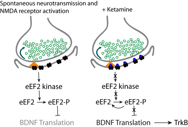
Elongation Factor 2 Kinase (eEF2K) is a type of protein kinase that phosphorylates and inactivates elongation factor 2 (eEF2), a crucial player in protein synthesis. Specifically, eEF2 is responsible for translocating the ribosome along the mRNA during translation, and its phosphorylation by eEF2K leads to a decrease in protein synthesis rates.
eEF2K is activated under conditions of cellular stress, such as nutrient deprivation or hypoxia, and functions to conserve energy by reducing protein synthesis. The kinase is also involved in various cellular processes, including autophagy, apoptosis, and cancer progression. Inhibition of eEF2K has been proposed as a potential therapeutic strategy for treating various diseases, including neurodegenerative disorders and cancer.
Translational peptide chain elongation is the process during protein synthesis where activated amino acids are added to the growing peptide chain in a sequence determined by the genetic code present in messenger RNA (mRNA). This process involves several steps:
1. Recognition of the start codon on the mRNA by the small ribosomal subunit, which binds to the mRNA and brings an initiator tRNA with a methionine or formylmethionine amino acid attached into the P site (peptidyl site) of the ribosome.
2. The large ribosomal subunit then joins the small subunit, forming a complete ribosome complex.
3. An incoming charged tRNA with an appropriate amino acid, complementary to the next codon on the mRNA, binds to the A site (aminoacyl site) of the ribosome.
4. Peptidyl transferase, a catalytic domain within the large ribosomal subunit, facilitates the formation of a peptide bond between the amino acids attached to the tRNAs in the P and A sites. The methionine or formylmethionine initiator amino acid is now covalently linked to the second amino acid via this peptide bond.
5. Translocation occurs, moving the tRNA with the growing peptide chain from the P site to the E site (exit site) and shifting the mRNA by one codon relative to the ribosome. The uncharged tRNA is then released from the E site.
6. The next charged tRNA carrying an appropriate amino acid binds to the A site, and the process repeats until a stop codon is reached on the mRNA.
7. Upon encountering a stop codon, release factors recognize it and facilitate the release of the completed polypeptide chain from the final tRNA in the P site. The ribosome then dissociates from the mRNA, allowing for further translational events to occur.
Translational peptide chain elongation is a crucial step in protein synthesis and requires precise coordination between various components of the translation machinery, including ribosomes, tRNAs, amino acids, and numerous accessory proteins.
Positive Transcriptional Elongation Factor B (P-TEFb) is a crucial protein complex in the process of transcription, which is the first step in gene expression. The main function of P-TEFb is to help RNA polymerase II, the enzyme responsible for transcribing DNA into RNA, to continue and complete the transcription of genes.
P-TEFb is composed of two subunits: cyclin T (CYCT) and CDK9 (cyclin-dependent kinase 9). The complex acts by phosphorylating several proteins that associate with RNA polymerase II, including the negative elongation factors NELF and DSIF. This phosphorylation converts NELF from a repressor to an activator of transcription elongation and relieves DSIF-mediated pausing of RNA polymerase II, allowing it to transcribe genes efficiently.
P-TEFb plays a significant role in regulating the expression of numerous genes, including those involved in cell growth, differentiation, and survival. Its dysregulation has been implicated in several diseases, such as cancer and HIV infection. In cancer, P-TEFb can contribute to oncogene activation and tumor progression, while in HIV, it is required for the transcription of viral genes during the early and late stages of infection.
Transfer RNA (tRNA) is a type of RNA molecule that plays a crucial role in protein synthesis. It serves as the adaptor molecule that translates the genetic code present in messenger RNA (mRNA) into the corresponding amino acids, which are then linked together to form a polypeptide chain during protein synthesis.
Aminoacyl tRNA is a specific type of tRNA molecule that has been charged or activated with an amino acid. This process is called aminoacylation and is carried out by enzymes called aminoacyl-tRNA synthetases. Each synthetase specifically recognizes and attaches a particular amino acid to its corresponding tRNA, ensuring the fidelity of protein synthesis. Once an amino acid is attached to a tRNA, it forms an aminoacyl-tRNA complex, which can then participate in translation and contribute to the formation of a new protein.
Guanosine triphosphate (GTP) is a nucleotide that plays a crucial role in various cellular processes, such as protein synthesis, signal transduction, and regulation of enzymatic activities. It serves as an energy currency, similar to adenosine triphosphate (ATP), and undergoes hydrolysis to guanosine diphosphate (GDP) or guanosine monophosphate (GMP) to release energy required for these processes. GTP is also a precursor for the synthesis of other essential molecules, including RNA and certain signaling proteins. Additionally, it acts as a molecular switch in many intracellular signaling pathways by binding and activating specific GTPase proteins.
Phosphatidylinositol 3-Kinases (PI3Ks) are a family of enzymes that play a crucial role in intracellular signal transduction. They phosphorylate the 3-hydroxyl group of the inositol ring in phosphatidylinositol and its derivatives, which results in the production of second messengers that regulate various cellular processes such as cell growth, proliferation, differentiation, motility, and survival.
PI3Ks are divided into three classes based on their structure and substrate specificity. Class I PI3Ks are further subdivided into two categories: class IA and class IB. Class IA PI3Ks are heterodimers consisting of a catalytic subunit (p110α, p110β, or p110δ) and a regulatory subunit (p85α, p85β, p55γ, or p50γ). They are primarily activated by receptor tyrosine kinases and G protein-coupled receptors. Class IB PI3Ks consist of a catalytic subunit (p110γ) and a regulatory subunit (p101 or p84/87). They are mainly activated by G protein-coupled receptors.
Dysregulation of PI3K signaling has been implicated in various human diseases, including cancer, diabetes, and autoimmune disorders. Therefore, PI3Ks have emerged as important targets for drug development in these areas.
Protein kinases are a group of enzymes that play a crucial role in many cellular processes by adding phosphate groups to other proteins, a process known as phosphorylation. This modification can activate or deactivate the target protein's function, thereby regulating various signaling pathways within the cell. Protein kinases are essential for numerous biological functions, including metabolism, signal transduction, cell cycle progression, and apoptosis (programmed cell death). Abnormal regulation of protein kinases has been implicated in several diseases, such as cancer, diabetes, and neurological disorders.
Protein-Serine-Threonine Kinases (PSTKs) are a type of protein kinase that catalyzes the transfer of a phosphate group from ATP to the hydroxyl side chains of serine or threonine residues on target proteins. This phosphorylation process plays a crucial role in various cellular signaling pathways, including regulation of metabolism, gene expression, cell cycle progression, and apoptosis. PSTKs are involved in many physiological and pathological processes, and their dysregulation has been implicated in several diseases, such as cancer, diabetes, and neurodegenerative disorders.
Ribosomes are complex macromolecular structures composed of ribonucleic acid (RNA) and proteins that play a crucial role in protein synthesis within cells. They serve as the site for translation, where messenger RNA (mRNA) is translated into a specific sequence of amino acids to create a polypeptide chain, which eventually folds into a functional protein.
Ribosomes consist of two subunits: a smaller subunit and a larger subunit. These subunits are composed of ribosomal RNA (rRNA) molecules and proteins. In eukaryotic cells, the smaller subunit is denoted as the 40S subunit, while the larger subunit is referred to as the 60S subunit. In prokaryotic cells, these subunits are named the 30S and 50S subunits, respectively. The ribosome's overall structure resembles a "doughnut" or a "cotton reel," with grooves and binding sites for various factors involved in protein synthesis.
Ribosomes can be found floating freely within the cytoplasm of cells or attached to the endoplasmic reticulum (ER) membrane, forming part of the rough ER. Membrane-bound ribosomes are responsible for synthesizing proteins that will be transported across the ER and ultimately secreted from the cell or inserted into the membrane. In contrast, cytoplasmic ribosomes synthesize proteins destined for use within the cytoplasm or organelles.
In summary, ribosomes are essential components of cells that facilitate protein synthesis by translating mRNA into functional polypeptide chains. They can be found in various cellular locations and exist as either free-floating entities or membrane-bound structures.
Mitogen-activated protein kinase (MAPK) signaling system is a crucial pathway for the transmission and regulation of various cellular responses in eukaryotic cells. It plays a significant role in several biological processes, including proliferation, differentiation, apoptosis, inflammation, and stress response. The MAPK cascade consists of three main components: MAP kinase kinase kinase (MAP3K or MEKK), MAP kinase kinase (MAP2K or MEK), and MAP kinase (MAPK).
The signaling system is activated by various extracellular stimuli, such as growth factors, cytokines, hormones, and stress signals. These stimuli initiate a phosphorylation cascade that ultimately leads to the activation of MAPKs. The activated MAPKs then translocate into the nucleus and regulate gene expression by phosphorylating various transcription factors and other regulatory proteins.
There are four major MAPK families: extracellular signal-regulated kinases (ERK1/2), c-Jun N-terminal kinases (JNK1/2/3), p38 MAPKs (p38α/β/γ/δ), and ERK5. Each family has distinct functions, substrates, and upstream activators. Dysregulation of the MAPK signaling system can lead to various diseases, including cancer, diabetes, cardiovascular diseases, and neurological disorders. Therefore, understanding the molecular mechanisms underlying this pathway is crucial for developing novel therapeutic strategies.
Molecular sequence data refers to the specific arrangement of molecules, most commonly nucleotides in DNA or RNA, or amino acids in proteins, that make up a biological macromolecule. This data is generated through laboratory techniques such as sequencing, and provides information about the exact order of the constituent molecules. This data is crucial in various fields of biology, including genetics, evolution, and molecular biology, allowing for comparisons between different organisms, identification of genetic variations, and studies of gene function and regulation.
Guanosine diphosphate (GDP) is a nucleotide that consists of a guanine base, a sugar molecule called ribose, and two phosphate groups. It is an ester of pyrophosphoric acid with the hydroxy group of the ribose sugar at the 5' position. GDP plays a crucial role as a secondary messenger in intracellular signaling pathways and also serves as an important intermediate in the synthesis of various biomolecules, such as proteins and polysaccharides.
In cells, GDP is formed from the hydrolysis of guanosine triphosphate (GTP) by enzymes called GTPases, which convert GTP to GDP and release energy that can be used to power various cellular processes. The conversion of GDP back to GTP can be facilitated by nucleotide diphosphate kinases, allowing for the recycling of these nucleotides within the cell.
It is important to note that while guanosine diphosphate has a significant role in biochemical processes, it is not typically associated with medical conditions or diseases directly. However, understanding its function and regulation can provide valuable insights into various physiological and pathophysiological mechanisms.
RNA Polymerase II is a type of enzyme responsible for transcribing DNA into RNA in eukaryotic cells. It plays a crucial role in the process of gene expression, where the information stored in DNA is used to create proteins. Specifically, RNA Polymerase II transcribes protein-coding genes to produce precursor messenger RNA (pre-mRNA), which is then processed into mature mRNA. This mature mRNA serves as a template for protein synthesis during translation.
RNA Polymerase II has a complex structure, consisting of multiple subunits, and it requires the assistance of various transcription factors and coactivators to initiate and regulate transcription. The enzyme recognizes specific promoter sequences in DNA, unwinds the double-stranded DNA, and synthesizes a complementary RNA strand using one of the unwound DNA strands as a template. This process results in the formation of a nascent RNA molecule that is further processed into mature mRNA for protein synthesis or other functional RNAs involved in gene regulation.
Transcription factors are proteins that play a crucial role in regulating gene expression by controlling the transcription of DNA to messenger RNA (mRNA). When referring to "General Transcription Factors," it indicates a specific group of these proteins that are involved in the basal transcription machinery, which is necessary for the transcription of protein-coding genes in all organisms. These general transcription factors are required for the initiation of transcription and include several conserved components:
1. TFIIA (Transcription Factor II A) - a heterotrimeric complex that binds to the TATA box region of the promoter, enhancing the stability and specificity of the pre-initiation complex.
2. TFIID (Transcription Factor II D) - a multi-subunit complex containing the TATA-binding protein (TBP) and several TBP-associated factors (TAFs). TBP recognizes and binds to the TATA box, while TAFs contribute to promoter recognition, chromatin remodeling, and transcription activation.
3. TFIIB - a single polypeptide that interacts with both TFIID and RNA polymerase II, helping to position the polymerase correctly at the transcription start site.
4. TFIIF - a heterotrimeric complex that stabilizes the interaction between TFIIB and RNA polymerase II, promoting the formation of the pre-initiation complex.
5. TFIIE - a heterodimeric complex that interacts with TFIIB, TFIIF, and RNA polymerase II, playing a role in promoter clearance and the transition from initiation to elongation.
6. TFIIH - a multi-subunit complex containing helicase and kinase activities. It is involved in promoter opening, DNA melting at the transcription start site, and phosphorylation of the C-terminal domain (CTD) of RNA polymerase II to facilitate elongation.
These general transcription factors work together to form a pre-initiation complex that enables RNA polymerase II to initiate transcription accurately and efficiently.
'Escherichia coli' (E. coli) is a type of gram-negative, facultatively anaerobic, rod-shaped bacterium that commonly inhabits the intestinal tract of humans and warm-blooded animals. It is a member of the family Enterobacteriaceae and one of the most well-studied prokaryotic model organisms in molecular biology.
While most E. coli strains are harmless and even beneficial to their hosts, some serotypes can cause various forms of gastrointestinal and extraintestinal illnesses in humans and animals. These pathogenic strains possess virulence factors that enable them to colonize and damage host tissues, leading to diseases such as diarrhea, urinary tract infections, pneumonia, and sepsis.
E. coli is a versatile organism with remarkable genetic diversity, which allows it to adapt to various environmental niches. It can be found in water, soil, food, and various man-made environments, making it an essential indicator of fecal contamination and a common cause of foodborne illnesses. The study of E. coli has contributed significantly to our understanding of fundamental biological processes, including DNA replication, gene regulation, and protein synthesis.
Calcium-calmodulin-dependent protein kinases (CAMKs) are a family of enzymes that play a crucial role in intracellular signaling pathways. They are activated by the binding of calcium ions and calmodulin, a ubiquitous calcium-binding protein, to their regulatory domain.
Once activated, CAMKs phosphorylate specific serine or threonine residues on target proteins, thereby modulating their activity, localization, or stability. This post-translational modification is essential for various cellular processes, including synaptic plasticity, gene expression, metabolism, and cell cycle regulation.
There are several subfamilies of CAMKs, including CaMKI, CaMKII, CaMKIII (also known as CaMKIV), and CaMK kinase (CaMKK). Each subfamily has distinct structural features, substrate specificity, and regulatory mechanisms. Dysregulation of CAMK signaling has been implicated in various pathological conditions, such as neurodegenerative diseases, cancer, and cardiovascular disorders.
Fusidic Acid is a steroid antibiotic, derived from the fungus Fusidium coccineum. It is primarily used to treat skin infections and other susceptible bacterial infections. It works by inhibiting bacterial protein synthesis. In medical terms, it can be defined as:
A triterpenoid antibiotic derived from the fungus Fusidium coccineum, used primarily to treat staphylococcal and streptococcal skin infections that are resistant to other antibiotics. It inhibits bacterial protein synthesis by binding to the bacterial elongation factor EF-G, preventing translocation of peptidyl tRNA from the A site to the P site on the ribosome.
It is important to note that resistance to fusidic acid can develop and its use should be reserved for infections caused by organisms known to be susceptible to it. It is not typically used as a first-line antibiotic, but rather as a secondary option when other treatments have failed or are contraindicated.
An amino acid sequence is the specific order of amino acids in a protein or peptide molecule, formed by the linking of the amino group (-NH2) of one amino acid to the carboxyl group (-COOH) of another amino acid through a peptide bond. The sequence is determined by the genetic code and is unique to each type of protein or peptide. It plays a crucial role in determining the three-dimensional structure and function of proteins.
Pyridones are a class of organic compounds that contain a pyridone ring, which is a heterocyclic ring consisting of a six-membered ring with five carbon atoms and one nitrogen atom, with one oxygen atom attached to the nitrogen atom by a double bond. Pyridones can be found in various natural sources, including plants and microorganisms, and they also have important applications in the pharmaceutical industry as building blocks for drug design and synthesis. Some drugs that contain pyridone rings include antihistamines, anti-inflammatory agents, and antiviral agents.
Protein kinase inhibitors (PKIs) are a class of drugs that work by interfering with the function of protein kinases. Protein kinases are enzymes that play a crucial role in many cellular processes by adding a phosphate group to specific proteins, thereby modifying their activity, localization, or interaction with other molecules. This process of adding a phosphate group is known as phosphorylation and is a key mechanism for regulating various cellular functions, including signal transduction, metabolism, and cell division.
In some diseases, such as cancer, protein kinases can become overactive or mutated, leading to uncontrolled cell growth and division. Protein kinase inhibitors are designed to block the activity of these dysregulated kinases, thereby preventing or slowing down the progression of the disease. These drugs can be highly specific, targeting individual protein kinases or families of kinases, making them valuable tools for targeted therapy in cancer and other diseases.
Protein kinase inhibitors can work in various ways to block the activity of protein kinases. Some bind directly to the active site of the enzyme, preventing it from interacting with its substrates. Others bind to allosteric sites, changing the conformation of the enzyme and making it inactive. Still, others target upstream regulators of protein kinases or interfere with their ability to form functional complexes.
Examples of protein kinase inhibitors include imatinib (Gleevec), which targets the BCR-ABL kinase in chronic myeloid leukemia, and gefitinib (Iressa), which inhibits the EGFR kinase in non-small cell lung cancer. These drugs have shown significant clinical benefits in treating these diseases and have become important components of modern cancer therapy.
Protein binding, in the context of medical and biological sciences, refers to the interaction between a protein and another molecule (known as the ligand) that results in a stable complex. This process is often reversible and can be influenced by various factors such as pH, temperature, and concentration of the involved molecules.
In clinical chemistry, protein binding is particularly important when it comes to drugs, as many of them bind to proteins (especially albumin) in the bloodstream. The degree of protein binding can affect a drug's distribution, metabolism, and excretion, which in turn influence its therapeutic effectiveness and potential side effects.
Protein-bound drugs may be less available for interaction with their target tissues, as only the unbound or "free" fraction of the drug is active. Therefore, understanding protein binding can help optimize dosing regimens and minimize adverse reactions.
Genetic transcription is the process by which the information in a strand of DNA is used to create a complementary RNA molecule. This process is the first step in gene expression, where the genetic code in DNA is converted into a form that can be used to produce proteins or functional RNAs.
During transcription, an enzyme called RNA polymerase binds to the DNA template strand and reads the sequence of nucleotide bases. As it moves along the template, it adds complementary RNA nucleotides to the growing RNA chain, creating a single-stranded RNA molecule that is complementary to the DNA template strand. Once transcription is complete, the RNA molecule may undergo further processing before it can be translated into protein or perform its functional role in the cell.
Transcription can be either "constitutive" or "regulated." Constitutive transcription occurs at a relatively constant rate and produces essential proteins that are required for basic cellular functions. Regulated transcription, on the other hand, is subject to control by various intracellular and extracellular signals, allowing cells to respond to changing environmental conditions or developmental cues.
Protein biosynthesis is the process by which cells generate new proteins. It involves two major steps: transcription and translation. Transcription is the process of creating a complementary RNA copy of a sequence of DNA. This RNA copy, or messenger RNA (mRNA), carries the genetic information to the site of protein synthesis, the ribosome. During translation, the mRNA is read by transfer RNA (tRNA) molecules, which bring specific amino acids to the ribosome based on the sequence of nucleotides in the mRNA. The ribosome then links these amino acids together in the correct order to form a polypeptide chain, which may then fold into a functional protein. Protein biosynthesis is essential for the growth and maintenance of all living organisms.
Transfer RNA (tRNA) is a type of RNA molecule that helps translate genetic information from messenger RNA (mRNA) into proteins. Each tRNA carries a specific amino acid to the growing polypeptide chain during protein synthesis, based on the anticodon sequence in its variable loop region that recognizes and binds to a complementary codon sequence in the mRNA.
Phenylalanine (Phe) is one of the twenty standard amino acids found in proteins. It has a hydrophobic side chain, which means it tends to repel water and interact with other non-polar molecules. In tRNA, phenylalanine is attached to a specific tRNA molecule known as tRNAPhe. This tRNA recognizes the mRNA codons UUC and UUU, which specify phenylalanine during protein synthesis.
SRC-family kinases (SFKs) are a group of non-receptor tyrosine kinases that play important roles in various cellular processes, including cell proliferation, differentiation, survival, and migration. They are named after the founding member, SRC, which was first identified as an oncogene in Rous sarcoma virus.
SFKs share a common structure, consisting of an N-terminal unique domain, a SH3 domain, a SH2 domain, a catalytic kinase domain, and a C-terminal regulatory tail with a negative regulatory tyrosine residue (Y527 in human SRC). In their inactive state, SFKs are maintained in a closed conformation through intramolecular interactions between the SH3 domain, SH2 domain, and the phosphorylated C-terminal tyrosine.
Upon activation by various signals, such as growth factors, cytokines, or integrin engagement, SFKs are activated through a series of events that involve dephosphorylation of the regulatory tyrosine residue, recruitment to membrane receptors via their SH2 and SH3 domains, and trans-autophosphorylation of the activation loop in the kinase domain.
Once activated, SFKs can phosphorylate a wide range of downstream substrates, including other protein kinases, adaptor proteins, and cytoskeletal components, thereby regulating various signaling pathways that control cell behavior. Dysregulation of SFK activity has been implicated in various diseases, including cancer, inflammation, and neurological disorders.
A base sequence in the context of molecular biology refers to the specific order of nucleotides in a DNA or RNA molecule. In DNA, these nucleotides are adenine (A), guanine (G), cytosine (C), and thymine (T). In RNA, uracil (U) takes the place of thymine. The base sequence contains genetic information that is transcribed into RNA and ultimately translated into proteins. It is the exact order of these bases that determines the genetic code and thus the function of the DNA or RNA molecule.
I'm sorry for any confusion, but "Poly U" is not a recognized medical term or abbreviation in the English language. It could potentially refer to Polytechnic University or Hong Kong Polytechnic University, but it does not have a specific medical connotation. If you have more context or information, I'd be happy to help further!
In the context of medicine and pharmacology, "kinetics" refers to the study of how a drug moves throughout the body, including its absorption, distribution, metabolism, and excretion (often abbreviated as ADME). This field is called "pharmacokinetics."
1. Absorption: This is the process of a drug moving from its site of administration into the bloodstream. Factors such as the route of administration (e.g., oral, intravenous, etc.), formulation, and individual physiological differences can affect absorption.
2. Distribution: Once a drug is in the bloodstream, it gets distributed throughout the body to various tissues and organs. This process is influenced by factors like blood flow, protein binding, and lipid solubility of the drug.
3. Metabolism: Drugs are often chemically modified in the body, typically in the liver, through processes known as metabolism. These changes can lead to the formation of active or inactive metabolites, which may then be further distributed, excreted, or undergo additional metabolic transformations.
4. Excretion: This is the process by which drugs and their metabolites are eliminated from the body, primarily through the kidneys (urine) and the liver (bile).
Understanding the kinetics of a drug is crucial for determining its optimal dosing regimen, potential interactions with other medications or foods, and any necessary adjustments for special populations like pediatric or geriatric patients, or those with impaired renal or hepatic function.
Transfer RNA (tRNA) is a type of RNA molecule that plays a crucial role in protein synthesis, the process by which cells create proteins. In protein synthesis, tRNAs serve as adaptors, translating the genetic code present in messenger RNA (mRNA) into the corresponding amino acids required to build a protein.
Each tRNA molecule has a distinct structure, consisting of approximately 70-90 nucleotides arranged in a cloverleaf shape with several loops and stems. The most important feature of a tRNA is its anticodon, a sequence of three nucleotides located in one of the loops. This anticodon base-pairs with a complementary codon on the mRNA during translation, ensuring that the correct amino acid is added to the growing polypeptide chain.
Before tRNAs can participate in protein synthesis, they must be charged with their specific amino acids through an enzymatic process involving aminoacyl-tRNA synthetases. These enzymes recognize and bind to both the tRNA and its corresponding amino acid, forming a covalent bond between them. Once charged, the aminoacyl-tRNA complex is ready to engage in translation and contribute to protein formation.
In summary, transfer RNA (tRNA) is a small RNA molecule that facilitates protein synthesis by translating genetic information from messenger RNA into specific amino acids, ultimately leading to the creation of functional proteins within cells.
A mutation is a permanent change in the DNA sequence of an organism's genome. Mutations can occur spontaneously or be caused by environmental factors such as exposure to radiation, chemicals, or viruses. They may have various effects on the organism, ranging from benign to harmful, depending on where they occur and whether they alter the function of essential proteins. In some cases, mutations can increase an individual's susceptibility to certain diseases or disorders, while in others, they may confer a survival advantage. Mutations are the driving force behind evolution, as they introduce new genetic variability into populations, which can then be acted upon by natural selection.
Phosphorylation is the process of adding a phosphate group (a molecule consisting of one phosphorus atom and four oxygen atoms) to a protein or other organic molecule, which is usually done by enzymes called kinases. This post-translational modification can change the function, localization, or activity of the target molecule, playing a crucial role in various cellular processes such as signal transduction, metabolism, and regulation of gene expression. Phosphorylation is reversible, and the removal of the phosphate group is facilitated by enzymes called phosphatases.
I apologize for the confusion, but "Aurodox" is not a medical term or a medication that is recognized in medical literature or by major healthcare organizations. It's possible that you may have misspelled the name or it could be a term used in a specific context, such as a research project or a product in development. If you have more information about where you encountered this term, I might be able to provide a more accurate response.
Protein Kinase C (PKC) is a family of serine-threonine kinases that play crucial roles in various cellular signaling pathways. These enzymes are activated by second messengers such as diacylglycerol (DAG) and calcium ions (Ca2+), which result from the activation of cell surface receptors like G protein-coupled receptors (GPCRs) and receptor tyrosine kinases (RTKs).
Once activated, PKC proteins phosphorylate downstream target proteins, thereby modulating their activities. This regulation is involved in numerous cellular processes, including cell growth, differentiation, apoptosis, and membrane trafficking. There are at least 10 isoforms of PKC, classified into three subfamilies based on their second messenger requirements and structural features: conventional (cPKC; α, βI, βII, and γ), novel (nPKC; δ, ε, η, and θ), and atypical (aPKC; ζ and ι/λ). Dysregulation of PKC signaling has been implicated in several diseases, such as cancer, diabetes, and neurological disorders.
Guanine nucleotides are molecules that play a crucial role in intracellular signaling, cellular regulation, and various biological processes within cells. They consist of a guanine base, a sugar (ribose or deoxyribose), and one or more phosphate groups. The most common guanine nucleotides are GDP (guanosine diphosphate) and GTP (guanosine triphosphate).
GTP is hydrolyzed to GDP and inorganic phosphate by certain enzymes called GTPases, releasing energy that drives various cellular functions such as protein synthesis, signal transduction, vesicle transport, and cell division. On the other hand, GDP can be rephosphorylated back to GTP by nucleotide diphosphate kinases, allowing for the recycling of these molecules within the cell.
In addition to their role in signaling and regulation, guanine nucleotides also serve as building blocks for RNA (ribonucleic acid) synthesis during transcription, where they pair with cytosine nucleotides via hydrogen bonds to form base pairs in the resulting RNA molecule.
Cyclin-Dependent Kinase 9 (CDK9) is a type of serine/threonine protein kinase that plays a crucial role in the regulation of transcription. It forms a complex with cyclin T1, K or H and gets activated by phosphorylation. This complex, known as P-TEFb, is involved in the phosphorylation and activation of the C-terminal domain of RNA polymerase II, which is essential for the transcription elongation of most protein-coding genes. CDK9 also regulates other cellular processes such as apoptosis, differentiation, and cell cycle progression. Dysregulation of CDK9 has been implicated in various diseases including cancer.
p38 Mitogen-Activated Protein Kinases (p38 MAPKs) are a family of conserved serine-threonine protein kinases that play crucial roles in various cellular processes, including inflammation, immune response, differentiation, apoptosis, and stress responses. They are activated by diverse stimuli such as cytokines, ultraviolet radiation, heat shock, osmotic stress, and lipopolysaccharides (LPS).
Once activated, p38 MAPKs phosphorylate and regulate several downstream targets, including transcription factors and other protein kinases. This regulation leads to the expression of genes involved in inflammation, cell cycle arrest, and apoptosis. Dysregulation of p38 MAPK signaling has been implicated in various diseases, such as cancer, neurodegenerative disorders, and autoimmune diseases. Therefore, p38 MAPKs are considered promising targets for developing new therapeutic strategies to treat these conditions.
Cyclic AMP (cAMP)-dependent protein kinases, also known as protein kinase A (PKA), are a family of enzymes that play a crucial role in intracellular signaling pathways. These enzymes are responsible for the regulation of various cellular processes, including metabolism, gene expression, and cell growth and differentiation.
PKA is composed of two regulatory subunits and two catalytic subunits. When cAMP binds to the regulatory subunits, it causes a conformational change that leads to the dissociation of the catalytic subunits. The freed catalytic subunits then phosphorylate specific serine and threonine residues on target proteins, thereby modulating their activity.
The cAMP-dependent protein kinases are activated in response to a variety of extracellular signals, such as hormones and neurotransmitters, that bind to G protein-coupled receptors (GPCRs) or receptor tyrosine kinases (RTKs). These signals lead to the activation of adenylyl cyclase, which catalyzes the conversion of ATP to cAMP. The resulting increase in intracellular cAMP levels triggers the activation of PKA and the downstream phosphorylation of target proteins.
Overall, cAMP-dependent protein kinases are essential regulators of many fundamental cellular processes and play a critical role in maintaining normal physiology and homeostasis. Dysregulation of these enzymes has been implicated in various diseases, including cancer, diabetes, and neurological disorders.
In the context of medical and biological sciences, a "binding site" refers to a specific location on a protein, molecule, or cell where another molecule can attach or bind. This binding interaction can lead to various functional changes in the original protein or molecule. The other molecule that binds to the binding site is often referred to as a ligand, which can be a small molecule, ion, or even another protein.
The binding between a ligand and its target binding site can be specific and selective, meaning that only certain ligands can bind to particular binding sites with high affinity. This specificity plays a crucial role in various biological processes, such as signal transduction, enzyme catalysis, or drug action.
In the case of drug development, understanding the location and properties of binding sites on target proteins is essential for designing drugs that can selectively bind to these sites and modulate protein function. This knowledge can help create more effective and safer therapeutic options for various diseases.
Mitogen-Activated Protein Kinase 1 (MAPK1), also known as Extracellular Signal-Regulated Kinase 2 (ERK2), is a protein kinase that plays a crucial role in intracellular signal transduction pathways. It is a member of the MAPK family, which regulates various cellular processes such as proliferation, differentiation, apoptosis, and stress response.
MAPK1 is activated by a cascade of phosphorylation events initiated by upstream activators like MAPKK (Mitogen-Activated Protein Kinase Kinase) in response to various extracellular signals such as growth factors, hormones, and mitogens. Once activated, MAPK1 phosphorylates downstream targets, including transcription factors and other protein kinases, thereby modulating their activities and ultimately influencing gene expression and cellular responses.
MAPK1 is widely expressed in various tissues and cells, and its dysregulation has been implicated in several pathological conditions, including cancer, inflammation, and neurodegenerative diseases. Therefore, understanding the regulation and function of MAPK1 signaling pathways has important implications for developing therapeutic strategies to treat these disorders.
Cyclin-dependent kinases (CDKs) are a family of serine/threonine protein kinases that play crucial roles in regulating the cell cycle, transcription, and other cellular processes. They are activated by binding to cyclin proteins, which accumulate and degrade at specific stages of the cell cycle. The activation of CDKs leads to phosphorylation of various downstream target proteins, resulting in the promotion or inhibition of different cell cycle events. Dysregulation of CDKs has been implicated in several human diseases, including cancer, and they are considered important targets for drug development.
"Saccharomyces cerevisiae" is not typically considered a medical term, but it is a scientific name used in the field of microbiology. It refers to a species of yeast that is commonly used in various industrial processes, such as baking and brewing. It's also widely used in scientific research due to its genetic tractability and eukaryotic cellular organization.
However, it does have some relevance to medical fields like medicine and nutrition. For example, certain strains of S. cerevisiae are used as probiotics, which can provide health benefits when consumed. They may help support gut health, enhance the immune system, and even assist in the digestion of certain nutrients.
In summary, "Saccharomyces cerevisiae" is a species of yeast with various industrial and potential medical applications.
'Artemia' is a genus of aquatic branchiopod crustaceans, also known as brine shrimp. They are commonly found in saltwater environments such as salt lakes and highly saline ponds. Artemia are known for their ability to produce cysts (also called "resting eggs") that can survive extreme environmental conditions, making them an important organism in research related to survival in harsh environments and space exploration.
In a medical context, Artemia is not typically used as a term but may be referenced in scientific studies related to biology, genetics, or astrobiology. The compounds derived from Artemia, such as astaxanthin and other carotenoids, have been studied for their potential health benefits, including antioxidant properties and support for eye and heart health. However, these applications are still under research and not yet considered part of mainstream medical practice.
Cyclin T is a type of cyclin protein that is encoded by the CCNT2 gene in humans. Cyclins are a family of regulatory proteins that play a crucial role in the cell cycle, which is the series of events that cells undergo as they grow and divide. Specifically, cyclin T is a component of the CDK9/cyclin T complex, also known as positive transcription elongation factor b (P-TEFb), which plays a key role in regulating gene expression by controlling the elongation phase of RNA polymerase II-mediated transcription.
Cyclin T is expressed at various stages of the cell cycle and has been shown to interact with several other proteins involved in cell cycle regulation, including the retinoblastoma protein (pRb) and the E2F family of transcription factors. Dysregulation of cyclin T expression or activity has been implicated in several human diseases, including cancer.
Peptide biosynthesis is the process by which cells synthesize peptides, short chains of amino acids. This process is mediated by enzymes called peptide synthetases, which catalyze the formation of peptide bonds between individual amino acids to create a longer chain. Peptide biosynthesis typically occurs through one of two pathways: ribosomal or non-ribosomal.
Ribosomal peptide biosynthesis involves the use of the cell's translational machinery, including the ribosome and transfer RNAs (tRNAs), to synthesize peptides from a messenger RNA (mRNA) template. This process is highly regulated and typically results in the production of small, linear peptides that are further modified by enzymes to create bioactive molecules such as hormones or neurotransmitters.
Non-ribosomal peptide biosynthesis (NRPS), on the other hand, is a more complex process that involves large multifunctional enzyme complexes called non-ribosomal peptide synthetases (NRPSs). These enzymes are capable of synthesizing a wide variety of structurally diverse peptides, including cyclic and branched peptides, as well as those containing non-proteinogenic amino acids. NRPSs typically consist of multiple modules, each responsible for adding a single amino acid to the growing peptide chain. The modular nature of NRPS systems allows for great diversity in the types of peptides that can be synthesized, making them important sources of bioactive molecules with potential therapeutic applications.
Transcription factors are proteins that play a crucial role in regulating gene expression by controlling the transcription of DNA to messenger RNA (mRNA). They function by binding to specific DNA sequences, known as response elements, located in the promoter region or enhancer regions of target genes. This binding can either activate or repress the initiation of transcription, depending on the properties and interactions of the particular transcription factor. Transcription factors often act as part of a complex network of regulatory proteins that determine the precise spatiotemporal patterns of gene expression during development, differentiation, and homeostasis in an organism.
Messenger RNA (mRNA) is a type of RNA (ribonucleic acid) that carries genetic information copied from DNA in the form of a series of three-base code "words," each of which specifies a particular amino acid. This information is used by the cell's machinery to construct proteins, a process known as translation. After being transcribed from DNA, mRNA travels out of the nucleus to the ribosomes in the cytoplasm where protein synthesis occurs. Once the protein has been synthesized, the mRNA may be degraded and recycled. Post-transcriptional modifications can also occur to mRNA, such as alternative splicing and addition of a 5' cap and a poly(A) tail, which can affect its stability, localization, and translation efficiency.
Diphtheria toxin is a potent exotoxin produced by the bacterium Corynebacterium diphtheriae, which causes the disease diphtheria. This toxin is composed of two subunits: A and B. The B subunit helps the toxin bind to and enter host cells, while the A subunit inhibits protein synthesis within those cells, leading to cell damage and tissue destruction.
The toxin can cause a variety of symptoms depending on the site of infection. In respiratory diphtheria, it typically affects the nose, throat, and tonsils, causing a thick gray or white membrane to form over the affected area, making breathing and swallowing difficult. In cutaneous diphtheria, it infects the skin, leading to ulcers and necrosis.
Diphtheria toxin can also have systemic effects, such as damage to the heart, nerves, and kidneys, which can be life-threatening if left untreated. Fortunately, diphtheria is preventable through vaccination with the diphtheria, tetanus, and pertussis (DTaP or Tdap) vaccine.
Ribosomal proteins are a type of protein that play a crucial role in the structure and function of ribosomes, which are complex molecular machines found within all living cells. Ribosomes are responsible for translating messenger RNA (mRNA) into proteins during the process of protein synthesis.
Ribosomal proteins can be divided into two categories based on their location within the ribosome:
1. Large ribosomal subunit proteins: These proteins are associated with the larger of the two subunits of the ribosome, which is responsible for catalyzing peptide bond formation during protein synthesis.
2. Small ribosomal subunit proteins: These proteins are associated with the smaller of the two subunits of the ribosome, which is responsible for binding to the mRNA and decoding the genetic information it contains.
Ribosomal proteins have a variety of functions, including helping to stabilize the structure of the ribosome, assisting in the binding of substrates and cofactors necessary for protein synthesis, and regulating the activity of the ribosome. Mutations in ribosomal proteins can lead to a variety of human diseases, including developmental disorders, neurological conditions, and cancer.
P21-activated kinases (PAKs) are a family of serine/threonine protein kinases that play crucial roles in various cellular processes, including cytoskeletal reorganization, cell motility, and gene transcription. They are activated by binding to small GTPases of the Rho family, such as Cdc42 and Rac, which become active upon stimulation of various extracellular signals. Once activated, PAKs phosphorylate a range of downstream targets, leading to changes in cell behavior and function. Aberrant regulation of PAKs has been implicated in several human diseases, including cancer and neurological disorders.
JNK (c-Jun N-terminal kinase) Mitogen-Activated Protein Kinases are a subgroup of the Ser/Thr protein kinases that are activated by stress stimuli and play important roles in various cellular processes, including inflammation, apoptosis, and differentiation. They are involved in the regulation of gene expression through phosphorylation of transcription factors such as c-Jun. JNKs are activated by a variety of upstream kinases, including MAP2Ks (MKK4/SEK1 and MKK7), which are in turn activated by MAP3Ks (such as ASK1, MEKK1, MLKs, and TAK1). JNK signaling pathways have been implicated in various diseases, including cancer, neurodegenerative disorders, and inflammatory diseases.
Tertiary protein structure refers to the three-dimensional arrangement of all the elements (polypeptide chains) of a single protein molecule. It is the highest level of structural organization and results from interactions between various side chains (R groups) of the amino acids that make up the protein. These interactions, which include hydrogen bonds, ionic bonds, van der Waals forces, and disulfide bridges, give the protein its unique shape and stability, which in turn determines its function. The tertiary structure of a protein can be stabilized by various factors such as temperature, pH, and the presence of certain ions. Any changes in these factors can lead to denaturation, where the protein loses its tertiary structure and thus its function.
'Thermus thermophilus' is not a medical term, but a scientific name for a species of bacteria. It is commonly used in molecular biology and genetics research. Here is the biological definition:
'Thermus thermophilus' is a gram-negative, rod-shaped, thermophilic bacterium found in hot springs and other high-temperature environments. Its optimum growth temperature ranges from 65 to 70°C (149-158°F), with some strains able to grow at temperatures as high as 85°C (185°F). The bacterium's DNA polymerase enzyme, Taq polymerase, is widely used in the Polymerase Chain Reaction (PCR) technique for amplifying and analyzing DNA. 'Thermus thermophilus' has a single circular chromosome and can also have one or more plasmids. Its genome has been fully sequenced, making it an important model organism for studying extremophiles and their adaptations to harsh environments.
Mitogen-Activated Protein Kinase Kinases (MAP2K or MEK) are a group of protein kinases that play a crucial role in intracellular signal transduction pathways. They are so named because they are activated by mitogens, which are substances that stimulate cell division, and other extracellular signals.
MAP2Ks are positioned upstream of the Mitogen-Activated Protein Kinases (MAPK) in a three-tiered kinase cascade. Once activated, MAP2Ks phosphorylate and activate MAPKs, which then go on to regulate various cellular processes such as proliferation, differentiation, survival, and apoptosis.
There are several subfamilies of MAP2Ks, including MEK1/2, MEK3/6 (also known as MKK3/6), MEK4/7 (also known as MKK4/7), and MEK5. Each MAP2K is specific to activating a particular MAPK, and they are activated by different MAP3Ks (MAP kinase kinase kinases) in response to various extracellular signals.
Dysregulation of the MAPK/MAP2K signaling pathways has been implicated in numerous diseases, including cancer, cardiovascular disease, and neurological disorders. Therefore, targeting these pathways with therapeutic agents has emerged as a promising strategy for treating various diseases.
Sequence homology, amino acid, refers to the similarity in the order of amino acids in a protein or a portion of a protein between two or more species. This similarity can be used to infer evolutionary relationships and functional similarities between proteins. The higher the degree of sequence homology, the more likely it is that the proteins are related and have similar functions. Sequence homology can be determined through various methods such as pairwise alignment or multiple sequence alignment, which compare the sequences and calculate a score based on the number and type of matching amino acids.
Mitogen-Activated Protein Kinase 3 (MAPK3), also known as extracellular signal-regulated kinase 1 (ERK1), is a serine/threonine protein kinase that plays a crucial role in intracellular signal transduction pathways. It is involved in the regulation of various cellular processes, including proliferation, differentiation, and survival, in response to extracellular stimuli such as growth factors, hormones, and stress.
MAPK3 is activated through a phosphorylation cascade that involves the activation of upstream MAPK kinases (MKK or MEK). Once activated, MAPK3 can phosphorylate and activate various downstream targets, including transcription factors, to regulate gene expression. Dysregulation of MAPK3 signaling has been implicated in several diseases, including cancer and neurological disorders.
Ribosomal Protein S6 Kinases (RSKs) are a family of serine/threonine protein kinases that play a crucial role in the regulation of cell growth, proliferation, and survival. They are so named because they phosphorylate and regulate the function of the ribosomal protein S6, which is a component of the 40S ribosomal subunit involved in protein synthesis.
RSKs are activated by various signals, including growth factors, hormones, and mitogens, through a cascade of phosphorylation events involving several upstream kinases such as MAPK/ERK kinase (MEK) and extracellular signal-regulated kinase (ERK). Once activated, RSKs phosphorylate a wide range of downstream targets, including transcription factors, regulators of translation, and cytoskeletal proteins, thereby modulating their activities and functions.
There are four isoforms of RSKs in humans, namely RSK1, RSK2, RSK3, and RSK4, which share a common structural organization and functional domains, including an N-terminal kinase domain, a C-terminal kinase domain, and a linker region that contains several regulatory motifs. Dysregulation of RSKs has been implicated in various pathological conditions, including cancer, cardiovascular diseases, neurological disorders, and diabetes, making them attractive targets for therapeutic intervention.
Casein Kinase II (CK2) is a serine/threonine protein kinase that is widely expressed in eukaryotic cells and is involved in the regulation of various cellular processes. It is a heterotetrameric enzyme, consisting of two catalytic subunits (alpha and alpha') and two regulatory subunits (beta).
CK2 phosphorylates a wide range of substrates, including transcription factors, signaling proteins, and other kinases. It is known to play roles in cell cycle regulation, apoptosis, DNA damage response, and protein stability, among others. CK2 activity is often found to be elevated in various types of cancer, making it a potential target for cancer therapy.
Saccharomyces cerevisiae proteins are the proteins that are produced by the budding yeast, Saccharomyces cerevisiae. This organism is a single-celled eukaryote that has been widely used as a model organism in scientific research for many years due to its relatively simple genetic makeup and its similarity to higher eukaryotic cells.
The genome of Saccharomyces cerevisiae has been fully sequenced, and it is estimated to contain approximately 6,000 genes that encode proteins. These proteins play a wide variety of roles in the cell, including catalyzing metabolic reactions, regulating gene expression, maintaining the structure of the cell, and responding to environmental stimuli.
Many Saccharomyces cerevisiae proteins have human homologs and are involved in similar biological processes, making this organism a valuable tool for studying human disease. For example, many of the proteins involved in DNA replication, repair, and recombination in yeast have human counterparts that are associated with cancer and other diseases. By studying these proteins in yeast, researchers can gain insights into their function and regulation in humans, which may lead to new treatments for disease.
Protein-Tyrosine Kinases (PTKs) are a type of enzyme that plays a crucial role in various cellular functions, including signal transduction, cell growth, differentiation, and metabolism. They catalyze the transfer of a phosphate group from ATP to the tyrosine residues of proteins, thereby modifying their activity, localization, or interaction with other molecules.
PTKs can be divided into two main categories: receptor tyrosine kinases (RTKs) and non-receptor tyrosine kinases (NRTKs). RTKs are transmembrane proteins that become activated upon binding to specific ligands, such as growth factors or hormones. NRTKs, on the other hand, are intracellular enzymes that can be activated by various signals, including receptor-mediated signaling and intracellular messengers.
Dysregulation of PTK activity has been implicated in several diseases, such as cancer, diabetes, and inflammatory disorders. Therefore, PTKs are important targets for drug development and therapy.
CDC2 protein kinase, also known as cell division cycle 2 or CDK1, is a type of enzyme that plays a crucial role in the regulation of the cell cycle. The cell cycle is the series of events that cells undergo as they grow, replicate their DNA, and divide into two daughter cells.
CDC2 protein kinase is a member of the cyclin-dependent kinase (CDK) family, which are serine/threonine protein kinases that are activated by binding to regulatory subunits called cyclins. CDC2 protein kinase is primarily associated with the regulation of the G2 phase and the entry into mitosis, the stage of the cell cycle where nuclear and cytoplasmic division occur.
CDC2 protein kinase functions by phosphorylating various target proteins, which alters their activity and contributes to the coordination of the different events that occur during the cell cycle. The activity of CDC2 protein kinase is tightly regulated through a variety of mechanisms, including phosphorylation and dephosphorylation, as well as the binding and destruction of cyclin subunits.
Dysregulation of CDC2 protein kinase has been implicated in various human diseases, including cancer, where uncontrolled cell division can lead to the formation of tumors. Therefore, understanding the regulation and function of CDC2 protein kinase is an important area of research in molecular biology and medicine.
Molecular models are three-dimensional representations of molecular structures that are used in the field of molecular biology and chemistry to visualize and understand the spatial arrangement of atoms and bonds within a molecule. These models can be physical or computer-generated and allow researchers to study the shape, size, and behavior of molecules, which is crucial for understanding their function and interactions with other molecules.
Physical molecular models are often made up of balls (representing atoms) connected by rods or sticks (representing bonds). These models can be constructed manually using materials such as plastic or wooden balls and rods, or they can be created using 3D printing technology.
Computer-generated molecular models, on the other hand, are created using specialized software that allows researchers to visualize and manipulate molecular structures in three dimensions. These models can be used to simulate molecular interactions, predict molecular behavior, and design new drugs or chemicals with specific properties. Overall, molecular models play a critical role in advancing our understanding of molecular structures and their functions.
Molecular cloning is a laboratory technique used to create multiple copies of a specific DNA sequence. This process involves several steps:
1. Isolation: The first step in molecular cloning is to isolate the DNA sequence of interest from the rest of the genomic DNA. This can be done using various methods such as PCR (polymerase chain reaction), restriction enzymes, or hybridization.
2. Vector construction: Once the DNA sequence of interest has been isolated, it must be inserted into a vector, which is a small circular DNA molecule that can replicate independently in a host cell. Common vectors used in molecular cloning include plasmids and phages.
3. Transformation: The constructed vector is then introduced into a host cell, usually a bacterial or yeast cell, through a process called transformation. This can be done using various methods such as electroporation or chemical transformation.
4. Selection: After transformation, the host cells are grown in selective media that allow only those cells containing the vector to grow. This ensures that the DNA sequence of interest has been successfully cloned into the vector.
5. Amplification: Once the host cells have been selected, they can be grown in large quantities to amplify the number of copies of the cloned DNA sequence.
Molecular cloning is a powerful tool in molecular biology and has numerous applications, including the production of recombinant proteins, gene therapy, functional analysis of genes, and genetic engineering.
Creatine kinase (CK) is a muscle enzyme that is normally present in small amounts in the blood. It is primarily found in tissues that require a lot of energy, such as the heart, brain, and skeletal muscles. When these tissues are damaged or injured, CK is released into the bloodstream, causing the levels to rise.
Creatine kinase exists in several forms, known as isoenzymes, which can be measured in the blood to help identify the location of tissue damage. The three main isoenzymes are:
1. CK-MM: Found primarily in skeletal muscle
2. CK-MB: Found primarily in heart muscle
3. CK-BB: Found primarily in the brain
Elevated levels of creatine kinase, particularly CK-MB, can indicate damage to the heart muscle, such as occurs with a heart attack. Similarly, elevated levels of CK-BB may suggest brain injury or disease. Overall, measuring creatine kinase levels is a useful diagnostic tool for assessing tissue damage and determining the severity of injuries or illnesses.
MAP (Mitogen-Activated Protein) Kinase Kinase Kinases (MAP3K or MAPKKK) are a group of protein kinases that play a crucial role in intracellular signal transduction pathways, which regulate various cellular processes such as proliferation, differentiation, survival, and apoptosis. They are called "kinases" because they catalyze the transfer of a phosphate group from ATP to specific serine or threonine residues on their target proteins.
MAP3Ks function upstream of MAP Kinase Kinases (MKKs or MAP2K) and MAP Kinases (MPKs or MAPK) in the MAP kinase cascade. Upon activation by various extracellular signals, such as growth factors, cytokines, stress, and hormones, MAP3Ks phosphorylate and activate MKKs, which subsequently phosphorylate and activate MPKs. Activated MPKs then regulate the activity of downstream transcription factors and other target proteins to elicit appropriate cellular responses.
There are several subfamilies of MAP3Ks, including ASK, DLK, TAK, MEKK, MLK, and ZAK, among others. Each subfamily has distinct structural features and functions in different signaling pathways. Dysregulation of MAP kinase cascades, including MAP3Ks, has been implicated in various human diseases, such as cancer, inflammation, and neurodegenerative disorders.
HeLa cells are a type of immortalized cell line used in scientific research. They are derived from a cancer that developed in the cervical tissue of Henrietta Lacks, an African-American woman, in 1951. After her death, cells taken from her tumor were found to be capable of continuous division and growth in a laboratory setting, making them an invaluable resource for medical research.
HeLa cells have been used in a wide range of scientific studies, including research on cancer, viruses, genetics, and drug development. They were the first human cell line to be successfully cloned and are able to grow rapidly in culture, doubling their population every 20-24 hours. This has made them an essential tool for many areas of biomedical research.
It is important to note that while HeLa cells have been instrumental in numerous scientific breakthroughs, the story of their origin raises ethical questions about informed consent and the use of human tissue in research.
Signal transduction is the process by which a cell converts an extracellular signal, such as a hormone or neurotransmitter, into an intracellular response. This involves a series of molecular events that transmit the signal from the cell surface to the interior of the cell, ultimately resulting in changes in gene expression, protein activity, or metabolism.
The process typically begins with the binding of the extracellular signal to a receptor located on the cell membrane. This binding event activates the receptor, which then triggers a cascade of intracellular signaling molecules, such as second messengers, protein kinases, and ion channels. These molecules amplify and propagate the signal, ultimately leading to the activation or inhibition of specific cellular responses.
Signal transduction pathways are highly regulated and can be modulated by various factors, including other signaling molecules, post-translational modifications, and feedback mechanisms. Dysregulation of these pathways has been implicated in a variety of diseases, including cancer, diabetes, and neurological disorders.
eIF-2 kinase is a type of protein kinase that phosphorylates the alpha subunit of eukaryotic initiation factor-2 (eIF-2) at serine 51. This phosphorylation event inhibits the guanine nucleotide exchange factor eIF-2B, thereby preventing the recycling of eIF-2 and reducing global protein synthesis.
There are four main subtypes of eIF-2 kinases:
1. HRI (heme-regulated inhibitor) - responds to heme deficiency and oxidative stress
2. PERK (PKR-like endoplasmic reticulum kinase) - activated by ER stress and misfolded proteins in the ER
3. GCN2 (general control non-derepressible 2) - responds to amino acid starvation
4. PKR (double-stranded RNA-activated protein kinase) - activated by double-stranded RNA during viral infections
These eIF-2 kinases play crucial roles in regulating cellular responses to various stress conditions, such as the integrated stress response (ISR), which helps maintain cellular homeostasis and promote survival under adverse conditions.
Casein kinases are a family of protein kinases that play important roles in various cellular processes, including signal transduction, cell cycle regulation, and DNA damage repair. These enzymes phosphorylate serine and threonine residues on their target proteins by transferring a phosphate group from ATP to the hydroxyl side chain of these amino acids.
There are several isoforms of casein kinases, including Casein Kinase 1 (CK1) and Casein Kinase 2 (CK2), which differ in their structure, substrate specificity, and cellular functions. CK1 is involved in various signaling pathways, such as the Wnt signaling pathway, and regulates processes such as gene transcription, DNA repair, and circadian rhythm. CK2, on the other hand, is a highly conserved serine/threonine protein kinase that plays a role in many cellular processes, including cell division, apoptosis, and transcriptional regulation.
Dysregulation of casein kinases has been implicated in various diseases, including cancer, neurodegenerative disorders, and cardiovascular disease. Therefore, these enzymes are considered important targets for the development of new therapeutic strategies.
A cell line is a culture of cells that are grown in a laboratory for use in research. These cells are usually taken from a single cell or group of cells, and they are able to divide and grow continuously in the lab. Cell lines can come from many different sources, including animals, plants, and humans. They are often used in scientific research to study cellular processes, disease mechanisms, and to test new drugs or treatments. Some common types of human cell lines include HeLa cells (which come from a cancer patient named Henrietta Lacks), HEK293 cells (which come from embryonic kidney cells), and HUVEC cells (which come from umbilical vein endothelial cells). It is important to note that cell lines are not the same as primary cells, which are cells that are taken directly from a living organism and have not been grown in the lab.
Recombinant proteins are artificially created proteins produced through the use of recombinant DNA technology. This process involves combining DNA molecules from different sources to create a new set of genes that encode for a specific protein. The resulting recombinant protein can then be expressed, purified, and used for various applications in research, medicine, and industry.
Recombinant proteins are widely used in biomedical research to study protein function, structure, and interactions. They are also used in the development of diagnostic tests, vaccines, and therapeutic drugs. For example, recombinant insulin is a common treatment for diabetes, while recombinant human growth hormone is used to treat growth disorders.
The production of recombinant proteins typically involves the use of host cells, such as bacteria, yeast, or mammalian cells, which are engineered to express the desired protein. The host cells are transformed with a plasmid vector containing the gene of interest, along with regulatory elements that control its expression. Once the host cells are cultured and the protein is expressed, it can be purified using various chromatography techniques.
Overall, recombinant proteins have revolutionized many areas of biology and medicine, enabling researchers to study and manipulate proteins in ways that were previously impossible.
Bacterial proteins are a type of protein that are produced by bacteria as part of their structural or functional components. These proteins can be involved in various cellular processes, such as metabolism, DNA replication, transcription, and translation. They can also play a role in bacterial pathogenesis, helping the bacteria to evade the host's immune system, acquire nutrients, and multiply within the host.
Bacterial proteins can be classified into different categories based on their function, such as:
1. Enzymes: Proteins that catalyze chemical reactions in the bacterial cell.
2. Structural proteins: Proteins that provide structural support and maintain the shape of the bacterial cell.
3. Signaling proteins: Proteins that help bacteria to communicate with each other and coordinate their behavior.
4. Transport proteins: Proteins that facilitate the movement of molecules across the bacterial cell membrane.
5. Toxins: Proteins that are produced by pathogenic bacteria to damage host cells and promote infection.
6. Surface proteins: Proteins that are located on the surface of the bacterial cell and interact with the environment or host cells.
Understanding the structure and function of bacterial proteins is important for developing new antibiotics, vaccines, and other therapeutic strategies to combat bacterial infections.
Thiostrepton is an antibiotic and antiproliferative agent that is derived from the bacterium Streptomyces azureus. It belongs to the family of thiostreptons, which are cyclic oligopeptides with unique structures and various biological activities. Thiostrepton has been used primarily in veterinary medicine for the treatment of infections caused by gram-positive bacteria, such as mastitis in cows.
In addition to its antibacterial properties, thiostrepton has also been found to have antiproliferative and proapoptotic effects on various cancer cells, including breast, ovarian, and colon cancer cells. These effects are thought to be mediated by the inhibition of protein synthesis and the regulation of gene expression. However, its use as a therapeutic agent in humans is still being investigated due to its potential toxicity and limited bioavailability.
It's worth noting that thiostrepton is not commonly used in clinical practice, and its medical definition is mainly related to its chemical structure, antibacterial properties, and potential anticancer effects.
Pyruvate kinase is an enzyme that plays a crucial role in the final step of glycolysis, a process by which glucose is broken down to produce energy in the form of ATP (adenosine triphosphate). Specifically, pyruvate kinase catalyzes the transfer of a phosphate group from phosphoenolpyruvate (PEP) to adenosine diphosphate (ADP), resulting in the formation of pyruvate and ATP.
There are several isoforms of pyruvate kinase found in different tissues, including the liver, muscle, and brain. The type found in red blood cells is known as PK-RBC or PK-M2. Deficiencies in pyruvate kinase can lead to a genetic disorder called pyruvate kinase deficiency, which can result in hemolytic anemia due to the premature destruction of red blood cells.
Nuclear proteins are a category of proteins that are primarily found in the nucleus of a eukaryotic cell. They play crucial roles in various nuclear functions, such as DNA replication, transcription, repair, and RNA processing. This group includes structural proteins like lamins, which form the nuclear lamina, and regulatory proteins, such as histones and transcription factors, that are involved in gene expression. Nuclear localization signals (NLS) often help target these proteins to the nucleus by interacting with importin proteins during active transport across the nuclear membrane.
Biological models, also known as physiological models or organismal models, are simplified representations of biological systems, processes, or mechanisms that are used to understand and explain the underlying principles and relationships. These models can be theoretical (conceptual or mathematical) or physical (such as anatomical models, cell cultures, or animal models). They are widely used in biomedical research to study various phenomena, including disease pathophysiology, drug action, and therapeutic interventions.
Examples of biological models include:
1. Mathematical models: These use mathematical equations and formulas to describe complex biological systems or processes, such as population dynamics, metabolic pathways, or gene regulation networks. They can help predict the behavior of these systems under different conditions and test hypotheses about their underlying mechanisms.
2. Cell cultures: These are collections of cells grown in a controlled environment, typically in a laboratory dish or flask. They can be used to study cellular processes, such as signal transduction, gene expression, or metabolism, and to test the effects of drugs or other treatments on these processes.
3. Animal models: These are living organisms, usually vertebrates like mice, rats, or non-human primates, that are used to study various aspects of human biology and disease. They can provide valuable insights into the pathophysiology of diseases, the mechanisms of drug action, and the safety and efficacy of new therapies.
4. Anatomical models: These are physical representations of biological structures or systems, such as plastic models of organs or tissues, that can be used for educational purposes or to plan surgical procedures. They can also serve as a basis for developing more sophisticated models, such as computer simulations or 3D-printed replicas.
Overall, biological models play a crucial role in advancing our understanding of biology and medicine, helping to identify new targets for therapeutic intervention, develop novel drugs and treatments, and improve human health.
Phenylalanine is an essential amino acid, meaning it cannot be produced by the human body and must be obtained through diet or supplementation. It's one of the building blocks of proteins and is necessary for the production of various molecules in the body, such as neurotransmitters (chemical messengers in the brain).
Phenylalanine has two forms: L-phenylalanine and D-phenylalanine. L-phenylalanine is the form found in proteins and is used by the body for protein synthesis, while D-phenylalanine has limited use in humans and is not involved in protein synthesis.
Individuals with a rare genetic disorder called phenylketonuria (PKU) must follow a low-phenylalanine diet or take special medical foods because they are unable to metabolize phenylalanine properly, leading to its buildup in the body and potential neurological damage.
MAPKKK1 or Mitogen-Activated Protein Kinase Kinase Kinase 1 is a serine/threonine protein kinase that belongs to the MAP3K family. It plays a crucial role in intracellular signal transduction pathways, particularly in the MAPK/ERK cascade, which is involved in various cellular processes such as proliferation, differentiation, and survival.
MAPKKK1 activates MAPKKs (Mitogen-Activated Protein Kinase Kinases) through phosphorylation of specific serine and threonine residues. In turn, activated MAPKKs phosphorylate and activate MAPKs (Mitogen-Activated Protein Kinases), which then regulate the activity of various transcription factors and other downstream targets to elicit appropriate cellular responses.
Mutations in MAPKKK1 have been implicated in several human diseases, including cancer and developmental disorders. Therefore, understanding its function and regulation is essential for developing novel therapeutic strategies to treat these conditions.
Receptor Protein-Tyrosine Kinases (RTKs) are a type of transmembrane receptors found on the cell surface that play a crucial role in signal transduction and regulation of various cellular processes, including cell growth, differentiation, metabolism, and survival. They are called "tyrosine kinases" because they possess an intrinsic enzymatic activity that catalyzes the transfer of a phosphate group from ATP to tyrosine residues on target proteins, thereby modulating their function.
RTKs are composed of three main domains: an extracellular domain that binds to specific ligands (growth factors, hormones, or cytokines), a transmembrane domain that spans the cell membrane, and an intracellular domain with tyrosine kinase activity. Upon ligand binding, RTKs undergo conformational changes that lead to their dimerization or oligomerization, which in turn activates their tyrosine kinase activity. Activated RTKs then phosphorylate specific tyrosine residues on downstream signaling proteins, initiating a cascade of intracellular signaling events that ultimately result in the appropriate cellular response.
Dysregulation of RTK signaling has been implicated in various human diseases, including cancer, diabetes, and developmental disorders. As such, RTKs are important targets for therapeutic intervention in these conditions.
Electrophoresis, polyacrylamide gel (EPG) is a laboratory technique used to separate and analyze complex mixtures of proteins or nucleic acids (DNA or RNA) based on their size and electrical charge. This technique utilizes a matrix made of cross-linked polyacrylamide, a type of gel, which provides a stable and uniform environment for the separation of molecules.
In this process:
1. The polyacrylamide gel is prepared by mixing acrylamide monomers with a cross-linking agent (bis-acrylamide) and a catalyst (ammonium persulfate) in the presence of a buffer solution.
2. The gel is then poured into a mold and allowed to polymerize, forming a solid matrix with uniform pore sizes that depend on the concentration of acrylamide used. Higher concentrations result in smaller pores, providing better resolution for separating smaller molecules.
3. Once the gel has set, it is placed in an electrophoresis apparatus containing a buffer solution. Samples containing the mixture of proteins or nucleic acids are loaded into wells on the top of the gel.
4. An electric field is applied across the gel, causing the negatively charged molecules to migrate towards the positive electrode (anode) while positively charged molecules move toward the negative electrode (cathode). The rate of migration depends on the size, charge, and shape of the molecules.
5. Smaller molecules move faster through the gel matrix and will migrate farther from the origin compared to larger molecules, resulting in separation based on size. Proteins and nucleic acids can be selectively stained after electrophoresis to visualize the separated bands.
EPG is widely used in various research fields, including molecular biology, genetics, proteomics, and forensic science, for applications such as protein characterization, DNA fragment analysis, cloning, mutation detection, and quality control of nucleic acid or protein samples.
Recombinant fusion proteins are artificially created biomolecules that combine the functional domains or properties of two or more different proteins into a single protein entity. They are generated through recombinant DNA technology, where the genes encoding the desired protein domains are linked together and expressed as a single, chimeric gene in a host organism, such as bacteria, yeast, or mammalian cells.
The resulting fusion protein retains the functional properties of its individual constituent proteins, allowing for novel applications in research, diagnostics, and therapeutics. For instance, recombinant fusion proteins can be designed to enhance protein stability, solubility, or immunogenicity, making them valuable tools for studying protein-protein interactions, developing targeted therapies, or generating vaccines against infectious diseases or cancer.
Examples of recombinant fusion proteins include:
1. Etaglunatide (ABT-523): A soluble Fc fusion protein that combines the heavy chain fragment crystallizable region (Fc) of an immunoglobulin with the extracellular domain of the human interleukin-6 receptor (IL-6R). This fusion protein functions as a decoy receptor, neutralizing IL-6 and its downstream signaling pathways in rheumatoid arthritis.
2. Etanercept (Enbrel): A soluble TNF receptor p75 Fc fusion protein that binds to tumor necrosis factor-alpha (TNF-α) and inhibits its proinflammatory activity, making it a valuable therapeutic option for treating autoimmune diseases like rheumatoid arthritis, ankylosing spondylitis, and psoriasis.
3. Abatacept (Orencia): A fusion protein consisting of the extracellular domain of cytotoxic T-lymphocyte antigen 4 (CTLA-4) linked to the Fc region of an immunoglobulin, which downregulates T-cell activation and proliferation in autoimmune diseases like rheumatoid arthritis.
4. Belimumab (Benlysta): A monoclonal antibody that targets B-lymphocyte stimulator (BLyS) protein, preventing its interaction with the B-cell surface receptor and inhibiting B-cell activation in systemic lupus erythematosus (SLE).
5. Romiplostim (Nplate): A fusion protein consisting of a thrombopoietin receptor agonist peptide linked to an immunoglobulin Fc region, which stimulates platelet production in patients with chronic immune thrombocytopenia (ITP).
6. Darbepoetin alfa (Aranesp): A hyperglycosylated erythropoiesis-stimulating protein that functions as a longer-acting form of recombinant human erythropoietin, used to treat anemia in patients with chronic kidney disease or cancer.
7. Palivizumab (Synagis): A monoclonal antibody directed against the F protein of respiratory syncytial virus (RSV), which prevents RSV infection and is administered prophylactically to high-risk infants during the RSV season.
8. Ranibizumab (Lucentis): A recombinant humanized monoclonal antibody fragment that binds and inhibits vascular endothelial growth factor A (VEGF-A), used in the treatment of age-related macular degeneration, diabetic retinopathy, and other ocular disorders.
9. Cetuximab (Erbitux): A chimeric monoclonal antibody that binds to epidermal growth factor receptor (EGFR), used in the treatment of colorectal cancer and head and neck squamous cell carcinoma.
10. Adalimumab (Humira): A fully humanized monoclonal antibody that targets tumor necrosis factor-alpha (TNF-α), used in the treatment of various inflammatory diseases, including rheumatoid arthritis, psoriasis, and Crohn's disease.
11. Bevacizumab (Avastin): A recombinant humanized monoclonal antibody that binds to VEGF-A, used in the treatment of various cancers, including colorectal, lung, breast, and kidney cancer.
12. Trastuzumab (Herceptin): A humanized monoclonal antibody that targets HER2/neu receptor, used in the treatment of breast cancer.
13. Rituximab (Rituxan): A chimeric monoclonal antibody that binds to CD20 antigen on B cells, used in the treatment of non-Hodgkin's lymphoma and rheumatoid arthritis.
14. Palivizumab (Synagis): A humanized monoclonal antibody that binds to the F protein of respiratory syncytial virus, used in the prevention of respiratory syncytial virus infection in high-risk infants.
15. Infliximab (Remicade): A chimeric monoclonal antibody that targets TNF-α, used in the treatment of various inflammatory diseases, including Crohn's disease, ulcerative colitis, rheumatoid arthritis, and ankylosing spondylitis.
16. Natalizumab (Tysabri): A humanized monoclonal antibody that binds to α4β1 integrin, used in the treatment of multiple sclerosis and Crohn's disease.
17. Adalimumab (Humira): A fully human monoclonal antibody that targets TNF-α, used in the treatment of various inflammatory diseases, including rheumatoid arthritis, psoriatic arthritis, ankylosing spondylitis, Crohn's disease, and ulcerative colitis.
18. Golimumab (Simponi): A fully human monoclonal antibody that targets TNF-α, used in the treatment of rheumatoid arthritis, psoriatic arthritis, ankylosing spondylitis, and ulcerative colitis.
19. Certolizumab pegol (Cimzia): A PEGylated Fab' fragment of a humanized monoclonal antibody that targets TNF-α, used in the treatment of rheumatoid arthritis, psoriatic arthritis, ankylosing spondylitis, and Crohn's disease.
20. Ustekinumab (Stelara): A fully human monoclonal antibody that targets IL-12 and IL-23, used in the treatment of psoriasis, psoriatic arthritis, and Crohn's disease.
21. Secukinumab (Cosentyx): A fully human monoclonal antibody that targets IL-17A, used in the treatment of psoriasis, psoriatic arthritis, and ankylosing spondylitis.
22. Ixekizumab (Taltz): A fully human monoclonal antibody that targets IL-17A, used in the treatment of psoriasis and psoriatic arthritis.
23. Brodalumab (Siliq): A fully human monoclonal antibody that targets IL-17 receptor A, used in the treatment of psoriasis.
24. Sarilumab (Kevzara): A fully human monoclonal antibody that targets the IL-6 receptor, used in the treatment of rheumatoid arthritis.
25. Tocilizumab (Actemra): A humanized monoclonal antibody that targets the IL-6 receptor, used in the treatment of rheumatoid arthritis, systemic juvenile idiopathic arthritis, polyarticular juvenile idiopathic arthritis, giant cell arteritis, and chimeric antigen receptor T-cell-induced cytokine release syndrome.
26. Siltuximab (Sylvant): A chimeric monoclonal antibody that targets IL-6, used in the treatment of multicentric Castleman disease.
27. Satralizumab (Enspryng): A humanized monoclonal antibody that targets IL-6 receptor alpha, used in the treatment of neuromyelitis optica spectrum disorder.
28. Sirukumab (Plivensia): A human monoclonal antibody that targets IL-6, used in the treatment
Eukaryotic Initiation Factor-1 (eIF-1) is a protein involved in the initiation phase of protein synthesis in eukaryotic cells. It plays a crucial role in the assembly and recognition of the 40S ribosomal subunit, which is a key step in the formation of the initiation complex during translation.
eIF-1 helps to maintain the correct positioning of the initiator tRNA (tRNAi) at the P site of the small ribosomal subunit and prevents premature binding of the large ribosomal subunit. This ensures that protein synthesis begins at the correct start codon (AUG) in the mRNA.
In addition to its role in translation initiation, eIF-1 has also been implicated in other cellular processes such as DNA repair and apoptosis. Dysregulation of eIF-1 function has been linked to various diseases, including cancer and neurological disorders.
Thymidine kinase (TK) is an enzyme that plays a crucial role in the synthesis of thymidine triphosphate (dTMP), a nucleotide required for DNA replication and repair. It catalyzes the phosphorylation of thymidine to thymidine monophosphate (dTMP) by transferring a phosphate group from adenosine triphosphate (ATP).
There are two major isoforms of thymidine kinase in humans: TK1 and TK2. TK1 is primarily found in the cytoplasm of proliferating cells, such as those involved in the cell cycle, while TK2 is located mainly in the mitochondria and is responsible for maintaining the dNTP pool required for mtDNA replication and repair.
Thymidine kinase activity has been used as a marker for cell proliferation, particularly in cancer cells, which often exhibit elevated levels of TK1 due to their high turnover rates. Additionally, measuring TK1 levels can help monitor the effectiveness of certain anticancer therapies that target DNA replication.
Protein conformation refers to the specific three-dimensional shape that a protein molecule assumes due to the spatial arrangement of its constituent amino acid residues and their associated chemical groups. This complex structure is determined by several factors, including covalent bonds (disulfide bridges), hydrogen bonds, van der Waals forces, and ionic bonds, which help stabilize the protein's unique conformation.
Protein conformations can be broadly classified into two categories: primary, secondary, tertiary, and quaternary structures. The primary structure represents the linear sequence of amino acids in a polypeptide chain. The secondary structure arises from local interactions between adjacent amino acid residues, leading to the formation of recurring motifs such as α-helices and β-sheets. Tertiary structure refers to the overall three-dimensional folding pattern of a single polypeptide chain, while quaternary structure describes the spatial arrangement of multiple folded polypeptide chains (subunits) that interact to form a functional protein complex.
Understanding protein conformation is crucial for elucidating protein function, as the specific three-dimensional shape of a protein directly influences its ability to interact with other molecules, such as ligands, nucleic acids, or other proteins. Any alterations in protein conformation due to genetic mutations, environmental factors, or chemical modifications can lead to loss of function, misfolding, aggregation, and disease states like neurodegenerative disorders and cancer.
Extracellular signal-regulated mitogen-activated protein kinases (ERKs or Extracellular signal-regulated kinases) are a subfamily of the MAPK (mitogen-activated protein kinase) family, which are serine/threonine protein kinases that regulate various cellular processes such as proliferation, differentiation, migration, and survival in response to extracellular signals.
ERKs are activated by a cascade of phosphorylation events initiated by the binding of growth factors, hormones, or other extracellular molecules to their respective receptors. This activation results in the formation of a complex signaling pathway that involves the sequential activation of several protein kinases, including Ras, Raf, MEK (MAPK/ERK kinase), and ERK.
Once activated, ERKs translocate to the nucleus where they phosphorylate and activate various transcription factors, leading to changes in gene expression that ultimately result in the appropriate cellular response. Dysregulation of the ERK signaling pathway has been implicated in a variety of diseases, including cancer, diabetes, and neurological disorders.
MAP Kinase Kinase 4 (MAP2K4 or MKK4) is a serine/threonine protein kinase that plays a crucial role in intracellular signal transduction pathways, particularly the mitogen-activated protein kinase (MAPK) cascades. These cascades are involved in various cellular processes such as proliferation, differentiation, survival, and apoptosis in response to extracellular stimuli like cytokines, growth factors, and stress signals.
MAP2K4 specifically activates the c-Jun N-terminal kinase (JNK) pathway by phosphorylating and activating JNK proteins. The activation of JNK leads to the phosphorylation and regulation of various transcription factors, ultimately influencing gene expression and cellular responses. Dysregulation of MAP2K4 has been implicated in several diseases, including cancer and inflammatory disorders.
Fungal proteins are a type of protein that is specifically produced and present in fungi, which are a group of eukaryotic organisms that include microorganisms such as yeasts and molds. These proteins play various roles in the growth, development, and survival of fungi. They can be involved in the structure and function of fungal cells, metabolism, pathogenesis, and other cellular processes. Some fungal proteins can also have important implications for human health, both in terms of their potential use as therapeutic targets and as allergens or toxins that can cause disease.
Fungal proteins can be classified into different categories based on their functions, such as enzymes, structural proteins, signaling proteins, and toxins. Enzymes are proteins that catalyze chemical reactions in fungal cells, while structural proteins provide support and protection for the cell. Signaling proteins are involved in communication between cells and regulation of various cellular processes, and toxins are proteins that can cause harm to other organisms, including humans.
Understanding the structure and function of fungal proteins is important for developing new treatments for fungal infections, as well as for understanding the basic biology of fungi. Research on fungal proteins has led to the development of several antifungal drugs that target specific fungal enzymes or other proteins, providing effective treatment options for a range of fungal diseases. Additionally, further study of fungal proteins may reveal new targets for drug development and help improve our ability to diagnose and treat fungal infections.
Phosphoproteins are proteins that have been post-translationally modified by the addition of a phosphate group (-PO3H2) onto specific amino acid residues, most commonly serine, threonine, or tyrosine. This process is known as phosphorylation and is mediated by enzymes called kinases. Phosphoproteins play crucial roles in various cellular processes such as signal transduction, cell cycle regulation, metabolism, and gene expression. The addition or removal of a phosphate group can activate or inhibit the function of a protein, thereby serving as a switch to control its activity. Phosphoproteins can be detected and quantified using techniques such as Western blotting, mass spectrometry, and immunofluorescence.
Substrate specificity in the context of medical biochemistry and enzymology refers to the ability of an enzyme to selectively bind and catalyze a chemical reaction with a particular substrate (or a group of similar substrates) while discriminating against other molecules that are not substrates. This specificity arises from the three-dimensional structure of the enzyme, which has evolved to match the shape, charge distribution, and functional groups of its physiological substrate(s).
Substrate specificity is a fundamental property of enzymes that enables them to carry out highly selective chemical transformations in the complex cellular environment. The active site of an enzyme, where the catalysis takes place, has a unique conformation that complements the shape and charge distribution of its substrate(s). This ensures efficient recognition, binding, and conversion of the substrate into the desired product while minimizing unwanted side reactions with other molecules.
Substrate specificity can be categorized as:
1. Absolute specificity: An enzyme that can only act on a single substrate or a very narrow group of structurally related substrates, showing no activity towards any other molecule.
2. Group specificity: An enzyme that prefers to act on a particular functional group or class of compounds but can still accommodate minor structural variations within the substrate.
3. Broad or promiscuous specificity: An enzyme that can act on a wide range of structurally diverse substrates, albeit with varying catalytic efficiencies.
Understanding substrate specificity is crucial for elucidating enzymatic mechanisms, designing drugs that target specific enzymes or pathways, and developing biotechnological applications that rely on the controlled manipulation of enzyme activities.
Western blotting is a laboratory technique used in molecular biology to detect and quantify specific proteins in a mixture of many different proteins. This technique is commonly used to confirm the expression of a protein of interest, determine its size, and investigate its post-translational modifications. The name "Western" blotting distinguishes this technique from Southern blotting (for DNA) and Northern blotting (for RNA).
The Western blotting procedure involves several steps:
1. Protein extraction: The sample containing the proteins of interest is first extracted, often by breaking open cells or tissues and using a buffer to extract the proteins.
2. Separation of proteins by electrophoresis: The extracted proteins are then separated based on their size by loading them onto a polyacrylamide gel and running an electric current through the gel (a process called sodium dodecyl sulfate-polyacrylamide gel electrophoresis or SDS-PAGE). This separates the proteins according to their molecular weight, with smaller proteins migrating faster than larger ones.
3. Transfer of proteins to a membrane: After separation, the proteins are transferred from the gel onto a nitrocellulose or polyvinylidene fluoride (PVDF) membrane using an electric current in a process called blotting. This creates a replica of the protein pattern on the gel but now immobilized on the membrane for further analysis.
4. Blocking: The membrane is then blocked with a blocking agent, such as non-fat dry milk or bovine serum albumin (BSA), to prevent non-specific binding of antibodies in subsequent steps.
5. Primary antibody incubation: A primary antibody that specifically recognizes the protein of interest is added and allowed to bind to its target protein on the membrane. This step may be performed at room temperature or 4°C overnight, depending on the antibody's properties.
6. Washing: The membrane is washed with a buffer to remove unbound primary antibodies.
7. Secondary antibody incubation: A secondary antibody that recognizes the primary antibody (often coupled to an enzyme or fluorophore) is added and allowed to bind to the primary antibody. This step may involve using a horseradish peroxidase (HRP)-conjugated or alkaline phosphatase (AP)-conjugated secondary antibody, depending on the detection method used later.
8. Washing: The membrane is washed again to remove unbound secondary antibodies.
9. Detection: A detection reagent is added to visualize the protein of interest by detecting the signal generated from the enzyme-conjugated or fluorophore-conjugated secondary antibody. This can be done using chemiluminescent, colorimetric, or fluorescent methods.
10. Analysis: The resulting image is analyzed to determine the presence and quantity of the protein of interest in the sample.
Western blotting is a powerful technique for identifying and quantifying specific proteins within complex mixtures. It can be used to study protein expression, post-translational modifications, protein-protein interactions, and more. However, it requires careful optimization and validation to ensure accurate and reproducible results.
Transfection is a term used in molecular biology that refers to the process of deliberately introducing foreign genetic material (DNA, RNA or artificial gene constructs) into cells. This is typically done using chemical or physical methods, such as lipofection or electroporation. Transfection is widely used in research and medical settings for various purposes, including studying gene function, producing proteins, developing gene therapies, and creating genetically modified organisms. It's important to note that transfection is different from transduction, which is the process of introducing genetic material into cells using viruses as vectors.
I'm sorry for any confusion, but "Indenes" is not a recognized medical term or concept in the field of medicine or healthcare. It may be that there is a spelling mistake or typo in your question. If you are referring to "Indenes" as a chemical compound, it is a polycyclic aromatic hydrocarbon (PAH) with the molecular formula C9H8. However, I would recommend consulting a chemistry or toxicology resource for information on its non-medical uses and properties.
Promoter regions in genetics refer to specific DNA sequences located near the transcription start site of a gene. They serve as binding sites for RNA polymerase and various transcription factors that regulate the initiation of gene transcription. These regulatory elements help control the rate of transcription and, therefore, the level of gene expression. Promoter regions can be composed of different types of sequences, such as the TATA box and CAAT box, and their organization and composition can vary between different genes and species.
'Gene expression regulation' refers to the processes that control whether, when, and where a particular gene is expressed, meaning the production of a specific protein or functional RNA encoded by that gene. This complex mechanism can be influenced by various factors such as transcription factors, chromatin remodeling, DNA methylation, non-coding RNAs, and post-transcriptional modifications, among others. Proper regulation of gene expression is crucial for normal cellular function, development, and maintaining homeostasis in living organisms. Dysregulation of gene expression can lead to various diseases, including cancer and genetic disorders.
Dichlororibofuranosylbenzimidazole is not a medical term, but it is a chemical compound with the formula C6H5Cl2N2O4. It is also known as tribuzole or 1-(2'-deoxy-2'-fluoro-β-D-erythro-pentofuranosyl)-2,2-dichlorobenzimidazole.
Tribuzole is an antiviral drug that has been studied for the treatment of HIV infection. It works by inhibiting the reverse transcriptase enzyme of the virus, which is necessary for the replication of the viral RNA into DNA. However, tribuzole has not been approved for clinical use due to its limited efficacy and unfavorable side effects profile.
Therefore, there is no medical definition for 'dichlororibofuranosylbenzimidazole' as it is not a term used in medical practice or literature.
In genetics, sequence alignment is the process of arranging two or more DNA, RNA, or protein sequences to identify regions of similarity or homology between them. This is often done using computational methods to compare the nucleotide or amino acid sequences and identify matching patterns, which can provide insight into evolutionary relationships, functional domains, or potential genetic disorders. The alignment process typically involves adjusting gaps and mismatches in the sequences to maximize the similarity between them, resulting in an aligned sequence that can be visually represented and analyzed.
Mitogen-Activated Protein Kinases (MAPKs) are a family of serine/threonine protein kinases that play crucial roles in various cellular processes, including proliferation, differentiation, transformation, and apoptosis, in response to diverse stimuli such as mitogens, growth factors, hormones, cytokines, and environmental stresses. They are highly conserved across eukaryotes and consist of a three-tiered kinase module composed of MAPK kinase kinases (MAP3Ks), MAPK kinases (MKKs or MAP2Ks), and MAPKs.
Activation of MAPKs occurs through a sequential phosphorylation and activation cascade, where MAP3Ks phosphorylate and activate MKKs, which in turn phosphorylate and activate MAPKs at specific residues (Thr-X-Tyr or Ser-Pro motifs). Once activated, MAPKs can further phosphorylate and regulate various downstream targets, including transcription factors and other protein kinases.
There are four major groups of MAPKs in mammals: extracellular signal-regulated kinases (ERK1/2), c-Jun N-terminal kinases (JNK1/2/3), p38 MAPKs (p38α/β/γ/δ), and ERK5/BMK1. Each group of MAPKs has distinct upstream activators, downstream targets, and cellular functions, allowing for a high degree of specificity in signal transduction and cellular responses. Dysregulation of MAPK signaling pathways has been implicated in various human diseases, including cancer, diabetes, neurodegenerative disorders, and inflammatory diseases.
1-Phosphatidylinositol 4-Kinase (PI4K) is a type of enzyme that belongs to the family of kinases, which are enzymes that transfer phosphate groups from high-energy donor molecules to specific target proteins or lipids in the cell. PI4K specifically phosphorylates the 4th position on the inositol ring of phosphatidylinositol (PI), a type of phospholipid found in the cell membrane, converting it to phosphatidylinositol 4-phosphate (PI4P).
PI4K has several isoforms, including PI4K alpha, beta, gamma, and delta, which are located in different cellular compartments and play distinct roles. For example, PI4K alpha and beta are primarily involved in vesicle trafficking and Golgi function, while PI4K gamma and delta are associated with the plasma membrane and regulate ion channels and other signaling pathways.
PI4P, the product of PI4K activity, is an important signaling molecule that regulates various cellular processes, including membrane trafficking, cytoskeleton organization, and protein sorting. Dysregulation of PI4K and its downstream pathways has been implicated in several human diseases, such as cancer, neurodegeneration, and viral infection.
CDC2 and CDC28 are members of the Serine/Threonine protein kinase family, which play crucial roles in the regulation of the cell cycle. These kinases were originally identified in yeast (CDC28) and humans (CDC2), but they are highly conserved across eukaryotes.
CDC2-CDC28 Kinases function as a part of larger complexes, often associated with cyclins, to control different phases of the cell cycle by phosphorylating specific substrates at key regulatory points. The activity of CDC2-CDC28 Kinases is tightly regulated through various mechanisms, including phosphorylation, dephosphorylation, and protein binding interactions.
During the G2 phase of the cell cycle, CDC2-CDC28 Kinases are inactivated by phosphorylation at specific residues (Tyr15 and Thr14). As the cell approaches mitosis, a family of phosphatases called Cdc25 removes these inhibitory phosphates, leading to activation of the kinase. Activated CDC2-CDC28 Kinases then initiate mitotic processes such as chromosome condensation and nuclear envelope breakdown.
In summary, CDC2-CDC28 Kinases are essential regulators of the eukaryotic cell cycle, controlling various aspects of cell division through phosphorylation of specific substrates. Their activity is tightly regulated to ensure proper progression through the cell cycle and prevent uncontrolled cell growth, which can lead to diseases such as cancer.
Rho-associated kinases (ROCKs) are serine/threonine kinases that are involved in the regulation of various cellular processes, including actin cytoskeleton organization, cell migration, and gene expression. They are named after their association with the small GTPase RhoA, which activates them upon binding.
ROCKs exist as two isoforms, ROCK1 and ROCK2, which share a high degree of sequence homology and have similar functions. They contain several functional domains, including a kinase domain, a coiled-coil region that mediates protein-protein interactions, and a Rho-binding domain (RBD) that binds to active RhoA.
Once activated by RhoA, ROCKs phosphorylate a variety of downstream targets, including myosin light chain (MLC), LIM kinase (LIMK), and moesin, leading to the regulation of actomyosin contractility, stress fiber formation, and focal adhesion turnover. Dysregulation of ROCK signaling has been implicated in various pathological conditions, such as cancer, cardiovascular diseases, neurological disorders, and fibrosis. Therefore, ROCKs have emerged as promising therapeutic targets for the treatment of these diseases.
AMP-activated protein kinases (AMPK) are a group of heterotrimeric enzymes that play a crucial role in cellular energy homeostasis. They are composed of a catalytic subunit (α) and two regulatory subunits (β and γ). AMPK is activated under conditions of low energy charge, such as ATP depletion, hypoxia, or exercise, through an increase in the AMP:ATP ratio.
Once activated, AMPK phosphorylates and regulates various downstream targets involved in metabolic pathways, including glycolysis, fatty acid oxidation, and protein synthesis. This results in the inhibition of energy-consuming processes and the promotion of energy-producing processes, ultimately helping to restore cellular energy balance.
AMPK has been implicated in a variety of physiological processes, including glucose and lipid metabolism, autophagy, mitochondrial biogenesis, and inflammation. Dysregulation of AMPK activity has been linked to several diseases, such as diabetes, obesity, cancer, and neurodegenerative disorders. Therefore, AMPK is an attractive target for therapeutic interventions in these conditions.
Serine is an amino acid, which is a building block of proteins. More specifically, it is a non-essential amino acid, meaning that the body can produce it from other compounds, and it does not need to be obtained through diet. Serine plays important roles in the body, such as contributing to the formation of the protective covering of nerve fibers (myelin sheath), helping to synthesize another amino acid called tryptophan, and taking part in the metabolism of fatty acids. It is also involved in the production of muscle tissues, the immune system, and the forming of cell structures. Serine can be found in various foods such as soy, eggs, cheese, meat, peanuts, lentils, and many others.
Glycogen Synthase Kinase 3 (GSK-3) is a serine/threonine protein kinase that plays a crucial role in the regulation of several cellular processes, including glycogen metabolism, cell signaling, gene transcription, and apoptosis. It was initially discovered as a key enzyme involved in glycogen metabolism due to its ability to phosphorylate and inhibit glycogen synthase, an enzyme responsible for the synthesis of glycogen from glucose.
GSK-3 exists in two isoforms, GSK-3α and GSK-3β, which share a high degree of sequence similarity and are widely expressed in various tissues. Both isoforms are constitutively active under normal conditions and are regulated through inhibitory phosphorylation by several upstream signaling pathways, such as insulin, Wnt, and Hedgehog signaling.
Dysregulation of GSK-3 has been implicated in the pathogenesis of various diseases, including diabetes, neurodegenerative disorders, and cancer. In recent years, GSK-3 has emerged as an attractive therapeutic target for the development of novel drugs to treat these conditions.
Enzyme activation refers to the process by which an enzyme becomes biologically active and capable of carrying out its specific chemical or biological reaction. This is often achieved through various post-translational modifications, such as proteolytic cleavage, phosphorylation, or addition of cofactors or prosthetic groups to the enzyme molecule. These modifications can change the conformation or structure of the enzyme, exposing or creating a binding site for the substrate and allowing the enzymatic reaction to occur.
For example, in the case of proteolytic cleavage, an inactive precursor enzyme, known as a zymogen, is cleaved into its active form by a specific protease. This is seen in enzymes such as trypsin and chymotrypsin, which are initially produced in the pancreas as inactive precursors called trypsinogen and chymotrypsinogen, respectively. Once they reach the small intestine, they are activated by enteropeptidase, a protease that cleaves a specific peptide bond, releasing the active enzyme.
Phosphorylation is another common mechanism of enzyme activation, where a phosphate group is added to a specific serine, threonine, or tyrosine residue on the enzyme by a protein kinase. This modification can alter the conformation of the enzyme and create a binding site for the substrate, allowing the enzymatic reaction to occur.
Enzyme activation is a crucial process in many biological pathways, as it allows for precise control over when and where specific reactions take place. It also provides a mechanism for regulating enzyme activity in response to various signals and stimuli, such as hormones, neurotransmitters, or changes in the intracellular environment.
Isoenzymes, also known as isoforms, are multiple forms of an enzyme that catalyze the same chemical reaction but differ in their amino acid sequence, structure, and/or kinetic properties. They are encoded by different genes or alternative splicing of the same gene. Isoenzymes can be found in various tissues and organs, and they play a crucial role in biological processes such as metabolism, detoxification, and cell signaling. Measurement of isoenzyme levels in body fluids (such as blood) can provide valuable diagnostic information for certain medical conditions, including tissue damage, inflammation, and various diseases.
Valine-tRNA Ligase is an enzyme that plays a crucial role in protein synthesis in the body. Its specific function is to catalyze the attachment of the amino acid valine to its corresponding transfer RNA (tRNA) molecule during translation, the process by which genetic information encoded in messenger RNA (mRNA) is used to synthesize proteins.
The reaction catalyzed by Valine-tRNA Ligase involves the activation of valine through the formation of an adenylate intermediate with ATP, followed by the transfer of valine to the appropriate tRNA molecule. This enzyme is essential for maintaining the fidelity and efficiency of protein synthesis, as it ensures that the correct amino acid is incorporated into the growing polypeptide chain during translation.
Valine-tRNA Ligase is a member of the class II aminoacyl-tRNA synthetases and contains several functional domains, including an anticodon-binding domain that recognizes and binds to specific tRNA molecules, and a catalytic domain that carries out the reaction with valine. Mutations in the gene encoding Valine-tRNA Ligase have been associated with various genetic disorders, highlighting its importance in maintaining normal cellular function.
The "tat" gene in the Human Immunodeficiency Virus (HIV) produces the Tat protein, which is a regulatory protein that plays a crucial role in the replication of the virus. The Tat protein functions by enhancing the transcription of the viral genome, increasing the production of viral RNA and ultimately leading to an increase in the production of new virus particles. This protein is essential for the efficient replication of HIV and is a target for potential antiretroviral therapies.
I-kappa B kinase (IKK) is a protein complex that plays a crucial role in the activation of NF-kB (nuclear factor kappa-light-chain-enhancer of activated B cells), a transcription factor involved in the regulation of immune response, inflammation, cell survival, and proliferation.
The IKK complex is composed of two catalytic subunits, IKKα and IKKβ, and a regulatory subunit, IKKγ (also known as NEMO). Upon stimulation by various signals such as cytokines, pathogens, or stress, the IKK complex becomes activated and phosphorylates I-kappa B (IkB), an inhibitor protein that keeps NF-kB in an inactive state in the cytoplasm.
Once IkB is phosphorylated by the IKK complex, it undergoes ubiquitination and degradation, leading to the release and nuclear translocation of NF-kB, where it can bind to specific DNA sequences and regulate gene expression. Dysregulation of IKK activity has been implicated in various pathological conditions, including chronic inflammation, autoimmune diseases, and cancer.
Aurora kinases are a family of serine/threonine protein kinases that play crucial roles in the regulation of cell division. There are three members of the Aurora kinase family, designated as Aurora A, Aurora B, and Aurora C. These kinases are involved in the proper separation of chromosomes during mitosis and meiosis, and their dysregulation has been implicated in various types of cancer.
Aurora A is primarily located at the centrosomes and spindle poles during cell division, where it regulates centrosome maturation, bipolar spindle formation, and chromosome segregation. Aurora B, on the other hand, is a component of the chromosomal passenger complex (CPC) that localizes to the centromeres during prophase and moves to the spindle midzone during anaphase. It plays essential roles in kinetochore-microtubule attachment, chromosome alignment, and cytokinesis. Aurora C is most similar to Aurora B and appears to have overlapping functions with it, although its specific roles are less well understood.
Dysregulation of Aurora kinases has been associated with various types of cancer, including breast, ovarian, colon, and lung cancers. Overexpression or amplification of Aurora A is observed in many cancers, leading to chromosomal instability and aneuploidy. Inhibition of Aurora kinases has emerged as a potential therapeutic strategy for cancer treatment, with several small molecule inhibitors currently under investigation in clinical trials.
DNA-binding proteins are a type of protein that have the ability to bind to DNA (deoxyribonucleic acid), the genetic material of organisms. These proteins play crucial roles in various biological processes, such as regulation of gene expression, DNA replication, repair and recombination.
The binding of DNA-binding proteins to specific DNA sequences is mediated by non-covalent interactions, including electrostatic, hydrogen bonding, and van der Waals forces. The specificity of binding is determined by the recognition of particular nucleotide sequences or structural features of the DNA molecule.
DNA-binding proteins can be classified into several categories based on their structure and function, such as transcription factors, histones, and restriction enzymes. Transcription factors are a major class of DNA-binding proteins that regulate gene expression by binding to specific DNA sequences in the promoter region of genes and recruiting other proteins to modulate transcription. Histones are DNA-binding proteins that package DNA into nucleosomes, the basic unit of chromatin structure. Restriction enzymes are DNA-binding proteins that recognize and cleave specific DNA sequences, and are widely used in molecular biology research and biotechnology applications.
"Cells, cultured" is a medical term that refers to cells that have been removed from an organism and grown in controlled laboratory conditions outside of the body. This process is called cell culture and it allows scientists to study cells in a more controlled and accessible environment than they would have inside the body. Cultured cells can be derived from a variety of sources, including tissues, organs, or fluids from humans, animals, or cell lines that have been previously established in the laboratory.
Cell culture involves several steps, including isolation of the cells from the tissue, purification and characterization of the cells, and maintenance of the cells in appropriate growth conditions. The cells are typically grown in specialized media that contain nutrients, growth factors, and other components necessary for their survival and proliferation. Cultured cells can be used for a variety of purposes, including basic research, drug development and testing, and production of biological products such as vaccines and gene therapies.
It is important to note that cultured cells may behave differently than they do in the body, and results obtained from cell culture studies may not always translate directly to human physiology or disease. Therefore, it is essential to validate findings from cell culture experiments using additional models and ultimately in clinical trials involving human subjects.
"Thermus" is not a medical term, but rather a genus of bacteria that are capable of growing in extreme temperatures. These bacteria are named after the Greek word "therme," which means heat. They are commonly found in hot springs and deep-sea hydrothermal vents, where the temperature can reach up to 70°C (158°F).
Some species of Thermus have been found to produce enzymes that remain active at high temperatures, making them useful in various industrial applications such as molecular biology and DNA amplification techniques like polymerase chain reaction (PCR). However, Thermus itself is not a medical term or concept.
Selenocysteine (Sec) is a rare, naturally occurring amino acid that contains selenium. It is encoded by the opal (TGA) codon, which typically signals stop translation in mRNA. However, when followed by a specific hairpin-like structure called the Sec insertion sequence (SECIS) element in the 3' untranslated region of the mRNA, the TGA codon is interpreted as a signal for selenocysteine incorporation during protein synthesis.
Selenocysteine plays an essential role in several enzymes involved in antioxidant defense and redox homeostasis, such as glutathione peroxidases, thioredoxin reductases, and iodothyronine deiodinases. These enzymes require selenocysteine for their catalytic activity due to its unique chemical properties, which allow them to neutralize harmful reactive oxygen species (ROS) and maintain proper cellular function.
In summary, selenocysteine is a specialized amino acid containing selenium that is encoded by the TGA codon in mRNA when accompanied by a SECIS element. It is crucial for the activity of several enzymes involved in antioxidant defense and redox homeostasis.
Molecular weight, also known as molecular mass, is the mass of a molecule. It is expressed in units of atomic mass units (amu) or daltons (Da). Molecular weight is calculated by adding up the atomic weights of each atom in a molecule. It is a useful property in chemistry and biology, as it can be used to determine the concentration of a substance in a solution, or to calculate the amount of a substance that will react with another in a chemical reaction.
Protein Kinase C-delta (PKC-δ) is a specific isoform of the Protein Kinase C (PKC) family, which are serine/threonine protein kinases that play crucial roles in various cellular signaling pathways. PKC-δ is involved in several cellular processes such as proliferation, differentiation, apoptosis, and motility. It is activated by second messengers like diacylglycerol (DAG) and calcium ions (Ca2+), and its activation leads to the phosphorylation of specific target proteins, thereby modulating their functions. Aberrant regulation of PKC-δ has been implicated in various diseases, including cancer and neurodegenerative disorders.
A Structure-Activity Relationship (SAR) in the context of medicinal chemistry and pharmacology refers to the relationship between the chemical structure of a drug or molecule and its biological activity or effect on a target protein, cell, or organism. SAR studies aim to identify patterns and correlations between structural features of a compound and its ability to interact with a specific biological target, leading to a desired therapeutic response or undesired side effects.
By analyzing the SAR, researchers can optimize the chemical structure of lead compounds to enhance their potency, selectivity, safety, and pharmacokinetic properties, ultimately guiding the design and development of novel drugs with improved efficacy and reduced toxicity.
A plasmid is a small, circular, double-stranded DNA molecule that is separate from the chromosomal DNA of a bacterium or other organism. Plasmids are typically not essential for the survival of the organism, but they can confer beneficial traits such as antibiotic resistance or the ability to degrade certain types of pollutants.
Plasmids are capable of replicating independently of the chromosomal DNA and can be transferred between bacteria through a process called conjugation. They often contain genes that provide resistance to antibiotics, heavy metals, and other environmental stressors. Plasmids have also been engineered for use in molecular biology as cloning vectors, allowing scientists to replicate and manipulate specific DNA sequences.
Plasmids are important tools in genetic engineering and biotechnology because they can be easily manipulated and transferred between organisms. They have been used to produce vaccines, diagnostic tests, and genetically modified organisms (GMOs) for various applications, including agriculture, medicine, and industry.
Macromolecular substances, also known as macromolecules, are large, complex molecules made up of repeating subunits called monomers. These substances are formed through polymerization, a process in which many small molecules combine to form a larger one. Macromolecular substances can be naturally occurring, such as proteins, DNA, and carbohydrates, or synthetic, such as plastics and synthetic fibers.
In the context of medicine, macromolecular substances are often used in the development of drugs and medical devices. For example, some drugs are designed to bind to specific macromolecules in the body, such as proteins or DNA, in order to alter their function and produce a therapeutic effect. Additionally, macromolecular substances may be used in the creation of medical implants, such as artificial joints and heart valves, due to their strength and durability.
It is important for healthcare professionals to have an understanding of macromolecular substances and how they function in the body, as this knowledge can inform the development and use of medical treatments.
Transcription elongation, genetic is the process in which RNA polymerase synthesizes an RNA molecule from DNA template by adding nucleotides one by one to the growing chain in a continuous manner, after the initiation of transcription has occurred. During this process, the RNA polymerase moves along the DNA template, reading the sequence of nucleotide bases and adding complementary RNA nucleotides to the growing RNA strand until the end of the gene is reached. Transcription elongation is regulated by various factors, including protein complexes that interact with the RNA polymerase and modify its activity. Dysregulation of transcription elongation has been implicated in several human diseases, including cancer.
Protein Kinase C-alpha (PKC-α) is a specific isoform of the Protein Kinase C (PKC) family, which are serine/threonine protein kinases that play crucial roles in various cellular processes such as proliferation, differentiation, and apoptosis. PKC-α is activated by diacylglycerol (DAG) and calcium ions (Ca2+). It is involved in signal transduction pathways related to cell growth, differentiation, and oncogenic transformation. Mutations or dysregulation of PKC-alpha have been implicated in several diseases including cancer, diabetes, and neurological disorders.
Adenosine diphosphate ribose (ADPR) is a molecule that plays a role in various cellular processes, including the modification of proteins and the regulation of enzyme activity. It is formed by the attachment of a diphosphate group and a ribose sugar to the adenine base of a nucleotide. ADPR is involved in the transfer of chemical energy within cells and is also a precursor in the synthesis of other important molecules, such as NAD+ (nicotinamide adenine dinucleotide). It should be noted that ADPR is not a medication or a drug, but rather a naturally occurring biomolecule.
TOR (Target Of Rapamycin) Serine-Threonine Kinases are a family of conserved protein kinases that play crucial roles in the regulation of cell growth, proliferation, and metabolism in response to various environmental cues such as nutrients, growth factors, and energy status. They are named after their ability to phosphorylate serine and threonine residues on target proteins.
Mammalian cells express two distinct TOR kinases, mTORC1 and mTORC2, which have different protein compositions and functions. mTORC1 is rapamycin-sensitive and regulates cell growth, proliferation, and metabolism by phosphorylating downstream targets such as p70S6 kinase and 4E-BP1, thereby controlling protein synthesis, autophagy, and lysosome biogenesis. mTORC2 is rapamycin-insensitive and regulates cell survival, cytoskeleton organization, and metabolism by phosphorylating AGC kinases such as AKT and PKCα.
Dysregulation of TOR Serine-Threonine Kinases has been implicated in various human diseases, including cancer, diabetes, and neurological disorders. Therefore, targeting TOR kinases has emerged as a promising therapeutic strategy for the treatment of these diseases.
DNA primers are short single-stranded DNA molecules that serve as a starting point for DNA synthesis. They are typically used in laboratory techniques such as the polymerase chain reaction (PCR) and DNA sequencing. The primer binds to a complementary sequence on the DNA template through base pairing, providing a free 3'-hydroxyl group for the DNA polymerase enzyme to add nucleotides and synthesize a new strand of DNA. This allows for specific and targeted amplification or analysis of a particular region of interest within a larger DNA molecule.
DNA-directed RNA polymerases are enzymes that synthesize RNA molecules using a DNA template in a process called transcription. These enzymes read the sequence of nucleotides in a DNA molecule and use it as a blueprint to construct a complementary RNA strand.
The RNA polymerase moves along the DNA template, adding ribonucleotides one by one to the growing RNA chain. The synthesis is directional, starting at the promoter region of the DNA and moving towards the terminator region.
In bacteria, there is a single type of RNA polymerase that is responsible for transcribing all types of RNA (mRNA, tRNA, and rRNA). In eukaryotic cells, however, there are three different types of RNA polymerases: RNA polymerase I, II, and III. Each type is responsible for transcribing specific types of RNA.
RNA polymerases play a crucial role in gene expression, as they link the genetic information encoded in DNA to the production of functional proteins. Inhibition or mutation of these enzymes can have significant consequences for cellular function and survival.
Proto-oncogene proteins are normal cellular proteins that play crucial roles in various cellular processes, such as signal transduction, cell cycle regulation, and apoptosis (programmed cell death). They are involved in the regulation of cell growth, differentiation, and survival under physiological conditions.
When proto-oncogene proteins undergo mutations or aberrations in their expression levels, they can transform into oncogenic forms, leading to uncontrolled cell growth and division. These altered proteins are then referred to as oncogene products or oncoproteins. Oncogenic mutations can occur due to various factors, including genetic predisposition, environmental exposures, and aging.
Examples of proto-oncogene proteins include:
1. Ras proteins: Involved in signal transduction pathways that regulate cell growth and differentiation. Activating mutations in Ras genes are found in various human cancers.
2. Myc proteins: Regulate gene expression related to cell cycle progression, apoptosis, and metabolism. Overexpression of Myc proteins is associated with several types of cancer.
3. EGFR (Epidermal Growth Factor Receptor): A transmembrane receptor tyrosine kinase that regulates cell proliferation, survival, and differentiation. Mutations or overexpression of EGFR are linked to various malignancies, such as lung cancer and glioblastoma.
4. Src family kinases: Intracellular tyrosine kinases that regulate signal transduction pathways involved in cell proliferation, survival, and migration. Dysregulation of Src family kinases is implicated in several types of cancer.
5. Abl kinases: Cytoplasmic tyrosine kinases that regulate various cellular processes, including cell growth, differentiation, and stress responses. Aberrant activation of Abl kinases, as seen in chronic myelogenous leukemia (CML), leads to uncontrolled cell proliferation.
Understanding the roles of proto-oncogene proteins and their dysregulation in cancer development is essential for developing targeted cancer therapies that aim to inhibit or modulate these aberrant signaling pathways.
Hydrolysis is a chemical process, not a medical one. However, it is relevant to medicine and biology.
Hydrolysis is the breakdown of a chemical compound due to its reaction with water, often resulting in the formation of two or more simpler compounds. In the context of physiology and medicine, hydrolysis is a crucial process in various biological reactions, such as the digestion of food molecules like proteins, carbohydrates, and fats. Enzymes called hydrolases catalyze these hydrolysis reactions to speed up the breakdown process in the body.
Transfer RNA (tRNA) aminoacylation is the process by which an amino acid is chemically linked to a specific tRNA molecule through an ester bond. This reaction is catalyzed by an enzyme called aminoacyl-tRNA synthetase, which plays a crucial role in protein synthesis. Each type of tRNA corresponds to a particular amino acid, and the correct pairing between them ensures that the genetic code carried by messenger RNA (mRNA) is accurately translated into the corresponding amino acid sequence during protein synthesis. This precise matching of tRNAs with their respective amino acids is essential for maintaining the fidelity of the translation process and ultimately, for the proper functioning of proteins in living organisms.
Transfer RNA (tRNA) are small RNA molecules that play a crucial role in protein synthesis. They are responsible for translating the genetic code contained within messenger RNA (mRNA) into the specific sequence of amino acids during protein synthesis.
Amino acid-specific tRNAs are specialized tRNAs that recognize and bind to specific amino acids. Each tRNA has an anticodon region that can base-pair with a complementary codon on the mRNA, which determines the specific amino acid that will be added to the growing polypeptide chain during protein synthesis.
Therefore, a more detailed medical definition of "RNA, Transfer, Amino Acid-Specific" would be:
A type of transfer RNA (tRNA) molecule that is specific to a particular amino acid and plays a role in translating the genetic code contained within messenger RNA (mRNA) into the specific sequence of amino acids during protein synthesis. The anticodon region of an amino acid-specific tRNA base-pairs with a complementary codon on the mRNA, which determines the specific amino acid that will be added to the growing polypeptide chain during protein synthesis.
Protein-kinase B, also known as AKT, is a group of intracellular proteins that play a crucial role in various cellular processes such as glucose metabolism, apoptosis, cell proliferation, transcription, and cell migration. The AKT family includes three isoforms: AKT1, AKT2, and AKT3, which are encoded by the genes PKBalpha, PKBbeta, and PKBgamma, respectively.
Proto-oncogene proteins c-AKT refer to the normal, non-mutated forms of these proteins that are involved in the regulation of cell growth and survival under physiological conditions. However, when these genes are mutated or overexpressed, they can become oncogenes, leading to uncontrolled cell growth and cancer development.
Activation of c-AKT occurs through a signaling cascade that begins with the binding of extracellular ligands such as insulin-like growth factor 1 (IGF-1) or epidermal growth factor (EGF) to their respective receptors on the cell surface. This triggers a series of phosphorylation events that ultimately lead to the activation of c-AKT, which then phosphorylates downstream targets involved in various cellular processes.
In summary, proto-oncogene proteins c-AKT are normal intracellular proteins that play essential roles in regulating cell growth and survival under physiological conditions. However, their dysregulation can contribute to cancer development and progression.
In the field of medicine, "time factors" refer to the duration of symptoms or time elapsed since the onset of a medical condition, which can have significant implications for diagnosis and treatment. Understanding time factors is crucial in determining the progression of a disease, evaluating the effectiveness of treatments, and making critical decisions regarding patient care.
For example, in stroke management, "time is brain," meaning that rapid intervention within a specific time frame (usually within 4.5 hours) is essential to administering tissue plasminogen activator (tPA), a clot-busting drug that can minimize brain damage and improve patient outcomes. Similarly, in trauma care, the "golden hour" concept emphasizes the importance of providing definitive care within the first 60 minutes after injury to increase survival rates and reduce morbidity.
Time factors also play a role in monitoring the progression of chronic conditions like diabetes or heart disease, where regular follow-ups and assessments help determine appropriate treatment adjustments and prevent complications. In infectious diseases, time factors are crucial for initiating antibiotic therapy and identifying potential outbreaks to control their spread.
Overall, "time factors" encompass the significance of recognizing and acting promptly in various medical scenarios to optimize patient outcomes and provide effective care.
Diacylglycerol kinase (DGK) is an enzyme that plays a role in regulating cell signaling pathways. It catalyzes the conversion of diacylglycerol (DAG), a lipid second messenger, to phosphatidic acid (PA). This reaction helps to terminate DAG-mediated signals and initiate PA-mediated signals, which are involved in various cellular processes such as proliferation, differentiation, and survival. There are several isoforms of DGK that differ in their regulation, subcellular localization, and substrate specificity. Inhibition or genetic deletion of DGK has been shown to affect a variety of physiological and pathological processes, including inflammation, immunity, cancer, and neurological disorders.
Cell cycle proteins are a group of regulatory proteins that control the progression of the cell cycle, which is the series of events that take place in a eukaryotic cell leading to its division and duplication. These proteins can be classified into several categories based on their functions during different stages of the cell cycle.
The major groups of cell cycle proteins include:
1. Cyclin-dependent kinases (CDKs): CDKs are serine/threonine protein kinases that regulate key transitions in the cell cycle. They require binding to a regulatory subunit called cyclin to become active. Different CDK-cyclin complexes are activated at different stages of the cell cycle.
2. Cyclins: Cyclins are a family of regulatory proteins that bind and activate CDKs. Their levels fluctuate throughout the cell cycle, with specific cyclins expressed during particular phases. For example, cyclin D is important for the G1 to S phase transition, while cyclin B is required for the G2 to M phase transition.
3. CDK inhibitors (CKIs): CKIs are regulatory proteins that bind to and inhibit CDKs, thereby preventing their activation. CKIs can be divided into two main families: the INK4 family and the Cip/Kip family. INK4 family members specifically inhibit CDK4 and CDK6, while Cip/Kip family members inhibit a broader range of CDKs.
4. Anaphase-promoting complex/cyclosome (APC/C): APC/C is an E3 ubiquitin ligase that targets specific proteins for degradation by the 26S proteasome. During the cell cycle, APC/C regulates the metaphase to anaphase transition and the exit from mitosis by targeting securin and cyclin B for degradation.
5. Other regulatory proteins: Several other proteins play crucial roles in regulating the cell cycle, such as p53, a transcription factor that responds to DNA damage and arrests the cell cycle, and the polo-like kinases (PLKs), which are involved in various aspects of mitosis.
Overall, cell cycle proteins work together to ensure the proper progression of the cell cycle, maintain genomic stability, and prevent uncontrolled cell growth, which can lead to cancer.
Nucleic acid conformation refers to the three-dimensional structure that nucleic acids (DNA and RNA) adopt as a result of the bonding patterns between the atoms within the molecule. The primary structure of nucleic acids is determined by the sequence of nucleotides, while the conformation is influenced by factors such as the sugar-phosphate backbone, base stacking, and hydrogen bonding.
Two common conformations of DNA are the B-form and the A-form. The B-form is a right-handed helix with a diameter of about 20 Å and a pitch of 34 Å, while the A-form has a smaller diameter (about 18 Å) and a shorter pitch (about 25 Å). RNA typically adopts an A-form conformation.
The conformation of nucleic acids can have significant implications for their function, as it can affect their ability to interact with other molecules such as proteins or drugs. Understanding the conformational properties of nucleic acids is therefore an important area of research in molecular biology and medicine.
Site-directed mutagenesis is a molecular biology technique used to introduce specific and targeted changes to a specific DNA sequence. This process involves creating a new variant of a gene or a specific region of interest within a DNA molecule by introducing a planned, deliberate change, or mutation, at a predetermined site within the DNA sequence.
The methodology typically involves the use of molecular tools such as PCR (polymerase chain reaction), restriction enzymes, and/or ligases to introduce the desired mutation(s) into a plasmid or other vector containing the target DNA sequence. The resulting modified DNA molecule can then be used to transform host cells, allowing for the production of large quantities of the mutated gene or protein for further study.
Site-directed mutagenesis is a valuable tool in basic research, drug discovery, and biotechnology applications where specific changes to a DNA sequence are required to understand gene function, investigate protein structure/function relationships, or engineer novel biological properties into existing genes or proteins.
The cell nucleus is a membrane-bound organelle found in the eukaryotic cells (cells with a true nucleus). It contains most of the cell's genetic material, organized as DNA molecules in complex with proteins, RNA molecules, and histones to form chromosomes.
The primary function of the cell nucleus is to regulate and control the activities of the cell, including growth, metabolism, protein synthesis, and reproduction. It also plays a crucial role in the process of mitosis (cell division) by separating and protecting the genetic material during this process. The nuclear membrane, or nuclear envelope, surrounding the nucleus is composed of two lipid bilayers with numerous pores that allow for the selective transport of molecules between the nucleoplasm (nucleus interior) and the cytoplasm (cell exterior).
The cell nucleus is a vital structure in eukaryotic cells, and its dysfunction can lead to various diseases, including cancer and genetic disorders.
Adenosine diphosphate (ADP) sugars, also known as sugar nucleotides, are molecules that play a crucial role in the biosynthesis of complex carbohydrates, such as glycoproteins and glycolipids. These molecules consist of a sugar molecule, usually glucose or galactose, linked to a molecule of adenosine diphosphate (ADP).
The ADP portion of the molecule provides the energy needed for the transfer of the sugar moiety to other molecules during the process of glycosylation. The reaction is catalyzed by enzymes called glycosyltransferases, which transfer the sugar from the ADP-sugar donor to an acceptor molecule, such as a protein or lipid.
ADP-sugars are important in various biological processes, including cell recognition, signal transduction, and protein folding. Abnormalities in the metabolism of ADP-sugars have been implicated in several diseases, including cancer, inflammation, and neurodegenerative disorders.
A cell line that is derived from tumor cells and has been adapted to grow in culture. These cell lines are often used in research to study the characteristics of cancer cells, including their growth patterns, genetic changes, and responses to various treatments. They can be established from many different types of tumors, such as carcinomas, sarcomas, and leukemias. Once established, these cell lines can be grown and maintained indefinitely in the laboratory, allowing researchers to conduct experiments and studies that would not be feasible using primary tumor cells. It is important to note that tumor cell lines may not always accurately represent the behavior of the original tumor, as they can undergo genetic changes during their time in culture.
A precipitin test is a type of immunodiagnostic test used to detect and measure the presence of specific antibodies or antigens in a patient's serum. The test is based on the principle of antigen-antibody interaction, where the addition of an antigen to a solution containing its corresponding antibody results in the formation of an insoluble immune complex known as a precipitin.
In this test, a small amount of the patient's serum is added to a solution containing a known antigen or antibody. If the patient has antibodies or antigens that correspond to the added reagent, they will bind and form a visible precipitate. The size and density of the precipitate can be used to quantify the amount of antibody or antigen present in the sample.
Precipitin tests are commonly used in the diagnosis of various infectious diseases, autoimmune disorders, and allergies. They can also be used in forensic science to identify biological samples. However, they have largely been replaced by more modern immunological techniques such as enzyme-linked immunosorbent assays (ELISAs) and radioimmunoassays (RIAs).
Reticulocytes are immature red blood cells that still contain remnants of organelles, such as ribosomes and mitochondria, which are typically found in developing cells. These organelles are involved in the process of protein synthesis and energy production, respectively. Reticulocytes are released from the bone marrow into the bloodstream, where they continue to mature into fully developed red blood cells called erythrocytes.
Reticulocytes can be identified under a microscope by their staining characteristics, which reveal a network of fine filaments or granules known as the reticular apparatus. This apparatus is composed of residual ribosomal RNA and other proteins that have not yet been completely eliminated during the maturation process.
The percentage of reticulocytes in the blood can be used as a measure of bone marrow function and erythropoiesis, or red blood cell production. An increased reticulocyte count may indicate an appropriate response to blood loss, hemolysis, or other conditions that cause anemia, while a decreased count may suggest impaired bone marrow function or a deficiency in erythropoietin, the hormone responsible for stimulating red blood cell production.
I believe there may be some confusion in your question. "Rabbits" is a common name used to refer to the Lagomorpha species, particularly members of the family Leporidae. They are small mammals known for their long ears, strong legs, and quick reproduction.
However, if you're referring to "rabbits" in a medical context, there is a term called "rabbit syndrome," which is a rare movement disorder characterized by repetitive, involuntary movements of the fingers, resembling those of a rabbit chewing. It is also known as "finger-chewing chorea." This condition is usually associated with certain medications, particularly antipsychotics, and typically resolves when the medication is stopped or adjusted.
Protein synthesis inhibitors are a class of medications or chemical substances that interfere with the process of protein synthesis in cells. Protein synthesis is the biological process by which cells create proteins, essential components for the structure, function, and regulation of tissues and organs. This process involves two main stages: transcription and translation.
Translation is the stage where the genetic information encoded in messenger RNA (mRNA) is translated into a specific sequence of amino acids, resulting in a protein molecule. Protein synthesis inhibitors work by targeting various components of the translation machinery, such as ribosomes, transfer RNAs (tRNAs), or translation factors, thereby preventing or disrupting the formation of new proteins.
These inhibitors have clinical applications in treating various conditions, including bacterial and viral infections, cancer, and autoimmune disorders. Some examples of protein synthesis inhibitors include:
1. Antibiotics: Certain antibiotics, like tetracyclines, macrolides, aminoglycosides, and chloramphenicol, target bacterial ribosomes and inhibit their ability to synthesize proteins, thereby killing or inhibiting the growth of bacteria.
2. Antiviral drugs: Protein synthesis inhibitors are used to treat viral infections by targeting various stages of the viral replication cycle, including protein synthesis. For example, ribavirin is an antiviral drug that can inhibit viral RNA-dependent RNA polymerase and mRNA capping, which are essential for viral protein synthesis.
3. Cancer therapeutics: Some chemotherapeutic agents target rapidly dividing cancer cells by interfering with their protein synthesis machinery. For instance, puromycin is an aminonucleoside antibiotic that can be incorporated into elongating polypeptide chains during translation, causing premature termination and inhibiting overall protein synthesis in cancer cells.
4. Immunosuppressive drugs: Protein synthesis inhibitors are also used as immunosuppressants to treat autoimmune disorders and prevent organ rejection after transplantation. For example, tacrolimus and cyclosporine bind to and inhibit the activity of calcineurin, a protein phosphatase that plays a crucial role in T-cell activation and cytokine production.
In summary, protein synthesis inhibitors are valuable tools for treating various diseases, including bacterial and viral infections, cancer, and autoimmune disorders. By targeting the protein synthesis machinery of pathogens or abnormal cells, these drugs can selectively inhibit their growth and proliferation while minimizing harm to normal cells.
Cell division is the process by which a single eukaryotic cell (a cell with a true nucleus) divides into two identical daughter cells. This complex process involves several stages, including replication of DNA, separation of chromosomes, and division of the cytoplasm. There are two main types of cell division: mitosis and meiosis.
Mitosis is the type of cell division that results in two genetically identical daughter cells. It is a fundamental process for growth, development, and tissue repair in multicellular organisms. The stages of mitosis include prophase, prometaphase, metaphase, anaphase, and telophase, followed by cytokinesis, which divides the cytoplasm.
Meiosis, on the other hand, is a type of cell division that occurs in the gonads (ovaries and testes) during the production of gametes (sex cells). Meiosis results in four genetically unique daughter cells, each with half the number of chromosomes as the parent cell. This process is essential for sexual reproduction and genetic diversity. The stages of meiosis include meiosis I and meiosis II, which are further divided into prophase, prometaphase, metaphase, anaphase, and telophase.
In summary, cell division is the process by which a single cell divides into two daughter cells, either through mitosis or meiosis. This process is critical for growth, development, tissue repair, and sexual reproduction in multicellular organisms.
Tyrosine is an non-essential amino acid, which means that it can be synthesized by the human body from another amino acid called phenylalanine. Its name is derived from the Greek word "tyros," which means cheese, as it was first isolated from casein, a protein found in cheese.
Tyrosine plays a crucial role in the production of several important substances in the body, including neurotransmitters such as dopamine, norepinephrine, and epinephrine, which are involved in various physiological processes, including mood regulation, stress response, and cognitive functions. It also serves as a precursor to melanin, the pigment responsible for skin, hair, and eye color.
In addition, tyrosine is involved in the structure of proteins and is essential for normal growth and development. Some individuals may require tyrosine supplementation if they have a genetic disorder that affects tyrosine metabolism or if they are phenylketonurics (PKU), who cannot metabolize phenylalanine, which can lead to elevated tyrosine levels in the blood. However, it is important to consult with a healthcare professional before starting any supplementation regimen.
Peptides are short chains of amino acid residues linked by covalent bonds, known as peptide bonds. They are formed when two or more amino acids are joined together through a condensation reaction, which results in the elimination of a water molecule and the formation of an amide bond between the carboxyl group of one amino acid and the amino group of another.
Peptides can vary in length from two to about fifty amino acids, and they are often classified based on their size. For example, dipeptides contain two amino acids, tripeptides contain three, and so on. Oligopeptides typically contain up to ten amino acids, while polypeptides can contain dozens or even hundreds of amino acids.
Peptides play many important roles in the body, including serving as hormones, neurotransmitters, enzymes, and antibiotics. They are also used in medical research and therapeutic applications, such as drug delivery and tissue engineering.
A "gene product" is the biochemical material that results from the expression of a gene. This can include both RNA and protein molecules. In the case of the tat (transactivator of transcription) gene in human immunodeficiency virus (HIV), the gene product is a regulatory protein that plays a crucial role in the viral replication cycle.
The tat protein is a viral transactivator, which means it increases the transcription of HIV genes by interacting with various components of the host cell's transcription machinery. Specifically, tat binds to a complex called TAR (transactivation response element), which is located in the 5' untranslated region of all nascent HIV mRNAs. By binding to TAR, tat recruits and activates positive transcription elongation factor b (P-TEFb), which then phosphorylates the carboxy-terminal domain of RNA polymerase II, leading to efficient elongation of HIV transcripts.
The tat protein is essential for HIV replication, as it enhances viral gene expression and promotes the production of new virus particles. Inhibiting tat function has been a target for developing antiretroviral therapies against HIV infection.
RNA (Ribonucleic Acid) is a single-stranded, linear polymer of ribonucleotides. It is a nucleic acid present in the cells of all living organisms and some viruses. RNAs play crucial roles in various biological processes such as protein synthesis, gene regulation, and cellular signaling. There are several types of RNA including messenger RNA (mRNA), ribosomal RNA (rRNA), transfer RNA (tRNA), small nuclear RNA (snRNA), microRNA (miRNA), and long non-coding RNA (lncRNA). These RNAs differ in their structure, function, and location within the cell.
RNA-binding proteins (RBPs) are a class of proteins that selectively interact with RNA molecules to form ribonucleoprotein complexes. These proteins play crucial roles in the post-transcriptional regulation of gene expression, including pre-mRNA processing, mRNA stability, transport, localization, and translation. RBPs recognize specific RNA sequences or structures through their modular RNA-binding domains, which can be highly degenerate and allow for the recognition of a wide range of RNA targets. The interaction between RBPs and RNA is often dynamic and can be regulated by various post-translational modifications of the proteins or by environmental stimuli, allowing for fine-tuning of gene expression in response to changing cellular needs. Dysregulation of RBP function has been implicated in various human diseases, including neurological disorders and cancer.
Deoxyribonucleic acid (DNA) is the genetic material present in the cells of organisms where it is responsible for the storage and transmission of hereditary information. DNA is a long molecule that consists of two strands coiled together to form a double helix. Each strand is made up of a series of four nucleotide bases - adenine (A), guanine (G), cytosine (C), and thymine (T) - that are linked together by phosphate and sugar groups. The sequence of these bases along the length of the molecule encodes genetic information, with A always pairing with T and C always pairing with G. This base-pairing allows for the replication and transcription of DNA, which are essential processes in the functioning and reproduction of all living organisms.
Proteins are complex, large molecules that play critical roles in the body's functions. They are made up of amino acids, which are organic compounds that are the building blocks of proteins. Proteins are required for the structure, function, and regulation of the body's tissues and organs. They are essential for the growth, repair, and maintenance of body tissues, and they play a crucial role in many biological processes, including metabolism, immune response, and cellular signaling. Proteins can be classified into different types based on their structure and function, such as enzymes, hormones, antibodies, and structural proteins. They are found in various foods, especially animal-derived products like meat, dairy, and eggs, as well as plant-based sources like beans, nuts, and grains.
Carrier proteins, also known as transport proteins, are a type of protein that facilitates the movement of molecules across cell membranes. They are responsible for the selective and active transport of ions, sugars, amino acids, and other molecules from one side of the membrane to the other, against their concentration gradient. This process requires energy, usually in the form of ATP (adenosine triphosphate).
Carrier proteins have a specific binding site for the molecule they transport, and undergo conformational changes upon binding, which allows them to move the molecule across the membrane. Once the molecule has been transported, the carrier protein returns to its original conformation, ready to bind and transport another molecule.
Carrier proteins play a crucial role in maintaining the balance of ions and other molecules inside and outside of cells, and are essential for many physiological processes, including nerve impulse transmission, muscle contraction, and nutrient uptake.
Focal Adhesion Kinase 1 (FAK1), also known as Protein Tyrosine Kinase 2 (PTK2), is a cytoplasmic tyrosine kinase that plays a crucial role in cellular processes such as cell adhesion, migration, and survival. It is recruited to focal adhesions, which are specialized structures that form at the sites of integrin-mediated attachment of the cell to the extracellular matrix (ECM).
FAK1 becomes activated through autophosphorylation upon integrin clustering and ECM binding. Once activated, FAK1 can phosphorylate various downstream substrates, leading to the activation of several signaling pathways that regulate cell behavior. These pathways include the Ras/MAPK, PI3K/AKT, and JNK signaling cascades, which are involved in cell proliferation, survival, and motility.
FAK1 has been implicated in various physiological and pathological processes, including embryonic development, wound healing, angiogenesis, and tumorigenesis. Dysregulation of FAK1 signaling has been associated with several diseases, such as cancer, fibrosis, and neurological disorders. Therefore, FAK1 is considered a potential therapeutic target for the treatment of these conditions.
Intracellular signaling peptides and proteins are molecules that play a crucial role in transmitting signals within cells, which ultimately lead to changes in cell behavior or function. These signals can originate from outside the cell (extracellular) or within the cell itself. Intracellular signaling molecules include various types of peptides and proteins, such as:
1. G-protein coupled receptors (GPCRs): These are seven-transmembrane domain receptors that bind to extracellular signaling molecules like hormones, neurotransmitters, or chemokines. Upon activation, they initiate a cascade of intracellular signals through G proteins and secondary messengers.
2. Receptor tyrosine kinases (RTKs): These are transmembrane receptors that bind to growth factors, cytokines, or hormones. Activation of RTKs leads to autophosphorylation of specific tyrosine residues, creating binding sites for intracellular signaling proteins such as adapter proteins, phosphatases, and enzymes like Ras, PI3K, and Src family kinases.
3. Second messenger systems: Intracellular second messengers are small molecules that amplify and propagate signals within the cell. Examples include cyclic adenosine monophosphate (cAMP), cyclic guanosine monophosphate (cGMP), diacylglycerol (DAG), inositol triphosphate (IP3), calcium ions (Ca2+), and nitric oxide (NO). These second messengers activate or inhibit various downstream effectors, leading to changes in cellular responses.
4. Signal transduction cascades: Intracellular signaling proteins often form complex networks of interacting molecules that relay signals from the plasma membrane to the nucleus. These cascades involve kinases (protein kinases A, B, C, etc.), phosphatases, and adapter proteins, which ultimately regulate gene expression, cell cycle progression, metabolism, and other cellular processes.
5. Ubiquitination and proteasome degradation: Intracellular signaling pathways can also control protein stability by modulating ubiquitin-proteasome degradation. E3 ubiquitin ligases recognize specific substrates and conjugate them with ubiquitin molecules, targeting them for proteasomal degradation. This process regulates the abundance of key signaling proteins and contributes to signal termination or amplification.
In summary, intracellular signaling pathways involve a complex network of interacting proteins that relay signals from the plasma membrane to various cellular compartments, ultimately regulating gene expression, metabolism, and other cellular processes. Dysregulation of these pathways can contribute to disease development and progression, making them attractive targets for therapeutic intervention.
Adenosine Triphosphate (ATP) is a high-energy molecule that stores and transports energy within cells. It is the main source of energy for most cellular processes, including muscle contraction, nerve impulse transmission, and protein synthesis. ATP is composed of a base (adenine), a sugar (ribose), and three phosphate groups. The bonds between these phosphate groups contain a significant amount of energy, which can be released when the bond between the second and third phosphate group is broken, resulting in the formation of adenosine diphosphate (ADP) and inorganic phosphate. This process is known as hydrolysis and can be catalyzed by various enzymes to drive a wide range of cellular functions. ATP can also be regenerated from ADP through various metabolic pathways, such as oxidative phosphorylation or substrate-level phosphorylation, allowing for the continuous supply of energy to cells.
Transcription factors (TFs) are proteins that regulate the transcription of genetic information from DNA to RNA by binding to specific DNA sequences. They play a crucial role in controlling gene expression, which is the process by which information in genes is converted into a functional product, such as a protein.
TFII, on the other hand, refers to a general class of transcription factors that are involved in the initiation of RNA polymerase II-dependent transcription. These proteins are often referred to as "general transcription factors" because they are required for the transcription of most protein-coding genes in eukaryotic cells.
TFII factors help to assemble the preinitiation complex (PIC) at the promoter region of a gene, which is a group of proteins that includes RNA polymerase II and other cofactors necessary for transcription. Once the PIC is assembled, TFII factors help to recruit RNA polymerase II to the promoter and initiate transcription.
Some examples of TFII factors include TFIIA, TFIIB, TFIID, TFIIE, TFIIF, and TFIIH. Each of these factors plays a specific role in the initiation of transcription, such as recognizing and binding to specific DNA sequences or modifying the chromatin structure around the promoter to make it more accessible to RNA polymerase II.
Apoptosis is a programmed and controlled cell death process that occurs in multicellular organisms. It is a natural process that helps maintain tissue homeostasis by eliminating damaged, infected, or unwanted cells. During apoptosis, the cell undergoes a series of morphological changes, including cell shrinkage, chromatin condensation, and fragmentation into membrane-bound vesicles called apoptotic bodies. These bodies are then recognized and engulfed by neighboring cells or phagocytic cells, preventing an inflammatory response. Apoptosis is regulated by a complex network of intracellular signaling pathways that involve proteins such as caspases, Bcl-2 family members, and inhibitors of apoptosis (IAPs).
Tosylphenylalanyl Chloromethyl Ketone (TPCK) is not a medical term per se, but it is a chemical compound that has been used in medical research. Here's the definition of this compound:
Tosylphenylalanyl Chloromethyl Ketone is a synthetic chemical compound with the formula C14H12ClNO3S. It is a white crystalline powder that is soluble in organic solvents and has a molecular weight of 307.75 g/mol.
TPCK is an irreversible inhibitor of serine proteases, which are enzymes that cut other proteins at specific amino acid sequences. TPCK works by reacting with the active site of these enzymes and forming a covalent bond, thereby blocking their activity. It has been used in research to study the role of serine proteases in various biological processes, including inflammation, blood coagulation, and cancer.
It is important to note that TPCK is highly toxic and should be handled with appropriate safety precautions, including the use of personal protective equipment (PPE) such as gloves and lab coats, and proper disposal in accordance with local regulations.
Actin is a type of protein that forms part of the contractile apparatus in muscle cells, and is also found in various other cell types. It is a globular protein that polymerizes to form long filaments, which are important for many cellular processes such as cell division, cell motility, and the maintenance of cell shape. In muscle cells, actin filaments interact with another type of protein called myosin to enable muscle contraction. Actins can be further divided into different subtypes, including alpha-actin, beta-actin, and gamma-actin, which have distinct functions and expression patterns in the body.
Puromycin is an antibiotic and antiviral protein synthesis inhibitor. It works by being incorporated into the growing peptide chain during translation, causing premature termination and release of the incomplete polypeptide. This results in the inhibition of protein synthesis and ultimately leads to cell death. In research, puromycin is often used as a selective agent in cell culture to kill cells that have not been transfected with a plasmid containing a resistance gene for puromycin.
Ricin is defined as a highly toxic protein that is derived from the seeds of the castor oil plant (Ricinus communis). It can be produced as a white, powdery substance or a mistable aerosol. Ricin works by getting inside cells and preventing them from making the proteins they need. Without protein, cells die. Eventually, this can cause organ failure and death.
It is not easily inhaled or absorbed through the skin, but if ingested or injected, it can be lethal in very small amounts. There is no antidote for ricin poisoning - treatment consists of supportive care. Ricin has been used as a bioterrorism agent in the past and continues to be a concern due to its relative ease of production and potential high toxicity.
Phylogeny is the evolutionary history and relationship among biological entities, such as species or genes, based on their shared characteristics. In other words, it refers to the branching pattern of evolution that shows how various organisms have descended from a common ancestor over time. Phylogenetic analysis involves constructing a tree-like diagram called a phylogenetic tree, which depicts the inferred evolutionary relationships among organisms or genes based on molecular sequence data or other types of characters. This information is crucial for understanding the diversity and distribution of life on Earth, as well as for studying the emergence and spread of diseases.
Myosin-Light-Chain Kinase (MLCK) is an enzyme that plays a crucial role in muscle contraction. It phosphorylates the regulatory light chains of myosin, a protein involved in muscle contraction, leading to the activation of myosin and the initiation of the contractile process. MLCK is activated by calcium ions and calmodulin, and its activity is essential for various cellular processes, including cytokinesis, cell motility, and maintenance of cell shape. In addition to its role in muscle contraction, MLCK has been implicated in several pathological conditions, such as hypertension, atherosclerosis, and cancer.
A phenotype is the physical or biochemical expression of an organism's genes, or the observable traits and characteristics resulting from the interaction of its genetic constitution (genotype) with environmental factors. These characteristics can include appearance, development, behavior, and resistance to disease, among others. Phenotypes can vary widely, even among individuals with identical genotypes, due to differences in environmental influences, gene expression, and genetic interactions.
Phosphoric monoester hydrolases are a class of enzymes that catalyze the hydrolysis of phosphoric monoesters into alcohol and phosphate. This class of enzymes includes several specific enzymes, such as phosphatases and nucleotidases, which play important roles in various biological processes, including metabolism, signal transduction, and regulation of cellular processes.
Phosphoric monoester hydrolases are classified under the EC number 3.1.3 by the Nomenclature Committee of the International Union of Biochemistry and Molecular Biology (IUBMB). The enzymes in this class share a common mechanism of action, which involves the nucleophilic attack on the phosphorus atom of the substrate by a serine or cysteine residue in the active site of the enzyme. This results in the formation of a covalent intermediate, which is then hydrolyzed to release the products.
Phosphoric monoester hydrolases are important therapeutic targets for the development of drugs that can modulate their activity. For example, inhibitors of phosphoric monoester hydrolases have been developed as potential treatments for various diseases, including cancer, neurodegenerative disorders, and infectious diseases.
Cyclins are a family of regulatory proteins that play a crucial role in the cell cycle, which is the series of events that take place as a cell grows, divides, and produces two daughter cells. They are called cyclins because their levels fluctuate or cycle during the different stages of the cell cycle.
Cyclins function as subunits of serine/threonine protein kinase complexes, forming an active enzyme that adds phosphate groups to other proteins, thereby modifying their activity. This post-translational modification is a critical mechanism for controlling various cellular processes, including the regulation of the cell cycle.
There are several types of cyclins (A, B, D, and E), each of which is active during specific phases of the cell cycle:
1. Cyclin D: Expressed in the G1 phase, it helps to initiate the cell cycle by activating cyclin-dependent kinases (CDKs) that promote progression through the G1 restriction point.
2. Cyclin E: Active during late G1 and early S phases, it forms a complex with CDK2 to regulate the transition from G1 to S phase, where DNA replication occurs.
3. Cyclin A: Expressed in the S and G2 phases, it associates with both CDK2 and CDK1 to control the progression through the S and G2 phases and entry into mitosis (M phase).
4. Cyclin B: Active during late G2 and M phases, it partners with CDK1 to regulate the onset of mitosis by controlling the breakdown of the nuclear envelope, chromosome condensation, and spindle formation.
The activity of cyclins is tightly controlled through several mechanisms, including transcriptional regulation, protein degradation, and phosphorylation/dephosphorylation events. Dysregulation of cyclin expression or function can lead to uncontrolled cell growth and proliferation, which are hallmarks of cancer.
Immunoblotting, also known as western blotting, is a laboratory technique used in molecular biology and immunogenetics to detect and quantify specific proteins in a complex mixture. This technique combines the electrophoretic separation of proteins by gel electrophoresis with their detection using antibodies that recognize specific epitopes (protein fragments) on the target protein.
The process involves several steps: first, the protein sample is separated based on size through sodium dodecyl sulfate-polyacrylamide gel electrophoresis (SDS-PAGE). Next, the separated proteins are transferred onto a nitrocellulose or polyvinylidene fluoride (PVDF) membrane using an electric field. The membrane is then blocked with a blocking agent to prevent non-specific binding of antibodies.
After blocking, the membrane is incubated with a primary antibody that specifically recognizes the target protein. Following this, the membrane is washed to remove unbound primary antibodies and then incubated with a secondary antibody conjugated to an enzyme such as horseradish peroxidase (HRP) or alkaline phosphatase (AP). The enzyme catalyzes a colorimetric or chemiluminescent reaction that allows for the detection of the target protein.
Immunoblotting is widely used in research and clinical settings to study protein expression, post-translational modifications, protein-protein interactions, and disease biomarkers. It provides high specificity and sensitivity, making it a valuable tool for identifying and quantifying proteins in various biological samples.
'Arabidopsis' is a genus of small flowering plants that are part of the mustard family (Brassicaceae). The most commonly studied species within this genus is 'Arabidopsis thaliana', which is often used as a model organism in plant biology and genetics research. This plant is native to Eurasia and Africa, and it has a small genome that has been fully sequenced. It is known for its short life cycle, self-fertilization, and ease of growth, making it an ideal subject for studying various aspects of plant biology, including development, metabolism, and response to environmental stresses.
Focal adhesion protein-tyrosine kinases (FAKs) are a group of non-receptor tyrosine kinases that play crucial roles in the regulation of various cellular processes, including cell adhesion, migration, proliferation, and survival. They are primarily localized at focal adhesions, which are specialized structures formed at the sites of integrin-mediated attachment of cells to the extracellular matrix (ECM).
FAKs consist of two major domains: an N-terminal FERM (4.1 protein, ezrin, radixin, moesin) domain and a C-terminal kinase domain. The FERM domain is responsible for the interaction with various proteins, including integrins, growth factor receptors, and cytoskeletal components, while the kinase domain possesses enzymatic activity that phosphorylates tyrosine residues on target proteins.
FAKs are activated in response to various extracellular signals, such as ECM stiffness, growth factors, and integrin engagement. Once activated, FAKs initiate a cascade of intracellular signaling events that ultimately regulate cell behavior. Dysregulation of FAK signaling has been implicated in several pathological conditions, including cancer, fibrosis, and cardiovascular diseases.
In summary, focal adhesion protein-tyrosine kinases are essential regulators of cellular processes that localize to focal adhesions and modulate intracellular signaling pathways in response to extracellular cues.
Janus Kinase 2 (JAK2) is a tyrosine kinase enzyme that plays a crucial role in intracellular signal transduction. It is named after the Roman god Janus, who is depicted with two faces, as JAK2 has two similar phosphate-transferring domains. JAK2 is involved in various cytokine receptor-mediated signaling pathways and contributes to hematopoiesis, immune function, and cell growth.
Mutations in the JAK2 gene have been associated with several myeloproliferative neoplasms (MPNs), including polycythemia vera, essential thrombocythemia, and primary myelofibrosis. The most common mutation is JAK2 V617F, which results in a constitutively active enzyme that promotes uncontrolled cell proliferation and survival, contributing to the development of these MPNs.
Ribosomal Protein S6 Kinases, 90-kDa (RSKs) are a group of serine/threonine protein kinases that play a crucial role in signal transduction pathways linked to cell growth, proliferation, and survival. They are so named because they were initially discovered as protein kinases that phosphorylate the 40S ribosomal protein S6, a component of the ribosome involved in translation regulation.
RSKs consist of four isoforms (RSK1-4) encoded by separate genes but sharing similar structures and functions. They have an N-terminal kinase domain, a C-terminal kinase domain, and a linker region containing several regulatory phosphorylation sites. RSKs are activated through the Ras/MAPK (Mitogen-Activated Protein Kinase) signaling cascade, where Ras activates Raf, which in turn activates MEK, ultimately leading to the activation of ERK. Activated ERK then phosphorylates and activates RSKs by promoting a conformational change that allows for autophosphorylation and full kinase activity.
Once activated, RSKs can phosphorylate various substrates involved in transcriptional regulation, cytoskeletal reorganization, protein synthesis, and cell cycle progression. Dysregulation of RSK signaling has been implicated in several diseases, including cancer, where they contribute to tumor growth, metastasis, and drug resistance. Therefore, RSKs are considered potential therapeutic targets for cancer treatment.
Ribosomal Protein S6 Kinases, 70-kDa (p70S6K or RPS6KB1) are serine/threonine protein kinases that play a crucial role in the regulation of cell growth and metabolism. They are so named because they phosphorylate the 40S ribosomal protein S6, which is a component of the small ribosomal subunit. This phosphorylation event is believed to contribute to the control of protein synthesis rates in response to various cellular signals, including growth factors and nutrients.
p70S6K is activated by the PI3K/AKT/mTOR signaling pathway, which is a critical regulator of cell growth, proliferation, and survival. The activation of p70S6K involves a series of phosphorylation events, primarily by mTORC1 (mammalian target of rapamycin complex 1). Once activated, p70S6K promotes several processes related to cell growth, such as:
1. Translation initiation and elongation: Phosphorylation of ribosomal protein S6 and other translation factors enhances the translation of specific mRNAs involved in cell cycle progression, ribosome biogenesis, and metabolic enzymes.
2. Nucleolar formation and rRNA transcription: p70S6K promotes nucleolar formation and increases rRNA transcription by phosphorylating upstream binding factor (UBF), a critical transcriptional regulator of rDNA.
3. mRNA stability: Phosphorylation of certain RNA-binding proteins, such as 4E-BP1, by p70S6K can lead to increased mRNA stability and translation efficiency.
Abnormal regulation of p70S6K has been implicated in various diseases, including cancer, diabetes, and cardiovascular disorders. Therefore, understanding the function and regulation of p70S6K is essential for developing novel therapeutic strategies targeting these conditions.
Bacterial RNA refers to the genetic material present in bacteria that is composed of ribonucleic acid (RNA). Unlike higher organisms, bacteria contain a single circular chromosome made up of DNA, along with smaller circular pieces of DNA called plasmids. These bacterial genetic materials contain the information necessary for the growth and reproduction of the organism.
Bacterial RNA can be divided into three main categories: messenger RNA (mRNA), ribosomal RNA (rRNA), and transfer RNA (tRNA). mRNA carries genetic information copied from DNA, which is then translated into proteins by the rRNA and tRNA molecules. rRNA is a structural component of the ribosome, where protein synthesis occurs, while tRNA acts as an adapter that brings amino acids to the ribosome during protein synthesis.
Bacterial RNA plays a crucial role in various cellular processes, including gene expression, protein synthesis, and regulation of metabolic pathways. Understanding the structure and function of bacterial RNA is essential for developing new antibiotics and other therapeutic strategies to combat bacterial infections.
'Tumor cells, cultured' refers to the process of removing cancerous cells from a tumor and growing them in controlled laboratory conditions. This is typically done by isolating the tumor cells from a patient's tissue sample, then placing them in a nutrient-rich environment that promotes their growth and multiplication.
The resulting cultured tumor cells can be used for various research purposes, including the study of cancer biology, drug development, and toxicity testing. They provide a valuable tool for researchers to better understand the behavior and characteristics of cancer cells outside of the human body, which can lead to the development of more effective cancer treatments.
It is important to note that cultured tumor cells may not always behave exactly the same way as they do in the human body, so findings from cell culture studies must be validated through further research, such as animal models or clinical trials.
Nucleoside diphosphate sugars (NDP-sugars) are essential activated sugars that play a crucial role in the biosynthesis of complex carbohydrates, such as glycoproteins and glycolipids. They consist of a sugar molecule linked to a nucleoside diphosphate, which is formed from a nucleotide by removal of one phosphate group.
NDP-sugars are created through the action of enzymes called nucleoside diphosphate sugars synthases or transferases, which transfer a sugar molecule from a donor to a nucleoside diphosphate, forming an NDP-sugar. The resulting NDP-sugar can then be used as a substrate for various glycosyltransferases that catalyze the addition of sugars to other molecules, such as proteins or lipids.
NDP-sugars are involved in many important biological processes, including cell signaling, protein targeting, and immune response. They also play a critical role in maintaining the structural integrity of cells and tissues.
Peptide chain termination, translational, refers to the process in protein synthesis where the addition of new amino acids to a growing peptide chain is stopped. This event occurs when a special type of transfer RNA (tRNA), carrying a specific termination codon (UAA, UAG, or UGA) instead of an amino acid, binds to the corresponding stop codon at the ribosome.
This interaction recruits release factors, which hydrolyze the bond between the last amino acid and the tRNA, releasing the completed polypeptide chain from the ribosome. The process of peptide chain termination is essential for accurate protein synthesis and preventing errors during translation. Dysregulation or mutations in this process can lead to various genetic disorders and diseases.
A hypocotyl is not a medical term per se, but it is a term used in the field of botany, which is a branch of biology that deals with the study of plants. Therefore, I'd be happy to provide you with a definition of hypocotyl in a botanical context:
The hypocotyl is the portion of the embryo or seedling of a plant that lies between the cotyledons (the embryonic leaves) and the radicle (the embryonic root). In other words, it is the stem-like structure that connects the shoot and the root systems in a developing plant.
When a seed germinates, the hypocotyl elongates and pushes the cotyledons upward through the soil, allowing the young plant to emerge into the light. The hypocotyl can vary in length depending on the species of plant, and its growth is influenced by various environmental factors such as light and temperature.
While the term "hypocotyl" may not be commonly used in medical contexts, understanding basic botanical concepts like this one can still be useful for healthcare professionals who work with patients who have plant-related allergies or other health issues related to plants.
"Cattle" is a term used in the agricultural and veterinary fields to refer to domesticated animals of the genus *Bos*, primarily *Bos taurus* (European cattle) and *Bos indicus* (Zebu). These animals are often raised for meat, milk, leather, and labor. They are also known as bovines or cows (for females), bulls (intact males), and steers/bullocks (castrated males). However, in a strict medical definition, "cattle" does not apply to humans or other animals.
Threonine is an essential amino acid, meaning it cannot be synthesized by the human body and must be obtained through the diet. Its chemical formula is HO2CCH(NH2)CH(OH)CH3. Threonine plays a crucial role in various biological processes, including protein synthesis, immune function, and fat metabolism. It is particularly important for maintaining the structural integrity of proteins, as it is often found in their hydroxyl-containing regions. Foods rich in threonine include animal proteins such as meat, dairy products, and eggs, as well as plant-based sources like lentils and soybeans.
Small interfering RNA (siRNA) is a type of short, double-stranded RNA molecule that plays a role in the RNA interference (RNAi) pathway. The RNAi pathway is a natural cellular process that regulates gene expression by targeting and destroying specific messenger RNA (mRNA) molecules, thereby preventing the translation of those mRNAs into proteins.
SiRNAs are typically 20-25 base pairs in length and are generated from longer double-stranded RNA precursors called hairpin RNAs or dsRNAs by an enzyme called Dicer. Once generated, siRNAs associate with a protein complex called the RNA-induced silencing complex (RISC), which uses one strand of the siRNA (the guide strand) to recognize and bind to complementary sequences in the target mRNA. The RISC then cleaves the target mRNA, leading to its degradation and the inhibition of protein synthesis.
SiRNAs have emerged as a powerful tool for studying gene function and have shown promise as therapeutic agents for a variety of diseases, including viral infections, cancer, and genetic disorders. However, their use as therapeutics is still in the early stages of development, and there are challenges associated with delivering siRNAs to specific cells and tissues in the body.
X-ray crystallography is a technique used in structural biology to determine the three-dimensional arrangement of atoms in a crystal lattice. In this method, a beam of X-rays is directed at a crystal and diffracts, or spreads out, into a pattern of spots called reflections. The intensity and angle of each reflection are measured and used to create an electron density map, which reveals the position and type of atoms in the crystal. This information can be used to determine the molecular structure of a compound, including its shape, size, and chemical bonds. X-ray crystallography is a powerful tool for understanding the structure and function of biological macromolecules such as proteins and nucleic acids.
The cell cycle is a series of events that take place in a cell leading to its division and duplication. It consists of four main phases: G1 phase, S phase, G2 phase, and M phase.
During the G1 phase, the cell grows in size and synthesizes mRNA and proteins in preparation for DNA replication. In the S phase, the cell's DNA is copied, resulting in two complete sets of chromosomes. During the G2 phase, the cell continues to grow and produces more proteins and organelles necessary for cell division.
The M phase is the final stage of the cell cycle and consists of mitosis (nuclear division) and cytokinesis (cytoplasmic division). Mitosis results in two genetically identical daughter nuclei, while cytokinesis divides the cytoplasm and creates two separate daughter cells.
The cell cycle is regulated by various checkpoints that ensure the proper completion of each phase before progressing to the next. These checkpoints help prevent errors in DNA replication and division, which can lead to mutations and cancer.
Protein Kinase C-epsilon (PKCε) is a serine-threonine protein kinase that belongs to the family of Protein Kinase C (PKC) enzymes. These enzymes play crucial roles in various cellular processes, including signal transduction, cell survival, differentiation, and apoptosis.
PKCε is specifically involved in regulating several signaling pathways related to inflammation, proliferation, and carcinogenesis. It can be activated by different stimuli such as diacylglycerol (DAG) and phorbol esters, which lead to its translocation from the cytosol to the plasma membrane, where it phosphorylates and modulates the activity of various target proteins.
Abnormal regulation or expression of PKCε has been implicated in several diseases, including cancer, cardiovascular diseases, and neurodegenerative disorders. Therefore, PKCε is considered a potential therapeutic target for these conditions, and inhibitors of this enzyme are being developed and tested in preclinical and clinical studies.
'Escherichia coli (E. coli) proteins' refer to the various types of proteins that are produced and expressed by the bacterium Escherichia coli. These proteins play a critical role in the growth, development, and survival of the organism. They are involved in various cellular processes such as metabolism, DNA replication, transcription, translation, repair, and regulation.
E. coli is a gram-negative, facultative anaerobe that is commonly found in the intestines of warm-blooded organisms. It is widely used as a model organism in scientific research due to its well-studied genetics, rapid growth, and ability to be easily manipulated in the laboratory. As a result, many E. coli proteins have been identified, characterized, and studied in great detail.
Some examples of E. coli proteins include enzymes involved in carbohydrate metabolism such as lactase, sucrase, and maltose; proteins involved in DNA replication such as the polymerases, single-stranded binding proteins, and helicases; proteins involved in transcription such as RNA polymerase and sigma factors; proteins involved in translation such as ribosomal proteins, tRNAs, and aminoacyl-tRNA synthetases; and regulatory proteins such as global regulators, two-component systems, and transcription factors.
Understanding the structure, function, and regulation of E. coli proteins is essential for understanding the basic biology of this important organism, as well as for developing new strategies for combating bacterial infections and improving industrial processes involving bacteria.
Chromosomal proteins, non-histone, are a diverse group of proteins that are associated with chromatin, the complex of DNA and histone proteins, but do not have the characteristic structure of histones. These proteins play important roles in various nuclear processes such as DNA replication, transcription, repair, recombination, and chromosome condensation and segregation during cell division. They can be broadly classified into several categories based on their functions, including architectural proteins, enzymes, transcription factors, and structural proteins. Examples of non-histone chromosomal proteins include high mobility group (HMG) proteins, poly(ADP-ribose) polymerases (PARPs), and condensins.
MAP Kinase Kinase Kinase 1 (MAP3K1) is a serine/threonine protein kinase that belongs to the MAPKKK family. It plays a crucial role in intracellular signaling pathways, particularly the mitogen-activated protein kinase (MAPK) cascades. These cascades are involved in various cellular processes such as proliferation, differentiation, and apoptosis.
MAP3K1 activates MAPKKs (MAP Kinase Kinases) by phosphorylating them on specific serine and threonine residues. In turn, activated MAPKKs phosphorylate and activate MAPKs, which then regulate the activity of various transcription factors and other downstream targets.
Mutations in MAP3K1 have been implicated in several human diseases, including cancer and developmental disorders. For example, gain-of-function mutations in MAP3K1 can lead to aberrant activation of MAPK signaling pathways, promoting tumor growth and progression. On the other hand, loss-of-function mutations in MAP3K1 have been associated with developmental defects such as craniofacial anomalies and skeletal malformations.
A genetic template refers to the sequence of DNA or RNA that contains the instructions for the development and function of an organism or any of its components. These templates provide the code for the synthesis of proteins and other functional molecules, and determine many of the inherited traits and characteristics of an individual. In this sense, genetic templates serve as the blueprint for life and are passed down from one generation to the next through the process of reproduction.
In molecular biology, the term "template" is used to describe the strand of DNA or RNA that serves as a guide or pattern for the synthesis of a complementary strand during processes such as transcription and replication. During transcription, the template strand of DNA is transcribed into a complementary RNA molecule, while during replication, each parental DNA strand serves as a template for the synthesis of a new complementary strand.
In genetic engineering and synthetic biology, genetic templates can be manipulated and modified to introduce new functions or alter existing ones in organisms. This is achieved through techniques such as gene editing, where specific sequences in the genetic template are targeted and altered using tools like CRISPR-Cas9. Overall, genetic templates play a crucial role in shaping the structure, function, and evolution of all living organisms.
Trypsin is a proteolytic enzyme, specifically a serine protease, that is secreted by the pancreas as an inactive precursor, trypsinogen. Trypsinogen is converted into its active form, trypsin, in the small intestine by enterokinase, which is produced by the intestinal mucosa.
Trypsin plays a crucial role in digestion by cleaving proteins into smaller peptides at specific arginine and lysine residues. This enzyme helps to break down dietary proteins into amino acids, allowing for their absorption and utilization by the body. Additionally, trypsin can activate other zymogenic pancreatic enzymes, such as chymotrypsinogen and procarboxypeptidases, thereby contributing to overall protein digestion.
'Euglena gracilis' is a species of unicellular flagellate belonging to the genus Euglena. It is a common freshwater organism, characterized by its elongated, flexible shape and distinct eyespot that allows it to move towards light sources. 'Euglena gracilis' contains chloroplasts for photosynthesis but can also consume other organic matter through phagocytosis, making it a facultative autotroph. It is often used as a model organism in scientific research due to its unique combination of features from both plant and animal kingdoms.
Calcium-calmodulin-dependent protein kinase type 2 (CAMK2) is a type of serine/threonine protein kinase that plays a crucial role in signal transduction pathways related to synaptic plasticity, learning, and memory. It is composed of four subunits, each with a catalytic domain and a regulatory domain that contains an autoinhibitory region and a calmodulin-binding site.
The activation of CAMK2 requires the binding of calcium ions (Ca^2+^) to calmodulin, which then binds to the regulatory domain of CAMK2, relieving the autoinhibition and allowing the kinase to phosphorylate its substrates. Once activated, CAMK2 can also undergo a process called autophosphorylation, which results in a persistent activation state that can last for hours or even days.
CAMK2 has many downstream targets, including ion channels, transcription factors, and other protein kinases. Dysregulation of CAMK2 signaling has been implicated in various neurological disorders, such as Alzheimer's disease, Parkinson's disease, and epilepsy.
Histidine is an essential amino acid, meaning it cannot be synthesized by the human body and must be obtained through dietary sources. Its chemical formula is C6H9N3O2. Histidine plays a crucial role in several physiological processes, including:
1. Protein synthesis: As an essential amino acid, histidine is required for the production of proteins, which are vital components of various tissues and organs in the body.
2. Hemoglobin synthesis: Histidine is a key component of hemoglobin, the protein in red blood cells responsible for carrying oxygen throughout the body. The imidazole side chain of histidine acts as a proton acceptor/donor, facilitating the release and uptake of oxygen by hemoglobin.
3. Acid-base balance: Histidine is involved in maintaining acid-base homeostasis through its role in the biosynthesis of histamine, which is a critical mediator of inflammatory responses and allergies. The decarboxylation of histidine results in the formation of histamine, which can increase vascular permeability and modulate immune responses.
4. Metal ion binding: Histidine has a high affinity for metal ions such as zinc, copper, and iron. This property allows histidine to participate in various enzymatic reactions and maintain the structural integrity of proteins.
5. Antioxidant defense: Histidine-containing dipeptides, like carnosine and anserine, have been shown to exhibit antioxidant properties by scavenging reactive oxygen species (ROS) and chelating metal ions. These compounds may contribute to the protection of proteins and DNA from oxidative damage.
Dietary sources of histidine include meat, poultry, fish, dairy products, and wheat germ. Histidine deficiency is rare but can lead to growth retardation, anemia, and impaired immune function.
RNA interference (RNAi) is a biological process in which RNA molecules inhibit the expression of specific genes. This process is mediated by small RNA molecules, including microRNAs (miRNAs) and small interfering RNAs (siRNAs), that bind to complementary sequences on messenger RNA (mRNA) molecules, leading to their degradation or translation inhibition.
RNAi plays a crucial role in regulating gene expression and defending against foreign genetic elements, such as viruses and transposons. It has also emerged as an important tool for studying gene function and developing therapeutic strategies for various diseases, including cancer and viral infections.
Post-translational protein processing refers to the modifications and changes that proteins undergo after their synthesis on ribosomes, which are complex molecular machines responsible for protein synthesis. These modifications occur through various biochemical processes and play a crucial role in determining the final structure, function, and stability of the protein.
The process begins with the translation of messenger RNA (mRNA) into a linear polypeptide chain, which is then subjected to several post-translational modifications. These modifications can include:
1. Proteolytic cleavage: The removal of specific segments or domains from the polypeptide chain by proteases, resulting in the formation of mature, functional protein subunits.
2. Chemical modifications: Addition or modification of chemical groups to the side chains of amino acids, such as phosphorylation (addition of a phosphate group), glycosylation (addition of sugar moieties), methylation (addition of a methyl group), acetylation (addition of an acetyl group), and ubiquitination (addition of a ubiquitin protein).
3. Disulfide bond formation: The oxidation of specific cysteine residues within the polypeptide chain, leading to the formation of disulfide bonds between them. This process helps stabilize the three-dimensional structure of proteins, particularly in extracellular environments.
4. Folding and assembly: The acquisition of a specific three-dimensional conformation by the polypeptide chain, which is essential for its function. Chaperone proteins assist in this process to ensure proper folding and prevent aggregation.
5. Protein targeting: The directed transport of proteins to their appropriate cellular locations, such as the nucleus, mitochondria, endoplasmic reticulum, or plasma membrane. This is often facilitated by specific signal sequences within the protein that are recognized and bound by transport machinery.
Collectively, these post-translational modifications contribute to the functional diversity of proteins in living organisms, allowing them to perform a wide range of cellular processes, including signaling, catalysis, regulation, and structural support.
Tetradecanoylphorbol acetate (TPA) is defined as a pharmacological agent that is a derivative of the phorbol ester family. It is a potent tumor promoter and activator of protein kinase C (PKC), a group of enzymes that play a role in various cellular processes such as signal transduction, proliferation, and differentiation. TPA has been widely used in research to study PKC-mediated signaling pathways and its role in cancer development and progression. It is also used in topical treatments for skin conditions such as psoriasis.
Flavonoids are a type of plant compounds with antioxidant properties that are beneficial to health. They are found in various fruits, vegetables, grains, and wine. Flavonoids have been studied for their potential to prevent chronic diseases such as heart disease and cancer due to their ability to reduce inflammation and oxidative stress.
There are several subclasses of flavonoids, including:
1. Flavanols: Found in tea, chocolate, grapes, and berries. They have been shown to improve blood flow and lower blood pressure.
2. Flavones: Found in parsley, celery, and citrus fruits. They have anti-inflammatory and antioxidant properties.
3. Flavanonols: Found in citrus fruits, onions, and tea. They have been shown to improve blood flow and reduce inflammation.
4. Isoflavones: Found in soybeans and legumes. They have estrogen-like effects and may help prevent hormone-related cancers.
5. Anthocyanidins: Found in berries, grapes, and other fruits. They have antioxidant properties and may help improve vision and memory.
It is important to note that while flavonoids have potential health benefits, they should not be used as a substitute for medical treatment or a healthy lifestyle. It is always best to consult with a healthcare professional before starting any new supplement regimen.
GTP (Guanosine Triphosphate) Phosphohydrolases are a group of enzymes that catalyze the hydrolysis of GTP to GDP (Guanosine Diphosphate) and inorganic phosphate. This reaction plays a crucial role in regulating various cellular processes, including signal transduction pathways, protein synthesis, and vesicle trafficking.
The human genome encodes several different types of GTP Phosphohydrolases, such as GTPase-activating proteins (GAPs), GTPase effectors, and G protein-coupled receptors (GPCRs). These enzymes share a common mechanism of action, in which they utilize the energy released from GTP hydrolysis to drive conformational changes that enable them to interact with downstream effector molecules and modulate their activity.
Dysregulation of GTP Phosphohydrolases has been implicated in various human diseases, including cancer, neurodegenerative disorders, and infectious diseases. Therefore, understanding the structure, function, and regulation of these enzymes is essential for developing novel therapeutic strategies to target these conditions.
A cell-free system is a biochemical environment in which biological reactions can occur outside of an intact living cell. These systems are often used to study specific cellular processes or pathways, as they allow researchers to control and manipulate the conditions in which the reactions take place. In a cell-free system, the necessary enzymes, substrates, and cofactors for a particular reaction are provided in a test tube or other container, rather than within a whole cell.
Cell-free systems can be derived from various sources, including bacteria, yeast, and mammalian cells. They can be used to study a wide range of cellular processes, such as transcription, translation, protein folding, and metabolism. For example, a cell-free system might be used to express and purify a specific protein, or to investigate the regulation of a particular metabolic pathway.
One advantage of using cell-free systems is that they can provide valuable insights into the mechanisms of cellular processes without the need for time-consuming and resource-intensive cell culture or genetic manipulation. Additionally, because cell-free systems are not constrained by the limitations of a whole cell, they offer greater flexibility in terms of reaction conditions and the ability to study complex or transient interactions between biological molecules.
Overall, cell-free systems are an important tool in molecular biology and biochemistry, providing researchers with a versatile and powerful means of investigating the fundamental processes that underlie life at the cellular level.
Temperature, in a medical context, is a measure of the degree of hotness or coldness of a body or environment. It is usually measured using a thermometer and reported in degrees Celsius (°C), degrees Fahrenheit (°F), or kelvin (K). In the human body, normal core temperature ranges from about 36.5-37.5°C (97.7-99.5°F) when measured rectally, and can vary slightly depending on factors such as time of day, physical activity, and menstrual cycle. Elevated body temperature is a common sign of infection or inflammation, while abnormally low body temperature can indicate hypothermia or other medical conditions.
Protein Kinase C beta (PKCβ) is a serine-threonine protein kinase that belongs to the family of Protein Kinase C (PKC) enzymes. It plays a crucial role in various cellular processes, including signal transduction, cell survival, differentiation, and apoptosis. PKCβ is activated by diacylglycerol (DAG) and calcium ions (Ca2+), which results in its translocation from the cytosol to the plasma membrane, where it phosphorylates downstream target proteins.
There are two isoforms of PKCβ, PKCβI and PKCβII, which differ in their regulatory domains but have similar catalytic domains. PKCβ has been implicated in several diseases, including cancer, diabetes, and inflammatory disorders, making it a potential therapeutic target for drug development.
Cyclin-Dependent Kinase 2 (CDK2) is a type of enzyme that plays a crucial role in the regulation of the cell cycle, which is the process by which cells grow and divide. CDK2 is activated when it binds to a regulatory subunit called a cyclin.
During the cell cycle, CDK2 helps to control the progression from the G1 phase to the S phase, where DNA replication occurs. Specifically, CDK2 phosphorylates various target proteins that are involved in the regulation of DNA replication and the initiation of mitosis, which is the process of cell division.
CDK2 activity is tightly regulated through a variety of mechanisms, including phosphorylation, dephosphorylation, and protein degradation. Dysregulation of CDK2 activity has been implicated in various human diseases, including cancer. Therefore, CDK2 is an important target for the development of therapies aimed at treating these diseases.
"Plant proteins" refer to the proteins that are derived from plant sources. These can include proteins from legumes such as beans, lentils, and peas, as well as proteins from grains like wheat, rice, and corn. Other sources of plant proteins include nuts, seeds, and vegetables.
Plant proteins are made up of individual amino acids, which are the building blocks of protein. While animal-based proteins typically contain all of the essential amino acids that the body needs to function properly, many plant-based proteins may be lacking in one or more of these essential amino acids. However, by consuming a variety of plant-based foods throughout the day, it is possible to get all of the essential amino acids that the body needs from plant sources alone.
Plant proteins are often lower in calories and saturated fat than animal proteins, making them a popular choice for those following a vegetarian or vegan diet, as well as those looking to maintain a healthy weight or reduce their risk of chronic diseases such as heart disease and cancer. Additionally, plant proteins have been shown to have a number of health benefits, including improving gut health, reducing inflammation, and supporting muscle growth and repair.
Polyribosomes, also known as polysomes, are clusters of ribosomes that are translating the same mRNA molecule simultaneously. They can be found in the cytoplasm of eukaryotic cells and are responsible for the synthesis of proteins. The mRNA molecule serves as a template for the translation process, with multiple ribosomes moving along it and producing multiple copies of the same protein. This allows for efficient and rapid production of large quantities of a single protein. Polyribosomes can be found in high numbers in cells that are actively synthesizing proteins, such as secretory cells or cells undergoing growth and division.
3T3 cells are a type of cell line that is commonly used in scientific research. The name "3T3" is derived from the fact that these cells were developed by treating mouse embryo cells with a chemical called trypsin and then culturing them in a flask at a temperature of 37 degrees Celsius.
Specifically, 3T3 cells are a type of fibroblast, which is a type of cell that is responsible for producing connective tissue in the body. They are often used in studies involving cell growth and proliferation, as well as in toxicity tests and drug screening assays.
One particularly well-known use of 3T3 cells is in the 3T3-L1 cell line, which is a subtype of 3T3 cells that can be differentiated into adipocytes (fat cells) under certain conditions. These cells are often used in studies of adipose tissue biology and obesity.
It's important to note that because 3T3 cells are a type of immortalized cell line, they do not always behave exactly the same way as primary cells (cells that are taken directly from a living organism). As such, researchers must be careful when interpreting results obtained using 3T3 cells and consider any potential limitations or artifacts that may arise due to their use.
Fungal genes refer to the genetic material present in fungi, which are eukaryotic organisms that include microorganisms such as yeasts and molds, as well as larger organisms like mushrooms. The genetic material of fungi is composed of DNA, just like in other eukaryotes, and is organized into chromosomes located in the nucleus of the cell.
Fungal genes are segments of DNA that contain the information necessary to produce proteins and RNA molecules required for various cellular functions. These genes are transcribed into messenger RNA (mRNA) molecules, which are then translated into proteins by ribosomes in the cytoplasm.
Fungal genomes have been sequenced for many species, revealing a diverse range of genes that encode proteins involved in various cellular processes such as metabolism, signaling, and regulation. Comparative genomic analyses have also provided insights into the evolutionary relationships among different fungal lineages and have helped to identify unique genetic features that distinguish fungi from other eukaryotes.
Understanding fungal genes and their functions is essential for advancing our knowledge of fungal biology, as well as for developing new strategies to control fungal pathogens that can cause diseases in humans, animals, and plants.
Complementary DNA (cDNA) is a type of DNA that is synthesized from a single-stranded RNA molecule through the process of reverse transcription. In this process, the enzyme reverse transcriptase uses an RNA molecule as a template to synthesize a complementary DNA strand. The resulting cDNA is therefore complementary to the original RNA molecule and is a copy of its coding sequence, but it does not contain non-coding regions such as introns that are present in genomic DNA.
Complementary DNA is often used in molecular biology research to study gene expression, protein function, and other genetic phenomena. For example, cDNA can be used to create cDNA libraries, which are collections of cloned cDNA fragments that represent the expressed genes in a particular cell type or tissue. These libraries can then be screened for specific genes or gene products of interest. Additionally, cDNA can be used to produce recombinant proteins in heterologous expression systems, allowing researchers to study the structure and function of proteins that may be difficult to express or purify from their native sources.
Enzyme inhibitors are substances that bind to an enzyme and decrease its activity, preventing it from catalyzing a chemical reaction in the body. They can work by several mechanisms, including blocking the active site where the substrate binds, or binding to another site on the enzyme to change its shape and prevent substrate binding. Enzyme inhibitors are often used as drugs to treat various medical conditions, such as high blood pressure, abnormal heart rhythms, and bacterial infections. They can also be found naturally in some foods and plants, and can be used in research to understand enzyme function and regulation.
Calcium is an essential mineral that is vital for various physiological processes in the human body. The medical definition of calcium is as follows:
Calcium (Ca2+) is a crucial cation and the most abundant mineral in the human body, with approximately 99% of it found in bones and teeth. It plays a vital role in maintaining structural integrity, nerve impulse transmission, muscle contraction, hormonal secretion, blood coagulation, and enzyme activation.
Calcium homeostasis is tightly regulated through the interplay of several hormones, including parathyroid hormone (PTH), calcitonin, and vitamin D. Dietary calcium intake, absorption, and excretion are also critical factors in maintaining optimal calcium levels in the body.
Hypocalcemia refers to low serum calcium levels, while hypercalcemia indicates high serum calcium levels. Both conditions can have detrimental effects on various organ systems and require medical intervention to correct.
A conserved sequence in the context of molecular biology refers to a pattern of nucleotides (in DNA or RNA) or amino acids (in proteins) that has remained relatively unchanged over evolutionary time. These sequences are often functionally important and are highly conserved across different species, indicating strong selection pressure against changes in these regions.
In the case of protein-coding genes, the corresponding amino acid sequence is deduced from the DNA sequence through the genetic code. Conserved sequences in proteins may indicate structurally or functionally important regions, such as active sites or binding sites, that are critical for the protein's activity. Similarly, conserved non-coding sequences in DNA may represent regulatory elements that control gene expression.
Identifying conserved sequences can be useful for inferring evolutionary relationships between species and for predicting the function of unknown genes or proteins.
Gene expression is the process by which the information encoded in a gene is used to synthesize a functional gene product, such as a protein or RNA molecule. This process involves several steps: transcription, RNA processing, and translation. During transcription, the genetic information in DNA is copied into a complementary RNA molecule, known as messenger RNA (mRNA). The mRNA then undergoes RNA processing, which includes adding a cap and tail to the mRNA and splicing out non-coding regions called introns. The resulting mature mRNA is then translated into a protein on ribosomes in the cytoplasm through the process of translation.
The regulation of gene expression is a complex and highly controlled process that allows cells to respond to changes in their environment, such as growth factors, hormones, and stress signals. This regulation can occur at various stages of gene expression, including transcriptional activation or repression, RNA processing, mRNA stability, and translation. Dysregulation of gene expression has been implicated in many diseases, including cancer, genetic disorders, and neurological conditions.
MAP Kinase Kinase 2 (MKK2 or MAP2K2) is a serine/threonine protein kinase that plays a crucial role in the mitogen-activated protein kinase (MAPK) signal transduction pathways. These pathways are involved in various cellular processes, including proliferation, differentiation, and stress responses. MKK2 is specifically a part of the JNK (c-Jun N-terminal kinase) signaling module, where it acts as an upstream kinase that activates JNK by phosphorylating its activation loop at threonine and tyrosine residues.
MKK2 is activated in response to various stimuli such as cytokines, growth factors, and environmental stresses. Once activated, MKK2 phosphorylates and activates JNK, which then regulates the activity of several transcription factors leading to changes in gene expression and ultimately modulating cellular responses.
In summary, MAP Kinase Kinase 2 is a protein kinase involved in the activation of the JNK signaling pathway, which plays essential roles in regulating various cellular processes, including stress response, inflammation, and programmed cell death (apoptosis).
A dose-response relationship in the context of drugs refers to the changes in the effects or symptoms that occur as the dose of a drug is increased or decreased. Generally, as the dose of a drug is increased, the severity or intensity of its effects also increases. Conversely, as the dose is decreased, the effects of the drug become less severe or may disappear altogether.
The dose-response relationship is an important concept in pharmacology and toxicology because it helps to establish the safe and effective dosage range for a drug. By understanding how changes in the dose of a drug affect its therapeutic and adverse effects, healthcare providers can optimize treatment plans for their patients while minimizing the risk of harm.
The dose-response relationship is typically depicted as a curve that shows the relationship between the dose of a drug and its effect. The shape of the curve may vary depending on the drug and the specific effect being measured. Some drugs may have a steep dose-response curve, meaning that small changes in the dose can result in large differences in the effect. Other drugs may have a more gradual dose-response curve, where larger changes in the dose are needed to produce significant effects.
In addition to helping establish safe and effective dosages, the dose-response relationship is also used to evaluate the potential therapeutic benefits and risks of new drugs during clinical trials. By systematically testing different doses of a drug in controlled studies, researchers can identify the optimal dosage range for the drug and assess its safety and efficacy.
Amino acids are organic compounds that serve as the building blocks of proteins. They consist of a central carbon atom, also known as the alpha carbon, which is bonded to an amino group (-NH2), a carboxyl group (-COOH), a hydrogen atom (H), and a variable side chain (R group). The R group can be composed of various combinations of atoms such as hydrogen, oxygen, sulfur, nitrogen, and carbon, which determine the unique properties of each amino acid.
There are 20 standard amino acids that are encoded by the genetic code and incorporated into proteins during translation. These include:
1. Alanine (Ala)
2. Arginine (Arg)
3. Asparagine (Asn)
4. Aspartic acid (Asp)
5. Cysteine (Cys)
6. Glutamine (Gln)
7. Glutamic acid (Glu)
8. Glycine (Gly)
9. Histidine (His)
10. Isoleucine (Ile)
11. Leucine (Leu)
12. Lysine (Lys)
13. Methionine (Met)
14. Phenylalanine (Phe)
15. Proline (Pro)
16. Serine (Ser)
17. Threonine (Thr)
18. Tryptophan (Trp)
19. Tyrosine (Tyr)
20. Valine (Val)
Additionally, there are several non-standard or modified amino acids that can be incorporated into proteins through post-translational modifications, such as hydroxylation, methylation, and phosphorylation. These modifications expand the functional diversity of proteins and play crucial roles in various cellular processes.
Amino acids are essential for numerous biological functions, including protein synthesis, enzyme catalysis, neurotransmitter production, energy metabolism, and immune response regulation. Some amino acids can be synthesized by the human body (non-essential), while others must be obtained through dietary sources (essential).
A protein subunit refers to a distinct and independently folding polypeptide chain that makes up a larger protein complex. Proteins are often composed of multiple subunits, which can be identical or different, that come together to form the functional unit of the protein. These subunits can interact with each other through non-covalent interactions such as hydrogen bonds, ionic bonds, and van der Waals forces, as well as covalent bonds like disulfide bridges. The arrangement and interaction of these subunits contribute to the overall structure and function of the protein.
Arabidopsis proteins refer to the proteins that are encoded by the genes in the Arabidopsis thaliana plant, which is a model organism commonly used in plant biology research. This small flowering plant has a compact genome and a short life cycle, making it an ideal subject for studying various biological processes in plants.
Arabidopsis proteins play crucial roles in many cellular functions, such as metabolism, signaling, regulation of gene expression, response to environmental stresses, and developmental processes. Research on Arabidopsis proteins has contributed significantly to our understanding of plant biology and has provided valuable insights into the molecular mechanisms underlying various agronomic traits.
Some examples of Arabidopsis proteins include transcription factors, kinases, phosphatases, receptors, enzymes, and structural proteins. These proteins can be studied using a variety of techniques, such as biochemical assays, protein-protein interaction studies, and genetic approaches, to understand their functions and regulatory mechanisms in plants.
A codon is a sequence of three adjacent nucleotides in DNA or RNA that specifies the insertion of a particular amino acid during protein synthesis, or signals the beginning or end of translation. In DNA, these triplets are read during transcription to produce a complementary mRNA molecule, which is then translated into a polypeptide chain during translation. There are 64 possible codons in the standard genetic code, with 61 encoding for specific amino acids and three serving as stop codons that signal the termination of protein synthesis.
Cyclic guanosine monophosphate (cGMP)-dependent protein kinases (PKGs) are a type of enzyme that add phosphate groups to other proteins, thereby modifying their function. These kinases are activated by cGMP, which is a second messenger molecule that helps transmit signals within cells. PKGs play important roles in various cellular processes, including smooth muscle relaxation, platelet aggregation, and cardiac contractility. They have been implicated in the regulation of a number of physiological functions, such as blood flow, inflammation, and learning and memory. There are two main isoforms of cGMP-dependent protein kinases, PKG I and PKG II, which differ in their tissue distribution, regulatory properties, and substrate specificity.
Androstadienes are a class of steroid hormones that are derived from androstenedione, which is a weak male sex hormone. Androstadienes include various compounds such as androstadiene-3,17-dione and androstanedione, which are intermediate products in the biosynthesis of more potent androgens like testosterone and dihydrotestosterone.
Androstadienes are present in both males and females but are found in higher concentrations in men. They can be detected in various bodily fluids, including blood, urine, sweat, and semen. In addition to their role in steroid hormone synthesis, androstadienes have been studied for their potential use as biomarkers of physiological processes and disease states.
It's worth noting that androstadienes are sometimes referred to as "androstenes" in the literature, although this term can also refer to other related compounds.
A two-hybrid system technique is a type of genetic screening method used in molecular biology to identify protein-protein interactions within an organism, most commonly baker's yeast (Saccharomyces cerevisiae) or Escherichia coli. The name "two-hybrid" refers to the fact that two separate proteins are being examined for their ability to interact with each other.
The technique is based on the modular nature of transcription factors, which typically consist of two distinct domains: a DNA-binding domain (DBD) and an activation domain (AD). In a two-hybrid system, one protein of interest is fused to the DBD, while the second protein of interest is fused to the AD. If the two proteins interact, the DBD and AD are brought in close proximity, allowing for transcriptional activation of a reporter gene that is linked to a specific promoter sequence recognized by the DBD.
The main components of a two-hybrid system include:
1. Bait protein (fused to the DNA-binding domain)
2. Prey protein (fused to the activation domain)
3. Reporter gene (transcribed upon interaction between bait and prey proteins)
4. Promoter sequence (recognized by the DBD when brought in proximity due to interaction)
The two-hybrid system technique has several advantages, including:
1. Ability to screen large libraries of potential interacting partners
2. High sensitivity for detecting weak or transient interactions
3. Applicability to various organisms and protein types
4. Potential for high-throughput analysis
However, there are also limitations to the technique, such as false positives (interactions that do not occur in vivo) and false negatives (lack of detection of true interactions). Additionally, the fusion proteins may not always fold or localize correctly, leading to potential artifacts. Despite these limitations, two-hybrid system techniques remain a valuable tool for studying protein-protein interactions and have contributed significantly to our understanding of various cellular processes.
Cyclin-Dependent Kinase 5 (CDK5) is a type of protein kinase that plays crucial roles in the regulation of various cellular processes, particularly in neurons. Unlike other cyclin-dependent kinases, CDK5 is activated by associating with regulatory subunits called cyclins, specifically cyclin I and cyclin D1, but not during the cell cycle.
CDK5 activity is primarily involved in the development and functioning of the nervous system, where it regulates neuronal migration, differentiation, and synaptic plasticity. It has been implicated in several neurological disorders, including Alzheimer's disease, Parkinson's disease, and various neurodevelopmental conditions.
CDK5 activity is tightly regulated by phosphorylation and interacting partners. Dysregulation of CDK5 can lead to abnormal neuronal function and contribute to the pathogenesis of neurological disorders.
Peptide initiation factors are a group of proteins involved in the process of protein synthesis in cells, specifically during the initial stage of elongation called initiation. In this phase, they assist in the assembly of the ribosome, an organelle composed of ribosomal RNA and proteins, at the start codon of a messenger RNA (mRNA) molecule. This marks the beginning of the translation process where the genetic information encoded in the mRNA is translated into a specific protein sequence.
There are three main peptide initiation factors in eukaryotic cells:
1. eIF-2 (eukaryotic Initiation Factor 2): This factor plays a crucial role in binding methionyl-tRNAi, the initiator tRNA, to the small ribosomal subunit. It does so by forming a complex with GTP and the methionyl-tRNAi, which then binds to the 40S ribosomal subunit. Once bound, eIF-2-GTP-Met-tRNAi recognizes the start codon (AUG) on the mRNA.
2. eIF-3: This is a large multiprotein complex that interacts with both the small and large ribosomal subunits and helps stabilize their interaction during initiation. It also plays a role in recruiting other initiation factors to the preinitiation complex.
3. eIF-4F: This factor is a heterotrimeric protein complex consisting of eIF-4A (an ATP-dependent RNA helicase), eIF-4E (which binds the m7G cap structure at the 5' end of most eukaryotic mRNAs), and eIF-4G (a scaffolding protein that bridges interactions between eIF-4A, eIF-4E, and other initiation factors). eIF-4F helps unwind secondary structures in the 5' untranslated region (5' UTR) of mRNAs, promoting efficient recruitment of the 43S preinitiation complex to the mRNA.
Together, these peptide initiation factors facilitate the recognition of the correct start codon and ensure efficient translation initiation in eukaryotic cells.
Phosphoglycerate Kinase (PGK) is an enzyme that plays a crucial role in the glycolytic pathway, which is a series of reactions that convert glucose into pyruvate, producing ATP and NADH as energy-rich compounds. PGK catalyzes the conversion of 1,3-bisphosphoglycerate (1,3-BPG) to 3-phosphoglycerate (3-PG), concomitantly transferring a phosphate group to ADP to form ATP. This reaction is the fourth step in the glycolytic pathway and is reversible under certain conditions.
In humans, there are two isoforms of PGK: PGK1 and PGK2. PGK1 is widely expressed in various tissues, while PGK2 is primarily found in sperm cells. Deficiencies or mutations in the PGK1 gene can lead to a rare metabolic disorder called Phosphoglycerate Kinase Deficiency (PGKD), which can present with hemolytic anemia and neurological symptoms.
Phosphoprotein phosphatases (PPPs) are a family of enzymes that play a crucial role in the regulation of various cellular processes by removing phosphate groups from serine, threonine, and tyrosine residues on proteins. Phosphorylation is a post-translational modification that regulates protein function, localization, and stability, and dephosphorylation by PPPs is essential for maintaining the balance of this regulation.
The PPP family includes several subfamilies, such as PP1, PP2A, PP2B (also known as calcineurin), PP4, PP5, and PP6. Each subfamily has distinct substrate specificities and regulatory mechanisms. For example, PP1 and PP2A are involved in the regulation of metabolism, signal transduction, and cell cycle progression, while PP2B is involved in immune response and calcium signaling.
Dysregulation of PPPs has been implicated in various diseases, including cancer, neurodegenerative disorders, and cardiovascular disease. Therefore, understanding the function and regulation of PPPs is important for developing therapeutic strategies to target these diseases.
Gel chromatography is a type of liquid chromatography that separates molecules based on their size or molecular weight. It uses a stationary phase that consists of a gel matrix made up of cross-linked polymers, such as dextran, agarose, or polyacrylamide. The gel matrix contains pores of various sizes, which allow smaller molecules to penetrate deeper into the matrix while larger molecules are excluded.
In gel chromatography, a mixture of molecules is loaded onto the top of the gel column and eluted with a solvent that moves down the column by gravity or pressure. As the sample components move down the column, they interact with the gel matrix and get separated based on their size. Smaller molecules can enter the pores of the gel and take longer to elute, while larger molecules are excluded from the pores and elute more quickly.
Gel chromatography is commonly used to separate and purify proteins, nucleic acids, and other biomolecules based on their size and molecular weight. It is also used in the analysis of polymers, colloids, and other materials with a wide range of applications in chemistry, biology, and medicine.
Chromones are a type of chemical compound that contain a benzopyran ring, which is a structural component made up of a benzene ring fused to a pyran ring. They can be found in various plants and have been used in medicine for their anti-inflammatory, antimicrobial, and antitussive (cough suppressant) properties. Some chromones are also known to have estrogenic activity and have been studied for their potential use in hormone replacement therapy. Additionally, some synthetic chromones have been developed as drugs for the treatment of asthma and other respiratory disorders.
Gene deletion is a type of mutation where a segment of DNA, containing one or more genes, is permanently lost or removed from a chromosome. This can occur due to various genetic mechanisms such as homologous recombination, non-homologous end joining, or other types of genomic rearrangements.
The deletion of a gene can have varying effects on the organism, depending on the function of the deleted gene and its importance for normal physiological processes. If the deleted gene is essential for survival, the deletion may result in embryonic lethality or developmental abnormalities. However, if the gene is non-essential or has redundant functions, the deletion may not have any noticeable effects on the organism's phenotype.
Gene deletions can also be used as a tool in genetic research to study the function of specific genes and their role in various biological processes. For example, researchers may use gene deletion techniques to create genetically modified animal models to investigate the impact of gene deletion on disease progression or development.
Two-dimensional (2D) gel electrophoresis is a type of electrophoretic technique used in the separation and analysis of complex protein mixtures. This method combines two types of electrophoresis – isoelectric focusing (IEF) and sodium dodecyl sulfate polyacrylamide gel electrophoresis (SDS-PAGE) – to separate proteins based on their unique physical and chemical properties in two dimensions.
In the first dimension, IEF separates proteins according to their isoelectric points (pI), which is the pH at which a protein carries no net electrical charge. The proteins are focused into narrow zones along a pH gradient established within a gel strip. In the second dimension, SDS-PAGE separates the proteins based on their molecular weights by applying an electric field perpendicular to the first dimension.
The separated proteins form distinct spots on the 2D gel, which can be visualized using various staining techniques. The resulting protein pattern provides valuable information about the composition and modifications of the protein mixture, enabling researchers to identify and compare different proteins in various samples. Two-dimensional gel electrophoresis is widely used in proteomics research, biomarker discovery, and quality control in protein production.
Protein transport, in the context of cellular biology, refers to the process by which proteins are actively moved from one location to another within or between cells. This is a crucial mechanism for maintaining proper cell function and regulation.
Intracellular protein transport involves the movement of proteins within a single cell. Proteins can be transported across membranes (such as the nuclear envelope, endoplasmic reticulum, Golgi apparatus, or plasma membrane) via specialized transport systems like vesicles and transport channels.
Intercellular protein transport refers to the movement of proteins from one cell to another, often facilitated by exocytosis (release of proteins in vesicles) and endocytosis (uptake of extracellular substances via membrane-bound vesicles). This is essential for communication between cells, immune response, and other physiological processes.
It's important to note that any disruption in protein transport can lead to various diseases, including neurological disorders, cancer, and metabolic conditions.
Down-regulation is a process that occurs in response to various stimuli, where the number or sensitivity of cell surface receptors or the expression of specific genes is decreased. This process helps maintain homeostasis within cells and tissues by reducing the ability of cells to respond to certain signals or molecules.
In the context of cell surface receptors, down-regulation can occur through several mechanisms:
1. Receptor internalization: After binding to their ligands, receptors can be internalized into the cell through endocytosis. Once inside the cell, these receptors may be degraded or recycled back to the cell surface in smaller numbers.
2. Reduced receptor synthesis: Down-regulation can also occur at the transcriptional level, where the expression of genes encoding for specific receptors is decreased, leading to fewer receptors being produced.
3. Receptor desensitization: Prolonged exposure to a ligand can lead to a decrease in receptor sensitivity or affinity, making it more difficult for the cell to respond to the signal.
In the context of gene expression, down-regulation refers to the decreased transcription and/or stability of specific mRNAs, leading to reduced protein levels. This process can be induced by various factors, including microRNA (miRNA)-mediated regulation, histone modification, or DNA methylation.
Down-regulation is an essential mechanism in many physiological processes and can also contribute to the development of several diseases, such as cancer and neurodegenerative disorders.
Histones are highly alkaline proteins found in the chromatin of eukaryotic cells. They are rich in basic amino acid residues, such as arginine and lysine, which give them their positive charge. Histones play a crucial role in packaging DNA into a more compact structure within the nucleus by forming a complex with it called a nucleosome. Each nucleosome contains about 146 base pairs of DNA wrapped around an octamer of eight histone proteins (two each of H2A, H2B, H3, and H4). The N-terminal tails of these histones are subject to various post-translational modifications, such as methylation, acetylation, and phosphorylation, which can influence chromatin structure and gene expression. Histone variants also exist, which can contribute to the regulation of specific genes and other nuclear processes.
Mutagenesis is the process by which the genetic material (DNA or RNA) of an organism is changed in a way that can alter its phenotype, or observable traits. These changes, known as mutations, can be caused by various factors such as chemicals, radiation, or viruses. Some mutations may have no effect on the organism, while others can cause harm, including diseases and cancer. Mutagenesis is a crucial area of study in genetics and molecular biology, with implications for understanding evolution, genetic disorders, and the development of new medical treatments.
Cricetinae is a subfamily of rodents that includes hamsters, gerbils, and relatives. These small mammals are characterized by having short limbs, compact bodies, and cheek pouches for storing food. They are native to various parts of the world, particularly in Europe, Asia, and Africa. Some species are popular pets due to their small size, easy care, and friendly nature. In a medical context, understanding the biology and behavior of Cricetinae species can be important for individuals who keep them as pets or for researchers studying their physiology.
Gene expression regulation in fungi refers to the complex cellular processes that control the production of proteins and other functional gene products in response to various internal and external stimuli. This regulation is crucial for normal growth, development, and adaptation of fungal cells to changing environmental conditions.
In fungi, gene expression is regulated at multiple levels, including transcriptional, post-transcriptional, translational, and post-translational modifications. Key regulatory mechanisms include:
1. Transcription factors (TFs): These proteins bind to specific DNA sequences in the promoter regions of target genes and either activate or repress their transcription. Fungi have a diverse array of TFs that respond to various signals, such as nutrient availability, stress, developmental cues, and quorum sensing.
2. Chromatin remodeling: The organization and compaction of DNA into chromatin can influence gene expression. Fungi utilize ATP-dependent chromatin remodeling complexes and histone modifying enzymes to alter chromatin structure, thereby facilitating or inhibiting the access of transcriptional machinery to genes.
3. Non-coding RNAs: Small non-coding RNAs (sncRNAs) play a role in post-transcriptional regulation of gene expression in fungi. These sncRNAs can guide RNA-induced transcriptional silencing (RITS) complexes to specific target loci, leading to the repression of gene expression through histone modifications and DNA methylation.
4. Alternative splicing: Fungi employ alternative splicing mechanisms to generate multiple mRNA isoforms from a single gene, thereby increasing proteome diversity. This process can be regulated by RNA-binding proteins that recognize specific sequence motifs in pre-mRNAs and promote or inhibit splicing events.
5. Protein stability and activity: Post-translational modifications (PTMs) of proteins, such as phosphorylation, ubiquitination, and sumoylation, can influence their stability, localization, and activity. These PTMs play a crucial role in regulating various cellular processes, including signal transduction, stress response, and cell cycle progression.
Understanding the complex interplay between these regulatory mechanisms is essential for elucidating the molecular basis of fungal development, pathogenesis, and drug resistance. This knowledge can be harnessed to develop novel strategies for combating fungal infections and improving agricultural productivity.
Medical Definition of "Multiprotein Complexes" :
Multiprotein complexes are large molecular assemblies composed of two or more proteins that interact with each other to carry out specific cellular functions. These complexes can range from relatively simple dimers or trimers to massive structures containing hundreds of individual protein subunits. They are formed through a process known as protein-protein interaction, which is mediated by specialized regions on the protein surface called domains or motifs.
Multiprotein complexes play critical roles in many cellular processes, including signal transduction, gene regulation, DNA replication and repair, protein folding and degradation, and intracellular transport. The formation of these complexes is often dynamic and regulated in response to various stimuli, allowing for precise control of their function.
Disruption of multiprotein complexes can lead to a variety of diseases, including cancer, neurodegenerative disorders, and infectious diseases. Therefore, understanding the structure, composition, and regulation of these complexes is an important area of research in molecular biology and medicine.
Gene expression regulation, enzymologic refers to the biochemical processes and mechanisms that control the transcription and translation of specific genes into functional proteins or enzymes. This regulation is achieved through various enzymatic activities that can either activate or repress gene expression at different levels, such as chromatin remodeling, transcription factor activation, mRNA processing, and protein degradation.
Enzymologic regulation of gene expression involves the action of specific enzymes that catalyze chemical reactions involved in these processes. For example, histone-modifying enzymes can alter the structure of chromatin to make genes more or less accessible for transcription, while RNA polymerase and its associated factors are responsible for transcribing DNA into mRNA. Additionally, various enzymes are involved in post-transcriptional modifications of mRNA, such as splicing, capping, and tailing, which can affect the stability and translation of the transcript.
Overall, the enzymologic regulation of gene expression is a complex and dynamic process that allows cells to respond to changes in their environment and maintain proper physiological function.
Reverse Transcriptase Polymerase Chain Reaction (RT-PCR) is a laboratory technique used in molecular biology to amplify and detect specific DNA sequences. This technique is particularly useful for the detection and quantification of RNA viruses, as well as for the analysis of gene expression.
The process involves two main steps: reverse transcription and polymerase chain reaction (PCR). In the first step, reverse transcriptase enzyme is used to convert RNA into complementary DNA (cDNA) by reading the template provided by the RNA molecule. This cDNA then serves as a template for the PCR amplification step.
In the second step, the PCR reaction uses two primers that flank the target DNA sequence and a thermostable polymerase enzyme to repeatedly copy the targeted cDNA sequence. The reaction mixture is heated and cooled in cycles, allowing the primers to anneal to the template, and the polymerase to extend the new strand. This results in exponential amplification of the target DNA sequence, making it possible to detect even small amounts of RNA or cDNA.
RT-PCR is a sensitive and specific technique that has many applications in medical research and diagnostics, including the detection of viruses such as HIV, hepatitis C virus, and SARS-CoV-2 (the virus that causes COVID-19). It can also be used to study gene expression, identify genetic mutations, and diagnose genetic disorders.
Phosphoserine is not a medical term per se, but rather a biochemical term. It refers to a post-translationally modified amino acid called serine that has a phosphate group attached to its side chain. This modification plays a crucial role in various cellular processes, including signal transduction and regulation of protein function. In medical contexts, abnormalities in the regulation of phosphorylation (the addition of a phosphate group) and dephosphorylation (the removal of a phosphate group) have been implicated in several diseases, such as cancer and neurological disorders.
Species specificity is a term used in the field of biology, including medicine, to refer to the characteristic of a biological entity (such as a virus, bacterium, or other microorganism) that allows it to interact exclusively or preferentially with a particular species. This means that the biological entity has a strong affinity for, or is only able to infect, a specific host species.
For example, HIV is specifically adapted to infect human cells and does not typically infect other animal species. Similarly, some bacterial toxins are species-specific and can only affect certain types of animals or humans. This concept is important in understanding the transmission dynamics and host range of various pathogens, as well as in developing targeted therapies and vaccines.
Phosphorus radioisotopes are radioactive isotopes or variants of the element phosphorus that emit radiation. Phosphorus has several radioisotopes, with the most common ones being phosphorus-32 (^32P) and phosphorus-33 (^33P). These radioisotopes are used in various medical applications such as cancer treatment and diagnostic procedures.
Phosphorus-32 has a half-life of approximately 14.3 days and emits beta particles, making it useful for treating certain types of cancer, such as leukemia and lymphoma. It can also be used in brachytherapy, a type of radiation therapy that involves placing a radioactive source close to the tumor.
Phosphorus-33 has a shorter half-life of approximately 25.4 days and emits both beta particles and gamma rays. This makes it useful for diagnostic procedures, such as positron emission tomography (PET) scans, where the gamma rays can be detected and used to create images of the body's internal structures.
It is important to note that handling and using radioisotopes requires specialized training and equipment to ensure safety and prevent radiation exposure.
Transcriptional activation is the process by which a cell increases the rate of transcription of specific genes from DNA to RNA. This process is tightly regulated and plays a crucial role in various biological processes, including development, differentiation, and response to environmental stimuli.
Transcriptional activation occurs when transcription factors (proteins that bind to specific DNA sequences) interact with the promoter region of a gene and recruit co-activator proteins. These co-activators help to remodel the chromatin structure around the gene, making it more accessible for the transcription machinery to bind and initiate transcription.
Transcriptional activation can be regulated at multiple levels, including the availability and activity of transcription factors, the modification of histone proteins, and the recruitment of co-activators or co-repressors. Dysregulation of transcriptional activation has been implicated in various diseases, including cancer and genetic disorders.
Ribonucleoproteins (RNPs) are complexes composed of ribonucleic acid (RNA) and proteins. They play crucial roles in various cellular processes, including gene expression, RNA processing, transport, stability, and degradation. Different types of RNPs exist, such as ribosomes, spliceosomes, and signal recognition particles, each having specific functions in the cell.
Ribosomes are large RNP complexes responsible for protein synthesis, where messenger RNA (mRNA) is translated into proteins. They consist of two subunits: a smaller subunit containing ribosomal RNA (rRNA) and proteins that recognize the start codon on mRNA, and a larger subunit with rRNA and proteins that facilitate peptide bond formation during translation.
Spliceosomes are dynamic RNP complexes involved in pre-messenger RNA (pre-mRNA) splicing, where introns (non-coding sequences) are removed, and exons (coding sequences) are joined together to form mature mRNA. Spliceosomes consist of five small nuclear ribonucleoproteins (snRNPs), each containing a specific small nuclear RNA (snRNA) and several proteins, as well as numerous additional proteins.
Other RNP complexes include signal recognition particles (SRPs), which are responsible for targeting secretory and membrane proteins to the endoplasmic reticulum during translation, and telomerase, an enzyme that maintains the length of telomeres (the protective ends of chromosomes) by adding repetitive DNA sequences using its built-in RNA component.
In summary, ribonucleoproteins are essential complexes in the cell that participate in various aspects of RNA metabolism and protein synthesis.
ADP Ribose Transferases are a group of enzymes that catalyze the transfer of ADP-ribose groups from donor molecules, such as NAD+ (nicotinamide adenine dinucleotide), to specific acceptor molecules. This transfer process plays a crucial role in various cellular processes, including DNA repair, gene expression regulation, and modulation of protein function.
The reaction catalyzed by ADP Ribose Transferases can be represented as follows:
Donor (NAD+ or NADP+) + Acceptor → Product (NR + ADP-ribosylated acceptor)
There are two main types of ADP Ribose Transferases based on their function and the type of modification they perform:
1. Poly(ADP-ribose) polymerases (PARPs): These enzymes add multiple ADP-ribose units to a single acceptor protein, forming long, linear, or branched chains known as poly(ADP-ribose) (PAR). PARylation is involved in DNA repair, genomic stability, and cell death pathways.
2. Monomeric ADP-ribosyltransferases: These enzymes transfer a single ADP-ribose unit to an acceptor protein, which is called mono(ADP-ribosyl)ation. This modification can regulate protein function, localization, and stability in various cellular processes, such as signal transduction, inflammation, and stress response.
Dysregulation of ADP Ribose Transferases has been implicated in several diseases, including cancer, neurodegenerative disorders, and cardiovascular diseases. Therefore, understanding the function and regulation of these enzymes is essential for developing novel therapeutic strategies to target these conditions.
Phosphorylase Kinase (PhK) is a key enzyme in the regulation of glycogen metabolism, primarily involved in the breakdown of glycogen to glucose-1-phosphate. It is a serine/threonine protein kinase that catalyzes the phosphorylation of glycogen phosphorylase b, an isoform of glycogen phosphorylase, converting it into its active form, glycogen phosphorylase a.
PhK is composed of four different subunits: α, β, γ, and δ. The γ subunit contains the catalytic site, while the other subunits play regulatory roles. PhK itself can be activated by calcium ions (Ca2+) and protein kinase A (PKA)-mediated phosphorylation.
Phosphorylase Kinase is primarily located in the sarcoplasmic reticulum of muscle cells, where it plays a crucial role in regulating energy production during muscle contraction and relaxation. Dysregulation or mutations in PhK have been implicated in several genetic disorders, such as Debré-akaki syndrome, which is characterized by muscle weakness and cardiac abnormalities.
I'm sorry for any confusion, but "Morpholines" is not a medical term. It is a chemical term that refers to a class of heterocyclic organic compounds containing one nitrogen atom and one oxygen atom in the ring. They are widely used as intermediates in the synthesis of various pharmaceuticals, agrochemicals, and dyes. If you have any questions about a medical issue or term, I'd be happy to try to help answer those for you!
Fungal DNA refers to the genetic material present in fungi, which are a group of eukaryotic organisms that include microorganisms such as yeasts and molds, as well as larger organisms like mushrooms. The DNA of fungi, like that of all living organisms, is made up of nucleotides that are arranged in a double helix structure.
Fungal DNA contains the genetic information necessary for the growth, development, and reproduction of fungi. This includes the instructions for making proteins, which are essential for the structure and function of cells, as well as other important molecules such as enzymes and nucleic acids.
Studying fungal DNA can provide valuable insights into the biology and evolution of fungi, as well as their potential uses in medicine, agriculture, and industry. For example, researchers have used genetic engineering techniques to modify the DNA of fungi to produce drugs, biofuels, and other useful products. Additionally, understanding the genetic makeup of pathogenic fungi can help scientists develop new strategies for preventing and treating fungal infections.
Eukaryotic Initiation Factor-2 (eIF-2) is a crucial protein complex in the process of protein synthesis, also known as translation, in eukaryotic cells. It plays a role in the initiation phase of translation, where it helps to recruit and position the initiator tRNA (tRNAiMet) at the start codon on the mRNA molecule.
The eIF-2 complex is made up of three subunits: α, β, and γ. Phosphorylation of the α subunit (eIF-2α) plays a regulatory role in protein synthesis. When eIF-2α is phosphorylated by one of several eIF-2 kinases in response to various stress signals, it leads to a decrease in global protein synthesis, allowing the cell to conserve resources and survive during times of stress. This process is known as the integrated stress response (ISR).
In summary, Eukaryotic Initiation Factor-2 (eIF-2) is a protein complex that plays a critical role in the initiation phase of protein synthesis in eukaryotic cells, and its activity can be regulated by phosphorylation of the α subunit.
Cyclic adenosine monophosphate (cAMP) is a key secondary messenger in many biological processes, including the regulation of metabolism, gene expression, and cellular excitability. It is synthesized from adenosine triphosphate (ATP) by the enzyme adenylyl cyclase and is degraded by the enzyme phosphodiesterase.
In the body, cAMP plays a crucial role in mediating the effects of hormones and neurotransmitters on target cells. For example, when a hormone binds to its receptor on the surface of a cell, it can activate a G protein, which in turn activates adenylyl cyclase to produce cAMP. The increased levels of cAMP then activate various effector proteins, such as protein kinases, which go on to regulate various cellular processes.
Overall, the regulation of cAMP levels is critical for maintaining proper cellular function and homeostasis, and abnormalities in cAMP signaling have been implicated in a variety of diseases, including cancer, diabetes, and neurological disorders.
Arginine kinase is an enzyme that catalyzes the phosphorylation of arginine, a basic amino acid, to form phosphoarginine. This reaction plays a crucial role in energy metabolism in various organisms, including invertebrates and microorganisms. Phosphoarginine serves as an energy storage molecule, similar to how phosphocreatine is used in vertebrate muscle tissue. Arginine kinase is not typically found in mammals, but it is present in other animals such as insects, crustaceans, and mollusks. The enzyme helps facilitate rapid energy transfer during high-intensity activities, supporting the organism's physiological functions.
Catalysis is the process of increasing the rate of a chemical reaction by adding a substance known as a catalyst, which remains unchanged at the end of the reaction. A catalyst lowers the activation energy required for the reaction to occur, thereby allowing the reaction to proceed more quickly and efficiently. This can be particularly important in biological systems, where enzymes act as catalysts to speed up metabolic reactions that are essential for life.
Sequence homology in nucleic acids refers to the similarity or identity between the nucleotide sequences of two or more DNA or RNA molecules. It is often used as a measure of biological relationship between genes, organisms, or populations. High sequence homology suggests a recent common ancestry or functional constraint, while low sequence homology may indicate a more distant relationship or different functions.
Nucleic acid sequence homology can be determined by various methods such as pairwise alignment, multiple sequence alignment, and statistical analysis. The degree of homology is typically expressed as a percentage of identical or similar nucleotides in a given window of comparison.
It's important to note that the interpretation of sequence homology depends on the biological context and the evolutionary distance between the sequences compared. Therefore, functional and experimental validation is often necessary to confirm the significance of sequence homology.
A plant root is not a medical term per se, but it is a term from botany, which is the study of plants. However, I can certainly provide a definition for you.
Plant roots are the underground organs of a plant that typically grow downward into the soil. They serve several important functions, including:
1. Anchorage: Roots help to stabilize the plant and keep it upright in the ground.
2. Absorption: Roots absorb water and nutrients from the soil, which are essential for the plant's growth and development.
3. Conduction: Roots conduct water and nutrients up to the above-ground parts of the plant, such as the stem and leaves.
4. Vegetative reproduction: Some plants can reproduce vegetatively through their roots, producing new plants from root fragments or specialized structures called rhizomes or tubers.
Roots are composed of several different tissues, including the epidermis, cortex, endodermis, and vascular tissue. The epidermis is the outermost layer of the root, which secretes a waxy substance called suberin that helps to prevent water loss. The cortex is the middle layer of the root, which contains cells that store carbohydrates and other nutrients. The endodermis is a thin layer of cells that surrounds the vascular tissue and regulates the movement of water and solutes into and out of the root. The vascular tissue consists of xylem and phloem, which transport water and nutrients throughout the plant.
Membrane proteins are a type of protein that are embedded in the lipid bilayer of biological membranes, such as the plasma membrane of cells or the inner membrane of mitochondria. These proteins play crucial roles in various cellular processes, including:
1. Cell-cell recognition and signaling
2. Transport of molecules across the membrane (selective permeability)
3. Enzymatic reactions at the membrane surface
4. Energy transduction and conversion
5. Mechanosensation and signal transduction
Membrane proteins can be classified into two main categories: integral membrane proteins, which are permanently associated with the lipid bilayer, and peripheral membrane proteins, which are temporarily or loosely attached to the membrane surface. Integral membrane proteins can further be divided into three subcategories based on their topology:
1. Transmembrane proteins, which span the entire width of the lipid bilayer with one or more alpha-helices or beta-barrels.
2. Lipid-anchored proteins, which are covalently attached to lipids in the membrane via a glycosylphosphatidylinositol (GPI) anchor or other lipid modifications.
3. Monotopic proteins, which are partially embedded in the membrane and have one or more domains exposed to either side of the bilayer.
Membrane proteins are essential for maintaining cellular homeostasis and are targets for various therapeutic interventions, including drug development and gene therapy. However, their structural complexity and hydrophobicity make them challenging to study using traditional biochemical methods, requiring specialized techniques such as X-ray crystallography, nuclear magnetic resonance (NMR) spectroscopy, and single-particle cryo-electron microscopy (cryo-EM).
Nucleoside-phosphate kinase (NPK) is an enzyme that plays a crucial role in the synthesis and metabolism of nucleotides, which are the building blocks of DNA and RNA. NPK catalyzes the transfer of a phosphate group from a donor molecule, typically ATP, to a nucleoside or deoxynucleoside, forming a nucleoside monophosphate (NMP) or deoxynucleoside monophosphate (dNMP).
There are several isoforms of NPK found in different cellular compartments and tissues, each with distinct substrate specificities. These enzymes play essential roles in maintaining the balance of nucleotides required for various cellular processes, including DNA replication, repair, and transcription, as well as RNA synthesis and metabolism.
Abnormalities in NPK activity or expression have been implicated in several human diseases, such as cancer, viral infections, and neurological disorders. Therefore, understanding the function and regulation of NPK is crucial for developing novel therapeutic strategies to target these conditions.
MAP Kinase Kinase 6 (MAP2K6) is a serine/threonine protein kinase that plays a crucial role in intracellular signaling transduction pathways. It is also known as MKK6 or MITogen-Activated Protein Kinase Kinase 6. This enzyme is a member of the MAPK kinase family, which activates mitogen-activated protein kinases (MAPKs) by phosphorylating their activation loop residues.
MAP2K6 specifically activates p38 MAPK, another serine/threonine protein kinase involved in various cellular responses to stress stimuli, cytokines, and hormones. The MAP2K6-p38 MAPK signaling pathway is essential for regulating processes such as inflammation, differentiation, apoptosis, and autophagy. Dysregulation of this pathway has been implicated in several diseases, including cancer, cardiovascular disease, and neurodegenerative disorders.
Ribosomal RNA (rRNA) is a type of RNA molecule that is a key component of ribosomes, which are the cellular structures where protein synthesis occurs in cells. In ribosomes, rRNA plays a crucial role in the process of translation, where genetic information from messenger RNA (mRNA) is translated into proteins.
Ribosomal RNA is synthesized in the nucleus and then transported to the cytoplasm, where it assembles with ribosomal proteins to form ribosomes. Within the ribosome, rRNA provides a structural framework for the assembly of the ribosome and also plays an active role in catalyzing the formation of peptide bonds between amino acids during protein synthesis.
There are several different types of rRNA molecules, including 5S, 5.8S, 18S, and 28S rRNA, which vary in size and function. These rRNA molecules are highly conserved across different species, indicating their essential role in protein synthesis and cellular function.
Chromatin is the complex of DNA, RNA, and proteins that make up the chromosomes in the nucleus of a cell. It is responsible for packaging the long DNA molecules into a more compact form that fits within the nucleus. Chromatin is made up of repeating units called nucleosomes, which consist of a histone protein octamer wrapped tightly by DNA. The structure of chromatin can be altered through chemical modifications to the histone proteins and DNA, which can influence gene expression and other cellular processes.
Fibroblasts are specialized cells that play a critical role in the body's immune response and wound healing process. They are responsible for producing and maintaining the extracellular matrix (ECM), which is the non-cellular component present within all tissues and organs, providing structural support and biochemical signals for surrounding cells.
Fibroblasts produce various ECM proteins such as collagens, elastin, fibronectin, and laminins, forming a complex network of fibers that give tissues their strength and flexibility. They also help in the regulation of tissue homeostasis by controlling the turnover of ECM components through the process of remodeling.
In response to injury or infection, fibroblasts become activated and start to proliferate rapidly, migrating towards the site of damage. Here, they participate in the inflammatory response, releasing cytokines and chemokines that attract immune cells to the area. Additionally, they deposit new ECM components to help repair the damaged tissue and restore its functionality.
Dysregulation of fibroblast activity has been implicated in several pathological conditions, including fibrosis (excessive scarring), cancer (where they can contribute to tumor growth and progression), and autoimmune diseases (such as rheumatoid arthritis).
Cell proliferation is the process by which cells increase in number, typically through the process of cell division. In the context of biology and medicine, it refers to the reproduction of cells that makes up living tissue, allowing growth, maintenance, and repair. It involves several stages including the transition from a phase of quiescence (G0 phase) to an active phase (G1 phase), DNA replication in the S phase, and mitosis or M phase, where the cell divides into two daughter cells.
Abnormal or uncontrolled cell proliferation is a characteristic feature of many diseases, including cancer, where deregulated cell cycle control leads to excessive and unregulated growth of cells, forming tumors that can invade surrounding tissues and metastasize to distant sites in the body.
Casein Kinase 1 (CK1) is a type of serine/threonine protein kinase that plays a crucial role in various cellular processes, including the regulation of circadian rhythms, signal transduction, and DNA damage response. CK1 phosphorylates specific serine or threonine residues on its target proteins, thereby modulating their activity, localization, or stability.
There are several isoforms of CK1, including CK1α, CK1δ, CK1ε, and CK1γ, which exhibit distinct subcellular distributions and functions. Dysregulation of CK1 has been implicated in several human diseases, such as cancer, neurodegenerative disorders, and metabolic syndromes. Therefore, understanding the molecular mechanisms underlying CK1 function is essential for developing novel therapeutic strategies to treat these conditions.
Cell enlargement is a process in which the size of a cell increases due to various reasons. This can occur through an increase in the amount of cytoplasm, organelles, or both within the cell. Cell enlargement can be a normal physiological response to stimuli such as growth and development, or it can be a pathological change associated with certain medical conditions.
There are several mechanisms by which cells can enlarge. One way is through the process of hypertrophy, in which individual cells increase in size due to an increase in the size of their component parts, such as organelles and cytoplasm. This type of cell enlargement is often seen in response to increased functional demands on the cell, such as in the case of muscle cells that enlarge in response to exercise.
Another mechanism by which cells can enlarge is through the process of hyperplasia, in which the number of cells in a tissue or organ increases due to an increase in the rate of cell division. While this does not result in individual cells becoming larger, it can lead to an overall increase in the size of the tissue or organ.
Cell enlargement can also occur as a result of abnormal accumulations of fluids or other materials within the cell. For example, cells may become enlarged due to the accumulation of lipids, glycogen, or other storage products, or due to the accumulation of waste products that are not properly cleared from the cell.
In some cases, cell enlargement can be a sign of a medical condition or disease process. For example, certain types of cancer cells may exhibit abnormal growth and enlargement, as can cells affected by certain genetic disorders or infections. In these cases, cell enlargement may be accompanied by other symptoms or signs that can help to diagnose the underlying condition.
Ribose is a simple carbohydrate, specifically a monosaccharide, which means it is a single sugar unit. It is a type of sugar known as a pentose, containing five carbon atoms. Ribose is a vital component of ribonucleic acid (RNA), one of the essential molecules in all living cells, involved in the process of transcribing and translating genetic information from DNA to proteins. The term "ribose" can also refer to any sugar alcohol derived from it, such as D-ribose or Ribitol.
Peptide mapping is a technique used in proteomics and analytical chemistry to analyze and identify the sequence and structure of peptides or proteins. This method involves breaking down a protein into smaller peptide fragments using enzymatic or chemical digestion, followed by separation and identification of these fragments through various analytical techniques such as liquid chromatography (LC) and mass spectrometry (MS).
The resulting peptide map serves as a "fingerprint" of the protein, providing information about its sequence, modifications, and structure. Peptide mapping can be used for a variety of applications, including protein identification, characterization of post-translational modifications, and monitoring of protein degradation or cleavage.
In summary, peptide mapping is a powerful tool in proteomics that enables the analysis and identification of proteins and their modifications at the peptide level.
The liver is a large, solid organ located in the upper right portion of the abdomen, beneath the diaphragm and above the stomach. It plays a vital role in several bodily functions, including:
1. Metabolism: The liver helps to metabolize carbohydrates, fats, and proteins from the food we eat into energy and nutrients that our bodies can use.
2. Detoxification: The liver detoxifies harmful substances in the body by breaking them down into less toxic forms or excreting them through bile.
3. Synthesis: The liver synthesizes important proteins, such as albumin and clotting factors, that are necessary for proper bodily function.
4. Storage: The liver stores glucose, vitamins, and minerals that can be released when the body needs them.
5. Bile production: The liver produces bile, a digestive juice that helps to break down fats in the small intestine.
6. Immune function: The liver plays a role in the immune system by filtering out bacteria and other harmful substances from the blood.
Overall, the liver is an essential organ that plays a critical role in maintaining overall health and well-being.
MAP Kinase Kinase 3 (MKK3) is a serine/threonine protein kinase that plays a crucial role in intracellular signaling pathways, particularly in the mitogen-activated protein kinase (MAPK) cascades. MAPK cascades are evolutionarily conserved signal transduction modules that regulate various cellular processes, including proliferation, differentiation, survival, and stress responses.
MKK3 is specifically involved in the p38 MAPK signaling pathway, which responds to diverse stimuli such as cytokines, environmental stresses, and inflammatory mediators. Upon activation, MKK3 phosphorylates and activates p38 MAPK, leading to the regulation of downstream targets that mediate various cellular responses.
In summary, MAP Kinase Kinase 3 (MKK3) is a protein kinase involved in the p38 MAPK signaling pathway, which regulates essential cellular processes in response to extracellular signals and stresses.
Mitogen-Activated Protein Kinase 8 (MAPK8), also known as JNK1 (c-Jun N-terminal kinase 1), is a serine/threonine protein kinase that plays a crucial role in signal transduction pathways involved in various cellular processes, including inflammation, differentiation, apoptosis, and stress response. It is activated by dual phosphorylation on its threonine and tyrosine residues in the activation loop by upstream MAP2Ks (MKK4/SEK1 and MKK7). Once activated, MAPK8 can phosphorylate and regulate the activity of various transcription factors, such as c-Jun, ATF-2, and ELK1, thereby modulating gene expression. Dysregulation of this kinase has been implicated in several pathological conditions, including cancer, neurodegenerative diseases, and inflammatory disorders.
A gene is a specific sequence of nucleotides in DNA that carries genetic information. Genes are the fundamental units of heredity and are responsible for the development and function of all living organisms. They code for proteins or RNA molecules, which carry out various functions within cells and are essential for the structure, function, and regulation of the body's tissues and organs.
Each gene has a specific location on a chromosome, and each person inherits two copies of every gene, one from each parent. Variations in the sequence of nucleotides in a gene can lead to differences in traits between individuals, including physical characteristics, susceptibility to disease, and responses to environmental factors.
Medical genetics is the study of genes and their role in health and disease. It involves understanding how genes contribute to the development and progression of various medical conditions, as well as identifying genetic risk factors and developing strategies for prevention, diagnosis, and treatment.
Cell survival refers to the ability of a cell to continue living and functioning normally, despite being exposed to potentially harmful conditions or treatments. This can include exposure to toxins, radiation, chemotherapeutic drugs, or other stressors that can damage cells or interfere with their normal processes.
In scientific research, measures of cell survival are often used to evaluate the effectiveness of various therapies or treatments. For example, researchers may expose cells to a particular drug or treatment and then measure the percentage of cells that survive to assess its potential therapeutic value. Similarly, in toxicology studies, measures of cell survival can help to determine the safety of various chemicals or substances.
It's important to note that cell survival is not the same as cell proliferation, which refers to the ability of cells to divide and multiply. While some treatments may promote cell survival, they may also inhibit cell proliferation, making them useful for treating diseases such as cancer. Conversely, other treatments may be designed to specifically target and kill cancer cells, even if it means sacrificing some healthy cells in the process.
Sparsomycin is an antitumor antibiotic that is isolated from Streptomyces sp. It is used in research and biochemical studies as an inhibitor of the protein synthesis elongation factor-1 (EF-1) and has been investigated for its potential therapeutic use in cancer treatment. However, it has not been approved for clinical use in humans due to its narrow therapeutic index and significant toxicity.
In medical terms, sparsomycin is defined as:
"A cytotoxic antibiotic produced by Streptomyces sp., with the molecular formula C46H72N10O15P. It inhibits protein synthesis in eukaryotic cells by binding to elongation factor-1 (EF-1) and preventing the formation of the ternary complex required for peptide bond formation during translation. Sparsomycin has been studied for its potential therapeutic use in cancer treatment, but its clinical development has been limited due to its significant toxicity."
Aurora Kinase A is a type of serine/threonine kinase that plays a crucial role in the regulation of cell division and mitosis. It is encoded by the AURKA gene in humans. This enzyme is responsible for proper chromosome alignment and segregation during mitosis, and its dysregulation has been implicated in various types of cancer. Aurora Kinase A is often overexpressed in cancer cells, leading to chromosomal instability and aneuploidy, which contribute to tumor growth and progression. Inhibitors of Aurora Kinase A are being investigated as potential cancer therapeutics.
A cell membrane, also known as the plasma membrane, is a thin semi-permeable phospholipid bilayer that surrounds all cells in animals, plants, and microorganisms. It functions as a barrier to control the movement of substances in and out of the cell, allowing necessary molecules such as nutrients, oxygen, and signaling molecules to enter while keeping out harmful substances and waste products. The cell membrane is composed mainly of phospholipids, which have hydrophilic (water-loving) heads and hydrophobic (water-fearing) tails. This unique structure allows the membrane to be flexible and fluid, yet selectively permeable. Additionally, various proteins are embedded in the membrane that serve as channels, pumps, receptors, and enzymes, contributing to the cell's overall functionality and communication with its environment.
3-Phosphoinositide-Dependent Protein Kinases (PDPKs) are a family of serine/threonine protein kinases that play crucial roles in regulating various cellular processes, including cell survival, proliferation, and metabolism. They are named after their ability to phosphorylate and activate downstream targets in response to the binding of 3-phosphoinositides, which are lipid second messengers generated by the activation of phosphatidylinositol 3-kinases (PI3Ks).
PDPKs consist of two main isoforms: PDPK1 and PDK2. PDPK1 is also known as the mammalian target of rapamycin complex 2 (mTORC2) associated protein, mSin1 kinase, or Rictor-binding protein. It primarily phosphorylates and activates AGC kinases, such as Akt/PKB, p70 S6 kinase, and protein kinase C (PKC). PDK2, on the other hand, is also known as ILK-associated kinase (ILKAP) or PDPK2. It primarily phosphorylates and activates PKC isoforms.
PDPKs are often deregulated in various human diseases, including cancer, diabetes, and neurological disorders. Therefore, they represent potential therapeutic targets for the development of novel drugs to treat these conditions.
Focal Adhesion Kinase 2 (FAK2), also known as Protein Tyrosine Kinase 2 beta (PTK2B), is a cytoplasmic tyrosine kinase that plays a crucial role in various cellular processes, including cell adhesion, migration, proliferation, and survival. FAK2 is structurally similar to Focal Adhesion Kinase 1 (FAK1 or PTK2A) but has distinct functions and expression patterns.
FAK2 contains several functional domains, such as an N-terminal FERM domain, a central kinase domain, a C-terminal focal adhesion targeting (FAT) domain, and proline-rich regions that interact with various signaling proteins. FAK2 is activated by autophosphorylation at the Y397 residue upon integrin clustering or growth factor receptor activation, which leads to the recruitment of downstream effectors and the initiation of intracellular signaling cascades.
FAK2 has been implicated in several pathological conditions, such as cancer, neurodegenerative diseases, and cardiovascular disorders. In cancer, FAK2 overexpression or hyperactivation promotes tumor cell survival, invasion, and metastasis, making it an attractive therapeutic target for anticancer therapy. However, the role of FAK2 in physiological processes is still not fully understood and requires further investigation.
DNA Sequence Analysis is the systematic determination of the order of nucleotides in a DNA molecule. It is a critical component of modern molecular biology, genetics, and genetic engineering. The process involves determining the exact order of the four nucleotide bases - adenine (A), guanine (G), cytosine (C), and thymine (T) - in a DNA molecule or fragment. This information is used in various applications such as identifying gene mutations, studying evolutionary relationships, developing molecular markers for breeding, and diagnosing genetic diseases.
The process of DNA Sequence Analysis typically involves several steps, including DNA extraction, PCR amplification (if necessary), purification, sequencing reaction, and electrophoresis. The resulting data is then analyzed using specialized software to determine the exact sequence of nucleotides.
In recent years, high-throughput DNA sequencing technologies have revolutionized the field of genomics, enabling the rapid and cost-effective sequencing of entire genomes. This has led to an explosion of genomic data and new insights into the genetic basis of many diseases and traits.
Calmodulin is a small, ubiquitous calcium-binding protein that plays a critical role in various intracellular signaling pathways. It functions as a calcium sensor, binding to and regulating the activity of numerous target proteins upon calcium ion (Ca^2+^) binding. Calmodulin is expressed in all eukaryotic cells and participates in many cellular processes, including muscle contraction, neurotransmitter release, gene expression, metabolism, and cell cycle progression.
The protein contains four EF-hand motifs that can bind Ca^2+^ ions. Upon calcium binding, conformational changes occur in the calmodulin structure, exposing hydrophobic surfaces that facilitate its interaction with target proteins. Calmodulin's targets include enzymes (such as protein kinases and phosphatases), ion channels, transporters, and cytoskeletal components. By modulating the activity of these proteins, calmodulin helps regulate essential cellular functions in response to changes in intracellular Ca^2+^ concentrations.
Calmodulin's molecular weight is approximately 17 kDa, and it consists of a single polypeptide chain with 148-150 amino acid residues. The protein can be found in both the cytoplasm and the nucleus of cells. In addition to its role as a calcium sensor, calmodulin has been implicated in various pathological conditions, including cancer, neurodegenerative diseases, and cardiovascular disorders.
Mitogen-Activated Protein Kinase 13 (MAPK13) is a protein kinase that belongs to the MAPK family. It plays a crucial role in intracellular signal transduction pathways, which are involved in various cellular processes such as proliferation, differentiation, and apoptosis.
MAPK13 is also known as p38δ or stress-activated protein kinase 2 (SAPK2b). It is activated by a variety of stimuli, including cytokines, growth factors, and environmental stresses such as UV radiation, osmotic shock, and inflammatory mediators. Once activated, MAPK13 phosphorylates downstream targets, leading to the regulation of gene expression and other cellular responses.
MAPK13 has been implicated in several pathological conditions, including cancer, neurodegenerative diseases, and inflammation. Therefore, it is an attractive target for therapeutic intervention in these diseases.
Phosphatidylinositol 3-Kinase (PI3K) is an intracellular lipid kinase that phosphorylates the 3-hydroxyl group of the inositol ring of phosphatidylinositol and its phosphorylated derivatives, converting PIP2 (phosphatidylinositol 4,5-bisphosphate) to PIP3 (phosphatidylinositol 3,4,5-trisphosphate). This enzyme plays a crucial role in various cellular functions such as cell growth, proliferation, differentiation, motility, survival, and intracellular trafficking. PI3Ks are classified into three classes (I, II, and III) based on their structure, regulation, and substrate specificity. Class I PI3Ks are further divided into two subclasses (IA and IB), which are involved in signal transduction downstream of receptor tyrosine kinases and G protein-coupled receptors. Dysregulation of PI3K signaling has been implicated in various human diseases, including cancer, diabetes, and autoimmune disorders.
Fluorescence microscopy is a type of microscopy that uses fluorescent dyes or proteins to highlight and visualize specific components within a sample. In this technique, the sample is illuminated with high-energy light, typically ultraviolet (UV) or blue light, which excites the fluorescent molecules causing them to emit lower-energy, longer-wavelength light, usually visible light in the form of various colors. This emitted light is then collected by the microscope and detected to produce an image.
Fluorescence microscopy has several advantages over traditional brightfield microscopy, including the ability to visualize specific structures or molecules within a complex sample, increased sensitivity, and the potential for quantitative analysis. It is widely used in various fields of biology and medicine, such as cell biology, neuroscience, and pathology, to study the structure, function, and interactions of cells and proteins.
There are several types of fluorescence microscopy techniques, including widefield fluorescence microscopy, confocal microscopy, two-photon microscopy, and total internal reflection fluorescence (TIRF) microscopy, each with its own strengths and limitations. These techniques can provide valuable insights into the behavior of cells and proteins in health and disease.
Cell differentiation is the process by which a less specialized cell, or stem cell, becomes a more specialized cell type with specific functions and structures. This process involves changes in gene expression, which are regulated by various intracellular signaling pathways and transcription factors. Differentiation results in the development of distinct cell types that make up tissues and organs in multicellular organisms. It is a crucial aspect of embryonic development, tissue repair, and maintenance of homeostasis in the body.
A catalytic domain is a portion or region within a protein that contains the active site, where the chemical reactions necessary for the protein's function are carried out. This domain is responsible for the catalysis of biological reactions, hence the name "catalytic domain." The catalytic domain is often composed of specific amino acid residues that come together to form the active site, creating a unique three-dimensional structure that enables the protein to perform its specific function.
In enzymes, for example, the catalytic domain contains the residues that bind and convert substrates into products through chemical reactions. In receptors, the catalytic domain may be involved in signal transduction or other regulatory functions. Understanding the structure and function of catalytic domains is crucial to understanding the mechanisms of protein function and can provide valuable insights for drug design and therapeutic interventions.
Ion exchange chromatography is a type of chromatography technique used to separate and analyze charged molecules (ions) based on their ability to exchange bound ions in a solid resin or gel with ions of similar charge in the mobile phase. The stationary phase, often called an ion exchanger, contains fixed ated functional groups that can attract counter-ions of opposite charge from the sample mixture.
In this technique, the sample is loaded onto an ion exchange column containing the charged resin or gel. As the sample moves through the column, ions in the sample compete for binding sites on the stationary phase with ions already present in the column. The ions that bind most strongly to the stationary phase will elute (come off) slower than those that bind more weakly.
Ion exchange chromatography can be performed using either cation exchangers, which exchange positive ions (cations), or anion exchangers, which exchange negative ions (anions). The pH and ionic strength of the mobile phase can be adjusted to control the binding and elution of specific ions.
Ion exchange chromatography is widely used in various applications such as water treatment, protein purification, and chemical analysis.
'Drosophila proteins' refer to the proteins that are expressed in the fruit fly, Drosophila melanogaster. This organism is a widely used model system in genetics, developmental biology, and molecular biology research. The study of Drosophila proteins has contributed significantly to our understanding of various biological processes, including gene regulation, cell signaling, development, and aging.
Some examples of well-studied Drosophila proteins include:
1. HSP70 (Heat Shock Protein 70): A chaperone protein involved in protein folding and protection from stress conditions.
2. TUBULIN: A structural protein that forms microtubules, important for cell division and intracellular transport.
3. ACTIN: A cytoskeletal protein involved in muscle contraction, cell motility, and maintenance of cell shape.
4. BETA-GALACTOSIDASE (LACZ): A reporter protein often used to monitor gene expression patterns in transgenic flies.
5. ENDOGLIN: A protein involved in the development of blood vessels during embryogenesis.
6. P53: A tumor suppressor protein that plays a crucial role in preventing cancer by regulating cell growth and division.
7. JUN-KINASE (JNK): A signaling protein involved in stress response, apoptosis, and developmental processes.
8. DECAPENTAPLEGIC (DPP): A member of the TGF-β (Transforming Growth Factor Beta) superfamily, playing essential roles in embryonic development and tissue homeostasis.
These proteins are often studied using various techniques such as biochemistry, genetics, molecular biology, and structural biology to understand their functions, interactions, and regulation within the cell.
A ribosome is a complex molecular machine found in all living cells that serves as the site for protein synthesis. In bacteria, ribosomes are composed of two subunits: a smaller subunit and a larger subunit. The large bacterial ribosomal subunit is referred to as the 50S subunit.
The 50S subunit of bacterial ribosomes is a large ribonucleoprotein complex with an estimated molecular weight of approximately 1.5-2 MDa. It is composed of three ribosomal RNA (rRNA) molecules and around 30 distinct proteins. The rRNA molecules in the 50S subunit include the 23S rRNA, which plays a crucial role in peptidyl transferase activity, and the 5S rRNA, which is involved in ribosome stability and translation fidelity.
The large ribosomal subunit is responsible for catalyzing the formation of peptide bonds between amino acids during protein synthesis. It also contains binding sites for transfer RNAs (tRNAs) and various antibiotics that inhibit bacterial protein synthesis. The 50S subunit has a complex structure, with several distinct domains and functional centers, including the peptidyl transferase center, the decoding center, and the exit tunnel for nascent polypeptides.
Understanding the structure and function of the large bacterial ribosomal subunit is important for developing new antibiotics that target bacterial protein synthesis and for understanding the mechanisms of antibiotic resistance.
Magnesium is an essential mineral that plays a crucial role in various biological processes in the human body. It is the fourth most abundant cation in the body and is involved in over 300 enzymatic reactions, including protein synthesis, muscle and nerve function, blood glucose control, and blood pressure regulation. Magnesium also contributes to the structural development of bones and teeth.
In medical terms, magnesium deficiency can lead to several health issues, such as muscle cramps, weakness, heart arrhythmias, and seizures. On the other hand, excessive magnesium levels can cause symptoms like diarrhea, nausea, and muscle weakness. Magnesium supplements or magnesium-rich foods are often recommended to maintain optimal magnesium levels in the body.
Some common dietary sources of magnesium include leafy green vegetables, nuts, seeds, legumes, whole grains, and dairy products. Magnesium is also available in various forms as a dietary supplement, including magnesium oxide, magnesium citrate, magnesium chloride, and magnesium glycinate.
Adenosine kinase (ADK) is an enzyme that plays a crucial role in the regulation of adenosine levels in cells. The medical definition of adenosine kinase is:
"An enzyme (EC 2.7.1.20) that catalyzes the phosphorylation of adenosine to form adenosine monophosphate (AMP) using ATP as the phosphate donor. This reaction helps maintain the balance between adenosine and its corresponding nucleotides in cells, and it plays a significant role in purine metabolism, cell signaling, and energy homeostasis."
Adenosine kinase is widely distributed in various tissues, including the brain, heart, liver, and muscles. Dysregulation of adenosine kinase activity has been implicated in several pathological conditions, such as ischemia-reperfusion injury, neurodegenerative disorders, and cancer. Therefore, modulating adenosine kinase activity has emerged as a potential therapeutic strategy for treating these diseases.
Developmental gene expression regulation refers to the processes that control the activation or repression of specific genes during embryonic and fetal development. These regulatory mechanisms ensure that genes are expressed at the right time, in the right cells, and at appropriate levels to guide proper growth, differentiation, and morphogenesis of an organism.
Developmental gene expression regulation is a complex and dynamic process involving various molecular players, such as transcription factors, chromatin modifiers, non-coding RNAs, and signaling molecules. These regulators can interact with cis-regulatory elements, like enhancers and promoters, to fine-tune the spatiotemporal patterns of gene expression during development.
Dysregulation of developmental gene expression can lead to various congenital disorders and developmental abnormalities. Therefore, understanding the principles and mechanisms governing developmental gene expression regulation is crucial for uncovering the etiology of developmental diseases and devising potential therapeutic strategies.
Sprague-Dawley rats are a strain of albino laboratory rats that are widely used in scientific research. They were first developed by researchers H.H. Sprague and R.C. Dawley in the early 20th century, and have since become one of the most commonly used rat strains in biomedical research due to their relatively large size, ease of handling, and consistent genetic background.
Sprague-Dawley rats are outbred, which means that they are genetically diverse and do not suffer from the same limitations as inbred strains, which can have reduced fertility and increased susceptibility to certain diseases. They are also characterized by their docile nature and low levels of aggression, making them easier to handle and study than some other rat strains.
These rats are used in a wide variety of research areas, including toxicology, pharmacology, nutrition, cancer, and behavioral studies. Because they are genetically diverse, Sprague-Dawley rats can be used to model a range of human diseases and conditions, making them an important tool in the development of new drugs and therapies.
Transfer RNA (tRNA) is a type of RNA molecule that plays a crucial role in protein synthesis, the process by which cells create proteins. During protein synthesis, tRNAs serve as adaptors, translating the genetic code present in messenger RNA (mRNA) into the corresponding amino acids required to build a protein.
Each tRNA molecule has an anticodon region that can base-pair with specific codons (three-nucleotide sequences) on the mRNA. At the other end of the tRNA is the acceptor stem, which contains a binding site for the corresponding amino acid. When an amino acid attaches to the tRNA, it forms an ester bond between the carboxyl group of the amino acid and the 3'-hydroxyl group of the ribose in the tRNA. This aminoacylated tRNA then participates in the translation process, delivering the amino acid to the growing polypeptide chain at the ribosome.
In summary, transfer RNA (tRNA) is a type of RNA molecule that facilitates protein synthesis by transporting and delivering specific amino acids to the ribosome for incorporation into a polypeptide chain, based on the codon-anticodon pairing between tRNAs and messenger RNA (mRNA).
Glutathione transferases (GSTs) are a group of enzymes involved in the detoxification of xenobiotics and endogenous compounds. They facilitate the conjugation of these compounds with glutathione, a tripeptide consisting of cysteine, glutamic acid, and glycine, which results in more water-soluble products that can be easily excreted from the body.
GSTs play a crucial role in protecting cells against oxidative stress and chemical injury by neutralizing reactive electrophilic species and peroxides. They are found in various tissues, including the liver, kidneys, lungs, and intestines, and are classified into several families based on their structure and function.
Abnormalities in GST activity have been associated with increased susceptibility to certain diseases, such as cancer, neurological disorders, and respiratory diseases. Therefore, GSTs have become a subject of interest in toxicology, pharmacology, and clinical research.
Multienzyme complexes are specialized protein structures that consist of multiple enzymes closely associated or bound together, often with other cofactors and regulatory subunits. These complexes facilitate the sequential transfer of substrates along a series of enzymatic reactions, also known as a metabolic pathway. By keeping the enzymes in close proximity, multienzyme complexes enhance reaction efficiency, improve substrate specificity, and maintain proper stoichiometry between different enzymes involved in the pathway. Examples of multienzyme complexes include the pyruvate dehydrogenase complex, the citrate synthase complex, and the fatty acid synthetase complex.
LIM kinases are a group of serine/threonine protein kinases that play important roles in various cellular processes, including actin dynamics, microtubule organization, and cell motility. They are named after their conserved N-terminal LIM domains, which are zinc-finger domains involved in protein-protein interactions.
LIM kinase 1 (LIMK1) and LIM kinase 2 (LIMK2) are the two main isoforms found in mammals. They are activated by upstream regulators such as Rho GTPases, PAK kinases, and ROCK kinases, which bind to and activate the LIM kinases in response to various cellular signals.
Once activated, LIM kinases phosphorylate and regulate the activity of cofilin, an actin-binding protein that severs and depolymerizes actin filaments. By inhibiting cofilin's activity, LIM kinases promote the stabilization and bundling of actin filaments, which is important for various cellular functions such as cell migration, cytokinesis, and neurite outgrowth.
Dysregulation of LIM kinases has been implicated in various diseases, including cancer, neurodegenerative disorders, and cardiovascular diseases. Therefore, understanding the regulation and function of LIM kinases is an important area of research with potential therapeutic implications.
Lysine is an essential amino acid, which means that it cannot be synthesized by the human body and must be obtained through the diet. Its chemical formula is (2S)-2,6-diaminohexanoic acid. Lysine is necessary for the growth and maintenance of tissues in the body, and it plays a crucial role in the production of enzymes, hormones, and antibodies. It is also essential for the absorption of calcium and the formation of collagen, which is an important component of bones and connective tissue. Foods that are good sources of lysine include meat, poultry, fish, eggs, and dairy products.
Aminoacyl-tRNA synthetases (also known as aminoacyl-tRNA ligases) are a group of enzymes that play a crucial role in protein synthesis. They are responsible for attaching specific amino acids to their corresponding transfer RNAs (tRNAs), creating aminoacyl-tRNA complexes. These complexes are then used in the translation process to construct proteins according to the genetic code.
Each aminoacyl-tRNA synthetase is specific to a particular amino acid, and there are 20 different synthetases in total, one for each of the standard amino acids. The enzymes catalyze the reaction between an amino acid and ATP to form an aminoacyl-AMP intermediate, which then reacts with the appropriate tRNA to create the aminoacyl-tRNA complex. This two-step process ensures the fidelity of the translation process by preventing mismatching of amino acids with their corresponding tRNAs.
Defects in aminoacyl-tRNA synthetases can lead to various genetic disorders and diseases, such as Charcot-Marie-Tooth disease type 2D, distal spinal muscular atrophy, and leukoencephalopathy with brainstem and spinal cord involvement and lactate acidosis (LBSL).
Cytoplasm is the material within a eukaryotic cell (a cell with a true nucleus) that lies between the nuclear membrane and the cell membrane. It is composed of an aqueous solution called cytosol, in which various organelles such as mitochondria, ribosomes, endoplasmic reticulum, Golgi apparatus, lysosomes, and vacuoles are suspended. Cytoplasm also contains a variety of dissolved nutrients, metabolites, ions, and enzymes that are involved in various cellular processes such as metabolism, signaling, and transport. It is where most of the cell's metabolic activities take place, and it plays a crucial role in maintaining the structure and function of the cell.
Adaptor proteins are a type of protein that play a crucial role in intracellular signaling pathways by serving as a link between different components of the signaling complex. Specifically, "signal transducing adaptor proteins" refer to those adaptor proteins that are involved in signal transduction processes, where they help to transmit signals from the cell surface receptors to various intracellular effectors. These proteins typically contain modular domains that allow them to interact with multiple partners, thereby facilitating the formation of large signaling complexes and enabling the integration of signals from different pathways.
Signal transducing adaptor proteins can be classified into several families based on their structural features, including the Src homology 2 (SH2) domain, the Src homology 3 (SH3) domain, and the phosphotyrosine-binding (PTB) domain. These domains enable the adaptor proteins to recognize and bind to specific motifs on other signaling molecules, such as receptor tyrosine kinases, G protein-coupled receptors, and cytokine receptors.
One well-known example of a signal transducing adaptor protein is the growth factor receptor-bound protein 2 (Grb2), which contains an SH2 domain that binds to phosphotyrosine residues on activated receptor tyrosine kinases. Grb2 also contains an SH3 domain that interacts with proline-rich motifs on other signaling proteins, such as the guanine nucleotide exchange factor SOS. This interaction facilitates the activation of the Ras small GTPase and downstream signaling pathways involved in cell growth, differentiation, and survival.
Overall, signal transducing adaptor proteins play a critical role in regulating various cellular processes by modulating intracellular signaling pathways in response to extracellular stimuli. Dysregulation of these proteins has been implicated in various diseases, including cancer and inflammatory disorders.
A sequence deletion in a genetic context refers to the removal or absence of one or more nucleotides (the building blocks of DNA or RNA) from a specific region in a DNA or RNA molecule. This type of mutation can lead to the loss of genetic information, potentially resulting in changes in the function or expression of a gene. If the deletion involves a critical portion of the gene, it can cause diseases, depending on the role of that gene in the body. The size of the deleted sequence can vary, ranging from a single nucleotide to a large segment of DNA.
Peptide chain initiation in translational terms refers to the process by which the synthesis of a protein begins on a ribosome. This is the first step in translation, where the small ribosomal subunit binds to an mRNA molecule at the start codon (usually AUG), bringing with it the initiator tRNA charged with a specific amino acid (often N-formylmethionine in prokaryotes or methionine in eukaryotes). The large ribosomal subunit then joins this complex, forming a functional initiation complex. This marks the beginning of the elongation phase, where subsequent amino acids are added to the growing peptide chain until termination is reached.
In a medical context, "hot temperature" is not a standard medical term with a specific definition. However, it is often used in relation to fever, which is a common symptom of illness. A fever is typically defined as a body temperature that is higher than normal, usually above 38°C (100.4°F) for adults and above 37.5-38°C (99.5-101.3°F) for children, depending on the source.
Therefore, when a medical professional talks about "hot temperature," they may be referring to a body temperature that is higher than normal due to fever or other causes. It's important to note that a high environmental temperature can also contribute to an elevated body temperature, so it's essential to consider both the body temperature and the environmental temperature when assessing a patient's condition.
Genetic suppression is a concept in genetics that refers to the phenomenon where the expression or function of one gene is reduced or silenced by another gene. This can occur through various mechanisms such as:
* Allelic exclusion: When only one allele (version) of a gene is expressed, while the other is suppressed.
* Epigenetic modifications: Chemical changes to the DNA or histone proteins that package DNA can result in the suppression of gene expression.
* RNA interference: Small RNAs can bind to and degrade specific mRNAs (messenger RNAs), preventing their translation into proteins.
* Transcriptional repression: Proteins called transcription factors can bind to DNA and prevent the recruitment of RNA polymerase, which is necessary for gene transcription.
Genetic suppression plays a crucial role in regulating gene expression and maintaining proper cellular function. It can also contribute to diseases such as cancer when genes that suppress tumor growth are suppressed themselves.
A "knockout" mouse is a genetically engineered mouse in which one or more genes have been deleted or "knocked out" using molecular biology techniques. This allows researchers to study the function of specific genes and their role in various biological processes, as well as potential associations with human diseases. The mice are generated by introducing targeted DNA modifications into embryonic stem cells, which are then used to create a live animal. Knockout mice have been widely used in biomedical research to investigate gene function, disease mechanisms, and potential therapeutic targets.
Indole is not strictly a medical term, but it is a chemical compound that can be found in the human body and has relevance to medical and biological research. Indoles are organic compounds that contain a bicyclic structure consisting of a six-membered benzene ring fused to a five-membered pyrrole ring.
In the context of medicine, indoles are particularly relevant due to their presence in certain hormones and other biologically active molecules. For example, the neurotransmitter serotonin contains an indole ring, as does the hormone melatonin. Indoles can also be found in various plant-based foods, such as cruciferous vegetables (e.g., broccoli, kale), and have been studied for their potential health benefits.
Some indoles, like indole-3-carbinol and diindolylmethane, are found in these vegetables and can have anti-cancer properties by modulating estrogen metabolism, reducing inflammation, and promoting cell death (apoptosis) in cancer cells. However, it is essential to note that further research is needed to fully understand the potential health benefits and risks associated with indoles.
I'm sorry for any confusion, but "Pyridines" is not a medical term. It is a chemical term that refers to a class of organic compounds with the chemical structure of a six-membered ring containing one nitrogen atom and five carbon atoms (heterocyclic aromatic compound).
In a biological or medical context, pyridine derivatives can be found in various natural and synthetic substances. For example, some medications contain pyridine rings as part of their chemical structure. However, "Pyridines" itself is not a medical term or condition.
HEK293 cells, also known as human embryonic kidney 293 cells, are a line of cells used in scientific research. They were originally derived from human embryonic kidney cells and have been adapted to grow in a lab setting. HEK293 cells are widely used in molecular biology and biochemistry because they can be easily transfected (a process by which DNA is introduced into cells) and highly express foreign genes. As a result, they are often used to produce proteins for structural and functional studies. It's important to note that while HEK293 cells are derived from human tissue, they have been grown in the lab for many generations and do not retain the characteristics of the original embryonic kidney cells.
Nucleoside-diphosphate kinase (NDK) is an enzyme that plays a crucial role in the regulation of intracellular levels of nucleoside triphosphates and diphosphates. These nucleotides are essential for various cellular processes, including DNA replication, transcription, translation, and energy metabolism.
NDK catalyzes the transfer of a phosphate group from a nucleoside triphosphate (most commonly ATP or GTP) to a nucleoside diphosphate (NDP), converting it into a nucleoside triphosphate (NTP). The reaction can be summarized as follows:
NTP + NDP ↔ NDP + NTP
The enzyme has several isoforms, which are differentially expressed in various tissues and cellular compartments. In humans, there are nine known isoforms of NDK, classified into three subfamilies: NM23-H (NME1), NM23-H2 (NME2), and NME4-8. These isoforms share a conserved catalytic core but differ in their regulatory domains and cellular localization.
NDK has been implicated in several physiological processes, such as cell proliferation, differentiation, and survival. Dysregulation of NDK activity has been associated with various pathological conditions, including cancer, neurodegenerative diseases, and viral infections.
Choline kinase is an enzyme that plays a role in the synthesis of phosphatidylcholine, a major component of cell membranes. It catalyzes the phosphorylation of choline to form phosphocholine, which is then used in the synthesis of phosphatidylcholine. Choline kinase exists as multiple isoforms, and its activity has been found to be elevated in some types of cancer cells, making it a potential target for cancer therapy.
Cross-linking reagents are chemical agents that are used to create covalent bonds between two or more molecules, creating a network of interconnected molecules known as a cross-linked structure. In the context of medical and biological research, cross-linking reagents are often used to stabilize protein structures, study protein-protein interactions, and develop therapeutic agents.
Cross-linking reagents work by reacting with functional groups on adjacent molecules, such as amino groups (-NH2) or sulfhydryl groups (-SH), to form a covalent bond between them. This can help to stabilize protein structures and prevent them from unfolding or aggregating.
There are many different types of cross-linking reagents, each with its own specificity and reactivity. Some common examples include glutaraldehyde, formaldehyde, disuccinimidyl suberate (DSS), and bis(sulfosuccinimidyl) suberate (BS3). The choice of cross-linking reagent depends on the specific application and the properties of the molecules being cross-linked.
It is important to note that cross-linking reagents can also have unintended effects, such as modifying or disrupting the function of the proteins they are intended to stabilize. Therefore, it is essential to use them carefully and with appropriate controls to ensure accurate and reliable results.
Mitosis is a type of cell division in which the genetic material of a single cell, called the mother cell, is equally distributed into two identical daughter cells. It's a fundamental process that occurs in multicellular organisms for growth, maintenance, and repair, as well as in unicellular organisms for reproduction.
The process of mitosis can be broken down into several stages: prophase, prometaphase, metaphase, anaphase, and telophase. During prophase, the chromosomes condense and become visible, and the nuclear envelope breaks down. In prometaphase, the nuclear membrane is completely disassembled, and the mitotic spindle fibers attach to the chromosomes at their centromeres.
During metaphase, the chromosomes align at the metaphase plate, an imaginary line equidistant from the two spindle poles. In anaphase, sister chromatids are pulled apart by the spindle fibers and move toward opposite poles of the cell. Finally, in telophase, new nuclear envelopes form around each set of chromosomes, and the chromosomes decondense and become less visible.
Mitosis is followed by cytokinesis, a process that divides the cytoplasm of the mother cell into two separate daughter cells. The result of mitosis and cytokinesis is two genetically identical cells, each with the same number and kind of chromosomes as the original parent cell.
Northern blotting is a laboratory technique used in molecular biology to detect and analyze specific RNA molecules (such as mRNA) in a mixture of total RNA extracted from cells or tissues. This technique is called "Northern" blotting because it is analogous to the Southern blotting method, which is used for DNA detection.
The Northern blotting procedure involves several steps:
1. Electrophoresis: The total RNA mixture is first separated based on size by running it through an agarose gel using electrical current. This separates the RNA molecules according to their length, with smaller RNA fragments migrating faster than larger ones.
2. Transfer: After electrophoresis, the RNA bands are denatured (made single-stranded) and transferred from the gel onto a nitrocellulose or nylon membrane using a technique called capillary transfer or vacuum blotting. This step ensures that the order and relative positions of the RNA fragments are preserved on the membrane, similar to how they appear in the gel.
3. Cross-linking: The RNA is then chemically cross-linked to the membrane using UV light or heat treatment, which helps to immobilize the RNA onto the membrane and prevent it from washing off during subsequent steps.
4. Prehybridization: Before adding the labeled probe, the membrane is prehybridized in a solution containing blocking agents (such as salmon sperm DNA or yeast tRNA) to minimize non-specific binding of the probe to the membrane.
5. Hybridization: A labeled nucleic acid probe, specific to the RNA of interest, is added to the prehybridization solution and allowed to hybridize (form base pairs) with its complementary RNA sequence on the membrane. The probe can be either a DNA or an RNA molecule, and it is typically labeled with a radioactive isotope (such as ³²P) or a non-radioactive label (such as digoxigenin).
6. Washing: After hybridization, the membrane is washed to remove unbound probe and reduce background noise. The washing conditions (temperature, salt concentration, and detergent concentration) are optimized based on the stringency required for specific hybridization.
7. Detection: The presence of the labeled probe is then detected using an appropriate method, depending on the type of label used. For radioactive probes, this typically involves exposing the membrane to X-ray film or a phosphorimager screen and analyzing the resulting image. For non-radioactive probes, detection can be performed using colorimetric, chemiluminescent, or fluorescent methods.
8. Data analysis: The intensity of the signal is quantified and compared to controls (such as housekeeping genes) to determine the relative expression level of the RNA of interest. This information can be used for various purposes, such as identifying differentially expressed genes in response to a specific treatment or comparing gene expression levels across different samples or conditions.
Guanylyl Imidodiphosphate (GIP) is not a medical term itself, but it is a biochemical compound that plays a crucial role in the body's signaling pathways. It is a vital intracellular second messenger involved in various physiological processes, including vasodilation and smooth muscle relaxation.
To be more specific, GIP is a nucleotide that activates a family of enzymes called guanylyl cyclases (GCs). Once activated, these enzymes convert guanosine triphosphate (GTP) to cyclic guanosine monophosphate (cGMP), another essential second messenger. The increased levels of cGMP then mediate the relaxation of smooth muscle and vasodilation by activating protein kinases and ion channels, among other mechanisms.
In summary, Guanylyl Imidodiphosphate (GIP) is a biochemical compound that plays a critical role in intracellular signaling pathways, leading to vasodilation and smooth muscle relaxation.
Cytosol refers to the liquid portion of the cytoplasm found within a eukaryotic cell, excluding the organelles and structures suspended in it. It is the site of various metabolic activities and contains a variety of ions, small molecules, and enzymes. The cytosol is where many biochemical reactions take place, including glycolysis, protein synthesis, and the regulation of cellular pH. It is also where some organelles, such as ribosomes and vesicles, are located. In contrast to the cytosol, the term "cytoplasm" refers to the entire contents of a cell, including both the cytosol and the organelles suspended within it.
Trans-activators are proteins that increase the transcriptional activity of a gene or a set of genes. They do this by binding to specific DNA sequences and interacting with the transcription machinery, thereby enhancing the recruitment and assembly of the complexes needed for transcription. In some cases, trans-activators can also modulate the chromatin structure to make the template more accessible to the transcription machinery.
In the context of HIV (Human Immunodeficiency Virus) infection, the term "trans-activator" is often used specifically to refer to the Tat protein. The Tat protein is a viral regulatory protein that plays a critical role in the replication of HIV by activating the transcription of the viral genome. It does this by binding to a specific RNA structure called the Trans-Activation Response Element (TAR) located at the 5' end of all nascent HIV transcripts, and recruiting cellular cofactors that enhance the processivity and efficiency of RNA polymerase II, leading to increased viral gene expression.
Far-Western blotting is a technique used in molecular biology to detect and analyze specific protein-protein interactions. This method is similar to the traditional Western blotting procedure but is performed in reverse order, hence the name "Far-Western." Here's a step-by-step description of how Far-Western blotting works:
1. Proteins are first separated by size using a technique like SDS-PAGE (Sodium Dodecyl Sulfate Polyacrylamide Gel Electrophoresis).
2. The proteins are then transferred from the gel to a solid support, such as a nitrocellulose or PVDF membrane, creating a protein blot.
3. The membrane is then blocked with a suitable blocking agent (e.g., non-fat dry milk, BSA) to prevent non-specific binding of the probe proteins in subsequent steps.
4. A purified probe protein, often labeled with biotin or radioisotopes, is then added to the membrane and allowed to interact with the immobilized proteins. This step enables the identification of specific protein-protein interactions between the probe protein and its targets on the blot.
5. The membrane is washed thoroughly to remove unbound probe proteins.
6. A detection system, such as a streptavidin-horseradish peroxidase conjugate for biotinylated probes or an X-ray film for radioisotope-labeled probes, is used to visualize the protein-protein interactions.
Far-Western blotting allows researchers to study and characterize specific protein-protein interactions in a complex mixture of proteins. This technique can be helpful in understanding signaling pathways, identifying binding partners, or studying post-translational modifications.
DNA replication is the biological process by which DNA makes an identical copy of itself during cell division. It is a fundamental mechanism that allows genetic information to be passed down from one generation of cells to the next. During DNA replication, each strand of the double helix serves as a template for the synthesis of a new complementary strand. This results in the creation of two identical DNA molecules. The enzymes responsible for DNA replication include helicase, which unwinds the double helix, and polymerase, which adds nucleotides to the growing strands.
The HIV Long Terminal Repeat (LTR) is a regulatory region of the human immunodeficiency virus (HIV) genome that contains important sequences necessary for the transcription and replication of the virus. The LTR is divided into several functional regions, including the U3, R, and U5 regions.
The U3 region contains various transcription factor binding sites that regulate the initiation of viral transcription. The R region contains a promoter element that helps to recruit the enzyme RNA polymerase II for the transcription process. The U5 region contains signals required for the proper processing and termination of viral RNA transcription.
The LTR plays a crucial role in the life cycle of HIV, as it is involved in the integration of the viral genome into the host cell's DNA, allowing the virus to persist and replicate within the infected cell. Understanding the function and regulation of the HIV LTR has been an important area of research in the development of HIV therapies and potential vaccines.
Viomycin is an antibiotic that belongs to the class of drugs known as aminoglycosides. It works by binding to bacterial ribosomes and interfering with protein synthesis, leading to bacterial cell death. Viomycin is primarily used to treat tuberculosis and other mycobacterial infections that are resistant to other antibiotics. However, its use is limited due to its potential toxicity to the kidneys and hearing.
Here's a medical definition of Viomycin from Stedman's Medical Dictionary:
"A crystalline, basic polypeptide antibiotic produced by certain strains of Streptomyces floridae var. violaceusniger; used in the treatment of tuberculosis and other mycobacterial infections."
A bacterial gene is a segment of DNA (or RNA in some viruses) that contains the genetic information necessary for the synthesis of a functional bacterial protein or RNA molecule. These genes are responsible for encoding various characteristics and functions of bacteria such as metabolism, reproduction, and resistance to antibiotics. They can be transmitted between bacteria through horizontal gene transfer mechanisms like conjugation, transformation, and transduction. Bacterial genes are often organized into operons, which are clusters of genes that are transcribed together as a single mRNA molecule.
It's important to note that the term "bacterial gene" is used to describe genetic elements found in bacteria, but not all genetic elements in bacteria are considered genes. For example, some DNA sequences may not encode functional products and are therefore not considered genes. Additionally, some bacterial genes may be plasmid-borne or phage-borne, rather than being located on the bacterial chromosome.
RAF kinases are a family of serine/threonine protein kinases that play crucial roles in intracellular signal transduction pathways, most notably the RAS-RAF-MEK-ERK signaling cascade. This pathway is essential for regulating various cellular processes such as proliferation, differentiation, and survival. There are three main isoforms of RAF kinases in humans: RAF-1 (CRAF), A-RAF, and B-RAF. These kinases become activated through a series of phosphorylation events, ultimately leading to the activation of MEK and ERK kinases, which then regulate various transcription factors and other downstream targets. Dysregulation of RAF kinases has been implicated in several diseases, particularly cancers.
C57BL/6 (C57 Black 6) is an inbred strain of laboratory mouse that is widely used in biomedical research. The term "inbred" refers to a strain of animals where matings have been carried out between siblings or other closely related individuals for many generations, resulting in a population that is highly homozygous at most genetic loci.
The C57BL/6 strain was established in 1920 by crossing a female mouse from the dilute brown (DBA) strain with a male mouse from the black strain. The resulting offspring were then interbred for many generations to create the inbred C57BL/6 strain.
C57BL/6 mice are known for their robust health, longevity, and ease of handling, making them a popular choice for researchers. They have been used in a wide range of biomedical research areas, including studies of cancer, immunology, neuroscience, cardiovascular disease, and metabolism.
One of the most notable features of the C57BL/6 strain is its sensitivity to certain genetic modifications, such as the introduction of mutations that lead to obesity or impaired glucose tolerance. This has made it a valuable tool for studying the genetic basis of complex diseases and traits.
Overall, the C57BL/6 inbred mouse strain is an important model organism in biomedical research, providing a valuable resource for understanding the genetic and molecular mechanisms underlying human health and disease.
MAPK kinase 7 (MKK7) is a serine/threonine protein kinase that is also known as MAP2K7 or Mitogen-activated protein kinase kinase 7. It is a member of the MAPK kinase family, which are protein kinases that activate MAPKs (mitogen-activated protein kinases) by phosphorylating them on specific serine and threonine residues.
MKK7 specifically activates c-Jun N-terminal kinase (JNK), a subgroup of the MAPK family, by phosphorylating it on threonine and tyrosine residues. JNK plays important roles in various cellular processes such as proliferation, differentiation, survival, and apoptosis, and its activity is regulated by upstream kinases including MKK7.
MKK7 has been implicated in several signaling pathways that are activated in response to stress signals, inflammatory cytokines, and growth factors. Dysregulation of the MKK7-JNK signaling pathway has been associated with various diseases, including cancer, neurodegenerative disorders, and cardiovascular disease.
28S ribosomal RNA (rRNA) is a component of the large subunit of the eukaryotic ribosome, which is the site of protein synthesis in the cell. The ribosome is composed of two subunits, one large and one small, that come together around an mRNA molecule to translate it into a protein.
The 28S rRNA is a type of rRNA that is found in the large subunit of the eukaryotic ribosome, along with the 5S and 5.8S rRNAs. Together, these rRNAs make up the structural framework of the ribosome and play a crucial role in the process of translation.
The 28S rRNA is synthesized in the nucleolus as a precursor RNA (pre-rRNA) that undergoes several processing steps, including cleavage and modification, to produce the mature 28S rRNA molecule. The length of the 28S rRNA varies between species, but it is typically around 4700-5000 nucleotides long in humans.
Abnormalities in the structure or function of the 28S rRNA can lead to defects in protein synthesis and have been implicated in various diseases, including cancer and neurological disorders.
Amino acid motifs are recurring patterns or sequences of amino acids in a protein molecule. These motifs can be identified through various sequence analysis techniques and often have functional or structural significance. They can be as short as two amino acids in length, but typically contain at least three to five residues.
Some common examples of amino acid motifs include:
1. Active site motifs: These are specific sequences of amino acids that form the active site of an enzyme and participate in catalyzing chemical reactions. For example, the catalytic triad in serine proteases consists of three residues (serine, histidine, and aspartate) that work together to hydrolyze peptide bonds.
2. Signal peptide motifs: These are sequences of amino acids that target proteins for secretion or localization to specific organelles within the cell. For example, a typical signal peptide consists of a positively charged n-region, a hydrophobic h-region, and a polar c-region that directs the protein to the endoplasmic reticulum membrane for translocation.
3. Zinc finger motifs: These are structural domains that contain conserved sequences of amino acids that bind zinc ions and play important roles in DNA recognition and regulation of gene expression.
4. Transmembrane motifs: These are sequences of hydrophobic amino acids that span the lipid bilayer of cell membranes and anchor transmembrane proteins in place.
5. Phosphorylation sites: These are specific serine, threonine, or tyrosine residues that can be phosphorylated by protein kinases to regulate protein function.
Understanding amino acid motifs is important for predicting protein structure and function, as well as for identifying potential drug targets in disease-associated proteins.
23S Ribosomal RNA (rRNA) is a type of rRNA that is a component of the large ribosomal subunit in both prokaryotic and eukaryotic cells. In prokaryotes, the large ribosomal subunit contains 50S, which consists of 23S rRNA, 5S rRNA, and around 33 proteins. The 23S rRNA plays a crucial role in the decoding of mRNA during protein synthesis and also participates in the formation of the peptidyl transferase center, where peptide bonds are formed between amino acids.
The 23S rRNA is a long RNA molecule that contains both coding and non-coding regions. It has a complex secondary structure, which includes several domains and subdomains, as well as numerous stem-loop structures. These structures are important for the proper functioning of the ribosome during protein synthesis.
In addition to its role in protein synthesis, 23S rRNA has been used as a target for antibiotics that inhibit bacterial growth. For example, certain antibiotics bind to specific regions of the 23S rRNA and interfere with the function of the ribosome, thereby preventing bacterial protein synthesis and growth. However, because eukaryotic cells do not have a 23S rRNA equivalent, these antibiotics are generally not toxic to human cells.
Protein isoforms are different forms or variants of a protein that are produced from a single gene through the process of alternative splicing, where different exons (or parts of exons) are included in the mature mRNA molecule. This results in the production of multiple, slightly different proteins that share a common core structure but have distinct sequences and functions. Protein isoforms can also arise from genetic variations such as single nucleotide polymorphisms or mutations that alter the protein-coding sequence of a gene. These differences in protein sequence can affect the stability, localization, activity, or interaction partners of the protein isoform, leading to functional diversity and specialization within cells and organisms.
Secondary protein structure refers to the local spatial arrangement of amino acid chains in a protein, typically described as regular repeating patterns held together by hydrogen bonds. The two most common types of secondary structures are the alpha-helix (α-helix) and the beta-pleated sheet (β-sheet). In an α-helix, the polypeptide chain twists around itself in a helical shape, with each backbone atom forming a hydrogen bond with the fourth amino acid residue along the chain. This forms a rigid rod-like structure that is resistant to bending or twisting forces. In β-sheets, adjacent segments of the polypeptide chain run parallel or antiparallel to each other and are connected by hydrogen bonds, forming a pleated sheet-like arrangement. These secondary structures provide the foundation for the formation of tertiary and quaternary protein structures, which determine the overall three-dimensional shape and function of the protein.
Sarcosine is not a medical condition or disease, but rather it is an organic compound that is classified as a natural amino acid. It is a metabolite that can be found in the human body, and it is involved in various biochemical processes. Specifically, sarcosine is formed from the conversion of the amino acid glycine by the enzyme glycine sarcosine N-methyltransferase (GSMT) and is then converted to glycine betaine (also known as trimethylglycine) by the enzyme betaine-homocysteine S-methyltransferase (BHMT).
Abnormal levels of sarcosine have been found in various disease states, including cancer. Some studies have suggested that high levels of sarcosine in urine or prostate tissue may be associated with an increased risk of developing prostate cancer or a more aggressive form of the disease. However, more research is needed to confirm these findings and establish the clinical significance of sarcosine as a biomarker for cancer or other diseases.
An amino acid substitution is a type of mutation in which one amino acid in a protein is replaced by another. This occurs when there is a change in the DNA sequence that codes for a particular amino acid in a protein. The genetic code is redundant, meaning that most amino acids are encoded by more than one codon (a sequence of three nucleotides). As a result, a single base pair change in the DNA sequence may not necessarily lead to an amino acid substitution. However, if a change does occur, it can have a variety of effects on the protein's structure and function, depending on the nature of the substituted amino acids. Some substitutions may be harmless, while others may alter the protein's activity or stability, leading to disease.
Ribonucleic acid (RNA) is a type of nucleic acid that plays a crucial role in the process of gene expression. There are several types of RNA molecules, including messenger RNA (mRNA), ribosomal RNA (rRNA), and transfer RNA (tRNA). These RNA molecules help to transcribe DNA into mRNA, which is then translated into proteins by the ribosomes.
Fungi are a group of eukaryotic organisms that include microorganisms such as yeasts and molds, as well as larger organisms like mushrooms. Like other eukaryotes, fungi contain DNA and RNA as part of their genetic material. The RNA in fungi is similar to the RNA found in other organisms, including humans, and plays a role in gene expression and protein synthesis.
A specific medical definition of "RNA, fungal" does not exist, as RNA is a fundamental component of all living organisms, including fungi. However, RNA can be used as a target for antifungal drugs, as certain enzymes involved in RNA synthesis and processing are unique to fungi and can be inhibited by these drugs. For example, the antifungal drug flucytosine is converted into a toxic metabolite that inhibits fungal RNA and DNA synthesis.
Green Fluorescent Protein (GFP) is not a medical term per se, but a scientific term used in the field of molecular biology. GFP is a protein that exhibits bright green fluorescence when exposed to light, particularly blue or ultraviolet light. It was originally discovered in the jellyfish Aequorea victoria.
In medical and biological research, scientists often use recombinant DNA technology to introduce the gene for GFP into other organisms, including bacteria, plants, and animals, including humans. This allows them to track the expression and localization of specific genes or proteins of interest in living cells, tissues, or even whole organisms.
The ability to visualize specific cellular structures or processes in real-time has proven invaluable for a wide range of research areas, from studying the development and function of organs and organ systems to understanding the mechanisms of diseases and the effects of therapeutic interventions.
A "reporter gene" is a type of gene that is linked to a gene of interest in order to make the expression or activity of that gene detectable. The reporter gene encodes for a protein that can be easily measured and serves as an indicator of the presence and activity of the gene of interest. Commonly used reporter genes include those that encode for fluorescent proteins, enzymes that catalyze colorimetric reactions, or proteins that bind to specific molecules.
In the context of genetics and genomics research, a reporter gene is often used in studies involving gene expression, regulation, and function. By introducing the reporter gene into an organism or cell, researchers can monitor the activity of the gene of interest in real-time or after various experimental treatments. The information obtained from these studies can help elucidate the role of specific genes in biological processes and diseases, providing valuable insights for basic research and therapeutic development.
'Cercopithecus aethiops' is the scientific name for the monkey species more commonly known as the green monkey. It belongs to the family Cercopithecidae and is native to western Africa. The green monkey is omnivorous, with a diet that includes fruits, nuts, seeds, insects, and small vertebrates. They are known for their distinctive greenish-brown fur and long tail. Green monkeys are also important animal models in biomedical research due to their susceptibility to certain diseases, such as SIV (simian immunodeficiency virus), which is closely related to HIV.
Phosphotyrosine is not a medical term per se, but rather a biochemical term used in the field of medicine and life sciences.
Phosphotyrosine is a post-translational modification of tyrosine residues in proteins, where a phosphate group is added to the hydroxyl side chain of tyrosine by protein kinases. This modification plays a crucial role in intracellular signaling pathways and regulates various cellular processes such as cell growth, differentiation, and apoptosis. Abnormalities in phosphotyrosine-mediated signaling have been implicated in several diseases, including cancer and diabetes.
Cryo-electron microscopy (Cryo-EM) is a type of electron microscopy where the sample is studied at cryogenic temperatures, typically liquid nitrogen temperatures. This technique is used to investigate the structure and shape of biological molecules and complexes, viruses, and other nanoscale particles.
In Cryo-EM, the sample is rapidly frozen to preserve its natural structure and then imaged using a beam of electrons. The images are collected at different angles and then computationally combined to generate a 3D reconstruction of the sample. This technique allows researchers to visualize biological structures in their native environment with near-atomic resolution, providing valuable insights into their function and behavior.
Cryo-EM has become an increasingly popular tool in structural biology due to its ability to image large and complex structures that are difficult or impossible to crystallize for X-ray crystallography. It has been used to determine the structures of many important biological molecules, including membrane proteins, ribosomes, viruses, and protein complexes involved in various cellular processes.
Genetic models are theoretical frameworks used in genetics to describe and explain the inheritance patterns and genetic architecture of traits, diseases, or phenomena. These models are based on mathematical equations and statistical methods that incorporate information about gene frequencies, modes of inheritance, and the effects of environmental factors. They can be used to predict the probability of certain genetic outcomes, to understand the genetic basis of complex traits, and to inform medical management and treatment decisions.
There are several types of genetic models, including:
1. Mendelian models: These models describe the inheritance patterns of simple genetic traits that follow Mendel's laws of segregation and independent assortment. Examples include autosomal dominant, autosomal recessive, and X-linked inheritance.
2. Complex trait models: These models describe the inheritance patterns of complex traits that are influenced by multiple genes and environmental factors. Examples include heart disease, diabetes, and cancer.
3. Population genetics models: These models describe the distribution and frequency of genetic variants within populations over time. They can be used to study evolutionary processes, such as natural selection and genetic drift.
4. Quantitative genetics models: These models describe the relationship between genetic variation and phenotypic variation in continuous traits, such as height or IQ. They can be used to estimate heritability and to identify quantitative trait loci (QTLs) that contribute to trait variation.
5. Statistical genetics models: These models use statistical methods to analyze genetic data and infer the presence of genetic associations or linkage. They can be used to identify genetic risk factors for diseases or traits.
Overall, genetic models are essential tools in genetics research and medical genetics, as they allow researchers to make predictions about genetic outcomes, test hypotheses about the genetic basis of traits and diseases, and develop strategies for prevention, diagnosis, and treatment.
Paromomycin is an antiprotozoal medication, which belongs to the class of aminoglycoside antibiotics. It is primarily used to treat various intestinal infectious diseases caused by protozoa, such as amebiasis (an infection caused by Entamoeba histolytica) and giardiasis (an infection caused by Giardia lamblia). Paromomycin works by inhibiting the protein synthesis in the parasites, leading to their death. It is not typically used to treat bacterial infections in humans, as other aminoglycosides are.
It's important to note that paromomycin has limited systemic absorption and is primarily active within the gastrointestinal tract when taken orally. This makes it a valuable option for treating intestinal parasitic infections without causing significant harm to the beneficial bacteria in the gut or systemically affecting other organs.
Paromomycin is also used in veterinary medicine to treat various protozoal infections in animals, including leishmaniasis in dogs. The medication is available in different forms, such as tablets, capsules, and powder for oral suspension. As with any medication, paromomycin should be taken under the supervision of a healthcare professional, and its use may be subject to specific dosage, frequency, and duration guidelines.
Restriction mapping is a technique used in molecular biology to identify the location and arrangement of specific restriction endonuclease recognition sites within a DNA molecule. Restriction endonucleases are enzymes that cut double-stranded DNA at specific sequences, producing fragments of various lengths. By digesting the DNA with different combinations of these enzymes and analyzing the resulting fragment sizes through techniques such as agarose gel electrophoresis, researchers can generate a restriction map - a visual representation of the locations and distances between recognition sites on the DNA molecule. This information is crucial for various applications, including cloning, genome analysis, and genetic engineering.
"Geobacillus stearothermophilus" is a species of gram-positive, rod-shaped bacteria that is thermophilic, meaning it thrives at relatively high temperatures. It is commonly found in soil and hot springs, and can also be found in other environments such as compost piles, oil fields, and even in some food products.
The bacterium is known for its ability to form endospores that are highly resistant to heat, radiation, and chemicals, making it a useful organism for sterility testing and bioprotection applications. It has an optimum growth temperature of around 60-70°C (140-158°F) and can survive at temperatures up to 80°C (176°F).
In the medical field, "Geobacillus stearothermophilus" is not typically associated with human disease or infection. However, there have been rare cases of infections reported in immunocompromised individuals who have come into contact with contaminated medical devices or materials.
Exotoxins are a type of toxin that are produced and released by certain bacteria into their external environment, including the surrounding tissues or host's bloodstream. These toxins can cause damage to cells and tissues, and contribute to the symptoms and complications associated with bacterial infections.
Exotoxins are typically proteins, and they can have a variety of effects on host cells, depending on their specific structure and function. Some exotoxins act by disrupting the cell membrane, leading to cell lysis or death. Others interfere with intracellular signaling pathways, alter gene expression, or modify host immune responses.
Examples of bacterial infections that are associated with the production of exotoxins include:
* Botulism, caused by Clostridium botulinum
* Diphtheria, caused by Corynebacterium diphtheriae
* Tetanus, caused by Clostridium tetani
* Pertussis (whooping cough), caused by Bordetella pertussis
* Food poisoning, caused by Staphylococcus aureus or Bacillus cereus
Exotoxins can be highly potent and dangerous, and some have been developed as biological weapons. However, many exotoxins are also used in medicine for therapeutic purposes, such as botulinum toxin (Botox) for the treatment of wrinkles or dystonia.
CHO cells, or Chinese Hamster Ovary cells, are a type of immortalized cell line that are commonly used in scientific research and biotechnology. They were originally derived from the ovaries of a female Chinese hamster (Cricetulus griseus) in the 1950s.
CHO cells have several characteristics that make them useful for laboratory experiments. They can grow and divide indefinitely under appropriate conditions, which allows researchers to culture large quantities of them for study. Additionally, CHO cells are capable of expressing high levels of recombinant proteins, making them a popular choice for the production of therapeutic drugs, vaccines, and other biologics.
In particular, CHO cells have become a workhorse in the field of biotherapeutics, with many approved monoclonal antibody-based therapies being produced using these cells. The ability to genetically modify CHO cells through various methods has further expanded their utility in research and industrial applications.
It is important to note that while CHO cells are widely used in scientific research, they may not always accurately represent human cell behavior or respond to drugs and other compounds in the same way as human cells do. Therefore, results obtained using CHO cells should be validated in more relevant systems when possible.
Ribonucleotides are organic compounds that consist of a ribose sugar, a phosphate group, and a nitrogenous base. They are the building blocks of RNA (ribonucleic acid), one of the essential molecules in all living organisms. The nitrogenous bases found in ribonucleotides include adenine, uracil, guanine, and cytosine. These molecules play crucial roles in various biological processes, such as protein synthesis, gene expression, and cellular energy production. Ribonucleotides can also be involved in cell signaling pathways and serve as important cofactors for enzymatic reactions.
Qβ replicase, also known as MS2 replicase or R17 replicase, is not a medical term per se, but rather a scientific term used in the field of molecular biology. It refers to an enzyme that is derived from the Qβ bacteriophage (a type of virus that infects bacteria) and is capable of synthesizing RNA complementary to an RNA template. Specifically, Qβ replicase is involved in the replication of the single-stranded RNA genome of the Qβ phage. It has been used in various laboratory settings as a tool for studying RNA replication and as a component in the production of RNA molecules for research purposes.
Uracil is not a medical term, but it is a biological molecule. Medically or biologically, uracil can be defined as one of the four nucleobases in the nucleic acid of RNA (ribonucleic acid) that is linked to a ribose sugar by an N-glycosidic bond. It forms base pairs with adenine in double-stranded RNA and DNA. Uracil is a pyrimidine derivative, similar to thymine found in DNA, but it lacks the methyl group (-CH3) that thymine has at the 5 position of its ring.
Imidoesters are organic compounds that contain the functional group -N=C(O)R, where R is an organic group. They are derivatives of imidic acids and can be considered as esters of imidic acids. These compounds are reactive and can undergo various chemical reactions, including hydrolysis and condensation with other reagents. Imidoesters have been used in the synthesis of heterocyclic compounds and other organic compounds. They may also have potential applications in medicinal chemistry and drug discovery. However, they are not a commonly used class of compounds in medical or clinical settings.
I apologize for any confusion, but "Schizosaccharomyces" is not a medical term. It is a genus name in the field of microbiology and genetics, referring to a group of budding, tear-shaped yeasts that are widely used as model organisms in scientific research. The most well-known species within this genus is Schizosaccharomyces pombe, which has been extensively studied for its cell cycle regulation, DNA repair mechanisms, and other fundamental biological processes.
If you have any questions related to medical terminology or health-related topics, I would be happy to help.
Repressor proteins are a type of regulatory protein in molecular biology that suppress the transcription of specific genes into messenger RNA (mRNA) by binding to DNA. They function as part of gene regulation processes, often working in conjunction with an operator region and a promoter region within the DNA molecule. Repressor proteins can be activated or deactivated by various signals, allowing for precise control over gene expression in response to changing cellular conditions.
There are two main types of repressor proteins:
1. DNA-binding repressors: These directly bind to specific DNA sequences (operator regions) near the target gene and prevent RNA polymerase from transcribing the gene into mRNA.
2. Allosteric repressors: These bind to effector molecules, which then cause a conformational change in the repressor protein, enabling it to bind to DNA and inhibit transcription.
Repressor proteins play crucial roles in various biological processes, such as development, metabolism, and stress response, by controlling gene expression patterns in cells.
Aurora Kinase B is a type of enzyme that plays a crucial role in the regulation of cell division and mitosis. It is a member of the Aurora kinase family, which includes three different isoforms (Aurora A, B, and C). Among these, Aurora Kinase B is specifically involved in the proper alignment and separation of chromosomes during cell division.
During mitosis, Aurora Kinase B forms a complex with other proteins to form the chromosomal passenger complex (CPC), which plays a critical role in ensuring accurate chromosome segregation. The CPC is responsible for regulating various events during mitosis, including the attachment of microtubules to kinetochores (protein structures that connect chromosomes to spindle fibers), the correction of erroneous kinetochore-microtubule attachments, and the regulation of the anaphase promoting complex/cyclosome (APC/C), which targets specific proteins for degradation during mitosis.
Dysregulation of Aurora Kinase B has been implicated in various human diseases, including cancer. Overexpression or amplification of this kinase can lead to chromosomal instability and aneuploidy, contributing to tumorigenesis and cancer progression. As a result, Aurora Kinase B is considered a promising target for the development of anti-cancer therapies, with several inhibitors currently being investigated in preclinical and clinical studies.
Cyclin-Dependent Kinase 4 (CDK4) is a type of enzyme, specifically a serine/threonine protein kinase, that plays a crucial role in the regulation of the cell cycle. The cell cycle is the series of events that take place in a cell leading to its division and duplication. CDK4, when activated by binding to cyclin D, helps to promote the transition from the G1 phase to the S phase of the cell cycle. This transition is a critical point in the regulation of cell growth and division, and dysregulation of this process can lead to uncontrolled cell growth and cancer. CDK4 inhibitors are used in the treatment of certain types of cancer, such as breast and lung cancer, to block the activity of CDK4 and prevent tumor cell proliferation.
Yeasts are single-celled microorganisms that belong to the fungus kingdom. They are characterized by their ability to reproduce asexually through budding or fission, and they obtain nutrients by fermenting sugars and other organic compounds. Some species of yeast can cause infections in humans, known as candidiasis or "yeast infections." These infections can occur in various parts of the body, including the skin, mouth, genitals, and internal organs. Common symptoms of a yeast infection may include itching, redness, irritation, and discharge. Yeast infections are typically treated with antifungal medications.
Tubulin is a type of protein that forms microtubules, which are hollow cylindrical structures involved in the cell's cytoskeleton. These structures play important roles in various cellular processes, including maintaining cell shape, cell division, and intracellular transport. There are two main types of tubulin proteins: alpha-tubulin and beta-tubulin. They polymerize to form heterodimers, which then assemble into microtubules. The assembly and disassembly of microtubules are dynamic processes that are regulated by various factors, including GTP hydrolysis, motor proteins, and microtubule-associated proteins (MAPs). Tubulin is an essential component of the eukaryotic cell and has been a target for anti-cancer drugs such as taxanes and vinca alkaloids.
A small bacterial ribosomal subunit refers to a component of the ribosome in bacteria, which is responsible for protein synthesis. Specifically, it refers to the 30S subunit, which is composed of one 16S rRNA molecule and approximately 21 distinct proteins. This subunit plays a crucial role in decoding the mRNA template during translation, ensuring that the correct amino acids are added to the growing polypeptide chain. The small ribosomal subunit interacts with the mRNA and tRNAs during this process, facilitating accurate and efficient protein synthesis.
Phenylalanine-tRNA ligase, also known as Phe-tRNA synthetase, is an enzyme that plays a crucial role in protein synthesis. Its primary function is to catalyze the attachment of the amino acid phenylalanine to its corresponding transfer RNA (tRNA) molecule. This reaction forms a phenylalanine-tRNA complex, which is then used in the translation process to create proteins according to the genetic code. The systematic name for this enzyme is phenylalanyl-tRNA synthetase (EC 6.1.1.20). Any defects or mutations in the Phe-tRNA ligase can lead to various medical conditions, including neurological disorders and impaired growth.
Pyrimidines are heterocyclic aromatic organic compounds similar to benzene and pyridine, containing two nitrogen atoms at positions 1 and 3 of the six-member ring. They are one of the two types of nucleobases found in nucleic acids, the other being purines. The pyrimidine bases include cytosine (C) and thymine (T) in DNA, and uracil (U) in RNA, which pair with guanine (G) and adenine (A), respectively, through hydrogen bonding to form the double helix structure of nucleic acids. Pyrimidines are also found in many other biomolecules and have various roles in cellular metabolism and genetic regulation.
Janus Kinase 1 (JAK1) is not a medical condition, but rather a protein involved in intracellular signal transduction. It is a member of the Janus kinase family, which are cytoplasmic tyrosine kinases that play a critical role in signal transduction of cytokines and growth factors. JAK1 is involved in the signaling of several cytokines and hormones, including interleukin-6 (IL-6), interferons (IFNs), and various growth factors. Mutations in JAK1 can lead to abnormal signal transduction and have been implicated in certain diseases such as autoimmune disorders and cancer.
Therefore, a medical definition of 'Janus Kinase 1' would be: "A cytoplasmic tyrosine kinase that is involved in the intracellular signaling of several cytokines and hormones, including IL-6, IFNs, and various growth factors. JAK1 mutations have been associated with certain diseases such as autoimmune disorders and cancer."
Thiazoles are organic compounds that contain a heterocyclic ring consisting of a nitrogen atom and a sulfur atom, along with two carbon atoms and two hydrogen atoms. They have the chemical formula C3H4NS. Thiazoles are present in various natural and synthetic substances, including some vitamins, drugs, and dyes. In the context of medicine, thiazole derivatives have been developed as pharmaceuticals for their diverse biological activities, such as anti-inflammatory, antifungal, antibacterial, and antihypertensive properties. Some well-known examples include thiazide diuretics (e.g., hydrochlorothiazide) used to treat high blood pressure and edema, and the antidiabetic drug pioglitazone.
Mass spectrometry (MS) is an analytical technique used to identify and quantify the chemical components of a mixture or compound. It works by ionizing the sample, generating charged molecules or fragments, and then measuring their mass-to-charge ratio in a vacuum. The resulting mass spectrum provides information about the molecular weight and structure of the analytes, allowing for identification and characterization.
In simpler terms, mass spectrometry is a method used to determine what chemicals are present in a sample and in what quantities, by converting the chemicals into ions, measuring their masses, and generating a spectrum that shows the relative abundances of each ion type.
Staurosporine is an alkaloid compound that is derived from the bacterium Streptomyces staurosporeus. It is a potent and broad-spectrum protein kinase inhibitor, which means it can bind to and inhibit various types of protein kinases, including protein kinase C (PKC), cyclin-dependent kinases (CDKs), and tyrosine kinases.
Protein kinases are enzymes that play a crucial role in cell signaling by adding phosphate groups to other proteins, thereby modulating their activity. The inhibition of protein kinases by staurosporine can disrupt these signaling pathways and lead to various biological effects, such as the induction of apoptosis (programmed cell death) and the inhibition of cell proliferation.
Staurosporine has been widely used in research as a tool to study the roles of protein kinases in various cellular processes and diseases, including cancer, neurodegenerative disorders, and inflammation. However, its use as a therapeutic agent is limited due to its lack of specificity and high toxicity.
Polymerase Chain Reaction (PCR) is a laboratory technique used to amplify specific regions of DNA. It enables the production of thousands to millions of copies of a particular DNA sequence in a rapid and efficient manner, making it an essential tool in various fields such as molecular biology, medical diagnostics, forensic science, and research.
The PCR process involves repeated cycles of heating and cooling to separate the DNA strands, allow primers (short sequences of single-stranded DNA) to attach to the target regions, and extend these primers using an enzyme called Taq polymerase, resulting in the exponential amplification of the desired DNA segment.
In a medical context, PCR is often used for detecting and quantifying specific pathogens (viruses, bacteria, fungi, or parasites) in clinical samples, identifying genetic mutations or polymorphisms associated with diseases, monitoring disease progression, and evaluating treatment effectiveness.
Affinity chromatography is a type of chromatography technique used in biochemistry and molecular biology to separate and purify proteins based on their biological characteristics, such as their ability to bind specifically to certain ligands or molecules. This method utilizes a stationary phase that is coated with a specific ligand (e.g., an antibody, antigen, receptor, or enzyme) that selectively interacts with the target protein in a sample.
The process typically involves the following steps:
1. Preparation of the affinity chromatography column: The stationary phase, usually a solid matrix such as agarose beads or magnetic beads, is modified by covalently attaching the ligand to its surface.
2. Application of the sample: The protein mixture is applied to the top of the affinity chromatography column, allowing it to flow through the stationary phase under gravity or pressure.
3. Binding and washing: As the sample flows through the column, the target protein selectively binds to the ligand on the stationary phase, while other proteins and impurities pass through. The column is then washed with a suitable buffer to remove any unbound proteins and contaminants.
4. Elution of the bound protein: The target protein can be eluted from the column using various methods, such as changing the pH, ionic strength, or polarity of the buffer, or by introducing a competitive ligand that displaces the bound protein.
5. Collection and analysis: The eluted protein fraction is collected and analyzed for purity and identity, often through techniques like SDS-PAGE or mass spectrometry.
Affinity chromatography is a powerful tool in biochemistry and molecular biology due to its high selectivity and specificity, enabling the efficient isolation of target proteins from complex mixtures. However, it requires careful consideration of the binding affinity between the ligand and the protein, as well as optimization of the elution conditions to minimize potential damage or denaturation of the purified protein.
Cotton fiber is not a medical term, but I can provide you with some information about cotton fibers in a general context.
Cotton fibers are soft, fluffy staple fibers that grow in a protective case called a boll around the seeds of cotton plants. Cotton fibers are primarily composed of cellulose, which is an organic compound and the main structural component of plant cells. These fibers are typically spun into yarn or thread and used to make a wide variety of textiles, including clothing and other soft goods.
Cotton fibers have some unique properties that make them suitable for medical applications as well:
1. Hypoallergenic: Cotton is generally considered hypoallergenic, meaning it is less likely to cause an allergic reaction compared to other materials. This makes cotton a popular choice for medical bandages, swabs, and other products that come into contact with the skin.
2. Absorbent: Cotton fibers are highly absorbent, which can be useful in medical settings for managing wounds, incontinence, or excessive sweating.
3. Breathable: Cotton is a breathable material, allowing air to pass through and helping to maintain a comfortable body temperature. This property makes cotton an excellent choice for medical garments, bedding, and other products that require good ventilation.
4. Comfortable: Cotton fibers are soft, lightweight, and gentle on the skin, making them a preferred material for medical textiles and clothing designed for people with sensitive skin or medical conditions like eczema or dermatitis.
5. Durable: Although cotton fibers can be delicate when wet, they are relatively strong and durable in dry conditions. This makes cotton an appropriate choice for reusable medical products like gowns, scrubs, and linens.
Up-regulation is a term used in molecular biology and medicine to describe an increase in the expression or activity of a gene, protein, or receptor in response to a stimulus. This can occur through various mechanisms such as increased transcription, translation, or reduced degradation of the molecule. Up-regulation can have important functional consequences, for example, enhancing the sensitivity or response of a cell to a hormone, neurotransmitter, or drug. It is a normal physiological process that can also be induced by disease or pharmacological interventions.
Molecular structure, in the context of biochemistry and molecular biology, refers to the arrangement and organization of atoms and chemical bonds within a molecule. It describes the three-dimensional layout of the constituent elements, including their spatial relationships, bond lengths, and angles. Understanding molecular structure is crucial for elucidating the functions and reactivities of biological macromolecules such as proteins, nucleic acids, lipids, and carbohydrates. Various experimental techniques, like X-ray crystallography, nuclear magnetic resonance (NMR) spectroscopy, and cryo-electron microscopy (cryo-EM), are employed to determine molecular structures at atomic resolution, providing valuable insights into their biological roles and potential therapeutic targets.
Anti-bacterial agents, also known as antibiotics, are a type of medication used to treat infections caused by bacteria. These agents work by either killing the bacteria or inhibiting their growth and reproduction. There are several different classes of anti-bacterial agents, including penicillins, cephalosporins, fluoroquinolones, macrolides, and tetracyclines, among others. Each class of antibiotic has a specific mechanism of action and is used to treat certain types of bacterial infections. It's important to note that anti-bacterial agents are not effective against viral infections, such as the common cold or flu. Misuse and overuse of antibiotics can lead to antibiotic resistance, which is a significant global health concern.
Cyclin-Dependent Kinase Inhibitor p27, also known as CDKN1B or p27Kip1, is a protein that regulates the cell cycle. It inhibits the activity of certain cyclin-dependent kinases (CDKs), which are enzymes that play key roles in regulating the progression of the cell cycle.
The cell cycle is a series of events that cells undergo as they grow and divide. Cyclins and CDKs help to control the different stages of the cell cycle by activating and deactivating various proteins at specific times. The p27 protein acts as a brake on the cell cycle, preventing cells from dividing too quickly or abnormally.
When p27 binds to a CDK-cyclin complex, it prevents the complex from phosphorylating its target proteins, which are necessary for the progression of the cell cycle. By inhibiting CDK activity, p27 helps to ensure that cells divide only when the proper conditions are met.
Mutations in the CDKN1B gene, which encodes p27, have been associated with several types of cancer, including breast, lung, and prostate cancer. These mutations can lead to decreased levels of p27 or impaired function, allowing cells to divide uncontrollably and form tumors.
NIH 3T3 cells are a type of mouse fibroblast cell line that was developed by the National Institutes of Health (NIH). The "3T3" designation refers to the fact that these cells were derived from embryonic Swiss mouse tissue and were able to be passaged (i.e., subcultured) more than three times in tissue culture.
NIH 3T3 cells are widely used in scientific research, particularly in studies involving cell growth and differentiation, signal transduction, and gene expression. They have also been used as a model system for studying the effects of various chemicals and drugs on cell behavior. NIH 3T3 cells are known to be relatively easy to culture and maintain, and they have a stable, flat morphology that makes them well-suited for use in microscopy studies.
It is important to note that, as with any cell line, it is essential to verify the identity and authenticity of NIH 3T3 cells before using them in research, as contamination or misidentification can lead to erroneous results.
Neurons, also known as nerve cells or neurocytes, are specialized cells that constitute the basic unit of the nervous system. They are responsible for receiving, processing, and transmitting information and signals within the body. Neurons have three main parts: the dendrites, the cell body (soma), and the axon. The dendrites receive signals from other neurons or sensory receptors, while the axon transmits these signals to other neurons, muscles, or glands. The junction between two neurons is called a synapse, where neurotransmitters are released to transmit the signal across the gap (synaptic cleft) to the next neuron. Neurons vary in size, shape, and structure depending on their function and location within the nervous system.
A "cell line, transformed" is a type of cell culture that has undergone a stable genetic alteration, which confers the ability to grow indefinitely in vitro, outside of the organism from which it was derived. These cells have typically been immortalized through exposure to chemical or viral carcinogens, or by introducing specific oncogenes that disrupt normal cell growth regulation pathways.
Transformed cell lines are widely used in scientific research because they offer a consistent and renewable source of biological material for experimentation. They can be used to study various aspects of cell biology, including signal transduction, gene expression, drug discovery, and toxicity testing. However, it is important to note that transformed cells may not always behave identically to their normal counterparts, and results obtained using these cells should be validated in more physiologically relevant systems when possible.
A peptide fragment is a short chain of amino acids that is derived from a larger peptide or protein through various biological or chemical processes. These fragments can result from the natural breakdown of proteins in the body during regular physiological processes, such as digestion, or they can be produced experimentally in a laboratory setting for research or therapeutic purposes.
Peptide fragments are often used in research to map the structure and function of larger peptides and proteins, as well as to study their interactions with other molecules. In some cases, peptide fragments may also have biological activity of their own and can be developed into drugs or diagnostic tools. For example, certain peptide fragments derived from hormones or neurotransmitters may bind to receptors in the body and mimic or block the effects of the full-length molecule.
'Drosophila melanogaster' is the scientific name for a species of fruit fly that is commonly used as a model organism in various fields of biological research, including genetics, developmental biology, and evolutionary biology. Its small size, short generation time, large number of offspring, and ease of cultivation make it an ideal subject for laboratory studies. The fruit fly's genome has been fully sequenced, and many of its genes have counterparts in the human genome, which facilitates the understanding of genetic mechanisms and their role in human health and disease.
Here is a brief medical definition:
Drosophila melanogaster (droh-suh-fih-luh meh-lon-guh-ster): A species of fruit fly used extensively as a model organism in genetic, developmental, and evolutionary research. Its genome has been sequenced, revealing many genes with human counterparts, making it valuable for understanding genetic mechanisms and their role in human health and disease.
Eukaryotic cells are complex cells that characterize the cells of all living organisms except bacteria and archaea. They are typically larger than prokaryotic cells and contain a true nucleus and other membrane-bound organelles. The nucleus houses the genetic material, DNA, which is organized into chromosomes. Other organelles include mitochondria, responsible for energy production; chloroplasts, present in plant cells and responsible for photosynthesis; endoplasmic reticulum, involved in protein synthesis; Golgi apparatus, involved in the processing and transport of proteins and lipids; lysosomes, involved in digestion and waste disposal; and vacuoles, involved in storage and waste management. Eukaryotic cells also have a cytoskeleton made up of microtubules, intermediate filaments, and actin filaments that provide structure, support, and mobility to the cell.
A point mutation is a type of genetic mutation where a single nucleotide base (A, T, C, or G) in DNA is altered, deleted, or substituted with another nucleotide. Point mutations can have various effects on the organism, depending on the location of the mutation and whether it affects the function of any genes. Some point mutations may not have any noticeable effect, while others might lead to changes in the amino acids that make up proteins, potentially causing diseases or altering traits. Point mutations can occur spontaneously due to errors during DNA replication or be inherited from parents.
Microtubules are hollow, cylindrical structures composed of tubulin proteins in the cytoskeleton of eukaryotic cells. They play crucial roles in various cellular processes such as maintaining cell shape, intracellular transport, and cell division (mitosis and meiosis). Microtubules are dynamic, undergoing continuous assembly and disassembly, which allows them to rapidly reorganize in response to cellular needs. They also form part of important cellular structures like centrioles, basal bodies, and cilia/flagella.
Methylation, in the context of genetics and epigenetics, refers to the addition of a methyl group (CH3) to a molecule, usually to the nitrogenous base of DNA or to the side chain of amino acids in proteins. In DNA methylation, this process typically occurs at the 5-carbon position of cytosine residues that precede guanine residues (CpG sites) and is catalyzed by enzymes called DNA methyltransferases (DNMTs).
DNA methylation plays a crucial role in regulating gene expression, genomic imprinting, X-chromosome inactivation, and suppression of repetitive elements. Hypermethylation or hypomethylation of specific genes can lead to altered gene expression patterns, which have been associated with various human diseases, including cancer.
In summary, methylation is a fundamental epigenetic modification that influences genomic stability, gene regulation, and cellular function by introducing methyl groups to DNA or proteins.
MAP Kinase Kinase Kinase 5 (MAP3K5) is a protein kinase that belongs to the serine/threonine family of kinases. It is also known as MEKK5 or apoptosis signal-regulating kinase 1 (ASK1). This enzyme plays a crucial role in intracellular signaling pathways, particularly those involved in stress responses, inflammation, and programmed cell death (apoptosis). MAP3K5 activates downstream MAP kinases such as p38 and JNK by phosphorylating them, which subsequently regulate various cellular processes like gene expression, proliferation, differentiation, and survival. Mutations in the MAP3K5 gene have been associated with several diseases, including neurodegenerative disorders, cardiovascular diseases, and cancer.
Isatin is not a medical term, but rather an organic compound that has been used in various biochemical and medicinal research contexts. Here's the chemical definition:
Isatin, also known as indole-2,3-dione, is an organic compound with the formula C8H5NO2. It is a derivative of indole and consists of a benzene ring fused to a pyrrole ring, with two ketone functional groups (=O) at positions 2 and 3. Isatin is a white crystalline solid that is slightly soluble in water and more soluble in organic solvents. It occurs naturally in some plants and animals and can be synthesized in the laboratory.
In medical and biochemical research, isatin has been studied for its potential role as an inhibitor of various enzymes and biological targets, including monoamine oxidases, tyrosinase, and carbonic anhydrase. Some isatin derivatives have shown promising results in preclinical studies for the treatment of various diseases, such as cancer, neurodegenerative disorders, and infectious diseases. However, more research is needed to determine their safety and efficacy in humans before they can be approved for medical use.
Maleimides are a class of chemical compounds that contain a maleimide functional group, which is characterized by a five-membered ring containing two carbon atoms and three nitrogen atoms. The double bond in the maleimide ring makes it highly reactive towards nucleophiles, particularly thiol groups found in cysteine residues of proteins.
In medical and biological contexts, maleimides are often used as cross-linking agents to modify or label proteins, peptides, and other biomolecules. For example, maleimide-functionalized probes such as fluorescent dyes, biotin, or radioisotopes can be covalently attached to thiol groups in proteins for various applications, including protein detection, purification, and imaging.
However, it is important to note that maleimides can also react with other nucleophiles such as amines, although at a slower rate. Therefore, careful control of reaction conditions is necessary to ensure specificity towards thiol groups.
Cell movement, also known as cell motility, refers to the ability of cells to move independently and change their location within tissue or inside the body. This process is essential for various biological functions, including embryonic development, wound healing, immune responses, and cancer metastasis.
There are several types of cell movement, including:
1. **Crawling or mesenchymal migration:** Cells move by extending and retracting protrusions called pseudopodia or filopodia, which contain actin filaments. This type of movement is common in fibroblasts, immune cells, and cancer cells during tissue invasion and metastasis.
2. **Amoeboid migration:** Cells move by changing their shape and squeezing through tight spaces without forming protrusions. This type of movement is often observed in white blood cells (leukocytes) as they migrate through the body to fight infections.
3. **Pseudopodial extension:** Cells extend pseudopodia, which are temporary cytoplasmic projections containing actin filaments. These protrusions help the cell explore its environment and move forward.
4. **Bacterial flagellar motion:** Bacteria use a whip-like structure called a flagellum to propel themselves through their environment. The rotation of the flagellum is driven by a molecular motor in the bacterial cell membrane.
5. **Ciliary and ependymal movement:** Ciliated cells, such as those lining the respiratory tract and fallopian tubes, have hair-like structures called cilia that beat in coordinated waves to move fluids or mucus across the cell surface.
Cell movement is regulated by a complex interplay of signaling pathways, cytoskeletal rearrangements, and adhesion molecules, which enable cells to respond to environmental cues and navigate through tissues.
A genetic complementation test is a laboratory procedure used in molecular genetics to determine whether two mutated genes can complement each other's function, indicating that they are located at different loci and represent separate alleles. This test involves introducing a normal or wild-type copy of one gene into a cell containing a mutant version of the same gene, and then observing whether the presence of the normal gene restores the normal function of the mutated gene. If the introduction of the normal gene results in the restoration of the normal phenotype, it suggests that the two genes are located at different loci and can complement each other's function. However, if the introduction of the normal gene does not restore the normal phenotype, it suggests that the two genes are located at the same locus and represent different alleles of the same gene. This test is commonly used to map genes and identify genetic interactions in a variety of organisms, including bacteria, yeast, and animals.
Gene expression regulation, viral, refers to the processes that control the production of viral gene products, such as proteins and nucleic acids, during the viral life cycle. This can involve both viral and host cell factors that regulate transcription, RNA processing, translation, and post-translational modifications of viral genes.
Viral gene expression regulation is critical for the virus to replicate and produce progeny virions. Different types of viruses have evolved diverse mechanisms to regulate their gene expression, including the use of promoters, enhancers, transcription factors, RNA silencing, and epigenetic modifications. Understanding these regulatory processes can provide insights into viral pathogenesis and help in the development of antiviral therapies.
NF-κB (Nuclear Factor kappa-light-chain-enhancer of activated B cells) is a protein complex that plays a crucial role in regulating the immune response to infection and inflammation, as well as in cell survival, differentiation, and proliferation. It is composed of several subunits, including p50, p52, p65 (RelA), c-Rel, and RelB, which can form homodimers or heterodimers that bind to specific DNA sequences called κB sites in the promoter regions of target genes.
Under normal conditions, NF-κB is sequestered in the cytoplasm by inhibitory proteins known as IκBs (inhibitors of κB). However, upon stimulation by various signals such as cytokines, bacterial or viral products, and stress, IκBs are phosphorylated, ubiquitinated, and degraded, leading to the release and activation of NF-κB. Activated NF-κB then translocates to the nucleus, where it binds to κB sites and regulates the expression of target genes involved in inflammation, immunity, cell survival, and proliferation.
Dysregulation of NF-κB signaling has been implicated in various pathological conditions such as cancer, chronic inflammation, autoimmune diseases, and neurodegenerative disorders. Therefore, targeting NF-κB signaling has emerged as a potential therapeutic strategy for the treatment of these diseases.
Glycogen synthase kinases (GSKs) are a family of enzymes that play a crucial role in the regulation of glycogen metabolism. Glycogen is a complex carbohydrate that serves as a primary energy storage form in animals, fungi, and bacteria.
GSKs function as serine/threonine protein kinases, which means they add phosphate groups to specific serine or threonine residues on their target proteins. In the case of glycogen synthase kinases, their primary target is glycogen synthase, an enzyme responsible for synthesizing glycogen from glucose-1-phosphate during the process of glycogenesis (glycogen synthesis).
There are several isoforms of GSKs identified in humans, including GSK3α and GSK3β. These kinases are involved in various cellular processes, such as:
1. Regulation of glycogen metabolism: By phosphorylating and inhibiting glycogen synthase, GSKs help control the balance between glycogen storage and glucose utilization.
2. Cell signaling pathways: GSKs participate in several intracellular signaling cascades, including the Wnt signaling pathway, insulin signaling pathway, and the PI3K/AKT pathway, which regulate various cellular functions such as proliferation, differentiation, survival, and metabolism.
3. Regulation of gene expression: GSKs can modulate transcription factors' activity, thereby influencing gene expression and contributing to various cellular responses.
4. Neuronal function: In the brain, GSKs are involved in regulating synaptic plasticity, learning, and memory processes.
5. Disease pathogenesis: Dysregulation of GSKs has been implicated in several diseases, such as diabetes, neurodegenerative disorders (e.g., Alzheimer's disease), and cancer.
In summary, glycogen synthase kinases are a family of protein kinases that regulate glycogen metabolism and participate in various cell signaling pathways, influencing numerous cellular functions and being implicated in several diseases.
I believe you are asking for a medical definition of "Krebs 2 carcinoma." In this context, "Krebs" is likely a reference to the German physician and scientist, Dr. Wilhelm Krebs, who made significant contributions to the field of cancer research. However, I could not find any specific type of carcinoma directly associated with him.
"Carcinoma 2" or "Carcinoma, Type 2" is not a standard medical classification. Carcinomas are cancers that begin in cells that line the inner or outer surfaces of certain organs. When discussing a specific carcinoma, it's usually named after the organ where it originates, such as lung carcinoma, breast carcinoma, or colon carcinoma.
If you meant to ask about a specific type of carcinoma or a particular cancer-related term in German, please provide more context so I can give you an accurate and helpful response.
Mitogen-Activated Protein Kinase 9 (MAPK9), also known as c-Jun N-terminal kinase 1 (JNK1), is a serine/threonine protein kinase that plays a crucial role in signal transduction pathways involved in various cellular processes, including inflammation, differentiation, apoptosis, and stress response. It is a member of the MAPK family and is activated by dual phosphorylation on threonine and tyrosine residues within its activation loop by upstream MAPK kinases (MKKs). Once activated, MAPK9/JNK1 translocates to the nucleus where it phosphorylates and regulates the activity of various transcription factors, such as c-Jun, ATF2, and Elk-1, thereby modulating gene expression. Its activation is primarily triggered by stress signals, inflammatory cytokines, and mitogens, making it a key player in the integration and interpretation of extracellular signals to regulate cellular responses.
Deoxycytidine kinase (dCK) is an enzyme that plays a crucial role in the phosphorylation of deoxycytidine and its analogs, which are important components in the intracellular metabolism of DNA precursors. The enzyme catalyzes the transfer of a phosphate group from adenosine triphosphate (ATP) to the hydroxyl group at the 5' carbon atom of deoxycytidine, forming deoxycytidine monophosphate (dCMP).
Deoxycytidine kinase is a key enzyme in the salvage pathway of pyrimidine nucleotide synthesis and is also involved in the activation of many antiviral and anticancer drugs that are analogs of deoxycytidine. The activity of dCK is tightly regulated, and its expression levels can vary depending on the cell type and physiological conditions.
In addition to its role in nucleotide metabolism, dCK has been implicated in various biological processes, including DNA damage response, cell cycle regulation, and apoptosis. Abnormalities in dCK activity or expression have been associated with several human diseases, including cancer and viral infections. Therefore, modulation of dCK activity has emerged as a potential therapeutic strategy for the treatment of these conditions.
Janus Kinase 3 (JAK3) is a tyrosine kinase enzyme that plays a crucial role in the signaling of cytokines, which are substances secreted by certain cells of the immune system to influence the behavior of other cells. JAK3 is primarily expressed in hematopoietic cells, which are blood-forming cells. It is involved in the activation of the signal transducer and activator of transcription (STAT) proteins, which regulate gene expression in response to cytokine stimulation.
JAK3 is unique among the JAK family members because it is predominantly associated with the interleukin-2 receptor complex, which includes the common gamma chain (γc), and is essential for the development and function of T and B lymphocytes, which are crucial components of the adaptive immune system.
Mutations in JAK3 can lead to severe combined immunodeficiency (SCID) disorders, characterized by profound defects in T and B cell development and function. Conversely, inhibition of JAK3 has been explored as a therapeutic strategy for the treatment of autoimmune diseases and certain types of cancer.
Subcellular fractions refer to the separation and collection of specific parts or components of a cell, including organelles, membranes, and other structures, through various laboratory techniques such as centrifugation and ultracentrifugation. These fractions can be used in further biochemical and molecular analyses to study the structure, function, and interactions of individual cellular components. Examples of subcellular fractions include nuclear extracts, mitochondrial fractions, microsomal fractions (membrane vesicles), and cytosolic fractions (cytoplasmic extracts).
Checkpoint Kinase 2 (Chk2) is a serine/threonine protein kinase that plays a crucial role in the DNA damage response and the regulation of the cell cycle. It is activated by various types of DNA damage, including double-strand breaks, and phosphorylates several downstream targets involved in cell cycle arrest, DNA repair, and apoptosis. Chk2 is a key player in the G2/M checkpoint, which prevents cells with damaged DNA from entering mitosis and dividing. Mutations in the Chk2 gene have been associated with increased risk of cancer.
TYK2 (Tyrosine Kinase 2) is a member of the Janus kinase (JAK) family of intracellular non-receptor protein tyrosine kinases. It plays a crucial role in the signaling of various cytokines and growth factors, including interferons, interleukin-6, -10, -12, and -23, by associating with their receptors and mediating downstream signal transduction.
The activation of TYK2 leads to the phosphorylation of signal transducers and activators of transcription (STAT) proteins, which then dimerize and translocate to the nucleus, where they regulate gene expression involved in various cellular processes such as immune responses, hematopoiesis, and cell growth. Dysregulation of TYK2 has been implicated in several autoimmune diseases and cancer, making it an attractive target for therapeutic intervention.
Mitochondria are specialized structures located inside cells that convert the energy from food into ATP (adenosine triphosphate), which is the primary form of energy used by cells. They are often referred to as the "powerhouses" of the cell because they generate most of the cell's supply of chemical energy. Mitochondria are also involved in various other cellular processes, such as signaling, differentiation, and apoptosis (programmed cell death).
Mitochondria have their own DNA, known as mitochondrial DNA (mtDNA), which is inherited maternally. This means that mtDNA is passed down from the mother to her offspring through the egg cells. Mitochondrial dysfunction has been linked to a variety of diseases and conditions, including neurodegenerative disorders, diabetes, and aging.
The Epidermal Growth Factor Receptor (EGFR) is a type of receptor found on the surface of many cells in the body, including those of the epidermis or outer layer of the skin. It is a transmembrane protein that has an extracellular ligand-binding domain and an intracellular tyrosine kinase domain.
EGFR plays a crucial role in various cellular processes such as proliferation, differentiation, migration, and survival. When EGF (Epidermal Growth Factor) or other ligands bind to the extracellular domain of EGFR, it causes the receptor to dimerize and activate its intrinsic tyrosine kinase activity. This leads to the autophosphorylation of specific tyrosine residues on the receptor, which in turn recruits and activates various downstream signaling molecules, resulting in a cascade of intracellular signaling events that ultimately regulate gene expression and cell behavior.
Abnormal activation of EGFR has been implicated in several human diseases, including cancer. Overexpression or mutation of EGFR can lead to uncontrolled cell growth and division, angiogenesis, and metastasis, making it an important target for cancer therapy.
Jurkat cells are a type of human immortalized T lymphocyte (a type of white blood cell) cell line that is commonly used in scientific research. They were originally isolated from the peripheral blood of a patient with acute T-cell leukemia. Jurkat cells are widely used as a model system to study T-cell activation, signal transduction, and apoptosis (programmed cell death). They are also used in the study of HIV infection and replication, as they can be infected with the virus and used to investigate viral replication and host cell responses.
"Drosophila" is a genus of small flies, also known as fruit flies. The most common species used in scientific research is "Drosophila melanogaster," which has been a valuable model organism for many areas of biological and medical research, including genetics, developmental biology, neurobiology, and aging.
The use of Drosophila as a model organism has led to numerous important discoveries in genetics and molecular biology, such as the identification of genes that are associated with human diseases like cancer, Parkinson's disease, and obesity. The short reproductive cycle, large number of offspring, and ease of genetic manipulation make Drosophila a powerful tool for studying complex biological processes.
Ethanolamine is an organic compound that is a primary amine and a secondary alcohol. It is a colorless, viscous liquid with an odor similar to ammonia. Ethanolamine is used in the manufacture of a wide variety of products including detergents, pharmaceuticals, polishes, inks, textiles, and plastics. In the body, ethanolamine is a component of many important molecules, such as phosphatidylethanolamine, which is a major constituent of cell membranes. It is also involved in the synthesis of neurotransmitters and hormones.
"Mucor" is a genus of fungi that belongs to the order Mucorales. These fungi are commonly found in soil, decaying organic matter, and sometimes on fruits and vegetables. Some species of Mucor can cause mucormycosis, a rare but serious invasive fungal infection that primarily affects people with weakened immune systems, such as those with uncontrolled diabetes, cancer, organ transplant recipients, and those using high-dose corticosteroids.
Mucormycosis can affect various parts of the body, including the sinuses, lungs, skin, and gastrointestinal tract. The infection can quickly spread through the bloodstream and cause severe damage to tissues and organs. Early diagnosis and prompt treatment with antifungal medications and surgical debridement are crucial for managing mucormycosis and improving outcomes.
Insulin is a hormone produced by the beta cells of the pancreatic islets, primarily in response to elevated levels of glucose in the circulating blood. It plays a crucial role in regulating blood glucose levels and facilitating the uptake and utilization of glucose by peripheral tissues, such as muscle and adipose tissue, for energy production and storage. Insulin also inhibits glucose production in the liver and promotes the storage of excess glucose as glycogen or triglycerides.
Deficiency in insulin secretion or action leads to impaired glucose regulation and can result in conditions such as diabetes mellitus, characterized by chronic hyperglycemia and associated complications. Exogenous insulin is used as a replacement therapy in individuals with diabetes to help manage their blood glucose levels and prevent long-term complications.
Tumor suppressor proteins are a type of regulatory protein that helps control the cell cycle and prevent cells from dividing and growing in an uncontrolled manner. They work to inhibit tumor growth by preventing the formation of tumors or slowing down their progression. These proteins can repair damaged DNA, regulate gene expression, and initiate programmed cell death (apoptosis) if the damage is too severe for repair.
Mutations in tumor suppressor genes, which provide the code for these proteins, can lead to a decrease or loss of function in the resulting protein. This can result in uncontrolled cell growth and division, leading to the formation of tumors and cancer. Examples of tumor suppressor proteins include p53, Rb (retinoblastoma), and BRCA1/2.
Imidazoles are a class of heterocyclic organic compounds that contain a double-bonded nitrogen atom and two additional nitrogen atoms in the ring. They have the chemical formula C3H4N2. In a medical context, imidazoles are commonly used as antifungal agents. Some examples of imidazole-derived antifungals include clotrimazole, miconazole, and ketoconazole. These medications work by inhibiting the synthesis of ergosterol, a key component of fungal cell membranes, leading to increased permeability and death of the fungal cells. Imidazoles may also have anti-inflammatory, antibacterial, and anticancer properties.
Nucleotides are the basic structural units of nucleic acids, such as DNA and RNA. They consist of a nitrogenous base (adenine, guanine, cytosine, thymine or uracil), a pentose sugar (ribose in RNA and deoxyribose in DNA) and one to three phosphate groups. Nucleotides are linked together by phosphodiester bonds between the sugar of one nucleotide and the phosphate group of another, forming long chains known as polynucleotides. The sequence of these nucleotides determines the genetic information carried in DNA and RNA, which is essential for the functioning, reproduction and survival of all living organisms.
Transfer RNA (tRNA) is a type of RNA molecule that plays a crucial role in protein synthesis in the cell. It carries amino acids to the ribosome, where they are joined together in a specific sequence to form a polypeptide chain, which eventually becomes a protein.
Each tRNA molecule has a unique structure and is responsible for carrying a specific amino acid to the ribosome during protein synthesis. The amino acids are attached to the tRNA at a site called the acceptor stem, which contains a three-base sequence known as the anticodon.
Val (or V) is one of the twenty standard amino acids found in proteins. It stands for Valine, and its codons are GUA, GUC, GUG, and GUU. Therefore, tRNA Val refers to a specific type of transfer RNA molecule that carries valine to the ribosome during protein synthesis.
Inosine Diphosphate (IDP) is not a medical condition, but a biochemical compound. It is a nucleotide that plays a crucial role in the synthesis of RNA and certain important chemical compounds in the body. Medically, it might be relevant in understanding biochemical processes or in specific metabolic or genetic conditions.
Selenoproteins are a specific group of proteins that contain the essential micronutrient selenium in the form of selenocysteine (Sec), which is a naturally occurring amino acid. Selenocysteine is encoded by the opal codon UGA, which typically serves as a stop codon in mRNA.
There are 25 known human selenoproteins, and they play crucial roles in various physiological processes, including antioxidant defense, DNA synthesis, thyroid hormone metabolism, and immune function. Some of the well-known selenoproteins include glutathione peroxidases (GPxs), thioredoxin reductases (TrxRs), and iodothyronine deiodinases (IDIs).
The presence of selenocysteine in these proteins makes them particularly efficient at catalyzing redox reactions, which involve the gain or loss of electrons. This property is essential for their functions as antioxidants and regulators of cellular signaling pathways.
Deficiencies in selenium can lead to impaired function of selenoproteins, potentially resulting in various health issues, such as increased oxidative stress, weakened immune response, and disrupted thyroid hormone metabolism.
A kinase anchor protein (AKAP) is a type of scaffolding protein that plays a role in organizing and targeting various signaling molecules within cells. AKAPs are so named because they can bind to and anchor protein kinases, enzymes that add phosphate groups to other proteins, thereby modulating their activity. This allows for the localized regulation of signaling pathways and helps ensure that specific cellular responses occur in the correct location and at the right time. AKAPs can also bind to other signaling molecules, such as phosphatases, ion channels, and second messenger systems, forming large complexes that facilitate efficient communication between different parts of the cell.
There are many different AKAPs identified in various organisms, and they play crucial roles in a wide range of cellular processes, including cell division, signal transduction, and gene expression. Mutations or dysregulation of AKAPs have been implicated in several diseases, including cancer, cardiovascular disease, and neurological disorders. Therefore, understanding the structure, function, and regulation of AKAPs is an important area of research with potential therapeutic implications.
Butadienes are a class of organic compounds that contain a chemical structure consisting of two carbon-carbon double bonds arranged in a conjugated system. The most common butadiene is 1,3-butadiene, which is an important industrial chemical used in the production of synthetic rubber and plastics.
1,3-Butadiene is a colorless gas that is highly flammable and has a mild sweet odor. It is produced as a byproduct of petroleum refining and is also released during the combustion of fossil fuels. Exposure to butadienes can occur through inhalation, skin contact, or ingestion, and prolonged exposure has been linked to an increased risk of cancer, particularly leukemia.
Other forms of butadiene include 1,2-butadiene and 1,4-butadiene, which have different chemical properties and uses. Overall, butadienes are important industrial chemicals with a wide range of applications, but their potential health hazards require careful handling and regulation.
Aminoglycosides are a class of antibiotics that are derived from bacteria and are used to treat various types of infections caused by gram-negative and some gram-positive bacteria. These antibiotics work by binding to the 30S subunit of the bacterial ribosome, which inhibits protein synthesis and ultimately leads to bacterial cell death.
Some examples of aminoglycosides include gentamicin, tobramycin, neomycin, and streptomycin. These antibiotics are often used in combination with other antibiotics to treat severe infections, such as sepsis, pneumonia, and urinary tract infections.
Aminoglycosides can have serious side effects, including kidney damage and hearing loss, so they are typically reserved for use in serious infections that cannot be treated with other antibiotics. They are also used topically to treat skin infections and prevent wound infections after surgery.
It's important to note that aminoglycosides should only be used under the supervision of a healthcare professional, as improper use can lead to antibiotic resistance and further health complications.
Plant growth regulators (PGRs) are natural or synthetic chemical substances that, when present in low concentrations, can influence various physiological and biochemical processes in plants. These processes include cell division, elongation, and differentiation; flowering and fruiting; leaf senescence; and stress responses. PGRs can be classified into several categories based on their mode of action and chemical structure, including auxins, gibberellins, cytokinins, abscisic acid, ethylene, and others. They are widely used in agriculture to improve crop yield and quality, regulate plant growth and development, and enhance stress tolerance.
Viral proteins are the proteins that are encoded by the viral genome and are essential for the viral life cycle. These proteins can be structural or non-structural and play various roles in the virus's replication, infection, and assembly process. Structural proteins make up the physical structure of the virus, including the capsid (the protein shell that surrounds the viral genome) and any envelope proteins (that may be present on enveloped viruses). Non-structural proteins are involved in the replication of the viral genome and modulation of the host cell environment to favor viral replication. Overall, a thorough understanding of viral proteins is crucial for developing antiviral therapies and vaccines.
Gene knockdown techniques are methods used to reduce the expression or function of specific genes in order to study their role in biological processes. These techniques typically involve the use of small RNA molecules, such as siRNAs (small interfering RNAs) or shRNAs (short hairpin RNAs), which bind to and promote the degradation of complementary mRNA transcripts. This results in a decrease in the production of the protein encoded by the targeted gene.
Gene knockdown techniques are often used as an alternative to traditional gene knockout methods, which involve completely removing or disrupting the function of a gene. Knockdown techniques allow for more subtle and reversible manipulation of gene expression, making them useful for studying genes that are essential for cell survival or have redundant functions.
These techniques are widely used in molecular biology research to investigate gene function, genetic interactions, and disease mechanisms. However, it is important to note that gene knockdown can have off-target effects and may not completely eliminate the expression of the targeted gene, so results should be interpreted with caution.
Cyclin-dependent kinase inhibitor p21, also known as CDKN1A or p21/WAF1/CIP1, is a protein that regulates the cell cycle. It inhibits the activity of cyclin-dependent kinases (CDKs), which are enzymes that play crucial roles in controlling the progression of the cell cycle.
The binding of p21 to CDKs prevents the phosphorylation and activation of downstream targets, leading to cell cycle arrest. This protein is transcriptionally activated by tumor suppressor protein p53 in response to DNA damage or other stress signals, and it functions as an important mediator of p53-dependent growth arrest.
By inhibiting CDKs, p21 helps to ensure that cells do not proceed through the cell cycle until damaged DNA has been repaired, thereby preventing the propagation of potentially harmful mutations. Additionally, p21 has been implicated in other cellular processes such as apoptosis, differentiation, and senescence. Dysregulation of p21 has been associated with various human diseases, including cancer.
Skeletal muscle, also known as striated or voluntary muscle, is a type of muscle that is attached to bones by tendons or aponeuroses and functions to produce movements and support the posture of the body. It is composed of long, multinucleated fibers that are arranged in parallel bundles and are characterized by alternating light and dark bands, giving them a striped appearance under a microscope. Skeletal muscle is under voluntary control, meaning that it is consciously activated through signals from the nervous system. It is responsible for activities such as walking, running, jumping, and lifting objects.
Chromatin Immunoprecipitation (ChIP) is a molecular biology technique used to analyze the interaction between proteins and DNA in the cell. It is a powerful tool for studying protein-DNA binding, such as transcription factor binding to specific DNA sequences, histone modification, and chromatin structure.
In ChIP assays, cells are first crosslinked with formaldehyde to preserve protein-DNA interactions. The chromatin is then fragmented into small pieces using sonication or other methods. Specific antibodies against the protein of interest are added to precipitate the protein-DNA complexes. After reversing the crosslinking, the DNA associated with the protein is purified and analyzed using PCR, sequencing, or microarray technologies.
ChIP assays can provide valuable information about the regulation of gene expression, epigenetic modifications, and chromatin structure in various biological processes and diseases, including cancer, development, and differentiation.
"Xenopus laevis" is not a medical term itself, but it refers to a specific species of African clawed frog that is often used in scientific research, including biomedical and developmental studies. Therefore, its relevance to medicine comes from its role as a model organism in laboratories.
In a broader sense, Xenopus laevis has contributed significantly to various medical discoveries, such as the understanding of embryonic development, cell cycle regulation, and genetic research. For instance, the Nobel Prize in Physiology or Medicine was awarded in 1963 to John R. B. Gurdon and Sir Michael J. Bishop for their discoveries concerning the genetic mechanisms of organism development using Xenopus laevis as a model system.
Diacylglycerols (also known as diglycerides) are a type of glyceride, which is a compound that consists of glycerol and one or more fatty acids. Diacylglycerols contain two fatty acid chains bonded to a glycerol molecule through ester linkages. They are important intermediates in the metabolism of lipids and can be found in many types of food, including vegetable oils and dairy products. In the body, diacylglycerols can serve as a source of energy and can also play roles in cell signaling processes.
Biological transport refers to the movement of molecules, ions, or solutes across biological membranes or through cells in living organisms. This process is essential for maintaining homeostasis, regulating cellular functions, and enabling communication between cells. There are two main types of biological transport: passive transport and active transport.
Passive transport does not require the input of energy and includes:
1. Diffusion: The random movement of molecules from an area of high concentration to an area of low concentration until equilibrium is reached.
2. Osmosis: The diffusion of solvent molecules (usually water) across a semi-permeable membrane from an area of lower solute concentration to an area of higher solute concentration.
3. Facilitated diffusion: The assisted passage of polar or charged substances through protein channels or carriers in the cell membrane, which increases the rate of diffusion without consuming energy.
Active transport requires the input of energy (in the form of ATP) and includes:
1. Primary active transport: The direct use of ATP to move molecules against their concentration gradient, often driven by specific transport proteins called pumps.
2. Secondary active transport: The coupling of the movement of one substance down its electrochemical gradient with the uphill transport of another substance, mediated by a shared transport protein. This process is also known as co-transport or counter-transport.
Isoquinolines are not a medical term per se, but a chemical classification. They refer to a class of organic compounds that consist of a benzene ring fused to a piperidine ring. This structure is similar to that of quinoline, but with the nitrogen atom located at a different position in the ring.
Isoquinolines have various biological activities and can be found in some natural products, including certain alkaloids. Some isoquinoline derivatives have been developed as drugs for the treatment of various conditions, such as cardiovascular diseases, neurological disorders, and cancer. However, specific medical definitions related to isoquinolines typically refer to the use or effects of these specific drugs rather than the broader class of compounds.
Transcription initiation, genetic is the process by which the transcription of a gene is initiated. It is the first step in gene expression, where the information encoded in DNA is copied into RNA. This process involves the unwinding of the double-stranded DNA at the promoter region of the gene, followed by the recruitment of the RNA polymerase enzyme and other transcription factors to the promoter site. Once assembled, the RNA polymerase begins to synthesize an RNA copy of the gene's sequence, starting from the transcription start site (TSS). This RNA molecule, known as messenger RNA (mRNA), will then be translated into a protein or used to produce non-coding RNAs with various functions. Transcription initiation is tightly regulated and can be influenced by various factors such as promoter strength, transcription factor availability, and chromatin structure.
Magnesium Chloride is an inorganic compound with the chemical formula MgCl2. It is a white, deliquescent solid that is highly soluble in water. Medically, magnesium chloride is used as a source of magnesium ions, which are essential for many biochemical reactions in the human body.
It can be administered orally, intravenously, or topically to treat or prevent magnesium deficiency, cardiac arrhythmias, seizures, and preterm labor. Topical application is also used as a mineral supplement and for skin care purposes due to its moisturizing properties. However, high doses of magnesium chloride can have side effects such as diarrhea, nausea, and muscle weakness, and should be used under medical supervision.
Archaeal proteins are proteins that are encoded by the genes found in archaea, a domain of single-celled microorganisms. These proteins are crucial for various cellular functions and structures in archaea, which are adapted to survive in extreme environments such as high temperatures, high salt concentrations, and low pH levels.
Archaeal proteins share similarities with both bacterial and eukaryotic proteins, but they also have unique features that distinguish them from each other. For example, many archaeal proteins contain unusual amino acids or modifications that are not commonly found in other organisms. Additionally, the three-dimensional structures of some archaeal proteins are distinct from their bacterial and eukaryotic counterparts.
Studying archaeal proteins is important for understanding the biology of these unique organisms and for gaining insights into the evolution of life on Earth. Furthermore, because some archaea can survive in extreme environments, their proteins may have properties that make them useful in industrial and medical applications.
An anticodon is a sequence of three ribonucleotides (RNA bases) in a transfer RNA (tRNA) molecule that pair with a complementary codon in a messenger RNA (mRNA) molecule during protein synthesis. This interaction occurs within the ribosome during translation, where the genetic code in the mRNA is translated into an amino acid sequence in a polypeptide. Specifically, each tRNA carries a specific amino acid that corresponds to its anticodon sequence, allowing for the accurate and systematic addition of amino acids to the growing polypeptide chain.
In summary, an anticodon is a crucial component of the translation machinery, facilitating the precise decoding of genetic information and enabling the synthesis of proteins according to the instructions encoded in mRNA molecules.
Epidermal Growth Factor (EGF) is a small polypeptide that plays a significant role in various biological processes, including cell growth, proliferation, differentiation, and survival. It primarily binds to the Epidermal Growth Factor Receptor (EGFR) on the surface of target cells, leading to the activation of intracellular signaling pathways that regulate these functions.
EGF is naturally produced in various tissues, such as the skin, and is involved in wound healing, tissue regeneration, and maintaining the integrity of epithelial tissues. In addition to its physiological roles, EGF has been implicated in several pathological conditions, including cancer, where it can contribute to tumor growth and progression by promoting cell proliferation and survival.
As a result, EGF and its signaling pathways have become targets for therapeutic interventions in various diseases, particularly cancer. Inhibitors of EGFR or downstream signaling components are used in the treatment of several types of malignancies, such as non-small cell lung cancer, colorectal cancer, and head and neck cancer.
Eukaryotic initiation factors (eIFs) are a group of proteins that play a crucial role in the process of protein synthesis, also known as translation, in eukaryotic cells. During the initiation phase of translation, these factors help to assemble the necessary components for the formation of the initiation complex on the small ribosomal subunit and facilitate the recruitment of messenger RNA (mRNA) and the transfer RNA carrying the initiator methionine (tRNAi^Met).
There are several eukaryotic initiation factors, each with a specific function in the initiation process. Some of the key eIFs include:
1. eIF1: helps to maintain the correct conformation of the 40S ribosomal subunit and prevents premature binding of tRNAi^Met.
2. eIF1A: stabilizes the interaction between eIF1 and the 40S ribosomal subunit, and also promotes the recruitment of tRNAi^Met.
3. eIF2: forms a ternary complex with GTP and tRNAi^Met, which binds to the 40S ribosomal subunit in an AUG-specific manner.
4. eIF3: interacts with the 40S ribosomal subunit and helps to recruit other initiation factors, including eIF1, eIF1A, and eIF2.
5. eIF4F: a heterotrimeric complex that includes eIF4E (cap-binding protein), eIF4A (DEAD-box RNA helicase), and eIF4G (scaffolding protein). This complex recognizes the 5' cap structure of mRNAs and facilitates their recruitment to the ribosome.
6. eIF5: promotes the hydrolysis of GTP in the eIF2-GTP-tRNAi^Met ternary complex, leading to the dissociation of eIF2-GDP and the formation of a stable 43S preinitiation complex.
7. eIF5B: catalyzes the joining of the 60S ribosomal subunit to form an 80S initiation complex and facilitates the release of eIF1A, eIF2-GDP, and eIF5 from the complex.
These initiation factors play crucial roles in ensuring accurate translation initiation, maintaining translational fidelity, and regulating gene expression at the level of translation. Dysregulation of these processes can lead to various human diseases, including cancer, neurodegenerative disorders, and viral infections.
Peptide termination factors, also known as release factors, are proteins involved in the process of protein biosynthesis in cells. Specifically, they play a crucial role in the termination step of translation, which is the process by which the genetic code in messenger RNA (mRNA) is translated into a specific sequence of amino acids to form a protein.
During translation, ribosomes move along the mRNA and read the codons (three-nucleotide sequences) to add the corresponding amino acids to the growing polypeptide chain. When the ribosome encounters a stop codon (UAA, UAG, or UGA), peptide termination factors recognize it and bind to the ribosome. The specific factor that recognizes each stop codon is called a class 1 release factor.
In eukaryotic cells, there are two main class 1 release factors: eRF1 (eukaryotic release factor 1) and eRF3. eRF1 recognizes all three stop codons and promotes the hydrolysis of the peptidyl-tRNA bond, releasing the completed polypeptide chain from the ribosome. eRF3 acts as a GTPase and interacts with eRF1 to facilitate its binding to the ribosome.
Once the polypeptide is released, the ribosome dissociates from the mRNA, allowing for another round of translation or degradation of the mRNA. Peptide termination factors are essential for accurate protein synthesis and preventing errors due to premature termination or readthrough of stop codons.
Gene expression regulation in bacteria refers to the complex cellular processes that control the production of proteins from specific genes. This regulation allows bacteria to adapt to changing environmental conditions and ensure the appropriate amount of protein is produced at the right time.
Bacteria have a variety of mechanisms for regulating gene expression, including:
1. Operon structure: Many bacterial genes are organized into operons, which are clusters of genes that are transcribed together as a single mRNA molecule. The expression of these genes can be coordinately regulated by controlling the transcription of the entire operon.
2. Promoter regulation: Transcription is initiated at promoter regions upstream of the gene or operon. Bacteria have regulatory proteins called sigma factors that bind to the promoter and recruit RNA polymerase, the enzyme responsible for transcribing DNA into RNA. The binding of sigma factors can be influenced by environmental signals, allowing for regulation of transcription.
3. Attenuation: Some operons have regulatory regions called attenuators that control transcription termination. These regions contain hairpin structures that can form in the mRNA and cause transcription to stop prematurely. The formation of these hairpins is influenced by the concentration of specific metabolites, allowing for regulation of gene expression based on the availability of those metabolites.
4. Riboswitches: Some bacterial mRNAs contain regulatory elements called riboswitches that bind small molecules directly. When a small molecule binds to the riboswitch, it changes conformation and affects transcription or translation of the associated gene.
5. CRISPR-Cas systems: Bacteria use CRISPR-Cas systems for adaptive immunity against viruses and plasmids. These systems incorporate short sequences from foreign DNA into their own genome, which can then be used to recognize and cleave similar sequences in invading genetic elements.
Overall, gene expression regulation in bacteria is a complex process that allows them to respond quickly and efficiently to changing environmental conditions. Understanding these regulatory mechanisms can provide insights into bacterial physiology and help inform strategies for controlling bacterial growth and behavior.
DNA-activated protein kinase (DNA-PK) is a type of serine/threonine protein kinase that plays a crucial role in the DNA damage response and repair processes in cells. It is composed of a catalytic subunit, DNA-PKcs, and a regulatory subunit, Ku, which binds to double-stranded DNA breaks and recruits DNA-PKcs to the site of damage.
Once activated by DNA damage, DNA-PK phosphorylates various downstream targets involved in DNA repair, including proteins involved in non-homologous end joining (NHEJ) and homologous recombination (HR). NHEJ is a major pathway for the repair of double-stranded DNA breaks, while HR is a more accurate but slower process that requires a template for repair.
Dysregulation of DNA-PK has been implicated in various human diseases, including cancer and neurological disorders. Inhibitors of DNA-PK are being investigated as potential therapeutic agents for the treatment of cancer, particularly in combination with other DNA damage response inhibitors or radiation therapy.
Adenosine triphosphatases (ATPases) are a group of enzymes that catalyze the conversion of adenosine triphosphate (ATP) into adenosine diphosphate (ADP) and inorganic phosphate. This reaction releases energy, which is used to drive various cellular processes such as muscle contraction, transport of ions across membranes, and synthesis of proteins and nucleic acids.
ATPases are classified into several types based on their structure, function, and mechanism of action. Some examples include:
1. P-type ATPases: These ATPases form a phosphorylated intermediate during the reaction cycle and are involved in the transport of ions across membranes, such as the sodium-potassium pump and calcium pumps.
2. F-type ATPases: These ATPases are found in mitochondria, chloroplasts, and bacteria, and are responsible for generating a proton gradient across the membrane, which is used to synthesize ATP.
3. V-type ATPases: These ATPases are found in vacuolar membranes and endomembranes, and are involved in acidification of intracellular compartments.
4. A-type ATPases: These ATPases are found in the plasma membrane and are involved in various functions such as cell signaling and ion transport.
Overall, ATPases play a crucial role in maintaining the energy balance of cells and regulating various physiological processes.
Prokaryotic Initiation Factor-2 (IF-2) is a protein factor that plays an essential role in the initiation phase of protein synthesis in prokaryotes. It is involved in the binding of the small 30S ribosomal subunit to the initiator tRNA (tRNA^fMet or tRNA^met) and mRNA, forming the 30S initiation complex. This factor aids in positioning the initiator tRNA at the correct start codon (AUG) on the mRNA, thereby facilitating the accurate initiation of translation. IF-2 is one of three initiation factors (IF-1, IF-2, and IF-3) that are required for the initiation phase of protein synthesis in prokaryotes.
'RNA, Transfer, Ala' refers to a specific type of transfer RNA (tRNA) molecule that is involved in protein synthesis. In molecular biology, the term 'RNA' stands for ribonucleic acid, which is a nucleic acid present in the cells of all living organisms. Transfer RNAs are a type of RNA that help translate genetic information from messenger RNA (mRNA) into proteins during the process of protein synthesis or translation.
'Transfer, Ala' more specifically refers to a transfer RNA molecule that carries the amino acid alanine (Ala) to the ribosome during protein synthesis. Each tRNA has a specific anticodon sequence that can base-pair with a complementary codon sequence in the mRNA, and it also carries a specific amino acid that corresponds to that codon. In this case, the anticodon on the 'Transfer, Ala' tRNA molecule is capable of base-pairing with any one of the three codons (GCU, GCC, GCA, or GCG) that specify alanine in the genetic code.
Therefore, 'RNA, Transfer, Ala' can be defined as a type of transfer RNA molecule that carries and delivers the amino acid alanine to the growing polypeptide chain during protein synthesis.
Dimerization is a process in which two molecules, usually proteins or similar structures, bind together to form a larger complex. This can occur through various mechanisms, such as the formation of disulfide bonds, hydrogen bonding, or other non-covalent interactions. Dimerization can play important roles in cell signaling, enzyme function, and the regulation of gene expression.
In the context of medical research and therapy, dimerization is often studied in relation to specific proteins that are involved in diseases such as cancer. For example, some drugs have been developed to target and inhibit the dimerization of certain proteins, with the goal of disrupting their function and slowing or stopping the progression of the disease.
Confocal microscopy is a powerful imaging technique used in medical and biological research to obtain high-resolution, contrast-rich images of thick samples. This super-resolution technology provides detailed visualization of cellular structures and processes at various depths within a specimen.
In confocal microscopy, a laser beam focused through a pinhole illuminates a small spot within the sample. The emitted fluorescence or reflected light from this spot is then collected by a detector, passing through a second pinhole that ensures only light from the focal plane reaches the detector. This process eliminates out-of-focus light, resulting in sharp images with improved contrast compared to conventional widefield microscopy.
By scanning the laser beam across the sample in a raster pattern and collecting fluorescence at each point, confocal microscopy generates optical sections of the specimen. These sections can be combined to create three-dimensional reconstructions, allowing researchers to study cellular architecture and interactions within complex tissues.
Confocal microscopy has numerous applications in medical research, including studying protein localization, tracking intracellular dynamics, analyzing cell morphology, and investigating disease mechanisms at the cellular level. Additionally, it is widely used in clinical settings for diagnostic purposes, such as analyzing skin lesions or detecting pathogens in patient samples.
"Terminator regions" is a term used in molecular biology and genetics to describe specific sequences within DNA that control the termination of transcription, which is the process of creating an RNA copy of a sequence of DNA. These regions are also sometimes referred to as "transcription termination sites."
In the context of genetic terminators, the term "terminator" refers to the sequence of nucleotides that signals the end of the gene and the beginning of the termination process. The terminator region typically contains a specific sequence of nucleotides that recruits proteins called termination factors, which help to disrupt the transcription bubble and release the newly synthesized RNA molecule from the DNA template.
It's important to note that there are different types of terminators in genetics, including "Rho-dependent" and "Rho-independent" terminators, which differ in their mechanisms for terminating transcription. Rho-dependent terminators rely on the action of a protein called Rho, while Rho-independent terminators form a stable hairpin structure that causes the transcription machinery to stall and release the RNA.
In summary, "Terminator regions" in genetics are specific sequences within DNA that control the termination of transcription by signaling the end of the gene and recruiting proteins or forming structures that disrupt the transcription bubble and release the newly synthesized RNA molecule.
HIV-1 (Human Immunodeficiency Virus type 1) is a species of the retrovirus genus that causes acquired immunodeficiency syndrome (AIDS). It is primarily transmitted through sexual contact, exposure to infected blood or blood products, and from mother to child during pregnancy, childbirth, or breastfeeding. HIV-1 infects vital cells in the human immune system, such as CD4+ T cells, macrophages, and dendritic cells, leading to a decline in their numbers and weakening of the immune response over time. This results in the individual becoming susceptible to various opportunistic infections and cancers that ultimately cause death if left untreated. HIV-1 is the most prevalent form of HIV worldwide and has been identified as the causative agent of the global AIDS pandemic.
Lysine-tRNA ligase is an enzyme involved in the process of protein synthesis, specifically during the step of translation. Its primary function is to catalyze the attachment of the amino acid lysine to its corresponding transfer RNA (tRNA) molecule. This reaction forms a covalent bond between the carboxyl group of the lysine and the 3'-hydroxyl group of the tRNA, creating a charged lysine-tRNA complex.
The resulting complex is then transported to the ribosome, where it participates in the elongation phase of translation. Here, the lysine-tRNA complex binds to the appropriate codon on the mRNA and contributes to the formation of a polypeptide chain. The proper matching of amino acids to their corresponding tRNAs is crucial for maintaining the fidelity of protein synthesis and ensuring that the correct proteins are produced in the cell.
There are two main types of lysine-tRNA ligases: Lys-tRNA^Lys ligase (also known as lysyl-tRNA synthetase) and Lys-tRNA^UUG ligase (also known as bifunctional lysyl-tRNA synthetase). These enzymes differ in their substrate specificity, with the former recognizing tRNA^Lys molecules and the latter recognizing tRNA^UUG molecules. Both enzymes play essential roles in maintaining the accuracy of protein synthesis and ensuring proper cellular function.
I believe there may be a slight error in the term you're asking about. "Asp" doesn't specifically relate to RNA (Ribonucleic Acid) or its types. However, I can provide a definition for "Transfer RNA" (tRNA).
Transfer RNA (tRNA) is a type of RNA that plays a crucial role in protein synthesis. It carries and transfers specific amino acids to the growing polypeptide chain during translation, according to the genetic code provided by messenger RNA (mRNA). Each tRNA molecule has an anticodon region which can base-pair with a complementary codon in the mRNA, and a corresponding amino acid attached to its other end. This enables the correct matching of amino acids to form proteins according to the genetic information encoded in mRNA.
Immunohistochemistry (IHC) is a technique used in pathology and laboratory medicine to identify specific proteins or antigens in tissue sections. It combines the principles of immunology and histology to detect the presence and location of these target molecules within cells and tissues. This technique utilizes antibodies that are specific to the protein or antigen of interest, which are then tagged with a detection system such as a chromogen or fluorophore. The stained tissue sections can be examined under a microscope, allowing for the visualization and analysis of the distribution and expression patterns of the target molecule in the context of the tissue architecture. Immunohistochemistry is widely used in diagnostic pathology to help identify various diseases, including cancer, infectious diseases, and immune-mediated disorders.
Death-associated protein kinases (DAPKs) are a group of serine/threonine protein kinases that have been implicated in the regulation of programmed cell death, also known as apoptosis. There are several isoforms of DAPKs, including DAPK1, DAPK2, and DAPK3, each with distinct functions and regulatory mechanisms.
DAPK1 was the first to be identified and is perhaps the best studied. It plays a critical role in various forms of programmed cell death, including apoptosis, autophagy, and necroptosis. DAPK1 can be activated by various stimuli, such as calcium influx, oxidative stress, and DNA damage, and its activation leads to the phosphorylation of several downstream targets that contribute to the execution of cell death.
DAPK2 and DAPK3 have also been shown to regulate programmed cell death, although their functions are less well understood than those of DAPK1. DAPK2 has been implicated in the regulation of autophagy, while DAPK3 has been suggested to play a role in the regulation of both apoptosis and necroptosis.
Overall, DAPKs are important regulators of programmed cell death and have been implicated in various physiological and pathological processes, including development, neurodegeneration, ischemia-reperfusion injury, and cancer.
A mutant protein is a protein that has undergone a genetic mutation, resulting in an altered amino acid sequence and potentially changed structure and function. These changes can occur due to various reasons such as errors during DNA replication, exposure to mutagenic substances, or inherited genetic disorders. The alterations in the protein's structure and function may have no significant effects, lead to benign phenotypic variations, or cause diseases, depending on the type and location of the mutation. Some well-known examples of diseases caused by mutant proteins include cystic fibrosis, sickle cell anemia, and certain types of cancer.
A nonmammalian embryo refers to the developing organism in animals other than mammals, from the fertilized egg (zygote) stage until hatching or birth. In nonmammalian species, the developmental stages and terminology differ from those used in mammals. The term "embryo" is generally applied to the developing organism up until a specific stage of development that is characterized by the formation of major organs and structures. After this point, the developing organism is referred to as a "larva," "juvenile," or other species-specific terminology.
The study of nonmammalian embryos has played an important role in our understanding of developmental biology and evolutionary developmental biology (evo-devo). By comparing the developmental processes across different animal groups, researchers can gain insights into the evolutionary origins and diversification of body plans and structures. Additionally, nonmammalian embryos are often used as model systems for studying basic biological processes, such as cell division, gene regulation, and pattern formation.
A genetic vector is a vehicle, often a plasmid or a virus, that is used to introduce foreign DNA into a host cell as part of genetic engineering or gene therapy techniques. The vector contains the desired gene or genes, along with regulatory elements such as promoters and enhancers, which are needed for the expression of the gene in the target cells.
The choice of vector depends on several factors, including the size of the DNA to be inserted, the type of cell to be targeted, and the efficiency of uptake and expression required. Commonly used vectors include plasmids, adenoviruses, retroviruses, and lentiviruses.
Plasmids are small circular DNA molecules that can replicate independently in bacteria. They are often used as cloning vectors to amplify and manipulate DNA fragments. Adenoviruses are double-stranded DNA viruses that infect a wide range of host cells, including human cells. They are commonly used as gene therapy vectors because they can efficiently transfer genes into both dividing and non-dividing cells.
Retroviruses and lentiviruses are RNA viruses that integrate their genetic material into the host cell's genome. This allows for stable expression of the transgene over time. Lentiviruses, a subclass of retroviruses, have the advantage of being able to infect non-dividing cells, making them useful for gene therapy applications in post-mitotic tissues such as neurons and muscle cells.
Overall, genetic vectors play a crucial role in modern molecular biology and medicine, enabling researchers to study gene function, develop new therapies, and modify organisms for various purposes.
Guanosine tetraphosphate, also known as P1,P3-cyclic di-GMP or cdG, is a second messenger molecule that plays a role in the regulation of various cellular processes in bacteria and some plants. It is a cyclic compound consisting of two guanosine monophosphate (GMP) units linked by two phosphate groups.
This molecule is involved in the regulation of diverse bacterial functions, such as biofilm formation, motility, virulence, and stress response. The intracellular levels of c-di-GMP are controlled through the activity of enzymes called diguanylate cyclases (DGCs) and phosphodiesterases (PDEs). DGCs synthesize c-di-GMP from two GTP molecules, while PDEs degrade it into linear forms.
While guanosine tetraphosphate is not a common term in human or animal medicine, understanding its role in bacterial signaling and regulation can contribute to the development of novel strategies for controlling bacterial infections and other related applications.
Phosphates, in a medical context, refer to the salts or esters of phosphoric acid. Phosphates play crucial roles in various biological processes within the human body. They are essential components of bones and teeth, where they combine with calcium to form hydroxyapatite crystals. Phosphates also participate in energy transfer reactions as phosphate groups attached to adenosine diphosphate (ADP) and adenosine triphosphate (ATP). Additionally, they contribute to buffer systems that help maintain normal pH levels in the body.
Abnormal levels of phosphates in the blood can indicate certain medical conditions. High phosphate levels (hyperphosphatemia) may be associated with kidney dysfunction, hyperparathyroidism, or excessive intake of phosphate-containing products. Low phosphate levels (hypophosphatemia) might result from malnutrition, vitamin D deficiency, or certain diseases affecting the small intestine or kidneys. Both hypophosphatemia and hyperphosphatemia can have significant impacts on various organ systems and may require medical intervention.
SRC homology domains, often abbreviated as SH domains, are conserved protein modules that were first identified in the SRC family of non-receptor tyrosine kinases. These domains are involved in various intracellular signaling processes and mediate protein-protein interactions. There are several types of SH domains, including:
1. SH2 domain: This domain is approximately 100 amino acids long and binds to specific phosphotyrosine-containing motifs in other proteins, thereby mediating signal transduction.
2. SH3 domain: This domain is about 60 amino acids long and recognizes proline-rich sequences in target proteins, playing a role in protein-protein interactions and intracellular signaling.
3. SH1 domain: Also known as the tyrosine kinase catalytic domain, this region contains the active site responsible for transferring a phosphate group from ATP to specific tyrosine residues on target proteins.
4. SH4 domain: This domain is present in some SRC family members and serves as a membrane-targeting module by interacting with lipids or transmembrane proteins.
These SH domains allow SRC kinases and other proteins containing them to participate in complex signaling networks that regulate various cellular processes, such as proliferation, differentiation, survival, and migration.
Southern blotting is a type of membrane-based blotting technique that is used in molecular biology to detect and locate specific DNA sequences within a DNA sample. This technique is named after its inventor, Edward M. Southern.
In Southern blotting, the DNA sample is first digested with one or more restriction enzymes, which cut the DNA at specific recognition sites. The resulting DNA fragments are then separated based on their size by gel electrophoresis. After separation, the DNA fragments are denatured to convert them into single-stranded DNA and transferred onto a nitrocellulose or nylon membrane.
Once the DNA has been transferred to the membrane, it is hybridized with a labeled probe that is complementary to the sequence of interest. The probe can be labeled with radioactive isotopes, fluorescent dyes, or chemiluminescent compounds. After hybridization, the membrane is washed to remove any unbound probe and then exposed to X-ray film (in the case of radioactive probes) or scanned (in the case of non-radioactive probes) to detect the location of the labeled probe on the membrane.
The position of the labeled probe on the membrane corresponds to the location of the specific DNA sequence within the original DNA sample. Southern blotting is a powerful tool for identifying and characterizing specific DNA sequences, such as those associated with genetic diseases or gene regulation.
I apologize for any confusion, but "plants" is not a term that has a medical definition. The term "plants" refers to a large and diverse group of organisms that obtain their energy through photosynthesis, which is the process of converting sunlight into chemical energy. Plants are typically characterized by having cells with cell walls containing cellulose, chloroplasts containing the pigment chlorophyll, and the ability to synthesize their own food through photosynthesis.
In a medical or biological context, you might be thinking of "plant-based" or "phytomedicine," which refer to the use of plants or plant extracts as a form of medicine or treatment. Phytomedicines have been used for thousands of years in many traditional systems of medicine, and some plant-derived compounds have been found to have therapeutic benefits in modern medicine as well. However, "plants" itself does not have a medical definition.
PC12 cells are a type of rat pheochromocytoma cell line, which are commonly used in scientific research. Pheochromocytomas are tumors that develop from the chromaffin cells of the adrenal gland, and PC12 cells are a subtype of these cells.
PC12 cells have several characteristics that make them useful for research purposes. They can be grown in culture and can be differentiated into a neuron-like phenotype when treated with nerve growth factor (NGF). This makes them a popular choice for studies involving neuroscience, neurotoxicity, and neurodegenerative disorders.
PC12 cells are also known to express various neurotransmitter receptors, ion channels, and other proteins that are relevant to neuronal function, making them useful for studying the mechanisms of drug action and toxicity. Additionally, PC12 cells can be used to study the regulation of cell growth and differentiation, as well as the molecular basis of cancer.
I'm sorry for any confusion, but "Schizosaccharomyces pombe proteins" is not a medical term or concept. Schizosaccharomyces pombe is a type of single-celled microorganism called a yeast, which is often used as a model organism in scientific research. Proteins are complex molecules that do most of the work in cells and are necessary for the structure, function, and regulation of the body's tissues and organs.
In the context of scientific research, "Schizosaccharomyces pombe proteins" would refer to the specific proteins found in or studied using this particular type of yeast. These proteins may have similarities to human proteins and can be used to help understand basic biological processes, as well as diseases that occur in humans. However, it is important to note that while research using model organisms like Schizosaccharomyces pombe has led to many important discoveries, the findings may not always translate directly to humans.
Protein interaction mapping is a research approach used to identify and characterize the physical interactions between different proteins within a cell or organism. This process often involves the use of high-throughput experimental techniques, such as yeast two-hybrid screening, mass spectrometry-based approaches, or protein fragment complementation assays, to detect and quantify the binding affinities of protein pairs. The resulting data is then used to construct a protein interaction network, which can provide insights into functional relationships between proteins, help elucidate cellular pathways, and inform our understanding of biological processes in health and disease.
Gene expression profiling is a laboratory technique used to measure the activity (expression) of thousands of genes at once. This technique allows researchers and clinicians to identify which genes are turned on or off in a particular cell, tissue, or organism under specific conditions, such as during health, disease, development, or in response to various treatments.
The process typically involves isolating RNA from the cells or tissues of interest, converting it into complementary DNA (cDNA), and then using microarray or high-throughput sequencing technologies to determine which genes are expressed and at what levels. The resulting data can be used to identify patterns of gene expression that are associated with specific biological states or processes, providing valuable insights into the underlying molecular mechanisms of diseases and potential targets for therapeutic intervention.
In recent years, gene expression profiling has become an essential tool in various fields, including cancer research, drug discovery, and personalized medicine, where it is used to identify biomarkers of disease, predict patient outcomes, and guide treatment decisions.
Medical Definition:
Microtubule-associated proteins (MAPs) are a diverse group of proteins that bind to microtubules, which are key components of the cytoskeleton in eukaryotic cells. MAPs play crucial roles in regulating microtubule dynamics and stability, as well as in mediating interactions between microtubules and other cellular structures. They can be classified into several categories based on their functions, including:
1. Microtubule stabilizers: These MAPs promote the assembly of microtubules and protect them from disassembly by enhancing their stability. Examples include tau proteins and MAP2.
2. Microtubule dynamics regulators: These MAPs modulate the rate of microtubule polymerization and depolymerization, allowing for dynamic reorganization of the cytoskeleton during cell division and other processes. Examples include stathmin and XMAP215.
3. Microtubule motor proteins: These MAPs use energy from ATP hydrolysis to move along microtubules, transporting various cargoes within the cell. Examples include kinesin and dynein.
4. Adapter proteins: These MAPs facilitate interactions between microtubules and other cellular structures, such as membranes, organelles, or signaling molecules. Examples include MAP4 and CLASPs.
Dysregulation of MAPs has been implicated in several diseases, including neurodegenerative disorders like Alzheimer's disease (where tau proteins form abnormal aggregates called neurofibrillary tangles) and cancer (where altered microtubule dynamics can contribute to uncontrolled cell division).
Phorbol 12,13-dibutyrate (PDB) is not a medical term per se, but a chemical compound used in scientific research. It's a type of phorbol ester, which are tumor promoters and active components of croton oil. PDB is often used as a biochemical tool to study cell signaling pathways, particularly those involving protein kinase C (PKC) activation.
Medically, it may be mentioned in research or clinical studies related to cellular processes, cancer, or inflammation. However, it is not something that a patient would typically encounter in a medical setting.
Mitogen-Activated Protein Kinase 7 (MAPK7), also known as Extracellular Signal-Regulated Kinase 5 (ERK5), is a serine/threonine protein kinase that plays a crucial role in signal transduction pathways involved in various cellular processes, including proliferation, differentiation, survival, and migration. MAPK7 is the least studied member of the MAPK family and is activated by the upstream MAPKKs, MAP2K5/MEK5 and MAP3K1/2/5/11/14. Once activated, MAPK7 can phosphorylate and regulate various transcription factors and other downstream targets, ultimately leading to changes in gene expression and cellular responses. Dysregulation of the MAPK7 pathway has been implicated in several diseases, including cancer and neurological disorders.
Thionucleotides are chemical compounds that are analogs of nucleotides, which are the building blocks of DNA and RNA. In thionucleotides, one or more of the oxygen atoms in the nucleotide's chemical structure is replaced by a sulfur atom. This modification can affect the way the thionucleotide interacts with other molecules, including enzymes that work with nucleotides and nucleic acids.
Thionucleotides are sometimes used in research to study the biochemistry of nucleic acids and their interactions with other molecules. They can also be used as inhibitors of certain enzymes, such as reverse transcriptase, which is an important target for HIV/AIDS therapy. However, thionucleotides are not normally found in natural biological systems and are not themselves components of DNA or RNA.
Ethylmaleimide is a chemical compound that is commonly used in research and scientific studies. Its chemical formula is C7H10N2S. It is known to modify proteins by forming covalent bonds with them, which can alter their function or structure. This property makes it a useful tool in the study of protein function and interactions.
In a medical context, Ethylmaleimide is not used as a therapeutic agent due to its reactivity and potential toxicity. However, it has been used in research to investigate various physiological processes, including the regulation of ion channels and the modulation of enzyme activity. It is important to note that the use of Ethylmaleimide in medical research should be carried out with appropriate precautions and safety measures due to its potential hazards.
Nitriles, in a medical context, refer to a class of organic compounds that contain a cyano group (-CN) bonded to a carbon atom. They are widely used in the chemical industry and can be found in various materials, including certain plastics and rubber products.
In some cases, nitriles can pose health risks if ingested, inhaled, or come into contact with the skin. Short-term exposure to high levels of nitriles can cause irritation to the eyes, nose, throat, and respiratory tract. Prolonged or repeated exposure may lead to more severe health effects, such as damage to the nervous system, liver, and kidneys.
However, it's worth noting that the medical use of nitriles is not very common. Some nitrile gloves are used in healthcare settings due to their resistance to many chemicals and because they can provide a better barrier against infectious materials compared to latex or vinyl gloves. But beyond this application, nitriles themselves are not typically used as medications or therapeutic agents.
Electron microscopy (EM) is a type of microscopy that uses a beam of electrons to create an image of the sample being examined, resulting in much higher magnification and resolution than light microscopy. There are several types of electron microscopy, including transmission electron microscopy (TEM), scanning electron microscopy (SEM), and reflection electron microscopy (REM).
In TEM, a beam of electrons is transmitted through a thin slice of the sample, and the electrons that pass through the sample are focused to form an image. This technique can provide detailed information about the internal structure of cells, viruses, and other biological specimens, as well as the composition and structure of materials at the atomic level.
In SEM, a beam of electrons is scanned across the surface of the sample, and the electrons that are scattered back from the surface are detected to create an image. This technique can provide information about the topography and composition of surfaces, as well as the structure of materials at the microscopic level.
REM is a variation of SEM in which the beam of electrons is reflected off the surface of the sample, rather than scattered back from it. This technique can provide information about the surface chemistry and composition of materials.
Electron microscopy has a wide range of applications in biology, medicine, and materials science, including the study of cellular structure and function, disease diagnosis, and the development of new materials and technologies.
Phosphothreonine is not a medical term per se, but rather a biochemical term that refers to a specific post-translational modification of the amino acid threonine. In this modification, a phosphate group is added to the hydroxyl side chain of threonine, which can affect the function and regulation of proteins in which it occurs.
In medical or clinical contexts, phosphothreonine may be mentioned in relation to various disease processes or signaling pathways that involve protein kinases, enzymes that add phosphate groups to specific amino acids (including threonine) in proteins. For example, abnormal regulation of protein kinases and phosphatases (enzymes that remove phosphate groups) can contribute to the development of cancer, neurological disorders, and other diseases.
Indole-3-acetic acid (IAA) is not exactly a medical term, but rather a scientific term used in the field of biochemistry and physiology. It is a type of auxin, which is a plant hormone that regulates various growth and development processes in plants. IAA is the most abundant and best-studied natural auxin.
Medically, indole-3-acetic acid may be mentioned in the context of certain medical conditions or treatments related to plants or plant-derived substances. For example, some research has investigated the potential use of IAA in promoting wound healing in plants or in agricultural applications. However, it is not a substance that is typically used in medical treatment for humans or animals.
Chromosome mapping, also known as physical mapping, is the process of determining the location and order of specific genes or genetic markers on a chromosome. This is typically done by using various laboratory techniques to identify landmarks along the chromosome, such as restriction enzyme cutting sites or patterns of DNA sequence repeats. The resulting map provides important information about the organization and structure of the genome, and can be used for a variety of purposes, including identifying the location of genes associated with genetic diseases, studying evolutionary relationships between organisms, and developing genetic markers for use in breeding or forensic applications.
A neoplasm is a tumor or growth that is formed by an abnormal and excessive proliferation of cells, which can be benign or malignant. Neoplasm proteins are therefore any proteins that are expressed or produced in these neoplastic cells. These proteins can play various roles in the development, progression, and maintenance of neoplasms.
Some neoplasm proteins may contribute to the uncontrolled cell growth and division seen in cancer, such as oncogenic proteins that promote cell cycle progression or inhibit apoptosis (programmed cell death). Others may help the neoplastic cells evade the immune system, allowing them to proliferate undetected. Still others may be involved in angiogenesis, the formation of new blood vessels that supply the tumor with nutrients and oxygen.
Neoplasm proteins can also serve as biomarkers for cancer diagnosis, prognosis, or treatment response. For example, the presence or level of certain neoplasm proteins in biological samples such as blood or tissue may indicate the presence of a specific type of cancer, help predict the likelihood of cancer recurrence, or suggest whether a particular therapy will be effective.
Overall, understanding the roles and behaviors of neoplasm proteins can provide valuable insights into the biology of cancer and inform the development of new diagnostic and therapeutic strategies.
The brain is the central organ of the nervous system, responsible for receiving and processing sensory information, regulating vital functions, and controlling behavior, movement, and cognition. It is divided into several distinct regions, each with specific functions:
1. Cerebrum: The largest part of the brain, responsible for higher cognitive functions such as thinking, learning, memory, language, and perception. It is divided into two hemispheres, each controlling the opposite side of the body.
2. Cerebellum: Located at the back of the brain, it is responsible for coordinating muscle movements, maintaining balance, and fine-tuning motor skills.
3. Brainstem: Connects the cerebrum and cerebellum to the spinal cord, controlling vital functions such as breathing, heart rate, and blood pressure. It also serves as a relay center for sensory information and motor commands between the brain and the rest of the body.
4. Diencephalon: A region that includes the thalamus (a major sensory relay station) and hypothalamus (regulates hormones, temperature, hunger, thirst, and sleep).
5. Limbic system: A group of structures involved in emotional processing, memory formation, and motivation, including the hippocampus, amygdala, and cingulate gyrus.
The brain is composed of billions of interconnected neurons that communicate through electrical and chemical signals. It is protected by the skull and surrounded by three layers of membranes called meninges, as well as cerebrospinal fluid that provides cushioning and nutrients.
Glucose is a simple monosaccharide (or single sugar) that serves as the primary source of energy for living organisms. It's a fundamental molecule in biology, often referred to as "dextrose" or "grape sugar." Glucose has the molecular formula C6H12O6 and is vital to the functioning of cells, especially those in the brain and nervous system.
In the body, glucose is derived from the digestion of carbohydrates in food, and it's transported around the body via the bloodstream to cells where it can be used for energy. Cells convert glucose into a usable form through a process called cellular respiration, which involves a series of metabolic reactions that generate adenosine triphosphate (ATP)—the main currency of energy in cells.
Glucose is also stored in the liver and muscles as glycogen, a polysaccharide (multiple sugar) that can be broken down back into glucose when needed for energy between meals or during physical activity. Maintaining appropriate blood glucose levels is crucial for overall health, and imbalances can lead to conditions such as diabetes mellitus.
A gene in plants, like in other organisms, is a hereditary unit that carries genetic information from one generation to the next. It is a segment of DNA (deoxyribonucleic acid) that contains the instructions for the development and function of an organism. Genes in plants determine various traits such as flower color, plant height, resistance to diseases, and many others. They are responsible for encoding proteins and RNA molecules that play crucial roles in the growth, development, and reproduction of plants. Plant genes can be manipulated through traditional breeding methods or genetic engineering techniques to improve crop yield, enhance disease resistance, and increase nutritional value.
Acetate kinase is an enzyme that catalyzes the reversible phosphorylation of acetate to form acetyl phosphate and ADP (adenosine diphosphate) from ATP (adenosine triphosphate). The reaction is as follows:
Acetate + ATP -> Acetyl phosphate + ADP
This enzyme plays a role in the metabolism of certain bacteria and archaea, where it helps to generate energy in the form of ATP. It is not typically found in humans or other mammals.
Virulence factors are characteristics or components of a microorganism, such as bacteria, viruses, fungi, or parasites, that contribute to its ability to cause damage or disease in a host organism. These factors can include various structures, enzymes, or toxins that allow the pathogen to evade the host's immune system, attach to and invade host tissues, obtain nutrients from the host, or damage host cells directly.
Examples of virulence factors in bacteria include:
1. Endotoxins: lipopolysaccharides found in the outer membrane of Gram-negative bacteria that can trigger a strong immune response and inflammation.
2. Exotoxins: proteins secreted by some bacteria that have toxic effects on host cells, such as botulinum toxin produced by Clostridium botulinum or diphtheria toxin produced by Corynebacterium diphtheriae.
3. Adhesins: structures that help the bacterium attach to host tissues, such as fimbriae or pili in Escherichia coli.
4. Capsules: thick layers of polysaccharides or proteins that surround some bacteria and protect them from the host's immune system, like those found in Streptococcus pneumoniae or Klebsiella pneumoniae.
5. Invasins: proteins that enable bacteria to invade and enter host cells, such as internalins in Listeria monocytogenes.
6. Enzymes: proteins that help bacteria obtain nutrients from the host by breaking down various molecules, like hemolysins that lyse red blood cells to release iron or hyaluronidases that degrade connective tissue.
Understanding virulence factors is crucial for developing effective strategies to prevent and treat infectious diseases caused by these microorganisms.
Small nuclear ribonucleoproteins (snRNPs) are a type of ribonucleoprotein (RNP) found within the nucleus of eukaryotic cells. They are composed of small nuclear RNA (snRNA) molecules and associated proteins, which are involved in various aspects of RNA processing, particularly in the modification and splicing of messenger RNA (mRNA).
The snRNPs play a crucial role in the formation of spliceosomes, large ribonucleoprotein complexes that remove introns (non-coding sequences) from pre-mRNA and join exons (coding sequences) together to form mature mRNA. Each snRNP contains a specific snRNA molecule, such as U1, U2, U4, U5, or U6, which recognizes and binds to specific sequences within the pre-mRNA during splicing. The associated proteins help stabilize the snRNP structure and facilitate its interactions with other components of the spliceosome.
In addition to their role in splicing, some snRNPs are also involved in other cellular processes, such as transcription regulation, RNA export, and DNA damage response. Dysregulation or mutations in snRNP components have been implicated in various human diseases, including cancer, neurological disorders, and autoimmune diseases.
Molecular evolution is the process of change in the DNA sequence or protein structure over time, driven by mechanisms such as mutation, genetic drift, gene flow, and natural selection. It refers to the evolutionary study of changes in DNA, RNA, and proteins, and how these changes accumulate and lead to new species and diversity of life. Molecular evolution can be used to understand the history and relationships among different organisms, as well as the functional consequences of genetic changes.
HSP70 heat-shock proteins are a family of highly conserved molecular chaperones that play a crucial role in protein folding and protection against stress-induced damage. They are named after the fact that they were first discovered in response to heat shock, but they are now known to be produced in response to various stressors, such as oxidative stress, inflammation, and exposure to toxins.
HSP70 proteins bind to exposed hydrophobic regions of unfolded or misfolded proteins, preventing their aggregation and assisting in their proper folding. They also help target irreversibly damaged proteins for degradation by the proteasome. In addition to their role in protein homeostasis, HSP70 proteins have been shown to have anti-inflammatory and immunomodulatory effects, making them a subject of interest in various therapeutic contexts.
A "gene library" is not a recognized term in medical genetics or molecular biology. However, the closest concept that might be referred to by this term is a "genomic library," which is a collection of DNA clones that represent the entire genetic material of an organism. These libraries are used for various research purposes, such as identifying and studying specific genes or gene functions.
Proto-oncogene proteins c-ABL are normal cellular proteins that play crucial roles in various cellular processes, including regulation of cell growth, differentiation, and survival. They belong to the family of non-receptor tyrosine kinases and are encoded by the c-ABL gene located on chromosome 9 in humans.
The c-ABL protein is composed of several functional domains, including an N-terminal cap domain, a SRC homology 3 (SH3) domain, a SRC homology 2 (SH2) domain, and a C-terminal tyrosine kinase domain. These domains enable c-ABL to interact with other proteins and participate in signal transduction pathways that control essential cellular functions.
However, when the c-ABL gene is altered or mutated, it can become an oncogene, leading to the production of a dysregulated c-ABL protein. This abnormal protein can contribute to uncontrolled cell growth and division, ultimately resulting in cancer. One such example is the Philadelphia chromosome, a genetic alteration found in chronic myelogenous leukemia (CML) and some types of acute lymphoblastic leukemia (ALL). This abnormality arises from a reciprocal translocation between chromosomes 9 and 22, resulting in the formation of the BCR-ABL fusion gene. The resulting BCR-ABL fusion protein has constitutively active tyrosine kinase activity, leading to uncontrolled cell growth and division, which is characteristic of leukemia.
In summary, proto-oncogene proteins c-ABL are essential regulators of normal cellular processes. However, when they become dysregulated due to genetic alterations or mutations, they can contribute to the development of cancer.
Transfer RNA (tRNA) that carries the amino acid cysteine (Cys) is a type of adaptor molecule in the process of translation during protein synthesis. The genetic code for cysteine is UGU and UGC, which are the anticodon sequences on specific tRNAs. These tRNA molecules recognize and bind to the corresponding mRNA codons through base-pairing, allowing for the addition of cysteine to the growing polypeptide chain in a ribosome. The tRNA^Cys plays a crucial role in maintaining the fidelity and efficiency of protein synthesis.
Beta-adrenergic receptor kinases (β-ARKs), also known as G protein-coupled receptor kinases (GRKs), are a family of enzymes that play a crucial role in the regulation of G protein-coupled receptors (GPCRs), including beta-adrenergic receptors. These enzymes phosphorylate activated GPCRs, which leads to their desensitization and internalization, thereby preventing overstimulation of the signaling pathways linked to these receptors. There are seven isoforms of GRKs identified in humans (GRK1-7), each with distinct expression patterns, subcellular localizations, and functions. Among them, GRK2 and GRK5 are primarily responsible for the regulation of β-adrenergic receptors.
"Sulfolobus" is a genus of archaea, which are single-celled microorganisms that share characteristics with both bacteria and eukaryotes. These archaea are extremophiles, meaning they thrive in extreme environments that are hostile to most other life forms. Specifically, Sulfolobus species are acidothermophiles, capable of growing at temperatures between 75-85°C and pH levels near 3. They are commonly found in volcanic hot springs and other acidic, high-temperature environments. The cells of Sulfolobus are typically irregular in shape and have a unique system for replicating their DNA. Some species are capable of oxidizing sulfur compounds as a source of energy.
Piperazines are a class of heterocyclic organic compounds that contain a seven-membered ring with two nitrogen atoms at positions 1 and 4. They have the molecular formula N-NRR' where R and R' can be alkyl or aryl groups. Piperazines have a wide range of uses in pharmaceuticals, agrochemicals, and as building blocks in organic synthesis.
In a medical context, piperazines are used in the manufacture of various drugs, including some antipsychotics, antidepressants, antihistamines, and anti-worm medications. For example, the antipsychotic drug trifluoperazine and the antidepressant drug nefazodone both contain a piperazine ring in their chemical structure.
However, it's important to note that some piperazines are also used as recreational drugs due to their stimulant and euphoric effects. These include compounds such as BZP (benzylpiperazine) and TFMPP (trifluoromethylphenylpiperazine), which have been linked to serious health risks, including addiction, seizures, and death. Therefore, the use of these substances should be avoided.
Polyenes are a group of antibiotics that contain a long, unsaturated hydrocarbon chain with alternating double and single bonds. They are characterized by their ability to bind to ergosterol, a steroid found in fungal cell membranes, forming pores that increase the permeability of the membrane and lead to fungal cell death.
The most well-known polyene antibiotic is amphotericin B, which is used to treat serious systemic fungal infections such as candidiasis, aspergillosis, and cryptococcosis. Other polyenes include nystatin and natamycin, which are primarily used to treat topical fungal infections of the skin or mucous membranes.
While polyenes are effective antifungal agents, they can also cause significant side effects, particularly when used systemically. These may include kidney damage, infusion reactions, and electrolyte imbalances. Therefore, their use is typically reserved for severe fungal infections that are unresponsive to other treatments.
Cycloheximide is an antibiotic that is primarily used in laboratory settings to inhibit protein synthesis in eukaryotic cells. It is derived from the actinobacteria species Streptomyces griseus. In medical terms, it is not used as a therapeutic drug in humans due to its significant side effects, including liver toxicity and potential neurotoxicity. However, it remains a valuable tool in research for studying protein function and cellular processes.
The antibiotic works by binding to the 60S subunit of the ribosome, thereby preventing the transfer RNA (tRNA) from delivering amino acids to the growing polypeptide chain during translation. This inhibition of protein synthesis can be lethal to cells, making cycloheximide a useful tool in studying cellular responses to protein depletion or misregulation.
In summary, while cycloheximide has significant research applications due to its ability to inhibit protein synthesis in eukaryotic cells, it is not used as a therapeutic drug in humans because of its toxic side effects.
Gibberellins (GAs) are a type of plant hormones that regulate various growth and developmental processes, including stem elongation, germination of seeds, leaf expansion, and flowering. They are a large family of diterpenoid compounds that are synthesized from geranylgeranyl pyrophosphate (GGPP) in the plastids and then modified through a series of enzymatic reactions in the endoplasmic reticulum and cytoplasm.
GAs exert their effects by binding to specific receptors, which activate downstream signaling pathways that ultimately lead to changes in gene expression and cellular responses. The biosynthesis and perception of GAs are tightly regulated, and disruptions in these processes can result in various developmental abnormalities and growth disorders in plants.
In addition to their role in plant growth and development, GAs have also been implicated in the regulation of various physiological processes, such as stress tolerance, nutrient uptake, and senescence. They have also attracted interest as potential targets for crop improvement, as modulating GA levels and sensitivity can enhance traits such as yield, disease resistance, and abiotic stress tolerance.
"Triticum" is the genus name for a group of cereal grains that includes common wheat (T. aestivum), durum wheat (T. durum), and spelt (T. spelta). These grains are important sources of food for humans, providing carbohydrates, proteins, and various nutrients. They are used to make a variety of foods such as bread, pasta, and breakfast cereals. Triticum species are also known as "wheat" in layman's terms.
Affinity labels are chemical probes or reagents that can selectively and covalently bind to a specific protein or biomolecule based on its biological function or activity. These labels contain a functional group that interacts with the target molecule, often through non-covalent interactions such as hydrogen bonding, van der Waals forces, or ionic bonds. Once bound, the label then forms a covalent bond with the target molecule, allowing for its isolation and further study.
Affinity labels are commonly used in biochemistry and molecular biology research to identify and characterize specific proteins, enzymes, or receptors. They can be designed to bind to specific active sites, binding pockets, or other functional regions of a protein, allowing researchers to study the structure-function relationships of these molecules.
One example of an affinity label is a substrate analogue that contains a chemically reactive group. This type of affinity label can be used to identify and characterize enzymes by binding to their active sites and forming a covalent bond with the enzyme. The labeled enzyme can then be purified and analyzed to determine its structure, function, and mechanism of action.
Overall, affinity labels are valuable tools for studying the properties and functions of biological molecules in vitro and in vivo.
Morphogenesis is a term used in developmental biology and refers to the process by which cells give rise to tissues and organs with specific shapes, structures, and patterns during embryonic development. This process involves complex interactions between genes, cells, and the extracellular environment that result in the coordinated movement and differentiation of cells into specialized functional units.
Morphogenesis is a dynamic and highly regulated process that involves several mechanisms, including cell proliferation, death, migration, adhesion, and differentiation. These processes are controlled by genetic programs and signaling pathways that respond to environmental cues and regulate the behavior of individual cells within a developing tissue or organ.
The study of morphogenesis is important for understanding how complex biological structures form during development and how these processes can go awry in disease states such as cancer, birth defects, and degenerative disorders.
In medical terms, "seeds" are often referred to as a small amount of a substance, such as a radioactive material or drug, that is inserted into a tissue or placed inside a capsule for the purpose of treating a medical condition. This can include procedures like brachytherapy, where seeds containing radioactive materials are used in the treatment of cancer to kill cancer cells and shrink tumors. Similarly, in some forms of drug delivery, seeds containing medication can be used to gradually release the drug into the body over an extended period of time.
It's important to note that "seeds" have different meanings and applications depending on the medical context. In other cases, "seeds" may simply refer to small particles or structures found in the body, such as those present in the eye's retina.
A codon is a sequence of three adjacent nucleotides in DNA or RNA that specifies a particular amino acid during the process of protein synthesis, or codes for the termination of translation. In DNA, these triplets are read in a 5' to 3' direction, while in mRNA, they are read in a 5' to 3' direction as well. There are 64 possible codons (4^3) in the genetic code, and 61 of them specify amino acids. The remaining three codons, UAA, UAG, and UGA, are terminator or stop codons that signal the end of protein synthesis.
Terminator codons, also known as nonsense codons, do not code for any amino acids. Instead, they cause the release of the newly synthesized polypeptide chain from the ribosome, which is the complex machinery responsible for translating the genetic code into a protein. This process is called termination or translation termination.
In prokaryotic cells, termination occurs when a release factor recognizes and binds to the stop codon in the A site of the ribosome. This triggers the hydrolysis of the peptidyl-tRNA bond, releasing the completed polypeptide chain from the tRNA and the ribosome. In eukaryotic cells, a similar process occurs, but it involves different release factors and additional steps to ensure accurate termination.
In summary, a codon is a sequence of three adjacent nucleotides in DNA or RNA that specifies an amino acid or signals the end of protein synthesis. Terminator codons are specific codons that do not code for any amino acids and instead signal the end of translation, leading to the release of the newly synthesized polypeptide chain from the ribosome.
The Fluorescent Antibody Technique (FAT) is a type of immunofluorescence assay used in laboratory medicine and pathology for the detection and localization of specific antigens or antibodies in tissues, cells, or microorganisms. In this technique, a fluorescein-labeled antibody is used to selectively bind to the target antigen or antibody, forming an immune complex. When excited by light of a specific wavelength, the fluorescein label emits light at a longer wavelength, typically visualized as green fluorescence under a fluorescence microscope.
The FAT is widely used in diagnostic microbiology for the identification and characterization of various bacteria, viruses, fungi, and parasites. It has also been applied in the diagnosis of autoimmune diseases and certain cancers by detecting specific antibodies or antigens in patient samples. The main advantage of FAT is its high sensitivity and specificity, allowing for accurate detection and differentiation of various pathogens and disease markers. However, it requires specialized equipment and trained personnel to perform and interpret the results.
Centrifugation, Density Gradient is a medical laboratory technique used to separate and purify different components of a mixture based on their size, density, and shape. This method involves the use of a centrifuge and a density gradient medium, such as sucrose or cesium chloride, to create a stable density gradient within a column or tube.
The sample is carefully layered onto the top of the gradient and then subjected to high-speed centrifugation. During centrifugation, the particles in the sample move through the gradient based on their size, density, and shape, with heavier particles migrating faster and further than lighter ones. This results in the separation of different components of the mixture into distinct bands or zones within the gradient.
This technique is commonly used to purify and concentrate various types of biological materials, such as viruses, organelles, ribosomes, and subcellular fractions, from complex mixtures. It allows for the isolation of pure and intact particles, which can then be collected and analyzed for further study or use in downstream applications.
In summary, Centrifugation, Density Gradient is a medical laboratory technique used to separate and purify different components of a mixture based on their size, density, and shape using a centrifuge and a density gradient medium.
Phorbol esters are a type of chemical compound that is derived from the seeds of croton plants. They are known for their ability to activate certain proteins in cells, specifically the protein kinase C (PKC) enzymes. This activation can lead to a variety of cellular responses, including changes in gene expression and cell growth.
Phorbol esters are often used in laboratory research as tools to study cell signaling pathways and have been shown to have tumor-promoting properties. They are also found in some types of skin irritants and have been used in traditional medicine in some cultures. However, due to their potential toxicity and carcinogenicity, they are not used medically in humans.
The ribosomal spacer in DNA refers to the non-coding sequences of DNA that are located between the genes for ribosomal RNA (rRNA). These spacer regions are present in the DNA of organisms that have a nuclear genome, including humans and other animals, plants, and fungi.
In prokaryotic cells, such as bacteria, there are two ribosomal RNA genes, 16S and 23S, separated by a spacer region known as the intergenic spacer (IGS). In eukaryotic cells, there are multiple copies of ribosomal RNA genes arranged in clusters called nucleolar organizer regions (NORs), which are located on the short arms of several acrocentric chromosomes. Each cluster contains hundreds to thousands of copies of the 18S, 5.8S, and 28S rRNA genes, separated by non-transcribed spacer regions known as internal transcribed spacers (ITS) and external transcribed spacers (ETS).
The ribosomal spacer regions in DNA are often used as molecular markers for studying evolutionary relationships among organisms because they evolve more rapidly than the rRNA genes themselves. The sequences of these spacer regions can be compared among different species to infer their phylogenetic relationships and to estimate the time since they diverged from a common ancestor. Additionally, the length and composition of ribosomal spacers can vary between individuals within a species, making them useful for studying genetic diversity and population structure.
"Fusarium" is a genus of fungi that are widely distributed in the environment, particularly in soil, water, and on plants. They are known to cause a variety of diseases in animals, including humans, as well as in plants. In humans, Fusarium species can cause localized and systemic infections, particularly in immunocompromised individuals. These infections often manifest as keratitis (eye infection), onychomycosis (nail infection), and invasive fusariosis, which can affect various organs such as the lungs, brain, and bloodstream. Fusarium species produce a variety of toxins that can contaminate crops and pose a threat to food safety and human health.
Chromatography is a technique used in analytical chemistry for the separation, identification, and quantification of the components of a mixture. It is based on the differential distribution of the components of a mixture between a stationary phase and a mobile phase. The stationary phase can be a solid or liquid, while the mobile phase is a gas, liquid, or supercritical fluid that moves through the stationary phase carrying the sample components.
The interaction between the sample components and the stationary and mobile phases determines how quickly each component will move through the system. Components that interact more strongly with the stationary phase will move more slowly than those that interact more strongly with the mobile phase. This difference in migration rates allows for the separation of the components, which can then be detected and quantified.
There are many different types of chromatography, including paper chromatography, thin-layer chromatography (TLC), gas chromatography (GC), liquid chromatography (LC), and high-performance liquid chromatography (HPLC). Each type has its own strengths and weaknesses, and is best suited for specific applications.
In summary, chromatography is a powerful analytical technique used to separate, identify, and quantify the components of a mixture based on their differential distribution between a stationary phase and a mobile phase.
Proteomics is the large-scale study and analysis of proteins, including their structures, functions, interactions, modifications, and abundance, in a given cell, tissue, or organism. It involves the identification and quantification of all expressed proteins in a biological sample, as well as the characterization of post-translational modifications, protein-protein interactions, and functional pathways. Proteomics can provide valuable insights into various biological processes, diseases, and drug responses, and has applications in basic research, biomedicine, and clinical diagnostics. The field combines various techniques from molecular biology, chemistry, physics, and bioinformatics to study proteins at a systems level.
Antimetabolites are a class of drugs that interfere with the normal metabolic processes of cells, particularly those involved in DNA replication and cell division. They are commonly used as chemotherapeutic agents to treat various types of cancer because many cancer cells divide more rapidly than normal cells. Antimetabolites work by mimicking natural substances needed for cell growth and division, such as nucleotides or amino acids, and getting incorporated into the growing cells' DNA or protein structures, which ultimately leads to the termination of cell division and death of the cancer cells. Examples of antimetabolites include methotrexate, 5-fluorouracil, and capecitabine.
Calcium-calmodulin-dependent protein kinase kinase (CAMKK) is a type of serine/threonine protein kinase that plays a crucial role in intracellular signaling pathways. It is called calcium-calmodulin-dependent because its activity is regulated by the binding of calcium ions and calmodulin, a ubiquitous calcium-binding protein.
CAMKK phosphorylates and activates other protein kinases, most notably the calcium-calmodulin-dependent protein kinases (CAMKs) such as CAMKI, CAMKII, and CAMKIV. These downstream kinases then go on to regulate various cellular processes, including gene expression, metabolism, synaptic plasticity, and cell survival.
There are two major isoforms of CAMKK, known as CAMKK1 and CAMKK2, which share structural similarities but have distinct functions and patterns of expression. CAMKK1 is primarily expressed in the brain, while CAMKK2 is more widely expressed throughout various tissues. Dysregulation of CAMKK signaling has been implicated in several diseases, including cancer, neurodegenerative disorders, and cardiovascular disease.
Nucleic acid hybridization is a process in molecular biology where two single-stranded nucleic acids (DNA, RNA) with complementary sequences pair together to form a double-stranded molecule through hydrogen bonding. The strands can be from the same type of nucleic acid or different types (i.e., DNA-RNA or DNA-cDNA). This process is commonly used in various laboratory techniques, such as Southern blotting, Northern blotting, polymerase chain reaction (PCR), and microarray analysis, to detect, isolate, and analyze specific nucleic acid sequences. The hybridization temperature and conditions are critical to ensure the specificity of the interaction between the two strands.
A plant stem is not typically defined in a medical context, but rather in the field of botany. However, I can certainly provide a general biological definition for you.
In plants, stems are organs that serve primarily as support structures, holding leaves, flowers, and fruits aloft where they can receive sunlight and exchange gases. They also act as conduits, transporting water, nutrients, and sugars made during photosynthesis between the roots and shoots of a plant.
The stem is usually composed of three main tissue systems: dermal, vascular, and ground. The dermal tissue system forms the outermost layer(s) of the stem, providing protection and sometimes participating in gas exchange. The vascular tissue system contains the xylem (which transports water and nutrients upward) and phloem (which transports sugars and other organic compounds downward). The ground tissue system, located between the dermal and vascular tissues, is responsible for food storage and support.
While not a direct medical definition, understanding the structure and function of plant stems can be relevant in fields such as nutrition, agriculture, and environmental science, which have implications for human health.
Proto-oncogene proteins, such as c-Fyn, are normal cellular proteins that play crucial roles in various cellular processes, including signal transduction, cell growth, differentiation, and survival. They are involved in the regulation of the cell cycle and apoptosis (programmed cell death). Proto-oncogenes can become oncogenes when they undergo mutations or aberrant regulations, leading to uncontrolled cell growth and tumor formation.
The c-Fyn protein is a member of the Src family of non-receptor tyrosine kinases. It is encoded by the FYN gene, which is a proto-oncogene. The c-Fyn protein is involved in various signaling pathways that regulate cellular functions, such as:
1. Cell adhesion and motility: c-Fyn helps to regulate the formation of focal adhesions, structures that allow cells to interact with the extracellular matrix and move.
2. Immune response: c-Fyn is essential for T-cell activation and signaling, contributing to the immune response.
3. Neuronal development and function: c-Fyn plays a role in neurite outgrowth, synaptic plasticity, and learning and memory processes.
4. Cell proliferation and survival: c-Fyn can contribute to the regulation of cell cycle progression and apoptosis, depending on the context and specific signaling pathways it is involved in.
Dysregulation or mutations in the FYN gene or its protein product, c-Fyn, have been implicated in several diseases, including cancer, neurodegenerative disorders, and immune system dysfunctions.
Post-transcriptional RNA processing refers to the modifications and regulations that occur on RNA molecules after the transcription of DNA into RNA. This process includes several steps:
1. 5' capping: The addition of a cap structure, usually a methylated guanosine triphosphate (GTP), to the 5' end of the RNA molecule. This helps protect the RNA from degradation and plays a role in its transport, stability, and translation.
2. 3' polyadenylation: The addition of a string of adenosine residues (poly(A) tail) to the 3' end of the RNA molecule. This process is important for mRNA stability, export from the nucleus, and translation initiation.
3. Intron removal and exon ligation: Eukaryotic pre-messenger RNAs (pre-mRNAs) contain intronic sequences that do not code for proteins. These introns are removed by a process called splicing, where the flanking exons are joined together to form a continuous mRNA sequence. Alternative splicing can lead to different mature mRNAs from a single pre-mRNA, increasing transcriptomic and proteomic diversity.
4. RNA editing: Specific nucleotide changes in RNA molecules that alter the coding potential or regulatory functions of RNA. This process is catalyzed by enzymes like ADAR (Adenosine Deaminases Acting on RNA) and APOBEC (Apolipoprotein B mRNA Editing Catalytic Polypeptide-like).
5. Chemical modifications: Various chemical modifications can occur on RNA nucleotides, such as methylation, pseudouridination, and isomerization. These modifications can influence RNA stability, localization, and interaction with proteins or other RNAs.
6. Transport and localization: Mature mRNAs are transported from the nucleus to the cytoplasm for translation. In some cases, specific mRNAs are localized to particular cellular compartments to ensure local protein synthesis.
7. Degradation: RNA molecules have finite lifetimes and undergo degradation by various ribonucleases (RNases). The rate of degradation can be influenced by factors such as RNA structure, modifications, or interactions with proteins.
Hydrogen-ion concentration, also known as pH, is a measure of the acidity or basicity of a solution. It is defined as the negative logarithm (to the base 10) of the hydrogen ion activity in a solution. The standard unit of measurement is the pH unit. A pH of 7 is neutral, less than 7 is acidic, and greater than 7 is basic.
In medical terms, hydrogen-ion concentration is important for maintaining homeostasis within the body. For example, in the stomach, a high hydrogen-ion concentration (low pH) is necessary for the digestion of food. However, in other parts of the body such as blood, a high hydrogen-ion concentration can be harmful and lead to acidosis. Conversely, a low hydrogen-ion concentration (high pH) in the blood can lead to alkalosis. Both acidosis and alkalosis can have serious consequences on various organ systems if not corrected.
Adenosine diphosphate (ADP) is a chemical compound that plays a crucial role in energy transfer within cells. It is a nucleotide, which consists of a adenosine molecule (a sugar molecule called ribose attached to a nitrogenous base called adenine) and two phosphate groups.
In the cell, ADP functions as an intermediate in the conversion of energy from one form to another. When a high-energy phosphate bond in ADP is broken, energy is released and ADP is converted to adenosine triphosphate (ATP), which serves as the main energy currency of the cell. Conversely, when ATP donates a phosphate group to another molecule, it is converted back to ADP, releasing energy for the cell to use.
ADP also plays a role in blood clotting and other physiological processes. In the coagulation cascade, ADP released from damaged red blood cells can help activate platelets and initiate the formation of a blood clot.
Bacterial toxins are poisonous substances produced and released by bacteria. They can cause damage to the host organism's cells and tissues, leading to illness or disease. Bacterial toxins can be classified into two main types: exotoxins and endotoxins.
Exotoxins are proteins secreted by bacterial cells that can cause harm to the host. They often target specific cellular components or pathways, leading to tissue damage and inflammation. Some examples of exotoxins include botulinum toxin produced by Clostridium botulinum, which causes botulism; diphtheria toxin produced by Corynebacterium diphtheriae, which causes diphtheria; and tetanus toxin produced by Clostridium tetani, which causes tetanus.
Endotoxins, on the other hand, are components of the bacterial cell wall that are released when the bacteria die or divide. They consist of lipopolysaccharides (LPS) and can cause a generalized inflammatory response in the host. Endotoxins can be found in gram-negative bacteria such as Escherichia coli and Pseudomonas aeruginosa.
Bacterial toxins can cause a wide range of symptoms depending on the type of toxin, the dose, and the site of infection. They can lead to serious illnesses or even death if left untreated. Vaccines and antibiotics are often used to prevent or treat bacterial infections and reduce the risk of severe complications from bacterial toxins.
An oocyte, also known as an egg cell or female gamete, is a large specialized cell found in the ovary of female organisms. It contains half the number of chromosomes as a normal diploid cell, as it is the product of meiotic division. Oocytes are surrounded by follicle cells and are responsible for the production of female offspring upon fertilization with sperm. The term "oocyte" specifically refers to the immature egg cell before it reaches full maturity and is ready for fertilization, at which point it is referred to as an ovum or egg.
Transcription Factor IIH (TFIIH) is a multi-subunit protein complex that plays a crucial role in the process of transcription, which is the synthesis of RNA from DNA. Specifically, TFIIH is involved in the initiation phase of transcription for protein-coding genes in eukaryotic cells.
TFIIH has two main enzymatic activities: helicase and kinase. The helicase activity is provided by the XPB and XPD subunits, which are responsible for unwinding the DNA double helix at the transcription start site. This creates a single-stranded DNA template for the RNA polymerase II (Pol II) enzyme to bind and begin transcribing the gene.
The kinase activity of TFIIH is provided by the CAK subcomplex, which consists of the CDK7, Cyclin H, and MAT1 proteins. This kinase phosphorylates the carboxy-terminal domain (CTD) of the largest subunit of Pol II, leading to the recruitment of additional transcription factors and the initiation of RNA synthesis.
In addition to its role in transcription, TFIIH is also involved in DNA repair processes, particularly nucleotide excision repair (NER). During NER, TFIIH helps to recognize and remove damaged DNA lesions, such as those caused by UV radiation or chemical mutagens. The XPB and XPD subunits of TFIIH are essential for this process, as they help to unwind the DNA around the damage site and create a bubble structure that allows other repair factors to access and fix the lesion.
Mutations in the genes encoding various subunits of TFIIH can lead to several human diseases, including xeroderma pigmentosum (XP), Cockayne syndrome (CS), trichothiodystrophy (TTD), and combined XP/CS/TTD. These disorders are characterized by increased sensitivity to UV radiation, developmental abnormalities, and neurological dysfunction.
A viral RNA (ribonucleic acid) is the genetic material found in certain types of viruses, as opposed to viruses that contain DNA (deoxyribonucleic acid). These viruses are known as RNA viruses. The RNA can be single-stranded or double-stranded and can exist as several different forms, such as positive-sense, negative-sense, or ambisense RNA. Upon infecting a host cell, the viral RNA uses the host's cellular machinery to translate the genetic information into proteins, leading to the production of new virus particles and the continuation of the viral life cycle. Examples of human diseases caused by RNA viruses include influenza, COVID-19 (SARS-CoV-2), hepatitis C, and polio.
Mitogen-Activated Protein Kinase 14 (MAPK14), also known as p38 MAP kinase, is a serine/threonine protein kinase that plays a crucial role in signal transduction pathways involved in cellular responses to stress, inflammation, and immune responses. It is activated by various stimuli such as pro-inflammatory cytokines, environmental stressors, and growth factors. Once activated, MAPK14 regulates the expression of genes involved in processes like apoptosis, cell cycle arrest, and differentiation through phosphorylation of downstream transcription factors and other proteins. Dysregulation of this kinase has been implicated in several pathological conditions, including cancer, neurodegenerative diseases, and autoimmune disorders.
"Competitive binding" is a term used in pharmacology and biochemistry to describe the behavior of two or more molecules (ligands) competing for the same binding site on a target protein or receptor. In this context, "binding" refers to the physical interaction between a ligand and its target.
When a ligand binds to a receptor, it can alter the receptor's function, either activating or inhibiting it. If multiple ligands compete for the same binding site, they will compete to bind to the receptor. The ability of each ligand to bind to the receptor is influenced by its affinity for the receptor, which is a measure of how strongly and specifically the ligand binds to the receptor.
In competitive binding, if one ligand is present in high concentrations, it can prevent other ligands with lower affinity from binding to the receptor. This is because the higher-affinity ligand will have a greater probability of occupying the binding site and blocking access to the other ligands. The competition between ligands can be described mathematically using equations such as the Langmuir isotherm, which describes the relationship between the concentration of ligand and the fraction of receptors that are occupied by the ligand.
Competitive binding is an important concept in drug development, as it can be used to predict how different drugs will interact with their targets and how they may affect each other's activity. By understanding the competitive binding properties of a drug, researchers can optimize its dosage and delivery to maximize its therapeutic effect while minimizing unwanted side effects.
High-performance liquid chromatography (HPLC) is a type of chromatography that separates and analyzes compounds based on their interactions with a stationary phase and a mobile phase under high pressure. The mobile phase, which can be a gas or liquid, carries the sample mixture through a column containing the stationary phase.
In HPLC, the mobile phase is a liquid, and it is pumped through the column at high pressures (up to several hundred atmospheres) to achieve faster separation times and better resolution than other types of liquid chromatography. The stationary phase can be a solid or a liquid supported on a solid, and it interacts differently with each component in the sample mixture, causing them to separate as they travel through the column.
HPLC is widely used in analytical chemistry, pharmaceuticals, biotechnology, and other fields to separate, identify, and quantify compounds present in complex mixtures. It can be used to analyze a wide range of substances, including drugs, hormones, vitamins, pigments, flavors, and pollutants. HPLC is also used in the preparation of pure samples for further study or use.
Tritium is not a medical term, but it is a term used in the field of nuclear physics and chemistry. Tritium (symbol: T or 3H) is a radioactive isotope of hydrogen with two neutrons and one proton in its nucleus. It is also known as heavy hydrogen or superheavy hydrogen.
Tritium has a half-life of about 12.3 years, which means that it decays by emitting a low-energy beta particle (an electron) to become helium-3. Due to its radioactive nature and relatively short half-life, tritium is used in various applications, including nuclear weapons, fusion reactors, luminous paints, and medical research.
In the context of medicine, tritium may be used as a radioactive tracer in some scientific studies or medical research, but it is not a term commonly used to describe a medical condition or treatment.
Protein Phosphatase 1 (PP1) is a type of serine/threonine protein phosphatase that plays a crucial role in the regulation of various cellular processes, including metabolism, signal transduction, and cell cycle progression. PP1 functions by removing phosphate groups from specific serine and threonine residues on target proteins, thereby reversing the effects of protein kinases and controlling protein activity, localization, and stability.
PP1 is a highly conserved enzyme found in eukaryotic cells and is composed of a catalytic subunit associated with one or more regulatory subunits that determine its substrate specificity, subcellular localization, and regulation. The human genome encodes several isoforms of the PP1 catalytic subunit, including PP1α, PP1β/δ, and PP1γ, which share a high degree of sequence similarity and functional redundancy.
PP1 has been implicated in various physiological processes, such as muscle contraction, glycogen metabolism, DNA replication, transcription, and RNA processing. Dysregulation of PP1 activity has been associated with several pathological conditions, including neurodegenerative diseases, cancer, and diabetes. Therefore, understanding the molecular mechanisms that regulate PP1 function is essential for developing novel therapeutic strategies to treat these disorders.
I'm sorry for any confusion, but "Active Transport, Cell Nucleus" is not a widely recognized or established medical term. Active transport typically refers to the energy-dependent process by which cells move molecules across their membranes against their concentration gradient. This process is facilitated by transport proteins and requires ATP as an energy source. However, this process primarily occurs in the cell membrane and not in the cell nucleus.
The cell nucleus, on the other hand, contains genetic material (DNA) and is responsible for controlling various cellular activities such as gene expression, replication, and repair. While there are transport processes that occur within the nucleus, they do not typically involve active transport in the same way that it occurs at the cell membrane.
Therefore, a medical definition of "Active Transport, Cell Nucleus" would not be applicable or informative in this context.
Cell adhesion refers to the binding of cells to extracellular matrices or to other cells, a process that is fundamental to the development, function, and maintenance of multicellular organisms. Cell adhesion is mediated by various cell surface receptors, such as integrins, cadherins, and immunoglobulin-like cell adhesion molecules (Ig-CAMs), which interact with specific ligands in the extracellular environment. These interactions lead to the formation of specialized junctions, such as tight junctions, adherens junctions, and desmosomes, that help to maintain tissue architecture and regulate various cellular processes, including proliferation, differentiation, migration, and survival. Disruptions in cell adhesion can contribute to a variety of diseases, including cancer, inflammation, and degenerative disorders.
Sirolimus is a medication that belongs to a class of drugs called immunosuppressants. It is also known as rapamycin. Sirolimus works by inhibiting the mammalian target of rapamycin (mTOR), which is a protein that plays a key role in cell growth and division.
Sirolimus is primarily used to prevent rejection of transplanted organs, such as kidneys, livers, and hearts. It works by suppressing the activity of the immune system, which can help to reduce the risk of the body rejecting the transplanted organ. Sirolimus is often used in combination with other immunosuppressive drugs, such as corticosteroids and calcineurin inhibitors.
Sirolimus is also being studied for its potential therapeutic benefits in a variety of other conditions, including cancer, tuberous sclerosis complex, and lymphangioleiomyomatosis. However, more research is needed to fully understand the safety and efficacy of sirolimus in these contexts.
It's important to note that sirolimus can have significant side effects, including increased risk of infections, mouth sores, high blood pressure, and kidney damage. Therefore, it should only be used under the close supervision of a healthcare provider.
Oligodeoxyribonucleotides (ODNs) are relatively short, synthetic single-stranded DNA molecules. They typically contain 15 to 30 nucleotides, but can range from 2 to several hundred nucleotides in length. ODNs are often used as tools in molecular biology research for various applications such as:
1. Nucleic acid detection and quantification (e.g., real-time PCR)
2. Gene regulation (antisense, RNA interference)
3. Gene editing (CRISPR-Cas systems)
4. Vaccine development
5. Diagnostic purposes
Due to their specificity and affinity towards complementary DNA or RNA sequences, ODNs can be designed to target a particular gene or sequence of interest. This makes them valuable tools in understanding gene function, regulation, and interaction with other molecules within the cell.
An allele is a variant form of a gene that is located at a specific position on a specific chromosome. Alleles are alternative forms of the same gene that arise by mutation and are found at the same locus or position on homologous chromosomes.
Each person typically inherits two copies of each gene, one from each parent. If the two alleles are identical, a person is said to be homozygous for that trait. If the alleles are different, the person is heterozygous.
For example, the ABO blood group system has three alleles, A, B, and O, which determine a person's blood type. If a person inherits two A alleles, they will have type A blood; if they inherit one A and one B allele, they will have type AB blood; if they inherit two B alleles, they will have type B blood; and if they inherit two O alleles, they will have type O blood.
Alleles can also influence traits such as eye color, hair color, height, and other physical characteristics. Some alleles are dominant, meaning that only one copy of the allele is needed to express the trait, while others are recessive, meaning that two copies of the allele are needed to express the trait.
Antineoplastic agents are a class of drugs used to treat malignant neoplasms or cancer. These agents work by inhibiting the growth and proliferation of cancer cells, either by killing them or preventing their division and replication. Antineoplastic agents can be classified based on their mechanism of action, such as alkylating agents, antimetabolites, topoisomerase inhibitors, mitotic inhibitors, and targeted therapy agents.
Alkylating agents work by adding alkyl groups to DNA, which can cause cross-linking of DNA strands and ultimately lead to cell death. Antimetabolites interfere with the metabolic processes necessary for DNA synthesis and replication, while topoisomerase inhibitors prevent the relaxation of supercoiled DNA during replication. Mitotic inhibitors disrupt the normal functioning of the mitotic spindle, which is essential for cell division. Targeted therapy agents are designed to target specific molecular abnormalities in cancer cells, such as mutated oncogenes or dysregulated signaling pathways.
It's important to note that antineoplastic agents can also affect normal cells and tissues, leading to various side effects such as nausea, vomiting, hair loss, and myelosuppression (suppression of bone marrow function). Therefore, the use of these drugs requires careful monitoring and management of their potential adverse effects.
Protein denaturation is a process in which the native structure of a protein is altered, leading to loss of its biological activity. This can be caused by various factors such as changes in temperature, pH, or exposure to chemicals or radiation. The three-dimensional shape of a protein is crucial for its function, and denaturation causes the protein to lose this shape, resulting in impaired or complete loss of function. Denaturation is often irreversible and can lead to the aggregation of proteins, which can have negative effects on cellular function and can contribute to diseases such as Alzheimer's and Parkinson's.
Guanylate kinase is an enzyme that plays a crucial role in the synthesis of guanosine triphosphate (GTP) in cells. GTP is a vital energy currency and a key player in various cellular processes, such as protein synthesis, signal transduction, and gene regulation.
The primary function of guanylate kinase is to catalyze the transfer of a phosphate group from adenosine triphosphate (ATP) to guanosine monophosphate (GMP), resulting in the formation of GTP and adenosine diphosphate (ADP). The reaction can be represented as follows:
GMP + ATP → GTP + ADP
There are two main types of guanylate kinases, based on their structure and function:
1. **Classical Guanylate Kinase:** This type of guanylate kinase is found in various organisms, including bacteria, archaea, and eukaryotes. They typically contain around 180-200 amino acids and share a conserved catalytic domain. In humans, there are two classical guanylate kinases (GK1 and GK2) that play essential roles in DNA damage response and neuronal development.
2. **Ubiquitous Guanylate Kinase-like Proteins:** These proteins share structural similarities with the catalytic domain of classical guanylate kinases but lack enzymatic activity. They are involved in various cellular processes, such as transcription regulation and RNA processing.
Guanylate kinase deficiency has been linked to neurological disorders, developmental delays, and seizures in humans. Additionally, inhibiting guanylate kinase activity can be a potential therapeutic strategy for treating certain types of cancer, as it may interfere with the energy production required for uncontrolled cell growth and proliferation.
I'm sorry for any confusion, but "seedling" is not a term that has a specific medical definition. It is an agricultural and horticultural term that refers to a young plant grown from a seed, typically during the early stages of its growth. If you have any questions related to health or medicine, I'd be happy to try to help with those!
Cell size refers to the volume or spatial dimensions of a cell, which can vary widely depending on the type and function of the cell. In general, eukaryotic cells (cells with a true nucleus) tend to be larger than prokaryotic cells (cells without a true nucleus). The size of a cell is determined by various factors such as genetic makeup, the cell's role in the organism, and its environment.
The study of cell size and its relationship to cell function is an active area of research in biology, with implications for our understanding of cellular processes, evolution, and disease. For example, changes in cell size have been linked to various pathological conditions, including cancer and neurodegenerative disorders. Therefore, measuring and analyzing cell size can provide valuable insights into the health and function of cells and tissues.
'Zea mays' is the biological name for corn or maize, which is not typically considered a medical term. However, corn or maize can have medical relevance in certain contexts. For example, cornstarch is sometimes used as a diluent for medications and is also a component of some skin products. Corn oil may be found in topical ointments and creams. In addition, some people may have allergic reactions to corn or corn-derived products. But generally speaking, 'Zea mays' itself does not have a specific medical definition.
Calcium-calmodulin-dependent protein kinase type 1 (CAMK1) is a type of serine/threonine protein kinase that plays a crucial role in signal transduction pathways involved in various cellular processes, including synaptic plasticity, learning, and memory. It is activated by the binding of calcium ions (Ca2+) and calmodulin, a ubiquitous calcium-binding protein, to its regulatory domain.
Once activated, CAMK1 phosphorylates various downstream target proteins, leading to changes in their activity or function. In the brain, CAMK1 is primarily expressed in neurons and has been implicated in the regulation of synaptic strength and transmission, as well as in the modulation of gene expression and cell survival. Dysregulation of CAMK1 has been associated with several neurological disorders, including Alzheimer's disease, Parkinson's disease, and epilepsy.
The genetic code is the set of rules that dictates how DNA and RNA sequences are translated into proteins. It consists of a 64-unit "alphabet" formed by all possible combinations of four nucleotide bases - adenine (A), guanine (G), cytosine (C), and thymine (T) in DNA or uracil (U) in RNA. These triplets, also known as codons, specify the addition of specific amino acids during protein synthesis or signal the start or stop of translation. This code is universal across all known organisms, with only a few exceptions.
'Caenorhabditis elegans' is a species of free-living, transparent nematode (roundworm) that is widely used as a model organism in scientific research, particularly in the fields of biology and genetics. It has a simple anatomy, short lifespan, and fully sequenced genome, making it an ideal subject for studying various biological processes and diseases.
Some notable features of C. elegans include:
* Small size: Adult hermaphrodites are about 1 mm in length.
* Short lifespan: The average lifespan of C. elegans is around 2-3 weeks, although some strains can live up to 4 weeks under laboratory conditions.
* Development: C. elegans has a well-characterized developmental process, with adults developing from eggs in just 3 days at 20°C.
* Transparency: The transparent body of C. elegans allows researchers to observe its internal structures and processes easily.
* Genetics: C. elegans has a fully sequenced genome, which contains approximately 20,000 genes. Many of these genes have human homologs, making it an excellent model for studying human diseases.
* Neurobiology: C. elegans has a simple nervous system, with only 302 neurons in the hermaphrodite and 383 in the male. This simplicity makes it an ideal organism for studying neural development, function, and behavior.
Research using C. elegans has contributed significantly to our understanding of various biological processes, including cell division, apoptosis, aging, learning, and memory. Additionally, studies on C. elegans have led to the discovery of many genes associated with human diseases such as cancer, neurodegenerative disorders, and metabolic conditions.
DEAE-cellulose chromatography is a method of purification and separation of biological molecules such as proteins, nucleic acids, and enzymes. DEAE stands for diethylaminoethyl, which is a type of charged functional group that is covalently bound to cellulose, creating a matrix with positive charges.
In this method, the mixture of biological molecules is applied to a column packed with DEAE-cellulose. The positively charged DEAE groups attract and bind negatively charged molecules in the mixture, such as nucleic acids and proteins, while allowing uncharged or neutrally charged molecules to pass through.
By adjusting the pH, ionic strength, or concentration of salt in the buffer solution used to elute the bound molecules from the column, it is possible to selectively elute specific molecules based on their charge and binding affinity to the DEAE-cellulose matrix. This makes DEAE-cellulose chromatography a powerful tool for purifying and separating biological molecules with high resolution and efficiency.
ZAP-70 (zeta-associated protein-70) is a protein tyrosine kinase that plays a critical role in T-cell antigen receptor (TCR) signal transduction. It is primarily expressed in T-cells and natural killer cells. Upon TCR engagement, ZAP-70 becomes activated and phosphorylates downstream signaling molecules, leading to the activation of various cellular responses such as cytokine production, proliferation, differentiation, and survival.
Defects in ZAP-70 function have been implicated in various immune disorders, including severe combined immunodeficiency (SCID) and autoimmune diseases. Mutations in the ZAP-70 gene can lead to impaired T-cell activation and differentiation, resulting in immunodeficiency. On the other hand, overactivation of ZAP-70 has been associated with the development of autoimmunity. Therefore, maintaining appropriate regulation of ZAP-70 activity is essential for normal immune function.
Magnetic Resonance Spectroscopy (MRS) is a non-invasive diagnostic technique that provides information about the biochemical composition of tissues, including their metabolic state. It is often used in conjunction with Magnetic Resonance Imaging (MRI) to analyze various metabolites within body tissues, such as the brain, heart, liver, and muscles.
During MRS, a strong magnetic field, radio waves, and a computer are used to produce detailed images and data about the concentration of specific metabolites in the targeted tissue or organ. This technique can help detect abnormalities related to energy metabolism, neurotransmitter levels, pH balance, and other biochemical processes, which can be useful for diagnosing and monitoring various medical conditions, including cancer, neurological disorders, and metabolic diseases.
There are different types of MRS, such as Proton (^1^H) MRS, Phosphorus-31 (^31^P) MRS, and Carbon-13 (^13^C) MRS, each focusing on specific elements or metabolites within the body. The choice of MRS technique depends on the clinical question being addressed and the type of information needed for diagnosis or monitoring purposes.
Luminescent proteins are a type of protein that emit light through a chemical reaction, rather than by absorbing and re-emitting light like fluorescent proteins. This process is called bioluminescence. The light emitted by luminescent proteins is often used in scientific research as a way to visualize and track biological processes within cells and organisms.
One of the most well-known luminescent proteins is Green Fluorescent Protein (GFP), which was originally isolated from jellyfish. However, GFP is actually a fluorescent protein, not a luminescent one. A true example of a luminescent protein is the enzyme luciferase, which is found in fireflies and other bioluminescent organisms. When luciferase reacts with its substrate, luciferin, it produces light through a process called oxidation.
Luminescent proteins have many applications in research, including as reporters for gene expression, as markers for protein-protein interactions, and as tools for studying the dynamics of cellular processes. They are also used in medical imaging and diagnostics, as well as in the development of new therapies.
"Swine" is a common term used to refer to even-toed ungulates of the family Suidae, including domestic pigs and wild boars. However, in a medical context, "swine" often appears in the phrase "swine flu," which is a strain of influenza virus that typically infects pigs but can also cause illness in humans. The 2009 H1N1 pandemic was caused by a new strain of swine-origin influenza A virus, which was commonly referred to as "swine flu." It's important to note that this virus is not transmitted through eating cooked pork products; it spreads from person to person, mainly through respiratory droplets produced when an infected person coughs or sneezes.
Muscle proteins are a type of protein that are found in muscle tissue and are responsible for providing structure, strength, and functionality to muscles. The two major types of muscle proteins are:
1. Contractile proteins: These include actin and myosin, which are responsible for the contraction and relaxation of muscles. They work together to cause muscle movement by sliding along each other and shortening the muscle fibers.
2. Structural proteins: These include titin, nebulin, and desmin, which provide structural support and stability to muscle fibers. Titin is the largest protein in the human body and acts as a molecular spring that helps maintain the integrity of the sarcomere (the basic unit of muscle contraction). Nebulin helps regulate the length of the sarcomere, while desmin forms a network of filaments that connects adjacent muscle fibers together.
Overall, muscle proteins play a critical role in maintaining muscle health and function, and their dysregulation can lead to various muscle-related disorders such as muscular dystrophy, myopathies, and sarcopenia.
Bacterial DNA refers to the genetic material found in bacteria. It is composed of a double-stranded helix containing four nucleotide bases - adenine (A), thymine (T), guanine (G), and cytosine (C) - that are linked together by phosphodiester bonds. The sequence of these bases in the DNA molecule carries the genetic information necessary for the growth, development, and reproduction of bacteria.
Bacterial DNA is circular in most bacterial species, although some have linear chromosomes. In addition to the main chromosome, many bacteria also contain small circular pieces of DNA called plasmids that can carry additional genes and provide resistance to antibiotics or other environmental stressors.
Unlike eukaryotic cells, which have their DNA enclosed within a nucleus, bacterial DNA is present in the cytoplasm of the cell, where it is in direct contact with the cell's metabolic machinery. This allows for rapid gene expression and regulation in response to changing environmental conditions.
Archaea are a domain of single-celled microorganisms that lack membrane-bound nuclei and other organelles. They are characterized by the unique structure of their cell walls, membranes, and ribosomes. Archaea were originally classified as bacteria, but they differ from bacteria in several key ways, including their genetic material and metabolic processes.
Archaea can be found in a wide range of environments, including some of the most extreme habitats on Earth, such as hot springs, deep-sea vents, and highly saline lakes. Some species of Archaea are able to survive in the absence of oxygen, while others require oxygen to live.
Archaea play important roles in global nutrient cycles, including the nitrogen cycle and the carbon cycle. They are also being studied for their potential role in industrial processes, such as the production of biofuels and the treatment of wastewater.
A lethal gene is a type of gene that causes the death of an organism or prevents it from surviving to maturity. This can occur when the gene contains a mutation that disrupts the function of a protein essential for the organism's survival. In some cases, the presence of two copies of a lethal gene (one inherited from each parent) can result in a condition that is incompatible with life, and the organism will not survive beyond embryonic development or shortly after birth.
Lethal genes can also contribute to genetic disorders, where the disruption of protein function caused by the mutation leads to progressive degeneration and ultimately death. In some cases, lethal genes may only cause harm when expressed in certain tissues or at specific stages of development, leading to a range of phenotypes from embryonic lethality to adult-onset disorders.
It's important to note that the term "lethal" is relative and can depend on various factors such as genetic background, environmental conditions, and the presence of modifier genes. Additionally, some lethal genes may be targeted for gene editing or other therapeutic interventions to prevent their harmful effects.
Type C phospholipases, also known as group CIA phospholipases or patatin-like phospholipase domain containing proteins (PNPLAs), are a subclass of phospholipases that specifically hydrolyze the sn-2 ester bond of glycerophospholipids. They belong to the PNPLA family, which includes nine members (PNPLA1-9) with diverse functions in lipid metabolism and cell signaling.
Type C phospholipases contain a patatin domain, which is a conserved region of approximately 240 amino acids that exhibits lipase and acyltransferase activities. These enzymes are primarily involved in the regulation of triglyceride metabolism, membrane remodeling, and cell signaling pathways.
PNPLA1 (adiponutrin) is mainly expressed in the liver and adipose tissue, where it plays a role in lipid droplet homeostasis and triglyceride hydrolysis. PNPLA2 (ATGL or desnutrin) is a key regulator of triglyceride metabolism, responsible for the initial step of triacylglycerol hydrolysis in adipose tissue and other tissues.
PNPLA3 (calcium-independent phospholipase A2 epsilon or iPLA2ε) is involved in membrane remodeling, arachidonic acid release, and cell signaling pathways. Mutations in PNPLA3 have been associated with an increased risk of developing nonalcoholic fatty liver disease (NAFLD), alcoholic liver disease, and hepatic steatosis.
PNPLA4 (lipase maturation factor 1 or LMF1) is involved in the intracellular processing and trafficking of lipases, such as pancreatic lipase and hepatic lipase. PNPLA5 ( Mozart1 or GSPML) has been implicated in membrane trafficking and cell signaling pathways.
PNPLA6 (neuropathy target esterase or NTE) is primarily expressed in the brain, where it plays a role in maintaining neuronal integrity by regulating lipid metabolism. Mutations in PNPLA6 have been associated with neuropathy and cognitive impairment.
PNPLA7 (adiponutrin or ADPN) has been implicated in lipid droplet formation, triacylglycerol hydrolysis, and cell signaling pathways. Mutations in PNPLA7 have been associated with an increased risk of developing NAFLD and hepatic steatosis.
PNPLA8 (diglyceride lipase or DGLα) is involved in the regulation of intracellular triacylglycerol metabolism, particularly in adipocytes and muscle cells. PNPLA9 (calcium-independent phospholipase A2 gamma or iPLA2γ) has been implicated in membrane remodeling, arachidonic acid release, and cell signaling pathways.
PNPLA10 (calcium-independent phospholipase A2 delta or iPLA2δ) is involved in the regulation of intracellular triacylglycerol metabolism, particularly in adipocytes and muscle cells. PNPLA11 (calcium-independent phospholipase A2 epsilon or iPLA2ε) has been implicated in membrane remodeling, arachidonic acid release, and cell signaling pathways.
PNPLA12 (calcium-independent phospholipase A2 zeta or iPLA2ζ) is involved in the regulation of intracellular triacylglycerol metabolism, particularly in adipocytes and muscle cells. PNPLA13 (calcium-independent phospholipase A2 eta or iPLA2η) has been implicated in membrane remodeling, arachidonic acid release, and cell signaling pathways.
PNPLA14 (calcium-independent phospholipase A2 theta or iPLA2θ) is involved in the regulation of intracellular triacylglycerol metabolism, particularly in adipocytes and muscle cells. PNPLA15 (calcium-independent phospholipase A2 iota or iPLA2ι) has been implicated in membrane remodeling, arachidonic acid release, and cell signaling pathways.
PNPLA16 (calcium-independent phospholipase A2 kappa or iPLA2κ) is involved in the regulation of intracellular triacylglycerol metabolism, particularly in adipocytes and muscle cells. PNPLA17 (calcium-independent phospholipase A2 lambda or iPLA2λ) has been implicated in membrane remodeling, arachidonic acid release, and cell signaling pathways.
PNPLA18 (calcium-independent phospholipase A2 mu or iPLA2μ) is involved in the regulation of intracellular triacylglycerol metabolism, particularly in adipocytes and muscle cells. PNPLA19 (calcium-independent phospholipase A2 nu or iPLA2ν) has been implicated in membrane remodeling, arachidonic acid release, and cell signaling pathways.
PNPLA20 (calcium-independent phospholipase A2 xi or iPLA2ξ) is involved in the regulation of intracellular triacylglycerol metabolism, particularly in adipocytes and muscle cells. PNPLA21 (calcium-independent phospholipase A2 omicron or iPLA2ο) has been implicated in membrane remodeling, arachidonic acid release, and cell signaling pathways.
PNPLA22 (calcium-independent phospholipase A2 pi or iPLA2π) is involved in the regulation of intracellular triacylglycerol metabolism, particularly in adipocytes and muscle cells. PNPLA23 (calcium-independent phospholipase A2 rho or iPLA2ρ) has been implicated in membrane remodeling, arachidonic acid release, and cell signaling pathways.
PNPLA24 (calcium-independent phospholipase A2 sigma or iPLA2σ) is involved in the regulation of intracellular triacylglycerol metabolism, particularly in adipocytes and muscle cells. PNPLA25 (calcium-independent phospholipase A2 tau or iPLA2τ) has been implicated in membrane remodeling, arachidonic acid release, and cell signaling pathways.
PNPLA26 (calcium-independent phospholipase A2 upsilon or iPLA2υ) is involved in the regulation of intracellular triacylglycerol metabolism, particularly in adipocytes and muscle cells. PNPLA27 (calcium-independent phospholipase A2 phi or iPLA2φ) has been implicated in membrane remodeling, arachidonic acid release, and cell signaling pathways.
PNPLA28 (calcium-independent phospholipase A2 chi or iPLA2χ) is involved in the regulation of intracellular triacylglycerol metabolism, particularly in adipocytes and muscle cells. PNPLA29 (calcium-independent phospholipase A2 psi or iPLA2ψ) has been implicated in membrane remodeling, arachidonic acid release, and cell signaling pathways.
PNPLA30 (calcium-independent phospholipase A2 omega or iPLA2ω) is involved in the regulation of intracellular triacylglycerol metabolism, particularly in adipocytes and muscle cells. PNPLA31 (calcium-independent phospholipase A2 pi or iPLA2π) has been implicated in membrane remodeling, arachidonic acid release, and cell signaling pathways.
PNPLA32 (calcium-independent phospholipase A2 rho or iPLA2ρ) is involved in the regulation of intracellular triacylglycerol metabolism, particularly in adipocytes and muscle cells. PNPLA33 (calcium-independent phospholipase A2 sigma or iPLA2σ) has been implicated in membrane remodeling, ar
Chloroplasts are specialized organelles found in the cells of green plants, algae, and some protists. They are responsible for carrying out photosynthesis, which is the process by which these organisms convert light energy from the sun into chemical energy in the form of organic compounds, such as glucose.
Chloroplasts contain the pigment chlorophyll, which absorbs light energy from the sun. They also contain a system of membranes and enzymes that convert carbon dioxide and water into glucose and oxygen through a series of chemical reactions known as the Calvin cycle. This process not only provides energy for the organism but also releases oxygen as a byproduct, which is essential for the survival of most life forms on Earth.
Chloroplasts are believed to have originated from ancient cyanobacteria that were engulfed by early eukaryotic cells and eventually became integrated into their host's cellular machinery through a process called endosymbiosis. Over time, chloroplasts evolved to become an essential component of plant and algal cells, contributing to their ability to carry out photosynthesis and thrive in a wide range of environments.
Microfilament proteins are a type of structural protein that form part of the cytoskeleton in eukaryotic cells. They are made up of actin monomers, which polymerize to form long, thin filaments. These filaments are involved in various cellular processes such as muscle contraction, cell division, and cell motility. Microfilament proteins also interact with other cytoskeletal components like intermediate filaments and microtubules to maintain the overall shape and integrity of the cell. Additionally, they play a crucial role in the formation of cell-cell junctions and cell-matrix adhesions, which are essential for tissue structure and function.
Virus replication is the process by which a virus produces copies or reproduces itself inside a host cell. This involves several steps:
1. Attachment: The virus attaches to a specific receptor on the surface of the host cell.
2. Penetration: The viral genetic material enters the host cell, either by invagination of the cell membrane or endocytosis.
3. Uncoating: The viral genetic material is released from its protective coat (capsid) inside the host cell.
4. Replication: The viral genetic material uses the host cell's machinery to produce new viral components, such as proteins and nucleic acids.
5. Assembly: The newly synthesized viral components are assembled into new virus particles.
6. Release: The newly formed viruses are released from the host cell, often through lysis (breaking) of the cell membrane or by budding off the cell membrane.
The specific mechanisms and details of virus replication can vary depending on the type of virus. Some viruses, such as DNA viruses, use the host cell's DNA polymerase to replicate their genetic material, while others, such as RNA viruses, use their own RNA-dependent RNA polymerase or reverse transcriptase enzymes. Understanding the process of virus replication is important for developing antiviral therapies and vaccines.
A transgene is a segment of DNA that has been artificially transferred from one organism to another, typically between different species, to introduce a new trait or characteristic. The term "transgene" specifically refers to the genetic material that has been transferred and has become integrated into the host organism's genome. This technology is often used in genetic engineering and biomedical research, including the development of genetically modified organisms (GMOs) for agricultural purposes or the creation of animal models for studying human diseases.
Transgenes can be created using various techniques, such as molecular cloning, where a desired gene is isolated, manipulated, and then inserted into a vector (a small DNA molecule, such as a plasmid) that can efficiently enter the host organism's cells. Once inside the cell, the transgene can integrate into the host genome, allowing for the expression of the new trait in the resulting transgenic organism.
It is important to note that while transgenes can provide valuable insights and benefits in research and agriculture, their use and release into the environment are subjects of ongoing debate due to concerns about potential ecological impacts and human health risks.
Nerve tissue proteins are specialized proteins found in the nervous system that provide structural and functional support to nerve cells, also known as neurons. These proteins include:
1. Neurofilaments: These are type IV intermediate filaments that provide structural support to neurons and help maintain their shape and size. They are composed of three subunits - NFL (light), NFM (medium), and NFH (heavy).
2. Neuronal Cytoskeletal Proteins: These include tubulins, actins, and spectrins that provide structural support to the neuronal cytoskeleton and help maintain its integrity.
3. Neurotransmitter Receptors: These are specialized proteins located on the postsynaptic membrane of neurons that bind neurotransmitters released by presynaptic neurons, triggering a response in the target cell.
4. Ion Channels: These are transmembrane proteins that regulate the flow of ions across the neuronal membrane and play a crucial role in generating and transmitting electrical signals in neurons.
5. Signaling Proteins: These include enzymes, receptors, and adaptor proteins that mediate intracellular signaling pathways involved in neuronal development, differentiation, survival, and death.
6. Adhesion Proteins: These are cell surface proteins that mediate cell-cell and cell-matrix interactions, playing a crucial role in the formation and maintenance of neural circuits.
7. Extracellular Matrix Proteins: These include proteoglycans, laminins, and collagens that provide structural support to nerve tissue and regulate neuronal migration, differentiation, and survival.
Carbazoles are aromatic organic compounds that consist of a tricyclic structure with two benzene rings fused to a five-membered ring containing two nitrogen atoms. The chemical formula for carbazole is C12H9N. Carbazoles are found in various natural sources, including coal tar and certain plants. They also have various industrial applications, such as in the production of dyes, pigments, and pharmaceuticals. In a medical context, carbazoles are not typically referred to as a single entity but rather as a class of compounds with potential therapeutic activity. Some carbazole derivatives have been studied for their anti-cancer, anti-inflammatory, and anti-microbial properties.
Methionine is an essential amino acid, which means that it cannot be synthesized by the human body and must be obtained through the diet. It plays a crucial role in various biological processes, including:
1. Protein synthesis: Methionine is one of the building blocks of proteins, helping to create new proteins and maintain the structure and function of cells.
2. Methylation: Methionine serves as a methyl group donor in various biochemical reactions, which are essential for DNA synthesis, gene regulation, and neurotransmitter production.
3. Antioxidant defense: Methionine can be converted to cysteine, which is involved in the formation of glutathione, a potent antioxidant that helps protect cells from oxidative damage.
4. Homocysteine metabolism: Methionine is involved in the conversion of homocysteine back to methionine through a process called remethylation, which is essential for maintaining normal homocysteine levels and preventing cardiovascular disease.
5. Fat metabolism: Methionine helps facilitate the breakdown and metabolism of fats in the body.
Foods rich in methionine include meat, fish, dairy products, eggs, and some nuts and seeds.
Endoribonucleases are enzymes that cleave RNA molecules internally, meaning they cut the phosphodiester bond between nucleotides within the RNA chain. These enzymes play crucial roles in various cellular processes, such as RNA processing, degradation, and quality control. Different endoribonucleases recognize specific sequences or structural features in RNA substrates, allowing them to target particular regions for cleavage. Some well-known examples of endoribonucleases include RNase III, RNase T1, and RNase A, each with distinct substrate preferences and functions.
Physiological stress is a response of the body to a demand or threat that disrupts homeostasis and activates the autonomic nervous system and hypothalamic-pituitary-adrenal (HPA) axis. This results in the release of stress hormones such as adrenaline, cortisol, and noradrenaline, which prepare the body for a "fight or flight" response. Increased heart rate, rapid breathing, heightened sensory perception, and increased alertness are some of the physiological changes that occur during this response. Chronic stress can have negative effects on various bodily functions, including the immune, cardiovascular, and nervous systems.
Benzamides are a class of organic compounds that consist of a benzene ring (a aromatic hydrocarbon) attached to an amide functional group. The amide group can be bound to various substituents, leading to a variety of benzamide derivatives with different biological activities.
In a medical context, some benzamides have been developed as drugs for the treatment of various conditions. For example, danzol (a benzamide derivative) is used as a hormonal therapy for endometriosis and breast cancer. Additionally, other benzamides such as sulpiride and amisulpride are used as antipsychotic medications for the treatment of schizophrenia and related disorders.
It's important to note that while some benzamides have therapeutic uses, others may be toxic or have adverse effects, so they should only be used under the supervision of a medical professional.
According to the medical definition, ultraviolet (UV) rays are invisible radiations that fall in the range of the electromagnetic spectrum between 100-400 nanometers. UV rays are further divided into three categories: UVA (320-400 nm), UVB (280-320 nm), and UVC (100-280 nm).
UV rays have various sources, including the sun and artificial sources like tanning beds. Prolonged exposure to UV rays can cause damage to the skin, leading to premature aging, eye damage, and an increased risk of skin cancer. UVA rays penetrate deeper into the skin and are associated with skin aging, while UVB rays primarily affect the outer layer of the skin and are linked to sunburns and skin cancer. UVC rays are the most harmful but fortunately, they are absorbed by the Earth's atmosphere and do not reach the surface.
Healthcare professionals recommend limiting exposure to UV rays, wearing protective clothing, using broad-spectrum sunscreen with an SPF of at least 30, and avoiding tanning beds to reduce the risk of UV-related health problems.
Genistein is defined as a type of isoflavone, which is a plant-derived compound with estrogen-like properties. It is found in soybeans and other legumes. Genistein acts as a phytoestrogen, meaning it can bind to estrogen receptors and have both weak estrogenic and anti-estrogenic effects in the body.
In addition to its estrogenic activity, genistein has been found to have various biological activities, such as antioxidant, anti-inflammatory, and anticancer properties. It has been studied for its potential role in preventing or treating a variety of health conditions, including cancer, cardiovascular disease, osteoporosis, and menopausal symptoms. However, more research is needed to fully understand the potential benefits and risks of genistein supplementation.
Osmolar concentration is a measure of the total number of solute particles (such as ions or molecules) dissolved in a solution per liter of solvent (usually water), which affects the osmotic pressure. It is expressed in units of osmoles per liter (osmol/L). Osmolarity and osmolality are related concepts, with osmolarity referring to the number of osmoles per unit volume of solution, typically measured in liters, while osmolality refers to the number of osmoles per kilogram of solvent. In clinical contexts, osmolar concentration is often used to describe the solute concentration of bodily fluids such as blood or urine.
Alanine-tRNA ligase is an enzyme that plays a crucial role in protein synthesis. Its primary function is to join alanine, one of the 20 standard amino acids, with its corresponding transfer RNA (tRNA). This enzyme catalyzes the formation of an alanine-tRNA complex, which is essential for translating genetic information from messenger RNA (mRNA) into a specific sequence of amino acids during protein synthesis.
In humans, there are two types of alanine-tRNA ligases: cytoplasmic and mitochondrial. The cytoplasmic enzyme is responsible for attaching alanine to cytosolic tRNAs, while the mitochondrial enzyme performs this function for mitochondrial tRNAs. Both forms of the enzyme are necessary for maintaining proper cellular functions and overall health.
Deficiencies or mutations in alanine-tRNA ligase can lead to various genetic disorders, such as mitochondrial disorders, that may result in neurological symptoms, muscle weakness, and other health issues.
Cyclic AMP-dependent protein kinase type II (PKA II) is a subtype of cyclic AMP (cAMP)-dependent protein kinase, which is a crucial enzyme in many cellular processes. PKA II is composed of two regulatory subunits and two catalytic subunits. When cAMP levels are low, the regulatory subunits bind to and inhibit the catalytic subunits. However, when cAMP levels rise, cAMP molecules bind to the regulatory subunits, causing a conformational change that releases and activates the catalytic subunits.
The activated catalytic subunits then phosphorylate specific serine and threonine residues on target proteins, thereby modulating their activity, localization, or stability. PKA II is widely expressed in various tissues and plays a role in regulating diverse cellular functions such as metabolism, gene expression, cell growth, differentiation, and apoptosis.
PKA II is distinct from the other subtype of cAMP-dependent protein kinase, PKA I, in its regulatory subunit composition and tissue distribution. While both PKA I and PKA II contain identical catalytic subunits, they differ in their regulatory subunits: PKA I contains the RIα, RIβ, or RIIβ regulatory subunits, while PKA II contains the RIIα regulatory subunit. Additionally, PKA II is predominantly expressed in tissues such as the brain, heart, and skeletal muscle, whereas PKA I is more widely distributed throughout the body.
CDC28 protein kinase in Saccharomyces cerevisiae (Baker's yeast) is a crucial cell cycle regulator, specifically a cyclin-dependent kinase (CDK). It plays a pivotal role in controlling the G1 to S phase transition during the cell division cycle. CDC28 forms complexes with various cyclins, such as G1 cyclins CLN1, CLN2, and CLN3, and S phase cyclin CLB5, to regulate different stages of the cell cycle. The activity of CDC28 is tightly controlled through phosphorylation, dephosphorylation, and proteolysis of the cyclin subunits. Inhibition or mutation of CDC28 can lead to cell cycle arrest and various developmental defects in yeast.
"Spodoptera" is not a medical term, but a genus name in the insect family Noctuidae. It includes several species of moths commonly known as armyworms or cutworms due to their habit of consuming leaves and roots of various plants, causing significant damage to crops.
Some well-known species in this genus are Spodoptera frugiperda (fall armyworm), Spodoptera litura (tobacco cutworm), and Spodoptera exigua (beet armyworm). These pests can be a concern for medical entomology when they transmit pathogens or cause allergic reactions. For instance, their frass (feces) and shed skins may trigger asthma symptoms in susceptible individuals. However, the insects themselves are not typically considered medical issues unless they directly affect human health.
A "5' terminal oligopyrimidine sequence" (TOP) is a structural feature found in certain RNAs, including ribosomal RNAs (rRNAs) and some messenger RNAs (mRNAs). The term "pyrimidine" refers to the single-ringed nitrogenous bases thymine (T) and cytosine (C) found in RNA (as opposed to the double-ringed purines adenine [A] and guanine [G]).
In a 5' TOP sequence, a series of pyrimidines (usually Ts) are located at the 5' end of the RNA molecule. Specifically, a 5' TOP sequence is characterized by a conserved motif of CUAAC followed by a varying number of pyrimidines (usually T's). These sequences play an essential role in regulating the translation of proteins involved in cell growth and proliferation, particularly under conditions of nutrient deprivation.
Inhibition of 5' TOP mRNA translation is a key mechanism by which certain drugs, such as rapamycin, exert their effects on cell growth and metabolism.
Methanosarcinaceae is a family of archaea within the order Methanosarcinales. These organisms are known for their ability to produce methane as a metabolic byproduct, specifically through the process of methanogenesis. They are commonly found in anaerobic environments such as wetlands, digestive tracts of animals, and sewage treatment facilities.
Methanosarcinaceae species are unique among methanogens because they can utilize a variety of substrates for methane production, including acetate, methanol, and carbon dioxide with hydrogen. This versatility allows them to thrive in diverse anaerobic habitats. Some notable genera within this family include Methanosarcina, Methanosaeta, and Methanothrix.
It is important to note that methanogens like those found in Methanosarcinaceae play a significant role in the global carbon cycle, contributing to greenhouse gas emissions and climate change. Additionally, they have potential applications in biotechnology for waste treatment and biofuel production.
1-Propanol is a primary alcohol with the chemical formula CH3CH2CH2OH. It is also known as n-propanol or propan-1-ol. It is a colorless, flammable liquid that is used as a solvent and in the production of other chemicals. 1-Propanol has a wide range of applications including as a disinfectant, an intermediate in the synthesis of other chemicals, and as a component in various industrial and consumer products such as cosmetics, cleaning agents, and pharmaceuticals. It is also used as a fuel additive to increase the octane rating of gasoline.
Carbon radioisotopes are radioactive isotopes of carbon, which is an naturally occurring chemical element with the atomic number 6. The most common and stable isotope of carbon is carbon-12 (^12C), but there are also several radioactive isotopes, including carbon-11 (^11C), carbon-14 (^14C), and carbon-13 (^13C). These radioisotopes have different numbers of neutrons in their nuclei, which makes them unstable and causes them to emit radiation.
Carbon-11 has a half-life of about 20 minutes and is used in medical imaging techniques such as positron emission tomography (PET) scans. It is produced by bombarding nitrogen-14 with protons in a cyclotron.
Carbon-14, also known as radiocarbon, has a half-life of about 5730 years and is used in archaeology and geology to date organic materials. It is produced naturally in the atmosphere by cosmic rays.
Carbon-13 is stable and has a natural abundance of about 1.1% in carbon. It is not radioactive, but it can be used as a tracer in medical research and in the study of metabolic processes.
Tetrahymena pyriformis is not a medical term, but rather it's a species of ciliated protozoan that is commonly used in biological research. Here's a scientific definition:
Tetrahymena pyriformis is a free-living, freshwater ciliate protozoan species with a pear-shaped (pyriform) morphology. It belongs to the genus Tetrahymena and the family Euplotidae in the phylum Ciliophora. This microorganism is widely used as a model organism in various research fields, including cell biology, genetics, and molecular biology. Its relatively large size (50-60 µm), rapid growth rate, and ease of culturing make it an ideal subject for experimental studies. Tetrahymena pyriformis has complex cellular structures, such as macronuclei and micronuclei, which are involved in its reproduction and genetic inheritance. Additionally, this species is known for its ability to undergo rapid evolutionary changes, making it a valuable tool for studying evolution and adaptation.
Protein interaction domains and motifs refer to specific regions or sequences within proteins that are involved in mediating interactions between two or more proteins. These elements can be classified into two main categories: domains and motifs.
Domains are structurally conserved regions of a protein that can fold independently and perform specific functions, such as binding to other molecules like DNA, RNA, or other proteins. They typically range from 25 to 500 amino acids in length and can be found in multiple copies within a single protein or shared among different proteins.
Motifs, on the other hand, are shorter sequences of 3-10 amino acids that mediate more localized interactions with other molecules. Unlike domains, motifs may not have well-defined structures and can be found in various contexts within a protein.
Together, these protein interaction domains and motifs play crucial roles in many biological processes, including signal transduction, gene regulation, enzyme function, and protein complex formation. Understanding the specificity and dynamics of these interactions is essential for elucidating cellular functions and developing therapeutic strategies.
Phosphatidylinositol phosphates (PIPs) are a family of lipid molecules that play crucial roles as secondary messengers in intracellular signaling pathways. They are formed by the phosphorylation of the hydroxyl group on the inositol ring of phosphatidylinositol (PI), a fundamental component of cell membranes.
There are seven main types of PIPs, classified based on the number and position of phosphate groups attached to the inositol ring:
1. Phosphatidylinositol 4-monophosphate (PI4P) - one phosphate group at the 4th position
2. Phosphatidylinositol 5-monophosphate (PI5P) - one phosphate group at the 5th position
3. Phosphatidylinositol 3,4-bisphosphate (PI(3,4)P2) - two phosphate groups at the 3rd and 4th positions
4. Phosphatidylinositol 3,5-bisphosphate (PI(3,5)P2) - two phosphate groups at the 3rd and 5th positions
5. Phosphatidylinositol 4,5-bisphosphate [PI(4,5)P2] - two phosphate groups at the 4th and 5th positions
6. Phosphatidylinositol 3,4,5-trisphosphate [PI(3,4,5)P3] - three phosphate groups at the 3rd, 4th, and 5th positions
7. Phosphatidylinositol 3-phosphate (PI3P) - one phosphate group at the 3rd position
These PIPs are involved in various cellular processes such as membrane trafficking, cytoskeleton organization, cell survival, and metabolism. Dysregulation of PIP metabolism has been implicated in several diseases, including cancer, diabetes, and neurological disorders.
Epithelial cells are types of cells that cover the outer surfaces of the body, line the inner surfaces of organs and glands, and form the lining of blood vessels and body cavities. They provide a protective barrier against the external environment, regulate the movement of materials between the internal and external environments, and are involved in the sense of touch, temperature, and pain. Epithelial cells can be squamous (flat and thin), cuboidal (square-shaped and of equal height), or columnar (tall and narrow) in shape and are classified based on their location and function.
G-Protein-Coupled Receptor Kinase 1 (GRK1) is a serine/threonine kinase that specifically phosphorylates and desensitizes G-protein-coupled receptors (GPCRs) upon agonist activation. GRK1 plays a crucial role in the regulation of GPCR signaling, which is involved in various physiological processes, including sensory perception, neurotransmission, and hormonal regulation.
GRK1 is primarily expressed in the retina and testis, where it regulates the activity of rhodopsin and β-adrenergic receptors, respectively. The kinase activity of GRK1 leads to the recruitment of arrestin proteins, which uncouple the receptor from its G protein, thereby terminating the signaling response. Additionally, GRK1-mediated phosphorylation creates binding sites for β-arrestins, leading to receptor internalization and subsequent degradation or recycling.
Mutations in GRK1 have been associated with various diseases, including retinitis pigmentosa, a genetic disorder that causes progressive vision loss. Therefore, understanding the function and regulation of GRK1 is essential for developing therapeutic strategies targeting GPCR-mediated diseases.
A multigene family is a group of genetically related genes that share a common ancestry and have similar sequences or structures. These genes are arranged in clusters on a chromosome and often encode proteins with similar functions. They can arise through various mechanisms, including gene duplication, recombination, and transposition. Multigene families play crucial roles in many biological processes, such as development, immunity, and metabolism. Examples of multigene families include the globin genes involved in oxygen transport, the immune system's major histocompatibility complex (MHC) genes, and the cytochrome P450 genes associated with drug metabolism.
Cell extracts refer to the mixture of cellular components that result from disrupting or breaking open cells. The process of obtaining cell extracts is called cell lysis. Cell extracts can contain various types of molecules, such as proteins, nucleic acids (DNA and RNA), carbohydrates, lipids, and metabolites, depending on the methods used for cell disruption and extraction.
Cell extracts are widely used in biochemical and molecular biology research to study various cellular processes and pathways. For example, cell extracts can be used to measure enzyme activities, analyze protein-protein interactions, characterize gene expression patterns, and investigate metabolic pathways. In some cases, specific cellular components can be purified from the cell extracts for further analysis or application, such as isolating pure proteins or nucleic acids.
It is important to note that the composition of cell extracts may vary depending on the type of cells, the growth conditions, and the methods used for cell disruption and extraction. Therefore, it is essential to optimize the experimental conditions to obtain representative and meaningful results from cell extract studies.
Gene silencing is a process by which the expression of a gene is blocked or inhibited, preventing the production of its corresponding protein. This can occur naturally through various mechanisms such as RNA interference (RNAi), where small RNAs bind to and degrade specific mRNAs, or DNA methylation, where methyl groups are added to the DNA molecule, preventing transcription. Gene silencing can also be induced artificially using techniques such as RNAi-based therapies, antisense oligonucleotides, or CRISPR-Cas9 systems, which allow for targeted suppression of gene expression in research and therapeutic applications.
Uridine kinase is an enzyme that phosphorylates the pyrimidine nucleoside uridine to produce uridine monophosphate (UMP). This reaction plays a crucial role in the salvage pathway of pyrimidine nucleotide synthesis, which recycles nucleosides generated from the degradation of RNA.
The human genome encodes two isoforms of uridine kinase, UCK1 and UCK2, which share a high degree of sequence similarity but have distinct tissue expression patterns and subcellular localization. UCK1 is primarily expressed in the liver and kidney, while UCK2 is more widely expressed in various tissues.
Uridine kinase activity has been implicated in several physiological processes, including the regulation of intracellular nucleotide pools, the biosynthesis of glycosaminoglycans and proteoglycans, and the modulation of antiviral responses. Dysregulation of uridine kinase activity has been associated with various pathological conditions, such as cancer, viral infections, and neurological disorders.
I believe there may be a slight misunderstanding in your question. "Plant leaves" are not a medical term, but rather a general biological term referring to a specific organ found in plants.
Leaves are organs that are typically flat and broad, and they are the primary site of photosynthesis in most plants. They are usually green due to the presence of chlorophyll, which is essential for capturing sunlight and converting it into chemical energy through photosynthesis.
While leaves do not have a direct medical definition, understanding their structure and function can be important in various medical fields, such as pharmacognosy (the study of medicinal plants) or environmental health. For example, certain plant leaves may contain bioactive compounds that have therapeutic potential, while others may produce allergens or toxins that can impact human health.
Transgenic mice are genetically modified rodents that have incorporated foreign DNA (exogenous DNA) into their own genome. This is typically done through the use of recombinant DNA technology, where a specific gene or genetic sequence of interest is isolated and then introduced into the mouse embryo. The resulting transgenic mice can then express the protein encoded by the foreign gene, allowing researchers to study its function in a living organism.
The process of creating transgenic mice usually involves microinjecting the exogenous DNA into the pronucleus of a fertilized egg, which is then implanted into a surrogate mother. The offspring that result from this procedure are screened for the presence of the foreign DNA, and those that carry the desired genetic modification are used to establish a transgenic mouse line.
Transgenic mice have been widely used in biomedical research to model human diseases, study gene function, and test new therapies. They provide a valuable tool for understanding complex biological processes and developing new treatments for a variety of medical conditions.
Neoplastic gene expression regulation refers to the processes that control the production of proteins and other molecules from genes in neoplastic cells, or cells that are part of a tumor or cancer. In a normal cell, gene expression is tightly regulated to ensure that the right genes are turned on or off at the right time. However, in cancer cells, this regulation can be disrupted, leading to the overexpression or underexpression of certain genes.
Neoplastic gene expression regulation can be affected by a variety of factors, including genetic mutations, epigenetic changes, and signals from the tumor microenvironment. These changes can lead to the activation of oncogenes (genes that promote cancer growth and development) or the inactivation of tumor suppressor genes (genes that prevent cancer).
Understanding neoplastic gene expression regulation is important for developing new therapies for cancer, as targeting specific genes or pathways involved in this process can help to inhibit cancer growth and progression.
An open reading frame (ORF) is a continuous stretch of DNA or RNA sequence that has the potential to be translated into a protein. It begins with a start codon (usually "ATG" in DNA, which corresponds to "AUG" in RNA) and ends with a stop codon ("TAA", "TAG", or "TGA" in DNA; "UAA", "UAG", or "UGA" in RNA). The sequence between these two points is called a coding sequence (CDS), which, when transcribed into mRNA and translated into amino acids, forms a polypeptide chain.
In eukaryotic cells, ORFs can be located in either protein-coding genes or non-coding regions of the genome. In prokaryotic cells, multiple ORFs may be present on a single strand of DNA, often organized into operons that are transcribed together as a single mRNA molecule.
It's important to note that not all ORFs necessarily represent functional proteins; some may be pseudogenes or result from errors in genome annotation. Therefore, additional experimental evidence is typically required to confirm the expression and functionality of a given ORF.
Okadaic acid is a type of toxin that is produced by certain species of marine algae, including Dinophysis and Prorocentrum. It is a potent inhibitor of protein phosphatases 1 and 2A, which are important enzymes that help regulate cellular processes in the body.
Okadaic acid can accumulate in shellfish that feed on these algae, and consumption of contaminated seafood can lead to a serious illness known as diarrhetic shellfish poisoning (DSP). Symptoms of DSP include nausea, vomiting, diarrhea, and abdominal cramps. In severe cases, it can also cause neurological symptoms such as dizziness, disorientation, and tingling or numbness in the lips, tongue, and fingers.
It is important to note that okadaic acid is not only a marine toxin but also used in scientific research as a tool to study the role of protein phosphatases in cellular processes. However, exposure to this compound should be avoided due to its toxic effects.
Methyltransferases are a class of enzymes that catalyze the transfer of a methyl group (-CH3) from a donor molecule to an acceptor molecule, which is often a protein, DNA, or RNA. This transfer of a methyl group can modify the chemical and physical properties of the acceptor molecule, playing a crucial role in various cellular processes such as gene expression, signal transduction, and DNA repair.
In biochemistry, methyltransferases are classified based on the type of donor molecule they use for the transfer of the methyl group. The most common methyl donor is S-adenosylmethionine (SAM), a universal methyl group donor found in many organisms. Methyltransferases that utilize SAM as a cofactor are called SAM-dependent methyltransferases.
Abnormal regulation or function of methyltransferases has been implicated in several diseases, including cancer and neurological disorders. Therefore, understanding the structure, function, and regulation of these enzymes is essential for developing targeted therapies to treat these conditions.
"Gossypium" is the scientific name for the cotton plant. It belongs to the Malvaceae family and is native to tropical and subtropical regions around the world. The cotton plant produces soft, fluffy fibers that are used to make a wide variety of textiles, including clothing, bedding, and other household items.
The medical community may use the term "Gossypium" in certain contexts, such as when discussing allergic reactions or sensitivities to cotton products. However, it is more commonly used in botany and agriculture than in medical terminology.
Interleukin-1 Receptor-Associated Kinases (IRAKs) are a group of serine/threonine protein kinases that play a crucial role in the signaling pathways of Toll-like receptors (TLRs) and Interleukin-1 receptors (IL-1Rs). These receptors are involved in the recognition and response to various pathogen-associated molecular patterns (PAMPs) and damage-associated molecular patterns (DAMPs), which are essential for the activation of innate immune responses.
There are four known members of the IRAK family, namely IRAK1, IRAK2, IRAK3 (also known as IRAK-M), and IRAK4. Among these, IRAK4 is an upstream kinase that gets recruited to the receptor complex upon IL-1R or TLR activation. Once recruited, IRAK4 phosphorylates and activates IRAK1 and IRAK2, which in turn recruit additional signaling proteins leading to the activation of various transcription factors such as NF-κB and AP-1. These transcription factors regulate the expression of genes involved in inflammation, immune response, and cell survival.
IRAK3, on the other hand, is a negative regulator of TLR and IL-1R signaling. It lacks kinase activity and inhibits IRAK1 and IRAK4 activation, thereby dampening the immune response and preventing excessive inflammation. Dysregulation of IRAKs has been implicated in various inflammatory diseases, making them attractive targets for drug development.
Calcium-calmodulin-dependent protein kinase type 4 (CAMK4) is a type of serine/threonine protein kinase that plays a crucial role in signal transduction pathways related to synaptic plasticity, learning, and memory. It is activated by the binding of calcium ions and calmodulin, a regulatory protein that binds calcium ions, to its calcium-calmodulin binding domain.
Once activated, CAMK4 phosphorylates various downstream target proteins, including transcription factors, ion channels, and other kinases, thereby modulating their activities. This enzyme is widely expressed in various tissues, but it is particularly abundant in the brain, where it has been implicated in long-term potentiation (LTP), a form of synaptic plasticity that underlies learning and memory.
Mutations or dysregulation of CAMK4 have been associated with several neurological disorders, including Alzheimer's disease, Parkinson's disease, and epilepsy. Therefore, understanding the molecular mechanisms underlying CAMK4 activation and regulation is an important area of research in neuroscience and pharmacology.
I'm sorry for any confusion, but "thermodynamics" is not a term that has a specific medical definition. It is a branch of physics that deals with the relationships between heat and other forms of energy. However, the principles of thermodynamics can be applied to biological systems, including those in the human body, such as in the study of metabolism or muscle function. But in a medical context, "thermodynamics" would not be a term used independently as a diagnosis, treatment, or any medical condition.
RNA-dependent RNA polymerase, also known as RNA replicase, is an enzyme that catalyzes the production of RNA from an RNA template. It plays a crucial role in the replication of certain viruses, such as positive-strand RNA viruses and retroviruses, which use RNA as their genetic material. The enzyme uses the existing RNA strand as a template to create a new complementary RNA strand, effectively replicating the viral genome. This process is essential for the propagation of these viruses within host cells and is a target for antiviral therapies.
The spindle apparatus is a microtubule-based structure that plays a crucial role in the process of cell division, specifically during mitosis and meiosis. It consists of three main components:
1. The spindle poles: These are organized structures composed of microtubules and associated proteins that serve as the anchoring points for the spindle fibers. In animal cells, these poles are typically formed by centrosomes, while in plant cells, they form around nucleation sites called microtubule-organizing centers (MTOCs).
2. The spindle fibers: These are dynamic arrays of microtubules that extend between the two spindle poles. They can be categorized into three types: kinetochore fibers, which connect to the kinetochores on chromosomes; astral fibers, which radiate from the spindle poles and help position the spindle within the cell; and interpolar fibers, which lie between the two spindle poles and contribute to their separation during anaphase.
3. Regulatory proteins: Various motor proteins, such as dynein and kinesin, as well as non-motor proteins like tubulin and septins, are involved in the assembly, maintenance, and dynamics of the spindle apparatus. These proteins help to generate forces that move chromosomes, position the spindle, and ultimately segregate genetic material between two daughter cells during cell division.
The spindle apparatus is essential for ensuring accurate chromosome separation and maintaining genomic stability during cell division. Dysfunction of the spindle apparatus can lead to various abnormalities, including aneuploidy (abnormal number of chromosomes) and chromosomal instability, which have been implicated in several diseases, such as cancer and developmental disorders.
Protein multimerization refers to the process where multiple protein subunits assemble together to form a complex, repetitive structure called a multimer or oligomer. This can involve the association of identical or similar protein subunits through non-covalent interactions such as hydrogen bonding, ionic bonding, and van der Waals forces. The resulting multimeric structures can have various shapes, sizes, and functions, including enzymatic activity, transport, or structural support. Protein multimerization plays a crucial role in many biological processes and is often necessary for the proper functioning of proteins within cells.
I believe there might be a misunderstanding in your question. "Ethylenes" is not a medical term or a medical condition. Ethylene is actually a colorless gas with a sweet and musky odor, which belongs to the class of hydrocarbons called alkenes. It is used widely in industry, including the production of polyethylene, antifreeze, and other chemicals.
However, if you meant something else or need information on a specific medical topic related to ethylene or its derivatives, please provide more context or clarify your question, and I would be happy to help.
"Saccharomyces" is a genus of fungi that are commonly known as baker's yeast or brewer's yeast. These organisms are single-celled and oval-shaped, and they reproduce through budding. They are widely used in the food industry for fermentation processes, such as making bread, beer, and wine.
In a medical context, Saccharomyces cerevisiae, one of the species within this genus, has been studied for its potential health benefits when taken orally. Some research suggests that it may help to support gut health and immune function, although more studies are needed to confirm these effects and establish appropriate dosages and safety guidelines.
It's worth noting that while Saccharomyces is generally considered safe for most people, there have been rare cases of infection in individuals with weakened immune systems or underlying medical conditions. As with any supplement, it's important to talk to your healthcare provider before starting to take Saccharomyces cerevisiae or any other probiotic strain.
G-Protein-Coupled Receptor Kinase 2 (GRK2) is a serine/threonine protein kinase that plays a crucial role in the regulation of G-protein-coupled receptors (GPCRs). GRK2 phosphorylates activated GPCRs, which promotes the binding of arrestin proteins and leads to the desensitization and internalization of the receptor. This process helps to fine-tune the signaling responses mediated by GPCRs and is important for maintaining normal cellular function.
GRK2 is widely expressed in various tissues, including the heart, brain, and lungs, and has been implicated in several physiological processes, such as cardiac contractility, neurotransmission, and inflammation. Dysregulation of GRK2 activity has been associated with several diseases, including heart failure, cancer, and neurological disorders. Therefore, GRK2 is an important target for the development of therapeutic strategies aimed at modulating GPCR signaling in various disease contexts.
Cell surface receptors, also known as membrane receptors, are proteins located on the cell membrane that bind to specific molecules outside the cell, known as ligands. These receptors play a crucial role in signal transduction, which is the process of converting an extracellular signal into an intracellular response.
Cell surface receptors can be classified into several categories based on their structure and mechanism of action, including:
1. Ion channel receptors: These receptors contain a pore that opens to allow ions to flow across the cell membrane when they bind to their ligands. This ion flux can directly activate or inhibit various cellular processes.
2. G protein-coupled receptors (GPCRs): These receptors consist of seven transmembrane domains and are associated with heterotrimeric G proteins that modulate intracellular signaling pathways upon ligand binding.
3. Enzyme-linked receptors: These receptors possess an intrinsic enzymatic activity or are linked to an enzyme, which becomes activated when the receptor binds to its ligand. This activation can lead to the initiation of various signaling cascades within the cell.
4. Receptor tyrosine kinases (RTKs): These receptors contain intracellular tyrosine kinase domains that become activated upon ligand binding, leading to the phosphorylation and activation of downstream signaling molecules.
5. Integrins: These receptors are transmembrane proteins that mediate cell-cell or cell-matrix interactions by binding to extracellular matrix proteins or counter-receptors on adjacent cells. They play essential roles in cell adhesion, migration, and survival.
Cell surface receptors are involved in various physiological processes, including neurotransmission, hormone signaling, immune response, and cell growth and differentiation. Dysregulation of these receptors can contribute to the development of numerous diseases, such as cancer, diabetes, and neurological disorders.
Cell shape refers to the physical form or configuration of a cell, which is determined by the cytoskeleton (the internal framework of the cell) and the extracellular matrix (the external environment surrounding the cell). The shape of a cell can vary widely depending on its type and function. For example, some cells are spherical, such as red blood cells, while others are elongated or irregularly shaped. Changes in cell shape can be indicative of various physiological or pathological processes, including development, differentiation, migration, and disease.
Neoplastic cell transformation is a process in which a normal cell undergoes genetic alterations that cause it to become cancerous or malignant. This process involves changes in the cell's DNA that result in uncontrolled cell growth and division, loss of contact inhibition, and the ability to invade surrounding tissues and metastasize (spread) to other parts of the body.
Neoplastic transformation can occur as a result of various factors, including genetic mutations, exposure to carcinogens, viral infections, chronic inflammation, and aging. These changes can lead to the activation of oncogenes or the inactivation of tumor suppressor genes, which regulate cell growth and division.
The transformation of normal cells into cancerous cells is a complex and multi-step process that involves multiple genetic and epigenetic alterations. It is characterized by several hallmarks, including sustained proliferative signaling, evasion of growth suppressors, resistance to cell death, enabling replicative immortality, induction of angiogenesis, activation of invasion and metastasis, reprogramming of energy metabolism, and evading immune destruction.
Neoplastic cell transformation is a fundamental concept in cancer biology and is critical for understanding the molecular mechanisms underlying cancer development and progression. It also has important implications for cancer diagnosis, prognosis, and treatment, as identifying the specific genetic alterations that underlie neoplastic transformation can help guide targeted therapies and personalized medicine approaches.
"Wistar rats" are a strain of albino rats that are widely used in laboratory research. They were developed at the Wistar Institute in Philadelphia, USA, and were first introduced in 1906. Wistar rats are outbred, which means that they are genetically diverse and do not have a fixed set of genetic characteristics like inbred strains.
Wistar rats are commonly used as animal models in biomedical research because of their size, ease of handling, and relatively low cost. They are used in a wide range of research areas, including toxicology, pharmacology, nutrition, cancer, cardiovascular disease, and behavioral studies. Wistar rats are also used in safety testing of drugs, medical devices, and other products.
Wistar rats are typically larger than many other rat strains, with males weighing between 500-700 grams and females weighing between 250-350 grams. They have a lifespan of approximately 2-3 years. Wistar rats are also known for their docile and friendly nature, making them easy to handle and work with in the laboratory setting.
Ribonucleosides are organic compounds that consist of a nucleoside bound to a ribose sugar. Nucleosides are formed when a nitrogenous base (such as adenine, guanine, uracil, cytosine, or thymine) is attached to a sugar molecule (either ribose or deoxyribose) via a beta-glycosidic bond. In the case of ribonucleosides, the sugar component is D-ribose. Ribonucleosides play important roles in various biological processes, particularly in the storage, transfer, and expression of genetic information within cells. When ribonucleosides are phosphorylated, they become the building blocks of RNA (ribonucleic acid), a crucial biomolecule involved in protein synthesis and other cellular functions. Examples of ribonucleosides include adenosine, guanosine, uridine, cytidine, and inosine.
Genetically modified plants (GMPs) are plants that have had their DNA altered through genetic engineering techniques to exhibit desired traits. These modifications can be made to enhance certain characteristics such as increased resistance to pests, improved tolerance to environmental stresses like drought or salinity, or enhanced nutritional content. The process often involves introducing genes from other organisms, such as bacteria or viruses, into the plant's genome. Examples of GMPs include Bt cotton, which has a gene from the bacterium Bacillus thuringiensis that makes it resistant to certain pests, and golden rice, which is engineered to contain higher levels of beta-carotene, a precursor to vitamin A. It's important to note that genetically modified plants are subject to rigorous testing and regulation to ensure their safety for human consumption and environmental impact before they are approved for commercial use.
The myocardium is the middle layer of the heart wall, composed of specialized cardiac muscle cells that are responsible for pumping blood throughout the body. It forms the thickest part of the heart wall and is divided into two sections: the left ventricle, which pumps oxygenated blood to the rest of the body, and the right ventricle, which pumps deoxygenated blood to the lungs.
The myocardium contains several types of cells, including cardiac muscle fibers, connective tissue, nerves, and blood vessels. The muscle fibers are arranged in a highly organized pattern that allows them to contract in a coordinated manner, generating the force necessary to pump blood through the heart and circulatory system.
Damage to the myocardium can occur due to various factors such as ischemia (reduced blood flow), infection, inflammation, or genetic disorders. This damage can lead to several cardiac conditions, including heart failure, arrhythmias, and cardiomyopathy.
Transfer RNA (tRNA) that carries asparagine (Asn) is a type of RNA molecule that plays a crucial role in protein synthesis. Specifically, tRNAs are responsible for delivering the appropriate amino acids to the ribosome during translation, the process by which genetic information encoded in messenger RNA (mRNA) is translated into proteins.
In the case of tRNA-Asn, this RNA molecule carries the amino acid asparagine, which is one of the 20 standard amino acids used to build proteins. The tRNA-Asn molecule recognizes a specific codon (a sequence of three nucleotides) in the mRNA that corresponds to asparagine, and then brings the appropriate amino acid to the ribosome to be incorporated into the growing polypeptide chain.
The correct pairing of tRNAs with their corresponding codons is facilitated by anticodon loops present on the tRNA molecules, which contain complementary sequences to the codons in the mRNA. In the case of tRNA-Asn, the anticodon loop contains the sequence UGU, which is complementary to the asparagine codons AAU and AAC in the mRNA.
Overall, tRNAs like tRNA-Asn are essential for the accurate and efficient synthesis of proteins in all living organisms.
Transfer RNA (tRNA) is a type of RNA molecule that plays a crucial role in protein synthesis in the cell. It carries amino acids to the ribosome, where they are joined together in a specific sequence to form a polypeptide chain, which ultimately becomes a protein.
Each tRNA molecule has a unique structure and is responsible for carrying a specific amino acid. The genetic information that specifies which amino acid a particular tRNA carries is encoded in the form of a three-nucleotide sequence called an anticodon, which is located on one end of the tRNA molecule.
Threonine (Thr) is one of the twenty standard amino acids found in proteins. It is encoded by the codons ACU, ACA, ACC, and ACG in the genetic code. Therefore, a tRNA molecule with an anticodon complementary to any of these codons will carry threonine during protein synthesis.
So, to provide a medical definition of 'RNA, Transfer, Thr', it would be: A type of transfer RNA (tRNA) that carries the amino acid threonine (Thr) to the ribosome during protein synthesis and has an anticodon sequence complementary to one or more of the codons ACU, ACA, ACC, or ACG.
The proteasome endopeptidase complex is a large protein complex found in the cells of eukaryotic organisms, as well as in archaea and some bacteria. It plays a crucial role in the degradation of damaged or unneeded proteins through a process called proteolysis. The proteasome complex contains multiple subunits, including both regulatory and catalytic particles.
The catalytic core of the proteasome is composed of four stacked rings, each containing seven subunits, forming a structure known as the 20S core particle. Three of these rings are made up of beta-subunits that contain the proteolytic active sites, while the fourth ring consists of alpha-subunits that control access to the interior of the complex.
The regulatory particles, called 19S or 11S regulators, cap the ends of the 20S core particle and are responsible for recognizing, unfolding, and translocating targeted proteins into the catalytic chamber. The proteasome endopeptidase complex can cleave peptide bonds in various ways, including hydrolysis of ubiquitinated proteins, which is an essential mechanism for maintaining protein quality control and regulating numerous cellular processes, such as cell cycle progression, signal transduction, and stress response.
In summary, the proteasome endopeptidase complex is a crucial intracellular machinery responsible for targeted protein degradation through proteolysis, contributing to various essential regulatory functions in cells.
Enzyme induction is a process by which the activity or expression of an enzyme is increased in response to some stimulus, such as a drug, hormone, or other environmental factor. This can occur through several mechanisms, including increasing the transcription of the enzyme's gene, stabilizing the mRNA that encodes the enzyme, or increasing the translation of the mRNA into protein.
In some cases, enzyme induction can be a beneficial process, such as when it helps the body to metabolize and clear drugs more quickly. However, in other cases, enzyme induction can have negative consequences, such as when it leads to the increased metabolism of important endogenous compounds or the activation of harmful procarcinogens.
Enzyme induction is an important concept in pharmacology and toxicology, as it can affect the efficacy and safety of drugs and other xenobiotics. It is also relevant to the study of drug interactions, as the induction of one enzyme by a drug can lead to altered metabolism and effects of another drug that is metabolized by the same enzyme.
Organ specificity, in the context of immunology and toxicology, refers to the phenomenon where a substance (such as a drug or toxin) or an immune response primarily affects certain organs or tissues in the body. This can occur due to various reasons such as:
1. The presence of specific targets (like antigens in the case of an immune response or receptors in the case of drugs) that are more abundant in these organs.
2. The unique properties of certain cells or tissues that make them more susceptible to damage.
3. The way a substance is metabolized or cleared from the body, which can concentrate it in specific organs.
For example, in autoimmune diseases, organ specificity describes immune responses that are directed against antigens found only in certain organs, such as the thyroid gland in Hashimoto's disease. Similarly, some toxins or drugs may have a particular affinity for liver cells, leading to liver damage or specific drug interactions.
Abrin is a protein toxin found in the seeds of the rosary pea plant (Abrus precatorius), also known as jequirity bean. It is a highly potent toxin, similar in structure and function to ricin, which is found in castor beans. Abrin inhibits protein synthesis in cells by removing a critical adenine residue from the 28S rRNA of the 60S ribosomal subunit, thereby preventing peptide bond formation and ultimately leading to cell death.
Ingesting or inhaling abrin can cause severe illness or death in both humans and animals. Symptoms of abrin poisoning may include nausea, vomiting, diarrhea, abdominal pain, and fever, followed by respiratory distress, multi-organ failure, and potentially fatal shock. There is no antidote for abrin poisoning, and treatment is primarily supportive, focusing on managing symptoms and maintaining vital organ function.
It's important to note that abrin is classified as a potential bioterrorism agent due to its high toxicity and potential use in malicious attacks. As such, handling or coming into contact with abrin should be avoided, and any suspected exposure should be reported to medical professionals immediately.
Molecular conformation, also known as spatial arrangement or configuration, refers to the specific three-dimensional shape and orientation of atoms that make up a molecule. It describes the precise manner in which bonds between atoms are arranged around a molecular framework, taking into account factors such as bond lengths, bond angles, and torsional angles.
Conformational isomers, or conformers, are different spatial arrangements of the same molecule that can interconvert without breaking chemical bonds. These isomers may have varying energies, stability, and reactivity, which can significantly impact a molecule's biological activity and function. Understanding molecular conformation is crucial in fields such as drug design, where small changes in conformation can lead to substantial differences in how a drug interacts with its target.
Ubiquitin-protein ligases, also known as E3 ubiquitin ligases, are a group of enzymes that play a crucial role in the ubiquitination process. Ubiquitination is a post-translational modification where ubiquitin molecules are attached to specific target proteins, marking them for degradation by the proteasome or for other regulatory functions.
Ubiquitin-protein ligases catalyze the final step in this process by binding to both the ubiquitin protein and the target protein, facilitating the transfer of ubiquitin from an E2 ubiquitin-conjugating enzyme to the target protein. There are several different types of ubiquitin-protein ligases, each with their own specificity for particular target proteins and regulatory functions.
Ubiquitin-protein ligases have been implicated in various cellular processes such as protein degradation, DNA repair, signal transduction, and regulation of the cell cycle. Dysregulation of ubiquitination has been associated with several diseases, including cancer, neurodegenerative disorders, and inflammatory responses. Therefore, understanding the function and regulation of ubiquitin-protein ligases is an important area of research in biology and medicine.
Cytoskeletal proteins are a type of structural proteins that form the cytoskeleton, which is the internal framework of cells. The cytoskeleton provides shape, support, and structure to the cell, and plays important roles in cell division, intracellular transport, and maintenance of cell shape and integrity.
There are three main types of cytoskeletal proteins: actin filaments, intermediate filaments, and microtubules. Actin filaments are thin, rod-like structures that are involved in muscle contraction, cell motility, and cell division. Intermediate filaments are thicker than actin filaments and provide structural support to the cell. Microtubules are hollow tubes that are involved in intracellular transport, cell division, and maintenance of cell shape.
Cytoskeletal proteins are composed of different subunits that polymerize to form filamentous structures. These proteins can be dynamically assembled and disassembled, allowing cells to change their shape and move. Mutations in cytoskeletal proteins have been linked to various human diseases, including cancer, neurological disorders, and muscular dystrophies.
A consensus sequence in genetics refers to the most common nucleotide (DNA or RNA) or amino acid at each position in a multiple sequence alignment. It is derived by comparing and analyzing several sequences of the same gene or protein from different individuals or organisms. The consensus sequence provides a general pattern or motif that is shared among these sequences and can be useful in identifying functional regions, conserved domains, or evolutionary relationships. However, it's important to note that not every sequence will exactly match the consensus sequence, as variations can occur naturally due to mutations or genetic differences among individuals.
Iodoacetamide is not typically defined in a medical context, but it is a chemical compound with the formula CH3C(=NH)COI. It is used in laboratory settings as a reagent for various chemical reactions. In a biochemical context, iodoacetamide is an alkylating agent that can react with cysteine residues in proteins, modifying their structure and function. This property has made it useful in research applications such as the study of protein function and enzyme kinetics.
However, it's important to note that iodoacetamide is not used as a therapeutic agent in medicine due to its potential toxicity and reactivity with various biological molecules. Therefore, there is no medical definition for this compound.
The Prokaryotic Initiation Factor-3 (IF3) is a protein factor involved in the initiation phase of protein synthesis in prokaryotic organisms, such as bacteria. Specifically, IF3 plays a crucial role in the accurate selection and binding of initiator tetra codon (AUG) during the formation of the initiation complex on the small ribosomal subunit.
In prokaryotes, protein synthesis begins with the formation of a 30S initiation complex, which consists of the 30S ribosomal subunit, initiator tRNA (tRNA^fMet^), mRNA, and various initiation factors, including IF3. The primary function of IF3 is to prevent non-initiator tRNAs from binding to the P site on the 30S ribosomal subunit, ensuring that only the initiator tRNA can bind to the correct start codon (AUG) during initiation.
IF3 has two distinct domains: an N-terminal domain responsible for interacting with the 30S ribosomal subunit and a C-terminal domain involved in binding to the initiator tRNA. After the formation of the 30S initiation complex, IF3 is released from the complex following the hydrolysis of GTP by another initiation factor (IF2). This release allows for the joining of the large ribosomal subunit and the beginning of elongation phase of protein synthesis.
In summary, Prokaryotic Initiation Factor-3 is a critical player in prokaryotic translation, ensuring accurate initiation by promoting the binding of initiator tRNA to the correct start codon on the small ribosomal subunit.
Alanine is an alpha-amino acid that is used in the biosynthesis of proteins. The molecular formula for alanine is C3H7NO2. It is a non-essential amino acid, which means that it can be produced by the human body through the conversion of other nutrients, such as pyruvate, and does not need to be obtained directly from the diet.
Alanine is classified as an aliphatic amino acid because it contains a simple carbon side chain. It is also a non-polar amino acid, which means that it is hydrophobic and tends to repel water. Alanine plays a role in the metabolism of glucose and helps to regulate blood sugar levels. It is also involved in the transfer of nitrogen between tissues and helps to maintain the balance of nitrogen in the body.
In addition to its role as a building block of proteins, alanine is also used as a neurotransmitter in the brain and has been shown to have a calming effect on the nervous system. It is found in many foods, including meats, poultry, fish, eggs, dairy products, and legumes.
A mammalian embryo is the developing offspring of a mammal, from the time of implantation of the fertilized egg (blastocyst) in the uterus until the end of the eighth week of gestation. During this period, the embryo undergoes rapid cell division and organ differentiation to form a complex structure with all the major organs and systems in place. This stage is followed by fetal development, which continues until birth. The study of mammalian embryos is important for understanding human development, evolution, and reproductive biology.
G-protein-coupled receptor kinases (GRKs) are a family of serine/threonine kinases that specifically phosphorylate and desensitize G-protein-coupled receptors (GPCRs) in response to agonist activation. There are seven known GRK isoforms, which are divided into three subfamilies based on structural similarities: GRK1/7, GRK2/3, and GRK4/5/6.
GRKs play a crucial role in the regulation of GPCR signaling by phosphorylating activated receptors, which leads to the recruitment of arrestin proteins that prevent further interaction between the receptor and its cognate G-protein. This process is known as receptor desensitization and internalization, and it helps to limit and terminate GPCR signaling.
In addition to their role in receptor desensitization, GRKs have also been implicated in various other cellular processes, including the regulation of ion channels, cytoskeletal dynamics, and gene transcription. Dysregulation of GRK function has been linked to a variety of human diseases, including cardiovascular disease, cancer, and neurological disorders.
Quaternary protein structure refers to the arrangement and interaction of multiple folded protein molecules in a multi-subunit complex. These subunits can be identical or different forms of the same protein or distinctly different proteins that associate to form a functional complex. The quaternary structure is held together by non-covalent interactions, such as hydrogen bonds, ionic bonds, and van der Waals forces. Understanding quaternary structure is crucial for comprehending the function, regulation, and assembly of many protein complexes involved in various cellular processes.
Trifluoperazine is an antipsychotic medication that belongs to the class of drugs called phenothiazines. It works by blocking the action of dopamine, a neurotransmitter in the brain, and helps to reduce symptoms of schizophrenia such as hallucinations, delusions, paranoia, and disordered thought. Trifluoperazine may also be used to manage anxiety or agitation in certain medical conditions. It is available in the form of tablets for oral administration. As with any medication, trifluoperazine should be taken under the supervision of a healthcare provider due to potential side effects and risks associated with its use.
Cell polarity refers to the asymmetric distribution of membrane components, cytoskeleton, and organelles in a cell. This asymmetry is crucial for various cellular functions such as directed transport, cell division, and signal transduction. The plasma membrane of polarized cells exhibits distinct domains with unique protein and lipid compositions that define apical, basal, and lateral surfaces of the cell.
In epithelial cells, for example, the apical surface faces the lumen or external environment, while the basolateral surface interacts with other cells or the extracellular matrix. The establishment and maintenance of cell polarity are regulated by various factors including protein complexes, lipids, and small GTPases. Loss of cell polarity has been implicated in several diseases, including cancer and neurological disorders.
Molecular mimicry is a phenomenon in immunology where structurally similar molecules from different sources can induce cross-reactivity of the immune system. This means that an immune response against one molecule also recognizes and responds to another molecule due to their structural similarity, even though they may be from different origins.
In molecular mimicry, a foreign molecule (such as a bacterial or viral antigen) shares sequence or structural homology with self-antigens present in the host organism. The immune system might not distinguish between these two similar molecules, leading to an immune response against both the foreign and self-antigens. This can potentially result in autoimmune diseases, where the immune system attacks the body's own tissues or organs.
Molecular mimicry has been implicated as a possible mechanism for the development of several autoimmune disorders, including rheumatic fever, Guillain-Barré syndrome, and multiple sclerosis. However, it is essential to note that molecular mimicry alone may not be sufficient to trigger an autoimmune response; other factors like genetic predisposition and environmental triggers might also play a role in the development of these conditions.
A gene suppressor, also known as a tumor suppressor gene, is a type of gene that regulates cell growth and division by producing proteins to prevent uncontrolled cell proliferation. When these genes are mutated or deleted, they can lose their ability to regulate cell growth, leading to the development of cancer.
Tumor suppressor genes work to repair damaged DNA, regulate the cell cycle, and promote programmed cell death (apoptosis) when necessary. Some examples of tumor suppressor genes include TP53, BRCA1, and BRCA2. Mutations in these genes have been linked to an increased risk of developing various types of cancer, such as breast, ovarian, and colon cancer.
In contrast to oncogenes, which promote cell growth and division when mutated, tumor suppressor genes typically act to inhibit or slow down cell growth and division. Both types of genes play crucial roles in maintaining the proper functioning of cells and preventing the development of cancer.
Gravitropism is the growth or movement of a plant in response to gravity. It is a type of tropism, which is the growth or movement of an organism in response to a stimulus. In gravitropism, plant cells can sense the direction of gravity and grow or bend towards or away from it. Roots typically exhibit positive gravitropism, growing downwards in response to gravity, while shoots exhibit negative gravitropism, growing upwards against gravity. This growth pattern helps plants establish themselves in their environment and optimize their access to resources such as water and light.
Eukaryotic Initiation Factor-4E (eIF4E) is a protein that plays a crucial role in the initiation phase of protein synthesis in eukaryotic cells. It is a subunit of the eIF4F complex, which also includes eIF4A and eIF4G proteins.
The primary function of eIF4E is to recognize and bind to the 5' cap structure (m7GpppN) of messenger RNA (mRNA), a modified guanine nucleotide that is added to the 5' end of mRNA during transcription. This binding event helps recruit other initiation factors, including eIF4A and eIF4G, to form the eIF4F complex, which subsequently binds to the small ribosomal subunit and promotes the scanning of the 5' untranslated region (5' UTR) of mRNA for the start codon (AUG).
The activity of eIF4E is tightly regulated through various post-translational modifications, such as phosphorylation, and interactions with other regulatory proteins. Dysregulation of eIF4E has been implicated in several human diseases, including cancer, where increased eIF4E expression and activity have been associated with poor prognosis and resistance to therapy.
The cytoskeleton is a complex network of various protein filaments that provides structural support, shape, and stability to the cell. It plays a crucial role in maintaining cellular integrity, intracellular organization, and enabling cell movement. The cytoskeleton is composed of three major types of protein fibers: microfilaments (actin filaments), intermediate filaments, and microtubules. These filaments work together to provide mechanical support, participate in cell division, intracellular transport, and help maintain the cell's architecture. The dynamic nature of the cytoskeleton allows cells to adapt to changing environmental conditions and respond to various stimuli.
In the context of medical terminology, "light" doesn't have a specific or standardized definition on its own. However, it can be used in various medical terms and phrases. For example, it could refer to:
1. Visible light: The range of electromagnetic radiation that can be detected by the human eye, typically between wavelengths of 400-700 nanometers. This is relevant in fields such as ophthalmology and optometry.
2. Therapeutic use of light: In some therapies, light is used to treat certain conditions. An example is phototherapy, which uses various wavelengths of ultraviolet (UV) or visible light for conditions like newborn jaundice, skin disorders, or seasonal affective disorder.
3. Light anesthesia: A state of reduced consciousness in which the patient remains responsive to verbal commands and physical stimulation. This is different from general anesthesia where the patient is completely unconscious.
4. Pain relief using light: Certain devices like transcutaneous electrical nerve stimulation (TENS) units have a 'light' setting, indicating lower intensity or frequency of electrical impulses used for pain management.
Without more context, it's hard to provide a precise medical definition of 'light'.
Antibodies are proteins produced by the immune system in response to the presence of a foreign substance, such as a bacterium or virus. They are capable of identifying and binding to specific antigens (foreign substances) on the surface of these invaders, marking them for destruction by other immune cells. Antibodies are also known as immunoglobulins and come in several different types, including IgA, IgD, IgE, IgG, and IgM, each with a unique function in the immune response. They are composed of four polypeptide chains, two heavy chains and two light chains, that are held together by disulfide bonds. The variable regions of the heavy and light chains form the antigen-binding site, which is specific to a particular antigen.
Polynucleotide adenylyltransferase is not a medical term per se, but rather a biological term used to describe an enzyme that catalyzes the addition of adenine residues to the 3'-hydroxyl end of polynucleotides. In other words, these enzymes transfer AMP (adenosine monophosphate) molecules to the ends of DNA or RNA strands, creating a chain of adenine nucleotides.
One of the most well-known examples of this class of enzyme is terminal transferase, which is often used in research settings for various molecular biology techniques such as adding homopolymeric tails to DNA molecules. It's worth noting that while these enzymes have important applications in scientific research, they are not typically associated with medical diagnoses or treatments.
Potassium chloride is an essential electrolyte that is often used in medical settings as a medication. It's a white, crystalline salt that is highly soluble in water and has a salty taste. In the body, potassium chloride plays a crucial role in maintaining fluid and electrolyte balance, nerve function, and muscle contraction.
Medically, potassium chloride is commonly used to treat or prevent low potassium levels (hypokalemia) in the blood. Hypokalemia can occur due to various reasons such as certain medications, kidney diseases, vomiting, diarrhea, or excessive sweating. Potassium chloride is available in various forms, including tablets, capsules, and liquids, and it's usually taken by mouth.
It's important to note that potassium chloride should be used with caution and under the supervision of a healthcare provider, as high levels of potassium (hyperkalemia) can be harmful and even life-threatening. Hyperkalemia can cause symptoms such as muscle weakness, irregular heartbeat, and cardiac arrest.
Small nuclear RNA (snRNA) are a type of RNA molecules that are typically around 100-300 nucleotides in length. They are found within the nucleus of eukaryotic cells and are components of small nuclear ribonucleoproteins (snRNPs), which play important roles in various aspects of RNA processing, including splicing of pre-messenger RNA (pre-mRNA) and regulation of transcription.
There are several classes of snRNAs, each with a distinct function. The most well-studied class is the spliceosomal snRNAs, which include U1, U2, U4, U5, and U6 snRNAs. These snRNAs form complexes with proteins to form small nuclear ribonucleoprotein particles (snRNPs) that recognize specific sequences in pre-mRNA and catalyze the removal of introns during splicing.
Other classes of snRNAs include signal recognition particle (SRP) RNA, which is involved in targeting proteins to the endoplasmic reticulum, and Ro60 RNA, which is associated with autoimmune diseases such as systemic lupus erythematosus.
Overall, small nuclear RNAs are essential components of the cellular machinery that regulates gene expression and protein synthesis in eukaryotic cells.
Oligonucleotide Array Sequence Analysis is a type of microarray analysis that allows for the simultaneous measurement of the expression levels of thousands of genes in a single sample. In this technique, oligonucleotides (short DNA sequences) are attached to a solid support, such as a glass slide, in a specific pattern. These oligonucleotides are designed to be complementary to specific target mRNA sequences from the sample being analyzed.
During the analysis, labeled RNA or cDNA from the sample is hybridized to the oligonucleotide array. The level of hybridization is then measured and used to determine the relative abundance of each target sequence in the sample. This information can be used to identify differences in gene expression between samples, which can help researchers understand the underlying biological processes involved in various diseases or developmental stages.
It's important to note that this technique requires specialized equipment and bioinformatics tools for data analysis, as well as careful experimental design and validation to ensure accurate and reproducible results.
Mammals are a group of warm-blooded vertebrates constituting the class Mammalia, characterized by the presence of mammary glands (which produce milk to feed their young), hair or fur, three middle ear bones, and a neocortex region in their brain. They are found in a diverse range of habitats and come in various sizes, from tiny shrews to large whales. Examples of mammals include humans, apes, monkeys, dogs, cats, bats, mice, raccoons, seals, dolphins, horses, and elephants.
'Bacillus subtilis' is a gram-positive, rod-shaped bacterium that is commonly found in soil and vegetation. It is a facultative anaerobe, meaning it can grow with or without oxygen. This bacterium is known for its ability to form durable endospores during unfavorable conditions, which allows it to survive in harsh environments for long periods of time.
'Bacillus subtilis' has been widely studied as a model organism in microbiology and molecular biology due to its genetic tractability and rapid growth. It is also used in various industrial applications, such as the production of enzymes, antibiotics, and other bioproducts.
Although 'Bacillus subtilis' is generally considered non-pathogenic, there have been rare cases of infection in immunocompromised individuals. It is important to note that this bacterium should not be confused with other pathogenic species within the genus Bacillus, such as B. anthracis (causative agent of anthrax) or B. cereus (a foodborne pathogen).
A nucleosome is a basic unit of DNA packaging in eukaryotic cells, consisting of a segment of DNA coiled around an octamer of histone proteins. This structure forms a repeating pattern along the length of the DNA molecule, with each nucleosome resembling a "bead on a string" when viewed under an electron microscope. The histone octamer is composed of two each of the histones H2A, H2B, H3, and H4, and the DNA wraps around it approximately 1.65 times. Nucleosomes play a crucial role in compacting the large DNA molecule within the nucleus and regulating access to the DNA for processes such as transcription, replication, and repair.
Hydrogen peroxide (H2O2) is a colorless, odorless, clear liquid with a slightly sweet taste, although drinking it is harmful and can cause poisoning. It is a weak oxidizing agent and is used as an antiseptic and a bleaching agent. In diluted form, it is used to disinfect wounds and kill bacteria and viruses on the skin; in higher concentrations, it can be used to bleach hair or remove stains from clothing. It is also used as a propellant in rocketry and in certain industrial processes. Chemically, hydrogen peroxide is composed of two hydrogen atoms and two oxygen atoms, and it is structurally similar to water (H2O), with an extra oxygen atom. This gives it its oxidizing properties, as the additional oxygen can be released and used to react with other substances.
Galactokinase is a medical/biochemical term that refers to the enzyme responsible for the first step in the metabolic pathway of galactose, a simple sugar or monosaccharide. This enzyme catalyzes the phosphorylation of D-galactose to form D-galactose 1-phosphate, using ATP as the phosphate donor.
Galactokinase is a crucial enzyme in the metabolism of lactose and other galactose-containing carbohydrates. Deficiency or mutation in this enzyme can lead to a genetic disorder called Galactokinase Deficiency, which results in the accumulation of galactose and its derivatives in body tissues, potentially causing cataracts and other symptoms associated with galactosemia.
Fungal spores are defined as the reproductive units of fungi that are produced by specialized structures called hyphae. These spores are typically single-celled and can exist in various shapes such as round, oval, or ellipsoidal. They are highly resistant to extreme environmental conditions like heat, cold, and dryness, which allows them to survive for long periods until they find a suitable environment to germinate and grow into a new fungal organism. Fungal spores can be found in the air, water, soil, and on various surfaces, making them easily dispersible and capable of causing infections in humans, animals, and plants.
Transfer RNA (tRNA) that is specific for the amino acid glutamic acid (Glu or E) is referred to as "tRNA-Glu" or "tRNAGlu." This tRNA carries the amino acid glutamic acid to the ribosome during protein synthesis, where it gets incorporated into a growing polypeptide chain according to the genetic code.
The transfer RNA molecules are small adaptor molecules that facilitate translation of the genetic code present in messenger RNA (mRNA) into the corresponding amino acid sequence of proteins. Each tRNA has an anticodon region, which recognizes and binds to a specific codon on the mRNA through base-pairing interactions. The other end of the tRNA contains a binding site for the corresponding amino acid, ensuring that the correct amino acid is added during protein synthesis.
In summary, "tRNA-Glu" or "tRNAGlu" refers to the specific transfer RNA molecule responsible for transporting and incorporating glutamic acid into proteins during translation.
Immunoprecipitation (IP) is a research technique used in molecular biology and immunology to isolate specific antigens or antibodies from a mixture. It involves the use of an antibody that recognizes and binds to a specific antigen, which is then precipitated out of solution using various methods, such as centrifugation or chemical cross-linking.
In this technique, an antibody is first incubated with a sample containing the antigen of interest. The antibody specifically binds to the antigen, forming an immune complex. This complex can then be captured by adding protein A or G agarose beads, which bind to the constant region of the antibody. The beads are then washed to remove any unbound proteins, leaving behind the precipitated antigen-antibody complex.
Immunoprecipitation is a powerful tool for studying protein-protein interactions, post-translational modifications, and signal transduction pathways. It can also be used to detect and quantify specific proteins in biological samples, such as cells or tissues, and to identify potential biomarkers of disease.
Glycosides are organic compounds that consist of a glycone (a sugar component) linked to a non-sugar component, known as an aglycone, via a glycosidic bond. They can be found in various plants, microorganisms, and some animals. Depending on the nature of the aglycone, glycosides can be classified into different types, such as anthraquinone glycosides, cardiac glycosides, and saponin glycosides.
These compounds have diverse biological activities and pharmacological effects. For instance:
* Cardiac glycosides, like digoxin and digitoxin, are used in the treatment of heart failure and certain cardiac arrhythmias due to their positive inotropic (contractility-enhancing) and negative chronotropic (heart rate-slowing) effects on the heart.
* Saponin glycosides have potent detergent properties and can cause hemolysis (rupture of red blood cells). They are used in various industries, including cosmetics and food processing, and have potential applications in drug delivery systems.
* Some glycosides, like amygdalin found in apricot kernels and bitter almonds, can release cyanide upon hydrolysis, making them potentially toxic.
It is important to note that while some glycosides have therapeutic uses, others can be harmful or even lethal if ingested or otherwise introduced into the body in large quantities.
A ribosome is a complex molecular machine found in all living cells, responsible for protein synthesis. It consists of two subunits: the smaller **ribosomal subunit** and the larger **ribosomal subunit**. These subunits are composed of ribosomal RNA (rRNA) and ribosomal proteins.
The small ribosomal subunit is responsible for decoding messenger RNA (mRNA) during protein synthesis, while the large ribosomal subunit facilitates peptide bond formation between amino acids. In eukaryotic cells, the small ribosomal subunit is composed of one 18S rRNA and approximately 30 ribosomal proteins, whereas the large ribosomal subunit contains three larger rRNAs (5S, 5.8S, and 28S or 25S) and around 45-50 ribosomal proteins.
In prokaryotic cells like bacteria, the small ribosomal subunit consists of a single 16S rRNA and approximately 21 ribosomal proteins, while the large ribosomal subunit contains three rRNAs (5S, 5.8S, and 23S) and around 30-33 ribosomal proteins.
These ribosome subunits come together during protein synthesis to form a functional ribosome, which translates the genetic code present in mRNA into a polypeptide chain (protein).
Ubiquitination is a post-translational modification process in which a ubiquitin protein is covalently attached to a target protein. This process plays a crucial role in regulating various cellular functions, including protein degradation, DNA repair, and signal transduction. The addition of ubiquitin can lead to different outcomes depending on the number and location of ubiquitin molecules attached to the target protein. Monoubiquitination (the attachment of a single ubiquitin molecule) or multiubiquitination (the attachment of multiple ubiquitin molecules) can mark proteins for degradation by the 26S proteasome, while specific types of ubiquitination (e.g., K63-linked polyubiquitination) can serve as a signal for nonproteolytic functions such as endocytosis, autophagy, or DNA repair. Ubiquitination is a highly regulated process that involves the coordinated action of three enzymes: E1 ubiquitin-activating enzyme, E2 ubiquitin-conjugating enzyme, and E3 ubiquitin ligase. Dysregulation of ubiquitination has been implicated in various diseases, including cancer, neurodegenerative disorders, and inflammatory conditions.
"Newborn animals" refers to the very young offspring of animals that have recently been born. In medical terminology, newborns are often referred to as "neonates," and they are classified as such from birth until about 28 days of age. During this time period, newborn animals are particularly vulnerable and require close monitoring and care to ensure their survival and healthy development.
The specific needs of newborn animals can vary widely depending on the species, but generally, they require warmth, nutrition, hydration, and protection from harm. In many cases, newborns are unable to regulate their own body temperature or feed themselves, so they rely heavily on their mothers for care and support.
In medical settings, newborn animals may be examined and treated by veterinarians to ensure that they are healthy and receiving the care they need. This can include providing medical interventions such as feeding tubes, antibiotics, or other treatments as needed to address any health issues that arise. Overall, the care and support of newborn animals is an important aspect of animal medicine and conservation efforts.
A muscle is a soft tissue in our body that contracts to produce force and motion. It is composed mainly of specialized cells called muscle fibers, which are bound together by connective tissue. There are three types of muscles: skeletal (voluntary), smooth (involuntary), and cardiac. Skeletal muscles attach to bones and help in movement, while smooth muscles are found within the walls of organs and blood vessels, helping with functions like digestion and circulation. Cardiac muscle is the specific type that makes up the heart, allowing it to pump blood throughout the body.
Oxidative stress is defined as an imbalance between the production of reactive oxygen species (free radicals) and the body's ability to detoxify them or repair the damage they cause. This imbalance can lead to cellular damage, oxidation of proteins, lipids, and DNA, disruption of cellular functions, and activation of inflammatory responses. Prolonged or excessive oxidative stress has been linked to various health conditions, including cancer, cardiovascular diseases, neurodegenerative disorders, and aging-related diseases.
The isoelectric point (pI) is a term used in biochemistry and molecular biology to describe the pH at which a molecule, such as a protein or peptide, carries no net electrical charge. At this pH, the positive and negative charges on the molecule are equal and balanced. The pI of a protein can be calculated based on its amino acid sequence and is an important property that affects its behavior in various chemical and biological environments. Proteins with different pIs may have different solubilities, stabilities, and interactions with other molecules, which can impact their function and role in the body.
"Xenopus" is not a medical term, but it is a genus of highly invasive aquatic frogs native to sub-Saharan Africa. They are often used in scientific research, particularly in developmental biology and genetics. The most commonly studied species is Xenopus laevis, also known as the African clawed frog.
In a medical context, Xenopus might be mentioned when discussing their use in research or as a model organism to study various biological processes or diseases.
Cyclin-Dependent Kinase 6 (CDK6) is a type of enzyme known as a protein kinase, which adds phosphate groups to other proteins in the cell. CDK6 is primarily involved in regulating the cell cycle, the process by which cells divide and grow.
CDK6 functions by binding to cyclin proteins, forming active complexes that help drive the progression of the cell cycle from one phase to the next. Specifically, CDK6 plays a crucial role in the transition from the G1 phase to the S phase of the cell cycle, where DNA replication occurs.
CDK6 activity is tightly regulated by various mechanisms, including phosphorylation and dephosphorylation, as well as by binding to inhibitory proteins such as p16INK4a and p21CIP1. Dysregulation of CDK6 has been implicated in the development of several types of cancer, making it a potential target for cancer therapy.
Sequence analysis in the context of molecular biology and genetics refers to the systematic examination and interpretation of DNA or protein sequences to understand their features, structures, functions, and evolutionary relationships. It involves using various computational methods and bioinformatics tools to compare, align, and analyze sequences to identify patterns, conserved regions, motifs, or mutations that can provide insights into molecular mechanisms, disease associations, or taxonomic classifications.
In a medical context, sequence analysis can be applied to diagnose genetic disorders, predict disease susceptibility, inform treatment decisions, and guide research in personalized medicine. For example, analyzing the sequence of a gene associated with a particular inherited condition can help identify the specific mutation responsible for the disorder, providing valuable information for genetic counseling and family planning. Similarly, comparing the sequences of pathogens from different patients can reveal drug resistance patterns or transmission dynamics, informing infection control strategies and therapeutic interventions.
Mitogen-Activated Protein Kinase 6 (MAPK6) is a serine/threonine protein kinase that plays a role in intracellular signal transduction pathways involved in various cellular processes, including proliferation, differentiation, and survival. MAPK6 is activated by upstream MAPK kinases (MKKs) in response to diverse stimuli such as mitogens, growth factors, and stress signals. Once activated, MAPK6 phosphorylates downstream target proteins, thereby regulating their functions and contributing to the regulation of various cellular responses. Mutations or dysregulation of MAPK6 have been implicated in several human diseases, including cancer and neurological disorders.
A lentivirus is a type of slow-acting retrovirus that can cause chronic diseases and cancers. The term "lentivirus" comes from the Latin word "lentus," which means slow. Lentiviruses are characterized by their ability to establish a persistent infection, during which they continuously produce new viral particles.
Lentiviruses have a complex genome that includes several accessory genes, in addition to the typical gag, pol, and env genes found in all retroviruses. These accessory genes play important roles in regulating the virus's replication cycle and evading the host's immune response.
One of the most well-known lentiviruses is the human immunodeficiency virus (HIV), which causes AIDS. Other examples include the feline immunodeficiency virus (FIV) and the simian immunodeficiency virus (SIV). Lentiviruses have also been used as vectors for gene therapy, as they can efficiently introduce new genes into both dividing and non-dividing cells.
Anaphase is a stage in the cell division process called mitosis, where sister chromatids (the two copies of each chromosome formed during DNA replication) separate at the centromeres and move toward opposite poles of the cell. This separation is facilitated by the attachment of microtubules from the spindle apparatus to the kinetochores, protein structures located on the centromeres of each sister chromatid. Anaphase is followed by telophase, during which the nuclear membrane reforms around each set of separated chromosomes, and cytokinesis, the division of the cytoplasm to form two separate daughter cells.
Protein Phosphatase 2 (PP2A) is a type of serine/threonine protein phosphatase that plays a crucial role in the regulation of various cellular processes, including signal transduction, cell cycle progression, and metabolism. PP2A is a heterotrimeric enzyme composed of a catalytic subunit (C), a regulatory subunit A (A), and a variable regulatory subunit B (B). The different combinations of the B subunits confer specificity to PP2A, allowing it to regulate a diverse array of cellular targets.
PP2A is responsible for dephosphorylating many proteins that have been previously phosphorylated by protein kinases. This function is essential for maintaining the balance of phosphorylation and dephosphorylation in cells, which is necessary for proper protein function and cell signaling. Dysregulation of PP2A has been implicated in various diseases, including cancer, neurodegenerative disorders, and cardiovascular disease.
The Mediator complex is a multi-subunit protein structure that acts as a bridge in the communication between regulatory elements, such as transcription factors, and the RNA polymerase II enzyme. It plays a crucial role in the regulation of gene expression by modulating the initiation and rate of transcription.
The Mediator complex is composed of approximately 30 subunits that are highly conserved across eukaryotes. The complex can be divided into four modules: the head, middle, tail, and kinase modules. Each module has a unique set of functions in regulating gene expression. For example, the tail module interacts with transcription factors to receive signals about which genes should be activated or repressed, while the kinase module phosphorylates the carboxy-terminal domain (CTD) of RNA polymerase II to promote its recruitment and activation at gene promoters.
Overall, the Mediator complex is an essential component of the eukaryotic transcriptional machinery, playing a critical role in regulating various cellular processes such as development, differentiation, and metabolism. Dysregulation of the Mediator complex has been implicated in several human diseases, including cancer and neurological disorders.
DNA Polymerase II is a type of enzyme involved in DNA replication and repair in eukaryotic cells. It plays a crucial role in the process of proofreading and correcting errors that may occur during DNA synthesis.
During DNA replication, DNA polymerase II helps to fill in gaps or missing nucleotides behind the main replicative enzyme, DNA Polymerase epsilon. It also plays a significant role in repairing damaged DNA by removing and replacing incorrect or damaged nucleotides.
DNA Polymerase II is highly accurate and has a strong proofreading activity, which allows it to correct most of the errors that occur during DNA synthesis. This enzyme is also involved in the process of translesion synthesis, where it helps to bypass lesions or damage in the DNA template, allowing replication to continue.
Overall, DNA Polymerase II is an essential enzyme for maintaining genomic stability and preventing the accumulation of mutations in eukaryotic cells.
Oligonucleotides are short sequences of nucleotides, the building blocks of DNA and RNA. They typically contain fewer than 100 nucleotides, and can be synthesized chemically to have specific sequences. Oligonucleotides are used in a variety of applications in molecular biology, including as probes for detecting specific DNA or RNA sequences, as inhibitors of gene expression, and as components of diagnostic tests and therapies. They can also be used in the study of protein-nucleic acid interactions and in the development of new drugs.
The actin cytoskeleton is a complex, dynamic network of filamentous (threadlike) proteins that provides structural support and shape to cells, allows for cell movement and division, and plays a role in intracellular transport. Actin filaments are composed of actin monomers that polymerize to form long, thin fibers. These filaments can be organized into different structures, such as stress fibers, which provide tension and support, or lamellipodia and filopodia, which are involved in cell motility. The actin cytoskeleton is constantly remodeling in response to various intracellular and extracellular signals, allowing for changes in cell shape and behavior.
Ribonucleases (RNases) are a group of enzymes that catalyze the degradation of ribonucleic acid (RNA) molecules by hydrolyzing the phosphodiester bonds. These enzymes play crucial roles in various biological processes, such as RNA processing, turnover, and quality control. They can be classified into several types based on their specificities, mechanisms, and cellular localizations.
Some common classes of ribonucleases include:
1. Endoribonucleases: These enzymes cleave RNA internally, at specific sequences or structural motifs. Examples include RNase A, which targets single-stranded RNA; RNase III, which cuts double-stranded RNA at specific stem-loop structures; and RNase T1, which recognizes and cuts unpaired guanosine residues in RNA molecules.
2. Exoribonucleases: These enzymes remove nucleotides from the ends of RNA molecules. They can be further divided into 5'-3' exoribonucleases, which degrade RNA starting from the 5' end, and 3'-5' exoribonucleases, which start at the 3' end. Examples include Xrn1, a 5'-3' exoribonuclease involved in mRNA decay; and Dis3/RRP6, a 3'-5' exoribonuclease that participates in ribosomal RNA processing and degradation.
3. Specific ribonucleases: These enzymes target specific RNA molecules or regions with high precision. For example, RNase P is responsible for cleaving the 5' leader sequence of precursor tRNAs (pre-tRNAs) during their maturation; and RNase MRP is involved in the processing of ribosomal RNA and mitochondrial RNA molecules.
Dysregulation or mutations in ribonucleases have been implicated in various human diseases, such as neurological disorders, cancer, and viral infections. Therefore, understanding their functions and mechanisms is crucial for developing novel therapeutic strategies.
The proteome is the entire set of proteins produced or present in an organism, system, organ, or cell at a certain time under specific conditions. It is a dynamic collection of protein species that changes over time, responding to various internal and external stimuli such as disease, stress, or environmental factors. The study of the proteome, known as proteomics, involves the identification and quantification of these protein components and their post-translational modifications, providing valuable insights into biological processes, functional pathways, and disease mechanisms.
The G1 phase, or Gap 1 phase, is the first phase of the cell cycle, during which the cell grows in size and synthesizes mRNA and proteins in preparation for subsequent steps leading to mitosis. During this phase, the cell also checks its growth and makes sure that it is large enough to proceed through the cell cycle. If the cell is not large enough, it will arrest in the G1 phase until it has grown sufficiently. The G1 phase is followed by the S phase, during which DNA replication occurs.
A cell wall is a rigid layer found surrounding the plasma membrane of plant cells, fungi, and many types of bacteria. It provides structural support and protection to the cell, maintains cell shape, and acts as a barrier against external factors such as chemicals and mechanical stress. The composition of the cell wall varies among different species; for example, in plants, it is primarily made up of cellulose, hemicellulose, and pectin, while in bacteria, it is composed of peptidoglycan.
DNA damage refers to any alteration in the structure or composition of deoxyribonucleic acid (DNA), which is the genetic material present in cells. DNA damage can result from various internal and external factors, including environmental exposures such as ultraviolet radiation, tobacco smoke, and certain chemicals, as well as normal cellular processes such as replication and oxidative metabolism.
Examples of DNA damage include base modifications, base deletions or insertions, single-strand breaks, double-strand breaks, and crosslinks between the two strands of the DNA helix. These types of damage can lead to mutations, genomic instability, and chromosomal aberrations, which can contribute to the development of diseases such as cancer, neurodegenerative disorders, and aging-related conditions.
The body has several mechanisms for repairing DNA damage, including base excision repair, nucleotide excision repair, mismatch repair, and double-strand break repair. However, if the damage is too extensive or the repair mechanisms are impaired, the cell may undergo apoptosis (programmed cell death) to prevent the propagation of potentially harmful mutations.
An insulin receptor is a transmembrane protein found on the surface of cells, primarily in the liver, muscle, and adipose tissue. It plays a crucial role in regulating glucose metabolism in the body. When insulin binds to its receptor, it triggers a series of intracellular signaling events that promote the uptake and utilization of glucose by cells, as well as the storage of excess glucose as glycogen or fat.
Insulin receptors are composed of two extracellular alpha subunits and two transmembrane beta subunits, which are linked together by disulfide bonds. The binding of insulin to the alpha subunits activates the tyrosine kinase activity of the beta subunits, leading to the phosphorylation of intracellular proteins and the initiation of downstream signaling pathways.
Abnormalities in insulin receptor function or number can contribute to the development of insulin resistance and type 2 diabetes.
In the context of medicine, "chemistry" often refers to the field of study concerned with the properties, composition, and structure of elements and compounds, as well as their reactions with one another. It is a fundamental science that underlies much of modern medicine, including pharmacology (the study of drugs), toxicology (the study of poisons), and biochemistry (the study of the chemical processes that occur within living organisms).
In addition to its role as a basic science, chemistry is also used in medical testing and diagnosis. For example, clinical chemistry involves the analysis of bodily fluids such as blood and urine to detect and measure various substances, such as glucose, cholesterol, and electrolytes, that can provide important information about a person's health status.
Overall, chemistry plays a critical role in understanding the mechanisms of diseases, developing new treatments, and improving diagnostic tests and techniques.
Oxidation-Reduction (redox) reactions are a type of chemical reaction involving a transfer of electrons between two species. The substance that loses electrons in the reaction is oxidized, and the substance that gains electrons is reduced. Oxidation and reduction always occur together in a redox reaction, hence the term "oxidation-reduction."
In biological systems, redox reactions play a crucial role in many cellular processes, including energy production, metabolism, and signaling. The transfer of electrons in these reactions is often facilitated by specialized molecules called electron carriers, such as nicotinamide adenine dinucleotide (NAD+/NADH) and flavin adenine dinucleotide (FAD/FADH2).
The oxidation state of an element in a compound is a measure of the number of electrons that have been gained or lost relative to its neutral state. In redox reactions, the oxidation state of one or more elements changes as they gain or lose electrons. The substance that is oxidized has a higher oxidation state, while the substance that is reduced has a lower oxidation state.
Overall, oxidation-reduction reactions are fundamental to the functioning of living organisms and are involved in many important biological processes.
Leucine is an essential amino acid, meaning it cannot be produced by the human body and must be obtained through the diet. It is one of the three branched-chain amino acids (BCAAs), along with isoleucine and valine. Leucine is critical for protein synthesis and muscle growth, and it helps to regulate blood sugar levels, promote wound healing, and produce growth hormones.
Leucine is found in various food sources such as meat, dairy products, eggs, and certain plant-based proteins like soy and beans. It is also available as a dietary supplement for those looking to increase their intake for athletic performance or muscle recovery purposes. However, it's important to consult with a healthcare professional before starting any new supplement regimen.
Tobacco is not a medical term, but it refers to the leaves of the plant Nicotiana tabacum that are dried and fermented before being used in a variety of ways. Medically speaking, tobacco is often referred to in the context of its health effects. According to the World Health Organization (WHO), "tobacco" can also refer to any product prepared from the leaf of the tobacco plant for smoking, sucking, chewing or snuffing.
Tobacco use is a major risk factor for a number of diseases, including cancer, heart disease, stroke, lung disease, and various other medical conditions. The smoke produced by burning tobacco contains thousands of chemicals, many of which are toxic and can cause serious health problems. Nicotine, one of the primary active constituents in tobacco, is highly addictive and can lead to dependence.
NAD (Nicotinamide Adenine Dinucleotide) is a coenzyme found in all living cells. It plays an essential role in cellular metabolism, particularly in redox reactions, where it acts as an electron carrier. NAD exists in two forms: NAD+, which accepts electrons and becomes reduced to NADH. This pairing of NAD+/NADH is involved in many fundamental biological processes such as generating energy in the form of ATP during cellular respiration, and serving as a critical cofactor for various enzymes that regulate cellular functions like DNA repair, gene expression, and cell death.
Maintaining optimal levels of NAD+/NADH is crucial for overall health and longevity, as it declines with age and in certain disease states. Therefore, strategies to boost NAD+ levels are being actively researched for their potential therapeutic benefits in various conditions such as aging, neurodegenerative disorders, and metabolic diseases.
In a medical context, "latex" refers to the natural rubber milk-like substance that is tapped from the incisions made in the bark of the rubber tree (Hevea brasiliensis). This sap is then processed to create various products such as gloves, catheters, and balloons. It's important to note that some people may have a latex allergy, which can cause mild to severe reactions when they come into contact with latex products.
Ribosomal Protein S6 (RP S6) is a protein component of the 40S subunit of eukaryotic ribosomes, which are complexes responsible for protein synthesis in cells. Specifically, RP S6 is part of the heterodimer that makes up the head of the 40S subunit.
RP S6 plays a role in regulating translation, the process by which mRNA (messenger RNA) molecules are decoded to produce proteins. It has been found to be involved in the initiation and elongation steps of translation, particularly in response to various cellular signals such as growth factors, hormones, and nutrients.
Phosphorylation of RP S6 is a key regulatory mechanism that modulates its activity during translation. This phosphorylation can be mediated by several kinases, including the p70S6 kinase (p70S6K), which is activated in response to growth factor signaling and nutrient availability.
Abnormalities in RP S6 regulation have been implicated in various diseases, such as cancer, where increased RP S6 phosphorylation has been observed in many tumor types, suggesting a role in promoting cell proliferation and survival.
 Elongation factor 2 kinase - Wikipedia
 Post-meal responses of elongation factor 2 (eEF2) and adenosine monophosphate-activated protein kinase (AMPK) to leucine and...
 Calcium/Calmodulin-Dependent Protein Kinase II and Eukaryotic Elongation Factor 2 Kinase Pathways Mediate the Antidepressant...
 A functional eEF2K-eEF2 pathway in the NAc is critical for the expression of cocaine-induced psychomotor sensitisation and...
 Genomic overview of protein kinases
 Claire Eyers - Institute of Systems, Molecular and Integrative Biology - University of Liverpool
 Kelly Marshall McNagny - UBC Professor - Faculty Member - Researcher - Supervisor
![CALM1 calmodulin 1 [Homo sapiens (human)] - Gene - NCBI](data:image/png;base64,iVBORw0KGgoAAAANSUhEUgAAABAAAAAQCAYAAAAf8/9hAAAB1ElEQVQ4jaWSPWgTcRjGf/ehuWhobO2JxGJRY3taTTRV2yoqSpW6iIWO4iAoUsRBioNDKUWKLU7i4KA4OfhVREQnETRia03k7IdiS0LaQYKJQg3mLtfc30GySNUDn/V5nx/vy/vAf0pqad3db2xquiBJku93s2Tb2eEHdw1rTcsxol23sObTjN7oIp9KVmaU9kMdTxcLAyiqGtA0bfms+XKQULSdQG2EmnUx0q9ughAA8p/CFW0IN3Sv0vUI5p2zIMpUrd5JeP/Jii//80ZJUlrb9lyV8qn3zI5dB8A4MoBWtcITAKBmZe3eRmPzccYf9uIUsyzx6zQd7fMMAIjFdgxpkuPy4clFANbu6qa6fouybXtznxeAoqoBn0/zz5kvBqVQ5DBasJ5gXaPnDQAWFpwCkiwLZekyAMp2wTPAsqy5d8nEZcIHThPQo7jlIua9854BibdvekqKX8PouARAOn6F+c8pT4Bc7svz6U8f77O1cwDVV439PcPU4yHw8AUhhDPyOn4OfWOMuuZfBZp41INTLACorhC2/Jc2zsxMX8vl8lMcPBUHFL5mnpEZGa748sS42esKYS8WLtl2NjE22s/6fScIhtr48W2S5O0zIFwvp3vST6Z+myCvkaonAAAAAElFTkSuQmCC) CALM1 calmodulin 1 [Homo sapiens (human)] - Gene - NCBI
CALM1 calmodulin 1 [Homo sapiens (human)] - Gene - NCBI Knockout of the predominant conventional PKC isoform, PKCalpha, in mouse skeletal muscle does not affect contraction-stimulated...
 ID 2.7.11.20
 Associate Professor Peter Psaltis | Researcher Profiles
 Focus on Anti-Oxidants - Part 1
 Calcium/Calmodulin-dependent protein kinase - Proteopedia, life in 3D
 Isotype: MIgG2a
 GSE12392 WT VS IFNB KO CD8A NEG SPLEEN DC DN
 CIRCA: Circadian gene expression profiles.
 Nutrients | Free Full-Text | History of U.S. Iodine Fortification and Supplementation
 Silvestrol induces early autophagy and apoptosis in human melanoma cells | BMC Cancer | Full Text
 Pesquisa | Portal Regional da BVS
 CRISPR/Cas9-Mediated Knockin Application in Cell Therapy: A Non-viral Procedure for Bystander Treatment of Glioma in Mice
 EEF2K, Active(E01-10G) - SignalChem Online Store
 Durdağı Lab