Electronystagmography
Nystagmus, Pathologic
Vestibular Function Tests
Caloric Tests
Vestibular Diseases
Vertigo
Sympathetic contralateral vestibulopathy after unilateral zoster oticus. (1/81)
A unique case of initially right sided varicella zoster induced Ramsay-Hunt syndrome with complete vestibular loss is reported. The patient subsequently developed deficits of the left vestibule 5 months later. An autoimmune pathogenesis of the left vestibular failure rather than bilateral varicella zoster infection was suggested by the following data: (1) no evidence of vesicular eruptions on the left auricle and the virtual absence of antiviral antibodies after onset of bilateral vestibulopathy; (2) prompt response of the left vestibule to immunosuppressive therapy with corticosteroids; and (3) presence of atypical nervous tissue specific autoantibodies against a 45 kDa protein. (+info)Postequatorial horizontal rectus recession in the management of congenital nystagmus. (2/81)
Postequatorial (12 mm) recession of all four horizontal recti was done in nine patients with congenital nystagmus. Fifteen of 18 eyes showed decreased amplitude of nystagmus while 12 eyes also showed an increase in visual acuity. Functionally, significant limitation of ocular motility was not encountered despite unconventionally large recessions. (+info)Arnold-Chiari malformation and nystagmus of skew. (3/81)
The Arnold-Chiari malfomation is typically associated with downbeat nystagmus. Eye movement recordings in two patients with Arnold-Chiari malfomation type 1 showed, in addition to downbeat and gaze evoked nystagmus, intermittent nystagmus of skew. To date this finding has not been reported in association with Arnold-Chiari malfomation. Nystagmus of skew should raise the suspicion of Arnold-Chiari malfomation and prompt sagittal head MRI examination. (+info)Cross-axis adaptation of pursuit initiation in humans. (4/81)
PURPOSE: The initial acceleration of pursuit in the open-loop period is under adaptive control and undergoes motor learning. The current study was undertaken to examine the hypothesis that the direction of pursuit initiation can also be adaptively modified. METHODS: Four neurologically and ophthalmologically normal subjects participated in the experiment. A modified step-ramp paradigm was used to induce cross-axis adaptation, in which a ramp target changed its direction orthogonally just after the target crossed the center. Four direction changes were tested in separate experiments: left to up, left to down, down to left, and up to left. During a 30-minute adaptation session, the target moved in one of two randomly chosen directions (right to left or up to down) at one of two randomly chosen speeds (15.6 or 22.3 deg/sec), but the target changed orthogonally in only one direction. A linear regression fit to the initial 100-msec segment of the pursuit trace was used to determine the direction of pursuit initiation. RESULTS: In all cases, an adaptive change in pursuit initiation was gradually induced in the direction called for by the training paradigm. Adaptation was usually completed (90 degrees shift) within the 30-minute training session but declined quickly to an approximate 30 degrees -shift after training. The latency and vectorial amplitude of the initial acceleration remained unchanged. The adaptation was specific for the direction but not the velocity of the target. CONCLUSIONS: This study showed that the direction of pursuit initiation is under adaptive control, as has been shown for saccadic eye movements and the vestibulo-ocular reflex. (+info)Vestibular dysfunction in familial dysautonomia. The Riley-Day syndrome. (5/81)
We report the bilateral absence of response to tests of vestibular function in 5 patients with familial dysautonomia. (+info)Blast injury of the ear in a confined space explosion: auditory and vestibular evaluation. (6/81)
BACKGROUND: The ear is the most frequent organ affected during an explosion. Recognition of possible damage to its auditory and vestibular components, and particularly the recovery time of the incurred damage, may help in planning the optimal treatment strategies for the otologic manifestations of blast injury and preventing deleterious consequences. OBJECTIVE: To report the results of the oto-vestibular initial evaluation and follow-up of 17 survivors of a suicide terrorist attack on a municipal bus. METHODS: These 17 patients underwent periodic ear inspections and pure tone audiometry for 6 months. Balance studies, consisting of electronystagmography and computerized dynamic posturography were performed at the first time possible. RESULTS: Complaints of earache, aural fullness and tinnitus resolved, whereas dizziness persisted in most of the patients. By the end of the follow-up, 15 (55.6%) of the eardrum perforations had healed spontaneously. Hearing impairment was detected in 33 of the 34 tested ears. Recovery of hearing was complete in 6 ears and partial in another 11. ENG and CDP were performed in 13 patients: 5 had abnormal results on CDP while the ENG was normal in all the patients. Of the seven patients who complained of vertigo, only one improved and was free of symptoms 1 month after the explosion. CONCLUSION: Exposure to a high powered explosion in a confined space may result in severe auditory and vestibular damage. Awareness of these possible ear injuries may prevent many of the deleterious consequences of such injuries. (+info)Motor and sensory characteristics of infantile nystagmus. (7/81)
BACKGROUND/AIMS: Past studies have explored some of the associations between particular motor and sensory characteristics and specific categories of non-neurological infantile nystagmus. The purpose of this case study is to extend this body of work significantly by describing the trends and associations found in a database of 224 subjects who have undergone extensive clinical and psychophysical evaluations. METHODS: The records of 224 subjects with infantile nystagmus were examined, where 62% were idiopaths, 28% albinos, and 10% exhibited ocular anomalies. Recorded variables included age, mode of inheritance, birth history, nystagmus presentation, direction of the nystagmus, waveform types, spatial and temporal null zones, head postures and nodding, convergence, foveation, ocular alignment, refractive error, visual acuity, stereoacuity, and oscillopsia. RESULTS: The age distribution of the 224 patients was between 1 month and 71 years, with the mean age and mode being 23 (SD 16) years and 16-20 years respectively. By far the most common pattern of inheritance was found to be autosomal dominant (n = 40), with the nystagmus being observed by the age of 6 months in 87% of the sample (n = 128). 139 (62%) of the 224 subjects were classified as idiopaths, 63 (28%) as albinos, and 22 (10%) exhibited ocular anomalies. Conjugate uniplanar horizontal oscillations were found in 174 (77.7%) of the sample. 32 (14.3%) had a torsional component to their nystagmus. 182 (81.2%) were classed as congenital nystagmus (CN), 32 (14.3%) as manifest latent nystagmus (MLN), and 10 (4.5%) as a CN/MLN hybrid. Neither CN nor MLN waveforms were related to any of the three subject groups (idiopaths, albinos, and ocular anomalies) MLN was found in idiopaths and albinos, but most frequently in the ocular anomaly group. The most common oscillation was a horizontal jerk with extended foveation (n = 49; 27%). The amplitudes and frequencies of the nystagmus ranged between 0.3-15.7 degrees and 0.5-8 Hz, respectively. Periodic alternating nystagmus is commonly found in albinos. Albino subjects did not show a statistically significantly higher nystagmus intensity when compared with the idiopaths (p>0.01). 105 of 143 subjects (73%) had spatial nulls within plus or minus 10 degrees of the primary position although 98 subjects (69%) employed a compensatory head posture. Subjects with spatial null zones at or beyond plus or minus 20 degrees always adopted constant head postures. Head nodding was found in 38 subjects (27% of the sample). Horizontal tropias were very common (133 out of 213; 62.4%) and all but one of the 32 subjects with MLN exhibited a squint. Adult visual acuity is strongly related to the duration and accuracy of the foveation period. Visual acuity and stereoacuity were significantly better (p<0.01) in the idiopaths compared to the albino and ocular anomaly groups. 66 subjects out of a sample of 168 (39%) indicated that they had experienced oscillopsia at some time. CONCLUSIONS: There are strong ocular motor and sensory patterns and associations that can help define an infantile nystagmus. These include the nystagmus being bilateral, conjugate, horizontal uniplanar, and having an accelerating slow phase (that is, CN). Decelerating slow phases (that is, MLN) are frequently associated with strabismus and early form deprivation. Waveform shape (CN or MLN) is not pathognomonic of any of the three subject groups (idiopaths, albinos, or ocular anomalies). There is no one single stand alone ocular motor characteristic that can differentiate a benign form of infantile nystagmus (CN, MLN) from a neurological one. Rather, the clinician must consider a host of clinical features. (+info)Diagnostic value of nystagmus: spontaneous and induced ocular oscillations. (8/81)
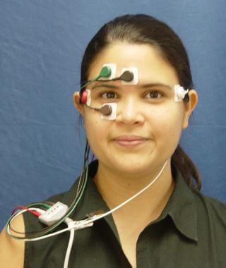
Research over the past 20 years has provided a clearer understanding of the pathogenesis of most forms of nystagmus and other ocular oscillations. To the clinician, these advances translate into greater accuracy of nystagmus as a diagnostic sign. However, to capitalise on these advances, it is important to systematically examine eye movements and interpret the findings with reference to pathophysiology. In this review we describe a scheme for examining the patient with nystagmus and interpreting common ocular oscillations; some examples are provided as video clips. (+info)Electronystagmography (ENG) is a medical test used to assess the function of the vestibular system, which is responsible for maintaining balance and eye movements. This test measures involuntary eye movements, called nystagmus, which can be indicative of various conditions affecting the inner ear or brainstem.
During the ENG test, electrodes are placed around the eyes to record eye movements while the patient undergoes a series of stimuli, such as changes in head position, visual stimuli, and caloric irrigations (where warm or cool water is introduced into the ear canal to stimulate the inner ear). The recorded data is then analyzed to evaluate the function of the vestibular system and identify any abnormalities.
ENG testing can help diagnose conditions such as vestibular neuritis, labyrinthitis, benign paroxysmal positional vertigo (BPPV), Meniere's disease, and other balance disorders. It is also used to assess the effectiveness of various treatments for these conditions.
Eye manifestations refer to any changes or abnormalities in the eye that can be observed or detected. These manifestations can be related to various medical conditions, diseases, or disorders affecting the eye or other parts of the body. They can include structural changes, such as swelling or bulging of the eye, as well as functional changes, such as impaired vision or sensitivity to light. Examples of eye manifestations include cataracts, glaucoma, diabetic retinopathy, macular degeneration, and uveitis.
Pathological nystagmus is an abnormal, involuntary movement of the eyes that can occur in various directions (horizontal, vertical, or rotatory) and can be rhythmical or arrhythmic. It is typically a result of a disturbance in the vestibular system, central nervous system, or ocular motor pathways. Pathological nystagmus can cause visual symptoms such as blurred vision, difficulty with fixation, and oscillopsia (the sensation that one's surroundings are moving). The type, direction, and intensity of the nystagmus may vary depending on the underlying cause, which can include conditions such as brainstem or cerebellar lesions, multiple sclerosis, drug toxicity, inner ear disorders, and congenital abnormalities.
Vestibular function tests are a series of diagnostic assessments used to determine the functionality and health of the vestibular system, which is responsible for maintaining balance and spatial orientation. These tests typically include:
1. **Caloric Testing:** This test evaluates the response of each ear to stimulation with warm and cold water or air. The resulting responses are recorded and analyzed to assess the function of the horizontal semicircular canals and the vestibular-ocular reflex (VOR).
2. **Rotary Chair Testing:** This test measures how well the vestibular system adapts to different speeds of rotation. The patient sits in a chair that moves in a controlled, consistent manner while their eye movements are recorded.
3. **Videonystagmography (VNG):** This test uses video goggles to record eye movements in response to various stimuli, such as changes in head position, temperature, and visual environment.
4. **Electronystagmography (ENG):** Similar to VNG, this test records eye movements but uses electrodes placed near the eyes instead of video goggles.
5. **Dix-Hallpike Test:** This is a clinical maneuver used to diagnose benign paroxysmal positional vertigo (BPPV). It involves rapidly moving the patient's head from an upright position to a position where their head is hanging off the end of the examination table.
6. **Head Shaking Test:** This test involves shaking the head back and forth for 15-20 seconds and then observing the patient's eye movements for nystagmus (involuntary eye movement).
These tests help diagnose various vestibular disorders, including benign paroxysmal positional vertigo, labyrinthitis, vestibular neuritis, Meniere's disease, and other balance disorders.
Caloric tests are a type of diagnostic test used in otology and neurotology to evaluate the function of the vestibular system, which is responsible for maintaining balance and eye movements. The tests involve stimulating the vestibular system with warm or cool air or water, and then observing and measuring the resulting eye movements.
During the test, the patient sits in a chair with their head tilted back at a 30-degree angle. A special goggles device is placed over their eyes to measure and record eye movements. Then, warm or cool air or water is introduced into each ear canal, alternately, for about 20-30 seconds.
The stimulation of the inner ear with warm or cold temperatures creates a difference in temperature between the inner ear and the brain, which activates the vestibular system and causes eye movements called nystagmus. The direction and intensity of the nystagmus are then analyzed to determine if there is any damage or dysfunction in the vestibular system.
Caloric tests can help identify lesions in the vestibular system, such as vestibular neuritis or labyrinthitis, and can also help differentiate between peripheral and central vestibular disorders.
Vestibular diseases are a group of disorders that affect the vestibular system, which is responsible for maintaining balance and spatial orientation. The vestibular system includes the inner ear and parts of the brain that process sensory information related to movement and position.
These diseases can cause symptoms such as vertigo (a spinning sensation), dizziness, imbalance, nausea, and visual disturbances. Examples of vestibular diseases include:
1. Benign paroxysmal positional vertigo (BPPV): a condition in which small crystals in the inner ear become dislodged and cause brief episodes of vertigo triggered by changes in head position.
2. Labyrinthitis: an inner ear infection that can cause sudden onset of vertigo, hearing loss, and tinnitus (ringing in the ears).
3. Vestibular neuronitis: inflammation of the vestibular nerve that causes severe vertigo, nausea, and imbalance but typically spares hearing.
4. Meniere's disease: a disorder characterized by recurrent episodes of vertigo, tinnitus, hearing loss, and a feeling of fullness in the affected ear.
5. Vestibular migraine: a type of migraine that includes vestibular symptoms such as dizziness, imbalance, and disorientation.
6. Superior canal dehiscence syndrome: a condition in which there is a thinning or absence of bone over the superior semicircular canal in the inner ear, leading to vertigo, sound- or pressure-induced dizziness, and hearing loss.
7. Bilateral vestibular hypofunction: reduced function of both vestibular systems, causing chronic imbalance, unsteadiness, and visual disturbances.
Treatment for vestibular diseases varies depending on the specific diagnosis but may include medication, physical therapy, surgery, or a combination of these approaches.
Vertigo is a specific type of dizziness characterized by the sensation that you or your surroundings are spinning or moving, even when you're perfectly still. It's often caused by issues with the inner ear or the balance-sensing systems of the body. Vertigo can be brought on by various conditions, such as benign paroxysmal positional vertigo (BPPV), labyrinthitis, vestibular neuritis, Meniere's disease, and migraines. In some cases, vertigo may also result from head or neck injuries, brain disorders like stroke or tumors, or certain medications. Treatment for vertigo depends on the underlying cause and can include specific exercises, medication, or surgery in severe cases.
Dizziness is a term used to describe a range of sensations, such as feeling lightheaded, faint, unsteady, or a false sense of spinning or moving. Medically, dizziness is often described as a non-specific symptom that can be caused by various underlying conditions or factors. These may include:
1. Inner ear disorders (such as benign paroxysmal positional vertigo, labyrinthitis, vestibular neuronitis, or Meniere's disease)
2. Cardiovascular problems (like low blood pressure, arrhythmias, or orthostatic hypotension)
3. Neurological issues (such as migraines, multiple sclerosis, or stroke)
4. Anxiety disorders and panic attacks
5. Side effects of medications
6. Dehydration or overheating
7. Infections (like viral infections or bacterial meningitis)
8. Head or neck injuries
9. Low blood sugar levels (hypoglycemia)
It is essential to consult a healthcare professional if you experience persistent dizziness, as it can be a sign of a more severe underlying condition. The appropriate treatment will depend on the specific cause of the dizziness.
 Electronystagmography
Milind Vasant Kirtane
Nystagmus
Tinnitus
Caloric reflex test
Audiometry
Godfrey Edward Arnold
Cerebellopontine angle syndrome
Ivo Padovan
Lars Leksell
Posturography
Vertigo
Cranio-corpography
Medical procedure
Non-invasive procedure
List of medical tests
Audiology
Electrography (disambiguation)
List of MeSH codes (E01)
ENG
Electronystagmography - Wikipedia
 Electronystagmography: MedlinePlus Medical Encyclopedia
 Detection of ophthalmic impairments indirectly with electronystagmography. | Division of Vestibular Sciences
fourwhitepaws: Electronystagmography
 Electronystagmography (ENG) Service, Cost in Sheikhupura, Pakistan | oladoc.com
 Labyrinthitis | Lima Memorial Health System
 Recognition and management of horizontal canal benign positional vertigo
 Publications | Plural Publishing
 Online CEUs, Job listings, and Journal for the Audiology Profession | AudiologyOnline
 Audiology
 Ear, Nose, and Throat | Cigna
 March 2007 - ENTtoday
Mutations of ESPN cause autosomal recessive deafness and vestibular dysfunction | Journal of Medical Genetics
HIE Multimedia - Cholesteatoma
 Banner Desert Medical Center Ear Nose Throat Services | Banner Health
 Barnes-Jewish Hospital > Health Library > List...
 2020 Audiology Certification Standards
Career Center
 Acoustic Neuroma: Symptoms and Causes | Tampa General Hospital
 Part I: Vestibular Dysfunction in Children: Incidence, Diagnoses, Assessment and Intervention - Vestibular Disorders Association
 Vestibular disorders in migrainous children and adolescents | The Journal of Headache and Pain | Full Text
 WTS database | WHO FCTC
 Dizziness | Multimedia Encyclopedia | Health Information | St. Luke's Hospital
 Ears
Ears
Migraine-Associated Vertigo: Overview, Epidemiology, IHS Migraine Classification
 Salus University - Program Spotlight: Understanding a Career in Audiology
Physiotherapy in Chestermere, Airdrie, Calgary locations: Saddleridge, Abbeydale, Macleod, Legacy, Royal Oak, Savanna for...
Videonystagmography3
- Electronystagmography (ENG) and videonystagmography (VNG) are eye movement recording methods used for the evaluation of balance disorders. (nih.gov)
- Electronystagmography versus videonystagmography in diagnosis of vertigo. (nih.gov)
- Electronystagmography (ENG) and videonystagmography (VNG) tests record your eye movements, as your vision system plays a large role in balance. (centamedical.com)
Vertigo4
- Electronystagmography (ENG) is a study used to clinically evaluate patients with dizziness , vertigo , or balance dysfunction. (medscape.com)
- Worldwide cases of ear damage, vertigo and dizziness have boosted the need for electronystagmography testing. (bccresearch.com)
- The vertigo application segment is the largest segment of the electronystagmography testing market, driven by the increasing prevalence of vertigo and dizziness-related disorders. (buffalonynews.net)
- Materials and Methods: one thousand three hundred fourty three patients in Mexico,City with vertigo using craneocorporgraphy and computerized electronystagmography and ultrasonography doppler of head and neck. (neurootology.org)
Disorders3
- However, the lack of awareness about the ear and balance disorders, and electronystagmography tests, may hamper the market growth. (bccresearch.com)
- The global electronystagmography testing market is growing steadily, driven by the increasing prevalence of balance and hearing disorders and the need for accurate diagnostic tools. (buffalonynews.net)
- Overall, the electronystagmography testing market is expected to grow in the coming years, driven by the increasing prevalence of balance and hearing disorders and the need for accurate diagnostic tools. (buffalonynews.net)
Posturography1
- Depending on your symptoms and overall health, we may recommend a battery of balance tests including a hearing test, Electronystagmography (ENG) tests, and posturography. (totalaudiology.com)
Audiogram2
- An audiogram or electronystagmography test is mainly done to differentiate the cause of symptoms. (hpathy.com)
- Once the symptoms appear, a thorough ear examination and hearing and balance testing (audiogram, electronystagmography, auditory brainstem responses) are essential for proper diagnosis. (nih.gov)
Movements6
- Electronystagmography (ENG) is a diagnostic test to record involuntary movements of the eye caused by a condition known as nystagmus. (wikipedia.org)
- Electronystagmography is used to assess voluntary and involuntary eye movements. (wikipedia.org)
- Electronystagmography is a test that looks at eye movements to see how well nerves in the brain are working. (medlineplus.gov)
- Electronystagmography is very useful because it can record movements behind closed eyelids or with the head in many positions. (medlineplus.gov)
- Electronystagmography (ENG) - Consists of a battery of tests that assess inner ear and brain pathways involved in controlling eye movements. (uclahealth.org)
- Electronystagmography (ENG) measures the same eye movements as VNG, but instead of using a camera, the movements are measured with electrodes placed on the skin surrounding the eyes. (georgeresnickdc.com)
Caloric test2
- There are several types of Electronystagmography Test is available such as - caloric test, oculomotor test, positional test, pendulum tracking test, and water caloric test. (bccresearch.com)
- Caloric Test, Electronystagmography (ENG) / Video Nystagmography (VNG). (bangkokpattayahospital.com)
Electrodes1
- Electronystagmography balance testing uses electrodes to test your involuntary rapid eye movement and the muscles that control eye movement to determine the cause of your balance issues. (williamsburghears.com)
Diagnostic test1
- Electronystagmography (ENG) testing is a type of diagnostic test used to evaluate and diagnose certain conditions related to the vestibular system, which controls balance and eye movement. (buffalonynews.net)
Underwent1
- Whiplash patients referred for neurootological investigation underwent the full test battery including electronystagmography for spontaneous and positional nystgmus, ocular smooth pursuit, saccades, caloric responses and visual suppression during the caloric response. (neurootology.org)
Examination2
- Mohamed A. Hamid, MD, PhD, believes, as a matter of principle, that an office vestibular examination is necessary before ordering electronystagmography (ENG)-or any other vestibular diagnostic tests, for that matter. (enttoday.org)
- Methods: ABBT results of 218 patients having normal otoneurological examination, normal hearing, and normal electronystagmography (ENG) were retrospectively analyzed to generate norms for all subtests. (haifa.ac.il)
Tests1
- Technical improvements have resulted in the refinement of eye movement analysis (computerized electronystagmography (ENG) and led to the development of tests of rotation (rotary chair test) and standing balance / posture (computerized dynamic posturegraphy). (juniperpublishers.com)
Rehabilitation1
- Facilities like Electronystagmography (ENG) and vestibular rehabilitation therapy will be provided. (drrmlims.ac.in)
Outlook1
- Region-wise and country-wise fragmentation of the Electronystagmography Testing market to grasp the revenue, and growth outlook in these areas. (buffalonynews.net)
Global4
- The latest survey on Global Electronystagmography Testing Market is conducted covering various organizations of the industry from different geographies to come up with a 100+ page report. (bccresearch.com)
- The "Global Electronystagmography Testing Market" study report will provide a valuable insight with an emphasis on global market including some of the major players such as Interacoustics, DIFRA Instrumentation, Micromedical Technologies, Otometrics, SensoMotoric Instruments, Balanceback, BioMed Jena. (bccresearch.com)
- Factors (Positive and Negative) impacting the growth of the global Electronystagmography Testing market. (buffalonynews.net)
- Tentatively, the global electronystagmography testing market can be segmented on the basis of test type, indication, end user, and geography. (buffalonynews.net)
Analysis1
- The study is a perfect mix of qualitative and quantitative information highlighting key market developments, challenges that industry and competition are facing along with gap analysis and new opportunities available and trend in the Electronystagmography Testing Market. (bccresearch.com)
Testing7
- What is Electronystagmography Testing? (bccresearch.com)
- The electronystagmography testing market is segmented by product type, application, end-user, and region. (buffalonynews.net)
- The major players in the electronystagmography testing market include Interacoustics, GN Otometrics, Medtronic, Micromedical Technologies Inc., and Neuro Kinetics, Inc. These companies are investing in research and development to introduce new and innovative electronystagmography testing solutions to the market, including devices with improved accuracy and ease of use. (buffalonynews.net)
- Regional breakdown of the Electronystagmography Testing market based on predefined taxonomy. (buffalonynews.net)
- Innovative manufacturing processes implemented by Electronystagmography Testing vendors in detail. (buffalonynews.net)
- The market survey conducted by fact.mr offers key developments and challenges affecting the pharmaceutical industry, subsequently influencing demand and sales in the Electronystagmography Testing market. (buffalonynews.net)
- We recommend that future studies should include health-related quality of life and symptom-based outcome measures, in addition to objective measures of vestibular improvement, such as caloric testing and electronystagmography. (qxmd.com)