Radioisotopes
Diagnostic Techniques and Procedures
Sensitivity and Specificity
Diagnostic Techniques, Radioisotope
Zinc Radioisotopes
Diagnostic Techniques, Otological
Diagnostic Techniques, Surgical
Radionuclide Imaging
Radioisotope Dilution Technique
Strontium Radioisotopes
Iodine Radioisotopes
Azure Stains
Diagnostic Techniques, Urological
Polymerase Chain Reaction
Krypton Radioisotopes
Diagnostic Techniques, Obstetrical and Gynecological
Diagnostic Tests, Routine
Diagnostic Techniques, Digestive System
Indium Radioisotopes
Clinical Laboratory Techniques
Diagnostic Techniques, Respiratory System
Sodium Radioisotopes
Reproducibility of Results
Radioactivity
Barium Radioisotopes
Cytodiagnosis
Evaluation Studies as Topic
Biopsy, Needle
Diagnostic Techniques, Neurological
Yttrium Radioisotopes
Tomography, X-Ray Computed
Predictive Value of Tests
Tin Radioisotopes
Carbon Radioisotopes
Iron Radioisotopes
Copper Radioisotopes
Diagnostic Techniques, Endocrine
Phosphorus Radioisotopes
Ultrasonography
Feces
Magnetic Resonance Imaging
Technetium
Biopsy
Biopsy, Fine-Needle
Microscopy
Technetium Tc 99m Sulfur Colloid
Diagnostic Techniques, Cardiovascular
Mercury Radioisotopes
Cesium Isotopes
Cerium Radioisotopes
Cobalt Isotopes
Hafnium
Gold Radioisotopes
Dog Diseases
Enzyme-Linked Immunosorbent Assay
Isotope Labeling
Lead Radioisotopes
Retrospective Studies
Zinc Isotopes
Radiopharmaceuticals
Sulfur Radioisotopes
Mycoses
Prospective Studies
Cadmium Radioisotopes
Astatine
Radioimmunotherapy
Lutetium
Samarium
Immunoenzyme Techniques
Subdural Effusion
Scintillation Counting
Bromine Radioisotopes
Calcium Isotopes
Radioactive Waste
Serum Albumin, Radio-Iodinated
Diagnostic Imaging
Biological Markers
Ruthenium Radioisotopes
Radiometric Dating
DNA Primers
Sodium Pertechnetate Tc 99m
Treatment Outcome
Selenium Radioisotopes
Prevalence
Alpha Particles
Nuclear Medicine
Heterocyclic Compounds, 1-Ring
Disease Outbreaks
Molecular Sequence Data
Pentetic Acid
Image Processing, Computer-Assisted
Fluorescent Antibody Technique
Tungsten
Isotopes
Tissue Distribution
Prognosis
Radioisotope Teletherapy
Technetium Tc 99m Pentetate
Spectrometry, Gamma
Incidence
Risk Factors
Radiometry
Dogs
United States
Rosaniline Dyes
Positron-Emission Tomography
Reagent Kits, Diagnostic
Radiation Dosage
Tritium
Follow-Up Studies
Immunohistochemistry
Nostoc commune
Potassium Radioisotopes
Iodohippuric Acid
Diagnostic Techniques, Ophthalmological
Carbon Isotopes
Organometallic Compounds
Technetium Tc 99m Medronate
Vitamin B 12
ROC Curve
Diagnostic and Statistical Manual of Mental Disorders
Sentinel Lymph Node Biopsy
Diagnostic Services
Avidin
Absorption
Autoradiography
Brachytherapy
Cesium Radioisotopes
Iridium Radioisotopes
Phosphorus Isotopes
Tomography, Emission-Computed, Single-Photon
Tomography, Emission-Computed
Octreotide
Feasibility Studies
Half-Life
Diagnosis
Mass Spectrometry
Cobalt Radioisotopes
Receptors, Somatostatin
Biological Assay
Zinc
Neoplasms
Mice, Nude
Liver
Metabolic Clearance Rate
False Positive Reactions
Erythrocytes
Kidney
Bone and Bones
Fluorescent Dyes
Nitrogen Radioisotopes
Lung
Iron
Radioisotope Renography
Diagnosis, Computer-Assisted
False Negative Reactions
Rabbits
Tumor Markers, Biological
Observer Variation
Rubidium Radioisotopes
Biological Transport
Chromium Radioisotopes
Chromatography, High Pressure Liquid
Gallium Radioisotopes
Nucleic Acid Hybridization
Calcium Radioisotopes
Immunoassay
Xenon Radioisotopes
Algorithms
Swine
DNA
Early Diagnosis
Severity of Illness Index
Sodium
Physical Examination
Radiography
Glucose
Quantification of regional ventilation-perfusion ratios with PET. (1/14)
The topographic matching of alveolar ventilation (V(A)) and perfusion (Q) is the main determinant of gas exchange efficiency of the lung. However, no pulmonary functional imaging technique has been shown to predict whole-lung gas exchange in health and disease. This study aims to present a PET-based method to estimate regional alveolar ventilation-to-perfusion ratios (V(A)/Q) predictive of arterial blood gases. METHODS: The method is based on the regional tracer kinetics of (13)N-nitrogen ((13)NN) after an intravenous bolus injection during a breath-hold period and subsequent washout from the lungs with resumption of breathing. The method takes into account the presence of inter- and intraregional nonuniformities at length scales smaller than the imaging spatial resolution. An algorithm used regional tracer washout to classify regional V(A)Q/ uniformity. Intraregional V(A)/Q mismatch in nonuniform regions was described with a 2-compartment model. Regional V(A)/Q estimates were combined into a whole-lung distribution of V(A)/Q ratios and were used to compute global arterial blood gases. The method was applied to 3-dimensional PET data from anesthetized and mechanically ventilated sheep before and after methacholine bronchoconstriction (n = 3) and pulmonary embolism (n = 3) and after saline lung lavage (n = 3). RESULTS: PET images revealed regional changes in ventilation and perfusion consistent with the different disease models. Quantification of the images using PET-derived V(A)Q/ distributions showed unimodal and narrow distributions in control conditions that became wider and unimodal after pulmonary embolism and saline lung lavage and bimodal after bronchoconstriction. Images of regional gas exchange allowed for visualization of regional gas exchange. Arterial blood gases estimated from the PET-based V(A)/Q distributions closely agreed with measured values (partial pressure of oxygen, arterial [PaO(2)]: r(2) = 0.97, P < 0.001; partial pressure of carbon dioxide, arterial [PaCO(2)]: r(2) = 0.96, P < 0.001). CONCLUSION: Tracer kinetics analysis of PET images after an intravenous injection of (13)NN provides a quantitative assessment of regional V(A)/Q heterogeneity including that corresponding to length scales smaller than the spatial resolution of the imaging method. Quantification of V(A)/Q mismatch obtained with the presented technique is directly related to severity of gas exchange impairment as determined by arterial blood gases. (+info)Potential impact of molecular imaging in oncology. (2/14)
Despite all progress in molecular imaging methods, the actual number of validated new markers is still limited. The breakthrough of individual efforts is often hampered by lack of critical mass of resources. To overcome these shortcomings a "network" of multidisciplinary experts is indispensable. Focusing on cancer therapy, molecular imaging has a high potential impact in (a) early therapy monitoring and (b) prediction of therapeutic response. Novel molecular markers with high diagnostic potential reflecting apoptosis, proliferation and glucose metabolism are currently used in clinical trials for monitoring of tumor response to therapy. A further innovative approach for supporting individualized cancer therapy may be the application of radio-labelled therapeutic drugs at tracer concentrations in order to estimate their individual uptake in the tumor tissue. To meet this challenge, our group initiated a research project focusing on radio-labelling of selected molecules with proven therapeutic potential. (+info)Planning optimal measurements of isotopomer distributions for estimation of metabolic fluxes. (3/14)
MOTIVATION: Flux estimation using isotopomer information of metabolites is currently the most reliable method to obtain quantitative estimates of the activity of metabolic pathways. However, the development of isotopomer measurement techniques for intermediate metabolites is a demanding task. Careful planning of isotopomer measurements is thus needed to maximize the available flux information while minimizing the experimental effort. RESULTS: In this paper we study the question of finding the smallest subset of metabolites to measure that ensure the same level of isotopomer information as the measurement of every metabolite in the metabolic network. We study the computational complexity of this optimization problem in the case of the so-called positional enrichment data, give methods for obtaining exact and fast approximate solutions, and evaluate empirically the efficacy of the proposed methods by analyzing a metabolic network that models the central carbon metabolism of Saccharomyces cerevisiae. (+info)Nuclear medicine in the rehabilitative treatment evaluation in stroke recovery. Role of diaschisis resolution and cerebral reorganization. (4/14)
There has recently been a tremendous increase in imaging technology and imaging methodology enabling noninvasive exploration of brain function to such an intricate degree as to enable measurements of very small spatial and short temporal cerebral operations responsible for neurological and functional recovery after stroke. This has allowed conceptualization of rehabilitation strategies designed to maximally enhance rehabilitation protocols tailored to the individual patient's deficits. Rehabilitation strategies may now be designed and optimized by employing methods to synchronize functional training of brain regions ascribed to those areas innately undergoing neuronal plasticity change responsible for stroke recovery. In order to effectively apply these noninvasive imaging methods, one must have a clear understanding of the physics and technique of the imaging methodologies and how these are best applied to understand brain physiology during the stroke recovery process to provide a solid rationale for development of rehabilitation protocols. Nuclear medicine imaging is first presented as a diagnostic method to assess the stroke process. The initial brain damage and resulting neurological disability can be primarily assessed in terms of changes in the vascular and hemodynamic status of the cerebral circulation in addition to alterations in the metabolic status around the infarction region. Techniques for assessing perfusion and metabolism include regional cerebral blood flow (rCBF), single photon emission computed tomography (SPECT), and F-18 2-Fluoro-2-deoxy-D-glucose (F-18 FDG) positron emission tomography (PET). In addition, hemodynamic vascular insufficiency can be assessed using O-15 O2 oxygen extraction PET and rest and Diamox rCBF SPECT. The status of the peri-infarction region can be characterized in terms of components of diaschisis and ischemia using proton magnetic resonance spectroscopy imaging ((1)H MRSI) and rest/stress rCBF assessment of cerebral vascular reserve. As the brain recovers from cerebral infarction, areas of reorganization and energy utilization by the brain can be measured using oxygen extraction methods with PET, F-18 FDG glucose utilization by PET, and functional magnetic resonance imaging (fMRI) measures using the blood oxygenation level dependent (BOLD) technique. In addition, high field MRI imaging of the brain is now able to provide detailed fractional anisotropy (FA) maps to characterize changes in white matter by fiber tracking mapping using diffusion tensor imaging. Imaging of the stroke recovery process focuses on the physiologic model of stroke characterized by rCBF, metabolism, 1H spectroscopic measures of N-acetyl aspartate (NAA), choline (Ch) and creatine (Cr) in the peri-infarction zone as well as in the extended stroke penumbra including areas of distant ''pure'' diaschisis unencumbered with the confound of cerebral ischemia. Data is presented describing the results of application of imaging methodologies as the patient undergoes rehabilitation that demonstrates the importance of blood flow and metabolic changes in the contralesional frontal lobe both during the resting state and during motor and speech activation paradigms. The results of advanced imaging technologies on cerebral damage and cerebral reorganization during rehabilitation are presented in the context of furthering designs of rehabilitation strategies. Success can be monitored to assess the optimization of rehabilitation strategy design to maximize neurological recovery from stroke by employing facilitatory methods to maximally synchronize rehabilitation techniques with recovery of functionally counterpart areas of viable brain. (+info)PET imaging of CCND1 mRNA in human MCF7 estrogen receptor positive breast cancer xenografts with oncogene-specific [64Cu]chelator-peptide nucleic acid-IGF1 analog radiohybridization probes. (5/14)
Treatment of breast cancer is hampered by a large unmet need for rapid, sensitive, specific staging and stratification of palpable and nonpalpable abnormalities. Mammography and physical examination miss many early breast cancers, yet detect many benign lesions. Cyclin D1, encoded by CCND1 messenger RNA (mRNA), and insulin-like growth factor 1 receptor (IGF1R) are key regulators of cell proliferation that are overexpressed in most breast cancers. Therefore, we hypothesized that malignant breast masses could be imaged and quantitated externally by PET with a dual-specificity probe that targets both CCND1 mRNA and IGF1R. METHODS: We designed a CCND1-specific peptide nucleic acid (PNA) hybridization sequence (CTGGTGTTCCAT), separated by a C-terminal spacer to a cyclized IGF1 peptide analog (d-Cys-Ser-Lys-Cys), for IGF1R-mediated endocytosis. On the N-terminus we attached a chelator (1,4,7-tris(carboxymethylaza)cyclododecane-10-azaacetyl [DO3A]) for the positron-emitting nuclide (64)Cu. We administered the [(64)Cu]CCND1-IGF1 analog radiohybridization probes, as well as sequence controls, by tail vein to immunocompromised female NCr mice bearing human MCF7 estrogen-dependent, receptor-positive xenografts. We imaged the mice by PET and CT 4 and 24 h later, and measured tissue distribution of the radiohybridization probes. RESULTS: We observed 8 +/- 2-fold higher PET intensity in the center of the breast cancer xenografts than in the contralateral tissues at 24 h after injection of the [(64)Cu]CCND1-IGF1 analog radiohybridization probe. IGF1 blocking yielded significantly weaker images (P < 0.05) relative to the tumor-free side at 24 h after injection, as did a PNA mismatch probe, a peptide mismatch probe, and free (64)CuCl(2). CONCLUSION: These results are consistent with our hypothesis for radiohybridization PET of overexpressed CCND1 mRNA, dependent on IGF1R-mediated endocytosis, in suspect masses. Early noninvasive detection of initial cancerous transformation, as well as invasive or recurrent breast cancer, with dual-specificity radiohybridization probes, might enable molecularly targeted staging, stratification, and choice of therapy. (+info)Clinical efficacy and problems with CT lymphography in identifying the sentinel node in breast cancer. (6/14)
(+info)Functional immunoassay technology (FIT), a new approach for measuring physiological functions: application of FIT to measure glomerular filtration rate (GFR). (7/14)
(+info)Choline autoradiography of human prostate cancer xenograft: effect of castration. (8/14)
The purpose of this study was to investigate the effects of castration and tracer uptake time interval on the level of radiolabeled choline accumulation in murine-implanted human prostate tumor xenografts using quantitative autoradiography. We implanted androgen-dependent (CWR22) and androgen-independent (PC3) human prostate cancer cells in castrated (n = 9) and noncastrated (n = 9) athymic male mice and allowed tumors to grow to 1 cm3. The mice were euthanized at 5, 10, and 20 minutes after injection of 5 microCi [14C]-choline. Mice were prepared for quantitative autoradiography with density light units of viable tumor sections converted to units of radioactivity (nCi/mm2) using calibration. Two-group comparisons were performed using a two-tailed Student t-test with unequal variance and with a significance probability level of less than .05. Two-group comparisons between the means of the tracer uptake level for each tumor type at each of three time points for each of two host types showed that (1) the level of tracer localization in the two tumor types was affected little in relation to the host type and (2) PC3 tumor uptake level tended to increase slowly with time only in the noncastrated host, whereas this was not observed in the castrated host or with CWR22 tumor in either host type. The uptake time interval and castration do not appear to significantly affect the level of radiolabeled choline uptake by the human prostate cancer xenograft. (+info)Radioisotopes, also known as radioactive isotopes or radionuclides, are variants of chemical elements that have unstable nuclei and emit radiation in the form of alpha particles, beta particles, gamma rays, or conversion electrons. These isotopes are formed when an element's nucleus undergoes natural or artificial radioactive decay.
Radioisotopes can be produced through various processes, including nuclear fission, nuclear fusion, and particle bombardment in a cyclotron or other types of particle accelerators. They have a wide range of applications in medicine, industry, agriculture, research, and energy production. In the medical field, radioisotopes are used for diagnostic imaging, radiation therapy, and in the labeling of molecules for research purposes.
It is important to note that handling and using radioisotopes requires proper training, safety measures, and regulatory compliance due to their ionizing radiation properties, which can pose potential health risks if not handled correctly.
Diagnostic techniques and procedures are methods used by medical professionals to identify the cause of symptoms, illnesses, or diseases. These can include physical examinations, patient interviews, review of medical history, and various diagnostic tests. Diagnostic tests may involve invasive procedures such as biopsies or surgical interventions, or non-invasive imaging techniques like X-rays, CT scans, MRI scans, or ultrasounds. Functional tests, such as stress testing or electroencephalogram (EEG), can also be used to evaluate the functioning of specific organs or systems in the body. Laboratory tests, including blood tests, urine tests, and genetic tests, are also common diagnostic procedures. The choice of diagnostic technique or procedure depends on the presenting symptoms, the patient's medical history, and the suspected underlying condition.
Sensitivity and specificity are statistical measures used to describe the performance of a diagnostic test or screening tool in identifying true positive and true negative results.
* Sensitivity refers to the proportion of people who have a particular condition (true positives) who are correctly identified by the test. It is also known as the "true positive rate" or "recall." A highly sensitive test will identify most or all of the people with the condition, but may also produce more false positives.
* Specificity refers to the proportion of people who do not have a particular condition (true negatives) who are correctly identified by the test. It is also known as the "true negative rate." A highly specific test will identify most or all of the people without the condition, but may also produce more false negatives.
In medical testing, both sensitivity and specificity are important considerations when evaluating a diagnostic test. High sensitivity is desirable for screening tests that aim to identify as many cases of a condition as possible, while high specificity is desirable for confirmatory tests that aim to rule out the condition in people who do not have it.
It's worth noting that sensitivity and specificity are often influenced by factors such as the prevalence of the condition in the population being tested, the threshold used to define a positive result, and the reliability and validity of the test itself. Therefore, it's important to consider these factors when interpreting the results of a diagnostic test.
Diagnostic techniques using radioisotopes, also known as nuclear medicine, are medical diagnostic procedures that use small amounts of radioactive material, called radioisotopes or radionuclides, to diagnose and monitor various diseases and conditions. The radioisotopes are introduced into the body through different routes (such as injection, inhalation, or ingestion) and accumulate in specific organs or tissues.
The gamma rays or photons emitted by these radioisotopes are then detected by specialized imaging devices, such as gamma cameras or PET scanners, which generate images that provide information about the structure and function of the organ or tissue being examined. This information helps healthcare professionals to make accurate diagnoses, monitor disease progression, assess treatment response, and plan appropriate therapies.
Common diagnostic techniques using radioisotopes include:
1. Radionuclide imaging (also known as scintigraphy): A gamma camera is used to produce images of specific organs or tissues after the administration of a radioisotope. Examples include bone scans, lung scans, heart scans, and brain scans.
2. Positron emission tomography (PET) scans: A PET scanner detects pairs of gamma rays emitted indirectly by a positron-emitting radionuclide, such as fluorodeoxyglucose (FDG), which is often used in oncology to assess metabolic activity and identify cancerous lesions.
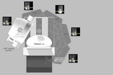
3. Single-photon emission computed tomography (SPECT): A specialized gamma camera rotates around the patient, acquiring multiple images from different angles that are then reconstructed into a 3D image, providing detailed information about organ function and structure.
Diagnostic techniques using radioisotopes offer several advantages, including high sensitivity, non-invasiveness, and the ability to assess both anatomical and functional aspects of organs and tissues. However, they also involve exposure to ionizing radiation, so their use should be balanced against potential risks and benefits, and alternative diagnostic methods should be considered when appropriate.
Zinc radioisotopes are unstable isotopes or variants of the element zinc that undergo radioactive decay, emitting radiation in the process. These isotopes have a different number of neutrons than the stable isotope of zinc (zinc-64), which contributes to their instability and tendency to decay.
Examples of zinc radioisotopes include zinc-65, zinc-70, and zinc-72. These isotopes are often used in medical research and diagnostic procedures due to their ability to emit gamma rays or positrons, which can be detected using specialized equipment.
Zinc radioisotopes may be used as tracers to study the metabolism and distribution of zinc in the body, or as therapeutic agents to deliver targeted radiation therapy to cancer cells. However, it is important to note that the use of radioisotopes carries potential risks, including exposure to ionizing radiation and the potential for damage to healthy tissues.
Molecular diagnostic techniques are a group of laboratory methods used to analyze biological markers in DNA, RNA, and proteins to identify specific health conditions or diseases at the molecular level. These techniques include various methods such as polymerase chain reaction (PCR), DNA sequencing, gene expression analysis, fluorescence in situ hybridization (FISH), and mass spectrometry.
Molecular diagnostic techniques are used to detect genetic mutations, chromosomal abnormalities, viral and bacterial infections, and other molecular changes associated with various diseases, including cancer, genetic disorders, infectious diseases, and neurological disorders. These techniques provide valuable information for disease diagnosis, prognosis, treatment planning, and monitoring of treatment response.
Compared to traditional diagnostic methods, molecular diagnostic techniques offer several advantages, such as higher sensitivity, specificity, and speed. They can detect small amounts of genetic material or proteins, even in early stages of the disease, and provide accurate results with a lower risk of false positives or negatives. Additionally, molecular diagnostic techniques can be automated, standardized, and performed in high-throughput formats, making them suitable for large-scale screening and research applications.
Diagnostic techniques in otology refer to the methods and tests used by healthcare professionals to identify and diagnose various conditions related to the ear. These techniques can include:
1. Otoscopy: A visual examination of the external auditory canal and eardrum using an otoscope. This helps to identify any physical abnormalities, such as wax buildup, inflammation, or foreign objects in the ear.
2. Audiometry: A hearing test that measures a person's ability to hear different sounds, pitches, and volumes. This can help to identify any hearing loss or auditory processing issues.
3. Tympanometry: A test that measures the function of the middle ear by creating variations in air pressure in the ear canal. This can help to identify any issues with the eardrum or middle ear bones.
4. Acoustic reflex testing: A test that measures the body's involuntary response to loud sounds. This can help to identify any damage to the hearing nerves or brainstem.
5. Otoacoustic emissions (OAE) testing: A test that measures the sound waves produced by the inner ear in response to stimuli. This can help to identify any issues with the cochlea or hair cells in the inner ear.
6. Auditory brainstem response (ABR) testing: A test that measures the electrical activity of the hearing nerve and brainstem in response to sound. This can help to identify any issues with the auditory nervous system.
7. Vestibular testing: A series of tests that measure a person's balance and equilibrium. This can help to identify any issues with the vestibular system, which is responsible for maintaining balance.
These diagnostic techniques are used to diagnose various otological conditions such as hearing loss, tinnitus, vertigo, ear infections, and tumors of the ear.
Diagnostic techniques, surgical refers to the use of surgical procedures or methods to diagnose and evaluate various medical conditions. These techniques are often used when non-invasive tests are inconclusive or when more detailed information is required. Here are some examples:
1. Biopsy: A small sample of tissue is removed from the body for examination under a microscope. This can help to confirm a diagnosis of cancer, infection, or other diseases.
2. Endoscopy: A flexible tube with a light and camera on the end is inserted into the body through a natural opening (such as the mouth or anus) or a small incision. This allows the doctor to visualize internal organs and tissues, and may also involve taking biopsy samples.
3. Imaging studies: Various imaging techniques such as X-rays, CT scans, MRI scans, and ultrasound can be used to produce detailed images of internal structures. These can help to diagnose a wide range of medical conditions, from broken bones to tumors.
4. Exploratory surgery: In some cases, a surgical incision may be made to directly visualize and examine an organ or tissue. This can help to diagnose conditions that are difficult to detect with non-invasive tests.
5. Functional testing: Some surgical techniques involve stimulating or measuring the function of an organ or system. For example, a cardiac stress test may be performed during surgery to assess heart function.
Overall, diagnostic techniques, surgical play an important role in the diagnosis and management of many medical conditions. They can provide valuable information that helps doctors to make informed decisions about treatment options and improve patient outcomes.
Radionuclide imaging, also known as nuclear medicine, is a medical imaging technique that uses small amounts of radioactive material, called radionuclides or radiopharmaceuticals, to diagnose and treat various diseases and conditions. The radionuclides are introduced into the body through injection, inhalation, or ingestion and accumulate in specific organs or tissues. A special camera then detects the gamma rays emitted by these radionuclides and converts them into images that provide information about the structure and function of the organ or tissue being studied.
Radionuclide imaging can be used to evaluate a wide range of medical conditions, including heart disease, cancer, neurological disorders, gastrointestinal disorders, and bone diseases. The technique is non-invasive and generally safe, with minimal exposure to radiation. However, it should only be performed by qualified healthcare professionals in accordance with established guidelines and regulations.
The Radioisotope Dilution Technique is a method used in nuclear medicine to measure the volume and flow rate of a particular fluid in the body. It involves introducing a known amount of a radioactive isotope, or radioisotope, into the fluid, such as blood. The isotope mixes with the fluid, and samples are then taken from the fluid at various time points.
By measuring the concentration of the radioisotope in each sample, it is possible to calculate the total volume of the fluid based on the amount of the isotope introduced and the dilution factor. The flow rate can also be calculated by measuring the concentration of the isotope over time and using the formula:
Flow rate = Volume/Time
This technique is commonly used in medical research and clinical settings to measure cardiac output, cerebral blood flow, and renal function, among other applications. It is a safe and reliable method that has been widely used for many years. However, it does require the use of radioactive materials and specialized equipment, so it should only be performed by trained medical professionals in appropriate facilities.
Strontium radioisotopes are radioactive isotopes of the element strontium. Strontium is an alkaline earth metal that is found in nature and has several isotopes, some of which are stable and some of which are radioactive. The radioactive isotopes of strontium, also known as strontium radionuclides, decay and emit radiation in the form of beta particles.
Strontium-89 (^89Sr) and strontium-90 (^90Sr) are two common radioisotopes of strontium that are used in medical applications. Strontium-89 is a pure beta emitter with a half-life of 50.5 days, which makes it useful for the treatment of bone pain associated with metastatic cancer. When administered, strontium-89 is taken up by bones and irradiates the bone tissue, reducing pain and improving quality of life in some patients.
Strontium-90, on the other hand, has a longer half-life of 28.8 years and emits more powerful beta particles than strontium-89. It is used as a component in radioactive waste and in some nuclear weapons, but it is not used in medical applications due to its long half-life and high radiation dose.
It's important to note that exposure to strontium radioisotopes can be harmful to human health, especially if ingested or inhaled. Therefore, handling and disposal of strontium radioisotopes require special precautions and regulations.
Iodine radioisotopes are radioactive isotopes of the element iodine, which decays and emits radiation in the form of gamma rays. Some commonly used iodine radioisotopes include I-123, I-125, I-131. These radioisotopes have various medical applications such as in diagnostic imaging, therapy for thyroid disorders, and cancer treatment.
For example, I-131 is commonly used to treat hyperthyroidism and differentiated thyroid cancer due to its ability to destroy thyroid tissue. On the other hand, I-123 is often used in nuclear medicine scans of the thyroid gland because it emits gamma rays that can be detected by a gamma camera, allowing for detailed images of the gland's structure and function.
It is important to note that handling and administering radioisotopes require specialized training and safety precautions due to their radiation-emitting properties.
'Azure stains' is a term used in pathology to describe a histological staining technique that uses a type of dye called methyl blue, which turns the stained structures a blue-purple color. This technique is often used to stain acid mucins, which are found in various types of tissues and can be indicative of certain medical conditions.
In particular, azure stains are sometimes used to help diagnose certain types of cancer, such as mucoepidermoid carcinoma, a type of salivary gland tumor that produces acid mucins. The staining technique can help pathologists identify the presence and distribution of these mucins within the tumor cells, which can aid in making an accurate diagnosis and determining the best course of treatment.
It's worth noting that there are several different types of histological stains that use various dyes to highlight different structures or features within tissues. Azure stains are just one example of these techniques, and they are typically used in conjunction with other staining methods to provide a comprehensive picture of the tissue being examined.
Diagnostic techniques in urology are methods used to identify and diagnose various urological conditions affecting the urinary tract and male reproductive system. These techniques include:
1. Urinalysis: A laboratory examination of a urine sample to detect abnormalities such as infection, kidney stones, or other underlying medical conditions.
2. Urine Culture: A test used to identify and grow bacteria from the urine to determine the type of bacterial infection present in the urinary tract.
3. Imaging Studies: Various imaging techniques such as X-rays, ultrasound, CT scans, and MRI scans are used to visualize the internal structures of the urinary tract and identify any abnormalities.
4. Cystoscopy: A procedure that involves inserting a thin tube with a camera into the bladder through the urethra to examine the bladder and urethra for signs of disease or abnormality.
5. Urodynamics: A series of tests used to evaluate bladder function, including measuring bladder pressure and urine flow rate.
6. Biopsy: The removal and examination of tissue from the urinary tract or male reproductive system to diagnose conditions such as cancer.
7. Prostate-Specific Antigen (PSA) Test: A blood test used to screen for prostate cancer by measuring the level of PSA, a protein produced by the prostate gland.
8. Voiding Diary: A record of urinary habits, including the frequency and volume of urination, that can help diagnose conditions such as overactive bladder or urinary incontinence.
Polymerase Chain Reaction (PCR) is a laboratory technique used to amplify specific regions of DNA. It enables the production of thousands to millions of copies of a particular DNA sequence in a rapid and efficient manner, making it an essential tool in various fields such as molecular biology, medical diagnostics, forensic science, and research.
The PCR process involves repeated cycles of heating and cooling to separate the DNA strands, allow primers (short sequences of single-stranded DNA) to attach to the target regions, and extend these primers using an enzyme called Taq polymerase, resulting in the exponential amplification of the desired DNA segment.
In a medical context, PCR is often used for detecting and quantifying specific pathogens (viruses, bacteria, fungi, or parasites) in clinical samples, identifying genetic mutations or polymorphisms associated with diseases, monitoring disease progression, and evaluating treatment effectiveness.
Krypton is a noble gas with the symbol Kr and atomic number 36. It exists in various radioisotopes, which are unstable isotopes of krypton that undergo radioactive decay. A few examples include:
1. Krypton-81: This radioisotope has a half-life of about 2.1 x 10^5 years and decays via electron capture to rubidium-81. It is produced naturally in the atmosphere by cosmic rays.
2. Krypton-83: With a half-life of approximately 85.7 days, this radioisotope decays via beta decay to bromine-83. It can be used in medical imaging for lung ventilation studies.
3. Krypton-85: This radioisotope has a half-life of about 10.7 years and decays via beta decay to rubidium-85. It is produced as a byproduct of nuclear fission and can be found in trace amounts in the atmosphere.
4. Krypton-87: With a half-life of approximately 76.3 minutes, this radioisotope decays via beta decay to rubidium-87. It is not found naturally on Earth but can be produced artificially.
It's important to note that while krypton radioisotopes have medical applications, they are also associated with potential health risks due to their radioactivity. Proper handling and safety precautions must be taken when working with these substances.
Diagnostic techniques in obstetrics and gynecology refer to the various methods used by healthcare professionals to diagnose and monitor conditions related to the female reproductive system and pregnancy. Here are some commonly used diagnostic techniques:
1. Physical examination: A thorough physical exam, including a pelvic exam, can help identify any abnormalities in the reproductive organs.
2. Medical history: A detailed medical history, including information about menstrual cycles, sexual activity, and family health, can provide valuable clues to diagnose various conditions.
3. Imaging tests: Ultrasound, CT scans, and MRIs can help healthcare professionals visualize the reproductive organs and detect any abnormalities.
4. Laboratory tests: Blood tests, urine tests, and cultures can help identify infections, hormonal imbalances, and other conditions.
5. Biopsy: A small sample of tissue is taken from the affected area and examined under a microscope to diagnose conditions such as cancer.
6. Colposcopy: This procedure involves using a special magnifying device to examine the cervix and vagina for signs of abnormalities.
7. Hysterosalpingography: This is an X-ray procedure that involves injecting a dye into the uterus and fallopian tubes to detect any blockages or other abnormalities.
8. Sonohysterography: This is an ultrasound procedure that involves injecting a fluid into the uterus to help visualize its interior and detect any abnormalities.
9. Minimally invasive surgery: Procedures such as laparoscopy and hysteroscopy can help healthcare professionals diagnose and treat various conditions related to the reproductive organs.
These diagnostic techniques can help healthcare professionals identify and manage a wide range of conditions, including infertility, pregnancy complications, infections, hormonal imbalances, and cancer.
'Diagnostic tests, routine' is a medical term that refers to standard or commonly used tests that are performed to help diagnose, monitor, or manage a patient's health condition. These tests are typically simple, non-invasive, and safe, and they may be ordered as part of a regular check-up or when a patient presents with specific symptoms.
Routine diagnostic tests may include:
1. Complete Blood Count (CBC): A test that measures the number of red and white blood cells, platelets, and hemoglobin in the blood. It can help diagnose conditions such as anemia, infection, and inflammation.
2. Urinalysis: A test that examines a urine sample for signs of infection, kidney disease, or other medical conditions.
3. Blood Chemistry Tests: Also known as a chemistry panel or comprehensive metabolic panel, this test measures various chemicals in the blood such as glucose, electrolytes, and enzymes to evaluate organ function and overall health.
4. Electrocardiogram (ECG): A test that records the electrical activity of the heart, which can help diagnose heart conditions such as arrhythmias or heart attacks.
5. Chest X-ray: An imaging test that creates pictures of the structures inside the chest, including the heart, lungs, and bones, to help diagnose conditions such as pneumonia or lung cancer.
6. Fecal Occult Blood Test (FOBT): A test that checks for hidden blood in the stool, which can be a sign of colon cancer or other gastrointestinal conditions.
7. Pap Smear: A test that collects cells from the cervix to check for abnormalities that may indicate cervical cancer or other gynecological conditions.
These are just a few examples of routine diagnostic tests that healthcare providers may order. The specific tests ordered will depend on the patient's age, sex, medical history, and current symptoms.
Diagnostic techniques for the digestive system are medical tests and procedures used to diagnose and evaluate various conditions and diseases related to the gastrointestinal (GI) tract, including the esophagus, stomach, small intestine, large intestine, liver, gallbladder, pancreas, and associated organs. These techniques can be categorized into invasive and non-invasive methods.
Non-invasive diagnostic techniques:
1. Imaging tests: These include X-rays, computed tomography (CT) scans, magnetic resonance imaging (MRI), positron emission tomography (PET) scans, and ultrasounds. They help visualize the structure and function of the digestive organs without requiring any invasive procedures.
2. Laboratory tests: Blood, stool, and urine samples can be analyzed to detect signs of infection, inflammation, or other abnormalities related to digestive system disorders. Examples include complete blood count (CBC), liver function tests (LFTs), coagulation studies, and fecal occult blood test (FOBT).
3. Breath tests: These are used to diagnose conditions like lactose intolerance, small intestinal bacterial overgrowth (SIBO), or helicobacter pylori infection by analyzing the patient's exhaled air after consuming a specific substance.
Invasive diagnostic techniques:
1. Endoscopy: A thin, flexible tube with a light and camera attached to its end is inserted through the mouth or rectum to directly visualize the GI tract's inner lining. There are different types of endoscopies, such as gastroscopy (esophagus, stomach, and duodenum), colonoscopy (colon and rectum), sigmoidoscopy (lower part of the colon), and enteroscopy (small intestine).
2. Endoscopic ultrasound (EUS): This combines endoscopy with ultrasound technology to provide detailed images of the digestive organs' structure and surrounding tissues, allowing for accurate diagnosis and staging of conditions like cancer.
3. Biopsy: During an endoscopy or surgery, a small tissue sample can be taken from the affected area for further examination under a microscope to confirm a diagnosis or assess the severity of a condition.
4. Capsule endoscopy: A patient swallows a tiny camera-equipped capsule that transmits images as it passes through the GI tract, allowing doctors to diagnose conditions in the small intestine that may be difficult to reach with traditional endoscopes.
5. Imaging studies: Procedures like computed tomography (CT), magnetic resonance imaging (MRI), or positron emission tomography (PET) scans can provide detailed images of the digestive organs and help diagnose conditions like tumors, inflammation, or obstructions.
These diagnostic techniques help healthcare providers identify and manage various gastrointestinal conditions, ensuring appropriate treatment and improved patient outcomes.
Indium radioisotopes refer to specific types of radioactive indium atoms, which are unstable and emit radiation as they decay. Indium is a chemical element with the symbol In and atomic number 49. Its radioisotopes are often used in medical imaging and therapy due to their unique properties.
For instance, one commonly used indium radioisotope is Indium-111 (^111In), which has a half-life of approximately 2.8 days. It emits gamma rays, making it useful for diagnostic imaging techniques such as single-photon emission computed tomography (SPECT). In clinical applications, indium-111 is often attached to specific molecules or antibodies that target particular cells or tissues in the body, allowing medical professionals to monitor biological processes and identify diseases like cancer.
Another example is Indium-113m (^113mIn), which has a half-life of about 99 minutes. It emits low-energy gamma rays and is used as a source for in vivo counting, typically in the form of indium chloride (InCl3) solution. This radioisotope can be used to measure blood flow, ventilation, and other physiological parameters.
It's important to note that handling and using radioisotopes require proper training and safety measures due to their ionizing radiation properties.
Clinical laboratory techniques are methods and procedures used in medical laboratories to perform various tests and examinations on patient samples. These techniques help in the diagnosis, treatment, and prevention of diseases by analyzing body fluids, tissues, and other specimens. Some common clinical laboratory techniques include:
1. Clinical chemistry: It involves the analysis of bodily fluids such as blood, urine, and cerebrospinal fluid to measure the levels of chemicals, hormones, enzymes, and other substances in the body. These measurements can help diagnose various medical conditions, monitor treatment progress, and assess overall health.
2. Hematology: This technique focuses on the study of blood and its components, including red and white blood cells, platelets, and clotting factors. Hematological tests are used to diagnose anemia, infections, bleeding disorders, and other hematologic conditions.
3. Microbiology: It deals with the identification and culture of microorganisms such as bacteria, viruses, fungi, and parasites. Microbiological techniques are essential for detecting infectious diseases, determining appropriate antibiotic therapy, and monitoring the effectiveness of treatment.
4. Immunology: This technique involves studying the immune system and its response to various antigens, such as bacteria, viruses, and allergens. Immunological tests are used to diagnose autoimmune disorders, immunodeficiencies, and allergies.
5. Histopathology: It is the microscopic examination of tissue samples to identify any abnormalities or diseases. Histopathological techniques are crucial for diagnosing cancer, inflammatory conditions, and other tissue-related disorders.
6. Molecular biology: This technique deals with the study of DNA, RNA, and proteins at the molecular level. Molecular biology tests can be used to detect genetic mutations, identify infectious agents, and monitor disease progression.
7. Cytogenetics: It involves analyzing chromosomes and genes in cells to diagnose genetic disorders, cancer, and other diseases. Cytogenetic techniques include karyotyping, fluorescence in situ hybridization (FISH), and comparative genomic hybridization (CGH).
8. Flow cytometry: This technique measures physical and chemical characteristics of cells or particles as they flow through a laser beam. Flow cytometry is used to analyze cell populations, identify specific cell types, and detect abnormalities in cells.
9. Diagnostic radiology: It uses imaging technologies such as X-rays, computed tomography (CT), magnetic resonance imaging (MRI), and ultrasound to diagnose various medical conditions.
10. Clinical chemistry: This technique involves analyzing body fluids, such as blood and urine, to measure the concentration of various chemicals and substances. Clinical chemistry tests are used to diagnose metabolic disorders, electrolyte imbalances, and other health conditions.
Diagnostic techniques for the respiratory system are methods used to identify and diagnose various diseases and conditions affecting the lungs and breathing. Here are some commonly used diagnostic techniques:
1. Physical Examination: A healthcare provider will listen to your chest with a stethoscope to check for abnormal breath sounds, such as wheezing or crackles. They may also observe your respiratory rate and effort.
2. Chest X-ray: This imaging test can help identify abnormalities in the lungs, such as tumors, fluid accumulation, or collapsed lung sections.
3. Computed Tomography (CT) Scan: A CT scan uses X-rays to create detailed cross-sectional images of the lungs and surrounding structures. It can help detect nodules, cysts, or other abnormalities that may not be visible on a chest X-ray.
4. Pulmonary Function Tests (PFTs): These tests measure how well your lungs are working by assessing your ability to inhale and exhale air. Common PFTs include spirometry, lung volume measurement, and diffusing capacity testing.
5. Bronchoscopy: A thin, flexible tube with a camera and light is inserted through the nose or mouth into the airways to examine the lungs' interior and obtain tissue samples for biopsy.
6. Bronchoalveolar Lavage (BAL): During a bronchoscopy, fluid is introduced into a specific area of the lung and then suctioned out to collect cells and other materials for analysis.
7. Sleep Studies: These tests monitor your breathing patterns during sleep to diagnose conditions like sleep apnea or other sleep-related breathing disorders.
8. Sputum Analysis: A sample of coughed-up mucus is examined under a microscope to identify any abnormal cells, bacteria, or other organisms that may be causing respiratory issues.
9. Blood Tests: Blood tests can help diagnose various respiratory conditions by measuring oxygen and carbon dioxide levels, identifying specific antibodies or antigens, or detecting genetic markers associated with certain diseases.
10. Positron Emission Tomography (PET) Scan: A PET scan uses a small amount of radioactive material to create detailed images of the body's internal structures and functions, helping identify areas of abnormal cell growth or metabolic activity in the lungs.
Sodium radioisotopes are unstable forms of sodium, an element naturally occurring in the human body, that emit radiation as they decay over time. These isotopes can be used for medical purposes such as imaging and treatment of various diseases. Commonly used sodium radioisotopes include Sodium-22 (^22Na) and Sodium-24 (^24Na).
It's important to note that the use of radioisotopes in medicine should be under the supervision of trained medical professionals, as improper handling or exposure can pose health risks.
Reproducibility of results in a medical context refers to the ability to obtain consistent and comparable findings when a particular experiment or study is repeated, either by the same researcher or by different researchers, following the same experimental protocol. It is an essential principle in scientific research that helps to ensure the validity and reliability of research findings.
In medical research, reproducibility of results is crucial for establishing the effectiveness and safety of new treatments, interventions, or diagnostic tools. It involves conducting well-designed studies with adequate sample sizes, appropriate statistical analyses, and transparent reporting of methods and findings to allow other researchers to replicate the study and confirm or refute the results.
The lack of reproducibility in medical research has become a significant concern in recent years, as several high-profile studies have failed to produce consistent findings when replicated by other researchers. This has led to increased scrutiny of research practices and a call for greater transparency, rigor, and standardization in the conduct and reporting of medical research.
Radioactivity is not typically considered within the realm of medical definitions, but since it does have medical applications and implications, here is a brief explanation:
Radioactivity is a natural property of certain elements (referred to as radioisotopes) that emit particles or electromagnetic waves due to changes in their atomic nuclei. This process can occur spontaneously without any external influence, leading to the emission of alpha particles, beta particles, gamma rays, or neutrons. These emissions can penetrate various materials and ionize atoms along their path, which can cause damage to living tissues.
In a medical context, radioactivity is used in both diagnostic and therapeutic settings:
1. Diagnostic applications include imaging techniques such as positron emission tomography (PET) scans and single-photon emission computed tomography (SPECT), where radioisotopes are introduced into the body to visualize organ function or detect diseases like cancer.
2. Therapeutic uses involve targeting radioisotopes directly at cancer cells, either through external beam radiation therapy or internal radiotherapy, such as brachytherapy, where a radioactive source is placed near or within the tumor.
While radioactivity has significant medical benefits, it also poses risks due to ionizing radiation exposure. Proper handling and safety measures are essential when working with radioactive materials to minimize potential harm.
Barium radioisotopes are radioactive forms of the element barium, which are used in medical imaging procedures to help diagnose various conditions. The radioisotopes emit gamma rays that can be detected by external devices, allowing doctors to visualize the inside of the body. Barium sulfate is often used as a contrast agent in X-rays and CT scans, but when combined with a radioisotope such as barium-133, barium-198, or barium-207, it can provide more detailed images of specific organs or systems.
For example, barium sulfate mixed with barium-133 may be used in a lung scan to help diagnose pulmonary embolism or other respiratory conditions. Barium-207 is sometimes used in bone scans to detect fractures, tumors, or infections.
It's important to note that the use of radioisotopes carries some risks, including exposure to radiation and potential allergic reactions to the barium compound. However, these risks are generally considered low compared to the benefits of accurate diagnosis and effective treatment.
Cytodiagnosis is the rapid, initial evaluation and diagnosis of a disease based on the examination of individual cells obtained from a body fluid or tissue sample. This technique is often used in cytopathology to investigate abnormalities such as lumps, bumps, or growths that may be caused by cancerous or benign conditions.
The process involves collecting cells through various methods like fine-needle aspiration (FNA), body fluids such as urine, sputum, or washings from the respiratory, gastrointestinal, or genitourinary tracts. The collected sample is then spread onto a microscope slide, stained, and examined under a microscope for abnormalities in cell size, shape, structure, and organization.
Cytodiagnosis can provide crucial information to guide further diagnostic procedures and treatment plans. It is often used as an initial screening tool due to its speed, simplicity, and cost-effectiveness compared to traditional histopathological methods that require tissue biopsy and more extensive processing. However, cytodiagnosis may not always be able to distinguish between benign and malignant conditions definitively; therefore, additional tests or follow-up evaluations might be necessary for a conclusive diagnosis.
"Evaluation studies" is a broad term that refers to the systematic assessment or examination of a program, project, policy, intervention, or product. The goal of an evaluation study is to determine its merits, worth, and value by measuring its effects, efficiency, and impact. There are different types of evaluation studies, including formative evaluations (conducted during the development or implementation of a program to provide feedback for improvement), summative evaluations (conducted at the end of a program to determine its overall effectiveness), process evaluations (focusing on how a program is implemented and delivered), outcome evaluations (assessing the short-term and intermediate effects of a program), and impact evaluations (measuring the long-term and broad consequences of a program).
In medical contexts, evaluation studies are often used to assess the safety, efficacy, and cost-effectiveness of new treatments, interventions, or technologies. These studies can help healthcare providers make informed decisions about patient care, guide policymakers in developing evidence-based policies, and promote accountability and transparency in healthcare systems. Examples of evaluation studies in medicine include randomized controlled trials (RCTs) that compare the outcomes of a new treatment to those of a standard or placebo treatment, observational studies that examine the real-world effectiveness and safety of interventions, and economic evaluations that assess the costs and benefits of different healthcare options.
A needle biopsy is a medical procedure in which a thin, hollow needle is used to remove a small sample of tissue from a suspicious or abnormal area of the body. The tissue sample is then examined under a microscope to check for cancer cells or other abnormalities. Needle biopsies are often used to diagnose lumps or masses that can be felt through the skin, but they can also be guided by imaging techniques such as ultrasound, CT scan, or MRI to reach areas that cannot be felt. There are several types of needle biopsy procedures, including fine-needle aspiration (FNA) and core needle biopsy. FNA uses a thin needle and gentle suction to remove fluid and cells from the area, while core needle biopsy uses a larger needle to remove a small piece of tissue. The type of needle biopsy used depends on the location and size of the abnormal area, as well as the reason for the procedure.
Neurological diagnostic techniques are medical tests and examinations used to identify and diagnose conditions related to the nervous system, which includes the brain, spinal cord, nerves, and muscles. These techniques can be divided into several categories:
1. Clinical Examination: A thorough physical examination, including a neurological evaluation, is often the first step in diagnosing neurological conditions. This may involve assessing a person's mental status, muscle strength, coordination, reflexes, sensation, and gait.
2. Imaging Techniques: These are used to produce detailed images of the brain and nervous system. Common imaging techniques include:
- Computed Tomography (CT): This uses X-rays to create cross-sectional images of the brain and other parts of the body.
- Magnetic Resonance Imaging (MRI): This uses a strong magnetic field and radio waves to produce detailed images of the brain and other internal structures.
- Functional MRI (fMRI): This is a type of MRI that measures brain activity by detecting changes in blood flow.
- Positron Emission Tomography (PET): This uses small amounts of radioactive material to produce detailed images of brain function.
- Single Photon Emission Computed Tomography (SPECT): This is a type of nuclear medicine imaging that uses a gamma camera and a computer to produce detailed images of brain function.
3. Electrophysiological Tests: These are used to measure the electrical activity of the brain and nervous system. Common electrophysiological tests include:
- Electroencephalography (EEG): This measures the electrical activity of the brain.
- Evoked Potentials (EPs): These measure the electrical response of the brain and nervous system to sensory stimuli, such as sound or light.
- Nerve Conduction Studies (NCS): These measure the speed and strength of nerve impulses.
- Electromyography (EMG): This measures the electrical activity of muscles.
4. Laboratory Tests: These are used to analyze blood, cerebrospinal fluid, and other bodily fluids for signs of neurological conditions. Common laboratory tests include:
- Complete Blood Count (CBC): This measures the number and type of white and red blood cells in the body.
- Blood Chemistry Tests: These measure the levels of various chemicals in the blood.
- Lumbar Puncture (Spinal Tap): This is used to collect cerebrospinal fluid for analysis.
- Genetic Testing: This is used to identify genetic mutations associated with neurological conditions.
5. Imaging Studies: These are used to produce detailed images of the brain and nervous system. Common imaging studies include:
- Magnetic Resonance Imaging (MRI): This uses a strong magnetic field and radio waves to produce detailed images of the brain and nervous system.
- Computed Tomography (CT): This uses X-rays to produce detailed images of the brain and nervous system.
- Functional MRI (fMRI): This measures changes in blood flow in the brain during cognitive tasks.
- Diffusion Tensor Imaging (DTI): This is used to assess white matter integrity in the brain.
- Magnetic Resonance Spectroscopy (MRS): This is used to measure chemical levels in the brain.
Yttrium radioisotopes are radioactive isotopes or variants of the element Yttrium, which is a rare earth metal. These radioisotopes are artificially produced and have unstable nuclei that emit radiation in the form of gamma rays or high-speed particles. Examples of yttrium radioisotopes include Yttrium-90 and Yttrium-86, which are used in medical applications such as radiotherapy for cancer treatment and molecular imaging for diagnostic purposes.
Yttrium-90 is a pure beta emitter with a half-life of 64.1 hours, making it useful for targeted radionuclide therapy. It can be used to treat liver tumors, leukemia, and lymphoma by attaching it to monoclonal antibodies or other targeting agents that selectively bind to cancer cells.
Yttrium-86 is a positron emitter with a half-life of 14.7 hours, making it useful for positron emission tomography (PET) imaging. It can be used to label radiopharmaceuticals and track their distribution in the body, providing information on the location and extent of disease.
It is important to note that handling and use of radioisotopes require specialized training and equipment due to their potential radiation hazards.
X-ray computed tomography (CT or CAT scan) is a medical imaging method that uses computer-processed combinations of many X-ray images taken from different angles to produce cross-sectional (tomographic) images (virtual "slices") of the body. These cross-sectional images can then be used to display detailed internal views of organs, bones, and soft tissues in the body.
The term "computed tomography" is used instead of "CT scan" or "CAT scan" because the machines take a series of X-ray measurements from different angles around the body and then use a computer to process these data to create detailed images of internal structures within the body.
CT scanning is a noninvasive, painless medical test that helps physicians diagnose and treat medical conditions. CT imaging provides detailed information about many types of tissue including lung, bone, soft tissue and blood vessels. CT examinations can be performed on every part of the body for a variety of reasons including diagnosis, surgical planning, and monitoring of therapeutic responses.
In computed tomography (CT), an X-ray source and detector rotate around the patient, measuring the X-ray attenuation at many different angles. A computer uses this data to construct a cross-sectional image by the process of reconstruction. This technique is called "tomography". The term "computed" refers to the use of a computer to reconstruct the images.
CT has become an important tool in medical imaging and diagnosis, allowing radiologists and other physicians to view detailed internal images of the body. It can help identify many different medical conditions including cancer, heart disease, lung nodules, liver tumors, and internal injuries from trauma. CT is also commonly used for guiding biopsies and other minimally invasive procedures.
In summary, X-ray computed tomography (CT or CAT scan) is a medical imaging technique that uses computer-processed combinations of many X-ray images taken from different angles to produce cross-sectional images of the body. It provides detailed internal views of organs, bones, and soft tissues in the body, allowing physicians to diagnose and treat medical conditions.
Parasitology is a branch of biology that deals with the study of parasites, their life cycles, the relationship between parasites and their hosts, the transmission of parasitic diseases, and the development of methods for their control and elimination. It involves understanding various types of parasites including protozoa, helminths, and arthropods that can infect humans, animals, and plants. Parasitologists also study the evolution, genetics, biochemistry, and ecology of parasites to develop effective strategies for their diagnosis, treatment, and prevention.
The Predictive Value of Tests, specifically the Positive Predictive Value (PPV) and Negative Predictive Value (NPV), are measures used in diagnostic tests to determine the probability that a positive or negative test result is correct.
Positive Predictive Value (PPV) is the proportion of patients with a positive test result who actually have the disease. It is calculated as the number of true positives divided by the total number of positive results (true positives + false positives). A higher PPV indicates that a positive test result is more likely to be a true positive, and therefore the disease is more likely to be present.
Negative Predictive Value (NPV) is the proportion of patients with a negative test result who do not have the disease. It is calculated as the number of true negatives divided by the total number of negative results (true negatives + false negatives). A higher NPV indicates that a negative test result is more likely to be a true negative, and therefore the disease is less likely to be present.
The predictive value of tests depends on the prevalence of the disease in the population being tested, as well as the sensitivity and specificity of the test. A test with high sensitivity and specificity will generally have higher predictive values than a test with low sensitivity and specificity. However, even a highly sensitive and specific test can have low predictive values if the prevalence of the disease is low in the population being tested.
Tin radioisotopes refer to specific variants of the element tin that have unstable nuclei and emit radiation as they decay towards a more stable state. These isotopes are often produced in nuclear reactors or particle accelerators and can be used in a variety of medical applications, such as:
1. Medical Imaging: Tin-117m, for example, is used as a radiopharmaceutical in medical imaging studies to help diagnose various conditions, including bone disorders and liver diseases.
2. Radiation Therapy: Tin-125 can be used in the treatment of certain types of cancer, such as prostate cancer, through brachytherapy - a type of radiation therapy that involves placing a radioactive source directly into or near the tumor.
3. Radioisotope Production: Tin-106 is used as a parent isotope in the production of other medical radioisotopes, such as iodine-125 and gallium-67.
It's important to note that handling and using radioisotopes requires specialized training and equipment due to their potential radiation hazards.
Carbon radioisotopes are radioactive isotopes of carbon, which is an naturally occurring chemical element with the atomic number 6. The most common and stable isotope of carbon is carbon-12 (^12C), but there are also several radioactive isotopes, including carbon-11 (^11C), carbon-14 (^14C), and carbon-13 (^13C). These radioisotopes have different numbers of neutrons in their nuclei, which makes them unstable and causes them to emit radiation.
Carbon-11 has a half-life of about 20 minutes and is used in medical imaging techniques such as positron emission tomography (PET) scans. It is produced by bombarding nitrogen-14 with protons in a cyclotron.
Carbon-14, also known as radiocarbon, has a half-life of about 5730 years and is used in archaeology and geology to date organic materials. It is produced naturally in the atmosphere by cosmic rays.
Carbon-13 is stable and has a natural abundance of about 1.1% in carbon. It is not radioactive, but it can be used as a tracer in medical research and in the study of metabolic processes.
"Iron radioisotopes" refer to specific forms of the element iron that have unstable nuclei and emit radiation. These isotopes are often used in medical imaging and treatment procedures due to their ability to be detected by specialized equipment. Common iron radioisotopes include Iron-52, Iron-55, Iron-59, and Iron-60. They can be used as tracers to study the distribution, metabolism, or excretion of iron in the body, or for targeted radiation therapy in conditions such as cancer.
Copper radioisotopes are radioactive isotopes or variants of the chemical element copper. These isotopes have an unstable nucleus and emit radiation as they decay over time. Copper has several radioisotopes, including copper-64, copper-67, and copper-60, among others. These radioisotopes are used in various medical applications such as diagnostic imaging, therapy, and research. For example, copper-64 is used in positron emission tomography (PET) scans to help diagnose diseases like cancer, while copper-67 is used in targeted radionuclide therapy for cancer treatment. The use of radioisotopes in medicine requires careful handling and regulation due to their radiation hazards.
Diagnostic techniques in endocrinology are methods used to identify and diagnose various endocrine disorders. These techniques include:
1. Hormone measurements: Measuring the levels of hormones in blood, urine, or saliva can help identify excess or deficiency of specific hormones. This is often done through immunoassays, which use antibodies to detect and quantify hormones.
2. Provocative and suppression tests: These tests involve administering a medication that stimulates or suppresses the release of a particular hormone. Blood samples are taken before and after the medication is given to assess changes in hormone levels. Examples include the glucose tolerance test for diabetes, the ACTH stimulation test for adrenal insufficiency, and the thyroid suppression test for hyperthyroidism.
3. Imaging studies: Various imaging techniques can be used to visualize endocrine glands and identify structural abnormalities such as tumors or nodules. These include X-rays, ultrasound, computed tomography (CT), magnetic resonance imaging (MRI), and nuclear medicine scans using radioactive tracers.
4. Genetic testing: Molecular genetic tests can be used to identify genetic mutations associated with certain endocrine disorders, such as multiple endocrine neoplasia type 1 or 2, or congenital adrenal hyperplasia.
5. Biopsy: In some cases, a small sample of tissue may be removed from an endocrine gland for microscopic examination (biopsy). This can help confirm the presence of cancer or other abnormalities.
6. Functional tests: These tests assess the ability of an endocrine gland to produce and secrete hormones in response to various stimuli. Examples include the glucagon stimulation test for gastrinoma and the calcium infusion test for hyperparathyroidism.
7. Wearable monitoring devices: Continuous glucose monitoring systems (CGMS) are wearable devices that measure interstitial glucose levels continuously over several days, providing valuable information about glycemic control in patients with diabetes.
Phosphorus radioisotopes are radioactive isotopes or variants of the element phosphorus that emit radiation. Phosphorus has several radioisotopes, with the most common ones being phosphorus-32 (^32P) and phosphorus-33 (^33P). These radioisotopes are used in various medical applications such as cancer treatment and diagnostic procedures.
Phosphorus-32 has a half-life of approximately 14.3 days and emits beta particles, making it useful for treating certain types of cancer, such as leukemia and lymphoma. It can also be used in brachytherapy, a type of radiation therapy that involves placing a radioactive source close to the tumor.
Phosphorus-33 has a shorter half-life of approximately 25.4 days and emits both beta particles and gamma rays. This makes it useful for diagnostic procedures, such as positron emission tomography (PET) scans, where the gamma rays can be detected and used to create images of the body's internal structures.
It is important to note that handling and using radioisotopes requires specialized training and equipment to ensure safety and prevent radiation exposure.
Ultrasonography, also known as sonography, is a diagnostic medical procedure that uses high-frequency sound waves (ultrasound) to produce dynamic images of organs, tissues, or blood flow inside the body. These images are captured in real-time and can be used to assess the size, shape, and structure of various internal structures, as well as detect any abnormalities such as tumors, cysts, or inflammation.
During an ultrasonography procedure, a small handheld device called a transducer is placed on the patient's skin, which emits and receives sound waves. The transducer sends high-frequency sound waves into the body, and these waves bounce back off internal structures and are recorded by the transducer. The recorded data is then processed and transformed into visual images that can be interpreted by a medical professional.
Ultrasonography is a non-invasive, painless, and safe procedure that does not use radiation like other imaging techniques such as CT scans or X-rays. It is commonly used to diagnose and monitor conditions in various parts of the body, including the abdomen, pelvis, heart, blood vessels, and musculoskeletal system.
Feces are the solid or semisolid remains of food that could not be digested or absorbed in the small intestine, along with bacteria and other waste products. After being stored in the colon, feces are eliminated from the body through the rectum and anus during defecation. Feces can vary in color, consistency, and odor depending on a person's diet, health status, and other factors.
Bronchoscopy is a medical procedure that involves the examination of the inside of the airways and lungs with a flexible or rigid tube called a bronchoscope. This procedure allows healthcare professionals to directly visualize the airways, take tissue samples for biopsy, and remove foreign objects or secretions. Bronchoscopy can be used to diagnose and manage various respiratory conditions such as lung infections, inflammation, cancer, and bleeding. It is usually performed under local or general anesthesia to minimize discomfort and risks associated with the procedure.
Medical Definition:
Magnetic Resonance Imaging (MRI) is a non-invasive diagnostic imaging technique that uses a strong magnetic field and radio waves to create detailed cross-sectional or three-dimensional images of the internal structures of the body. The patient lies within a large, cylindrical magnet, and the scanner detects changes in the direction of the magnetic field caused by protons in the body. These changes are then converted into detailed images that help medical professionals to diagnose and monitor various medical conditions, such as tumors, injuries, or diseases affecting the brain, spinal cord, heart, blood vessels, joints, and other internal organs. MRI does not use radiation like computed tomography (CT) scans.
Technetium is not a medical term itself, but it is a chemical element with the symbol Tc and atomic number 43. However, in the field of nuclear medicine, which is a branch of medicine that uses small amounts of radioactive material to diagnose or treat diseases, Technetium-99m (a radioisotope of technetium) is commonly used for various diagnostic procedures.
Technetium-99m is a metastable nuclear isomer of technetium-99, and it emits gamma rays that can be detected outside the body to create images of internal organs or tissues. It has a short half-life of about 6 hours, which makes it ideal for diagnostic imaging since it decays quickly and reduces the patient's exposure to radiation.
Technetium-99m is used in a variety of medical procedures, such as bone scans, lung scans, heart scans, liver-spleen scans, brain scans, and kidney scans, among others. It can be attached to different pharmaceuticals or molecules that target specific organs or tissues, allowing healthcare professionals to assess their function or identify any abnormalities.
Beta particles, also known as beta rays, are a type of ionizing radiation that consist of high-energy electrons or positrons emitted from the nucleus of certain radioactive isotopes during their decay process. When a neutron in the nucleus decays into a proton, it results in an excess energy state and one electron is ejected from the atom at high speed. This ejected electron is referred to as a beta particle.
Beta particles can have both positive and negative charges, depending on the type of decay process. Negative beta particles (β−) are equivalent to electrons, while positive beta particles (β+) are equivalent to positrons. They possess kinetic energy that varies in range, with higher energies associated with greater penetrating power.
Beta particles can cause ionization and excitation of atoms and molecules they encounter, leading to chemical reactions and potential damage to living tissues. Therefore, appropriate safety measures must be taken when handling materials that emit beta radiation.
A biopsy is a medical procedure in which a small sample of tissue is taken from the body to be examined under a microscope for the presence of disease. This can help doctors diagnose and monitor various medical conditions, such as cancer, infections, or autoimmune disorders. The type of biopsy performed will depend on the location and nature of the suspected condition. Some common types of biopsies include:
1. Incisional biopsy: In this procedure, a surgeon removes a piece of tissue from an abnormal area using a scalpel or other surgical instrument. This type of biopsy is often used when the lesion is too large to be removed entirely during the initial biopsy.
2. Excisional biopsy: An excisional biopsy involves removing the entire abnormal area, along with a margin of healthy tissue surrounding it. This technique is typically employed for smaller lesions or when cancer is suspected.
3. Needle biopsy: A needle biopsy uses a thin, hollow needle to extract cells or fluid from the body. There are two main types of needle biopsies: fine-needle aspiration (FNA) and core needle biopsy. FNA extracts loose cells, while a core needle biopsy removes a small piece of tissue.
4. Punch biopsy: In a punch biopsy, a round, sharp tool is used to remove a small cylindrical sample of skin tissue. This type of biopsy is often used for evaluating rashes or other skin abnormalities.
5. Shave biopsy: During a shave biopsy, a thin slice of tissue is removed from the surface of the skin using a sharp razor-like instrument. This technique is typically used for superficial lesions or growths on the skin.
After the biopsy sample has been collected, it is sent to a laboratory where a pathologist will examine the tissue under a microscope and provide a diagnosis based on their findings. The results of the biopsy can help guide further treatment decisions and determine the best course of action for managing the patient's condition.
A fine-needle biopsy (FNB) is a medical procedure in which a thin, hollow needle is used to obtain a sample of cells or tissue from a suspicious or abnormal area in the body, such as a lump or mass. The needle is typically smaller than that used in a core needle biopsy, and it is guided into place using imaging techniques such as ultrasound, CT scan, or MRI.
The sample obtained during an FNB can be used to diagnose various medical conditions, including cancer, infection, or inflammation. The procedure is generally considered safe and well-tolerated, with minimal risks of complications such as bleeding, infection, or discomfort. However, the accuracy of the diagnosis depends on the skill and experience of the healthcare provider performing the biopsy, as well as the adequacy of the sample obtained.
Overall, FNB is a valuable diagnostic tool that can help healthcare providers make informed decisions about treatment options and improve patient outcomes.
Microscopy is a technical field in medicine that involves the use of microscopes to observe structures and phenomena that are too small to be seen by the naked eye. It allows for the examination of samples such as tissues, cells, and microorganisms at high magnifications, enabling the detection and analysis of various medical conditions, including infections, diseases, and cellular abnormalities.
There are several types of microscopy used in medicine, including:
1. Light Microscopy: This is the most common type of microscopy, which uses visible light to illuminate and magnify samples. It can be used to examine a wide range of biological specimens, such as tissue sections, blood smears, and bacteria.
2. Electron Microscopy: This type of microscopy uses a beam of electrons instead of light to produce highly detailed images of samples. It is often used in research settings to study the ultrastructure of cells and tissues.
3. Fluorescence Microscopy: This technique involves labeling specific molecules within a sample with fluorescent dyes, allowing for their visualization under a microscope. It can be used to study protein interactions, gene expression, and cell signaling pathways.
4. Confocal Microscopy: This type of microscopy uses a laser beam to scan a sample point by point, producing high-resolution images with reduced background noise. It is often used in medical research to study the structure and function of cells and tissues.
5. Scanning Probe Microscopy: This technique involves scanning a sample with a physical probe, allowing for the measurement of topography, mechanical properties, and other characteristics at the nanoscale. It can be used in medical research to study the structure and function of individual molecules and cells.
Bacteriological techniques refer to the various methods and procedures used in the laboratory for the cultivation, identification, and study of bacteria. These techniques are essential in fields such as medicine, biotechnology, and research. Here are some common bacteriological techniques:
1. **Sterilization**: This is a process that eliminates or kills all forms of life, including bacteria, viruses, fungi, and spores. Common sterilization methods include autoclaving (using steam under pressure), dry heat (in an oven), chemical sterilants, and radiation.
2. **Aseptic Technique**: This refers to practices used to prevent contamination of sterile materials or environments with microorganisms. It includes the use of sterile equipment, gloves, and lab coats, as well as techniques such as flaming, alcohol swabbing, and using aseptic transfer devices.
3. **Media Preparation**: This involves the preparation of nutrient-rich substances that support bacterial growth. There are various types of media, including solid (agar), liquid (broth), and semi-solid (e.g., stab agar). The choice of medium depends on the type of bacteria being cultured and the purpose of the investigation.
4. **Inoculation**: This is the process of introducing a bacterial culture into a medium. It can be done using a loop, swab, or needle. The inoculum should be taken from a pure culture to avoid contamination.
5. **Incubation**: After inoculation, the bacteria are allowed to grow under controlled conditions of temperature, humidity, and atmospheric composition. This process is called incubation.
6. **Staining and Microscopy**: Bacteria are too small to be seen with the naked eye. Therefore, they need to be stained and observed under a microscope. Gram staining is a common method used to differentiate between two major groups of bacteria based on their cell wall composition.
7. **Biochemical Tests**: These are tests used to identify specific bacterial species based on their biochemical characteristics, such as their ability to ferment certain sugars, produce particular enzymes, or resist certain antibiotics.
8. **Molecular Techniques**: Advanced techniques like PCR and DNA sequencing can provide more precise identification of bacteria. They can also be used for genetic analysis and epidemiological studies.
Remember, handling microorganisms requires careful attention to biosafety procedures to prevent accidental infection or environmental contamination.
Technetium Tc 99m Sulfur Colloid is a radioactive tracer used in medical imaging procedures, specifically in nuclear medicine. It is composed of tiny particles of sulfur colloid that are labeled with the radioisotope Technetium-99m. This compound is typically injected into the patient's body, where it accumulates in certain organs or tissues, depending on the specific medical test being conducted.
The radioactive emissions from Technetium Tc 99m Sulfur Colloid are then detected by a gamma camera, which produces images that can help doctors diagnose various medical conditions, such as liver disease, inflammation, or tumors. The half-life of Technetium-99m is approximately six hours, which means that its radioactivity decreases rapidly and is eliminated from the body within a few days.
Diagnostic techniques in cardiovascular medicine refer to the various tests and methods used to diagnose and evaluate conditions related to the heart and blood vessels. These techniques can be non-invasive or invasive and are designed to provide critical information about a patient's cardiovascular health, such as heart function, blood flow, and the presence of any abnormalities or diseases. Here are some common diagnostic techniques used in cardiovascular medicine:
1. Electrocardiogram (ECG): An ECG is a non-invasive test that records the electrical activity of the heart. It can help detect heart conditions such as arrhythmias, heart attacks, and structural abnormalities.
2. Echocardiogram: This is a non-invasive ultrasound test that produces images of the heart's structures, including the chambers, valves, and major blood vessels. It can help assess heart function, identify damage from heart attacks, and detect various cardiovascular conditions.
3. Stress testing: A stress test involves exercising on a treadmill or stationary bike while being monitored by an ECG to evaluate the heart's response to physical exertion. It can help diagnose coronary artery disease, assess exercise capacity, and determine the need for further testing or treatment.
4. Cardiac catheterization: This is an invasive procedure where a thin, flexible tube (catheter) is inserted into a blood vessel in the arm or leg and guided to the heart. It can help diagnose and treat various cardiovascular conditions, such as blocked arteries, heart valve problems, and congenital heart defects.
5. Coronary angiography: During a cardiac catheterization, a special dye is injected into the coronary arteries to visualize blood flow using X-ray imaging. This can help identify blockages or narrowing in the coronary arteries and guide treatment decisions.
6. Nuclear stress testing: This test combines the use of a radioactive tracer with exercise or pharmacological stress to evaluate heart function and blood flow. It can help diagnose coronary artery disease, assess the effectiveness of treatments, and determine the need for further interventions.
7. Cardiac magnetic resonance imaging (MRI): This non-invasive imaging technique uses a strong magnetic field and radio waves to create detailed images of the heart's structure and function. It can help diagnose various cardiovascular conditions, such as heart muscle disorders, valve problems, and congenital heart defects.
8. Transesophageal echocardiography (TEE): This is a specialized ultrasound technique where a probe is inserted through the esophagus to obtain detailed images of the heart's structure and function. It can help diagnose conditions such as blood clots, valve problems, and infective endocarditis.
9. Positron emission tomography (PET) scanning: This imaging technique uses a small amount of radioactive tracer to evaluate the metabolic activity of the heart. It can help diagnose coronary artery disease, assess the effectiveness of treatments, and determine the need for further interventions.
10. Electrophysiology studies (EPS): These are invasive procedures where catheters are inserted into the heart to study its electrical system. They can help diagnose and treat various arrhythmias, such as atrial fibrillation, ventricular tachycardia, and Wolff-Parkinson-White syndrome.
Mercury radioisotopes refer to specific variants of the element mercury that have unstable nuclei and emit radiation as they decay towards a more stable state. These isotopes are often produced in nuclear reactors or particle accelerators for various medical, industrial, and research applications. In the medical field, mercury-203 (^203Hg) and mercury-207 (^207Hg) are used as gamma emitters in diagnostic procedures and therapeutic treatments. However, due to environmental and health concerns associated with mercury, its use in medical applications has significantly decreased over time.
Cesium is a chemical element with the atomic number 55 and the symbol Cs. There are several isotopes of cesium, which are variants of the element that have different numbers of neutrons in their nuclei. The most stable and naturally occurring cesium isotope is cesium-133, which has 78 neutrons and a half-life of more than 3 x 10^20 years (effectively stable).
However, there are also radioactive isotopes of cesium, including cesium-134 and cesium-137. Cesium-134 has a half-life of about 2 years, while cesium-137 has a half-life of about 30 years. These isotopes are produced naturally in trace amounts by the decay of uranium and thorium in the Earth's crust, but they can also be produced artificially in nuclear reactors and nuclear weapons tests.
Cesium isotopes are commonly used in medical research and industrial applications. For example, cesium-137 is used as a radiation source in cancer therapy and industrial radiography. However, exposure to high levels of radioactive cesium can be harmful to human health, causing symptoms such as nausea, vomiting, diarrhea, and potentially more serious effects such as damage to the central nervous system and an increased risk of cancer.
Cerium is a naturally occurring element found in the Earth's crust, and it has several radioisotopes, which are radioactive isotopes or forms of cerium. These isotopes have unstable nuclei that emit radiation as they decay into more stable forms. Here are some examples of cerium radioisotopes:
* Cerium-134: This radioisotope has a half-life of about 3.12 days, which means that half of its atoms will decay into other elements in this time period. It decays by emitting beta particles and gamma rays.
* Cerium-137: This is a long-lived radioisotope with a half-life of about 2.5 years. It decays by emitting beta particles and gamma rays, and it can be used in medical imaging and cancer treatment. However, if released into the environment, it can pose a radiation hazard to humans and other organisms.
* Cerium-141: This radioisotope has a half-life of about 32.5 days, and it decays by emitting beta particles and gamma rays. It is produced in nuclear reactors and can be used for research purposes.
* Cerium-144: This radioisotope has a half-life of about 284 days, and it decays by emitting beta particles and gamma rays. It is produced in nuclear reactors and can be used for research purposes.
It's important to note that handling and working with radioisotopes requires proper training and protective measures to ensure safety and prevent radiation exposure.
Cobalt isotopes are variants of the chemical element Cobalt (Co) that have different numbers of neutrons in their atomic nuclei. This results in the different isotopes having slightly different masses and varying levels of stability.
The most naturally occurring stable cobalt isotope is Co-59, which contains 27 neutrons in its nucleus. However, there are also several radioactive isotopes of cobalt, including Co-60, which is a commonly used medical and industrial radioisotope.
Co-60 has 30 neutrons in its nucleus and undergoes beta decay, emitting gamma rays and becoming Nickel-60. It has a half-life of approximately 5.27 years, making it useful for a variety of applications, including cancer treatment, industrial radiography, and sterilization of medical equipment.
Other radioactive isotopes of cobalt include Co-57, which has a half-life of 271.8 days and is used in medical imaging, and Co-56, which has a half-life of just 77.2 seconds and is used in research.
I'm sorry for any confusion, but Hafnium is not a medical term. It is a chemical element with the symbol Hf and atomic number 72. Hafnium is a lustrous, silvery, tetravalent transition metal that chemically resembles zirconium and is found in zirconium minerals.
It has no relevance to medical terminology or healthcare. If you have any questions related to medical definitions or health-related topics, I'd be happy to try to help with those instead!
Gold radioisotopes are unstable forms of gold that emit radiation as they decay into more stable elements. They are not typically used for medical purposes, but there have been some experimental uses in the treatment of cancer. For example, Gold-198 is a radioisotope that has been used in the brachytherapy (internal radiation therapy) of certain types of tumors. It releases high-energy gamma rays and is often used as a sealed source for the treatment of cancer.
It's important to note that the use of radioisotopes in medicine, including gold radioisotopes, should only be performed under the supervision of trained medical professionals and radiation safety experts due to the potential risks associated with radiation exposure.
There is no medical definition for "dog diseases" as it is too broad a term. However, dogs can suffer from various health conditions and illnesses that are specific to their species or similar to those found in humans. Some common categories of dog diseases include:
1. Infectious Diseases: These are caused by viruses, bacteria, fungi, or parasites. Examples include distemper, parvovirus, kennel cough, Lyme disease, and heartworms.
2. Hereditary/Genetic Disorders: Some dogs may inherit certain genetic disorders from their parents. Examples include hip dysplasia, elbow dysplasia, progressive retinal atrophy (PRA), and degenerative myelopathy.
3. Age-Related Diseases: As dogs age, they become more susceptible to various health issues. Common age-related diseases in dogs include arthritis, dental disease, cancer, and cognitive dysfunction syndrome (CDS).
4. Nutritional Disorders: Malnutrition or improper feeding can lead to various health problems in dogs. Examples include obesity, malnutrition, and vitamin deficiencies.
5. Environmental Diseases: These are caused by exposure to environmental factors such as toxins, allergens, or extreme temperatures. Examples include heatstroke, frostbite, and toxicities from ingesting harmful substances.
6. Neurological Disorders: Dogs can suffer from various neurological conditions that affect their nervous system. Examples include epilepsy, intervertebral disc disease (IVDD), and vestibular disease.
7. Behavioral Disorders: Some dogs may develop behavioral issues due to various factors such as anxiety, fear, or aggression. Examples include separation anxiety, noise phobias, and resource guarding.
It's important to note that regular veterinary care, proper nutrition, exercise, and preventative measures can help reduce the risk of many dog diseases.
An Enzyme-Linked Immunosorbent Assay (ELISA) is a type of analytical biochemistry assay used to detect and quantify the presence of a substance, typically a protein or peptide, in a liquid sample. It takes its name from the enzyme-linked antibodies used in the assay.
In an ELISA, the sample is added to a well containing a surface that has been treated to capture the target substance. If the target substance is present in the sample, it will bind to the surface. Next, an enzyme-linked antibody specific to the target substance is added. This antibody will bind to the captured target substance if it is present. After washing away any unbound material, a substrate for the enzyme is added. If the enzyme is present due to its linkage to the antibody, it will catalyze a reaction that produces a detectable signal, such as a color change or fluorescence. The intensity of this signal is proportional to the amount of target substance present in the sample, allowing for quantification.
ELISAs are widely used in research and clinical settings to detect and measure various substances, including hormones, viruses, and bacteria. They offer high sensitivity, specificity, and reproducibility, making them a reliable choice for many applications.
Isotope labeling is a scientific technique used in the field of medicine, particularly in molecular biology, chemistry, and pharmacology. It involves replacing one or more atoms in a molecule with a radioactive or stable isotope of the same element. This modified molecule can then be traced and analyzed to study its structure, function, metabolism, or interaction with other molecules within biological systems.
Radioisotope labeling uses unstable radioactive isotopes that emit radiation, allowing for detection and quantification of the labeled molecule using various imaging techniques, such as positron emission tomography (PET) or single-photon emission computed tomography (SPECT). This approach is particularly useful in tracking the distribution and metabolism of drugs, hormones, or other biomolecules in living organisms.
Stable isotope labeling, on the other hand, employs non-radioactive isotopes that do not emit radiation. These isotopes have different atomic masses compared to their natural counterparts and can be detected using mass spectrometry. Stable isotope labeling is often used in metabolic studies, protein turnover analysis, or for identifying the origin of specific molecules within complex biological samples.
In summary, isotope labeling is a versatile tool in medical research that enables researchers to investigate various aspects of molecular behavior and interactions within biological systems.
Lead radioisotopes refer to specific types of radioactive isotopes (or radionuclides) of the element lead. These isotopes have unstable nuclei and emit radiation as they decay over time, changing into different elements in the process. Examples of lead radioisotopes include lead-210, lead-212, and lead-214. These isotopes are often found in the decay chains of heavier radioactive elements such as uranium and thorium, and they have various applications in fields like nuclear medicine, research, and industrial radiography. However, exposure to high levels of radiation from lead radioisotopes can pose significant health risks, including damage to DNA and increased risk of cancer.
Retrospective studies, also known as retrospective research or looking back studies, are a type of observational study that examines data from the past to draw conclusions about possible causal relationships between risk factors and outcomes. In these studies, researchers analyze existing records, medical charts, or previously collected data to test a hypothesis or answer a specific research question.
Retrospective studies can be useful for generating hypotheses and identifying trends, but they have limitations compared to prospective studies, which follow participants forward in time from exposure to outcome. Retrospective studies are subject to biases such as recall bias, selection bias, and information bias, which can affect the validity of the results. Therefore, retrospective studies should be interpreted with caution and used primarily to generate hypotheses for further testing in prospective studies.
Zinc isotopes refer to variants of the chemical element zinc, each with a different number of neutrons in their atomic nucleus. Zinc has five stable isotopes: zinc-64, zinc-66, zinc-67, zinc-68, and zinc-70. These isotopes have naturally occurring abundances that vary, with zinc-64 being the most abundant at approximately 48.6%.
Additionally, there are also several radioactive isotopes of zinc, including zinc-65, zinc-71, and zinc-72, among others. These isotopes have unstable nuclei that decay over time, emitting radiation in the process. They are not found naturally on Earth and must be produced artificially through nuclear reactions.
Medical Definition: Zinc isotopes refer to variants of the chemical element zinc with different numbers of neutrons in their atomic nucleus, including stable isotopes such as zinc-64, zinc-66, zinc-67, zinc-68, and zinc-70, and radioactive isotopes such as zinc-65, zinc-71, and zinc-72.
Radiopharmaceuticals are defined as pharmaceutical preparations that contain radioactive isotopes and are used for diagnosis or therapy in nuclear medicine. These compounds are designed to interact specifically with certain biological targets, such as cells, tissues, or organs, and emit radiation that can be detected and measured to provide diagnostic information or used to destroy abnormal cells or tissue in therapeutic applications.
The radioactive isotopes used in radiopharmaceuticals have carefully controlled half-lives, which determine how long they remain radioactive and how long the pharmaceutical preparation remains effective. The choice of radioisotope depends on the intended use of the radiopharmaceutical, as well as factors such as its energy, range of emission, and chemical properties.
Radiopharmaceuticals are used in a wide range of medical applications, including imaging, cancer therapy, and treatment of other diseases and conditions. Examples of radiopharmaceuticals include technetium-99m for imaging the heart, lungs, and bones; iodine-131 for treating thyroid cancer; and samarium-153 for palliative treatment of bone metastases.
The use of radiopharmaceuticals requires specialized training and expertise in nuclear medicine, as well as strict adherence to safety protocols to minimize radiation exposure to patients and healthcare workers.
Sulfur radioisotopes are unstable forms of the element sulfur that emit radiation as they decay into more stable forms. These isotopes can be used in medical imaging and treatment, such as in the detection and treatment of certain cancers. Common sulfur radioisotopes used in medicine include sulfur-35 and sulfur-32. Sulfur-35 is used in research and diagnostic applications, while sulfur-32 is used in brachytherapy, a type of internal radiation therapy. It's important to note that handling and usage of radioisotopes should be done by trained professionals due to the potential radiation hazards they pose.
Mycoses are a group of diseases caused by fungal infections. These infections can affect various parts of the body, including the skin, nails, hair, lungs, and internal organs. The severity of mycoses can range from superficial, mild infections to systemic, life-threatening conditions, depending on the type of fungus and the immune status of the infected individual. Some common types of mycoses include candidiasis, dermatophytosis, histoplasmosis, coccidioidomycosis, and aspergillosis. Treatment typically involves antifungal medications, which can be topical or systemic, depending on the location and severity of the infection.
In the field of medicine, "time factors" refer to the duration of symptoms or time elapsed since the onset of a medical condition, which can have significant implications for diagnosis and treatment. Understanding time factors is crucial in determining the progression of a disease, evaluating the effectiveness of treatments, and making critical decisions regarding patient care.
For example, in stroke management, "time is brain," meaning that rapid intervention within a specific time frame (usually within 4.5 hours) is essential to administering tissue plasminogen activator (tPA), a clot-busting drug that can minimize brain damage and improve patient outcomes. Similarly, in trauma care, the "golden hour" concept emphasizes the importance of providing definitive care within the first 60 minutes after injury to increase survival rates and reduce morbidity.
Time factors also play a role in monitoring the progression of chronic conditions like diabetes or heart disease, where regular follow-ups and assessments help determine appropriate treatment adjustments and prevent complications. In infectious diseases, time factors are crucial for initiating antibiotic therapy and identifying potential outbreaks to control their spread.
Overall, "time factors" encompass the significance of recognizing and acting promptly in various medical scenarios to optimize patient outcomes and provide effective care.
Prospective studies, also known as longitudinal studies, are a type of cohort study in which data is collected forward in time, following a group of individuals who share a common characteristic or exposure over a period of time. The researchers clearly define the study population and exposure of interest at the beginning of the study and follow up with the participants to determine the outcomes that develop over time. This type of study design allows for the investigation of causal relationships between exposures and outcomes, as well as the identification of risk factors and the estimation of disease incidence rates. Prospective studies are particularly useful in epidemiology and medical research when studying diseases with long latency periods or rare outcomes.
Cadmium radioisotopes are unstable forms of the heavy metal cadmium that emit radiation as they decay into more stable elements. These isotopes can be created through various nuclear reactions, such as bombarding a cadmium atom with a high-energy particle. Some common cadmium radioisotopes include cadmium-109, cadmium-113, and cadmium-115.
These radioisotopes have a wide range of applications in medicine, particularly in diagnostic imaging and radiation therapy. For example, cadmium-109 is used as a gamma ray source for medical imaging, while cadmium-115 has been studied as a potential therapeutic agent for cancer treatment.
However, exposure to cadmium radioisotopes can also be hazardous to human health, as they can cause damage to tissues and organs through ionizing radiation. Therefore, handling and disposal of these materials must be done with care and in accordance with established safety protocols.
Astatine is a naturally occurring, radioactive, semi-metallic chemical element with the symbol At and atomic number 85. It is the rarest naturally occurring element in the Earth's crust, and the heaviest of the halogens. Astatine is not found free in nature, but is always found in combination with other elements, such as uranium and thorium.
Astatine is a highly reactive element that exists in several allotropic forms and is characterized by its metallic appearance and chemical properties similar to those of iodine. It has a short half-life, ranging from a few hours to a few days, depending on the isotope, and emits alpha, beta, and gamma radiation.
Due to its rarity, radioactivity, and short half-life, astatine has limited practical applications. However, it has been studied for potential use in medical imaging and cancer therapy due to its ability to selectively accumulate in tumors.
Contrast media are substances that are administered to a patient in order to improve the visibility of internal body structures or processes in medical imaging techniques such as X-rays, CT scans, MRI scans, and ultrasounds. These media can be introduced into the body through various routes, including oral, rectal, or intravenous administration.
Contrast media work by altering the appearance of bodily structures in imaging studies. For example, when a patient undergoes an X-ray examination, contrast media can be used to highlight specific organs, tissues, or blood vessels, making them more visible on the resulting images. In CT and MRI scans, contrast media can help to enhance the differences between normal and abnormal tissues, allowing for more accurate diagnosis and treatment planning.
There are several types of contrast media available, each with its own specific properties and uses. Some common examples include barium sulfate, which is used as a contrast medium in X-ray studies of the gastrointestinal tract, and iodinated contrast media, which are commonly used in CT scans to highlight blood vessels and other structures.
While contrast media are generally considered safe, they can sometimes cause adverse reactions, ranging from mild symptoms such as nausea or hives to more serious complications such as anaphylaxis or kidney damage. As a result, it is important for healthcare providers to carefully evaluate each patient's medical history and individual risk factors before administering contrast media.
Radioimmunotherapy (RIT) is a medical treatment that combines the specificity of antibodies and the therapeutic effects of radiation to target and destroy cancer cells. It involves the use of radioactive isotopes, which are attached to monoclonal antibodies, that recognize and bind to antigens expressed on the surface of cancer cells. Once bound, the radioactivity emitted from the isotope irradiates the cancer cells, causing damage to their DNA and leading to cell death. This targeted approach helps minimize radiation exposure to healthy tissues and reduces side effects compared to conventional radiotherapy techniques. RIT has been used in the treatment of various hematological malignancies, such as non-Hodgkin lymphoma, and is being investigated for solid tumors as well.
Diagnostic errors refer to inaccurate or delayed diagnoses of a patient's medical condition, which can lead to improper or unnecessary treatment and potentially serious harm to the patient. These errors can occur due to various factors such as lack of clinical knowledge, failure to consider all possible diagnoses, inadequate communication between healthcare providers and patients, and problems with testing or interpretation of test results. Diagnostic errors are a significant cause of preventable harm in medical care and have been identified as a priority area for quality improvement efforts.
Lutetium is a chemical element with the symbol Lu and atomic number 71. It is a rare earth metal that belongs to the lanthanide series. In its pure form, lutetium is a silvery-white metal that is solid at room temperature.
Medically, lutetium is used in the form of radioactive isotopes for diagnostic and therapeutic purposes. For example, lutetium-177 (^177Lu) is a radiopharmaceutical agent that can be used to treat certain types of cancer, such as neuroendocrine tumors. The radioactivity of ^177Lu can be harnessed to destroy cancer cells while minimizing damage to healthy tissue.
It's important to note that the use of lutetium in medical treatments should only be performed under the supervision of trained medical professionals, and with appropriate safety measures in place to protect patients and healthcare workers from radiation exposure.
I'm sorry for any confusion, but "Rhenium" is not a medical term. It is a chemical element with the symbol "Re" and atomic number 75. Rhenium is a heavy, silvery-white, metallic element that is highly resistant to corrosion and is used in high-temperature alloys and electronics.
It does not have any direct medical relevance or application as a drug, treatment, or diagnostic tool in human medicine. However, like many other elements, rhenium compounds are being studied for their potential medicinal uses, such as in cancer therapy. But it's important to note that these are still in the research phase and have not yet been approved for use in humans.
Samarium is not a medical term itself, but it is a chemical element with the symbol Sm and atomic number 62. It is a silvery-white metallic element that belongs to the lanthanide series in the periodic table.
However, samarium-153 (Sm-153) is a radioactive isotope of samarium that has medical applications. It is used as a therapeutic agent for the treatment of painful bone metastases, particularly in patients with prostate or breast cancer. Sm-153 is combined with a chelating agent to form a complex that can be injected into the patient's bloodstream. The chelating agent helps to ensure that the samarium is distributed throughout the body and is not taken up by healthy tissues. Once inside the body, Sm-153 emits beta particles, which can destroy cancer cells in the bones and relieve pain.
Therefore, while samarium is not a medical term itself, it does have medical applications as a therapeutic agent for the treatment of bone metastases.
Radioactive soil pollutants refer to radioactive substances that contaminate and negatively impact the chemical, physical, and biological properties of soil. These pollutants can arise from various sources such as nuclear accidents, industrial activities, agricultural practices, and military testing. They include radionuclides such as uranium, plutonium, cesium-137, and strontium-90, among others.
Exposure to radioactive soil pollutants can have serious health consequences for humans and other living organisms. Direct contact with contaminated soil can result in radiation exposure, while ingestion or inhalation of contaminated soil particles can lead to internal radiation exposure. This can increase the risk of cancer, genetic mutations, and other health problems.
Radioactive soil pollutants can also have negative impacts on the environment, such as reducing soil fertility, disrupting ecosystems, and contaminating water sources. Therefore, it is essential to monitor and regulate radioactive soil pollution to protect human health and the environment.
Immunoenzyme techniques are a group of laboratory methods used in immunology and clinical chemistry that combine the specificity of antibody-antigen reactions with the sensitivity and amplification capabilities of enzyme reactions. These techniques are primarily used for the detection, quantitation, or identification of various analytes (such as proteins, hormones, drugs, viruses, or bacteria) in biological samples.
In immunoenzyme techniques, an enzyme is linked to an antibody or antigen, creating a conjugate. This conjugate then interacts with the target analyte in the sample, forming an immune complex. The presence and amount of this immune complex can be visualized or measured by detecting the enzymatic activity associated with it.
There are several types of immunoenzyme techniques, including:
1. Enzyme-linked Immunosorbent Assay (ELISA): A widely used method for detecting and quantifying various analytes in a sample. In ELISA, an enzyme is attached to either the capture antibody or the detection antibody. After the immune complex formation, a substrate is added that reacts with the enzyme, producing a colored product that can be measured spectrophotometrically.
2. Immunoblotting (Western blot): A method used for detecting specific proteins in a complex mixture, such as a protein extract from cells or tissues. In this technique, proteins are separated by gel electrophoresis and transferred to a membrane, where they are probed with an enzyme-conjugated antibody directed against the target protein.
3. Immunohistochemistry (IHC): A method used for detecting specific antigens in tissue sections or cells. In IHC, an enzyme-conjugated primary or secondary antibody is applied to the sample, and the presence of the antigen is visualized using a chromogenic substrate that produces a colored product at the site of the antigen-antibody interaction.
4. Immunofluorescence (IF): A method used for detecting specific antigens in cells or tissues by employing fluorophore-conjugated antibodies. The presence of the antigen is visualized using a fluorescence microscope.
5. Enzyme-linked immunosorbent assay (ELISA): A method used for detecting and quantifying specific antigens or antibodies in liquid samples, such as serum or culture supernatants. In ELISA, an enzyme-conjugated detection antibody is added after the immune complex formation, and a substrate is added that reacts with the enzyme to produce a colored product that can be measured spectrophotometrically.
These techniques are widely used in research and diagnostic laboratories for various applications, including protein characterization, disease diagnosis, and monitoring treatment responses.
A subdural effusion is an abnormal accumulation of fluid in the potential space between the dura mater (the outermost layer of the meninges that covers the brain and spinal cord) and the arachnoid membrane (one of the three layers of the meninges that surround the brain and spinal cord) in the subdural space.
Subdural effusions can occur due to various reasons, including head trauma, infection, or complications from neurosurgical procedures. The fluid accumulation may result from bleeding (subdural hematoma), inflammation, or increased cerebrospinal fluid pressure. Depending on the underlying cause and the amount of fluid accumulated, subdural effusions can cause various symptoms, such as headaches, altered mental status, or neurological deficits.
Subdural effusions are often asymptomatic and may resolve independently; however, in some cases, medical intervention might be necessary to alleviate the pressure on the brain or address the underlying condition. Imaging techniques like computed tomography (CT) or magnetic resonance imaging (MRI) scans are typically used to diagnose and monitor subdural effusions.
Scintillation counting is a method used in medical physics and nuclear medicine to detect and quantify radioactivity. It relies on the principle that certain materials, known as scintillators, emit light flashes (scintillations) when they absorb ionizing radiation. This light can then be detected and measured to determine the amount of radiation present.
In a scintillation counting system, the sample containing radioisotopes is placed in close proximity to the scintillator. When radiation is emitted from the sample, it interacts with the scintillator material, causing it to emit light. This light is then detected by a photomultiplier tube (PMT), which converts the light into an electrical signal that can be processed and counted by electronic circuits.
The number of counts recorded over a specific period of time is proportional to the amount of radiation emitted by the sample, allowing for the quantification of radioactivity. Scintillation counting is widely used in various applications such as measuring radioactive decay rates, monitoring environmental radiation levels, and analyzing radioisotopes in biological samples.
Bromine radioisotopes are unstable forms of the element bromine that emit radiation as they decay into more stable forms. These isotopes can be used in various medical applications, such as diagnostic imaging and cancer treatment. Some commonly used bromine radioisotopes include Bromine-75, Bromine-76, and Bromine-77.
Bromine-75 is a positron-emitting radionuclide that can be used in positron emission tomography (PET) scans to image and diagnose various diseases, including cancer. It has a half-life of about 97 minutes.
Bromine-76 is also a positron-emitting radionuclide with a longer half-life of approximately 16.2 hours. It can be used in PET imaging to study the pharmacokinetics and metabolism of drugs, as well as for tumor imaging.
Bromine-77 is a gamma-emitting radionuclide with a half-life of about 57 hours. It can be used in various medical applications, such as in the labeling of antibodies and other biomolecules for diagnostic purposes.
It's important to note that handling and using radioisotopes require specialized training and equipment due to their potential radiation hazards.
Bacterial DNA refers to the genetic material found in bacteria. It is composed of a double-stranded helix containing four nucleotide bases - adenine (A), thymine (T), guanine (G), and cytosine (C) - that are linked together by phosphodiester bonds. The sequence of these bases in the DNA molecule carries the genetic information necessary for the growth, development, and reproduction of bacteria.
Bacterial DNA is circular in most bacterial species, although some have linear chromosomes. In addition to the main chromosome, many bacteria also contain small circular pieces of DNA called plasmids that can carry additional genes and provide resistance to antibiotics or other environmental stressors.
Unlike eukaryotic cells, which have their DNA enclosed within a nucleus, bacterial DNA is present in the cytoplasm of the cell, where it is in direct contact with the cell's metabolic machinery. This allows for rapid gene expression and regulation in response to changing environmental conditions.
Calcium isotopes refer to variants of the chemical element calcium (ca) that have different numbers of neutrons in their atomic nuclei, and therefore differ in their atomic masses while having the same number of protons. The most common and stable calcium isotope is Calcium-40, which contains 20 protons and 20 neutrons. However, calcium has several other isotopes, including Calcium-42, Calcium-43, Calcium-44, and Calcium-46 to -52, each with different numbers of neutrons. Some of these isotopes are radioactive and decay over time. The relative abundances of calcium isotopes can vary in different environments and can provide information about geological and biological processes.
Radioactive waste is defined in the medical context as any material that contains radioactive nuclides in sufficient concentrations or for such durations that it is considered a threat to human health and the environment. It includes materials ranging from used hospital supplies, equipment, and substances contaminated with radionuclides, to liquids and gases released during the reprocessing of spent nuclear fuel.
Radioactive waste can be classified into two main categories:
1. Exempt waste: Waste that does not require long-term management as a radioactive waste due to its low activity and short half-life.
2. Radioactive waste: Waste that requires long-term management as a radioactive waste due to its higher activity or longer half-life, which can pose a threat to human health and the environment for many years.
Radioactive waste management is a critical aspect of nuclear medicine and radiation safety, with regulations in place to ensure proper handling, storage, transportation, and disposal of such materials.
Radio-iodinated serum albumin refers to human serum albumin that has been chemically bonded with radioactive iodine isotopes, typically I-125 or I-131. This results in a radiolabeled protein that can be used in medical imaging and research to track the distribution and movement of the protein in the body.
In human physiology, serum albumin is the most abundant protein in plasma, synthesized by the liver, and it plays a crucial role in maintaining oncotic pressure and transporting various molecules in the bloodstream. Radio-iodination of serum albumin allows for non-invasive monitoring of its behavior in vivo, which can be useful in evaluating conditions such as protein losing enteropathies, nephrotic syndrome, or liver dysfunction.
It is essential to handle and dispose of radio-iodinated serum albumin with proper radiation safety protocols due to its radioactive nature.
Diagnostic imaging is a medical specialty that uses various technologies to produce visual representations of the internal structures and functioning of the body. These images are used to diagnose injury, disease, or other abnormalities and to monitor the effectiveness of treatment. Common modalities of diagnostic imaging include:
1. Radiography (X-ray): Uses ionizing radiation to produce detailed images of bones, teeth, and some organs.
2. Computed Tomography (CT) Scan: Combines X-ray technology with computer processing to create cross-sectional images of the body.
3. Magnetic Resonance Imaging (MRI): Uses a strong magnetic field and radio waves to generate detailed images of soft tissues, organs, and bones.
4. Ultrasound: Employs high-frequency sound waves to produce real-time images of internal structures, often used for obstetrics and gynecology.
5. Nuclear Medicine: Involves the administration of radioactive tracers to assess organ function or detect abnormalities within the body.
6. Positron Emission Tomography (PET) Scan: Uses a small amount of radioactive material to produce detailed images of metabolic activity in the body, often used for cancer detection and monitoring treatment response.
7. Fluoroscopy: Utilizes continuous X-ray imaging to observe moving structures or processes within the body, such as swallowing studies or angiography.
Diagnostic imaging plays a crucial role in modern medicine, allowing healthcare providers to make informed decisions about patient care and treatment plans.
A biological marker, often referred to as a biomarker, is a measurable indicator that reflects the presence or severity of a disease state, or a response to a therapeutic intervention. Biomarkers can be found in various materials such as blood, tissues, or bodily fluids, and they can take many forms, including molecular, histologic, radiographic, or physiological measurements.
In the context of medical research and clinical practice, biomarkers are used for a variety of purposes, such as:
1. Diagnosis: Biomarkers can help diagnose a disease by indicating the presence or absence of a particular condition. For example, prostate-specific antigen (PSA) is a biomarker used to detect prostate cancer.
2. Monitoring: Biomarkers can be used to monitor the progression or regression of a disease over time. For instance, hemoglobin A1c (HbA1c) levels are monitored in diabetes patients to assess long-term blood glucose control.
3. Predicting: Biomarkers can help predict the likelihood of developing a particular disease or the risk of a negative outcome. For example, the presence of certain genetic mutations can indicate an increased risk for breast cancer.
4. Response to treatment: Biomarkers can be used to evaluate the effectiveness of a specific treatment by measuring changes in the biomarker levels before and after the intervention. This is particularly useful in personalized medicine, where treatments are tailored to individual patients based on their unique biomarker profiles.
It's important to note that for a biomarker to be considered clinically valid and useful, it must undergo rigorous validation through well-designed studies, including demonstrating sensitivity, specificity, reproducibility, and clinical relevance.
Ruthenium radioisotopes refer to unstable isotopes of the element ruthenium, which decays or disintegrates spontaneously emitting radiation. Ruthenium is a rare transition metal with the atomic number 44 and has several radioisotopes, including ruthenium-97, ruthenium-103, ruthenium-105, and ruthenium-106. These radioisotopes have medical applications in diagnostic imaging, radiation therapy, and brachytherapy (a type of internal radiation therapy).
For instance, ruthenium-106 is used as a radiation source in ophthalmic treatments for conditions such as neovascular age-related macular degeneration and diabetic retinopathy. Ruthenium-103 is also used in brachytherapy seeds for the treatment of prostate cancer.
It's important to note that handling and using radioisotopes require specialized training, equipment, and safety measures due to their radiation hazards.
Radiometric dating is a method used to determine the age of objects, including rocks and other fossilized materials, based on the decay rates of radioactive isotopes. This technique relies on the fact that certain elements, such as carbon-14, potassium-40, and uranium-238, are unstable and gradually decay into different elements over time.
By measuring the ratio of the remaining radioactive isotope to the stable end product, scientists can calculate the age of a sample using the following formula:
age = (ln(Nf/N0)) / λ
where Nf is the number of atoms of the decayed isotope, N0 is the initial number of atoms of the radioactive isotope, and λ is the decay constant.
Radiometric dating has been used to date objects ranging from a few thousand years old to billions of years old, making it an essential tool for archaeologists, geologists, and other scientists who study the history of our planet.
DNA primers are short single-stranded DNA molecules that serve as a starting point for DNA synthesis. They are typically used in laboratory techniques such as the polymerase chain reaction (PCR) and DNA sequencing. The primer binds to a complementary sequence on the DNA template through base pairing, providing a free 3'-hydroxyl group for the DNA polymerase enzyme to add nucleotides and synthesize a new strand of DNA. This allows for specific and targeted amplification or analysis of a particular region of interest within a larger DNA molecule.
Sodium Pertechnetate Tc 99m is a radioactive pharmaceutical preparation used in medical diagnostic imaging. It is a technetium-99m radiopharmaceutical, where technetium-99m is a metastable nuclear isomer of technetium-99, which emits gamma rays and has a half-life of 6 hours. Sodium Pertechnetate Tc 99m is used as a contrast agent in various diagnostic procedures, such as imaging of the thyroid, salivary glands, or the brain, to evaluate conditions like inflammation, tumors, or abnormalities in blood flow. It is typically administered intravenously, and its short half-life ensures that the radiation exposure is limited.
Treatment outcome is a term used to describe the result or effect of medical treatment on a patient's health status. It can be measured in various ways, such as through symptoms improvement, disease remission, reduced disability, improved quality of life, or survival rates. The treatment outcome helps healthcare providers evaluate the effectiveness of a particular treatment plan and make informed decisions about future care. It is also used in clinical research to compare the efficacy of different treatments and improve patient care.
Selenium radioisotopes are unstable forms of the element selenium that emit radiation as they decay into more stable forms. These isotopes can be produced through various nuclear reactions, such as irradiating a stable selenium target with protons or alpha particles. Some examples of selenium radioisotopes include selenium-75, selenium-79, and selenium-81.
Selenium-75 is commonly used in medical imaging to study the function of the thyroid gland, as it accumulates in this gland and can be detected using a gamma camera. Selenium-79 and selenium-81 have potential uses in cancer treatment, as they can be incorporated into compounds that selectively target and destroy cancer cells. However, more research is needed to fully understand the potential benefits and risks of using these radioisotopes in medical treatments.
It's important to note that handling and using radioisotopes requires special training and precautions, as they can be dangerous if not handled properly. Exposure to radiation from radioisotopes can increase the risk of cancer and other health problems, so it's essential to use them only under controlled conditions and with appropriate safety measures in place.
Prevalence, in medical terms, refers to the total number of people in a given population who have a particular disease or condition at a specific point in time, or over a specified period. It is typically expressed as a percentage or a ratio of the number of cases to the size of the population. Prevalence differs from incidence, which measures the number of new cases that develop during a certain period.
Alpha particles are a type of radiation that consist of two protons and two neutrons. They are essentially the nuclei of helium atoms and are produced during the decay of radioactive isotopes, such as uranium or radon. When an alpha particle is emitted from a radioactive atom, it carries away energy and causes the atom to transform into a different element with a lower atomic number and mass number.
Alpha particles have a positive charge and are relatively massive compared to other types of radiation, such as beta particles (which are high-energy electrons) or gamma rays (which are high-energy photons). Because of their charge and mass, alpha particles can cause significant ionization and damage to biological tissue. However, they have a limited range in air and cannot penetrate the outer layers of human skin, making them generally less hazardous than other forms of radiation if exposure is external.
Internal exposure to alpha-emitting radionuclides, however, can be much more dangerous because alpha particles can cause significant damage to cells and DNA when they are emitted inside the body. This is why inhaling or ingesting radioactive materials that emit alpha particles can pose a serious health risk.
Nuclear medicine is a branch of medical imaging that uses small amounts of radioactive material, called radiopharmaceuticals, to diagnose and treat various diseases. The radiopharmaceuticals are taken internally, usually through injection or oral administration, and accumulate in specific organs or tissues. A special camera then detects the radiation emitted by these substances, which helps create detailed images of the body's internal structures and functions.
The images produced in nuclear medicine can help doctors identify abnormalities such as tumors, fractures, infection, or inflammation. Additionally, some radiopharmaceuticals can be used to treat certain conditions, like hyperthyroidism or cancer, by delivering targeted doses of radiation directly to the affected area. Overall, nuclear medicine provides valuable information for the diagnosis, treatment planning, and monitoring of many medical conditions.
Heterocyclic compounds are organic molecules that contain a ring structure made up of at least one atom that is not carbon, known as a heteroatom. These heteroatoms can include nitrogen, oxygen, sulfur, or other elements. In the case of "1-ring" heterocyclic compounds, the molecule contains a single ring structure composed of these heteroatoms and carbon atoms. Examples of 1-ring heterocyclic compounds include pyridine (contains one nitrogen atom in the ring), furan (contains one oxygen atom in the ring), and thiophene (contains one sulfur atom in the ring). These compounds play important roles in various biological processes and are also found in many drugs, dyes, and materials.
A disease outbreak is defined as the occurrence of cases of a disease in excess of what would normally be expected in a given time and place. It may affect a small and localized group or a large number of people spread over a wide area, even internationally. An outbreak may be caused by a new agent, a change in the agent's virulence or host susceptibility, or an increase in the size or density of the host population.
Outbreaks can have significant public health and economic impacts, and require prompt investigation and control measures to prevent further spread of the disease. The investigation typically involves identifying the source of the outbreak, determining the mode of transmission, and implementing measures to interrupt the chain of infection. This may include vaccination, isolation or quarantine, and education of the public about the risks and prevention strategies.
Examples of disease outbreaks include foodborne illnesses linked to contaminated food or water, respiratory infections spread through coughing and sneezing, and mosquito-borne diseases such as Zika virus and West Nile virus. Outbreaks can also occur in healthcare settings, such as hospitals and nursing homes, where vulnerable populations may be at increased risk of infection.
Molecular sequence data refers to the specific arrangement of molecules, most commonly nucleotides in DNA or RNA, or amino acids in proteins, that make up a biological macromolecule. This data is generated through laboratory techniques such as sequencing, and provides information about the exact order of the constituent molecules. This data is crucial in various fields of biology, including genetics, evolution, and molecular biology, allowing for comparisons between different organisms, identification of genetic variations, and studies of gene function and regulation.
Pentetic Acid, also known as DTPA (Diethylenetriaminepentaacetic acid), is not a medication itself but a chelating agent used in the preparation of pharmaceutical products. A chelating agent is a compound that can form multiple bonds with metal ions, allowing them to be excreted from the body.
Pentetic Acid is used in medical treatments to remove or decrease the levels of certain toxic metals, such as lead, plutonium, americium, and curium, from the body. It can be given intravenously or orally, depending on the specific situation and the formulation of the medication.
It is important to note that the use of Pentetic Acid should be under the supervision of a healthcare professional, as it can also bind to essential metals like zinc, calcium, and iron, which can lead to deficiencies if not properly managed.
Computer-assisted image processing is a medical term that refers to the use of computer systems and specialized software to improve, analyze, and interpret medical images obtained through various imaging techniques such as X-ray, CT (computed tomography), MRI (magnetic resonance imaging), ultrasound, and others.
The process typically involves several steps, including image acquisition, enhancement, segmentation, restoration, and analysis. Image processing algorithms can be used to enhance the quality of medical images by adjusting contrast, brightness, and sharpness, as well as removing noise and artifacts that may interfere with accurate diagnosis. Segmentation techniques can be used to isolate specific regions or structures of interest within an image, allowing for more detailed analysis.
Computer-assisted image processing has numerous applications in medical imaging, including detection and characterization of lesions, tumors, and other abnormalities; assessment of organ function and morphology; and guidance of interventional procedures such as biopsies and surgeries. By automating and standardizing image analysis tasks, computer-assisted image processing can help to improve diagnostic accuracy, efficiency, and consistency, while reducing the potential for human error.
The Fluorescent Antibody Technique (FAT) is a type of immunofluorescence assay used in laboratory medicine and pathology for the detection and localization of specific antigens or antibodies in tissues, cells, or microorganisms. In this technique, a fluorescein-labeled antibody is used to selectively bind to the target antigen or antibody, forming an immune complex. When excited by light of a specific wavelength, the fluorescein label emits light at a longer wavelength, typically visualized as green fluorescence under a fluorescence microscope.
The FAT is widely used in diagnostic microbiology for the identification and characterization of various bacteria, viruses, fungi, and parasites. It has also been applied in the diagnosis of autoimmune diseases and certain cancers by detecting specific antibodies or antigens in patient samples. The main advantage of FAT is its high sensitivity and specificity, allowing for accurate detection and differentiation of various pathogens and disease markers. However, it requires specialized equipment and trained personnel to perform and interpret the results.
I'm sorry for any confusion, but 'Tungsten' is not a medical term. It is a chemical element with the symbol W and atomic number 74. Tungsten is a rare metal found naturally on Earth, and it has many industrial uses due to its hardness, high density, and high melting point.
In the context of medicine or healthcare, tungsten may be encountered in certain medical devices, such as X-ray tubes and electrodes, where its properties are utilized for their durability and heat resistance. However, it is not a term that would typically have a formal medical definition.
Isotopes are variants of a chemical element that have the same number of protons in their atomic nucleus, but a different number of neutrons. This means they have different atomic masses, but share similar chemical properties. Some isotopes are stable and do not decay naturally, while others are unstable and radioactive, undergoing radioactive decay and emitting radiation in the process. These radioisotopes are often used in medical imaging and treatment procedures.
Tissue distribution, in the context of pharmacology and toxicology, refers to the way that a drug or xenobiotic (a chemical substance found within an organism that is not naturally produced by or expected to be present within that organism) is distributed throughout the body's tissues after administration. It describes how much of the drug or xenobiotic can be found in various tissues and organs, and is influenced by factors such as blood flow, lipid solubility, protein binding, and the permeability of cell membranes. Understanding tissue distribution is important for predicting the potential effects of a drug or toxin on different parts of the body, and for designing drugs with improved safety and efficacy profiles.
Prognosis is a medical term that refers to the prediction of the likely outcome or course of a disease, including the chances of recovery or recurrence, based on the patient's symptoms, medical history, physical examination, and diagnostic tests. It is an important aspect of clinical decision-making and patient communication, as it helps doctors and patients make informed decisions about treatment options, set realistic expectations, and plan for future care.
Prognosis can be expressed in various ways, such as percentages, categories (e.g., good, fair, poor), or survival rates, depending on the nature of the disease and the available evidence. However, it is important to note that prognosis is not an exact science and may vary depending on individual factors, such as age, overall health status, and response to treatment. Therefore, it should be used as a guide rather than a definitive forecast.
In the context of medical research, "methods" refers to the specific procedures or techniques used in conducting a study or experiment. This includes details on how data was collected, what measurements were taken, and what statistical analyses were performed. The methods section of a medical paper allows other researchers to replicate the study if they choose to do so. It is considered one of the key components of a well-written research article, as it provides transparency and helps establish the validity of the findings.
Radioisotope teletherapy is a type of cancer treatment that uses high-energy radiation from a radioisotope to destroy cancer cells. In this procedure, the radioisotope is placed outside the body and aimed at the tumor site, rather than being inserted into the body like in brachytherapy. The radiation travels through space and penetrates the tissue to reach the tumor, where it damages the DNA of cancer cells and inhibits their ability to divide and grow. This type of radiotherapy is often used for larger or more difficult-to-reach tumors, as well as for palliative care in advanced stages of cancer. Examples of radioisotopes commonly used in teletherapy include cobalt-60 and cesium-137.
Technetium Tc 99m Pentetate is a radioactive pharmaceutical preparation used as a radiopharmaceutical agent in medical imaging. It is a salt of technetium-99m, a metastable nuclear isomer of technetium-99, which emits gamma rays and has a half-life of 6 hours.
Technetium Tc 99m Pentetate is used in various diagnostic procedures, including renal imaging, brain scans, lung perfusion studies, and bone scans. It is distributed throughout the body after intravenous injection and is excreted primarily by the kidneys, making it useful for evaluating renal function and detecting abnormalities in the urinary tract.
The compound itself is a colorless, sterile, pyrogen-free solution that is typically supplied in a lead shielded container to protect against radiation exposure. It should be used promptly after preparation and handled with care to minimize radiation exposure to healthcare workers and patients.
Gamma spectrometry is a type of spectrometry used to identify and measure the energy and intensity of gamma rays emitted by radioactive materials. It utilizes a device called a gamma spectrometer, which typically consists of a scintillation detector or semiconductor detector, coupled with electronic circuitry that records and analyzes the energy of each detected gamma ray.
Gamma rays are a form of ionizing radiation, characterized by their high energy and short wavelength. When they interact with matter, such as the detector in a gamma spectrometer, they can cause the ejection of electrons from atoms or molecules, leading to the creation of charged particles that can be detected and measured.
In gamma spectrometry, the energy of each detected gamma ray is used to identify the radioactive isotope that emitted it, based on the characteristic energy levels associated with different isotopes. The intensity of the gamma rays can also be measured, providing information about the quantity or activity of the radioactive material present.
Gamma spectrometry has a wide range of applications in fields such as nuclear medicine, radiation protection, environmental monitoring, and nuclear non-proliferation.
In epidemiology, the incidence of a disease is defined as the number of new cases of that disease within a specific population over a certain period of time. It is typically expressed as a rate, with the number of new cases in the numerator and the size of the population at risk in the denominator. Incidence provides information about the risk of developing a disease during a given time period and can be used to compare disease rates between different populations or to monitor trends in disease occurrence over time.
Medical Definition:
"Risk factors" are any attribute, characteristic or exposure of an individual that increases the likelihood of developing a disease or injury. They can be divided into modifiable and non-modifiable risk factors. Modifiable risk factors are those that can be changed through lifestyle choices or medical treatment, while non-modifiable risk factors are inherent traits such as age, gender, or genetic predisposition. Examples of modifiable risk factors include smoking, alcohol consumption, physical inactivity, and unhealthy diet, while non-modifiable risk factors include age, sex, and family history. It is important to note that having a risk factor does not guarantee that a person will develop the disease, but rather indicates an increased susceptibility.
Radiometry is the measurement of electromagnetic radiation, including visible light. It quantifies the amount and characteristics of radiant energy in terms of power or intensity, wavelength, direction, and polarization. In medical physics, radiometry is often used to measure therapeutic and diagnostic radiation beams used in various imaging techniques and cancer treatments such as X-rays, gamma rays, and ultraviolet or infrared light. Radiometric measurements are essential for ensuring the safe and effective use of these medical technologies.
I believe there might be a misunderstanding in your question. "Dogs" is not a medical term or condition. It is the common name for a domesticated carnivore of the family Canidae, specifically the genus Canis, which includes wolves, foxes, and other extant and extinct species of mammals. Dogs are often kept as pets and companions, and they have been bred in a wide variety of forms and sizes for different purposes, such as hunting, herding, guarding, assisting police and military forces, and providing companionship and emotional support.
If you meant to ask about a specific medical condition or term related to dogs, please provide more context so I can give you an accurate answer.
I'm sorry for any confusion, but "United States" is a geopolitical entity, specifically the name of the country consisting of 50 states, and it is not a medical term or concept. Therefore, I can't provide a medical definition for it. If you have any questions related to health, medicine, or biology, I would be happy to try to help answer those!
A newborn infant is a baby who is within the first 28 days of life. This period is also referred to as the neonatal period. Newborns require specialized care and attention due to their immature bodily systems and increased vulnerability to various health issues. They are closely monitored for signs of well-being, growth, and development during this critical time.
Rosaniline dyes are a type of basic dye that were first synthesized in the late 19th century. They are named after rosaniline, which is a primary chemical used in their production. Rosaniline dyes are characterized by their ability to form complexes with metal ions, which can then bind to proteins and other biological molecules. This property makes them useful as histological stains, which are used to highlight specific structures or features within tissues and cells.
Rosaniline dyes include a range of different chemicals, such as methyl violet, crystal violet, and basic fuchsin. These dyes are often used in combination with other staining techniques to provide contrast and enhance the visibility of specific cellular components. For example, they may be used to stain nuclei, cytoplasm, or other structures within cells, allowing researchers and clinicians to visualize and analyze tissue samples more effectively.
It's worth noting that some rosaniline dyes have been found to have potential health hazards, particularly when used in certain forms or concentrations. Therefore, it's important to follow proper safety protocols when handling these chemicals and to use them only under the guidance of trained professionals.
Whole-body counting is a non-invasive nuclear medicine technique used for the detection and measurement of radioactivity in the human body. It involves the use of sensitive radiation detectors that can measure the gamma rays emitted by radionuclides present within the body tissues.
The individual lies on a table or sits in a chair with their entire body inside a large detector, which is typically a scintillation camera or a NaI(Tl) crystal. The detector measures the number and energy of gamma rays emitted from the body, allowing for the identification and quantification of specific radionuclides present within the body.
Whole-body counting has several clinical applications, including monitoring patients who have received therapeutic radioisotopes, evaluating the effectiveness of radiation therapy, detecting and measuring internal contamination due to accidental exposure or intentional intake, and assessing the distribution and retention of radionuclides in research studies.
It is important to note that whole-body counting does not provide anatomical information like other imaging techniques (e.g., CT, MRI), but rather offers functional data on the presence and quantity of radioactivity within the body.
Positron-Emission Tomography (PET) is a type of nuclear medicine imaging that uses small amounts of radioactive material, called a radiotracer, to produce detailed, three-dimensional images. This technique measures metabolic activity within the body, such as sugar metabolism, to help distinguish between healthy and diseased tissue, identify cancerous cells, or examine the function of organs.
During a PET scan, the patient is injected with a radiotracer, typically a sugar-based compound labeled with a positron-emitting radioisotope, such as fluorine-18 (^18^F). The radiotracer accumulates in cells that are metabolically active, like cancer cells. As the radiotracer decays, it emits positrons, which then collide with electrons in nearby tissue, producing gamma rays. A special camera, called a PET scanner, detects these gamma rays and uses this information to create detailed images of the body's internal structures and processes.
PET is often used in conjunction with computed tomography (CT) or magnetic resonance imaging (MRI) to provide both functional and anatomical information, allowing for more accurate diagnosis and treatment planning. Common applications include detecting cancer recurrence, staging and monitoring cancer, evaluating heart function, and assessing brain function in conditions like dementia and epilepsy.
Reagent kits, diagnostic are prepackaged sets of chemical reagents and other components designed for performing specific diagnostic tests or assays. These kits are often used in clinical laboratories to detect and measure the presence or absence of various biomarkers, such as proteins, antibodies, antigens, nucleic acids, or small molecules, in biological samples like blood, urine, or tissues.
Diagnostic reagent kits typically contain detailed instructions for their use, along with the necessary reagents, controls, and sometimes specialized equipment or supplies. They are designed to simplify the testing process, reduce human error, and increase standardization, ensuring accurate and reliable results. Examples of diagnostic reagent kits include those used for pregnancy tests, infectious disease screening, drug testing, genetic testing, and cancer biomarker detection.
Radiation dosage, in the context of medical physics, refers to the amount of radiation energy that is absorbed by a material or tissue, usually measured in units of Gray (Gy), where 1 Gy equals an absorption of 1 Joule of radiation energy per kilogram of matter. In the clinical setting, radiation dosage is used to plan and assess the amount of radiation delivered to a patient during treatments such as radiotherapy. It's important to note that the biological impact of radiation also depends on other factors, including the type and energy level of the radiation, as well as the sensitivity of the irradiated tissues or organs.
Tritium is not a medical term, but it is a term used in the field of nuclear physics and chemistry. Tritium (symbol: T or 3H) is a radioactive isotope of hydrogen with two neutrons and one proton in its nucleus. It is also known as heavy hydrogen or superheavy hydrogen.
Tritium has a half-life of about 12.3 years, which means that it decays by emitting a low-energy beta particle (an electron) to become helium-3. Due to its radioactive nature and relatively short half-life, tritium is used in various applications, including nuclear weapons, fusion reactors, luminous paints, and medical research.
In the context of medicine, tritium may be used as a radioactive tracer in some scientific studies or medical research, but it is not a term commonly used to describe a medical condition or treatment.
Follow-up studies are a type of longitudinal research that involve repeated observations or measurements of the same variables over a period of time, in order to understand their long-term effects or outcomes. In medical context, follow-up studies are often used to evaluate the safety and efficacy of medical treatments, interventions, or procedures.
In a typical follow-up study, a group of individuals (called a cohort) who have received a particular treatment or intervention are identified and then followed over time through periodic assessments or data collection. The data collected may include information on clinical outcomes, adverse events, changes in symptoms or functional status, and other relevant measures.
The results of follow-up studies can provide important insights into the long-term benefits and risks of medical interventions, as well as help to identify factors that may influence treatment effectiveness or patient outcomes. However, it is important to note that follow-up studies can be subject to various biases and limitations, such as loss to follow-up, recall bias, and changes in clinical practice over time, which must be carefully considered when interpreting the results.
Immunohistochemistry (IHC) is a technique used in pathology and laboratory medicine to identify specific proteins or antigens in tissue sections. It combines the principles of immunology and histology to detect the presence and location of these target molecules within cells and tissues. This technique utilizes antibodies that are specific to the protein or antigen of interest, which are then tagged with a detection system such as a chromogen or fluorophore. The stained tissue sections can be examined under a microscope, allowing for the visualization and analysis of the distribution and expression patterns of the target molecule in the context of the tissue architecture. Immunohistochemistry is widely used in diagnostic pathology to help identify various diseases, including cancer, infectious diseases, and immune-mediated disorders.
"Nostoc commune" is not a medical term, but a scientific name for a type of cyanobacteria (blue-green algae). It's commonly found in various environments such as freshwater, soil, and on rocks. This organism can form colonies that appear as slimy, dark green or black mats.
While not a direct medical term, certain species of cyanobacteria, including Nostoc commune, can produce toxins that may pose health risks to humans and animals if ingested, inhaled, or contact skin. These toxins can cause various symptoms, such as rashes, nausea, vomiting, diarrhea, and liver damage. However, not all strains of Nostoc commune produce toxins, and the health risks associated with this specific species are relatively low compared to other cyanobacteria.
Nonetheless, it is essential to be aware of potential health hazards when encountering cyanobacterial blooms in recreational water bodies or drinking water sources and follow local guidelines for reporting and managing such incidents.
Potassium radioisotopes refer to unstable isotopes or variants of the element potassium that emit radiation as they decay towards a stable form. A common example is Potassium-40 (40K), which occurs naturally in small amounts in potassium-containing substances. It decays through beta decay and positron emission, as well as electron capture, with a half-life of approximately 1.25 billion years.
Radioisotopes like 40K have medical applications such as in dating archaeological artifacts or studying certain biological processes. However, exposure to high levels of radiation from potassium radioisotopes can be harmful and potentially lead to health issues like radiation sickness or cancer.
Iodohippuric acid is not typically defined in medical textbooks, but it is a radiocontrast agent used in imaging studies. Here's the definition from a chemistry perspective:
Iodohippuric acid, also known as iodine-131 hippuran or Hippuran, is an organic compound with the formula C6H5IO2 + . It is a derivative of hippuric acid, where one hydrogen atom has been replaced by radioactive iodine-131.
In medical imaging, it is used as a radiocontrast agent for renal function studies, such as renography, to assess the functioning and anatomy of the kidneys. The compound is excreted primarily by the kidneys, so its clearance rate can be used to estimate the glomerular filtration rate (GFR), which is a measure of kidney function.
Therefore, while not a medical definition per se, iodohippuric acid is an essential compound in nuclear medicine for evaluating renal function.
Diagnostic techniques in ophthalmology refer to the various methods and tests used by eye specialists (ophthalmologists) to examine, evaluate, and diagnose conditions related to the eyes and visual system. Here are some commonly used diagnostic techniques:
1. Visual Acuity Testing: This is a basic test to measure the sharpness of a person's vision. It typically involves reading letters or numbers from an eye chart at a specific distance.
2. Refraction Test: This test helps determine the correct lens prescription for glasses or contact lenses by measuring how light is bent as it passes through the cornea and lens.
3. Slit Lamp Examination: A slit lamp is a microscope that allows an ophthalmologist to examine the structures of the eye, including the cornea, iris, lens, and retina, in great detail.
4. Tonometry: This test measures the pressure inside the eye (intraocular pressure) to detect conditions like glaucoma. Common methods include applanation tonometry and non-contact tonometry.
5. Retinal Imaging: Several techniques are used to capture images of the retina, including fundus photography, fluorescein angiography, and optical coherence tomography (OCT). These tests help diagnose conditions like macular degeneration, diabetic retinopathy, and retinal detachments.
6. Color Vision Testing: This test evaluates a person's ability to distinguish between different colors, which can help detect color vision deficiencies or neurological disorders affecting the visual pathway.
7. Visual Field Testing: This test measures a person's peripheral (or side) vision and can help diagnose conditions like glaucoma, optic nerve damage, or brain injuries.
8. Pupillary Reactions Tests: These tests evaluate how the pupils respond to light and near objects, which can provide information about the condition of the eye's internal structures and the nervous system.
9. Ocular Motility Testing: This test assesses eye movements and alignment, helping diagnose conditions like strabismus (crossed eyes) or nystagmus (involuntary eye movement).
10. Corneal Topography: This non-invasive imaging technique maps the curvature of the cornea, which can help detect irregularities, assess the fit of contact lenses, and plan refractive surgery procedures.
Carbon isotopes are variants of the chemical element carbon that have different numbers of neutrons in their atomic nuclei. The most common and stable isotope of carbon is carbon-12 (^{12}C), which contains six protons and six neutrons. However, carbon can also come in other forms, known as isotopes, which contain different numbers of neutrons.
Carbon-13 (^{13}C) is a stable isotope of carbon that contains seven neutrons in its nucleus. It makes up about 1.1% of all carbon found on Earth and is used in various scientific applications, such as in tracing the metabolic pathways of organisms or in studying the age of fossilized materials.
Carbon-14 (^{14}C), also known as radiocarbon, is a radioactive isotope of carbon that contains eight neutrons in its nucleus. It is produced naturally in the atmosphere through the interaction of cosmic rays with nitrogen gas. Carbon-14 has a half-life of about 5,730 years, which makes it useful for dating organic materials, such as archaeological artifacts or fossils, up to around 60,000 years old.
Carbon isotopes are important in many scientific fields, including geology, biology, and medicine, and are used in a variety of applications, from studying the Earth's climate history to diagnosing medical conditions.
Monoclonal antibodies are a type of antibody that are identical because they are produced by a single clone of cells. They are laboratory-produced molecules that act like human antibodies in the immune system. They can be designed to attach to specific proteins found on the surface of cancer cells, making them useful for targeting and treating cancer. Monoclonal antibodies can also be used as a therapy for other diseases, such as autoimmune disorders and inflammatory conditions.
Monoclonal antibodies are produced by fusing a single type of immune cell, called a B cell, with a tumor cell to create a hybrid cell, or hybridoma. This hybrid cell is then able to replicate indefinitely, producing a large number of identical copies of the original antibody. These antibodies can be further modified and engineered to enhance their ability to bind to specific targets, increase their stability, and improve their effectiveness as therapeutic agents.
Monoclonal antibodies have several mechanisms of action in cancer therapy. They can directly kill cancer cells by binding to them and triggering an immune response. They can also block the signals that promote cancer growth and survival. Additionally, monoclonal antibodies can be used to deliver drugs or radiation directly to cancer cells, increasing the effectiveness of these treatments while minimizing their side effects on healthy tissues.
Monoclonal antibodies have become an important tool in modern medicine, with several approved for use in cancer therapy and other diseases. They are continuing to be studied and developed as a promising approach to treating a wide range of medical conditions.
Organometallic compounds are a type of chemical compound that contain at least one metal-carbon bond. This means that the metal is directly attached to carbon atom(s) from an organic molecule. These compounds can be synthesized through various methods, and they have found widespread use in industrial and medicinal applications, including catalysis, polymerization, and pharmaceuticals.
It's worth noting that while organometallic compounds contain metal-carbon bonds, not all compounds with metal-carbon bonds are considered organometallic. For example, in classical inorganic chemistry, simple salts of metal carbonyls (M(CO)n) are not typically classified as organometallic, but rather as metal carbonyl complexes. The distinction between these classes of compounds can sometimes be subtle and is a matter of ongoing debate among chemists.
Intestinal absorption refers to the process by which the small intestine absorbs water, nutrients, and electrolytes from food into the bloodstream. This is a critical part of the digestive process, allowing the body to utilize the nutrients it needs and eliminate waste products. The inner wall of the small intestine contains tiny finger-like projections called villi, which increase the surface area for absorption. Nutrients are absorbed into the bloodstream through the walls of the capillaries in these villi, and then transported to other parts of the body for use or storage.
Technetium Tc 99m Medronate is a radiopharmaceutical agent used in nuclear medicine for bone scintigraphy. It is a technetium-labeled bisphosphonate compound, which accumulates in areas of increased bone turnover and metabolism. This makes it useful for detecting and evaluating various bone diseases and conditions, such as fractures, tumors, infections, and arthritis.
The "Tc 99m" refers to the radioisotope technetium-99m, which has a half-life of approximately 6 hours and emits gamma rays that can be detected by a gamma camera. The medronate component is a bisphosphonate molecule that binds to hydroxyapatite crystals in bone tissue, allowing the radiolabeled compound to accumulate in areas of active bone remodeling.
Overall, Technetium Tc 99m Medronate is an important tool in nuclear medicine for diagnosing and managing various musculoskeletal disorders.
Vitamin B12, also known as cobalamin, is a water-soluble vitamin that plays a crucial role in the synthesis of DNA, formation of red blood cells, and maintenance of the nervous system. It is involved in the metabolism of every cell in the body, particularly affecting DNA regulation and neurological function.
Vitamin B12 is unique among vitamins because it contains a metal ion, cobalt, from which its name is derived. This vitamin can be synthesized only by certain types of bacteria and is not produced by plants or animals. The major sources of vitamin B12 in the human diet include animal-derived foods such as meat, fish, poultry, eggs, and dairy products, as well as fortified plant-based milk alternatives and breakfast cereals.
Deficiency in vitamin B12 can lead to various health issues, including megaloblastic anemia, fatigue, neurological symptoms such as numbness and tingling in the extremities, memory loss, and depression. Since vitamin B12 is not readily available from plant-based sources, vegetarians and vegans are at a higher risk of deficiency and may require supplementation or fortified foods to meet their daily requirements.
A Receiver Operating Characteristic (ROC) curve is a graphical representation used in medical decision-making and statistical analysis to illustrate the performance of a binary classifier system, such as a diagnostic test or a machine learning algorithm. It's a plot that shows the tradeoff between the true positive rate (sensitivity) and the false positive rate (1 - specificity) for different threshold settings.
The x-axis of an ROC curve represents the false positive rate (the proportion of negative cases incorrectly classified as positive), while the y-axis represents the true positive rate (the proportion of positive cases correctly classified as positive). Each point on the curve corresponds to a specific decision threshold, with higher points indicating better performance.
The area under the ROC curve (AUC) is a commonly used summary measure that reflects the overall performance of the classifier. An AUC value of 1 indicates perfect discrimination between positive and negative cases, while an AUC value of 0.5 suggests that the classifier performs no better than chance.
ROC curves are widely used in healthcare to evaluate diagnostic tests, predictive models, and screening tools for various medical conditions, helping clinicians make informed decisions about patient care based on the balance between sensitivity and specificity.
The Diagnostic and Statistical Manual of Mental Disorders (DSM) is a publication of the American Psychiatric Association (APA) that provides diagnostic criteria for mental disorders. It is widely used by mental health professionals in the United States and around the world to diagnose and classify mental health conditions.
The DSM includes detailed descriptions of symptoms, clinical examples, and specific criteria for each disorder, which are intended to facilitate accurate diagnosis and improve communication among mental health professionals. The manual is regularly updated to reflect current research and clinical practice, with the most recent edition being the DSM-5, published in 2013.
It's important to note that while the DSM is a valuable tool for mental health professionals, it is not without controversy. Some critics argue that the manual medicalizes normal human experiences and that its categories may be too broad or overlapping. Nonetheless, it remains an essential resource for clinicians, researchers, and policymakers in the field of mental health.
A sentinel lymph node biopsy is a surgical procedure used in cancer staging to determine if the cancer has spread beyond the primary tumor to the lymphatic system. This procedure involves identifying and removing the sentinel lymph node(s), which are the first few lymph nodes to which cancer cells are most likely to spread from the primary tumor site.
The sentinel lymph node(s) are identified by injecting a tracer substance (usually a radioactive material and/or a blue dye) near the tumor site. The tracer substance is taken up by the lymphatic vessels and transported to the sentinel lymph node(s), allowing the surgeon to locate and remove them.
The removed sentinel lymph node(s) are then examined under a microscope for the presence of cancer cells. If no cancer cells are found, it is unlikely that the cancer has spread to other lymph nodes or distant sites in the body. However, if cancer cells are present, further lymph node dissection and/or additional treatment may be necessary.
Sentinel lymph node biopsy is commonly used in the staging of melanoma, breast cancer, and some types of head and neck cancer.
Diagnostic services refer to medical tests, procedures, and evaluations performed by healthcare professionals to identify the nature and cause of a patient's health condition or symptom. These services may include various imaging techniques such as X-rays, CT scans, MRI, and ultrasound; laboratory tests such as blood tests, urine tests, and cultures; genetic testing; and specialized diagnostic procedures such as endoscopy, colonoscopy, and biopsy.
The goal of diagnostic services is to provide accurate and timely information about a patient's health status, which can help healthcare providers make informed decisions about treatment plans, monitor disease progression, and evaluate the effectiveness of therapy. Diagnostic services are an essential component of modern medicine and play a critical role in the prevention, diagnosis, and management of various medical conditions.
Bismuth is a heavy, brittle, white metallic element (symbol: Bi; atomic number: 83) that is found in various minerals and is used in several industrial, medical, and household products. In medicine, bismuth compounds are commonly used as antidiarrheal and anti-ulcer agents due to their antibacterial properties. They can be found in medications like Pepto-Bismol and Kaopectate. It's important to note that bismuth itself is not used medically, but its compounds have medical applications.
Avidin is a protein found in the white of eggs (egg whites) and some other animal tissues. It has a high binding affinity for biotin, also known as vitamin B7 or vitamin H, which is an essential nutrient for humans and other organisms. This property makes avidin useful in various biochemical and medical applications, such as immunohistochemistry, blotting techniques, and drug delivery systems.
Biotin-avidin interactions are among the strongest non-covalent interactions known in nature, with a dissociation constant (Kd) of approximately 10^-15 M. This means that once biotin is bound to avidin, it is very difficult to separate them. In some cases, this property can be exploited to create stable and specific complexes for various applications.
However, it's worth noting that the high affinity of avidin for biotin can also have negative effects in certain contexts. For example, raw egg whites contain large amounts of avidin, which can bind to biotin in the gut and prevent its absorption if consumed in sufficient quantities. This can lead to biotin deficiency, which can cause various health problems. Cooking egg whites denatures avidin and reduces its ability to bind to biotin, making cooked eggs a safe source of biotin.
In medicine, "absorption" refers to the process by which substances, including nutrients, medications, or toxins, are taken up and assimilated into the body's tissues or bloodstream after they have been introduced into the body via various routes (such as oral, intravenous, or transdermal).
The absorption of a substance depends on several factors, including its chemical properties, the route of administration, and the presence of other substances that may affect its uptake. For example, some medications may be better absorbed when taken with food, while others may require an empty stomach for optimal absorption.
Once a substance is absorbed into the bloodstream, it can then be distributed to various tissues throughout the body, where it may exert its effects or be metabolized and eliminated by the body's detoxification systems. Understanding the process of absorption is crucial in developing effective medical treatments and determining appropriate dosages for medications.
Autoradiography is a medical imaging technique used to visualize and localize the distribution of radioactively labeled compounds within tissues or organisms. In this process, the subject is first exposed to a radioactive tracer that binds to specific molecules or structures of interest. The tissue is then placed in close contact with a radiation-sensitive film or detector, such as X-ray film or an imaging plate.
As the radioactive atoms decay, they emit particles (such as beta particles) that interact with the film or detector, causing chemical changes and leaving behind a visible image of the distribution of the labeled compound. The resulting autoradiogram provides information about the location, quantity, and sometimes even the identity of the molecules or structures that have taken up the radioactive tracer.
Autoradiography has been widely used in various fields of biology and medical research, including pharmacology, neuroscience, genetics, and cell biology, to study processes such as protein-DNA interactions, gene expression, drug metabolism, and neuronal connectivity. However, due to the use of radioactive materials and potential hazards associated with them, this technique has been gradually replaced by non-radioactive alternatives like fluorescence in situ hybridization (FISH) or immunofluorescence techniques.
Brachytherapy is a type of cancer treatment that involves placing radioactive material directly into or near the tumor site. The term "brachy" comes from the Greek word for "short," which refers to the short distance that the radiation travels. This allows for a high dose of radiation to be delivered directly to the tumor while minimizing exposure to healthy surrounding tissue.
There are two main types of brachytherapy:
1. Intracavitary brachytherapy: The radioactive material is placed inside a body cavity, such as the uterus or windpipe.
2. Interstitial brachytherapy: The radioactive material is placed directly into the tumor or surrounding tissue using needles, seeds, or catheters.
Brachytherapy can be used alone or in combination with other cancer treatments such as surgery, external beam radiation therapy, and chemotherapy. It may be recommended for a variety of cancers, including prostate, cervical, vaginal, vulvar, head and neck, and skin cancers. The specific type of brachytherapy used will depend on the size, location, and stage of the tumor.
The advantages of brachytherapy include its ability to deliver a high dose of radiation directly to the tumor while minimizing exposure to healthy tissue, which can result in fewer side effects compared to other forms of radiation therapy. Additionally, brachytherapy is often a shorter treatment course than external beam radiation therapy, with some treatments lasting only a few minutes or hours.
However, there are also potential risks and side effects associated with brachytherapy, including damage to nearby organs and tissues, bleeding, infection, and pain. Patients should discuss the benefits and risks of brachytherapy with their healthcare provider to determine if it is an appropriate treatment option for them.
Cesium radioisotopes are different forms of the element cesium that have unstable nuclei and emit radiation. Some commonly used medical cesium radioisotopes include Cs-134 and Cs-137, which are produced from nuclear reactions in nuclear reactors or during nuclear weapons testing.
In medicine, cesium radioisotopes have been used in cancer treatment for the brachytherapy of certain types of tumors. Brachytherapy involves placing a small amount of radioactive material directly into or near the tumor to deliver a high dose of radiation to the cancer cells while minimizing exposure to healthy tissues.
Cesium-137, for example, has been used in the treatment of cervical, endometrial, and prostate cancers. However, due to concerns about potential long-term risks associated with the use of cesium radioisotopes, their use in cancer therapy is becoming less common.
It's important to note that handling and using radioactive materials requires specialized training and equipment to ensure safety and prevent radiation exposure.
Iridium radioisotopes are unstable isotopes or variants of the element iridium that emit radiation as they decay into more stable forms. These isotopes can be used in various medical applications, such as brachytherapy, a type of cancer treatment where a small amount of radioactive material is placed inside the body near the tumor site to deliver targeted radiation therapy.
Iridium-192 is one commonly used iridium radioisotope for this purpose. It has a half-life of 74.2 days and emits gamma rays, making it useful for treating various types of cancer, including breast, gynecological, prostate, and head and neck cancers.
It's important to note that handling and using radioisotopes requires specialized training and equipment due to the potential radiation hazards associated with them.
Phosphorus isotopes are different forms of the element phosphorus that have different numbers of neutrons in their atomic nuclei, while the number of protons remains the same. The most common and stable isotope of phosphorus is 31P, which contains 15 protons and 16 neutrons. However, there are also several other isotopes of phosphorus that exist, including 32P and 33P, which are radioactive and have 15 protons and 17 or 18 neutrons, respectively. These radioactive isotopes are often used in medical research and treatment, such as in the form of radiopharmaceuticals to diagnose and treat various diseases.
Emission-Computed Tomography, Single-Photon (SPECT) is a type of nuclear medicine imaging procedure that generates detailed, three-dimensional images of the distribution of radioactive pharmaceuticals within the body. It uses gamma rays emitted by a radiopharmaceutical that is introduced into the patient's body, and a specialized gamma camera to detect these gamma rays and create tomographic images. The data obtained from the SPECT imaging can be used to diagnose various medical conditions, evaluate organ function, and guide treatment decisions. It is commonly used to image the heart, brain, and bones, among other organs and systems.
Emission computed tomography (ECT) is a type of tomographic imaging technique in which an emission signal from within the body is detected to create cross-sectional images of that signal's distribution. In Emission-Computed Tomography (ECT), a radionuclide is introduced into the body, usually through injection, inhalation or ingestion. The radionuclide emits gamma rays that are then detected by external gamma cameras.
The data collected from these cameras is then used to create cross-sectional images of the distribution of the radiopharmaceutical within the body. This allows for the identification and quantification of functional information about specific organs or systems within the body, such as blood flow, metabolic activity, or receptor density.
One common type of Emission-Computed Tomography is Single Photon Emission Computed Tomography (SPECT), which uses a single gamma camera that rotates around the patient to collect data from multiple angles. Another type is Positron Emission Tomography (PET), which uses positron-emitting radionuclides and detects the coincident gamma rays emitted by the annihilation of positrons and electrons.
Overall, ECT is a valuable tool in medical imaging for diagnosing and monitoring various diseases, including cancer, heart disease, and neurological disorders.
Octreotide is a synthetic analogue of the natural hormone somatostatin, which is used in medical treatment. It is a octapeptide with similar effects to somatostatin, but with a longer duration of action. Octreotide is primarily used in the management of acromegaly, gastroenteropancreatic neuroendocrine tumors (GEP-NETs), and diarrhea and flushing associated with carcinoid syndrome.
It works by inhibiting the release of several hormones, including growth hormone, insulin, glucagon, and gastrin. This results in a decrease in symptoms caused by excessive hormone secretion, such as reduced growth hormone levels in acromegaly, decreased tumor size in some GEP-NETs, and improved diarrhea and flushing in carcinoid syndrome.
Octreotide is available in several forms, including short-acting subcutaneous injections (Sandostatin®), long-acting depot intramuscular injections (Sandostatin LAR®), and a slow-release formulation for the treatment of diarrhea associated with AIDS (Mycapssa™).
The medical definition of Octreotide is:
A synthetic octapeptide analogue of somatostatin, used in the management of acromegaly, gastroenteropancreatic neuroendocrine tumors (GEP-NETs), and diarrhea and flushing associated with carcinoid syndrome. Octreotide inhibits the release of several hormones, including growth hormone, insulin, glucagon, and gastrin, leading to symptomatic improvement in these conditions. It is available as short-acting subcutaneous injections, long-acting depot intramuscular injections, and a slow-release formulation for diarrhea associated with AIDS.
Bone neoplasms are abnormal growths or tumors that develop in the bone. They can be benign (non-cancerous) or malignant (cancerous). Benign bone neoplasms do not spread to other parts of the body and are rarely a threat to life, although they may cause problems if they grow large enough to press on surrounding tissues or cause fractures. Malignant bone neoplasms, on the other hand, can invade and destroy nearby tissue and may spread (metastasize) to other parts of the body.
There are many different types of bone neoplasms, including:
1. Osteochondroma - a benign tumor that develops from cartilage and bone
2. Enchondroma - a benign tumor that forms in the cartilage that lines the inside of the bones
3. Chondrosarcoma - a malignant tumor that develops from cartilage
4. Osteosarcoma - a malignant tumor that develops from bone cells
5. Ewing sarcoma - a malignant tumor that develops in the bones or soft tissues around the bones
6. Giant cell tumor of bone - a benign or occasionally malignant tumor that develops from bone tissue
7. Fibrosarcoma - a malignant tumor that develops from fibrous tissue in the bone
The symptoms of bone neoplasms vary depending on the type, size, and location of the tumor. They may include pain, swelling, stiffness, fractures, or limited mobility. Treatment options depend on the type and stage of the tumor but may include surgery, radiation therapy, chemotherapy, or a combination of these treatments.
A feasibility study is a preliminary investigation or analysis conducted to determine the viability of a proposed project, program, or product. In the medical field, feasibility studies are often conducted before implementing new treatments, procedures, equipment, or facilities. These studies help to assess the practicality and effectiveness of the proposed intervention, as well as its potential benefits and risks.
Feasibility studies in healthcare typically involve several steps:
1. Problem identification: Clearly define the problem that the proposed project, program, or product aims to address.
2. Objectives setting: Establish specific, measurable, achievable, relevant, and time-bound (SMART) objectives for the study.
3. Literature review: Conduct a thorough review of existing research and best practices related to the proposed intervention.
4. Methodology development: Design a methodology for data collection and analysis that will help answer the research questions and achieve the study's objectives.
5. Resource assessment: Evaluate the availability and adequacy of resources, including personnel, time, and finances, required to carry out the proposed intervention.
6. Risk assessment: Identify potential risks and challenges associated with the implementation of the proposed intervention and develop strategies to mitigate them.
7. Cost-benefit analysis: Estimate the costs and benefits of the proposed intervention, including direct and indirect costs, as well as short-term and long-term benefits.
8. Stakeholder engagement: Engage relevant stakeholders, such as patients, healthcare providers, administrators, and policymakers, to gather their input and support for the proposed intervention.
9. Decision-making: Based on the findings of the feasibility study, make an informed decision about whether or not to proceed with the proposed project, program, or product.
Feasibility studies are essential in healthcare as they help ensure that resources are allocated efficiently and effectively, and that interventions are evidence-based, safe, and beneficial for patients.
In the context of pharmacology, "half-life" refers to the time it takes for the concentration or amount of a drug in the body to be reduced by half during its elimination phase. This is typically influenced by factors such as metabolism and excretion rates of the drug. It's a key factor in determining dosage intervals and therapeutic effectiveness of medications, as well as potential side effects or toxicity risks.
A diagnosis is the process of determining a disease or condition based on the patient's symptoms, medical history, and diagnostic tests. It is the conclusion reached by a healthcare professional after evaluating all available information about the patient's health. A diagnosis can be simple or complex, depending on the presenting symptoms and the underlying cause.
The process of making a diagnosis typically involves taking a thorough medical history, performing a physical examination, and ordering diagnostic tests such as blood tests, imaging studies, or genetic testing. The results of these tests are then analyzed to determine the most likely cause of the patient's symptoms. In some cases, a definitive diagnosis may not be possible, and the healthcare professional may use a process of elimination to narrow down the list of possible causes.
Once a diagnosis is made, the healthcare professional can develop an appropriate treatment plan for the patient. Accurate diagnosis is essential for effective treatment, as it allows healthcare professionals to target the underlying cause of the patient's symptoms and avoid unnecessary or ineffective treatments.
Mass spectrometry (MS) is an analytical technique used to identify and quantify the chemical components of a mixture or compound. It works by ionizing the sample, generating charged molecules or fragments, and then measuring their mass-to-charge ratio in a vacuum. The resulting mass spectrum provides information about the molecular weight and structure of the analytes, allowing for identification and characterization.
In simpler terms, mass spectrometry is a method used to determine what chemicals are present in a sample and in what quantities, by converting the chemicals into ions, measuring their masses, and generating a spectrum that shows the relative abundances of each ion type.
Cobalt radioisotopes are radioactive forms of the element cobalt, which are used in various medical applications. The most commonly used cobalt radioisotope is Cobalt-60 (Co-60), which has a half-life of 5.27 years.
Co-60 emits gamma rays and beta particles, making it useful for radiation therapy to treat cancer, as well as for sterilizing medical equipment and food irradiation. In radiation therapy, Co-60 is used in teletherapy machines to deliver a focused beam of radiation to tumors, helping to destroy cancer cells while minimizing damage to surrounding healthy tissue.
It's important to note that handling and disposal of cobalt radioisotopes require strict safety measures due to their radioactive nature, as they can pose risks to human health and the environment if not managed properly.
Somatostatin receptors (SSTRs) are a group of G protein-coupled receptors that bind to the neuropeptide hormone somatostatin. There are five subtypes of SSTRs, named SSTR1 through SSTR5, each with distinct physiological roles and tissue distributions.
Somatostatin is a small peptide that is widely distributed throughout the body, including in the central nervous system, gastrointestinal tract, pancreas, and other endocrine organs. It has multiple functions, including inhibition of hormone release, regulation of cell proliferation, and modulation of neurotransmission.
SSTRs are expressed on the surface of many different types of cells, including neurons, endocrine cells, and immune cells. They play important roles in regulating various physiological processes, such as inhibiting the release of hormones like insulin, glucagon, and growth hormone. SSTRs have also been implicated in a number of pathophysiological conditions, including cancer, neurodegenerative diseases, and inflammatory disorders.
In recent years, SSTRs have become an important target for the development of new therapeutic strategies, particularly in the treatment of neuroendocrine tumors (NETs). Several radiolabeled somatostatin analogues have been developed that can selectively bind to SSTRs on NET cells and deliver targeted radiation therapy. These agents have shown promising results in clinical trials and are now being used as standard of care for patients with advanced NETs.
A biological assay is a method used in biology and biochemistry to measure the concentration or potency of a substance (like a drug, hormone, or enzyme) by observing its effect on living cells or tissues. This type of assay can be performed using various techniques such as:
1. Cell-based assays: These involve measuring changes in cell behavior, growth, or viability after exposure to the substance being tested. Examples include proliferation assays, apoptosis assays, and cytotoxicity assays.
2. Protein-based assays: These focus on measuring the interaction between the substance and specific proteins, such as enzymes or receptors. Examples include enzyme-linked immunosorbent assays (ELISAs), radioimmunoassays (RIAs), and pull-down assays.
3. Genetic-based assays: These involve analyzing the effects of the substance on gene expression, DNA structure, or protein synthesis. Examples include quantitative polymerase chain reaction (qPCR) assays, reporter gene assays, and northern blotting.
Biological assays are essential tools in research, drug development, and diagnostic applications to understand biological processes and evaluate the potential therapeutic efficacy or toxicity of various substances.
Zinc is an essential mineral that is vital for the functioning of over 300 enzymes and involved in various biological processes in the human body, including protein synthesis, DNA synthesis, immune function, wound healing, and cell division. It is a component of many proteins and participates in the maintenance of structural integrity and functionality of proteins. Zinc also plays a crucial role in maintaining the sense of taste and smell.
The recommended daily intake of zinc varies depending on age, sex, and life stage. Good dietary sources of zinc include red meat, poultry, seafood, beans, nuts, dairy products, and fortified cereals. Zinc deficiency can lead to various health problems, including impaired immune function, growth retardation, and developmental delays in children. On the other hand, excessive intake of zinc can also have adverse effects on health, such as nausea, vomiting, and impaired immune function.
In the context of medicine and pharmacology, "kinetics" refers to the study of how a drug moves throughout the body, including its absorption, distribution, metabolism, and excretion (often abbreviated as ADME). This field is called "pharmacokinetics."
1. Absorption: This is the process of a drug moving from its site of administration into the bloodstream. Factors such as the route of administration (e.g., oral, intravenous, etc.), formulation, and individual physiological differences can affect absorption.
2. Distribution: Once a drug is in the bloodstream, it gets distributed throughout the body to various tissues and organs. This process is influenced by factors like blood flow, protein binding, and lipid solubility of the drug.
3. Metabolism: Drugs are often chemically modified in the body, typically in the liver, through processes known as metabolism. These changes can lead to the formation of active or inactive metabolites, which may then be further distributed, excreted, or undergo additional metabolic transformations.
4. Excretion: This is the process by which drugs and their metabolites are eliminated from the body, primarily through the kidneys (urine) and the liver (bile).
Understanding the kinetics of a drug is crucial for determining its optimal dosing regimen, potential interactions with other medications or foods, and any necessary adjustments for special populations like pediatric or geriatric patients, or those with impaired renal or hepatic function.
Neoplasms are abnormal growths of cells or tissues in the body that serve no physiological function. They can be benign (non-cancerous) or malignant (cancerous). Benign neoplasms are typically slow growing and do not spread to other parts of the body, while malignant neoplasms are aggressive, invasive, and can metastasize to distant sites.
Neoplasms occur when there is a dysregulation in the normal process of cell division and differentiation, leading to uncontrolled growth and accumulation of cells. This can result from genetic mutations or other factors such as viral infections, environmental exposures, or hormonal imbalances.
Neoplasms can develop in any organ or tissue of the body and can cause various symptoms depending on their size, location, and type. Treatment options for neoplasms include surgery, radiation therapy, chemotherapy, immunotherapy, and targeted therapy, among others.
"Nude mice" is a term used in the field of laboratory research to describe a strain of mice that have been genetically engineered to lack a functional immune system. Specifically, nude mice lack a thymus gland and have a mutation in the FOXN1 gene, which results in a failure to develop a mature T-cell population. This means that they are unable to mount an effective immune response against foreign substances or organisms.
The name "nude" refers to the fact that these mice also have a lack of functional hair follicles, resulting in a hairless or partially hairless phenotype. This feature is actually a secondary consequence of the same genetic mutation that causes their immune deficiency.
Nude mice are commonly used in research because their weakened immune system makes them an ideal host for transplanted tumors, tissues, and cells from other species, including humans. This allows researchers to study the behavior of these foreign substances in a living organism without the complication of an immune response. However, it's important to note that because nude mice lack a functional immune system, they must be kept in sterile conditions and are more susceptible to infection than normal mice.
Heterologous transplantation is a type of transplantation where an organ or tissue is transferred from one species to another. This is in contrast to allogeneic transplantation, where the donor and recipient are of the same species, or autologous transplantation, where the donor and recipient are the same individual.
In heterologous transplantation, the immune systems of the donor and recipient are significantly different, which can lead to a strong immune response against the transplanted organ or tissue. This is known as a graft-versus-host disease (GVHD), where the immune cells in the transplanted tissue attack the recipient's body.
Heterologous transplantation is not commonly performed in clinical medicine due to the high risk of rejection and GVHD. However, it may be used in research settings to study the biology of transplantation and to develop new therapies for transplant rejection.
The liver is a large, solid organ located in the upper right portion of the abdomen, beneath the diaphragm and above the stomach. It plays a vital role in several bodily functions, including:
1. Metabolism: The liver helps to metabolize carbohydrates, fats, and proteins from the food we eat into energy and nutrients that our bodies can use.
2. Detoxification: The liver detoxifies harmful substances in the body by breaking them down into less toxic forms or excreting them through bile.
3. Synthesis: The liver synthesizes important proteins, such as albumin and clotting factors, that are necessary for proper bodily function.
4. Storage: The liver stores glucose, vitamins, and minerals that can be released when the body needs them.
5. Bile production: The liver produces bile, a digestive juice that helps to break down fats in the small intestine.
6. Immune function: The liver plays a role in the immune system by filtering out bacteria and other harmful substances from the blood.
Overall, the liver is an essential organ that plays a critical role in maintaining overall health and well-being.
Metabolic clearance rate is a term used in pharmacology to describe the volume of blood or plasma from which a drug is completely removed per unit time by metabolic processes. It is a measure of the body's ability to eliminate a particular substance and is usually expressed in units of volume (e.g., milliliters or liters) per time (e.g., minutes, hours, or days).
The metabolic clearance rate can be calculated by dividing the total amount of drug eliminated by the plasma concentration of the drug and the time over which it was eliminated. It provides important information about the pharmacokinetics of a drug, including its rate of elimination and the potential for drug-drug interactions that may affect metabolism.
It is worth noting that there are different types of clearance rates, such as renal clearance rate (which refers to the removal of a drug by the kidneys) or hepatic clearance rate (which refers to the removal of a drug by the liver). Metabolic clearance rate specifically refers to the elimination of a drug through metabolic processes, which can occur in various organs throughout the body.
Radiotherapy dosage refers to the total amount of radiation energy that is absorbed by tissues or organs, typically measured in units of Gray (Gy), during a course of radiotherapy treatment. It is the product of the dose rate (the amount of radiation delivered per unit time) and the duration of treatment. The prescribed dosage for cancer treatments can range from a few Gray to more than 70 Gy, depending on the type and location of the tumor, the patient's overall health, and other factors. The goal of radiotherapy is to deliver a sufficient dosage to destroy the cancer cells while minimizing damage to surrounding healthy tissues.
Breast neoplasms refer to abnormal growths in the breast tissue that can be benign or malignant. Benign breast neoplasms are non-cancerous tumors or growths, while malignant breast neoplasms are cancerous tumors that can invade surrounding tissues and spread to other parts of the body.
Breast neoplasms can arise from different types of cells in the breast, including milk ducts, milk sacs (lobules), or connective tissue. The most common type of breast cancer is ductal carcinoma, which starts in the milk ducts and can spread to other parts of the breast and nearby structures.
Breast neoplasms are usually detected through screening methods such as mammography, ultrasound, or MRI, or through self-examination or clinical examination. Treatment options for breast neoplasms depend on several factors, including the type and stage of the tumor, the patient's age and overall health, and personal preferences. Treatment may include surgery, radiation therapy, chemotherapy, hormone therapy, or targeted therapy.
A "false positive reaction" in medical testing refers to a situation where a diagnostic test incorrectly indicates the presence of a specific condition or disease in an individual who does not actually have it. This occurs when the test results give a positive outcome, while the true health status of the person is negative or free from the condition being tested for.
False positive reactions can be caused by various factors including:
1. Presence of unrelated substances that interfere with the test result (e.g., cross-reactivity between similar molecules).
2. Low specificity of the test, which means it may detect other conditions or irrelevant factors as positive.
3. Contamination during sample collection, storage, or analysis.
4. Human errors in performing or interpreting the test results.
False positive reactions can have significant consequences, such as unnecessary treatments, anxiety, and increased healthcare costs. Therefore, it is essential to confirm any positive test result with additional tests or clinical evaluations before making a definitive diagnosis.
Erythrocytes, also known as red blood cells (RBCs), are the most common type of blood cell in circulating blood in mammals. They are responsible for transporting oxygen from the lungs to the body's tissues and carbon dioxide from the tissues to the lungs.
Erythrocytes are formed in the bone marrow and have a biconcave shape, which allows them to fold and bend easily as they pass through narrow blood vessels. They do not have a nucleus or mitochondria, which makes them more flexible but also limits their ability to reproduce or repair themselves.
In humans, erythrocytes are typically disc-shaped and measure about 7 micrometers in diameter. They contain the protein hemoglobin, which binds to oxygen and gives blood its red color. The lifespan of an erythrocyte is approximately 120 days, after which it is broken down in the liver and spleen.
Abnormalities in erythrocyte count or function can lead to various medical conditions, such as anemia, polycythemia, and sickle cell disease.
A kidney, in medical terms, is one of two bean-shaped organs located in the lower back region of the body. They are essential for maintaining homeostasis within the body by performing several crucial functions such as:
1. Regulation of water and electrolyte balance: Kidneys help regulate the amount of water and various electrolytes like sodium, potassium, and calcium in the bloodstream to maintain a stable internal environment.
2. Excretion of waste products: They filter waste products from the blood, including urea (a byproduct of protein metabolism), creatinine (a breakdown product of muscle tissue), and other harmful substances that result from normal cellular functions or external sources like medications and toxins.
3. Endocrine function: Kidneys produce several hormones with important roles in the body, such as erythropoietin (stimulates red blood cell production), renin (regulates blood pressure), and calcitriol (activated form of vitamin D that helps regulate calcium homeostasis).
4. pH balance regulation: Kidneys maintain the proper acid-base balance in the body by excreting either hydrogen ions or bicarbonate ions, depending on whether the blood is too acidic or too alkaline.
5. Blood pressure control: The kidneys play a significant role in regulating blood pressure through the renin-angiotensin-aldosterone system (RAAS), which constricts blood vessels and promotes sodium and water retention to increase blood volume and, consequently, blood pressure.
Anatomically, each kidney is approximately 10-12 cm long, 5-7 cm wide, and 3 cm thick, with a weight of about 120-170 grams. They are surrounded by a protective layer of fat and connected to the urinary system through the renal pelvis, ureters, bladder, and urethra.
"Bone" is the hard, dense connective tissue that makes up the skeleton of vertebrate animals. It provides support and protection for the body's internal organs, and serves as a attachment site for muscles, tendons, and ligaments. Bone is composed of cells called osteoblasts and osteoclasts, which are responsible for bone formation and resorption, respectively, and an extracellular matrix made up of collagen fibers and mineral crystals.
Bones can be classified into two main types: compact bone and spongy bone. Compact bone is dense and hard, and makes up the outer layer of all bones and the shafts of long bones. Spongy bone is less dense and contains large spaces, and makes up the ends of long bones and the interior of flat and irregular bones.
The human body has 206 bones in total. They can be further classified into five categories based on their shape: long bones, short bones, flat bones, irregular bones, and sesamoid bones.
Fluorescent dyes are substances that emit light upon excitation by absorbing light of a shorter wavelength. In a medical context, these dyes are often used in various diagnostic tests and procedures to highlight or mark certain structures or substances within the body. For example, fluorescent dyes may be used in imaging techniques such as fluorescence microscopy or fluorescence angiography to help visualize cells, tissues, or blood vessels. These dyes can also be used in flow cytometry to identify and sort specific types of cells. The choice of fluorescent dye depends on the specific application and the desired properties, such as excitation and emission spectra, quantum yield, and photostability.
Nitrogen radioisotopes are unstable isotopes of the element nitrogen that emit radiation as they decay into more stable forms. Nitrogen has several radioisotopes, with the most common being nitrogen-13 and nitrogen-15. These isotopes have 7 protons in their nucleus, but differ in the number of neutrons.
Nitrogen-13 has a half-life of about 10 minutes, making it useful for medical imaging studies such as positron emission tomography (PET) scans. When nitrogen-13 decays, it emits a positron, which then collides with an electron and produces gamma rays that can be detected by a PET scanner.
Nitrogen-15, on the other hand, has a half-life of about 3 minutes and is not typically used for medical imaging. However, it is widely used in research settings as a stable isotope tracer to study metabolic processes in the body.
It's important to note that handling and using radioisotopes requires specialized training and equipment due to their potential radiation hazards.
A lung is a pair of spongy, elastic organs in the chest that work together to enable breathing. They are responsible for taking in oxygen and expelling carbon dioxide through the process of respiration. The left lung has two lobes, while the right lung has three lobes. The lungs are protected by the ribcage and are covered by a double-layered membrane called the pleura. The trachea divides into two bronchi, which further divide into smaller bronchioles, leading to millions of tiny air sacs called alveoli, where the exchange of gases occurs.
In the context of medicine, iron is an essential micromineral and key component of various proteins and enzymes. It plays a crucial role in oxygen transport, DNA synthesis, and energy production within the body. Iron exists in two main forms: heme and non-heme. Heme iron is derived from hemoglobin and myoglobin in animal products, while non-heme iron comes from plant sources and supplements.
The recommended daily allowance (RDA) for iron varies depending on age, sex, and life stage:
* For men aged 19-50 years, the RDA is 8 mg/day
* For women aged 19-50 years, the RDA is 18 mg/day
* During pregnancy, the RDA increases to 27 mg/day
* During lactation, the RDA for breastfeeding mothers is 9 mg/day
Iron deficiency can lead to anemia, characterized by fatigue, weakness, and shortness of breath. Excessive iron intake may result in iron overload, causing damage to organs such as the liver and heart. Balanced iron levels are essential for maintaining optimal health.
Radioisotope renography is a type of nuclear medicine test used to evaluate the function and anatomy of the kidneys. It involves the intravenous administration of a small amount of radioactive material, called a radiopharmaceutical or radioisotope, which is taken up by the kidneys and emits gamma rays that can be detected by a special camera.
The most commonly used radiopharmaceutical for renography is technetium-99m mercaptoacetyltriglycine (Tc-99m MAG3). The patient is positioned under the gamma camera, and images are taken at various intervals after the injection of the radioisotope.
The test provides information about the blood flow to the kidneys, the glomerular filtration rate (GFR), which measures how well the kidneys filter waste products from the blood, and the drainage of urine from the kidneys into the bladder. Renography can help diagnose conditions such as renal artery stenosis, hydronephrosis, and kidney obstruction.
It is important to note that while radioisotope renography involves exposure to a small amount of radiation, the benefits of the test in terms of diagnostic accuracy and patient management often outweigh the risks associated with the radiation exposure.
Computer-assisted diagnosis (CAD) is the use of computer systems to aid in the diagnostic process. It involves the use of advanced algorithms and data analysis techniques to analyze medical images, laboratory results, and other patient data to help healthcare professionals make more accurate and timely diagnoses. CAD systems can help identify patterns and anomalies that may be difficult for humans to detect, and they can provide second opinions and flag potential errors or uncertainties in the diagnostic process.
CAD systems are often used in conjunction with traditional diagnostic methods, such as physical examinations and patient interviews, to provide a more comprehensive assessment of a patient's health. They are commonly used in radiology, pathology, cardiology, and other medical specialties where imaging or laboratory tests play a key role in the diagnostic process.
While CAD systems can be very helpful in the diagnostic process, they are not infallible and should always be used as a tool to support, rather than replace, the expertise of trained healthcare professionals. It's important for medical professionals to use their clinical judgment and experience when interpreting CAD results and making final diagnoses.
A "false negative" reaction in medical testing refers to a situation where a diagnostic test incorrectly indicates the absence of a specific condition or disease, when in fact it is present. This can occur due to various reasons such as issues with the sensitivity of the test, improper sample collection, or specimen handling and storage.
False negative results can have serious consequences, as they may lead to delayed treatment, misdiagnosis, or a false sense of security for the patient. Therefore, it is essential to interpret medical test results in conjunction with other clinical findings, patient history, and physical examination. In some cases, repeating the test or using a different diagnostic method may be necessary to confirm the initial result.
I believe there may be some confusion in your question. "Rabbits" is a common name used to refer to the Lagomorpha species, particularly members of the family Leporidae. They are small mammals known for their long ears, strong legs, and quick reproduction.
However, if you're referring to "rabbits" in a medical context, there is a term called "rabbit syndrome," which is a rare movement disorder characterized by repetitive, involuntary movements of the fingers, resembling those of a rabbit chewing. It is also known as "finger-chewing chorea." This condition is usually associated with certain medications, particularly antipsychotics, and typically resolves when the medication is stopped or adjusted.
Tumor markers are substances that can be found in the body and their presence can indicate the presence of certain types of cancer or other conditions. Biological tumor markers refer to those substances that are produced by cancer cells or by other cells in response to cancer or certain benign (non-cancerous) conditions. These markers can be found in various bodily fluids such as blood, urine, or tissue samples.
Examples of biological tumor markers include:
1. Proteins: Some tumor markers are proteins that are produced by cancer cells or by other cells in response to the presence of cancer. For example, prostate-specific antigen (PSA) is a protein produced by normal prostate cells and in higher amounts by prostate cancer cells.
2. Genetic material: Tumor markers can also include genetic material such as DNA, RNA, or microRNA that are shed by cancer cells into bodily fluids. For example, circulating tumor DNA (ctDNA) is genetic material from cancer cells that can be found in the bloodstream.
3. Metabolites: Tumor markers can also include metabolic products produced by cancer cells or by other cells in response to cancer. For example, lactate dehydrogenase (LDH) is an enzyme that is released into the bloodstream when cancer cells break down glucose for energy.
It's important to note that tumor markers are not specific to cancer and can be elevated in non-cancerous conditions as well. Therefore, they should not be used alone to diagnose cancer but rather as a tool in conjunction with other diagnostic tests and clinical evaluations.
Observer variation, also known as inter-observer variability or measurement agreement, refers to the difference in observations or measurements made by different observers or raters when evaluating the same subject or phenomenon. It is a common issue in various fields such as medicine, research, and quality control, where subjective assessments are involved.
In medical terms, observer variation can occur in various contexts, including:
1. Diagnostic tests: Different radiologists may interpret the same X-ray or MRI scan differently, leading to variations in diagnosis.
2. Clinical trials: Different researchers may have different interpretations of clinical outcomes or adverse events, affecting the consistency and reliability of trial results.
3. Medical records: Different healthcare providers may document medical histories, physical examinations, or treatment plans differently, leading to inconsistencies in patient care.
4. Pathology: Different pathologists may have varying interpretations of tissue samples or laboratory tests, affecting diagnostic accuracy.
Observer variation can be minimized through various methods, such as standardized assessment tools, training and calibration of observers, and statistical analysis of inter-rater reliability.
Rubidium radioisotopes are unstable isotopes of the element rubidium that emit radiation as they decay towards a stable state. This means that rubidium atoms with an excess of neutrons in their nuclei will emit subatomic particles (such as beta particles) and/or gamma rays to transform into a more stable form, often resulting in a different element.
Rubidium has two common radioisotopes: Rubidium-82 and Rubidium-87.
* Rubidium-82 (^82Rb) is a positron emitter with a half-life of 1.25 minutes, which is commonly used in medical imaging for myocardial perfusion studies to assess blood flow to the heart muscle. It is produced by the decay of Strontium-82 (^82Sr), typically via a generator system in the hospital's radiopharmacy.
* Rubidium-87 (^87Rb) has a half-life of 48.8 billion years, which is much longer than the age of the universe. It occurs naturally and decays into Strontium-87 (^87Sr) through beta decay. This process can be used for geological dating purposes in rocks and minerals.
It's important to note that radioisotopes, including rubidium isotopes, should only be handled by trained professionals in controlled environments due to their radiation hazards.
Biological transport refers to the movement of molecules, ions, or solutes across biological membranes or through cells in living organisms. This process is essential for maintaining homeostasis, regulating cellular functions, and enabling communication between cells. There are two main types of biological transport: passive transport and active transport.
Passive transport does not require the input of energy and includes:
1. Diffusion: The random movement of molecules from an area of high concentration to an area of low concentration until equilibrium is reached.
2. Osmosis: The diffusion of solvent molecules (usually water) across a semi-permeable membrane from an area of lower solute concentration to an area of higher solute concentration.
3. Facilitated diffusion: The assisted passage of polar or charged substances through protein channels or carriers in the cell membrane, which increases the rate of diffusion without consuming energy.
Active transport requires the input of energy (in the form of ATP) and includes:
1. Primary active transport: The direct use of ATP to move molecules against their concentration gradient, often driven by specific transport proteins called pumps.
2. Secondary active transport: The coupling of the movement of one substance down its electrochemical gradient with the uphill transport of another substance, mediated by a shared transport protein. This process is also known as co-transport or counter-transport.
Chromium radioisotopes are unstable isotopes or variants of the chemical element chromium that emit radiation as they decay into more stable forms. These isotopes have an excess of energy and particles, making them unstable and capable of emitting ionizing radiation in the form of gamma rays or subatomic particles such as alpha or beta particles.
Chromium has several radioisotopes, including chromium-50, chromium-51, and chromium-53, among others. Chromium-51 is one of the most commonly used radioisotopes in medical applications, particularly in diagnostic procedures such as red blood cell labeling and imaging studies.
It's important to note that handling and using radioisotopes require proper training and safety measures due to their potential radiation hazards.
High-performance liquid chromatography (HPLC) is a type of chromatography that separates and analyzes compounds based on their interactions with a stationary phase and a mobile phase under high pressure. The mobile phase, which can be a gas or liquid, carries the sample mixture through a column containing the stationary phase.
In HPLC, the mobile phase is a liquid, and it is pumped through the column at high pressures (up to several hundred atmospheres) to achieve faster separation times and better resolution than other types of liquid chromatography. The stationary phase can be a solid or a liquid supported on a solid, and it interacts differently with each component in the sample mixture, causing them to separate as they travel through the column.
HPLC is widely used in analytical chemistry, pharmaceuticals, biotechnology, and other fields to separate, identify, and quantify compounds present in complex mixtures. It can be used to analyze a wide range of substances, including drugs, hormones, vitamins, pigments, flavors, and pollutants. HPLC is also used in the preparation of pure samples for further study or use.
Gallium radioisotopes refer to specific types of gallium atoms that have unstable nuclei and emit radiation as they decay towards a more stable state. These isotopes are commonly used in medical imaging, such as in gallium scans, to help diagnose conditions like inflammation, infection, or cancer.
Gallium-67 (^67^Ga) is one of the most commonly used radioisotopes for medical purposes. It has a half-life of about 3.26 days and decays by emitting gamma rays. When administered to a patient, gallium-67 binds to transferrin, a protein that carries iron in the blood, and is taken up by cells with increased metabolic activity, such as cancer cells or immune cells responding to infection or inflammation. The distribution of gallium-67 in the body can then be visualized using a gamma camera, providing valuable diagnostic information.
Nucleic acid hybridization is a process in molecular biology where two single-stranded nucleic acids (DNA, RNA) with complementary sequences pair together to form a double-stranded molecule through hydrogen bonding. The strands can be from the same type of nucleic acid or different types (i.e., DNA-RNA or DNA-cDNA). This process is commonly used in various laboratory techniques, such as Southern blotting, Northern blotting, polymerase chain reaction (PCR), and microarray analysis, to detect, isolate, and analyze specific nucleic acid sequences. The hybridization temperature and conditions are critical to ensure the specificity of the interaction between the two strands.
Calcium radioisotopes are radioactive isotopes of the element calcium. An isotope is a variant of an element that has the same number of protons in its atoms but a different number of neutrons, resulting in different mass numbers. Calcium has several radioisotopes, including calcium-41, calcium-45, calcium-47, and calcium-49.
These radioisotopes are used in various medical applications, such as in diagnostic imaging and research. For example, calcium-45 is commonly used in bone scans to help diagnose conditions like fractures, tumors, or infections. When administered to the patient, the calcium-45 is taken up by the bones, and a special camera can detect the gamma rays emitted by the radioisotope, providing images of the skeleton.
Similarly, calcium-47 is used in research to study calcium metabolism and bone physiology. The short half-life and low energy of the radiation emitted by these radioisotopes make them relatively safe for medical use, with minimal risk of harm to patients. However, as with any medical procedure involving radiation, appropriate precautions must be taken to ensure safety and minimize exposure.
An immunoassay is a biochemical test that measures the presence or concentration of a specific protein, antibody, or antigen in a sample using the principles of antibody-antigen reactions. It is commonly used in clinical laboratories to diagnose and monitor various medical conditions such as infections, hormonal disorders, allergies, and cancer.
Immunoassays typically involve the use of labeled reagents, such as enzymes, radioisotopes, or fluorescent dyes, that bind specifically to the target molecule. The amount of label detected is proportional to the concentration of the target molecule in the sample, allowing for quantitative analysis.
There are several types of immunoassays, including enzyme-linked immunosorbent assay (ELISA), radioimmunoassay (RIA), fluorescence immunoassay (FIA), and chemiluminescent immunoassay (CLIA). Each type has its own advantages and limitations, depending on the sensitivity, specificity, and throughput required for a particular application.
Xenon radioisotopes are unstable isotopes of the element xenon that emit radiation as they decay into more stable forms. These isotopes can be produced through various nuclear reactions and have a wide range of applications, including medical imaging and cancer treatment. Examples of commonly used xenon radioisotopes include xenon-127, xenon-131m, xenon-133, and xenon-135.
It's important to note that the use of radioisotopes in medical settings must be carefully regulated and monitored to ensure safety and minimize potential risks to patients and healthcare workers.
An algorithm is not a medical term, but rather a concept from computer science and mathematics. In the context of medicine, algorithms are often used to describe step-by-step procedures for diagnosing or managing medical conditions. These procedures typically involve a series of rules or decision points that help healthcare professionals make informed decisions about patient care.
For example, an algorithm for diagnosing a particular type of heart disease might involve taking a patient's medical history, performing a physical exam, ordering certain diagnostic tests, and interpreting the results in a specific way. By following this algorithm, healthcare professionals can ensure that they are using a consistent and evidence-based approach to making a diagnosis.
Algorithms can also be used to guide treatment decisions. For instance, an algorithm for managing diabetes might involve setting target blood sugar levels, recommending certain medications or lifestyle changes based on the patient's individual needs, and monitoring the patient's response to treatment over time.
Overall, algorithms are valuable tools in medicine because they help standardize clinical decision-making and ensure that patients receive high-quality care based on the latest scientific evidence.
"Swine" is a common term used to refer to even-toed ungulates of the family Suidae, including domestic pigs and wild boars. However, in a medical context, "swine" often appears in the phrase "swine flu," which is a strain of influenza virus that typically infects pigs but can also cause illness in humans. The 2009 H1N1 pandemic was caused by a new strain of swine-origin influenza A virus, which was commonly referred to as "swine flu." It's important to note that this virus is not transmitted through eating cooked pork products; it spreads from person to person, mainly through respiratory droplets produced when an infected person coughs or sneezes.
Deoxyribonucleic acid (DNA) is the genetic material present in the cells of organisms where it is responsible for the storage and transmission of hereditary information. DNA is a long molecule that consists of two strands coiled together to form a double helix. Each strand is made up of a series of four nucleotide bases - adenine (A), guanine (G), cytosine (C), and thymine (T) - that are linked together by phosphate and sugar groups. The sequence of these bases along the length of the molecule encodes genetic information, with A always pairing with T and C always pairing with G. This base-pairing allows for the replication and transcription of DNA, which are essential processes in the functioning and reproduction of all living organisms.
Early diagnosis refers to the identification and detection of a medical condition or disease in its initial stages, before the appearance of significant symptoms or complications. This is typically accomplished through various screening methods, such as medical history reviews, physical examinations, laboratory tests, and imaging studies. Early diagnosis can allow for more effective treatment interventions, potentially improving outcomes and quality of life for patients, while also reducing the overall burden on healthcare systems.
Serologic tests are laboratory tests that detect the presence or absence of antibodies or antigens in a patient's serum (the clear liquid that separates from clotted blood). These tests are commonly used to diagnose infectious diseases, as well as autoimmune disorders and other medical conditions.
In serologic testing for infectious diseases, a sample of the patient's blood is collected and allowed to clot. The serum is then separated from the clot and tested for the presence of antibodies that the body has produced in response to an infection. The test may be used to identify the specific type of infection or to determine whether the infection is active or has resolved.
Serologic tests can also be used to diagnose autoimmune disorders, such as rheumatoid arthritis and lupus, by detecting the presence of antibodies that are directed against the body's own tissues. These tests can help doctors confirm a diagnosis and monitor the progression of the disease.
It is important to note that serologic tests are not always 100% accurate and may produce false positive or false negative results. Therefore, they should be interpreted in conjunction with other clinical findings and laboratory test results.
A Severity of Illness Index is a measurement tool used in healthcare to assess the severity of a patient's condition and the risk of mortality or other adverse outcomes. These indices typically take into account various physiological and clinical variables, such as vital signs, laboratory values, and co-morbidities, to generate a score that reflects the patient's overall illness severity.
Examples of Severity of Illness Indices include the Acute Physiology and Chronic Health Evaluation (APACHE) system, the Simplified Acute Physiology Score (SAPS), and the Mortality Probability Model (MPM). These indices are often used in critical care settings to guide clinical decision-making, inform prognosis, and compare outcomes across different patient populations.
It is important to note that while these indices can provide valuable information about a patient's condition, they should not be used as the sole basis for clinical decision-making. Rather, they should be considered in conjunction with other factors, such as the patient's overall clinical presentation, treatment preferences, and goals of care.
Sodium is an essential mineral and electrolyte that is necessary for human health. In a medical context, sodium is often discussed in terms of its concentration in the blood, as measured by serum sodium levels. The normal range for serum sodium is typically between 135 and 145 milliequivalents per liter (mEq/L).
Sodium plays a number of important roles in the body, including:
* Regulating fluid balance: Sodium helps to regulate the amount of water in and around your cells, which is important for maintaining normal blood pressure and preventing dehydration.
* Facilitating nerve impulse transmission: Sodium is involved in the generation and transmission of electrical signals in the nervous system, which is necessary for proper muscle function and coordination.
* Assisting with muscle contraction: Sodium helps to regulate muscle contractions by interacting with other minerals such as calcium and potassium.
Low sodium levels (hyponatremia) can cause symptoms such as confusion, seizures, and coma, while high sodium levels (hypernatremia) can lead to symptoms such as weakness, muscle cramps, and seizures. Both conditions require medical treatment to correct.
A cell line that is derived from tumor cells and has been adapted to grow in culture. These cell lines are often used in research to study the characteristics of cancer cells, including their growth patterns, genetic changes, and responses to various treatments. They can be established from many different types of tumors, such as carcinomas, sarcomas, and leukemias. Once established, these cell lines can be grown and maintained indefinitely in the laboratory, allowing researchers to conduct experiments and studies that would not be feasible using primary tumor cells. It is important to note that tumor cell lines may not always accurately represent the behavior of the original tumor, as they can undergo genetic changes during their time in culture.
A physical examination is a methodical and systematic process of evaluating a patient's overall health status. It involves inspecting, palpating, percussing, and auscultating different parts of the body to detect any abnormalities or medical conditions. The primary purpose of a physical examination is to gather information about the patient's health, identify potential health risks, diagnose medical conditions, and develop an appropriate plan for prevention, treatment, or further evaluation.
During a physical examination, a healthcare provider may assess various aspects of a patient's health, including their vital signs (such as blood pressure, heart rate, temperature, and respiratory rate), height, weight, body mass index (BMI), and overall appearance. They may also examine different organ systems, such as the cardiovascular, respiratory, gastrointestinal, neurological, musculoskeletal, and genitourinary systems, to identify any signs of disease or abnormalities.
Physical examinations are an essential part of preventive healthcare and are typically performed during routine check-ups, annual physicals, and when patients present with symptoms or concerns about their health. The specific components of a physical examination may vary depending on the patient's age, sex, medical history, and presenting symptoms.
Radiography is a diagnostic technique that uses X-rays, gamma rays, or similar types of radiation to produce images of the internal structures of the body. It is a non-invasive procedure that can help healthcare professionals diagnose and monitor a wide range of medical conditions, including bone fractures, tumors, infections, and foreign objects lodged in the body.
During a radiography exam, a patient is positioned between an X-ray machine and a special film or digital detector. The machine emits a beam of radiation that passes through the body and strikes the film or detector, creating a shadow image of the internal structures. Denser tissues, such as bones, block more of the radiation and appear white on the image, while less dense tissues, such as muscles and organs, allow more of the radiation to pass through and appear darker.
Radiography is a valuable tool in modern medicine, but it does involve exposure to ionizing radiation, which can carry some risks. Healthcare professionals take steps to minimize these risks by using the lowest possible dose of radiation necessary to produce a diagnostic image, and by shielding sensitive areas of the body with lead aprons or other protective devices.
Glucose is a simple monosaccharide (or single sugar) that serves as the primary source of energy for living organisms. It's a fundamental molecule in biology, often referred to as "dextrose" or "grape sugar." Glucose has the molecular formula C6H12O6 and is vital to the functioning of cells, especially those in the brain and nervous system.
In the body, glucose is derived from the digestion of carbohydrates in food, and it's transported around the body via the bloodstream to cells where it can be used for energy. Cells convert glucose into a usable form through a process called cellular respiration, which involves a series of metabolic reactions that generate adenosine triphosphate (ATP)—the main currency of energy in cells.
Glucose is also stored in the liver and muscles as glycogen, a polysaccharide (multiple sugar) that can be broken down back into glucose when needed for energy between meals or during physical activity. Maintaining appropriate blood glucose levels is crucial for overall health, and imbalances can lead to conditions such as diabetes mellitus.
Point-of-care (POC) systems refer to medical diagnostic tests or tools that are performed at or near the site where a patient receives care, such as in a doctor's office, clinic, or hospital room. These systems provide rapid and convenient results, allowing healthcare professionals to make immediate decisions regarding diagnosis, treatment, and management of a patient's condition.
POC systems can include various types of diagnostic tests, such as:
1. Lateral flow assays (LFAs): These are paper-based devices that use capillary action to detect the presence or absence of a target analyte in a sample. Examples include pregnancy tests and rapid strep throat tests.
2. Portable analyzers: These are compact devices used for measuring various parameters, such as blood glucose levels, coagulation status, or electrolytes, using small volumes of samples.
3. Imaging systems: Handheld ultrasound machines and portable X-ray devices fall under this category, providing real-time imaging at the point of care.
4. Monitoring devices: These include continuous glucose monitors, pulse oximeters, and blood pressure cuffs that provide real-time data to help manage patient conditions.
POC systems offer several advantages, such as reduced turnaround time for test results, decreased need for sample transportation, and increased patient satisfaction due to faster decision-making and treatment initiation. However, it is essential to ensure the accuracy and reliability of these tests by following proper testing procedures and interpreting results correctly.
Diagnostic equipment refers to the instruments, tools, and machines used by healthcare professionals to identify and diagnose various medical conditions and diseases. These devices can range from simple handheld tools to complex imaging systems and laboratory analyzers. Some common examples of diagnostic equipment include:
1. Stethoscope: A handheld device used to listen to the internal sounds of the body, such as heartbeats, lung sounds, and intestinal movements.
2. Blood pressure monitor: A device that measures a person's blood pressure using an inflatable cuff placed around the arm or wrist.
3. Otoscope: A tool used to examine the ear canal and eardrum for signs of infection, injury, or other abnormalities.
4. Thermometer: A device used to measure body temperature, which can help identify fever or hypothermia.
5. Pulse oximeter: A non-invasive device that measures a person's oxygen saturation levels in the blood.
6. Electrocardiogram (ECG) machine: A device that records the electrical activity of the heart, which can help diagnose heart conditions such as arrhythmias or heart attacks.
7. X-ray machines: Equipment used to produce images of internal structures, such as bones and organs, to detect fractures, tumors, or other abnormalities.
8. Magnetic resonance imaging (MRI) scanners: Machines that use magnetic fields and radio waves to create detailed images of the body's internal structures, which can help diagnose a wide range of medical conditions.
9. Computed tomography (CT) scanners: Devices that use X-rays to produce cross-sectional images of the body, allowing healthcare professionals to visualize internal structures in three dimensions.
10. Laboratory analyzers: Machines used to analyze various bodily fluids, such as blood and urine, to detect signs of infection, disease, or other medical conditions.
Delayed diagnosis is a term used in the medical field to describe a situation where a medical condition or disease was not diagnosed in a timely manner, despite the patient having sought medical attention and presented with symptoms that should have led to an earlier diagnosis. This can occur due to various reasons such as failure to recognize symptoms, misinterpretation of test results, lack of appropriate follow-up care, or communication breakdowns between healthcare providers and patients.
A delayed diagnosis can result in worsening of the medical condition, increased severity of symptoms, decreased treatment options, and potentially poorer outcomes for the patient. It may also lead to additional medical expenses, longer recovery times, and emotional distress for the patient and their family members. In some cases, a delayed diagnosis may be considered medical malpractice if it can be shown that the healthcare provider failed to meet the standard of care required in diagnosing the condition.
Calcium is an essential mineral that is vital for various physiological processes in the human body. The medical definition of calcium is as follows:
Calcium (Ca2+) is a crucial cation and the most abundant mineral in the human body, with approximately 99% of it found in bones and teeth. It plays a vital role in maintaining structural integrity, nerve impulse transmission, muscle contraction, hormonal secretion, blood coagulation, and enzyme activation.
Calcium homeostasis is tightly regulated through the interplay of several hormones, including parathyroid hormone (PTH), calcitonin, and vitamin D. Dietary calcium intake, absorption, and excretion are also critical factors in maintaining optimal calcium levels in the body.
Hypocalcemia refers to low serum calcium levels, while hypercalcemia indicates high serum calcium levels. Both conditions can have detrimental effects on various organ systems and require medical intervention to correct.
Specimen handling is a set of procedures and practices followed in the collection, storage, transportation, and processing of medical samples or specimens (e.g., blood, tissue, urine, etc.) for laboratory analysis. Proper specimen handling ensures accurate test results, patient safety, and data integrity. It includes:
1. Correct labeling of the specimen container with required patient information.
2. Using appropriate containers and materials to collect, store, and transport the specimen.
3. Following proper collection techniques to avoid contamination or damage to the specimen.
4. Adhering to specific storage conditions (temperature, time, etc.) before testing.
5. Ensuring secure and timely transportation of the specimen to the laboratory.
6. Properly documenting all steps in the handling process for traceability and quality assurance.
Pleural effusion is a medical condition characterized by the abnormal accumulation of fluid in the pleural space, which is the thin, fluid-filled space that surrounds the lungs and lines the inside of the chest wall. This space typically contains a small amount of fluid to allow for smooth movement of the lungs during breathing. However, when an excessive amount of fluid accumulates, it can cause symptoms such as shortness of breath, coughing, and chest pain.
Pleural effusions can be caused by various underlying medical conditions, including pneumonia, heart failure, cancer, pulmonary embolism, and autoimmune disorders. The fluid that accumulates in the pleural space can be transudative or exudative, depending on the cause of the effusion. Transudative effusions are caused by increased pressure in the blood vessels or decreased protein levels in the blood, while exudative effusions are caused by inflammation, infection, or cancer.
Diagnosis of pleural effusion typically involves a physical examination, chest X-ray, and analysis of the fluid in the pleural space. Treatment depends on the underlying cause of the effusion and may include medications, drainage of the fluid, or surgery.
Medical mass screening, also known as population screening, is a public health service that aims to identify and detect asymptomatic individuals in a given population who have or are at risk of a specific disease. The goal is to provide early treatment, reduce morbidity and mortality, and prevent the spread of diseases within the community.
A mass screening program typically involves offering a simple, quick, and non-invasive test to a large number of people in a defined population, regardless of their risk factors or symptoms. Those who test positive are then referred for further diagnostic tests and appropriate medical interventions. Examples of mass screening programs include mammography for breast cancer detection, PSA (prostate-specific antigen) testing for prostate cancer, and fecal occult blood testing for colorectal cancer.
It is important to note that mass screening programs should be evidence-based, cost-effective, and ethically sound, with clear benefits outweighing potential harms. They should also consider factors such as the prevalence of the disease in the population, the accuracy and reliability of the screening test, and the availability and effectiveness of treatment options.
Oxygen radioisotopes are unstable isotopes of the element oxygen that emit radiation as they decay to a more stable form. These isotopes can be used in medical imaging and treatment, such as positron emission tomography (PET) scans. Common oxygen radioisotopes used in medicine include oxygen-15 and oxygen-18. Oxygen-15 has a very short half-life of about 2 minutes, while oxygen-18 has a longer half-life of about 2 hours. These isotopes can be incorporated into molecules such as water or carbon dioxide, which can then be used to study blood flow, metabolism and other physiological processes in the body.
A case-control study is an observational research design used to identify risk factors or causes of a disease or health outcome. In this type of study, individuals with the disease or condition (cases) are compared with similar individuals who do not have the disease or condition (controls). The exposure history or other characteristics of interest are then compared between the two groups to determine if there is an association between the exposure and the disease.
Case-control studies are often used when it is not feasible or ethical to conduct a randomized controlled trial, as they can provide valuable insights into potential causes of diseases or health outcomes in a relatively short period of time and at a lower cost than other study designs. However, because case-control studies rely on retrospective data collection, they are subject to biases such as recall bias and selection bias, which can affect the validity of the results. Therefore, it is important to carefully design and conduct case-control studies to minimize these potential sources of bias.
Reference standards in a medical context refer to the established and widely accepted norms or benchmarks used to compare, evaluate, or measure the performance, accuracy, or effectiveness of diagnostic tests, treatments, or procedures. These standards are often based on extensive research, clinical trials, and expert consensus, and they help ensure that healthcare practices meet certain quality and safety thresholds.
For example, in laboratory medicine, reference standards may consist of well-characterized samples with known concentrations of analytes (such as chemicals or biological markers) that are used to calibrate instruments and validate testing methods. In clinical practice, reference standards may take the form of evidence-based guidelines or best practices that define appropriate care for specific conditions or patient populations.
By adhering to these reference standards, healthcare professionals can help minimize variability in test results, reduce errors, improve diagnostic accuracy, and ensure that patients receive consistent, high-quality care.
Immunologic tests are a type of diagnostic assay that detect and measure the presence or absence of specific immune responses in a sample, such as blood or tissue. These tests can be used to identify antibodies, antigens, immune complexes, or complement components in a sample, which can provide information about the health status of an individual, including the presence of infection, autoimmune disease, or immunodeficiency.
Immunologic tests use various methods to detect these immune components, such as enzyme-linked immunosorbent assays (ELISAs), Western blots, immunofluorescence assays, and radioimmunoassays. The results of these tests can help healthcare providers diagnose and manage medical conditions, monitor treatment effectiveness, and assess immune function.
It's important to note that the interpretation of immunologic test results should be done by a qualified healthcare professional, as false positives or negatives can occur, and the results must be considered in conjunction with other clinical findings and patient history.
A cohort study is a type of observational study in which a group of individuals who share a common characteristic or exposure are followed up over time to determine the incidence of a specific outcome or outcomes. The cohort, or group, is defined based on the exposure status (e.g., exposed vs. unexposed) and then monitored prospectively to assess for the development of new health events or conditions.
Cohort studies can be either prospective or retrospective in design. In a prospective cohort study, participants are enrolled and followed forward in time from the beginning of the study. In contrast, in a retrospective cohort study, researchers identify a cohort that has already been assembled through medical records, insurance claims, or other sources and then look back in time to assess exposure status and health outcomes.
Cohort studies are useful for establishing causality between an exposure and an outcome because they allow researchers to observe the temporal relationship between the two. They can also provide information on the incidence of a disease or condition in different populations, which can be used to inform public health policy and interventions. However, cohort studies can be expensive and time-consuming to conduct, and they may be subject to bias if participants are not representative of the population or if there is loss to follow-up.
An acute disease is a medical condition that has a rapid onset, develops quickly, and tends to be short in duration. Acute diseases can range from minor illnesses such as a common cold or flu, to more severe conditions such as pneumonia, meningitis, or a heart attack. These types of diseases often have clear symptoms that are easy to identify, and they may require immediate medical attention or treatment.
Acute diseases are typically caused by an external agent or factor, such as a bacterial or viral infection, a toxin, or an injury. They can also be the result of a sudden worsening of an existing chronic condition. In general, acute diseases are distinct from chronic diseases, which are long-term medical conditions that develop slowly over time and may require ongoing management and treatment.
Examples of acute diseases include:
* Acute bronchitis: a sudden inflammation of the airways in the lungs, often caused by a viral infection.
* Appendicitis: an inflammation of the appendix that can cause severe pain and requires surgical removal.
* Gastroenteritis: an inflammation of the stomach and intestines, often caused by a viral or bacterial infection.
* Migraine headaches: intense headaches that can last for hours or days, and are often accompanied by nausea, vomiting, and sensitivity to light and sound.
* Myocardial infarction (heart attack): a sudden blockage of blood flow to the heart muscle, often caused by a buildup of plaque in the coronary arteries.
* Pneumonia: an infection of the lungs that can cause coughing, chest pain, and difficulty breathing.
* Sinusitis: an inflammation of the sinuses, often caused by a viral or bacterial infection.
It's important to note that while some acute diseases may resolve on their own with rest and supportive care, others may require medical intervention or treatment to prevent complications and promote recovery. If you are experiencing symptoms of an acute disease, it is always best to seek medical attention to ensure proper diagnosis and treatment.
The term "Area Under Curve" (AUC) is commonly used in the medical field, particularly in the analysis of diagnostic tests or pharmacokinetic studies. The AUC refers to the mathematical calculation of the area between a curve and the x-axis in a graph, typically representing a concentration-time profile.
In the context of diagnostic tests, the AUC is used to evaluate the performance of a test by measuring the entire two-dimensional area underneath the receiver operating characteristic (ROC) curve, which plots the true positive rate (sensitivity) against the false positive rate (1-specificity) at various threshold settings. The AUC ranges from 0 to 1, where a higher AUC indicates better test performance:
* An AUC of 0.5 suggests that the test is no better than chance.
* An AUC between 0.7 and 0.8 implies moderate accuracy.
* An AUC between 0.8 and 0.9 indicates high accuracy.
* An AUC greater than 0.9 signifies very high accuracy.
In pharmacokinetic studies, the AUC is used to assess drug exposure over time by calculating the area under a plasma concentration-time curve (AUC(0-t) or AUC(0-\∞)) following drug administration. This value can help determine dosing regimens and evaluate potential drug interactions:
* AUC(0-t): Represents the area under the plasma concentration-time curve from time zero to the last measurable concentration (t).
* AUC(0-\∞): Refers to the area under the plasma concentration-time curve from time zero to infinity, which estimates total drug exposure.
A cell line is a culture of cells that are grown in a laboratory for use in research. These cells are usually taken from a single cell or group of cells, and they are able to divide and grow continuously in the lab. Cell lines can come from many different sources, including animals, plants, and humans. They are often used in scientific research to study cellular processes, disease mechanisms, and to test new drugs or treatments. Some common types of human cell lines include HeLa cells (which come from a cancer patient named Henrietta Lacks), HEK293 cells (which come from embryonic kidney cells), and HUVEC cells (which come from umbilical vein endothelial cells). It is important to note that cell lines are not the same as primary cells, which are cells that are taken directly from a living organism and have not been grown in the lab.
Endosonography, also known as endoscopic ultrasound (EUS), is a medical procedure that combines endoscopy and ultrasound to obtain detailed images and information about the digestive tract and surrounding organs. An endoscope, which is a flexible tube with a light and camera at its tip, is inserted through the mouth or rectum to reach the area of interest. A high-frequency ultrasound transducer at the tip of the endoscope generates sound waves that bounce off body tissues and create echoes, which are then translated into detailed images by a computer.
Endosonography allows doctors to visualize structures such as the esophageal, stomach, and intestinal walls, lymph nodes, blood vessels, and organs like the pancreas, liver, and gallbladder. It can help diagnose conditions such as tumors, inflammation, and infections, and it can also be used to guide biopsies or fine-needle aspirations of suspicious lesions.
Overall, endosonography is a valuable tool for the diagnosis and management of various gastrointestinal and related disorders.
A diagnosis that is made based on the examination and evaluation of the oral cavity, including the teeth, gums, tongue, and other soft tissues. This type of diagnosis may involve a visual exam, medical history review, and various diagnostic tests such as imaging studies or tissue biopsies. The goal of an oral diagnosis is to identify any underlying conditions or diseases that may be present in the oral cavity and determine the appropriate course of treatment. Dentists, dental specialists, and other healthcare professionals may perform oral diagnoses.
Thoracic radiography is a type of diagnostic imaging that involves using X-rays to produce images of the chest, including the lungs, heart, bronchi, great vessels, and the bones of the spine and chest wall. It is a commonly used tool in the diagnosis and management of various respiratory, cardiovascular, and thoracic disorders such as pneumonia, lung cancer, heart failure, and rib fractures.
During the procedure, the patient is positioned between an X-ray machine and a cassette containing a film or digital detector. The X-ray beam is directed at the chest, and the resulting image is captured on the film or detector. The images produced can help identify any abnormalities in the structure or function of the organs within the chest.
Thoracic radiography may be performed as a routine screening test for certain conditions, such as lung cancer, or it may be ordered when a patient presents with symptoms suggestive of a respiratory or cardiovascular disorder. It is a safe and non-invasive procedure that can provide valuable information to help guide clinical decision making and improve patient outcomes.
Psychiatric Status Rating Scales are standardized assessment tools used by mental health professionals to evaluate and rate the severity of a person's psychiatric symptoms and functioning. These scales provide a systematic and structured approach to measuring various aspects of an individual's mental health, such as mood, anxiety, psychosis, behavior, and cognitive abilities.
The purpose of using Psychiatric Status Rating Scales is to:
1. Assess the severity and improvement of psychiatric symptoms over time.
2. Aid in diagnostic decision-making and treatment planning.
3. Monitor treatment response and adjust interventions accordingly.
4. Facilitate communication among mental health professionals about a patient's status.
5. Provide an objective basis for research and epidemiological studies.
Examples of Psychiatric Status Rating Scales include:
1. Clinical Global Impression (CGI): A brief, subjective rating scale that measures overall illness severity, treatment response, and improvement.
2. Positive and Negative Syndrome Scale (PANSS): A comprehensive scale used to assess the symptoms of psychosis, including positive, negative, and general psychopathology domains.
3. Hamilton Rating Scale for Depression (HRSD) or Montgomery-Åsberg Depression Rating Scale (MADRS): Scales used to evaluate the severity of depressive symptoms.
4. Young Mania Rating Scale (YMRS): A scale used to assess the severity of manic or hypomanic symptoms.
5. Brief Psychiatric Rating Scale (BPRS) or Symptom Checklist-90 Revised (SCL-90-R): Scales that measure a broad range of psychiatric symptoms and psychopathology.
6. Global Assessment of Functioning (GAF): A scale used to rate an individual's overall psychological, social, and occupational functioning on a hypothetical continuum of mental health-illness.
It is important to note that Psychiatric Status Rating Scales should be administered by trained mental health professionals to ensure accurate and reliable results.
Pregnancy is a physiological state or condition where a fertilized egg (zygote) successfully implants and grows in the uterus of a woman, leading to the development of an embryo and finally a fetus. This process typically spans approximately 40 weeks, divided into three trimesters, and culminates in childbirth. Throughout this period, numerous hormonal and physical changes occur to support the growing offspring, including uterine enlargement, breast development, and various maternal adaptations to ensure the fetus's optimal growth and well-being.
Fluorine radioisotopes are radioactive isotopes or variants of the chemical element Fluorine (F, atomic number 9). These radioisotopes have an unstable nucleus that emits radiation in the form of alpha particles, beta particles, or gamma rays. Examples of Fluorine radioisotopes include Fluorine-18 and Fluorine-19.
Fluorine-18 is a positron-emitting radionuclide with a half-life of approximately 110 minutes, making it useful for medical imaging techniques such as Positron Emission Tomography (PET) scans. It is commonly used in the production of fluorodeoxyglucose (FDG), a radiopharmaceutical that can be used to detect cancer and other metabolic disorders.
Fluorine-19, on the other hand, is a stable isotope of Fluorine and does not emit radiation. However, it can be enriched and used as a non-radioactive tracer in medical research and diagnostic applications.
A questionnaire in the medical context is a standardized, systematic, and structured tool used to gather information from individuals regarding their symptoms, medical history, lifestyle, or other health-related factors. It typically consists of a series of written questions that can be either self-administered or administered by an interviewer. Questionnaires are widely used in various areas of healthcare, including clinical research, epidemiological studies, patient care, and health services evaluation to collect data that can inform diagnosis, treatment planning, and population health management. They provide a consistent and organized method for obtaining information from large groups or individual patients, helping to ensure accurate and comprehensive data collection while minimizing bias and variability in the information gathered.
A psychological interview is a clinical assessment tool used by mental health professionals to gather information about a person's cognitive, emotional, and behavioral status. It is a structured or unstructured conversation between the clinician and the client aimed at understanding the client's symptoms, concerns, personal history, current life situation, and any other relevant factors that contribute to their psychological state.
The interview may cover various topics such as the individual's mental health history, family background, social relationships, education, occupation, coping mechanisms, and substance use. The clinician will also assess the person's cognitive abilities, emotional expression, thought processes, and behavior during the interview to help form a diagnosis or treatment plan.
The psychological interview is an essential component of a comprehensive mental health evaluation, as it provides valuable insights into the individual's subjective experiences and helps establish a therapeutic relationship between the clinician and the client. It can be conducted in various settings, including hospitals, clinics, private practices, or community centers.
A cross-sectional study is a type of observational research design that examines the relationship between variables at one point in time. It provides a snapshot or a "cross-section" of the population at a particular moment, allowing researchers to estimate the prevalence of a disease or condition and identify potential risk factors or associations.
In a cross-sectional study, data is collected from a sample of participants at a single time point, and the variables of interest are measured simultaneously. This design can be used to investigate the association between exposure and outcome, but it cannot establish causality because it does not follow changes over time.
Cross-sectional studies can be conducted using various data collection methods, such as surveys, interviews, or medical examinations. They are often used in epidemiology to estimate the prevalence of a disease or condition in a population and to identify potential risk factors that may contribute to its development. However, because cross-sectional studies only provide a snapshot of the population at one point in time, they cannot account for changes over time or determine whether exposure preceded the outcome.
Therefore, while cross-sectional studies can be useful for generating hypotheses and identifying potential associations between variables, further research using other study designs, such as cohort or case-control studies, is necessary to establish causality and confirm any findings.
Medical history taking is the process of obtaining and documenting a patient's health information through a series of questions and observations. It is a critical component of the medical assessment and helps healthcare providers understand the patient's current health status, past medical conditions, medications, allergies, lifestyle habits, and family medical history.
The information gathered during medical history taking is used to make informed decisions about diagnosis, treatment, and management plans for the patient's care. The process typically includes asking open-ended questions, actively listening to the patient's responses, clarifying any uncertainties, and documenting the findings in a clear and concise manner.
Medical history taking can be conducted in various settings, including hospitals, clinics, or virtual consultations, and may be performed by physicians, nurses, or other healthcare professionals. It is essential to ensure that medical history taking is conducted in a private and confidential setting to protect the patient's privacy and maintain trust in the provider-patient relationship.
Image enhancement in the medical context refers to the process of improving the quality and clarity of medical images, such as X-rays, CT scans, MRI scans, or ultrasound images, to aid in the diagnosis and treatment of medical conditions. Image enhancement techniques may include adjusting contrast, brightness, or sharpness; removing noise or artifacts; or applying specialized algorithms to highlight specific features or structures within the image.
The goal of image enhancement is to provide clinicians with more accurate and detailed information about a patient's anatomy or physiology, which can help inform medical decision-making and improve patient outcomes.
Abdominal pain is defined as discomfort or painful sensation in the abdomen. The abdomen is the region of the body between the chest and the pelvis, and contains many important organs such as the stomach, small intestine, large intestine, liver, gallbladder, pancreas, and spleen. Abdominal pain can vary in intensity from mild to severe, and can be acute or chronic depending on the underlying cause.
Abdominal pain can have many different causes, ranging from benign conditions such as gastritis, indigestion, or constipation, to more serious conditions such as appendicitis, inflammatory bowel disease, or abdominal aortic aneurysm. The location, quality, and duration of the pain can provide important clues about its cause. For example, sharp, localized pain in the lower right quadrant of the abdomen may indicate appendicitis, while crampy, diffuse pain in the lower abdomen may suggest irritable bowel syndrome.
It is important to seek medical attention if you experience severe or persistent abdominal pain, especially if it is accompanied by other symptoms such as fever, vomiting, or bloody stools. A thorough physical examination, including a careful history and a focused abdominal exam, can help diagnose the underlying cause of the pain and guide appropriate treatment.
Thallium radioisotopes are radioactive isotopes or variants of the element thallium (Tl), which decays and emits radiation. Thallium has several radioisotopes, with the most commonly used being thallium-201 (^201Tl). This radioisotope is used in medical imaging, specifically in myocardial perfusion scintigraphy, to evaluate blood flow to the heart muscle. It decays by electron capture and emits gamma radiation with a half-life of 73 hours, making it suitable for diagnostic procedures.
It's important to note that handling and using radioisotopes require proper training and safety measures due to their ionizing radiation properties.
"Age factors" refer to the effects, changes, or differences that age can have on various aspects of health, disease, and medical care. These factors can encompass a wide range of issues, including:
1. Physiological changes: As people age, their bodies undergo numerous physical changes that can affect how they respond to medications, illnesses, and medical procedures. For example, older adults may be more sensitive to certain drugs or have weaker immune systems, making them more susceptible to infections.
2. Chronic conditions: Age is a significant risk factor for many chronic diseases, such as heart disease, diabetes, cancer, and arthritis. As a result, age-related medical issues are common and can impact treatment decisions and outcomes.
3. Cognitive decline: Aging can also lead to cognitive changes, including memory loss and decreased decision-making abilities. These changes can affect a person's ability to understand and comply with medical instructions, leading to potential complications in their care.
4. Functional limitations: Older adults may experience physical limitations that impact their mobility, strength, and balance, increasing the risk of falls and other injuries. These limitations can also make it more challenging for them to perform daily activities, such as bathing, dressing, or cooking.
5. Social determinants: Age-related factors, such as social isolation, poverty, and lack of access to transportation, can impact a person's ability to obtain necessary medical care and affect their overall health outcomes.
Understanding age factors is critical for healthcare providers to deliver high-quality, patient-centered care that addresses the unique needs and challenges of older adults. By taking these factors into account, healthcare providers can develop personalized treatment plans that consider a person's age, physical condition, cognitive abilities, and social circumstances.
Sprague-Dawley rats are a strain of albino laboratory rats that are widely used in scientific research. They were first developed by researchers H.H. Sprague and R.C. Dawley in the early 20th century, and have since become one of the most commonly used rat strains in biomedical research due to their relatively large size, ease of handling, and consistent genetic background.
Sprague-Dawley rats are outbred, which means that they are genetically diverse and do not suffer from the same limitations as inbred strains, which can have reduced fertility and increased susceptibility to certain diseases. They are also characterized by their docile nature and low levels of aggression, making them easier to handle and study than some other rat strains.
These rats are used in a wide variety of research areas, including toxicology, pharmacology, nutrition, cancer, and behavioral studies. Because they are genetically diverse, Sprague-Dawley rats can be used to model a range of human diseases and conditions, making them an important tool in the development of new drugs and therapies.
A syndrome, in medical terms, is a set of symptoms that collectively indicate or characterize a disease, disorder, or underlying pathological process. It's essentially a collection of signs and/or symptoms that frequently occur together and can suggest a particular cause or condition, even though the exact physiological mechanisms might not be fully understood.
For example, Down syndrome is characterized by specific physical features, cognitive delays, and other developmental issues resulting from an extra copy of chromosome 21. Similarly, metabolic syndromes like diabetes mellitus type 2 involve a group of risk factors such as obesity, high blood pressure, high blood sugar, and abnormal cholesterol or triglyceride levels that collectively increase the risk of heart disease, stroke, and diabetes.
It's important to note that a syndrome is not a specific diagnosis; rather, it's a pattern of symptoms that can help guide further diagnostic evaluation and management.
"Diagnostic test approval" refers to the process by which a governmental regulatory agency, such as the US Food and Drug Administration (FDA), grants permission for a diagnostic test to be marketed and sold for clinical use. The approval process typically involves a rigorous evaluation of the test's safety, efficacy, and overall performance, based on data from clinical trials and other studies.
The regulatory agency reviews the manufacturer's application, which includes information about the test's design, development, and performance characteristics, as well as any potential risks or adverse effects associated with its use. The agency may also inspect the manufacturing facilities to ensure that they meet appropriate quality standards.
Once a diagnostic test is approved, it can be marketed and sold for clinical use. However, the regulatory agency may continue to monitor the test's performance in real-world settings and may take further action if new safety or efficacy concerns arise.
It's important to note that not all diagnostic tests require regulatory approval before they can be marketed and sold. Some tests, such as those that are intended for use in research settings or that pose minimal risks to users, may be exempt from the approval process. However, even if a test does not require formal approval, it should still meet appropriate standards of quality and performance.
A mental disorder is a syndrome characterized by clinically significant disturbance in an individual's cognition, emotion regulation, or behavior. It's associated with distress and/or impaired functioning in social, occupational, or other important areas of life, often leading to a decrease in quality of life. These disorders are typically persistent and can be severe and disabling. They may be related to factors such as genetics, early childhood experiences, or trauma. Examples include depression, anxiety disorders, bipolar disorder, schizophrenia, and personality disorders. It's important to note that a diagnosis should be made by a qualified mental health professional.
A Solitary Pulmonary Nodule (SPN) is a single, round or oval-shaped lung shadow that measures up to 3 cm in diameter on a chest radiograph. It is also known as a "coin lesion" due to its appearance. SPNs are usually discovered incidentally during routine chest X-rays or CT scans. They can be benign or malignant, and their nature is determined through further diagnostic tests such as PET scans, biopsies, or follow-up imaging studies.
Practice guidelines, also known as clinical practice guidelines, are systematically developed statements that aim to assist healthcare professionals and patients in making informed decisions about appropriate health care for specific clinical circumstances. They are based on a thorough evaluation of the available scientific evidence, consensus of expert opinion, and consideration of patient preferences. Practice guidelines can cover a wide range of topics, including diagnosis, management, prevention, and treatment options for various medical conditions. They are intended to improve the quality and consistency of care, reduce unnecessary variations in practice, and promote evidence-based medicine. However, they should not replace clinical judgment or individualized patient care.
**Referral:**
A referral in the medical context is the process where a healthcare professional (such as a general practitioner or primary care physician) sends or refers a patient to another healthcare professional who has specialized knowledge and skills to address the patient's specific health condition or concern. This could be a specialist, a consultant, or a facility that provides specialized care. The referral may involve transferring the patient's care entirely to the other professional or may simply be for a consultation and advice.
**Consultation:**
A consultation in healthcare is a process where a healthcare professional seeks the opinion or advice of another professional regarding a patient's medical condition. This can be done in various ways, such as face-to-face meetings, phone calls, or written correspondence. The consulting professional provides their expert opinion to assist in the diagnosis, treatment plan, or management of the patient's condition. The ultimate decision and responsibility for the patient's care typically remain with the referring or primary healthcare provider.
Genetic testing is a type of medical test that identifies changes in chromosomes, genes, or proteins. The results of a genetic test can confirm or rule out a suspected genetic condition or help determine a person's chance of developing or passing on a genetic disorder. Genetic tests are performed on a sample of blood, hair, skin, amniotic fluid (the fluid that surrounds a fetus during pregnancy), or other tissue. For example, a physician may recommend genetic testing to help diagnose a genetic condition, confirm the presence of a gene mutation known to increase the risk of developing certain cancers, or determine the chance for a couple to have a child with a genetic disorder.
There are several types of genetic tests, including:
* Diagnostic testing: This type of test is used to identify or confirm a suspected genetic condition in an individual. It may be performed before birth (prenatal testing) or at any time during a person's life.
* Predictive testing: This type of test is used to determine the likelihood that a person will develop a genetic disorder. It is typically offered to individuals who have a family history of a genetic condition but do not show any symptoms themselves.
* Carrier testing: This type of test is used to determine whether a person carries a gene mutation for a genetic disorder. It is often offered to couples who are planning to have children and have a family history of a genetic condition or belong to a population that has an increased risk of certain genetic disorders.
* Preimplantation genetic testing: This type of test is used in conjunction with in vitro fertilization (IVF) to identify genetic changes in embryos before they are implanted in the uterus. It can help couples who have a family history of a genetic disorder or who are at risk of having a child with a genetic condition to conceive a child who is free of the genetic change in question.
* Pharmacogenetic testing: This type of test is used to determine how an individual's genes may affect their response to certain medications. It can help healthcare providers choose the most effective medication and dosage for a patient, reducing the risk of adverse drug reactions.
It is important to note that genetic testing should be performed under the guidance of a qualified healthcare professional who can interpret the results and provide appropriate counseling and support.
Reference values, also known as reference ranges or reference intervals, are the set of values that are considered normal or typical for a particular population or group of people. These values are often used in laboratory tests to help interpret test results and determine whether a patient's value falls within the expected range.
The process of establishing reference values typically involves measuring a particular biomarker or parameter in a large, healthy population and then calculating the mean and standard deviation of the measurements. Based on these statistics, a range is established that includes a certain percentage of the population (often 95%) and excludes extreme outliers.
It's important to note that reference values can vary depending on factors such as age, sex, race, and other demographic characteristics. Therefore, it's essential to use reference values that are specific to the relevant population when interpreting laboratory test results. Additionally, reference values may change over time due to advances in measurement technology or changes in the population being studied.
A chronic disease is a long-term medical condition that often progresses slowly over a period of years and requires ongoing management and care. These diseases are typically not fully curable, but symptoms can be managed to improve quality of life. Common chronic diseases include heart disease, stroke, cancer, diabetes, arthritis, and COPD (chronic obstructive pulmonary disease). They are often associated with advanced age, although they can also affect children and younger adults. Chronic diseases can have significant impacts on individuals' physical, emotional, and social well-being, as well as on healthcare systems and society at large.
Virology is the study of viruses, their classification, and their effects on living organisms. It involves the examination of viral genetic material, viral replication, how viruses cause disease, and the development of antiviral drugs and vaccines to treat or prevent virus infections. Virologists study various types of viruses that can infect animals, plants, and microorganisms, as well as understand their evolution and transmission patterns.
Radiology is a medical specialty that uses imaging technologies to diagnose and treat diseases. These imaging technologies include X-rays, computed tomography (CT) scans, magnetic resonance imaging (MRI) scans, positron emission tomography (PET) scans, ultrasound, and mammography. Radiologists are medical doctors who have completed specialized training in interpreting these images to diagnose medical conditions and guide treatment plans. They also perform image-guided procedures such as biopsies and tumor ablations. The goal of radiology is to provide accurate and timely information to help physicians make informed decisions about patient care.
Computer-assisted image interpretation is the use of computer algorithms and software to assist healthcare professionals in analyzing and interpreting medical images. These systems use various techniques such as pattern recognition, machine learning, and artificial intelligence to help identify and highlight abnormalities or patterns within imaging data, such as X-rays, CT scans, MRI, and ultrasound images. The goal is to increase the accuracy, consistency, and efficiency of image interpretation, while also reducing the potential for human error. It's important to note that these systems are intended to assist healthcare professionals in their decision making process and not to replace them.
"Quality control" is a term that is used in many industries, including healthcare and medicine, to describe the systematic process of ensuring that products or services meet certain standards and regulations. In the context of healthcare, quality control often refers to the measures taken to ensure that the care provided to patients is safe, effective, and consistent. This can include processes such as:
1. Implementing standardized protocols and guidelines for care
2. Training and educating staff to follow these protocols
3. Regularly monitoring and evaluating the outcomes of care
4. Making improvements to processes and systems based on data and feedback
5. Ensuring that equipment and supplies are maintained and functioning properly
6. Implementing systems for reporting and addressing safety concerns or errors.
The goal of quality control in healthcare is to provide high-quality, patient-centered care that meets the needs and expectations of patients, while also protecting their safety and well-being.
Dental radiography is a specific type of imaging that uses radiation to produce detailed images of the teeth, bones, and soft tissues surrounding them. It is a crucial tool in dental diagnostics and treatment planning. There are several types of dental radiographs, including:
1. Intraoral Radiographs: These are taken inside the mouth and provide detailed images of individual teeth or small groups of teeth. They can help detect cavities, assess periodontal health, plan for restorations, and monitor tooth development in children. Common types of intraoral radiographs include bitewing, periapical, and occlusal radiographs.
2. Extraoral Radiographs: These are taken outside the mouth and provide images of larger areas, such as the entire jaw or skull. They can help diagnose issues related to the temporomandibular joint (TMJ), detect impacted teeth, assess bone health, and identify any abnormalities in the facial structure. Common types of extraoral radiographs include panoramic, cephalometric, and sialography radiographs.
3. Cone Beam Computed Tomography (CBCT): This is a specialized type of dental radiography that uses a cone-shaped X-ray beam to create detailed 3D images of the teeth, bones, and soft tissues. It is particularly useful in planning complex treatments such as dental implants, orthodontic treatment, and oral surgery.
Dental radiographs are typically taken using a specialized machine that emits a low dose of radiation. Patients are provided with protective lead aprons to minimize exposure to radiation. The frequency of dental radiographs depends on the patient's individual needs and medical history. Dentists follow strict guidelines to ensure that dental radiography is safe and effective for their patients.
DNA Sequence Analysis is the systematic determination of the order of nucleotides in a DNA molecule. It is a critical component of modern molecular biology, genetics, and genetic engineering. The process involves determining the exact order of the four nucleotide bases - adenine (A), guanine (G), cytosine (C), and thymine (T) - in a DNA molecule or fragment. This information is used in various applications such as identifying gene mutations, studying evolutionary relationships, developing molecular markers for breeding, and diagnosing genetic diseases.
The process of DNA Sequence Analysis typically involves several steps, including DNA extraction, PCR amplification (if necessary), purification, sequencing reaction, and electrophoresis. The resulting data is then analyzed using specialized software to determine the exact sequence of nucleotides.
In recent years, high-throughput DNA sequencing technologies have revolutionized the field of genomics, enabling the rapid and cost-effective sequencing of entire genomes. This has led to an explosion of genomic data and new insights into the genetic basis of many diseases and traits.
Cost-benefit analysis (CBA) is a systematic process used to compare the costs and benefits of different options to determine which one provides the greatest net benefit. In a medical context, CBA can be used to evaluate the value of medical interventions, treatments, or policies by estimating and monetizing all the relevant costs and benefits associated with each option.
The costs included in a CBA may include direct costs such as the cost of the intervention or treatment itself, as well as indirect costs such as lost productivity or time away from work. Benefits may include improved health outcomes, reduced morbidity or mortality, and increased quality of life.
Once all the relevant costs and benefits have been identified and quantified, they are typically expressed in monetary terms to allow for a direct comparison. The option with the highest net benefit (i.e., the difference between total benefits and total costs) is considered the most cost-effective.
It's important to note that CBA has some limitations and can be subject to various biases and assumptions, so it should be used in conjunction with other evaluation methods to ensure a comprehensive understanding of the value of medical interventions or policies.
The International Classification of Diseases (ICD) is a standardized system for classifying and coding mortality and morbidity data, established by the World Health Organization (WHO). It provides a common language and framework for health professionals, researchers, and policymakers to share and compare health-related information across countries and regions.
The ICD codes are used to identify diseases, injuries, causes of death, and other health conditions. The classification includes categories for various body systems, mental disorders, external causes of injury and poisoning, and factors influencing health status. It also includes a section for symptoms, signs, and abnormal clinical and laboratory findings.
The ICD is regularly updated to incorporate new scientific knowledge and changing health needs. The most recent version, ICD-11, was adopted by the World Health Assembly in May 2019 and will come into effect on January 1, 2022. It includes significant revisions and expansions in several areas, such as mental, behavioral, neurological disorders, and conditions related to sexual health.
In summary, the International Classification of Diseases (ICD) is a globally recognized system for classifying and coding diseases, injuries, causes of death, and other health-related information, enabling standardized data collection, comparison, and analysis across countries and regions.
Electrocardiography (ECG or EKG) is a medical procedure that records the electrical activity of the heart. It provides a graphic representation of the electrical changes that occur during each heartbeat. The resulting tracing, called an electrocardiogram, can reveal information about the heart's rate and rhythm, as well as any damage to its cells or abnormalities in its conduction system.
During an ECG, small electrodes are placed on the skin of the chest, arms, and legs. These electrodes detect the electrical signals produced by the heart and transmit them to a machine that amplifies and records them. The procedure is non-invasive, painless, and quick, usually taking only a few minutes.
ECGs are commonly used to diagnose and monitor various heart conditions, including arrhythmias, coronary artery disease, heart attacks, and electrolyte imbalances. They can also be used to evaluate the effectiveness of certain medications or treatments.
Fluorodeoxyglucose F18 (FDG-18) is not a medical condition, but a radiopharmaceutical used in medical imaging. It is a type of glucose (a simple sugar) that has been chemically combined with a small amount of a radioactive isotope called fluorine-18.
FDG-18 is used in positron emission tomography (PET) scans to help identify areas of the body where cells are using more energy than normal, such as cancerous tumors. The FDG-18 is injected into the patient's vein and travels throughout the body. Because cancer cells often use more glucose than normal cells, they tend to absorb more FDG-18.
Once inside the body, the FDG-18 emits positrons, which interact with electrons in nearby tissue, producing gamma rays that can be detected by a PET scanner. The resulting images can help doctors locate and assess the size and activity of cancerous tumors, as well as monitor the effectiveness of treatment.
Gastrointestinal endoscopy is a medical procedure that allows direct visualization of the inner lining of the digestive tract, which includes the esophagus, stomach, small intestine, large intestine (colon), and sometimes the upper part of the small intestine (duodenum). This procedure is performed using an endoscope, a long, thin, flexible tube with a light and camera at its tip. The endoscope is inserted through the mouth for upper endoscopy or through the rectum for lower endoscopy (colonoscopy), and the images captured by the camera are transmitted to a monitor for the physician to view.
Gastrointestinal endoscopy can help diagnose various conditions, such as inflammation, ulcers, tumors, polyps, or bleeding in the digestive tract. It can also be used for therapeutic purposes, such as removing polyps, taking tissue samples (biopsies), treating bleeding, and performing other interventions to manage certain digestive diseases.
There are different types of gastrointestinal endoscopy procedures, including:
1. Upper Endoscopy (Esophagogastroduodenoscopy or EGD): This procedure examines the esophagus, stomach, and duodenum.
2. Colonoscopy: This procedure examines the colon and rectum.
3. Sigmoidoscopy: A limited examination of the lower part of the colon (sigmoid colon) using a shorter endoscope.
4. Enteroscopy: An examination of the small intestine, which can be performed using various techniques, such as push enteroscopy, single-balloon enteroscopy, or double-balloon enteroscopy.
5. Capsule Endoscopy: A procedure that involves swallowing a small capsule containing a camera, which captures images of the digestive tract as it passes through.
Gastrointestinal endoscopy is generally considered safe when performed by experienced medical professionals. However, like any medical procedure, there are potential risks and complications, such as bleeding, infection, perforation, or adverse reactions to sedatives used during the procedure. Patients should discuss these risks with their healthcare provider before undergoing gastrointestinal endoscopy.
Clinical pathology is a medical specialty that focuses on the diagnosis of diseases through the examination of organs, tissues, and bodily fluids, such as blood and urine. It involves the use of laboratory tests to identify abnormalities in the body's cells, chemicals, and functions that may indicate the presence of a specific disease or condition. Clinical pathologists work closely with other healthcare professionals to help manage patient care, provide treatment recommendations, and monitor the effectiveness of treatments. They are responsible for supervising the laboratory testing process, ensuring accurate results, and interpreting the findings in the context of each patient's medical history and symptoms. Overall, clinical pathology plays a critical role in the diagnosis, treatment, and prevention of many different types of diseases and conditions.
Comorbidity is the presence of one or more additional health conditions or diseases alongside a primary illness or condition. These co-occurring health issues can have an impact on the treatment plan, prognosis, and overall healthcare management of an individual. Comorbidities often interact with each other and the primary condition, leading to more complex clinical situations and increased healthcare needs. It is essential for healthcare professionals to consider and address comorbidities to provide comprehensive care and improve patient outcomes.
Chest pain is a discomfort or pain that you feel in the chest area. The pain can be sharp, dull, burning, crushing, heaviness, or tightness. It may be accompanied by other symptoms such as shortness of breath, sweating, nausea, dizziness, or pain that radiates to the arm, neck, jaw, or back.
Chest pain can have many possible causes, including heart-related conditions such as angina or a heart attack, lung conditions such as pneumonia or pleurisy, gastrointestinal problems such as acid reflux or gastritis, musculoskeletal issues such as costochondritis or muscle strain, and anxiety or panic attacks.
It is important to seek immediate medical attention if you experience chest pain that is severe, persistent, or accompanied by other concerning symptoms, as it may be a sign of a serious medical condition. A healthcare professional can evaluate your symptoms, perform tests, and provide appropriate treatment.
Risk assessment in the medical context refers to the process of identifying, evaluating, and prioritizing risks to patients, healthcare workers, or the community related to healthcare delivery. It involves determining the likelihood and potential impact of adverse events or hazards, such as infectious diseases, medication errors, or medical devices failures, and implementing measures to mitigate or manage those risks. The goal of risk assessment is to promote safe and high-quality care by identifying areas for improvement and taking action to minimize harm.
"Terminology as a topic" in the context of medical education and practice refers to the study and use of specialized language and terms within the field of medicine. This includes understanding the meaning, origins, and appropriate usage of medical terminology in order to effectively communicate among healthcare professionals and with patients. It may also involve studying the evolution and cultural significance of medical terminology. The importance of "terminology as a topic" lies in promoting clear and accurate communication, which is essential for providing safe and effective patient care.
Fever of Unknown Origin (FUO) is a medical condition defined as a fever that remains undiagnosed after one week of inpatient evaluation or three days of outpatient evaluation, with temperatures repeatedly measuring at or above 38.3°C (101°F). The fevers can be continuous or intermittent and are often associated with symptoms such as fatigue, weight loss, and general malaise.
The causes of FUO can be broadly categorized into four groups: infections, inflammatory diseases, neoplasms (cancers), and miscellaneous conditions. Infections account for a significant proportion of cases, particularly in immunocompromised individuals. Other possible causes include connective tissue disorders, vasculitides, drug reactions, and factitious fever.
The diagnostic approach to FUO involves a thorough history and physical examination, laboratory tests, and imaging studies. The goal is to identify the underlying cause of the fever and provide appropriate treatment. In some cases, despite extensive evaluation, the cause may remain undiagnosed, and management focuses on supportive care and monitoring for any new symptoms or complications.
Clinical enzyme tests are laboratory tests that measure the amount or activity of certain enzymes in biological samples, such as blood or bodily fluids. These tests are used to help diagnose and monitor various medical conditions, including organ damage, infection, inflammation, and genetic disorders.
Enzymes are proteins that catalyze chemical reactions in the body. Some enzymes are found primarily within specific organs or tissues, so elevated levels of these enzymes in the blood can indicate damage to those organs or tissues. For example, high levels of creatine kinase (CK) may suggest muscle damage, while increased levels of aspartate aminotransferase (AST) and alanine aminotransferase (ALT) can indicate liver damage.
There are several types of clinical enzyme tests, including:
1. Serum enzyme tests: These measure the level of enzymes in the blood serum, which is the liquid portion of the blood after clotting. Examples include CK, AST, ALT, alkaline phosphatase (ALP), and lactate dehydrogenase (LDH).
2. Urine enzyme tests: These measure the level of enzymes in the urine. An example is N-acetyl-β-D-glucosaminidase (NAG), which can indicate kidney damage.
3. Enzyme immunoassays (EIAs): These use antibodies to detect and quantify specific enzymes or proteins in a sample. They are often used for the diagnosis of infectious diseases, such as HIV or hepatitis.
4. Genetic enzyme tests: These can identify genetic mutations that cause deficiencies in specific enzymes, leading to inherited metabolic disorders like phenylketonuria (PKU) or Gaucher's disease.
It is important to note that the interpretation of clinical enzyme test results should be done by a healthcare professional, taking into account the patient's medical history, symptoms, and other diagnostic tests.
Lung neoplasms refer to abnormal growths or tumors in the lung tissue. These tumors can be benign (non-cancerous) or malignant (cancerous). Malignant lung neoplasms are further classified into two main types: small cell lung carcinoma and non-small cell lung carcinoma. Lung neoplasms can cause symptoms such as cough, chest pain, shortness of breath, and weight loss. They are often caused by smoking or exposure to secondhand smoke, but can also occur due to genetic factors, radiation exposure, and other environmental carcinogens. Early detection and treatment of lung neoplasms is crucial for improving outcomes and survival rates.
Malignant pleural effusion is a medical condition characterized by the abnormal accumulation of fluid in the pleural space (the area between the lungs and the chest wall) due to the spread of malignant (cancerous) cells from a primary tumor located elsewhere in the body. This type of effusion is typically associated with advanced-stage cancer, and it can cause symptoms such as shortness of breath, coughing, and chest pain. The presence of malignant pleural effusion often indicates a poor prognosis, and treatment is generally focused on palliating symptoms and improving quality of life.
Coronary angiography is a medical procedure that uses X-ray imaging to visualize the coronary arteries, which supply blood to the heart muscle. During the procedure, a thin, flexible catheter is inserted into an artery in the arm or groin and threaded through the blood vessels to the heart. A contrast dye is then injected through the catheter, and X-ray images are taken as the dye flows through the coronary arteries. These images can help doctors diagnose and treat various heart conditions, such as blockages or narrowing of the arteries, that can lead to chest pain or heart attacks. It is also known as coronary arteriography or cardiac catheterization.
Thyroid neoplasms refer to abnormal growths or tumors in the thyroid gland, which can be benign (non-cancerous) or malignant (cancerous). These growths can vary in size and may cause a noticeable lump or nodule in the neck. Thyroid neoplasms can also affect the function of the thyroid gland, leading to hormonal imbalances and related symptoms. The exact causes of thyroid neoplasms are not fully understood, but risk factors include radiation exposure, family history, and certain genetic conditions. It is important to note that most thyroid nodules are benign, but a proper medical evaluation is necessary to determine the nature of the growth and develop an appropriate treatment plan.
Molecular pathology is a branch of pathology that involves the study and diagnosis of diseases at the molecular level. It utilizes various molecular biology techniques such as DNA sequencing, polymerase chain reaction (PCR), and others to identify genetic mutations, gene expression changes, and protein abnormalities that underlie various diseases including cancer, genetic disorders, infectious diseases, and autoimmune conditions. The information obtained from molecular testing can help guide clinical decision-making, inform prognosis, and monitor response to therapy. Additionally, molecular pathology plays a critical role in the development of personalized medicine, which tailors treatment strategies based on an individual's unique genetic makeup and disease characteristics.
A laboratory (often abbreviated as lab) is a facility that provides controlled conditions in which scientific or technological research, experiments, and measurements may be performed. In the medical field, laboratories are specialized spaces for conducting diagnostic tests and analyzing samples of bodily fluids, tissues, or other substances to gain insights into patients' health status.
There are various types of medical laboratories, including:
1. Clinical Laboratories: These labs perform tests on patient specimens to assist in the diagnosis, treatment, and prevention of diseases. They analyze blood, urine, stool, CSF (cerebrospinal fluid), and other samples for chemical components, cell counts, microorganisms, and genetic material.
2. Pathology Laboratories: These labs focus on the study of disease processes, causes, and effects. Histopathology involves examining tissue samples under a microscope to identify abnormalities or signs of diseases, while cytopathology deals with individual cells.
3. Microbiology Laboratories: In these labs, microorganisms like bacteria, viruses, fungi, and parasites are cultured, identified, and studied to help diagnose infections and determine appropriate treatments.
4. Molecular Biology Laboratories: These labs deal with the study of biological molecules, such as DNA, RNA, and proteins, to understand their structure, function, and interactions. They often use techniques like PCR (polymerase chain reaction) and gene sequencing for diagnostic purposes.
5. Immunology Laboratories: These labs specialize in the study of the immune system and its responses to various stimuli, including infectious agents and allergens. They perform tests to diagnose immunological disorders, monitor immune function, and assess vaccine effectiveness.
6. Toxicology Laboratories: These labs analyze biological samples for the presence and concentration of chemicals, drugs, or toxins that may be harmful to human health. They help identify potential causes of poisoning, drug interactions, and substance abuse.
7. Blood Banks: Although not traditionally considered laboratories, blood banks are specialized facilities that collect, test, store, and distribute blood and its components for transfusion purposes.
Medical laboratories play a crucial role in diagnosing diseases, monitoring disease progression, guiding treatment decisions, and assessing patient outcomes. They must adhere to strict quality control measures and regulatory guidelines to ensure accurate and reliable results.
Decision support techniques are methods used to help individuals or groups make informed and effective decisions in a medical context. These techniques can involve various approaches, such as:
1. **Clinical Decision Support Systems (CDSS):** Computerized systems that provide clinicians with patient-specific information and evidence-based recommendations to assist in decision-making. CDSS can be integrated into electronic health records (EHRs) or standalone applications.
2. **Evidence-Based Medicine (EBM):** A systematic approach to clinical decision-making that involves the integration of best available research evidence, clinician expertise, and patient values and preferences. EBM emphasizes the importance of using high-quality scientific studies to inform medical decisions.
3. **Diagnostic Reasoning:** The process of formulating a diagnosis based on history, physical examination, and diagnostic tests. Diagnostic reasoning techniques may include pattern recognition, hypothetico-deductive reasoning, or a combination of both.
4. **Predictive Modeling:** The use of statistical models to predict patient outcomes based on historical data and clinical variables. These models can help clinicians identify high-risk patients and inform treatment decisions.
5. **Cost-Effectiveness Analysis (CEA):** An economic evaluation technique that compares the costs and benefits of different medical interventions to determine which option provides the most value for money. CEA can assist decision-makers in allocating resources efficiently.
6. **Multicriteria Decision Analysis (MCDA):** A structured approach to decision-making that involves identifying, evaluating, and comparing multiple criteria or objectives. MCDA can help clinicians and patients make complex decisions by accounting for various factors, such as efficacy, safety, cost, and patient preferences.
7. **Shared Decision-Making (SDM):** A collaborative approach to decision-making that involves the clinician and patient working together to choose the best course of action based on the available evidence, clinical expertise, and patient values and preferences. SDM aims to empower patients to participate actively in their care.
These techniques can be used individually or in combination to support medical decision-making and improve patient outcomes.
Prenatal diagnosis is the medical testing of fetuses, embryos, or pregnant women to detect the presence or absence of certain genetic disorders or birth defects. These tests can be performed through various methods such as chorionic villus sampling (CVS), amniocentesis, or ultrasound. The goal of prenatal diagnosis is to provide early information about the health of the fetus so that parents and healthcare providers can make informed decisions about pregnancy management and newborn care. It allows for early intervention, treatment, or planning for the child's needs after birth.
Pathology is a significant branch of medical science that deals with the study of the nature of diseases, their causes, processes, development, and consequences. It involves the examination of tissues, organs, bodily fluids, and autopsies to diagnose disease and determine the course of treatment. Pathology can be divided into various sub-specialties such as anatomical pathology, clinical pathology, molecular pathology, and forensic pathology. Ultimately, pathology aims to understand the mechanisms of diseases and improve patient care through accurate diagnosis and effective treatment plans.
Sputum is defined as a mixture of saliva and phlegm that is expelled from the respiratory tract during coughing, sneezing or deep breathing. It can be clear, mucoid, or purulent (containing pus) depending on the underlying cause of the respiratory issue. Examination of sputum can help diagnose various respiratory conditions such as infections, inflammation, or other lung diseases.
Logistic models, specifically logistic regression models, are a type of statistical analysis used in medical and epidemiological research to identify the relationship between the risk of a certain health outcome or disease (dependent variable) and one or more independent variables, such as demographic factors, exposure variables, or other clinical measurements.
In contrast to linear regression models, logistic regression models are used when the dependent variable is binary or dichotomous in nature, meaning it can only take on two values, such as "disease present" or "disease absent." The model uses a logistic function to estimate the probability of the outcome based on the independent variables.
Logistic regression models are useful for identifying risk factors and estimating the strength of associations between exposures and health outcomes, adjusting for potential confounders, and predicting the probability of an outcome given certain values of the independent variables. They can also be used to develop clinical prediction rules or scores that can aid in decision-making and patient care.
Radiographic image enhancement refers to the process of improving the quality and clarity of radiographic images, such as X-rays, CT scans, or MRI images, through various digital techniques. These techniques may include adjusting contrast, brightness, and sharpness, as well as removing noise and artifacts that can interfere with image interpretation.
The goal of radiographic image enhancement is to provide medical professionals with clearer and more detailed images, which can help in the diagnosis and treatment of medical conditions. This process may be performed using specialized software or hardware tools, and it requires a strong understanding of imaging techniques and the specific needs of medical professionals.
The Chi-square distribution is a continuous probability distribution that is often used in statistical hypothesis testing. It is the distribution of a sum of squares of k independent standard normal random variables. The resulting quantity follows a chi-square distribution with k degrees of freedom, denoted as χ²(k).
The probability density function (pdf) of the Chi-square distribution with k degrees of freedom is given by:
f(x; k) = (1/ (2^(k/2) * Γ(k/2))) \* x^((k/2)-1) \* e^(-x/2), for x > 0 and 0, otherwise.
Where Γ(k/2) is the gamma function evaluated at k/2. The mean and variance of a Chi-square distribution with k degrees of freedom are k and 2k, respectively.
The Chi-square distribution has various applications in statistical inference, including testing goodness-of-fit, homogeneity of variances, and independence in contingency tables.
Endoscopy of the digestive system, also known as gastrointestinal (GI) endoscopy, is a medical procedure that allows healthcare professionals to visually examine the inside lining of the digestive tract using a flexible tube with a light and camera attached to it, called an endoscope. This procedure can help diagnose and treat various conditions affecting the digestive system, including gastroesophageal reflux disease (GERD), ulcers, inflammatory bowel disease (IBD), and cancer.
There are several types of endoscopy procedures that focus on different parts of the digestive tract:
1. Esophagogastroduodenoscopy (EGD): This procedure examines the esophagus, stomach, and duodenum (the first part of the small intestine). It is often used to investigate symptoms such as difficulty swallowing, abdominal pain, or bleeding in the upper GI tract.
2. Colonoscopy: This procedure explores the large intestine (colon) and rectum. It is commonly performed to screen for colon cancer, as well as to diagnose and treat conditions like inflammatory bowel disease, diverticulosis, or polyps.
3. Sigmoidoscopy: Similar to a colonoscopy, this procedure examines the lower part of the colon (sigmoid colon) and rectum. It is often used as a screening tool for colon cancer and to investigate symptoms like rectal bleeding or changes in bowel habits.
4. Upper GI endoscopy: This procedure focuses on the esophagus, stomach, and duodenum, using a thin, flexible tube with a light and camera attached to it. It is used to diagnose and treat conditions such as GERD, ulcers, and difficulty swallowing.
5. Capsule endoscopy: This procedure involves swallowing a small capsule containing a camera that captures images of the digestive tract as it passes through. It can help diagnose conditions in the small intestine that may be difficult to reach with traditional endoscopes.
Endoscopy is typically performed under sedation or anesthesia to ensure patient comfort during the procedure. The images captured by the endoscope are displayed on a monitor, allowing the healthcare provider to assess the condition of the digestive tract and make informed treatment decisions.
Immunoglobulin G (IgG) is a type of antibody, which is a protective protein produced by the immune system in response to foreign substances like bacteria or viruses. IgG is the most abundant type of antibody in human blood, making up about 75-80% of all antibodies. It is found in all body fluids and plays a crucial role in fighting infections caused by bacteria, viruses, and toxins.
IgG has several important functions:
1. Neutralization: IgG can bind to the surface of bacteria or viruses, preventing them from attaching to and infecting human cells.
2. Opsonization: IgG coats the surface of pathogens, making them more recognizable and easier for immune cells like neutrophils and macrophages to phagocytose (engulf and destroy) them.
3. Complement activation: IgG can activate the complement system, a group of proteins that work together to help eliminate pathogens from the body. Activation of the complement system leads to the formation of the membrane attack complex, which creates holes in the cell membranes of bacteria, leading to their lysis (destruction).
4. Antibody-dependent cellular cytotoxicity (ADCC): IgG can bind to immune cells like natural killer (NK) cells and trigger them to release substances that cause target cells (such as virus-infected or cancerous cells) to undergo apoptosis (programmed cell death).
5. Immune complex formation: IgG can form immune complexes with antigens, which can then be removed from the body through various mechanisms, such as phagocytosis by immune cells or excretion in urine.
IgG is a critical component of adaptive immunity and provides long-lasting protection against reinfection with many pathogens. It has four subclasses (IgG1, IgG2, IgG3, and IgG4) that differ in their structure, function, and distribution in the body.
Clinical competence is the ability of a healthcare professional to provide safe and effective patient care, demonstrating the knowledge, skills, and attitudes required for the job. It involves the integration of theoretical knowledge with practical skills, judgment, and decision-making abilities in real-world clinical situations. Clinical competence is typically evaluated through various methods such as direct observation, case studies, simulations, and feedback from peers and supervisors.
A clinically competent healthcare professional should be able to:
1. Demonstrate a solid understanding of the relevant medical knowledge and its application in clinical practice.
2. Perform essential clinical skills proficiently and safely.
3. Communicate effectively with patients, families, and other healthcare professionals.
4. Make informed decisions based on critical thinking and problem-solving abilities.
5. Exhibit professionalism, ethical behavior, and cultural sensitivity in patient care.
6. Continuously evaluate and improve their performance through self-reflection and ongoing learning.
Maintaining clinical competence is essential for healthcare professionals to ensure the best possible outcomes for their patients and stay current with advances in medical science and technology.
Cattle diseases are a range of health conditions that affect cattle, which include but are not limited to:
1. Bovine Respiratory Disease (BRD): Also known as "shipping fever," BRD is a common respiratory illness in feedlot cattle that can be caused by several viruses and bacteria.
2. Bovine Viral Diarrhea (BVD): A viral disease that can cause a variety of symptoms, including diarrhea, fever, and reproductive issues.
3. Johne's Disease: A chronic wasting disease caused by the bacterium Mycobacterium avium subspecies paratuberculosis. It primarily affects the intestines and can cause severe diarrhea and weight loss.
4. Digital Dermatitis: Also known as "hairy heel warts," this is a highly contagious skin disease that affects the feet of cattle, causing lameness and decreased productivity.
5. Infectious Bovine Keratoconjunctivitis (IBK): Also known as "pinkeye," IBK is a common and contagious eye infection in cattle that can cause blindness if left untreated.
6. Salmonella: A group of bacteria that can cause severe gastrointestinal illness in cattle, including diarrhea, dehydration, and septicemia.
7. Leptospirosis: A bacterial disease that can cause a wide range of symptoms in cattle, including abortion, stillbirths, and kidney damage.
8. Blackleg: A highly fatal bacterial disease that causes rapid death in young cattle. It is caused by Clostridium chauvoei and vaccination is recommended for prevention.
9. Anthrax: A serious infectious disease caused by the bacterium Bacillus anthracis. Cattle can become infected by ingesting spores found in contaminated soil, feed or water.
10. Foot-and-Mouth Disease (FMD): A highly contagious viral disease that affects cloven-hooved animals, including cattle. It is characterized by fever and blisters on the feet, mouth, and teats. FMD is not a threat to human health but can have serious economic consequences for the livestock industry.
It's important to note that many of these diseases can be prevented or controlled through good management practices, such as vaccination, biosecurity measures, and proper nutrition. Regular veterinary care and monitoring are also crucial for early detection and treatment of any potential health issues in your herd.
Nucleic acid amplification techniques (NAATs) are medical laboratory methods used to increase the number of copies of a specific DNA or RNA sequence. These techniques are widely used in molecular biology and diagnostics, including the detection and diagnosis of infectious diseases, genetic disorders, and cancer.
The most commonly used NAAT is the polymerase chain reaction (PCR), which involves repeated cycles of heating and cooling to separate and replicate DNA strands. Other NAATs include loop-mediated isothermal amplification (LAMP), nucleic acid sequence-based amplification (NASBA), and transcription-mediated amplification (TMA).
NAATs offer several advantages over traditional culture methods for detecting pathogens, including faster turnaround times, increased sensitivity and specificity, and the ability to detect viable but non-culturable organisms. However, they also require specialized equipment and trained personnel, and there is a risk of contamination and false positive results if proper precautions are not taken.
Reagent strips, also known as diagnostic or test strips, are narrow pieces of plastic material that have been impregnated with chemical reagents. They are used in the qualitative or semi-quantitative detection of various substances, such as glucose, proteins, ketones, blood, and white blood cells, in body fluids like urine or blood.
Reagent strips typically contain multiple pad areas, each with a different reagent that reacts to a specific substance. To perform the test, a small amount of the fluid is applied to the strip, and the reaction between the reagents and the target substance produces a visible color change. The resulting color can then be compared to a standardized color chart to determine the concentration or presence of the substance.
Reagent strips are widely used in point-of-care testing, providing quick and convenient results for healthcare professionals and patients alike. They are commonly used for monitoring conditions such as diabetes (urine or blood glucose levels), urinary tract infections (leukocytes and nitrites), and kidney function (protein and blood).
Three-dimensional (3D) imaging in medicine refers to the use of technologies and techniques that generate a 3D representation of internal body structures, organs, or tissues. This is achieved by acquiring and processing data from various imaging modalities such as X-ray computed tomography (CT), magnetic resonance imaging (MRI), ultrasound, or confocal microscopy. The resulting 3D images offer a more detailed visualization of the anatomy and pathology compared to traditional 2D imaging techniques, allowing for improved diagnostic accuracy, surgical planning, and minimally invasive interventions.
In 3D imaging, specialized software is used to reconstruct the acquired data into a volumetric model, which can be manipulated and viewed from different angles and perspectives. This enables healthcare professionals to better understand complex anatomical relationships, detect abnormalities, assess disease progression, and monitor treatment response. Common applications of 3D imaging include neuroimaging, orthopedic surgery planning, cancer staging, dental and maxillofacial reconstruction, and interventional radiology procedures.
Magnetic Resonance Angiography (MRA) is a non-invasive medical imaging technique that uses magnetic fields and radio waves to create detailed images of the blood vessels or arteries within the body. It is a type of Magnetic Resonance Imaging (MRI) that focuses specifically on the circulatory system.
MRA can be used to diagnose and evaluate various conditions related to the blood vessels, such as aneurysms, stenosis (narrowing of the vessel), or the presence of plaques or tumors. It can also be used to plan for surgeries or other treatments related to the vascular system. The procedure does not use radiation and is generally considered safe, although people with certain implants like pacemakers may not be able to have an MRA due to safety concerns.
Appendicitis is a medical condition characterized by inflammation of the appendix, a small finger-like structure that projects from the colon located in the lower right abdomen. The appendix doesn't have a known function, and its removal (appendectomy) does not appear to affect a person's health.
The inflammation of the appendix can be caused by various factors, such as obstruction due to hardened stool, foreign bodies, or tumors. The blockage can lead to increased pressure within the appendix, reduced blood flow, and bacterial growth, resulting in infection and inflammation. If left untreated, appendicitis can progress to peritonitis (inflammation of the lining of the abdominal cavity) or even sepsis, a life-threatening condition.
Common symptoms of appendicitis include:
* Sudden onset of pain in the lower right abdomen, which may start around the navel and shift to the lower right side over several hours
* Pain that worsens with movement, coughing, or sneezing
* Nausea and vomiting
* Loss of appetite
* Fever and chills
* Constipation or diarrhea
* Abdominal swelling or bloating
If you suspect appendicitis, it's essential to seek immediate medical attention. The standard treatment for appendicitis is surgical removal of the appendix (appendectomy), which can be performed as an open surgery or laparoscopically. Antibiotics are also administered to treat any existing infection. Delaying treatment can lead to serious complications, so it's crucial not to ignore symptoms and seek medical help promptly.
Microbiological techniques refer to the various methods and procedures used in the laboratory for the cultivation, identification, and analysis of microorganisms such as bacteria, fungi, viruses, and parasites. These techniques are essential in fields like medical microbiology, food microbiology, environmental microbiology, and industrial microbiology.
Some common microbiological techniques include:
1. Microbial culturing: This involves growing microorganisms on nutrient-rich media in Petri dishes or test tubes to allow them to multiply. Different types of media are used to culture different types of microorganisms.
2. Staining and microscopy: Various staining techniques, such as Gram stain, acid-fast stain, and methylene blue stain, are used to visualize and identify microorganisms under a microscope.
3. Biochemical testing: These tests involve the use of specific biochemical reactions to identify microorganisms based on their metabolic characteristics. Examples include the catalase test, oxidase test, and sugar fermentation tests.
4. Molecular techniques: These methods are used to identify microorganisms based on their genetic material. Examples include polymerase chain reaction (PCR), DNA sequencing, and gene probes.
5. Serological testing: This involves the use of antibodies or antigens to detect the presence of specific microorganisms in a sample. Examples include enzyme-linked immunosorbent assay (ELISA) and Western blotting.
6. Immunofluorescence: This technique uses fluorescent dyes to label antibodies or antigens, allowing for the visualization of microorganisms under a fluorescence microscope.
7. Electron microscopy: This method uses high-powered electron beams to produce detailed images of microorganisms, allowing for the identification and analysis of their structures.
These techniques are critical in diagnosing infectious diseases, monitoring food safety, assessing environmental quality, and developing new drugs and vaccines.
Reverse Transcriptase Polymerase Chain Reaction (RT-PCR) is a laboratory technique used in molecular biology to amplify and detect specific DNA sequences. This technique is particularly useful for the detection and quantification of RNA viruses, as well as for the analysis of gene expression.
The process involves two main steps: reverse transcription and polymerase chain reaction (PCR). In the first step, reverse transcriptase enzyme is used to convert RNA into complementary DNA (cDNA) by reading the template provided by the RNA molecule. This cDNA then serves as a template for the PCR amplification step.
In the second step, the PCR reaction uses two primers that flank the target DNA sequence and a thermostable polymerase enzyme to repeatedly copy the targeted cDNA sequence. The reaction mixture is heated and cooled in cycles, allowing the primers to anneal to the template, and the polymerase to extend the new strand. This results in exponential amplification of the target DNA sequence, making it possible to detect even small amounts of RNA or cDNA.
RT-PCR is a sensitive and specific technique that has many applications in medical research and diagnostics, including the detection of viruses such as HIV, hepatitis C virus, and SARS-CoV-2 (the virus that causes COVID-19). It can also be used to study gene expression, identify genetic mutations, and diagnose genetic disorders.
Bacterial antigens are substances found on the surface or produced by bacteria that can stimulate an immune response in a host organism. These antigens can be proteins, polysaccharides, teichoic acids, lipopolysaccharides, or other molecules that are recognized as foreign by the host's immune system.
When a bacterial antigen is encountered by the host's immune system, it triggers a series of responses aimed at eliminating the bacteria and preventing infection. The host's immune system recognizes the antigen as foreign through the use of specialized receptors called pattern recognition receptors (PRRs), which are found on various immune cells such as macrophages, dendritic cells, and neutrophils.
Once a bacterial antigen is recognized by the host's immune system, it can stimulate both the innate and adaptive immune responses. The innate immune response involves the activation of inflammatory pathways, the recruitment of immune cells to the site of infection, and the production of antimicrobial peptides.
The adaptive immune response, on the other hand, involves the activation of T cells and B cells, which are specific to the bacterial antigen. These cells can recognize and remember the antigen, allowing for a more rapid and effective response upon subsequent exposures.
Bacterial antigens are important in the development of vaccines, as they can be used to stimulate an immune response without causing disease. By identifying specific bacterial antigens that are associated with virulence or pathogenicity, researchers can develop vaccines that target these antigens and provide protection against infection.
Discriminant analysis is a statistical method used for classifying observations or individuals into distinct categories or groups based on multiple predictor variables. It is commonly used in medical research to help diagnose or predict the presence or absence of a particular condition or disease.
In discriminant analysis, a linear combination of the predictor variables is created, and the resulting function is used to determine the group membership of each observation. The function is derived from the means and variances of the predictor variables for each group, with the goal of maximizing the separation between the groups while minimizing the overlap.
There are two types of discriminant analysis:
1. Linear Discriminant Analysis (LDA): This method assumes that the predictor variables are normally distributed and have equal variances within each group. LDA is used when there are two or more groups to be distinguished.
2. Quadratic Discriminant Analysis (QDA): This method does not assume equal variances within each group, allowing for more flexibility in modeling the distribution of predictor variables. QDA is used when there are two or more groups to be distinguished.
Discriminant analysis can be useful in medical research for developing diagnostic models that can accurately classify patients based on a set of clinical or laboratory measures. It can also be used to identify which predictor variables are most important in distinguishing between different groups, providing insights into the underlying biological mechanisms of disease.
Bacterial antibodies are a type of antibodies produced by the immune system in response to an infection caused by bacteria. These antibodies are proteins that recognize and bind to specific antigens on the surface of the bacterial cells, marking them for destruction by other immune cells. Bacterial antibodies can be classified into several types based on their structure and function, including IgG, IgM, IgA, and IgE. They play a crucial role in the body's defense against bacterial infections and provide immunity to future infections with the same bacteria.
"Acute abdomen" is a medical term used to describe a sudden and severe abdominal pain that requires immediate medical attention. This condition can be caused by various factors such as inflammation, infection, obstruction, or perforation of the abdominal organs. Common causes of acute abdomen include appendicitis, cholecystitis, diverticulitis, intestinal obstruction, and perforated ulcers.
The symptoms of acute abdomen may include severe and localized or generalized abdominal pain, tenderness, rigidity, rebound tenderness, fever, nausea, vomiting, and loss of appetite. The diagnosis of acute abdomen is usually made based on the patient's history, physical examination, laboratory tests, and imaging studies such as X-rays, ultrasound, or CT scan.
Treatment of acute abdomen depends on the underlying cause and may include antibiotics, intravenous fluids, pain management, and surgery in severe cases. Delayed diagnosis and treatment of acute abdomen can lead to serious complications such as sepsis, peritonitis, and even death.
Pulmonary tuberculosis (TB) is an infectious disease caused by the bacterium Mycobacterium tuberculosis. It primarily affects the lungs and can spread to other parts of the body through the bloodstream or lymphatic system. The infection typically enters the body when a person inhales droplets containing the bacteria, which are released into the air when an infected person coughs, sneezes, or talks.
The symptoms of pulmonary TB can vary but often include:
* Persistent cough that lasts for more than three weeks and may produce phlegm or blood-tinged sputum
* Chest pain or discomfort, particularly when breathing deeply or coughing
* Fatigue and weakness
* Unexplained weight loss
* Fever and night sweats
* Loss of appetite
Pulmonary TB can cause serious complications if left untreated, including damage to the lungs, respiratory failure, and spread of the infection to other parts of the body. Treatment typically involves a course of antibiotics that can last several months, and it is essential for patients to complete the full treatment regimen to ensure that the infection is fully eradicated.
Preventive measures include vaccination with the Bacillus Calmette-Guérin (BCG) vaccine, which can provide some protection against severe forms of TB in children, and measures to prevent the spread of the disease, such as covering the mouth and nose when coughing or sneezing, wearing a mask in public places, and avoiding close contact with people who have active TB.
Fever, also known as pyrexia or febrile response, is a common medical sign characterized by an elevation in core body temperature above the normal range of 36.5-37.5°C (97.7-99.5°F) due to a dysregulation of the body's thermoregulatory system. It is often a response to an infection, inflammation, or other underlying medical conditions, and it serves as a part of the immune system's effort to combat the invading pathogens or to repair damaged tissues.
Fevers can be classified based on their magnitude:
* Low-grade fever: 37.5-38°C (99.5-100.4°F)
* Moderate fever: 38-39°C (100.4-102.2°F)
* High-grade or severe fever: above 39°C (102.2°F)
It is important to note that a single elevated temperature reading does not necessarily indicate the presence of a fever, as body temperature can fluctuate throughout the day and can be influenced by various factors such as physical activity, environmental conditions, and the menstrual cycle in females. The diagnosis of fever typically requires the confirmation of an elevated core body temperature on at least two occasions or a consistently high temperature over a period of time.
While fevers are generally considered beneficial in fighting off infections and promoting recovery, extremely high temperatures or prolonged febrile states may necessitate medical intervention to prevent potential complications such as dehydration, seizures, or damage to vital organs.
Malaria is not a medical definition itself, but it is a disease caused by parasites that are transmitted to people through the bites of infected female Anopheles mosquitoes. Here's a simple definition:
Malaria: A mosquito-borne infectious disease caused by Plasmodium parasites, characterized by cycles of fever, chills, and anemia. It can be fatal if not promptly diagnosed and treated. The five Plasmodium species known to cause malaria in humans are P. falciparum, P. vivax, P. ovale, P. malariae, and P. knowlesi.
Primary health care is defined by the World Health Organization (WHO) as:
"Essential health care that is based on practical, scientifically sound and socially acceptable methods and technology made universally accessible to individuals and families in the community through their full participation and at a cost that the community and country can afford. It forms an integral part both of the country's health system, of which it is the central function and main focus, and of the overall social and economic development of the community. It is the first level of contact of individuals, the family and community with the national health system bringing health care as close as possible to where people live and work, and constitutes the first element of a continuing health care process."
Primary health care includes a range of services such as preventive care, health promotion, curative care, rehabilitation, and palliative care. It is typically provided by a team of health professionals including doctors, nurses, midwives, pharmacists, and other community health workers. The goal of primary health care is to provide comprehensive, continuous, and coordinated care to individuals and families in a way that is accessible, affordable, and culturally sensitive.
Hysteroscopy is a diagnostic procedure that allows healthcare professionals to examine the interior of the uterus (hyster(o)- and -scopy from Greek "womb" + "examination"). It is performed using a hysteroscope, which is a thin, lighted tube with a camera attached to its end. The hysteroscope is inserted through the vagina and cervix into the uterus, enabling the visualization of the uterine cavity and the detection of any abnormalities, such as polyps, fibroids, or structural issues like a septum.
Hysteroscopy can be performed in a doctor's office or an outpatient surgical center under local, regional, or general anesthesia depending on the situation and patient comfort. The procedure may also be used for minor surgical interventions, such as removing polyps or fibroids, or to assist with other procedures like laparoscopy.
In summary, hysteroscopy is a medical examination of the uterine cavity using a thin, lighted tube called a hysteroscope, which can aid in diagnosing and treating various conditions affecting the uterus.
Tuberculosis (TB) is a chronic infectious disease caused by the bacterium Mycobacterium tuberculosis. It primarily affects the lungs but can also involve other organs and tissues in the body. The infection is usually spread through the air when an infected person coughs, sneezes, or talks.
The symptoms of pulmonary TB include persistent cough, chest pain, coughing up blood, fatigue, fever, night sweats, and weight loss. Diagnosis typically involves a combination of medical history, physical examination, chest X-ray, and microbiological tests such as sputum smear microscopy and culture. In some cases, molecular tests like polymerase chain reaction (PCR) may be used for rapid diagnosis.
Treatment usually consists of a standard six-month course of multiple antibiotics, including isoniazid, rifampin, ethambutol, and pyrazinamide. In some cases, longer treatment durations or different drug regimens might be necessary due to drug resistance or other factors. Preventive measures include vaccination with the Bacillus Calmette-Guérin (BCG) vaccine and early detection and treatment of infected individuals to prevent transmission.
Angiography is a medical procedure in which an x-ray image is taken to visualize the internal structure of blood vessels, arteries, or veins. This is done by injecting a radiopaque contrast agent (dye) into the blood vessel using a thin, flexible catheter. The dye makes the blood vessels visible on an x-ray image, allowing doctors to diagnose and treat various medical conditions such as blockages, narrowing, or malformations of the blood vessels.
There are several types of angiography, including:
* Cardiac angiography (also called coronary angiography) - used to examine the blood vessels of the heart
* Cerebral angiography - used to examine the blood vessels of the brain
* Peripheral angiography - used to examine the blood vessels in the limbs or other parts of the body.
Angiography is typically performed by a radiologist, cardiologist, or vascular surgeon in a hospital setting. It can help diagnose conditions such as coronary artery disease, aneurysms, and peripheral arterial disease, among others.
Spiral Computed Tomography (CT), also known as Helical CT, is a type of computed tomography scan in which the X-ray tube and detector rotate around the patient in a spiral path, capturing data as the table moves the patient through the scanner. This continuous spiral motion allows for faster and more detailed volumetric imaging of internal organs and structures, reducing the need for multiple slices and providing improved image reconstruction. It is commonly used to diagnose and monitor various medical conditions, including cancer, heart disease, and trauma injuries.
Gene expression profiling is a laboratory technique used to measure the activity (expression) of thousands of genes at once. This technique allows researchers and clinicians to identify which genes are turned on or off in a particular cell, tissue, or organism under specific conditions, such as during health, disease, development, or in response to various treatments.
The process typically involves isolating RNA from the cells or tissues of interest, converting it into complementary DNA (cDNA), and then using microarray or high-throughput sequencing technologies to determine which genes are expressed and at what levels. The resulting data can be used to identify patterns of gene expression that are associated with specific biological states or processes, providing valuable insights into the underlying molecular mechanisms of diseases and potential targets for therapeutic intervention.
In recent years, gene expression profiling has become an essential tool in various fields, including cancer research, drug discovery, and personalized medicine, where it is used to identify biomarkers of disease, predict patient outcomes, and guide treatment decisions.
Nonparametric statistics is a branch of statistics that does not rely on assumptions about the distribution of variables in the population from which the sample is drawn. In contrast to parametric methods, nonparametric techniques make fewer assumptions about the data and are therefore more flexible in their application. Nonparametric tests are often used when the data do not meet the assumptions required for parametric tests, such as normality or equal variances.
Nonparametric statistical methods include tests such as the Wilcoxon rank-sum test (also known as the Mann-Whitney U test) for comparing two independent groups, the Wilcoxon signed-rank test for comparing two related groups, and the Kruskal-Wallis test for comparing more than two independent groups. These tests use the ranks of the data rather than the actual values to make comparisons, which allows them to be used with ordinal or continuous data that do not meet the assumptions of parametric tests.
Overall, nonparametric statistics provide a useful set of tools for analyzing data in situations where the assumptions of parametric methods are not met, and can help researchers draw valid conclusions from their data even when the data are not normally distributed or have other characteristics that violate the assumptions of parametric tests.
I'm sorry for any confusion, but "Germany" is not a medical term or concept. It is the name of a country in central Europe. If you have any questions related to medical topics, I would be happy to try and help answer those for you!
'Mycobacterium tuberculosis' is a species of slow-growing, aerobic, gram-positive bacteria that demonstrates acid-fastness. It is the primary causative agent of tuberculosis (TB) in humans. This bacterium has a complex cell wall rich in lipids, including mycolic acids, which provides a hydrophobic barrier and makes it resistant to many conventional antibiotics. The ability of M. tuberculosis to survive within host macrophages and resist the immune response contributes to its pathogenicity and the difficulty in treating TB infections.
M. tuberculosis is typically transmitted through inhalation of infectious droplets containing the bacteria, which primarily targets the lungs but can spread to other parts of the body (extrapulmonary TB). The infection may result in a spectrum of clinical manifestations, ranging from latent TB infection (LTBI) to active disease. LTBI represents a dormant state where individuals are infected with M. tuberculosis but do not show symptoms and cannot transmit the bacteria. However, they remain at risk of developing active TB throughout their lifetime, especially if their immune system becomes compromised.
Effective prevention and control strategies for TB rely on early detection, treatment, and public health interventions to limit transmission. The current first-line treatments for drug-susceptible TB include a combination of isoniazid, rifampin, ethambutol, and pyrazinamide for at least six months. Multidrug-resistant (MDR) and extensively drug-resistant (XDR) strains of M. tuberculosis present significant challenges in TB control and require more complex treatment regimens.
 Gamma ray
Angela N. H. Creager
Medical imaging
Nuclear industry in Canada
CERN-MEDICIS
Technological and industrial history of 20th-century Canada
Gallium
Pakistan Institute of Nuclear Science & Technology
Castell's sign
Scintigraphy
Tracerco
Molecular imaging
Radioactive nanoparticle
Industrial radiography
Bone metastasis
List of MeSH codes (E01)
Medical physics
Positron emission tomography
Radioisotope renography
Thymidine kinase in clinical chemistry
Technetium-99m
Jamshed Bomanji
Radioactivity in the life sciences
Leptomeningeal cancer
Single-photon emission computed tomography
Cytogenetics
Bhabha Atomic Research Centre
Radiation therapy
Phosphorus-32
Nuclear medicine
Gamma ray - Wikipedia
 Advanced Search Results - Public Health Image Library(PHIL)
 radioactive scanning
ERAWAST | LRC | Paul Scherrer Institut (PSI)
 Department of Nuclear Medicine and Theranostics with Radiopharmaceutical Development Group » Almazov National Medical Research...
 Mayo Clinic, GE Healthcare Partner on Medical Imaging Advancement | mddionline.com
 Alzheimer Disease Imaging: Practice Essentials, Computed Tomography, Magnetic Resonance Imaging
Alzheimer Disease Imaging: Practice Essentials, Computed Tomography, Magnetic Resonance Imaging
 Click Chemistry for Peptide Synthesis
Nuclear Medicine Technologists (29-2033) in Connecticut
Tomography, Emission-Computed | Profiles RNS
 Biology BS Microbiology | Minnesota State University, Mankato
 Glossary
 The predictive value of methylene blue dye as a single technique in breast cancer sentinel node biopsy: a study from Dharmais...
 Search Results | University Of Cincinnati
A radiopharmaceutical for imaging areas of lymphocytic infiltration: 123I-interleukin-2. Labelling procedure and animal studies
 You Are Here
 Christopher Marshall - Innovation - Cardiff University
 Pesquisa | Prevenção e Controle de Câncer
 PPT - The Electromagnetic Spectrum PowerPoint presentation | free to download - id: 6ae5aa-YWFlM
 Radioimmunoassay (RIA) Test - Definition, Principle, Procedure, Results, Uses
 Cross immune reactions between strains of Bordetella pertussis: Standard Tahoma I versus local isolates.
 Atom Calculator (neutrons)
Digestive System
 Gamma ray - wikidoc
 Jaimie Henderson, MD's Profile | Stanford Profiles
 A physician's experience: Theranostics offers personalized care for prostate cancer
 Hereditary cardiac amyloidosis associated with Pro24Ser transthyretin mutation: a case report | Journal of Medical Case Reports...
Therapeutic9
- Prepare, administer, and measure radioactive isotopes in therapeutic, diagnostic, and tracer studies using a variety of radioisotope equipment. (ct.us)
- The most hospice care, these reports provide common ones were miscellaneous diagnostic and therapeutic procedures. (cdc.gov)
- conduct tests on pulmonary or cardiovascular systems of patients for diagnostic, therapeutic, or research purposes. (owlguru.com)
- 1 Theranostics refers to a process involving structurally similar diagnostic and therapeutic agents that share a molecular-specific target. (siemens-healthineers.com)
- BUSINESS WIRE )-- NorthStar Medical Radioisotopes , LLC, a global innovator in the development, production and commercialization of radiopharmaceuticals used for therapeutic and medical imaging applications, today announced a corporate update highlighting progress across its key programs during the past twelve months, and indicating important upcoming milestones. (businesswire.com)
- NorthStar is poised to be the first commercial-scale producer of therapeutic radioisotopes Cu-67 and non-carrier added (n.c.a. (businesswire.com)
- Based on product type, the global nuclear medicines market is segmented into diagnostic nuclear medicine and therapeutic nuclear medicine. (reportsanddata.com)
- Penn State Health - Holy Spirit HospitalLocation: US:PA:Camp Hill Work Type: Part Time FTE: 0.50 Shift: Day Hours: Day Shift, Call ExpectationsSUMMARY OF POSITION: Prepare, administer, and measure radioactive isotopes in therapeutic, diagnostic, and tracer studies using a variety of radioisotope equipment. (advamed.org)
- Hospital department which is responsible for the administration and provision of x-ray diagnostic and therapeutic services. (lookformedical.com)
SPECT5
- Second-tier imaging with molecular methods, preferably with fluorodeoxyglucose positron emission tomography (FDG-PET) (or single-photon emission CT [SPECT] if PET is unavailable), can provide greater diagnostic specificity. (medscape.com)
- Besides diagnostic imaging that includes PET/CT and SPECT/CT , the theranostics care pathway also requires a clinical team trained to work safely with radiation. (siemens-healthineers.com)
- Image acquisitions may be performed using various combinations of gamma camera, PET and SPECT scanning techniques. (bvsalud.org)
- SPECT (single-photon emission computerized tomography) scans rely on radioisotopes that are injected into the bloodstream of patients. (nvidia.com)
- C o n c l u s i o n s: G-SPECT tends to overerestimate LVEF compared to echocardiography so these imaging techniques should not be used interchangeably. (pan.pl)
Imaging28
- Studying the diagnostic effectiveness of radionuclide imaging methods in patients with internal diseases. (almazovcentre.ru)
- The Research Department for Nuclear Medicine and Theranostics together with Radiopharmaceutical Production Department, Radiopharmaceutical Quality Control Department and Radioisotope Imaging/PET Department are comprised in the Nuclear Medicine Research Unit. (almazovcentre.ru)
- The Strategic Collaboration for Innovation in Medical Imaging and Theranostics will focus on four core areas, including the rapid acceleration of the development and clinical translation of advanced Magnetic Resonance technologies and techniques through AI enabled exams, and the integration of precise imaging and dosimetry of highly targeted theranostic agents, enabling broader access and more efficient radioisotope production. (mddionline.com)
- Delivering multi-million pound projects gave me the opportunity to come to Cardiff, where I was appointed Director of the new Wales Research and Diagnostic Positron Emission Tomography Imaging Centre (PETIC). (cardiff.ac.uk)
- CONCLUSIONS: In a limited data set for each individual imaging modality, computed tomography urography appears the most sensitive imaging modality for the diagnostic evaluation of microhematuria. (bvsalud.org)
- They are used in a variety of imaging techniques, including neutron radiography and neutron scattering, which can be used to study the structure of materials at the atomic level. (symbolab.com)
- Neutrons are also used in the production of radioisotopes, which are used in medicine for diagnostic imaging and cancer treatment. (symbolab.com)
- Throughout the theranostics process, molecular imaging techniques are used to identify, personalize, and monitor therapy response. (siemens-healthineers.com)
- The theranostics care pathway involves a series of steps and a range of laboratory diagnostic and imaging modalities . (siemens-healthineers.com)
- Thus, we propose that the nuclear imaging technique should be taken into account even for an exploratory diagnosis of transthyretin cardiac amyloidosis. (biomedcentral.com)
- noninvasive diagnostic imaging method that demonstrates the biological function of the body before anatomical changes take place. (hstreasures.com)
- Our track record of success includes our program to supply U.S.-produced, non-uranium based Mo-99 for use in diagnostic imaging. (businesswire.com)
- This diagnostic imaging procedure uses a combination of X-rays and computer technology to produce horizontal, or axial, images (often called slices) of the body. (beaumont.org)
- Ultrasound is a diagnostic imaging technique that uses high-frequency sound waves and a computer to create images of blood vessels, tissues, and organs. (beaumont.org)
- The imaging diagnostic methods can have te-ra-to-genic effects on the fetus, especially in the first tri-mes-ter of pregnancy, and the oncological treatment can have an impact on fetal growth, the neurological and car-dio-vas-cu-lar system, and it is known that cytostatics cross the placenta in large quantities. (medichub.ro)
- Technetium-99m diethylene triamine pentaacetic acid (Tc-99m DTPA) renal dynamic scintigraphy is a widely used imaging technique that evaluates renal function of patients with extrarenal abnormalities, but its clinical value in potentially offering us information on proliferation of liposarcoma has not yet been reported. (netlify.app)
- It is preferred because of its short half-life and because the emitted photon has an appropriate energy for normal imaging techniques. (netlify.app)
- In addition to reviewing the anatomy and physiology of the lymphatic system, we review physiologic principles of lymphatic imaging with lymphoscintigraphy, discuss different qualitative and quantitative lymphoscintigraphic techniques and their clinical applications, and present clinical cases depicting typical lymphoscintigraphic findings. (snmjournals.org)
- Siemens Healthineers and Cardiff University will work together to scale the potential of MR imaging and in vitro diagnostics. (dotmed.com)
- Over the next ten years, Siemens Healthineers and Cardiff University Brain Research Imaging Centre (CUBRIC) will refine MR imaging and in vitro diagnostic technologies to help clinicians better understand complex anatomies and diagnose and treat neurological and psychiatric conditions earlier. (dotmed.com)
- In combination with imaging devices which register the gamma rays emitted from within, Radioisotopes can be used for imaging to study the dynamic processes taking place in various parts of the body. (thecaucusblog.com)
- Transforming cancer care through precise imaging and dosimetry of highly targeted theranostic agents and enabling broader access through more efficient radioisotope production. (dlit.co)
- New approaches in imaging, diagnostics therapies using photonics are helping to meet these challenges. (uk-cpi.com)
- Advanced imaging techniques for assistance in clinical procedures and diagnostics. (uk-cpi.com)
- Radionuclide imaging techniques appear to be appropriate in risk assessment, prognosis and evaluation of therapy in patients after acute myocardial infarction. (cnr.it)
- Radioisotopes administered in medical imaging are low-level carcinogens for patients, expensive for imaging facilities to obtain and require special handling. (nvidia.com)
- Mediso used its neural network solutions running on GPUs to help to minimize that imaging "noise" while reducing the radioisotope dose administered to patients by one-eighth. (nvidia.com)
- With time and improvements in technology, these imaging techniques may yield acceptable neuroimaging biomarkers for Alzheimer disease. (medscape.com)
Positron Emission T2
- Positron Emission Tomography is a diagnostic technique in nuclear medicine to achieve quantitative measurements of biochemical processes in vivo, regarding its ability to detect positron-emitter-labeled radiopharmaceuticals at picomolar levels. (metu.edu.tr)
- It could be observed that the use of positron emission tomography improves the diagnostic approach of the patients, especially when positron emission tomography is allied to computed tomography. (bvsalud.org)
Ultrasound2
- Additionally, the collaboration intends to make diagnostic and interventional ultrasound easier and faster to use and read while also advancing automation and quantification to improve consistency or care, clinician experience, and patient access. (mddionline.com)
- We summarize the diagnostic test characteristics of computed tomography urography, renal ultrasound, and magnetic resonance urography in comparison with surgical pathology for the diagnosis of upper urinary tract cancer in microhematuria and gross hematuria patients. (bvsalud.org)
Radionuclide2
- In radioisotope scanning (also called radionuclide scanning), a radioisotope is introduced into the body, usually by means of intravenous injection. (britannica.com)
- Nuclear medicine and related radionuclide applications in developing countries : proceedings of an International Symposium on Nuclear Medicine and Related Medical Applications of Nuclear Techniques in Developing Countries, held in Vienna, 26-30 August 1985 / organized by the International Atomic Energy Agency, in co-operation with the World Health Organization. (who.int)
Clinical3
- This sensitivity is important in a variety of applications, including clinical diagnostics and medical research. (microbiologynote.com)
- Rapidly accelerating the development and clinical translation of advanced Magnetic Resonance (MR) technologies and techniques , transforming patient care through artificial intelligence (AI) enabled exams from order to report. (dlit.co)
- The multi-focal collaboration brings together clinical research, product development, application of new clinical uses and techniques, and increased access for patients. (dlit.co)
Radiography1
- cineradiography is a diagnostic technique combining the techniques of fluoroscopy, radiography, and cinematography by filming the images that develop on a fluorescent screen with a movie camera. (hstreasures.com)
Ionizing radiation1
- painless, noninvasive diagnostic X-ray procedure using ionizing radiation that produces a cross-sectional image of the body. (hstreasures.com)
Laboratory2
- Execute blood volume, red cell survival, and fat absorption studies following standard laboratory techniques. (ct.us)
- Radioimmunoassay (RIA) is a highly sensitive laboratory technique that uses radioisotopes to detect and measure the concentration of antigens or antibodies in a sample. (microbiologynote.com)
Technetium2
- Injection of the medical radioisotope Technetium-99m, used for various diagnostic techniques. (nuclear-21.net)
- Technetium-99m is used in over 20 million diagnostic nuclear medical procedures every year, half of which are bone scans, and the other half are roughly divided between kidney, heart and lung scans. (netlify.app)
Medical radioisotopes2
- Currently these medical radioisotopes cannot be produced in the UK, and many of them have a shelf-life of only a few days. (nuclear-21.net)
- NorthStar's industry-leading reputation is grounded in technological innovation, successful execution and proven expertise, and we have made tremendous strides in advancing our portfolio over the past year," said Stephen Merrick, Chief Executive Officer of NorthStar Medical Radioisotopes. (businesswire.com)
Pathology3
- The oncological pathology during pregnancy remains a chal-lenge, due to the side effects of diagnostic methods and treat-ment on the pregnant woman, on the fetus and later on the newborn. (medichub.ro)
- Medical device and In-vitro diagnostic devices from bedside to pathology lab. (uk-cpi.com)
- A number of in vivo neuroimaging techniques that can be used to reliably and noninvasively assess aspects of neuroanatomy, chemistry, physiology, and pathology hold promise. (medscape.com)
Procedures1
- Ultimately she built a full-fledged Radioisotope Service at the Bronx VA. Radioisotopes have become an essential part of medical diagnostic procedures. (thecaucusblog.com)
Procedure4
- By focusing on the users of surgical or diagnostic procedure related to their admission for care. (cdc.gov)
- specialized diagnostic procedure in which a catheter is introduced into a large vein or artery, usually of an arm or a leg, and then threaded through the circulatory system to the heart. (hstreasures.com)
- diagnostic procedure for studying the structure and motion of the heart. (hstreasures.com)
- MRE is a diagnostic procedure that uses a combination of large magnets, radiofrequencies, and a computer to produce detailed images of organs and structures within the body. (beaumont.org)
Innovator1
- GE HealthCare is a leading global medical technology, pharmaceutical diagnostics, and digital solutions innovator, dedicated to providing integrated solutions, services, and data analytics to make hospitals more efficient, clinicians more effective, therapies more precise, and patients healthier and happier. (dlit.co)
Diagnosis5
- New neuroimaging methods not only facilitate diagnosis of the most common neurodegenerative conditions (particularly AD) after symptom onset but also show diagnostic promise even at very early or presymptomatic phases of the disease. (medscape.com)
- Careers in the Diagnostic Services pathway use tests and evaluations that aid in the detection, diagnosis and treatment of diseases, injuries or other physical conditions. (owlguru.com)
- In addition, we propose a new diagnostic strategy of cardiac amyloidosis, which may contribute to establishing an early and differential diagnosis of this rare cardiac disease. (biomedcentral.com)
- Introduction of nuclear medicines for diagnostic purposes is being driven by rising demand for reliable diagnosis and monitoring of medical disorders. (reportsanddata.com)
- Towards livestock disease diagnosis and control in the 21st century : proceedings of an international symposium on diagnosis and control of livestock diseases using nuclear and related techniques / jointly organized by the International Atomic Energy Agency and the Food and Agriculture Organization of the United Nations and held in Vienna, 7-11 April 1997. (who.int)
Clinicians1
- Diagnostic medical images can now be altered to give clinicians more information and insights from the same data. (medical-imaging.tech)
Summarize1
- A systematic review is defined as a scientific technique to identify and summarize evidence on effectiveness of interventions and to allow the consistency of research. (cnr.it)
Tracer1
- The limited facilities of radioisotope tracer and isosulfan or patent blue dye (PBD) have been the major obstacles to perform sentinel node biopsy (SNB) in our country. (nih.gov)
Healthcare2
- We are proud to be a leader in expanding the horizons of patient health by providing innovative solutions to ensure robust, reliable access to radioisotopes and radiopharmaceutical products that can make a positive difference in healthcare for people around the world. (businesswire.com)
- Diagnostic nuclear medicine is a vital instrument in the healthcare sector due to its capacity to deliver precise, non-invasive diagnoses with high sensitivity and specificity. (reportsanddata.com)
Radioactive5
- Besides the online production of the desired radioactive isotope, extraction and post-accelerating (so-called ISOL technique, carried out for instance at CERN ISOLDE or CYCLONE at the CRC/UCL), also offline technology by use of an ECR source is applied. (psi.ch)
- As a result, non-radioactive labeling techniques such as enzyme-linked immunosorbent test (ELISA) have been developed. (microbiologynote.com)
- During this test, the patient eats food containing a radioisotope, which is a slightly radioactive substance that will show up on a scan. (beaumont.org)
- Besides improving the image quality of scans, the main issue for nuclear medicine is the need to lower the dose of injected radioactive isotopes without impairing the diagnostic value of the acquired images. (nvidia.com)
- The lowered dosages are a boon to patients and the facilities that administer the radioactive substances, and the GPU-accelerated technique behind it holds great promise across the field. (nvidia.com)
Radiopharmaceuticals1
- Developing and implementing the modern radioisotope techniques, new radiopharmaceuticals and software applications for the processing of diagnostic and research data. (almazovcentre.ru)
Nuclear medical1
- In the particular case of nuclear medical isotopes, used in many diagnostic and treatment applications , a stoppage in trade could be devastating, as doctors and patients will no longer be able to import these materials. (nuclear-21.net)
Diseases3
- and the development of novel neuromodulatory techniques for the treatment of movement disorders, epilepsy, pain, and other neurological diseases. (stanford.edu)
- The demand for diagnostic nuclear medicine is being driven by rising prevalence of chronic diseases such as cancer, cardiovascular problems, and neurological disorders. (reportsanddata.com)
- The Welsh government says the facility will be a global center of excellence in nuclear medicine, supplying providers within the U.K.'s National Health Service with radioisotopes to diagnose and treat diseases such as cancer. (dotmed.com)
Measurement3
- Also, any isobaric background is drastically suppressed by means of nuclear measurement techniques which are involved. (psi.ch)
- Measurement of radio emission: The radioisotopes conjugated to the antigens emit radioactivity, which can be measured. (microbiologynote.com)
- The technique they invented allowed the precise measurement of minute concentrations of antigens in the human body. (thecaucusblog.com)
Approaches1
- New approaches in systemic radioisotope therapy require radionuclides that have different nuclear properties in comparison to those used for diagnostics. (psi.ch)
Cardiovascular1
- Metodele de diagnostic imagistic pot avea efecte teratogene asupra fă-tu-lui, mai ales în primul trimestru de sarcină, iar tratamentul on-co-lo-gic poate avea impact asupra creşterii fătului, dar şi asu-pra sistemului neurologic şi cardiovascular, fiind cunoscut fap-tul că citostaticele traversează placenta în cantitate mare. (medichub.ro)
Cancer4
- The application of 1% MBD as a single technique in breast cancer SNB has favorable identification rates and predictive values. (nih.gov)
- I studied at the University of Aberdeen, where I investigated if nuclear medicine techniques could monitor and predict the response of locally-advanced breast cancer to chemotherapy before surgery is considered. (cardiff.ac.uk)
- 2 A radioligand is made up of a radioisotope that damages cancer cells and a target ligand that binds to specific markers on cancer cells. (siemens-healthineers.com)
- These agreements have the potential to bring novel new treatments to patients with cancer, using radioisotopes to selectively destroy a wide range of cancer cells. (businesswire.com)
Gamma2
- Natural sources of gamma rays on Earth include gamma decay from naturally occurring radioisotopes such as potassium-40, and also as a secondary radiation from various atmospheric interactions with cosmic ray particles. (wikipedia.org)
- Examination of any part of the body for diagnostic purposes by means of X-RAYS or GAMMA RAYS, recording the image on a sensitized surface (such as photographic film). (lookformedical.com)
Administer1
- take x-rays and CAT scans or administer nonradioactive materials into patient's bloodstream for diagnostic or research purposes. (owlguru.com)
Dosage1
- The dosage of radiation from the radioisotope is very small and not harmful, but allows the radiologist to see the food in the stomach and how quickly it leaves the stomach, while the patient lies under a machine. (beaumont.org)
Disease1
- Also, it is predicted that during the projection period, the expanding elderly population and increased awareness of early disease detection are driving revenue growth of the diagnostic nuclear medicine segment. (reportsanddata.com)
Structures2
- Click chemistry provides a number of avenues for peptide synthesis and modifications and could be combined with other techniques to make complex structures and multicomponent functionalized systems with ease. (genengnews.com)
- Technique involving the passage of X-rays through oral structures to create a film record while a central tab or wing of dental X-ray film is being held between upper and lower teeth. (lookformedical.com)
Production1
- Conventional techniques in commercial radioisotope production - restricted mainly on reactor-based or accelerator-driven production routes - are approaching their limitations. (psi.ch)
Purposes3
- About 80% of the worldwide produced radioisotopes are being used for medical purposes. (psi.ch)
- maintain and use equipment and supplies necessary to demonstrate portions of the human body on x-ray film or fluoroscopic screen for diagnostic purposes. (owlguru.com)
- In fibrous dysplasia, the features on a bone scan are nonspecific for diagnostic purposes. (medscape.com)
Applications3
- Because of its possibility to determine the radioisotopes at the faintest level, in some cases isotopic concentrations down to the range of 10-16, extremely wide applications in different fields are evolved. (psi.ch)
- It is a highly sensitive approach based on competitive binding that allows for precise measurements of target compounds in a variety of research and diagnostic applications. (microbiologynote.com)
- Moreover, the growing use of nuclear medications for diagnostic applications is driving market revenue growth. (reportsanddata.com)
Processes2
- Similar considerations as for AMS evolve for the young but growing technique of highly selective laser based mass spectrometry, which utilizes the isobaric and isotopic selective properties of resonant step-wise optical ionization processes. (psi.ch)
- and a user interface integrated with DotGo solutions that enables intuitive protocol management, user guidance, and smart automation techniques to keep processes running. (dotmed.com)
Services2
- A commercial business unit is established to supply specialist diagnostic services and instruments worldwide. (tracerco.com)
- An increased demand for process diagnostic services to the North Sea offshore oil and gas industry leads to the opening of a new office in Aberdeen, UK. (tracerco.com)
Research1
- My research gave me chance to become Head of Radioisotopes for the Northern Ireland Regional Medical Physics Agency. (cardiff.ac.uk)
Detection2
- In conclusion, this technique is highly specific and easy to perform and we suggest its application in humans for in vivo detection of areas of lymphocytic infiltration. (nih.gov)
- Instead of enzymes, radioisotopes are used as detection markers. (microbiologynote.com)
Definitive1
- In analysis, our experience did not permit definitive evaluation of the method's diagnostic and staging accuracy. (cnr.it)
Conclusion1
- In conclusion, radioimmunoassay (RIA) is a strong immunoassay technique that uses radioisotopes as labels to detect and quantify antigens or antibodies. (microbiologynote.com)
Optical1
- Optical characterisation techniques using advanced confocal microscopy, able to image cells through the volume of medical samples and produce high-resolution high-contrast 3 D models of the samples. (uk-cpi.com)
Test1
- Upper GI series is a diagnostic test that examines the organs of the upper part of the digestive system: the esophagus, stomach, and duodenum (the first section of the small intestine). (beaumont.org)
Risks1
- However, because of the possible risks connected with radioactivity, the use of radioisotopes in RIA necessitates additional measures. (microbiologynote.com)
Synthesis1
- Synthesis and chemical modification of peptides are becoming popular with click chemistry techniques. (genengnews.com)
Images1
- It is the least expensive and safest technique for creating images of the gallbladder and bile ducts. (msdmanuals.com)