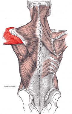
Deltoid Muscle
Shoulder
Stanozolol
Muscle, Skeletal
Shoulder Joint
Rotator Cuff
Injections, Intramuscular
Electromyography
Shoulder Pain
Muscle Proteins
Muscle, Smooth
Muscle Fibers, Skeletal
Nerve Transfer
Muscle Contraction
Muscle Development
Brachial Plexus
How repaired rotator cuff function influences Constant scoring. (1/16)
(+info)Anatomical and functional segments of the deltoid muscle. (2/16)
(+info)Arthroscopic release of the deltoid contracture. (3/16)
BACKGROUND: The deltoid contracture is an uncommon disorder. Long-standing contracture produces winged scapula, abduction and extension contracture of the shoulder. Surgical release has been considered the treatment of choice. However, the method of approach has not been well defined. The purpose of this study was to evaluate the results of arthroscopic release of the deltoid contracture. METHODS: A retrospective study was undertaken to evaluate the results of arthroscopic release in six patients (seven shoulders) who had a contracture of the deltoid muscle. All patients had arthroscopic release. The abduction-contracture and horizontal-adduction angle was measured after operation. The average duration of follow-up was 16 months (range, from 4 to 41 months). RESULTS: The preoperative abduction contracture resolved completely in three shoulders. Two had a residual abduction contracture of 5 degrees to 7 degrees and two had a poor result with 15 degrees abduction-contracture angle. The average postoperative abduction-contracture angle was 6 degrees (range, 0 degrees to 15 degrees ). The preoperative horizontal-adduction contracture was corrected, permitting at least 130 degrees of adduction, in five shoulders. The remaining two shoulders had a postoperative horizontal-adduction angle of 120 degrees and 110 degrees . Overall, the average postoperative horizontal-adduction angle was 130 degrees (range, 110 degrees to 140 degrees ). CONCLUSION: Arthroscopic release is an effective surgical technique to treat the deltoid contracture. (+info)A biomechanical study of a suture between the deltoid muscle and a free tendon graft for reconstruction of the elbow extension. (4/16)
AIMS: It is possible to reconstruct the elbow motion in tetraplegic patients using the posterior portion of the deltoid muscle. In this surgery however, it is a problem to achieve a firm suture between the deltoid muscle and the tendon graft which extends the muscle and is sewn in order to compensate for the plegic musculus triceps brachii function. This study assesses two methods of attachment between muscle and free tendon graft from the biomechanical point of view. METHODS: The assessment was made on 7 fresh-frozen cadaveric samples where the rear portion of the deltoid muscle was sewn with the strip of fascia lata (A1-A7) and 7 samples (B1-B7) where the free tendon graft was attached with a strengthened part of deltoid fascia. The character of the attachment defect was evaluated as strength and elongation parameters using the device Zwick Z020-TND. RESULTS: The ANOVA showed a statistically significant greater suture solidity connecting the muscle and tendon for group B (B1-B7) than group A. The deformation of the actual suture location was smaller in group B than the deformation of attachment surroundings. CONCLUSION: From the biomechanical solidity point of view, it is more efficient to use the strengthened fascia of the deltoid muscle on its inner side for the suture with the tendon graft for reconstruction of the elbow extension in tetraplegic patients. (+info)Electromyographic activity of shoulder muscles during exercises performed with oscillatory and non-oscillatory poles. (5/16)
(+info)An evidence based protocol for the prevention of upper arm injury related to vaccine administration (UAIRVA). (6/16)
(+info)Relationship between humeral geometry and shoulder muscle power among suspensory, knuckle-walking, and digitigrade/palmigrade quadrupedal primates. (7/16)
(+info)Thin-filament length correlates with fiber type in human skeletal muscle. (8/16)
(+info)The deltoid muscle is a large, triangular-shaped muscle that covers the shoulder joint. It is responsible for shoulder abduction (raising the arm away from the body), flexion (lifting the arm forward), and extension (pulling the arm backward). The muscle is divided into three sections: the anterior deltoid, which lies on the front of the shoulder and is responsible for flexion and internal rotation; the middle deltoid, which lies on the side of the shoulder and is responsible for abduction; and the posterior deltoid, which lies on the back of the shoulder and is responsible for extension and external rotation. Together, these muscles work to provide stability and mobility to the shoulder joint.
In anatomical terms, the shoulder refers to the complex joint of the human body that connects the upper limb to the trunk. It is formed by the union of three bones: the clavicle (collarbone), scapula (shoulder blade), and humerus (upper arm bone). The shoulder joint is a ball-and-socket type of synovial joint, allowing for a wide range of movements such as flexion, extension, abduction, adduction, internal rotation, and external rotation.
The shoulder complex includes not only the glenohumeral joint but also other structures that contribute to its movement and stability, including:
1. The acromioclavicular (AC) joint: where the clavicle meets the acromion process of the scapula.
2. The coracoclavicular (CC) ligament: connects the coracoid process of the scapula to the clavicle, providing additional stability to the AC joint.
3. The rotator cuff: a group of four muscles (supraspinatus, infraspinatus, teres minor, and subscapularis) that surround and reinforce the shoulder joint, contributing to its stability and range of motion.
4. The biceps tendon: originates from the supraglenoid tubercle of the scapula and passes through the shoulder joint, helping with flexion, supination, and stability.
5. Various ligaments and capsular structures that provide additional support and limit excessive movement in the shoulder joint.
The shoulder is a remarkable joint due to its wide range of motion, but this also makes it susceptible to injuries and disorders such as dislocations, subluxations, sprains, strains, tendinitis, bursitis, and degenerative conditions like osteoarthritis. Proper care, exercise, and maintenance are essential for maintaining shoulder health and function throughout one's life.
Stanozolol is a synthetic anabolic-androgenic steroid (AAS) derivative of dihydrotestosterone (DHT). It is commonly used in medicine for the treatment of hereditary angioedema and was formerly used to promote muscle growth in weakened or catabolic patients. Stanozolol has a high anabolic and moderate androgenic activity, with reduced estrogenic properties compared to testosterone. Its chemical formula is (17α-methyl-5α-androstano[2,3-c]pyrazol-17β-ol). It is important to note that the use of Stanozolol for performance enhancement is considered illegal and subject to severe penalties in many countries, including disqualification from sports events and criminal charges.
Skeletal muscle, also known as striated or voluntary muscle, is a type of muscle that is attached to bones by tendons or aponeuroses and functions to produce movements and support the posture of the body. It is composed of long, multinucleated fibers that are arranged in parallel bundles and are characterized by alternating light and dark bands, giving them a striped appearance under a microscope. Skeletal muscle is under voluntary control, meaning that it is consciously activated through signals from the nervous system. It is responsible for activities such as walking, running, jumping, and lifting objects.
The shoulder joint, also known as the glenohumeral joint, is the most mobile joint in the human body. It is a ball and socket synovial joint that connects the head of the humerus (upper arm bone) to the glenoid cavity of the scapula (shoulder blade). The shoulder joint allows for a wide range of movements including flexion, extension, abduction, adduction, internal rotation, and external rotation. It is surrounded by a group of muscles and tendons known as the rotator cuff that provide stability and enable smooth movement of the joint.
Arthroplasty is a surgical procedure to restore the integrity and function of a joint. The term is derived from two Greek words: "arthro" meaning joint, and "plasty" meaning to mold or form. There are several types of arthroplasty, but most involve resurfacing the damaged joint cartilage with artificial materials such as metal, plastic, or ceramic.
The goal of arthroplasty is to relieve pain, improve mobility, and restore function in a joint that has been damaged by arthritis, injury, or other conditions. The most common types of arthroplasty are total joint replacement (TJR) and partial joint replacement (PJR).
In TJR, the surgeon removes the damaged ends of the bones in the joint and replaces them with artificial components called prostheses. These prostheses can be made of metal, plastic, or ceramic materials, and are designed to mimic the natural movement and function of the joint.
In PJR, only one side of the joint is resurfaced, typically because the damage is less extensive. This procedure is less invasive than TJR and may be recommended for younger patients who are still active or have a higher risk of complications from a full joint replacement.
Other types of arthroplasty include osteotomy, in which the surgeon cuts and reshapes the bone to realign the joint; arthrodesis, in which the surgeon fuses two bones together to create a stable joint; and resurfacing, in which the damaged cartilage is removed and replaced with a smooth, artificial surface.
Arthroplasty is typically recommended for patients who have tried other treatments, such as physical therapy, medication, or injections, but have not found relief from their symptoms. While arthroplasty can be highly effective in relieving pain and improving mobility, it is not without risks, including infection, blood clots, and implant failure. Patients should discuss the benefits and risks of arthroplasty with their healthcare provider to determine if it is the right treatment option for them.
A laceration is a type of injury that results in a tear or ragged cut in the skin or mucous membrane, often caused by some form of trauma. This can include cuts from sharp objects, blunt force trauma, or accidents. Lacerations can vary greatly in severity, from minor injuries that only affect the top layer of skin to more serious wounds that penetrate deeper into underlying tissues and structures.
Lacerations are typically irregular in shape and may have jagged edges, unlike clean incisions caused by sharp objects. They can also be accompanied by bruising, swelling, and bleeding, depending on the severity of the injury. In some cases, lacerations may require medical attention to properly clean, close, and manage the wound to prevent infection and promote healing.
It is essential to assess the depth, location, and extent of a laceration to determine the appropriate course of action. Deeper lacerations that expose underlying tissues or structures, such as muscles, tendons, nerves, or blood vessels, may require sutures (stitches), staples, or adhesive strips to close the wound. In some instances, surgical intervention might be necessary to repair damaged tissues properly. Always consult a healthcare professional for proper evaluation and treatment of lacerations.
The rotator cuff is a group of four muscles and their tendons that attach to the shoulder blade (scapula) and help stabilize and move the shoulder joint. These muscles are the supraspinatus, infraspinatus, teres minor, and subscapularis. The rotator cuff helps to keep the head of the humerus (upper arm bone) centered in the glenoid fossa (shoulder socket), providing stability during shoulder movements. It also allows for rotation and elevation of the arm. Rotator cuff injuries or conditions, such as tears or tendinitis, can cause pain and limit shoulder function.
A muscle is a soft tissue in our body that contracts to produce force and motion. It is composed mainly of specialized cells called muscle fibers, which are bound together by connective tissue. There are three types of muscles: skeletal (voluntary), smooth (involuntary), and cardiac. Skeletal muscles attach to bones and help in movement, while smooth muscles are found within the walls of organs and blood vessels, helping with functions like digestion and circulation. Cardiac muscle is the specific type that makes up the heart, allowing it to pump blood throughout the body.
"Intramuscular injections" refer to a medical procedure where a medication or vaccine is administered directly into the muscle tissue. This is typically done using a hypodermic needle and syringe, and the injection is usually given into one of the large muscles in the body, such as the deltoid (shoulder), vastus lateralis (thigh), or ventrogluteal (buttock) muscles.
Intramuscular injections are used for a variety of reasons, including to deliver medications that need to be absorbed slowly over time, to bypass stomach acid and improve absorption, or to ensure that the medication reaches the bloodstream quickly and directly. Common examples of medications delivered via intramuscular injection include certain vaccines, antibiotics, and pain relievers.
It is important to follow proper technique when administering intramuscular injections to minimize pain and reduce the risk of complications such as infection or injury to surrounding tissues. Proper site selection, needle length and gauge, and injection technique are all critical factors in ensuring a safe and effective intramuscular injection.
Electromyography (EMG) is a medical diagnostic procedure that measures the electrical activity of skeletal muscles during contraction and at rest. It involves inserting a thin needle electrode into the muscle to record the electrical signals generated by the muscle fibers. These signals are then displayed on an oscilloscope and may be heard through a speaker.
EMG can help diagnose various neuromuscular disorders, such as muscle weakness, numbness, or pain, and can distinguish between muscle and nerve disorders. It is often used in conjunction with other diagnostic tests, such as nerve conduction studies, to provide a comprehensive evaluation of the nervous system.
EMG is typically performed by a neurologist or a physiatrist, and the procedure may cause some discomfort or pain, although this is usually minimal. The results of an EMG can help guide treatment decisions and monitor the progression of neuromuscular conditions over time.
A cadaver is a deceased body that is used for medical research or education. In the field of medicine, cadavers are often used in anatomy lessons, surgical training, and other forms of medical research. The use of cadavers allows medical professionals to gain a deeper understanding of the human body and its various systems without causing harm to living subjects. Cadavers may be donated to medical schools or obtained through other means, such as through consent of the deceased or their next of kin. It is important to handle and treat cadavers with respect and dignity, as they were once living individuals who deserve to be treated with care even in death.
Shoulder pain is a condition characterized by discomfort or hurt in the shoulder joint, muscles, tendons, ligaments, or surrounding structures. The shoulder is one of the most mobile joints in the body, and this mobility makes it prone to injury and pain. Shoulder pain can result from various causes, including overuse, trauma, degenerative conditions, or referred pain from other areas of the body.
The shoulder joint is a ball-and-socket joint made up of three bones: the humerus (upper arm bone), scapula (shoulder blade), and clavicle (collarbone). The rotator cuff, a group of four muscles that surround and stabilize the shoulder joint, can also be a source of pain if it becomes inflamed or torn.
Shoulder pain can range from mild to severe, and it may be accompanied by stiffness, swelling, bruising, weakness, numbness, tingling, or reduced mobility in the affected arm. The pain may worsen with movement, lifting objects, or performing certain activities, such as reaching overhead or behind the back.
Medical evaluation is necessary to determine the underlying cause of shoulder pain and develop an appropriate treatment plan. Treatment options may include rest, physical therapy, medication, injections, or surgery, depending on the severity and nature of the condition.
In medical terms, the arm refers to the upper limb of the human body, extending from the shoulder to the wrist. It is composed of three major bones: the humerus in the upper arm, and the radius and ulna in the lower arm. The arm contains several joints, including the shoulder joint, elbow joint, and wrist joint, which allow for a wide range of motion. The arm also contains muscles, blood vessels, nerves, and other soft tissues that are essential for normal function.
Muscle proteins are a type of protein that are found in muscle tissue and are responsible for providing structure, strength, and functionality to muscles. The two major types of muscle proteins are:
1. Contractile proteins: These include actin and myosin, which are responsible for the contraction and relaxation of muscles. They work together to cause muscle movement by sliding along each other and shortening the muscle fibers.
2. Structural proteins: These include titin, nebulin, and desmin, which provide structural support and stability to muscle fibers. Titin is the largest protein in the human body and acts as a molecular spring that helps maintain the integrity of the sarcomere (the basic unit of muscle contraction). Nebulin helps regulate the length of the sarcomere, while desmin forms a network of filaments that connects adjacent muscle fibers together.
Overall, muscle proteins play a critical role in maintaining muscle health and function, and their dysregulation can lead to various muscle-related disorders such as muscular dystrophy, myopathies, and sarcopenia.
Smooth muscle, also known as involuntary muscle, is a type of muscle that is controlled by the autonomic nervous system and functions without conscious effort. These muscles are found in the walls of hollow organs such as the stomach, intestines, bladder, and blood vessels, as well as in the eyes, skin, and other areas of the body.
Smooth muscle fibers are shorter and narrower than skeletal muscle fibers and do not have striations or sarcomeres, which give skeletal muscle its striped appearance. Smooth muscle is controlled by the autonomic nervous system through the release of neurotransmitters such as acetylcholine and norepinephrine, which bind to receptors on the smooth muscle cells and cause them to contract or relax.
Smooth muscle plays an important role in many physiological processes, including digestion, circulation, respiration, and elimination. It can also contribute to various medical conditions, such as hypertension, gastrointestinal disorders, and genitourinary dysfunction, when it becomes overactive or underactive.
Skeletal muscle fibers, also known as striated muscle fibers, are the type of muscle cells that make up skeletal muscles, which are responsible for voluntary movements of the body. These muscle fibers are long, cylindrical, and multinucleated, meaning they contain multiple nuclei. They are surrounded by a connective tissue layer called the endomysium, and many fibers are bundled together into fascicles, which are then surrounded by another layer of connective tissue called the perimysium.
Skeletal muscle fibers are composed of myofibrils, which are long, thread-like structures that run the length of the fiber. Myofibrils contain repeating units called sarcomeres, which are responsible for the striated appearance of skeletal muscle fibers. Sarcomeres are composed of thick and thin filaments, which slide past each other during muscle contraction to shorten the sarcomere and generate force.
Skeletal muscle fibers can be further classified into two main types based on their contractile properties: slow-twitch (type I) and fast-twitch (type II). Slow-twitch fibers have a high endurance capacity and are used for sustained, low-intensity activities such as maintaining posture. Fast-twitch fibers, on the other hand, have a higher contractile speed and force generation capacity but fatigue more quickly and are used for powerful, explosive movements.
A smooth muscle within the vascular system refers to the involuntary, innervated muscle that is found in the walls of blood vessels. These muscles are responsible for controlling the diameter of the blood vessels, which in turn regulates blood flow and blood pressure. They are called "smooth" muscles because their individual muscle cells do not have the striations, or cross-striped patterns, that are observed in skeletal and cardiac muscle cells. Smooth muscle in the vascular system is controlled by the autonomic nervous system and by hormones, and can contract or relax slowly over a period of time.
A nerve transfer is a surgical procedure where a functioning nerve is connected to an injured nerve to restore movement, sensation or function. The functioning nerve, called the donor nerve, usually comes from another less critical location in the body and has spare nerve fibers that can be used to reinnervate the injured nerve, called the recipient nerve.
During the procedure, a small section of the donor nerve is carefully dissected and prepared for transfer. The recipient nerve is also prepared by removing any damaged or non-functioning portions. The two ends are then connected using microsurgical techniques under a microscope. Over time, the nerve fibers from the donor nerve grow along the recipient nerve and reinnervate the muscles or sensory structures that were previously innervated by the injured nerve.
Nerve transfers can be used to treat various types of nerve injuries, including brachial plexus injuries, facial nerve palsy, and peripheral nerve injuries. The goal of the procedure is to restore function as quickly and efficiently as possible, allowing for a faster recovery and improved quality of life for the patient.
Muscle contraction is the physiological process in which muscle fibers shorten and generate force, leading to movement or stability of a body part. This process involves the sliding filament theory where thick and thin filaments within the sarcomeres (the functional units of muscles) slide past each other, facilitated by the interaction between myosin heads and actin filaments. The energy required for this action is provided by the hydrolysis of adenosine triphosphate (ATP). Muscle contractions can be voluntary or involuntary, and they play a crucial role in various bodily functions such as locomotion, circulation, respiration, and posture maintenance.
Muscle development, also known as muscle hypertrophy, refers to the increase in size and mass of the muscles through a process called myofiber growth. This is primarily achieved through resistance or strength training exercises that cause micro-tears in the muscle fibers, leading to an inflammatory response and the release of hormones that promote muscle growth. As the muscles repair themselves, they become larger and stronger than before. Proper nutrition, including adequate protein intake, and rest are also essential components of muscle development.
It is important to note that while muscle development can lead to an increase in strength and muscular endurance, it does not necessarily result in improved athletic performance or overall fitness. A well-rounded exercise program that includes cardiovascular activity, flexibility training, and resistance exercises is recommended for optimal health and fitness outcomes.
The brachial plexus is a network of nerves that originates from the spinal cord in the neck region and supplies motor and sensory innervation to the upper limb. It is formed by the ventral rami (branches) of the lower four cervical nerves (C5-C8) and the first thoracic nerve (T1). In some cases, contributions from C4 and T2 may also be included.
The brachial plexus nerves exit the intervertebral foramen, pass through the neck, and travel down the upper chest before branching out to form major peripheral nerves of the upper limb. These include the axillary, radial, musculocutaneous, median, and ulnar nerves, which further innervate specific muscles and sensory areas in the arm, forearm, and hand.
Damage to the brachial plexus can result in various neurological deficits, such as weakness or paralysis of the upper limb, numbness, or loss of sensation in the affected area, depending on the severity and location of the injury.
 Deltoid muscle - Wikipedia
 Deltoid (Middle) - Stretching - Learn Muscles
 Muscle of the Month: May - The Deltoid
 Deltoid muscle: Origin, insertion, innervation, function | Kenhub
 Side Deltoid Head - Muscle Profiles - Steroids Live
 Deltoid Fibrosis: Practice Essentials, Anatomy, Pathophysiology
 Deltoid Muscle: Definition, Anatomy And Its Extraordinary Function.
 Deltoid Exercises at Home- Train All Deltoid Muscles at Home
Deltoid Fibrosis Workup: Approach Considerations, Imaging Studies
Steroid for deltoid muscle, boldenone acetate vs boldenone undecylenate | AMEER TRANSPORTATION
 Treating Shoulder Pain - Deltoid Muscles | Niel Asher Education Blogs and Articles blog
 How to Target and Grow Your Rear Deltoids (Workouts Included!) | Muscle & Strength
 Deltoid pain: Causes, exercises, and relief
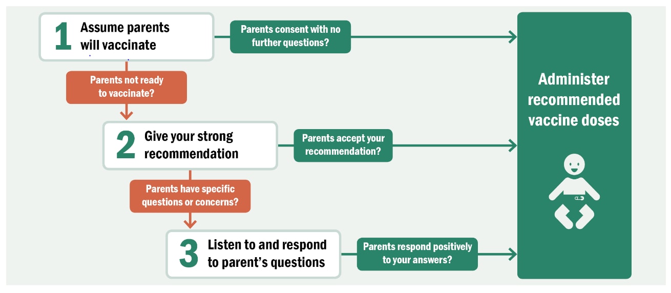
 ACIP Vaccine Administration Guidelines for Immunization | CDC
 Very best Exercises to Bulk In the Shoulder muscles -- Intended for Big Deltoids
Vaccine Administration Route and Site | CDC
 Dermatomes, Myotomes, And Associated Paresthesias - ProProfs Quiz
 PDF) Comparison among the EMG Activity of the Anterior Deltoid and Medial Deltoid During Two Variations of Dumbbell Shoulder...
 Upper Body Workout for Women: 10 Best Exercises
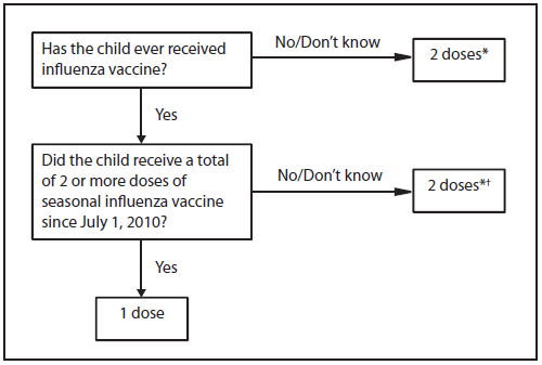
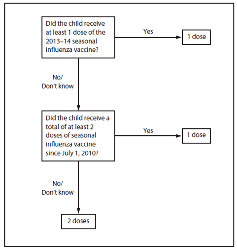
Prevention and Control of Influenza: Recommendations of the Advisory Committee on Immunization Practices (ACIP)
 Medical Abbreviations - R - GlobalRPH
 What Part of the Deltoid Does the Arnold Press Work On? | Woman - The Nest
Heart Rate Variability: Ignoring a Harbinger of Health?
 Dr Aamer Iqbal
 Physostigma modalities etc - ABC Homeopathy
Oxalicum Acidum extremities, limbs symptoms - ABC Homeopathy
 Innomed Shoulder Instruments retractors & elevators
Baris KOCAOGLU | Orthopaedics and traumatology | Research profile
Triceps3
- The secondary muscles stressed are the triceps and trapezius. (nfpt.com)
- Choose exercises that target the deltoids, triceps, abdominals and trapezius muscle. (catalogs.com)
- Thanks to the converging trajectory of the levers on the scapular plane, this machine allows to effectively train the deltoid muscles (the front head) and the clavicular bundles of the pectoralis major, with the involvement of the upper trapezius, triceps and large anterior dentate. (panattasport.com)
Rotator cuff muscles11
- A unique characteristic of the deltoid muscle is its extensive overlap with the rotator cuff muscles. (bodyworksprime.com)
- The rotator cuff muscles are the supraspinatus , infraspinatus , teres minor , and the subscapularis . (bodyworksprime.com)
- The deltoids sit on top of the shoulder girdle and have anterior and posterior sections that work together with the rotator cuff muscles that provide a full range of motion to the arms. (elpasobackclinic.com)
- In contrast, the posterior section works with the rotator cuff muscles for mobility. (elpasobackclinic.com)
- But crucially, the press requires plenty of stability and control from your shoulders, meaning your rotator cuff muscles must work harder. (tomsguide.com)
- Your rotator cuff muscles comprise four muscles that surround your scapulae (shoulder blades) and support your shoulder through movement. (tomsguide.com)
- As one of the rotator cuff muscles, it holds the arm in the shoulder when you release what you are throwing. (sportsinjuryclinic.net)
- Few athletes people bother to train the rotator cuff muscles, preferring to concentrate more on the muscles which accelerate the arm rather than decelerate it. (sportsinjuryclinic.net)
- These work the rotator cuff muscles. (harvard.edu)
- This exercise works the rotator cuff muscles. (harvard.edu)
- In this piece, we'll overview those pixie rotator cuff muscles and look at why they can be such a pain - in the shoulder - and how understanding the movements these muscles support may help reduce injury risk. (begin2dig.com)
Posterior17
- Anatomically, the deltoid muscle appears to be made up of three distinct sets of muscle fibers, namely the anterior or clavicular part (pars clavicularis) posterior or scapular part (pars scapularis) intermediate or acromial part (pars acromialis) However, electromyography suggests that it consists of at least seven groups that can be independently coordinated by the nervous system. (wikipedia.org)
- Three of these lie in the anatomical anterior head of the deltoid, one in the anatomical middle head, and three in the anatomical posterior head of the deltoid. (wikipedia.org)
- Other transverse extensors, the infraspinatus and teres minor, also work in tandem with the posterior deltoid as external (lateral) rotators, antagonists to strong internal rotators like the pecs and lats. (wikipedia.org)
- This muscle, positioned in the lateral aspect of the shoulder, can be conceptually divided into three distinct portions based on their fiber orientation - the anterior (clavicular), lateral (acromial), and posterior (spinal). (bodyworksprime.com)
- Here we can see the deltoid muscle highlighted in red amongst the other muscles of the body, seen from a posterior view. (bodyworksprime.com)
- The deltoid muscle is typically further divided into three segments: the anterior, middle, and posterior segments. (bodyworksprime.com)
- The posterior deltoid originates from the posterior third of the acromion and the spine of the scapula. (bodyworksprime.com)
- In the region of the proximal deltoid, the tendons associated with the anterior and posterior segments of the deltoid divide, producing three additional intramuscular tendons. (bodyworksprime.com)
- The Deltoid muscle consists of anterior and posterior parts or fibers and a lateral part (or the intermediate fibers). (jotscroll.com)
- The parts of the deltoid muscle are the clavicular parts (anterior deltoid) , acromial parts (middle deltoid) , and spinal parts (posterior deltoid) . (jotscroll.com)
- the anterior and posterior deltoid actions are just like guy ropes that help to steady the arm as it is abducted. (jotscroll.com)
- The anterior and posterior fibers, arising from the clavicle and the scapular spine are unipennate fibers (the fibers run in the same direction) that converge on the anterior and posterior margins of the deltoid tuberosity with greater range of movement. (jotscroll.com)
- the posterior deltoid assist the latissimus dorsi in extending the arm and act as a lateral rotator. (jotscroll.com)
- The Deltoid is supplied by the Axillary nerve ( C5 and C6) from the posterior cord of the brachial plexus. (jotscroll.com)
- For the deltoid muscles, when active trigger points affect the deltoids' anterior or posterior sections, it can cause the muscles to twitch and later cause moderate tension. (elpasobackclinic.com)
- Your shoulder muscles (the anterior, medial, and posterior deltoid muscles) are engaged in most popular bodyweight exercises, like planks and push-ups. (greatist.com)
- There should be specific exercises for your anterior, lateral, and posterior deltoids, so be sure to add Lateral Raises, Front Raises and Scapula Raises. (stack.com)
Trapezius4
- Stabilizer muscles include the anterior deltoids (front of shoulders) and the trapezius. (shapefit.com)
- This shoulder workout is proven to add size to your trapezius and deltoid muscles. (stack.com)
- Most people don't realize how strong their trapezius muscles are, so they can't reach their full potential in certain lifts like Shrugs for example. (stack.com)
- Regarding the trapezius muscles, you should be pressing overhead with Dumbbell Shoulder Presses, and it's good to add variations such as the Arnold Press. (stack.com)
Middle deltoid2
- The middle deltoid originates from the middle third of the lateral aspect of the acromion. (bodyworksprime.com)
- Optimised pads are strategically positioned for stimulating the middle deltoid muscles and the simple incremental weight stack system means training couldn't be easier. (powerhouse-fitness.co.uk)
Latissimus dorsi1
- This makes the deltoid an antagonist muscle of the pectoralis major and latissimus dorsi during arm adduction. (wikipedia.org)
Function of the deltoid2
- An important function of the deltoid in humans is preventing the dislocation of the humeral head when a person carries heavy loads. (wikipedia.org)
- Another action or function of the deltoid is its ability to act as a shunt muscle by resisting the downward displacement of the head of the humerus from the glenoid cavity. (jotscroll.com)
Shoulders20
- The shoulders have many muscles, ligaments, and tendons that help support the joints from injuries and utilize the motor function to do everyday actions like throwing a ball or stretching for long distances. (elpasobackclinic.com)
- Even though the shoulders help stabilize the upper extremities, they are still prone to injuries since the shoulder muscles are constantly used throughout the day. (elpasobackclinic.com)
- Today's article examines the deltoid muscles, how trigger points affect the deltoids and shoulders, and managing trigger points associated with the deltoid muscles. (elpasobackclinic.com)
- We refer patients to certified providers specializing in shoulder pain treatments to aid individuals suffering from trigger points associated with the deltoid muscles along the shoulders. (elpasobackclinic.com)
- Studies reveal that the deltoid muscles have a more complex structure since the intramuscular tendons from the deltoids help provide different functions when it comes to the shoulders and arms' motor function. (elpasobackclinic.com)
- How Do Trigger Points Affect The Deltoids & Shoulders? (elpasobackclinic.com)
- Regarding the shoulders, they can succumb to various injuries that can become an issue over time, lead to the development of trigger points in the shoulder muscles, and cause referred pain to the upper arms. (elpasobackclinic.com)
- Trigger points or myofascial pain syndrome along the deltoid muscles may invoke referred pain to the shoulders. (elpasobackclinic.com)
- fortunately, there are various ways to manage the pain associated with trigger points along the shoulders and deltoid muscles. (elpasobackclinic.com)
- Have you been feeling stiffness along the shoulders or the deltoid muscles? (elpasobackclinic.com)
- I'm a fan of building the bench press, overhead presses, and push-ups into my upper-body programs to develop strength and muscle in my shoulders, chest, and arms. (tomsguide.com)
- The muscle gets stretched even more when the arm is raised above the shoulders and extended out correctly. (yogacards.com)
- Holding that position for extended periods strengthens small muscles that promote shoulder stability and improve hunched posture, which is bad for the shoulders," Crowley says. (harvard.edu)
- These work the main muscles in your shoulders, called the deltoids. (harvard.edu)
- This works the rhomboid muscles in the upper back, which support the shoulders. (harvard.edu)
- The full commercial Insignia Lateral Raise Machine is a first-class strength training machine designed to help build strong, well defined shoulders, particularly the middle deltoids. (powerhouse-fitness.co.uk)
- Bodyweight exercises are great for shoulders because they're muscles that generally don't require a lot of weight to achieve muscle fatigue or gains," says Alex Davis, co-creator of the wellness site Ryan and Alex Duo Life . (greatist.com)
- Selecting shoulders generated a crisp, high-definition 3-D image of the shoulder section, starting at the skin level, with the option to click through to see the muscles, nerves and vessels, and bone. (stanford.edu)
- Shoulder workouts should increase the size and strength of every muscle in your shoulders. (stack.com)
- Working on developing the rear deltoids will help with shoulder mobility and working the trapezium will help stabilize the shoulders, which will both help to improve posture. (keine-ruhe.org)
Profunda brachi1
- The deltoid is supplied by the thoracoacromial artery (acromial and deltoid branches), the circumflex humeral arteries, and the profunda brachii artery (deltoid branch). (wikipedia.org)
Tendons3
- The anterior origin lies adjacent to the lateral fibers of the pectoralis major muscle as do the end tendons of both muscles. (wikipedia.org)
- This segmentation is determined by the presence of three intramuscular tendons, each of which inserts into the deltoid tuberosity on the humerus. (bodyworksprime.com)
- The muscles and tendons of the rotator cuff hold the ball and socket of the shoulder joint in place. (medlineplus.gov)
Innervated by the axillary nerve1
- The deltoid is innervated by the axillary nerve. (wikipedia.org)
Tuberosity1
- The point of insertion of the deltoid muscle is on the deltoid tuberosity on the lateral aspect of the humerus. (jotscroll.com)
Humerus7
- The anterior deltoid also works in tandem with the subscapularis, pecs and lats to internally (medially) rotate the humerus. (wikipedia.org)
- the deltoid is part of the six scapulohumeral muscles that pass from the scapula to the humerus and act on the shoulder joint. (jotscroll.com)
- The six scapulohumeral muscles include the deltoid, teres major , supraspinatus , infraspinatus , subscapularis, and teres minor these are relatively short muscles that pass from the scapula to the humerus and act on the glenohumeral joint (shoulder joint). (jotscroll.com)
- the deltoid has convex shape because the upper end of the humerus lies below it. (jotscroll.com)
- The nerve runs transversely round the back and lateral side of the surgical neck the humerus giving off numerous branches that enter the deltoid muscle in radial directions thus, splitting the muscle vertically does not damage the nerve supply. (jotscroll.com)
- The Supraspinatus muscle runs along the top of the shoulder blade (scapula) and inserts via the tendon at the top of the arm or humerus (upper arm) bone. (sportsinjuryclinic.net)
- The top muscle, the supraspinatus (supra=above the spiney bit), attaches from the top of the scapula at the big scapula spine, runs along the top of that ridge, goes under the acromium process of the scapular spine, over the bursa on the top of the glenohumeral capsule and then attaches onto a big bump at the top of the humerus, the greater tubercle. (begin2dig.com)
Axillary4
- These neuromuscular segments are supplied by smaller branches of the axillary nerve, and work in coordination with other muscles of the shoulder girdle include pectoralis major and supraspinatus. (wikipedia.org)
- Iatrogenic axillary neuropathy after intramuscular injection of the deltoid muscle. (medscape.com)
- The axillary nerve supplying the deltoid contains the fifth and sixth cervical segments of the spinal cord, hence the numbers C5 and C6, the main supply is the C5 segment that is why it is in bold face. (jotscroll.com)
- They may have motor and sensory deficits (eg, if the axillary nerve is injured, decreased sensation over the deltoid). (msdmanuals.com)
Abduction8
- When all its fibers contract simultaneously, the deltoid is the prime mover of arm abduction along the frontal plane. (wikipedia.org)
- METHODS: The fatty infiltration (Goutallier stages), atrophy (tangent sign) and selective myotendinous retraction of the rotator cuff, as well as the thickness and the area of seven anatomically defined segments of the deltoid muscle were measured on MR-arthrographies and correlated with shoulder function (i.e. active abduction). (uzh.ch)
- Unexpectedly however, we were unable to detect a relation of the deltoid muscle shape with the degree of active glenohumeral abduction. (uzh.ch)
- Deltoid muscle abduction contracture. (medscape.com)
- Fibrous replacement of the deltoid muscle: a remediable cause of abduction contracture of the shoulder in scleroderma. (medscape.com)
- This is a very important muscle with the primary action of abduction of the arm (abduction in anatomy means the movement of a part of the body away from the middle part). (jotscroll.com)
- This muscle has three (3) parts that can act separately as well as together giving this muscle the ability to carry out four actions with the primary action being the abduction of the arm (which is the movement of the arm away from the central part of the body). (jotscroll.com)
- So when this muscle contracts, it's going to help pull the arm up to 90 degrees from one's side - called abduction. (begin2dig.com)
Lateral6
- Furthermore, the deltoid fascia contributes to the brachial fascia and is connected to the medial and lateral intermuscular septa. (wikipedia.org)
- Pictured here we can see the deltoid muscle from a lateral view. (bodyworksprime.com)
- The origin of the deltoid muscle is from the anterior border and upper surface of the lateral one-third of the clavicle and from the whole of the lateral border of the acromion and also from the inferior lip of the crest of the scapular spine. (jotscroll.com)
- From the lateral border of the acromion, there are four ridges that may be seen that are caused by the four fibrous septa that pass down into the muscle. (jotscroll.com)
- The full commercial Insignia Lateral Raise Machine is the ticket to strong, well defined deltoid muscles. (powerhouse-fitness.co.uk)
- Good exercises to work your lateral deltoid or side shoulder muscles. (makeoverfitness.com)
Glenohumeral joint1
- Structural changes in muscle and glenohumeral joint deformity in neonatal brachial plexus palsy. (medscape.com)
Fibers8
- These muscle fibers are closely related and only a small chiasmatic space, through which the cephalic vein passes, prevents the two muscles from forming a continuous muscle mass. (wikipedia.org)
- [ 26 ] The histology of deltoid fibrosis shows dense fibrous tissue with some atrophied muscle fibers. (medscape.com)
- These parts of the deltoid are actually the divisions of the muscle fibers that have the ability to act separately or as a whole. (jotscroll.com)
- The spaces between the septa are filled with fleshy muscle fibers attached to contiguous septa. (jotscroll.com)
- involuntary striated) muscle has branching fibers and forms most of the wall of the heart. (dummies.com)
- Skeletal muscle fibers are arranged in bundles, but smooth muscles form sheets of fibers that wrap around tubes and vessels. (dummies.com)
- Cardiac muscle has branching fibers forming most of the wall of the heart and controlling the contractions producing the heartbeat. (dummies.com)
- You should feel more activation in the lower fibers of your chest muscles with an incline push-up than with a traditional push-up on a flat surface. (greatist.com)
Gluteal3
- Syndrome of deltoid and/or gluteal fibrotic contracture: an injection myopathy. (medscape.com)
- Administer monthly maintenance in either the deltoid or gluteal muscle. (globalrph.com)
- Follow with monthly injections of 78 mg in either the deltoid or gluteal muscle. (globalrph.com)
Biceps muscle4
- Exam now revealing left deltoid and biceps muscle atrophy. (medscape.com)
- This type of muscle can be easily seen by flexing the forearm, which makes the biceps muscle become hard and thick. (dummies.com)
- The biceps muscle is made up of several parts: biceps brachii, brachialis and brachioradialis. (shapefit.com)
- 2 Intercostal nerves grafted to Biceps muscle, -Free-Gracilis muscle transfer to Biceps Region innervated with 2 Intercostal nerves grafts. (ubpn.org)
Exercises1
- The reason to do that is because these exercises activate multiple muscle groups, including your core and lower body (depending on which of the three you do). (stack.com)
Supraspinatus muscle1
- A heavy fall onto the shoulder can also result in injuring the supraspinatus muscle, which may also lead to bursitis. (sportsinjuryclinic.net)
Fibrosis1
- No specific laboratory studies have been helpful in the evaluation or treatment of deltoid fibrosis. (medscape.com)
Infraspinatus1
- These outcomes suggest that increasing muscular strength and endurance of the supraspinatus and infraspinatus muscles could prevent any increased superior humeral head translation. (cdc.gov)
Forearms1
- Standing cable curls target the biceps which are the primary muscle group engaged along with the forearms which are the secondary muscle group involved with this movement. (shapefit.com)
Point of insertion1
- Near the point of insertion, the muscle narrows and is connected to the bone by a tendon. (dummies.com)
Atrophy3
- Cite this: Deltoid Muscle Atrophy - Medscape - Oct 01, 2003. (medscape.com)
- We set out to analyse the size and shape of the deltoid muscle on MR-arthrographies, and analyse its influence on shoulder function and its adaption (i.e. atrophy) for reduced shoulder function. (uzh.ch)
- Furthermore, long-standing rotator cuff tears did not appear to influence the deltoid shape, i.e. did not lead to muscle atrophy. (uzh.ch)
Hyperechoic1
- A hyperechoic deltoid muscle was also a strong predictor of pre-diabetes. (rsna.org)
Study of muscle biopsies1
- A study of muscle biopsies in patients with diabetes found that muscle glycogen levels are decreased up to 65 percent. (rsna.org)
Gluteus1
- This unique torso depicts both the superficial and deep muscles, and the two main muscles, the deltoid and gluteus maximus can even be removed for closer studies. (3bscientific.com)
Triangular muscle1
- Thick triangular muscle in the SHOULDER whose function is to abduct, flex, and extend the arm. (nih.gov)
Insertion2
- Though traditionally described as a single insertion, the deltoid insertion is divided into two or three discernible areas corresponding to the muscle's three areas of origin. (wikipedia.org)
- These limbs of the V-shape region and the vertical ridge form the points of insertion of deltoid. (jotscroll.com)
Secondary1
- The secondary purpose was to assess muscle activation patterns during these motions. (cdc.gov)
Hamstrings1
- For people who have tight hamstrings and struggle to extend their legs, you have a few options: sit cross-legged, use a wall, or place a block under your sit-bones to tilt your pelvis forward - you might find this $10 accessory helps you sit taller and engage your hips and core muscles. (tomsguide.com)
Segments2
- Studies have shown that there are seven neuromuscular segments to the deltoid muscle. (wikipedia.org)
- As previously noted, the deltoid muscle is technically composed of seven segments rather than the traditionally identified three. (bodyworksprime.com)
Histology1
- In this patient, both the laboratory screening and muscle histology were normal and only the biochemical study of muscle allowed us to confirm the diagnosis. (hindawi.com)
Injection3
- For older children and adults, the deltoid muscle can be used for more than one intramuscular injection. (cdc.gov)
- Common COVID-19 vaccine side effects that children aged 4 and older may experience after their second dose include: Pain, swelling, and redness at the injection site, , fever (usually low grade), tiredness, headache, chills, muscle or joint pain, and swollen lymph nodes. (virginia.gov)
- Participants will get a dose of the study vaccine as an injection in a muscle in the upper arm. (nih.gov)
Lats1
- We'll also take a quick look at the big muscles like the lats that opperate on the main shoulder joint, the glenohumral joint. (begin2dig.com)
Superficial1
- The deltoid muscles are superficial, and trigger points can cause referred pain to the muscles that mimic arthritis in the shoulder joints. (elpasobackclinic.com)
Originates1
- The subsacpularis muscle originates from the underside of the shoulder blade and inserts at the front of the upper arm (humorous). (yogacards.com)
Vaccine2
- Local muscle cells that take in the vaccine produce the spike protein and place it on the surface of the cell where it is recognized by the immune system. (science20.com)
- Vaccine that is not taken up by muscle is drained into the local lymph nodes where lymphatic cells absorb the vaccine and similarly make spike protein. (science20.com)
Brightness of the Shoulder1
- Based on the brightness of the shoulder muscle, the radiologists were asked to classify the patients into one of three categories namely - normal, suspected diabetes and definite diabetes. (medindia.net)
Bone2
- Ogawa K, Yoshida A, Inokuchi W. Deltoid contracture: a radiographic survey of bone and joint abnormalities. (medscape.com)
- When the muscle contracts, another bone to which it is attached, does move. (dummies.com)
Subcutaneous2
Scapular1
- The study's results showed a compensatory increase in humeral head translation, scapular upward rotation, and deltoid muscle activation due to the nerve block. (cdc.gov)
Intramuscular injections1
- Contracture of the deltoid muscle in the adult after intramuscular injections. (medscape.com)
Skeletal3
- There are three classes of muscles: skeletal, visceral, and cardiac. (dummies.com)
- are also called skeletal or voluntary muscles. (dummies.com)
- You already know that skeletal muscles, or striated muscles, are the muscles that move the bones of the body. (dummies.com)
Chest1
- these bands are best used only when working large muscle groups such as the chest, back, and legs. (bellaonline.com)
Movement8
- The muscles - all 600 of them and more - are responsible for movement. (dummies.com)
- The skeleton provides attachment points and support for muscles, but it's the muscle tissue's ability to extend and contract that makes movement happen. (dummies.com)
- Internal movement involves the contraction and relaxation of involuntary muscles, The muscles that provide external movement are known as voluntary muscles, as they perform movements on command. (dummies.com)
- Movement of cardiac muscle cannot be consciously controlled. (dummies.com)
- a movement that involves multiple joints and/or muscles groups. (theglobeandmail.com)
- The primary muscles stressed in this movement are the shoulder muscles (deltoid). (nfpt.com)
- This is important to prevent loss of movement in the shoulder and eventually muscle weakness. (sportsinjuryclinic.net)
- This exercise allows for focused movement with lighter weights and higher repetitions for pumping more blood into the muscles. (keine-ruhe.org)
Shoulder joint2
- These muscles work in a coordinated manner to balance the forces acting on the shoulder joint and also provide stability during arm movements. (bodyworksprime.com)
- The main take away was that significant muscles like the traps, rhomboids, pec minor, serratus anterior and levator scapula all work pretty much *just* to move the scapula and so reposition the shoulder joint socket to extend range of motion for the arm moving in the shoulder. (begin2dig.com)
Exercise8
- Our study supports the hypothesis that mitochondrial impairment in muscle from long-term NRTI-treated patients could lead to exercise limitation, especially in LA patients. (natap.org)
- The Z press strengthens multiple muscle groups and joints, making it a truly functional compound exercise. (tomsguide.com)
- To prevent injury, these muscles must activate, but rotator cuff tears or overuse injuries often happen because of poor mechanics, so perform this exercise with care. (tomsguide.com)
- Prior research has also shown that the muscles of athletes appear brighter on US after exercise, when their glycogen stores are depleted. (rsna.org)
- Do around ten reps per exercise and vary your workout to develop all muscles. (catalogs.com)
- This exercise forces you to engage additional muscles for balance and stability since you are performing the exercise while standing and pulling on the cable which makes your legs work a little harder to support the tension. (shapefit.com)
- What this means is that the efficiency, consistency and smoothness with which you perform the cable exercise will be more effective and using cables also adds some nice variety to a workout routine by hitting the muscles from different angles and unique types of resistance. (shapefit.com)
- The standing dumbbell rear delt row is an easy exercise targeting several areas of the deltoid area. (keine-ruhe.org)
Humeral head1
- Using fluoroscopy and electromyography, humeral head translation and muscle activation were measured before and after a suprascapular nerve block. (cdc.gov)
Serratus anterior1
- This strengthens the serratus anterior muscles, which keep the shoulder blades in place. (harvard.edu)
Brighter2
- The reasons for the brighter-appearing shoulder muscle on US among patients with diabetes is not completely understood, according to Dr. Soliman, but the researchers suspect it is due to low levels of glycogen in the muscle. (rsna.org)
- The reasons for the brighter-appearing shoulder muscle on ultrasound in patients with diabetes is not fully clear, but scientists suspect that it could be related to decreased levels of glycogen in the muscle (a chemical that is a major source of energy for the body that is stored mainly in the liver and muscles for future use). (medindia.net)
Stretch1
- This lying spinal twist can be a create way to stretch the subsacpularis muscle. (yogacards.com)
Contractures2
- Deltoid contracture: a case with multiple muscle contractures. (medscape.com)
- Adjust range of motion criterion to accommodate for muscle contractures. (nih.gov)