Corneal Stroma
Cornea
Fibroblasts
Rabbits
Cells, Cultured
Fluorescent Antibody Technique, Indirect
Actins
RNA, Messenger
Blotting, Western
Keratan Sulfate
Epithelium, Corneal
Keratectomy, Subepithelial, Laser-Assisted
Photorefractive Keratectomy
Lasers, Excimer
Myopia
Astigmatism
Hyperopia
Refractive Errors
Apitherapy
Honey
Oxidative Stress
Stem Cells
Hydrogen Peroxide
Limbus Corneae
Insulin growth factor promotes human corneal fibroblast network formation in vitro. (1/66)
(+info)Polyethylenimine-conjugated gold nanoparticles: Gene transfer potential and low toxicity in the cornea. (2/66)
(+info)Synergistic induction of eotaxin and VCAM-1 expression in human corneal fibroblasts by staphylococcal peptidoglycan and either IL-4 or IL-13. (3/66)
(+info)An explanation for the central to peripheral thickness variation in the mouse cornea. (4/66)
(+info)Localization of thrombospondin-1 and myofibroblasts during corneal wound repair. (5/66)
(+info)Hydroxyapatite for keratoprosthesis biointegration. (6/66)
(+info)Assessment of corneal thickness and keratocyte density in a rabbit model of laser in situ keratomileusis using scanning laser confocal microscopy. (7/66)
(+info)Coxsackievirus A24 variant uses sialic acid-containing O-linked glycoconjugates as cellular receptors on human ocular cells. (8/66)
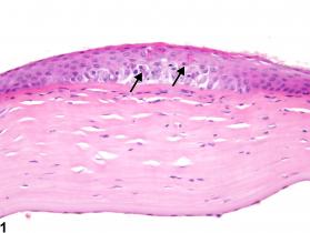
(+info)Corneal keratocytes are specialized cells located within the stroma, which is the thickest layer of the cornea, which is the clear front "window" of the eye. These cells play a crucial role in maintaining the transparency and structural integrity of the cornea. Keratocytes are star-shaped cells that produce and maintain the extracellular matrix (ECM) of the corneal stroma, which consists mainly of collagen fibrils and proteoglycans.
In a healthy cornea, keratocytes exist in a quiescent state, but they can become activated and undergo phenotypic changes in response to injury or disease. Activated keratocytes can differentiate into fibroblasts or myofibroblasts, which participate in the wound healing process by synthesizing ECM components and contracting to help close wounds. However, an overactive or dysregulated wound healing response can lead to corneal opacity, scarring, and visual impairment.
Therefore, understanding the behavior and regulation of corneal keratocytes is essential for developing effective therapies and treatments for various corneal disorders and diseases.
The corneal stroma, also known as the substantia propria, is the thickest layer of the cornea, which is the clear, dome-shaped surface at the front of the eye. The cornea plays a crucial role in focusing vision.
The corneal stroma makes up about 90% of the cornea's thickness and is composed of parallel bundles of collagen fibers that are arranged in regular, repeating patterns. These fibers give the cornea its strength and transparency. The corneal stroma also contains a small number of cells called keratocytes, which produce and maintain the collagen fibers.
Disorders that affect the corneal stroma can cause vision loss or other eye problems. For example, conditions such as keratoconus, in which the cornea becomes thin and bulges outward, can distort vision and make it difficult to see clearly. Other conditions, such as corneal scarring or infection, can also affect the corneal stroma and lead to vision loss or other eye problems.
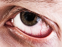
The cornea is the clear, dome-shaped surface at the front of the eye. It plays a crucial role in focusing vision. The cornea protects the eye from harmful particles and microorganisms, and it also serves as a barrier against UV light. Its transparency allows light to pass through and get focused onto the retina. The cornea does not contain blood vessels, so it relies on tears and the fluid inside the eye (aqueous humor) for nutrition and oxygen. Any damage or disease that affects its clarity and shape can significantly impact vision and potentially lead to blindness if left untreated.
Fibroblasts are specialized cells that play a critical role in the body's immune response and wound healing process. They are responsible for producing and maintaining the extracellular matrix (ECM), which is the non-cellular component present within all tissues and organs, providing structural support and biochemical signals for surrounding cells.
Fibroblasts produce various ECM proteins such as collagens, elastin, fibronectin, and laminins, forming a complex network of fibers that give tissues their strength and flexibility. They also help in the regulation of tissue homeostasis by controlling the turnover of ECM components through the process of remodeling.
In response to injury or infection, fibroblasts become activated and start to proliferate rapidly, migrating towards the site of damage. Here, they participate in the inflammatory response, releasing cytokines and chemokines that attract immune cells to the area. Additionally, they deposit new ECM components to help repair the damaged tissue and restore its functionality.
Dysregulation of fibroblast activity has been implicated in several pathological conditions, including fibrosis (excessive scarring), cancer (where they can contribute to tumor growth and progression), and autoimmune diseases (such as rheumatoid arthritis).
I believe there may be some confusion in your question. "Rabbits" is a common name used to refer to the Lagomorpha species, particularly members of the family Leporidae. They are small mammals known for their long ears, strong legs, and quick reproduction.
However, if you're referring to "rabbits" in a medical context, there is a term called "rabbit syndrome," which is a rare movement disorder characterized by repetitive, involuntary movements of the fingers, resembling those of a rabbit chewing. It is also known as "finger-chewing chorea." This condition is usually associated with certain medications, particularly antipsychotics, and typically resolves when the medication is stopped or adjusted.
Wound healing is a complex and dynamic process that occurs after tissue injury, aiming to restore the integrity and functionality of the damaged tissue. It involves a series of overlapping phases: hemostasis, inflammation, proliferation, and remodeling.
1. Hemostasis: This initial phase begins immediately after injury and involves the activation of the coagulation cascade to form a clot, which stabilizes the wound and prevents excessive blood loss.
2. Inflammation: Activated inflammatory cells, such as neutrophils and monocytes/macrophages, infiltrate the wound site to eliminate pathogens, remove debris, and release growth factors that promote healing. This phase typically lasts for 2-5 days post-injury.
3. Proliferation: In this phase, various cell types, including fibroblasts, endothelial cells, and keratinocytes, proliferate and migrate to the wound site to synthesize extracellular matrix (ECM) components, form new blood vessels (angiogenesis), and re-epithelialize the wounded area. This phase can last up to several weeks depending on the size and severity of the wound.
4. Remodeling: The final phase of wound healing involves the maturation and realignment of collagen fibers, leading to the restoration of tensile strength in the healed tissue. This process can continue for months to years after injury, although the tissue may never fully regain its original structure and function.
It is important to note that wound healing can be compromised by several factors, including age, nutrition, comorbidities (e.g., diabetes, vascular disease), and infection, which can result in delayed healing or non-healing chronic wounds.
"Cells, cultured" is a medical term that refers to cells that have been removed from an organism and grown in controlled laboratory conditions outside of the body. This process is called cell culture and it allows scientists to study cells in a more controlled and accessible environment than they would have inside the body. Cultured cells can be derived from a variety of sources, including tissues, organs, or fluids from humans, animals, or cell lines that have been previously established in the laboratory.
Cell culture involves several steps, including isolation of the cells from the tissue, purification and characterization of the cells, and maintenance of the cells in appropriate growth conditions. The cells are typically grown in specialized media that contain nutrients, growth factors, and other components necessary for their survival and proliferation. Cultured cells can be used for a variety of purposes, including basic research, drug development and testing, and production of biological products such as vaccines and gene therapies.
It is important to note that cultured cells may behave differently than they do in the body, and results obtained from cell culture studies may not always translate directly to human physiology or disease. Therefore, it is essential to validate findings from cell culture experiments using additional models and ultimately in clinical trials involving human subjects.
The Fluorescent Antibody Technique (FAT), Indirect is a type of immunofluorescence assay used to detect the presence of specific antigens in a sample. In this method, the sample is first incubated with a primary antibody that binds to the target antigen. After washing to remove unbound primary antibodies, a secondary fluorescently labeled antibody is added, which recognizes and binds to the primary antibody. This indirect labeling approach allows for amplification of the signal, making it more sensitive than direct methods. The sample is then examined under a fluorescence microscope to visualize the location and amount of antigen based on the emitted light from the fluorescent secondary antibody. It's commonly used in diagnostic laboratories for detection of various bacteria, viruses, and other antigens in clinical specimens.
Cell culture is a technique used in scientific research to grow and maintain cells from plants, animals, or humans in a controlled environment outside of their original organism. This environment typically consists of a sterile container called a cell culture flask or plate, and a nutrient-rich liquid medium that provides the necessary components for the cells' growth and survival, such as amino acids, vitamins, minerals, and hormones.
There are several different types of cell culture techniques used in research, including:
1. Adherent cell culture: In this technique, cells are grown on a flat surface, such as the bottom of a tissue culture dish or flask. The cells attach to the surface and spread out, forming a monolayer that can be observed and manipulated under a microscope.
2. Suspension cell culture: In suspension culture, cells are grown in liquid medium without any attachment to a solid surface. These cells remain suspended in the medium and can be agitated or mixed to ensure even distribution of nutrients.
3. Organoid culture: Organoids are three-dimensional structures that resemble miniature organs and are grown from stem cells or other progenitor cells. They can be used to study organ development, disease processes, and drug responses.
4. Co-culture: In co-culture, two or more different types of cells are grown together in the same culture dish or flask. This technique is used to study cell-cell interactions and communication.
5. Conditioned medium culture: In this technique, cells are grown in a medium that has been conditioned by previous cultures of other cells. The conditioned medium contains factors secreted by the previous cells that can influence the growth and behavior of the new cells.
Cell culture techniques are widely used in biomedical research to study cellular processes, develop drugs, test toxicity, and investigate disease mechanisms. However, it is important to note that cell cultures may not always accurately represent the behavior of cells in a living organism, and results from cell culture experiments should be validated using other methods.
Actin is a type of protein that forms part of the contractile apparatus in muscle cells, and is also found in various other cell types. It is a globular protein that polymerizes to form long filaments, which are important for many cellular processes such as cell division, cell motility, and the maintenance of cell shape. In muscle cells, actin filaments interact with another type of protein called myosin to enable muscle contraction. Actins can be further divided into different subtypes, including alpha-actin, beta-actin, and gamma-actin, which have distinct functions and expression patterns in the body.
Messenger RNA (mRNA) is a type of RNA (ribonucleic acid) that carries genetic information copied from DNA in the form of a series of three-base code "words," each of which specifies a particular amino acid. This information is used by the cell's machinery to construct proteins, a process known as translation. After being transcribed from DNA, mRNA travels out of the nucleus to the ribosomes in the cytoplasm where protein synthesis occurs. Once the protein has been synthesized, the mRNA may be degraded and recycled. Post-transcriptional modifications can also occur to mRNA, such as alternative splicing and addition of a 5' cap and a poly(A) tail, which can affect its stability, localization, and translation efficiency.
Western blotting is a laboratory technique used in molecular biology to detect and quantify specific proteins in a mixture of many different proteins. This technique is commonly used to confirm the expression of a protein of interest, determine its size, and investigate its post-translational modifications. The name "Western" blotting distinguishes this technique from Southern blotting (for DNA) and Northern blotting (for RNA).
The Western blotting procedure involves several steps:
1. Protein extraction: The sample containing the proteins of interest is first extracted, often by breaking open cells or tissues and using a buffer to extract the proteins.
2. Separation of proteins by electrophoresis: The extracted proteins are then separated based on their size by loading them onto a polyacrylamide gel and running an electric current through the gel (a process called sodium dodecyl sulfate-polyacrylamide gel electrophoresis or SDS-PAGE). This separates the proteins according to their molecular weight, with smaller proteins migrating faster than larger ones.
3. Transfer of proteins to a membrane: After separation, the proteins are transferred from the gel onto a nitrocellulose or polyvinylidene fluoride (PVDF) membrane using an electric current in a process called blotting. This creates a replica of the protein pattern on the gel but now immobilized on the membrane for further analysis.
4. Blocking: The membrane is then blocked with a blocking agent, such as non-fat dry milk or bovine serum albumin (BSA), to prevent non-specific binding of antibodies in subsequent steps.
5. Primary antibody incubation: A primary antibody that specifically recognizes the protein of interest is added and allowed to bind to its target protein on the membrane. This step may be performed at room temperature or 4°C overnight, depending on the antibody's properties.
6. Washing: The membrane is washed with a buffer to remove unbound primary antibodies.
7. Secondary antibody incubation: A secondary antibody that recognizes the primary antibody (often coupled to an enzyme or fluorophore) is added and allowed to bind to the primary antibody. This step may involve using a horseradish peroxidase (HRP)-conjugated or alkaline phosphatase (AP)-conjugated secondary antibody, depending on the detection method used later.
8. Washing: The membrane is washed again to remove unbound secondary antibodies.
9. Detection: A detection reagent is added to visualize the protein of interest by detecting the signal generated from the enzyme-conjugated or fluorophore-conjugated secondary antibody. This can be done using chemiluminescent, colorimetric, or fluorescent methods.
10. Analysis: The resulting image is analyzed to determine the presence and quantity of the protein of interest in the sample.
Western blotting is a powerful technique for identifying and quantifying specific proteins within complex mixtures. It can be used to study protein expression, post-translational modifications, protein-protein interactions, and more. However, it requires careful optimization and validation to ensure accurate and reproducible results.
Keratan sulfate is a type of glycosaminoglycan (GAG), which is a complex carbohydrate found in connective tissues, including the cornea and cartilage. It is composed of repeating disaccharide units of galactose and N-acetylglucosamine, with sulfate groups attached to some of the sugar molecules.
Keratan sulfate is unique among GAGs because it contains a high proportion of non-sulfated sugars and is often found covalently linked to proteins in structures called proteoglycans. In the cornea, keratan sulfate plays important roles in maintaining transparency and regulating hydration. In cartilage, it contributes to the elasticity and resilience of the tissue.
Abnormalities in keratan sulfate metabolism have been associated with several genetic disorders, including corneal dystrophies and skeletal dysplasias.
The corneal epithelium is the outermost layer of the cornea, which is the clear, dome-shaped surface at the front of the eye. It is a stratified squamous epithelium, consisting of several layers of flat, scale-like cells that are tightly packed together. The corneal epithelium serves as a barrier to protect the eye from microorganisms, dust, and other foreign particles. It also provides a smooth surface for the refraction of light, contributes to the maintenance of corneal transparency, and plays a role in the eye's sensitivity to touch and pain. The corneal epithelium is constantly being renewed through the process of cell division and shedding, with new cells produced by stem cells located at the limbus, the border between the cornea and the conjunctiva.
Subepithelial laser-assisted keratectomy (SELAK) is a type of refractive surgery used to correct vision problems such as myopia (nearsightedness), hyperopia (farsightedness), and astigmatism. In this procedure, a precise and controlled laser beam is used to remove a thin layer of tissue from the cornea, specifically from the subepithelial region, which lies just beneath the surface epithelium.
The goal of SELAK is to reshape the cornea and improve its focusing power, thereby reducing or eliminating the need for corrective lenses such as glasses or contact lenses. The laser-assisted technique allows for a high degree of precision and customization, enabling the surgeon to tailor the procedure to each patient's individual needs.
It is important to note that while SELAK can be an effective treatment option for many people, it may not be suitable for everyone. A thorough eye examination and consultation with an eye care professional are necessary to determine whether this procedure is appropriate for a particular individual.
Photorefractive Keratectomy (PRK) is a type of refractive surgery used to correct vision issues such as nearsightedness, farsightedness, and astigmatism. It works by reshaping the cornea using a laser, which alters how light enters the eye and focuses on the retina.
In PRK, the surgeon removes the thin outer layer of the cornea (epithelium) with an alcohol solution or a blunt surgical instrument before using the laser to reshape the underlying stromal layer. The epithelium then grows back during the healing process, which can take several days.
Compared to LASIK (another type of refractive surgery), PRK has a longer recovery time and may cause more discomfort in the first few days after surgery. However, it is an option for people who are not good candidates for LASIK due to thin corneas or other eye conditions.
It's important to note that while refractive surgeries like PRK can significantly improve vision and reduce dependence on glasses or contact lenses, they may not completely eliminate the need for corrective eyewear in all cases. Additionally, as with any surgical procedure, there are potential risks and complications associated with PRK, including infection, dry eye, and visual disturbances such as glare or halos around lights.
An excimer laser is a type of laser that is used in various medical procedures, particularly in ophthalmology and dermatology. The term "excimer" is derived from "excited dimer," which refers to a short-lived molecule formed when two atoms combine in an excited state.
Excimer lasers emit light at a specific wavelength that is determined by the type of gas used in the laser. In medical applications, excimer lasers typically use noble gases such as argon, krypton, or xenon, combined with halogens such as fluorine or chlorine. The most commonly used excimer laser in medical procedures is the excimer laser that uses a mixture of argon and fluoride gas to produce light at a wavelength of 193 nanometers (nm).
In ophthalmology, excimer lasers are primarily used for refractive surgery, such as LASIK and PRK, to correct vision problems like myopia, hyperopia, and astigmatism. The laser works by vaporizing tiny amounts of tissue from the cornea, reshaping its curvature to improve the way light is focused onto the retina.
In dermatology, excimer lasers are used for various skin conditions, including psoriasis, vitiligo, and atopic dermatitis. The laser works by emitting high-energy ultraviolet (UV) light that selectively targets and destroys the abnormal cells responsible for these conditions while leaving surrounding healthy tissue intact.
Excimer lasers are known for their precision, accuracy, and minimal side effects, making them a popular choice in medical procedures where fine detail and tissue preservation are critical.
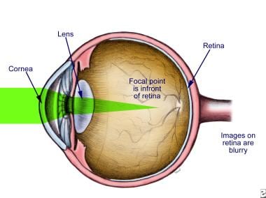
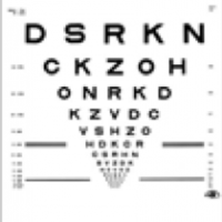
Myopia, also known as nearsightedness, is a common refractive error of the eye. It occurs when the eye is either too long or the cornea (the clear front part of the eye) is too curved. As a result, light rays focus in front of the retina instead of directly on it, causing distant objects to appear blurry while close objects remain clear.
Myopia typically develops during childhood and can progress gradually or rapidly until early adulthood. It can be corrected with glasses, contact lenses, or refractive surgery such as LASIK. Regular eye examinations are essential for people with myopia to monitor any changes in their prescription and ensure proper correction.
While myopia is generally not a serious condition, high levels of nearsightedness can increase the risk of certain eye diseases, including cataracts, glaucoma, retinal detachment, and myopic degeneration. Therefore, it's crucial to manage myopia effectively and maintain regular follow-ups with an eye care professional.
Astigmatism is a common eye condition that occurs when the cornea or lens has an irregular shape, causing blurred or distorted vision. The cornea and lens are typically smooth and curved uniformly in all directions, allowing light to focus clearly on the retina. However, if the cornea or lens is not smoothly curved and has a steeper curve in one direction than the other, it causes light to focus unevenly on the retina, leading to astigmatism.
Astigmatism can cause blurred vision at all distances, as well as eye strain, headaches, and fatigue. It is often present from birth and can be hereditary, but it can also develop later in life due to eye injuries or surgery. Astigmatism can be corrected with glasses, contact lenses, or refractive surgery such as LASIK.
Hyperopia, also known as farsightedness, is a refractive error in which the eye does not focus light directly on the retina when looking at a distant object. Instead, light is focused behind the retina, causing close-up objects to appear blurry. This condition usually results from the eyeball being too short or the cornea having too little curvature. It can be corrected with eyeglasses, contact lenses, or refractive surgery.
Refractive errors are a group of vision conditions that include nearsightedness (myopia), farsightedness (hyperopia), astigmatism, and presbyopia. These conditions occur when the shape of the eye prevents light from focusing directly on the retina, causing blurred or distorted vision.
Myopia is a condition where distant objects appear blurry while close-up objects are clear. This occurs when the eye is too long or the cornea is too curved, causing light to focus in front of the retina instead of directly on it.
Hyperopia, on the other hand, is a condition where close-up objects appear blurry while distant objects are clear. This happens when the eye is too short or the cornea is not curved enough, causing light to focus behind the retina.
Astigmatism is a condition that causes blurred vision at all distances due to an irregularly shaped cornea or lens.
Presbyopia is a natural aging process that affects everyone as they get older, usually around the age of 40. It causes difficulty focusing on close-up objects and can be corrected with reading glasses, bifocals, or progressive lenses.
Refractive errors can be diagnosed through a comprehensive eye exam and are typically corrected with eyeglasses, contact lenses, or refractive surgery such as LASIK.
Apitherapy is a form of alternative medicine that uses products derived from honey bees, such as honey, pollen, propolis, royal jelly, and venom, for therapeutic purposes. It has been practiced for thousands of years in various cultures and is believed to provide a range of health benefits, including anti-inflammatory, antimicrobial, and analgesic effects. Some proponents also claim that apitherapy can help boost the immune system, reduce pain and swelling, and treat a variety of medical conditions, such as arthritis, allergies, and wound healing. However, it is important to note that while some studies suggest potential health benefits of certain bee products, more research is needed to fully understand their safety and effectiveness. Additionally, the use of bee venom in apitherapy carries a risk of allergic reactions and anaphylaxis, so it should only be administered under the supervision of a trained healthcare professional.
I'm sorry for any confusion, but "honey" is not typically considered a medical term. It is a natural sweet substance produced by honeybees from the nectar of flowers. Honey is composed primarily of fructose and glucose, with small amounts of other sugars, water, proteins, amino acids, vitamins, minerals, enzymes, and various organic compounds.
While honey does have some potential health benefits, such as its antimicrobial properties and its use in wound healing, it is not a medical treatment or intervention. If you have any questions about the medicinal uses of honey or its role in health, I would recommend consulting with a healthcare professional.
Oxidative stress is defined as an imbalance between the production of reactive oxygen species (free radicals) and the body's ability to detoxify them or repair the damage they cause. This imbalance can lead to cellular damage, oxidation of proteins, lipids, and DNA, disruption of cellular functions, and activation of inflammatory responses. Prolonged or excessive oxidative stress has been linked to various health conditions, including cancer, cardiovascular diseases, neurodegenerative disorders, and aging-related diseases.
According to the National Institutes of Health (NIH), stem cells are "initial cells" or "precursor cells" that have the ability to differentiate into many different cell types in the body. They can also divide without limit to replenish other cells for as long as the person or animal is still alive.
There are two main types of stem cells: embryonic stem cells, which come from human embryos, and adult stem cells, which are found in various tissues throughout the body. Embryonic stem cells have the ability to differentiate into all cell types in the body, while adult stem cells have more limited differentiation potential.
Stem cells play an essential role in the development and repair of various tissues and organs in the body. They are currently being studied for their potential use in the treatment of a wide range of diseases and conditions, including cancer, diabetes, heart disease, and neurological disorders. However, more research is needed to fully understand the properties and capabilities of these cells before they can be used safely and effectively in clinical settings.
Hydrogen peroxide (H2O2) is a colorless, odorless, clear liquid with a slightly sweet taste, although drinking it is harmful and can cause poisoning. It is a weak oxidizing agent and is used as an antiseptic and a bleaching agent. In diluted form, it is used to disinfect wounds and kill bacteria and viruses on the skin; in higher concentrations, it can be used to bleach hair or remove stains from clothing. It is also used as a propellant in rocketry and in certain industrial processes. Chemically, hydrogen peroxide is composed of two hydrogen atoms and two oxygen atoms, and it is structurally similar to water (H2O), with an extra oxygen atom. This gives it its oxidizing properties, as the additional oxygen can be released and used to react with other substances.
The limbus cornea, also known as the corneoscleral junction, is the border between the transparent cornea and the opaque sclera in the eye. It's a circular, narrow region that contains cells called limbal stem cells, which are essential for maintaining the health and clarity of the cornea. These stem cells continuously regenerate and differentiate into corneal epithelial cells, replacing the outermost layer of the cornea. Any damage or disorder in this area can lead to vision impairment or loss.
 Corneal keratocyte
Neurotensin receptor 2
Ocular immune system
Acanthamoeba keratitis
Thrombospondin-2
Thrombospondin 3
Keratan sulfate
VSX1
Stroma of cornea
Prostaglandin EP4 receptor
Prostaglandin EP3 receptor
Haptotaxis
Dermal fibroblast
Post-LASIK ectasia
Transketolase
Cornea
Corneal endothelium
Dendrite (non-neuronal)
Contact lens
List of human cell types derived from the germ layers
Steven E. Wilson
Effects of long-term contact lens wear on the cornea
Bioelectricity
Boston keratoprosthesis
Mooren's ulcer
Electrotaxis
Cell culture
Corneal keratocyte - Wikipedia
Mechanical interactions and crosstalk between corneal keratocytes and the extracellular matrix - Fingerprint - University...
 Journal of Complementary and Integrative Medicine
 Pharmaceutics | Free Full-Text | Ultrasound and Microbubbles for the Treatment of Ocular Diseases: From Preclinical Research...
 Regeneration of rabbit cornea following excimer laser photorefractive keratectomy: a study on gap junctions, epithelial...
 PRIME PubMed | Thyroxine affects expression of KSPG-related genes, the carbonic anhydrase II gene, and KS sulfation in the...
 Epithelial-Stromal and Stromal Corneal Dystrophies
 Swelling Pressure Changes After Corneal UVA Riboflavin Collagen Cross-linking | IOVS | ARVO Journals
 Neovascularization, Corneal, CL-related: Background, Pathophysiology, Epidemiology
Laser-Assisted Subepithelial Keratectomy (LASEK): Background, History of the Procedure, Problem
Browsing by Author "Hänninen, Oona"
 2020-2021 BCSC Basic and Clinical Science Course™
 Molecular Vision: Quiescent keratocytes fail to repair MMC induced DNA damage leading to the long-term inhibition of...
2020-2021 BCSC Basic and Clinical Science Course™
 Apitherapy News: May 2014
 SciELO - Brazil - |i|In vivo|/i| confocal microstructural analysis of corneas presenting Kayser-Fleischer rings in patients...
![CBP/KAT3A Antibody (257003) [Unconjugated] (MAB2676): Novus Biologicals](data:image/png;base64,iVBORw0KGgoAAAANSUhEUgAAABAAAAAQCAYAAAAf8/9hAAAB+ElEQVQ4jaWSv2tTURTHP/e+H02aUhONWMTBUHBoxw4O7SLULaCI0jGkIoKIUERE/4GuTgqKhugiKLilBCouFgSX4tSgCB1EKJq2MTQ/3nv3OLz3ktcGJ79w4Zx77/me7/neC/8JNYhKlQXgLZDFiEarXylb+wqU+ObH3OnMzfLsyS9/+oECZGV5UYYEpYoDrAJ3EZhwdGNuKlObPZHWClwB8il7P5+29wIRC+gD6yvLi5t21F8DaQA8w4Xp7Idi4dhlT6QgEl4wIgRxEuI7MB0TmGgBQm7M7ilwvOBQwVEUAGICBThRPY6lPlla1QUWFFgC4mplUrZuAuMd39z2jeSTBI8BFwBHU/u66z2/V1wD1uJ2dx7VJurfdu8DM8VzuWdnJ8duxLMDXBoYqhU/93vx/gAvNnfmG83uw8bvzpX17ZZ74JtuUoEfio+QskYI2oHJ4miNEVqemQqMuEkFHrCTiJsjlmll4tDSqFhxrGCc8BnfA1vAFqXKZHRmqJbbkUpQcOCJEaEKw48UAB3gNdADcpE6FRXWCZ/5FQDCE16WbyUVKCADXB+RHuIi8G6Yipl/8MbdWL3Wjz34/I/CGDtAe5AppTa2W4c8WALORz74QB44Hp31o3GWEoRdHG2GHhxFqTIDPAXOEM7uAqeAANgDrlItfwT4C49dqlgw5A0bAAAAAElFTkSuQmCC) CBP/KAT3A Antibody (257003) [Unconjugated] (MAB2676): Novus Biologicals
CBP/KAT3A Antibody (257003) [Unconjugated] (MAB2676): Novus Biologicals
 Stem cells from your teeth could repair your eyes - Futurity
 RhoA Pull-down Activation Assay - Cytoskeleton, Inc.
 Provectus Biopharmaceuticals announces exclusive license option agreement with University of Miami
 Module 1: Cell Culture and Cell Phenotyping: NEI Vision Research Core Facility - UT Southwestern, Dallas, Texas
 Interactions between MSCs and Immune Cells: Implications for Bone Healing
 Frontiers | Identification of Common Genes and Pathways in Eight Fibrosis Diseases
Risk of cornea melt reviewed
Organisms | Open Access Articles | Digital Commons Network™
 Tech Science Press - Publisher of Open Access Journals
 CRSToday | News
 Ophthalmic Technologies XVIII | (2008) | Publications | Spie
Epithelial9
- Mitotic activity in the migrating corneal epithelial cells is also a novel finding which is probably the sign of the excessive demand for new epithelial cells in larger wounds not met alone by the proliferating limbal stock. (nih.gov)
- Corneal epithelial-stromal and stromal dystrophies are a group of inherited disorders of the cornea that are caused by progressive accumulation of deposits within the layers of the cornea. (uiowa.edu)
- The 2015 International Committee for Classification of Corneal Dystrophies (IC3D) classification system has divided corneal dystrophies into 4 categories: epithelial and subepithelial dystrophies, epithelial-stromal dystrophies, stromal dystrophies, and endothelial dystrophies. (uiowa.edu)
- Lattice corneal dystrophy (LCD) is the most common of the corneal epithelial-stromal dystrophies. (uiowa.edu)
- This technique involved the use of alcohol to separate the corneal epithelium from the stroma to create an epithelial sheet that could be repositioned over the ablated stroma. (medscape.com)
- A modern technique, serial block face scanning electron microscopy (SB-EM), was used to reconstruct a three-dimensional model of a feline corneal squamous epithelial cell and the apical corneal surface. (helsinki.fi)
- SB-EM analysis of feline corneal epithelial cell ultrastructure revealed intricate finger like protrusions which adjacent cells use to interlock with each other. (helsinki.fi)
- Glucose is the primary metabolic substrate for the epithelial cells, stromal keratocytes (corneal fibroblasts residing in the stroma), and endothelial cells. (aao.org)
- The hydrogel does not affect the viability of keratocytes and corneal epithelial cells. (reviewofoptometry.com)
Cornea11
- In the unperturbed cornea keratocytes stay dormant, coming into action after any kind of injury or inflammation. (wikipedia.org)
- Any glitch in the precisely orchestrated process of healing may cloud the cornea, while excessive keratocyte apoptosis may be a part of the pathological process in the degenerative corneal disorders such as keratoconus, and these considerations prompt the ongoing research into the function of these cells. (wikipedia.org)
- By the end of eye development an interconnected keratocyte network is established in the cornea, with dendrites of neighbouring cells contacting each other. (wikipedia.org)
- After an injury to the cornea, some keratocytes undergo apoptosis, prompted by the signaling molecules secreted by the upper layers, such as IL1 alpha and TNF-alpha. (wikipedia.org)
- In a healthy cornea the programmed cell death is a rare occasion, but immediately after an injury to the uppermost layer keratocytes directly under the injury site commit apoptosis. (wikipedia.org)
- With additional control and sequestrum specimens the morphology of keratocytes in the normal feline cornea and corneal sequestra could be compared. (helsinki.fi)
- 0.05) throughout the cornea and corneas receiving 0.2 mg/ml MMC treatment 2 months before LK injury showed complete absence of any corneal scarring. (molvis.org)
- Dr. Kugler, a surgical associate and cornea/refractive surgery fellow with Ming Wang, MD, PhD, in Nashville, TN, briefly described four cases of corneal melt that had one similarity, namely, that the complication developed in the area of the incision. (ophthalmologytimes.com)
- PURPOSE: We hypothesized that Transforming growth factor beta receptor 2 (Tgfbr2) deletion in keratocyte (Tgfbr2kera-cko), the corneal stroma cell, can result in corneal thinning and generate a potential model for Cornea Ectasia (CE). (bvsalud.org)
- TEM showed that keratocytes were unhealthy and stromal collagen fibril density was significantly reduced in Tgfbr2kera-cko as compared with that in Tgfbr2Ctrl cornea. (bvsalud.org)
- The cornea phenotype manifested in these Tgfbr2kera-cko mice resembles corneal ectasia disease in humans. (bvsalud.org)
Epithelium8
- During regeneration both connexins were expressed throughout the corneal epithelium including the migrating cells. (nih.gov)
- Transient up-regulation and relocation of connexins within the regenerating epithelium may reflect the involvement of direct cell-cell communication in corneal wound healing. (nih.gov)
- TGFβI is located on chromosome 5q31 and codes for keratoepithelin, a protein secreted by corneal epithelium. (uiowa.edu)
- By retaining a flap of corneal epithelium, LASEK may decrease the risk of infection and incidence of corneal haze, while reducing recovery time and postoperative discomfort when compared with PRK. (medscape.com)
- In the epithelium and endothelium, the HMP pathway breaks down 35%-65% of the glucose, but the keratocytes of the stroma metabolize very little glucose via this pathway. (aao.org)
- Histopathologic study showed decreased keratocyte density and thinning of the epithelium. (ophthalmologytimes.com)
- The cell proliferation marker Ki67 expression level increased â ¼9% in Tgfbr2kera-cko corneal epithelium as compared with that in Tgfbr2Ctrl, however, the Krt14 and Krt12 expression pattern was not obviously changed in Tgfbr2kera-cko corneal epithelium. (bvsalud.org)
- Irregularity or edema of the corneal epithelium disrupts the smoothness of the air-tear film interface, the most significant component of the total refractive power of the eye, thereby reducing visual acuity. (wikidoc.org)
Thickness9
- This corneal layer, representing about 85-90% of corneal thickness, is built up from highly regular collagenous lamellae and extracellular matrix components. (wikipedia.org)
- Corneal transparency was measured digitally and thickness was determined from cryostat cross sections. (unboundmedicine.com)
- In 34 porcine eyes, the central corneal thickness (CCT) was determined by ultrasound pachymetry. (arvojournals.org)
- A non-significant reduction of central corneal thickness after treatment was observed in the CXL group (p = 0.815), and in the hydration change in the CXL group compared to the control group (p = 0.200). (arvojournals.org)
- The results obtained, when testing full thickness corneal buttons, may not detect the changes induced by CXL, since only the anterior segment of the stroma is cross-linked. (arvojournals.org)
- Since its introduction, LASIK has been associated with various complications, specifically when performed on eyes with decreased corneal thickness, irregular astigmatism, dryness, preexisting ocular surface disease, or glaucoma, to the point where several of these entities have become relative contraindications to performing LASIK. (medscape.com)
- Microstructural analysis using confocal laser scanning microscopy evaluated increased corneal thickness, decreased number of cells, increased debris or specific deposits, and unusual microstructures. (scielo.br)
- Following the thinning, the corneal thickness begins to increase over a matter of weeks to months. (aao.org)
- METHODS: Corneal thickness of Tgfbr2kera-cko and Tgfbr2Ctrl was examined with Optical Coherence Tomography (OCT) at post-natal (P) days 42 and 70, respectively. (bvsalud.org)
Fibroblasts residing in t1
- Corneal keratocytes (corneal fibroblasts) are specialized fibroblasts residing in the stroma. (wikipedia.org)
Dystrophy11
- genetic disruption of its synthesis leads to the macular corneal dystrophy. (wikipedia.org)
- According to comparative research, their functions drastically diverge from the norm in keratoconus, the most frequent form of corneal dystrophy. (wikipedia.org)
- Reis-Bücklers, formerly known as Granular corneal dystrophy type III or Corneal Dystrophy of Bowman's type I, typically present with normal corneas at birth but develop painful recurrent erosions, opacification, and progressive vision loss within the first decade of life (1). (uiowa.edu)
- The hyaline-like material consists of rod-like bodies ultrastructurally, which helps distinguish it from Thiel-Behnke corneal dystrophy (1, 2). (uiowa.edu)
- The purpose of this study was to identify the genetic cause and describe the clinical phenotype of Schnyder corneal dystrophy (SCD) in six unrelated probands. (biomedcentral.com)
- The phosphoinositide kinase, FYVE finger containing ( PIKFYVE ) gene has been identified as a gene responsible for fleck corneal dystrophy (FCD). (molvis.org)
- The purpose of this study is to report a novel mutation of the PIKFYVE gene in a Japanese patient with fleck corneal dystrophy. (molvis.org)
- To the best of our knowledge, this is the first study to show that a novel mutation (p.Glu1389AspfsX16) causing the truncation of the PIKFYVE protein causes fleck corneal dystrophy in the Japanese population. (molvis.org)
- Fleck corneal dystrophy (FCD, Online Mendelian Inheritance in Man (OMIM) #121850) was first reported in 1957 by Francois and Neetens [ 9 ], and is one of the hereditary corneal dystrophies in which the causative genes have already been identified. (molvis.org)
- This corneal dystrophy is a rare autosomal dominant disease characterized by numerous tiny, dot-like white flecks scattered in all layers of the corneal stroma. (molvis.org)
- The patient had no obvious vision loss or any complaints related to this corneal dystrophy, and the appropriateness of our identified mutation as a causative one for FCD is theoretically discussed. (molvis.org)
Stromal cells3
- Experiments showed that stem cells of the dental pulp, obtained from routine human third molar, or wisdom tooth, could be turned into corneal stromal cells called keratocytes, which have the same embryonic origin. (futurity.org)
- BACKGROUND: To develop an in vitro model to study the effects of excimer laser keratectomy on corneal stromal cells, we evaluated two types of collagen gel populated with keratocytes. (johnshopkins.edu)
- We have also demonstrated that SPL metabolism is altered in "injured" corneal stromal cells, and that stimulation of healthy corneal stromal cells with Sphingosine 1-Phosphate (S1P), a bioactive SPL, induces TGF-ß1 expression and fibrosis, signaling through the S1P receptor 3 (S1P3). (hhs.gov)
Rabbit corneal keratocytes1
- The purpose of this study was to determine the acute and long-term effects of mitomycin C (MMC) on quiescent rabbit corneal keratocytes regarding cell proliferation, myofibroblast differentiation and DNA repair. (molvis.org)
Myofibroblasts7
- To accomplish this goal, a protocol to isolate and culture equine corneal keratocytes, fibroblasts and myofibroblasts was developed. (bepress.com)
- These events lead to transdifferentiation of keratocytes into myofibroblasts through fibroblasts and often results in permanent corneal scarring. (bepress.com)
- Hence, therapeutic targets that reduce transdifferentiation of fibroblasts into myofibroblasts may provide a clinically relevant approach to treat corneal fibrosis and improve long-term visual outcomes. (bepress.com)
- Corneal fibrosis is characterized by the formation of corneal scars from over-accumulation of disorganized extracellular matrix (ECM) produced by fibroblasts and myofibroblasts after they are activated by injury or infection. (hhs.gov)
- Our recent advances, with the aid of an NIH/NEI R21 (EY025256), have led us to discover a novel potential mechanism that combines sphingolipid (SPL) signaling with classical transforming growth factor-ß (TGF-ß) pathways to mediate corneal fibrosis via activation/differentiation of keratocytes into myofibroblasts. (hhs.gov)
- Based on these discoveries, we hypothesize that S1P is a key mediator of corneal fibrosis via activation of TGF-ß, and TGF-ß in turn induces expression of S1P signaling proteins and thus forms a positive feedback loop which drives irreversible activation of corneal fibroblasts and differentiation to myofibroblasts. (hhs.gov)
- LM and TEM showed activated keratocytes (myofibroblasts), inflammatory debris and vascular tissues in the stroma. (edu.au)
Apoptosis4
- Some keratocytes underlying the site of injury, even a light one, undergo apoptosis immediately after the injury. (wikipedia.org)
- Apoptosis of keratocytes, either in quiescent or active state, is a process that attracts special attention. (wikipedia.org)
- a hypothesis exists that presents excessive keratocyte apoptosis as a major pathological event in keratoconus. (wikipedia.org)
- These findings suggest increased keratocyte apoptosis and tissue digestion. (ophthalmologytimes.com)
Haze7
- These data indicate that the effect of MMC on corneal scarring and haze is related to the generation of DNA ICLs leading to defective cell replication and gene expression. (molvis.org)
- Corneal arcus and stromal haze were the most prominent phenotypical feature in two probands. (biomedcentral.com)
- Although de novo occurrence of mutations in UBIAD1 is extremely rare, SCD should be considered in the differential diagnosis of bilateral corneal haze and/or crystal deposition, especially in children. (biomedcentral.com)
- For example, although both DLK and CTK both lead to a reduction in corneal clarity and a hyperopic shift, the post-surgical opacification seen in DLK tends to be nonlocalized [7] and subepithelial [9] while the post-surgical haze in CTK is centralized and extends anteriorly or posteriorly from the interface. (aao.org)
- Eyes were analyzed for corneal haze and matrix remodeling components using slit lamp biomicroscopy, in vivo confocal microscopy, light microscopy (LM), transmission electron microscopy (TEM), immunohistochemistry (IHC) and western blotting (WB). (edu.au)
- Hevin -/- mice developed corneal haze as early as 1-2 weeks post IrrPTK-treatment compared to the WT group, which peaked at 3-4 weeks. (edu.au)
- Hevin -/- mice develop early corneal haze characterized by severe chronic inflammation and stromal fibrosis that can be rescued with exogenous administration of rhHevin. (edu.au)
Treat corneal3
- Objective-Mitomycin C (MMC) is used clinically to treat corneal scarring in human patients. (bepress.com)
- We investigated the safety and efficacy of MMC to treat corneal scarring in horses by examining its effects at the early and late stages of disease using an in-vitro model. (bepress.com)
- But researchers at the University of New Hampshire are currently experimenting with a hydrogel lens they created to treat corneal melting-a significant cause for blindness with no cure. (reviewofoptometry.com)
Stroma and keratocytes1
- This study can be continued in attempt to image corneal stroma and keratocytes. (helsinki.fi)
Settled in the stroma1
- Once settled in the stroma, keratocytes start synthesizing collagen molecules of different types (I, V, VI) and keratan sulfate. (wikipedia.org)
Endothelial cells1
- keratocytes and endothelial cells expressed T4 receptor beta (THRB) mRNA. (unboundmedicine.com)
Refractive6
- Concepts of corneal refractive surgery, such as keratectomy, keratotomy, and thermokeratoplasty, were first described in 1898 by Lans who published a set of experiments that focused on treating astigmatism in rabbits. (medscape.com)
- Refractive surgery, as it is known today, was not realized until 1966 when Pureskin first appreciated its potential with the demonstration that refractive changes could be made by removing central tissue underneath a corneal flap. (medscape.com)
- The FDA's Dermatologic and Ophthalmic Drugs Advisory Committee and Ophthalmic Devices Panel heard testimony on the company's combined riboflavin solutions and UV irradiation device, which are indicated for progressive keratoconus and corneal ectasia following refractive surgery. (crstoday.com)
- On the question of "Has substantial evidence of efficacy and safety been demonstrated for the drug device combination of Photrexa Viscous and Photrexa and the KXL System to support approval for corneal ectasia following refractive surgery? (crstoday.com)
- The Avedro new drug application submission encompasses data from three prospective, randomized, parallel-group, open-label, sham-controlled, 12-month trials conducted in the United States to determine the safety and effectiveness of riboflavin ophthalmic solutions used in conjunction with UVA irradiation for performing corneal cross-linking in eyes with keratoconus and corneal ectasia following refractive surgery. (crstoday.com)
- Patients with CTK typically develop dense central corneal opacification, stromal tissue loss, striae, and significant hyperopic refractive shift [7] [8] . (aao.org)
Endothelium3
- Cellular and molecular assessment of rose bengal photodynamic antimicrobial therapy on keratocytes, corneal endothelium and limbal stem cell niche. (ophthalmologytimes.com)
- a thin acellular layer that serves as the modified basement membrane of the corneal endothelium. (wikidoc.org)
- The corneal endothelium is bathed by aqueous humour , not by blood or lymph , and has a very different origin, function, and appearance from vascular endothelia . (wikidoc.org)
Keratitis2
- Pseudomonas aeruginosa-Derived Extracellular Vesicles Modulate Corneal Inflammation: Role in Microbial Keratitis? (elsevierpure.com)
- This review will highlight evidence from experimental studies identifying components of the ocular ECS and discuss the functional role of the ECS during different ocular inflammatory disease states, including uveitis and corneal keratitis. (frontiersin.org)
Wound10
- Corneal wound repair was investigated in rabbits following excimer laser ablation of a 6 mm diameter and 90 microm deep disc. (nih.gov)
- Acacia honey is a natural product which has proven to have therapeutic effects on skin wound healing, but its potential healing effects in corneal wound healing have not been studied. (blogspot.com)
- The results of the present study show promising role of AH role in accelerating the initial stage of corneal wound healing. (blogspot.com)
- Circumferential and radial forces caused by implantation cause wound gape, which, in turn, can increase the risk of developing corneal melt. (ophthalmologytimes.com)
- Objective-To establish an in vitro model for the investigation of equine corneal wound healing. (bepress.com)
- Keratocytes are specialized fibroblasts that play roles in wound healing, maintaining corneal transparency, and corneal component synthesis. (sciencellonline.com)
- The corneal wound-healing, as well as the process of fibrosis, is driven by multiple complex pathways involving many cytokines, growth factors, and chemokines, which are not completely understood. (hhs.gov)
- and in vivo models of corneal wound healing in wild type, Sphk1, and S1P3 knockout mice, along with testing the therapeutic potential of targeting S1P and TGF-ß signaling using selective inhibitors in these models (Aim 2). (hhs.gov)
- In this study, we examined the functional role of hevin using a corneal stromal wound healing model achieved by an excimer laser-induced irregular phototherapeutic keratectomy (IrrPTK) in hevin-null (hevin -/- ) mice. (edu.au)
- Thus, hevin plays a pivotal role in the corneal wound healing. (edu.au)
Corneas6
- Incidence of subsequent corneal graft rejection is estimated by one study to be 1.7 times higher in a setting of vascularized rather than nonvascularized host corneas. (medscape.com)
- Pallikaris then used the excimer laser ablation on the corneal stromal bed under a hinged flap in rabbit corneas. (medscape.com)
- Compared with the control corneas, there were fewer keratocytes in the anterior stroma (17.380 vs. 22.380/mm 3 ). (scielo.br)
- Corneal blindness, which affects millions of people worldwide, is typically treated with transplants of donor corneas, says senior investigator James Funderburgh, professor of ophthalmology at the University of Pittsburgh School of Medicine. (futurity.org)
- The researchers injected the engineered keratocytes into the corneas of healthy mice, where they integrated without signs of rejection. (futurity.org)
- Dr. Kugler cited a report by Sépideh Samimi, MD, and associates of histopathologic findings in corneas from eyes that required a corneal transplant following implantation of the inserts. (ophthalmologytimes.com)
Transparency4
- Corneal crystallins, like the lens ones, are thought to help maintain the transparency and optimal refraction. (wikipedia.org)
- The present study was conducted to determine whether corneal cells differentially express genes for T4 regulation, keratan sulfate proteoglycan (KSPG) synthesis, crystallins, and endothelial cell ion transporters during transparency development and whether these expressions are altered when E9 embryos are treated with T4. (unboundmedicine.com)
- The lack of knowledge regarding this process is a critical barrier to developing new treatment strategies to minimize scarring and retain or restore corneal transparency. (hhs.gov)
- Corneal structure is highly organized and unified in architecture with structural and functional integration which mediates transparency and vision. (lboro.ac.uk)
Proliferation4
- By the moment of eye opening after birth the proliferation of keratocytes is all but finished and most of them are in the quiescent state. (wikipedia.org)
- MMC significantly blocked TGFβ-induced cell proliferation and myofibroblast differentiation in cultured quiescent keratocytes and altered the transcriptional regulation of macrophage chemotactic protein-1 (MCP-1) and alpha smooth muscle actin (αSMA). (molvis.org)
- The optimal dose of AH in the basal medium (FD) and medium containing serum (FDS) for keratocytes proliferation was identified using MTT assay. (blogspot.com)
- Histological H&E staining, transmission electron micrograph (TEM), and immunofluorescence staining (IFS) were harnessed to examine corneal cell morphology, proliferation, differentiation, and collagen fibrils. (bvsalud.org)
Human keratocytes1
- Crystallins expressed by human keratocytes are ALDH1A1, ALDH3A1, ALDH2 and TKT. (wikipedia.org)
Flecks appear1
- Histologically, these flecks appear to be keratocytes distended with lipid and mucopolysaccharide filled intracytoplasmic vacuoles. (utsouthwestern.edu)
Morphology2
- This study aimed to explore the effects of Acacia honey (AH) on corneal keratocytes morphology, proliferative capacity, cell cycle, gene and protein analyses. (blogspot.com)
- Corneal keratocytes cultured in media supplemented with 0.025% AH showed an increase in proliferative capacity while retaining their morphology, gene and protein expressions with normal cell cycle. (blogspot.com)
Phenotype1
- Both eyes exhibited small, dot-like, white flecks scattered throughout all layers of the corneal stroma, which corresponds to the typical FCD phenotype. (molvis.org)
Quiescent keratocytes4
- Quiescent keratocytes synthesize the so-called crystallins, known primarily for their role in the lens. (wikipedia.org)
- Quiescent keratocytes cultured in serum-free media were exposed to various concentrations of MMC and then treated with transforming growth factor-β (TGFβ). (molvis.org)
- The long-term ability of quiescent keratocytes to repair MMC induced damage in vivo was evaluated in rabbits treated with MMC 2 months before 100 μm deep lamellar keratectomy (LK) injury. (molvis.org)
- The morphological changes, gene and protein expressions of aldehyde dehydrogenase (ALDH), marker for quiescent keratocytes and vimentin, marker for fibroblasts were detected using q-RTPCR and immunocytochemistry respectively. (blogspot.com)
Edema3
- Preliminary data suggest that the swelling pressure is reduced in the anterior approximately 200µm stroma in the treatment group, indicating that CXL treatment can reduce corneal edema in vivo . (arvojournals.org)
- An alternative theory to the observed thinning proposes that micro edema into the corneal periphery causes mild stretching [13] . (aao.org)
- Moreover, mechanical eye-rubbing on Tgfbr2kera-cko resulted in corneal hydrops and edema. (bvsalud.org)
Differentiation1
- Controlling keratocyte activation and differentiation are key for the inhibition and prevention of fibrosis. (lboro.ac.uk)
Methods1
- METHODS: Keratocyte-populated collagen gels were prepared with type I collagen in 6-well plates or in culture plate inserts, the bottom of which consisted of a nitrocellulose membrane, contained within 6-well plates. (johnshopkins.edu)
Transplantation2
- No effective therapies for corneal fibrosis have been developed and the most reliable treatment option is corneal transplantation, which has numerous limitations/ and complications, including post-surgical corneal fibrosis. (hhs.gov)
- Appropriate corneal models may lead to a reduced need for corneal transplantation as presently there are insufficient numbers or suitable tissue to meet demand. (lboro.ac.uk)
Vivo1
- In vivo confocal microscopy is a useful tool for evaluating the corneal microstructure when a Kayser-Fleischer ring is clinically present. (scielo.br)
Autosomal2
- It is typically an autosomal dominant, bilateral disease that typically presents toward the end of the first decade of life with symptoms of recurrent corneal erosions and decreased vision. (uiowa.edu)
- an autosomal dominant disorder characterized by numerous small white flecks present in all layers of the corneal stroma. (utsouthwestern.edu)
Incidence3
- Silicone hydrogel CLs with oxygen permeabilities approaching 100-200 Fatt Dk units have decreased the incidence of corneal NV among CL users. (medscape.com)
- Corneal melt can develop after implantation of corneal inserts, although incidence is rare. (ophthalmologytimes.com)
- Nashville, TN -Corneal melt can develop after implantation of corneal inserts (Intacs, Addition Technology), although the incidence is rare, said Lance Kugler, MD. (ophthalmologytimes.com)
Intraocular pressure1
- The control eyes all presented normal visual acuity, intraocular pressure, and corneal appearance. (scielo.br)
Fibrosis3
- Up to one fourth of all cases of blindness worldwide are attributable to corneal opacities generated by scarring, or fibrosis, representing a huge economic and societal burden. (hhs.gov)
- The role of S1P in corneal fibrosis has not received substantial attention and we are currently the only group pursuing SPL-based processes as part of the potential mechanism. (hhs.gov)
- If successful, the results from the proposed studies could have far-reaching scientific and clinical significance, as the understanding of corneal fibrotic mechanisms and the role of S1P as an important mediator would not only be important for clinical management/treatment of corneal fibrosis, but could also be applicable to many diseases in which fibrosis is a major pathological outcome, such as liver, lung, and cardiac fibrosis. (hhs.gov)
Collagen gel2
- RESULTS: Keratocytes cultured in collagen gel developed cytoplasmic processes and formed networks of interconnected cells. (johnshopkins.edu)
- CONCLUSIONS: These results suggest the use of keratocyte-populated collagen gel as an in vitro model of cellular response to excimer laser keratectomy and also suggest that gel prepared in culture plate inserts is the preferred method. (johnshopkins.edu)
Posterior1
- Buratto performed excimer laser ablation on the posterior surface of the resected corneal disc before replacing and resuturing it back to its original position. (medscape.com)
Microscopy1
- Slit-lamp microscopy, corneal topography, and optical coherence tomography were performed for the clinical examination of the patient's eye. (molvis.org)
Abnormal2
- Intact, though abnormal, keratocytes contained and were surrounded by numerous electron lucent profiles. (helsinki.fi)
- MMC induces DNA damage to quiescent corneal keratocytes, which remains unrepaired, resulting in abnormal cell replication and gene transcription that leads to long-term effects on corneal repair. (molvis.org)
Tissue4
- Other neighbouring keratocytes, when acted upon by the same molecules, become active, proliferate and start synthesizing matrix metalloproteinases that cause tissue remodeling. (wikipedia.org)
- This could also be new source of corneal transplant tissue made from the patient's own cells. (futurity.org)
- They also used the cells to develop constructs of corneal stroma akin to natural tissue. (futurity.org)
- Dr. Kugler suggested that corneal melt might occur because the corneal tissue is weaker or inflamed around the corneal inserts, and there is evidence of decreased keratocyte density. (ophthalmologytimes.com)
Patient's1
- According to one study, patient's keratocytes have decreased levels of one of the alcohol dehydrogenase subforms, they secrete significantly less superoxide dismutase 3, according to another. (wikipedia.org)
Dystrophies1
- The old classification for corneal stromal dystrophies is listed in Table 3. (uiowa.edu)
Density2
- According to one study, average keratocyte density in the human stroma is about 20500 cells per mm3, or 9600 in a column of 1 mm2 in section. (wikipedia.org)
- The ring consists of granular, bright particles that increase in density toward Descemet's membrane, and is associated with a decreased number of keratocytes and peculiar dark, round areas in all stromal layers, probably a sign of corneal damage. (scielo.br)
Ocular1
- Unlike conventional MMP inhibitors, this hydrogel lens minimizes the risk of serious, nonspecific side effects and provides a method to slow down the progression of corneal melting and other related ocular diseases, the researchers say. (reviewofoptometry.com)
Genes1
- This array kit profiles 88 key genes involved in corneal keratocyte function. (sciencellonline.com)
Myofibroblast1
- The equine corneal fibroblast or myofibroblast cultures were produced by growing primary ECF in the presence or absence of transforming growth factor beta-1 (TGFβ1) under serum-free conditions. (bepress.com)
Squamous1
- a simple squamous or low cuboidal monolayer of mitochondria-rich cells responsible for regulating fluid and solute transport between the aqueous and corneal stromal compartments. (wikidoc.org)
Anterior1
- Bowman's layer (also erroneously known as the anterior limiting membrane , when in fact it is not a membrane but a condensed layer of collagen): a tough layer that protects the corneal stroma, consisting of irregularly-arranged collagen fibers. (wikidoc.org)
Morphological1
- Cultured keratocytes supplemented with AH showed no morphological changes compared to control. (blogspot.com)
Melt1
- In his practice, four cases of corneal melt have occurred in association with implantation of the inserts. (ophthalmologytimes.com)
Nerves1
- Corneal nerves have been observed to terminate in a logarithmic spiral pattern. (wikidoc.org)