Colonography, Computed Tomographic
Colonic Polyps
Laxatives
Enema
Radiographic Image Interpretation, Computer-Assisted
Colorectal Neoplasms
Tomography, X-Ray Computed
Insufflation
Iothalamate Meglumine
Sensitivity and Specificity
Barium Sulfate
Radiographic Image Enhancement
Imaging, Three-Dimensional
Tomography Scanners, X-Ray Computed
Mass Screening
Colonic Diseases
Tomography
Reproducibility of Results
Colon
Radiology
Image Processing, Computer-Assisted
False Positive Reactions
Tomography, Optical
Pattern Recognition, Automated
Iohexol
Early Detection of Cancer
Incidental Findings
Polyps
Feasibility Studies
Diagnosis, Computer-Assisted
Wisconsin
Patient Preference
Cost-Benefit Analysis
Algorithms
Tomography, Emission-Computed
Cone-Beam Computed Tomography
Infarction
Prospective Studies
Observer Variation
Magnetic Resonance Imaging
Performance of multidetector computed tomography colonography compared with conventional colonoscopy. (1/182)
BACKGROUND AND AIMS: This was a prospective blinded study to compare computed tomography (CT) colonography, performed with multidetector arrays CT scan (MDCT), with conventional colonoscopy for the detection of colorectal neoplasia. METHODS: Fifty patients were examined by MDCT after standard bowel preparation and rectal air insufflation in the supine and prone positions. Data sets were examined by one radiologist and one gastroenterologist blinded to the patient's history and colonoscopy results. Patients subsequently underwent colonoscopy on the same day, which served as the gold standard. RESULTS: Nine of 11 lesions >10 mm (82%), 5/15 lesions of 6-9 mm (33%), and 1/42 polyps <5 mm (3%) were detected by MDCT colonography. One false positive result for a structure larger than 10 mm was described. Nineteen of 21 patients who had no lesions during conventional colonoscopy were considered free of lesions by MDCT colonography, yielding a per patient specificity of 90%. CONCLUSION: MDCT colonography provides good data quality and has good sensitivity and specificity for the detection of colonic lesions of 10 mm or more. (+info)Virtual endoscopy: a promising new technology. (2/182)
Growing evidence shows that early detection of cancer can substantially reduce mortality, necessitating screening programs that encourage patient compliance. Radiology is already established as a screening tool, as in mammography for breast cancer and ultrasonography for congenital anomalies. Advanced processing of helical computed tomographic data sets permits three-dimensional and virtual endoscopic models. Such models are noninvasive and require minimal patient preparation, making them ideal for screening. Virtual endoscopy has been used to evaluate the colon, bronchi, stomach, blood vessels, bladder, kidney, larynx, and paranasal sinuses. The most promising role for virtual endoscopy is in screening patients for colorectal cancer. The technique has also been used to evaluate the tracheobronchial tree for bronchogenic carcinoma. Three-dimensional and virtual endoscopy can screen, diagnose, evaluate and assist determination of surgical approach, and provide surveillance of certain malignancies. (+info)Computer-aided detection and diagnosis at the start of the third millennium. (3/182)
Computer-aided diagnosis has been under development for more than 3 decades. The rate of progress appears exponential, with either recent approval or pending approval for devices focusing on mammography, chest radiographs, and chest CT. Related technologies improve diagnosis for many other types of medical images including virtual colonography, vascular imaging, as well as automated quantitation of image-derived metrics. A variety of techniques are currently employed with success, likely reflecting the variety of imagery used, as well as the variety of tasks. Most areas of medical imaging have had efforts at computer assistance, and some have even received FDA approval and can be reimbursed. We anticipate that the rapid advance of these technologies will continue, and that application will broaden to cover much of medical imaging. Acceptance of, and integration of computer-aided diagnosis technology with the electronic radiology practice is a current challenge. These challenges will be overcome, and we expect that computer-aided diagnosis will be routinely applied to medical images. (+info)Current and evolving strategies for colorectal cancer screening. (4/182)
BACKGROUND: Colorectal cancer is a major cause of cancer mortality and morbidity. Screening can potentially prevent most colorectal cancers by detection and removal of precursor adenomas. METHODS: The literature and clinical practice guidelines are reviewed, with an emphasis on advances of the last 10 years and evolving screening methods. RESULTS: Colonoscopy has come to be used for screening in persons at average risk for colorectal cancer because of the comparative ineffectiveness of other methods, although these methods continue to be recommended. Virtual colonoscopy and fecal DNA testing are emerging technologies with promise to be more effective than fecal occult blood testing or sigmoidoscopy in selecting those persons who should undergo colonoscopy. Next to age, family history is the most common risk factor for colorectal cancer and one that warrants more aggressive screening and, in some instances, genetic counseling and testing. Hereditary nonpolyposis colorectal cancer accounts for as many as 1 in 20 colorectal cancers, but to take advantage of recent advances in genetic testing for this disorder, a high level of clinical suspicion must be maintained. CONCLUSIONS: If we are to reduce mortality and morbidity from colorectal cancer, practicing clinicians need to be aware of current and evolving strategies for colorectal screening, and assertively recommend the appropriate strategy to their patients. (+info)Chemotherapy and surgery: new perspectives on the treatment of unresectable liver metastases. (5/182)
Liver metastases concern half of patients with colorectal cancer, and are frequently unresectable, jeopardizing patient outcome. Owing to increased efficacy, chemotherapy can render initially inoperable patients amenable to potentially curative resection. The 34% 5-year and 20% 10-year survival of patients resected following neoadjuvant chronomodulated chemotherapy with 5-fluorouracil, folinic acid and oxaliplatin is similar to that of patients whose disease was operable at diagnosis. Recently, a group of 16 patients were treated with irinotecan and became resectable after treatment. Their survival (56% at 3 years) matches that of patients treated with other forms of chemotherapy. The poor prognosis of patients with non-resectable hepatic metastases might now be improved by the combination of chemotherapy and surgery. (+info)Extracolonic findings at computed tomography colonography are a challenge. (6/182)
AIM: Our aim was to perform a prospective evaluation of the frequency and diagnostic consequences of extracolonic findings at multidetector array computed tomography colonography (MDCTC) in asymptomatic patients undergoing surveillance for former colorectal polyps or cancer. PATIENTS AND METHODS: Seventy five consecutive patients undergoing surveillance for former colorectal cancer (CRC) or large bowel adenoma were examined with MDCTC. Two independent observers evaluated the images with regard to extracolonic findings. Patient records and radiological information systems were reviewed to determine the results and consequences of the workup derived from MDCTC. RESULTS: Sixty five per cent (95% confidence interval (CI) 55-73%) of patients had extracolonic abnormalities and in 12% (CI 7-18%) of patients additional workup was indicated. Two patients (3% (CI 1-6%)) underwent surgery because of the findings (one) or because of complications of the workup (one). CONCLUSION: MDCTC identifies a large number of extracolonic findings. Approximately 12% of asymptomatic patients undergo additional workup, of benefit to only a few. The high prevalence of extracolonic findings may make MDCTC a problematic colorectal screening tool for both ethical and economic reasons. (+info)Computed tomographic virtual colonoscopy to screen for colorectal neoplasia in asymptomatic adults. (7/182)
BACKGROUND: We evaluated the performance characteristics of computed tomographic (CT) virtual colonoscopy for the detection of colorectal neoplasia in an average-risk screening population. METHODS: A total of 1233 asymptomatic adults (mean age, 57.8 years) underwent same-day virtual and optical colonoscopy. Radiologists used the three-dimensional endoluminal display for the initial detection of polyps on CT virtual colonoscopy. For the initial examination of each colonic segment, the colonoscopists were unaware of the findings on virtual colonoscopy, which were revealed to them before any subsequent reexamination. The sensitivity and specificity of virtual colonoscopy and the sensitivity of optical colonoscopy were calculated with the use of the findings of the final, unblinded optical colonoscopy as the reference standard. RESULTS: The sensitivity of virtual colonoscopy for adenomatous polyps was 93.8 percent for polyps at least 10 mm in diameter, 93.9 percent for polyps at least 8 mm in diameter, and 88.7 percent for polyps at least 6 mm in diameter. The sensitivity of optical colonoscopy for adenomatous polyps was 87.5 percent, 91.5 percent, and 92.3 percent for the three sizes of polyps, respectively. The specificity of virtual colonoscopy for adenomatous polyps was 96.0 percent for polyps at least 10 mm in diameter, 92.2 percent for polyps at least 8 mm in diameter, and 79.6 percent for polyps at least 6 mm in diameter. Two polyps were malignant; both were detected on virtual colonoscopy, and one of them was missed on optical colonoscopy before the results on virtual colonoscopy were revealed. CONCLUSIONS: CT virtual colonoscopy with the use of a three-dimensional approach is an accurate screening method for the detection of colorectal neoplasia in asymptomatic average-risk adults and compares favorably with optical colonoscopy in terms of the detection of clinically relevant lesions. (+info)Postprocessing techniques of CT colonography in detection of colorectal carcinoma. (8/182)
AIM: To evaluate the value of postprocessing techniques of CT colonography, including multiplanar reformation (MPR), virtual colonoscopy (VC), shaded surface display (SSD) and Raysum, in detection of colorectal carcinomas. METHODS: Sixty-four patients with colorectal carcinoma underwent volume scanning with spiral CT. MPR, VC, SSD and Raysum images were obtained by using four kinds of postprocessing techniques in workstation. The results were comparatively analyzed according to circumferential extent, lesion length and pathology pattern of colorectal carcinomas. All diagnoses were proved pathologically and surgically. RESULTS: The accuracy of circumferential extent of colorectal carcinoma determined by MPR, VC, SSD and Raysum was 100.0%, 82.8%, 79.7% and 79.7%, respectively. There was a significant statistical difference between MPR and VC. The consistent rate of lesion length was 89.1%, 76.6%, 95.3% and 100.0%, respectively. There was a statistical difference between VC and SSD. The accuracy of discriminating pathology pattern was 81.3%, 92.2%, 71.9% and 71.9%, respectively. There was a statistical difference between VC and SSD. MPR could determine accurately the circumference of colorectal carcinoma, Raysum could determine the length of lesion more precisely than SSD, VC was helpful in discriminating pathology patterns. CONCLUSION: MPR, VC, SSD and Raysum have advantage and disadvantage in detection of colorectal carcinoma, use of these methods in combination can disclose the lesion more accurately. (+info)Computed tomographic colonography (CTC), also known as virtual colonoscopy, is a medical imaging technique that uses computed tomography (CT) scans to produce detailed images of the large intestine (colon) and rectum. In CTC, specialized software creates two- and three-dimensional images of the colon's inner surface, allowing healthcare providers to examine the colon for polyps, tumors, and other abnormalities.
During a CTC procedure, patients are usually given a mild laxative and asked to follow a clear liquid diet beforehand to clean out the colon. A small tube is inserted into the rectum to inflate the colon with air or carbon dioxide, making it easier to visualize any abnormalities. The patient lies on their back and then their stomach while the CT scanner takes multiple images of the abdomen and pelvis from different angles.
CTC has several advantages over traditional colonoscopy, including less invasiveness, lower risk of complications, faster recovery time, and the ability to examine the entire colon without missing any areas. However, if polyps or other abnormalities are detected during a CTC, a follow-up diagnostic colonoscopy may be necessary for removal or further evaluation.
It is important to note that CTC does not replace traditional colonoscopy as a screening tool for colorectal cancer. While it has similar accuracy in detecting large polyps and cancers, its ability to detect smaller polyps is less reliable compared to optical colonoscopy. Therefore, guidelines recommend using CTC as an alternative option for individuals who cannot or do not wish to undergo traditional colonoscopy, or as a supplemental screening tool for those at higher risk of colorectal cancer.
Colonic polyps are abnormal growths that protrude from the inner wall of the colon (large intestine). They can vary in size, shape, and number. Most colonic polyps are benign, meaning they are not cancerous. However, some types of polyps, such as adenomas, have a higher risk of becoming cancerous over time if left untreated.
Colonic polyps often do not cause any symptoms, especially if they are small. Larger polyps may lead to symptoms like rectal bleeding, changes in bowel habits, abdominal pain, or iron deficiency anemia. The exact cause of colonic polyps is not known, but factors such as age, family history, and certain medical conditions (like inflammatory bowel disease) can increase the risk of developing them.
Regular screening exams, such as colonoscopies, are recommended for individuals over the age of 50 to detect and remove polyps before they become cancerous. If you have a family history of colonic polyps or colorectal cancer, your doctor may recommend earlier or more frequent screenings.
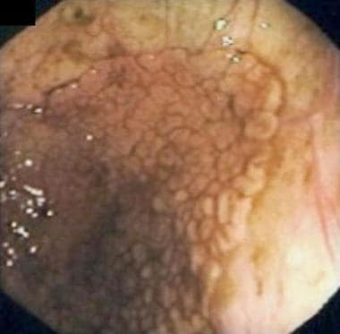
A colonoscopy is a medical procedure used to examine the large intestine, also known as the colon and rectum. It is performed using a flexible tube with a tiny camera on the end, called a colonoscope, which is inserted into the rectum and gently guided through the entire length of the colon.
The procedure allows doctors to visually inspect the lining of the colon for any abnormalities such as polyps, ulcers, inflammation, or cancer. If any polyps are found during the procedure, they can be removed immediately using special tools passed through the colonoscope. Colonoscopy is an important tool in the prevention and early detection of colorectal cancer, which is one of the leading causes of cancer-related deaths worldwide.
Patients are usually given a sedative to help them relax during the procedure, which is typically performed on an outpatient basis in a hospital or clinic setting. The entire procedure usually takes about 30-60 minutes to complete, although patients should plan to spend several hours at the medical facility for preparation and recovery.
Diatrizoate Meglumine is a type of contrast medium that is used during X-ray examinations, such as CT scans and angiography. It is a radiopaque substance, which means that it contains atoms that absorb X-rays, making it possible to visualize the internal structures of the body on an X-ray image.
Diatrizoate Meglumine is a salt of diatrizoic acid, which is a type of ionic contrast medium. It works by increasing the contrast between different tissues and organs in the body, making them easier to distinguish on an X-ray image. This can help doctors to diagnose a wide range of medical conditions, including injuries, tumors, and vascular diseases.
Like all medications, Diatrizoate Meglumine can have side effects, including allergic reactions, kidney damage, and thyroid problems. It is important for patients to discuss any potential risks and benefits with their doctor before undergoing an X-ray examination that involves the use of this contrast medium.
Laxatives are substances or medications that are used to promote bowel movements or loosen the stools, thereby helping in the treatment of constipation. They work by increasing the amount of water in the stool or stimulating the muscles in the intestines to contract and push the stool through. Laxatives can be categorized into several types based on their mechanism of action, including bulk-forming laxatives, lubricant laxatives, osmotic laxatives, saline laxatives, stimulant laxatives, and stool softeners. It is important to use laxatives only as directed by a healthcare professional, as overuse or misuse can lead to serious health complications.
An enema is a medical procedure in which liquid is introduced into the lower part of the large intestine, specifically the sigmoid colon or rectum, through the anus using a special device called an enema kit. The liquid used can be plain water, saline solution, or a medicated solution, and it is typically retained for a short period of time before being expelled.
The purpose of an enema may vary, but it is often used to relieve constipation, prepare the bowel for medical procedures such as colonoscopy, or administer medications or nutrients that cannot be taken by mouth. Enemas can also be used for therapeutic purposes, such as to stimulate the immune system or promote relaxation.
It is important to follow proper instructions when administering an enema to avoid injury or discomfort. Possible side effects of enemas may include cramping, bloating, nausea, or electrolyte imbalances. If you have any health concerns or conditions that may be affected by an enema, it is recommended to consult with a healthcare professional before using one.
Cathartics are a type of medication that stimulates bowel movements and evacuates the intestinal tract. They are often used to treat constipation or to prepare the bowel for certain medical procedures, such as colonoscopies. Common cathartic medications include laxatives, enemas, and suppositories.
Cathartics work by increasing the muscle contractions of the intestines, which helps to move stool through the digestive tract more quickly. They may also increase the amount of water in the stool, making it softer and easier to pass. Some cathartics, such as bulk-forming laxatives, work by absorbing water and swelling in the intestines, which helps to bulk up the stool and stimulate a bowel movement.
While cathartics can be effective at relieving constipation, they should be used with caution. Overuse of cathartics can lead to dependence on them for bowel movements, as well as electrolyte imbalances and other complications. It is important to follow the instructions carefully when using cathartic medications and to speak with a healthcare provider if constipation persists or worsens.
Computer-assisted radiographic image interpretation is the use of computer algorithms and software to assist and enhance the interpretation and analysis of medical images produced by radiography, such as X-rays, CT scans, and MRI scans. The computer-assisted system can help identify and highlight certain features or anomalies in the image, such as tumors, fractures, or other abnormalities, which may be difficult for the human eye to detect. This technology can improve the accuracy and speed of diagnosis, and may also reduce the risk of human error. It's important to note that the final interpretation and diagnosis is always made by a qualified healthcare professional, such as a radiologist, who takes into account the computer-assisted analysis in conjunction with their clinical expertise and knowledge.
Colorectal neoplasms refer to abnormal growths in the colon or rectum, which can be benign or malignant. These growths can arise from the inner lining (mucosa) of the colon or rectum and can take various forms such as polyps, adenomas, or carcinomas.
Benign neoplasms, such as hyperplastic polyps and inflammatory polyps, are not cancerous but may need to be removed to prevent the development of malignant tumors. Adenomas, on the other hand, are precancerous lesions that can develop into colorectal cancer if left untreated.
Colorectal cancer is a malignant neoplasm that arises from the uncontrolled growth and division of cells in the colon or rectum. It is one of the most common types of cancer worldwide and can spread to other parts of the body through the bloodstream or lymphatic system.
Regular screening for colorectal neoplasms is recommended for individuals over the age of 50, as early detection and removal of precancerous lesions can significantly reduce the risk of developing colorectal cancer.
X-ray computed tomography (CT or CAT scan) is a medical imaging method that uses computer-processed combinations of many X-ray images taken from different angles to produce cross-sectional (tomographic) images (virtual "slices") of the body. These cross-sectional images can then be used to display detailed internal views of organs, bones, and soft tissues in the body.
The term "computed tomography" is used instead of "CT scan" or "CAT scan" because the machines take a series of X-ray measurements from different angles around the body and then use a computer to process these data to create detailed images of internal structures within the body.
CT scanning is a noninvasive, painless medical test that helps physicians diagnose and treat medical conditions. CT imaging provides detailed information about many types of tissue including lung, bone, soft tissue and blood vessels. CT examinations can be performed on every part of the body for a variety of reasons including diagnosis, surgical planning, and monitoring of therapeutic responses.
In computed tomography (CT), an X-ray source and detector rotate around the patient, measuring the X-ray attenuation at many different angles. A computer uses this data to construct a cross-sectional image by the process of reconstruction. This technique is called "tomography". The term "computed" refers to the use of a computer to reconstruct the images.
CT has become an important tool in medical imaging and diagnosis, allowing radiologists and other physicians to view detailed internal images of the body. It can help identify many different medical conditions including cancer, heart disease, lung nodules, liver tumors, and internal injuries from trauma. CT is also commonly used for guiding biopsies and other minimally invasive procedures.
In summary, X-ray computed tomography (CT or CAT scan) is a medical imaging technique that uses computer-processed combinations of many X-ray images taken from different angles to produce cross-sectional images of the body. It provides detailed internal views of organs, bones, and soft tissues in the body, allowing physicians to diagnose and treat medical conditions.
Insufflation is a medical term that refers to the act of introducing a gas or vapor into a body cavity or passage, typically through a tube or surgical instrument. This procedure is often used in medical and surgical settings for various purposes, such as:
* To administer anesthesia during surgery (e.g., introducing nitrous oxide or other gases into the lungs)
* To introduce medication or other substances into the body (e.g., insufflating steroids into a joint)
* To perform diagnostic procedures (e.g., insufflating air or a contrast agent into the gastrointestinal tract to visualize it with X-rays)
* To clean out a body cavity (e.g., irrigating and insufflating the bladder during urological procedures).
It's important to note that insufflation should be performed under controlled conditions, as there are potential risks associated with introducing gases or vapors into the body, such as barotrauma (damage caused by changes in pressure) and infection.
Iothalamate Meglumine is not a medical condition, but rather a diagnostic contrast agent used in various imaging studies such as computed tomography (CT) scans and magnetic resonance imaging (MRI) exams. Iothalamate Meglumine is a type of radiocontrast medium that contains iodine atoms which help to enhance the visibility of internal structures during these imaging tests.
The medical definition of Iothalamate Meglumine is:
A radiocontrast agent used in diagnostic imaging, specifically in CT scans and MR urography exams. It contains iodine atoms that help to improve the contrast and visibility of internal structures such as the urinary tract. Iothalamate Meglumine is typically administered intravenously or instilled directly into the bladder.
It's important to note that while Iothalamate Meglumine is generally considered safe, it can cause allergic reactions or kidney damage in some individuals, particularly those with pre-existing kidney disease or diabetes. Therefore, it's essential to inform your healthcare provider of any medical conditions or allergies before undergoing an imaging exam that involves the use of this contrast agent.
Sensitivity and specificity are statistical measures used to describe the performance of a diagnostic test or screening tool in identifying true positive and true negative results.
* Sensitivity refers to the proportion of people who have a particular condition (true positives) who are correctly identified by the test. It is also known as the "true positive rate" or "recall." A highly sensitive test will identify most or all of the people with the condition, but may also produce more false positives.
* Specificity refers to the proportion of people who do not have a particular condition (true negatives) who are correctly identified by the test. It is also known as the "true negative rate." A highly specific test will identify most or all of the people without the condition, but may also produce more false negatives.
In medical testing, both sensitivity and specificity are important considerations when evaluating a diagnostic test. High sensitivity is desirable for screening tests that aim to identify as many cases of a condition as possible, while high specificity is desirable for confirmatory tests that aim to rule out the condition in people who do not have it.
It's worth noting that sensitivity and specificity are often influenced by factors such as the prevalence of the condition in the population being tested, the threshold used to define a positive result, and the reliability and validity of the test itself. Therefore, it's important to consider these factors when interpreting the results of a diagnostic test.
Barium sulfate is a medication that is commonly used as a contrast material in medical imaging procedures, such as X-rays and CT scans. It works by coating the inside of the digestive tract, making it visible on an X-ray or CT scan and allowing doctors to see detailed images of the stomach, intestines, and other parts of the digestive system.
Barium sulfate is a white, chalky powder that is mixed with water to create a thick, milky liquid. It is generally safe and does not cause significant side effects when used in medical imaging procedures. However, it should not be taken by individuals who have a known allergy to barium or who have certain digestive conditions, such as obstructions or perforations of the bowel.
It's important to note that while barium sulfate is an important tool for medical diagnosis, it is not a treatment for any medical condition and should only be used under the direction of a healthcare professional.
Radiographic image enhancement refers to the process of improving the quality and clarity of radiographic images, such as X-rays, CT scans, or MRI images, through various digital techniques. These techniques may include adjusting contrast, brightness, and sharpness, as well as removing noise and artifacts that can interfere with image interpretation.
The goal of radiographic image enhancement is to provide medical professionals with clearer and more detailed images, which can help in the diagnosis and treatment of medical conditions. This process may be performed using specialized software or hardware tools, and it requires a strong understanding of imaging techniques and the specific needs of medical professionals.
Contrast media are substances that are administered to a patient in order to improve the visibility of internal body structures or processes in medical imaging techniques such as X-rays, CT scans, MRI scans, and ultrasounds. These media can be introduced into the body through various routes, including oral, rectal, or intravenous administration.
Contrast media work by altering the appearance of bodily structures in imaging studies. For example, when a patient undergoes an X-ray examination, contrast media can be used to highlight specific organs, tissues, or blood vessels, making them more visible on the resulting images. In CT and MRI scans, contrast media can help to enhance the differences between normal and abnormal tissues, allowing for more accurate diagnosis and treatment planning.
There are several types of contrast media available, each with its own specific properties and uses. Some common examples include barium sulfate, which is used as a contrast medium in X-ray studies of the gastrointestinal tract, and iodinated contrast media, which are commonly used in CT scans to highlight blood vessels and other structures.
While contrast media are generally considered safe, they can sometimes cause adverse reactions, ranging from mild symptoms such as nausea or hives to more serious complications such as anaphylaxis or kidney damage. As a result, it is important for healthcare providers to carefully evaluate each patient's medical history and individual risk factors before administering contrast media.
Three-dimensional (3D) imaging in medicine refers to the use of technologies and techniques that generate a 3D representation of internal body structures, organs, or tissues. This is achieved by acquiring and processing data from various imaging modalities such as X-ray computed tomography (CT), magnetic resonance imaging (MRI), ultrasound, or confocal microscopy. The resulting 3D images offer a more detailed visualization of the anatomy and pathology compared to traditional 2D imaging techniques, allowing for improved diagnostic accuracy, surgical planning, and minimally invasive interventions.
In 3D imaging, specialized software is used to reconstruct the acquired data into a volumetric model, which can be manipulated and viewed from different angles and perspectives. This enables healthcare professionals to better understand complex anatomical relationships, detect abnormalities, assess disease progression, and monitor treatment response. Common applications of 3D imaging include neuroimaging, orthopedic surgery planning, cancer staging, dental and maxillofacial reconstruction, and interventional radiology procedures.
X-ray computed tomography (CT) scanner is a medical imaging device that uses computer-processed combinations of many X-ray images taken from different angles to produce cross-sectional (tomographic) images (virtual "slices") of the body. These cross-sections can then be manipulated, through either additional computer processing or interactive viewing, to show various bodily structures and functions in 2D or 3D.
In contrast to conventional X-ray imaging, CT scanning provides detailed images of many types of tissue including lung, bone, soft tissue and blood vessels. CT is often used when rapid, detailed images are needed such as in trauma situations or for the detection and diagnosis of stroke, cancer, appendicitis, pulmonary embolism, and musculoskeletal disorders.
CT scanning is associated with some risks, particularly from exposure to ionizing radiation, which can lead to cancer and other diseases. However, the benefits of CT scanning, in particular its ability to detect life-threatening conditions early and accurately, generally outweigh the risks. As a result, it has become an important tool in modern medicine.
Medical mass screening, also known as population screening, is a public health service that aims to identify and detect asymptomatic individuals in a given population who have or are at risk of a specific disease. The goal is to provide early treatment, reduce morbidity and mortality, and prevent the spread of diseases within the community.
A mass screening program typically involves offering a simple, quick, and non-invasive test to a large number of people in a defined population, regardless of their risk factors or symptoms. Those who test positive are then referred for further diagnostic tests and appropriate medical interventions. Examples of mass screening programs include mammography for breast cancer detection, PSA (prostate-specific antigen) testing for prostate cancer, and fecal occult blood testing for colorectal cancer.
It is important to note that mass screening programs should be evidence-based, cost-effective, and ethically sound, with clear benefits outweighing potential harms. They should also consider factors such as the prevalence of the disease in the population, the accuracy and reliability of the screening test, and the availability and effectiveness of treatment options.
Colonic diseases refer to a group of medical conditions that affect the colon, also known as the large intestine or large bowel. The colon is the final segment of the digestive system, responsible for absorbing water and electrolytes, and storing and eliminating waste products.
Some common colonic diseases include:
1. Inflammatory bowel disease (IBD): This includes conditions such as Crohn's disease and ulcerative colitis, which cause inflammation and irritation in the lining of the digestive tract.
2. Diverticular disease: This occurs when small pouches called diverticula form in the walls of the colon, leading to symptoms such as abdominal pain, bloating, and changes in bowel movements.
3. Colorectal cancer: This is a type of cancer that develops in the colon or rectum, often starting as benign polyps that grow and become malignant over time.
4. Irritable bowel syndrome (IBS): This is a functional gastrointestinal disorder characterized by abdominal pain, bloating, and changes in bowel movements, but without any underlying structural or inflammatory causes.
5. Constipation: This is a common condition characterized by infrequent bowel movements, difficulty passing stools, or both.
6. Infectious colitis: This occurs when the colon becomes infected with bacteria, viruses, or parasites, leading to symptoms such as diarrhea, abdominal cramps, and fever.
Treatment for colonic diseases varies depending on the specific condition and its severity. Treatment options may include medications, lifestyle changes, surgery, or a combination of these approaches.
Tomography is a medical imaging technique used to produce cross-sectional images or slices of specific areas of the body. This technique uses various forms of radiation (X-rays, gamma rays) or sound waves (ultrasound) to create detailed images of the internal structures, such as organs, bones, and tissues. Common types of tomography include Computerized Tomography (CT), Positron Emission Tomography (PET), and Magnetic Resonance Imaging (MRI). The primary advantage of tomography is its ability to provide clear and detailed images of internal structures, allowing healthcare professionals to accurately diagnose and monitor a wide range of medical conditions.
Reproducibility of results in a medical context refers to the ability to obtain consistent and comparable findings when a particular experiment or study is repeated, either by the same researcher or by different researchers, following the same experimental protocol. It is an essential principle in scientific research that helps to ensure the validity and reliability of research findings.
In medical research, reproducibility of results is crucial for establishing the effectiveness and safety of new treatments, interventions, or diagnostic tools. It involves conducting well-designed studies with adequate sample sizes, appropriate statistical analyses, and transparent reporting of methods and findings to allow other researchers to replicate the study and confirm or refute the results.
The lack of reproducibility in medical research has become a significant concern in recent years, as several high-profile studies have failed to produce consistent findings when replicated by other researchers. This has led to increased scrutiny of research practices and a call for greater transparency, rigor, and standardization in the conduct and reporting of medical research.
The colon, also known as the large intestine, is a part of the digestive system in humans and other vertebrates. It is an organ that eliminates waste from the body and is located between the small intestine and the rectum. The main function of the colon is to absorb water and electrolytes from digested food, forming and storing feces until they are eliminated through the anus.
The colon is divided into several regions, including the cecum, ascending colon, transverse colon, descending colon, sigmoid colon, rectum, and anus. The walls of the colon contain a layer of muscle that helps to move waste material through the organ by a process called peristalsis.
The inner surface of the colon is lined with mucous membrane, which secretes mucus to lubricate the passage of feces. The colon also contains a large population of bacteria, known as the gut microbiota, which play an important role in digestion and immunity.
Radiology is a medical specialty that uses imaging technologies to diagnose and treat diseases. These imaging technologies include X-rays, computed tomography (CT) scans, magnetic resonance imaging (MRI) scans, positron emission tomography (PET) scans, ultrasound, and mammography. Radiologists are medical doctors who have completed specialized training in interpreting these images to diagnose medical conditions and guide treatment plans. They also perform image-guided procedures such as biopsies and tumor ablations. The goal of radiology is to provide accurate and timely information to help physicians make informed decisions about patient care.
Computer-assisted image processing is a medical term that refers to the use of computer systems and specialized software to improve, analyze, and interpret medical images obtained through various imaging techniques such as X-ray, CT (computed tomography), MRI (magnetic resonance imaging), ultrasound, and others.
The process typically involves several steps, including image acquisition, enhancement, segmentation, restoration, and analysis. Image processing algorithms can be used to enhance the quality of medical images by adjusting contrast, brightness, and sharpness, as well as removing noise and artifacts that may interfere with accurate diagnosis. Segmentation techniques can be used to isolate specific regions or structures of interest within an image, allowing for more detailed analysis.
Computer-assisted image processing has numerous applications in medical imaging, including detection and characterization of lesions, tumors, and other abnormalities; assessment of organ function and morphology; and guidance of interventional procedures such as biopsies and surgeries. By automating and standardizing image analysis tasks, computer-assisted image processing can help to improve diagnostic accuracy, efficiency, and consistency, while reducing the potential for human error.
A "false positive reaction" in medical testing refers to a situation where a diagnostic test incorrectly indicates the presence of a specific condition or disease in an individual who does not actually have it. This occurs when the test results give a positive outcome, while the true health status of the person is negative or free from the condition being tested for.
False positive reactions can be caused by various factors including:
1. Presence of unrelated substances that interfere with the test result (e.g., cross-reactivity between similar molecules).
2. Low specificity of the test, which means it may detect other conditions or irrelevant factors as positive.
3. Contamination during sample collection, storage, or analysis.
4. Human errors in performing or interpreting the test results.
False positive reactions can have significant consequences, such as unnecessary treatments, anxiety, and increased healthcare costs. Therefore, it is essential to confirm any positive test result with additional tests or clinical evaluations before making a definitive diagnosis.
Optical Tomography (OT) is a non-invasive imaging technique that uses light to visualize and measure the optical properties of tissue, such as absorption and scattering coefficients. This modality can be used to produce cross-sectional or three-dimensional images of internal structures, providing functional information about tissue physiology. It has applications in various fields including biomedical research, dermatology, and oncology for the detection and monitoring of diseases. There are different types of optical tomography, such as diffuse optical tomography (DOT) and near-infrared spectroscopy (NIRS), which differ in their light sources, detection schemes, and data analysis methods.
Automated Pattern Recognition in a medical context refers to the use of computer algorithms and artificial intelligence techniques to identify, classify, and analyze specific patterns or trends in medical data. This can include recognizing visual patterns in medical images, such as X-rays or MRIs, or identifying patterns in large datasets of physiological measurements or electronic health records.
The goal of automated pattern recognition is to assist healthcare professionals in making more accurate diagnoses, monitoring disease progression, and developing personalized treatment plans. By automating the process of pattern recognition, it can help reduce human error, increase efficiency, and improve patient outcomes.
Examples of automated pattern recognition in medicine include using machine learning algorithms to identify early signs of diabetic retinopathy in eye scans or detecting abnormal heart rhythms in electrocardiograms (ECGs). These techniques can also be used to predict patient risk based on patterns in their medical history, such as identifying patients who are at high risk for readmission to the hospital.
The prone position is a body posture in which an individual lies on their stomach, with their face down and chest facing the floor or bed. This position is often used in medical settings for various purposes, such as during certain surgical procedures, respiratory support, or to alleviate pressure ulcers. It's also important to note that the prone position can have implications for patient safety, particularly in critically ill patients, and should be carefully monitored.
Iohexol is a non-ionic, water-soluble contrast medium primarily used in radiographic imaging procedures such as computed tomography (CT) scans and angiography. It belongs to a class of medications known as radiocontrast agents. Iohexol works by increasing the X-ray absorption of body tissues, making them more visible on X-ray images. This helps healthcare professionals to better diagnose and assess various medical conditions, including injuries, tumors, and vascular diseases.
The chemical structure of iohexol consists of an iodine atom surrounded by organic molecules, which makes it safe for intravenous administration. It is eliminatted from the body primarily through urinary excretion. Iohexol has a low risk of allergic reactions compared to ionic contrast media and is generally well-tolerated in patients with normal renal function. However, its use should be avoided or closely monitored in individuals with impaired kidney function, as it may increase the risk of nephrotoxicity.
Early detection of cancer refers to the identification of malignant cells or tumors in their initial stages, before they have had a chance to grow and spread. This is typically achieved through various screening methods and tests that are designed to detect specific types of cancers. The goal of early detection is to increase the chances of successful treatment and improve the overall prognosis for patients.
Some common methods used for early cancer detection include:
1. Regular screenings such as mammograms, colonoscopies, and Pap tests, which can help identify precancerous or cancerous cells in their earliest stages.
2. Imaging tests like CT scans, MRIs, and PET scans, which can help detect tumors that may not be visible through other screening methods.
3. Blood tests that look for specific biomarkers or tumor markers, which can indicate the presence of cancer in the body.
4. Genetic testing to identify individuals who may be at higher risk of developing certain types of cancer due to inherited genetic mutations.
It's important to note that while early detection is an important tool in the fight against cancer, it is not a guarantee of successful treatment or cure. However, it can significantly improve the odds of successful treatment and increase the chances of survival for many patients.
Incidental findings are diagnoses or conditions that are discovered unintentionally while evaluating a patient for a different condition or symptom. These findings are not related to the primary reason for the medical examination, investigation, or procedure. They can occur in various contexts such as radiology studies, laboratory tests, or physical examinations.
Incidental findings can sometimes lead to further evaluation and management, depending on their nature and potential clinical significance. However, they also pose challenges related to communication, informed consent, and potential patient anxiety or harm. Therefore, it is essential to have clear guidelines for managing incidental findings in clinical practice.
Occult blood refers to the presence of blood in the stool or gastrointestinal tract that is not visible to the naked eye. It is typically detected through chemical tests, such as fecal occult blood tests (FOBT) or fecal immunochemical tests (FIT), which can detect small amounts of blood in the stool. The presence of occult blood may indicate a variety of gastrointestinal conditions, including colorectal cancer, polyps, ulcers, inflammatory bowel disease, and other digestive disorders. It is important to follow up with medical evaluation if occult blood is detected, as early detection and treatment of underlying conditions can improve outcomes.
A polyp is a general term for a small growth that protrudes from a mucous membrane, such as the lining of the nose or the digestive tract. Polyps can vary in size and shape, but they are usually cherry-sized or smaller and have a stalk or a broad base. They are often benign (noncancerous), but some types of polyps, especially those in the colon, can become cancerous over time.
In the digestive tract, polyps can form in the colon, rectum, stomach, or small intestine. Colorectal polyps are the most common type and are usually found during routine colonoscopies. There are several types of colorectal polyps, including:
* Adenomatous polyps (adenomas): These polyps can become cancerous over time and are the most likely to turn into cancer.
* Hyperplastic polyps: These polyps are usually small and benign, but some types may have a higher risk of becoming cancerous.
* Inflammatory polyps: These polyps are caused by chronic inflammation in the digestive tract, such as from inflammatory bowel disease (IBD).
Polyps can also form in other parts of the body, including the nose, sinuses, ears, and uterus. In most cases, polyps are benign and do not cause any symptoms. However, if they become large enough, they may cause problems such as bleeding, obstruction, or discomfort. Treatment typically involves removing the polyp through a surgical procedure.
A feasibility study is a preliminary investigation or analysis conducted to determine the viability of a proposed project, program, or product. In the medical field, feasibility studies are often conducted before implementing new treatments, procedures, equipment, or facilities. These studies help to assess the practicality and effectiveness of the proposed intervention, as well as its potential benefits and risks.
Feasibility studies in healthcare typically involve several steps:
1. Problem identification: Clearly define the problem that the proposed project, program, or product aims to address.
2. Objectives setting: Establish specific, measurable, achievable, relevant, and time-bound (SMART) objectives for the study.
3. Literature review: Conduct a thorough review of existing research and best practices related to the proposed intervention.
4. Methodology development: Design a methodology for data collection and analysis that will help answer the research questions and achieve the study's objectives.
5. Resource assessment: Evaluate the availability and adequacy of resources, including personnel, time, and finances, required to carry out the proposed intervention.
6. Risk assessment: Identify potential risks and challenges associated with the implementation of the proposed intervention and develop strategies to mitigate them.
7. Cost-benefit analysis: Estimate the costs and benefits of the proposed intervention, including direct and indirect costs, as well as short-term and long-term benefits.
8. Stakeholder engagement: Engage relevant stakeholders, such as patients, healthcare providers, administrators, and policymakers, to gather their input and support for the proposed intervention.
9. Decision-making: Based on the findings of the feasibility study, make an informed decision about whether or not to proceed with the proposed project, program, or product.
Feasibility studies are essential in healthcare as they help ensure that resources are allocated efficiently and effectively, and that interventions are evidence-based, safe, and beneficial for patients.
Computer-assisted diagnosis (CAD) is the use of computer systems to aid in the diagnostic process. It involves the use of advanced algorithms and data analysis techniques to analyze medical images, laboratory results, and other patient data to help healthcare professionals make more accurate and timely diagnoses. CAD systems can help identify patterns and anomalies that may be difficult for humans to detect, and they can provide second opinions and flag potential errors or uncertainties in the diagnostic process.
CAD systems are often used in conjunction with traditional diagnostic methods, such as physical examinations and patient interviews, to provide a more comprehensive assessment of a patient's health. They are commonly used in radiology, pathology, cardiology, and other medical specialties where imaging or laboratory tests play a key role in the diagnostic process.
While CAD systems can be very helpful in the diagnostic process, they are not infallible and should always be used as a tool to support, rather than replace, the expertise of trained healthcare professionals. It's important for medical professionals to use their clinical judgment and experience when interpreting CAD results and making final diagnoses.
I'm sorry for any confusion, but "Wisconsin" is a U.S. state located in the Midwest and is not a medical term or condition. If you have any medical questions or terms you would like defined, I'd be happy to help with those!
Adenomatous polyps, also known as adenomas, are benign (noncancerous) growths that develop in the lining of the glandular tissue of certain organs, most commonly occurring in the colon and rectum. These polyps are composed of abnormal glandular cells that can grow excessively and form a mass.
Adenomatous polyps can vary in size, ranging from a few millimeters to several centimeters in diameter. They may be flat or have a stalk (pedunculated). While adenomas are generally benign, they can potentially undergo malignant transformation and develop into colorectal cancer over time if left untreated. The risk of malignancy increases with the size of the polyp and the presence of certain histological features, such as dysplasia (abnormal cell growth).
Regular screening for adenomatous polyps is essential to detect and remove them early, reducing the risk of colorectal cancer. Screening methods include colonoscopy, sigmoidoscopy, and stool-based tests.
Patient preference, in the context of medical decision-making, refers to the individual desires, values, and concerns that a patient considers when choosing between different treatment options. It is based on the patient's own experiences, beliefs, and needs, and may take into account factors such as potential benefits, risks, side effects, costs, and convenience. Patient preferences should be respected and integrated into clinical decision-making processes whenever possible, in order to promote patient-centered care and improve outcomes.
Cost-benefit analysis (CBA) is a systematic process used to compare the costs and benefits of different options to determine which one provides the greatest net benefit. In a medical context, CBA can be used to evaluate the value of medical interventions, treatments, or policies by estimating and monetizing all the relevant costs and benefits associated with each option.
The costs included in a CBA may include direct costs such as the cost of the intervention or treatment itself, as well as indirect costs such as lost productivity or time away from work. Benefits may include improved health outcomes, reduced morbidity or mortality, and increased quality of life.
Once all the relevant costs and benefits have been identified and quantified, they are typically expressed in monetary terms to allow for a direct comparison. The option with the highest net benefit (i.e., the difference between total benefits and total costs) is considered the most cost-effective.
It's important to note that CBA has some limitations and can be subject to various biases and assumptions, so it should be used in conjunction with other evaluation methods to ensure a comprehensive understanding of the value of medical interventions or policies.
An algorithm is not a medical term, but rather a concept from computer science and mathematics. In the context of medicine, algorithms are often used to describe step-by-step procedures for diagnosing or managing medical conditions. These procedures typically involve a series of rules or decision points that help healthcare professionals make informed decisions about patient care.
For example, an algorithm for diagnosing a particular type of heart disease might involve taking a patient's medical history, performing a physical exam, ordering certain diagnostic tests, and interpreting the results in a specific way. By following this algorithm, healthcare professionals can ensure that they are using a consistent and evidence-based approach to making a diagnosis.
Algorithms can also be used to guide treatment decisions. For instance, an algorithm for managing diabetes might involve setting target blood sugar levels, recommending certain medications or lifestyle changes based on the patient's individual needs, and monitoring the patient's response to treatment over time.
Overall, algorithms are valuable tools in medicine because they help standardize clinical decision-making and ensure that patients receive high-quality care based on the latest scientific evidence.
Emission computed tomography (ECT) is a type of tomographic imaging technique in which an emission signal from within the body is detected to create cross-sectional images of that signal's distribution. In Emission-Computed Tomography (ECT), a radionuclide is introduced into the body, usually through injection, inhalation or ingestion. The radionuclide emits gamma rays that are then detected by external gamma cameras.
The data collected from these cameras is then used to create cross-sectional images of the distribution of the radiopharmaceutical within the body. This allows for the identification and quantification of functional information about specific organs or systems within the body, such as blood flow, metabolic activity, or receptor density.
One common type of Emission-Computed Tomography is Single Photon Emission Computed Tomography (SPECT), which uses a single gamma camera that rotates around the patient to collect data from multiple angles. Another type is Positron Emission Tomography (PET), which uses positron-emitting radionuclides and detects the coincident gamma rays emitted by the annihilation of positrons and electrons.
Overall, ECT is a valuable tool in medical imaging for diagnosing and monitoring various diseases, including cancer, heart disease, and neurological disorders.
Cone-beam computed tomography (CBCT) is a medical imaging technique that uses a cone-shaped X-ray beam to create detailed, cross-sectional images of the body. In dental and maxillofacial radiology, CBCT is used to produce three-dimensional images of the teeth, jaws, and surrounding bones.
CBCT differs from traditional computed tomography (CT) in that it uses a cone-shaped X-ray beam instead of a fan-shaped beam, which allows for a faster scan time and lower radiation dose. The X-ray beam is rotated around the patient's head, capturing data from multiple angles, which is then reconstructed into a three-dimensional image using specialized software.
CBCT is commonly used in dental implant planning, orthodontic treatment planning, airway analysis, and the diagnosis and management of jaw pathologies such as tumors and fractures. It provides detailed information about the anatomy of the teeth, jaws, and surrounding structures, which can help clinicians make more informed decisions about patient care.
However, it is important to note that CBCT should only be used when necessary, as it still involves exposure to ionizing radiation. The benefits of using CBCT must be weighed against the potential risks associated with radiation exposure.
Infarction is the term used in medicine to describe the death of tissue (also known as an "area of necrosis") due to the lack of blood supply. This can occur when a blood vessel that supplies oxygen and nutrients to a particular area of the body becomes blocked or obstructed, leading to the deprivation of oxygen and nutrients necessary for the survival of cells in that region.
The blockage in the blood vessel is usually caused by a clot (thrombus) or an embolus, which is a small particle that travels through the bloodstream and lodges in a smaller vessel. The severity and extent of infarction depend on several factors, including the size and location of the affected blood vessel, the duration of the obstruction, and the presence of collateral circulation (alternative blood vessels that can compensate for the blocked one).
Common examples of infarctions include myocardial infarction (heart attack), cerebral infarction (stroke), and pulmonary infarction (lung tissue death due to obstruction in the lung's blood vessels). Infarctions can lead to various symptoms, depending on the affected organ or tissue, and may require medical intervention to manage complications and prevent further damage.
Prospective studies, also known as longitudinal studies, are a type of cohort study in which data is collected forward in time, following a group of individuals who share a common characteristic or exposure over a period of time. The researchers clearly define the study population and exposure of interest at the beginning of the study and follow up with the participants to determine the outcomes that develop over time. This type of study design allows for the investigation of causal relationships between exposures and outcomes, as well as the identification of risk factors and the estimation of disease incidence rates. Prospective studies are particularly useful in epidemiology and medical research when studying diseases with long latency periods or rare outcomes.
Sigmoidoscopy is a medical procedure that involves the insertion of a sigmoidoscope, a flexible tube with a light and camera at the end, into the rectum and lower colon (sigmoid colon) to examine these areas for any abnormalities such as inflammation, ulcers, polyps, or cancer. The procedure typically allows for the detection of issues in the sigmoid colon and rectum, and can help diagnose conditions such as inflammatory bowel disease, diverticulosis, or colorectal cancer.
There are two types of sigmoidoscopy: flexible sigmoidoscopy and rigid sigmoidoscopy. Flexible sigmoidoscopy is more commonly performed because it provides a better view of the lower colon and is less uncomfortable for the patient. Rigid sigmoidoscopy, on the other hand, uses a solid, inflexible tube and is typically used in specific situations such as the removal of foreign objects or certain types of polyps.
During the procedure, patients are usually positioned on their left side with their knees drawn up to their chest. The sigmoidoscope is gently inserted into the rectum and advanced through the lower colon while the doctor examines the lining for any abnormalities. Air may be introduced through the scope to help expand the colon and provide a better view. If polyps or other abnormal tissues are found, they can often be removed during the procedure for further examination and testing.
Sigmoidoscopy is generally considered a safe and well-tolerated procedure. Some patients may experience mild discomfort, bloating, or cramping during or after the exam, but these symptoms typically resolve on their own within a few hours.
Observer variation, also known as inter-observer variability or measurement agreement, refers to the difference in observations or measurements made by different observers or raters when evaluating the same subject or phenomenon. It is a common issue in various fields such as medicine, research, and quality control, where subjective assessments are involved.
In medical terms, observer variation can occur in various contexts, including:
1. Diagnostic tests: Different radiologists may interpret the same X-ray or MRI scan differently, leading to variations in diagnosis.
2. Clinical trials: Different researchers may have different interpretations of clinical outcomes or adverse events, affecting the consistency and reliability of trial results.
3. Medical records: Different healthcare providers may document medical histories, physical examinations, or treatment plans differently, leading to inconsistencies in patient care.
4. Pathology: Different pathologists may have varying interpretations of tissue samples or laboratory tests, affecting diagnostic accuracy.
Observer variation can be minimized through various methods, such as standardized assessment tools, training and calibration of observers, and statistical analysis of inter-rater reliability.
Medical Definition:
Magnetic Resonance Imaging (MRI) is a non-invasive diagnostic imaging technique that uses a strong magnetic field and radio waves to create detailed cross-sectional or three-dimensional images of the internal structures of the body. The patient lies within a large, cylindrical magnet, and the scanner detects changes in the direction of the magnetic field caused by protons in the body. These changes are then converted into detailed images that help medical professionals to diagnose and monitor various medical conditions, such as tumors, injuries, or diseases affecting the brain, spinal cord, heart, blood vessels, joints, and other internal organs. MRI does not use radiation like computed tomography (CT) scans.
 Ronald Summers
Quantitative computed tomography
List of MeSH codes (H01)
Index of oncology articles
List of MeSH codes (E01)
Judy Yee
Colonoscopy
Spectral imaging (radiography)
CT scan
Virtual pathology: A new paradigm for interpretation of computed tomographic colonography<...
 Diarrhea-predominant irritable bowel syndrome is associated with diverticular disease: a population-based study
Prevalence and risk factors for post-investigation colorectal cancer ("interval cancer") after computed tomographic...
The potential of imaging techniques as a screening tool for colorectal cancer: a cost-effectiveness analysis
 Colorectal Cancer Guidelines: Colorectal Cancer Screening, Postpolypectomy Surveillance, Familial Adenomatous Polyposis
 Colorectal Cancer Screening Age Decreases to 45 | Health.mil
Colorectal Cancer Screening Age Decreases to 45 | Health.mil
Flexible Sigmoidoscopy Technique: Flexible Sigmoidoscopy, Complications
Ronald Summers - Wikipedia
 "Virtual" related terms, short phrases and...
 The Medicare Technology Crunch, by Matt Patterson - The National Center
 Sigmoid Perforation during CT Colonography in a Patient with an Inguinal Hernia and Concomitant Finding of a Right-Sided Colon...
 Iron Deficiency Anemia: Evaluation and Management | AAFP
Colorectal Cancer Guidelines: Colorectal Cancer Screening, Postpolypectomy Surveillance, Familial Adenomatous Polyposis
강북삼성병원
 Ontario Health Technology Assessment Series - Health Quality Ontario (HQO)
Items where Subject is "H673 Bioengineering" - The Lincoln Repository
Concepts :: The Health Plan
Risk factors for advanced neoplasia within subcentimetric polyps: implications for diagnostic imaging | Gut
 ACG Clinical Guidelines: Colorectal Cancer Screening 2021 : Official journal of the American College of Gastroenterology | ACG
 Colorectal Cancer in Primary Care | Natural Medicine Journal
 Vital Signs: Colorectal Cancer Screening Test Use - United States, 2018 | MMWR
 Diagnosis and management - GPnotebook
 News for Medical Independent Sales Representatives and Medical Distributors: iCAD Features Image Analysis Solutions for Breast,...
 Robotic complete mesocolic excision of right‑sided colon cancer with bulky lymph node metastases using the da Vinci<sup>®</sup>...
 Bibliografia 2015 - Pagina 130 - Endometriosi.it
Intestinal Polyposis Syndromes Imaging: Practice Essentials, Radiograph, CT SCAN
 Implementation of population screening for colorectal cancer by repeated fecal occult blood test in the Netherlands | BMC...
CISNET: Comparative Analyses in Colorectal Cancer
Colonoscopic Polypectomy: A critical review of recent literature<...
Colonoscopy19
- Subjects with at least one relevant test (colonoscopy, computed tomography (CT) scan, CT colonography, or barium enema) were included. (nih.gov)
- 6 CT colonography is a relatively new procedure for viewing the colon and large intestine to check for polyps and cancer, and is far less invasive, and far less dangerous, than traditional colonoscopy. (nationalcenter.org)
- CT colonography also referred to as virtual colonoscopy (VC) was first described in 1994 [ 1 ] and its use as a screening modality for colon cancer is controversial. (omicsonline.org)
- Diagnostic imaging by CT colonography and colon capsule endoscopy have been proposed as alternatives to optical colonoscopy for the detection of colonic neoplasia. (bmj.com)
- Poster C-2336 titled, Optimizing Time-efficient Use of Computer-aided Detection in Computed Tomographic Colonography (Virtual Colonoscopy), by Dr. Abraham Dachman. (salesandmarketingnetwork.com)
- There are several colorectal screening options for average-risk individuals, including colonoscopy every 10 years, flexible sigmoidoscopy every 5 years, double-contrast barium enema every 5 years, CT colonography every 5 years, and annual fecal occult blood testing. (medscape.com)
- CT and magnetic resonance (MR) colonography (virtual colonoscopy) techniques are being developed for the imaging of colorectal polyps and cancer. (medscape.com)
- Structural exams include flexible sigmoidoscopy, total colonoscopy and computed tomographic colonography (CTC). (biomedcentral.com)
- CT colonography has developed as an alternative or adjunct to colonoscopy but has not surpassed it in accuracy or therapeutic potential. (mssm.edu)
- PURPOSE: To determine the sensitivity and specificity of computed tomographic (CT) colonography for colorectal polyp and cancer detection by using colonoscopy as the reference standard. (elsevierpure.com)
- MATERIALS AND METHODS: Three hundred patients underwent CT colonography followed by Standard Colonoscopy. (elsevierpure.com)
- PURPOSE: To prospectively evaluate the diagnostic accuracy of low-radiation-dose computed tomographic (CT) colonography for detection of colorectal polyps by using two sequential colonoscopies, with the second colonoscopy as the reference standard. (duke.edu)
- The sensitivities of CT colonography and initial colonoscopy were calculated on a per-polyp and a per-patient basis. (duke.edu)
- RESULTS: Eighty-eight patients underwent CT colonography and initial conventional colonoscopy on the same day. (duke.edu)
- Per-polyp sensitivities were 62% and 83% for CT colonography and initial colonoscopy, respectively. (duke.edu)
- Sensitivities for detection of polyps 6 mm in diameter or larger were 86% and 84% for CT colonography and initial colonoscopy, respectively. (duke.edu)
- Initial colonoscopy failed to depict 16 polyps, six of which were correctly detected with CT colonography. (duke.edu)
- CONCLUSION: Low-dose CT colonography compares favorably with colonoscopy for detection of colorectal polyps 6 mm in diameter or larger, with markedly decreased performance for detection of polyps 5 mm in diameter or smaller. (duke.edu)
- Harms associated with CT colonography include radiation-induced effects, the downstream effects from detection of extracolonic findings, and the potential harms of follow-up colonoscopy. (kolorektum.cz)
Double contr1
- Patient acceptability of CT colonography compared with double contrast barium enema: results from a multicentre randomised controlled trial of symptomatic patients. (ox.ac.uk)
Tomography colonography2
- The use of computed tomography colonography or virtual colonography (VC) is gaining acceptance as a screening modality for colon cancer . (omicsonline.org)
- MATERIALS AND METHODS: Sixty-nine patients underwent computed tomography colonography (CTC) after bowel cleansing. (bvsalud.org)
Detection4
- RESULTS: The overall sensitivity and specificity of CT colonography for polyp detection were 90.1% (164 of 182) and 72.0% (85 of 118), respectively. (elsevierpure.com)
- CONCLUSION: CT colonography has excellent sensitivity for the detection of clinically important colorectal polyps and cancer. (elsevierpure.com)
- Scholars@Duke publication: Colorectal polyps: detection with low-dose multi-detector row helical CT colonography versus two sequential colonoscopies. (duke.edu)
- Detection of anatomical landmarks in human colon from computed tomographic colonography images. (sigmod.org)
Magnetic resonance1
- PURPOSE: To use perspective volume rendering (PVR) of computed tomographic (CT) and magnetic resonance (MR) imaging data sets to simulate endoscopic views of human organ systems. (rsna.org)
Barium1
- OBJECTIVES: To determine patient acceptability of barium enema (BE) or CT colonography (CTC). (ox.ac.uk)
Colon cancer3
- Turner J, Page M, Clark C, Khanduja K (2016) Sigmoid Perforation during CT Colonography in a Patient with an Inguinal Hernia and Concomitant Finding of a Right-Sided Colon Cancer. (omicsonline.org)
- We report a case of colon perforation in a patient where a colon cancer was diagnosed using VC colonography with manual insufflation. (omicsonline.org)
- Computed tomographic colonography (CTC) for colon cancer screening]. (aihta.at)
Diagnostic1
- Objective Diagnostic imaging by CT colonography and capsule endoscopy is used to detect colonic lesions. (bmj.com)
Stool2
- An international group of 23 experts convened by IARC performed a systematic review and evaluation of the pertinent scientific literature on stool-based tests, endoscopic methods, and computed tomographic colonography methods. (kolorektum.cz)
- CT colonography and stool DNA testing are new promising screening technologies that are less invasive, accurate, and suitable for the public more than the current screening procedures. (tau.ac.il)
Scan1
- But the researchers, funded by the National Institute for Health Research Health Technology Assessment and Cancer Research UK 1 , caution that guidelines are needed before this type of scan, also called CT colonography (CTC), is used more widely because its ability to detect relatively unimportant findings can result in patients being referred for unnecessary follow-up tests. (cancerresearchuk.org)
Adenoma1
- We aimed to assess the potential of CT colonography (CTC) and MR colonography (MRC) in terms of (cost-effectiveness) using the Adenoma and Serrated pathway to Colorectal CAncer model. (nih.gov)
Abstract1
- abstract = "The virtual pathology (VP), a display mode for interpretation of computed tomographic colonography (CTC), was discussed. (elsevierpure.com)
Patients1
- According to Peter J. Neumann, Sc.D., and Sean R. Tunis, M.D. writing on the New England Journal of Medicine website, the Center For Medicare Services (CMS) determined that "clinical trials showing a benefit of screening with CT colonography were not necessarily generalizable, because the mean age of trial participants was lower than that of Medicare patients. (nationalcenter.org)
Cancer4
- The life-saving potential of widespread CT colonography use is staggering: According to The National Cancer Institute Colorectal Cancer Progress Review Group, wider availability of screening procedures like CT colonography could save as many as 20,000 lives per year. (nationalcenter.org)
- Computed tomographic colonography (CTC) is currently not among the colorectal cancer screening modalities reimbursable by the Centers for Medicare and Medicaid Services (CMS. (unt.edu)
- CT colonography, an imaging method based on scanning technology, has been developed as a less invasive visualization technique for colorectal cancer screening. (kolorektum.cz)
- There is limited evidence that a single screening with CT colonography reduces colorectal cancer incidence and/or mortality. (kolorektum.cz)
Test2
- A computed tomography angiogram (CT angiogram) is a test that uses X-rays to provide detailed pictures of the heart and the blood vessels that go to the heart, lung, brain, kidneys, head, neck, legs, and arms. (healthplan.org)
- Single photon-emission computed tomography (SPECT) is a test that uses a special type of camera and a tracer (a radioactive substance in liquid form) to look at organs or bones in the body. (healthplan.org)
Summers1
- A 2022 paper from Summers' lab published in Radiology showed how computed tomography (CT) biomarkers are associated with diabetes and pre-diabetes. (wikipedia.org)
Analysis1
- Both CT and MR colonography allow an analysis in both the cross-sectional and virtual endoscopic formats. (medscape.com)
Colonoscopy9
- 6. Diagnostic value of computed tomography colonography (CTC) after incomplete optical colonoscopy. (nih.gov)
- 10. CT-colonography after incomplete colonoscopy: what is the diagnostic yield? (nih.gov)
- The sensitivity of computed tomographic (CT) virtual colonoscopy (CT colonography) for detecting polyps varies widely in recently reported large clinical trials. (nih.gov)
- It included studies that looked at old technology, did not consider the new huge costs of treating advanced colorectal cancer with targeted therapies, and didn't address the question of what happens if more people end up getting effectively screened for colorectal cancer and prevent the disease, as opposed to simplistically concluding that CT colonography versus traditional colonoscopy is an either/or decision. (auntminnie.com)
- To summarize the available information on the two methods of virtual colonoscopy: computed tomographic colonography and magnetic resonance colonography. (inahta.org)
- CT colonography, also called virtual colonoscopy, was introduced about a decade ago, has made great progress over the years, and is becoming more popular. (huji.ac.il)
- Computed Tomographic Colonography (CTC, Virtual Colonoscopy) -- Indication -- Strengths -- Weaknesses -- Comments -- 6. (edu.au)
- If a lesion suspicious of cancer is detected on CT colonography, a colonoscopy with biopsy to confirm the diagnosis should be performed. (surgerymithra.com)
- A computed Topography (CT) scan is taken, where the body is invasively scanned into slices, which are then reconstituted into a 3D model of a person's colon, for a painless colonoscopy. (virtualworldlets.net)
Polyps5
- Computed tomographic colonography (CTC) is a relatively new imaging modality for the examination of patients for colorectal polyps and cancer. (medscape.com)
- More recently, computed tomographic colonography (CTC) and stool analysis for DNA markers for polyps/malignancy have been accepted by some organizations and authorities as additional acceptable screening modalities. (medscape.com)
- 14. Computed tomographic colonography (CTC): Possibilities and limitations of clinical application in colorectal polyps and cancer. (nih.gov)
- Computed tomographic colonography (CTC) is a minimally invasive technique for colonic polyps and cancer screening. (nih.gov)
- CT colonography is an effective, safe method for examining the colon and rectum to detect abnormalities such as polyps and cancer. (surgerymithra.com)
Diagnosis1
- In fact, I would maintain that if we applied strict evidence criteria to the diagnosis and treatment of Medicare patients today -- as suggested by this article -- then we could solve the shortfall in the Medicare program tomorrow, because virtually no drug, no test, or any procedure would survive this rigorous "evidence-based analysis" that was applied to CT colonography. (auntminnie.com)
Axial tomography2
- Computed Tomography, or CT, also known as computed axial tomography or CAT, is a form of tomographic scanner which builds images up by slices. (virtualworldlets.net)
- It was later known as computed axial tomography (CAT or CT scan) and body section roentgenography . (wikidoc.org)
Versus2
Prevalence1
- 11. Computed tomographic colonography: prevalence, nature, and clinical significance of extracolonic findings in a community screening program. (nih.gov)
Retrospective1
- 5. Computed tomographic colonography (CTC): a retrospective analysis of a single site experience and a review of the literature on the status of CTC. (nih.gov)
Virtual1
- Virtual colonography is a relatively new field, which is completely non-invasive. (virtualworldlets.net)
Screening10
- CT colonography : progress beyond the Department of Defense screening trial / Perry J. Pickhardt. (nih.gov)
- 8. Potentially Important Extracolonic Findings at Screening CT Colonography: Incidence and Outcomes Data From a Clinical Screening Program. (nih.gov)
- 13. Five year colorectal cancer outcomes in a large negative CT colonography screening cohort. (nih.gov)
- 15. Extracolonic findings on CT colonography increases yield of colorectal cancer screening. (nih.gov)
- 16. CT Colonography Reporting and Data System (C-RADS): benchmark values from a clinical screening program. (nih.gov)
- 19. Screening CT colonography in an asymptomatic average-risk Asian population: a 2-year experience in a single institution. (nih.gov)
- 20. Comparison of extracolonic findings and clinical outcomes in a screening and diagnostic CT colonography population. (nih.gov)
- In an unprecedented endorsement of evidence-based medicine, the Centers for Medicare and Medicaid Services (CMS) recently decided to deny coverage of computed tomographic (CT) colonography for cancer screening, concluding that 'the evidence is inadequate. (auntminnie.com)
- Fajardo Laurie L, Wei HF , Brown Bruce P (2013) Clinical and Radiological Considerations for Incorporating Computed Tomographic Colonography into Colorectal Cancer Screening Programs. (umaryland.edu)
- Noncathartic CT colonography (i.e. without cathartic cleansing) including contrast-material fecal tagging can offer significant clinical advantages and can increase the screening compliance for colorectal cancer. (huji.ac.il)
Indications1
- In this article we will review the technical basics of CT colonography, its current indications, and performance. (huji.ac.il)
Bowel preparation1
- Cathartic bowel preparation as part of CT colonography examination (CTC) can be uncomfortable or even dangerous for certain patient groups. (huji.ac.il)
Detection1
- CTPA is an imaging test, used for the detection of PE, that employs computed tomography (CT). (cadth.ca)
Imaging5
- Current Imaging Issues, Including CT Colonography--Dr. Daniel Sullivan. (nih.gov)
- After finishing my Body Imaging Fellowship, I remained on staff at the University of Iowa Hospitals and Clinics as an attending in the Body Imaging Section for about 4+ years and developed the CT Colonography clinical service there. (umaryland.edu)
- A non-invasive imaging method that uses computed tomographic data combined with specialized imaging software to examine the colon. (bvsalud.org)
- V/Q scans can be performed using conventional planar scintigraphy or tomographic imaging (single- photon emission computed tomography [SPECT]) techniques. (cadth.ca)
- Computed tomography (CT) is a medical imaging method employing tomography . (wikidoc.org)
MeSH2
- Tomography, X-Ray Computed" is a descriptor in the National Library of Medicine's controlled vocabulary thesaurus, MeSH (Medical Subject Headings) . (rush.edu)
- Tomography, Spiral Computed" is a descriptor in the National Library of Medicine's controlled vocabulary thesaurus, MeSH (Medical Subject Headings) . (jefferson.edu)
Examination1
- Computed Tomography of the Heart is a Radiological Examination Process. (radiologie-uni-frankfurt.de)
Assessment2
- As a review is being undertaken by the Ministry of Long-Term Care in Ontario, CCOHTA will not undertake an assessment on computed tomographic colonography at this time. (inahta.org)
- The findings of preliminary Studies (eg, stress ECG, Cardiac Catheterization or surgical Reports) are very helpful in the Evaluation and assessment of a Computed Tomography of the Heart. (radiologie-uni-frankfurt.de)
Radiology3
- A 2022 paper from Summers' lab published in Radiology showed how computed tomography (CT) biomarkers are associated with diabetes and pre-diabetes. (wikipedia.org)
- Computed tomographic (CT) colonography can also be used as an alternative if the local radiology service can demonstrate competency in this technique. (surgerymithra.com)
- The Department of Radiology at the University of Wisconsin (UW) is engaged in cutting-edge research spanning the full spectrum of basic science, translational research, and clinical quality management in Computed Tomography (CT). (wisc.edu)
Scan2
- The introduction in 1998 of multi-detector row computed tomography (CT) by the major CT vendors was a milestone with regard to increased scan speed, improved z-axis spatial resolution, and better utilization of the available x-ray power. (rsna.org)
- Computed tomography was originally known as the "EMI scan" as it was developed at a research branch of EMI , a company best known today for its music and recording business. (wikidoc.org)
Conventional1
- This provides improved three-dimensional contrast and spatial resolution compared to conventional computed tomography, where data is obtained and computed from individual sequential exposures. (jefferson.edu)
Spiral3
- Computed tomography where there is continuous X-ray exposure to the patient while being transported in a spiral or helical pattern through the beam of irradiation. (jefferson.edu)
- This graph shows the total number of publications written about "Tomography, Spiral Computed" by people in this website by year, and whether "Tomography, Spiral Computed" was a major or minor topic of these publications. (jefferson.edu)
- Below are the most recent publications written about "Tomography, Spiral Computed" by people in Profiles. (jefferson.edu)
Institution1
- On the following Page we would like to inform you about the Computed Tomography of the Heart (Cardiac CT) at our Institution. (radiologie-uni-frankfurt.de)
Method1
- Computed tomographic colonography with the step-clipping method detects a previously bleeding diverticulum. (bvsalud.org)
Images2
- In CT-Scanner circles System from an X-Ray Tube or two X-Ray Tubes and a Detector System with 16 to 128 lines spiraling Around the Upper Body and there are Tomographic Images of the Heart. (radiologie-uni-frankfurt.de)
- The ECG is necessary to create the Computed Tomography Images of the Heart in good Quality. (radiologie-uni-frankfurt.de)
Evaluation1
- We investigated whether or not computed tomographic colonography (CTC) is a viable alternative to double-contrast barium enema (BE) for a preoperative rectal cancer evaluation. (nih.gov)
Data1
- [1] The first production X-ray CT machine (in fact called the "EMI-Scanner") was limited to making tomographic sections of the brain, but acquired the image data in about 4 minutes (scanning two adjacent slices) and the computation time (using a Data General Nova minicomputer) was about 7 minutes per picture. (wikidoc.org)
Medicine1
- The article, which was published online on May 27, 2009, in the New England Journal of Medicine , reviewed the basis for the decision denying coverage for CT colonography in the Medicare program. (auntminnie.com)
Program1
- The highly productive CT Colonography program is led by Perry J Pickhardt, MD and David Kim, MD, FACR, FSAR . (wisc.edu)
Test2
- The medical name of this test is CT colonography. (medlineplus.gov)
- And, it ignored the fact that you can't compute the cost of the test if you have not yet determined what the payment will be for that test in the Medicare fee schedule. (auntminnie.com)
Major1
- This graph shows the total number of publications written about "Tomography, X-Ray Computed" by people in this website by year, and whether "Tomography, X-Ray Computed" was a major or minor topic of these publications. (rush.edu)
Review1
- 9. Extracolonic findings at CT colonography: a review of 258 consecutive cases. (nih.gov)
Scanner2
- Toshiba's leviathan of a computed topography scanner, Aquilion ONE is an attempt to go beyond the need for multiple scanning tests. (virtualworldlets.net)
- Toshiba's new Aquilion ONE computed-tomography (CT) scanner could change the way doctors diagnose and treat such illnesses as stroke and heart attack, making many standard tests unnecessary. (virtualworldlets.net)
Service1
- 12. Implementation of a new CT colonography service: 5 year experience. (nih.gov)